University of Kentucky University of Kentucky UKnowledge UKnowledge Theses and Dissertations--Clinical and Translational Science Behavioral Science 2018 INTERMITTENT HYPOXEMIA IN PRETERM INFANTS INTERMITTENT HYPOXEMIA IN PRETERM INFANTS Elie G. Abu Jawdeh University of Kentucky, [email protected] Author ORCID Identifier: https://orcid.org/0000-0003-4414-7007 Digital Object Identifier: https://doi.org/10.13023/etd.2018.252 Right click to open a feedback form in a new tab to let us know how this document benefits you. Right click to open a feedback form in a new tab to let us know how this document benefits you. Recommended Citation Recommended Citation Abu Jawdeh, Elie G., "INTERMITTENT HYPOXEMIA IN PRETERM INFANTS" (2018). Theses and Dissertations--Clinical and Translational Science. 7. https://uknowledge.uky.edu/cts_etds/7 This Doctoral Dissertation is brought to you for free and open access by the Behavioral Science at UKnowledge. It has been accepted for inclusion in Theses and Dissertations--Clinical and Translational Science by an authorized administrator of UKnowledge. For more information, please contact [email protected].

Welcome message from author
This document is posted to help you gain knowledge. Please leave a comment to let me know what you think about it! Share it to your friends and learn new things together.
Transcript

University of Kentucky University of Kentucky
UKnowledge UKnowledge
Theses and Dissertations--Clinical and Translational Science Behavioral Science
2018
INTERMITTENT HYPOXEMIA IN PRETERM INFANTS INTERMITTENT HYPOXEMIA IN PRETERM INFANTS
Elie G. Abu Jawdeh University of Kentucky, [email protected] Author ORCID Identifier:
https://orcid.org/0000-0003-4414-7007 Digital Object Identifier: https://doi.org/10.13023/etd.2018.252
Right click to open a feedback form in a new tab to let us know how this document benefits you. Right click to open a feedback form in a new tab to let us know how this document benefits you.
Recommended Citation Recommended Citation Abu Jawdeh, Elie G., "INTERMITTENT HYPOXEMIA IN PRETERM INFANTS" (2018). Theses and Dissertations--Clinical and Translational Science. 7. https://uknowledge.uky.edu/cts_etds/7
This Doctoral Dissertation is brought to you for free and open access by the Behavioral Science at UKnowledge. It has been accepted for inclusion in Theses and Dissertations--Clinical and Translational Science by an authorized administrator of UKnowledge. For more information, please contact [email protected].

STUDENT AGREEMENT: STUDENT AGREEMENT:
I represent that my thesis or dissertation and abstract are my original work. Proper attribution
has been given to all outside sources. I understand that I am solely responsible for obtaining
any needed copyright permissions. I have obtained needed written permission statement(s)
from the owner(s) of each third-party copyrighted matter to be included in my work, allowing
electronic distribution (if such use is not permitted by the fair use doctrine) which will be
submitted to UKnowledge as Additional File.
I hereby grant to The University of Kentucky and its agents the irrevocable, non-exclusive, and
royalty-free license to archive and make accessible my work in whole or in part in all forms of
media, now or hereafter known. I agree that the document mentioned above may be made
available immediately for worldwide access unless an embargo applies.
I retain all other ownership rights to the copyright of my work. I also retain the right to use in
future works (such as articles or books) all or part of my work. I understand that I am free to
register the copyright to my work.
REVIEW, APPROVAL AND ACCEPTANCE REVIEW, APPROVAL AND ACCEPTANCE
The document mentioned above has been reviewed and accepted by the student’s advisor, on
behalf of the advisory committee, and by the Director of Graduate Studies (DGS), on behalf of
the program; we verify that this is the final, approved version of the student’s thesis including all
changes required by the advisory committee. The undersigned agree to abide by the statements
above.
Elie G. Abu Jawdeh, Student
Dr. Peter Giannone, Major Professor
Dr. Hannah Knudsen, Director of Graduate Studies
INTERMITTENT HYPOXEMIA IN PRETERM INFANTS
_____________________________________________
DISSERTATION _____________________________________________
A dissertation submitted in partial fulfillment of the requirements for the degree of Doctor of Philosophy in the
College of Medicine at the University of Kentucky
By
Elie G Abu Jawdeh
Lexington, Kentucky
Co-Directors: Dr. Peter Giannone, Professor of Pediatrics
and Dr. Yang Jiang, Associate Professor of Behavioral Sciences
Lexington, Kentucky
Copyright © Elie G Abu Jawdeh 2018

ABSTRACT OF DISSERTATION
INTERMITTENT HYPOXEMIA IN PRETERM INFANTS
Intermittent hypoxemia (IH) is defined as episodic drops in oxygen saturation (SpO2). Virtually all preterm infants have IH events. Extremely preterm infants have hundreds of IH events per day. The extent of IH is not apparent clinically as accurately documenting cardiorespiratory events for day-to-day patient care management is challenging. High resolution pulse oximeters with 2 second averaging time are currently the ideal methods to measure IH. We have developed novel methods and processes to accurately and efficiently calculate an IH profile that reflects to spectrum of the problem.
The natural progression of IH is dynamic. There is low incidence of IH in the few 2 weeks of life, followed by a progressive increase until peak IH at 4-5 week after which IH plateaus. Multiple factors place preterm infants at high risk for increased IH. These factors include respiratory immaturity, lung disease, and anemia. We also show that preterm infants prenatally exposed to opioids or inflammation (due to maternal chorioamnionitis) have increased IH measures compared to unexposed infants. Interestingly, the increased IH in the exposed groups persists beyond the immediate postnatal period.
Brief episodes of oxygen desaturations may seem clinically insignificant; however, these events may have a cumulative effect on neonatal outcomes. There is mounting evidence from both animal models and clinical studies suggesting that IH is associated with injury and poor outcomes such as impaired growth, retinopathy of prematurity and neurodevelopmental impairment. In addition data from neonatal animal models and adults with obstructive sleep apnea suggest that IH is pro inflammatory itself. We demonstrate in this document for the first time in preterm infants that IH is associated with increased serum inflammatory marker, C-reactive protein.
Finally, a valuable experience throughout this process is working with a talented and dedicated multidisciplinary team. We are a solid example of the value of team science during this new era of clinical and translational research. Our respiratory control research program is one of handful programs nationwide able to perform such high-fidelity studies related to cardiorespiratory events in preterm infants. We will continue to tackle complex questions involving health of infants.
KEYWORDS: Intermittent Hypoxemia, Preterm Infants, Prenatal Opioid Exposure, Chorioamnionitis, Inflammation
Elie G. Abu Jawdeh, M.D.
6/26/2018 Date
INTERMITTENT HYPOXEMIA IN PRETERM INFANTS
By
Elie G Abu Jawdeh
Peter Giannone, M.D. Co-Director of Dissertation
Yang Jiang, Ph.D.
Co-Director of Dissertation
Hannah Knudsen, Ph.D. Director of Graduate Studies
6/26/2018 Date

To my parents Giryes and Jeanne D’arc
To my brother Bassam and his family Manal, George and Michael
To my brother Dany
To Farah

iii
ACKNOWLEDGEMENTS
I would like to thank my mentor Dr. Peter Giannone for his sincere guidance and mentorship. Special thanks for his friendship and support at both the personal and academic levels. His encouragement to pursue this work and his close follow up were very valuable. Dr. Giannone is a role model and positive driving force through the various challenges faced during this work and academic development.
I would like to thank my mentor Dr. Henrietta Bada for her sincere guidance and mentorship. Special thanks for her friendship and support at both the personal and academic levels. Dr. Bada is a role model for physician-scientists. Thank you for pushing me hard and closely following up throughout the process.
I would like to thank Dr. Philip Westgate for his friendship, guidance and oversight over data analyses. Special thanks for his major contributions to data analyses method development.
I would like to thank Dr. Abhijit Patwardhan and his team (Yihua Zhao PhD, David Wasemiller MS and Sahar Alaei MS) for major contributions to method development. Thank you for your valuable feedback and support. Especial recognition for developing algorithms utilized for data processing and analyses.
I would like to thank my dissertation committee, Dr. Yang Jiang (Co-Director) and Dr. Mandar Joshi for their sincere guidance, valuable feedback, and for their support throughout the process.
I would like to thank to Dr. Richard Ingram (Outside Examiner).
I would like to thank Dr. Katrina Ibonia and Dr. Enrique Gomez for supporting the initiation of this research program including early methods development and data collection and analyses.
I would like to thank Dr. Aayush Gabrani, Dr. Divya Mamilla, Dr. Amrita Pant, Dr. Mandy Brasher, Audra Stacy (M4) and Dr. Friederike Strelow for their contributions to data collection and analyses.
I would like to thank the University of Kentucky, Department of Pediatrics and Division of Neonatology research nurses and staff including Vicki Whitehead RN CCRC, Deb Grider RN, Susan deGraaff, Holly Nieves DNP, Kimberly Walker DNP, Alisa (Beth) McKinney-Whitlock CCRP, Sarah Butler RN and Crystal Wilson LPN for various contributions including patient enrollment, logistics and data collection and analyses.

iv
I would like to thank Hong Huang MD PhD, Brandon Schanbacher MS, and especially Sean Carpenter BSBE for involvement with sample processing/analyses, logistics and method development. I thank Haleigh Whitlock and Himanshu Savardekar BS for involvement in validation portion of methods chapter.
I would like to thank the following University of Kentucky Neonatology and Pediatrics faculty collaborators and colleagues for their valuable feedback and involvement in various aspects of this work: John Bauer PhD, Prasad Bhandary MD, M. Douglas Cunningham MD, Zoran Danov MD, Nirmala Desai MD, Ricki Goldstein MD, Mina Hanna, MD, and Majd Makhoul MD.
I would like to thank all the Neonatology faculty and fellows for their support especially for their contributions to enrollment and informed consent. Thank you to the neonatal intensive care units (NICU) nurses and staff at the Division of Neonatology, University of Kentucky.
I would like thank mentors from afar for their contributions to this field.
I would like to thank the Center for Clinical and Translational Science (CCTS) and Department of Behavioral Sciences at University of Kentucky.
I would like to thank the Case Western Reserve University colleagues, friends and collaborators including Julianne Di Fiore BSEE, Anna Maria Hibbs MD MS and Thomas Raffay MD.
I would like to thank my long time mentor and source of inspiration Dr. Richard Martin for his guidance and genuine investment in my career development. Dr. Martin is an exemplary role model and a source of great motivation to me and many investigators worldwide.
Grant support: This dissertation was supported by funds from the Children’s Miracle Network, the Gerber Foundation, and the National Center for Research Resources, UL1RR033173, and is now at the National Center for Advancing Translational Sciences.

v
TABLE OF CONTENTS
ACKNOWLEDGEMENTS ..................................................................................... iii
LIST OF TABLES ............................................................................................... viii
LIST OF FIGURES ...............................................................................................ix
CHAPTER 1: INTRODUCTION AND CLINICAL RELEVANCE ............................ 1
I. Introduction ................................................................................................. 1
II. Natural Progression .................................................................................... 1
III. Factors that Influence Intermittent Hypoxemia ........................................ 2
IV. Monitoring ................................................................................................ 4
V. Consequences ............................................................................................ 4
VI. Conclusion ............................................................................................... 4
CHAPTER 2: METHOD DEVELOPMENT AND VALIDATION ............................. 5
I. Introduction ................................................................................................. 5
II. Data Acquisition .......................................................................................... 6
III. Data Filtering and Processing ................................................................. 8
IV. Intermittent Hypoxemia Profile ................................................................ 9
V. Statistical Analyses ................................................................................... 12
VI. Validation ............................................................................................... 13
VII. Discussion ............................................................................................. 14
VIII. Acknowledgements ............................................................................... 15
CHAPTER 3: PRENATAL OPIOID EXPOSURE AND INTERMITTENT HYPOXEMIA ...................................................................................................... 31
I. Introduction ............................................................................................... 31
II. Methods .................................................................................................... 32
III. Results .................................................................................................. 34
vi
IV. Discussion ............................................................................................. 36
V. Conclusion ................................................................................................ 38
VI. Acknowledgements ............................................................................... 39
CHAPTER 4: INTERMITTENT HYPOXEMIA IS ASSOCIATED WITH INCREASED SERUM C-REACTIVE PROTEIN IN PRETERM INFANTS .......... 45
I. Introduction ............................................................................................... 45
II. Methods .................................................................................................... 46
III. Results .................................................................................................. 47
IV. Discussion ............................................................................................. 48
V. Acknowledgments ..................................................................................... 50
CHAPTER 5: MATERNAL CHORIOAMNIONITIS AND INTERMITTENT HYPOXEMIA IN PRETERM INFANTS ............................................................... 62
I. Introduction ............................................................................................... 62
II. Methods .................................................................................................... 64
III. Results .................................................................................................. 66
IV. Discussion ............................................................................................. 67
V. Acknowledgements ................................................................................... 69
CHAPTER 6: ROLE OF INDOMETHACIN IN REDUCING INTERMITTENT HYPOXEMIA: PRELIMINARY ASSESSMENT................................................... 81
I. Introduction ............................................................................................... 81
II. Methods .................................................................................................... 82
III. Results .................................................................................................. 84
IV. Discussion ............................................................................................. 84
V. Acknowledgements ................................................................................... 87
CHAPTER 7: SUMMARY AND FUTURE DIRECTIONS .................................... 94
vii
APPENDIX A ...................................................................................................... 99
BLOOD TRANSFUSIONS IN PRETERM INFANTS: CHANGES ON PERFUSION INDEX AND INTERMITTENT HYPOXEMIA ............................. 99
APPENDIX B .................................................................................................... 119
RELATIONSHIP BETWEEN PERFUSION INDEX AND PATENT DUCTUS ARTERIOSUS IN PRETERM INFANTS ........................................................ 119
REFERENCES ................................................................................................. 140
VITA ................................................................................................................. 164

viii
LIST OF TABLES
Table 3. 1: Baseline Characteristics ................................................................... 40 Table 3. 2: Neonatal Morbidities and Outcomes ................................................. 41 Table 4. 1: Respiratory Characteristics ............................................................... 51 Table 5. 1: Baseline Characteristics for All Infant with and without MC or Funisitis ........................................................................................................................... 70 Table 5. 2: Baseline Characteristics for No MC or Funisitis versus MC only
infants .............................................................................................. 71 Table 5. 3: Baseline Characteristics for No MC or Funisitis versus Funisitis
exposed ........................................................................................... 72 Table 5. 4: Baseline Characteristics for Infant with MC versus Funisitis ............. 73 Table 6. 1: Baseline Characteristics ................................................................... 88 Table 6. 2: Respiratory Characteristics ............................................................... 89 Table 6. 3: Neonatal Morbidities ......................................................................... 90

ix
LIST OF FIGURES
Figure 2. 1: A sample showing the effect of averaging time on the number of IH events. ............................................................................................. 16
Figure 2. 2: Sample demonstration of frequency of IH events averaged over 3 intervals (weeks, days and hours). .................................................. 17
Figure 2. 3: Sample demonstration of frequency of hyperoxemic events averaged over 3 intervals (weeks, days and hours). ....................................... 18
Figure 2. 4: Sample demonstration of percent time spent with SpO2 below thresholds averaged over 3 intervals (weeks, days and hours). ...... 19
Figure 2. 5: Sample demonstration of percent time spent with SpO2 above thresholds (hyperoxemia) averaged over 3 intervals (weeks, days and hours). ....................................................................................... 20
Figure 2. 6: Mean SpO2 presented at different intervals (weeks, days, hours) from a sample patient. ..................................................................... 21
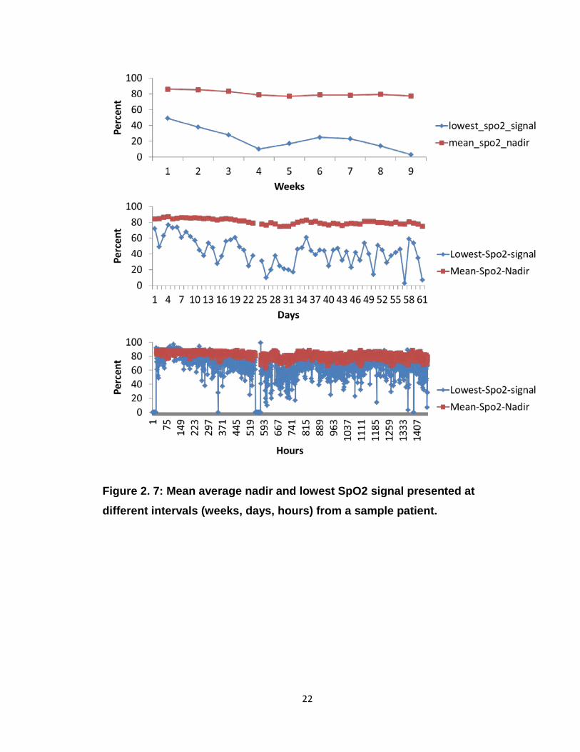
Figure 2. 7: Mean average nadir and lowest SpO2 signal presented at different intervals (weeks, days, hours) from a sample patient. ..................... 22
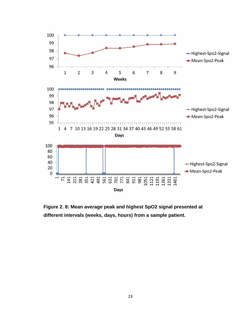
Figure 2. 8: Mean average peak and highest SpO2 signal presented at different intervals (weeks, days, hours) from a sample patient. ..................... 23
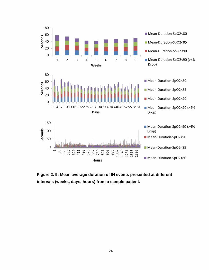
Figure 2. 9: Mean average duration of IH events presented at different intervals (weeks, days, hours) from a sample patient. ................................... 24
Figure 2. 10: Mean average duration of hyperoxemia events presented at different intervals (weeks, days, hours) from a sample patient. ....... 25
Figure 2. 11: Sample demonstration of bradycardia events averaged over 3 intervals (weeks, days and hours). .................................................. 26
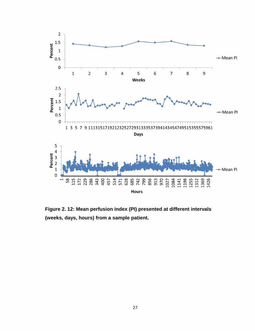
Figure 2. 12: Mean perfusion index (PI) presented at different intervals (weeks, days, hours) from a sample patient. ................................................. 27
Figure 2. 13: Inter-observer Pearson correlations among observers for the number of IH events (IH-SpO2<80). ................................................ 28
Figure 2. 14: A Pearson correlation comparing mean observer counts versus those calculated by IH Automated Analyses Algorithm (IH-AAA) for IH-SpO2<80 ..................................................................................... 29
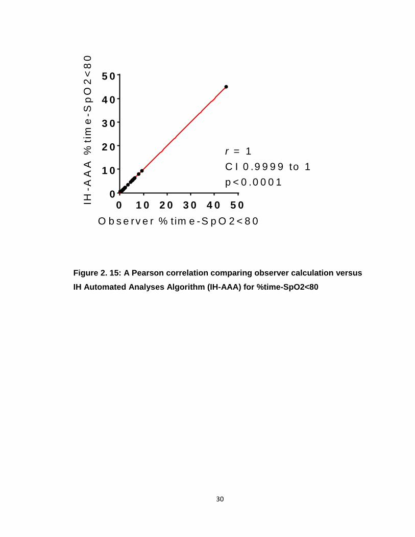
Figure 2. 15: A Pearson correlation comparing observer calculation versus IH Automated Analyses Algorithm (IH-AAA) for %time-SpO2<80 ........ 30

x
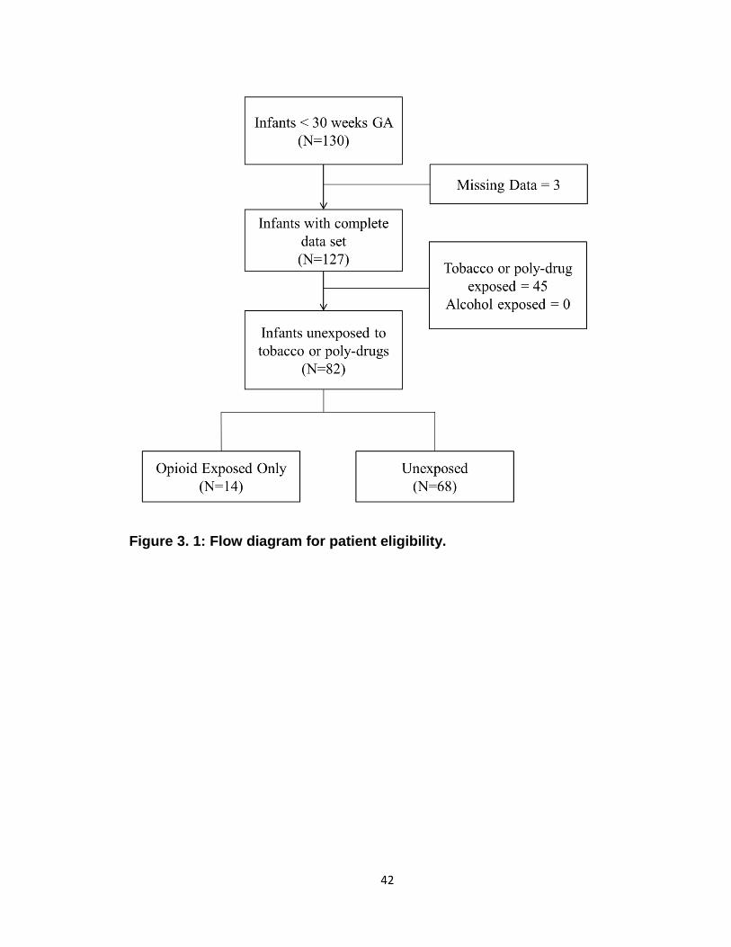
Figure 3. 1: Flow diagram for patient eligibility.................................................... 42 Figure 3. 2: Comparison of %time-SpO2<80 between opioid exposed and
unexposed. ...................................................................................... 43 Figure 3. 3: Comparison of IH-SpO2<80 between opioid exposed and
unexposed. ...................................................................................... 44
Figure 4. 1: Proposed vicious cycle related to apnea, IH and postnatal inflammation. ................................................................................... 52
Figure 4. 2: Scatter plot for CRP levels in studied patient population ................. 53 Figure 4. 3: Scatter plots for IH in studied patient population. ............................ 54 Figure 4. 4: Correlations comparing serum CRP and percent time below
thresholds. ....................................................................................... 55 Figure 4. 5: Correlations comparing serum CRP and IH frequency. ................... 56 Figure 4. 6: Correlations comparing serum CRP and IH duration....................... 57 Figure 4. 7: Correlations comparing serum CRP and primary outcome measure
%time-SpO2<80 at multiple duration intervals. ................................ 58 Figure 4. 8: Correlations comparing serum CRP and primary outcome measure
IH-SpO2<80 at multiple duration intervals. ...................................... 59 Figure 4. 9: Negative correlation between mean SpO2 and serum CRP. ........... 60 Figure 4. 10: Correlation between serum CRP and IH mean nadir/mean peak .. 61
Figure 5. 1: Increase in %time-SpO2<80 in preterm infants less than 30 weeks born with maternal chorioamnionitis (MC)........................................ 74
Figure 5. 2: Unadjusted differences in %time-SpO2<80 between pathologic maternal chorioamnionitis (MC) and/or Funisitis versus unexposed 75
Figure 5. 3: Adjusted differences in %time-SpO2<80 between pathologic MC and/or Funisitis versus unexposed .................................................. 76
Figure 5. 4: Unadjusted differences in IH-SpO2<80 between pathologic MC and/or Funisitis versus unexposed .................................................. 77
Figure 5. 5: Adjusted differences in IH-SpO2<80 between pathologic MC and/or Funisitis and unexposed (no MC of funisitis) ................................... 78
Figure 5. 6: Differences in severe bronchopulmonary dysplasia (BPD) among groups .............................................................................................. 79
Figure 5. 7: Proposed relationship between intermittent hypoxemia and inflammation and possible role of maternal chorioamnionitis. .......... 80

xi
Figure 6. 1: Potential benefit of indomethacin in reducing intermittent hypoxemia (IH) in preterm infants. ..................................................................... 91
Figure 6. 2: Potential benefit of indomethacin in reducing intermittent hypoxemia (IH) in preterm infants with maternal chorioamnionitis (MC). ........... 92
Figure 6. 3: Proposed relationship between inflammation and intermittent hypoxemia (IH) and potential benefit of indomethacin. .................... 93
1
CHAPTER 1: INTRODUCTION AND CLINICAL RELEVANCE
This chapter was published as a review article at the American Academy of
Pediatrics NeoReviews. The following is a summary of the review with
permission from the publisher. The full review is not open access and can be
found at the citation below. One section related to prenatal exposure was added
to this chapter that was not included in the original publication.
Citation: Abu Jawdeh EG. Intermittent Hypoxemia in Preterm Infants: Etiology
and Clinical Relevance. NeoReviews. 2017 November 01; 18(11):e637-e646.
I. Introduction
Intermittent hypoxemia (IH), generally defined as brief, episodic drops in
hemoglobin oxygen saturation (SpO2). Intermittent hypoxemia is a common
disorder in preterm infants with rising evidence linking IH to neonatal morbidities
and long term impairment. The definition and thresholds below which IH is
clinically relevant are debatable (1-4).
II. Natural Progression
Intermittent hypoxemia is inversely related to gestational age (GA) (5, 6).
Small for gestational age (SGA) are particularly at risk to having increased IH
compared to infants appropriate for gestational age (AGA). In addition, IH natural
progression varies by postnatal age (1, 2). There is a low frequency of IH during
the first week after birth, followed by a progressive increase by weeks 2-3, with a
peak around 4-5 weeks then plateau/decrease during weeks 6-10. The factors
that influence the rise in IH are poorly defined (7, 8).
2
III. Factors that Influence Intermittent Hypoxemia
The conventional definition of apnea of prematurity (AOP) may not be
applicable to the causality of IH in the current extremely premature NICU
population with lung immaturity and lung disease, because as IH can often occur
following very brief respiratory pauses, periodic breathing or ineffective
ventilation (9-11).
The “Perfect Storm”
The impaired respiratory control along with lung disease/immaturity
create a “perfect storm”, leading to an increased IH frequency(12). Factors that
contribute to increased respiratory pauses and resultant IH in preterm infants
include: upregulated inhibitory neurotransmitters, decreased central chemo-
sensitivity (7, 10, 13), paradoxical ventilatory depression in response to hypoxia
(10, 13), hyper-excitable carotid bodies (14), immature laryngeal chemo-reflex
(15) and low baseline functional residual capacity (FRC) (7, 10, 16).
Prenatal Exposure Prenatal environmental exposures such as opioids, tobacco, and other
drugs may have a sustained effect on apnea, lung disease and subsequently IH.
Prenatal opioid exposure alters the response to carbon dioxide and depresses
central respiratory control centers (17-21). Opioids are known to suppress
breathing and respiratory effort especially in neonates (22). Opioid exposed
infants often show intrauterine growth retardation and meconium staining, two
hallmarks of fetal hypoxia. Similar to the literature from sudden infant death,
prenatal opioid use may increase cardiorespiratory events in preterm infants.
Prenatal opioids, especially street heroin, cause chronic intrauterine hypoxia
leading to brainstem gliosis damaging the central respiratory centers; hence
likely more apnea events (19). In addition, infants with intrauterine exposure to
drugs of abuse have “down-regulation” of placental neurotransmitter receptors
(23). Abnormalities or depletion of receptor sites, especially if the same process

3
occurs in the fetal brain, could impair function of the normal neonatal respiratory
control network leading to frequent or prolonged apnea and subsequent IH.
Furthermore, prenatal exposure to other illicit drugs such as cocaine perturbs,
albeit subtly, the maturation of respiratory control, resulting in disruption of
postnatal respiration (24). Prenatal tobacco use is common; around 22% of
mothers smoke while pregnant in the USA (25). Prenatal nicotine exposure
increased apnea in neonatal mice (26). In addition, studies evaluating pulmonary
mechanics in infants of smoking mothers indicated prenatal exposure affects
pulmonary function by altering expiratory flow profiles, reducing respiratory
compliance and increasing airway resistance (25, 27). Furthermore, prenatal
tobacco alters chemoreceptor sensitivity and blunts response to hypoxia in
infants (25, 28). Given the rising epidemic of drug abuse in the USA, a larger
cohort aimed at understanding these relationships, especially opioids, is
imperative and may have a direct impact on management of preterm infants.
Role of Inflammation
Inflammation increases apnea events and worsens lung disease;
subsequently increasing IH (29-31). However, because IH is pro-inflammatory,
the relationship between inflammation and IH may be bidirectional (7, 14, 32-35).
Anemia
Preterm infants with anemia are at increased risk for IH. As the hematocrit
level decreases, the probability of apnea, bradycardia and IH events increases
(1, 16, 36).
Target Oxygen Saturation
The target oxygen saturation influences the frequency of IH (8, 37). A
lower SpO2 target is associated with greater incidence of IH events compared
with higher SpO2 target (16).

4
IV. Monitoring
Intermittent hypoxemia is very common in preterm infants with hundreds
of events per day and accurately documenting those events by bedside providers
is challenging without continuous automated recordings (1, 38, 39).
V. Consequences
There is rising evidence linking IH to neonatal morbidities and long term
impairment. These brief episodes of oxygen desaturations have been implicated
in the following. Data from animal models: neurocognitive handicap, impaired
myelination, decreased neuronal integrity, long-term neuro-functional deficits,
increased inflammation and oxidative stress, impaired growth and sleep
disordered breathing/apnea (32, 40-42). Data from human studies: Retinopathy
of prematurity (ROP), Neurodevelopmental Impairment (NDI) (cognitive, motor
and language delay) and death (3, 5, 43-46).
VI. Conclusion
Although IH is very common in preterm infants the extent of the problem is
often underestimated by clinical providers. Multiple factors in preterm infants
increase their risk for significant IH. Intermittent hypoxemia is clinically relevant
with rising evidence from both animal models and preterm infants linking IH to
poor outcomes.
5
CHAPTER 2: METHOD DEVELOPMENT AND VALIDATION
I. Introduction
Intermittent hypoxemia is a common problem in preterm infants due to
their immature respiratory control (apnea of prematurity) and lung
immaturity/disease (BPD). All preterm infants are at risk for IH. Extreme preterm
infants have highest risk for IH, due to their extremely immature respiratory
control and lung immaturity/disease. When oxygen saturation (SpO2) is
continuously recorded, extreme preterm infants have on average 150 to 200
severe IH events per day during which their SpO2 drops below 80% (1).
Intermittent hypoxemia (IH) is defined as episodic drops in blood oxygen
saturation. The specific definition of oxygen saturation (SpO2) drop varies by
research group, however most consider SpO2 drop to less than 80% as
significant (1-3, 36). Others consider a SpO2 of less than 90% as the starting
point (4). Calculating and establishing an IH profile that reflects the spectrum of
IH in terms of frequency, severity and duration is imperative.
Accurately documenting cardiorespiratory events for day-to-day patient
care management is challenging, as the extent of IH is not apparent clinically.
Pulse oximeters are the current standard of care for monitoring oxygenation in
the Neonatal Intensive Care Unit (NICU). Bedside providers under-recognize the
number of events compared to objective automated recordings. In one study,
compared to polysomnography, nursing staff recorded less than 30% and 40% of
IH and bradycardia events, respectively. The shorter the event, the less likely
that it was recognized by nursing staff (36). For example, bedside providers
documented 35% and 29% of IH events that lasted greater than 20 and 10
seconds, respectively (38). Pulse oximeters are the current standard of care for
monitoring oxygenation in the NICU. Hence, continuous physiologic recording is
required for accurate detection of IH.
6
In this section we describe the development of methods for SpO2
recording, filtering, analyses, selection of outcome measures and validation of
our novel programs.
II. Data Acquisition
Oxygen saturation data were prospectively collected from preterm infants
admitted to our level 4 NICU starting November 2014. We used Masimo Radical
7 (Masimo, Irvine, CA) pulse oximeters for continuous data acquisition. Masimo
pulse oximeters are widely used in NICUs worldwide due to their proprietary
Signal Extraction Technology (SET®) that measures through motion and low
perfusion; both important considerations in preterm infants (37, 47-52). All our
research pulse oximeters were updated to the latest software prior to study
initiation.
Pulse oximeters were equipped with serial data recorders (Acumen
Instruments Corp) for continuous data collection (4). The Acumen recorders were
connected to the RS232 port located on the Masimo pulse oximeter docking
station. Data was collected with 1Hz frequency (every second) and saved on
compact flash memory cards connected to the serial data recorders. The
compact flash memory cards saved data continuously and were manually
downloaded by research personnel to our encrypted servers provided by the
University of Kentucky. We programed the Acumen recorders to save the data in
daily files (midnight to midnight). The daily files were easier to transfer due to
smaller size. In addition, the daily files provided a visual check of data loss if any
and troubleshooting if necessary. The Acumen serial data recorder provided a
time stamp (date and time, including seconds) for every second of data
download. Time stamping is important while linking our IH data to other outcome
measures. We downloaded data from serial data recorders weekly. Initially we
7
had difficulty with memory cards not being reliable leading to data loss. However,
that problem was transient and resolved with a different brand of memory cards.
We also trialed a different serial data recorder (SeriaGhost Logger) that
was placed in series with the Acumen recorders. The SerialGhost recorders were
reliable and stored data accurately. The SerialGhost saved all data in one file
that at times was tens of gigabytes in size before post processing. The
SerialGhost utilized the timestamp from the pulse oximeters versus the Acumen
which had its own time stamp (in addition to that of the pulse oximeters). The
SerialGhost had the capacity for timestamping however in our experience it was
not reliable and was not linked to every second of data download. The
SerialGhost was downloaded once at the end of the study period as downloading
weekly was not feasible in the absence of a memory card. We tested a
SerialGhost with Wi-Fi capabilities. The goal was to download directly to our
encrypted serves. This was more challenging than expected given both 1)
hospital network restrictions and 2) network changes upon moving infants from
one room to another. Currently, we only use the Acumen serial data recorders.
A research monitoring unit is connected to the patient after informed
consent is obtained. Initially we docked our research units to the clinical stations
and utilized the same pulse oximeter for both for clinical and research purposes.
An alarm delay was set to avoid alarm fatigue. However, the serial data
recorders were sometimes left behind when moving patients among rooms. Early
during our study period we changed this practice and currently we utilize an
additional research pulse oximeter that moves with the patient. The research
pulse oximeter alarm settings are silenced to avoid further noise and alarm
fatigue. Patients are connected to the additional research pulse oximeter upon
enrollment and monitored for first 2 months of life or 36 weeks corrected age,
whichever came last.
8
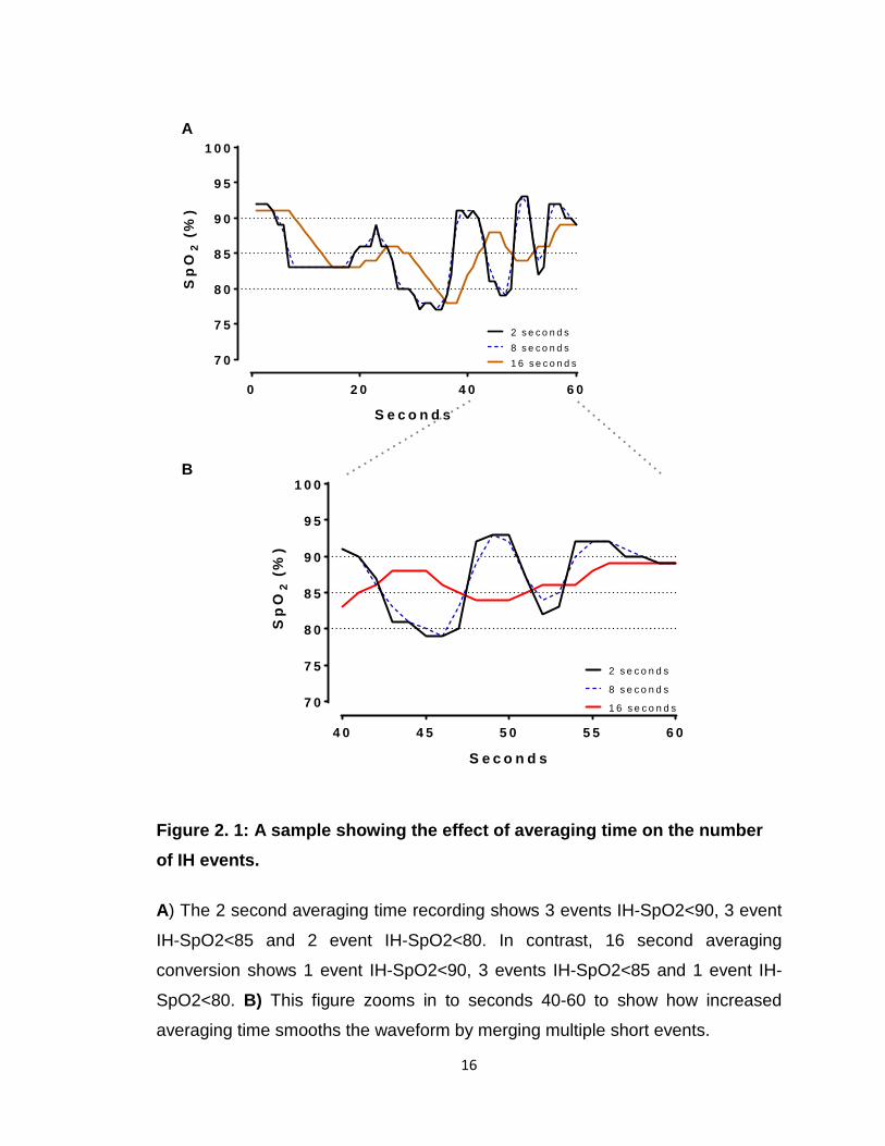
Averaging Time
Pulse oximeters are the current standard of care for monitoring
oxygenation in the NICU. However, the monitor settings, such as the averaging
time, affects the number of IH events recorded (39). Pulse oximeters average
SpO2 values over several heartbeats. Pulse oximeters set to longer averaging
times underestimate IH events of short duration and overestimate events of
longer duration. This is likely as a result of several short events merged together
as one prolonged event (Figure 2.1). Clinical pulse oximeters are set to longer
averaging time to decrease alarm fatigue for bedside providers (53). The default
averaging times in clinical pulse oximeters range between 8 to 10 seconds but
can be as long as 16 seconds. An option for centers who wish to use shorter
averaging time is setting a longer alarm delay time (10 to 15 seconds) to reduce
alarm fatigue (53). For research purposes, similar to other groups who study IH,
we utilized high-resolution pulse oximeters with 2-second averaging time for
continuous SpO2 monitoring (1, 2). We confirmed and tracked the pulse oximeter
settings weekly during data download.
III. Data Filtering and Processing
In collaboration with biomedical engineering (Dr. Abhijit Patwardhan
laboratory) we developed novel programs to filter and process SpO2 data to
analyze IH. Both algorithms were developed using Matlab (Matlab, Natick, MA).
The IH data filtering program excluded artifacts based on both the EXC
code provided by Masimo monitors and missing variables in the output. The
filtering program imported the raw data in text (.txt) format and exported clean
data in text (.txt) format as well. The exported data files were automatically
organized daily by the algorithm. The daily file names included the patient
identification number and the date of the recorded data. The filtering algorithm
has the capacity to filter multiple patients at the same time.

9
The second program is called Intermittent Hypoxemia Automated
Analyses Algorithm (IH-AAA). The IH-AAA process the filtered data files to
analyze the IH profile (below). The algorithm imported the clean daily text files (1
Hz frequency) and exported analyzed IH outcome measures in excel files
averaged over different durations and intervals (weekly, daily, hourly). This
program has the capacity to analyze multiple patients at the same time. The
algorithm exports multiple excel files for every patient to reflect the spectrum of
IH of different durations (e.g. 4-180 seconds, >180 seconds, etc.) and intervals
(weekly, daily, hourly). Each excel file is labeled with patients identification
number, date of the recorded data and interval. The IH-AAA also has the
capacity to filter raw data in text files.
IV. Intermittent Hypoxemia Profile
The clinical relevance of IH is a relatively new observation (2) with no
accurately defined threshold below which IH leads to morbidities and impairment;
the exact definition of IH is controversial (54). Therefore, we developed a
program that accounts for IH at multiple thresholds and calculate an IH profile.
The IH profile reflects the continuum of the IH problem making it possible to
demonstrate at what level IH causes injury.
In this section we describe the IH profile. For the purpose of
demonstration we used a sample patient. We selected the second patient
enrolled in our cohort (IH0002). The first patient had an early death and does not
have a complete data set.
Frequency
The number of IH events is calculated for every interval (weekly, daily,
hourly). The frequency of IH is a primary outcome measure that has been utilized
by us and other groups and linked to neonatal morbidities and mortality (1, 2, 4,
55, 56). We define severe IH events as a SpO2 drop to less than 80% (IH-
10
SpO2<80). Moderate and mild IH are defined as a drop in SpO2 to less than 85%
(IH-SpO2<85) and 90% (IH-SpO2<90), respectively. An additional outcome
measure is calculated based on Rhein et al. where mild IH is calculated based on
a change from baseline by more than 4% and to SpO2<90 (IH-SpO2<90 (>4%
Drop))(4). We have the capacity to change our thresholds for IH frequency. Our
program outputs frequency of IH at different intervals (weeks, days, hours) as
represented in Figure 2.2. An upper threshold is often set for IH to differentiate
intermittent from sustained hypoxemia. We also document sustained hypoxemia
measures.
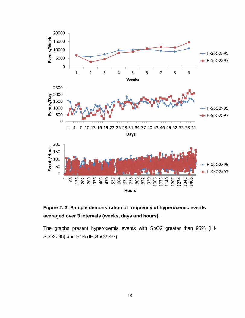
Similar to IH, hyperoxemic events are calculated. Documenting
hyperoxemia is important given both the associated morbidities and to assess
fluctuations in oxygenation. We currently have the hyperoxemia severity set at 2
thresholds with SpO2 more than 95% (IH-SpO2>95) and 97% (IH-SpO2>97).
Sample patient for hyperoxemic events frequency is presented in Figure 2.3.
Percent time
The percent time in hypoxemia is another primary measure. The benefit of
this outcome measure is that it represents cumulative IH events of short and long
duration. The same 3 SpO2 thresholds for percent time in hypoxemia are
selected here for severe (%time-SpO2<80), moderate (%time-SpO2<85) and
mild hypoxemia (%time-SpO2<90 and %time-SpO2<90 (>4% drop)) (Figure 2.4). This measure of percent time spent with SpO2 below threshold was chosen
per Poets et al. (3). Percent time is calculated at multiple intervals (weeks, days,
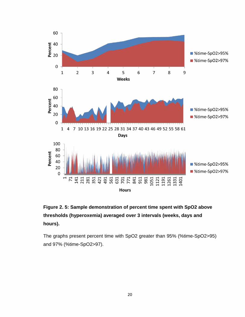
hours). Similarly, hyperoxemia is analyzed demonstrating percent time spent with
SpO2 more than 95% and 97%, (Figure 2.5). Percent time outcome measure is
not affected by averaging time and is clinically relevant in all NICUs (39).
Mean, Nadir and Peak
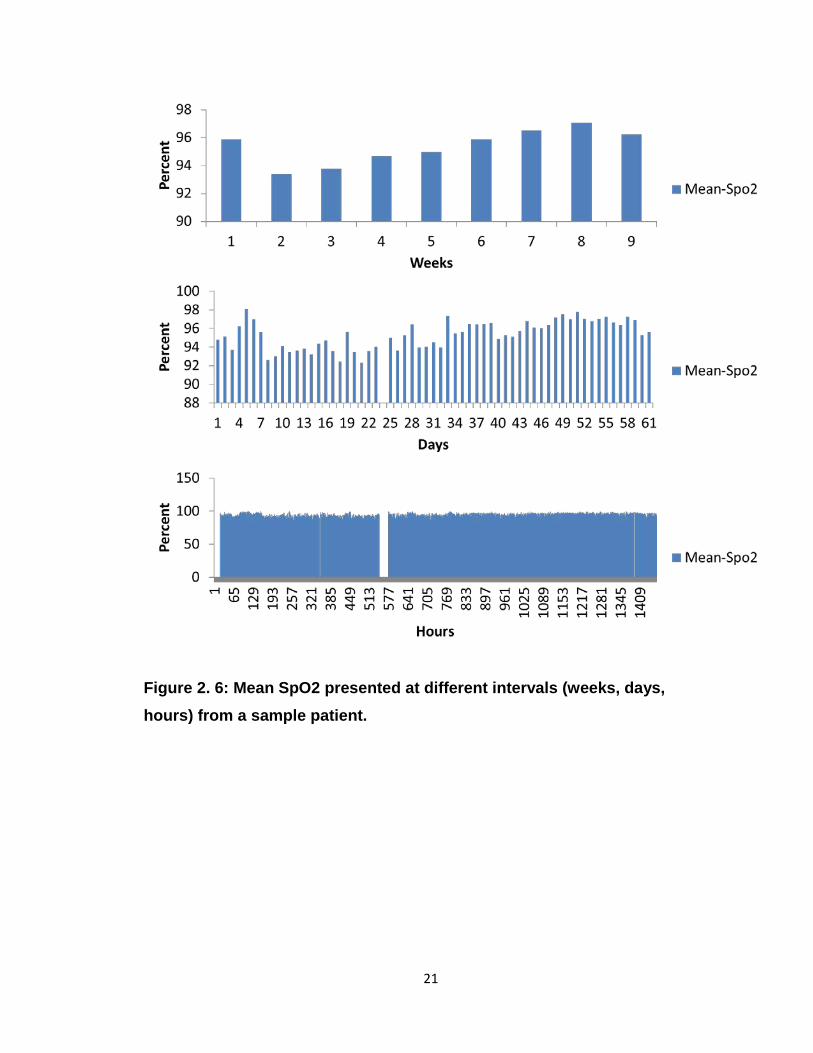
The mean, nadir and peak SpO2 measures provide an additional
perspective for IH. Mean is calculated for every interval (weeks, days, hours) and
provides a baseline for IH during that interval (Figure 2.6). Both an average nadir
11
for all events and lowest nadir are calculated. The nadir provides insight
regarding the severity of IH (Figure 2.7). Similarly the average peak and highest
signal are calculated for every interval (weeks, days, hours), (Figure 2.8).
Duration
The duration of IH is addressed in multiple forms. First, the average
duration of IH events below every threshold is calculated for the three intervals
(weeks, days, hours) (Figure 2.9). Similarly the average duration is calculated for
hyperoxemia (Figure 2.10). However, the duration of IH may vary widely and the
average duration may not be representative as it is influenced by outliers. Hence,
we developed our algorithm to output multiple files of different duration cutoffs
(for example multiples of 60 seconds). E.g. 1-59seconds, 60-119 seconds, 120-
179 seconds, 180-239 seconds, 240-299, >300 seconds. By dividing the duration
cutoffs, the average duration is influenced less by outliers. The duration cutoffs
can be easily adjusted to any duration (for example multiples of 30 seconds, etc.)
In addition, we use the 4-180 second cutoff for the primary measure as
previously described by Abu Jawdeh et al. and Rhein et al. (1, 4).
Bradycardia
Heart rate decelerations or bradycardia events are part of the apnea of
prematurity problem. Our algorithm calculates bradycardia below 2 thresholds of
80 and 100 beats per minute (bpm) (Figure 2.11) (11). The relative time of
bradycardia to IH is also calculated. However, the role of heart rate deceleration
is beyond the scope of this document.
Perfusion Index
Perfusion index (PI) is a noninvasive measure of perfusion thought to
reflect the general hemodynamic status of the preterm infant (57, 58). Perfusion
index assesses the pulse strength derived from pulse oximetry. Perfusion index
is measured by infrared light, and is calculated as the ratio of the pulsatile to non-
pulsatile components of the blood flow in tissue. The value of PI has been
12
demonstrated in multiple neonatal morbidities (59, 60), including prediction of
patent ductus arteriosus patency shown from this cohort (61). Figure 2.12
demonstrates PI in a sample patient.
V. Statistical Analyses
Statistical modeling must account for the covariance among repeated
measurements from the same subject. A general strategy to do so is to use linear
mixed models, also known as linear multilevel models, hierarchical models, or
multivariate Gaussian models (62). Correctly accounting for this correlation will
ensure standard errors are appropriately estimated, thus yielding correct p-
values and thus valid inference. Furthermore, in our experience and elsewhere,
the need for an appropriate transformation, such as the square root, is needed
for IH-based measures (2).
In order to attain valid inference, the model for the mean structure of the
given outcome over time, as well as the model for the covariance among
outcomes from the same subject (not of interest to the research question, but a
necessity for inference), must be correctly specified (62). Otherwise, inference
may be biased. A simple solution to this issue is to look at outcomes aggregated
over weekly periods; e.g., weekly IH totals. In such a case, a statistical model
can treat time as categorical to ensure a correctly specified mean structure with
respect to time. Furthermore, a working unstructured covariance can be used
with such a mean structure to ensure appropriate standard error estimation.
Finally, this flexible modeling of the mean and covariance structures allows
inference to be valid if any missing data are missing completely at random
(MCAR) or missing at random (MAR) (63).
Outcome data is sometimes not captured for periods of time for example
due to patient leaving the NICU for procedures or imaging. Therefore, as
outcomes are usually aggregated over time, e.g. the total IH count for a given

13
weekly period, such outcomes should be weighted by the amount of time they
were observed. For instance, if interest is in weekly IH count, but IH data were
only obtained for exactly half of the week, then that subject’s total IH count would
need to be doubled for use in the statistical analysis such that it represents the
desired weekly total. A weight of one half would then need to be assigned to this
outcome value in the analysis. If this weighting procedure is not done, estimated
means and standard errors may be biased.
VI. Validation
In order to validate the novel program, we performed an assessment
comparing IH measures calculated by the algorithm to those of independent
observers. The observers were masked (blind) to the algorithm analyses. We
obtained SpO2 data from 20 preterm infants less than 30 weeks GA randomly
selected from our cohort. A total of 60 hours were analyzed. Each subject
contributed 3 hours of SpO2 data; 1 hour from each postnatal age epoch (1
week, 1 months and end of study period) as defined by Abu Jawdeh et al. (1).
We included IH events 4-180 second duration per Abu Jawdeh et al. (1). The
validation presented focuses on two primary measures, IH-SpO2<80 and %time-
SpO2<80. Other thresholds of less than 85% and 90% were examined with
similar results.
The observers were masked to both other observers and algorithm
counts. Three observers manually counted the first measure of IH-SpO2<80 from
the raw data. The second measure of %time-SpO2<80 was analyzed by a
singled masked observer utilizing Microsoft Excel (Excel Version 2010). Pearson
correlations among observers and algorithm were performed using GraphPad
(Prism 7). There was excellent correlation among observers as presented in
Figure 2.13. For IH-SpO2<80, there was excellent correlation between mean
observer count and algorithm count as presented in Figure 2.14. For %time-
14
SpO2<80, there was excellent correlation between observer and algorithm as
presented in Figure 2.15.
VII. Discussion
There is rising evidence linking IH to both short and long term morbidities
in preterm infants and hence, accurate recording of these events is paramount in
determining their impact. In this chapter we described methods development to
collect and process SpO2 data to measure IH. We defined our IH outcome
measures with emphasis on IH profile. Finally, we presented the process for
validating IH-AAA for accurate and reliable measurement of IH. We have
developed an automated, convenient, and time efficient strategy to record such
events, with exceptional accuracy when compared to human measurements.
The clinical significance of IH in preterm infants is a relatively new
observation (2, 3, 54). In the past, brief IH events occur hundreds of times per
day seemed clinically insignificant. However in the last 5-10 years, the interest in
accurately documenting these IH events increased given the recent evidence
linking IH to neonatal morbidities. Accurately documenting IH should involve a
continuous physiologic recording with an automated system as bedside providers
under-recognize the number of events (36, 38). An important factor in continuous
monitoring is averaging time of pulse oximeters. As longer averaging times will
underestimate IH events of short duration and overestimate events of longer
duration (39, 64). This is likely as a result of several short events merged
together as one prolonged event (39, 64).
An additional challenge relates to IH is variation in definitions. This
variation is likely related to the unknown thresholds below which IH leads to
injury. Most centers however consider a SpO2 drop to less than 80% as clinically
relevant. We developed an IH profile that represents the continuum of IH. The IH
15
profile allows us to better define at what threshold (e.g. severity, duration, etc.) IH
matters clinically.
In conclusion, over the last 5 years we developed efficient and validated
methods to accurately assess IH. We are one of few centers in the nation able to
perform high fidelity studies related to IH in preterm infants.
VIII. Acknowledgements
Thank you to all team members listed in the acknowledgement section.
The methods and algorithms development would not have been possible without
Dr. Abhijit Patwardhan and his laboratory/team including: Yihua Zhao PhD, David
Wasemiller M.S. and Sahar Alaei M.S. Special thanks and recognition to Sean
Carpenter BSBE for involvement with logistics, algorithm development and
validation (observer 1) portion/figures (1, 13-15) in this chapter. I thank Haleigh
Whitlock (observer 2) and Himanshu Savardekar B.S (observer 3) for
involvement in validation portion of this chapter.
Thank you and special acknowledgments to Philip Westgate Ph.D. without
whom our analyses and statistical method development would not have been
possible. I thank Katrina Ibonia MD, Enrique Gomez-Pomar MD and Brandon
Schanbacher M.S. for early method development related to data acquisition.
Thank you to Aayush Gabrani MBBS, Divya Mamilla MBBS and Amrita Pant
MBBS and Crystal Wilson LPN for contributions to method development and data
analyses.
This method development was supported by funds from the Children’s
Miracle Network, the Gerber Foundation, and the National Center for Research
Resources, UL1RR033173, and is now at the National Center for Advancing
Translational Sciences.
16
Figure 2. 1: A sample showing the effect of averaging time on the number of IH events.
A) The 2 second averaging time recording shows 3 events IH-SpO2<90, 3 event
IH-SpO2<85 and 2 event IH-SpO2<80. In contrast, 16 second averaging
conversion shows 1 event IH-SpO2<90, 3 events IH-SpO2<85 and 1 event IH-
SpO2<80. B) This figure zooms in to seconds 40-60 to show how increased
averaging time smooths the waveform by merging multiple short events.
0 2 0 4 0 6 0
7 0
7 5
8 0
8 5
9 0
9 5
1 0 0
S e c o n d s
Sp
O2
(%
)
2 s e c o n d s
1 6 s e c o n d s8 s e c o n d s
4 0 4 5 5 0 5 5 6 0
7 0
7 5
8 0
8 5
9 0
9 5
1 0 0
S e c o n d s
Sp
O2
(%
)
2 s e c o n d s
8 s e c o n d s
1 6 s e c o n d s
B
A

17
Figure 2. 2: Sample demonstration of frequency of IH events averaged over 3 intervals (weeks, days and hours).
The graphs present IH below multiple thresholds: IH-SpO2<90 (>4% Drop), IH-
SpO2<90, IH-SpO2<85 and IH-SpO2<80.
18
Figure 2. 3: Sample demonstration of frequency of hyperoxemic events averaged over 3 intervals (weeks, days and hours).
The graphs present hyperoxemia events with SpO2 greater than 95% (IH-
SpO2>95) and 97% (IH-SpO2>97).

19
Figure 2. 4: Sample demonstration of percent time spent with SpO2 below thresholds averaged over 3 intervals (weeks, days and hours).
The graphs present percent time below multiple thresholds: %time-SpO2<90
(>4% Drop), %time-SpO2<90, %time-SpO2<85 and%time-SpO2<80.
20
Figure 2. 5: Sample demonstration of percent time spent with SpO2 above thresholds (hyperoxemia) averaged over 3 intervals (weeks, days and hours).
The graphs present percent time with SpO2 greater than 95% (%time-SpO2>95)
and 97% (%time-SpO2>97).
21
Figure 2. 6: Mean SpO2 presented at different intervals (weeks, days, hours) from a sample patient.
22
Figure 2. 7: Mean average nadir and lowest SpO2 signal presented at different intervals (weeks, days, hours) from a sample patient.
23
Figure 2. 8: Mean average peak and highest SpO2 signal presented at different intervals (weeks, days, hours) from a sample patient.
24
Figure 2. 9: Mean average duration of IH events presented at different intervals (weeks, days, hours) from a sample patient.

25
Figure 2. 10: Mean average duration of hyperoxemia events presented at different intervals (weeks, days, hours) from a sample patient.

26
Figure 2. 11: Sample demonstration of bradycardia events averaged over 3 intervals (weeks, days and hours).
The graphs present heart rate deceleration below two thresholds of 100 beats
per minute (bpm) and 80bpm.
27
Figure 2. 12: Mean perfusion index (PI) presented at different intervals (weeks, days, hours) from a sample patient.

28
Figure 2. 13: Inter-observer Pearson correlations among observers for the number of IH events (IH-SpO2<80).
(A) Observer 2 vs. Observer 1. (B) Observer 3 vs. Observer 1 (C) Observer 3 vs.
Observer 2.
r = 0 .9 9 6 6C I 0 .9 9 4 3 to 0 .9 9 8p < 0 .0 0 0 1
r = 0 .9 9 7 7C I 0 .9 9 6 1 to 0 .9 9 8 7p < 0 .0 0 0 1
r = 0 .9 9 4 9C I 0 .9 9 1 4 to 0 .9 9 7p < 0 .0 0 0 1
0 1 0 2 0 3 0 4 00
1 0
2 0
3 0
4 0
O b s e rv e r 1 E v e n ts /In te rv a l
Ob
se
rve
r 2
Ev
en
ts/I
nte
rva
l
0 1 0 2 0 3 0 4 00
1 0
2 0
3 0
4 0
O b s e rv e r 1 E v e n ts /In te rv a l
Ob
se
rve
r 3
Ev
en
ts/I
nte
rva
l
0 1 0 2 0 3 0 4 00
1 0
2 0
3 0
4 0
O b s e rv e r 2 E v e n ts /In te rv a l
Ob
se
rve
r 3
Ev
en
ts/I
nte
rva
l
A
B
C

29
Figure 2. 14: A Pearson correlation comparing mean observer counts versus those calculated by IH Automated Analyses Algorithm (IH-AAA) for IH-SpO2<80
0 1 0 2 0 3 0 4 00
1 0
2 0
3 0
4 0
O b s e rv e rs E v e n ts /In te rv a l
IH-A
AA
Ev
en
ts/I
nte
rva
l
r = 0 .9 9 8 2C I 0 .9 9 7 to 0 .9 9 8 9p < 0 .0 0 0 1
30
Figure 2. 15: A Pearson correlation comparing observer calculation versus IH Automated Analyses Algorithm (IH-AAA) for %time-SpO2<80
0 1 0 2 0 3 0 4 0 5 00
1 0
2 0
3 0
4 0
5 0
O b s e rv e r % tim e -S p O 2 < 8 0
IH-A
AA
%ti
me
-Sp
O2
<8
0
r = 1C I 0 .9 9 9 9 to 1p < 0 .0 0 0 1
31
CHAPTER 3: PRENATAL OPIOID EXPOSURE AND INTERMITTENT HYPOXEMIA
Citation: This chapter was published as an original manuscript in Frontiers in
Pediatrics: This chapter was not altered from the original publication.
Abu Jawdeh EG, Westgate PM, Pant A, Stacy AL, Mamilla D, Gabrani A,
Patwardhan A, Bada HS, Giannone P. Prenatal Opioid Exposure and Intermittent
Hypoxemia in Preterm Infants: A Retrospective Assessment. Front Pediatr. 2017
Dec 6;5:253. doi: 10.3389/fped.2017.00253.
I. Introduction
Intermittent hypoxemia (IH) is defined as brief, episodic drops in oxygen
saturation (SpO2) (1, 2). Preterm infants are at increased risk for IH due to their
respiratory control instability/apnea of prematurity superimposed on immature
lung structure/function. Intermittent hypoxemia in preterm infants can persist
beyond discharge from the neonatal intensive care unit (NICU) (4). Brief
episodes of oxygen desaturations may seem clinically insignificant, but these IH
episodes, occurring up to hundreds of times per day, have a cumulative effect on
neonatal morbidity and mortality. There is ample evidence showing a significant
effect of IH on neurocognitive handicap, decreased neuronal integrity, increased
inflammation and oxidative stress, and impaired growth (32, 41). Furthermore, IH
has been linked to severe retinopathy of prematurity and long term
neurodevelopmental impairment such as worse language and motor outcomes
(2, 3, 43, 44) (45). The clinical relevance of IH is a relatively new observation with
the advent of high-resolution pulse oximeters and assessing factors that
influence IH is imperative.
32
There is a rise in substance misuse in the USA reaching a nationwide
epidemic (65-70). There is an urgent need to understand the impact of prenatal
opioid exposure on neonatal outcomes (41). Opioid exposure is associated with
long-term neurobehavioral and developmental impairment in infants (71-78).
Opioids are known to suppress breathing and respiratory effort especially in
neonates (22). Since most mothers who misuse opioids have also been found to
smoke and use poly-drugs that affect breathing pattern, it has been challenging
to assess the isolated effect of prenatal opioid exposure on respiratory outcomes.
Prenatal tobacco exposure alters respiratory control and worsens lung function
(25-28, 79). Prenatal exposure to other illicit drugs such as cocaine perturbs
maturation of respiratory control, resulting in disruption of postnatal respiration
(24). Only few studies were able to assess the effect of isolated opioid exposure
on neonatal respiratory outcomes. However, these studies included mostly later
preterm and term infants or were limited to short monitoring times and small
sample sizes (17, 80). In this study, we utilize continuous high resolution pulse
oximeters to assess the relationship between isolated prenatal opioid exposure
and IH in preterm infants during the first 2 months of life.
II. Methods
Study Design and Data Collection
Oxygen saturation data were prospectively collected from 130 preterm
infants less than 30 weeks gestational age (GA) admitted to our level 4 NICU
between November 2014 and April 2017. We used high resolution pulse
oximeters (Radical 7: Masimo, Irvine, CA) set at 2 second averaging time and
1Hz sampling rate to continuously monitor patients during the first 8 weeks of life.
In order to differentiate intermittent from sustained hypoxemia, we included
events between 4-180 seconds (1). The exact threshold below which IH is
clinically significant is controversial. A drop in SpO2 to less than 80% is widely
33
considered to be clinically relevant (1-3). Therefore, the primary outcome
measure was defined as percent time spent with SpO2 below 80% (%time-
SpO2<80). The secondary outcome measure was defined as the number of
severe IH events with SpO2 less than 80% (IH-SpO2<80). Other outcome
measures such as length of stay and neonatal morbidities were collected.
Pulse oximeters were equipped with serial data recorders (Acumen
Instruments Corp) for continuous data collection. Novel programs were utilized to
filter and analyze data (Matlab, Natick, MA) (1, 61). Data with artifacts were
excluded. Only SpO2 data with good signal were included in the analyses.
Preterm infants less than 30 weeks GA were included. Infants with major
congenital malformations were excluded.
Data related to substance misuse and tobacco use were retrospectively
collected from medical charts. If a mother chronically used prenatal opioids and/or
the maternal/neonatal drug screens were positive for opioids, then the infant was
considered for screening. Infants were then excluded from the study if the mother
used tobacco, alcohol, or other drugs (such as cannabis); i.e., in order to assess for
isolated opioid exposure, patients with any other exposure were excluded. Infants
in our cohort who were not exposed to opioids, tobacco, or other drugs served as
controls. Neonatal meconium or urine drug screens are performed in the immediate
newborn period. Positive drug screens due to opioids and other medications used
for pain or sedation during delivery were excluded, as they do not represent
prenatal misuse. Tobacco and alcohol use were collected from mothers’ medical
records, as the toxicology screens at our hospital do not test for alcohol or tobacco
exposure. The study was approved by the University of Kentucky Institutional
Review Board, and informed consent was obtained prior to SpO2 data acquisition.
Statistical Analysis
Descriptive statistics for continuous variables are presented as either the
mean with standard deviation or median with interquartile range, and frequencies
and percentages are given for categorical variables. Two-sample t-tests and
34
Wilcoxon two-sample tests were used to compare opioid exposure to non-
exposure with respect to continuous variables, and chi-square or Fisher’s exact
tests were used for categorical variables. To compare opioid exposure to non-
exposure with respect to IH measures over time, we utilized multivariate
Gaussian linear modeling in order to account for repeated measurements from
subjects, and to adjust for the potential confounders of gestational age, birth
weight, APGAR score at 5 minutes of life, gender, and the use of prenatal
steroids. In order to meet statistical assumptions in these models, the square
root of the IH measures was taken. Furthermore, weekly observations were
weighted by the percentage of time IH was tracked during the given week.
Analyses were conducted in SAS version 9.4 (SAS Institute, Cary, N.C.), and all
tests were two-sided with a 5% significance level.
III. Results
Of the 127 infants in our database with complete data sets, 19.7%, 29.1%,
and 4.7% were prenatally exposed to opioids, tobacco and cannabis,
respectively. None were exposed to alcohol, cocaine and other illicit drugs.
Opioid exposed infants were positive for buprenorphine metabolites (64%),
oxycodone (16%) and other opioids such as heroin and fentanyl (20%). A total of
82 infants qualified for analysis as they were either unexposed to any illicit
drug/tobacco (n=68) or exposed to opioids only (n=14). Figure 3.1 presents the
flow diagram for patient eligibility and exclusion.
There were no significant differences in baseline characteristics as
presented in Table 1. The mean GA was 27 weeks in both groups. There were
no significant differences in birth weight, gender and Apgar scores at 5 minutes
of life. The vast majority of infants received prenatal steroids with no difference
between groups. There were no significant differences in respiratory outcomes
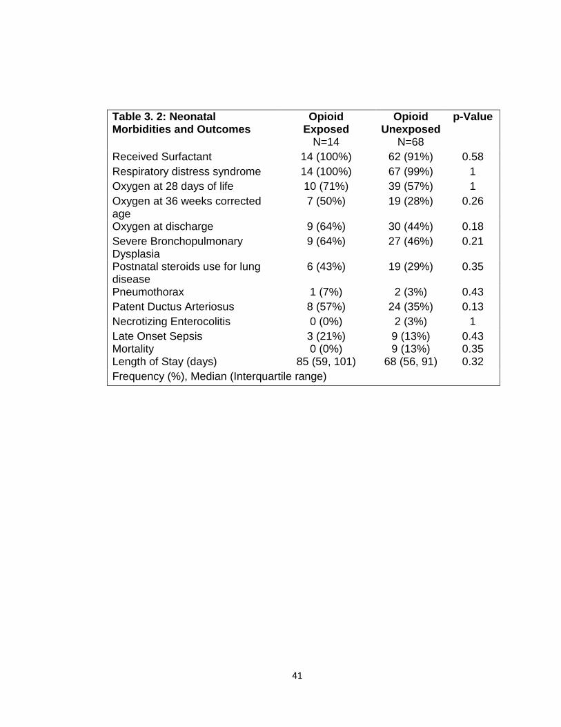
and neonatal morbidities between groups as presented in Table 2. Our cohort
included preterm infants less than 30 weeks GA. Essentially all infants had
35
respiratory distress syndrome and received surfactant. Severe
bronchopulmonary dysplasia, postnatal steroids use for lung disease, and
oxygen need at 28 days, 36 weeks postmenstrual age and at discharge did not
differ between opioid exposed and unexposed groups (all p=NS). Other neonatal
morbidities such as patent ductus arteriosus, late onset sepsis, and necrotizing
enterocolitis did not differ between groups (all p=NS). None of the exposed
infants died versus 9 deaths in the unexposed group (p= 0.35). The median
length of stay was 17 days longer in the opioid group (85 days) compared to
unexposed group (68 days); however, the results were not statistically significant
(p=0.32).
There was a statistically significant increase in our primary outcome
measure, %time-SpO2<80, as represented in Figure 3.2. The estimated
difference in the means of the square root of %time-SpO2<80 was 0.23 [95% CI:
(0.03, 0.43), p=0.03]. The mean number of IH events was estimated to be 2.95
[95% CI: (-0.35, 6.25), p-value = 0.08] higher in the opioid exposed group, as
represented in Figure 3.3; however, this did not reach statistical significance.
Note that these results represent the square root of means in order to meet
statistical assumptions in these models; estimated medians for IH measures are
calculated using our model results and are presented in Figures 3.2B and 3.3B.
Given increased death in the unexposed group, we then analyzed data excluding
deaths, and results were similar. Specifically, there was a statistically significant
increase in our primary outcome measure (%time-SpO2<80) in the opioid
exposed compared to the unexposed group, with an estimated mean difference
(square root) of 0.24 [95% CI: (0.05, 0.44), p-value = 0.02]. Furthermore, the
mean number of IH events was estimated to be 2.98 [95% CI: (-0.20, 6.16), p-
value = 0.07] higher in the opioid exposed group, not quite reaching statistical
significance.

36
IV. Discussion
These results suggest that prenatal opioid exposure is associated with
increased IH measures compared to unexposed preterm infants. This study has
two main findings. First, interestingly, the increased IH measures in opioid
exposed infants persisted beyond the early postnatal period. Preterm infants
were continuously monitored with high resolution pulse oximeters during the first
2 months of life. Second, we had the unique opportunity to assess the
relationship between isolated opioid exposure and respiratory instability in
preterm infants. It was challenging in the past to assess the relationship between
isolated prenatal opioid exposure and respiratory outcomes/IH, as the majority of
women who use opioids also smoke or misuse poly-drugs. Given our cohort
demographics, we had the ability to report this association in infants exposed to
opioids only.
Another interesting secondary finding in our study is the steady increase in
IH in the first month of life before plateauing and then decreasing. This natural
progression of IH has been described before from another cohort of preterm
infants less than 28 weeks GA (1, 2). Our study replicates this finding from a new
cohort of preterm infants less than 30 weeks GA. The rise in IH may be related to
peripheral chemoreceptor dysregulation and development of lung disease (7).
Patients in our opioid exposed and unexposed groups did not significantly
vary in terms of baseline characteristics (such as age, weight, gender) and
neonatal morbidities (such as lung disease, patent ductus arteriosus, late onset
sepsis and necrotizing enterocolitis). In addition, we adjusted in the model for
factors that may influence oxygenation in preterm infants such as GA and
prenatal steroids. The finding of 9 deaths in the unexposed group compared to
no deaths in the opioid exposed group may be due to chance. Secondary
analyses excluding deaths showed similar results with increased IH in the opioid
exposed group. A significant secondary finding in this study is the high
prevalence of tobacco and drug exposure in our cohort of preterm infants. The

37
frequency of opioid exposure in our preterm population is higher than previously
reported, thus creating urgency toward addressing this significant problem in this
vulnerable patient population (65, 67-70).
There are multiple proposed mechanisms by which prenatal opioid
exposure may affect breathing patterns and subsequent persistent IH in preterm
infants. Prenatal opioid exposure alters the response to carbon dioxide and
depresses central respiratory control centers (17-21); a main driver for
respiratory output. Olsen et al demonstrated a blunted response to carbon
dioxide in methadone exposed infants compared to controls (17). Ali et al
compared the response to hypercarbia among three groups of term patients who
were exposed to tobacco/substance misuse, tobacco alone, and unexposed
controls. The authors showed a lower increase in central respiratory drive in
response to hypercarbia in infants exposed to substance misuse as compared to
tobacco alone and unexposed controls (18). Another mechanism that explains
our results may be related to in utero hypoxia related to opioids. Prenatal opioids,
especially street heroin, cause chronic intrauterine hypoxia leading to brainstem
gliosis, resulting in injury to the central respiratory network. This may lead to
respiratory instability and subsequent IH (19). Finally, data from animal models
showed that exposure to opioid agonists caused down-regulation of placental
neurotransmitter receptors (23). Abnormalities or depletion of receptor sites,
especially if the same process occurs in the fetal brain, could impair the function
of the normal neonatal respiratory control network leading to frequent or
prolonged apnea and subsequent IH.
Many studies have assessed the impact of prenatal opioid exposure on
sudden infant death syndrome (SIDS) in infants with controversial results. This
study does not address SIDS; rather, it focuses on IH, the end result of apnea of
prematurity. However, the mechanism by which prenatal opioid exposure is
associated with increased SIDS and IH may be similar. Although our study period
focused on the inpatient setting, it is plausible that opioid exposed infants
continue to have increased cardiorespiratory events/IH after discharge.
38
Interestingly, compared to unexposed infants, opioid exposed infants had a trend
toward longer length of stay (68 versus 85 days, p=NS), which may be related, in
part, to persistent cardiorespiratory events.
A major limitation of this study is that data related to exposure were
retrospectively collected. Another limitation is a lack of reporting daily caffeine
use and daily respiratory support settings. At our center, virtually all infants with
GA less than 30 weeks are started on caffeine therapy. Furthermore, our study
focused on IH events and lacked reporting of apnea and bradycardia events.
Lack of addressing heart rate is a limitation since bradycardia events may be
associated with poor long term outcomes (3). Another limitation is the small
sample size; however, our sample size of isolated opioid exposure is relatively
large compared to existing literature. This is a single center study; hence, our
results may not be generalizable. Finally, we did not compare the long term
neurodevelopmental outcomes for exposed versus unexposed infants.
V. Conclusion
There is rising evidence linking IH to neonatal morbidities and impairment.
However, the exact threshold (frequency, duration, severity) by which IH leads to
injury in preterm infants needs further investigation; i.e., any increase in IH may
be associated with impairment in preterm infants. Furthermore, there is a need to
understand factors, such as prenatal opioid exposure, that may influence IH and
subsequently increase neonatal morbidities. In this study, we show an
association between prenatal opioid exposure and increased IH measures in
preterm infants. Studies to address the relationship between opioid exposures,
IH, and long term neurodevelopmental outcomes are imperative. Given the rising
epidemic of opioid misuse in the USA, understanding the relationship between
opioid exposure, IH and long term impairment is imperative. A larger prospective
study aimed at understanding these relationships may have a direct impact on
short and long term management of preterm infants.

39
VI. Acknowledgements
I thank the co-authors for their significant contributions to this manuscript.
Philip Westgate PhD, Amrita Pant MBBS, Audra Stacy (M4), Divya Mamilla
MBBS, Aayush Gabrani MBBS, Abhijit Patwardhan PhD, Henrietta Bada MD
MPH and Peter Giannone MD.
This project was supported by funds from the Gerber Foundation,
Children’s Miracle Network and the National Center for Research Resources,
UL1RR033173, and is now at the National Center for Advancing Translational
Sciences. The content is solely the responsibility of the authors and does not
necessarily represent the official views of the NIH
I thank all the team members mentioned in the acknowledgments section.
The authors thank the support of Katrina Ibonia MD, Enrique Gomez-Pomar MD,
Vicki Whitehead RN CCRC, Kimberly Walker BSN, Alisa (Beth) McKinney-
Whitlock CCRP, Sean Carpenter BSBE, John Bauer PhD, NICU nurses,
research nurses and staff, and neonatology faculty and fellows at the Division of
Neonatology, University of Kentucky.

40
Table 3. 1: Baseline Characteristics
Opioid Exposed Unexposed p-Value N=14 N=68
Gestational age (weeks) 27.0 ± 2.1 27.0 ± 1.6 0.97 Birth weight (grams) 948 ± 263 928 ± 247 0.79 Male 6 (43%) 23 (34%) 0.54 Apgar 5 min 7 (6, 7.5) 6 (5, 7) 0.21 Prenatal steroids 12 (86%) 61 (91%) 0.62 Mean ± SD, Median (Interquartile range)
41
Table 3. 2: Neonatal Morbidities and Outcomes
Opioid Exposed
Opioid Unexposed
p-Value
N=14 N=68 Received Surfactant 14 (100%) 62 (91%) 0.58 Respiratory distress syndrome 14 (100%) 67 (99%) 1 Oxygen at 28 days of life 10 (71%) 39 (57%) 1 Oxygen at 36 weeks corrected age
7 (50%) 19 (28%) 0.26
Oxygen at discharge 9 (64%) 30 (44%) 0.18 Severe Bronchopulmonary Dysplasia
9 (64%) 27 (46%) 0.21
Postnatal steroids use for lung disease
6 (43%) 19 (29%) 0.35
Pneumothorax 1 (7%) 2 (3%) 0.43 Patent Ductus Arteriosus 8 (57%) 24 (35%) 0.13 Necrotizing Enterocolitis 0 (0%) 2 (3%) 1 Late Onset Sepsis Mortality
3 (21%) 0 (0%)
9 (13%) 9 (13%)
0.43 0.35
Length of Stay (days) 85 (59, 101) 68 (56, 91) 0.32 Frequency (%), Median (Interquartile range)
42
Figure 3. 1: Flow diagram for patient eligibility.
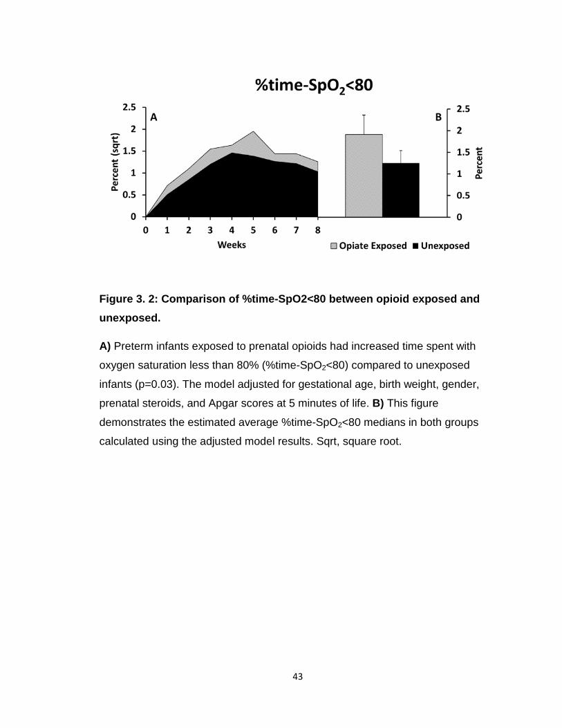
43
Figure 3. 2: Comparison of %time-SpO2<80 between opioid exposed and unexposed.
A) Preterm infants exposed to prenatal opioids had increased time spent with
oxygen saturation less than 80% (%time-SpO2<80) compared to unexposed
infants (p=0.03). The model adjusted for gestational age, birth weight, gender,
prenatal steroids, and Apgar scores at 5 minutes of life. B) This figure
demonstrates the estimated average %time-SpO2<80 medians in both groups
calculated using the adjusted model results. Sqrt, square root.
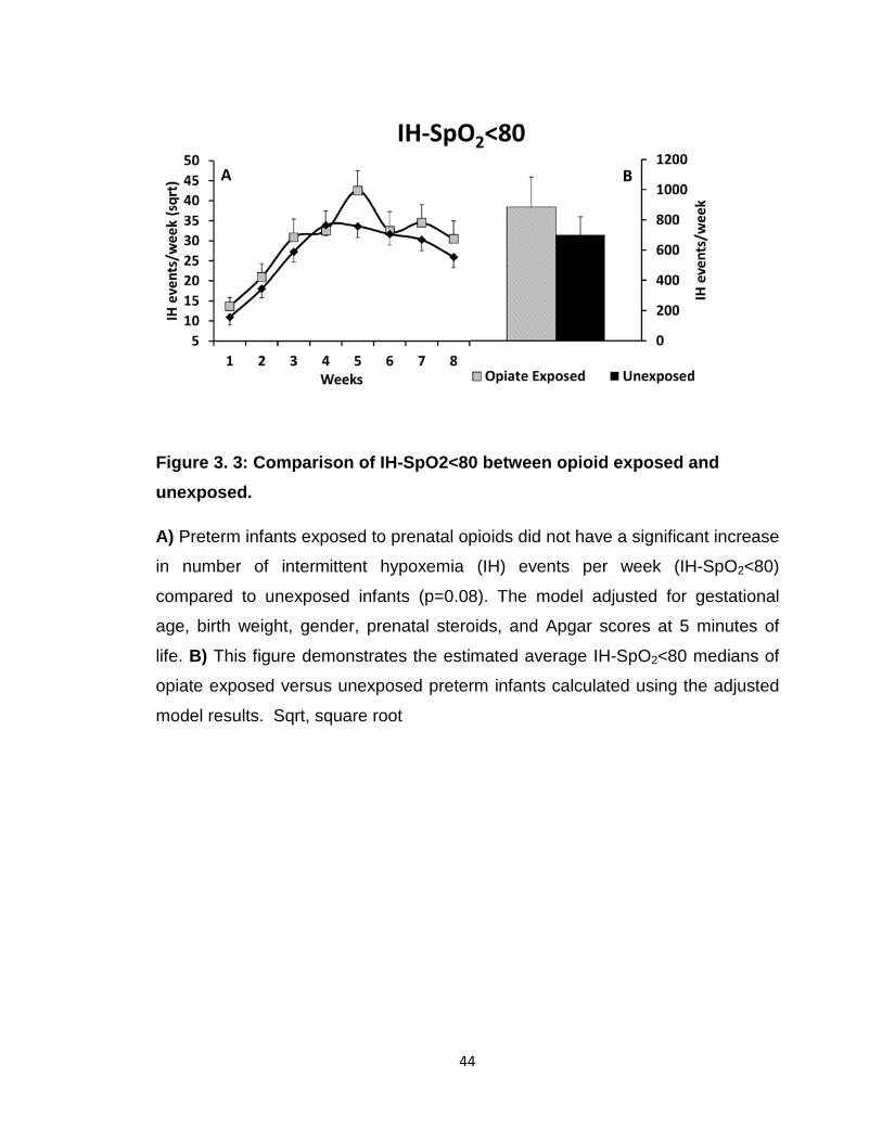
44
Figure 3. 3: Comparison of IH-SpO2<80 between opioid exposed and unexposed.
A) Preterm infants exposed to prenatal opioids did not have a significant increase
in number of intermittent hypoxemia (IH) events per week (IH-SpO2<80)
compared to unexposed infants (p=0.08). The model adjusted for gestational
age, birth weight, gender, prenatal steroids, and Apgar scores at 5 minutes of
life. B) This figure demonstrates the estimated average IH-SpO2<80 medians of
opiate exposed versus unexposed preterm infants calculated using the adjusted
model results. Sqrt, square root
45
CHAPTER 4: INTERMITTENT HYPOXEMIA IS ASSOCIATED WITH INCREASED SERUM C-REACTIVE PROTEIN IN PRETERM INFANTS
I. Introduction
Systemic inflammation perturbs breathing patterns, worsens apnea and
cardiorespiratory events. There is ample evidence in preterm infants and animal
models demonstrating that systemic inflammation increases apnea and
subsequent IH (29, 30, 81-83). Furthermore, apnea and subsequent increase in
IH is often an early sign of inflammatory processes such as sepsis and
necrotizing enterocolitis (NEC) in the neonatal intensive care unit (NICU) (29,
30, 81-83). However, interestingly, based on animal studies, the relationship
between inflammation and IH may be bidirectional, Figure 4.1 (7). I.e. IH may
be pro-inflammatory itself.
Mounting evidence, links IH with both short and long term neonatal
morbidities such as retinopathy of prematurity, sleep disordered breathing,
neurodevelopmental impairment and increased mortality (2, 3, 16, 45, 46, 54,
55, 84-86). Intermittent hypoxemic episodes due to obstructive sleep apnea in
adults are associated with increased levels of inflammatory biomarkers (87-93).
Multiple inflammatory markers have been tested in adults. There is ample
evidence demonstrating increased C-reactive protein (CRP) serum levels in
adult patients with obstructive sleep apnea (87-93). There are no studies in
preterm infants to support IH being pro-inflammatory; however, there is rising
evidence from neonatal animal models suggesting IH is pro-inflammatory. For
example, IH exposed rat-pups had increased serum inflammatory biomarkers
such as IFN-γ and IL-1β (32). Chronic IH in rodents increases inflammation
(monocyte chemoattractant protein-1, IL1β, TNF-α, and a 5 fold increase in IL-6)
in the carotid body chemoreceptors altering their function and subsequently
affecting respiratory control and apnea (14, 33-35). We wanted to assess the
46
relationship between IH and serum CRP for the first time in human preterm
infants.
II. Methods
Study Design and Data Collection
Oxygen saturation (SpO2) data were prospectively collected from 26
preterm infants less than 30 weeks gestational age (GA) admitted to our level 4
NICU between November 2014 and September 2015. We used high resolution
pulse oximeters (Radical 7: Masimo, Irvine, CA) set at 2 second averaging time
and 1Hz sampling rate to continuously monitor patients. Pulse oximeters were
equipped with serial data recorders (Acumen Instruments Corp) for continuous
data collection. Novel programs were utilized to filter and analyze data (Matlab,
Natick, MA) (1, 61). Data with artifacts were excluded. Only SpO2 data with good
signal were included in the analyses. Infants with major congenital
malformations were excluded.
We collected blood samples at 30 days of life to assess for systemic
inflammation at peak of IH. Blood samples for CRP were collected for research
purposes and not for clinical purposes; i.e. not because there was a concern for
illness or change in status. High sensitivity CRP was analyzed using commercial
ELISA kits. Data related to other morbidities that may increase CRP such as
sepsis and necrotizing enterocolitis (NEC) were collected. Other demographics,
morbidities, and respiratory characteristics were collected from medical records.
Statistical Analyses
Average IH measures (IH profile) were calculated for the week prior to
CRP collection. Plots of CRP and IH measures were performed to identify
outliers. We assessed the relationship between IH and CRP using GraphPad
47
Prism 7. Statistical analyses were based on Pearson correlation and linear
regressions. Since the exact threshold below which IH causes injury is
unknown, we calculated an IH profile reflecting the continuum of the problem.
We did not set a lower or upper limit for IH in this study. A drop in SpO2 to less
than 80% is widely considered to be clinically relevant (1-3) and therefore was
selected as the primary outcome measure. Other thresholds included SpO2 of
85% and 90%. Furthermore, 6 different IH duration intervals were calculated: 1-
59 seconds, 60-119 seconds, 120-179 seconds, 180-239 seconds, 240-299
seconds, more than or equal to 300 seconds.
III. Results
Of the 26 infants included, 25 had SpO2 data available during the week
prior to CRP collection. Blood samples for CRP analyses were obtained at
median day of life (DOL) 30 (IQR 29-32 days). Scatter plots identified 2 outliers
with CRP values of 20.829mg/dL and 69.128mg/dL (Figure 4.2). One of the
outliers had sepsis within 2 weeks (11 days) prior to the CRP collection date.
Three other patients had sepsis but occurred more than 2 week prior to our
assessment. No patients had NEC. Plots for IH measures to identify outliers are
presented in Figures 4.3.
Median GA is 27 weeks (Interquartile Range (IQR) 26 - 28 weeks).
Median birth weight is 980 grams (IQR 763 - 1230 grams). There were no small
for gestational age (SGA) infants. Median weight at the time of CRP is 1220
grams (IQR 900 – 1440 grams). Median CRP is 0.236mg/dL (IQR 0.025 - 1.648
mg/dL). Respiratory support data are presented in Table 4.1.
There was strong positive correlation between our primary measure,
%time-SpO2<80, and serum CRP levels (Figure 4.4). The positive correlation
between percent time below threshold and CRP persisted with higher SpO2
threshold of 85% and 90%. There was moderate positive correlation between our
48
primary measure, IH-SpO2<80, and serum CRP levels (Figure 4.5). The positive
correlation between IH events and CRP persisted with higher SpO2 threshold of
85%. There was a strong positive correlation between duration of events and
CRP; i.e. the longer the IH events the higher the serum CRP (Figure 4.6). Furthermore, there was a statistically significant positive correlation between
primary outcome IH measures and CRP at the 6 different duration intervals
examined (except for IH-SpO2<80 at 1-59 seconds, p-Value 0.06) (Figures 4.7 and 4.8). The mean SpO2 and CRP had a strong negative correlation; i.e. the
lower the mean SpO2 the higher the inflammatory marker (Figure 4.9). There
was no statistically significant correlation between IH mean nadir and CRP
(Figure 4.10A). There was moderate negative correlation between peak mean IH
and CRP as represented in Figure 4.10B.
IV. Discussion
Our results show that increased IH is associated with increased systemic
CRP. This relationship between IH and inflammatory markers is documented for
the first time in human preterm infants. Interestingly, most IH profile measures at
all three thresholds and 6 duration categories correlated with worse inflammation.
These results are clinically relevant as elevated inflammation during NICU stay,
mainly 28 days, has been shown to be associated with worse long term
outcomes (94).
Intermittent hypoxemia at all thresholds and durations was associated with
increased serum CRP. The strongest correlation was between %time-
SpO2<threshold and CRP. This is clinically relevant as percent time below
threshold is available to the clinical team from the clinical pulse oximeter
histograms. The frequency of IH correlated positively with CRP only with
moderate and severe IH. The lower the mean SpO2 is the higher the serum
CRP; an important finding with possible impact on the oxygen target saturation
controversy in the NICU (2, 8, 37, 46, 95, 96).

49
C-reactive protein in comprised of five identical, non-covalently associated
subunits (approximately 23 kD each) (97). C-reactive protein has both pro-
inflammatory and anti-inflammatory characteristics (98). Both acute and chronic
inflammation can increase CRP such as infection and metabolic stresses,
respectively (99, 100). We chose CRP as our inflammatory measure for multiple
reasons. First, compared to other markers of inflammation, CRP is widely used in
the NICU with known reference ranges (101-105). Second, CRP is a good and
stable marker for low grade inflammation (100, 106). Minor CRP elevations are
considered a marker of low-grade inflammation, sometimes called subclinical
inflammation or mini-inflammation. Low grade inflammation is the degree of
inflammation we expected will be associated with increased IH. We utilized high
sensitivity CRP commercial ELISA kits in order to measure low grade CRP
changes. Third, multiple adult studies including meta-analyses have
demonstrated increased CRP in patients with IH from obstructive sleep apnea
(87-93).
Our results suggest that IH may be pro-inflammatory itself. Since IH is pro-
inflammatory, that may lead to a spiral or snowball effect (positive feedback
loop). Apnea events cause IH and subsequent systemic postnatal inflammation
that is transferred to the respiratory control network, peripheral chemoreceptors
and lungs. The postnatal inflammation leads to a further cycle of increased
apnea events and consequently higher frequency of IH (Figure 4.1).
Interestingly, this phenomenon may be in part responsible for the IH peak at 4-5
weeks of age (54).
This study has multiple strength including the prospective design and
novel results. A major limitation for this study is the small sample size. However,
the results were consistent at multiple IH thresholds and duration intervals
suggesting a significant relationship between IH and increased CRP. Another
limitation is the use of a single inflammatory marker. Future studies should focus
on multiple inflammatory markers along the inflammation cascade. Other
markers that have been associated with IH in adults with obstructive sleep apnea
50
or IH in neonatal rodent models include, Interleukin (IL)-6, IL-1β, IL-8, Tumor
Necrosis Factor (TNF)-α, Intercellular Adhesion Molecule (ICAM)-1, Interferon
(IFN)-γ, Vascular Cell Adhesion Molecule (VCAM)-1 (14, 32-35, 89).
We demonstrate in this study, for the first time in preterm infants, that IH is
associated with increased inflammation, namely CRP. While there is mounting
evidence of adverse effects of IH, there has been no focus on inflammation in the
cycle of events in preterm infants. Our findings are significant as the increased
inflammation may be the mediator for increased morbidities and impairment in
infants with IH (2, 3, 16, 45, 46, 54, 55, 84-86). Future larger studies that
examine the role of inflammation as a mediator for long term injury from IH
should be examined.
V. Acknowledgments
I thank all the team members mentioned in the acknowledgments section.
I specially recognize Hong Huang MD, PhD for blood sample processing for C-
reactive protein analyses. I thank the Gerber Foundation and Children’s Miracle
Network for funding sample analyses.

51
Table 4. 1: Respiratory Characteristics
Frequency, n (%) Room Air 3 (12%) Continuous Positive Airway Pressure 7 (28%) Non-Invasive Nasal Ventilation 6 (24%) Conventional Ventilation 9 (36%) Oxygen Supplementation 12 (48%) These respiratory setting were collected on the day of CRP measurement.
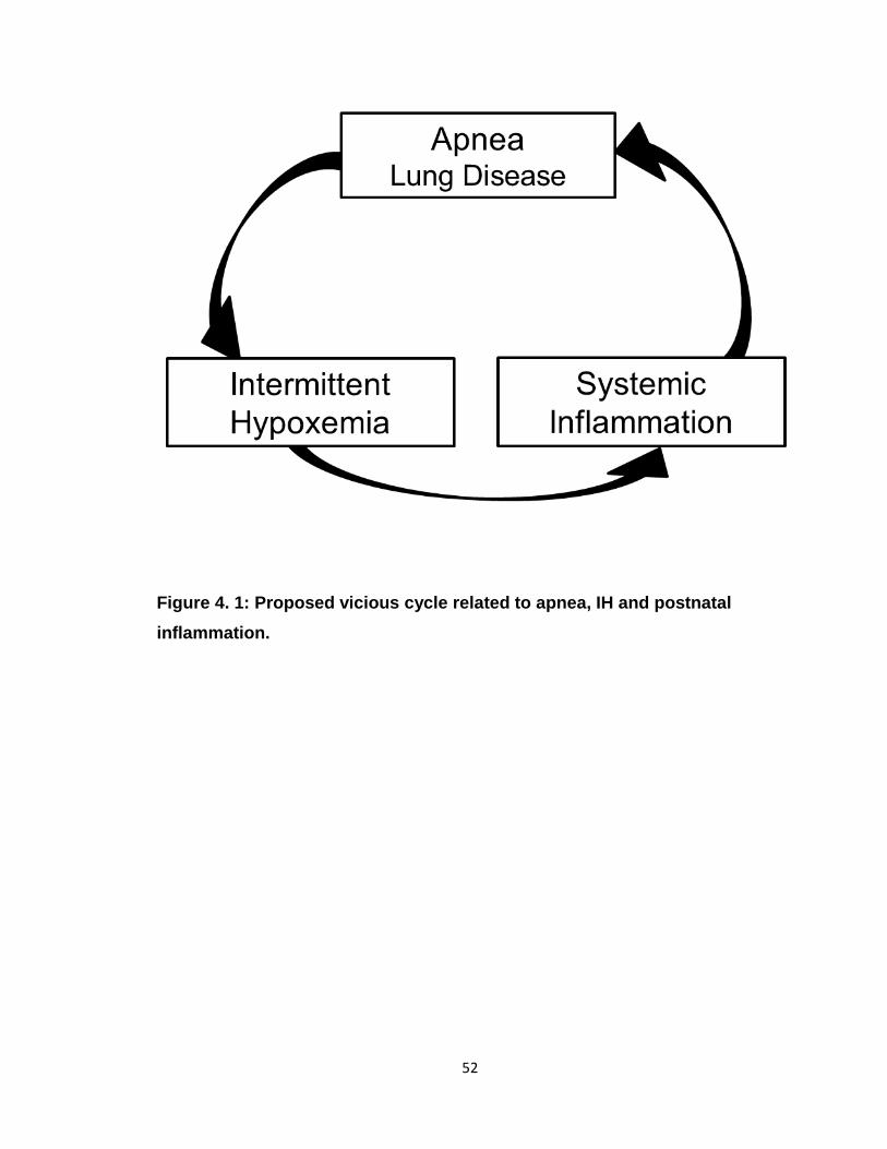
52
Figure 4. 1: Proposed vicious cycle related to apnea, IH and postnatal inflammation.
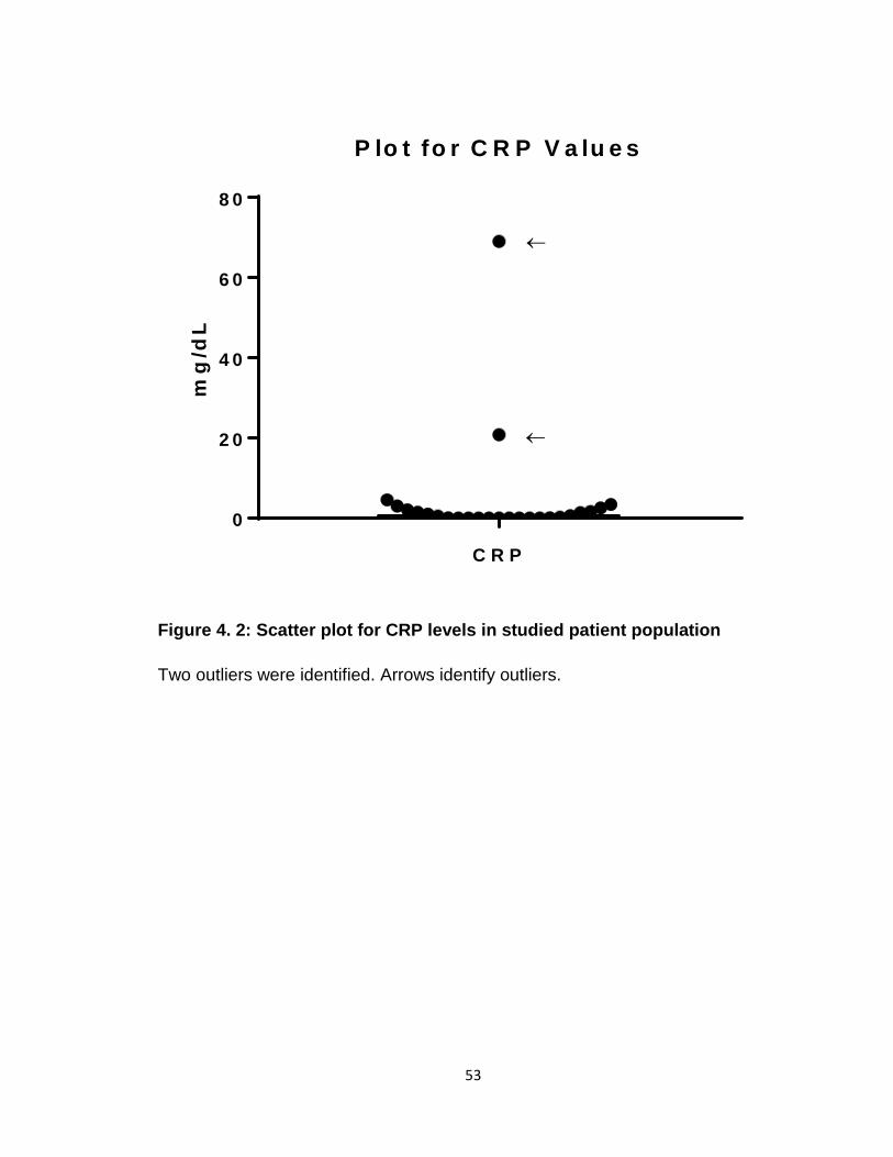
53
Figure 4. 2: Scatter plot for CRP levels in studied patient population
Two outliers were identified. Arrows identify outliers.
C R P
0
2 0
4 0
6 0
8 0
P lo t fo r C R P V a lu e s
mg
/dL
←
←
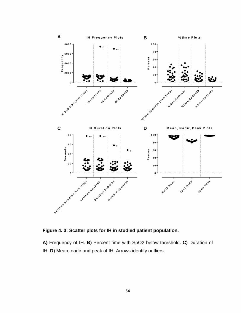
54
Figure 4. 3: Scatter plots for IH in studied patient population.
A) Frequency of IH. B) Percent time with SpO2 below threshold. C) Duration of
IH. D) Mean, nadir and peak of IH. Arrows identify outliers.
IH-S
p O2 < 9 0 (>
4% D
rop )
IH-S
p O2 < 9 0
IH-S
p O2 < 8 5
IH-S
p O2 < 8 0
0
2 0 0 0
4 0 0 0
6 0 0 0
8 0 0 0
IH F re q u e n c y P lo ts
Fre
qu
en
cy
%t im
e -Sp O
2 < 9 0 (>4%
Dro
p )
%tim
e -Sp O
2 < 9 0
%tim
e -Sp O
2 < 8 5
%tim
e -Sp O
2 < 8 0
0
2 0
4 0
6 0
8 0
1 0 0
% tim e P lo ts
Pe
rce
nt
D u rat io
n Sp O
2 < 9 0 (>4%
Dro
p )
D u rat io
n Sp O
2 < 9 0
D u rat io
n Sp O
2 < 8 5
D u rat io
n Sp O
2 < 8 0
0
2 0
4 0
6 0
8 0
IH D u ra tio n P lo ts
Se
co
nd
s
S p O2 M
e a n
S p o 2 Na d ir
S p O2 P
e a k
0
2 0
4 0
6 0
8 0
1 0 0
M e a n , N a d ir , P e a k P lo ts
Pe
rce
nt
←
←←
←
←←
A
C D
B
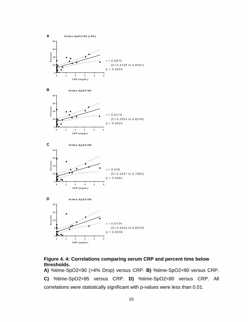
55
Figure 4. 4: Correlations comparing serum CRP and percent time below thresholds. A) %time-SpO2<90 (>4% Drop) versus CRP. B) %time-SpO2<90 versus CRP.
C) %time-SpO2<85 versus CRP. D) %time-SpO2<80 versus CRP. All
correlations were statistically significant with p-values were less than 0.01.
0 1 2 3 4 50
2 0
4 0
6 0
8 0
% tim e -S p O 2 < 9 0 (> 4 % )
C R P (m g /d L )
Pe
rce
nt
0 1 2 3 4 50
2 0
4 0
6 0
8 0
% tim e -S p O 2 < 9 0
C R P (m g /d L )
Pe
rce
nt
0 1 2 3 4 50
1 0
2 0
3 0
4 0
% tim e -S p O 2 < 8 5
C R P (m g /d L )
Pe
rce
nt
0 1 2 3 4 50
5
1 0
1 5
2 0
% tim e -S p O 2 < 8 0
C R P (m g /d L )
Pe
rce
nt
r = 0 .6 1 7 8(C I 0 .2 6 5 3 to 0 .8 2 4 6 )
p = 0 .0 0 2 2
r = 0 .6 8 7 2(C I 0 .3 7 3 9 to 0 .8 5 9 7 )
p = 0 .0 0 0 4
r = 0 .6 7 3 4(C I 0 .3 5 1 6 to 0 .8 5 2 9 )
p = 0 .0 0 0 6
r = 0 .5 4 9(C I 0 .1 6 5 7 to 0 .7 8 8 2 )
p = 0 .0 0 8 1
A
B
C
D
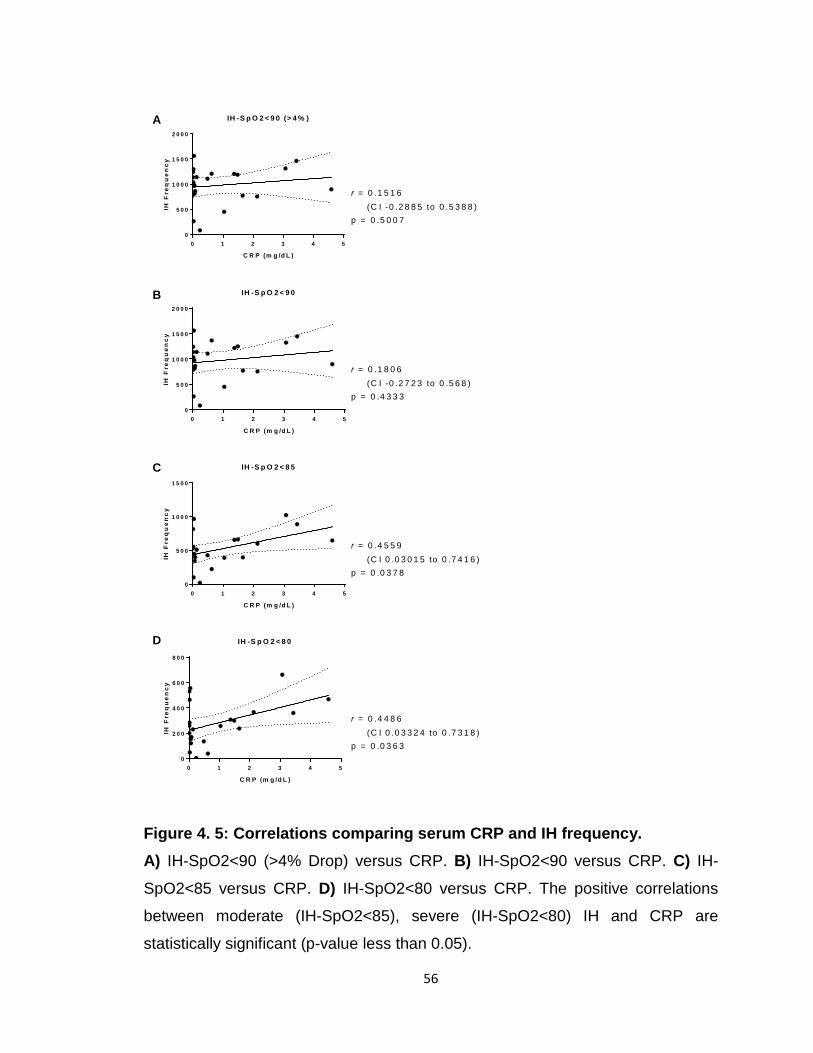
56
Figure 4. 5: Correlations comparing serum CRP and IH frequency. A) IH-SpO2<90 (>4% Drop) versus CRP. B) IH-SpO2<90 versus CRP. C) IH-
SpO2<85 versus CRP. D) IH-SpO2<80 versus CRP. The positive correlations
between moderate (IH-SpO2<85), severe (IH-SpO2<80) IH and CRP are
statistically significant (p-value less than 0.05).
0 1 2 3 4 50
5 0 0
1 0 0 0
1 5 0 0
2 0 0 0
IH -S p O 2 < 9 0 (> 4 % )
C R P (m g /d L )
IH F
req
ue
nc
y
0 1 2 3 4 50
5 0 0
1 0 0 0
1 5 0 0
2 0 0 0
IH -S p O 2 < 9 0
C R P (m g /d L )
IH F
req
ue
nc
y
0 1 2 3 4 50
5 0 0
1 0 0 0
1 5 0 0
IH -S p O 2 < 8 5
C R P (m g /d L )
IH F
req
ue
nc
y
0 1 2 3 4 50
2 0 0
4 0 0
6 0 0
8 0 0
IH -S p O 2 < 8 0
C R P (m g /d L )
IH F
req
ue
nc
y
r = 0 .1 8 0 6(C I -0 .2 7 2 3 to 0 .5 6 8 )
p = 0 .4 3 3 3
r = 0 .1 5 1 6(C I -0 .2 8 8 5 to 0 .5 3 8 8 )
p = 0 .5 0 0 7
r = 0 .4 4 8 6(C I 0 .0 3 3 2 4 to 0 .7 3 1 8 )
p = 0 .0 3 6 3
r = 0 .4 5 5 9(C I 0 .0 3 0 1 5 to 0 .7 4 1 6 )
p = 0 .0 3 7 8
A
B
C
D
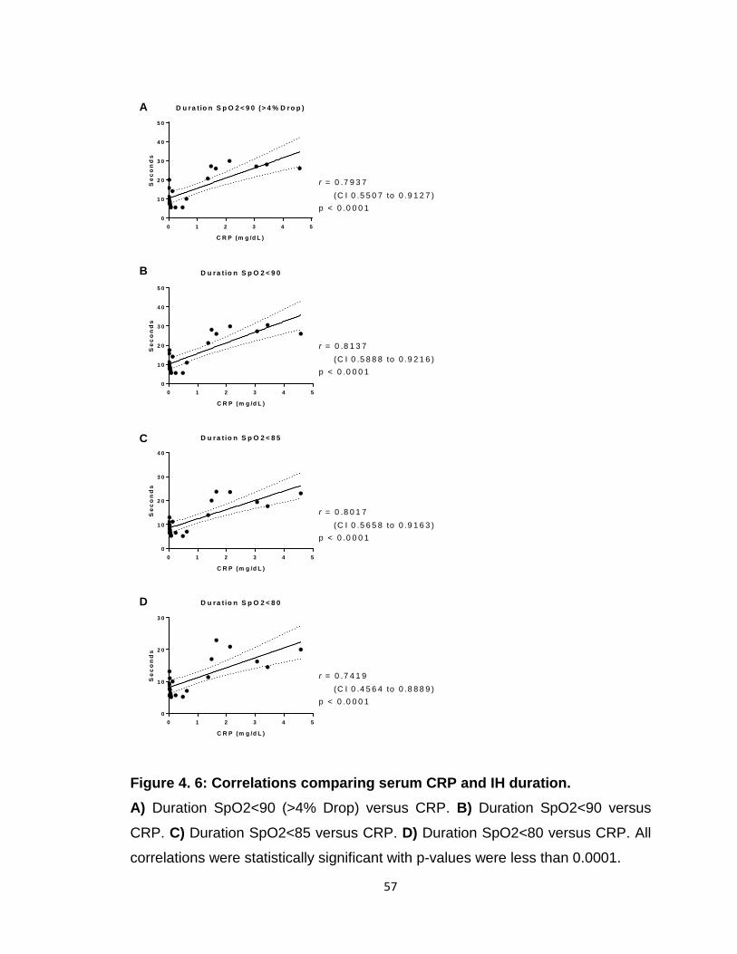
57
Figure 4. 6: Correlations comparing serum CRP and IH duration. A) Duration SpO2<90 (>4% Drop) versus CRP. B) Duration SpO2<90 versus
CRP. C) Duration SpO2<85 versus CRP. D) Duration SpO2<80 versus CRP. All
correlations were statistically significant with p-values were less than 0.0001.
0 1 2 3 4 50
1 0
2 0
3 0
4 0
5 0
D u ra t io n S p O 2 < 9 0 (> 4 % D ro p )
C R P (m g /d L )
Se
co
nd
s
0 1 2 3 4 50
1 0
2 0
3 0
4 0
5 0
D u ra t io n S p O 2 < 9 0
C R P (m g /d L )
Se
co
nd
s
0 1 2 3 4 50
1 0
2 0
3 0
4 0
D u ra t io n S p O 2 < 8 5
C R P (m g /d L )
Se
co
nd
s
0 1 2 3 4 50
1 0
2 0
3 0
D u ra t io n S p O 2 < 8 0
C R P (m g /d L )
Se
co
nd
s
r = 0 .8 1 3 7(C I 0 .5 8 8 8 to 0 .9 2 1 6 )
p < 0 .0 0 0 1
r = 0 .7 9 3 7(C I 0 .5 5 0 7 to 0 .9 1 2 7 )
p < 0 .0 0 0 1
r = 0 .7 4 1 9(C I 0 .4 5 6 4 to 0 .8 8 8 9 )
p < 0 .0 0 0 1
r = 0 .8 0 1 7(C I 0 .5 6 5 8 to 0 .9 1 6 3 )
p < 0 .0 0 0 1
A
B
C
D

58
G. CRP versus %time-SpO2<80
A (1-59s)
B (60-119s)
C (120-179s)
D (180-239s)
E (240-299s)
F (≥300s)
r 0.6215 0.5594 0.4713 0.6632 0.5861 0.4917
95% CI 0.2709 to
0.8265 0.1804 to
0.7938 0.062 to 0.7449
0.3353 to 0.8477
0.2185 to 0.808
0.0884 to 0.7565
p-Value 0.002 0.0068 0.0268 0.0008 0.0042 0.0201 Figure 4. 7: Correlations comparing serum CRP and primary outcome measure %time-SpO2<80 at multiple duration intervals. A) 1-59 seconds B) 60-119 seconds C) 120-179 seconds D) 180-239 seconds E) 240-299 seconds F) more than or equal to 300 seconds. G) This table presents
the correlation coefficients (r), 95% confidence intervals (CI) and p-values for all
intervals. All correlations were statistically significant.
0 1 2 3 4 50
2
4
6
8
1 0
% tim e -S p O 2 < 8 0 (1 -5 9 s )
C R P (m g /d L )
Pe
rce
nt
0 1 2 3 4 50
2
4
6
% tim e -S p O 2 < 8 0 (6 0 -1 1 9 s )
C R P (m g /d L )
Pe
rce
nt
0 1 2 3 4 50
1
2
3
% tim e -S p O 2 < 8 0 (1 2 0 -1 7 9 s )
C R P (m g /d L )
Pe
rce
nt
0 1 2 3 4 50 .0
0 .5
1 .0
1 .5
% t im e -S p O 2 < 8 0 (1 8 0 -2 3 9 s )
C R P (m g /d L )
Pe
rce
nt
0 1 2 3 4 50 .0
0 .2
0 .4
0 .6
0 .8
1 .0
% t im e -S p O 2 < 8 0 (2 4 0 -2 9 9 s )
C R P (m g /d L )
Pe
rce
nt
0 1 2 3 4 50 .0
0 .5
1 .0
1 .5
% t im e -S p O 2 < 8 0 (≥ 3 0 0 s )
C R P (m g /d L )
Pe
rce
nt
A
C
E
B
D
F
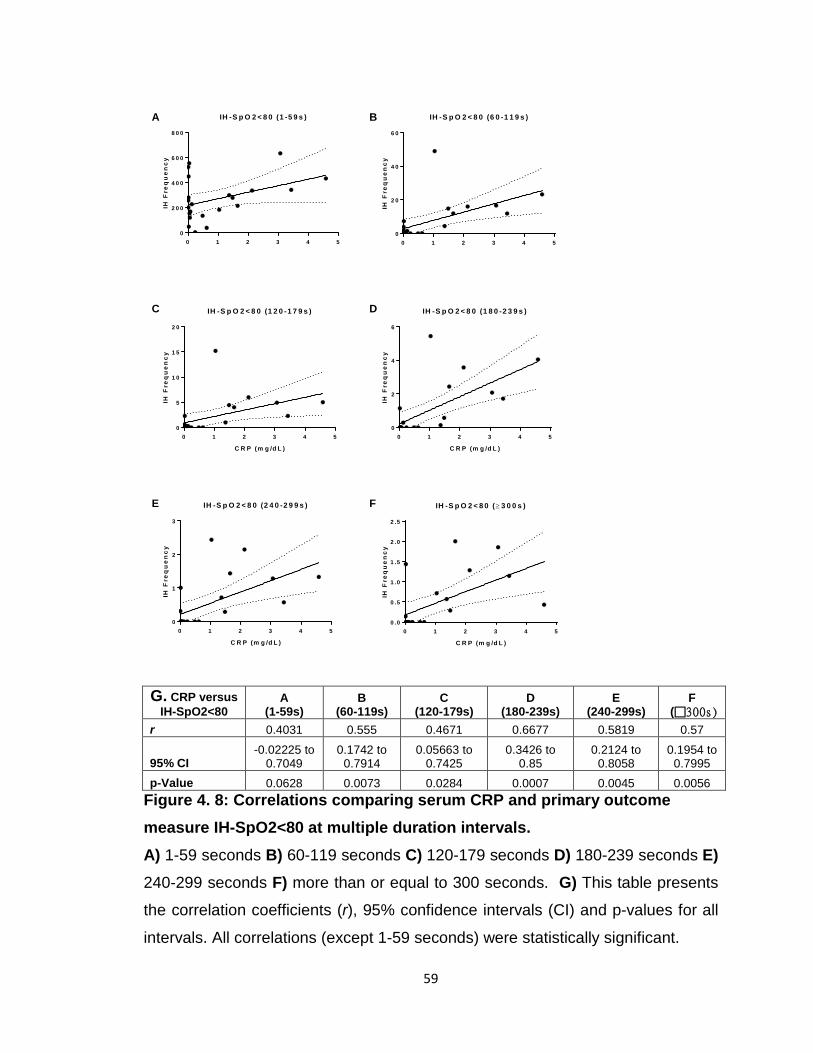
59
G. CRP versus IH-SpO2<80
A (1-59s)
B (60-119s)
C (120-179s)
D (180-239s)
E (240-299s)
F (300s )
r 0.4031 0.555 0.4671 0.6677 0.5819 0.57
95% CI -0.02225 to
0.7049 0.1742 to
0.7914 0.05663 to
0.7425 0.3426 to
0.85 0.2124 to
0.8058 0.1954 to
0.7995 p-Value 0.0628 0.0073 0.0284 0.0007 0.0045 0.0056
Figure 4. 8: Correlations comparing serum CRP and primary outcome measure IH-SpO2<80 at multiple duration intervals. A) 1-59 seconds B) 60-119 seconds C) 120-179 seconds D) 180-239 seconds E) 240-299 seconds F) more than or equal to 300 seconds. G) This table presents
the correlation coefficients (r), 95% confidence intervals (CI) and p-values for all
intervals. All correlations (except 1-59 seconds) were statistically significant.
0 1 2 3 4 50
2 0 0
4 0 0
6 0 0
8 0 0
IH -S p O 2 < 8 0 (1 -5 9 s )
IH F
req
ue
nc
y
0 1 2 3 4 50
2 0
4 0
6 0
IH -S p O 2 < 8 0 (6 0 -1 1 9 s )
IH F
req
ue
nc
y
0 1 2 3 4 50
5
1 0
1 5
2 0
IH -S p O 2 < 8 0 (1 2 0 -1 7 9 s )
C R P (m g /d L )
IH F
req
ue
nc
y
0 1 2 3 4 50
2
4
6
IH -S p O 2 < 8 0 (1 8 0 -2 3 9 s )
C R P (m g /d L )
IH F
req
ue
nc
y
0 1 2 3 4 50
1
2
3
IH -S p O 2 < 8 0 (2 4 0 -2 9 9 s )
C R P (m g /d L )
IH F
req
ue
nc
y
0 1 2 3 4 50 .0
0 .5
1 .0
1 .5
2 .0
2 .5
IH -S p O 2 < 8 0 (≥ 3 0 0 s )
C R P (m g /d L )
IH F
req
ue
nc
y
A
C
E
B
D
F
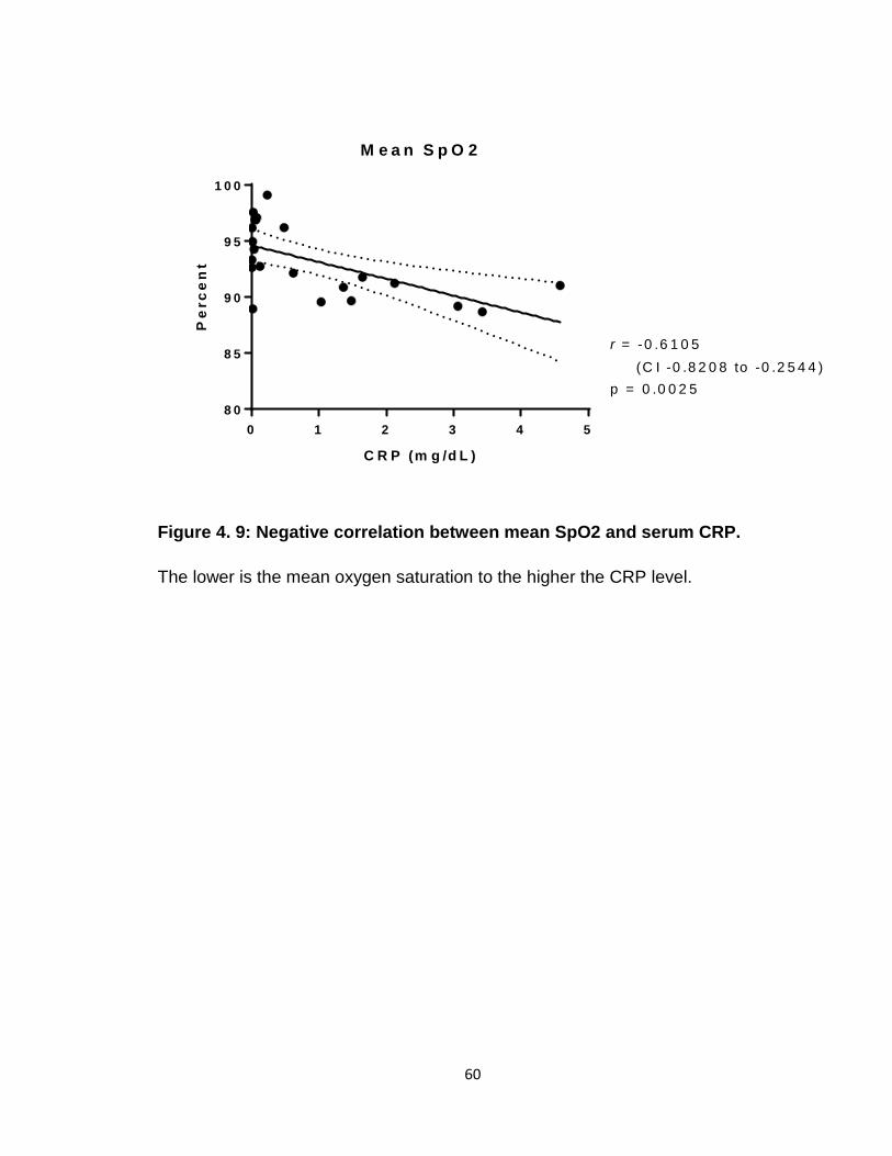
60
Figure 4. 9: Negative correlation between mean SpO2 and serum CRP.
The lower is the mean oxygen saturation to the higher the CRP level.
0 1 2 3 4 58 0
8 5
9 0
9 5
1 0 0
M e a n S p O 2
C R P (m g /d L )
Pe
rce
nt
r = -0 .6 1 0 5(C I -0 .8 2 0 8 to -0 .2 5 4 4 )
p = 0 .0 0 2 5
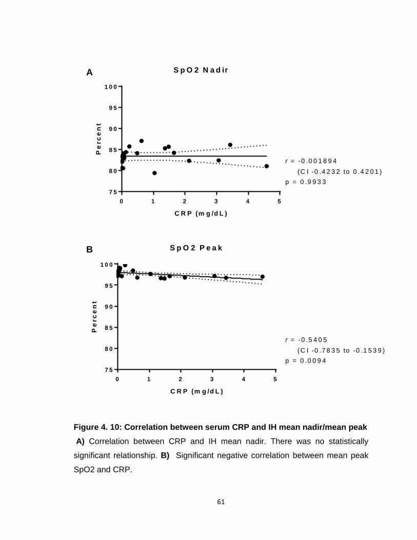
61
Figure 4. 10: Correlation between serum CRP and IH mean nadir/mean peak A) Correlation between CRP and IH mean nadir. There was no statistically
significant relationship. B) Significant negative correlation between mean peak
SpO2 and CRP.
0 1 2 3 4 57 5
8 0
8 5
9 0
9 5
1 0 0
S p O 2 N a d ir
C R P (m g /d L )
Pe
rce
nt
0 1 2 3 4 57 5
8 0
8 5
9 0
9 5
1 0 0
S p O 2 P e a k
C R P (m g /d L )
Pe
rce
nt
r = -0 .0 0 1 8 9 4(C I -0 .4 2 3 2 to 0 .4 2 0 1 )
p = 0 .9 9 3 3
r = -0 .5 4 0 5(C I -0 .7 8 3 5 to -0 .1 5 3 9 )
p = 0 .0 0 9 4
A
B
62
CHAPTER 5: MATERNAL CHORIOAMNIONITIS AND INTERMITTENT HYPOXEMIA IN PRETERM INFANTS
I. Introduction
Four million babies are born per year in the United States and close to a
half million are premature (<37 weeks gestation) (107). The total societal
economic cost of preterm birth is estimated at 26 billion dollars (108-110). Mean
costs of care associated with extreme prematurity are nearly a quarter of a
million dollars in the first 4 years of life; approximately 20 times higher than late
preterm infants (111). Although significant progress has been made in the care of
preterm infants, they continue to suffer from significant morbidities such as
apnea, chronic lung disease (bronchopulmonary dysplasia (BPD)), retinopathy of
prematurity (ROP), and neurodevelopmental impairments (NDI) (112, 113).
Intermittent Hypoxemia (IH), contributes to the aforementioned morbidities (54).
Brief episodes of oxygen desaturations may seem clinically insignificant, but
these IH episodes, occurring up to hundreds of events/day, have a cumulative
effect on morbidities and mortality. As presented in Chapter 1, mounting
evidence, links IH with both short and long term neonatal morbidities such as
ROP, NDI, sleep disordered breathing, and increased mortality (2, 3, 16, 45, 46,
54, 55, 84-86). Laboratory and animal data show IH results in increased
inflammatory cytokines, increased free radicals and oxidative stress, increased
white matter injury, neurocognitive handicap, and poor growth (4, 41, 54, 114-
120).
Several predictors that influence IH have been investigated. Gestational
age and IH are inversely related (5); with extremely preterm infants having the
highest prevalence of IH. Infants with BPD have increased IH that persists on
mechanical ventilation (1, 2, 7). Preterm infants with anemia are at increased
risk for IH (1, 36); as hematocrit level decreases the probability of apnea/IH
events increases. Intermittent hypoxemia natural progression changes with
63
postnatal age. There is low IH frequency during the 1st week of life, followed by
a progressive increase over weeks 2-3, peaks around 4-5 weeks, and
decreases at weeks 6-8 (1, 2, 54). The reasons leading to the rise in IH
postnatally are poorly defined but likely due to both developing lung disease and
chemoreceptor dysregulation possibly due to inflammation and hypoxia (7).
Systemic inflammation increases apnea events and subsequently IH (29).
Hofstetter et al. showed that systemic inflammation increased IL-1β that binds to
its receptors located on endothelial cells of the blood brain barrier (29). Activation
of IL-1β receptors leads to increased prostaglandins in the respiratory control
network in the brain leading to respiratory depression/apnea and subsequent IH
(29, 121). In addition, systemic inflammation worsens lung disease, decreases
lung reserves leading to more IH in the presence of apnea (16). Interestingly,
inflammation in the pulmonary system can be transmitted, likely through the
vagal nerve, to the central respiratory network in the brain stem leading to further
respiratory instability/apnea (30, 31, 122). In summary, inflammation increases
apnea, worsens lung disease and subsequently increases IH.
Prenatal (intrauterine) inflammation is a common cause of preterm birth.
Prenatal inflammation can happen with or without infection. Recently in 2016 the
National Institute of Child Health and Human Development (NICHD) suggested
the Triple I terminology referring to Intrauterine Inflammation, Infection or both in
order to replace chorioamnionitis. For the purpose of consistency in this
document, we will use the terminology of maternal chorioamnionitis (MC) as the
main contributor to prenatal inflammation (123). Funisitis is inflammation of the
umbilical cord (124). The majority of fetuses exposed to MC develop a systemic
fetal inflammatory response syndrome (FIRS), usually defined as elevated serum
interleukin-6 (IL-6). Fetal inflammatory response syndrome occurs due to the
infant being in direct contact with affected amniotic fluid and/or inflammatory cell
or cytokine transfer through placental circulation (125-127). Importantly, prenatal
inflammation is reported to be a major contributor to morbidities e.g. apnea, BPD,
ROP and brain injury (2, 127-150).
64
Pilot Assessment
A total of 30 infants less than 30 weeks GA were enrolled in this pilot trial
to test the hypothesis that prenatal inflammation is associated with increased IH
in postnatal life. Patients were monitored for 4 weeks. The presence of MC was
collected from medical records through our Vermont Oxford Network (VON)
database. Maternal chorioamnionitis as documented by the clinical team was
considered positive in the data base. Blood samples were collected on day of life
(DOL) 1 to measure high-sensitivity C-reactive protein (hsCRP); widely used in
the NICU and a reliable measure of low grade inflammation (106, 151-154). Data
related to MC and blood samples were available for 26 patients and, of those, 6
patients had MC. Median hsCRP on DOL 1 was more than 10 times greater in
patients with MC (0.82 mg/dl) compared to no MC (0.071mg/dl), however, these
differences were not statistically significant. Patients with MC had statistically
significant increased IH during the study period (Figure 5.1) that persisted after
adjusting for GA, gender, ethnicity, and severity of disease (SNAP-PE) scores.
A limitation of the pilot assessment was that MC was collected per the
clinical team and may not meet all clinical chorioamnionitis criteria (123). Hence,
we decided to define MC in the following study per placental pathology reports.
We wanted to test the hypothesis that pathologic MC or funisitis are associated
with increased IH in preterm infants. Since in funisitis, the umbilical cord is
affected, we hypothesized a greater impact on IH in those infants.
II. Methods
Study Design and Data Collection
Oxygen saturation data were prospectively collected from preterm infants
less than 35 weeks gestational age (GA) admitted to our level 4 NICU between
November 2014 and July 2017. We used high resolution pulse oximeters
65
(Radical 7: Masimo, Irvine, CA) set at 2 second averaging time and 1Hz
sampling rate to continuously monitor patients during the first 4 weeks of life. In
order to differentiate intermittent from sustained hypoxemia, we included events
between 4-180 seconds (1). The exact threshold below which IH is clinically
significant is controversial. A drop in SpO2 to less than 80% is widely
considered to be clinically relevant (1-3). Therefore, the primary outcome
measures were defined as percent time spent with SpO2 below 80% (%time-
SpO2<80) and frequency of IH events with SpO2 drop below 80% (IH-
SpO2<80).
Pulse oximeters were equipped with serial data recorders (Acumen
Instruments Corp) for continuous data collection. Novel programs were utilized
to filter and analyze data (Matlab, Natick, MA) (1, 61). Data with artifacts were
excluded. Only SpO2 data with good signal were included in the analyses.
Preterm infants less than 30 weeks GA were included. Infants with major
congenital malformations were excluded.
The presence of MC was collected from medical records. We chose our
exposure as pathologic MC (inflammation noted in the placenta on pathology
reports) or Funisitis (inflammation of the umbilical cord on pathology reports) in
attempt to have more objective data. We did not include clinical MC since the
data related to MC was collected retrospectively and hence clinical parameters
may not be always appropriately documented.
Severe BPD was investigated as a secondary outcome measure given
the controversial literature suggesting a relationship between MC and BPD.
Severe BPD was defined per the National Institute of Child Health and Human
Development (NICHD) criteria for respiratory status at 36 weeks corrected age
(155). Respiratory settings and other demographic and baseline characteristics
were collected from medical charts.

66
Statistical Analyses
Descriptive statistics for continuous variables are presented as either the
mean with standard deviation or median with interquartile range (IQR), and
frequencies and percentages are given for categorical variables. Two-sample t-
tests and Wilcoxon two-sample tests were used to compare MC or Funisitis
exposed to those not exposed with respect to continuous variables, and chi-
square or Fisher’s exact tests were used for categorical variables. Patients with
exposure or MC or Funisitis were compared to unexposed. In addition, infants
with MC only and Funisitis were separately compared to unexposed. To
compare MC or Funisitis infants to those not exposed with respect to IH
measures over time, we utilized multivariate Gaussian linear modeling in order
to account for repeated measurements from subjects, and to adjust for the
potential confounders of gestational age, small for gestational age (SGA) and
the use of prenatal steroids. In order to meet statistical assumptions in these
models, the square root of the IH measures was taken. Furthermore, weekly
observations were weighted by the percentage of time IH was tracked during the
given week. Analyses were conducted in SAS version 9.4 (SAS Institute, Cary,
N.C.) and GraphPad Prism.
III. Results
A total of 151 patient included in our cohort were reviewed. Of those, 121
infants had placental pathology reports and respiratory/IH outcomes data.
Baseline characteristics and comparisons between groups are presented in Tables 5.1 - 5.4. There was a difference in GA (p <0.0001) and birth weight (p=
0.0019) among groups. Deaths prior to discharge varies among groups
(p=0.0011) with increased mortality in the exposed compared to unexposed
infants. Other baseline characteristics did not vary among groups.
67
Contrary to our hypothesis, infants with funisitis had no major differences
in IH measures compared to unexposed (Figures 5.1 and 5.3). The differences
were most pronounced while comparing the MC only group versus unexposed
(Figures 5.2 and 5.4). After adjusting for GA, SGA and prenatal steroids,
statistically significant differences were noted while comparing the MC only
versus unexposed (Figures 5.3 and 5.5). Severe BPD tended to be higher in
any of the exposed groups compared to unexposed; however both unadjusted
and adjusted differences were not statistically significant (Figure 5.6)
IV. Discussion
Our results related to IH measures in infants exposed to perinatal
inflammation were inconsistent. The significant increase in IH in infants with
clinical MC noted in our pilot study was not consistently replicated in infants with
pathologic definition of MC. There were increased IH measures in infants
exposed to pathologic MC and/or funisitis compared to unexposed infants. After
adjusting for GA, SGA status and prenatal steroids, differences were statistically
significant in the MC only group. Severe BPD did not vary among groups,
however tended to be higher in pathologic MC and/or funisitis exposed infants
compared to unexposed.
There were no differences in SGA status between groups, a major risk
factor for increased IH. Infants with pathologic MC had lower GA and birth
weight compared to those unexposed. This is an expected finding given that the
incidence of prenatal inflammation is inversely related to GA, ranging from 75%
to 35% in 23 and 29 week GA infants respectively (127, 156-159). This
difference in GA may be responsible for the increased unadjusted IH in the
exposed groups. Statistically significant higher IH persisted in the MC only group
after adjusting for GA. Interestingly, infants with MC only group had a
statistically significant smaller GA compared to funisitis infants (Table 4). This
68
may suggest that the impact of MC on IH is most pronounced in extreme
prematurity; in contrast to our cohort that included older preterm infants of less
than 35 weeks GA. We did not adjust for birth weight in the model analyses
given the collinear relationship with GA.
Our results suggest that the effect of prenatal inflammation due to MC on
IH persisted far beyond the perinatal period; an interesting and important finding
documented for the first time in human preterm infants. The reasons for
persistently increased IH in MC exposed infants at 5-6 weeks postnatal age
(Figure 5.1 and Figure 5.3) are unknown. We speculate that perinatal
inflammation from MC exacerbates the IH/inflammation cycle by causing
chemoreceptor dysregulation and worsening of lung disease (Figure 5.7) (7).
A limitation of this study is that data related to MC were retrospectively
collected. The choice of pathologic definition of chorioamnionitis is another
limitation that likely had an impact on our results. Pathologic chorioamnionitis is
a histologic finding that may not be symptomatic with no change in maternal
clinical status and subsequently the infant. The placenta is thought to act as a
barrier that protects the infant and therefore without clinical symptoms the full
impact of inflammation may not have reached the infant. Our choice of
pathologic definition relates to inconsistent documentation in medical records of
symptoms of clinical chorioamnionitis such as uterine tenderness and foul
smelling amniotic fluid; hence we may underestimate the number of clinical
chorioamnionitis. The secondary outcome measure of severe BPD was chosen
as a dichotomous variable per the NICHD definition (155). The absence of
significant differences in severe BPD among groups may under estimate the
complexity and continuum of lung disease in preterm infants. Finally, this is a
single center study and our results may not be generalizable.
This study investigates relationship between prenatal inflammation due to
MC and IH. No other groups have studied this relationship in the past in preterm
infants. We demonstrated a persistently increased IH in the MC only group

69
beyond the perinatal period, long after the direct effect of inflammation resolves.
Our inconsistent results may be related to the pathologic definition of MC versus
clinical chorioamnionitis. Prospective studies investigating the impact of clinical
chorioamnionitis on IH may provide mechanistic insights in this understudied
relationship between inflammation and IH in preterm infants.
V. Acknowledgements
I thank all the team members as mentioned in the acknowledgements
section. Special thanks to Hong Huang MD, PhD for processing blood samples
for CRP analyses in the pilot assessment. Special recognition to Audra Stacy
(M4), Amrita Pant MBBS and Crystal Wilson LPN for contributions to data
collection related to this chapter.

70
Table 5. 1: Baseline Characteristics for All Infant with and without MC or Funisitis
No MC or Funisitis MC or Funisitis p-Value
N 58 61
Gestational Age 27 3/7 25 6/7-28 5/7 25.6 24 6/7-26 6/7 <0.0001
Birth Weight 1030 765-1155 830 685-980 0.006
Small for Gestational Age 3 5.2% 3 4.9% 1
Prenatal Steroids 53 91.4% 54 88.5% 1
Female 28 48.3% 31 50.8% 0.31
Non-Hispanic/ White 49 84.5% 52 85.2% 1
Deaths 2 3.4% 8 13.1% 0.001
Median IQR, n %, MC: Maternal Chorioamnionitis
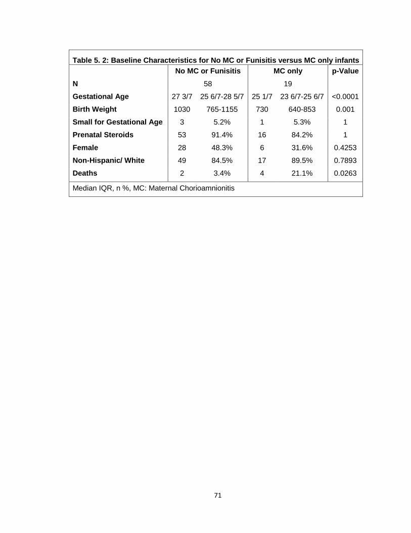
71
Table 5. 2: Baseline Characteristics for No MC or Funisitis versus MC only infants
No MC or Funisitis MC only p-Value
N 58 19
Gestational Age 27 3/7 25 6/7-28 5/7 25 1/7 23 6/7-25 6/7 <0.0001
Birth Weight 1030 765-1155 730 640-853 0.001
Small for Gestational Age 3 5.2% 1 5.3% 1
Prenatal Steroids 53 91.4% 16 84.2% 1
Female 28 48.3% 6 31.6% 0.4253
Non-Hispanic/ White 49 84.5% 17 89.5% 0.7893
Deaths 2 3.4% 4 21.1% 0.0263
Median IQR, n %, MC: Maternal Chorioamnionitis
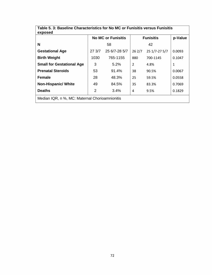
72
Table 5. 3: Baseline Characteristics for No MC or Funisitis versus Funisitis exposed
No MC or Funisitis Funisitis p-Value
N 58 42
Gestational Age 27 3/7 25 6/7-28 5/7 26 2/7 25 1/7-27 5/7 0.0093
Birth Weight 1030 765-1155 880 700-1145 0.1047
Small for Gestational Age 3 5.2% 2 4.8% 1
Prenatal Steroids 53 91.4% 38 90.5% 0.0067
Female 28 48.3% 25 59.5% 0.0558
Non-Hispanic/ White 49 84.5% 35 83.3% 0.7069
Deaths 2 3.4% 4 9.5% 0.1829
Median IQR, n %, MC: Maternal Chorioamnionitis
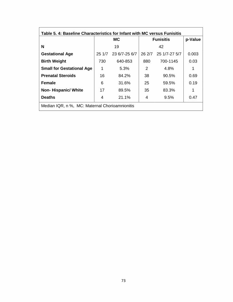
73
Table 5. 4: Baseline Characteristics for Infant with MC versus Funisitis
MC Funisitis p-Value
N 19 42
Gestational Age 25 1/7 23 6/7-25 6/7 26 2/7 25 1/7-27 5/7 0.003
Birth Weight 730 640-853 880 700-1145 0.03
Small for Gestational Age 1 5.3% 2 4.8% 1
Prenatal Steroids 16 84.2% 38 90.5% 0.69
Female 6 31.6% 25 59.5% 0.19
Non- Hispanic/ White 17 89.5% 35 83.3% 1
Deaths 4 21.1% 4 9.5% 0.47
Median IQR, n %, MC: Maternal Chorioamnionitis

74
Figure 5. 1: Increase in %time-SpO2<80 in preterm infants less than 30 weeks born with maternal chorioamnionitis (MC).
The %time spent with SpO2<80% was higher in the MC group compared to no
MC. Statistically significant difference noted in model analysis (adjusted)
between groups during study period, p<0.05. This data is from a pilot
assessment defining MC per clinical team.
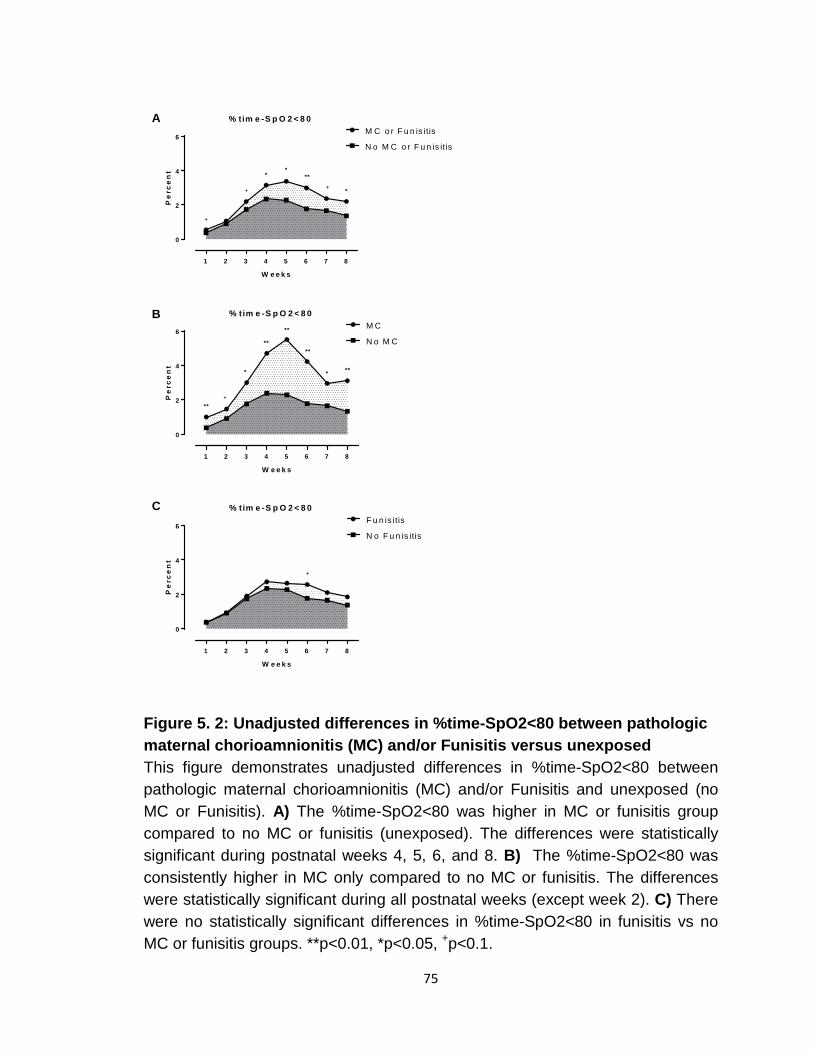
75
1 2 3 4 5 6 7 8
0
2
4
6
% tim e -S p O 2 < 8 0
W e e k s
Pe
rce
nt
M C o r F u n is itis
N o M C o r F u n is itis
1 2 3 4 5 6 7 8
0
2
4
6
% tim e -S p O 2 < 8 0
W e e k s
Pe
rce
nt
N o M C
M C
1 2 3 4 5 6 7 8
0
2
4
6
% tim e -S p O 2 < 8 0
W e e k s
Pe
rce
nt
F u n is itis
N o F u n is itis
+
+
**
**+
*
**
**
**
**
**+
* *
+
A
B
C
Figure 5. 2: Unadjusted differences in %time-SpO2<80 between pathologic maternal chorioamnionitis (MC) and/or Funisitis versus unexposed This figure demonstrates unadjusted differences in %time-SpO2<80 between pathologic maternal chorioamnionitis (MC) and/or Funisitis and unexposed (no MC or Funisitis). A) The %time-SpO2<80 was higher in MC or funisitis group compared to no MC or funisitis (unexposed). The differences were statistically significant during postnatal weeks 4, 5, 6, and 8. B) The %time-SpO2<80 was consistently higher in MC only compared to no MC or funisitis. The differences were statistically significant during all postnatal weeks (except week 2). C) There were no statistically significant differences in %time-SpO2<80 in funisitis vs no MC or funisitis groups. **p<0.01, *p<0.05, +p<0.1.
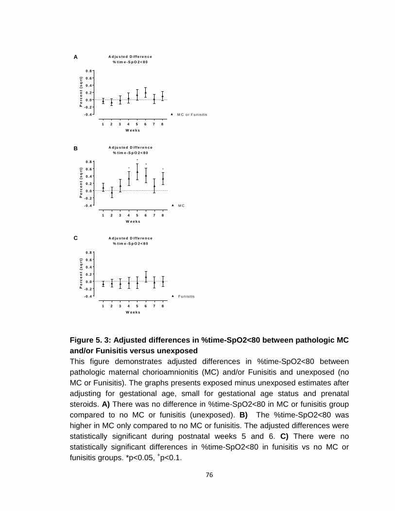
76
1 2 3 4 5 6 7 8
-0 .4
-0 .2
0 .0
0 .2
0 .4
0 .6
0 .8
A d ju s te d D iffe re n c e% tim e -S p O 2 < 8 0
W e e k s
Pe
rce
nt
(sq
rt)
M C o r F u n is itis
1 2 3 4 5 6 7 8
-0 .4
-0 .2
0 .0
0 .2
0 .4
0 .6
0 .8
A d ju s te d D iffe re n c e% tim e -S p O 2 < 8 0
W e e k s
Pe
rce
nt
(sq
rt)
M C
1 2 3 4 5 6 7 8
-0 .4
-0 .2
0 .0
0 .2
0 .4
0 .6
0 .8
A d ju s te d D iffe re n c e% tim e -S p O 2 < 8 0
W e e k s
Pe
rce
nt
(sq
rt)
F u n is itis
**
++
A
B
C
Figure 5. 3: Adjusted differences in %time-SpO2<80 between pathologic MC and/or Funisitis versus unexposed This figure demonstrates adjusted differences in %time-SpO2<80 between pathologic maternal chorioamnionitis (MC) and/or Funisitis and unexposed (no MC or Funisitis). The graphs presents exposed minus unexposed estimates after adjusting for gestational age, small for gestational age status and prenatal steroids. A) There was no difference in %time-SpO2<80 in MC or funisitis group compared to no MC or funisitis (unexposed). B) The %time-SpO2<80 was higher in MC only compared to no MC or funisitis. The adjusted differences were statistically significant during postnatal weeks 5 and 6. C) There were no statistically significant differences in %time-SpO2<80 in funisitis vs no MC or funisitis groups. *p<0.05, +p<0.1.
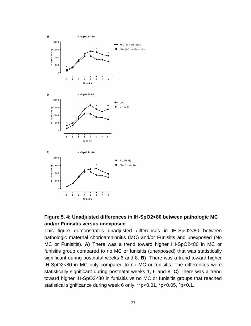
77
1 2 3 4 5 6 7 8
0
5 0 0
1 0 0 0
1 5 0 0
2 0 0 0
IH -S p O 2 < 8 0
W e e k s
IH F
req
ue
nc
y
M C o r F u n is itis
N o M C o r F u n is itis
1 2 3 4 5 6 7 8
0
5 0 0
1 0 0 0
1 5 0 0
2 0 0 0
IH -S p O 2 < 8 0
W e e k s
IH F
req
ue
nc
y
M C
N o M C
1 2 3 4 5 6 7 8
0
5 0 0
1 0 0 0
1 5 0 0
2 0 0 0
IH -S p O 2 < 8 0
W e e k s
IH F
req
ue
nc
y
F u n is itis
N o F u n is itis
+
+
**
*
* **
**
+
*
A
B
C
Figure 5. 4: Unadjusted differences in IH-SpO2<80 between pathologic MC and/or Funisitis versus unexposed This figure demonstrates unadjusted differences in IH-SpO2<80 between pathologic maternal chorioamnionitis (MC) and/or Funisitis and unexposed (No MC or Funisitis). A) There was a trend toward higher IH-SpO2<80 in MC or funisitis group compared to no MC or funisitis (unexposed) that was statistically significant during postnatal weeks 6 and 8. B) There was a trend toward higher IH-SpO2<80 in MC only compared to no MC or funisitis. The differences were statistically significant during postnatal weeks 1, 6 and 8. C) There was a trend toward higher IH-SpO2<80 in funisitis vs no MC or funisitis groups that reached statistical significance during week 6 only. **p<0.01, *p<0.05, +p<0.1.
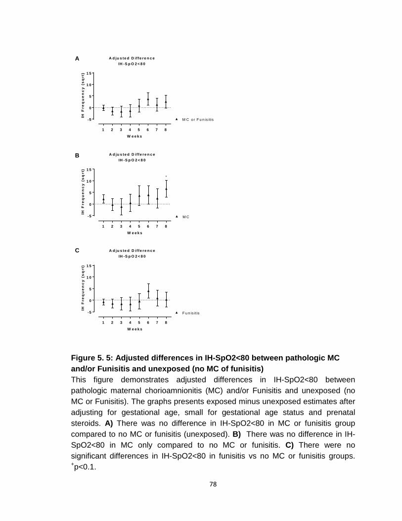
78
1 2 3 4 5 6 7 8
-5
0
5
1 0
1 5
A d ju s te d D iffe re n c eIH -S p O 2 < 8 0
W e e k s
IH F
req
ue
nc
y (
sq
rt)
M C o r F u n is itis
1 2 3 4 5 6 7 8
-5
0
5
1 0
1 5
A d ju s te d D iffe re n c eIH -S p O 2 < 8 0
W e e k s
IH F
req
ue
nc
y (
sq
rt)
M C
1 2 3 4 5 6 7 8
-5
0
5
1 0
1 5
A d ju s te d D iffe re n c eIH -S p O 2 < 8 0
W e e k s
IH F
req
ue
nc
y (
sq
rt)
F u n is itis
+
A
B
C
Figure 5. 5: Adjusted differences in IH-SpO2<80 between pathologic MC and/or Funisitis and unexposed (no MC of funisitis) This figure demonstrates adjusted differences in IH-SpO2<80 between pathologic maternal chorioamnionitis (MC) and/or Funisitis and unexposed (no MC or Funisitis). The graphs presents exposed minus unexposed estimates after adjusting for gestational age, small for gestational age status and prenatal steroids. A) There was no difference in IH-SpO2<80 in MC or funisitis group compared to no MC or funisitis (unexposed). B) There was no difference in IH-SpO2<80 in MC only compared to no MC or funisitis. C) There were no significant differences in IH-SpO2<80 in funisitis vs no MC or funisitis groups. +p<0.1.
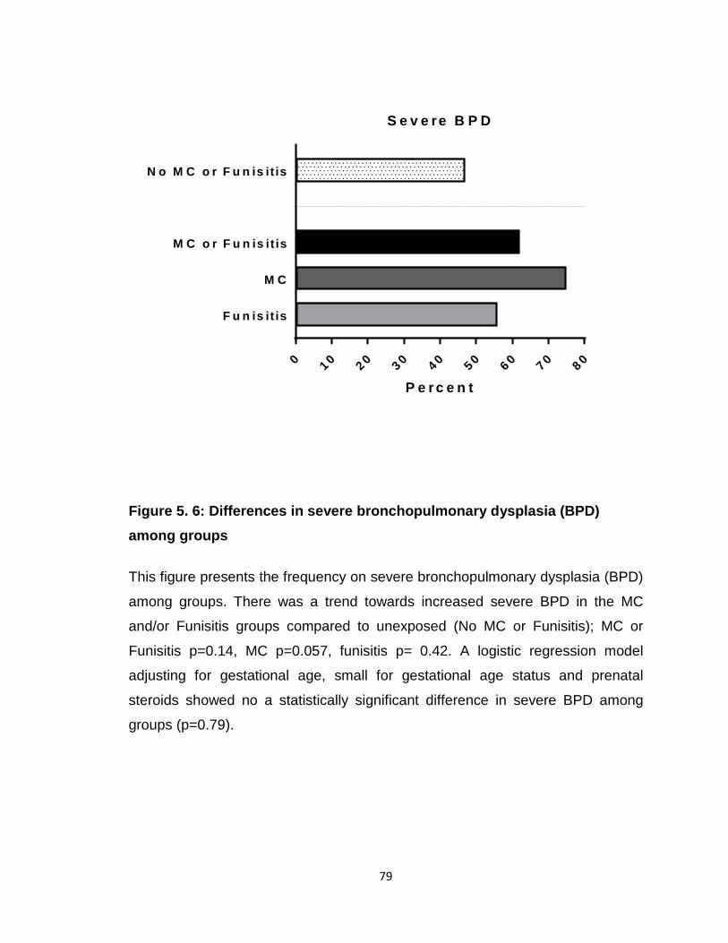
79
0 1 0 2 0 3 0 4 0 5 0 6 0 7 0 8 0
F u n is it is
M C
M C o r F u n is it is
N o M C o r F u n is it is
S e v e re B P D
P e r c e n t
Figure 5. 6: Differences in severe bronchopulmonary dysplasia (BPD) among groups
This figure presents the frequency on severe bronchopulmonary dysplasia (BPD)
among groups. There was a trend towards increased severe BPD in the MC
and/or Funisitis groups compared to unexposed (No MC or Funisitis); MC or
Funisitis p=0.14, MC p=0.057, funisitis p= 0.42. A logistic regression model
adjusting for gestational age, small for gestational age status and prenatal
steroids showed no a statistically significant difference in severe BPD among
groups (p=0.79).

80
Figure 5. 7: Proposed relationship between intermittent hypoxemia and inflammation and possible role of maternal chorioamnionitis.
The relationship between IH and inflammation is bidirectional with inflammation
worsening IH and subsequently IH increases inflammation leading to further
respiratory depression. Prenatal inflammation (maternal chorioamnionitis)
exacerbates the cycle leading to more IH.

81
CHAPTER 6: ROLE OF INDOMETHACIN IN REDUCING INTERMITTENT HYPOXEMIA: PRELIMINARY ASSESSMENT
I. Introduction
Although significant progress has been made in the care of preterm infants,
they continue to suffer from significant morbidities such as apnea,
bronchopulmonary dysplasia (BPD), retinopathy of prematurity (ROP), and
neurodevelopmental impairments (NDI) (112, 113). In addition, prematurity is
associated with elevated societal economic costs. Four million babies are born
per year in the United States and close to a half million are premature (107) with
total societal economic cost of approximately 26 billion dollars (108-110). Mean
costs of care associated with extreme prematurity are nearly a quarter of a
million dollars in the first 4 years of life; approximately 20 times higher than late
preterm infants (111).
Intermittent Hypoxemia (IH), episodic drops in oxygen saturations, contributes to the aforementioned morbidities (54). Brief episodes of oxygen
desaturations may seem clinically insignificant, but these IH episodes, occurring
up to hundreds of events/day, have a cumulative effect on morbidities and
mortality. Mounting evidence, links IH with both short and long term neonatal
morbidities such as retinopathy of prematurity (ROP), neurodevelopmental
impairment (NDI), sleep disordered breathing, and increased mortality (2, 3, 16,
45, 46, 54, 55, 84-86). Laboratory and animal data show IH results in increased
inflammatory cytokines, increased free radicals and oxidative stress, increased
white matter injury, neurocognitive handicap, and poor growth (4, 41, 54, 114-
120). Decreasing IH will lead to decreased associated morbidities and
impairment in preterm infants. In addition since cardiorespiratory events delay
discharge (6, 160), an intervention to decrease IH will reduce length of stay and
the burden on health care dollars.
82
Currently there are multiple strategies aimed at decreasing IH. Those
mainly include methyl xanthine use and respiratory support; i.e. focus on
treatment of apnea and management of lung disease. Although effective, the
aforementioned strategies do not eliminate IH or lead to lung injury with
subsequent long term consequences (4, 161-175). No current strategy focus on
other causes of increased IH such as inflammation. Since prenatal inflammation
plays a role in increased IH, finding strategies to ameliorate prenatal/perinatal
inflammation may be effective at decreasing IH and associated morbidities.
Preterm infants are commonly born through a prenatal inflammatory process
(123, 151, 176, 177). The increased systemic inflammation at birth worsens
apnea and lung disease leading to a rise in IH. Anti-inflammatory agents may
ameliorate systemic inflammation and decrease IH. Olsson et al. in rat pup
experiments showed that indomethacin reversed the depressive respiratory
effects of inflammation (caused by IL-1 β and lipopolysaccharide (LPS)) in
addition to hypoxia (144). In this preliminary assessment, we wanted to assess
the effect of indomethacin, anti-inflammatory agent, on IH in preterm infants.
II. Methods
Study Design and Data Collection
Oxygen saturation data were prospectively collected from 30 preterm
infants less than 30 weeks gestational age (GA) admitted to our level 4 NICU
between November 2014 and September 2015. We used high resolution pulse
oximeters (Radical 7: Masimo, Irvine, CA) set at 2 second averaging time and
1Hz sampling rate to continuously monitor patients during the first 4 weeks of
life. In order to differentiate intermittent from sustained hypoxemia, we included
events between 4-180 seconds (1). The exact threshold below which IH is
clinically significant is controversial. A drop in SpO2 to less than 80% is widely
considered to be clinically relevant (1-3). Therefore, the primary outcome
83
measure was defined as percent time spent with SpO2 below 80% (%time-
SpO2<80).
Pulse oximeters were equipped with serial data recorders (Acumen
Instruments Corp) for continuous data collection. Novel programs were utilized
to filter and analyze data (Matlab, Natick, MA) (1, 61). Data with artifacts were
excluded. Only SpO2 data with good signal were included in the analyses.
Infants with major congenital malformations were excluded.
Infants were randomized to placebo versus indomethacin in this
randomized controlled (double blind) trial (RCT). Indomethacin was given within
12 hours of birth and repeated every 24 hours for a total of 3 doses per the
current evidence based dosing regimen utilized for other indications (178-183).
Neonatal morbidities, including maternal chorioamnionitis (MC), were collected
from medical records. In regards to this assessment, after the intervention
infants received the standard clinical care per clinical team; except for the
additional pulse oximeter.
Statistical Analyses
Statistical analyses for IH were based on linear mixed models, which
statistically accounted for repeated measures. Intermittent hypoxemia (%time-
SpO2<80) and change of IH over time were compared in indomethacin versus
placebo groups using SAS version 9.4 (SAS Institute, Cary, N.C.). Analyses
were based on intention-to-treat, and tests were two-sided with a 5%
significance level. Comparisons were performed for all infants and in infants
with MC only; as we considered the latter most likely group to benefit given they
are born through and inflammatory process. Comparisons for baseline
characteristics, respiratory support and morbidities were performed using
GraphPad Prism 7 (GraphPad Software, La Jolla California USA).

84
III. Results
Oxygenation data was available on 26 preterm infants with 13 infants
each of the indomethacin and placebo groups. Table 1 represents baseline
characteristics between indomethacin and placebo groups. There were no
differences in GA, birth weight and gender and other baseline characteristics
(Table 6.1). Table 2 represents respiratory characteristics during and at the end
of study period showing no significant differences between groups. More infants
were on non-invasive support at 36 weeks corrected age, however these results
were not statistically significant (Table 6.2).
There were no statistically significant differences in neonatal morbidities
between groups as represented in Table 6.3. There was one death in the
indomethacin group versus none in placebo. Severe IVH was similar in both
groups. Infants in the placebo group tended to have more PDA, however, all
except for one were non-hemodynamically significant per Gomez et al (61). Late
onset sepsis and necrotizing enterocolitis rates were not different between
groups.
Although results were not statistically significant, there was a trend
toward lower IH rates in the indomethacin compared to placebo group. Figure 6.1 presents data for all infants. Figure 6.2 presents data from the patients born
with MC. There is attenuation of the peak %time-SpO2<80 at 4-5 weeks of life,
however it was not statistically significant.
IV. Discussion
This preliminary data demonstrate that indomethacin, administered
shortly after birth, may be a promising new therapy for reducing IH in preterm
infants. Infants with increased prenatal inflammation due to MC may benefit the
85
most from this intervention. Perinatal inflammation plays a major role in the
pathophysiology of IH and vice versa; and administering an anti-inflammatory
agent may break the IH/inflammation vicious cycle in its earliest stages leading
to decreased IH (Figure 6.3).
Current strategies aimed at decreasing IH focus on treatment of apnea
and management of lung disease. Caffeine, a competitive adenosine receptor
inhibitor, improves IH (4, 161, 162). Recent evidence suggest that caffeine may
also have mild anti-inflammatory effects (184). Caffeine is used in NICUs
worldwide and usually discontinued around 34-36 weeks corrected age (185).
Recently, Rhein et al. showed that prolonged caffeine use reduces IH frequency
until 37 weeks corrected age. Although caffeine is effective in decreasing IH, it
does not eliminate IH. Other approaches to ameliorate IH are respiratory support
measures such as mechanical ventilation, continuous positive airway pressure
(CPAP) and oxygen supplementation (13, 186). However, respiratory support,
even with current gentle ventilation strategies, leads to lung injury with
subsequent long term consequences (163-174). In addition, oxygen
supplementation in preterm infants leads to ROP (major cause of visual
impairment) (37). Furthermore, preterm infants continue to have frequent IH
events while on respiratory support (1, 2). A strategy that addresses other factors
that increases IH (such as inflammation) may have an additive impact on
amelioration of IH and hence improve long term outcomes. Although our results
are not statistically significant, our trends align with preclinical animal model data.
Olsson et al., demonstrated that indomethacin administration reversed the
depressive effects of inflammation on breathing patterns(144). Indomethacin is a
promising intervention that needs further investigation. Finding a strategy
(indomethacin) to decrease inflammation at birth may decrease IH and
subsequently decrease associated morbidities in preterm infants.
Prophylactic indomethacin has been tested in preterm infants to reduce
other neonatal morbidities such as IVH and PDA. Multiple studies demonstrated
that indomethacin decreases severe IVH by more than 30% (183, 187).
86
However, the decrease in IVH did not translate to improved long term outcomes
(187). Similarly prophylactic indomethacin use improves PDA closure (179,
188). However, prophylaxis was not more effective, compared to early treatment
of symptomatic PDA, at reducing mortality and respiratory outcomes (189). Both
the lack of long term benefit and increased risk benefit ratio, especially in infants
without PDA, led to increased practice variation in use of prophylactic
indomethacin. However, indomethacin has not been prospectively studied in
preterm infants born with MC. Since these infants are born through an
inflammatory process, we speculate they may benefit the most from an anti-
inflammatory agent. As shown in Figure 6.2, infants with MC who received
indomethacin tended to have lower IH peak at 1 month of life.
For future larger RCT involving infants with MC, indomethacin should be
considered for multiple reasons. First, in contrast to postnatal steroids,
indomethacin has a good safety profile and is not associated with long term NDI
in preterm infants (178, 180, 190-194). Adverse effects associated with
indomethacin include transient renal insufficiency (195); which can be
ameliorated by interventions to improve renal perfusion. Other reported but rare
adverse effects include increased risk of bleeding and intestinal perforation
(178, 180, 191, 193, 194). Second, indomethacin is associated with decreased
morbidities, mainly patent PDA and IVH (178-180, 192, 196, 197); morbidities
that may affect cardiorespiratory events in preterm infants. Third, indomethacin
also regulates blood flow to the brain which may lead to improved respiratory
control and less IH (another mechanism to improve IH); as preterm infants have
a paradoxical ventilatory depression in response to hypoxia/poor brain perfusion
(198, 199). Fourth, indomethacin is an effective anti-inflammatory agent that
reversibly inhibits cyclooxygenase (COX)-1 and COX-2 enzymes, which results
in decreased formation of prostaglandin precursors; main culprits leading to
apnea in the setting of inflammation (29, 30). Fifth, animal data show
indomethacin reverses the effects of inflammation on respiratory patterns(144).
Sixth, indomethacin is not the standard of care with wide practice variation both
87
locally and nationally creating equipoise and ability to test indomethacin (200).
Finally, although indomethacin has been studied in preterm infants, the focus of
those studies was not in the setting of perinatal inflammation or IH.
A major limitation of this study is the small sample size especially for the
subset involving MC patients. However, this was a preliminary assessment
aimed at generating pilot data to power future larger studies. Another limitation
of this study is the use of indomethacin dosing regimen for IVH and PDA
prophylaxis (179, 181-183). Indomethacin was administered in 3 doses
(0.2mg/kg/dose on DOL1 and 0.1mg/kg/dose on DOL 2 and 3) in the first 3 days
of life. This dosing regimen has documented safety but it may not be adequate
to suppress inflammation in infants with born with MC. Longer treatment course
may be necessary to have a significant impact on decreasing inflammation and
subsequent IH. Ideally, pre and post indomethacin inflammatory markers should
have been measured to document a decrease in systemic inflammation.
This is the first study to test the effect of indomethacin in management of
IH in preterm infants. This innovative pilot study possibly identified a subset of
preterm infants (with prenatal inflammation/MC) who may benefit the most from
indomethacin to reduce IH; an important discovery in the era of precision
medicine. Future larger studies should focus on investigating indomethacin in
patients born with MC.
V. Acknowledgements
I thank all the team members as mentioned in the acknowledgements
section. I especially acknowledge the principal investigators (Peter Giannone MD
and John Bauer PhD) for the “Comparative effectiveness of preventative
strategies for IVH in preterm infants” as this chapter utilized the trial’s
infrastructure.
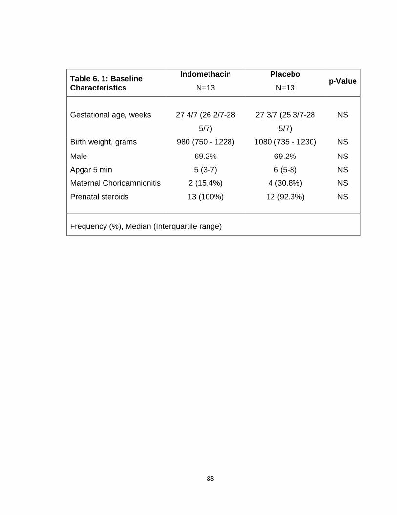
88
Table 6. 1: Baseline Characteristics
Indomethacin Placebo p-Value
N=13 N=13
Gestational age, weeks 27 4/7 (26 2/7-28
5/7)
27 3/7 (25 3/7-28
5/7)
NS
Birth weight, grams 980 (750 - 1228) 1080 (735 - 1230) NS
Male 69.2% 69.2% NS
Apgar 5 min 5 (3-7) 6 (5-8) NS
Maternal Chorioamnionitis 2 (15.4%) 4 (30.8%) NS
Prenatal steroids 13 (100%) 12 (92.3%) NS
Frequency (%), Median (Interquartile range)
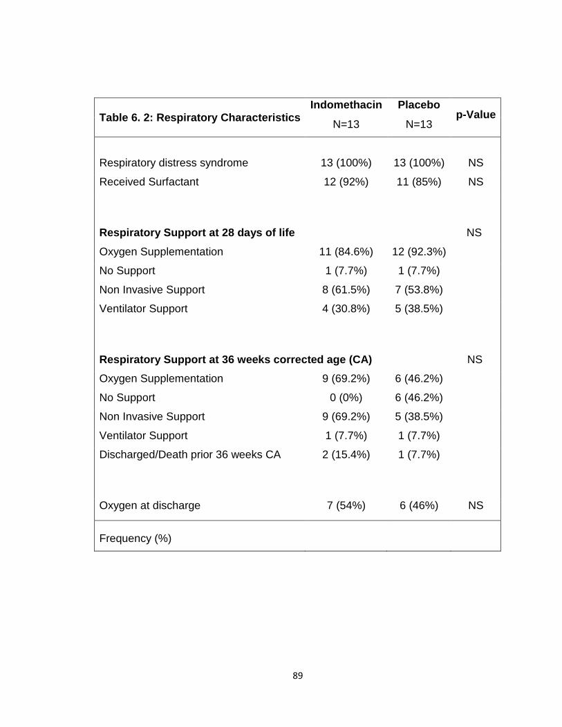
89
Table 6. 2: Respiratory Characteristics Indomethacin Placebo
p-Value N=13 N=13
Respiratory distress syndrome 13 (100%) 13 (100%) NS
Received Surfactant 12 (92%) 11 (85%) NS
Respiratory Support at 28 days of life NS Oxygen Supplementation 11 (84.6%) 12 (92.3%)
No Support 1 (7.7%) 1 (7.7%)
Non Invasive Support 8 (61.5%) 7 (53.8%)
Ventilator Support 4 (30.8%) 5 (38.5%)
Respiratory Support at 36 weeks corrected age (CA) NS Oxygen Supplementation 9 (69.2%) 6 (46.2%)
No Support 0 (0%) 6 (46.2%)
Non Invasive Support 9 (69.2%) 5 (38.5%)
Ventilator Support 1 (7.7%) 1 (7.7%)
Discharged/Death prior 36 weeks CA 2 (15.4%) 1 (7.7%)
Oxygen at discharge 7 (54%) 6 (46%) NS
Frequency (%)
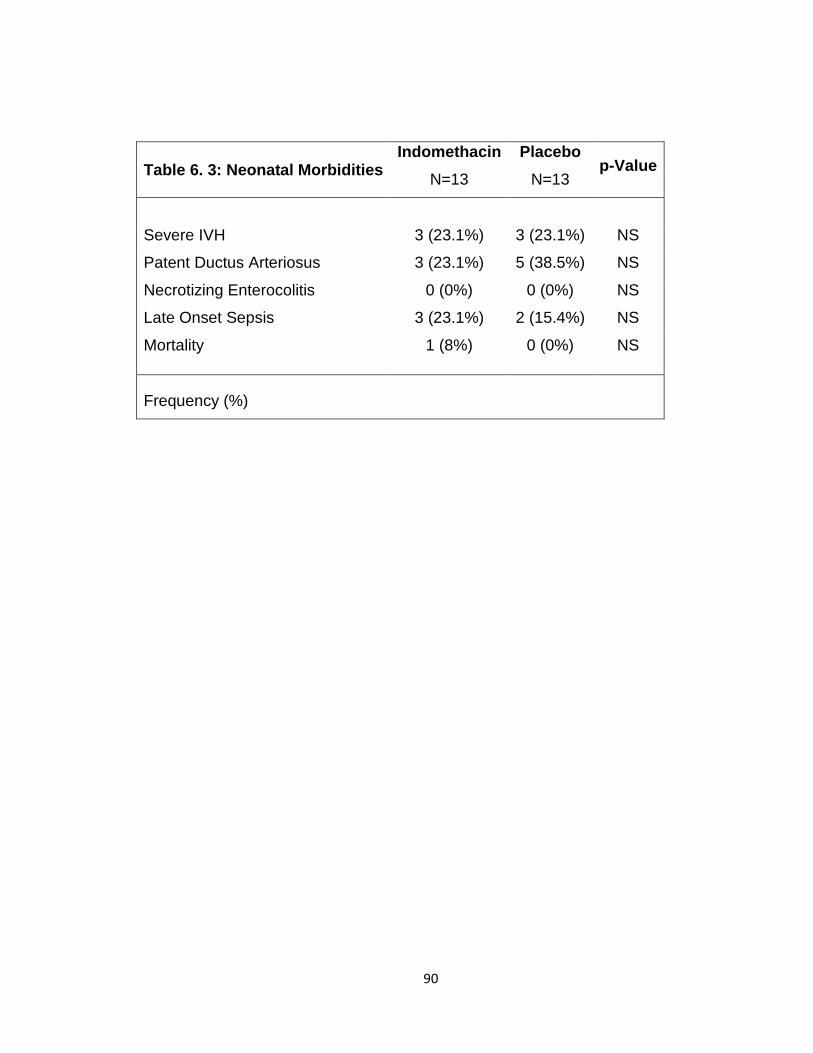
90
Table 6. 3: Neonatal Morbidities Indomethacin Placebo
p-Value N=13 N=13
Severe IVH 3 (23.1%) 3 (23.1%) NS
Patent Ductus Arteriosus 3 (23.1%) 5 (38.5%) NS
Necrotizing Enterocolitis 0 (0%) 0 (0%) NS
Late Onset Sepsis 3 (23.1%) 2 (15.4%) NS
Mortality 1 (8%) 0 (0%) NS
Frequency (%)

91
Figure 6. 1: Potential benefit of indomethacin in reducing intermittent hypoxemia (IH) in preterm infants.
Benefit of indomethacin (black) vs placebo (gray) on IH as reflected by percent
time spent with SpO2<80% (%time-SpO2<80). N=26, p=NS.

92
Figure 6. 2: Potential benefit of indomethacin in reducing intermittent hypoxemia (IH) in preterm infants with maternal chorioamnionitis (MC).
Benefit of indomethacin (black) vs placebo (gray) on IH as reflected by loss of 4-
5 weeks peak in percent time spent with SpO2<80% (%time-SpO2<80). N=6,
p=NS.
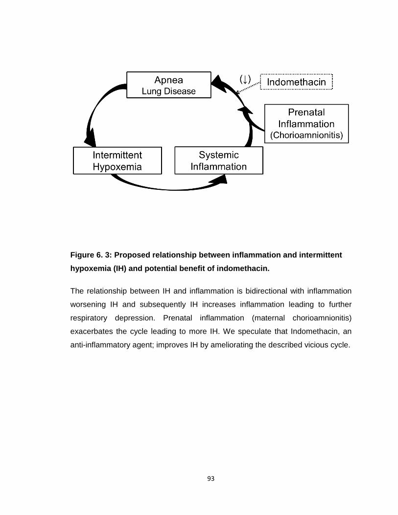
93
Figure 6. 3: Proposed relationship between inflammation and intermittent hypoxemia (IH) and potential benefit of indomethacin.
The relationship between IH and inflammation is bidirectional with inflammation
worsening IH and subsequently IH increases inflammation leading to further
respiratory depression. Prenatal inflammation (maternal chorioamnionitis)
exacerbates the cycle leading to more IH. We speculate that Indomethacin, an
anti-inflammatory agent; improves IH by ameliorating the described vicious cycle.

94
CHAPTER 7: SUMMARY AND FUTURE DIRECTIONS
Over the past 4 years we have made multiple major contributions to the
field; especially as it relates to Pediatrics and Neonatal Perinatal Medicine. We
investigated a clinically significant medical problem (IH) that although has
profound consequences has not been well studied in preterm infants.
First, we developed novel methods and processes to perform high fidelity
studies and accurately assess cardiorespiratory events/IH. We have the ability to
efficiently collect, post process and analyze bedside monitoring data. We utilized
high resolution pulse oximeters with 2 second averaging time and 1Hz sampling
rate. Our analyses IH Automated Analyses Algorithms (IH-AAA) have the
capacity to import multiple streams of raw data and export detailed IH profiles
from each subject. Furthermore, the IH profile is an innovative and unique
method to address this understudied problem. Calculating an IH profile provides
an enhanced representation of the continuum of the IH problem. Having a good
representation of the spectrum of the problem may help identify thresholds
(frequency, duration, severity) beyond which IH leads to neonatal morbidities and
impairments. These novel measures can be further developed to become part of
the routine monitoring strategies in the NICU for instantaneous feedback to
clinical care.
Second, we reconfirmed finding related to the dynamic natural progression
of IH in preterm infants. Di Fiore et al. and Abu Jawdeh et al. first reported from
a single center study that there is a low frequency of IH in extremely preterm
infants (less than 28 weeks GA) during the first week after birth, followed by a
progressive increase by weeks 2-3, with a peak around 4-5 weeks then
plateau/decrease during weeks 6-8 (1, 2). No other studies have addressed this
issue or replicated these findings in order to better understand mechanisms in
the future. We now reproduce this finding from a second center, utilizing an
expanded patient population of less than 30 weeks GA (versus 28 weeks shown
before) showing similar IH dynamic frequency until 10 weeks postnatal life
(versus 8 weeks shown before). The reasons leading to the rise in IH postnatally
95
are poorly defined but likely because of both developing lung disease and
chemoreceptor dysregulation possibly resulting from inflammation and hypoxia
(7). Now that these findings have been reproduced, studies should focus on
understanding further mechanisms and causes for the rise in IH during early
postnatal life.
Third, our findings demonstrate the importance of prenatal exposures and
their effects on postnatal outcomes. We had the unique opportunity to assess the
relationship between isolated opioid exposure and respiratory instability in
preterm infants. It was challenging in the past to assess the relationship between
isolated prenatal opioid exposure and respiratory outcomes/IH, as the majority of
women who use opioids also smoke or misuse poly-drugs. Our results suggest
that prenatal opioid exposure is associated with increased IH measures
compared to unexposed preterm infants. Interestingly, the increased IH
measures in opioid exposed infants persisted beyond the early postnatal period.
Another important finding is that the prevalence of opioid exposure in our local
preterm population is higher than previously reported nationally, thus creating
urgency toward addressing this significant problem in this vulnerable patient
population
Fourth, we translated and complemented the knowledge we have from
preclinical animal and bench studies to the clinical setting in preterm infants. We
showed for the first time in preterm infants that cumulative IH is associated with
increased markers of inflammation, namely C-reactive Protein (CRP). Our results
suggest that IH at any of the selected thresholds is associated with increased
CRP. In addition, we demonstrated that the longer IH events are associated with
higher CRP levels. These are important findings that shed light on possible
mechanisms by which IH causes neonatal morbidities and impairment. Future
longitudinal studies that focus on repeated measures of short and long acting
markers of inflammation throughout the inflammatory cascade will help define
mechanisms and better understand this relationship between IH and
inflammation.
96
Furthermore, our findings support our hypotheses of a bidirectional
relationship between inflammation and IH. It is well established that systemic
inflammation leads to increased apnea and subsequently IH (29, 30). In this
document, we demonstrated that IH may be pro-inflammatory itself. The pro-
inflammatory effects of IH may lead to a vicious cycle (positive feedback loop).
Apnea events cause IH (oxygen desaturations) and subsequent postnatal
inflammation systemically and hence in the respiratory control network,
peripheral chemoreceptors and lungs. The postnatal inflammation leads to a
further cycle of increased apnea events and consequently higher frequency of IH.
Interestingly, this phenomenon may be in part responsible for the IH peak at 4-5
weeks of age.
Fifth, we demonstrated that maternal chorioamnionitis may be associated
with increased IH during early postnatal life. No other groups have studied this
relationship in the past in preterm infants. We speculate that maternal
chorioamnionitis starts or exacerbates the aforementioned cycle early leading to
the snowball/spiral effect. Our inconsistent results in this chapter may be related
to the pathologic definition of MC versus clinical chorioamnionitis. Prospective
studies investigating the impact of chorioamnionitis (clinical and pathologic) on IH
may provide mechanistic insights in this understudied relationship between
inflammation and IH in preterm infants.
Sixth, our preliminary assessment suggests that indomethacin, a
commonly used medication in the NICU, may be used in a novel indication; to
decrease IH in patients born with increased inflammation due to MC. This is the
first study to test the effect of indomethacin in management of IH in preterm
infants. This innovative pilot study possibly identified a subset of preterm infants
(with prenatal inflammation/MC) who may benefit the most from indomethacin to
reduce IH; an important discovery in the era of precision medicine. A large
randomized clinical trial is needed to test the efficacy of this promising
intervention, in management of IH in preterm infants born with perinatal
inflammation.
97
Seventh, we present in Appendix A a recent publication showing that red
blood cell transfusion (RBC) decrease IH events beyond the first week of life. We
also demonstrated a lack of benefit/possible worsening in oxygenation after RBC
transfusion in the first week of life; an interesting finding now reported twice from
two separate cohorts. This finding requires further investigation especially after
possible worsening in oxygenation reported in this study. We also documented
factors, other than hematocrit, that should be considered before RBC transfusion
administration; including mechanical ventilation, FiO2 requirement and IH
measures. Our publication is a stepping stone towards larger studies aimed at
finding objective bedside measures to guide RBC transfusion administration.
Eighth, we present in Appendix B a publication addressing the relationship
between perfusion index (PI) and patent ductus arteriosus (PDA) in preterm
infants. Perfusion index (PI) is a noninvasive measure of perfusion collected from
the bedside utilizing our developed methods. Delta PI (ΔPI) is the difference
between PI measured pre-ductal versus post-ductal. We were able to
demonstrate that a lower mean ΔPI and pre PI values over a 4-hour period have
the potential to detect the presence of PDA in premature infants. We were the
first to report a lower variability in ∆PI in infants with PDA compared to those
without. This non-invasive measure (PI) is a promising bedside tool to assess for
PDA in preterm infants. Future studies are needed to determine the clinical utility
of PI in predicting hemodynamic significance and hence need for PDA treatment
in preterm infants.
We have multiple ongoing studies addressing IH from various
perspectives. A) We are assessing other factors that may influence IH in preterm
infant. For examples, we hypothesized that delayed cord clamping may reduce
IH through a rise in both hematocrit and progenitor cells. A bolus of blood and
progenitor cells from delayed clamping of the umbilical cord may have a lasting
impact on IH. This study is funded by the Gerber Foundation and we are near
completion of patient enrollment. B) We are assessing the utility of IH as a
clinical marker for patient management in the NICU. For examples, among other
98
markers, we are testing IH as a predictor for 1) readiness to discontinue
mechanical ventilation (extubation readiness) and 2) thresholds for RBC
transfusions in preterm infants. C) We are assessed the relationship between IH
and neonatal morbidities. For example, we are investigating the relationship
between IH and growth impairment in preterm infants. In addition, we completed
enrollment for a study funded by the Children’s Miracle Network assessing the
relationship between IH and acute kidney injury (IHAKI study) in preterm infants.
Finally, a valuable experience throughout this process is working with a
talented and dedicated multidisciplinary team. Our team encompasses multiple
divisions, departments, colleges and other institutions and universities. We are a
solid example of the value of team science during this new era of clinical and
translational research (201). Our respiratory control research program is one of
handful programs nationwide able to perform such complex high-fidelity studies
related to cardiorespiratory events in preterm infants. The team has established
an excellent working relationship and will continue to tackle complex questions
involving health of infants.
99
APPENDIX A
Citation: Ibonia KT, Bada H, Westgate P, Gomez EM, Bhandary P, Patwardhan
A, Abu Jawdeh EG. Blood Transfusions in Preterm Infants: Changes on
Perfusion Index and Intermittent Hypoxemia. Transfusion (In press)
BLOOD TRANSFUSIONS IN PRETERM INFANTS: CHANGES ON PERFUSION INDEX AND INTERMITTENT HYPOXEMIA
Katrina T Ibonia1, Henrietta S Bada1, Philip M Westgate2, Enrique Gomez-
Pomar1, Prasad Bhandary1, Abhijit Patwardhan3, Elie G Abu Jawdeh1
Affiliations: 1Division of Neonatology, Department of Pediatrics, University of
Kentucky, Lexington, Kentucky USA; 2Department of Biostatistics, College of
Public Health, University of Kentucky, Lexington, Kentucky USA; and 3Department of Biomedical Engineering, College of Engineering, University of
Kentucky, Lexington, Kentucky USA
Corresponding Author: Elie G Abu Jawdeh, Division of Neonatology,
Department of Pediatrics, University of Kentucky; 138 Leader Avenue, Lexington
KY USA 40508; Email: [email protected]; Phone number: 859-323-1496;
Fax number: 859-257 6066
Statement of Financial Support: The study was funded by: 1) National Center
for Research Resources, UL1RR033173 and is now at the National Center for
Advancing Translational Sciences; 2) The Gerber Foundation
Financial Disclosure Statement: The authors have no financial relationships
relevant to this article to disclose.
Conflict of Interest: None
Running Title: Transfusion, Perfusion Index, Hypoxemia
100
ABSTRACT
Background
Red blood cell (RBC) transfusion decreases intermittent hypoxemia (IH)
events beyond the first week of life. This benefit may be related to improved
perfusion to the respiratory control network. Perfusion index (PI) is a perfusion
measure provided by the pulse oximeter. We hypothesized that the benefit in IH
after RBC transfusion is associated with a rise in PI. In addition, we assessed the
value of PI and clinical measures in predicting the effect of RBC transfusion on
IH.
Study Design and Methods
We prospectively enrolled infants less than 30 weeks gestational age. PI
and oxygen saturation (SpO2) were monitored with high-resolution pulse
oximeters 24 hours pre and post RBC transfusion. Data was analyzed at three
postnatal periods, epoch 1: first week of life (1 to 7 days of life), epoch 2: 2 to 4
weeks of life (8 to 28 days of life), and epoch 3: 4 to 8 weeks of life.
Results
One hundred eighteen transfusions were analyzed. IH measures
significantly decreased post transfusion in epochs 2 and 3. PI significantly
increased after transfusion, but it did not correlate with the decrease in IH
measures. Mechanical ventilation, fraction of inspired oxygen (FiO2), and IH
measures influenced the effects on oxygenation.
Conclusions
RBC transfusion improved IH after the first week of life. The benefit in IH
did not correlate with PI increase after transfusion. Pre transfusion respiratory
support and IH measures predicted the effect of transfusion on oxygenation.
Key Words: red blood cell transfusion, preterm infants, perfusion, hypoxemia

101
INTRODUCTION
Intermittent Hypoxemia (IH), defined as episodic drops in oxygen
saturation, is common in preterm infants.(1-3) The incidence of IH in extremely
low gestational age infants changes during the first 2 months of life.(1, 2) There
is low IH frequency during the first week of life, followed by a progressive
increase over weeks 2-3, plateaus around 4 weeks, and decreases at weeks 6-
8.(1, 2) Intermittent hypoxemia is associated with both short and long term
morbidities such as retinopathy of prematurity,(2) neurodevelopmental
impairment, and late death.(1, 3-5) Red blood cell (RBC) transfusion results in IH
improvement, particularly beyond the first week of life.(1) Perhaps, the main
rationale for RBC transfusion in preterm infants is improvement in
oxygenation.(6) There are two proposed mechanisms for beneficial effect of RBC
transfusion on oxygenation. The first relates to greater cardiovascular stability
with increased perfusion to the respiratory control network leading to improved
central respiratory drive and subsequent less IH.(1, 7-9) The second suggests
greater stability of oxygenation due to a rise in hematocrit leading to less IH in
the presence of apnea.(1),(10)
Perfusion index (PI) is a noninvasive measure of perfusion provided by the
bedside pulse oximeter. Perfusion index is calculated from the ratio of the
pulsatile to non-pulsatile signal at the monitoring site.(11, 12) Perfusion index
correlates with superior vena cava flow,(13) detects critical left heart obstructive
disease,(14) and patent ductus arteriosus.(15, 16) Furthermore, Kanmaz et al.
noted that RBC transfusion is associated with a significant increase in PI and
suggested that PI may be a useful marker for the need of transfusion.(17)
Therefore, we wanted to assess if the benefit in IH seen after RBC transfusion is
associated with a rise in PI in preterm infants at different postnatal ages. In
addition, we assessed the predictive value of PI, hematocrit, mechanical
ventilation, fraction of inspired oxygen (FiO2) and IH; in order to identify infants
who will benefit the most from the RBC transfusion in terms of oxygenation.

102
MATERIALS AND METHODS
Study Design and Data Collection
This was a prospective cohort study conducted at the University of
Kentucky Medical Center Neonatal Intensive Care Unit between November 2014
and October 2015. The study was approved by the University of Kentucky
Institutional Review Board. Infants with gestational age (GA) less than 30 weeks
were approached in the first week of life and informed consent was obtained from
parent(s). Infants were then followed and oxygen saturation was continuously
monitored in the first 2 months of life. Infants who received RBC transfusion per
the NICU transfusion guidelines were included in the analyses. The following is a
summary of the local NICU transfusion guidelines: Hematocrit threshold of <35%
for mechanically ventilated neonates or FiO2 requirement >40%, hematocrit
<28% for infants on non-invasive respiratory support or FiO2 requirement <40%,
and hematocrit <22% for neonates on no respiratory support. RBC transfusion at
15ml/kg was administered over a 3 hour period. Oxygen saturation (SpO2) and PI
were monitored using continuous high-resolution (2s averaging time and 1Hz
sampling rate) pulse oximeters (Radical 7: Masimo, Irvine, CA, USA). The target
oxygen saturation in our unit is 90-95%. Patients were continuously monitored for
the first 8 weeks of life and data was stored on serial data recorders. Novel
programs were utilized to filter (Matlab, Natick, MA, USA) and analyze (SAS
Institute, Cary, NC, USA) data. Variables related to demographics, weight,
respiratory measures and medications were collected.
The primary outcome measures for IH were defined as 1) a drop in SpO2
to less than 80% for ≥4s and ≤3min duration (IH-SpO2<80) and 2) overall percent
time spent with SpO2 <80% (%time-SpO2<80). The lower limit of 4s duration was
based on the previous data by Abu Jawdeh et al. and the upper limit of 3 min
duration was used to differentiate intermittent from sustained hypoxemia.(1)
Other outcome measures included additional SpO2 thresholds of 85% and 90%.

103
A RBC transfusion was eligible for analysis if no other RBC transfusion
was administered 24 hours pre or post transfusion. We then analyzed changes in
IH frequency (IH-SpO2<80, IH-SpO2<85, IH-SpO2<90), percent time spent below
threshold (%time-SpO2<80, %time-SpO2<85, %time-SpO2<90), mean PI, and
variability of PI during the 24 hours pre and post RBC transfusion. Additionally,
we determined the associated changes in hematocrit and respiratory
characteristics.
To account for the effect of postnatal age on IH following RBC
transfusion(2), the 8-week monitoring period was stratified into three epochs and
analyzed separately; epoch 1: first week of life (1 to 7 days of life), epoch 2: 2 to
4 weeks of life (8 to 28 days of life), and epoch 3: 4 to 8 weeks of life.(1) In order
to assess which preterm infants benefit the most from RBC transfusion, we
evaluated the predictive value of the following pre RBC transfusion variables: PI,
hematocrit, mechanical ventilation, FiO2 requirement, and IH primary measures.
Statistical Analysis
To compare epochs in Table 1, continuous variables were presented as
mean ± standard deviation (SD) and categorical variables were expressed as
frequencies and percentages. Sample means and SDs were also utilized in
Figures 2 and 3 to visually compare pre and post RBC transfusion values for
each epoch. Pearson’s correlations were used to quantify associations between
changes in different variables. To account for statistical correlation arising from
repeated measurements, i.e. multiple observations per subject, generalized
estimating equations with robust standard errors were utilized for inference.
Finally, linear mixed models with robust standard errors were utilized to obtain
results for Table 3, in which change in IH measures (IH-SpO2<80 or %time-
SpO2<80) after RBC transfusion was the outcome of interest. The primary
predictors were pre RBC transfusion mechanical ventilation, FiO2 requirement,
and pre RBC transfusion IH measures. The models also controlled for pre RBC

104
transfusion PI and hematocrit. All tests were two-sided at the 5% significance
level. Analyses were conducted in SAS version 9.4 (SAS Institute, Cary, NC,
USA).
RESULTS
Fifty preterm infants met criteria for enrollment. Thirty-nine infants
received RBC transfusions that were eligible for analysis for a total of 118
transfusions (22, 63 and 33 RBC transfusions in epochs 1, 2, and 3,
respectively). The median (IQR) of eligible transfusions were as follows: 2(1-2),
5(3-7) and 8(6-10) for epochs 1, 2, and 3, respectively (Table 1). Figure1 shows
the flow diagram for patient enrollment, transfusion eligibility, and number of
infants who received transfusions during each epoch. There were no significant
differences in GA, birth weight, gender, and race across all 3 epochs (Table 1).
The majority of infants required respiratory support, supplemental oxygen and
caffeine therapy (Table 1). The FiO2 requirement (mean ± standard deviation)
increased to 35.2% ±11.6 (p=0.1), 43.7% ± 19.3 (p=0.9) and 47.8% ± 24.7
(p=0.3) in epochs 1, 2 and 3 respectively but was not statistically significant.
Changes in Measures Pre and Post RBC Transfusion
As represented in Figure 2A, there was a statistically significant but
minimal increase in mean 24 hour PI after RBC transfusion across all epochs.
There was no difference in variability of PI between pre and post RBC
transfusion in all 3 epochs (pre-post: -0.07 ± 0.33, p=0.2; -0.01 ± 0.12, p=0.5; -
0.05 ± 0.15, p=0.1 in epochs 1, 2 and 3 respectively). In epoch 1, there was no
change in IH-SpO2<80 and IH-SpO2<85 post RBC transfusion; interestingly,
there was a significant increase in IH-SpO2<90 (Figure 3). Overall, %time-
SpO2<80, %time-SpO2<85 and %time-SpO2<90 did not significantly change in
epoch 1 (Figure 3). In epochs 2 and 3, we found a significant decrease in IH-

105
SpO2<80 and IH-SpO2<85 and no change in IH-SpO2<90 (Figure 3). Overall
%time-SpO2<80% and %time-SpO2<85 improved in epoch 2 and 3 with no
changes in %time-SpO2<90. As expected, mean hematocrit significantly
increased 24 hours after RBC transfusion across all three epochs (Figure 2B).
Correlations of Changes Pre and Post RBC Transfusion
There was no significant correlation between changes in PI and IH pre
and post RBC transfusion in any of the 3 epochs (Table 2). There was no
correlation between changes in hematocrit and IH pre and post RBC transfusion
in epochs 1 and 2 (Table 2). In epoch 3, there was a positive correlation between
the change in hematocrit and IH measures that was statistically significant for
%time-SpO2<80 (Table 2).
Factors Associated with the Effect of RBC Transfusion on IH measures
Linear mixed models were utilized to assess factors that influenced the
effect of RBC transfusion on IH. The models controlled for pre RBC transfusion
PI, hematocrit, mechanical ventilation, FiO2 requirement and IH-SpO2<80 or
%time-SpO2<80. The results are presented in Table 3.
DISCUSSION
Our study shows an increase in perfusion (as represented by the rise in
PI) after RBC transfusion. However, this increase does not correlate with the
improvement in oxygenation. Consistent with Abu Jawdeh et al.,(1) our study
shows that IH improved post RBC transfusion only beyond the first week of
life.(2) In addition, our results replicate the lack of benefit in oxygenation after
RBC transfusion in the first week of life. This study also demonstrates that pre
106
RBC transfusion mechanical ventilation need, FiO2 requirement and IH measures
influence the effect of RBC transfusions on oxygenation.
Similar to a study by Kanmaz et al.,(17) our results show a significant
increase in PI post RBC transfusion. The increase in PI is minimal and may not
be clinically significant. The observed increase in PI did not correlate with a
decrease in IH measures following RBC transfusion. The effect of RBC
transfusion on PI may be related to volume expansion. In contrast, RBC
transfusion effect on IH is likely due to changes in oxygen carrying capacity and
stabilization of oxygenation.(6, 10, 18)
The effect of RBC transfusion on IH varied based on postnatal age. There
was significant improvement in oxygenation after RBC transfusion in epochs 2
and 3. However, there was no significant change in IH measures after RBC
transfusion during the first week of life; in fact, an increase in IH frequency
occurred for IH-SpO2<90. This increase in IH events in epoch 1 after transfusion
for IH-SpO2<90 reflects the increase in milder events (SpO2 ≥85%); although all
trended in the same direction. The etiology of this reproducible lack of benefit in
oxygenation after RBC transfusion in early postnatal life is unknown, but may be
influenced by multiple factors. The lack of benefit may be related to the already
low incidence of IH during this period.(1, 2) Other factors may include inadequate
compensatory mechanisms to overcome the changes in blood flow, volume
status and blood viscosity associated with RBC transfusion during early postnatal
life.(6, 18-20) Furthermore, the higher proportions of high-affinity fetal
hemoglobin in early postnatal life may have an impact on the effect of RBC
transfusion on oxygenation.(10),(20) The lack of benefit in oxygenation after RBC
transfusion in the first week of life raises important concerns regarding liberal
transfusion thresholds during early postnatal life and the need to further evaluate
any adverse respiratory effects in this time period. In addition, studies to further
evaluate mechanisms and factors that influence the effect of RBC transfusion on
IH in the first week of life are imperative.

107
Respiratory support (mechanical ventilation and FiO2) and IH measures
influenced the effect of RBC transfusions on oxygenation (Table 3). As expected,
patients on mechanical ventilation benefited more from RBC transfusion
compared to extubated infants in epoch 2 and approached significance in epoch
3. Interestingly, in epoch 1, patients on mechanical ventilation had no
improvement or worsening in oxygenation after RBC transfusion. We speculate
the findings seen in the first week of life in ventilated infants may relate to patient
characteristics including immaturity of compensatory mechanisms, severe lung
disease with poor pulmonary reserves and subsequent lung fluid overload from
RBC transfusion.(6, 10, 20) Increased FiO2 requirement pre RBC transfusion was
associated with a significant decrease in IH measures post transfusion during
epoch 1. After the first week of life, higher IH measures pre RBC transfusion
were associated with greater benefit in oxygenation that was statistically
significant in epoch 2 and approached significance (p=0.053) in epoch 3. Extent
of FiO2 requirement and IH measures are closely related as FiO2 adjustment is
often based on oxygen desaturations. Our sample size may not have been large
enough to reach statistical significance in all epochs; however, FiO2 and IH
measures are promising objective tools able to guide transfusion management.
Overall, the results of the study show that postnatal age, along with type of
respiratory support and IH measures, influence the effect of RBC transfusion on
oxygenation. Further studies to evaluate mechanisms as to how these factors
influence the effect of RBC transfusion on IH are needed.
Maintaining hematocrit above a certain consensus threshold is the major
indication for RBC transfusion in NICUs worldwide.(21, 22) Consistent with
previous studies, our results suggest that hematocrit alone is a weak predictor of
the effect of RBC transfusion on oxygenation.(1, 5, 7, 9, 23, 24) Although
hematocrit significantly increased post RBC transfusion, the change in hematocrit
did not correlate with improved oxygenation after RBC transfusion except in
epoch 3 where a poor correlation was noted (Table 2). We speculate that
hematocrits are closely followed in the NICU and the levels in our infants may not
have been low enough to result in significant cardiorespiratory instability.

108
A limitation to this study is not having evaluated other hemodynamic
factors such as blood pressure, heart rate, and volume status. We also lack
documentation of other neonatal morbidities that may have affected PI and
oxygenation such as presence of intraventricular hemorrhage, patent ductus
arteriosus, and sepsis. As our model included multiple variables, the current
sample size may have lacked sufficient power to reach significance in certain
epochs. The possible variation in RBC transfusion indications among providers is
a limitation, but likely minimized by our unit consensus transfusion guidelines.
CONCLUSION
Red blood cell transfusion is associated with decreased IH events after
the first week of life. The lack of benefit in oxygenation after RBC transfusion in
the first week of life is an interesting finding now reported twice from two
separate cohorts. This finding requires further investigation especially after
possible worsening in oxygenation reported in this study. Our primary aim to
assess the value of PI as an indication for RBC transfusion did not yield positive
findings. We documented factors, other than hematocrit, that should be
considered before RBC transfusion administration; including mechanical
ventilation, FiO2 requirement and IH measures. Our study is a stepping stone
towards larger studies aimed at finding objective bedside measures to guide
RBC transfusion administration.
ACKNOWLEDGMENTS
This project described was supported by: (1) the National Center for
Research Resources, UL1RR033173, and is now at the National Center for
Advancing Translational Sciences. The content is solely the responsibility of the
authors and does not necessarily represent the official views of the NIH and (2)
The Gerber Foundation.

109
The authors would like to thank all the Neonatal Intensive Care Unit
nurses, research nurses, and personnel, fellow physicians and attending
neonatologists at University of Kentucky Children’s Hospital who all have paved
the way in making sure we had good data collection.

110
TABLE 1 Baseline characteristics of enrolled patients among epochs
Epoch 1 Epoch 2 Epoch 3 p value
n=22 n=63 n=33
Gestational age in weeks (n) (Mean ± SD) (22) 25.8 ± 1.3 (62) 25.6 ± 1.3 (33) 25.6 ± 1.2 0.8
Birth weight in grams (n) (Mean ± SD) (22) 807 ± 162 (62) 796 ± 171 (33) 803 ± 179 0.94
Postnatal age in days (n) (Mean ± SD) (22) 4.6 ± 1.6 (62) 18.0 ± 6.4 (33) 43.5 ± 8.9 <0.001
Weight day of transfusion in grams (n) (Mean ± SD) (22) 808 ± 153 (62) 982 ± 247 (33) 1475 ± 434 <0.001
Number of Transfusions per patient, Median (IQR) (22) 2 (1-2) (63) 5 (3-7) (33) 8 (6-10) <0.001
Male, n (%) 15/22 (68%) 36/62 (58%) 21/33 (64%) 0.7
Caucasian, n (%) 17/22 (77%) 53/61 (87%) 27/33 (82%) 0.7
Respiratory Support
Conventional ventilator, n (%) 18 (86%) 50 (86%) 21 (68%) 0.22
Non-invasive ventilation, n (%) 3 (14%) 8 (14%) 10 (32%)
NIPPV, n (%) 2 (10%) 6 (10%) 7 (23%)
CPAP, n (%) 1 (5%) 2 (3%) 3 (10%)
Missing data for type of respiratory support, n (%) 1 (5%) 5 (8%) 2 (6%)
Supplemental oxygen, n (%) 19/21 (90%) 54/58 (93%) 29/31 (94%) 0.92
Pre RBC transfusion FiO2 (n) (Mean ± SD) (21) 30.0 ± 9.3 (59) 42.7 ± 21.0 (31) 44.8 ± 20.8 <0.001
Caffeine, n (%) 21/22 (95%) 62/62 (100%) 31/33 (94%) 0.55
aSD, standard deviation; bNIPPV, nasal intermittent positive pressure ventilation; cCPAP, continuous positive airway pressure; p for mean difference;

111
TABLE 2 Correlations of changes in PI, Hematocrit and IH
Epoch ΔIH Events < 80% Δ%time < 80%
r p value r p value
Δ Perfusion Index
1 -0.05 0.46 -0.18 0.61
2 0.18 0.13 0.14 0.38
3 -0.16 0.22 0.07 0.51
Δ Hematocrit
1 0.1 0.33 -0.03 0.88
2 -0.11 0.37 -0.05 0.86
3 0.271 0.08 0.322 0.02
aΔ represents change in value: post RBC transfusion - pre RBC transfusion. br = correlation coefficient
112
113
FIGURE 1: Flow diagram for patient enrollment and transfusion eligibility

114
FIGURE 2: Mean PI and Hematocrit levels for all the 3 epochs pre and post RBC
transfusion. There was a statistically significant increase in the PI (A) and
hematocrit (B) after RBC transfusion in all the three epochs (*p<0.05).
Mean/standard deviation
115
FIGURE 3: IH events/day and % time below threshold pre and post transfusion.
3A-C: IH-SpO2<80 and IH-SpO2<85 decreased in epochs 2 and 3 (*p<0.04) while
IH-SpO2<90 increased in epoch 1 (*p=0.04). 3D-F: % time-SpO2<80 and % time-
SpO2<85 decreased in epochs 2 and 3 (*p<0.04). There was a decrease in %
time-SpO2<90 in epochs 2 (p=0.2) and 3 (p=0.3) and increase in epoch 1
(p=0.07). Mean/standard deviation
116
REFERENCES (Appendix A)
1. Abu Jawdeh EG, Martin RJ, Dick TE, Walsh MC, Di Fiore JM. The effect of red blood cell transfusion on intermittent hypoxemia in ELBW infants. J Perinatol. 2014;34(12):921-5. doi: 10.1038/jp.2014.115. PubMed PMID: 24921411; PMCID: PMC4245392.
2. Di Fiore JM, Bloom JN, Orge F, Schutt A, Schluchter M, Cheruvu VK, Walsh M, Finer N, Martin RJ. A higher incidence of intermittent hypoxemic episodes is associated with severe retinopathy of prematurity. J Pediatr. 2010;157(1):69-73. doi: 10.1016/j.jpeds.2010.01.046. PubMed PMID: 20304417; PMCID: PMC4428609.
3. Martin RJ, Wang K, Koroglu O, Di Fiore J, Kc P. Intermittent hypoxic episodes in preterm infants: do they matter? Neonatology. 2011;100(3):303-10. doi: 10.1159/000329922. PubMed PMID: 21986336; PMCID: PMC3252018.
4. Di Fiore JM, Poets CF, Gauda E, Martin RJ, MacFarlane P. Cardiorespiratory events in preterm infants: interventions and consequences. J Perinatol. 2016;36(4):251-8. doi: 10.1038/jp.2015.165. PubMed PMID: 26583943.
5. Poets CF, Pauls U, Bohnhorst B. Effect of blood transfusion on apnoea, bradycardia and hypoxaemia in preterm infants. European journal of pediatrics. 1997;156(4):311-6. PubMed PMID: 9128818.
6. Banerjee J, Leung TS, Aladangady N. Cerebral blood flow and oximetry response to blood transfusion in relation to chronological age in preterm infants. Early human development. 2016;97:1-8. doi: 10.1016/j.earlhumdev.2015.10.017. PubMed PMID: 26619762.
7. Joshi A, Gerhardt T, Shandloff P, Bancalari E. Blood transfusion effect on the respiratory pattern of preterm infants. Pediatrics. 1987;80(1):79-84. PubMed PMID: 3601522.
8. Zagol K, Lake DE, Vergales B, Moorman ME, Paget-Brown A, Lee H, Rusin CG, Delos JB, Clark MT, Moorman JR, Kattwinkel J. Anemia, apnea of prematurity, and blood transfusions. J Pediatr. 2012;161(3):417-21 e1. doi: 10.1016/j.jpeds.2012.02.044. PubMed PMID: 22494873; PMCID: PMC5321065.
9. Seidel D, Blaser A, Gebauer C, Pulzer F, Thome U, Knupfer M. Changes in regional tissue oxygenation saturation and desaturations after red blood cell
117
transfusion in preterm infants. J Perinatol. 2013;33(4):282-7. doi: 10.1038/jp.2012.108. PubMed PMID: 22935773.
10. Sands SA, Edwards BA, Kelly VJ, Davidson MR, Wilkinson MH, Berger PJ. A model analysis of arterial oxygen desaturation during apnea in preterm infants. PLoS Comput Biol. 2009;5(12):e1000588. doi: 10.1371/journal.pcbi.1000588. PubMed PMID: 19997495; PMCID: PMC2778953.
11. Kroese JK, van Vonderen JJ, Narayen IC, Walther FJ, Hooper S, Te Pas AB. The perfusion index of healthy term infants during transition at birth. European journal of pediatrics. 2015. doi: 10.1007/s00431-015-2650-1. PubMed PMID: 26498646.
12. Piasek CZ, Van Bel F, Sola A. Perfusion index in newborn infants: a noninvasive tool for neonatal monitoring. Acta Paediatr. 2014;103(5):468-73. doi: 10.1111/apa.12574. PubMed PMID: 24471645.
13. Takahashi S, Kakiuchi S, Nanba Y, Tsukamoto K, Nakamura T, Ito Y. The perfusion index derived from a pulse oximeter for predicting low superior vena cava flow in very low birth weight infants. J Perinatol. 2010;30(4):265-9. doi: 10.1038/jp.2009.159. PubMed PMID: 19907430; PMCID: PMC2834357.
14. Granelli A, Ostman-Smith I. Noninvasive peripheral perfusion index as a possible tool for screening for critical left heart obstruction. Acta Paediatr. 2007;96(10):1455-9. doi: 10.1111/j.1651-2227.2007.00439.x. PubMed PMID: 17727691.
15. Gomez-Pomar E, Makhoul M, Westgate PM, Ibonia KT, Patwardhan A, Giannone PJ, Bada HS, Abu Jawdeh EG. Relationship between perfusion index and patent ductus arteriosus in preterm infants. Pediatr Res. 2017. doi: 10.1038/pr.2017.10. PubMed PMID: 28099422.
16. Khositseth A, Muangyod N, Nuntnarumit P. Perfusion index as a diagnostic tool for patent ductus arteriosus in preterm infants. Neonatology. 2013;104(4):250-4. doi: 10.1159/000353862. PubMed PMID: 24060737.
17. Kanmaz HG, Sarikabadayi YU, Canpolat E, Altug N, Oguz SS, Dilmen U. Effects of red cell transfusion on cardiac output and perfusion index in preterm infants. Early human development. 2013;89(9):683-6. doi: 10.1016/j.earlhumdev.2013.04.018. PubMed PMID: 23707049.
18. Dani C, Pratesi S, Fontanelli G, Barp J, Bertini G. Blood transfusions increase cerebral, splanchnic, and renal oxygenation in anemic preterm infants.
118
Transfusion. 2010;50(6):1220-6. doi: 10.1111/j.1537-2995.2009.02575.x. PubMed PMID: 20113454.
19. Nelle M, Hocker C, Zilow EP, Linderkamp O. Effects of red cell transfusion on cardiac output and blood flow velocities in cerebral and gastrointestinal arteries in premature infants. Archives of disease in childhood Fetal and neonatal edition. 1994;71(1):F45-8. PubMed PMID: 8092871; PMCID: 1061068.
20. Orkin SH, Nathan DG. Nathan and Oski's hematology of infancy and childhood. 7th ed. Philadelphia: Saunders/Elsevier; 2009. xxvi, 1841 p. p.
21. Kirpalani H, Whyte RK, Andersen C, Asztalos EV, Heddle N, Blajchman MA, Peliowski A, Rios A, LaCorte M, Connelly R, Barrington K, Roberts RS. The Premature Infants in Need of Transfusion (PINT) study: a randomized, controlled trial of a restrictive (low) versus liberal (high) transfusion threshold for extremely low birth weight infants. J Pediatr. 2006;149(3):301-7. doi: 10.1016/j.jpeds.2006.05.011. PubMed PMID: 16939737.
22. Bell EF, Strauss RG, Widness JA, Mahoney LT, Mock DM, Seward VJ, Cress GA, Johnson KJ, Kromer IJ, Zimmerman MB. Randomized trial of liberal versus restrictive guidelines for red blood cell transfusion in preterm infants. Pediatrics. 2005;115(6):1685-91. doi: 10.1542/peds.2004-1884. PubMed PMID: 15930233; PMCID: 2866196.
23. Westkamp E, Soditt V, Adrian S, Bohnhorst B, Groneck P, Poets CF. Blood transfusion in anemic infants with apnea of prematurity. Biol Neonate. 2002;82(4):228-32. doi: 65891. PubMed PMID: 12381929.
24. Keyes WG, Donohue PK, Spivak JL, Jones MD, Jr., Oski FA. Assessing the need for transfusion of premature infants and role of hematocrit, clinical signs, and erythropoietin level. Pediatrics. 1989;84(3):412-7. PubMed PMID: 2771544.

119
APPENDIX B
Citation: Gomez EM, Makhoul M, Westgate PM, Ibonia KT, Patwardhan A,
Schanbacher B, Giannone PJ, Bada H, Abu Jawdeh EG. The Relationship
Between Perfusion Index and Patent Ductus Arteriosus in the Premature Infant,
Pediatr Res. 2017 May;81(5):775-779. doi: 10.1038/pr.2017.10. Epub 2017 Jan
18.
RELATIONSHIP BETWEEN PERFUSION INDEX AND PATENT DUCTUS ARTERIOSUS IN PRETERM INFANTS
Enrique Gomez-Pomar1, Majd Makhoul2, Philip M. Westgate3,
Katrina T. Ibonia1, Abhijit Patwardhan4, Peter J. Giannone1, Henrietta S Bada1
and Elie G. Abu Jawdeh1
Affiliations: 1Division of Neonatology, Department of Pediatrics, University of
Kentucky, Lexington, Kentucky USA; 2Division of Cardiology, Department of
Pediatrics, University of Kentucky, Lexington, Kentucky USA; 3Department of
Biostatistics, College of Public Health, University of Kentucky, Lexington,
Kentucky USA; and 4Department of Biomedical Engineering, College of
Engineering, University of Kentucky, Lexington, Kentucky USA
Statement of Financial Support: The study was funded by: 1) National Center
for Research Resources, UL1RR033173 and is now at the National Center for
Advancing Translational Sciences; 2) The Gerber Foundation
Financial Disclosure Statement: The authors have no financial relationships
relevant to this article to disclose.
Conflict of Interest: None
120
ABSTRACT
Background
Perfusion index (PI) is a noninvasive measure of perfusion. ΔPI
(difference between pre- and postductal PI) may identify hemodynamically
significant PDA. However, studies are limited to brief and intermittent ΔPI
sampling. Our objective is to assess the value of continuous high resolution ΔPI
monitoring in the diagnosis of PDA.
Methods
Continuous ΔPI monitoring in preterm infants was prospectively performed
using two high-resolution pulse oximeters. Perfusion Index measures (ΔPI mean
and variability, pre- and postductal PI) were analyzed over a 4-h period prior to
echocardiography. A cardiologist blinded to the results evalu- ated for PDA on
echocardiography. Linear mixed regression models were utilized for analyses.
Results
We obtained 31 echocardiography observations. Mean ΔPI (−0.23 vs.
0.16; P < 0.05), mean pre-PI (0.86 vs. 1.26; P< 0.05), and ΔPI variability (0.39
vs. 0.61; P = 0.05) were lower in infants with PDA compared to infants without
PDA at the time of echocardiography.
Conclusion
Mean ΔPI, ΔPI variability, and mean pre-PI measured 4 h prior to
echocardiography detect PDA in pre- term infants. PI is dynamic and should be
assessed continu- ously. Perfusion index is a promising bedside measurement to
identify PDA in preterm infants.

121
BACKGROUND
Patent ductus arteriosus (PDA), a common condition in pre- term infants,
leads to shunting of blood between the systemic and the pulmonary circulations.
Approximately 65% of infants born between 25 and 28 wk gestational age (GA),
and 85% of those born at 24 wk GA will have PDA at first week of life (1).
Persistent patency is associated with adverse outcomes, including prolonged
assisted ventilation and higher rates of death, bronchopulmonary dysplasia,
pulmonary hemorrhage, necrotizing enterocolitis, impaired renal function,
intraventricular hemorrhage, periventricular leukomalacia, and cere- bral palsy
(1,2). Because of these associated complications, majority of infants < 28 wk GA
will receive medical or surgical therapy in an attempt to close the PDA (1–3).
Currently, the gold standard for PDA diagnosis is echocardiography (2, 4–8), and
often clinical symptoms are not associated with echocar- diography findings
(1,4).
Perfusion index (PI) is a noninvasive measure for monitoring the general
hemodynamic status of the preterm infant (9–11). Perfusion index provides
assessment for the pulse strength and is derived from pulse oximetry. PI,
measured by infrared light, is calculated as the ratio of the pulsatile (AC) to
nonpulsatile components (DC) of the blood flow in tissue (9,10,12,13). In
neonates, PI has clinical application. Granelli et al. (14) corre- lated lower PI
values in infants with critical left heart obstructive disease. In addition, De Felice
et al. (15) reported that PI was decreased in infants born to mothers with
chorioamnionitis.
Reports are inconsistent as to the value of PI in the assess- ment of PDA.
This may be attributed in part to location and the duration of PI measurements
(12,16). Khositseth et al.(16) hypothesized that the peripheral perfusion of the
lower extremities (postductal) is decreased compared to the right arm (preductal)
in preterm infants with hemodynamically significant patent ductus arteriosus
(hsPDA). This difference is due to left-to-right shunt across the ductus arteriosus
122
into the pulmonary artery. They reported that a difference in PI between the
upper and the lower extremity, or delta PI (∆PI), of more than 1.05% strongly
correlated with the echocardio- graphic diagnosis of hsPDA (sensitivity: 66.7%,
specificity: 100%, positive predictive value: 100% and negative predictive value:
86.4%). Their study was limited by a one-time obser- vation that may not be
reflective of the hemodynamic vari- ability of perfusion in infants with PDA.
Alternatively, Vidal et al. (12) conducted a study to evaluate the postductal PI of
premature infants in order to categorize the PDA status and found that postductal
PI did not correlate with PDA and was not influenced by ductal flow pattern.
We conducted a prospective study to assess the value of ∆PI in the
diagnosis of PDA in preterm infants, using high resolu- tion continuous pre- and
postductal monitoring.
METHODS
This prospective study was conducted at a level IV NICU between
November 2014 and July 2015. The study was approved by the Institutional
Review Board of the University of Kentucky and parental informed consent was
obtained in all cases. Infants with GA ≤ 29 wk were enrolled on the first day of life
and followed for a 2-wk period. Infants with major congenital malformations were
excluded. Those infants in which we had an echocardiography and adequate PI
data for 4 h prior to the echocardiography were chosen for analysis.
Perfusion Index Measurement
Perfusion index was continuously monitored using high resolution (2 s
averaging time, 1 Hz sampling rate) pulse oximeters (Masimo Radical Masimo
Corporation, Irvine, CA). In order to capture echocardiograms performed for PDA
assessment, data were recorded continuously during the first 14 d of life.

123
Subjects were connected to two pulse oximeters simultaneously; right upper
extremity for pre-ductal monitoring and either lower extremity for postductal
monitoring. Data from pulse oximeters were continuously stored to serial data
recorders. The pre- and postductal PI difference (ΔPI) was defined as the PI
measured preductal minus the PI measured postductal (16).
Echocardiography
Two-dimensional, color Doppler, spectral Doppler, and M-mode
echocardiography was performed to assess for PDA at the discre- tion of the
attending physician using a Phillips IE33 echocardiog- raphy machine with 12-
MHz transducer. A cardiologist, blinded to the results of the study, independently
examined the echo images and categorized subjects into the following three
groups: (i) hemodynami- cally significant PDA (hsPDA); (ii) nonhemodynamically
signifi- cant PDA (non-hsPDA), and (iii) no PDA. The definition of hsPDA
included infants with at least two of the following: (i) ductal diameter at the
pulmonary side ≥ 1.4 mm/kg; (ii) left atrial to aortic ratio ≥ 1.5;(iii) left pulmonary
artery (LPA) mean flow velocity of ≥ 0.42 m/s; and (iv) LPA end-diastolic velocity
of ≥ 0.2 m/s (3,5,7,17–19).
Sample Size
In order to determine the minimum sample size needed to assess the
value of ∆PI in PDA diagnosis, we utilized the results reported in the pilot data by
Khositseth et al. (16). Assuming the ∆PI (%) mean and SD are 1.00 and 0.70,
respectively, for children with PDA and 0.04 and 0.10, respectively, for children
with no PDA, we calculated a total required study sample size of 15 infants
(power 80%, p<0.05).

124
Data Management and Statistical Analysis
The pulse oximeters serial data recorders were time synced. Perfusion
Index sampling rate was 1Hz (every second). However, there were rare
occurrences of two values per second. In such cases, the average value for the
given second was utilized. In order to better visualize an example of PI values
over time (Figure 2), we plot PI values that were averaged over each minute.
Any given value, at any given second, by itself will not represent a true overall
reflection of PI for the duration of several hours, and thus cannot be used to
accurately predict PDA. We therefore decided, for predictive purposes, to assess
the utility of average ΔPI values during the 4 h prior to an echocardiography as a
single measure of PI to predict PDA which could better represent the
hemodynamic status of preterm infants. This period of 4 h will capture changes
resulting from the ultradian rhythm that has been reported in premature infants
(20). Subjects with 4 h of adequate monitoring prior to the echocardiography
were considered for analysis. Artifacts and extreme values, found in less than 2%
of PI measurements, were removed as they were associated with inadequate
signal capture.
Data analyses were conducted by a statistician. The primary outcome of
interest is the average ∆PI during the 4 h leading up to echo- cardiography and
pre- and postductal PI were secondary outcomes. Furthermore, PI variability was
analyzed by using the outcome of the SD of the individual PI values over the 4 h.
When comparing mean values for no PDA, non-hsPDA and hsPDA, linear mixed
regression models were utilized in order to account for repeated measurements
in subjects with multiple echocardiograms. The Kenward and Roger degrees of
freedom method was used for inference (21). Generalized estimating equations
with the Kauermann and Carroll correction (22) and between-within degrees of
freedom were used to evaluate base- line differences among groups defined by
PDA status. Analyses were conducted in SAS Version 9.4 (SAS Institute, Cary,
NC). All tests were two-sided at the 5% significance level.
125
RESULTS
A total of 40 infants were enrolled upon admission. Of these, 4 had no
echocardiography performed and 16 had missing PI data or artifacts during the
study period. Final analyses included data from 20 infants with a total of 31
echocardiography observations (each infant was observed at 1 to 3 occasions)
(Figure 1). Eighteen infants were found to have PDA on echocardiography. The
characteristics at birth of the infants did not significantly differ between those with
and without PDA, as shown in Table 1. The baseline characteristics of the infants
at the time of echocardiography are presented in Table 2; no statistically
significant differences were noted among groups. As represented in Figure 2, PI
values were found to be highly variable with changes every minute.
Mean ΔPI differed signifi antly between infants with PDA and without PDA
(Figure 3). Mean pre- and postductal PI values are presented in Figure 4. The
preductal PI was significantly elevated in infants without PDA as compared to
infants with PDA. Among the PDA subgroups, the preductal PI of those with non-
hsPDA was lower compared to infants without PDA (Figure 4). The mean
postductal PI did not differ among groups (Figure 4).
Variability of ΔPI, pre- and postductal PI is presented in Figure 5. ΔPI
variability was significantly lower in infants with PDA compared to no PDA.
Although not statistically significant, the PI variability is consistently low in infants
with PDA for pre- and postductal measures.
DISCUSSION
Our study demonstrates that the mean ΔPI, mean pre-PI and the ∆PI
variability can identify PDA in premature infants. Mean values of ∆PI, pre- and
postductal PI and ∆PI variability were continuously calculated over the 4-h period
prior to echocardiography compared to intermittent measures as previously
126
described (11,12,14,23,24). Our observations are somewhat contradictory to
initial expectations related to changes in pre-ductal PI and ΔPI. We expected to
observe a steady preduc- tal PI and a decreased postductal PI leading to a larger
ΔPI in infants with PDA. The negative ΔPI (Figure 3) is likely a combination of a
decreased preductal PI (reported by Karadag et al. (25)) and a postductal PI that
is either steady (reported by Vidal et al. (12)) or increased (reported by
Alderliesten et al.(9)). These results have a combined effect towards a negative
ΔPI value found in infants with PDA.
We found the preductal PI to be significantly lower in infants with PDA
compared to infants without PDA (Figure 4). To understand this result, we refer
to the definition of PI (AC/DC*100) (15,26), wherein AC is the pulsatile
component of the signal and DC is the nonpulsatile component. Infants with
PDA can have an absent or reverse flow during diastole in the postductal sites
but continuous forward blood flow in the preductal sites (7,8). In infants with PDA,
there is also an increase in the cardiac output to compensate for the decreased
perfusion in the postductal sites (27–29). This change in cardiac output increases
the preductal DC component in infants with PDA compared to no PDA;
explaining why the preduc- tal PI is lower in these infants. Our results are
consistent with Karadag et al. (25) who analyzed the preductal PI in infants
treated with surfactant. They found that the incidence of PDA was greater among
the infants with a lower preductal PI.
Our study shows no difference between mean postductal PI in infants with
PDA and no PDA. Our findings are consistent with Vidal et al. (12) who found no
statistically significant difference or correlation between postductal PI and PDA in
premature infants. Although not statistically significant, the postductal PI was
higher in our infants with PDA compared to infants without PDA (Figure 4). We
believe that with PDA there is a decrease in the DC component of the postductal
PI due to the overall lower perfusion and decreased mean arterial pressure at the
postductal sites (30,31). Furthermore, our find- ings are consistent with the report

127
by Alderliesten et al. (9) who found in a study of 342 neonates that infants with
hsPDA had higher postductal PI than infants without hsPDA. They attrib- uted
this finding to a hyperdynamic circulation with a widened pulse-pressure resulting
in an increase in the AC component. We believe that the increase in postductal
PI, if present, is the result of a combination of the effect of the elevated AC
component (due to the hyper-dynamic circulation) and a decreased DC
component (due to a decreased general perfusion).
Given that the mean ∆PI may not reflect instantaneous hemodynamic
changes, we also assessed the variability of the ∆PI over the 4-h monitoring
period. Since the correlation of blood fl w and PI has already been established
(11, 32), we believe that the ∆PI variability should also correlate with the
hemodynamic status of the infant. Our fi dings show that infants with PDA have
signifi antly lower ∆PI variability compared to those with no PDA (Figure 5).
Although trending in the same direction, changes in variability were not
statistically significant for pre- and postductal PI (Figure 5). The change in ∆PI
variability observed in our study is noteworthy since it has not been previ- ously
described. De Felice et al. (15) speculated that changes in PI variability may be
associated with neonatal morbidities, similar to heart rate variability. Decreased
heart rate variability in preterm infants with PDA was described by Prietsch et al.
(33). This decreased heart rate variability resolved after treatment with
indomethacin. ΔPI and heart rate variability are valuable at identifying subclinical
cardiovascular dysfunction in pre- term infants (15). The variable PI, as a refl
ction of the changing hemodynamic status of infants, may also explain the
discrepancy among PI values reported in different studies (9,13,14,23,24).
Compared to other studies (11,12,14,23,24), we measured PI with high
resolution (1s sampling rate) continuous pulse oximetry which gives our study the
strength of having high quality monitoring for long periods of time. We advocate
for continu- ous measurement of PI compared to spot checks; however, the
question that remains to be answered is the optimal monitoring duration needed
to detect hemodynamic instability.
128
The echocardiographic classification of hsPDA used in this study is
commonly reported in the literature (8,30,31) but did not correspond to the
clinical status of our infants. Those infants designated by echocardiography as
hsPDA required less mechanical ventilation, had less FiO2 requirement, and no
difference in acidosis compared to non-hsPDA; although not statistically
significant (Table 2). It is possible that mechanical ventilation may have an effect
on PI measures; however, our sample size does not allow to determine an
independent effect of ventilation on PI changes. Our study was not designed to
establish any correlation between the PI values and the clini- cal severity of the
ductus arteriosus. Even though the ductal stealing phenomenon in infants with
PDA is well known (17,30,31), its relationship with end organ hypoperfusion and
neonatal morbidity remains controversial (34).
Although we achieved the planned observations per our power calculation
(accounting for data loss), our sample size is small to evaluate other factors that
may affect PI values. Our study has the strength of offering continuous high-
quality monitoring throughout the study period. This allowed us to adequately
assess the relationship between PI and PDA.
We were able to demonstrate that a lower mean ΔPI and pre PI values
over a 4-h period have the potential to detect the presence of PDA in premature
infants. We are the first to report a lower variability in ∆PI in infants with PDA
compared to infants without PDA. Perfusion index provided by the bedside
monitor is a promising bedside tool to assess for PDA in preterm infants. Future
studies with a large cohort are needed to determine the clinical utility of PI in
predicting PDA and monitoring of its hemodynamic course through days of
treatment.
129
ACKNOWLEDGMENTS
The authors are thankful to the NICU faculty, nurses, research staff, and
families.
STATEMENT OF FINANCIAL SUPPORT
The study was funded by National Center for Research Resources,
UL1RR033173, and is now at the National Center for Advancing Translational
Sciences (E.G., E.G.A.J.); and The Gerber Foundation (E.G.A.J., P.W., P.G.,
H.B.). Disclosure: The authors have no financial relationships relevant to this
article to disclose. The authors have no conflicts of interest to disclose.
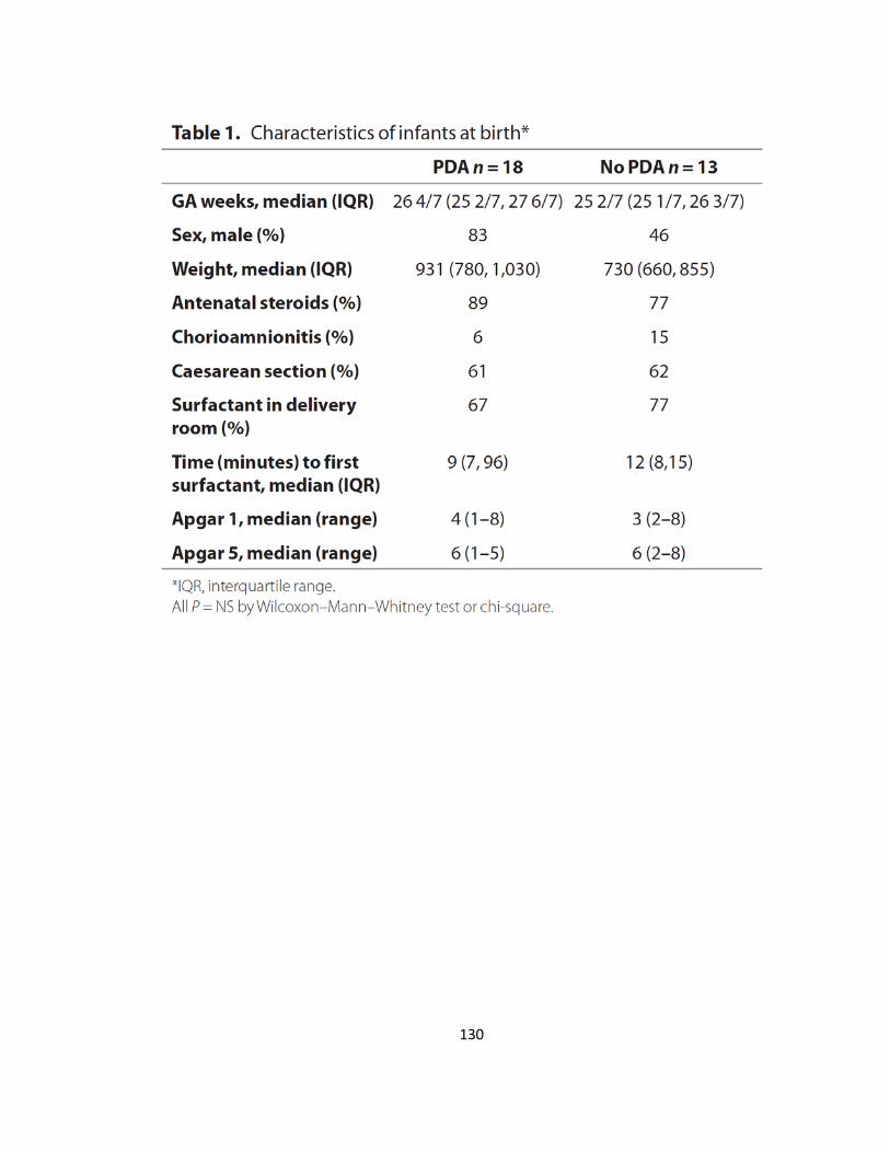
130
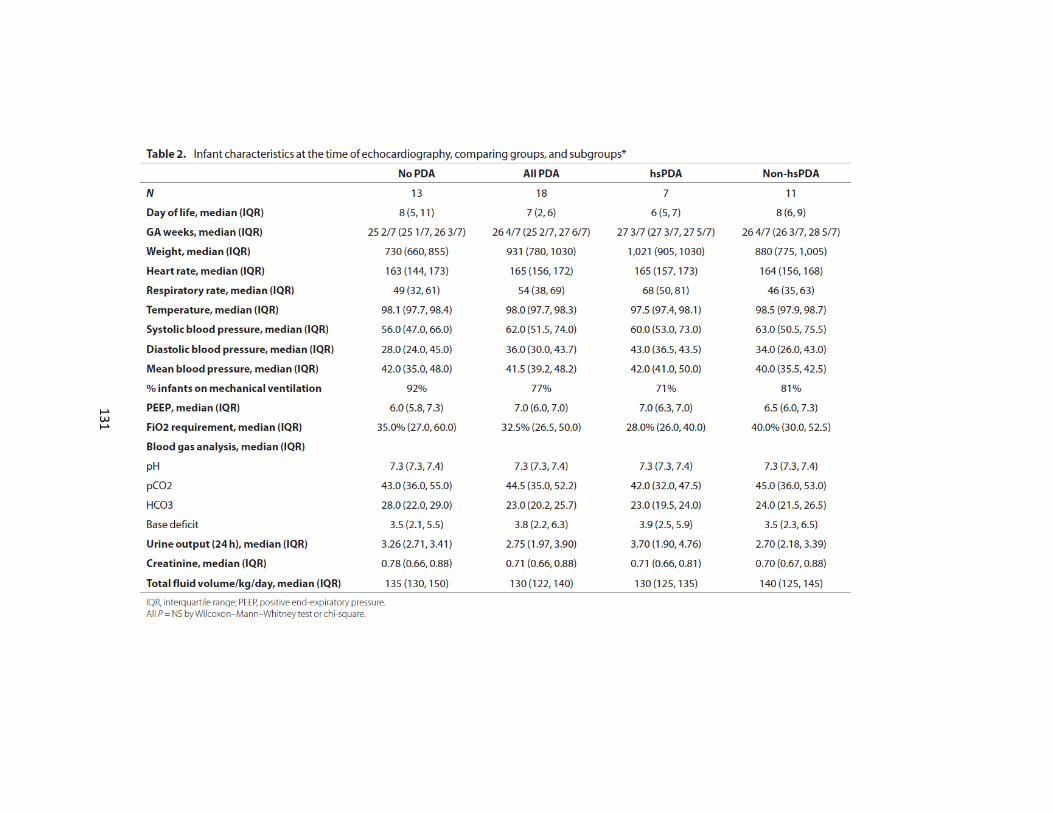
131
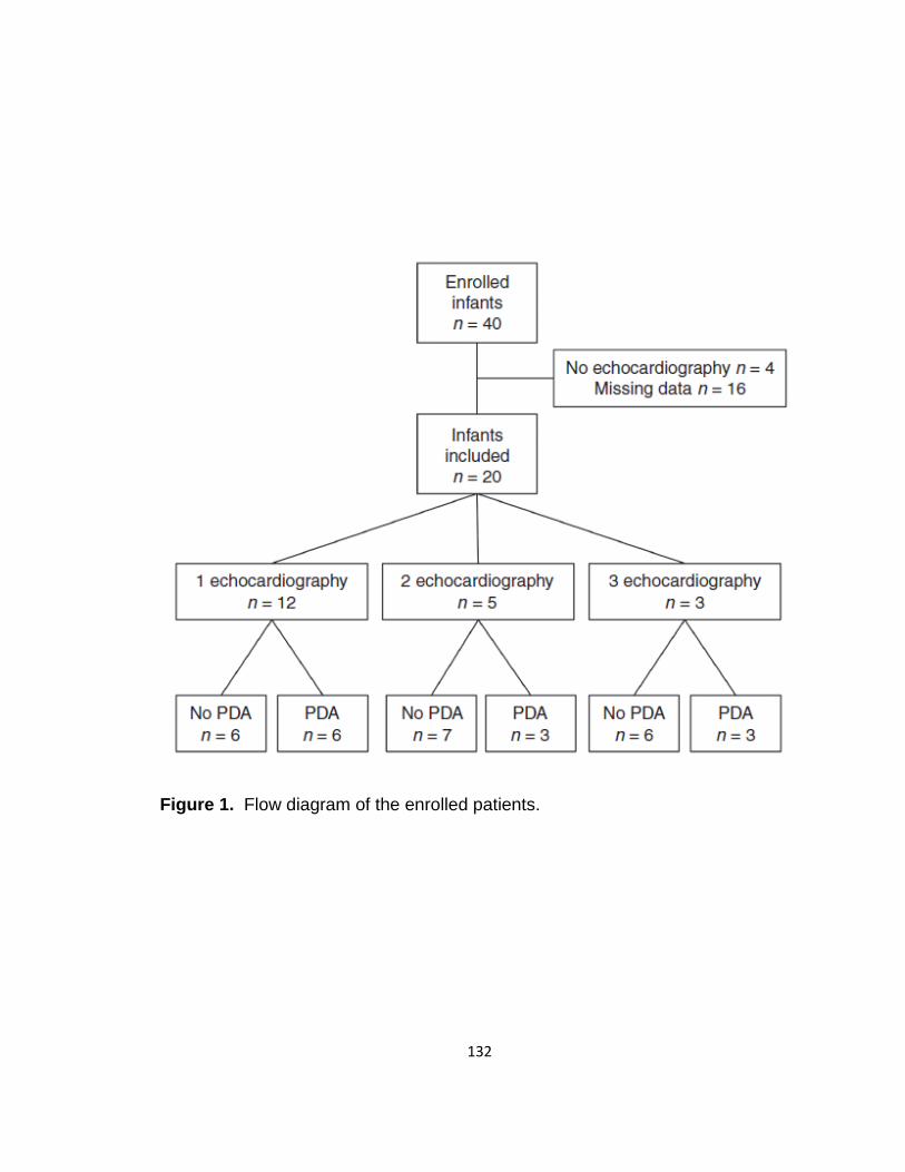
132
Figure 1. Flow diagram of the enrolled patients.
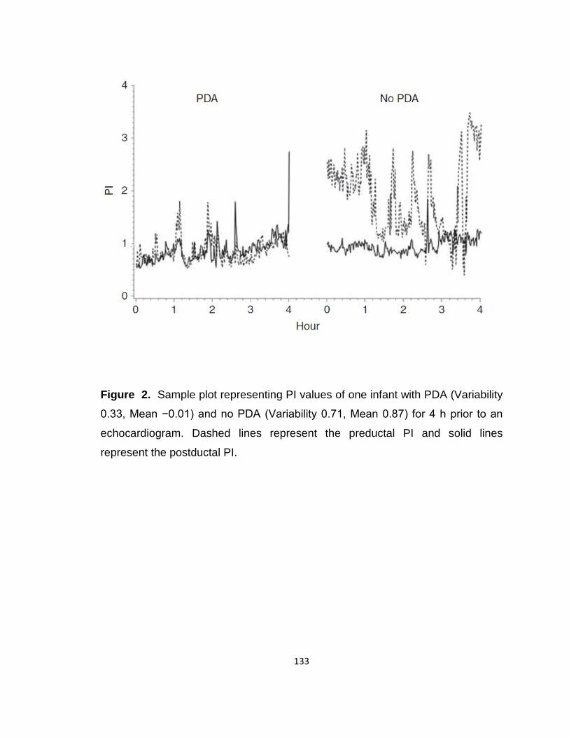
133
Figure 2. Sample plot representing PI values of one infant with PDA (Variability
0.33, Mean −0.01) and no PDA (Variability 0.71, Mean 0.87) for 4 h prior to an
echocardiogram. Dashed lines represent the preductal PI and solid lines
represent the postductal PI.
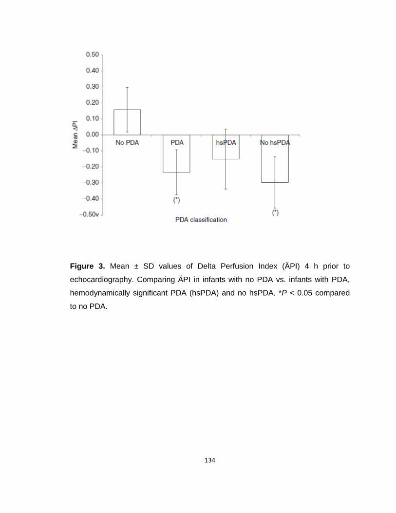
134
Figure 3. Mean ± SD values of Delta Perfusion Index (ÄPI) 4 h prior to
echocardiography. Comparing ÄPI in infants with no PDA vs. infants with PDA,
hemodynamically significant PDA (hsPDA) and no hsPDA. *P < 0.05 compared
to no PDA.
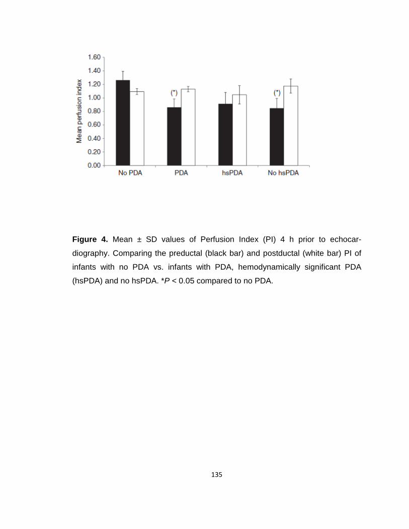
135
Figure 4. Mean ± SD values of Perfusion Index (PI) 4 h prior to echocar-
diography. Comparing the preductal (black bar) and postductal (white bar) PI of
infants with no PDA vs. infants with PDA, hemodynamically significant PDA
(hsPDA) and no hsPDA. *P < 0.05 compared to no PDA.

136
Figure 5. Mean ± SD values of the Variability of Perfusion Index (PI) 4 h prior to
echocardiography. Comparing ∆PI (dotted bar), preductal PI (black bar) and
postductal PI (white bar) variability for infants with no PDA vs. with PDA,
hemodynamically significant PDA (hsPDA) and no hsPDA. *P < 0.05 and ‡P =
0.08 compared to no PDA.

137
REFERENCES (Appendix B)
1. Benitz WE, Committee On Fetus and Newborn, 2016 Patent Ductus Arte- riosus in Preterm Infants. Pediatrics 137:1–6.
2. Dice JE, Bhatia J. Patent ductus arteriosus: an overview. J Pediatr Pharma- col Ther 2007;12:138–46.
3. Hamrick SE, Hansmann G. Patent ductus arteriosus of the preterm infant. Pediatrics 2010;125:1020–30.
4. Skelton R, Evans N, Smythe J. A blinded comparison of clinical and echo- cardiographic evaluation of the preterm infant for patent ductus arteriosus. J Paediatr Child Health 1994;30:406–11.
5. Visconti LF, Morhy SS, Deutsch AD, Tavares GM, Wilberg TJ, Rossi Fde S. Clinical and echocardiographic characteristics associated with the evolu- tion of the ductus arteriosus in the neonate with birth weight lower than 1,500g. Einstein (Sao Paulo) 2013;11:317–23.
6. Zonnenberg I, de Waal K. The definition of a haemodynamic significant duct in randomized controlled trials: a systematic literature review. Acta Paediatr 2012;101:247–51.
7. Evans N, Malcolm G, Osborn D, Kluckow M 2004 Diagnosis of patent duc- tus arteriosus in preterm infants. NeoReviews 5:e86–e97.
8. Evans N. Diagnosis of the preterm patent ductus arteriosus: clinical signs, biomarkers, or ultrasound? Semin Perinatol 2012;36:114–22. Alderliesten T, Lemmers PM, Baerts W, Groenendaal F, van Bel F. Perfu- sion index in preterm infants during the first 3 days of life: reference values and relation with clinical variables. Neonatology 2015;107:258–65.
9. Piasek CZ, Van Bel F, Sola A. Perfusion index in newborn infants: a nonin- vasive tool for neonatal monitoring. Acta Paediatr 2014;103:468–73.
10. Zaramella P, Freato F, Quaresima V, et al. Foot pulse oximeter perfusion index correlates with calf muscle perfusion measured by near-infrared spectroscopy in healthy neonates. J Perinatol 2005;25:417–22.
11. Vidal M, Ferragu F, Durand S, Baleine J, Batista-Novais AR, Cambonie G. Perfusion index and its dynamic changes in preterm neonates with patent ductus arteriosus. Acta Paediatr 2013;102:373–8.
138
12. Kinoshita M, Hawkes CP, Ryan CA, Dempsey EM. Perfusion index in the very preterm infant. Acta Paediatr 2013;102:e398–401.
13. Granelli Ad, Ostman-Smith I. Noninvasive peripheral perfusion index as a possible tool for screening for critical left heart obstruction. Acta Paediatr 2007;96:1455–9.
14. De Felice C, Goldstein MR, Parrini S, Verrotti A, Criscuolo M, Latini G. Early dynamic changes in pulse oximetry signals in preterm newborns with histologic chorioamnionitis. Pediatr Crit Care Med 2006;7:138–42.
15. Khositseth A, Muangyod N, Nuntnarumit P. Perfusion index as a diag- nostic tool for patent ductus arteriosus in preterm infants. Neonatology 2013;104:250–4.
16. Sehgal A, McNamara PJ. Does echocardiography facilitate determination of hemodynamic significance attributable to the ductus arteriosus? Eur J Pediatr 2009;168:907–14.
17. Sehgal A, Paul E, Menahem S. Functional echocardiography in stag- ing for ductal disease severity: role in predicting outcomes. Eur J Pediatr 2013;172:179–84.
18. El Hajjar M, Vaksmann G, Rakza T, Kongolo G, Storme L 2005 Severity of the ductal shunt: a comparison of different markers. Arch Dis Child Fetal Neonatal Ed 90:F419–422.
19. Shimada M, Takahashi K, Segawa M, Higurashi M, Samejim M, Horiu- chi K. Emerging and entraining patterns of the sleep-wake rhythm in pre- term and term infants. Brain Dev 1999;21:468–73.
20. Kenward MG, Roger JH. Small sample inference for fixed effects from restricted maximum likelihood. Biometrics 1997;53:983–97.
21. Liang K-Y, Zeger SL 1986 Longitudinal data analysis using generalized lin- ear models. Biometrika 73:13–22.
22. Cresi F, Pelle E, Calabrese R, Costa L, Farinasso D, Silvestro L. Perfusion index variations in clinically and hemodynamically stable preterm new- borns in the first week of life. Ital J Pediatr 2010;36:6.
23. Hakan N, Dilli D, Zenciroglu A, Aydin M, Okumus N. Reference values of perfusion indices in hemodynamically stable newborns during the early neonatal period. Eur J Pediatr 2014;173:597–602.
139
24. Karadag N, Dilli D, Zenciroglu A, Aydin B, Beken S, Okumus N. Perfusion index variability in preterm infants treated with two different natural sur- factants for respiratory distress syndrome. Am J Perinatol 2014;31:1015– 22.
25. De Felice C, Latini G, Vacca P, Kopotic RJ. The pulse oximeter perfusion index as a predictor for high illness severity in neonates. Eur J Pediatr 2002;161:561–2.
26. Evans N, Iyer P. Change in blood pressure after treatment of patent duc- tus arteriosus with indomethacin. Arch Dis Child 1993;68(5 Spec No): 584–7.
27. Freeman-Ladd M, Cohen JB, Carver JD, Huhta JC. The hemodynamic effects of neonatal patent ductus arteriosus shunting on superior mesen- teric artery blood flow. J Perinatol 2005;25:459–62.
28. Shimada S, Kasai T, Hoshi A, Murata A, Chida S. Cardiocirculatory effects of patent ductus arteriosus in extremely low-birth-weight infants with respiratory distress syndrome. Pediatr Int 2003;45:255–62.
29. Volpe JJ, Perlman JM, Hill A, McMenamin JB. Cerebral blood flow veloc- ity in the human newborn: the value of its determination. Pediatrics 1982;70:147–52.
30. Martin CG, Snider AR, Katz SM, Peabody JL, Brady JP. Abnormal cerebral blood flow patterns in preterm infants with a large patent ductus arterio- sus. J Pediatr 1982;101:587–93.
31. Takahashi S, Kakiuchi S, Nanba Y, Tsukamoto K, Nakamura T, Ito Y. The perfusion index derived from a pulse oximeter for predicting low superior vena cava flow in very low birth weight infants. J Perinatol 2010;30:265–9.
32. Prietsch V, Maier R, Schmitz L, Obladen M. Long-term variability of heart rate increases with successful closure of patent ductus arteriosus in pre- term infants. Biol Neonate 1992;61:142–149.
33. Patrick J McNamara AS 2007 Towards rational management of the patent ductus arteriosus: the need for disease staging. Arch Dis Child Fetal Neo- natal Ed. 92:F424–F427.

140
REFERENCES
1. Abu Jawdeh EG, Martin RJ, Dick TE, Walsh MC, Di Fiore JM. The effect of red blood cell transfusion on intermittent hypoxemia in ELBW infants. J Perinatol. 2014;34(12):921-5. doi: 10.1038/jp.2014.115. PubMed PMID: 24921411; PMCID: PMC4245392.
2. Di Fiore JM, Bloom JN, Orge F, Schutt A, Schluchter M, Cheruvu VK, Walsh M, Finer N, Martin RJ. A higher incidence of intermittent hypoxemic episodes is associated with severe retinopathy of prematurity. J Pediatr. 2010;157(1):69-73. doi: 10.1016/j.jpeds.2010.01.046. PubMed PMID: 20304417; PMCID: PMC4428609.
3. Poets CF, Roberts RS, Schmidt B, Whyte RK, Asztalos EV, Bader D, Bairam A, Moddemann D, Peliowski A, Rabi Y, Solimano A, Nelson H, Canadian Oxygen Trial I. Association Between Intermittent Hypoxemia or Bradycardia and Late Death or Disability in Extremely Preterm Infants. JAMA. 2015;314(6):595-603. doi: 10.1001/jama.2015.8841. PubMed PMID: 26262797.
4. Rhein LM, Dobson NR, Darnall RA, Corwin MJ, Heeren TC, Poets CF, McEntire BL, Hunt CE, Caffeine Pilot Study G. Effects of caffeine on intermittent hypoxia in infants born prematurely: a randomized clinical trial. JAMA Pediatr. 2014;168(3):250-7. doi: 10.1001/jamapediatrics.2013.4371. PubMed PMID: 24445955.
5. Fairchild K, Mohr M, Paget-Brown A, Tabacaru C, Lake D, Delos J, Moorman JR, Kattwinkel J. Clinical associations of immature breathing in preterm infants: part 1-central apnea. Pediatr Res. 2016;80(1):21-7. doi: 10.1038/pr.2016.43. PubMed PMID: 26959485; PMCID: PMC5015591.
6. Eichenwald EC, Aina A, Stark AR. Apnea frequently persists beyond term gestation in infants delivered at 24 to 28 weeks. Pediatrics. 1997;100(3 Pt 1):354-9. PubMed PMID: 9282705.
7. Martin RJ, Di Fiore JM, Walsh MC. Hypoxic Episodes in Bronchopulmonary Dysplasia. Clin Perinatol. 2015;42(4):825-38. doi: 10.1016/j.clp.2015.08.009. PubMed PMID: 26593081; PMCID: PMC4660265.
8. Di Fiore JM, Walsh M, Wrage L, Rich W, Finer N, Carlo WA, Martin RJ, Health SSGoEKSNIoC, Human Development Neonatal Research N. Low oxygen saturation target range is associated with increased incidence of intermittent
141
hypoxemia. J Pediatr. 2012;161(6):1047-52. doi: 10.1016/j.jpeds.2012.05.046. PubMed PMID: 22738947; PMCID: PMC3730286.
9. Waggener TB, Frantz ID, 3rd, Cohlan BA, Stark AR. Mixed and obstructive apneas are related to ventilatory oscillations in premature infants. J Appl Physiol (1985). 1989;66(6):2818-26. PubMed PMID: 2745345.
10. Poets CF. Apnea of prematurity: What can observational studies tell us about pathophysiology? Sleep Med. 2010;11(7):701-7. doi: 10.1016/j.sleep.2009.11.016. PubMed PMID: 20621558.
11. Finer NN, Higgins R, Kattwinkel J, Martin RJ. Summary proceedings from the apnea-of-prematurity group. Pediatrics. 2006;117(3 Pt 2):S47-51. doi: 10.1542/peds.2005-0620H. PubMed PMID: 16777822.
12. Di Fiore JM, Martin RJ, Gauda EB. Apnea of prematurity--perfect storm. Respir Physiol Neurobiol. 2013;189(2):213-22. doi: 10.1016/j.resp.2013.05.026. PubMed PMID: 23727228.
13. Martin RJ, Fanaroff AA, Walsh MC. Fanaroff and Martin's neonatal-perinatal medicine : diseases of the fetus and infant. 9th ed. Philadelphia: Saunders/Elsevier; 2011.
14. Gauda EB, Shirahata M, Mason A, Pichard LE, Kostuk EW, Chavez-Valdez R. Inflammation in the carotid body during development and its contribution to apnea of prematurity. Respir Physiol Neurobiol. 2013;185(1):120-31. doi: 10.1016/j.resp.2012.08.005. PubMed PMID: 22902305.
15. Fanaroff AA, Martin RJ, Walsh MC. Fanaroff and Martin's neonatal-perinatal medicine : diseases of the fetus and infant. 8th ed. Philadelphia, Pa.: Mosby Elsevier; 2006.
16. Sands SA, Edwards BA, Kelly VJ, Davidson MR, Wilkinson MH, Berger PJ. A model analysis of arterial oxygen desaturation during apnea in preterm infants. PLoS Comput Biol. 2009;5(12):e1000588. doi: 10.1371/journal.pcbi.1000588. PubMed PMID: 19997495; PMCID: PMC2778953.
17. Olsen GD, Lees MH. Ventilatory response to carbon dioxide of infants following chronic prenatal methadone exposure. J Pediatr. 1980;96(6):983-9. PubMed PMID: 6768869.
18. Ali K, Wolff K, Peacock JL, Hannam S, Rafferty GF, Bhat R, Greenough A. Ventilatory response to hypercarbia in newborns of smoking and substance-
142
misusing mothers. Ann Am Thorac Soc. 2014;11(6):933-8. doi: 10.1513/AnnalsATS.201403-124OC. PubMed PMID: 24983462.
19. Kandall SR, Gaines J. Maternal substance use and subsequent sudden infant death syndrome (SIDS) in offspring. Neurotoxicol Teratol. 1991;13(2):235-40. PubMed PMID: 2046641.
20. Ward SL, Schuetz S, Kirshna V, Bean X, Wingert W, Wachsman L, Keens TG. Abnormal sleeping ventilatory pattern in infants of substance-abusing mothers. Am J Dis Child. 1986;140(10):1015-20. PubMed PMID: 3752011.
21. Mueller RA, Lundberg DB, Breese GR, Hedner J, Hedner T, Jonason J. The neuropharmacology of respiratory control. Pharmacol Rev. 1982;34(3):255-85. PubMed PMID: 6185961.
22. Koehntop DE, Rodman JH, Brundage DM, Hegland MG, Buckley JJ. Pharmacokinetics of fentanyl in neonates. Anesth Analg. 1986;65(3):227-32. PubMed PMID: 3954090.
23. Perry BD, Pesavento DJ, Kussie PH, U'Prichard DC, Schnoll SH. Prenatal exposure to drugs of abuse in humans: effects on placental neurotransmitter receptors. Neurobehav Toxicol Teratol. 1984;6(4):295-301. PubMed PMID: 6096745.
24. Silvestri JM, Long JM, Weese-Mayer DE, Barkov GA. Effect of prenatal cocaine on respiration, heart rate, and sudden infant death syndrome. Pediatr Pulmonol. 1991;11(4):328-34. PubMed PMID: 1758757.
25. Hafstrom O, Milerad J, Sandberg KL, Sundell HW. Cardiorespiratory effects of nicotine exposure during development. Respir Physiol Neurobiol. 2005;149(1-3):325-41. doi: 10.1016/j.resp.2005.05.004. PubMed PMID: 15970470.
26. Robinson DM, Peebles KC, Kwok H, Adams BM, Clarke LL, Woollard GA, Funk GD. Prenatal nicotine exposure increases apnoea and reduces nicotinic potentiation of hypoglossal inspiratory output in mice. J Physiol. 2002;538(Pt 3):957-73. PubMed PMID: 11826179; PMCID: PMC2290085.
27. Hanrahan JP, Tager IB, Segal MR, Tosteson TD, Castile RG, Van Vunakis H, Weiss ST, Speizer FE. The effect of maternal smoking during pregnancy on early infant lung function. Am Rev Respir Dis. 1992;145(5):1129-35. doi: 10.1164/ajrccm/145.5.1129. PubMed PMID: 1586058.
143
28. Ueda Y, Stick SM, Hall G, Sly PD. Control of breathing in infants born to smoking mothers. J Pediatr. 1999;135(2 Pt 1):226-32. PubMed PMID: 10431118.
29. Hofstetter AO, Herlenius E. Interleukin-1beta depresses hypoxic gasping and autoresuscitation in neonatal DBA/1lacJ mice. Respir Physiol Neurobiol. 2005;146(2-3):135-46. doi: 10.1016/j.resp.2004.11.002. PubMed PMID: 15766902.
30. Hofstetter AO, Saha S, Siljehav V, Jakobsson PJ, Herlenius E. The induced prostaglandin E2 pathway is a key regulator of the respiratory response to infection and hypoxia in neonates. Proc Natl Acad Sci U S A. 2007;104(23):9894-9. doi: 10.1073/pnas.0611468104. PubMed PMID: 17535900; PMCID: PMC1877988.
31. Hansen MK, Taishi P, Chen Z, Krueger JM. Vagotomy blocks the induction of interleukin-1beta (IL-1beta) mRNA in the brain of rats in response to systemic IL-1beta. J Neurosci. 1998;18(6):2247-53. PubMed PMID: 9482809.
32. Darnall RA, Chen X, Nemani KV, Sirieix CM, Gimi B, Knoblach S, McEntire BL, Hunt CE. Early postnatal exposure to intermittent hypoxia in rodents is proinflammatory, impairs white matter integrity, and alters brain metabolism. Pediatr Res. 2017. doi: 10.1038/pr.2017.102. PubMed PMID: 28388601.
33. Julien CA, Joseph V, Bairam A. Alteration of carotid body chemoreflexes after neonatal intermittent hypoxia and caffeine treatment in rat pups. Respir Physiol Neurobiol. 2011;177(3):301-12. doi: 10.1016/j.resp.2011.05.006. PubMed PMID: 21609788.
34. Gauda EB, Carroll JL, Donnelly DF. Developmental maturation of chemosensitivity to hypoxia of peripheral arterial chemoreceptors--invited article. Adv Exp Med Biol. 2009;648:243-55. doi: 10.1007/978-90-481-2259-2_28. PubMed PMID: 19536487.
35. Liu X, He L, Stensaas L, Dinger B, Fidone S. Adaptation to chronic hypoxia involves immune cell invasion and increased expression of inflammatory cytokines in rat carotid body. Am J Physiol Lung Cell Mol Physiol. 2009;296(2):L158-66. doi: 10.1152/ajplung.90383.2008. PubMed PMID: 18978039; PMCID: PMC2643993.
36. Zagol K, Lake DE, Vergales B, Moorman ME, Paget-Brown A, Lee H, Rusin CG, Delos JB, Clark MT, Moorman JR, Kattwinkel J. Anemia, apnea of
144
prematurity, and blood transfusions. J Pediatr. 2012;161(3):417-21 e1. doi: 10.1016/j.jpeds.2012.02.044. PubMed PMID: 22494873; PMCID: PMC5321065.
37. Network SSGotEKSNNR, Carlo WA, Finer NN, Walsh MC, Rich W, Gantz MG, Laptook AR, Yoder BA, Faix RG, Das A, Poole WK, Schibler K, Newman NS, Ambalavanan N, Frantz ID, 3rd, Piazza AJ, Sanchez PJ, Morris BH, Laroia N, Phelps DL, Poindexter BB, Cotten CM, Van Meurs KP, Duara S, Narendran V, Sood BG, O'Shea TM, Bell EF, Ehrenkranz RA, Watterberg KL, Higgins RD. Target ranges of oxygen saturation in extremely preterm infants. N Engl J Med. 2010;362(21):1959-69. doi: 10.1056/NEJMoa0911781. PubMed PMID: 20472937; PMCID: PMC2891970.
38. Brockmann PE, Wiechers C, Pantalitschka T, Diebold J, Vagedes J, Poets CF. Under-recognition of alarms in a neonatal intensive care unit. Arch Dis Child Fetal Neonatal Ed. 2013;98(6):F524-7. doi: 10.1136/archdischild-2012-303369. PubMed PMID: 23716498.
39. Vagedes J, Poets CF, Dietz K. Averaging time, desaturation level, duration and extent. Arch Dis Child Fetal Neonatal Ed. 2013;98(3):F265-6. doi: 10.1136/archdischild-2012-302543. PubMed PMID: 22960097.
40. Julien C, Bairam A, Joseph V. Chronic intermittent hypoxia reduces ventilatory long-term facilitation and enhances apnea frequency in newborn rats. Am J Physiol Regul Integr Comp Physiol. 2008;294(4):R1356-66. doi: 10.1152/ajpregu.00884.2007. PubMed PMID: 18287216.
41. Pozo ME, Cave A, Koroglu OA, Litvin DG, Martin RJ, Di Fiore J, Kc P. Effect of postnatal intermittent hypoxia on growth and cardiovascular regulation of rat pups. Neonatology. 2012;102(2):107-13. doi: 10.1159/000338096. PubMed PMID: 22677790; PMCID: PMC3495107.
42. Schmid MB, Hopfner RJ, Lenhof S, Hummler HD, Fuchs H. Cerebral oxygenation during intermittent hypoxemia and bradycardia in preterm infants. Neonatology. 2015;107(2):137-46. doi: 10.1159/000368294. PubMed PMID: 25531368.
43. Schmidt B, Whyte RK, Asztalos EV, Moddemann D, Poets C, Rabi Y, Solimano A, Roberts RS, Canadian Oxygen Trial G. Effects of targeting higher vs lower arterial oxygen saturations on death or disability in extremely preterm infants: a randomized clinical trial. JAMA. 2013;309(20):2111-20. doi: 10.1001/jama.2013.5555. PubMed PMID: 23644995.
145
44. Janvier A, Khairy M, Kokkotis A, Cormier C, Messmer D, Barrington KJ. Apnea is associated with neurodevelopmental impairment in very low birth weight infants. J Perinatol. 2004;24(12):763-8. doi: 10.1038/sj.jp.7211182. PubMed PMID: 15329741.
45. Pillekamp F, Hermann C, Keller T, von Gontard A, Kribs A, Roth B. Factors influencing apnea and bradycardia of prematurity - implications for neurodevelopment. Neonatology. 2007;91(3):155-61. doi: 10.1159/000097446. PubMed PMID: 17377399.
46. Di Fiore JM, Martin RJ, Li H, Morris N, Carlo WA, Finer N, Walsh M, Health SSGotEKSNIoC, Human Development Neonatal Research N. Patterns of Oxygenation, Mortality, and Growth Status in the Surfactant Positive Pressure and Oxygen Trial Cohort. J Pediatr. 2017. doi: 10.1016/j.jpeds.2017.01.057. PubMed PMID: 28279433.
47. Oei JL, Finer NN, Saugstad OD, Wright IM, Rabi Y, Tarnow-Mordi W, Rich W, Kapadia V, Rook D, Smyth JP, Lui K, Vento M. Outcomes of oxygen saturation targeting during delivery room stabilisation of preterm infants. Arch Dis Child Fetal Neonatal Ed. 2017. doi: 10.1136/archdischild-2016-312366. PubMed PMID: 28988158.
48. Darlow BA, Marschner SL, Donoghoe M, Battin MR, Broadbent RS, Elder MJ, Hewson MP, Meyer MP, Ghadge A, Graham P, McNeill NJ, Kuschel CA, Tarnow-Mordi WO, Benefits Of Oxygen Saturation Targeting-New Zealand Collaborative G. Randomized controlled trial of oxygen saturation targets in very preterm infants: two year outcomes. J Pediatr. 2014;165(1):30-5 e2. doi: 10.1016/j.jpeds.2014.01.017. PubMed PMID: 24560181.
49. Khadawardi E, Al Hazzani F. Oxygen Saturation and Outcomes in Preterm Infants The BOOST II United Kingdom, Australia, and New Zealand Collaborative Groups. J Clin Neonatol. 2013;2(2):73-5. doi: 10.4103/2249-4847.116404. PubMed PMID: 24049747; PMCID: PMC3775139.
50. Group BIUKC, Group BIAC, Group BINZC, Stenson BJ, Tarnow-Mordi WO, Darlow BA, Simes J, Juszczak E, Askie L, Battin M, Bowler U, Broadbent R, Cairns P, Davis PG, Deshpande S, Donoghoe M, Doyle L, Fleck BW, Ghadge A, Hague W, Halliday HL, Hewson M, King A, Kirby A, Marlow N, Meyer M, Morley C, Simmer K, Tin W, Wardle SP, Brocklehurst P. Oxygen saturation and outcomes in preterm infants. N Engl J Med. 2013;368(22):2094-104. doi: 10.1056/NEJMoa1302298. PubMed PMID: 23642047.
146
51. Johnston ED, Boyle B, Juszczak E, King A, Brocklehurst P, Stenson BJ. Oxygen targeting in preterm infants using the Masimo SET Radical pulse oximeter. Arch Dis Child Fetal Neonatal Ed. 2011;96(6):F429-33. doi: 10.1136/adc.2010.206011. PubMed PMID: 21378398; PMCID: PMC3195299.
52. Ambalavanan N, Carlo WA, Wrage LA, Das A, Laughon M, Cotten CM, Kennedy KA, Laptook AR, Shankaran S, Walsh MC, Higgins RD, Network SSGotNNR. PaCO2 in surfactant, positive pressure, and oxygenation randomised trial (SUPPORT). Arch Dis Child Fetal Neonatal Ed. 2015;100(2):F145-9. doi: 10.1136/archdischild-2014-306802. PubMed PMID: 25425651; PMCID: PMC4336211.
53. McClure C, Jang SY, Fairchild K. Alarms, oxygen saturations, and SpO2 averaging time in the NICU. Journal of neonatal-perinatal medicine. 2016. doi: 10.3233/NPM-16162. PubMed PMID: 27834782.
54. Abu Jawdeh EG. Intermittent Hypoxemia in Preterm Infants: Etiology and Clinical Relevance. NeoReviews.18(11):e637-e46. PubMed PMID: 28099422.
55. Poets CF. Intermittent hypoxemia/bradycardia and the developing brain: how much is too much? Commentary on M.B. Schmid et al.: Cerebral oxygenation during intermittent hypoxemia and bradycardia in preterm infants (Neonatology 2015;107:137-146). Neonatology. 2015;107(2):147-9. doi: 10.1159/000369775. PubMed PMID: 25531535.
56. Abu Jawdeh EG. Prenatal Opioid Exposure and Intermittent Hypoxemia in Preterm Infants: A Retrospective Assessment. Pediatr Res. 2017;5:253. Epub 2017/01/19. doi: 10.1038/pr.2017.10
10.3389/fped.2017.00253. PubMed PMID: 29270395.
57. Alderliesten T, Lemmers PM, Baerts W, Groenendaal F, van Bel F. Perfusion Index in Preterm Infants during the First 3 Days of Life: Reference Values and Relation with Clinical Variables. Neonatology. 2015;107(4):258-65. doi: 10.1159/000370192. PubMed PMID: 25720415.
58. Piasek CZ, Van Bel F, Sola A. Perfusion index in newborn infants: a noninvasive tool for neonatal monitoring. Acta Paediatr. 2014;103(5):468-73. doi: 10.1111/apa.12574. PubMed PMID: 24471645.
59. Granelli A, Ostman-Smith I. Noninvasive peripheral perfusion index as a possible tool for screening for critical left heart obstruction. Acta Paediatr. 2007;96(10):1455-9. doi: 10.1111/j.1651-2227.2007.00439.x. PubMed PMID: 17727691.

147
60. De Felice C, Goldstein MR, Parrini S, Verrotti A, Criscuolo M, Latini G. Early dynamic changes in pulse oximetry signals in preterm newborns with histologic chorioamnionitis. Pediatr Crit Care Med. 2006;7(2):138-42. doi: 10.1097/01.PCC.0000201002.50708.62. PubMed PMID: 16474255.
61. Gomez-Pomar E, Makhoul M, Westgate PM, Ibonia KT, Patwardhan A, Giannone PJ, Bada HS, Abu Jawdeh EG. Relationship between perfusion index and patent ductus arteriosus in preterm infants. Pediatr Res. 2017;81(5):775-9. doi: 10.1038/pr.2017.10. PubMed PMID: 28099422.
62. Fitzmaurice GM, Laird NM, Ware JH. Applied longitudinal analysis. 2nd ed. Hoboken, N.J.: Wiley; 2011. xxv, 701 p. p.
63. Little RJA, Rubin DB. Statistical analysis with missing data. 2nd ed. Hoboken, N.J.: Wiley; 2002. xv, 381 p. p.
64. Vagedes J, Bialkowski A, Wiechers C, Poets CF, Dietz K. A conversion formula for comparing pulse oximeter desaturation rates obtained with different averaging times. PLoS One. 2014;9(1):e87280. doi: 10.1371/journal.pone.0087280. PubMed PMID: 24489887; PMCID: PMC3904986.
65. Wang X, Zhu Y, Dave CV, Alrwisan AA, Voils SA, Winterstein AG. Trends of Neonatal Abstinence Syndrome Epidemic and Maternal Risk Factors in Florida. Pharmacotherapy. 2017;37(7):806-13. doi: 10.1002/phar.1947. PubMed PMID: 28500694.
66. Jiang R, Lee I, Lee TA, Pickard AS. The societal cost of heroin use disorder in the United States. PLoS One. 2017;12(5):e0177323. doi: 10.1371/journal.pone.0177323. PubMed PMID: 28557994; PMCID: PMC5448739.
67. Corr TE, Hollenbeak CS. The economic burden of neonatal abstinence syndrome in the United States. Addiction. 2017. doi: 10.1111/add.13842. PubMed PMID: 28612362.
68. Patrick SW, Schumacher RE, Benneyworth BD, Krans EE, McAllister JM, Davis MM. Neonatal abstinence syndrome and associated health care expenditures: United States, 2000-2009. JAMA. 2012;307(18):1934-40. doi: 10.1001/jama.2012.3951. PubMed PMID: 22546608.
69. Patrick SW, Davis MM, Lehman CU, Cooper WO. Increasing incidence and geographic distribution of neonatal abstinence syndrome: United States
148
2009 to 2012. J Perinatol. 2015;35(8):667. doi: 10.1038/jp.2015.63. PubMed PMID: 26219703.
70. Ko JY, Patrick SW, Tong VT, Patel R, Lind JN, Barfield WD. Incidence of Neonatal Abstinence Syndrome - 28 States, 1999-2013. MMWR Morb Mortal Wkly Rep. 2016;65(31):799-802. doi: 10.15585/mmwr.mm6531a2. PubMed PMID: 27513154.
71. Reddy UM, Davis JM, Ren Z, Greene MF, Opioid Use in Pregnancy NAS, Childhood Outcomes Workshop Invited S. Opioid Use in Pregnancy, Neonatal Abstinence Syndrome, and Childhood Outcomes: Executive Summary of a Joint Workshop by the Eunice Kennedy Shriver National Institute of Child Health and Human Development, American College of Obstetricians and Gynecologists, American Academy of Pediatrics, Society for Maternal-Fetal Medicine, Centers for Disease Control and Prevention, and the March of Dimes Foundation. Obstet Gynecol. 2017;130(1):10-28. doi: 10.1097/AOG.0000000000002054. PubMed PMID: 28594753; PMCID: PMC5486414.
72. Heller NA, Logan BA, Morrison DG, Paul JA, Brown MS, Hayes MJ. Neonatal abstinence syndrome: Neurobehavior at 6 weeks of age in infants with or without pharmacological treatment for withdrawal. Dev Psychobiol. 2017;59(5):574-82. doi: 10.1002/dev.21532. PubMed PMID: 28561904.
73. Bada HS, Sithisarn T, Gibson J, Garlitz K, Caldwell R, Capilouto G, Li Y, Leggas M, Breheny P. Morphine versus clonidine for neonatal abstinence syndrome. Pediatrics. 2015;135(2):e383-91. doi: 10.1542/peds.2014-2377. PubMed PMID: 25624389.
74. Coyle MG, Salisbury AL, Lester BM, Jones HE, Lin H, Graf-Rohrmeister K, Fischer G. Neonatal neurobehavior effects following buprenorphine versus methadone exposure. Addiction. 2012;107 Suppl 1:63-73. doi: 10.1111/j.1360-0443.2012.04040.x. PubMed PMID: 23106928; PMCID: PMC4337995.
75. Jansson LM, Di Pietro JA, Elko A, Williams EL, Milio L, Velez M. Pregnancies exposed to methadone, methadone and other illicit substances, and poly-drugs without methadone: a comparison of fetal neurobehaviors and infant outcomes. Drug Alcohol Depend. 2012;122(3):213-9. doi: 10.1016/j.drugalcdep.2011.10.003. PubMed PMID: 22041255; PMCID: PMC3288292.
149
76. Bandstra ES, Morrow CE, Mansoor E, Accornero VH. Prenatal drug exposure: infant and toddler outcomes. J Addict Dis. 2010;29(2):245-58. doi: 10.1080/10550881003684871. PubMed PMID: 20407980.
77. Velez ML, Jansson LM, Schroeder J, Williams E. Prenatal methadone exposure and neonatal neurobehavioral functioning. Pediatr Res. 2009;66(6):704-9. doi: 10.1203/PDR.0b013e3181bc035d. PubMed PMID: 19690513; PMCID: PMC2796281.
78. Witt CE, Rudd KE, Bhatraju P, Rivara FP, Hawes SE, Weiss NS. Neonatal abstinence syndrome and early childhood morbidity and mortality in Washington state: a retrospective cohort study. J Perinatol. 2017. doi: 10.1038/jp.2017.106. PubMed PMID: 28682319.
79. Galland BC, Taylor BJ, Bolton DP, Sayers RM. Respiratory responses to hypoxia/hypercapnia in small for gestational age infants influenced by maternal smoking. Arch Dis Child Fetal Neonatal Ed. 2003;88(3):F217-22. PubMed PMID: 12719396; PMCID: PMC2291509.
80. Wingkun JG, Knisely JS, Schnoll SH, Gutcher GR. Decreased carbon dioxide sensitivity in infants of substance-abusing mothers. Pediatrics. 1995;95(6):864-7. PubMed PMID: 7761211.
81. Sullivan BA, Fairchild KD. Predictive monitoring for sepsis and necrotizing enterocolitis to prevent shock. Semin Fetal Neonatal Med. 2015;20(4):255-61. doi: 10.1016/j.siny.2015.03.006. PubMed PMID: 25823938.
82. Fairchild KD, Lake DE, Kattwinkel J, Moorman JR, Bateman DA, Grieve PG, Isler JR, Sahni R. Vital signs and their cross-correlation in sepsis and NEC: a study of 1,065 very-low-birth-weight infants in two NICUs. Pediatr Res. 2017;81(2):315-21. doi: 10.1038/pr.2016.215. PubMed PMID: 28001143; PMCID: PMC5309159.
83. Sullivan BA, Grice SM, Lake DE, Moorman JR, Fairchild KD. Infection and other clinical correlates of abnormal heart rate characteristics in preterm infants. J Pediatr. 2014;164(4):775-80. doi: 10.1016/j.jpeds.2013.11.038. PubMed PMID: 24412138; PMCID: PMC3962693.
84. Di Fiore JM, Poets CF, Gauda E, Martin RJ, MacFarlane P. Cardiorespiratory events in preterm infants: interventions and consequences. J Perinatol. 2016;36(4):251-8. doi: 10.1038/jp.2015.165. PubMed PMID: 26583943.
150
85. Marcus CL, Meltzer LJ, Roberts RS, Traylor J, Dix J, D'Ilario J, Asztalos E, Opie G, Doyle LW, Biggs SN, Nixon GM, Narang I, Bhattacharjee R, Davey M, Horne RS, Cheshire M, Gibbons J, Costantini L, Bradford R, Schmidt B, Caffeine for Apnea of Prematurity-Sleep S. Long-term effects of caffeine therapy for apnea of prematurity on sleep at school age. Am J Respir Crit Care Med. 2014;190(7):791-9. doi: 10.1164/rccm.201406-1092OC. PubMed PMID: 25171195; PMCID: PMC4299611.
86. Raffay TM, Dylag AM, Abu Jawdeh EG, Martin RJ, Di Fiore JM. Neonatal Intermittent Hypoxemia May Predict Bronchopulmonary Dysplasia Risk. Pediatric Academic Societies (PAS). 2017.
87. Gautier-Veyret E, Arnaud C, Back M, Pepin JL, Petri MH, Baguet JP, Tamisier R, Levy P, Stanke-Labesque F. Intermittent hypoxia-activated cyclooxygenase pathway: role in atherosclerosis. Eur Respir J. 2013;42(2):404-13. doi: 10.1183/09031936.00096512. PubMed PMID: 23060635.
88. Li K, Wei P, Qin Y, Wei Y. Is C-reactive protein a marker of obstructive sleep apnea?: A meta-analysis. Medicine (Baltimore). 2017;96(19):e6850. doi: 10.1097/MD.0000000000006850. PubMed PMID: 28489776; PMCID: PMC5428610.
89. Nadeem R, Molnar J, Madbouly EM, Nida M, Aggarwal S, Sajid H, Naseem J, Loomba R. Serum inflammatory markers in obstructive sleep apnea: a meta-analysis. J Clin Sleep Med. 2013;9(10):1003-12. doi: 10.5664/jcsm.3070. PubMed PMID: 24127144; PMCID: PMC3778171.
90. Wang J, Yu W, Gao M, Zhang F, Gu C, Yu Y, Wei Y. Impact of Obstructive Sleep Apnea Syndrome on Endothelial Function, Arterial Stiffening, and Serum Inflammatory Markers: An Updated Meta-analysis and Metaregression of 18 Studies. J Am Heart Assoc. 2015;4(11). doi: 10.1161/JAHA.115.002454. PubMed PMID: 26567373; PMCID: PMC4845236.
91. Wang Y, Chai Y, He X, Ai L, Sun X, Huang Y, Li Y. Intermittent hypoxia simulating obstructive sleep apnea causes pulmonary inflammation and activates the Nrf2/HO-1 pathway. Exp Ther Med. 2017;14(4):3463-70. doi: 10.3892/etm.2017.4971. PubMed PMID: 29042934; PMCID: PMC5639295.
92. Turnbull CD. Intermittent hypoxia, cardiovascular disease and obstructive sleep apnoea. J Thorac Dis. 2018;10(Suppl 1):S33-S9. doi: 10.21037/jtd.2017.10.33. PubMed PMID: 29445526; PMCID: PMC5803045.
151
93. Garvey JF, Taylor CT, McNicholas WT. Cardiovascular disease in obstructive sleep apnoea syndrome: the role of intermittent hypoxia and inflammation. Eur Respir J. 2009;33(5):1195-205. doi: 10.1183/09031936.00111208. PubMed PMID: 19407053.
94. Leviton A, Allred EN, Fichorova RN, Kuban KC, Michael O'Shea T, Dammann O, investigators Es. Systemic inflammation on postnatal days 21 and 28 and indicators of brain dysfunction 2years later among children born before the 28th week of gestation. Early Hum Dev. 2016;93:25-32. doi: 10.1016/j.earlhumdev.2015.11.004. PubMed PMID: 26735345; PMCID: PMC4733407.
95. Vaucher YE, Peralta-Carcelen M, Finer NN, Carlo WA, Gantz MG, Walsh MC, Laptook AR, Yoder BA, Faix RG, Das A, Schibler K, Rich W, Newman NS, Vohr BR, Yolton K, Heyne RJ, Wilson-Costello DE, Evans PW, Goldstein RF, Acarregui MJ, Adams-Chapman I, Pappas A, Hintz SR, Poindexter B, Dusick AM, McGowan EC, Ehrenkranz RA, Bodnar A, Bauer CR, Fuller J, O'Shea TM, Myers GJ, Higgins RD, Network SSGotEKSNNR. Neurodevelopmental outcomes in the early CPAP and pulse oximetry trial. N Engl J Med. 2012;367(26):2495-504. doi: 10.1056/NEJMoa1208506. PubMed PMID: 23268664; PMCID: PMC4140695.
96. Walsh MC, Di Fiore JM, Martin RJ, Gantz M, Carlo WA, Finer N. Association of Oxygen Target and Growth Status With Increased Mortality in Small for Gestational Age Infants: Further Analysis of the Surfactant, Positive Pressure and Pulse Oximetry Randomized Trial. JAMA Pediatr. 2016;170(3):292-4. doi: 10.1001/jamapediatrics.2015.3794. PubMed PMID: 26746140; PMCID: PMC5292772.
97. Osmand AP, Friedenson B, Gewurz H, Painter RH, Hofmann T, Shelton E. Characterization of C-reactive protein and the complement subcomponent C1t as homologous proteins displaying cyclic pentameric symmetry (pentraxins). Proc Natl Acad Sci U S A. 1977;74(2):739-43. PubMed PMID: 265538; PMCID: PMC392369.
98. Marnell L, Mold C, Du Clos TW. C-reactive protein: ligands, receptors and role in inflammation. Clin Immunol. 2005;117(2):104-11. doi: 10.1016/j.clim.2005.08.004. PubMed PMID: 16214080.
99. Hotamisligil GS. Inflammation and metabolic disorders. Nature. 2006;444(7121):860-7. doi: 10.1038/nature05485. PubMed PMID: 17167474.
152
100. Medzhitov R. Origin and physiological roles of inflammation. Nature. 2008;454(7203):428-35. doi: 10.1038/nature07201. PubMed PMID: 18650913.
101. Pourcyrous M, Korones SB, Arheart KL, Bada HS. Primary immunization of premature infants with gestational age <35 weeks: cardiorespiratory complications and C-reactive protein responses associated with administration of single and multiple separate vaccines simultaneously. J Pediatr. 2007;151(2):167-72. doi: 10.1016/j.jpeds.2007.02.059. PubMed PMID: 17643770.
102. Pourcyrous M, Korones SB, Yang W, Boulden TF, Bada HS. C-reactive protein in the diagnosis, management, and prognosis of neonatal necrotizing enterocolitis. Pediatrics. 2005;116(5):1064-9. doi: 10.1542/peds.2004-1806. PubMed PMID: 16263990.
103. Pourcyrous M, Bada HS, Korones SB, Baselski V, Wong SP. Significance of serial C-reactive protein responses in neonatal infection and other disorders. Pediatrics. 1993;92(3):431-5. PubMed PMID: 8361798.
104. Posen R, deLemos RA. C-reactive protein levels in the extremely premature infant: case studies and literature review. J Perinatol. 1998;18(2):138-41. PubMed PMID: 9605306.
105. Delanghe JR, Speeckaert MM. Translational research and biomarkers in neonatal sepsis. Clin Chim Acta. 2015;451(Pt A):46-64. doi: 10.1016/j.cca.2015.01.031. PubMed PMID: 25661089.
106. Kushner I, Samols D, Magrey M. A unifying biologic explanation for "high-sensitivity" C-reactive protein and "low-grade" inflammation. Arthritis Care Res (Hoboken). 2010;62(4):442-6. doi: 10.1002/acr.20052. PubMed PMID: 20391496.
107. Kuppala VS, Tabangin M, Haberman B, Steichen J, Yolton K. Current state of high-risk infant follow-up care in the United States: results of a national survey of academic follow-up programs. J Perinatol. 2012;32(4):293-8. doi: 10.1038/jp.2011.97. PubMed PMID: 21760588.
108. Vohr BR, Allan WC, Westerveld M, Schneider KC, Katz KH, Makuch RW, Ment LR. School-age outcomes of very low birth weight infants in the indomethacin intraventricular hemorrhage prevention trial. Pediatrics. 2003;111(4 Pt 1):e340-6. PubMed PMID: 12671149.

153
109. Hack M. Survival and neurodevelopmental outcomes of preterm infants. J Pediatr Gastroenterol Nutr. 2007;45 Suppl 3:S141-2. doi: 10.1097/01.mpg.0000302959.55428.05. PubMed PMID: 18185079.
110. Hack M. Young adult outcomes of very-low-birth-weight children. Semin Fetal Neonatal Med. 2006;11(2):127-37. doi: 10.1016/j.siny.2005.11.007. PubMed PMID: 16364703.
111. In: Behrman RE, Butler AS, editors. Preterm Birth: Causes, Consequences, and Prevention. Washington (DC)2007.
112. Bockli K, Andrews B, Pellerite M, Meadow W. Trends and challenges in United States neonatal intensive care units follow-up clinics. J Perinatol. 2014;34(1):71-4. doi: 10.1038/jp.2013.136. PubMed PMID: 24177221.
113. Younge N, Goldstein RF, Bann CM, Hintz SR, Patel RM, Smith PB, Bell EF, Rysavy MA, Duncan AF, Vohr BR, Das A, Goldberg RN, Higgins RD, Cotten CM, Eunice Kennedy Shriver National Institute of Child H, Human Development Neonatal Research N. Survival and Neurodevelopmental Outcomes among Periviable Infants. N Engl J Med. 2017;376(7):617-28. doi: 10.1056/NEJMoa1605566. PubMed PMID: 28199816.
114. Nagata N, Saji M, Ito T, Ikeno S, Takahashi H, Terakawa N. Repetitive intermittent hypoxia-ischemia and brain damage in neonatal rats. Brain Dev. 2000;22(5):315-20. PubMed PMID: 10891639.
115. Prabhakar NR. Oxygen sensing during intermittent hypoxia: cellular and molecular mechanisms. J Appl Physiol (1985). 2001;90(5):1986-94. PubMed PMID: 11299293.
116. Feldman JL, Mitchell GS, Nattie EE. Breathing: rhythmicity, plasticity, chemosensitivity. Annu Rev Neurosci. 2003;26:239-66. doi: 10.1146/annurev.neuro.26.041002.131103. PubMed PMID: 12598679; PMCID: PMC2811316.
117. Douglas RM, Miyasaka N, Takahashi K, Latuszek-Barrantes A, Haddad GG, Hetherington HP. Chronic intermittent but not constant hypoxia decreases NAA/Cr ratios in neonatal mouse hippocampus and thalamus. Am J Physiol Regul Integr Comp Physiol. 2007;292(3):R1254-9. doi: 10.1152/ajpregu.00404.2006. PubMed PMID: 17082353.
118. Ratner V, Kishkurno SV, Slinko SK, Sosunov SA, Sosunov AA, Polin RA, Ten VS. The contribution of intermittent hypoxemia to late neurological handicap
154
in mice with hyperoxia-induced lung injury. Neonatology. 2007;92(1):50-8. doi: 10.1159/000100086. PubMed PMID: 17596736.
119. Ratner V, Slinko S, Utkina-Sosunova I, Starkov A, Polin RA, Ten VS. Hypoxic stress exacerbates hyperoxia-induced lung injury in a neonatal mouse model of bronchopulmonary dysplasia. Neonatology. 2009;95(4):299-305. doi: 10.1159/000178798. PubMed PMID: 19052476; PMCID: PMC3659784.
120. Farahani R, Kanaan A, Gavrialov O, Brunnert S, Douglas RM, Morcillo P, Haddad GG. Differential effects of chronic intermittent and chronic constant hypoxia on postnatal growth and development. Pediatr Pulmonol. 2008;43(1):20-8. doi: 10.1002/ppul.20729. PubMed PMID: 18041750.
121. Polin RA, Burg FD. Workbook in practical neonatology. Philadelphia: Saunders; 1983. ix, 242 p. p.
122. Di Fiore JM, Poets CF, Gauda E, Martin RJ, MacFarlane P. Cardiorespiratory events in preterm infants: etiology and monitoring technologies. J Perinatol. 2016;36(3):165-71. doi: 10.1038/jp.2015.164. PubMed PMID: 26583939.
123. Higgins RD, Saade G, Polin RA, Grobman WA, Buhimschi IA, Watterberg K, Silver RM, Raju TN, Chorioamnionitis Workshop P. Evaluation and Management of Women and Newborns With a Maternal Diagnosis of Chorioamnionitis: Summary of a Workshop. Obstet Gynecol. 2016;127(3):426-36. doi: 10.1097/AOG.0000000000001246. PubMed PMID: 26855098; PMCID: PMC4764452.
124. Kim CJ, Romero R, Chaemsaithong P, Chaiyasit N, Yoon BH, Kim YM. Acute chorioamnionitis and funisitis: definition, pathologic features, and clinical significance. Am J Obstet Gynecol. 2015;213(4 Suppl):S29-52. doi: 10.1016/j.ajog.2015.08.040. PubMed PMID: 26428501; PMCID: PMC4774647.
125. Gotsch F, Romero R, Kusanovic JP, Mazaki-Tovi S, Pineles BL, Erez O, Espinoza J, Hassan SS. The fetal inflammatory response syndrome. Clin Obstet Gynecol. 2007;50(3):652-83. doi: 10.1097/GRF.0b013e31811ebef6. PubMed PMID: 17762416.
126. Andrews WW, Goldenberg RL, Faye-Petersen O, Cliver S, Goepfert AR, Hauth JC. The Alabama Preterm Birth study: polymorphonuclear and mononuclear cell placental infiltrations, other markers of inflammation, and outcomes in 23- to 32-week preterm newborn infants. Am J Obstet Gynecol. 2006;195(3):803-8. doi: 10.1016/j.ajog.2006.06.083. PubMed PMID: 16949415.

155
127. Galinsky R, Polglase GR, Hooper SB, Black MJ, Moss TJ. The consequences of chorioamnionitis: preterm birth and effects on development. J Pregnancy. 2013;2013:412831. doi: 10.1155/2013/412831. PubMed PMID: 23533760; PMCID: PMC3606792.
128. Watterberg KL, Demers LM, Scott SM, Murphy S. Chorioamnionitis and early lung inflammation in infants in whom bronchopulmonary dysplasia develops. Pediatrics. 1996;97(2):210-5. PubMed PMID: 8584379.
129. Westover AJ, Moss TJ. Effects of intrauterine infection or inflammation on fetal lung development. Clin Exp Pharmacol Physiol. 2012;39(9):824-30. doi: 10.1111/j.1440-1681.2012.05742.x. PubMed PMID: 22816773.
130. Henderson-Smart DJ, Hutchinson JL, Donoghue DA, Evans NJ, Simpson JM, Wright I, Australian, New Zealand Neonatal N. Prenatal predictors of chronic lung disease in very preterm infants. Arch Dis Child Fetal Neonatal Ed. 2006;91(1):F40-5. doi: 10.1136/adc.2005.072264. PubMed PMID: 16131530; PMCID: PMC2672649.
131. Jobe AH, Ikegami M. Prevention of bronchopulmonary dysplasia. Curr Opin Pediatr. 2001;13(2):124-9. PubMed PMID: 11317052.
132. Jobe AH. The new bronchopulmonary dysplasia. Curr Opin Pediatr. 2011;23(2):167-72. doi: 10.1097/MOP.0b013e3283423e6b. PubMed PMID: 21169836; PMCID: PMC3265791.
133. Ikegami T, Tsuda A, Karube A, Kodama H, Hirano H, Tanaka T. Effects of intrauterine IL-6 and IL-8 on the expression of surfactant apoprotein mRNAs in the fetal rat lung. Eur J Obstet Gynecol Reprod Biol. 2000;93(1):97-103. PubMed PMID: 11000512.
134. Moss TJ, Newnham JP, Willett KE, Kramer BW, Jobe AH, Ikegami M. Early gestational intra-amniotic endotoxin: lung function, surfactant, and morphometry. Am J Respir Crit Care Med. 2002;165(6):805-11. doi: 10.1164/ajrccm.165.6.2108053. PubMed PMID: 11897648.
135. Westover AJ, Hooper SB, Wallace MJ, Moss TJ. Prostaglandins mediate the fetal pulmonary response to intrauterine inflammation. Am J Physiol Lung Cell Mol Physiol. 2012;302(7):L664-78. doi: 10.1152/ajplung.00297.2011. PubMed PMID: 22287609.
136. Pillow JJ, Jobe AH, Collins RA, Hantos Z, Ikegami M, Moss TJ, Newnham JP, Willet KE, Sly PD. Variability in preterm lamb lung mechanics after intra-amniotic endotoxin is associated with changes in surfactant pool size and

156
morphometry. Am J Physiol Lung Cell Mol Physiol. 2004;287(5):L992-8. doi: 10.1152/ajplung.00158.2004. PubMed PMID: 15246974.
137. Polam S, Koons A, Anwar M, Shen-Schwarz S, Hegyi T. Effect of chorioamnionitis on neurodevelopmental outcome in preterm infants. Arch Pediatr Adolesc Med. 2005;159(11):1032-5. doi: 10.1001/archpedi.159.11.1032. PubMed PMID: 16275792.
138. Lee J, Dammann O. Perinatal infection, inflammation, and retinopathy of prematurity. Semin Fetal Neonatal Med. 2012;17(1):26-9. doi: 10.1016/j.siny.2011.08.007. PubMed PMID: 21903492; PMCID: PMC3242877.
139. Sato M, Nishimaki S, Yokota S, Seki K, Horiguchi H, An H, Ishida F, Fujita S, Ao K, Yatake H. Severity of chorioamnionitis and neonatal outcome. J Obstet Gynaecol Res. 2011;37(10):1313-9. doi: 10.1111/j.1447-0756.2010.01519.x. PubMed PMID: 21535310.
140. Liu PM, Fang PC, Huang CB, Kou HK, Chung MY, Yang YH, Chung CH. Risk factors of retinopathy of prematurity in premature infants weighing less than 1600 g. Am J Perinatol. 2005;22(2):115-20. doi: 10.1055/s-2005-837276. PubMed PMID: 15731992.
141. Kaukola T, Herva R, Perhomaa M, Paakko E, Kingsmore S, Vainionpaa L, Hallman M. Population cohort associating chorioamnionitis, cord inflammatory cytokines and neurologic outcome in very preterm, extremely low birth weight infants. Pediatr Res. 2006;59(3):478-83. doi: 10.1203/01.pdr.0000182596.66175.ee. PubMed PMID: 16492993.
142. Wu YW, Colford JM, Jr. Chorioamnionitis as a risk factor for cerebral palsy: A meta-analysis. JAMA. 2000;284(11):1417-24. PubMed PMID: 10989405.
143. Hansen-Pupp I, Hallin AL, Hellstrom-Westas L, Cilio C, Berg AC, Stjernqvist K, Fellman V, Ley D. Inflammation at birth is associated with subnormal development in very preterm infants. Pediatr Res. 2008;64(2):183-8. doi: 10.1203/PDR.0b013e318176144d. PubMed PMID: 18391842.
144. Olsson A, Kayhan G, Lagercrantz H, Herlenius E. IL-1 beta depresses respiration and anoxic survival via a prostaglandin-dependent pathway in neonatal rats. Pediatr Res. 2003;54(3):326-31. doi: 10.1203/01.PDR.0000076665.62641.A2. PubMed PMID: 12761362.
145. Balan KV, Kc P, Hoxha Z, Mayer CA, Wilson CG, Martin RJ. Vagal afferents modulate cytokine-mediated respiratory control at the neonatal medulla
157
oblongata. Respir Physiol Neurobiol. 2011;178(3):458-64. doi: 10.1016/j.resp.2011.03.003. PubMed PMID: 21397055; PMCID: PMC3150618.
146. Mitra S, Aune D, Speer CP, Saugstad OD. Chorioamnionitis as a risk factor for retinopathy of prematurity: a systematic review and meta-analysis. Neonatology. 2014;105(3):189-99. doi: 10.1159/000357556. PubMed PMID: 24481268.
147. Lee HJ, Kim EK, Kim HS, Choi CW, Kim BI, Choi JH. Chorioamnionitis, respiratory distress syndrome and bronchopulmonary dysplasia in extremely low birth weight infants. J Perinatol. 2011;31(3):166-70. doi: 10.1038/jp.2010.113. PubMed PMID: 20724990.
148. Been JV, Rours IG, Kornelisse RF, Jonkers F, de Krijger RR, Zimmermann LJ. Chorioamnionitis alters the response to surfactant in preterm infants. J Pediatr. 2010;156(1):10-5 e1. doi: 10.1016/j.jpeds.2009.07.044. PubMed PMID: 19833352.
149. Willet KE, Jobe AH, Ikegami M, Newnham J, Brennan S, Sly PD. Antenatal endotoxin and glucocorticoid effects on lung morphometry in preterm lambs. Pediatr Res. 2000;48(6):782-8. doi: 10.1203/00006450-200012000-00013. PubMed PMID: 11102547.
150. Moscuzza F, Belcari F, Nardini V, Bartoli A, Domenici C, Cuttano A, Ghirri P, Boldrini A. Correlation between placental histopathology and fetal/neonatal outcome: chorioamnionitis and funisitis are associated to intraventricular haemorrage and retinopathy of prematurity in preterm newborns. Gynecol Endocrinol. 2011;27(5):319-23. doi: 10.3109/09513590.2010.487619. PubMed PMID: 20528214.
151. Leviton A, Allred EN, Kuban KC, Dammann O, Fichorova RN, O'Shea TM, Paneth N, Co-Investigators ES. Blood protein concentrations in the first two postnatal weeks associated with early postnatal blood gas derangements among infants born before the 28th week of gestation. The ELGAN Study. Cytokine. 2011;56(2):392-8. doi: 10.1016/j.cyto.2011.07.014. PubMed PMID: 21821429; PMCID: PMC4747654.
152. Inforzato A, Bottazzi B, Garlanda C, Valentino S, Mantovani A. Pentraxins in humoral innate immunity. Adv Exp Med Biol. 2012;946:1-20. doi: 10.1007/978-1-4614-0106-3_1. PubMed PMID: 21948359.
158
153. Ridker PM. Clinical application of C-reactive protein for cardiovascular disease detection and prevention. Circulation. 2003;107(3):363-9. PubMed PMID: 12551853.
154. Roberts WL, Moulton L, Law TC, Farrow G, Cooper-Anderson M, Savory J, Rifai N. Evaluation of nine automated high-sensitivity C-reactive protein methods: implications for clinical and epidemiological applications. Part 2. Clin Chem. 2001;47(3):418-25. PubMed PMID: 11238291.
155. Jobe AH, Bancalari E. Bronchopulmonary dysplasia. Am J Respir Crit Care Med. 2001;163(7):1723-9. doi: 10.1164/ajrccm.163.7.2011060. PubMed PMID: 11401896.
156. Lahra MM, Beeby PJ, Jeffery HE. Maternal versus fetal inflammation and respiratory distress syndrome: a 10-year hospital cohort study. Arch Dis Child Fetal Neonatal Ed. 2009;94(1):F13-6. doi: 10.1136/adc.2007.135889. PubMed PMID: 18463119.
157. Lahra MM, Jeffery HE. A fetal response to chorioamnionitis is associated with early survival after preterm birth. Am J Obstet Gynecol. 2004;190(1):147-51. doi: 10.1016/j.ajog.2003.07.012. PubMed PMID: 14749651.
158. Kenyon S, Boulvain M, Neilson JP. Antibiotics for preterm rupture of membranes. Cochrane Database Syst Rev. 2013(12):CD001058. doi: 10.1002/14651858.CD001058.pub3. PubMed PMID: 24297389.
159. Yoon BH, Romero R, Moon JB, Shim SS, Kim M, Kim G, Jun JK. Clinical significance of intra-amniotic inflammation in patients with preterm labor and intact membranes. Am J Obstet Gynecol. 2001;185(5):1130-6. doi: 10.1067/mob.2001.117680. PubMed PMID: 11717646.
160. Eichenwald EC, Zupancic JA, Mao WY, Richardson DK, McCormick MC, Escobar GJ. Variation in diagnosis of apnea in moderately preterm infants predicts length of stay. Pediatrics. 2011;127(1):e53-8. doi: 10.1542/peds.2010-0495. PubMed PMID: 21187315.
161. Schmidt B, Roberts RS, Davis P, Doyle LW, Barrington KJ, Ohlsson A, Solimano A, Tin W, Caffeine for Apnea of Prematurity Trial G. Caffeine therapy for apnea of prematurity. N Engl J Med. 2006;354(20):2112-21. doi: 10.1056/NEJMoa054065. PubMed PMID: 16707748.
162. Henderson-Smart DJ, Steer PA. Caffeine versus theophylline for apnea in preterm infants. Cochrane Database Syst Rev. 2010(1):CD000273. doi: 10.1002/14651858.CD000273.pub2. PubMed PMID: 20091506.
159
163. Garland JS. Strategies to prevent ventilator-associated pneumonia in neonates. Clin Perinatol. 2010;37(3):629-43. doi: 10.1016/j.clp.2010.05.003. PubMed PMID: 20813275.
164. Attar MA, Donn SM. Mechanisms of ventilator-induced lung injury in premature infants. Semin Neonatol. 2002;7(5):353-60. PubMed PMID: 12464497.
165. Bancalari E, Gerhardt T. Bronchopulmonary dysplasia. Pediatric clinics of North America. 1986;33(1):1-23. PubMed PMID: 3513095.
166. Strong RM, Passy V. Endotracheal intubation. Complications in neonates. Archives of otolaryngology. 1977;103(6):329-35. PubMed PMID: 869765.
167. Knisely AS, Leal SM, Singer DB. Abnormalities of diaphragmatic muscle in neonates with ventilated lungs. J Pediatr. 1988;113(6):1074-7. PubMed PMID: 3142983.
168. Powers SK, Kavazis AN, Levine S. Prolonged mechanical ventilation alters diaphragmatic structure and function. Crit Care Med. 2009;37(10 Suppl):S347-53. doi: 10.1097/CCM.0b013e3181b6e760. PubMed PMID: 20046120; PMCID: PMC2909674.
169. Sassoon CS. Ventilator-associated diaphragmatic dysfunction. Am J Respir Crit Care Med. 2002;166(8):1017-8. doi: 10.1164/rccm.2207008. PubMed PMID: 12379541.
170. Walsh MC, Morris BH, Wrage LA, Vohr BR, Poole WK, Tyson JE, Wright LL, Ehrenkranz RA, Stoll BJ, Fanaroff AA, National Institutes of Child H, Human Development Neonatal Research N. Extremely low birthweight neonates with protracted ventilation: mortality and 18-month neurodevelopmental outcomes. J Pediatr. 2005;146(6):798-804. doi: 10.1016/j.jpeds.2005.01.047. PubMed PMID: 15973322.
171. Dylag AM, Mayer CA, Raffay TM, Martin RJ, Jafri A, MacFarlane PM. Long-term Effects of Recurrent Intermittent Hypoxia and Hyperoxia on Respiratory System Mechanics in Neonatal Mice. Pediatr Res. 2016. doi: 10.1038/pr.2016.240. PubMed PMID: 27842056.
172. Robertson B. The evolution of neonatal respiratory distress syndrome into chronic lung disease. Eur Respir J Suppl. 1989;3:33s-7s. PubMed PMID: 2742684.
160
173. Goldman SL, Gerhardt T, Sonni R, Feller R, Hehre D, Tapia JL, Bancalari E. Early prediction of chronic lung disease by pulmonary function testing. J Pediatr. 1983;102(4):613-7. PubMed PMID: 6834201.
174. Nilsson R, Grossmann G, Robertson B. Lung surfactant and the pathogenesis of neonatal bronchiolar lesions induced by artificial ventilation. Pediatr Res. 1978;12(4 Pt 1):249-55. PubMed PMID: 349493.
175. Tabacaru CR, Jang SY, Patel M, Davalian F, Zanelli S, Fairchild KD. Impact of Caffeine Boluses and Caffeine Discontinuation on Apnea and Hypoxemia in Preterm Infants. J Caffeine Res. 2017;7(3):103-10. doi: 10.1089/jcr.2017.0002. PubMed PMID: 28875061; PMCID: PMC5582590.
176. Goepfert AR, Andrews WW, Carlo W, Ramsey PS, Cliver SP, Goldenberg RL, Hauth JC. Umbilical cord plasma interleukin-6 concentrations in preterm infants and risk of neonatal morbidity. Am J Obstet Gynecol. 2004;191(4):1375-81. doi: 10.1016/j.ajog.2004.06.086. PubMed PMID: 15507968.
177. Leviton A, Kuban K, O'Shea TM, Paneth N, Fichorova R, Allred EN, Dammann O. The relationship between early concentrations of 25 blood proteins and cerebral white matter injury in preterm newborns: the ELGAN study. J Pediatr. 2011;158(6):897-903 e1-5. doi: 10.1016/j.jpeds.2010.11.059. PubMed PMID: 21238986.
178. Davis JM, Hendricks-Munoz KD, Hagberg D, Manning JA. The effects of indomethacin on renal function and intracranial hemorrhage in infants with patent ductus arteriosus. Dev Pharmacol Ther. 1990;14(1):15-9. PubMed PMID: 2311476.
179. Fowlie PW, Davis PG, McGuire W. Prophylactic intravenous indomethacin for preventing mortality and morbidity in preterm infants. Cochrane Database Syst Rev. 2010(7):CD000174. doi: 10.1002/14651858.CD000174.pub2. PubMed PMID: 20614421.
180. Gersony WM, Peckham GJ, Ellison RC, Miettinen OS, Nadas AS. Effects of indomethacin in premature infants with patent ductus arteriosus: results of a national collaborative study. J Pediatr. 1983;102(6):895-906. PubMed PMID: 6343572.
181. Rennie JM, Doyle J, Cooke RW. Early administration of indomethacin to preterm infants. Arch Dis Child. 1986;61(3):233-8. PubMed PMID: 3516077; PMCID: PMC1777724.
161
182. Vincer M, Allen A, Evans J, Nwaesei C, Stinson D, Rees E, Fraser A. Early intravenous indomethacin prolongs respiratory support in very low birth weight infants. Acta Paediatr Scand. 1987;76(6):894-7. PubMed PMID: 3321891.
183. Luque MJ, Tapia JL, Villarroel L, Marshall G, Musante G, Carlo W, Kattan J, Neocosur Neonatal N. A risk prediction model for severe intraventricular hemorrhage in very low birth weight infants and the effect of prophylactic indomethacin. J Perinatol. 2014;34(1):43-8. doi: 10.1038/jp.2013.127. PubMed PMID: 24113396.
184. Koroglu OA, MacFarlane PM, Balan KV, Zenebe WJ, Jafri A, Martin RJ, Kc P. Anti-inflammatory effect of caffeine is associated with improved lung function after lipopolysaccharide-induced amnionitis. Neonatology. 2014;106(3):235-40. doi: 10.1159/000363217. PubMed PMID: 25011471; PMCID: PMC4123217.
185. Abu Jawdeh EG, O'Riordan M, Limrungsikul A, Bandyopadhyay A, Argus BM, Nakad PE, Supapannachart S, Yunis KA, Davis PG, Martin RJ. Methylxanthine use for apnea of prematurity among an international cohort of neonatologists. Journal of neonatal-perinatal medicine. 2013;6(3):251-6. doi: 10.3233/NPM-1371013. PubMed PMID: 24246598.
186. Lemyre B, Davis PG, de Paoli AG. Nasal intermittent positive pressure ventilation (NIPPV) versus nasal continuous positive airway pressure (NCPAP) for apnea of prematurity. Cochrane Database Syst Rev. 2002(1):CD002272. doi: 10.1002/14651858.CD002272. PubMed PMID: 11869635.
187. Fowlie PW, Davis PG. Prophylactic intravenous indomethacin for preventing mortality and morbidity in preterm infants. Cochrane Database Syst Rev. 2002(3):CD000174. doi: 10.1002/14651858.CD000174. PubMed PMID: 12137607.
188. Schmidt B, Roberts RS, Fanaroff A, Davis P, Kirpalani HM, Nwaesei C, Vincer M, Investigators T. Indomethacin prophylaxis, patent ductus arteriosus, and the risk of bronchopulmonary dysplasia: further analyses from the Trial of Indomethacin Prophylaxis in Preterms (TIPP). J Pediatr. 2006;148(6):730-4. doi: 10.1016/j.jpeds.2006.01.047. PubMed PMID: 16769377.
189. Cordero L, Nankervis CA, Delooze D, Giannone PJ. Indomethacin prophylaxis or expectant treatment of patent ductus arteriosus in extremely low birth weight infants? J Perinatol. 2007;27(3):158-63. doi: 10.1038/sj.jp.7211659. PubMed PMID: 17251986.
162
190. Wilson-Costello D, Walsh MC, Langer JC, Guillet R, Laptook AR, Stoll BJ, Shankaran S, Finer NN, Van Meurs KP, Engle WA, Das A, Eunice Kennedy Shriver National Institute of Child H, Human Development Neonatal Research N. Impact of postnatal corticosteroid use on neurodevelopment at 18 to 22 months' adjusted age: effects of dose, timing, and risk of bronchopulmonary dysplasia in extremely low birth weight infants. Pediatrics. 2009;123(3):e430-7. doi: 10.1542/peds.2008-1928. PubMed PMID: 19204058; PMCID: PMC2846831.
191. Kelleher J, Salas AA, Bhat R, Ambalavanan N, Saha S, Stoll BJ, Bell EF, Walsh MC, Laptook AR, Sanchez PJ, Shankaran S, VanMeurs KP, Hale EC, Newman NS, Ball MB, Das A, Higgins RD, Peralta-Carcelen M, Carlo WA, Gdb Subcommittee EKSNIoCH, Human Development Neonatal Research N. Prophylactic indomethacin and intestinal perforation in extremely low birth weight infants. Pediatrics. 2014;134(5):e1369-77. doi: 10.1542/peds.2014-0183. PubMed PMID: 25349317; PMCID: PMC4533280.
192. Bada HS. Routine indomethacin prophylaxis: has the time come? Pediatrics. 1996;98(4 Pt 1):784-5. PubMed PMID: 8885962.
193. Ment LR, Oh W, Ehrenkranz RA, Philip AG, Vohr B, Allan W, Duncan CC, Scott DT, Taylor KJ, Katz KH, et al. Low-dose indomethacin and prevention of intraventricular hemorrhage: a multicenter randomized trial. Pediatrics. 1994;93(4):543-50. PubMed PMID: 8134206.
194. Attridge JT, Clark R, Walker MW, Gordon PV. New insights into spontaneous intestinal perforation using a national data set: (2) two populations of patients with perforations. J Perinatol. 2006;26(3):185-8. doi: 10.1038/sj.jp.7211439. PubMed PMID: 16493433.
195. Basivireddy J, Jacob M, Pulimood AB, Balasubramanian KA. Indomethacin-induced renal damage: role of oxygen free radicals. Biochem Pharmacol. 2004;67(3):587-99. doi: 10.1016/j.bcp.2003.09.023. PubMed PMID: 15037210.
196. Bada HS, Green RS, Pourcyrous M, Leffler CW, Korones SB, Magill HL, Arheart K, Fitch CW, Anderson GD, Somes G, et al. Indomethacin reduces the risks of severe intraventricular hemorrhage. J Pediatr. 1989;115(4):631-7. PubMed PMID: 2677294.
197. Nelin TD, Pena E, Giacomazzi T, Lee S, Logan JW, Moallem M, Bapat R, Shepherd EG, Nelin LD. Outcomes following indomethacin prophylaxis in

163
extremely preterm infants in an all-referral NICU. J Perinatol. 2017;37(8):932-7. doi: 10.1038/jp.2017.71. PubMed PMID: 28617424.
198. Joshi A, Gerhardt T, Shandloff P, Bancalari E. Blood transfusion effect on the respiratory pattern of preterm infants. Pediatrics. 1987;80(1):79-84. PubMed PMID: 3601522.
199. Fanaroff AA, Fanaroff JM, Klaus MH. Klaus & Fanaroff's care of the high-risk neonate. 6th ed. Philadelphia, PA: Elsevier/Saunders; 2013. xiv, 626 p. p.
200. Mirza H, Oh W, Laptook A, Vohr B, Tucker R, Stonestreet BS. Indomethacin prophylaxis to prevent intraventricular hemorrhage: association between incidence and timing of drug administration. J Pediatr. 2013;163(3):706-10 e1. doi: 10.1016/j.jpeds.2013.02.030. PubMed PMID: 23522865; PMCID: PMC3939677.
201. Bennett LM, Gadlin H. Collaboration and team science: from theory to practice. J Investig Med. 2012;60(5):768-75. doi: 10.2310/JIM.0b013e318250871d. PubMed PMID: 22525233; PMCID: PMC3652225.
164
VITA
ELIE G. ABU JAWDEH, MD, FAAP Revision Date: April 2018
EDUCATION Undergraduate 09/1998-06/1999 Brummana High School
Brummana, Lebanon Lebanese Baccalaureate Part II (Equivalent to freshman in USA)
Graduated with Honors 09/1999-06/2002 American University of Beirut Beirut, Lebanon
Bachelor of Science, Biology Major On Dean’s Honors list during senior year Professional/Graduate 09/2002-06/2006 American University of Beirut Beirut, Lebanon Medical Doctor (MD), Major Medicine, 01/2015-present University of Kentucky Lexington, KY Doctor of Philosophy (PhD), Clinical and
Translational Science GPA 4.0, Passed qualifying exam 01/2017 (PhD Candidate)
[In progress, expected defense 05/2018] Dissertation Title: Intermittent Hypoxemia in Preterm Infants.
Post Graduate
07/2006-06/2007 American University of Beirut Medical Center Department of Pediatrics Beirut, Lebanon
Post-doctoral Research Fellow (PI, G. Dbaibo)
165
06/2007-06/2010 Case Western Reserve University Rainbow Babies and Children's Hospital Cleveland, OH
Pediatrics Residency 07/2007-06/2010 Case Western Reserve University
Rainbow Babies and Children's Hospital Cleveland, OH International Health Track certificate (Global Child Health)
07/2010-06/2013 Case Western Reserve University
Rainbow Babies and Children's Hospital Cleveland, OH Neonatal Perinatal Medicine (Neonatology) Fellowship
01/2015-12/2016 University of Kentucky
Graduate Certificate in Clinical and Translational Science Graduate Certificate
ACADEMIC AND CLINICAL APPOINTMENTS 08/2013-present University of Kentucky, Lexington, KY
Assistant Professor of Pediatrics Neonatologist
04/2014-present University of Kentucky, Lexington, KY Director, Infant Respiratory Control (Apnea) Program
04/2014-present University of Kentucky, Lexington, KY Medical Director, Neonatal PA Residency Program 08/2017-present Baptist Health, Corbin, KY Neonatologist

166
AWARDS AND HONORS
06/2006 Graduation Ceremony Address American University of Beirut, Faculty of Medicine
07/2010 Ambulatory Care Award Case Western Reserve University, Rainbow Babies and Children’s Hospital, Awarded to the outstanding graduating resident in continuity clinic.
07/2010 The Zeithaml Award Case Western Reserve University, Rainbow Babies and Children’s Hospital, To the Graduating Resident who most demonstrates the characteristics of warmth, thoughtfulness, compassion, a willingness to assist others and a unique ability to relate to children and their families.
06/2014 Program Directors Award for Excellence in
Curriculum Development University of Kentucky, Pediatric Residency Program; In acknowledgment of enhancements to pediatric residency education.
06/2015 New Scientist Travel Award American SIDS Institute (AASPP Conference)
06/2016 Chairman’s Research Award University of Kentucky; In recognition for outstanding contributions to pediatric research.
12/2016 Omicron Delta Kappa National Leadership Honorary Society – Nu Circle
02/2018 Young Faculty Award Southern Society for Pediatric Research (SSPR) Other Honors & Awards 06/1999 Old Scholars Award, Brummana High School
Awarded for leadership, overall academic and extracurricular excellence.
08/2000-06/2002 Member at Large, Biology Student Society, American University of Beirut
167
06/2004 Corroborated scout, Lebanese Scout Association 10/2008-10/2010 Treasurer, WAAAUB (Worldwide Alumni Association of the American University of Beirut) Northeast Ohio Chapter LICENSURE AND CERTIFICATION 10/2006 Educational Commission for Foreign Medical
Graduates #06900443 07/2010-06/2013 Ohio State Medical Board #095735 06/2013-Present Kentucky Board of Medical Licensure #46100 09/2006 Lebanese Colloquium in Medicine, Diplomate 10/2010 Diplomate, American Board of Pediatrics 04/2014 Diplomate, American Board of Pediatrics Neonatal
Perinatal Medicine
MEMBERSHIPS IN PROFESSIONAL ORGANIZATIONS
07/2006-present Member, Lebanese Order of Physicians 07/2007-present Member, American Academy of Pediatrics 07/2010-present Member, American Academy of Pediatrics, Section
on Perinatal Pediatrics 09/2015-present Member, American Association of SIDS Prevention
Physicians 11/2016-present Elected Member, Society for Pediatric Research 1/2018-present Board Member, American Association of SIDS
Prevention Physicians NATIONAL/REGIONAL COMMITTEES 09/2002-06/2004 International Federation for Medical Students
Association Lebanese Medical Students International Committee Standing Committee of Public Health National Treasurer
06/2004-06/2006 Lebanese Scout Association Mount Lebanon District Commissioner

168
7/2010 – 6/2013 Member, Ohio Perinatal Quality Collaborative; A statewide quality-improvement collaborative aimed at reducing late-onset sepsis in preterm infants – Rainbow Babies and Children's Hospital.
1/2016 - Present Planning Committee Member, American
Association of SIDS Prevention Physicians (AASPP) Conference
ACADEMIC COMMITTEES 11/2005 – 7/2006 President, Student Representative Committee,
American University of Beirut, Faculty of Medicine.
3/2006 – 7/2006 Founding Chairperson, Students Curriculum Committee, American University of Beirut - Faculty of Medicine.
7/2010 – 6/2013 Member, Fellowship Educational Committee, Rainbow Babies and Children's Hospital, Division of Neonatology.
10/2013 – Present Member, NICU Operations Council Kentucky Children's Hospital, Neonatology
12/2013 – Present Chairperson, Multidisciplinary Rounding Work
Group, Kentucky Children's Hospital, Neonatology.
4/2014 – Present Member, Tiny Baby Workgroup, Kentucky Children's Hospital, Neonatology.
9/2014 – Present Member, Fellowship Clinical Competency Committee, University of Kentucky, Neonatology
10/2014 – 10/2016 Member, Family Centered Care, Kentucky Children's Hospital, Neonatology.
2/2015 – 12/2017 Physician Representative, New NICU Design Committee,
Kentucky Children's Hospital.

169
6/2016 – Present Member, Research, Training and Care Innovation Committee, Obstetrics/Maternal Fetal Medicine/Neonatology Academic Service Line,
University of Kentucky. 10/2016 – Present Member, Clinical Operations and Facilities
Committee, Obstetrics/Maternal Fetal Medicine/Neonatology Academic Service Line, University of Kentucky.
10/2016 – Present Member, Network and Brand Committee,
Obstetrics/Maternal Fetal Medicine/Neonatology Academic Service Line. University of Kentucky.
3/2017 – Present. Chairperson, Neonatology Wellness Board, Department of Pediatrics, Neonatology. University of Kentucky.
MENTORSHIP AND ADVISING ACTIVITIES
University of Kentucky Lexington, KY
07/2013-06/2016 Mentor (Primary), Chair of Scholarly Oversight
Committee Enrique Gomez-Pomar MD, MS. Neonatology Fellow, Department of Pediatrics and Masters in Clinical and Translational Science
07/2013-06/2016 Mentor (Primary), Chair of Scholarly Oversight
Committee Katrina Ibonia MD, MS. Neonatology Fellow, Department of Pediatrics and Masters in Clinical and Translational Science
06/2014-05/2017 Resident Advisor
Ryan Keith, MD/Pediatrics Resident 9/2015-01/2016 Research Mentor
Aayush Gabrani MBBS, Research Staff
09/2015-12/2015 Research Mentor Divya Mamilla MBBS, Research Staff

170
07/2015-06/2017 Research Co-Mentor / Member of Scholarly Oversight Committee Kelsey Montgomery MD, Neonatology Fellow/Department of Pediatrics
01/2016-6/2017 Mentor
Amrita Pant MBBS, Research Staff 04/2016-06/2017 Research Mentor
Mandy Brasher, Medical Student
05/2016-09/2017 Research Mentor Jordan Redfield, Medical Student
07/2016-Present Research Mentor Friederike Strelow, MD, Chief Resident/Pediatrics
02/2017-Present Research Mentor Audra Stacy, Medical Student
11/2017-Present Research Mentor
Hannah Graff, Medical Student 1/2018-present Medical Student Advisor Kaitlyn Senay, Medicine 2 Student Lauren Crossman, Medicine 2 Student
REVIEWER - Pediatric Research, official journal of the Society for Pediatric
Research
- BOAJ Pediatrics, editorial board member. Open access journal
- Pediatric Academic Societies (PAS) conference.
- Grant Review, Center for Clinical and Translational Science, University of Kentucky
171
PEER REVIEWED PUBLICATIONS 1. Abu Jawdeh EG, O'Riordan M, Limrungsikul A, Bandyopadhyay A,
Argus BM, Nakad PE, Supapannachart S, Yunis KA, Davis PG, Martin RJ. Methylxanthine use for apnea of prematurity among an international cohort of neonatologists. J Neonatal Perinatal Med. 2013 Jan 1;6(3):251-6. doi: 10.3233/NPM-1371013.
2. Abu Jawdeh EG, Martin RJ. Neonatal apnea and gastroesophageal
reflux (GER): is there a problem? Early Hum Dev. 2013 Jun;89 Suppl 1:S14-6. doi: 10.1016/S0378-3782(13)70005-7. Review.
3. Abu Jawdeh EG, Dick TE, Walsh MC, Martin RJ, and Di Fiore JM.
The Effect of Red Blood Cell (RBC) Transfusion on Intermittent Hypoxemia (IH) in ELBW Infants. J of Perinatology – 2014 Jun 27;97(12):1240-6
4. Gomez EM, Makhoul M, Westgate PM, Ibonia KT, Patwardhan A,
Schanbacher B, Giannone PJ, Bada H, Abu Jawdeh EG. The Relationship Between Perfusion Index and Patent Ductus Arteriosus in the Premature Infant, Pediatr Res. 2017 May;81(5):775-779. doi: 10.1038/pr.2017.10. Epub 2017 Jan 18.
5. Abu Jawdeh EG, Westgate PM, Pant A, Stacy AL, Mamilla D,
Gabrani A, Patwardhan A, Bada HS, Giannone P. Prenatal Opioid Exposure and Intermittent Hypoxemia in Preterm Infants: A Retrospective Assessment. Front Pediatr. 2017 Dec 6;5:253. doi: 10.3389/fped.2017.00253.
6. Ibonia KT, Bada H, Westgate P, Gomez EM, Bhandary P,
Patwardhan A, Abu Jawdeh EG. Changes in Perfusion Index and Intermittent Hypoxemia Following Red Blood Cell Transfusion in Preterm Infants. Transfusion (In press)
Under review
7. Abu Jawdeh EG, Hardin F, Kinnard T, Cunningham MD, Neonatal
Post-Graduate Training Program for Physician Assistants: Meeting a Need in Neonatal Care. (Under review)
8. Huang C, Gu Y, Chen J, Bahrani A, Abu Jawdeh EG, Bada HS, Yu G , Chen L. A wearable fiberless optical sensor for continuous monitoring of cerebral blood flow in mice. (Under review)
172
9. RHO Study Group. Use of Home Recorded Oximetry to Safely Discontinue Oxygen in Premature Infants with Bronchopulmonary Dysplasia (Under Review)
INVITED REVIEWS/BOOK CHAPTERS
1. Workbook in Practical Neonatology, 5th Edition – Richard Polin and
Mervin Yoder Chapter 12: Neonatal Apnea. Ribeiro A, Abu Jawdeh EG, Martin RJ
2. How to Help the Children in Humanitarian Disasters, 3rd Edition –
Karen Olness, Anna Mandalakas and Kristine Torjesen. Chapter 1: Care of the Neonate. Chapter 2: Hyperbilirubinemia. Chapter 3: Neonatal Sepsis. Abu Jawdeh EG
3. Abu Jawdeh EG. Intermittent Hypoxemia in Preterm Infants:
Etiology and Clinical Relevance. NeoReviews.18(11):e637-e46. PubMed PMID: 28099422
Under review - Workbook in Practical Neonatology, 6th Edition – Richard Polin and
Mervin Yoder Neonatal Apnea. Ribeiro A, Abu Jawdeh EG, Martin RJ
ABSTRACTS / RESEARCH PRESENTATIONS
1. 05/2008 Schnettler L, Solomon M, Abu Jawdeh EG, Madden J, O'Riordan MA, Furman LM. Maternal self-efficacy and feeding issues in full term infants in an inner-city pediatric practice. Pediatric Academic Societies (PAS) annual meeting. Baltimore Maryland. Poster Presentation.
2. 06/2009 Schnettler L, Solomon M, Abu Jawdeh EG, Madden J, O'Riordan MA, Furman LM. Maternal self-efficacy and feeding issues in full term infants in an inner-city pediatric practice. Rainbow Babies and Children’s Hospital 39th Annual Science Day. Podium presentation (Lisa Schnettler).
3. 06/2010 Abu Jawdeh EG, Mroueh S, Nabulsi M, Sabra R, Wright M. Development of 360-Degree Evaluations for Medical Students. Pilot Study - Fourth Year Medical Students - Pediatric

173
Clinical Clerkship. Rainbow Babies and Children’s Hospital 40th Annual Science Day 2010; Cleveland Ohio Podium presentation.
4. 06/2010 Abu Jawdeh EG, Ciener D, Stryker C, O’Riordan MA, Mercuri-Minich N, Bhola M, Wilson-Costello D. Impact of Inhaled Nitric Oxide Therapy on Very Low Birth Weight Infants. Rainbow Babies and Children’s Hospital 40th Annual Science Day 2010; Cleveland Ohio Podium presentation
5. 05/2012 Abu Jawdeh EG, O'Riordan MA, Limrungsikul A, Bandyopadhyay A, Argus BM, Nakad PE, Yunis KA, Davis PG, and Martin RJ. Prevalence of Prophylactic Caffeine Use Among an International Cohort of Neonatologists. Pediatric Academic Societies (PAS) annual meeting, Boston Massachusetts. Poster Presentation.
6. 05/2012 Abu Jawdeh EG, Martin RJ, and Di Fiore JM. The Beneficial Effect of Red Blood Cell (RBC) Transfusions on Intermittent Hypoxemia (IH) in VLBW Infants Varies with Postnatal Age. Pediatric Academic Societies (PAS) annual meeting, Boston Massachusetts. Poster Presentation.
7. 06/2012 Abu Jawdeh EG, O'Riordan MA, Limrungsikul A, Bandyopadhyay A, Argus BM, Nakad PE, Yunis KA, Davis PG, and Martin RJ. Practice Variation in Pharmacotherapy for Apnea among an International Cohort of Neonatologists. Rainbow Babies & Children’s Hospital 6th Annual Fellow’s Research Day 2012; Cleveland Ohio Podium presentation
8. 12/2012 Abu Jawdeh EG, Martin RJ, and Di Fiore JM. The Effect of Red Blood Cell (RBC) Transfusions on Intermittent Hypoxemia (IH) in VLBW Infants. American Academy of Pediatrics (AAP) Section on Perinatal Pediatrics - 81 Perinatal and Developmental Medicine Symposium, Marco Island, Florida. Podium.
9. 03/2015 Gomez EM, Makhoul M, Ibonia KT, Schanbacher B, Patwardhan A, Bauer J, Bada H, Abu Jawdeh EG. Perfusion Index for management of hemodynamically significant Patent Ductus Arteriosus (hsPDA) in extremely preterm infants. 10th Annual CCTS Spring Conference. UK Center for Clinical and Translational Science. Lexington Kentucky; Poster presentation
10. 03/2015 Ibonia KT, Bhandary P, Gomez EM, Westgate P, Patwardhan A, Schanbacher B, Abu Jawdeh EG. Perfusion Index Predicts the Effect of Red Blood Cell Transfusions on Oxygenation in Preterm Infants. 10th Annual CCTS Spring
174
Conference. UK Center for Clinical and Translational Science. Lexington Kentucky; Oral presentation (Katrina Ibonia)
11. 03/2015 Abu Jawdeh EG, Haynes SS, Westgate PM, Kinnard TB, Garlitz K, Ryzowicz T, Monroe B, Bhandary P. Multidisciplinary Rounding Improves Team Member Satisfaction and Engagement on NICU Rounds. 10th Annual CCTS Spring Conference. UK Center for Clinical and Translational Science. Lexington Kentucky; Poster session
12. 04/2015 Abu Jawdeh EG, Haynes SS, Westgate PM, Kinnard TB, Bhandary P. Standardized Rounding Processes Improve Team Member and Parent Engagement on NICU Rounds: Results of the Multidisciplinary Rounding Group. Pediatric Academic Societies (PAS) annual meeting, San Diego California. Podium.
13. 09/2015 Bhandary P MD, Abu Jawdeh EG MD, Hanna M MD, Subedi L MD, Gomez Pomar E MD, Barber G NNP, Haynes S BSN, Carpenter A MSN, Hanna M MD. Development of a Golden Hour Protocol for ELBW Infants to Improve Outcomes. Vermont Oxford Network, Chicago Illinois. Poster Presentation
14. 02/2016 Gomez E, Barber G, Abu Jawdeh, EG, Subedi L, Haynes S, Carpenter A, Bhandary P. Golden Hour Protocol Improves Quality and Efficiency of Care in Extremely Low Birth Weight Infants. Southern Society for Pediatric Research Annual Meeting, New Orleans, Louisiana February 2016. Poster session.
15. 02/2016 Ibonia KT, Bada H, Gomez EM, Bhandary P, Westgate P, Patwardhan A, Schanbacher B, Abu Jawdeh EG. Correlation of Changes in Perfusion Index And Intermittent Hypoxemia Following Red Blood Cell Transfusion In Preterm Infants. Southern Society for Pediatric Research Annual Meeting, New Orleans, Louisiana February 2016. Poster session.
16. 04/2016 Mamilla D, Westgate P, Gabrani A, Pant A, Wasemiller A, Joshi M, Bada H, Bauer J, Giannone PJ, Abu Jawdeh EG. Effect of Prenatal Maternal Tobacco Use on Intermittent Hypoxemia and Length of Stay in Preterm Infants: Pilot Study. 11th Annual CCTS Spring Conference. UK Center for Clinical and Translational Science. Lexington Kentucky. Poster session.

175
17. 04/2016 Abu Jawdeh EG, Kinnard TB, Jackson-Belcher L, Cunningham MD. A Neonatology Training Program for Post-Graduate Physician Assistants: Meeting a Need in Neonatal Care. Pediatric Academic Societies (PAS) annual meeting, Baltimore Maryland. Poster session.
18. 04/2016 Huang H, Joshi M, Schanbacher B, Abu Jawdeh EG, Giannone P, Bauer J, Bhandary, P. Variation in cord blood hematopoietic stem and progenitor cell subsets in preterm and term infants. Pediatric Academic Societies (PAS) annual meeting. Baltimore Maryland. Poster session.
19. 04/2016 Ibonia KT, Bada H, Gomez EM, Bhandary P, Westgate P, Patwardhan A, Schanbacher B, Abu Jawdeh EG. Changes in Perfusion Index And Intermittent Hypoxemia Following Red Blood Cell Transfusion In Preterm Infants. Pediatric Academic Societies (PAS) annual meeting. Baltimore Maryland. Poster session.
20. 04/2016 Gomez E, Barber G, Abu Jawdeh, EG, Subedi L, Haynes S, Carpenter A, Bhandary P. Golden Hour Protocol Improves Quality and Efficiency of Care in Extremely Low Birth Weight Infants. Pediatric Academic Societies (PAS) annual meeting. Baltimore Maryland. Poster session.
21. 04/2016 Gomez EM, Makhoul M, Westgate PM, Ibonia KT, Patwardhan A, Schanbacher B, Bada H, Abu Jawdeh EG. Perfusion Index does not diagnose hemodynamically significant Patent Ductus Arteriosus (hsPDA) in preterm infants. Pediatric Academic Societies (PAS) annual meeting. Baltimore Maryland. Poster session.
22. 04/2016 Gabrani A, Wasemiller D, Mamilla D, Schanbacher B, Patwardhan A, Giannone PJ, Cunningham MD, Abu Jawdeh EG. Extubation failure in preterm infants: A role for monitoring intermittent hypoxemia. 11th Annual CCTS Spring Conference. UK Center for Clinical and Translational Science. Lexington. Poster session.
23. 10/2016 Gomez EM, Makhoul M, Westgate PM, Ibonia KT, Patwardhan A, Schanbacher B, Bada H, Abu Jawdeh EG. The Relationship Between Perfusion Index and Patent Ductus Arteriosus in the Premature Infant. Third Annual Neonatal Cardiopulmonary Biology Young Investigators’ Forum, Chicago Illinois. Poster symposium (Enrique Gomez-Pomar).
176
24. 04/2016 Abu Jawdeh EG, Pant A, Mamilla D, Gabrani A, Westgate PM, Patwardhan A, Bada H, Bauer J, Giannone PJ Maternal Opiate and Tobacco Use: Effects on Intermittent Hypoxemia in Preterm Infants. Southern Society for Pediatric Research. Lexington Kentucky. Poster session.
25. 09/2016 Bhandary P, Hanna M, Patra A, Abu Jawdeh EG, Giannone P. Successful utilization of cord blood for admission testing in very low birth infants. Vermont Oxford Network. Chicago Illinois. Poster session.
26. 09/2016 Patra A, Bhandary P, Hanna M, Abu Jawdeh EG, Gomez Pomar E, Barber G, Subedi L, Carpenter A, Haynes S, Giannone P. Evidence Based Standardized Clinical Practice Guidelines Reduce Incidence of Severe Intraventricular Hemorrhage in ELBW Infants. Vermont Oxford Network. Chicago Illinois. Poster session.
27. 02/2017 Patra A, Bhandary P, Hanna M, Abu Jawdeh EG, Gomez Pomar E, Barber G, Subedi L, Carpenter A, Haynes S, Giannone P. Reducing Incidence Of Severe Intraventricular Hemorrhage In Extremely Premature Infants: A Quality Improvement Initiative. Southern Society for Pediatric Research (SSPR) Annual Meeting, New Orleans, Louisiana. Poster session.
28. 02/2017 Pant A, Westgate P, Raffay T, Gabrani A, Brasher M, Giannone P, Cunningham MD, Abu Jawdeh EG. Extubation Failure in Preterm Infants: A Role for Monitoring Intermittent Hypoxemia. Southern Society for Pediatric Research. New Orleans, Louisiana. Poster session.
29. 02/2017 Redfield J, Abu Jawdeh EG, Westgate P, Huang H, Pant A, Bada H, Giannone P, Hanna M. Relationship between Acute Kidney Injury and Intermittent Hypoxemia in Extremely Preterm Infants. University of Kentucky AOA conference. Lexington Kentucky. Poster session.
30. 03/2017 Montgomery KA, Abu Jawdeh EG, Goldstein RF, Yozwiak JA, Patra A, Huang H and Ragsdale L. Assessment of NICU Inter-Provider Communication and Patient Safety. Annual CCTS Spring Conference. UK Center for Clinical and Translational Science. Lexington Kentucky. Poster session.
31. 03/2017 Redfield J, Abu Jawdeh EG, Westgate P, Huang H, Pant A, Bada H, Giannone P, Hanna M. Relationship between Acute Kidney Injury and Intermittent Hypoxemia in Extremely Preterm Infants. Annual CCTS Spring Conference.

177
UK Center for Clinical and Translational Science. Lexington Kentucky. Poster session.
32. 03/2017 Abu Jawdeh EG, Carpenter S, Wasemiller D, Whitlock H, Savardekar H, Pant A, Schanbacher B, Bada HS, Giannone PJ, Bauer JA, Patwardhan A. Measurement of Intermittent Hypoxemia (IH) Events in Preterm Infants: Development of a Validated Method. Annual CCTS Spring Conference. UK Center for Clinical and Translational Science. Lexington Kentucky. Poster session.
33. 03/2017 Strelow F, Westgate P, Pant A, Patwardhan A, Bada HS, Giannone PJ, Desai N, Abu Jawdeh EG. Relationship between Postnatal Weight Gain and Intermittent Hypoxemia (IH) in Preterm Infants. Annual CCTS Spring Conference. UK Center for Clinical and Translational Science. Lexington Kentucky. Poster session.
34. 05/2017 Patra A, Bhandary P, Hanna M, Abu Jawdeh EG, Gomez Pomar E, Barber G, Subedi L, Carpenter A, Haynes S, Giannone P. Reducing Incidence Of Severe Intraventricular Hemorrhage In Extremely Premature Infants: A Quality Improvement Initiative. Pediatric Academic Societies (PAS), San Francisco, California. Poster session
35. 05/2017 Bhandary P, Savardekar H, Abu Jawdeh EG, Giannone PJ, Hanna M, Patra A, Differences in Sodium Measurements between Point of Care and Laboratory Analyzers in ELBW Infants During the First Week of Life. Pediatric Academic Societies (PAS), San Francisco, California. Poster session.
36. 05/2017 Raffay TM, Dylag A, Abu Jawdeh EG, Martin RJ, Di Fiore JM. Neonatal Intermittent Hypoxemia May Predict Bronchopulmonary Dysplasia Risk. Pediatric Academic Societies (PAS), San Francisco, California. Poster session.
37. 05/2017 Bhandary P, Patra A, Hanna M, Abu Jawdeh EG, McGee L, Haynes S, Giannone P. Decreasing Phlebotomy in Preterm Infants by Successful Utilization of Cord Blood for Admission Testing. Pediatric Academic Societies (PAS), San Francisco, California. Poster session.
38. 05/2017 Pant A, Westgate P, Raffay T, Gabrani A, Brasher M, Bada HS, Giannone P, Cunningham MD, Abu Jawdeh EG. Extubation Failure in Preterm Infants: A Role for Monitoring Intermittent Hypoxemia. Pediatric Academic Societies (PAS), San Francisco, California. Poster session.
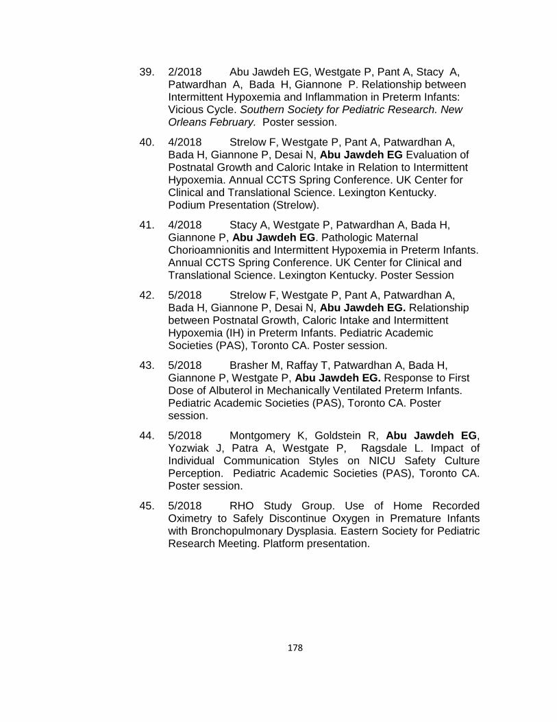
178
39. 2/2018 Abu Jawdeh EG, Westgate P, Pant A, Stacy A, Patwardhan A, Bada H, Giannone P. Relationship between Intermittent Hypoxemia and Inflammation in Preterm Infants: Vicious Cycle. Southern Society for Pediatric Research. New Orleans February. Poster session.
40. 4/2018 Strelow F, Westgate P, Pant A, Patwardhan A, Bada H, Giannone P, Desai N, Abu Jawdeh EG Evaluation of Postnatal Growth and Caloric Intake in Relation to Intermittent Hypoxemia. Annual CCTS Spring Conference. UK Center for Clinical and Translational Science. Lexington Kentucky. Podium Presentation (Strelow).
41. 4/2018 Stacy A, Westgate P, Patwardhan A, Bada H, Giannone P, Abu Jawdeh EG. Pathologic Maternal Chorioamnionitis and Intermittent Hypoxemia in Preterm Infants. Annual CCTS Spring Conference. UK Center for Clinical and Translational Science. Lexington Kentucky. Poster Session
42. 5/2018 Strelow F, Westgate P, Pant A, Patwardhan A, Bada H, Giannone P, Desai N, Abu Jawdeh EG. Relationship between Postnatal Growth, Caloric Intake and Intermittent Hypoxemia (IH) in Preterm Infants. Pediatric Academic Societies (PAS), Toronto CA. Poster session.
43. 5/2018 Brasher M, Raffay T, Patwardhan A, Bada H, Giannone P, Westgate P, Abu Jawdeh EG. Response to First Dose of Albuterol in Mechanically Ventilated Preterm Infants. Pediatric Academic Societies (PAS), Toronto CA. Poster session.
44. 5/2018 Montgomery K, Goldstein R, Abu Jawdeh EG, Yozwiak J, Patra A, Westgate P, Ragsdale L. Impact of Individual Communication Styles on NICU Safety Culture Perception. Pediatric Academic Societies (PAS), Toronto CA. Poster session.
45. 5/2018 RHO Study Group. Use of Home Recorded Oximetry to Safely Discontinue Oxygen in Premature Infants with Bronchopulmonary Dysplasia. Eastern Society for Pediatric Research Meeting. Platform presentation.
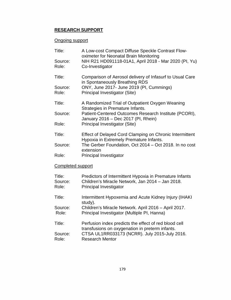
179
RESEARCH SUPPORT Ongoing support Title: A Low-cost Compact Diffuse Speckle Contrast Flow-
oximeter for Neonatal Brain Monitoring Source: NIH R21 HD091118-01A1, April 2018 - Mar 2020 (PI, Yu) Role: Co-Investigator Title: Comparison of Aerosol delivery of Infasurf to Usual Care
in Spontaneously Breathing RDS Source: ONY, June 2017- June 2019 (PI, Cummings) Role: Principal Investigator (Site) Title: A Randomized Trial of Outpatient Oxygen Weaning
Strategies in Premature Infants. Source: Patient-Centered Outcomes Research Institute (PCORI),
January 2016 – Dec 2017 (PI, Rhein) Role: Principal Investigator (Site) Title: Effect of Delayed Cord Clamping on Chronic Intermittent
Hypoxia in Extremely Premature Infants. Source: The Gerber Foundation, Oct 2014 – Oct 2018. In no cost
extension Role: Principal Investigator Completed support Title: Predictors of Intermittent Hypoxia in Premature Infants Source: Children’s Miracle Network, Jan 2014 – Jan 2018. Role: Principal Investigator Title: Intermittent Hypoxemia and Acute Kidney Injury (IHAKI
study). Source: Children’s Miracle Network. April 2016 – April 2017. Role: Principal Investigator (Multiple PI, Hanna) Title: Perfusion index predicts the effect of red blood cell
transfusions on oxygenation in preterm infants. Source: CTSA UL1RR033173 (NCRR). July 2015-July 2016. Role: Research Mentor
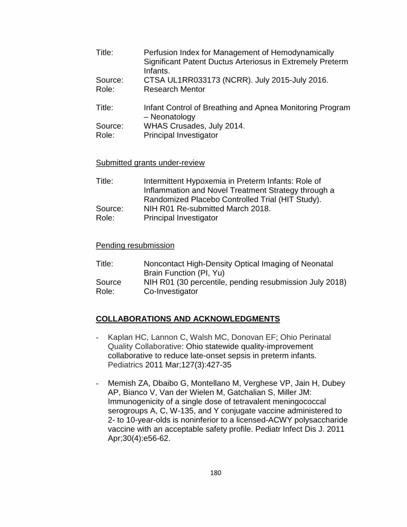
180
Title: Perfusion Index for Management of Hemodynamically Significant Patent Ductus Arteriosus in Extremely Preterm Infants.
Source: CTSA UL1RR033173 (NCRR). July 2015-July 2016. Role: Research Mentor Title: Infant Control of Breathing and Apnea Monitoring Program
– Neonatology Source: WHAS Crusades, July 2014. Role: Principal Investigator Submitted grants under-review Title: Intermittent Hypoxemia in Preterm Infants: Role of
Inflammation and Novel Treatment Strategy through a Randomized Placebo Controlled Trial (HIT Study).
Source: NIH R01 Re-submitted March 2018. Role: Principal Investigator Pending resubmission Title: Noncontact High-Density Optical Imaging of Neonatal
Brain Function (PI, Yu) Source NIH R01 (30 percentile, pending resubmission July 2018) Role: Co-Investigator COLLABORATIONS AND ACKNOWLEDGMENTS - Kaplan HC, Lannon C, Walsh MC, Donovan EF; Ohio Perinatal
Quality Collaborative: Ohio statewide quality-improvement collaborative to reduce late-onset sepsis in preterm infants. Pediatrics 2011 Mar;127(3):427-35
- Memish ZA, Dbaibo G, Montellano M, Verghese VP, Jain H, Dubey
AP, Bianco V, Van der Wielen M, Gatchalian S, Miller JM: Immunogenicity of a single dose of tetravalent meningococcal serogroups A, C, W-135, and Y conjugate vaccine administered to 2- to 10-year-olds is noninferior to a licensed-ACWY polysaccharide vaccine with an acceptable safety profile. Pediatr Infect Dis J. 2011 Apr;30(4):e56-62.
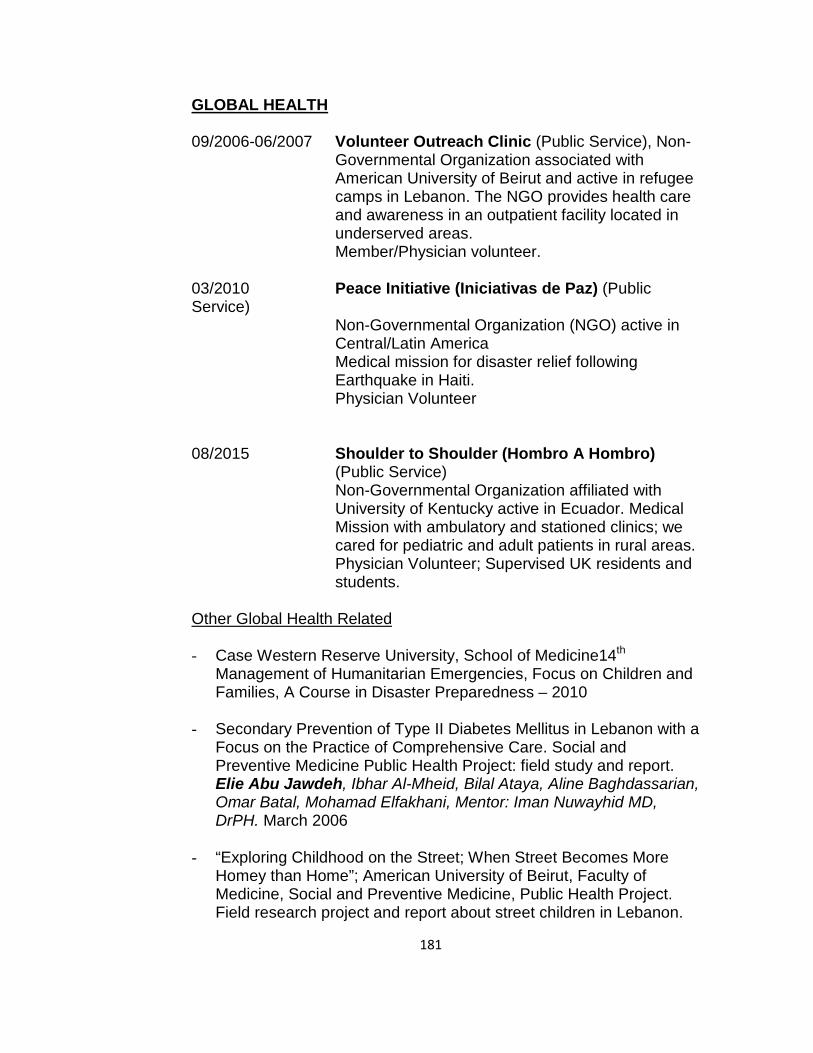
181
GLOBAL HEALTH 09/2006-06/2007 Volunteer Outreach Clinic (Public Service), Non-
Governmental Organization associated with American University of Beirut and active in refugee camps in Lebanon. The NGO provides health care and awareness in an outpatient facility located in underserved areas.
Member/Physician volunteer. 03/2010 Peace Initiative (Iniciativas de Paz) (Public Service)
Non-Governmental Organization (NGO) active in Central/Latin America Medical mission for disaster relief following Earthquake in Haiti.
Physician Volunteer 08/2015 Shoulder to Shoulder (Hombro A Hombro)
(Public Service) Non-Governmental Organization affiliated with University of Kentucky active in Ecuador. Medical Mission with ambulatory and stationed clinics; we cared for pediatric and adult patients in rural areas. Physician Volunteer; Supervised UK residents and students.
Other Global Health Related - Case Western Reserve University, School of Medicine14th
Management of Humanitarian Emergencies, Focus on Children and Families, A Course in Disaster Preparedness – 2010
- Secondary Prevention of Type II Diabetes Mellitus in Lebanon with a
Focus on the Practice of Comprehensive Care. Social and Preventive Medicine Public Health Project: field study and report. Elie Abu Jawdeh, Ibhar Al-Mheid, Bilal Ataya, Aline Baghdassarian, Omar Batal, Mohamad Elfakhani, Mentor: Iman Nuwayhid MD, DrPH. March 2006
- “Exploring Childhood on the Street; When Street Becomes More
Homey than Home”; American University of Beirut, Faculty of Medicine, Social and Preventive Medicine, Public Health Project. Field research project and report about street children in Lebanon.

182
Elie Abu Jawdeh, Joelle Abi Rached, Tarek Abou Hamdan, Joelle Amm, Aline Baghdassarian, George Mollayess Mentor: Iman Nuwayhid MD, DrPH. July 2003
SPECIAL CERTIFICATIONS - University Hospitals of Cleveland - Rainbow Babies and Children’s
Hospital, ECMO Physician Specialist (2011 - 2015)
- Case Western Reserve University, Collaborative Institutional Training Initiative (CITI), Continuing Research Education Credit Program (CREC) (2008 – 2014)
- American Academy of Pediatrics, Neonatal Resuscitation Program, Provider (2007 - present), Pediatric Advanced Life Support (2007 - 2011)
- University of Kentucky Collaborative Institutional Training Initiative
(CITI) Completion Certificate (11/2014-present)
INVITED PRESENTATIONS
08/2014 University of Kentucky
Department of Pediatrics Lexington, KY Neonatology Grand Rounds: Neonatal Apnea, Overview
05/2015 American University of Beirut
Beirut, Lebanon Rounded with the NICU team and presented to
residents/fellows “Apnea and Reflux in Preterm Infants” 05/2015 Contemporary Pediatrics Conference
Lexington, KY Invited Speaker: “Gastroesophageal Reflux in Infants”

183
06/2015 Case Western Reserve University 18th Management of Humanitarian Emergencies, Focus on Children, Women and Families. Cleveland, OH Invited Speaker: “Neonatal Resuscitation”
Moderator: “Case Discussion” 09/2015 American Association of SIDS Prevention
Physicians Pre-conference research session Naples, Florida
Invited Speaker: Intermittent Hypoxemia Research 11/2015 UHC/AACN Nurse Residency Program Annual
Conference, webinar Lexington, KY
Panelist; Life Adventure Center: A novel approach to improving team communication (Webinar)
12/2015 University of Kentucky
Department of Pediatrics Lexington, Kentucky Grand Rounds: Neonatal Apnea and Intermittent Hypoxemia
02/2016 University of Kentucky
Department of Pediatrics, Neonatology Lexington, KY
Neonatal Grand Rounds: Management of Gastroesophageal Reflux in the Preterm Infant
05/2016 Case Western Reserve University 19th Management of Humanitarian Emergencies, Focus on Children, Women and Families. Cleveland, OH Invited Speaker: “Neonatal Resuscitation”
Moderator: “Case Discussion” 09/2016 American University of Beirut
Department of Pediatrics Beirut, Lebanon Grand Rounds: Neonatal Apnea and Intermittent Hypoxemia
184
09/2016 American Association of SIDS Prevention Physicians Conference Naples, Florida Invited Speaker: Predictors Intermittent Hypoxemia Research
01/2017 University of Kentucky
Department of Pediatrics, Neonatology Lexington, KY
Grand Rounds: GERD? Probably Not! 02/2017 National Collaborative for Perinatal Neonatal
Network (NCPNN) Conference Beirut, Lebanon
Invited Speaker: Gastroesophageal Reflux in Preterm Infants Invited Speaker: Intermittent Hypoxemia in Preterm Infant
05/2017 Case Western Reserve University
Cleveland, OH Resident Workshop/Invited Speaker: Helping Babies Breathe; Neonatal
resuscitation in undeserved setting 09/2017 American Association of SIDS Prevention
Physicians Conference Naples, Florida Invited Speaker: Intermittent Hypoxemia in Preterm Infants: Consequences
Related Documents


