Interleukin-17 in transverse myelitis and multiple sclerosis Jerome J. Graber a,b,1 , S. Rameeza Allie a,1 , Katherine M. Mullen a , Melina V. Jones a , Tongguang Wang a , Chitra Krishnan a , Adam I. Kaplin c , Avi Nath a , Douglas A. Kerr a , Peter A. Calabresi a, ⁎ a The Johns Hopkins University School of Medicine, Department of Neurology, United States b University of Maryland at Baltimore School of Medicine, United States c The Johns Hopkins University Department of Psychiatry and Behavioral Sciences, United States Received 21 May 2007; received in revised form 5 February 2008; accepted 6 February 2008 Abstract CSF IL-6 is elevated in transverse myelitis (TM) and predicts disability. Since IL-17 regulates cytokines (TNFα, IL-1β and IL-6) known to stimulate IL-6 production by astrocytes, we sought to determine whether IL-17 was increased in TM and MS compared to healthy controls (HC) and other neurologic diseases (OND). IL-17 and IL-6 levels were measured in stimulated peripheral blood mononuclear cell (PBMC) supernatants from HC, MS, TM and OND. IL-17 was increased in TM compared to HC, MS, and OND (mean pg/ml ± standard error; HC: 36.1 ± 11.7, MS: 89.4 ± 23.3, TM: 302.6 ± 152.5, OND: 41.2 ± 13.0, p = 0.01). IL-6 was increased in TM relative to MS and HC (HC: 2624 pg/ml ± 641, MS: 6129 ± 982, TM: 12,536 ± 2657, OND: 6920 ± 1801, p b 0.002). MS patients with early disease (b 2 years) also had increased levels of IL-17 (p b 0.04) and IL-6 (p b 0.05). Cytokine neutralization experiments demonstrated that IL-6 was the main inducer of astrocyte IL-6 production. We conclude that IL-17 and IL-6 production from PBMC in TM and early MS are increased and induce astrocyte IL-6 production through IL-6. © 2008 Elsevier B.V. All rights reserved. Keywords: Multiple sclerosis; Transverse myelitis; Interleukin 17; Interleukin 6; Astrocytes; T cells 1. Introduction Multiple sclerosis (MS) is an inflammatory disorder of the central nervous system (CNS) characterized by recurring episodes of inflammatory demyelinating lesions with prominent astrogliosis. IL-17 producing cells (Th17), distinct from Th1 or Th2 cells, play a critical role in disease induction in experimental autoimmune encephalomyelitis (EAE) (Frohman et al., Greter et al., 2005; Heppner et al., 2005; Reder et al., 1998; Hollifield et al., 2003; Langrish et al., 2005; Harrington et al., 2005; Park et al., 2005). IL-17 levels in CSF are elevated in the optico-spinal form of MS in Japanese populations, which may have under- lying pathogenic differences from conventional MS (Ishizu et al., 2005). Idiopathic transverse myelitis (TM) is a rare disorder charac- terized by severe monofocal inflammatory lesions of the spinal cord with simultaneous motor, sensory and autonomic dysfunc- tion. The underlying immunopathology of TM bears many si- milarities to MS but clinical course differs with a very low rate of subsequent conversion to MS or Devic’s neuromyelitis optica (NMO) (Transverse Myelitis Consortium Working Group, 2002; Kerr and Ayetey, 2002; de Seze et al., 2005). We recently showed that IL-6 is increased in the CSF of TM patients and CSF IL-6 levels correlate with both acute and long term disability (Kaplin et al., 2005). IL-6 levels are also increased in both CSF and serum in MS though less than what we found in TM (Miljkovic et al., 2002). Astrocytes have been shown to produce IL-6 in the CNS in response to TNFα and IL-1β, both of which are increased in the presence of IL-17 (Van Wagoner and Ben- veniste, 1999; Jovanovic et al., 1998). TNFα and IL-1β interact synergistically to stimulate levels of IL-6 production by astro- Journal of Neuroimmunology 196 (2008) 124 – 132 www.elsevier.com/locate/jneuroim ⁎ Corresponding author. Johns Hopkins Hospital, Pathology Building 627, 600 N. Wolfe St., Baltimore MD 21287-6965, United States. E-mail address: [email protected] (P.A. Calabresi). 1 These authors contributed equally to this report. 0165-5728/$ - see front matter © 2008 Elsevier B.V. All rights reserved. doi:10.1016/j.jneuroim.2008.02.008

Welcome message from author
This document is posted to help you gain knowledge. Please leave a comment to let me know what you think about it! Share it to your friends and learn new things together.
Transcript
196 (2008) 124–132www.elsevier.com/locate/jneuroim
Journal of Neuroimmunology
Interleukin-17 in transverse myelitis and multiple sclerosis
Jerome J. Graber a,b,1, S. Rameeza Allie a,1, Katherine M. Mullen a, Melina V. Jones a,Tongguang Wang a, Chitra Krishnan a, Adam I. Kaplin c, Avi Nath a,
Douglas A. Kerr a, Peter A. Calabresi a,⁎
a The Johns Hopkins University School of Medicine, Department of Neurology, United Statesb University of Maryland at Baltimore School of Medicine, United States
c The Johns Hopkins University Department of Psychiatry and Behavioral Sciences, United States
Received 21 May 2007; received in revised form 5 February 2008; accepted 6 February 2008
Abstract
CSF IL-6 is elevated in transverse myelitis (TM) and predicts disability. Since IL-17 regulates cytokines (TNFα, IL-1β and IL-6) known tostimulate IL-6 production by astrocytes, we sought to determine whether IL-17 was increased in TM andMS compared to healthy controls (HC) andother neurologic diseases (OND). IL-17 and IL-6 levels were measured in stimulated peripheral blood mononuclear cell (PBMC) supernatants fromHC, MS, TM and OND. IL-17 was increased in TM compared to HC, MS, and OND (mean pg/ml±standard error; HC: 36.1±11.7, MS: 89.4±23.3,TM: 302.6±152.5, OND: 41.2±13.0, p=0.01). IL-6 was increased in TM relative to MS and HC (HC: 2624 pg/ml±641, MS: 6129±982, TM:12,536±2657, OND: 6920±1801, pb0.002). MS patients with early disease (b2 years) also had increased levels of IL-17 (pb0.04) and IL-6(pb0.05). Cytokine neutralization experiments demonstrated that IL-6 was the main inducer of astrocyte IL-6 production. We conclude that IL-17and IL-6 production from PBMC in TM and early MS are increased and induce astrocyte IL-6 production through IL-6.© 2008 Elsevier B.V. All rights reserved.
Keywords: Multiple sclerosis; Transverse myelitis; Interleukin 17; Interleukin 6; Astrocytes; T cells
1. Introduction
Multiple sclerosis (MS) is an inflammatory disorder of thecentral nervous system (CNS) characterized by recurringepisodes of inflammatory demyelinating lesions with prominentastrogliosis. IL-17 producing cells (Th17), distinct from Th1 orTh2 cells, play a critical role in disease induction in experimentalautoimmune encephalomyelitis (EAE) (Frohman et al., Greteret al., 2005; Heppner et al., 2005; Reder et al., 1998; Hollifieldet al., 2003; Langrish et al., 2005; Harrington et al., 2005; Parket al., 2005). IL-17 levels in CSF are elevated in the optico-spinalform of MS in Japanese populations, which may have under-
⁎ Corresponding author. Johns Hopkins Hospital, Pathology Building 627,600 N. Wolfe St., Baltimore MD 21287-6965, United States.
E-mail address: [email protected] (P.A. Calabresi).1 These authors contributed equally to this report.
0165-5728/$ - see front matter © 2008 Elsevier B.V. All rights reserved.doi:10.1016/j.jneuroim.2008.02.008
lying pathogenic differences from conventional MS (Ishizuet al., 2005).
Idiopathic transverse myelitis (TM) is a rare disorder charac-terized by severe monofocal inflammatory lesions of the spinalcord with simultaneous motor, sensory and autonomic dysfunc-tion. The underlying immunopathology of TM bears many si-milarities toMS but clinical course differs with a very low rate ofsubsequent conversion to MS or Devic’s neuromyelitis optica(NMO) (TransverseMyelitis ConsortiumWorkingGroup, 2002;Kerr and Ayetey, 2002; de Seze et al., 2005). We recentlyshowed that IL-6 is increased in the CSF of TM patients and CSFIL-6 levels correlate with both acute and long term disability(Kaplin et al., 2005). IL-6 levels are also increased in both CSFand serum in MS though less than what we found in TM(Miljkovic et al., 2002). Astrocytes have been shown to produceIL-6 in the CNS in response to TNFα and IL-1β, both of whichare increased in the presence of IL-17 (Van Wagoner and Ben-veniste, 1999; Jovanovic et al., 1998). TNFα and IL-1β interactsynergistically to stimulate levels of IL-6 production by astro-
125J.J. Graber et al. / Journal of Neuroimmunology 196 (2008) 124–132
cytes greater than either stimulates alone (Van Wagoner andBenveniste, 1999). In addition, IL-6 itself can stimulate in-creased IL-6 production by astrocytes via gp130 mediated“trans” signaling when soluble IL-6 receptor (sIL-6R) is present,as has been shown in MS patients’ CSF (Michalopoulou et al.,2004; Van Wagoner et al., 1999).
IL-17 is primarily made by a subset of activated memory CD4+T cells distinct from Th1 or Th2 cells, called Th17. These IL-17producing T cells arise from naïve precursors when TGF-β andIL-6 are present but Th1 and Th2 inducing cytokines (IFNγ andIL-4) are absent (Mangan et al., 2006). IL-23 produced by ma-crophages and dendritic cells appears to play a supporting role inthe continued stimulation and survival of Th17 cells (Harringtonet al., 2005; Park et al., 2005; Mangan et al., 2006; Bettelli et al.,2006; Veldhoen et al., 2006; Harrington et al., 2006). IL-23 andIL-12 have antagonistic properties, as IL-23 supports IL-17production, whereas IL-12 inhibits it (Langrish et al., 2005;Harrington et al., 2006; Gran et al., 2004). IL-17 in turn sti-mulates macrophage production of IL-6, TNFα and IL-1β, all ofwhich have been implicated in MS pathology and astrocyte IL-6production (Jovanovic et al., 1998; Van Wagoner et al., 1999).IL-17 mRNA expression is increased in acute MS lesions and inCSF and peripheral mononuclear cells during relapse (Locket al., 2002; Matusevicius et al., 1999). CSF IL-17 levels inJapanese optico-spinal MS correlate with the size of spinal cordlesions, which is one of the main distinguishing features bet-ween optico-spinal MS and conventional MS (Ishizu et al.,2005). In EAE, Th17 cells have been shown to develop in res-ponse to IL-23 after myelin protein immunization and invade theCNS to initiate inflammation (Langrish et al., 2005; Park et al.,2005).
To investigate the role of IL-17 and its possible link with IL-6in autoimmune inflammation of the CNS, we examined IL-17and associated cytokine levels in ex vivo stimulated cell super-natants from peripheral blood mononuclear cells (PBMC) ofhealthy controls (HC) and patients with MS and TM. We foundthat IL-17 and IL-6 production was increased in PBMCsupernatants from patients with TM and in the subset of MSpatients with early disease. Furthermore, we found that IL-6, butnot IL-17, could induce IL-6 production by astrocytes.
2. Methods
2.1. Patients
Patients with clinically definite relapsing-remitting multiplesclerosis (MS n=37) and idiopathic transverse myelitis (TM n=13) were recruited from the Johns Hopkins Medical Institutionsaccording to previously published diagnostic criteria (Trans-verse Myelitis Consortium Working Group, 2002). None ofthese patients had longitudinally extensive TM lesions (≥3vertebral segments in length) typical of Devic’s neuromyelitisoptica (NMO).When any clinical suspicion for disease beyond afocal TM lesion existed, further testing was performed. Twopatients had visual evoked potentials, both of which were nor-mal. Four TM patients were tested for the NMO-IgG antibodyagainst aquaporin-4, and all were negative (Lennon et al., 2004).
Healthy control (HC) volunteers with no history of inflammatoryneurologic disease were also recruited (n=16). Ten patients withother neurological diseases (OND) including Alzheimer’s disease(n=4), neurosarcoid (n=2), compressive myelopathy (n=2),spinal cavernoma (n=1), and acute disseminated encephalomye-litis (n=1) were also recruited. Within the MS group, one patienteach was taking Avonex, Rebif, Betaseron and Copaxone at thetime of sampling and one patient had stopped Copaxone twoweeks prior to sampling. In the TM group one patient was takingAvonex. The remaining MS, TM and OND patients had notreceived any steroids or other immunomodulatory drugs withinthe month prior to sampling. None of ourMS or TMpatients havesubsequently manifested a clinical course consistent with primaryprogressive MS or NMO. All patients gave written, informedconsent for participation in the study.
2.2. Cell culture
Peripheral blood mononuclear cells (PBMC) were obtainedby venous puncture and separation of whole blood over Ficollgradients. Cells were stored at −80 °C in whole T cell media(IMDM) with 10% DMSO. For culture, PBMC were thawed in5% human serum (Sigma) and PBS, counted using a hemato-cytometer, and plated at 2×10⁶ cells per well in a 24 well cultureplate and incubated in T cell media at 37 °C for 48 h. Cells wereactivated using soluble anti-CD3 and anti-CD28 at 1 µg/ml(Becton-Dickinson). Supernatants were harvested and stored at−20 °C until assay. For sorting experiments, cells were sortedusing negative selection with MACS magnetic beads to obtainuntouched CD4+ or CD8+ T cells (Miltenyi Biotech). CSF cellsfrom MS and TM patients were obtained by sterile lumbarpuncture and centrifugation of CSF, and stimulated as describedabove for 7 days in T cell media at 37 °C.
2.3. Cytokine assays and recombinant protein
Assays for IL-17 (R&D Systems), IL-6, IL-1β, TNFα, IFNγand sIL6R (Biosource International) were performed in dupli-cate according to manufacturer’s instructions. Recombinanthuman IL-23, IL-17, IL-12, IL-18, IL-1β (R&D Systems), IFNγand TNFα (Peprotech, Inc.) were added to astrocytes as indi-cated. Neutralizing antibodies to IL-23 (polyclonal goat anti-body against IL23 p19), IL-17, IL-12, IFNγ (R&DSystems), IL-6, IL-18 (Biosource International), IL-1β (Sigma, Inc.), andTNFα (BD Pharmingen) were used at neutralizing doses accord-ing to the manufacturer’s instructions and confirmed by our ownELISAs.
2.4. Astrocyte culture and staining
Both a human astrocyte-like cell line (SVGA) and primaryhuman astrocytes were studied under a Johns Hopkins Univ-ersity IRB approved protocol (Chauhan et al., 2003). SVGAcells were maintained in DMEM media with 5% fetal serumalbumin in an incubator at 37 °C and 5% CO2. For some expe-riments, human astrocytes were used. Briefly, human fetal brainspecimens of 12–17 weeks gestation were obtained in accor-
126 J.J. Graber et al. / Journal of Neuroimmunology 196 (2008) 124–132
dance with NIH guidelines. The tissues were then triturated afterremoving the meninges. Cells were then cultured in T75 flasks inopti-MEM (Invitrogen) with 5% fetal bovine serum (FBS,Invitrogen), 0.5% N2 supplement (Invitrogen) and 1% anti-biotics for at least one month. After shaking at 180 rpm for 1 h,cells were separated with trypsin/EDTA. Cells were then seededat 1×10⁵/ml on coverslips for another three weeks before treat-ment with Poly-IC (25 µg/ml) and IFNγ (500 U/ml) for positivecontrols or Tcell supernatants at 1:10 dilution, in Locke’s Buffer(154 mM NaCl, 5.6 mM KCl, 2.3 mM CaCl2·2H2O, 1.0 mMMgCl2·6H2O, 3.6 mM NaHCO3, 5 mM HEPES, 1.8 mg/mlglucose at pH 7.2) for 48 h. Golgi Stop (BD Pharmingen) wasadded for the final 6 h of culture at the concentration specified bythe vendor. Cells were fixed with 4% Paraformaldehyde for30 min and then blocked in 5% BSA with 0.005% Saponin,for 30 min. Rabbit anti-GFAP (Chemicon) at a dilution of1:200 was added and incubated for 1 h at room temperature.Cells were washed with PBS and 0.005% Saponin, donkeyanti-rabbit ALEXA 594 (7 µg/ml) (Molecular Probe) andFITC conjugated anti-IL-6 (1:10) (BD Pharmingen) wereadded. Cells were incubated at room temperature for 45 minand then washed and counter stained with DAPI (1:500)(Molecular Probe) and mounted with GEL mount (BioMed cat# M01). To immunodeplete IL-6 from stimulated PBMC su-pernatants, 1 µg of anti-IL-6 (Biosource Cat # AHC0562) wasadded to supernatants and incubated overnight at 4 °C withend-over-end shaking of the tubes. Thirty µl/ml of protein A/Gbeads was then added (Pierce cat # 53133) and incubated for1.5 h at 4 °C with end-over-end shaking. Samples were cen-trifuged for 2 min at 2000 rpm and tested by ELISA (BiosourceCat # CHC1263) for complete depletion of IL-6 or use inastrocyte culture. All astrocyte experiments were performed intriplicate.
2.5. Statistical analysis
Statistical analysis was performed using PRISM version 3.0.Differences between more than two groups were tested usingone-way ANOVA and significant results (pb0.05) were verifiedwith post-hoc Bonferroni correction for multiple comparisons.Differences between two groups were evaluated using t-tests forGaussian values (with Welch’s correction for unequal var-iances) or Mann–Whitney tests for non-Gaussian distributions.Because of the small sample size of the groups, any significantfindings from t-tests were confirmed by post-hoc Mann–Whitney. Binomial data were analyzed using Fisher’s exact test.Two-tailed p-values less than 0.05 were considered significant.
3. Results
3.1. Patient demographics
There was no significant difference in the ages of the HC, MSor TM groups (31.9±1.6 years, 39.3±1.9 years and 35.1±4.1 years, respectively). There was no significant difference inthe gender ratios of the MS and TM groups (24% and 31%male,respectively).
3.2. IL-17 and IL-6 are increased and TNFα production isdecreased in stimulated PBMC from transverse myelitispatients as compared to healthy controls
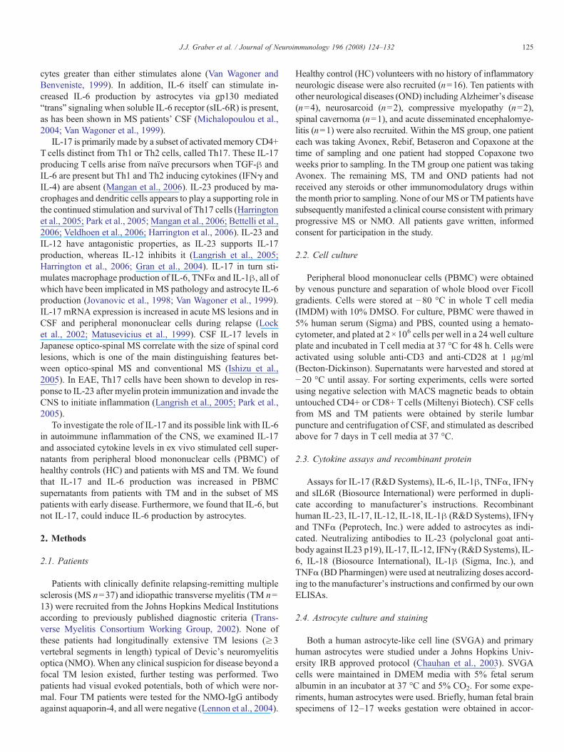
Stimulated PBMC from patients with TM produced signifi-cantly higher levels of IL-17 than those from HC, MS or OND(mean pg/ml±standard error (SE) HC: 36.1±11.7 (n=16), MS:89.4±23.3 (n=32), TM: 302.6±152.5 (n=10), OND: 41.2±13.0 (n=10), p=0.01 by ANOVA with post-hoc Bonferronipb0.05 for TM versus HC, MS and OND) (see Fig. 1A). TMPBMC also produced higher levels of IL-6 than HC and MSpatients (mean pg/ml±standard error (SE) HC: 2624±641(n=13), MS: 6129±982 (n=36), TM: 12,536±2657 (n=10),OND: 6920±1801 (n=9), pb0.002 by ANOVA with post-hocBonferroni p-value b0.05 for TM versus HC and MS) (see Fig.1B). Increased levels of IL-1β were also found in TM super-natants, but this difference was not significant (mean pg/ml±standard error (SE) HC: 308 pg/ml±110 (n=14), MS: 416±116(n=32), TM: 1114±604 (n=12), OND: 347±71 (n=10), p=0.12 by ANOVA). Interestingly, levels of TNFαwere decreasedin MS and TM supernatants as compared to HC though thisfinding did not reach statistical significance (mean pg/ml±standard error (SE)HC: 12,915 ng/ml±1754 (n=14),MS: 8079±1098 (n=32), TM: 7466±1815 (n=12), OND: 9770±2111(n=10), p=0.09 by ANOVA). Levels of IFNγ and soluble IL-6receptor were not significantly different between the four groups(data not shown).
3.3. IL-17 producing cells are present in CSF
Levels of IL-17 were undetectable in CSF from TM patients(n=7). Leukocytes obtained from the CSF of MS and TMpatients were cultured in T cell media for 7 days in the presenceof anti-CD3 and anti-CD28. Detectable levels of IL-17 werefound in 1 of 8MSCSF cell supernatants and 3 of 6 TMCSF cellsupernatants (mean pg/ml±SE; MS – 0.94±0.9, TM – 30.0±16.8, p=0.1812, Mann–Whitney). We set a cutoff value of15 pg/ml for high versus low levels of IL-17 production in CSFcell supernatants and reanalyzed the binomial data, whichrevealed a borderline significant difference between TM andMS(p=0.055, Fisher’s exact test), indicating that IL-17-producingleukocytes are present in the CSF andmay be increased in TM ascompared to MS.
3.4. IL-17 levels are increased in early MS as compared toestablished MS
We subdivided the group of MS patients into those with earlydisease (defined as patients with onset of disease less than twoyears previously) versus established disease (diagnosed morethan two years before sampling). PBMC from patients with earlyMS produced significantly higher levels of IL-17 than patientswith established MS (mean pg/ml±SE; early MS: 142±42.1(n=15), established MS: 37.1±17.7 (n=15), pb0.04, t-testwithWelch’s correction, confirmed by post-hocMann–Whitneyp=0.005 (see Fig. 1C)). When the patients currently on immu-nomodulatory drugs were removed from this analysis, the fin-
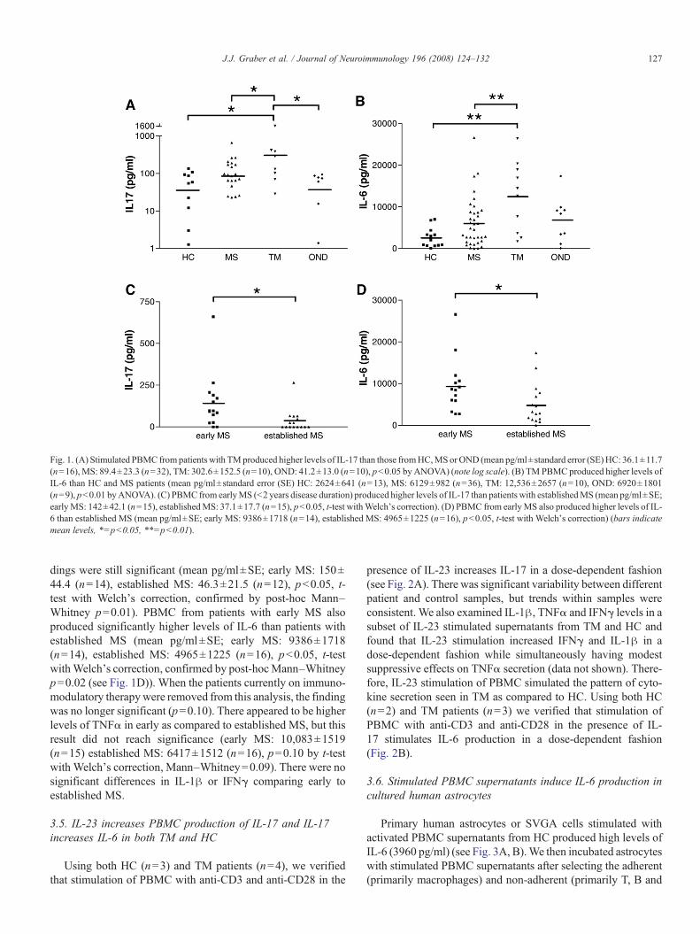
Fig. 1. (A) Stimulated PBMC from patients with TMproduced higher levels of IL-17 than those fromHC,MS or OND (mean pg/ml±standard error (SE)HC: 36.1±11.7(n=16), MS: 89.4±23.3 (n=32), TM: 302.6±152.5 (n=10), OND: 41.2±13.0 (n=10), pb0.05 by ANOVA) (note log scale). (B) TM PBMC produced higher levels ofIL-6 than HC and MS patients (mean pg/ml±standard error (SE) HC: 2624±641 (n=13), MS: 6129±982 (n=36), TM: 12,536±2657 (n=10), OND: 6920±1801(n=9), pb0.01 byANOVA). (C) PBMC from earlyMS (b2 years disease duration) produced higher levels of IL-17 than patients with establishedMS (mean pg/ml±SE;earlyMS: 142±42.1 (n=15), establishedMS: 37.1±17.7 (n=15), pb0.05, t-test withWelch's correction). (D) PBMC from earlyMS also produced higher levels of IL-6 than established MS (mean pg/ml±SE; early MS: 9386±1718 (n=14), established MS: 4965±1225 (n=16), pb0.05, t-test with Welch's correction) (bars indicatemean levels, ⁎=pb0.05, ⁎⁎=pb0.01).
127J.J. Graber et al. / Journal of Neuroimmunology 196 (2008) 124–132
dings were still significant (mean pg/ml±SE; early MS: 150±44.4 (n=14), established MS: 46.3±21.5 (n=12), pb0.05, t-test with Welch’s correction, confirmed by post-hoc Mann–Whitney p=0.01). PBMC from patients with early MS alsoproduced significantly higher levels of IL-6 than patients withestablished MS (mean pg/ml±SE; early MS: 9386±1718(n=14), established MS: 4965±1225 (n=16), pb0.05, t-testwithWelch’s correction, confirmed by post-hocMann–Whitneyp=0.02 (see Fig. 1D)). When the patients currently on immuno-modulatory therapywere removed from this analysis, the findingwas no longer significant (p=0.10). There appeared to be higherlevels of TNFα in early as compared to established MS, but thisresult did not reach significance (early MS: 10,083±1519(n=15) established MS: 6417±1512 (n=16), p=0.10 by t-testwith Welch’s correction, Mann–Whitney=0.09). There were nosignificant differences in IL-1β or IFNγ comparing early toestablished MS.
3.5. IL-23 increases PBMC production of IL-17 and IL-17increases IL-6 in both TM and HC
Using both HC (n=3) and TM patients (n=4), we verifiedthat stimulation of PBMC with anti-CD3 and anti-CD28 in the
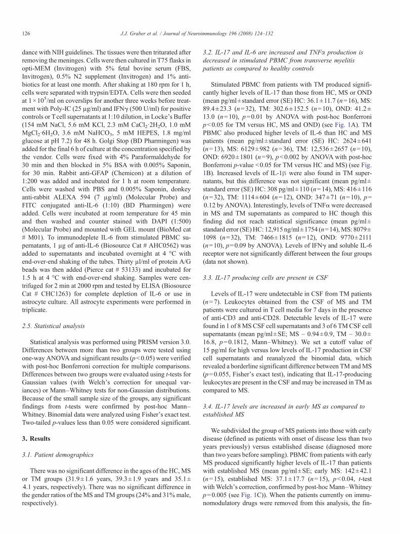
presence of IL-23 increases IL-17 in a dose-dependent fashion(see Fig. 2A). There was significant variability between differentpatient and control samples, but trends within samples wereconsistent. We also examined IL-1β, TNFα and IFNγ levels in asubset of IL-23 stimulated supernatants from TM and HC andfound that IL-23 stimulation increased IFNγ and IL-1β in adose-dependent fashion while simultaneously having modestsuppressive effects on TNFα secretion (data not shown). There-fore, IL-23 stimulation of PBMC simulated the pattern of cyto-kine secretion seen in TM as compared to HC. Using both HC(n=2) and TM patients (n=3) we verified that stimulation ofPBMC with anti-CD3 and anti-CD28 in the presence of IL-17 stimulates IL-6 production in a dose-dependent fashion(Fig. 2B).
3.6. Stimulated PBMC supernatants induce IL-6 production incultured human astrocytes
Primary human astrocytes or SVGA cells stimulated withactivated PBMC supernatants from HC produced high levels ofIL-6 (3960 pg/ml) (see Fig. 3A, B).We then incubated astrocyteswith stimulated PBMC supernatants after selecting the adherent(primarily macrophages) and non-adherent (primarily T, B and
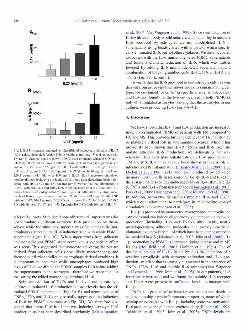
Fig. 2. IL-23 increases stimulated peripheral blood leukocyte production of IL-17(A) in a dose-dependent fashion in both healthy controls (n=3) and patients withTM (n=4). Grouped data are shown. PBMCwere stimulated with anti-CD3/anti-CD28 and IL-23 for six days in culture, mean levels of IL-17 in supernatants ofcultured PBMC were 52.5 pg/ml±78.0 SD without IL-23, 125.0 pg/ml±107.3SD with 1 ng/ml IL-23, 166.7 pg/ml±160.9 SD with 10 ng/ml IL-23 and220.2 pg/ml±194.4 SD with 100 ng/ml IL-23. IL-17 increases stimulatedperipheral blood leukocyte production of IL-6 in a dose-dependent fashion (B).Using both HC (n=2) and TM patients (n=3) we verified that stimulation ofPBMC with anti-CD3 and anti-CD28 in the presence of IL-17 stimulates IL-6production in a dose-dependent fashion (Fig. 2B). After 48 h in culture, meanlevels of IL-6 in supernatants of cultured PBMC were 279.2 pg/ml±242.3 SDwithout IL-17, 206.5 pg/ml±156.5 SD with 1 ng/ml IL-17, 540.3 pg/ml±369.7SD with 10 ng/ml IL-17, and 1453.5 pg/ml±408.8 SD with 100 ng/ml IL-17.
128 J.J. Graber et al. / Journal of Neuroimmunology 196 (2008) 124–132
NK) cell subsets. Stimulated non-adherent cell supernatants didnot stimulate significant astrocyte IL-6 production by them-selves, while the stimulated supernatants of adherent cells (ma-crophages) recreated the IL-6 induction seen with whole PBMCsupernatants (see Fig. 3C). When supernatants from adherentand non-adherent PBMC were combined, a synergistic effectwas seen. This suggested that astrocyte activating factors arederived from adherent cells (most likely macrophages) andfocused our further studies on macrophage derived cytokines. Itis important to note that while macrophages produced highlevels of IL-6, we diluted the PBMC samples 1:10 before addingthe supernatants to the astrocytes, therefore we were not justmeasuring the added macrophage produced IL-6.
Selective addition of TNFα and IL-1β alone to astrocytecultures stimulated IL-6 production at lower levels than the sti-mulated PBMC supernatants (Fig. 3A,B), and neutralization ofTNFα, IFNγ and IL-1β only partially suppressed the inductionof IL-6 by PBMC supernatants (Fig. 3D). We therefore sus-pected that it was IL-6 itself that was inducing astrocyte IL-6production as has been described previously (Michalopoulou
et al., 2004; Van Wagoner et al., 1999). Since neutralization ofIL-6 with an antibody would interfere with our ability tomeasureIL-6 produced by astrocytes we immunodepleted IL-6 insupernatants using beads coated with anti-IL-6, which specifi-cally eliminated IL-6, but not other cytokines.We then incubatedastrocytes with the IL-6 immunodepleted PBMC supernatantsand found a dramatic reduction of IL-6, which was furtherlowered by adding IL-6 immunodepleted supernatant and acombination of blocking antibodies to IL-12, IFNγ, IL-1β andTNFα (Fig. 3D, E, and F).
To verify that the IL-6 produced in our astrocyte cultures wasderived from astrocytes themselves and not a contaminating celltype, we co-stained for GFAP (a specific marker of astrocytes)and IL-6 and found that the two co-localized in both PBMC orpoly-IC stimulated astrocytes proving that the astrocytes in ourcultures were producing IL-6 (Fig. 4A–C).
4. Discussion
We have shown that IL-17 and IL-6 production are increasedin ex vivo stimulated PBMC of patients with TM compared toHC and MS. This provides further evidence that Th17 cells maybe playing a critical role in autoimmune diseases. While it haspreviously been shown that IL-1β, TNFα and IL-6 itself sti-mulate astrocyte IL-6 production, we delineate a pathwaywhereby Th17 cells may initiate astrocyte IL-6 production inTM and MS. IL-17 has already been shown to play a role ininfectious CNS inflammation (Infante-Duarte et al., 2000; DucDodon et al., 2004). IL-17 and IL-6, produced by activatedmemory CD4+ T cells in response to TGF-β, IL-6 and IL-23 inthe absence of Th1- or Th2-inducing IFNγ or IL-4, stimulate IL-6, TNFα and IL-1β from macrophages (Harrington et al., 2005;Park et al., 2005; Harrington et al., 2006; Jovanovic et al., 1998).In addition, astrocytes themselves produce IL-6 and IL-23,which would allow them to participate in an autocrine loop ofstimulation (Constantinescu et al., 2005).
IL-1β is produced by monocytes, macrophages, microglia andastrocytes and can induce oligodendrocyte damage via cytokineproduction (including IL-6 and TNFα), nitric oxide, matrixmetalloproteases, adhesion molecules and astrocyte-mediatedglutamate excitotoxicity, all of which have been demonstrated tobe involved in MS (Takahashi et al., 2003; John et al., 2005). IL-1β production by PBMC is increased during relapse and in MSlesions (Hollifield et al., 2003; Hofman et al., 1986). One ofthe major actions of IL-1β in the brain is the induction of areactive astrogliosis with astrocyte activation and IL-6 pro-duction, an effect that is strongly augmented in the presence ofTNFα, IFNγ, IL-6 and soluble IL-6 receptor (Van Wagonerand Benveniste, 1999; John et al., 2005). In our patients, IL-6levels were increased and we found that soluble IL-6 receptorand IFNγ were present in sufficient levels to interact withIL-1β.
TNFα is a product of activated macrophages and dendriticcells with multiple pro-inflammatory properties, many of whichoverlap or synergize with IL-1β, including astrocyte activation,IL-6 production and glutamate excitotoxicity (Reder et al., 1998;Takahashi et al., 2003; John et al., 2005). TNFα levels are
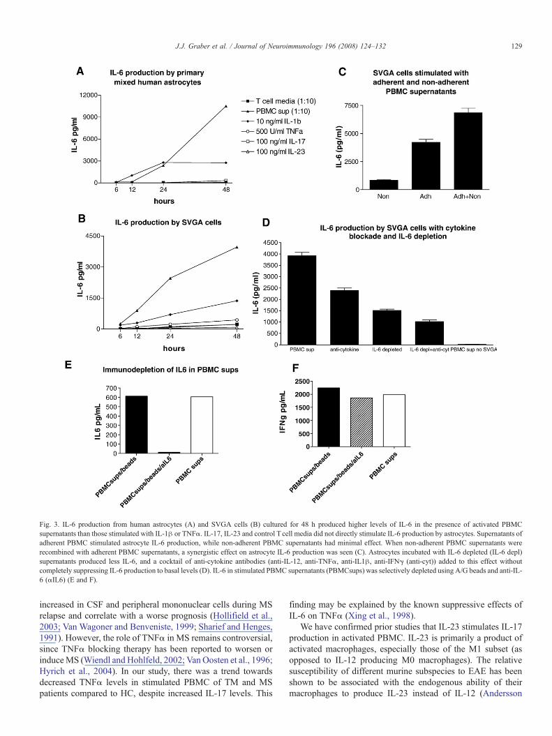
Fig. 3. IL-6 production from human astrocytes (A) and SVGA cells (B) cultured for 48 h produced higher levels of IL-6 in the presence of activated PBMCsupernatants than those stimulated with IL-1β or TNFα. IL-17, IL-23 and control T cell media did not directly stimulate IL-6 production by astrocytes. Supernatants ofadherent PBMC stimulated astrocyte IL-6 production, while non-adherent PBMC supernatants had minimal effect. When non-adherent PBMC supernatants wererecombined with adherent PBMC supernatants, a synergistic effect on astrocyte IL-6 production was seen (C). Astrocytes incubated with IL-6 depleted (IL-6 depl)supernatants produced less IL-6, and a cocktail of anti-cytokine antibodies (anti-IL-12, anti-TNFα, anti-IL1β, anti-IFNγ (anti-cyt)) added to this effect withoutcompletely suppressing IL-6 production to basal levels (D). IL-6 in stimulated PBMC supernatants (PBMCsups) was selectively depleted using A/G beads and anti-IL-6 (αIL6) (E and F).
129J.J. Graber et al. / Journal of Neuroimmunology 196 (2008) 124–132
increased in CSF and peripheral mononuclear cells during MSrelapse and correlate with a worse prognosis (Hollifield et al.,2003; Van Wagoner and Benveniste, 1999; Sharief and Henges,1991). However, the role of TNFα in MS remains controversial,since TNFα blocking therapy has been reported to worsen orinduceMS (Wiendl and Hohlfeld, 2002; Van Oosten et al., 1996;Hyrich et al., 2004). In our study, there was a trend towardsdecreased TNFα levels in stimulated PBMC of TM and MSpatients compared to HC, despite increased IL-17 levels. This
finding may be explained by the known suppressive effects ofIL-6 on TNFα (Xing et al., 1998).
We have confirmed prior studies that IL-23 stimulates IL-17production in activated PBMC. IL-23 is primarily a product ofactivated macrophages, especially those of the M1 subset (asopposed to IL-12 producing M0 macrophages). The relativesusceptibility of different murine subspecies to EAE has beenshown to be associated with the endogenous ability of theirmacrophages to produce IL-23 instead of IL-12 (Andersson
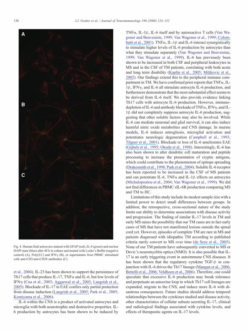
Fig. 4. Human fetal astrocytes stained with GFAP (red), IL-6 (green) and nuclearDAPI stain (blue) after 48 h in culture and treated with Locke's Buffer (negativecontrol) (A), Poly(I:C) and IFNγ (B), or supernatants from PBMC stimulatedwith anti-CD3/anti-CD28 antibodies (C).
130 J.J. Graber et al. / Journal of Neuroimmunology 196 (2008) 124–132
et al., 2004). IL-23 has been shown to support the persistence ofTh17 cells that produce IL-17, TNFα and IL-6, but low levels ofIFNγ (Cua et al., 2003; Aggarwal et al., 2003; Langrish et al.,2005). Blockade of IL-17 in EAE confers only partial protectionfrom disease induction (Langrish et al., 2005; Park et al., 2005;Komiyama et al., 2006).
IL-6 within the CNS is a product of activated astrocytes andmicroglia with both neurotrophic and destructive properties. IL-6 production by astrocytes has been shown to be induced by
TNFα, IL-1β, IL-6 itself and by autoreactive T cells (Van Wa-goner and Benveniste, 1999; Van Wagoner et al., 1999; Colom-batti et al., 2001). TNFα, IL-1β and IL-6 interact synergisticallyto stimulate higher levels of IL-6 production by astrocytes thanwhat they stimulate separately (Van Wagoner and Benveniste,1999; Van Wagoner et al., 1999). IL-6 has previously beenshown to be increased in both CSF and peripheral leukocytes inMS and in the CSF of TM patients, correlating with both acuteand long term disability (Kaplin et al., 2005; Miljkovic et al.,2002). Our findings extend this to the peripheral immune com-partment in TM.We have confirmed prior reports that TNFα, IL-1β, IFNγ, and IL-6 all stimulate astrocyte IL-6 production, andfurthermore demonstrate that themost substantial effect seems tobe derived from IL-6 itself. We also provide evidence linkingTh17 cells with astrocyte IL-6 production. However, immuno-depletion of IL-6 and antibody blockade of TNFα, IFNγ, and IL-1β did not completely suppress astrocyte IL-6 production, sug-gesting that other soluble factors may also be involved. WhileIL-6 can mediate neuronal and glial survival, it can also induceharmful nitric oxide metabolites and CNS damage. In murinemodels, IL-6 induces astrogliosis, microglial activation andpotentiates neurologic degeneration (Campbell et al., 1993;Tilgner et al., 2001). Blockade or loss of IL-6 ameliorates EAE(Gijbels et al., 1995; Okuda et al., 1998). Interestingly, IL-6 hasalso been shown to alter dendritic cell maturation and peptideprocessing to increase the presentation of cryptic antigens,which could contribute to the phenomenon of epitope spreading(Drakesmith et al., 1998; Park et al., 2004). Soluble IL-6 receptorhas been reported to be increased in the CSF of MS patientsand can potentiate IL-6, TNFα and IL-1β effects on astrocytes(Michalopoulou et al., 2004; Van Wagoner et al., 1999). We didnot find differences in PBMC sIL-6R production comparing MSand TM to HC.
Limitations of this study include its modest sample size with alimited power to detect small differences between groups. Inaddition, the retrospective, cross-sectional nature of the studylimits our ability to determine associations with disease activityand progression. The finding of similar IL-17 levels in TM andearly MS raises the possibility that our TM cases are in fact earlycases of MS that have not manifested lesions outside the spinalcord yet. However, episodes of complete TM are rare in MS andpatients diagnosed with idiopathic TM according to publishedcriteria rarely convert to MS over time (de Seze et al., 2005).None of our TM patients have subsequently converted to MS orDevic’s neuromyelitis optica (NMO). It is also possible that IL-17 is an early triggering event in autoimmune CNS diseases. Ithas been shown that the regulatory cytokine TGF-β in con-junction with IL-6 drives the Th17 lineage (Mangan et al., 2006;Bettelli et al., 2006; Veldhoen et al., 2006). Therefore, one couldspeculate that excessive IL-6 production may break toleranceand perpetuate an autocrine loop in which Th17 cell lineages areexpanded, migrate to the CNS, and induce more IL-6 with di-sastrous consequences. Future studies should address temporalrelationships between the cytokines studied and disease activity,other characteristics of cellular subsets secreting IL-17, clinicaland radiological findings associated with cytokine levels, andeffects of therapeutic agents on IL-17 levels.
131J.J. Graber et al. / Journal of Neuroimmunology 196 (2008) 124–132
In summary, we have demonstrated that stimulated PBMCfrom TM patients produce increased levels of IL-17 and IL-6 ascompared to healthy controls and MS patients. IL-17 and IL-6levels are also increased in early as compared to established MS.IL-17 and its downstream cytokine effects (IL-6) act on nume-rous cell subsets (astrocytes, microglia, macrophages and Tcells) so that even a small population of IL-17 producing cellsinvading the CNS could induce potent astrocyte activation withresultant toxic levels of IL-6, thereby accounting for the IL-6mediated cascade leading to destructive and disabling pathologyas we have described in TM (Kaplin et al., 2005). This reportlinks Th17 cells to a CNS IL-6 cascade mediated via activatedastrocytes.
Acknowledgments
This work was supported in part by the National MS SocietyCenter Award and The Nancy Davis Center Without Walls. Theauthors would like to thank the people who so generously vo-lunteered as subjects for this study, without whom this workcould never have been conducted.
References
Aggarwal, S., Ghilardi, N., Xie, M.H., deSauvage, F.J., Gurney, A.L., 2003.Interleukin 23 promotes a distinct CD4+ T cell activation state characterizedby the production of interleukin 17. J. Biol. Chem. 278, 1910–1914.
Andersson, A., Kokkola, R., Wefer, J., Erlandsson-Harris, H., Harris, R.A.,2004. Differential macrophage expression of IL-12 and IL-23 upon innateimmune activation defines rat autoimmune susceptibility. J. Leukoc. Biol.76, 1118–1124.
Bettelli, E., Carrier, Y., Gao, W., Korn, T., Strom, T.B., Oukaa, M., Weiner, H.L.,Kuchroo, V.K., 2006. Reciprocal developmental pathways for the generationof pathogenic effector Th17 and regulatory T cells. Nature 441, 235–238.
Campbell, I.L., Abraham, C.R., Masliah, E., Kemper, P., Inglis, J.D., Oldstone,M.B., 1993. Neurologic disease induced in transgenic mice by cerebraloverexpression of interleukin 6. PNAS 90, 10061–10065.
Chauhan, A., Turchan, J., Pocernich, C., Bruce-Keller, A., Roth, S., Butterfield,D.A., Nath, A., 2003. Intracellular human immunodeficiency virus Tatexpression in astrocytes promotes astrocyte survival but induces potentneurotoxicity at distant site via axonal transport. J. Biol. Chem. 278,13512–13519.
Colombatti, M., Moretto, G., Tommasi, M., 2001. Human MBP-specific T cellsregulate IL-6 gene expression in astrocytes through cell-cell contacts andsoluble factors. Glia 35, 224–233.
Constantinescu, C.S., Tani, M., Ransahoff, R.M., 2005. Astrocytes as antigen-presenting cells: expression of IL-12/IL-23. J. Neurochem. 95, 331–340.
Cua, D.J., Sherlock, J., Chen, Y., Murphy, C.A., Joyce, B., Seymour, B., Lucian,L., To, W., Kwan, S., Churakova, T., Zurawski, S., Wiekowsi, M., Lira, S.A.,Kastelein, R.A., Sedgwick, J.D., 2003. Interluekin-23 rather than inter-leukin-12 is the critical cytokine for autoimmune inflammation of the brain.Nature 421, 744–748.
de Seze, J., Lanctin, C., Lebrun, C., Malikova, I., Papeix, C., Wiertlewski, S.,Pelletier, J., Gout, O., Clerc, C., Moreau, C., Defer, G., Edan, G., Dubas, F.,Vermersch, P., 2005. Idiopathic acute transverse myelitis: application of therecent diagnostic criteria. Neurology 65, 1950–1953.
Drakesmith, H., O’Neil, D., Schneider, S.C., Binks, M., Medd, P., Sercarz, E.,Beverly, P., Chain, B., 1998. In vivo priming of T cells against crypticdeterminants by dendritic cells exposed to interleukin 6 and native antigen.PNAS 95, 14903–14908.
Duc Dodon, M., Li, Z., Hamaia, S., Gazzolo, L., 2004. Tax protein of humanT-cell leukaemia virus type 1 induces interleukin 17 gene expression in Tcells. J. Gen. Virol. 85, 1921–1932.
Frohman, E.M., Racke, M.K., Raine, C.S., Multiple sclerosis – the plaque andits pathogenesis. N. Engl. J. Med.;354:942–955.
Gijbels, K., Brocke, S., Abras, J.S., Steinman, L., 1995. Administration ofneutralizing antibodies to interleukin-6 reduces experimental autoimmuneencephalomyelitis and is associated with elevated levels of IL-6 bioactivityin central nervous system and circulation. Mol. Med. 1, 795–805.
Gran, B., Zhang, G.X., Rostami, A., 2004. Role of the IL-12/IL-23 system in theregulation of T-cell responses in central nervous system inflammatorydemyelination. Crit. Rev. Immunol. 23, 111–128.
Greter, M., Heppner, F.L., Lemos, M.P., Odermatt, B.M., Goebels, N., Laufer,T., Noelle, R.J., Becher, B., 2005. Dendritic cells permit immune invasion ofthe CNS in an animal model of multiple sclerosis. Nat. Med. 11, 328–334.
Harrington, L.E., Hatton, R.D., Mangan, P.R., Turner, H., Murphy, T.L.,Murphy, K.M., Weaver, C.T., 2005. Interleukin-17-producing CD4+ effect Tcells develop via a lineage distinct from the T helper type 1 and 2 lineages.Nat. Immunol. 6, 1123–1132.
Harrington, L.E., Mangan, P.R., Weaver, C.T., 2006. Expanding the effectorCD4 T-cell repertoire: the Th17 lineage. Curr. Opin. Immunol. 18, 349–356.
Heppner, F.L., Greter, M., Marino, D., Falsig, J., Raivich, G., Hovelmeyer, N.,Waisman, A., Rulicke, T., Prinz, M., Priller, J., Becher, B., Aguzzi, A., 2005.Experimental autoimmune encephalomyelitis repressed by microglialparalysis. Nat. Med. 11, 146–152.
Hofman, F.M., von Hanwehr, R.I., Dinarello, C.A., Mizel, S.B., Hinton, D.,Merrill, J.E., 1986. Immunoregulatory molecules and IL 2 receptorsidentified in multiple sclerosis brain. J. Immunol. 136, 3239–3245.
Hollifield, R.D., Harbige, L.S., Pham-Dinh, D., Sharief, M.K., 2003. Evidencefor cytokine dysregulation in multiple sclerosis: peripheral blood mono-nuclear cell production of pro-inflammatory and anti-inflammatory cyto-kines during relapse and remission. Autoimmunity 36, 133–141.
Hyrich, K.L., Silman, A.J., Watson, K.D., Symmons, D.P., 2004. Anti-tumornecrosis factor-alpha therapy for rheumatoid arthritis: an update on safety.Ann. Rheum. Dis. 63, 1538–1543.
Infante-Duarte, C., Horton, H.F., Byrne, M.C., Kamradt, T., 2000. Microbiallipopeptides induce the production of IL-17 in Th cells. J. Immunol. 165,6107–6115.
Ishizu, T., Osoegawa, M., Mei, F.J., Kikuchi, H., Tanaka, M., Takakura, Y.,Minohara, M., Murai, H., Mihara, F., Taniwaki, T., Kira, J., 2005. Intrathecalactivation of the IL-17/IL-8 axis in opticospinal multiple sclerosis. Brain128, 988–1002.
John, G.R., Lee, S.C., Song, Z., Rivieccio,M., Brosnan, C.F., 2005. IL-1-regulatedresponses in astrocytes: relevance to injury and recovery. Glia 49, 161–176.
Jovanovic, D.V., Di Battista, J.A., Marel-Pelletier, J., 1998. IL-17 stimulates theproduction and expression of proinflammatory cytokines, IL-beta and TNF-alpha, by human macrophages. J. Immunol. 160, 3513–3521.
Kaplin, A.I., Deshpande, D.M., Scott, E., Krishnana, C., Carmen, J.S., Shats, I.,Martinez, T., Drummond, J., Dike, S., Pletnikov, M., Keswani, S.C., Moran,T.H., Pardo, C.A., Calabresi, P.A., Kerr, D.A., 2005. Interleukin 6 inducesregionally selective spinal cord injury in patients with the neuroinflamma-tory disorder transverse myelitis. J. Clin. Invest. 10, 2731–2741.
Kerr, D.A., Ayetey, H., 2002. Immunopathogenesis of acute transverse myelitis.Curr Opin. Neurol. 15, 339–347.
Komiyama, Y., Nakae, S., Matsuki, T., Nambu, A., Ishigame, H., Kakuta, S.,Sudo, K., Iwakura, Y., 2006. IL-17 plays an important role in the developmentof experimental autoimmune encephalomyelitis. J. Immunol. 177, 566–573.
Langrish, C.L., Chen, Y., Blumenschein, W.M., Mattson, J., Basham, B.,Sedgwick, J.D., McClanaghan, T., Kastelstein, R.A., Cua, D.J., 2005. IL-23drives a pathogenic T cell population that induces autoimmune inflamma-tion. J. Exp. Med. 201, 233–240.
Lennon, V.A., Wingerchuk, D.M., Kryzer, T.J., Pittock, S.J., Lucchinetti, C.F.,Fujihara, K., Nakashima, I., Weinshenker, B.G., 2004. A serum autoantibodymarker of neuromyelitis optica: distinction from multiple sclerosis. Lancet364, 2106–2112.
Lock, C., Hermans, G., Pedotti, R., Brendolan, A., Schadt, E., Garren, H.,Langer-Gould, A., Strober, S., Cannella, B., Allard, J., Klonowski, P.,Austin, A., Lad, A., Kaminski, N., Galli, S.J., Oksenberg, J.R., Raine, C.S.,Heller, R., Steinman, L., 2002. Gene-microarray analysis of multiple scle-rosis lesions yields new targets validated in autoimmune encephalomyelitis.Nat. Med. 8, 500–508.
132 J.J. Graber et al. / Journal of Neuroimmunology 196 (2008) 124–132
Mangan, P.R., Harrington, L.E., O’Quinn, D.B., Helms, W.S., Bullard, D.C.,Elson, C.O., Hatton, R.D., Wahl, S.M., Schoeb, T.R., Weaver, C.T., 2006.Transforming growth factor-β induces development of the Th17 lineage.Nature 441, 231–234.
Matusevicius, D., Kivisakk, P., Kostulas, N., Ozenci, V., Fredrikson, S., Link,H., 1999. Interluekin-17 mRNA expression in blood and CSF mononuclearcells is augmented in multiple sclerosis. Mult. Scler. 5, 101–104.
Michalopoulou, M., Nikolaou, C., Tavernarakis, A., 2004. Soluble interleukin-6receptor in cerebrospinal fluid of patients with inflammatory and non-inflammatory neurologic diseases. Immunol. Lett. 94, 183–189.
Miljkovic, D.J., Drulovic, J., Trajkovic, V., Mesaros, S., Dujmovic, I.,Maksimovic, D., Samardzic, T., Stojsavljevic, N., Levic, Z., MostaricaStojkovic, M., 2002. Nitric oxide metabolites and interleukin-6 in cere-brospinal fluid from multiple sclerosis patients. Eur. J. Neurol. 9, 413–418.
Okuda, Y., Sakoda, S., Bernard, C.C., Fujimara, H., Saeki, Y., Kishimoto, T.,Yanagihara, T., 1998. IL-6 deficient mice are resistant to the induction ofexperimental autoimmune encephalomyelitis provoked by myelin oligoden-drocyte glycoprotein. Int. Immunol. 10, 703–708.
Park, S.J., Nakagawa, T., Kitamura, H., Atsumi, T., Kamon, H., Sawa, S.,Kamimura, D., Ueda, N., Iwakura, Y., Ishihara, K., Murakami, M., Hirano,T., 2004. IL-6 regulates in vivo dendritic cell differentiation through STAT3activation. J. Immunol. 173, 3844–3854.
Park, H., Li, Z., Yang, X.O., Chan, S.H., Nurieva, R., Wang, Y.H., Wang, Y.,Hood, L., Zhu, Z., Tian, Q., Dong, C., 2005. A distinct lineage of CD4 Tcellsregulates tissue inflammation by producing interleukin 17. Nat. Immunol. 6,1133–1141.
Reder, A.T., Kursad, G., Byshosh, P.V., Porrini, A.M., 1998. Monocyteactivation in multiple sclerosis. Mult. Scler. 4, 162–168.
Sharief, M.K., Henges, R., 1991. Assocation between tumor necrosis factor-αand disease progression in multiple sclerosis. N. Engl. J. Med. 325, 467–472.
Takahashi, J.L., Giuliani, F., Power, C., Imai, Y., Yong, V.W., 2003. Inter-leukine-1β promotes oligodendrocyte death through glutamate excitotoxi-city. Ann. Neurol. 53, 588–595.
Tilgner, J., Volk, B., Kaltschmidt, C., 2001. Continuous interleukin-6 appli-cation in vivo via macroencapsulation of interleukin-6-expressing COS-7cells induces massive gliosis. Glia 35, 234–245.
Transverse Myelitis Consortium Working Group, 2002. Proposed diagnosticcriteria and nosology of acute transverse myelitis. Neurology 59, 499–505.
Van Oosten, B.W., Barkhof, F., Truyen, L., Boringa, J.B., Bertelsmann, F.W.,von Bloberg, B.M., Woody, J.N., Hartung, H.P., Polman, C.H., 1996.Increased MRI and clinical activity in two multiple sclerosis patients treatedwith the monoclonal anti-tumor necrosis factor antibody cA2. Neurology 47,1531–1534.
Van Wagoner, N.J., Benveniste, E.N., 1999. Interleukin-6 expression andregulation in astrocytes. J. Neuroimmunol. 100, 124–139.
Van Wagoner, N.J., Oh, J., Repovic, P., Benveniste, E.N., 1999. Interleukin-6(IL-6) production by astrocytes: autocrine regulation by IL-6 and the solubleIL-6 receptor. J. Neurosci. 19, 5236–5244.
Veldhoen, M., Hocking, R.J., Atkins, C.J., Locksley, R.M., Stocking, B., 2006.TGF-β in the context of an inflammatory cytokine milieu supports de novodifferentiation of IL-17-producing T cells. Immunity 24, 179–189.
Wiendl, H., Hohlfeld, R., 2002. Therapeutic approaches in multiple sclerosis:lessons from failed and interrupted treatment trials. BioDrugs 16, 183–200.
Xing, Z., Gauldie, J., Cox, G., Baumann, H., Jordana, M., Lei, X.F., Achong,M.K., 1998. IL-6 is an anti-inflammatory cytokine required for controllinglocal or systemic acute inflammatory responses. J. Clin. Invest. 101, 311–320.
Related Documents