Interleukin I Stimulates Granulocyte Macrophage Colony-stimulating Activity Release by Vascular Endothelial Cells Grover C. Bagby, Jr., Charles A. Dinarello, Paul Wallace, Cynthia Wagner, Steven Hefeneider, and Elaine McCall Division ofHematology and Medical Oncology, Medical and Surgical Research Services, Veterans Administration Medical Center, the Oregon Health Sciences University, Portland, Oregon 97201; Department ofMedicine, Tufts University School ofMedicine, Boston, Massachusetts 02111 Abstract Studies designed to characterize monocyte-derived recruiting activity (MRA) a monokine that stimulates endothelial cells to produce granulocyte macrophage-colony-stimulating activity (CSA) by endothelial cells, show that it is a thermolabile protein of from 12,000 to 24,000 D which, on chromatofocusing, shows three separate peaks of eluted activity from pH 7.5 to 5.0. Be- cause these and many other properties of MRA are identical to those of interleukin 1 (IILi), we tested the hypothesis that MRA and IL-I are identical. We cultured vascular endothelial cells with various concentrations of purified native and recombinant IL-i (pI 7 form), then tested the endothelial cell supernatants for GM-CSA. Purified native IL-i and recombinant IL-i stim- ulated endothelial cells to release CSA. The MRA of native IL- 1, recombinant IIL-, and unfractionated monocyte conditioned medium was neutralized by a highly specific rabbit anti-human IL-1 antiserum. Chromatofocusing fractions that contained MRA contained immunoreactive IL-1 on immunoblotting and the bioactivity was neutralized completely by treatment with the an- tiserum. We conclude that IL-i induces the release of CSA by vascular endothelial cells, that IL-1 is constitutively produced by monocytes in vitro, and that MRA and IL-i are biologically, biophysically and, immunologically identical. Introduction Granulocytes and monocytes are descendants of committed progenitor cells (CFU-GM),1 which form granulocyte/macro- phage colonies in semisolid media. The clonal growth of these progenitors depends upon a family of growth factors known as colony-stimulating activity (CSA) (1-3), the most well defined of which are GM-CSF (4) and G-CSF (5). CSA is produced by a variety of cell types including T lymphocytes (6-8), fibroblasts (9), and vascular endothelial cells (10-14), all of which are com- ponents of the hematopoietic microenvironment (14, 15). Address reprints to Dr. Bagby, Division of Hematology and Medical Oncology, 151-H, Veterans Administration Medical Center, 3710 S.W. Veterans Hospital Road, Portland, OR 97201. Receivedfor publication 3 March 1986 and in revisedform 21 July 1986. 1. Abbreviations used in this paper: CSA, granulocyte/macrophage colony- stimulating activity; HPCM, human placental conditioned medium; HPLC, high performance liquid chromatography; IL-1, 2; interleukin 1 and 2; LAF, lymphocyte activating factor; LPS, lipopolysaccharide; MCM, monocyte-conditioned medium; MRA, monocyte-derived re- cruiting activity; PDGF, platelet-derived growth factor, PHA, phytohe- magglutinin M; TNF-a, tumor necrosis factor alpha. The Journal of Clinical Investigation, Inc. Volume 78, November 1986, 1316-1323 The release of CSA and other hematopoietic growth factors by stromal cells is under the influence of a factor or factors from mononuclear phagocytes, which we have termed monocyte-de- rived recruiting activity (MRA). (6, 9, 13, 14, 16-18). For the past 2 yr we have carried out a series of studies designed to characterize and purify the MRA species that stimulate the re- lease of CSA by vascular endothelial cells. In a recently published work we found that the production and release of MRA by mononuclear phagocytes in vitro requires serum, involves pro- tein synthesis, is markedly enhanced by exposure of the mono- cytes to endotoxin, and that induced release is maximal at 24 h (19). The studies reported herein document that MRA is pro- duced not only by peripheral blood monocytes but by a mac- rophage cell line (U937) and lung fibroblast cell line (IMR-90). In addition, MRA is sensitive to papain and leucine aminopep- tidase, but resistant to neuraminidase, and is hydrophobic and thermolabile, with an estimated molecular mass of from 12,000 to 25,000 and with at least two (-7.0 and -5.0) isoelectric points. Because these cellular and physical attributes are char- acteristic of the monokine known as interleukin 1 (IL-1) (20- 29) we carried out studies in which endothelial cells were exposed to purified human monocyte-derived IL-l (pI 7), or unfraction- ated monocyte conditioned medium in the presence and absence of a highly specific neutralizing anti-IL-l antibody. Methods Experimental design. MRA assays were carried out in three steps (14, 19); (a) peripheral blood mononuclear phagocytes were cultured to obtain monocyte-conditioned media (MCM); (b) MCM were diluted serially and incubated for 3 d with endothelial cells in vitro; (c) colony-stimulating activity (CSA) titers were measured in the endothelial cell conditioned medium. All MCM samples were also assayed for CSA content (see below). MCM. MCM was prepared according to methods previously described (9, 14, 16). Peripheral blood mononuclear phagocytes were prepared from heparinized peripheral blood obtained from normal consenting volunteers using Ficoll-Hypaque (Pharmacia Fine Chemicals, Piscataway, NJ) centrifugation. The low density cells were washed in RPMI 1640 twice and were adjusted to a concentration of 5 X 10⁵/ml. 1 ml of sus- pension was pipetted into 35-mm petri dishes coated with lactoferrin- depleted fetal calf serum (FCS) (7, 12, 17) and were incubated at 370C for 1 h. Nonadherent cells were removed by gentle washing. The cells remained on the dish foraperiod of 3 din RPMI 1640 with 5% lactoferrin- depleted FCS. All incubations were carried out at 370C in a fully hu- midified incubator set at 5% CO2. MCM were assayed for MRA and/or IL-1i. Cell lines. 3-d cultures of 1-2 X 10⁵ cells/ml of certain cell lines were used to generate conditioned media for MRA assay. In these cases, the conditioned media were serially diluted and assayed for MRA as above. As controls supernatant media were placed in "sham" culture dishes containing medium alone and cultured in parallel with the plates of endothelial cells for 3 d. Cell lines utilized in these studies included: U937 (American Tissue Culture Collection [ATCC]), IMR-90 (ATCC), 1316 Bagby, Dinarello, Wallace, Wagner, Hefeneider, and McCall

Welcome message from author
This document is posted to help you gain knowledge. Please leave a comment to let me know what you think about it! Share it to your friends and learn new things together.
Transcript
Interleukin I Stimulates Granulocyte MacrophageColony-stimulating Activity Release by Vascular Endothelial CellsGrover C. Bagby, Jr., Charles A. Dinarello, Paul Wallace, Cynthia Wagner, Steven Hefeneider, and Elaine McCallDivision ofHematology and Medical Oncology, Medical and Surgical Research Services, Veterans Administration Medical Center,the Oregon Health Sciences University, Portland, Oregon 97201; Department ofMedicine, Tufts UniversitySchool ofMedicine, Boston, Massachusetts 02111
Abstract
Studies designed to characterize monocyte-derived recruitingactivity (MRA) a monokine that stimulates endothelial cells toproduce granulocyte macrophage-colony-stimulating activity(CSA) by endothelial cells, show that it is a thermolabile proteinof from 12,000 to 24,000 D which, on chromatofocusing, showsthree separate peaks of eluted activity from pH 7.5 to 5.0. Be-cause these and many other properties ofMRA are identical tothose of interleukin 1 (IILi), we tested the hypothesis that MRAand IL-I are identical. We cultured vascular endothelial cellswith various concentrations of purified native and recombinantIL-i (pI 7 form), then tested the endothelial cell supernatantsfor GM-CSA. Purified native IL-i and recombinant IL-i stim-ulated endothelial cells to release CSA. The MRA of native IL-1, recombinant IIL-, and unfractionated monocyte conditionedmedium was neutralized by a highly specific rabbit anti-humanIL-1 antiserum. Chromatofocusing fractions that contained MRAcontained immunoreactive IL-1 on immunoblotting and thebioactivity was neutralized completely by treatment with the an-tiserum. We conclude that IL-i induces the release of CSA byvascular endothelial cells, that IL-1 is constitutively producedby monocytes in vitro, and that MRA and IL-i are biologically,biophysically and, immunologically identical.
Introduction
Granulocytes and monocytes are descendants of committedprogenitor cells (CFU-GM),1 which form granulocyte/macro-phage colonies in semisolid media. The clonal growth of theseprogenitors depends upon a family of growth factors known ascolony-stimulating activity (CSA) (1-3), the most well definedof which are GM-CSF (4) and G-CSF (5). CSA is produced bya variety ofcell types including T lymphocytes (6-8), fibroblasts(9), and vascular endothelial cells (10-14), all ofwhich are com-ponents of the hematopoietic microenvironment (14, 15).
Address reprints to Dr. Bagby, Division of Hematology and MedicalOncology, 151-H, Veterans Administration Medical Center, 3710 S.W.Veterans Hospital Road, Portland, OR 97201.
Receivedfor publication 3 March 1986 and in revisedform 21 July1986.
1. Abbreviations used in thispaper: CSA, granulocyte/macrophage colony-stimulating activity; HPCM, human placental conditioned medium;HPLC, high performance liquid chromatography; IL-1, 2; interleukin 1and 2; LAF, lymphocyte activating factor; LPS, lipopolysaccharide;MCM, monocyte-conditioned medium; MRA, monocyte-derived re-cruiting activity; PDGF, platelet-derived growth factor, PHA, phytohe-magglutinin M; TNF-a, tumor necrosis factor alpha.
The Journal of Clinical Investigation, Inc.Volume 78, November 1986, 1316-1323
The release ofCSA and other hematopoietic growth factorsby stromal cells is under the influence ofa factor or factors frommononuclear phagocytes, which we have termed monocyte-de-rived recruiting activity (MRA). (6, 9, 13, 14, 16-18). For thepast 2 yr we have carried out a series of studies designed tocharacterize and purify the MRA species that stimulate the re-lease ofCSA by vascular endothelial cells. In a recently publishedwork we found that the production and release of MRA bymononuclear phagocytes in vitro requires serum, involves pro-tein synthesis, is markedly enhanced by exposure ofthe mono-cytes to endotoxin, and that induced release is maximal at 24 h(19). The studies reported herein document that MRA is pro-duced not only by peripheral blood monocytes but by a mac-rophage cell line (U937) and lung fibroblast cell line (IMR-90).In addition, MRA is sensitive to papain and leucine aminopep-tidase, but resistant to neuraminidase, and is hydrophobic andthermolabile, with an estimated molecular mass offrom 12,000to 25,000 and with at least two (-7.0 and -5.0) isoelectricpoints. Because these cellular and physical attributes are char-acteristic of the monokine known as interleukin 1 (IL-1) (20-29) we carried out studies in which endothelial cells were exposedto purified human monocyte-derived IL-l (pI 7), or unfraction-ated monocyte conditioned medium in the presence and absenceof a highly specific neutralizing anti-IL-l antibody.
Methods
Experimental design. MRA assays were carried out in three steps (14,19); (a) peripheral blood mononuclear phagocytes were cultured to obtainmonocyte-conditioned media (MCM); (b) MCM were diluted seriallyand incubated for 3 d with endothelial cells in vitro; (c) colony-stimulatingactivity (CSA) titers were measured in the endothelial cell conditionedmedium. All MCM samples were also assayed for CSA content (seebelow).
MCM. MCM was prepared according to methods previously described(9, 14, 16). Peripheral blood mononuclear phagocytes were preparedfrom heparinized peripheral blood obtained from normal consentingvolunteers using Ficoll-Hypaque (Pharmacia Fine Chemicals, Piscataway,NJ) centrifugation. The low density cells were washed in RPMI 1640twice and were adjusted to a concentration of 5 X 10⁵/ml. 1 ml of sus-pension was pipetted into 35-mm petri dishes coated with lactoferrin-depleted fetal calf serum (FCS) (7, 12, 17) and were incubated at 370Cfor 1 h. Nonadherent cells were removed by gentle washing. The cellsremained on the dish foraperiod of3 din RPMI 1640 with 5% lactoferrin-depleted FCS. All incubations were carried out at 370C in a fully hu-midified incubator set at 5% CO2. MCM were assayed for MRAand/or IL-1i.
Cell lines. 3-d cultures of 1-2 X 10⁵ cells/ml ofcertain cell lines wereused to generate conditioned media for MRA assay. In these cases, theconditioned media were serially diluted and assayed for MRA as above.As controls supernatant media were placed in "sham" culture dishescontaining medium alone and cultured in parallel with the plates ofendothelial cells for 3 d. Cell lines utilized in these studies included:U937 (American Tissue Culture Collection [ATCC]), IMR-90 (ATCC),
1316 Bagby, Dinarello, Wallace, Wagner, Hefeneider, and McCall

CCDS (ATCC), CCDC (ATCC), THP-l (a gift from Dr. H. P. Koeffler,University of California at Los Angeles, UCLA), HL-60 (a gift of Dr.Robert Gallo, NIH, Bethesda, MD), and National Institutes of Health(NIH) 3T3 (ATCC).
Endothelial cell cultures. Multiply passaged confluent endothelial cellcultures were exposed to various conditioned media in the MRA assay.Human umbilical vein endothelial cells were prepared as previously de-scribed (14, 16, 19), using limited collagenase (type 1, 0.1% wt/vol inphosphate-buffered saline, PBS; Worthington Diagnostic Systems, Free-hold, NJ) treatment. The cells were suspended in RPMI 1640 mediumsupplemented with 25 mM Hepes buffer, 2 mM L-glutamine, 20% FCSantibiotics and 250 ,g/ml endothelial cell growth factor extracted frombovine hypothalami (Pel-Freeze, Rogers, AR) (14). The endothelial cellswere cultured in 16-mm tissue culture wells coated with fibronectin (2Ag/cm2 surface area; Collaborative Research, Bethesda, MD) for 3 d at370C in 5% CO2 in air (16, 19). The subcultured cells exhibited thecharacteristic "cobblestone" appearance of endothelial cells, and 90-98% of these cells contained Factor VIII-related antigen as determinedby indirect immunofluorescence using rabbit anti-Factor VIII and flu-orescein-labeled goat anti-rabbit IgG (Cappel Laboratories, Cochranville,PA). The cells did not react with monoclonal antibodies, OKM 1, MAC120, or OKT3 (16). In the experiments described below, endothelial cellsfrom 4th to 15th passage were used. Cell density ranged from 1 to 5X 10⁴ cells/cm2. The endothelial cell culture medium was replaced withMCM or dilutions thereof or sham-conditioned media diluted with me-dium and the cultures were returned to the incubator. Control wellscontained the same dilutions ofthe same media but no endothelial cells.At the end of the 3-d incubation period, the medium was aspirated andcentrifuged 200 g for 5 min and the supernatants were assayed for CSAon the day of harvest.
CSA assay. CSA assay was carried out as described previously (9,14, 16, 19). Low-density bone marrow cells from normal volunteerswere depleted ofmacrophages and T lymphocytes as previously described(7, 12, 14, 17). I0O cells were cultured in 1-ml vol of0.9% methylcellulosein alpha medium supplemented with 15% heat-inactivated FCS. Themethyl cellulose was layered on an agar gel (0.5% wt/vol in McCoy's 5Amedium) to which had been added 10% vol/vol of the samples to beassayed. In each experiment a positive CSA control consisted of 10%vol/vol human placental conditioned medium prepared as previouslydescribed (7, 31). Colonies (aggregates of >39 cells) were counted ondays 7-9 of culture. Representative colonies were plucked for morpho-logical analysis using Wright's/Giemsa and alpha-naphthyl butyrate es-terase stains (Sigma Chemical Co., St. Louis, MO).
The CSA assays were performed using several dilutions of the en-dothelial cell-conditioned medium. The CSA content of the sample wascalculated from the linear portion ofthe dilution curve and, in accordancewith convention (17, 32, 33), 1 U of CSA was defined as that amountresulting in the formation ofone granulocyte/macrophage colony. Sim-ilarly, MRA content was calculated from the linear portion of the MRAdilution curve. 1 U ofMRA is defined as that amount stimulating therelease of 1 U ofCSA (19).
IL-I assays. Two separate assays for IL- 1 were used in these studiesand represented modifications of previously published techniques(30, 34).
Thymocyte proliferation assay. Thymocytes prepared from Balb/Cmice (Simonson Laboratories, Gilroy, CA) were cultured at 10 X 106/ml with or without 1% PHA-M (Gibco, Grand Island, NY) in a finalvolume of 0.2 ml in flat-bottomed 96-well microplates containing variousdilutions oftest medium and control supernatants added in quadruplicate.The cultures were incubated for 72 h at 37°C and 5% CO2. 6 h beforeharvest, the wells were pulsed with 0.1 Ci [3H]thymidine (sp act 6.7 Ci/mmol; New England Nuclear, Boston, MA) and subsequently harvestedwith an automated sample harvester (Flow Laboratories, Rockville, MD).Samples were counted by liquid scintillation using a Tricarb beta counter.
IL-I-dependent production ofIL-2. IL-l assays were also performedusing a cell line LBRM-33-1A5, which produces IL-2 in response to IL-I and PHA-M (34). 100 yd of LBRM-33-1A5 cells (5 X 10' cells/ml)were cultured in 96-well flat-bottom plates in the presence of an equal
volume of tissue culture medium alone, PHA test samples, or controlsamples at several dilutions, or both PHA and the test material. After24 h of incubation at 370C and 5% C02, 100 ul of supernatant fromthe plate with the LBRM cells was transferred from each well to a du-plicate plate. 100 liters ofCTLL-2 cells (4 X 10⁴/ml) were added to eachwell of the duplicate plate. The microwell cultures were incubated foran additional 20 h followed by a 4-h pulse with 0.1 Ci of [3H]thymidine(sp act 6.7 Ci/mmol). The thymidine-pulsed cultures were harvestedonto glass fiber filter strips. Thymidine incorporation was measured byliquid scintillation counting. Units of IL-1 activity were determined usingthe reciprocal dilution ofthe sample that caused 50% maximum indicatorcell line proliferation. 1 U of activity was thus equivalent to the numberof microliters present in that culture that caused 50% of maximal pro-liferation (34).
Immunoblotting. 2-ml fractions from the chromatofocusing column(pH 9.6) were assayed both for MRA activity and IL-I immunoreactivityby immunoblotting. 0.75-ml samples from adjacent 2-ml fractions werepooled and the total of 1.5 ml filtered through a nitrocellulose membraneby a slot blot apparatus (Bethesda Research Laboratories, Gaithersburg,MD). 1, 10, and 50 ng/ml of recombinant IL- I (see below) in FCS werefiltered as positive controls through nitrocellulose. The membranes werefloated in buffer containing bovine serum albumin (BSA) to block proteinbinding sites, rabbit anti-human IL-I anti-serum was used as the primaryantibody and was bound to the membrane by incubating for 30 min.The membrane was subsequently washed. An anti-rabbit IgG alkalinephosphatase conjugate (Promega Biotec, Madision, WI) was subsequentlybound, the membrane was washed again and the color reaction developedwith nitroblue tetrazolium in 70% dimethyl formamide, and 5-bromo-4-chloro-3-indolyl phosphate in dimethyl formamide as substrates for 1h. The membranes were rinsed and photographed.
Treatment ofMCM. In experiments designed to investigate the prop-erties of MRA, MCM was subjected to a variety of treatments prior toits use in cultures of endothelial cells. These treatments included heatingat 56°C for 15 min, 80°C for 5 min, and 100°C for 2 min; threefoldfreeze/thaw cycles (-20°C) over an 8-h period; membrane filtrationthrough high and low binding 0.22 Mm filters (high binding, Millex GS;low binding Millex GV, Millipore Corp., Bedford, MA); and multipleenzyme treatments (neuraminidase [1 U/mg], alkaline phosphatase [10Mug/ml, Sigma Chemical Co., St. Louis, MO], pronase [5 mg, SigmaChemical Co.], proteinase K [5 mg/ml, EM Laboratories, Elmsford, NY],papain [10 Mg/ml], leucine aminopeptidase, [5 Mg/ml, Miles Laboratories,Elkhart, IN]). Enzyme treatments were carried out for 6 h at 37°C. Fol-lowing each of the above treatments, the conditioned media were cen-trifuged 10,000 g for 1 h at 4°C, the supernatants removed and seriallydiluted. Serial dilutions were placed upon endothelial cell monolayers(see above).MCM was dialyzed against phosphate-buffered saline (PBS) at 4°C
for 3 d (four exchanges, 3,000 D, and 10,000-12,000-D cutoff mem-branes). The material remaining in the dialysis membranes was assayedfor MRA on endothelial cells as above. MCM was further characterizedbiophysically by molecular-sieving, high performance liquid chroma-tography, and chromatofocusing.
High performance liquid chromatography. MCM was injected intoa 125I-protein column (0.8 X 30cm, Waters Associates, Milford, MA) at1,200 psi at a flow rate of 4 ml/min. Retention time and absorbance at254 and 280 nm were recorded and compared with molecular weightstandards (BSA [67,000], ovalbumin [45,000], trypsinogen [24,000], ly-sozyme [14,300]). Pooled samples were assayed for MRA and CSA.
Chromatofocusing. 25 ml of fresh MCM (50-200 mg protein) wasconcentrated to 2 ml by dialysis at room temperature in Spectropor 3dialysis tubing (3,500-D cutoff) against 25% wt/vol polyethylene glycol(8,000 mol wt, Fisher Scientific, Santa Clara, CA). The concentratedMCM sample in the same tubing was then further dialyzed for 24 hagainst 0.025 M imidazole buffer (pH 7.4, 4°C, four exchanges). A poly-buffer exchanger column (Pharmacia Fine Chemicals) of 1 cm internaldiameter and bed volume of 10 ml was equilibrated with 0.025 M im-idazole (Sigma Chemical Co.) buffer at pH 7.4. The sample of MCMwas applied to the column and was eluted with poly-buffer 74, diluted
Interleukin I Stimulates Endothelial Cells 1317

1:8 with water and adjusted to pH 4.0. Fractions of 3 ml each werecollected at 0.5 ml/min for 2-3 h. ThepH ofeach fraction was measuredand the fractions filtered through 0.22 um low-binding filters. Aliquotsof each fraction were applied to monolayers of endothelial cells and tosham cultures containing medium only.
Purified monocyte-derived IL-Iand anti-IL-i. IL-I(pI7 form) waspurified from adherent human blood monocytes as previously described(30). The anti-IL-I used in these studies was prepared from rabbits im-munized with human monocyte-derived IL-l as previously described(30, 31). For these studies, the antiserum had been adsorbed againsthuman peripheral blood mononuclear cells for 4 d at 41C and thenprecipitated with ammonium sulfate. The maximal inhibitory activityofthis neutralizing immunoglobulin was identified by incubation ofpu-rified monocyte-derived IL-l (1 U/ml) with the antibody for 16 h at4VC, centrifugation at 2,000 g for 10 min, then utilizing the mixture(with the approprate controls) in the lymphocyte activating factor (LAF)assays as previously described (35). The final concentrations of immu-noglobulin in the LAF experiment included 0.5, 0.05, and 0.005% vol/vol. Because the 0.5% final concentration of antibody was optimallyinhibitory in the LAF assay (see Fig. 2), all subsequent antibody neu-tralization studies forMRA analyses were performed using this dilution.Control studies were performed to test the effect of the antibody aloneat this concentration on the proliferation of T cells, the production ofIL-2, the release of CSA, and the proliferation of normal human bonemarrow cells. No endotoxin contamination ofMCM or ECM was de-tectable using the limulus amebocyte assay (Sigma Chemical Co.). Re-combinant IL-I contained 40 pg endotoxin/mg IL-l.
Recombinant IL-i. Recombinant human (pl 7) IL-l was expressedin Escherichia coli from amino acid 112 to 269 of the IL-i precursor(29). The IL-I was extracted and purified to homogeneity as previouslydescribed (35). The purified material was suspended in PBS and 1% FCS.The FCS used contained no bioactive or immunoreactive native IL- 1.Confluent endothelial cells in RPMI with 10% ECS or cell-free mediumwere exposed to 1, 10, 50, and 100 Mg/ml recombinant IL-I for 3 d invitro. The medium was harvested and tested in CSA assays as above.
Results
Ofthe cell lines tested for MRA activity, only two, U937 (a lineof human monocyte-like cells) (36) and IMR-90 cells (humanlung fibroblasts) produced detectable amounts. As shown in Ta-ble I, lines of other fibroblastoid cells failed to produce IL-l asdid the human monocytic leukemia cell line THP-1 and thehuman progranulocytic leukemia cell line HL-60. Neither ofthese latter two cell lines was induced to differentiate in vitrowhen studied. All ofthe cell lines were studied with and withoutexposure to lipopolysaccharide (LPS). The cell lines failing to
Table I. Cell Line Sources ofMRA*
Cell type CSA MRA
Am/Ml pm/ml (±SD)
U937 7±2 1,800±380IMR-90 0 4,060±500CCDS 0 0NIH-3T3 0 0THP-It 0 0HL-60t 0 0
0, none detected.
Table II. Effects ofHeat, Freeze Thawing,Membrane Filtration, and Enzyme Treatments ofMRA
Treatment Residual MRA atvity*
% controlHeat
560C, 15 min 100±680'C, 5 min 38±4100'C,2min 0
Freeze/thaw X3 in 8 h 33±14Membrane filtration
0.22 tAm, Millipore (Millex GS) 220C 34±160.22 AM, Millipore (Millex GV) 221C 80±21
EnzymetNeuraminidase 100±14Alkaline phosphatase 99±4Papain 43±29Leucine aminopeptidase 44±8
* Mean value for three experiments±SD. (100% MRA activity in threeexperiments: 3,840 and 6,080, and 3,040 U/ml)t Enzyme treatments were each for 6 h at 370C.
constitutively produce MRA also failed to releaseMRA followingstimulation with LPS. LPS enhanced MRA produced by theU937 cell line both temporally and quantitatively (data notshown). Colonial cell morphology included neutrophils andmononuclear phagocytes. No eosinophil colonies were notedwhen cell line conditioned media orMCM were used to stimulateCSA release by endothelial cells.
Table II summarizes the effects of heat, firzing and thawing,membrane filtration, and enzyme treatments ofMCM onMRAactivity. Substantial reductions ofMRA activity were noted afterheating for 5 min at 80'C and after freezing and thawing thricein 8 h. Complete inactivation ofMRA activity was noted aftertreatment at 1000C but MRA resisted heating at 560C for 15min. One filtration of MCM at 220C with a Millex (GS) filterresulted in a 66% reduction in MRA activity but filtration ofMCM through a "'low-binding" hydrophobic membrane (MQllexGV) resulted in only 20% loss ofMRA activity. In the enzyme-treatment studies, neither alkaline phosphatase nor neuramin-idase treatment altered the MRA titer of MCM. Pronase andproteinase K treatment, however, induced endothelial cell dam-age as detected morphologically and with trypan blue staining(>50% trypan blue positive cells, data not shown). Moreover,control studies detected substantial degradation of CSF usingthese two enzymes so that results of the MCM treatment couldnot be evaluated. Such was not the case, however for papainand leucine aminopeptidase, both of which were partially in-hibitory (57 and 56%, respectively).
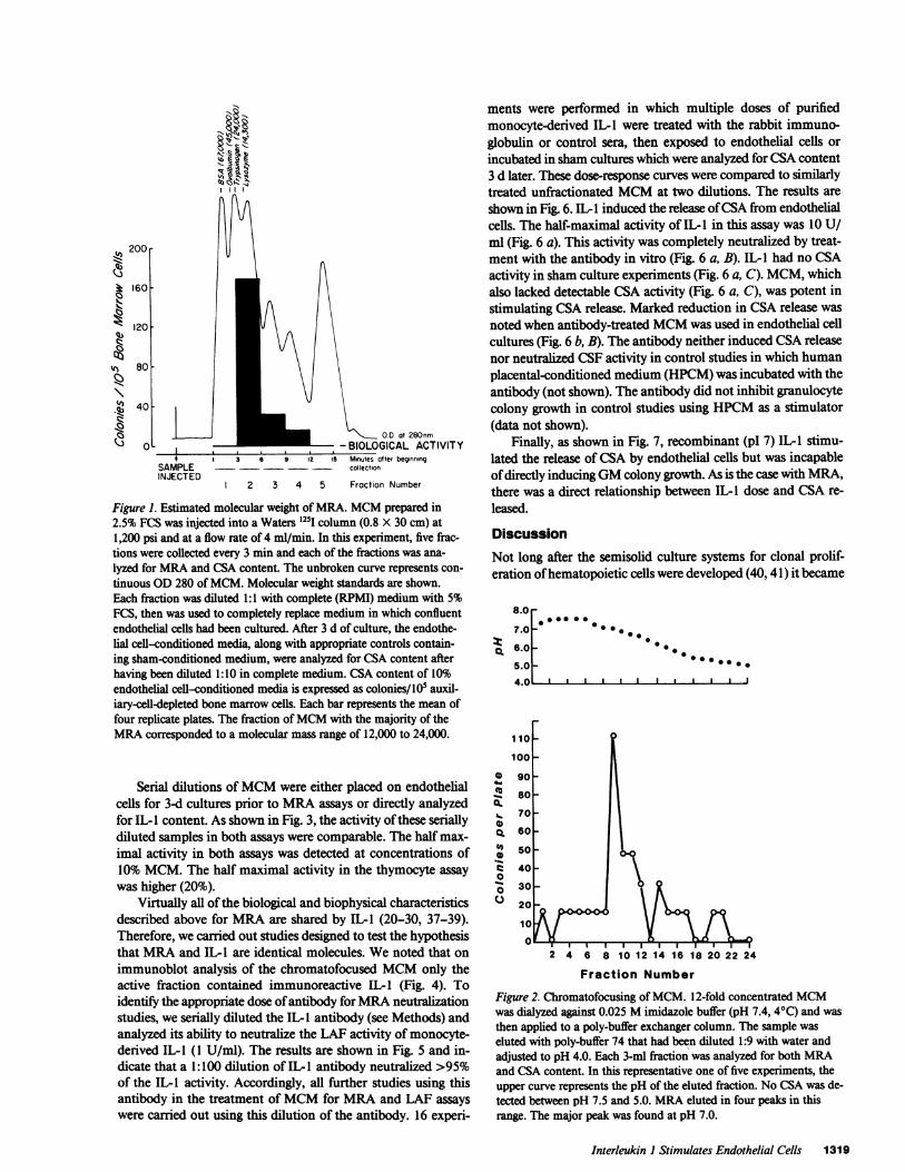
High performance liquid chromatography studies, a repre-sentative example of which is shown in Fig. 1, showed elutionof activity in the fractions ranging from 12 to 24,000 D. Minorpeaks of activity were also observed at lower molecular weightranges in some experiments. Five separate studies ofMCM usingchromatofocusing columns with a pH range of 7.5 to 4.0 con-sistently showed three peaks of activity between pH 7.0 and 5.0(Fig. 2). The major peak of activity ranged from 6.9 to 7.2.Minor peaks of activity were seen at pI 5.0-5.5 and 5.2-6.2.Often a fourth peak at pH 4.3 to 4.7 was also seen. This peakis not shown in Fig. 2.
1318 Bagby, Dinarello, Wallace, Wagner, Hefeneider, and McCall
* MRA detected ("+") in supernatants of 3-d cultures of IOW cells/ml.t Uninduced.
200
k 160
120
qs4 80
!i~AX 40
Vs
a 0Ij -\., OD at 280nm
- BIOLOGICAL ACTIVITY.* 3 6 5 12 15 Minutes after beginning
SAMPLE - - collectionINJECT ED
2 3 4 5 Fraction Number
Figure 1. Estimated molecular weight of MRA. MCM prepared in2.5% FCS was injected into a Waters l25l column (0.8 X 30 cm) at1,200 psi and at a flow rate of 4 ml/min. In this experiment, five frac-tions were collected every 3 min and each of the fractions was ana-lyzed for MRA and CSA content. The unbroken curve represents con-tinuous OD 280 ofMCM. Molecular weight standards are shown.Each fraction was diluted 1:1 with complete (RPMI) medium with 5%FCS, then was used to completely replace medium in which confluentendothelial cells had been cultured. After 3 d of culture, the endothe-lial cell-conditioned media, along with appropriate controls contain-ing sham-conditioned medium, were analyzed for CSA content afterhaving been diluted 1:10 in complete medium. CSA content of 10%endothelial cell-conditioned media is expressed as colonies/ I05 auxil-iary-cell-depleted bone marrow cells. Each bar represents the mean offour replicate plates. The fraction ofMCM with the majority of theMRA corresponded to a molecular mass range of 12,000 to 24,000.
Serial dilutions ofMCM were either placed on endothelialcells for 3-d cultures prior to MRA assays or directly analyzedfor IL- I content. As shown in Fig. 3, the activity ofthese seriallydiluted samples in both assays were comparable. The half max-imal activity in both assays was detected at concentrations of10% MCM. The half maximal activity in the thymocyte assaywas higher (20%).
Virtually all of the biological and biophysical characteristicsdescribed above for MRA are shared by IL-1 (20-30, 37-39).Therefore, we carried out studies designed to test the hypothesisthat MRA and IL-I are identical molecules. We noted that onimmunoblot analysis of the chromatofocused MCM only theactive fraction contained immunoreactive IL-I (Fig. 4). Toidentify the appropriate dose ofantibody forMRA neutralizationstudies, we serially diluted the IL-1 antibody (see Methods) andanalyzed its ability to neutralize the LAF activity of monocyte-derived IL-1 (1 U/ml). The results are shown in Fig. 5 and in-dicate that a 1:100 dilution of IL-I antibody neutralized >95%of the IL-I activity. Accordingly, all further studies using thisantibody in the treatment of MCM for MRA and LAF assayswere carried out using this dilution of the antibody. 16 experi-
ments were performed in which multiple doses of purifiedmonocyte-derived IL-1 were treated with the rabbit immuno-globulin or control sera, then exposed to endothelial cells orincubated in sham cultures which were analyzed for CSA content3 d later. These dose-response curves were compared to similarlytreated unfractionated MCM at two dilutions. The results areshown in Fig. 6. IL-I induced the release ofCSA from endothelialcells. The half-maximal activity of IL-I in this assay was 10 U/ml (Fig. 6 a). This activity was completely neutralized by treat-ment with the antibody in vitro (Fig. 6 a, B). IL- I had no CSAactivity in sham culture experiments (Fig. 6 a, C). MCM, whichalso lacked detectable CSA activity (Fig. 6 a, C), was potent instimulating CSA release. Marked reduction in CSA release wasnoted when antibody-treated MCM was used in endothelial cellcultures (Fig. 6 b, B). The antibody neither induced CSA releasenor neutralized CSF activity in control studies in which humanplacental-conditioned medium (HPCM) was incubated with theantibody (not shown). The antibody did not inhibit granulocytecolony growth in control studies using HPCM as a stimulator(data not shown).
Finally, as shown in Fig. 7, recombinant (pl 7) IL-l stimu-lated the release of CSA by endothelial cells but was incapableofdirectly inducingGM colony growth. As is the case with MRA,there was a direct relationship between IL-I dose and CSA re-leased.
DiscussionNot long after the semisolid culture systems for clonal prolif-eration of hematopoietic cells were developed (40, 41) it became
8.0
7.0
a 6.0
5.0A n.
CL*.
0()0
00C.)
110
100
go
00
00
_ *0
2 4 6 8 10 12 14 16 18 20 22 24
Fraction Number
Figure 2. Chromatofocusing ofMCM. 12-fold concentrated MCMwas dialyzed against 0.025 M imidazole buffer (pH 7.4, 40C) and wasthen applied to a poly-buffer exchanger column. The sample waseluted with poly-buffer 74 that had been diluted 1:9 with water andadjusted to pH 4.0. Each 3-ml fraction was analyzed for both MRAand CSA content. In this representative one of five experiments, theupper curve represents the pH of the eluted fraction. No GSA was de-tected between pH 7.5 and 5.0. MRA eluted in four peaks in thisrange. The major peak was found at pH 7.0.
Interleukin I Stimulates Endothelial Cells 1319
I
vL

50-
250~~~~~~~~~~~0
50 40 30 20 10 0
% MCM
Figure 3. Serial dilutions ofMCM in IL-I and MRA assays. DilutionsofMCM shown (x axis) represent the final concentration ofMCM inendothelial cell cultures (MRA assay) or in LAF assays (IL-1). MRA(U CSA/ml) and IL-I content (cpm [3H]thymidine incorporated) areexpressed as percent maximal stimulation. The dilution curve forMRA (mean [CSA] in three experiments±SD) is shown by the dashedline. Four separate experiments were done to define the dilution curvefor IL-l (-, n = 3): three (-, n = 3) utilized the LBRM assay (halfmaximal activity 5-10%), one (o, n = 1), the murine thymocyte assay.
clear that adherent cells, in particular mononuclear phagocytes,while incapable of forming colonies themselves, played an im-portant "accessory" role in the formation of granulocyte/mac-rophage colonies in vitro. Initially, mononuclear phagocytes werethought to provide a humoral factor, CSA, which stimulatedthe proliferation of the granulocyte/macrophage colony-formingunit (CFU-GM) (42, 43). Subsequent studies from our own lab-oratory and, more recently, other groups (13), have demonstratedthat monocytes require accessory cells to exert their colony-stimulating effects. Specifically, we have shown that mononuclearphagocytes produce and release into culture medium, a factoror factors, which we have operationally termed MRA. MRAstimulates endothelial cells and fibroblasts to release CSA (9,14, 19) and enhances CSA production by T lymphocytes (6).Macrophages are also important auxiliary cells in clonal growthof other committed hematopoietic progenitor cells as well. Forexample, Zuckerman reported that erythroid burst growth de-pended on monocytes in vitro (44), results that were later con-firmed by Levitt et al. (45). Recently, one mechanism by whichmonocytes effect their accessory role in other hematopoietic lin-eages has been described and resembles the effects ofmonocyteson granulopoiesis. Specifically, three collaborating groups, in-cluding our own, have shown that monocyte culture supernatantsinduce the release of factors into endothelial cell-conditionedmedium, which stimulates the clonal proliferation of erythroidburst forming units (16, 17), megakaryocytic colony formingunits, and multipotent colony forming units (18). Because mac-rophages, fibroblasts, endothelial cells, and colony forming cellsare each components ofthe hematopoietic marrow, we subscribeto the point of view that multilineage hematopoiesis in hema-topoietic microenvironments might be under the regulatorycontrol of mononuclear phagocytes.
For the past two years we have carried out a series of studiesdesigned to characterize and purify these monokines. We began
with the granulopoietic regulatory monokine using the endo-thelial cell CSA assay (see Methods) and in recently reportedstudies found that MRA production by monocytes requiresserum in the culture medium, declines after treatment with pro-tein synthesis inhibitors, and is substantially enhanced bothquantitatively and temporally by exposure of the monocytes toLPS; the maximal induced secretory response occurring at 24 h(17 [Table I]). Therefore, MRA could represent a novel mon-okine or a novel activity ofa previously reported monokine withthe above attributes. Such monokines include (a) tumor necrosisfactor alpha (TNF-a) (46-48), (b) platelet-derived growth factor(PDGF) (49), and (c) IL-1 (20, 21).
Further studies suggested that MRA shared more biologicalsimilarities with IL-I than with PDGF or TNF-a. The patternof cellular sources is more compatible with that reported for IL-1 (20, 26, 27) than with TNF (not constitutively produced byU937 cells [50]) or PDGF (not constitutively produced bymonocytes in vitro [49]). More importantly, PDGF (28,000-32,000 D [51]) has a higher molecular mass, higher pI (10 [52])and is markedly thermostable (49). TNF (17,000 D [46, 53])has a pl of 5.3 (47) and is more thermostable than IL-i (50).Taken together, these studies indicated that of the three knownmonokines, IL-l shared the greatest number of biological andbiophysical features with MRA. Other attributes ofMRA sharedwith IL-l are its thermolability, sensitivity to freezing and thaw-ing (28), neuraminidase resistance and papain sensitivity (20,22, 25), complex chromatofocusing profile (30) and its tendencyto bind to nonhydrophobic filtration membranes (21). For thesereasons, we carried out a series of experiments designed to de-termine with certainty whether IL-I exhibited MRA.
First, similar MCM dose-response curves were described inboth MRA and IL-I assays (Fig. 3). We next used purifiedmonocyte-derived IL-I and recombinant (pl 7) IL-I in ourMRAassay system. We found that these IL-I's induced the release ofCSA from multiply subcultured human umbilical vein endo-thelial cells (Figs. 6 and 7), and that treatment ofboth monocyte-
Sr
pH 7
10 15 20immunoblotd-|- + | _ | I_ |_ | _ 1l_|
Fraction
Figure 4. MCM was prepared as in Fig. 2 and was applied to a poly-buffer exchanger column with a range ofpH 9 to 6. Each 2-ml frac-tion was analyzed for MRA prior to and after 18 h exposure to theanti-IL-I antibody. 1.5-ml pools oftwo adjacent fractions were fil-tered through nitrocellulose for immunoblotting. Immunoreactive ma-terial is denoted "+." Nonreactive pools are marked "-." The pool oftwo fractions both positive in MRA assays was also positive for IL-Ion immunoblotting.
1320 Bagby, Dinarello, Wallace, Wagner, Hefeneider, and McCall
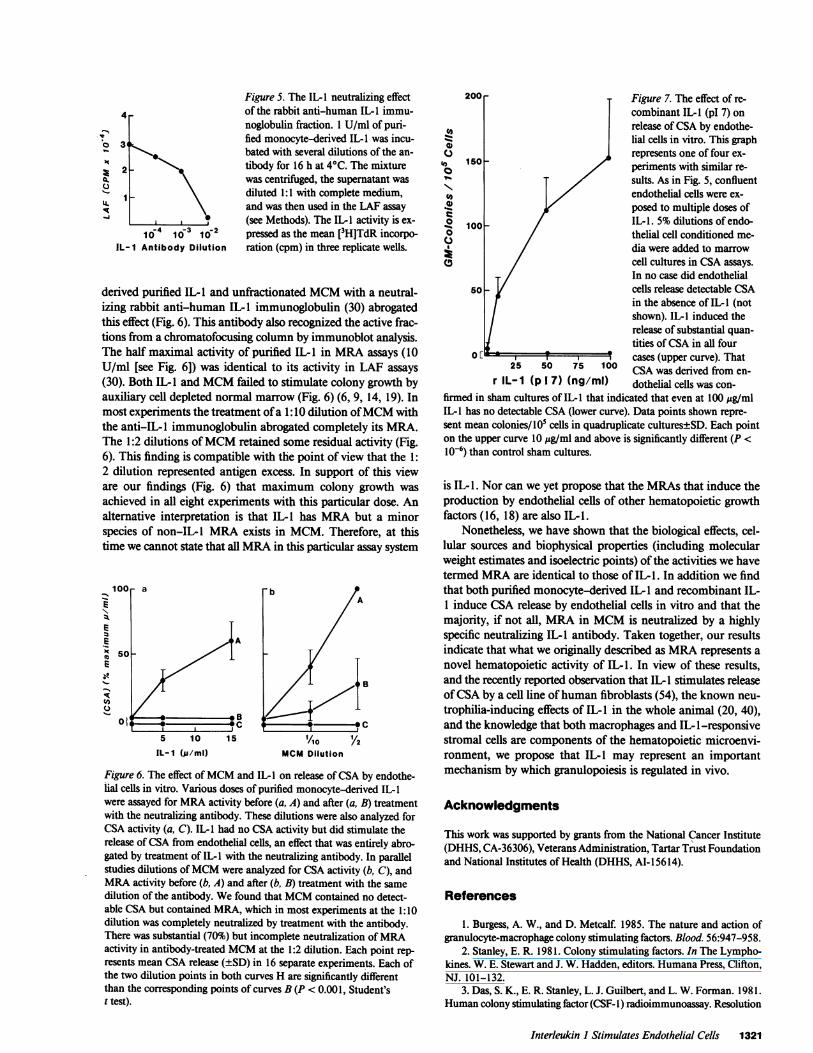
Figure 5. The IL-I neutralizing effect4 of the rabbit anti-human IL-I immu-
noglobulin fraction. 1 U/ml of puri-V
3 fied monocyte-derived IL-I was incu-bated with several dilutions of the an-
>2 _ > tibody for 16 h at 40C. The mixturewas centrifuged, the supernatant wasdiluted 1:1 with complete medium,and was then used in the LAF assay(see Methods). The IL-I activity is ex-
10-4 10-3 10-2 pressed as the mean [3H]TdR incorpo-IL- I Antibody Dilution ration (cpm) in three replicate wells.
derived purified IL- I and unfractionated MCM with a neutral-izing rabbit anti-human IL-1 immunoglobulin (30) abrogatedthis effect (Fig. 6). This antibody also recognized the active frac-tions from a chromatofocusing column by immunoblot analysis.The half maximal activity of purified IL-l in MRA assays (10U/ml [see Fig. 6]) was identical to its activity in LAF assays(30). Both IL-l and MCM failed to stimulate colony growth byauxiliary cell depleted normal marrow (Fig. 6) (6, 9, 14, 19). Inmost experiments the treatment ofa 1:10 dilution ofMCM withthe anti-IL-l immunoglobulin abrogated completely its MRA.The 1:2 dilutions ofMCM retained some residual activity (Fig.6). This finding is compatible with the point of view that the 1:2 dilution represented antigen excess. In support of this vieware our findings (Fig. 6) that maximum colony growth wasachieved in all eight experiments with this particular dose. Analternative interpretation is that IL-l has MRA but a minorspecies of non-IL-l MRA exists in MCM. Therefore, at thistime we cannot state that all MRA in this particular assay system
5 10 15
IL-1 (i/ml)
-b/A
1Ao Y2
MCM Dilution
Figure 6. The effect ofMCM and IL-I on release ofCSA by endothe-iial cells in vitro. Various doses of purified monocyte-derived IL-Iwere assayed for MRA activity before (a, A) and after (a, B) treatmentwith the neutralizing antibody. These dilutions were also analyzed forCSA activity (a, C). IL- I had no CSA activity but did stimulate therelease of CSA from endothelial cells, an effect that was entirely abro-gated by treatment of IL- with the neutralizing antibody. In parallelstudies dilutions ofMCM were analyzed for CSA activity (b, C), andMRA activity before (b, A) and after (b, B) treatment with the samedilution of the antibody. We found that MCM contained no detect-able CSA but contained MRA, which in most experiments at the 1:10dilution was completely neutralized by treatment with the antibody.There was substantial (70%) but incomplete neutralization ofMRAactivity in antibody-treated MCM at the 1:2 dilution. Each point rep-
resents mean CSA release (±SD) in 16 separate experiments. Each ofthe two dilution points in both curves H are significantly differentthan the corresponding points of curves B (P < 0.001, Student'st test).
200 Figure 7. The effect of re-combinant IL-I (pI 7) onrelease ofCSA by endothe-Hal cells in vitro. This graph
.) , , represents one of four ex-, 150 periments with similar re-
sults. As in Fig. 5, confluentendothelial cells were ex-
posed to multiple doses of°100 _ / IL-I. 5% dilutions of endo-
0 / thelial cell conditioned me-C./ dia were added to marrow0 / cell cultures in CSA assays.
In no case did endothelial50 - cells release detectable CSA
in the absence of IL- I (notshown). IL-I induced therelease of substantial quan-tities ofCSA in all four
0 * cases (upper curve). That25 50 75 100 CSA was derived from en-
r IL- 1 (p 1 7) (ng/ml) dothelial cells was con-firmed in sham cultures of IL-1 that indicated that even at 100 ug/mlIL-I has no detectable CSA (lower curve). Data points shown repre-sent mean colonies/105 cells in quadruplicate cultures±SD. Each pointon the upper curve 10 gg/ml and above is significantly different (P <10-6) than control sham cultures.
is IL-1. Nor can we yet propose that the MRAs that induce theproduction by endothelial cells of other hematopoietic growthfactors (16, 18) are also IL-1.
Nonetheless, we have shown that the biological effects, cel-lular sources and biophysical properties (including molecularweight estimates and isoelectric points) of the activities we havetermed MRA are identical to those of IL-1. In addition we findthat both purified monocyte-derived IL-I and recombinant IL-1 induce CSA release by endothelial cells in vitro and that themajority, if not all, MRA in MCM is neutralized by a highlyspecific neutralizing IL-l antibody. Taken together, our resultsindicate that what we originally described as MRA represents anovel hematopoietic activity of IL-1. In view of these results,and the recently reported observation that IL-l stimulates releaseofCSA by a cell line of human fibroblasts (54), the known neu-trophilia-inducing effects of IL-l in the whole animal (20, 40),and the knowledge that both macrophages and IL-l-responsivestromal cells are components of the hematopoietic microenvi-ronment, we propose that IL-1 may represent an importantmechanism by which granulopoiesis is regulated in vivo.
Acknowledgments
This work was supported by grants from the National Cancer Institute(DHHS, CA-36306), Veterans Administration, Tartar Trust Foundationand National Institutes of Health (DHHS, AI-156 14).
References
1. Burgess, A. W., and D. Metcalf. 1985. The nature and action ofgranulocyte-macrophage colony stimulating factors. Blood. 56:947-958.
2. Stanley, E. R. 1981. Colony stimulating factors. In The Lympho-kines. W. E. Stewart and J. W. Hadden, editors. Humana Press, Clifton,NJ. 101-132.
3. Das, S. K., E. R. Stanley, L. J. Guilbert, and L. W. Forman. 1981.Human colony stimulating factor (CSF- 1) radioimmunoassay. Resolution
Interleukin I Stimulates Endothelial Cells 1321
._E
:4
I-
of the subclasses of human colony-stimulating factors. Blood. 58:630-641.
4. Wong, G. G., J. S. Witek, P. A. Temple, K. M. Wilkens, A. C.Leary, D. P. Luxenberg, S. S. Jones, E. L. Brown, R. M. Kay, E. C. Orr,C. Shoemaker, D. W. Golde, R. J. Kaufman, R. M. Hewick, E. A. Wang,and S. C. Clark. 1985. Human GM-CSF: molecular cloning ofthe com-plementary DNA and purification of the natural and recombinant pro-teins. Science (Wash. DC). 228:810-815.
5. Souza, L. M., T. C. Boone, J. Gabrilove, P. H. Lai, K. Zsebo,D. C. Murdock, V. R. Chazin, J. Bruszewski, H. Lu, K. Chen, J. Barendt,E. Platzer, M. A. S. Moore, R. Mertelsmann, and K. Welte. 1986. Re-combinant human granulocyte colony stimulating factor: effects on nor-mal and leukemic myeloid cells. Science (Wash. DC). 232:61-65.
6. Bagby, G. C., V. D. Rigas, R. M. Bennett, A. A. Vandenbark, andH. S. Garewal. 1981. Interaction of lactoferrin, monocytes, and T lym-phocyte subsets in the regulation of steady-state granulopoiesis in vitro.J. Clin. Invest. 68:56-63.
7. Ruscetti, F. W., and P. A. Chervenick. 1975. Release of colonystimulating activity from thymus-derived lymphocytes. J. Clin. Invest.55:520-527.
8. Cline, M. J., and D. W. Golde. 1974. Production of colony stim-ulating activity by human lymphocytes. Nature (Lond.). 248:703-704.
9. Bagby, G. C., E. McCall, and D. L. Layman. 1983. Regulation ofcolony stimulating activity production. Interactions of fibroblasts,mononuclear phagocytes and lactoferrin. J. Clin. Invest. 71:340-344.
10. Knudtzon, S., and B. T. Mortenson. 1975. Growth stimulationofhuman bone marrow cells in agar culture by vascular cells. Blood. 46:937-943.
11. Quesenberry, P. J., and M. A. Gimbrone. 1980. Vascular en-dothelium as a regulator of granulopoiesis. Production of colony-stim-ulating activity by cultured human endothelial cells. Blood. 56:1060-1067.
12. Ascensao, J. L., G. M. Vercellotti, H. S. Jacob, and E. D. Zanjani.1984. Role of endothelial cells in human hematopoiesis: modulation ofmixed colony growth in vitro. Blood. 63:553-558.
13. Gerson, S. L., H. M. Friedman, and D. B. Cines. 1985. Viralinfection of vascular endothelial cells alters production of colony-stim-ulating activity. J. Clin. Invest. 76:1382-1390.
14. Bagby, G. C., E. McCall, K. A. Bergstrom, and D. Burger. 1983.A monokine regulates colony-stimulating activity production by vascularendothelial cells. Blood. 62:663-668.
15. Keating, A., J. W. Singer, P. D. Killen, G. E. Striker, A. C. Salo,J. Sanders, E. D. Thomas, D. Thorning, and P. J. Fialkow. 1982. Donororigin of the in vitro haematopoietic microenvironment after marrowtransplantation in man. Nature (Lond.). 298:280-282.
16. Zuckerman, K. S., G. C. Bagby, E. McCall, B. Sparks, J. Wells,and V. Patel. 1985. A monokine stimulates production of human ery-throid burst promoting activity by endothelial cells in vitro. J. Clin.Invest. 75:722-725.
17. Broudy, V., K. Zuckerman, S. Jetmalani, J. H. Fitchen, andG. C. Bagby. 1984. The production of burst promoting activity (BPA)and colony stimulating activity (CSA) by adherent fibroblastoid marrowcells (F) is regulated by autologous monocytes. Clin. Res. 32:305a. (Abstr.)
18. Segal, G. M., E. McCall, T. Stueve, and G. C. Bagby. 1985.Monokine stimulated endothelial cells promote human megakaryocyticand mixed cell colony growth in vitro. J. Immunol. (In press).
19. McCall, E., and G. C. Bagby. 1985. Monocyte-derived recruitingactivity. Kinetics ofproduction and effects ofendotoxin. Blood. 65:689-695.
20. Dinarello, C. A. 1984. Interleukin-l. Rev. Infect. Dis. 6:51-95.21. Lachman, L. B. 1983. Human interleukin-l: Purification and
properties. Fed. Proc. 42:2639-2645.22. Wood, D. D. 1979. Purification and properties of human B cell
activating factor. J. Immunol. 79:2395-2399.23. Luger, T. A., B. M. Stadler, B. M. Luger, B. J. Mathieson, M.
Mage, J. A. Schmidt, and J. J. Oppenheim. 1982. Murine epidermalcell-derived thymocyte-activating factor resembles murine interleukin-I. J. Immunol. 128:2147-2152.
24. Simon, P. L., and W. F. Willoughby. 1981. The role ofsubcellularfactors in pulmonary immune function: physical-chemical characteriza-tion oftwo distinct species of lymphocyte-activating factor produced byrabbit alveolar macrophages. J. Immunol. 126:1534-1541.
25. Mizel, S. B. 1979. Physicochemical characterization oflymphocyteactivating factor (LAF). J. Immunol. 122:2167-2172.
26. Amento, E. P., J. T. Kurnick, A. Epstein, and S. M. Crane. 1982.Modulation of synovial cell products by a factor from human cell line:T-lymphocyte induction of a mononuclear cell factor. Proc. Natl. Acad.Sci. USA. 79:5307-5311.
27. Mizel, S. B., and B. J. Andersen. 1983. Interleukin-l productionby a human monocytic leukemia cell line, THP- 1. In Interleukins, Lym-phokines and Cytokines: Proceedings of the Third International Lym-phokine Workshop. J. J. Oppenheim and S. Cohen, editors. AcademicPress, Orlando, FL. 401-407.
28. Cannon, J. G., and C. A. Dinarello. 1985. Increased plasma in-terleukin-l activity in women after ovulation. Science (Wash. DC). 227:1247-1249.
29. Gowen, M., D. D. Wood, and R. G. G. Russell. 1985. Stimulationof the proliferation of human bone cells in vitro by human monocyteproducts with interleukin-l activity. J. Clin. Invest. 75:1223-1229.
30. Dinarello, C. A., H. A. Bernheim, J. G. Cannon, G. LoPreste,S. J. C. Warner, A. C. Webb, and P. E. Auron. 1985. Purified, IIS-MET,3H-LEU-labelled human monocyte interleukin-l (IL-1) with endogenouspyrogen activity. Br. J. Rheumatol. 24(Suppl):59-64.
31. Auron, P. E., A. C. Webb, L. J. Rosenwasser, S. F. Mucci, A.Rich, S. M. Wolff, and C. A. Dinarello. 1984. Nucleotide sequence ofhuman monocyte interleukin- 1 precursor cDNA. Proc. Natl. Acad. Sci.USA. 81:7907-7911.
32. Schlunk, T., and M. Schleyer. 1980. The influence of cultureconditions on the production of colony stimulating activity by humanplacenta. Exp. Hematol. 8:179-184.
33. Metcalf, D., H. R. MacDonald, and H. M. Chester. 1975. Serumpotentiation of granulocyte and macrophage colony formation in vitro.Exp. Hematol. 3:261-270.
34. Wagner, C. R., R. M. Vetto, and D. R. Burger. 1985. Expressionof I-region-associated antigen (Ia) and interleukin-l by subcultured hu-man endothelial cells. Cell. Immunol. 93:91-104.
35. Dinarello, C. A., J. G. Cannon, J. W. Mier, H. A. Bernheim, G.LoPreste, D. L. Lynn, R. N. Love, A. C. Webb, P. C. Auron, R. C.Reuben, A. Dick, S. M. Wolff, and S. D. Putney. 1986. Multiple biologicalactivities ofhuman recombinant interleukin-l . J. Clin. Invest. 77:1734-1739.
36. Sundstrom, C., and K. Nilsson. 1976. Establishment and char-acterization of a human histiocytic lymphoma cell line (U937). Int. J.Cancer. 17:565-577.
37. Kaye, J., S. Gillis, S. B. Myzel, E. M. Sehevach, T. R. Malek,C. A. Dinarello, L. B. Lachman, and C. A. Janeway. 1982. Growth of acloned helper T cell line induced by a monoclonal antibody specific forthe antigen receptor: interleukin-1 is required for the expression of re-
ceptors for interleukin-2. J. Immunol. 133:1339-1345.38. Duff, G. W., and E. Atkins. 1982. The inhibitory effect of po-
lymyxin B on endotoxin-induced endogenous pyrogen production. J.Immunol. Methods. 52:333-340.
39. Oppenheim, J. J., B. M. Stadler, R. P. Siraganian, M. Mage, andB. Mathieson. 1982. Lymphokines: their role in lymphocyte responses.Properties of interleukin 1. Fed. Proc. 41:257-262.
40. Bradley, T. R., and D. Metcalf. 1966. The growth ofmouse bonemarrow cells in vitro. Aust. J. Exp. Biol. Med. Sci. 44:287-299.
41. Pluznik, D. H., and L. Sachs. 1965. The cloning ofnormal "mast"cells in tissue culture. J. Cell. Comp. Physiol. 66:319-324.
42. Golde, D. W., and M. J. Cline. 1972. Identification of colony-stimulating cells in human peripheral blood. J. Clin. Invest. 51:2981-2983.
43. Chervenick, P. A., and A. F. LoBuglio. 1972. Human bloodmonocytes, stimulators of granulocyte and mononuclear colony for-mation in vitro. Science (Wash. DC). 178:164-166.
44. Zuckerman, K. S. 1981. Human erythroid burst-forming units.
1322 Bagby, Dinarello, Wallace, Wagner, Hefeneider, and McCall
Growth in vitro is dependent on monocytes, but not T lymphocytes. J.Clin. Invest. 67:702-709.
45. Levitt, L., T. J. Kipps, E. G. Engleman, and P. L. Greenberg.1985. Human bone marrow and peripheral blood T-lymphocyte deple-tion: Efficacy and effects of both T-cells and monocytes on growth ofhematopoietic progenitors. Blood. 65:663-679.
46. Pennica, D., G. E. Nedwin, J. S. Hayflick, P. H. Seeburg, R.Derynck, M. A. Palladino, and G. E. Nedwin. 1984. Human tumournecrosis factor: precursor structure, expression and homology to lym-photoxin. Nature (Lond.). 312:724-729.
47. Aggarwal, B. B., W. J. Kohr, P. E. Hass, B. Moffat, S. A. Spencer,W. J. Henzel, T. S. Bringman, G. E. Nedwin, D. V. Goeddel, and R. N.Harkins. 1985. Human tumor necrosis factor: production, purification,and characterization. J. Biol. Chem. 260:2345-2354.
48. Mannel, D. M., R. N. Moore, and S. E. Mergenhagen. 1980.Generation and characterization of a lipopolysaccharide. Induced andserum derived cytotoxic factor for tumor cells. Infect. Immun. 28:204-211.
49. Martinet, Y., P. B. Bitterman, J-F. Mornex, G. R. Grotendorst,G. R. Martin, and R. G. Crystal. 1986. Activated human monocytes
express the c-sis proto-oncogene and release a mediator showing PDGFlike activity. Nature (Lond.). 319:158-160.
50. Ruff, M. R., and G. E. Gifford. 1981. Tumor Necrosis Factor.Lymphokines. 2:235-272.
51. Van Zoelen, E. J. J., W. J. M. Van de Ven, H. J. Franssen,T. M. J. Van Oostwaard, P. W. Van der Saag, C-H Heldin, and S. W.de Laat. 1985. Neuroblastoma cells express c-sis and produce a trans-forming growth factor antigenically related to the platelet-derived growthfactor. Mol. Cell. Bio. 5:2289-2297.
52. Deuel, T. F., and J. S. Huang. 1984. Platelet derived growthfactor, structure function, and roles in normal and transformed cells. J.Clin. Invest. 74:669-676.
53. Wang, A. M., A. A. Creasey, M. B. Ladner, L. S. Lin, J. Strickler,J. N. Van Arsdell, R. Yamamoto, and D. F. Mark. 1985. Molecularcloning of the complementary DNA for Human tumor necrosis factor.Science (Wash. DC). 228:149-154.
54. Zucali, J. R., C. A. Dinarello, D. J. Oblon, M. A. Gross, L.Anderson, and R. S. Weiner. 1986. Interleukin 1 stimulates fibroblaststo produce granulocyte-macrophage colony stimulating activity andprostaglandin E2. J. Clin. Invest. 77:1857-1863.
Interleukin I Stimulates Endothelial Cells 1323
Related Documents