Interleukin 1 receptor antagonist mediates the antiinflammatory and antifibrotic effect of mesenchymal stem cells during lung injury Luis A. Ortiz*, Maria DuTreil † , Cheryl Fattman*, Amitabh C. Pandey † , German Torres*, Kristina Go*, and Donald G. Phinney †‡ *Department of Environmental and Occupational Health, University of Pittsburgh, Pittsburgh, PA 15261; † Center for Gene Therapy, Tulane University Health Sciences Center, New Orleans, LA 70112 Communicated by Darwin J. Prockop, Tulane University, New Orleans, LA, May 16, 2007 (received for review April 3, 2007) Mesenchymal stem cells (MSCs) have been exploited as cellular vectors to treat a wide array of diseases but the mechanisms responsible for their therapeutic effect remain indeterminate. Previously, we reported that MSCs inhibit bleomycin (BLM)- induced inflammation and fibrosis within the lungs of mice. Inter- rogation of the MSC transcriptome identified interleukin 1 receptor antagonist (IL1RN) as a potential mediator of this effect. Fraction- ation studies indicated that MSCs are the principal source of IL1RN in murine bone marrow and that its expression is restricted to a unique subpopulation of cells. Moreover, MSC-conditioned media was shown to block proliferation of an IL-1-dependent T cell line and inhibit production of TNF- by activated macrophages in vitro. Studies conducted in mice revealed that MSC administration was more effective than recombinant IL1RN delivered via adenoviral infection or osmotic pumps in inhibiting BLM-induced increases in TNF-, IL-1, and IL1RN mRNA in lung, IL1RN protein in bronchoal- veolar lavage (BAL) fluid, and trafficking of lymphocytes and neutrophils into the lung. Therefore, MSCs protect lung tissue from BLM-induced injury by blocking TNF- and IL-1, two fundamental proinflammatory cytokines in lung. Identification of IL1RN- expressing human MSC subpopulations may provide a novel cel- lular vector for treating chronic inflammatory diseases in humans. bleomycin inflammation I nterstitial lung diseases (ILDs) are a heterogeneous group of 150 disorders characterized by epithelial injury, fibroblast proliferation, expansion of the lung matrix, and dyspnea. Among these diseases, idiopathic pulmonary fibrosis (IPF) is the most frequent and lethal. The median survival of IPF patients is 3–5 years irrespective of whether they receive therapy (1), and recent studies suggest that its incidence and prevalence is increasing (2). Inflammatory responses in IPF are mediated by release from activated macrophages and other leukocytes of the proinflam- matory cytokines IL-1 and TNF- (3). These cytokines induce endothelial cells to express adhesion molecules and chemokines that attract other white cells from the blood to the site of injury (4, 5). IL-1 and TNF- also stimulate proliferation of endothelial cells and fibroblasts that increase the blood supply at the site of injury and repair damage by formation of scar tissue (6). The IL-1 family includes the structurally related proteins IL1-, IL-1, and interleukin 1 receptor antagonist (IL1RN) that bind to the same receptor. However, IL1RN functions as a competitive inhibitor of IL-1 and IL-1 (7). Various studies have shown that the IL-1 family plays an important role in ILD. For example, injection of recombinant IL-1 protein into rodent tracheas promotes acute alveolar leakage and neutrophil inf lam- mation in lung (8, 9). Moreover, IL-1 expression levels in lung are correlated with the development of pulmonary fibrosis in ro- dents exposed to bleomycin (BLM) or radiation (10, 11) and up-regulated in fibro-proliferative areas within the lungs of idiopathic pulmonary fibrosis patients (12). In addition, patients with a particular IL1RN polymorphism demonstrate a higher risk for fibrosing alveolitis, indicating that an imbalance between IL-1 and IL1RN activity also contributes to ILD (13). The results are consistent with studies demonstrating that IL1RN adminis- tration blocks lung fibrosis induced in mice by exposure to BLM or silica (14). Over the past decade, stem cells from adult bone marrow have been exploited as therapeutic vectors to treat a wide variety of diseases (15, 16). However, limited information exists regarding the therapeutic potential of these cells in lung diseases. For example, several studies have demonstrated that hematopoietic stem cells (17) or marrow-derived stromal cells (18) contribute to airway and distal (type II) alveolar epithelium, but the capacity of these cells to ameliorate disease has been largely unexplored. Recently, we reported that mesenchymal stem cells (MSCs) administered to mice challenged with BLM engrafted at low levels but significantly reduced the extent of inflammation and fibrosis in lungs (19). Importantly, MSCs were efficacious in ameliorating lung injury only when administered at the time of BLM challenge and not at later time points. This result suggested their therapeutic effect was attributed to the production of soluble factors that modulate inflammation. Herein, we report the identification of murine and human MSC subpopulations that secrete high levels of IL1RN. We also provide in vitro and in vivo data that production of IL1RN by MSCs protects mice from BLM-induced lung injury by blocking the production and/or activity of TNF- and IL-1, the predominant proin- flammatory cytokines in lung tissue. Our discovery of human MSC subpopulations that express IL1RN provides a strong rationale for developing MSC-based therapies to treat ILD. Results Identification of an IL1RN-Expressing Subpopulation of MSCs. To identify proteins expressed by murine MSCs that may modulate the inf lammatory response, we interrogated their transcriptome, which we catalogued (20) by serial analysis of gene expression (SAGE). This SAGE library consisted of 59,007 sequenced SAGE tags that corresponded to 5,737 identifiable, unique mRNAs. Characterizing these expressed mRNAs based on gene ontologies revealed that MSCs expressed transcripts encoding regulatory proteins involved in angiogenesis, cell motility and communication, hematopoiesis, neural activities, and immunity and defense. In the latter case, numerous SAGE tags corre- Author contributions: L.A.O. and D.G.P. designed research; L.A.O., M.D., C.F., A.C.P., G.T., K.G., and D.G.P. performed research; L.A.O., M.D., C.F., and D.G.P. analyzed data; and L.A.O. and D.G.P. wrote the paper. The authors declare no conflict of interest. Freely available online through the PNAS open access option. Abbreviations: BAL, bronchoalveolar lavage; BLM, bleomycin; IL1RN, interluekin 1 receptor antagonist; ILD, interstitial lung disease; MSC, mesenchymal stem cell; SAGE, serial analysis of gene expression. ‡ To whom correspondence should be addressed. E-mail: [email protected]. This article contains supporting information online at www.pnas.org/cgi/content/full/ 0704421104/DC1. © 2007 by The National Academy of Sciences of the USA 11002–11007 PNAS June 26, 2007 vol. 104 no. 26 www.pnas.orgcgidoi10.1073pnas.0704421104

Welcome message from author
This document is posted to help you gain knowledge. Please leave a comment to let me know what you think about it! Share it to your friends and learn new things together.
Transcript
Interleukin 1 receptor antagonist mediates theantiinflammatory and antifibrotic effect ofmesenchymal stem cells during lung injuryLuis A. Ortiz*, Maria DuTreil†, Cheryl Fattman*, Amitabh C. Pandey†, German Torres*, Kristina Go*, and Donald G. Phinney†‡
*Department of Environmental and Occupational Health, University of Pittsburgh, Pittsburgh, PA 15261; †Center for Gene Therapy, Tulane University HealthSciences Center, New Orleans, LA 70112
Communicated by Darwin J. Prockop, Tulane University, New Orleans, LA, May 16, 2007 (received for review April 3, 2007)
Mesenchymal stem cells (MSCs) have been exploited as cellularvectors to treat a wide array of diseases but the mechanismsresponsible for their therapeutic effect remain indeterminate.Previously, we reported that MSCs inhibit bleomycin (BLM)-induced inflammation and fibrosis within the lungs of mice. Inter-rogation of the MSC transcriptome identified interleukin 1 receptorantagonist (IL1RN) as a potential mediator of this effect. Fraction-ation studies indicated that MSCs are the principal source of IL1RNin murine bone marrow and that its expression is restricted to aunique subpopulation of cells. Moreover, MSC-conditioned mediawas shown to block proliferation of an IL-1�-dependent T cell lineand inhibit production of TNF-� by activated macrophages in vitro.Studies conducted in mice revealed that MSC administration wasmore effective than recombinant IL1RN delivered via adenoviralinfection or osmotic pumps in inhibiting BLM-induced increases inTNF-�, IL-1�, and IL1RN mRNA in lung, IL1RN protein in bronchoal-veolar lavage (BAL) fluid, and trafficking of lymphocytes andneutrophils into the lung. Therefore, MSCs protect lung tissue fromBLM-induced injury by blocking TNF-� and IL-1, two fundamentalproinflammatory cytokines in lung. Identification of IL1RN-expressing human MSC subpopulations may provide a novel cel-lular vector for treating chronic inflammatory diseases in humans.
bleomycin � inflammation
Interstitial lung diseases (ILDs) are a heterogeneous group of�150 disorders characterized by epithelial injury, fibroblast
proliferation, expansion of the lung matrix, and dyspnea. Amongthese diseases, idiopathic pulmonary fibrosis (IPF) is the mostfrequent and lethal. The median survival of IPF patients is 3–5years irrespective of whether they receive therapy (1), and recentstudies suggest that its incidence and prevalence is increasing (2).Inflammatory responses in IPF are mediated by release fromactivated macrophages and other leukocytes of the proinflam-matory cytokines IL-1 and TNF-� (3). These cytokines induceendothelial cells to express adhesion molecules and chemokinesthat attract other white cells from the blood to the site of injury(4, 5). IL-1 and TNF-� also stimulate proliferation of endothelialcells and fibroblasts that increase the blood supply at the site ofinjury and repair damage by formation of scar tissue (6).
The IL-1 family includes the structurally related proteinsIL1-�, IL-1�, and interleukin 1 receptor antagonist (IL1RN)that bind to the same receptor. However, IL1RN functions as acompetitive inhibitor of IL-1� and IL-1� (7). Various studieshave shown that the IL-1 family plays an important role in ILD.For example, injection of recombinant IL-1 protein into rodenttracheas promotes acute alveolar leakage and neutrophil inflam-mation in lung (8, 9). Moreover, IL-1 expression levels in lung arecorrelated with the development of pulmonary fibrosis in ro-dents exposed to bleomycin (BLM) or radiation (10, 11) andup-regulated in fibro-proliferative areas within the lungs ofidiopathic pulmonary fibrosis patients (12). In addition, patientswith a particular IL1RN polymorphism demonstrate a higherrisk for fibrosing alveolitis, indicating that an imbalance between
IL-1 and IL1RN activity also contributes to ILD (13). The resultsare consistent with studies demonstrating that IL1RN adminis-tration blocks lung fibrosis induced in mice by exposure to BLMor silica (14).
Over the past decade, stem cells from adult bone marrow havebeen exploited as therapeutic vectors to treat a wide variety ofdiseases (15, 16). However, limited information exists regardingthe therapeutic potential of these cells in lung diseases. Forexample, several studies have demonstrated that hematopoieticstem cells (17) or marrow-derived stromal cells (18) contributeto airway and distal (type II) alveolar epithelium, but thecapacity of these cells to ameliorate disease has been largelyunexplored. Recently, we reported that mesenchymal stem cells(MSCs) administered to mice challenged with BLM engrafted atlow levels but significantly reduced the extent of inflammationand fibrosis in lungs (19). Importantly, MSCs were efficacious inameliorating lung injury only when administered at the time ofBLM challenge and not at later time points. This result suggestedtheir therapeutic effect was attributed to the production ofsoluble factors that modulate inflammation. Herein, we reportthe identification of murine and human MSC subpopulationsthat secrete high levels of IL1RN. We also provide in vitro andin vivo data that production of IL1RN by MSCs protects micefrom BLM-induced lung injury by blocking the productionand/or activity of TNF-� and IL-1�, the predominant proin-flammatory cytokines in lung tissue. Our discovery of humanMSC subpopulations that express IL1RN provides a strongrationale for developing MSC-based therapies to treat ILD.
ResultsIdentification of an IL1RN-Expressing Subpopulation of MSCs. Toidentify proteins expressed by murine MSCs that may modulatethe inflammatory response, we interrogated their transcriptome,which we catalogued (20) by serial analysis of gene expression(SAGE). This SAGE library consisted of 59,007 sequencedSAGE tags that corresponded to 5,737 identifiable, uniquemRNAs. Characterizing these expressed mRNAs based on geneontologies revealed that MSCs expressed transcripts encodingregulatory proteins involved in angiogenesis, cell motility andcommunication, hematopoiesis, neural activities, and immunityand defense. In the latter case, numerous SAGE tags corre-
Author contributions: L.A.O. and D.G.P. designed research; L.A.O., M.D., C.F., A.C.P., G.T.,K.G., and D.G.P. performed research; L.A.O., M.D., C.F., and D.G.P. analyzed data; andL.A.O. and D.G.P. wrote the paper.
The authors declare no conflict of interest.
Freely available online through the PNAS open access option.
Abbreviations: BAL, bronchoalveolar lavage; BLM, bleomycin; IL1RN, interluekin 1 receptorantagonist; ILD, interstitial lung disease; MSC, mesenchymal stem cell; SAGE, serial analysisof gene expression.
‡To whom correspondence should be addressed. E-mail: [email protected].
This article contains supporting information online at www.pnas.org/cgi/content/full/0704421104/DC1.
© 2007 by The National Academy of Sciences of the USA
11002–11007 � PNAS � June 26, 2007 � vol. 104 � no. 26 www.pnas.org�cgi�doi�10.1073�pnas.0704421104
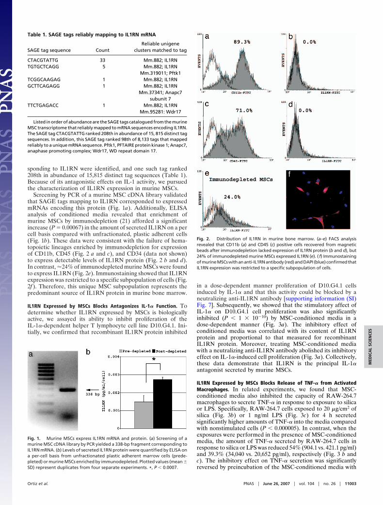
sponding to IL1RN were identified, and one such tag ranked208th in abundance of 15,815 distinct tag sequences (Table 1).Because of its antagonistic effects on IL-1 activity, we pursuedthe characterization of IL1RN expression in murine MSCs.
Screening by PCR of a murine MSC cDNA library validatedthat SAGE tags mapping to IL1RN corresponded to expressedmRNAs encoding this protein (Fig. 1a). Additionally, ELISAanalysis of conditioned media revealed that enrichment ofmurine MSCs by immunodepletion (21) afforded a significantincrease (P � 0.00067) in the amount of secreted IL1RN on a percell basis compared with unfractionated, plastic adherent cells(Fig. 1b). These data were consistent with the failure of hema-topoietic lineages enriched by immunodepletion for expressionof CD11b, CD45 (Fig. 2 a and c), and CD34 (data not shown)to express detectable levels of IL1RN protein (Fig. 2 b and d).In contrast, �24% of immunodepleted murine MSCs were foundto express IL1RN (Fig. 2e). Immunostaining showed that IL1RNexpression was restricted to a specific subpopulation of cells (Fig.2f ). Therefore, this unique MSC subpopulation represents thepredominant source of IL1RN protein in murine bone marrow.
IL1RN Expressed by MSCs Blocks Antagonizes IL-1� Function. Todetermine whether IL1RN expressed by MSCs is biologicallyactive, we assayed its ability to inhibit proliferation of theIL-1�-dependent helper T lymphocyte cell line D10.G4.1. Ini-tially, we confirmed that recombinant IL1RN protein inhibited
in a dose-dependent manner proliferation of D10.G4.1 cellsinduced by IL-1� and that this activity could be blocked by aneutralizing anti-IL1RN antibody [supporting information (SI)Fig. 7]. Subsequently, we showed that the stimulatory affect ofIL-1� on D10.G4.1 cell proliferation was also significantlyinhibited (P � 1 � 10�10) by MSC-conditioned media in adose-dependent manner (Fig. 3a). The inhibitory effect ofconditioned media was correlated with its content of IL1RNprotein and proportional to that measured for recombinantIL1RN protein. Moreover, treating MSC-conditioned mediawith a neutralizing anti-IL1RN antibody abolished its inhibitoryeffect on IL-1�-induced cell proliferation (Fig. 3a). Collectively,these data demonstrate that IL1RN is the principal IL-1�antagonist secreted by murine MSCs.
IL1RN Expressed by MSCs Blocks Release of TNF-� from ActivatedMacrophages. In related experiments, we found that MSC-conditioned media also inhibited the capacity of RAW-264.7macrophages to secrete TNF-� in response to exposure to silicaor LPS. Specifically, RAW-264.7 cells exposed to 20 �g/cm2 ofsilica (Fig. 3b) or 1 ng/ml LPS (Fig. 3c) for 4 h secretedsignificantly higher amounts of TNF-� into the media comparedwith nonstimulated cells (P � 0.000005). In contrast, when theexposures were performed in the presence of MSC-conditionedmedia, the amount of TNF-� secreted by RAW-264.7 cells inresponse to silica or LPS was reduced 54% (904.1 vs. 421.1 pg/ml)and 39.3% (34,040 vs. 20,652 pg/ml), respectively (Fig. 3 b andc). The inhibitory effect on TNF-� secretion was significantlyreversed by preincubation of the MSC-conditioned media with
Table 1. SAGE tags reliably mapping to IL1RN mRNA
SAGE tag sequence CountReliable unigene
clusters matched to tag
CTACGTATTG 33 Mm.882; IL1RNTGTGCTCAGG 5 Mm.882; IL1RN
Mm.319011; Pftk1TCGGCAAGAG 1 Mm.882; IL1RNGCTTCAGAGG 1 Mm.882; IL1RN
Mm.37341; Anapc7subunit 7
TTCTGAGACC 1 Mm.882; IL1RNMm.95281: Wdr17
Listed in order of abundance are the SAGE tags catalogued from the murineMSC transcriptome that reliably mapped to mRNA sequences encoding IL1RN.The SAGE tag CTACGTATTG ranked 208th in abundance of 15, 815 distinct tagsequences. In addition, this SAGE tag ranked 98th of 8,133 tags that mappedreliably to a unique mRNA sequence. Pftk1, PFTAIRE protein kinase 1; Anapc7,anaphase promoting complex; Wdr17, WD repeat domain 17.
Fig. 1. Murine MSCs express IL1RN mRNA and protein. (a) Screening of amurine MSC cDNA library by PCR yielded a 338-bp fragment corresponding toIL1RN mRNA. (b) Levels of secreted IL1RN protein were quantified by ELISA ona per-cell basis from unfractionated plastic adherent marrow cells (prede-pleted) or murine MSCs enriched by immunodepleted. Plotted values (mean �SD) represent duplicates from four separate experiments. *, P � 0.0007.
Fig. 2. Distribution of IL1RN in murine bone marrow. (a–e) FACS analysisrevealed that CD11b (a) and CD45 (c) positive cells recovered from magneticbeads after immunodepletion lacked expression of IL1RN protein (b and d), but24% of immunodepleted murine MSCs expressed IL1RN (e). (f) ImmunostainingofmurineMSCswithananti-IL1RNantibody (red)andDAPI (blue) confirmedthatIL1RN expression was restricted to a specific subpopulation of cells.
Ortiz et al. PNAS � June 26, 2007 � vol. 104 � no. 26 � 11003
MED
ICA
LSC
IEN
CES
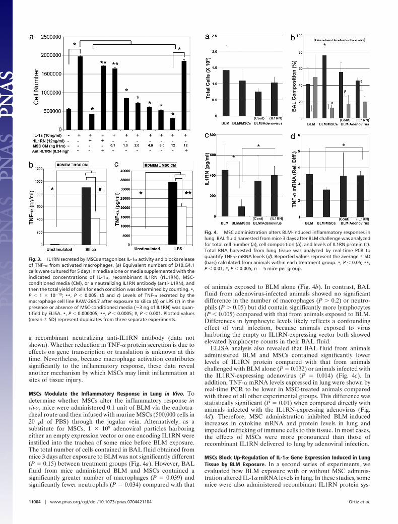
a recombinant neutralizing anti-IL1RN antibody (data notshown). Whether reduction in TNF-� protein secretion is due toeffects on gene transcription or translation is unknown at thistime. Nevertheless, because macrophage activation contributessignificantly to the inflammatory response, these data revealanother mechanism by which MSCs may limit inflammation atsites of tissue injury.
MSCs Modulate the Inflammatory Response in Lung in Vivo. Todetermine whether MSCs alter the inflammatory response invivo, mice were administered 0.1 unit of BLM via the endotra-cheal route and then infused with murine MSCs (500,000 cells in20 �l of PBS) through the jugular vein. Alternatively, as asubstitute for MSCs, 1 � 109 adenoviral particles harboringeither an empty expression vector or one encoding IL1RN wereinstilled into the trachea of some mice before BLM exposure.The total number of cells contained in BAL fluid obtained frommice 3 days after exposure to BLM was not significantly different(P � 0.15) between treatment groups (Fig. 4a). However, BALfluid from mice administered BLM and MSCs contained asignificantly greater number of macrophages (P � 0.039) andsignificantly fewer neutrophils (P � 0.034) compared with that
of animals exposed to BLM alone (Fig. 4b). In contrast, BALfluid from adenovirus-infected animals showed no significantdifference in the number of macrophages (P � 0.2) or neutro-phils (P � 0.05) but did contain significantly more lymphocytes(P � 0.005) compared with that from animals exposed to BLM.Differences in lymphocyte levels likely reflects a confoundingeffect of viral infection, because animals exposed to virusharboring the empty or IL1RN-expressing vector both showedelevated lymphocyte counts in their BAL fluid.
ELISA analysis also revealed that BAL fluid from animalsadministered BLM and MSCs contained significantly lowerlevels of IL1RN protein compared with that from animalschallenged with BLM alone (P � 0.032) or animals infected withthe IL1RN-expressing adenovirus (P � 0.014) (Fig. 4c). Inaddition, TNF-� mRNA levels expressed in lung were shown byreal-time PCR to be lower in MSC-treated animals comparedwith those of all other experimental groups. This difference wasstatistically significant (P � 0.01) when compared directly withanimals infected with the IL1RN-expressing adenovirus (Fig.4d). Therefore, MSC administration inhibited BLM-inducedincreases in cytokine mRNA and protein levels in lung andimpeded trafficking of immune cells to this tissue. In most cases,the effects of MSCs were more pronounced than those ofrecombinant IL1RN delivered to lung by adenoviral infection.
MSCs Block Up-Regulation of IL-1� Gene Expression Induced in LungTissue by BLM Exposure. In a second series of experiments, weevaluated how BLM exposure with or without MSC adminis-tration altered IL-1� mRNA levels in lung. In these studies, somemice were also administered recombinant IL1RN protein sys-
Fig. 3. IL1RN secreted by MSCs antagonizes IL-1� activity and blocks releaseof TNF-� from activated macrophages. (a) Equivalent numbers of D10.G4.1cells were cultured for 5 days in media alone or media supplemented with theindicated concentrations of IL-1�, recombinant IL1RN (rIL1RN), MSC-conditioned media (CM), or a neutralizing IL1RN antibody (anti-IL1RN), andthen the total yield of cells for each condition was determined by counting. *,P � 1 � 10�10; **, P � 0.005. (b and c) Levels of TNF-� secreted by themacrophage cell line RAW-264.7 after exposure to silica (b) or LPS (c) in thepresence or absence of MSC-conditioned media (�3 ng of IL1RN) was quan-tified by ELISA. *, P � 0.000005; **, P � 0.0005; #, P � 0.001. Plotted values(mean � SD) represent duplicates from three separate experiments.
Fig. 4. MSC administration alters BLM-induced inflammatory responses inlung. BAL fluid harvested from mice 3 days after BLM challenge was analyzedfor total cell number (a), cell composition (b), and levels of IL1RN protein (c).Total RNA harvested from lung tissue was analyzed by real-time PCR toquantify TNF-� mRNA levels (d). Reported values represent the average � SD(bars) calculated from animals within each treatment group. *, P � 0.05; **,P � 0.01; #, P � 0.005; n � 5 mice per group.
11004 � www.pnas.org�cgi�doi�10.1073�pnas.0704421104 Ortiz et al.
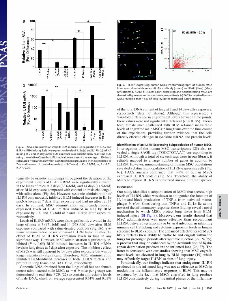
temically by osmotic minipumps throughout the duration of theexperiment. Levels of IL-1� mRNA were significantly elevatedin the lungs of mice at 7 days (38.6-fold) and 14 days (14.5-fold)after BLM exposure compared with control animals challengedwith saline alone (Fig. 5a). However, systemic administration ofIL1RN only modestly inhibited BLM-induced increases in IL-1�mRNA levels at 7 days after exposure and had no affect at 14days. In contrast, MSC administration significantly reducedexpressed levels of IL-1� mRNA induced in lung by BLMexposure by 7.3- and 3.3-fold at 7 and 14 days after exposure,respectively.
Levels of IL1RN mRNA were also significantly elevated in thelungs of mice at 7 (971-fold) and 14 (358-fold) days after BLMexposure compared with saline-treated controls (Fig. 5b). Sys-temic administration of recombinant IL1RN failed to alter theeffect of BLM on IL1RN expression levels in lung in anysignificant way. However, MSC administration significantly in-hibited (P � 0.05) BLM-induced increases in IL1RN mRNAlevels in lung tissue at 7 days after exposure. The inhibitory effectof MSCs was still apparent by 14 days after exposure but was nolonger statistically significant. Therefore, MSC administrationinhibited BLM-induced increases in both IL1RN mRNA andprotein in lung tissue and BAL fluid, respectively.
Genomic DNA obtained from the lungs of all but one femalemouse administered male MSCs (n � 6–9 mice per group) wasdetermined by real-time PCR (22) to contain appreciable levelsof male DNA, which on average represented 0.54% and 0.01%
of the total DNA content of lung at 7 and 14 days after exposure,respectively (data not shown). Although this represented a�40-fold difference in engraftment levels between time points,these values were not significantly different (P � 0.075). There-fore, female mice challenged with BLM retained measurablelevels of engrafted male MSCs in lung tissue over the time courseof the experiment, providing further evidence that the cellsdirectly effected changes in cytokine mRNA and protein levels.
Identification of an IL1RN-Expressing Subpopulation of Human MSCs.Interrogation of the human MSC transcriptome (23) also re-vealed a single SAGE tag (TGCCTGTAAT) corresponding toIL1RN. Although a total of six such tags were in our library, itreliably mapped to a large number of genes in addition toIL1RN. However, immunostaining of human MSC populationsrevealed a distinct subpopulation of IL1RN-expressing cells (Fig.6a). FACS analysis confirmed that �5% of human MSCsexpressed IL1RN protein (Fig. 6b). Therefore, the ability ofMSCs to express IL1RN is conserved among different species.
DiscussionOur study identifies a subpopulation of MSCs that secrete highlevels of IL1RN, which was shown to antagonize the function ofIL-1� and block production of TNF-� from activated macro-phages in vitro. Considering that TNF-� and IL-1� lie at thenexus of the inflammatory response, these findings reveal a novelmechanism by which MSCs protect lung tissue from BLM-induced injury (SI Fig. 8). Moreover, our results showed thatMSC administration was more effective than recombinantIL1RN, delivered systemically or by viral infection, in inhibitingimmune cell trafficking and cytokine expression levels in lung inresponse to BLM exposure. The enhanced effectiveness of MSCslikely reflects their ability to traffic to and engraft within thelungs for prolonged periods after systemic injection (22, 24, 25),a process that may be enhanced by the accumulation of hyalu-ronan degradation products in the inflamed lung (26, 27). Thelatter is consistent with our studies showing that MSC engraft-ment levels are elevated in lung by BLM exposure (19), whichmay effectively target IL1RN to sites of lung injury.
Paradoxically, our findings indicate that endogenous IL1RNproduced in the inflamed lung was not as effective as MSCs inmodulating the inflammatory response to BLM. This may beexplained by the fact that MSCs engrafted in lung produceIL1RN constitutively during the initial phases of the inflamma-
Fig. 5. MSC administration inhibits BLM-induced up-regulation of IL-1� andIL1RN mRNA in lung. Relative expression levels of IL-1� (a) and IL1RN (b) mRNAin lung at 7 and 14 days after BLM exposure was quantified by real-time PCR,using the relative Ct method. Plotted values represent the average � SD (bars)calculated from animals within each treatment group and then normalized to7 day saline control treated animals (n � 3–7 mice). *, P � 0.0002; **, P � 0.01;#, P � 0.05.
Fig. 6. IL1RN-expressing human MSCs. Photomicrographs of human MSCsimmuno-stained with an anti-IL1RN antibody (green) and DAPI (blue). (Mag-nifications: a, �200; b, �400) IL1RN-expressing and nonexpressing MSCs aredemarked by arrows and arrow heads, respectively. (c) FACS analysis of humanMSCs revealed that �5% of cells (R2 gate) expressed IL1RN protein.
Ortiz et al. PNAS � June 26, 2007 � vol. 104 � no. 26 � 11005
MED
ICA
LSC
IEN
CES
tory response, whereas local production only occurs later inresponse to inflammation. IL-1� is know to positively regulateexpression of IL1RN, providing a means to counterbalance itspotent activity in vivo (28). In this context, IL1RN expression intissues reflects the amount of IL-1 activity induced by tissueinjury. However, there exists in cells a significant delay betweenexposure to IL-1 and expression of IL1RN protein (29), and ourown work shows that IL-1� induces in MSCs a significantincrease in IL1RN secretion only after 72 h of exposure (SI Fig.9). Therefore, high endogenous IL1RN levels in lung reflect anongoing inflammatory response, which is down-regulated onlyafter significant tissue damage occurs. In contrast, MSCs protectlung tissue from bleomycin-induced injury by preventing theinflammatory response. This effect is consistent with the factthat MSC administration inhibited collagen accumulation andblocked matrix metalloproteinase activation in the lungs of micechallenged with BLM (19).
Our data also show that MSCs block recruitment of lympho-cytes and neutrophils into the injured lung, which is consistentwith our previous histological findings (19). Neutrophils con-tribute to parenchymal injury by producing toxic reactive oxygenintermediates, cytokines, and secreting proteolytic enzymes thatalter the lung cytoarchitecture (30, 31). In addition, lymphocytesproduce secondary immune effectors, such as IL-6, and induceepithelial cytotoxicity (32). Recent studies indicate that TNF-�also contributes to the pathophysiology of ILD by inducingapoptosis of epithelial cells by direct activation of the TNFR andindirectly by stimulating expression of IL-1 (29). Therefore,MSCs may directly enhance epithelial cell survival by blockingdownstream effects of TNF-� and IL-1. This outcome hasimportant implications, because injury to the alveolar epitheliumhas serious clinical consequences, including dysregulation ofsurfactant production and exposure of the basement membrane,which causes further macrophage activation.
It is well established that TNF-� and IL-1 also function aspotent bone-resorbing factors (33, 34), the effects of which canbe modulated by IL1RN (35). Therefore, IL1RN-expressingMSC subpopulations identified herein may represent a uniquesubtype of stromal cell that plays an important role in modu-lating bone turnover in vivo. This would explain why IL1RNexpression by MSCs is conserved across species. These resultsare consistent with our previous study showing the cellularcomposition of marrow stroma and its associated functions aremore complex than previously envisioned (20). Our data indi-cating that IL1RN-expressing subpopulations are less abundantin human vs. murine populations may reflect differences in theanatomical location and methods used to isolate and cultureexpand the cells. Nevertheless, the existence of IL1RN-expressing MSCs in human bone marrow may provide a novelcellular vector for treating chronic inflammatory diseases inhumans, particularly those affecting the lung.
MethodsIsolation of MSCs. Murine MSCs were isolated from 4 week-old,male Balb/C mice and purified by immunodepletion as describedin ref. 21. In some experiments CELLection Dynabeads (DynalBiotech, Brown Deer, WI) were used to recover immunode-pleted cell lineages. Human MSCs were isolated from smallvolume (2- to 5-ml) bone marrow aspirates obtained fromhealthy donors (23) as approved by the Institutional ReviewBoard of Tulane University.
ELISAs. Murine plastic adherent marrow cells or MSCs (50,000–200,000 cells) were cultured for 5–7 days in �-MEM media(Invitrogen, Carlsbad, CA) supplemented with 10% FCS (At-lanta Biological, Atlanta, GA), 100 units per ml penicillin, and100 �g/ml streptomycin. Cells were washed with HBSS andmaintained in 1 ml of serum-free �-MEM for 24 h, and the
conditioned media were collected. Levels of IL1RN in condi-tioned media were quantified by using the Mouse QuantikineELISA Kit (R&D Systems, Minneapolis, MN).
Immunostaining. Murine MSCs (2 � 104 cells per 0.4 cm2) werefixed for 15 min at room temperature, washed, fixed for 30 minin blocking buffer (PBS containing 0.1% BSA, 5% Tween-20,and 20% donkey sera), incubated overnight at 4°C with a 1:100dilution of an anti-mouse IL1RN antibody (Santa Cruz Biotech-nology, Santa Cruz, CA), and then 1 h at room temperature witha 1:2,000 dilution of an Alexa Fluor 546 donkey anti-goatIgG(H�L) (Molecular Bioprobes, Eugene, OR). Slides werecounterstained with DAPI, photographed with a Leica RX-DMV fluorescent microscope (Meyer Instruments, Houston,TX) with a Cooke Sensicam digital camera (Hamamatsu,Bridgewater, NJ), and rendered by using Slidebook software (3I,Denver, CO).
FACS Analysis. Murine MSCs (3 � 105) were incubated in 1 ml offixation buffer (Santa Cruz Biotechnology) for 30 min, washed,and incubated for 15 min with a 1:60 dilution of a rat anti-mouseCD16/CD32 antibody (BD Biosciences, San Jose, CA) at 4°C inthe dark. Cells were then incubated 15 min in permeabilizationbuffer for 1 h with a 1:100 dilution of an anti-IL1RN antibodyand then for 20 min with a 1:200 dilution of a FITC-conjugateddonkey anti-goat antibody (Santa Cruz Biotechnology). Cellswere washed and resuspended in 500 �l of wash buffer, and theextent of cell labeling was evaluated by using a Beckman CoulterModel Epics XL flow cytometer (Beckman Coulter, Fullerton,CA). Isotype controls were run in parallel, using the sameconcentration of each antibody tested. Human MSCs wereprocessed similarly except that use of the anti-CD16/CD32antibody was omitted.
PCR. SAGE tags corresponding to IL1RN were validated by screen-ing a murine MSC cDNA library (20) by PCR, using the followingprimers: 5-AGGCCCCACCACCAGCTT TGAGTC-3 and 5-TCACCCAGATGGCAGAGGCAACAA-3. An aliquot (50-�l)of the pooled cDNA phage library was boiled for 5 min, and then500 ng was used as input in a PCR (100 �l) containing 100 pmol offorward and reverse gene-specific primers, 1� PCR buffer, 0.2 mMdNTPs, and 0.5 �l of TaqDNA polymerase (Qiagen, Valencia,CA). After an initial denaturation step at 94°C for 3 min, reactionswere amplified for 30 cycles at 94°C for 30 sec, 59.9°C for 45 sec, and72°C for 90 sec, followed by a final incubation at 72°C for 7 min.PCR products were gel-purified by using GeneElute columns(Sigma, St. Louis, MO) and cloned by using the AdvanTAge PCRcloning kit (Clontech, Palo Alto, CA). Plasmid DNA was isolatedand sequenced to confirm the identity of the PCR product.
T-Cell Proliferation and Macrophage Activation Assays. D10.G4.1and RAW-264.7 cell lines were maintained as described by thesupplier (ATCC, Manassas, VA). D10.G4.1 cells (500,000) werecultured with the indicated concentration of recombinant mouseIL-1� alone or in combination with varying amounts of recom-binant mouse IL1RN, a neutralizing anti-IL1RN antibody (R&DSystems) or MSC-conditioned media. Cultures were maintainedfor 3–5 days with media changes each day, after which the totalnumber of viable cells were determined by staining with Tryptanblue and counting on a hemocytometer. RAW-264.7 cells wereexposed to 1 �g/ml LPS (Sigma) or 20 �g/cm2 silica (provided byA. Ghio, Environmental Protection Agency, Chapel Hill, NC)for 4 h in the presence of MSC-conditioned media preincubatedwith or without a neutralizing anti-IL1RN antibody. Subse-quently, levels of TNF-� secreted from cells were quantified byELISA as described in ELISAs. All assays were performed inquadruplicate.
11006 � www.pnas.org�cgi�doi�10.1073�pnas.0704421104 Ortiz et al.
BLM Exposures and Tissue Processing. Anesthetized female 6- to10-week-old C57BL/6 mice were exposed to 0.1 unit of BLM asdescribed in ref. 19. Murine MSCs (5 � 105 in 200 �l of PBS)were injected into the jugular vein immediately after BLMadministration. Some mice were infected with 1 � 109 particlesof a retrovirus encoding human IL1RN (provided by P. Robbins,University of Pittsburgh, Pittsburgh, PA) or had an osmoticpump (ALZET, Cupertino, CA) that secreted 0.5 mg/kg/hr ofrecombinant human IL1RN (ProSpec-Tany TehcnoGene, Re-hovot, Israel) surgically implanted into their peritoneal cavity 1day before BLM exposure. Animals were killed at 3, 7, or 14 daysafter BLM exposure, at which time BAL fluid was collected. Theleft lung was then removed, f lash-frozen in liquid nitrogen, andused to harvest genomic DNA and total RNA, using the AllPrepDNA/RNA Mini Kit (Qiagen). All experiments involving liveanimals were approved by the animal use and care committee ofTulane University Health Sciences Center and the University ofPittsburgh.
Quantification of Cytokine mRNA and Protein Levels in Vivo. Cyto-spins of BAL fluid were stained with Diff Quick (FisherScientific, Pittsburgh, PA), and cell differentials were deter-
mined by counting 400 consecutive cells. Total RNA (25 ng)prepared from lung was converted to cDNA and amplified byPCR, using the TaqMan EZ RT-PCR kit, a 7900 HT sequencedetector and the following Assay-On-Demand Taqmanprobes: TNF-� (Mm0043258), IL-1� (Mm00439620), andIL1RN (Mm00446185) (Applied Biosystems, Foster City, CA).Transcript levels were quantified by using the relative Ctmethod with GAPDH mRNA as an internal control. GenomicDNA (25 ng) isolated from lung tissue was also analyzed byreal-time PCR to quantify levels of engrafted MSCs as de-scribed in ref. 24.
Statistical Analysis. All values were expressed as the mean � SD.Differences between treatment groups were measured by Stu-dent’s t test or ANOVA with Fisher’s PLSD test for pair-wisecomparison (Statview 4 software; Abacus Concept, Piscataway,NJ). P � 0.05 was considered significant.
This research was supported by National Institutes of Health GrantsR01-NS39033-01A2, R01-NS39033-02S1, P40-RR17447, and P01-HL075161 (to D.G.P.) and R01-HL071953 and ES010859 (to L.A.O.); theLouisiana Gene Therapy Research Consortium (New Orleans, LA); and theHealth Care Company (Nashville, TN).
1. American Thoracic Society (2000) Am J Respir Crit Care Med 161:646–664.2. Raghu G, Weycker D, Edelsberg J, Bradford WZ, Oster G (2006) Am J Respir
Crit Care Med 74:810–816.3. Zhang K, Phan SH (1996) Biol Signals 5:232–239.4. Furie MB, Randolph GJ (1995) Am J Pathol 146:1287–1301.5. Kaneider NC, Leger AJ, Kuliopulos A (2006) FEBS J 273:4416–4424.6. Kolb M, Margetts PJ, Anthony DC, Pitossi F, Gauldie J (2001) J Clin Invest
107:1529–1536.7. Arend WP (1991) J Clin Invest 88:1445–1451.8. Hybertson BM, Lee YM, Repine JE (1997) Ann NY Acad Sci 832:266–273.9. Lee YM, Hybertson BM, Cho HG, Terada LS, Cho O, Repine AJ, Repine JE
(2000) Am J Physio Lung Cell Mol Physiol 279:L75–L80.10. Phan SH, Kunkel SI (1992) Exp Lung Res 18:29–43.11. Johnston CJ, Piedboeuf B, Rubin P, Williams JP, Baggs R, Finkelstein JN
(1996) Radiat Res 145:762–767.12. Pan LH, Ohtani H, Yamauchi K, Nagura H (1996) Pathol In 46:91–99.13. Whyte M, Hubbard R, Meliconi R, Whidborne M, Eaton V, Bingle C, Timms J,
Duff G, Facchini A, Pacilli A, et al. (2000) Am J Respir Crit Care Med 162:755–758.14. Piguet PF, Vesin C, Grau GE, Thompson RC (1993) Cytokine 5:57–61.15. Minguell JJ, Erices A (2006) Expt Biol Med 231:39–49.16. Phinney DG, Isakova I (2005) Curr Pharm Des 11:1255–1265.17. Krause DS, Theise ND, Collector MI, Henegariu O, Hwang S, Gardner R,
Neutzel S, Sharkis SJ (2001) Cell 105:369–377.18. Kotton DN, Ma BY, Cardoso WV, Sanderson EA, Summer RS, Williams MC,
Fine A (2001) Development (Cambridge, UK) 128:5181–5188.19. Ortiz LA, Gambelli F, McBride C, Gaupp D, Baddoo M, Kaminski N, Phinney
DG (2003) Proc Natl Acad Sci USA 100:8407–8411.20. Phinney DG, Hill K, Michelson C, DuTreil M, Hughes C, Humphries S,
Wilkinson R, Baddoo M, Bayly E (2006) Stem Cells 24:186–198.21. Baddoo M, Hill K, Wilkinson R, Gaupp D, Hughes C, Kopen GC, Phinney DG
(2003) J Cell Biochem 89:1235–1249.
22. McBride C, Gaupp D, Phinney DG (2003) Cytotherapy 5:7–18.23. Tremain N, Korkko J, Ibberson D, Kopen GC, DiGirolamo C, Phinney DG
(2001) Stem Cells 19:408–418.24. Rochefort GY, Vaudin P, Bonnet N, Pages JC, Domenech J, Charbord P, Eder
V (2005) Respir Res 6:125–138.25. Gao J, Dennis JE, Muzic RF, Lundberg M, Caplan AI (2001) Cells Tissues
Organs 169:12–20.26. Teder P, Vandivier RW, Jiang D, Liang J, Cohn L, Pure E, Henson PM, Noble
PW (2002) Science 296:155–158.27. Zhu H, Mitsuhashi N, Klein A, Barsky LW, Weinberg K, Barr ML, Demetriou
A, Wu GD (2006) Stem Cells 24:928–935.28. Juge-Aubry CE, Somm E, Chicheportiche R, Burger D, Pernin A, Cuenod-
Pittet B, Quinodoz P, Giusti V, Dayer J-M, Meier CA (2004) J Clin EndocrinMetabol 89:2652–2658.
29. Janes KA, Gaudet S, Albeck JG, Nielsen UB, Lauffenburger DA, Sorger PK(2006) Cell 124:1225–1239.
30. Strausz J, Muller-Quernheim J, Steppling H, Ferlinz R (1990) Am Rev RespirDis 141:124–128.
31. Gadek JE, Kelman JA, Fells G, Weinberger SE, Horwitz AL, Reynolds HY,Fulmer JD, Crystal RG (1979) N Engl J Med 301:737–742.
32. Kuwano K, Hagimoto N, Kawasaki M, Yatomi T, Nakamura N, Nagata S,Suda T, Kunitake R, Maeyama T, Miyazaki H, et al. (1999) J Clin Invest104:13–19.
33. Zhang YH, Heulsmann A, Tondravi MM, Mukherjee A, Abu-Amer Y (2001)J Biol Chem 276:563–568.
34. Lorenzo JA, Naprta A, Rao Y, Alander C, Glaccum M, Widmer M, GronowiczG, Kalinowski J, Pilbeam CC (1998) Endocrinology 139:3022–3025.
35. Bresnihan B, Alvaro-Gracia JM, Cobby M, Doherty M, Domljan Z, Emery P,Nuki G, Pavelka K, Rau R, Rozman B, et al. (1998) Arthritis Rheum 41:2196–2204.
Ortiz et al. PNAS � June 26, 2007 � vol. 104 � no. 26 � 11007
MED
ICA
LSC
IEN
CES
Related Documents