Matthew J. Sweet and Glen M. Scholz Lam, Eric C. Reynolds, John A. Hamilton, Yeng Divya Ramnath, Dominic De Nardo, Pui Mei Qi Kwa, Thao Nguyen, Jennifer Huynh, in Epithelial Cells 2-dependent Chemokine Gene Expression Differentially Regulates Toll-like Receptor Interferon Regulatory Factor 6 Immunology: doi: 10.1074/jbc.M114.584540 originally published online May 28, 2014 2014, 289:19758-19768. J. Biol. Chem. 10.1074/jbc.M114.584540 Access the most updated version of this article at doi: . JBC Affinity Sites Find articles, minireviews, Reflections and Classics on similar topics on the Alerts: When a correction for this article is posted • When this article is cited • to choose from all of JBC's e-mail alerts Click here http://www.jbc.org/content/289/28/19758.full.html#ref-list-1 This article cites 61 references, 27 of which can be accessed free at at University of Melbourne (CAUL) on September 7, 2014 http://www.jbc.org/ Downloaded from at University of Melbourne (CAUL) on September 7, 2014 http://www.jbc.org/ Downloaded from

Welcome message from author
This document is posted to help you gain knowledge. Please leave a comment to let me know what you think about it! Share it to your friends and learn new things together.
Transcript

Matthew J. Sweet and Glen M. ScholzLam, Eric C. Reynolds, John A. Hamilton,
YengDivya Ramnath, Dominic De Nardo, Pui Mei Qi Kwa, Thao Nguyen, Jennifer Huynh, in Epithelial Cells2-dependent Chemokine Gene ExpressionDifferentially Regulates Toll-like Receptor Interferon Regulatory Factor 6Immunology:
doi: 10.1074/jbc.M114.584540 originally published online May 28, 20142014, 289:19758-19768.J. Biol. Chem.
10.1074/jbc.M114.584540Access the most updated version of this article at doi:
.JBC Affinity SitesFind articles, minireviews, Reflections and Classics on similar topics on the
Alerts:
When a correction for this article is posted•
When this article is cited•
to choose from all of JBC's e-mail alertsClick here
http://www.jbc.org/content/289/28/19758.full.html#ref-list-1
This article cites 61 references, 27 of which can be accessed free at
at University of M
elbourne (CA
UL
) on September 7, 2014
http://ww
w.jbc.org/
Dow
nloaded from
at University of M
elbourne (CA
UL
) on September 7, 2014
http://ww
w.jbc.org/
Dow
nloaded from

Interferon Regulatory Factor 6 Differentially RegulatesToll-like Receptor 2-dependent Chemokine GeneExpression in Epithelial Cells*
Received for publication, May 25, 2014 Published, JBC Papers in Press, May 28, 2014, DOI 10.1074/jbc.M114.584540
Mei Qi Kwa‡§1, Thao Nguyen§1,2, Jennifer Huynh‡§1, Divya Ramnath¶�, Dominic De Nardo§3, Pui Yeng Lam§,Eric C. Reynolds‡, John A. Hamilton§, Matthew J. Sweet¶�, and Glen M. Scholz‡§4
From the ‡Oral Health Cooperative Research Centre, Melbourne Dental School, and Bio21 Institute, and §Department of Medicine,Royal Melbourne Hospital, The University of Melbourne, Victoria 3010, Australia, and the ¶Institute for Molecular Bioscience, and�Australian Infectious Disease Research Centre, The University of Queensland, Queensland 4072, Australia
Background: The IRF6 transcription factor is critical for epithelial barrier function; however, a role for IRF6 in signaling byToll-like receptors has not been addressed.Results: The IRAK1-mediated activation of IRF6 promotes TLR2-dependent CCL5 chemokine gene expression in epithelialcells.Conclusion: IRF6 differentially regulates TLR2 inflammatory responses in epithelial cells.Significance: Our results reveal an additional immune-related function for IRF6.
Epidermal and mucosal epithelial cells are integral to hostdefense. They not only act as a physical barrier but also utilizepattern recognition receptors, such as the Toll-like receptors(TLRs), to detect and respond to pathogens. Members of theinterferon regulatory factor (IRF) family of transcription factorsare key components of TLR signaling as they impart specificityto downstream responses. Although IRF6 is a critical regulatorof epithelial cell proliferation and differentiation, its role in TLRsignaling has not previously been addressed. We show here thatIRF6 is activated by IRAK1 as well as by MyD88 but not by TRIFor TBK1. Co-immunoprecipitation experiments further dem-onstrated that IRF6 can interact with IRAK1. Gene silencing inepithelial cells along with gene promoter reporter assaysshowed that IRAK1 mediates TLR2-inducible CCL5 geneexpression at least in part by promoting IRF6 activation. Con-versely, IRAK1 regulated CXCL8 gene expression independ-ently of IRF6, thus identifying a molecular mechanism by whichTLR2 signaling differentially regulates the expression of specificchemokines in epithelial cells. Bioinformatics analysis andmutagenesis-based experiments identified Ser-413 and Ser-424as key regulatory sites in IRF6. Phosphomimetic mutation ofthese residues resulted in greatly enhanced IRF6 dimerizationand trans-activator function. Collectively, our findings suggestthat, in addition to its importance for epithelial barrier function,IRF6 also contributes to host defense by providing specificity tothe regulation of inflammatory chemokine expression by TLR2in epithelial cells.
Epidermal and mucosal epithelial cells are positioned atthe interface between the host and the environment and thusplay pivotal roles in host defense. Although one of their pri-mary functions is to provide a physical barrier to pathogeninvasion (1, 2), they also express pattern recognition recep-tors (e.g. Toll-like receptors), thereby enabling them toactively participate in host defense by functioning asimmune sentinels (2–5). For example, the production ofinflammatory cytokines, chemokines, and type I interferons(IFNs)5 by epithelial cells serves to recruit and activate dif-ferent leukocyte cell populations. However, the dysregulatedproduction of such factors can lead to pathologic states ofchronic inflammation, as occurs in inflammatory bowel dis-ease, psoriasis, and chronic periodontitis (6 – 8). Chronicmucosal inflammation is also an important factor in somecancers (e.g. gastric cancer) (9).
Toll-like receptors (TLRs) are fundamental to the detectionand subsequent host response to pathogens (10, 11). The spec-ificity of TLR signaling is determined, in part, by differential useof adapter proteins, for example, MyD88 and TRIF. Accord-ingly, TLR signaling can be broadly divided into the MyD88-de-pendent and TRIF-dependent pathways. The former pathway isused by all TLRs with the exception of TLR3 and the latter onlyby TLR3 and TLR4 (10, 11). The MyD88-dependent pathwayemploys the protein kinase IL-1 receptor-associated kinase-1(IRAK1) to trigger the activation of various transcription fac-tors, including NF-�B and members of the interferon regula-tory factor family (e.g. IRF5 and IRF7), resulting in inflamma-tory gene expression. The induction of inflammatory geneexpression by the TRIF-dependent pathway occurs in responseto the activation of IRF3 by TBK1 along with TAK1-mediatedNF-�B activation (10 –12).
In addition to regulating type I IFN (e.g. IFN�) gene expres-sion, IRFs also play important roles in regulating the expression
* This work was supported by National Health and Medical Research CouncilProject Grant 628769 and the Oral Health CRC.
1 These authors made equal contributions.2 Present address: Australian Centre for Blood Diseases, 89 Commercial Road,
Melbourne 3004, Australia.3 Present address: Institute of Innate Immunity, University of Bonn, Sigmund-
Freud-Strasse 25, 53127 Bonn, Germany.4 To whom correspondence should be addressed: Melbourne Dental School,
The University of Melbourne, Victoria 3010, Australia. Tel.: 613-8344-2565;Fax: 613-8344-2545; E-mail: [email protected].
5 The abbreviations used are: IFN, interferon; TLR, Toll-like receptor; IRAK1,IL-1 receptor-associated kinase-1; IRF, interferon regulatory factor.
THE JOURNAL OF BIOLOGICAL CHEMISTRY VOL. 289, NO. 28, pp. 19758 –19768, July 11, 2014© 2014 by The American Society for Biochemistry and Molecular Biology, Inc. Published in the U.S.A.
19758 JOURNAL OF BIOLOGICAL CHEMISTRY VOLUME 289 • NUMBER 28 • JULY 11, 2014
at University of M
elbourne (CA
UL
) on September 7, 2014
http://ww
w.jbc.org/
Dow
nloaded from
of other inflammatory proteins, including chemokines (e.g.CCL5, CXCL8, and CXCL10) (12–17). Consequently, the dif-ferential regulation of inflammatory gene expression by IRFsallows them to appropriately shape the immune response byimparting signaling specificity to TLRs. The activation of IRF3by TLR3 and TLR4 signaling, for instance, enables these recep-tors to initiate TLR3- and TLR4-specific gene expressionresponses (18, 19). Analogously, IRF5 and IRF7 induce specificgene expression responses downstream of MyD88-dependentTLRs (20 –22).
Prior studies have largely focused on the roles of IRFs inmediating TLR-elicited responses in leukocyte cell populations(e.g. macrophages and dendritic cells). Although IRF3 has alsobeen shown to be important for TRIF-dependent TLRresponses in epithelial cells (23–25), the IRF(s) that mediatesMyD88-dependent responses in these cells is less clear. In con-trast to other IRFs, IRF6 expression appears for the most part tobe limited to epithelial cells (26 –30), where at least one func-tion is to regulate cell proliferation and differentiation (27–29,31, 32). Notably, IRF6-deficient mice exhibit defective epider-mal barrier function due to impaired keratinocyte differentia-tion; they also die perinatally (28, 29). Given the key roles ofother IRFs in orchestrating the TLR-elicited inflammatoryresponses of leukocytes (12, 13), we investigated whether IRF6was similarly important for specific TLR responses in epithelialcells. Our findings here link IRF6 to IRAK1-dependent TLR2responses (e.g. CCL5 expression) in epithelial cells, thus reveal-ing an additional immune-related function for IRF6 in thesecells.
EXPERIMENTAL PROCEDURES
Reagents—Cell culture medium and supplements, fetal calfserum (FCS), SuperScript III reverse transcriptase, randomprimers, deoxyribonucleotide triphosphates, TaqMan Univer-sal Master Mix II, Lipofectamine RNAiMAX, precast 10%NuPAGE gels, mouse anti-V5 antibodies (HRP-conjugated andunconjugated), Alexa Fluor 488-conjugated goat anti-rabbitIgG antibody, and ProLong� Gold Antifade reagent (containing4�,6-diamidino-2-phenylindole) were from Invitrogen. Recom-binant human CSF-1 was generously provided by Chiron.Restriction enzymes were from New England Biolabs, whereasPfu DNA polymerase, Passive Lysis Buffer, and the Dual-GloTM
luciferase assay system were from Promega. PCR primers weresynthesized by GeneWorks. FuGENE 6TM and CompleteTM
protease inhibitors were supplied by Roche Applied Science.The ON-TARGETplus IRF6 and IRAK1 siRNAs as well as thecontrol non-targeting siRNA were from Dharmacon. TLRligands were from InvivoGen. The rabbit anti-IRF6 antibodyand HRP-conjugated mouse anti-HA antibody were from CellSignaling, and the mouse anti-HSP90 antibody was from BDBiosciences.
Cell Culture—OKF6/TERT-2 cells (33) were cultured inkeratinocyte serum-free medium supplemented with 25 �g/mlbovine pituitary extract, 2 ng/ml EGF, 0.4 mM CaCl2, 100 units/mlpenicillin, 100 �g/ml streptomycin, and 2 mM GlutaMax-1TM.AGS and MKN28 cells were cultured in RPMI 1640 supplementedwith 10% FCS, 100 units/ml penicillin, 100 �g/ml streptomycin,and 2 mM GlutaMax-1TM. HT-29 cells were cultured in McCoy’s
5A medium supplemented with 10% FCS, 100 units/ml penicillin,100 �g/ml streptomycin, and 2 mM GlutaMax-1TM. HEK293Tcells were cultured in Dulbecco’s-modified Eagle’s medium sup-plemented with 10% FCS, 100 units/ml penicillin, 100 �g/mlstreptomycin, and 2 mM GlutaMax-TM. Human monocytes werepurified from buffy coats (Red Cross Blood Bank, Australia) usinga RosetteSep antibody mixture (Stem Cell Technologies) followedby Ficoll-Paque density gradient centrifugation. The monocyteswere cultured overnight in RPMI 1640 supplemented with 10%FCS, 100 units/ml penicillin, 100 �g/ml streptomycin, and 2 mM
GlutaMax-TM. Monocyte-derived macrophages were generatedby culturing monocytes in the presence of CSF-1 (2500 units/ml)for 6–8 days (34). All cells were cultured at 37 °C in a humidifiedatmosphere of 5% CO2.
Real-time PCR—Total RNA was purified using an RNAeasyMini kit (Qiagen). RNA (1 �g) was reverse-transcribed intocDNA using random primers and SuperScript III reverse tran-scriptase. Real-time PCR was performed (in triplicate) using anApplied Biosystems Prism 7900HT sequence detection systemand pre-developed TaqMan assays (Invitrogen) for the follow-ing genes: CCL5 (Hs00174575_m1), CXCL8 (Hs00174103_m1),IFN� (Hs02621180_s1), IRAK1 (Hs01018347_m1), IRAK2(Hs00176394_m1),IRF3(Hs00155574_m1),IRF5(Hs00158114_m1), IRF6 (Hs00196213_m1), IRF7 (Hs00185375_m1), TLR1(Hs00413978_m1), TLR2 (Hs00152932_m1), TLR3 (Hs01551078_m1), TLR4 (Hs01060206_m1), TLR5 (Hs00152825_m1), TLR6(Hs00271977_s1),TLR7 (Hs01933259_s1),TLR8 (Hs00607866_mH), and TLR9 (Hs00370913_s1). Messenger RNA levels, rel-ative to those of the endogenous control gene, HPRT, were cal-culated using the �Ct (cycle threshold) method.
Expression Vectors and Site-directed Mutagenesis—Thehuman IRF6 expression vector, pCMV6-XL6-IRF6, was pur-chased from Origene. The expression vector, pEF-HA-IRF6(expresses an N-terminal HA-tagged version of IRF6), was cre-ated by PCR using the primer pair F1 (5�-CG ACG CGT GCCCTC CAC CCC CGC AGA GTC CGG CTA AAG-3�) and R1(5�-CG ACG CGT TTA CTG GGG AGG CAG GGC AGGGGG CAG TTG-3�) and pCMV6-XL6-IRF6 as the template.The PCR product was digested with MluI and cloned into theexpression vector, pEF-HA. The expression vector, pEF-V5-IRF6 (expresses an N-terminal V5-tagged version of IRF6), wascreated by excising the cDNA insert from pEF-HA-IRF6 withMluI and cloning it into pEF-V5. The expression vector, pEF-HA-IRF6 S413A (Ser-413 replaced by alanine), was created byoverlapping PCR using the primer pairs F1 and R2 (5�-ACTGCC ACT ATC AAA GGC TCG TGT GAA ATC ACC-3�) andF2 (5�-GGT GAT TTC ACA CGA GCC TTT GAT AGT GGCAGT-3�) and R1 and pCMV6-XL6-IRF6 as the template. Theexpression vector, pEF-HA-IRF6 S424A (Ser-424 replaced byalanine), was created using the primer pairs F1 and R3 (5�-CTTGAT GTC TGG GGT TGC GAT CTG CAG GCG GAC-3�)and pairs F3 (5�-GTC CGC CTG CAG ATC GCA ACC CCAGAC ATC AAG-3�) and R1 and pCMV6-XL6-IRF6 as the tem-plate. The expression vector, pEF-HA-IRF6 S413A/S424A(Ser-413 and Ser-424 replaced by alanine), was created usingthe primer pairs F1 and R3 and pairs F3 and R1 and pEF-HA-IRF6 S413A as the template. The expression vector, pEF-HA-IRF6 S413E (Ser-413 replaced by glutamic acid), was created
IRF6 Differentially Regulates TLR2 Responses
JULY 11, 2014 • VOLUME 289 • NUMBER 28 JOURNAL OF BIOLOGICAL CHEMISTRY 19759
at University of M
elbourne (CA
UL
) on September 7, 2014
http://ww
w.jbc.org/
Dow
nloaded from

using the primer pairs F1 and R4 (5�-ACT GCC ACT ATCAAA TTC TCG TGT GAA ATC ACC-3�) and pairs F4 (5�-GGT GAT TTC ACA CGA GAA TTT GAT AGT GGC AGT-3�) and R1 and pCMV6-XL6-IRF6 as the template. The expres-sion vector, pEF-HA-IRF6 S424E (Ser-424 replaced by glutamicacid), was created using the primer pairs F1 and R5 (5�-CTTGAT GTC TGG GGT TTC GAT CTG CAG GCG GAC-3�)and pairs F5 (5�-GTC CGC CTG CAG ATC GAA ACC CCAGAC ATC AAG-3�) and R1 and pCMV6-XL6-IRF6 as the tem-plate. The expression vector, pEF-HA-IRF6 S413E/S424E (Ser-413 and Ser-424 replaced by glutamic acid), was created usingthe primer pairs F1 and R5 and pairs F5 and R1 and pEF-HA-IRF6 S413E as the template. The expression vector, pEF-V5-IRF6 S413E/S424E (expresses an N-terminal V5-tagged versionof IRF6 S413E/S424E), was created by excising the cDNA insertfrom pEF-HA-IRF6 S413E/S424E with MluI and cloning it intopEF-V5. The IRAK1 expression vectors, pEF-V5-IRAK1 andpEF-V5-IRAK1 K239A (express V5-tagged versions of wildtype and kinase-dead IRAK1, respectively), are as previouslydescribed (35, 36). The IRAK2 expression vector was a gener-ous gift from Dr. Luke O’Neill (Trinity College, Ireland),whereas the MyD88, TBK1, and TRIF expression vectors werekindly provided by Dr. Ashley Mansell (Monash Institute ofMedical Research, Australia).
Gene Promoter Reporter Assays—HEK293T cells were seededin 12-well tissue culture plates at a density of 3 � 105 cells perwell and transfected (in duplicate) the next day using FuGENE6TM transfection reagent. The total amount of plasmid in eachtransfection was kept constant by using empty vector whererequired. The cells were lysed 24 h post-transfection with Pas-sive Lysis Buffer and assayed for firefly and Renilla luciferaseactivities using the Dual-Glo™ luciferase assay system. Renillaluciferase activity was used to normalize transfection efficien-cies. The luciferase-based CCL5 (37), CXCL8 (38), and IFN�and IFN�4 gene promoter reporter plasmids were generouslyprovided by Drs. Paula Pitha (Johns Hopkins University), AllanBrasier (University of Texas Medical Branch), and Ashley Man-sell (Monash Institute of Medical Research, Australia), respec-tively. The Renilla luciferase reporter plasmid, pRL-TK, wasfrom Promega.
Silencing of IRAK1 and IRF6 Expression—A reverse-transfec-tion protocol was used for siRNA transfections. Briefly, theIRAK1- and IRF6-targeting siRNAs as well as the control non-targeting siRNA were diluted to 120 nM with 100 �l of Opti-MEM I-reduced serum medium (Invitrogen). The dilutedsiRNA was mixed with 100 �l of Opti-MEM I-reduced serummedium containing 1.0 �l of Lipofectamine RNAiMAXtransfection reagent and incubated at room temperature for20 min. OKF6/TERT-2 cells (2 � 105 cells in 1.0 ml of anti-biotic-free growth medium) were plated into 12-well plates,and the transfection mixture then added. The medium wasreplaced 24 h later, and the cells were analyzed or stimulated48 h post-transfection.
Cell Lysis and Western Blotting—Cells were washed twicewith ice-cold PBS and then lysed (20 mM Tris-HCl (pH 7.4), 150mM NaCl, 1 mM EDTA, 1% Nonidet P-40, 10% glycerol, 10 mM
�-glycerol phosphate, 10 mM NaF, and CompleteTM proteaseinhibitors) on ice for 60 min. The lysates were clarified by cen-
trifugation (13,000 � g for 10 min at 4 °C), and the proteinconcentrations measured using a protein assay kit (Bio-Rad).Cell lysates were subjected to electrophoresis on 10% NuPAGEgels followed by Western blotting according to standard proto-cols. Immunoreactive bands were visualized using ECLreagents (Millipore) and a LAS-3000 Imager (Fujifilm) or byexposure to x-ray film (Fujifilm). Films were scanned using aGS-800 Calibrated Imaging Densitometer (Bio-Rad).
In Vitro Protein Dephosphorylation Assay—TransfectedHEK293T cells were lysed as above, except phosphatase inhib-itors were omitted from the lysis buffer. Protein dephosphory-lation was carried out in 50-�l reactions consisting of 50 �g ofcell protein and 10 units of calf intestinal phosphatase. Thereactions were incubated at 37 °C for 30 min followed by SDS-PAGE and Western blotting.
Immunoprecipitation Assays—V5-tagged IRAK1 and IRF6were immunoprecipitated from transfected HEK293T cells byincubating 1 mg of cell lysate (in 1 ml lysis buffer) with 1 �g ofanti-V5 antibody and 20 �l of Protein G-Sepharose for 4 h at4 °C with constant mixing. The beads were washed 4 times withlysis buffer and then subjected to electrophoresis on 10%NuPAGE gels followed by Western blotting.
ImmunofluorescentStainingandConfocalMicroscopy—HEK293Tcells, which had been seeded onto glass coverslips prior totransfection, were fixed with 4% paraformaldehyde (30 min),solubilized with 0.1% Triton X-100 (5 min), and then blocked in5% goat serum (60 min), all at room temperature. The cells weresubsequently stained overnight (at 4 °C) with a rabbit anti-IRF6antibody. After three washes with PBS, the cells were probedwith an Alexa Fluor 488-conjugated goat anti-rabbit IgG anti-body for 60 min (at room temperature). The cells were washed3 times with PBS and finally mounted on glass microscopeslides using ProLong� Gold Antifade reagent containing DAPI.Mounted coverslips were allowed to cure for 24 h in the darkbefore images of the cells being acquired on an OlympusFV1000 scanning confocal microscope. No anti-IRF6 stainingwas apparent in HEK293T cells transfected with empty vectoronly.
Statistical Analysis—Data combined from three or moreindependent experiments are given as the means � S.E. Statis-tical analyses were performed using GraphPad Prism softwareVersion 6.01 (GraphPad Software, La Jolla, CA). Differencesbetween two groups were evaluated using Student’s t test. Formultiple comparisons, statistical analysis was performed usinga one-way analysis of variance and then the Sidak’s or Dunnett’stest as a post-hoc test. A p value �0.05 was considered to bestatistically significant.
RESULTS
Activation of IRF6 Trans-activator Function by IRAK1—Prior studies have demonstrated that IRAK1 mediates, eitherdirectly or indirectly, IRF5 and IRF7 activation in response toMyD88-dependent TLR signaling in myeloid cells (39 – 42).Given that phylogenetic analysis had also revealed that IRF6was most closely related to IRF5 (43), we investigated the abilityof IRAK1 to activate IRF6. IRF6 activity was assayed using anIFN� gene promoter reporter plasmid, which was activated in aconcentration-dependent manner by IRF6 (Fig. 1A). IRF6
IRF6 Differentially Regulates TLR2 Responses
19760 JOURNAL OF BIOLOGICAL CHEMISTRY VOLUME 289 • NUMBER 28 • JULY 11, 2014
at University of M
elbourne (CA
UL
) on September 7, 2014
http://ww
w.jbc.org/
Dow
nloaded from

trans-activator function was strongly potentiated in a kinase-dependent manner by IRAK1 (Fig. 1B). Its trans-activator func-tion was also potentiated by IRAK2 (Fig. 1C) and MyD88 (Fig.1D) but not by TBK1 (Fig. 1E) or TRIF (data not shown). As anindicator of some promoter specificity, IRF6, either alone orwhen co-expressed with IRAK1, did not trans-activate the pro-moter from the IFN�4 gene in this assay (Fig. 1F), althoughIRF3 robustly synergized with TBK1 in activating the IFN�4reporter.
The ability of IRF6 to interact with IRAK1 was also investi-gated through co-immunoprecipitation assays. An IRF6 dou-blet was detected when lysates of transfected HEK293T cellswere subjected to Western blotting (Fig. 1, G and H), with theupper band reported to arise from the cell cycle-dependentphosphorylation of IRF6 (27, 31). That the upper band is due toIRF6 phosphorylation was also confirmed here by an in vitrodephosphorylation assay (Fig. 1G). As previously shown (44),
several electrophoretically distinct forms of wild type, but notkinase-dead (KD), IRAK1 were also apparent (Fig. 1H, bottompanel). IRF6 interacted with the IRAK1 KD mutant, eitherdirectly or as part of a complex (Fig. 1I). In contrast, the co-immunoprecipitation of IRF6 with wild type IRAK1 was oftenlargely undetectable, suggesting that IRF6 interacted only tran-siently with active IRAK1. IRF6 did not co-immunoprecipitatewith ectopically expressed MyD88 (data not shown). Collec-tively, these data suggest that IRF6 may mediate IRAK1-depen-dent inflammatory gene expression in response to MyD88-de-pendent TLR activation in epithelial cells.
IRF6 and TLR2-inducible Chemokine Gene Expression in Epi-thelial Cells—To investigate whether IRF6 may mediate IFN�or chemokine gene expression in response to IRAK1-depen-dent TLR signaling in epithelial cells, we screened severalhuman cell lines for IRF6 expression. IRF6 mRNA was detectedin the non-transformed oral epithelial cell line, OKF6/TERT-2
FIGURE 1. Activation of IRF6 by IRAK1. A, HEK293T cells were transfected with an IFN� gene promoter reporter plasmid in the presence of increasing amountsof an expression vector encoding HA-tagged IRF6. Gene reporter activity was measured 24 h post-transfection and is shown as the -fold increase over cellstransfected with empty vector. Data from n � 3 experiments are presented as the mean � S.E. B–E, HEK293T cells were transfected with an IFN� gene promoterreporter plasmid together with expression vectors encoding the indicated proteins. Gene reporter activity was measured 24 h post-transfection and is shownas the -fold increase over cells transfected with empty vector. Data from n � 3 experiments are presented as the mean � S.E. (** � p � 0.01). F, HEK293T cellswere transfected with an IFN�4 gene promoter reporter plasmid together with expression vectors encoding the indicated proteins. Gene reporter activity wasmeasured 24 h post-transfection and is shown as the -fold increase over cells transfected with empty vector. Data from n � 3 experiments are presented as themean � S.E. (** � p � 0.01). G, lysates of HEK293T cells transiently expressing HA-IRF6 were incubated in the absence and presence of calf intestinalphosphatase (CIP) followed by Western blotting with an anti-HA antibody. Data are representative of n � 4 experiments. H, lysates of HEK293T cells transientlyexpressing the indicated proteins were subjected to Western blotting. The IRF6 doublet represents phosphorylated (upper band, denoted by p) and non-phosphorylated (lower band, denoted by np) forms. I, V5-tagged wild type and kinase-dead IRAK1 were immunoprecipitated from the cell lysates in H followedby Western blotting. Data are representative of n � 3 experiments.
IRF6 Differentially Regulates TLR2 Responses
JULY 11, 2014 • VOLUME 289 • NUMBER 28 JOURNAL OF BIOLOGICAL CHEMISTRY 19761
at University of M
elbourne (CA
UL
) on September 7, 2014
http://ww
w.jbc.org/
Dow
nloaded from

(hereafter referred to as OKF6 cells) (Fig. 2A); lower levels werealso detected in the gastric cancer epithelial cell lines, AGS andMKN28, and the intestinal cancer epithelial cell line, HT-29(Fig. 2A). Consistent with the concept that, by comparison toother IRF family members, IRF6 is preferentially expressed inepithelial cells (26 –30), IRF6 mRNA was not detected inhuman blood monocytes or in monocyte-derived macrophages(MDM) (Fig. 2A). IRF6 protein expression in OKF6 cells and itsabsence in HEK293T cells was confirmed by Western blotting(Fig.2A, inset);additionally,phosphorylatedandnon-phosphor-ylated forms of IRF6 were detected. IRF6 expression levels inOKF6 cells were also compared with those of IRF3, IRF5, andIRF7 (Fig. 2B). IRF6 mRNA levels were 5–10-fold higher thanthose of IRF3 and up to 100-fold higher than those of IRF5 and
IRF7. A similar analysis revealed that IRAK1 was expressed atlevels up to 10-fold higher than IRAK2 (Fig. 2C).
OKF6 cells expressed mRNA for TLR1–9, except TLR8 (Fig.2D). TLR2, which recognizes structural components of bothGram-positive and Gram-negative bacteria, including lipopep-tides and lipoteichoic acid (10, 11), was most abundantlyexpressed, at least at the mRNA level. Therefore, we tested theability of the lipopeptide, FSL-1, to induce IFN� and chemokinegene expression. IFN� gene expression was not induced byFSL-1 (data not shown), nor was it induced by ligands for TLR7(imiquimod) or TLR9 (CpG DNA) (data not shown). However,FSL-1 did robustly up-regulate CCL5 (Fig. 2E) and CXCL8 geneexpression (Fig. 2F). FSL-1-inducible CCL5 gene expressionwas markedly more sustained than CXCL8 expression.
IRAK1 Is Required for TLR2-inducible CCL5 and CXCL8Gene Expression in Epithelial Cells—The importance of IRAK1for the induction of CCL5 and CXCL8 gene expression by TLR2signaling in OKF6 cells was examined by gene silencing. Trans-fection of the cells with IRAK1-targeting siRNAs markedlyreduced IRAK1 expression (Fig. 3A) without affecting IRAK2(data not shown). Knock-down of IRAK1 expression greatlyinhibited (�70%) FSL-1-inducible CCL5 gene expression (Fig.3B). Similarly, the induction of CXCL8 gene expression wasalso strongly inhibited by IRAK1 knock-down (Fig. 3C). Thesedata, therefore, establish IRAK1 as an essential mediator ofTLR2-inducible CCL5 and CXCL8 gene expression in OKF6cells.
IRF6 Is Required for TLR2-inducible CCL5 Gene Expressionin Epithelial Cells—We next used the same approach to deter-mine the importance of IRF6 in the FSL-1-mediated inductionof CCL5 and CXCL8 gene expression. Transfection of an IRF6-targeting siRNA reduced levels of IRF6 mRNA (Fig. 4A) andprotein (Fig. 4B) in OKF6 cells without significantly affectingIRF3, IRF5, and IRF7 mRNA levels (data not shown). Knock-down of IRF6 expression inhibited FSL-1-inducible CCL5 geneexpression by �50% (Fig. 4C). In contrast, the induction ofCXCL8 gene expression by FSL-1 was not significantly affectedby IRF6 knock-down (Fig. 4D).
Co-immunoprecipitation experiments failed to detect anFSL-1-inducible interaction between endogenous IRF6 andIRAK1 in OKF6 cells (data not shown). This was not unex-pected given that the data in Fig. 1I suggest IRF6 and IRAK1interact only transiently. Therefore, the potential for IRF6 tomediate the IRAK1-dependent regulation of CCL5 geneexpression was further assessed through gene promoterreporter assays. Ectopic IRF6 expression was not sufficient toactivate the CCL5 reporter (Fig. 4E). However, IRAK1 co-ex-pression resulted in the robust trans-activation of the reporterby IRF6; IRAK1 only weakly activated the reporter in theabsence of IRF6 (Fig. 4E). In contrast, IRAK1 alone stronglyactivated the CXCL8 reporter, and this effect was only modestlyincreased by the co-expression of IRF6 (Fig. 4F). Takentogether, these findings are consistent with IRF6 mediating asub-set of TLR2/IRAK1-dependent inflammatory responses inepithelial cells.
Identification of Ser-413 and Ser-424 as Putative RegulatoryPhosphorylation Sites in IRF6—Little is known about how thetrans-activator function of IRF6 is regulated. The inducible-
FIGURE 2. Expression analysis of specific TLR signaling proteins andchemokine gene induction in epithelial cells. A, IRF6 mRNA expressionlevels in the indicated cells were measured by real-time PCR. Expression levelsare relative to those of the endogenous control gene, HPRT. Data from n � 3experiments are presented as the mean � S.E. (ND � not detected). Inset,lysates of HEK293T and OKF6 cells were subjected to Western blotting with ananti-IRF6 antibody. Phosphorylated (upper band, denoted by p) and non-phosphorylated (lower band, denoted by np) forms of IRF6 are indicated. B–D,the expression levels of IRF3, IRF5, IRF6, and IRF7 (B), IRAK1 and IRAK2 (C), andTLR1–9 (D) in OKF6 cells were measured by real-time PCR. Expression levelsare relative to those of HPRT. Data from n � 3 experiments are presented asthe mean � S.E. E and F, OKF6 cells were stimulated with FSL-1 (100 ng/ml) forthe times indicated. CCL5 (E) and CXCL8 (F) mRNA levels were measured byreal-time PCR and are shown as the -fold increase relative to mock-treatedcells. Data from n � 3 experiments are presented as the mean � S.E. (** � p �0.01).
IRF6 Differentially Regulates TLR2 Responses
19762 JOURNAL OF BIOLOGICAL CHEMISTRY VOLUME 289 • NUMBER 28 • JULY 11, 2014
at University of M
elbourne (CA
UL
) on September 7, 2014
http://ww
w.jbc.org/
Dow
nloaded from

phosphorylation of specific serine residues has been shown toplay a critical role in regulating the trans-activator functions ofother IRFs (14, 45– 48). To identify potential regulatory phos-phorylation sites in IRF6, the regions of IRF3, IRF5, and IRF7that contain such sites (e.g. Ser-437 and Ser-446 in IRF5) werealigned with the corresponding region in IRF6 (Fig. 5A). Ser-413 and Ser-424 in the C-terminal domain of IRF6 were con-served in IRF3, IRF5, and IRF7 and were also conserved in themouse, rat, bovine, chicken, and zebrafish IRF6 orthologs (datanot shown).
The importance of Ser-413 and Ser-424 for the regulation ofIRF6 trans-activator function by IRAK1 was investigated bymutating, either individually or together, these residues to ala-
nine. As shown in Fig. 5B, the mutation of Ser-413 to alaninepartially abrogated IRAK1-mediated IRF6 activation. By con-trast, IRF6 S424A and the IRF6 mutant in which both Ser-413and Ser-424 had been replaced by alanine, IRF6 S413A/S424A,were completely inactive in this assay (Fig. 5B). Co-immuno-precipitation experiments revealed that the IRF6 serine-to-ala-nine mutants, IRF6 S413A and IRF6 S413A/S424A, still inter-acted with IRAK1 at comparable levels to wild type IRF6 (Fig.5C). Therefore, although Ser-413 and Ser-424 do not appear tobe important for the interaction of IRF6 with IRAK1, they arecritical for optimal IRAK1-mediated IRF6 activation.
As mentioned earlier, cell cycle-dependent phosphorylationof IRF6 results in the detection of an IRF6 doublet in Westernblots of cell lysates (27, 31). Significantly, an IRF6 doublet wasstill detected after the mutation of Ser-413 and/or Ser-424 toalanine (Fig. 5C, bottom panel), thus suggesting these residuesare distinct from the site(s) that is phosphorylated in responseto growth factors.
Mutation of Ser-413 and Ser-424 to Glutamic Acid Results inConstitutive IRF6 Activation—The involvement of Ser-413 andSer-424 in the regulation of IRF6 trans-activator function was
FIGURE 3. IRAK1-dependent regulation of CCL5 and CXCL8 gene expres-sion by TLR2. A–C, OKF6 cells were transfected with a control non-targeting() siRNA or two separate IRAK1-targeting () siRNAs. Forty-eight hourspost-transfection IRAK1 mRNA levels were measured by real-time PCR (A).IRAK1 mRNA levels in cells transfected with the control siRNA were arbitrarilygiven a value of 100%. B–C, the cells were stimulated with FSL-1 (100 ng/ml)for the times indicated. CCL5 (B) and CXCL8 (C) mRNA levels were measuredby real-time PCR and are shown as -fold increase relative to mock-treatedcells. Data from n � 3 experiments are presented as the mean � S.E. (** � p �0.01; * � p � 0.05).
FIGURE 4. IRF6-dependent regulation of CCL5 gene expression by TLR2.A–D, OKF6 cells were transfected with a control non-targeting () or IRF6-targeting () siRNA. Forty-eight hours post-transfection IRF6 mRNA levelswere measured by real-time PCR (A). IRF6 mRNA levels in cells transfectedwith the control siRNA were arbitrarily given a value of 100%. B, cell lysateswere subjected to Western blotting with anti-IRF6 and anti-HSP90 (loadingcontrol) antibodies. C and D, the cells were stimulated with FSL-1 (100 ng/ml)for the times indicated. CCL5 (C) and CXCL8 (D) mRNA levels were measuredby real-time PCR and are shown as -fold increase relative to mock-treatedcells. Data from n � 3 experiments are presented as the mean � S.E. (* � p �0.05). E and F, HEK293T cells were transfected with a CCL5 (E) or CXCL8 (F)gene promoter reporter plasmid together with expression vectors encodingthe indicated proteins. Gene reporter activity was measured 24 h post-trans-fection and is shown as -fold increase over cells transfected with empty vec-tor. Data from n � 4 experiments are presented as the mean � S.E. (** � p �0.01).
IRF6 Differentially Regulates TLR2 Responses
JULY 11, 2014 • VOLUME 289 • NUMBER 28 JOURNAL OF BIOLOGICAL CHEMISTRY 19763
at University of M
elbourne (CA
UL
) on September 7, 2014
http://ww
w.jbc.org/
Dow
nloaded from

next addressed by mutating, either individually or together, thetwo residues to the phosphomimetic, glutamic acid. Mutationof Ser-413, Ser-424, or both Ser-413 and Ser-424 to glutamicacid increased IRF6 activity toward the IFN� gene promoterreporter 15-, 5-, and 40-fold, respectively (Fig. 6A). The abilityof IRAK1 to increase further the activities of these IRF6 gain-
of-function mutants was also tested. The activity of the IRF6S413E mutant was enhanced by IRAK1 co-expression, whereasthose of IRF6 S424E and IRF6 S413E/S424E were not (Fig. 6B).Notably, in the absence of IRAK1, the activity exhibited by theIRF6 S413E mutant was 3-fold higher than that when wild typeIRF6 was co-expressed with IRAK1, whereas the activity ofthe IRF6 S424E mutant was comparable to that exhibited bywild type IRF6 after co-expression with IRAK1 (Fig. 6B).
As was the case for wild type IRF6 and the IRF6 S413A/S424A mutant (Fig. 5C), an IRF6 S413E/S424E doublet wasdetected by Western blotting (Fig. 6C, bottom panel). The moreslowly migrating form of the IRF6 S413E/S424E mutant wasabsent when the cell lysates were treated with calf intestinalphosphatase before electrophoresis (data not shown).
Given that dimerization is a key step in the activation andnuclear translocation of IRFs (12, 13, 45, 47, 49), the effects ofmutating Ser-413 and Ser-424 to glutamic acid on IRF6dimerization were assessed. V5- and HA-tagged versions ofwild type IRF6 and IRF6 S413E/S424E were ectopicallyexpressed, and dimerization was then evaluated through co-immunoprecipitation experiments. The IRF6 S413E/S424Emutant exhibited increased levels of spontaneous dimerization(Fig. 6C), consistent with its greatly enhanced trans-activator
FIGURE 5. Identification of regulatory phosphorylation sites in IRF6. A,schematic representation of IRF6 (DBD � DNA binding domain; IAD � IRFassociation domain; CTD � C-terminal domain). Partial amino acid sequencealignment of human IRF6 with IRF3, IRF5, and IRF7 is shown. Serine residuesthat are conserved between all four IRFs are shown in bold. B, HEK293T cellswere transfected with an IFN� gene promoter reporter plasmid together withexpression vectors encoding the indicated proteins (IRF6 S413A, Ser-413replaced by alanine; IRF6 S424A, Ser-424 replaced by alanine; IRF6 2SA, Ser-413 and Ser-424 replaced by alanine). Gene reporter activity was measured24 h post-transfection and is shown as -fold increase over cells transfectedwith empty vector. Data from n � 3 experiments are presented as the mean �S.E. (** � p � 0.01). C, HEK293T cells transiently expressing the indicatedproteins were lysed 24 h post-transfection. Wild type and kinase-dead IRAK1were immunoprecipitated (IP) from the lysates using anti-V5 antibodies fol-lowed by Western blotting with anti-HA and anti-V5 antibodies. Phosphory-lated (upper band, denoted by p) and non-phosphorylated (lower band,denoted by np) forms of IRF6 are indicated. Cell lysates were subjected toWestern blotting with an anti-HA antibody. Data are representative of n � 3experiments.
FIGURE 6. Phosphomimetic-mediated activation of IRF6. A and B, HEK293Tcells were transfected with an IFN� gene promoter reporter plasmid togetherwith expression vectors encoding the indicated proteins (IRF6 S413E, Ser-413replaced by glutamic acid; IRF6 S424E, Ser-424 replaced by glutamic acid; IRF62SE, Ser-413 and Ser-424 replaced by glutamic acid). Gene reporter activitywas measured 24 h post-transfection and is shown as -fold increase over cellstransfected with empty vector. Data from at least n � 3 experiments arepresented as the mean � S.E. (** � p � 0.01, * � p � 0.05). C, HEK293T cellstransiently expressing the indicated proteins were lysed 24 h post-transfec-tion. V5-IRF6 and V5-IRF6 S413E/S424E were immunoprecipitated (IP) usinganti-V5 antibodies followed by Western blotting with anti-HA and anti-V5antibodies. Phosphorylated (upper band, denoted by p) and non-phosphory-lated (lower band, denoted by np) forms of IRF6 are indicated. Cell lysateswere subjected to Western blotting with an anti-HA antibody. Data are rep-resentative of n � 3 experiments. D, HEK293T cells were transiently trans-fected with an expression vector encoding IRF6 S413E/S424E. Twenty-fourhours later the cells were treated with 20 ng/ml leptomycin B (LMB) for 60min, or left untreated (LMB). The cells were stained with an anti-IRF6 anti-body (green staining); nuclei were stained with DAPI (blue staining). Data arerepresentative of n � 5 experiments.
IRF6 Differentially Regulates TLR2 Responses
19764 JOURNAL OF BIOLOGICAL CHEMISTRY VOLUME 289 • NUMBER 28 • JULY 11, 2014
at University of M
elbourne (CA
UL
) on September 7, 2014
http://ww
w.jbc.org/
Dow
nloaded from
function (Fig. 6A). Despite its strong trans-activator function,consistent nuclear localization of IRF6 S413E/S424E was notobserved (Fig. 6D). To test whether this may have been due toits rapid nuclear export, the cells were treated with the nuclearexport inhibitor, leptomycin B (50). Treatment with the inhib-itor resulted in partial IRF6 S413E/S424E nuclear localization(Fig. 6D). This not only suggests that IRF6 nuclear translocationis highly dynamic, but it may also involve mechanisms in addi-tion to IRF6 phosphorylation.
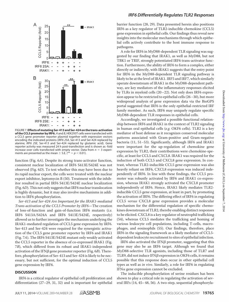
Ser-413 and Ser-424 Are Important for the IRAK1-mediatedTrans-activation of the CCL5 Promoter by IRF6—The creationof loss-of-function and gain-of-function IRF6 mutants (i.e.IRF6 S413A/S424A and IRF6 S413E/S424E, respectively)allowed us to further investigate the mechanism underlying theIRAK1-mediated regulation of CCL5 gene expression by IRF6.Ser-413 and Ser-424 were required for the synergistic activa-tion of the CCL5 gene promoter reporter by IRF6 and IRAK1(Fig. 7A). The IRF6 S413E/S424E mutant only weakly activatedthe CCL5 reporter in the absence of co-expressed IRAK1 (Fig.7B), which differed from its robust and IRAK1-independentactivation of the IFN� gene promoter reporter (Fig. 6B). There-fore, phosphorylation of Ser-413 and Ser-424 is likely to be nec-essary, but not sufficient, for the optimal induction of CCL5gene expression by IRF6.
DISCUSSION
IRF6 is a critical regulator of epithelial cell proliferation anddifferentiation (27–29, 31, 32) and is important for epithelial
barrier function (28, 29). Data presented herein also positionsIRF6 as a key regulator of TLR2-inducible chemokine (CCL5)gene expression in epithelial cells. Our findings thus reveal newinsights into the molecular mechanisms through which epithe-lial cells actively contribute to the host immune response topathogens.
A role for IRF6 in MyD88-dependent TLR signaling was sug-gested by our finding that IRAK1, as well as MyD88, but notTBK1 or TRIF, strongly potentiated IRF6 trans-activator func-tion. Furthermore, the ability of IRF6 to form a complex, eitherdirectly or indirectly, with IRAK1 suggests that the entry pointfor IRF6 in the MyD88-dependent TLR signaling pathway islikely to be at the level of IRAK1. IRF5 and IRF7, which similarlyoperate downstream of IRAK1 in the MyD88-dependent path-way, are key mediators of the inflammatory responses elicitedby TLRs in myeloid cells (20 –22). Not only does IRF6 expres-sion appear to be restricted to epithelial cells (26 –30), but morewidespread analysis of gene expression data via the BioGPSportal suggested that IRF6 is the only epithelial-restricted IRFfamily member. As such, IRF6 may uniquely regulate specificMyD88-dependent TLR responses in epithelial cells.
Accordingly, we investigated a possible functional relation-ship between IRF6 and IRAK1 in the context of TLR2 signalingin human oral epithelial cells (e.g. OKF6 cells). TLR2 is a keymediator of host defense as it recognizes conserved molecularpatterns associated with Gram-positive and Gram-negativebacteria (11, 51–53). Significantly, although IRF6 and IRAK1were important for the up-regulation of chemokine geneexpression by TLR2, their contributions were chemokine-spe-cific, at least for CCL5 and CXCL8. IRAK1 was required for theinduction of both CCL5 and CXCL8 gene expression. In con-trast, whereas TLR2-inducible CCL5 gene expression was alsoheavily reliant on IRF6, CXCL8 expression was induced inde-pendently of IRF6. In line with these findings, the CCL5 pro-moter was robustly activated by IRF6 and IRAK1 co-expres-sion, whereas IRAK1 strongly activated the CXCL8 promoterindependently of IRF6. Hence, IRAK1 likely mediates TLR2-inducible CCL5 gene expression, at least in part, by promotingthe activation of IRF6. The differing effect of IRF6 in regulatingCCL5 versus CXCL8 gene expression provides a molecularmechanism for the differential regulation of specific chemo-kines downstream of TLR2, thereby enabling distinct responsesto be elicited. CXCL8 is a key regulator of neutrophil trafficking(54), whereas CCL5 mediates the trafficking and homing ofvarious leukocyte cell populations, including T-cells, macro-phages, and eosinophils (55). Our findings, therefore, placeIRF6 in the signaling framework as a likely mediator of CCL5-dependent leukocyte recruitment to sites of epithelial infection.
IRF6 also activated the IFN� promoter, suggesting that thisgene may also be an IRF6 target. Although we found thatMyD88-selective TLR agonists, including those of TLR7 andTLR9, did not induce IFN� expression in OKF6 cells, it remainspossible that this response does occur in other epithelial celltypes as well as in vivo. Similarly, a role for IRF6 in regulatingIFN� gene expression cannot be excluded.
The inducible phosphorylation of serine residues has beenshown to play a critical role in regulating the activation of sev-eral IRFs (14, 45– 48, 56). A two-step, sequential phosphoryla-
FIGURE 7. Effects of mutating Ser-413 and Ser-424 on the trans-activationof the CCL5 promoter by IRF6. A and B, HEK293T cells were transfected witha CCL5 gene promoter reporter plasmid together with expression vectorsencoding the indicated proteins (IRF6 2SA, Ser-413 and Ser-424 replaced byalanine; IRF6 2SE, Ser-413 and Ser-424 replaced by glutamic acid). Genereporter activity was measured 24 h post-transfection and is shown as -foldincrease over cells transfected with empty vector. Data from n � 3 experi-ments are presented as the mean � S.E. (** � p � 0.01).
IRF6 Differentially Regulates TLR2 Responses
JULY 11, 2014 • VOLUME 289 • NUMBER 28 JOURNAL OF BIOLOGICAL CHEMISTRY 19765
at University of M
elbourne (CA
UL
) on September 7, 2014
http://ww
w.jbc.org/
Dow
nloaded from
tion model has been proposed for IRF3 activation (57). In thismodel, the phosphorylation of serine residues in “site 2” (e.g.Ser-396 in IRF3) is needed to alleviate autoinhibition and allowinteraction with the co-activator, CBP/p300; it also facilitatesthe phosphorylation of residues in “site 1” (e.g. Ser-386 in IRF3),which is required for IRF dimerization (57). We show here thatSer-413 and Ser-424, which correspond, respectively, to Ser-386 and Ser-396 in IRF3, are important for IRF6 function.Although not formally demonstrated, the marked increase inIRF6 trans-activator function by the phosphomimetic mutationof Ser-413 and Ser-424 to glutamic acid argues that they areregulatory phosphorylation sites. Thus, the trans-activatorfunction of IRF6 may likewise be regulated by a two-step,sequential phosphorylation mechanism in which Ser-424serves as a “gatekeeper” phosphorylation site.
Importantly, our data strongly suggest that Ser-424 is anIRAK1-regulated phosphorylation site, although it has not yetbeen established if IRAK1 directly regulates the phosphoryla-tion of Ser-424 or does so by activating another kinase.Although Ser-413 is likewise required for maximal IRAK1-me-diated IRF6 activation, IRAK1 did not further enhance thetrans-activator function of the IRF6 S424E mutant. Nonethe-less, phosphorylation of Ser-413 would be expected to beimportant for maximal IRF6 activity in view of the activatingeffect of its phosphomimetic mutation on IRF6 trans-activatorfunction. The activation of IRF7 by TLR7/9 signaling appears tobe mediated by both IRAK1 and IKK� (inhibitor of nuclearfactor �B kinase �) (39 – 41, 58). By analogy, IRF6 may be reg-ulated in a similar manner, with IRAK1 specifically regulating,either directly or indirectly, the phosphorylation of Ser-424 andanother kinase then phosphorylating Ser-413.
The phosphomimetic mutation of Ser-424 and Ser-413strongly enhanced IRF6 dimerization and trans-activator func-tion; however, it did not result in demonstrable nuclear local-ization. Nuclear translocation of the IRF6 S413E/S424E mutantwas apparent after treatment with the nuclear export inhibitor,leptomycin B. This would suggest that IRF6 is subject to tightlyregulated cytoplasmic-nuclear shuttling. Consequently, addi-tional posttranslational modifications (e.g. ubiquitination) mayalso be necessary for sustained IRF6 nuclear translocation.
The cooperation between IRFs and NF-�B is necessary forthe optimal expression of some inflammatory genes (59 – 61).This may also be the case for the regulation of CCL5 geneexpression by IRF6. Despite the IRF6 S413E/S424E mutantbeing highly active, it was largely ineffective in trans-activatingthe CCL5 promoter; the co-expression of IRAK1, which canalso activate endogenous NF-�B, was necessary for robust acti-vation of the CCL5 promoter. Virus-induced CCL5 geneexpression in alveolar epithelial cells was reported to requireboth IRF and NF-�B activity (15). Therefore, optimal inductionof CCL5 gene expression by IRAK1-mediated TLR2 signalingin epithelial cells may require functional cooperation betweenIRF6 and NF-�B.
In addition to its trans-activator function, phosphorylationalso appears to regulate the cell cycle-dependent degradation ofIRF6 (31). However, neither Ser-413 nor Ser-424 is likely to bethe phosphorylation site(s) that targets IRF6 for proteasomaldegradation as their mutation did not affect IRF6 protein levels.
The phosphorylation-mediated regulation of IRF6 trans-acti-vator function and degradation are thus likely to be regulated ina stimulus-dependent manner by distinct signaling pathways.
In summary, this study has uncovered a non-redundant rolefor IRF6 in differentially regulating TLR2-elicited chemokineresponses in epithelial cells. Given its role in also regulatingepithelial cell proliferation and differentiation, IRF6 may act asa pivotal nexus for distinct signaling pathways and regulateboth the barrier and inflammatory functions of epithelial cells.
REFERENCES1. Proksch, E., Brandner, J. M., and Jensen, J. M. (2008) The skin: an indis-
pensable barrier. Exp. Dermatol 17, 1063–10722. Goto, Y., and Kiyono, H. (2012) Epithelial barrier: an interface for the
cross-communication between gut flora and immune system. Immunol.Rev. 245, 147–163
3. Miller, L. S. (2008) Toll-like receptors in skin. Adv. Dermatol. 24, 71– 874. Saenz, S. A., Taylor, B. C., and Artis, D. (2008) Welcome to the neighbor-
hood: epithelial cell-derived cytokines license innate and adaptive im-mune responses at mucosal sites. Immunol. Rev. 226, 172–190
5. Abreu, M. T. (2010) Toll-like receptor signalling in the intestinal epithe-lium: how bacterial recognition shapes intestinal function. Nat. Rev. Im-munol. 10, 131–144
6. Gribar, S. C., Anand, R. J., Sodhi, C. P., and Hackam, D. J. (2008) The roleof epithelial Toll-like receptor signaling in the pathogenesis of intestinalinflammation. J. Leukoc. Biol. 83, 493– 498
7. Nickoloff, B. J., Xin, H., Nestle, F. O., and Qin, J. Z. (2007) The cytokine andchemokine network in psoriasis. Clin. Dermatol. 25, 568 –573
8. Darveau, R. P. (2010) Periodontitis: a polymicrobial disruption of hosthomeostasis. Nat. Rev. Microbiol. 8, 481– 490
9. Fukata, M., and Abreu, M. T. (2009) Pathogen recognition receptors, can-cer and inflammation in the gut. Curr. Opin. Pharmacol. 9, 680 – 687
10. Akira, S., Uematsu, S., and Takeuchi, O. (2006) Pathogen recognition andinnate immunity. Cell 124, 783– 801
11. Kawai, T., and Akira, S. (2010) The role of pattern-recognition receptorsin innate immunity: update on Toll-like receptors. Nat. Immunol. 11,373–384
12. Honda, K., and Taniguchi, T. (2006) IRFs: master regulators of signallingby Toll-like receptors and cytosolic pattern-recognition receptors. Nat.Rev. Immunol. 6, 644 – 658
13. Tamura, T., Yanai, H., Savitsky, D., and Taniguchi, T. (2008) The IRFfamily transcription factors in immunity and oncogenesis. Annu. Rev. Im-munol. 26, 535–584
14. Barnes, B. J., Kellum, M. J., Field, A. E., and Pitha, P. M. (2002) Multipleregulatory domains of IRF-5 control activation, cellular localization, andinduction of chemokines that mediate recruitment of T lymphocytes. Mol.Cell. Biol. 22, 5721–5740
15. Casola, A., Garofalo, R. P., Haeberle, H., Elliott, T. F., Lin, R., Jamaluddin,M., and Brasier, A. R. (2001) Multiple cis regulatory elements controlRANTES promoter activity in alveolar epithelial cells infected with respi-ratory syncytial virus. J. Virol. 75, 6428 – 6439
16. Fitzgerald, K. A., Rowe, D. C., Barnes, B. J., Caffrey, D. R., Visintin, A., Latz,E., Monks, B., Pitha, P. M., and Golenbock, D. T. (2003) LPS-TLR4 signal-ing to IRF-3/7 and NF-�B involves the toll adapters TRAM and TRIF. J.Exp. Med. 198, 1043–1055
17. Lin, R., Heylbroeck, C., Genin, P., Pitha, P. M., and Hiscott, J. (1999)Essential role of interferon regulatory factor 3 in direct activation ofRANTES chemokine transcription. Mol. Cell. Biol. 19, 959 –966
18. Doyle, S., Vaidya, S., O’Connell, R., Dadgostar, H., Dempsey, P., Wu, T.,Rao, G., Sun, R., Haberland, M., Modlin, R., and Cheng, G. (2002) IRF3mediates a TLR3/TLR4-specific antiviral gene program. Immunity 17,251–263
19. Sato, M., Suemori, H., Hata, N., Asagiri, M., Ogasawara, K., Nakao, K.,Nakaya, T., Katsuki, M., Noguchi, S., Tanaka, N., and Taniguchi, T. (2000)Distinct and essential roles of transcription factors IRF-3 and IRF-7 inresponse to viruses for IFN-�/� gene induction. Immunity 13, 539 –548
IRF6 Differentially Regulates TLR2 Responses
19766 JOURNAL OF BIOLOGICAL CHEMISTRY VOLUME 289 • NUMBER 28 • JULY 11, 2014
at University of M
elbourne (CA
UL
) on September 7, 2014
http://ww
w.jbc.org/
Dow
nloaded from

20. Honda, K., Yanai, H., Negishi, H., Asagiri, M., Sato, M., Mizutani, T.,Shimada, N., Ohba, Y., Takaoka, A., Yoshida, N., and Taniguchi, T. (2005)IRF-7 is the master regulator of type-I interferon-dependent immune re-sponses. Nature 434, 772–777
21. Krausgruber, T., Blazek, K., Smallie, T., Alzabin, S., Lockstone, H., Sahgal,N., Hussell, T., Feldmann, M., and Udalova, I. A. (2011) IRF5 promotesinflammatory macrophage polarization and TH1-TH17 responses. Nat.Immunol. 12, 231–238
22. Takaoka, A., Yanai, H., Kondo, S., Duncan, G., Negishi, H., Mizutani, T.,Kano, S., Honda, K., Ohba, Y., Mak, T. W., and Taniguchi, T. (2005)Integral role of IRF-5 in the gene induction programme activated by Toll-like receptors. Nature 434, 243–249
23. Kato, A., Favoreto, S., Jr., Avila, P. C., and Schleimer, R. P. (2007) TLR3-and Th2 cytokine-dependent production of thymic stromal lymphopoi-etin in human airway epithelial cells. J. Immunol. 179, 1080 –1087
24. Matsukura, S., Kokubu, F., Kurokawa, M., Kawaguchi, M., Ieki, K., Kuga,H., Odaka, M., Suzuki, S., Watanabe, S., Takeuchi, H., Kasama, T., andAdachi, M. (2006) Synthetic double-stranded RNA induces multiplegenes related to inflammation through Toll-like receptor 3 depending onNF-�B and/or IRF-3 in airway epithelial cells. Clin. Exp. Allergy 36,1049 –1062
25. Wang, Q., Nagarkar, D. R., Bowman, E. R., Schneider, D., Gosangi, B., Lei,J., Zhao, Y., McHenry, C. L., Burgens, R. V., Miller, D. J., Sajjan, U., andHershenson, M. B. (2009) Role of double-stranded RNA pattern recogni-tion receptors in rhinovirus-induced airway epithelial cell responses. J. Im-munol. 183, 6989 – 6997
26. Kondo, S., Schutte, B. C., Richardson, R. J., Bjork, B. C., Knight, A. S.,Watanabe, Y., Howard, E., de Lima, R. L., Daack-Hirsch, S., Sander, A.,McDonald-McGinn, D. M., Zackai, E. H., Lammer, E. J., Aylsworth, A. S.,Ardinger, H. H., Lidral, A. C., Pober, B. R., Moreno, L., Arcos-Burgos, M.,Valencia, C., Houdayer, C., Bahuau, M., Moretti-Ferreira, D., Richieri-Costa, A., Dixon, M. J., and Murray, J. C. (2002) Mutations in IRF6 causeVan der Woude and popliteal pterygium syndromes. Nat. Genet. 32,285–289
27. Bailey, C. M., Khalkhali-Ellis, Z., Kondo, S., Margaryan, N. V., Seftor, R. E.,Wheaton, W. W., Amir, S., Pins, M. R., Schutte, B. C., and Hendrix, M. J.(2005) Mammary serine protease inhibitor (Maspin) binds directly to in-terferon regulatory factor 6: identification of a novel serpin partnership.J. Biol. Chem. 280, 34210 –34217
28. Ingraham, C. R., Kinoshita, A., Kondo, S., Yang, B., Sajan, S., Trout, K. J.,Malik, M. I., Dunnwald, M., Goudy, S. L., Lovett, M., Murray, J. C., andSchutte, B. C. (2006) Abnormal skin, limb and craniofacial morphogenesisin mice deficient for interferon regulatory factor 6 (Irf6). Nat. Genet. 38,1335–1340
29. Richardson, R. J., Dixon, J., Malhotra, S., Hardman, M. J., Knowles, L.,Boot-Handford, R. P., Shore, P., Whitmarsh, A., and Dixon, M. J. (2006)Irf6 is a key determinant of the keratinocyte proliferation-differentiationswitch. Nat. Genet. 38, 1329 –1334
30. Knight, A. S., Schutte, B. C., Jiang, R., and Dixon, M. J. (2006) Develop-mental expression analysis of the mouse and chick orthologues of IRF6:the gene mutated in Van der Woude syndrome. Dev. Dyn. 235, 1441–1447
31. Bailey, C. M., Abbott, D. E., Margaryan, N. V., Khalkhali-Ellis, Z., andHendrix, M. J. (2008) Interferon regulatory factor 6 promotes cell cyclearrest and is regulated by the proteasome in a cell cycle-dependent man-ner. Mol. Cell. Biol. 28, 2235–2243
32. Biggs, L. C., Rhea, L., Schutte, B. C., and Dunnwald, M. (2012) Interferonregulatory factor 6 is necessary, but not sufficient, for keratinocyte differ-entiation. J. Invest. Dermatol. 132, 50 –58
33. Dickson, M. A., Hahn, W. C., Ino, Y., Ronfard, V., Wu, J. Y., Weinberg,R. A., Louis, D. N., Li, F. P., and Rheinwald, J. G. (2000) Human kera-tinocytes that express hTERT and also bypass a p16(INK4a)-enforcedmechanism that limits life span become immortal yet retain normalgrowth and differentiation characteristics. Mol. Cell. Biol. 20,1436 –1447
34. Way, K. J., Dinh, H., Keene, M. R., White, K. E., Clanchy, F. I., Lusby, P.,Roiniotis, J., Cook, A. D., Cassady, A. I., Curtis, D. J., and Hamilton, J. A.(2009) The generation and properties of human macrophage populationsfrom hemopoietic stem cells. J. Leukoc. Biol. 85, 766 –778
35. De Nardo, D., Nguyen, T., Hamilton, J. A., and Scholz, G. M. (2009) Down-regulation of IRAK-4 is a component of LPS- and CpG DNA-inducedtolerance in macrophages. Cell. Signal. 21, 246 –252
36. Nguyen, T., De Nardo, D., Masendycz, P., Hamilton, J. A., and Scholz,G. M. (2009) Regulation of IRAK-1 activation by its C-terminal domain.Cell. Signal. 21, 719 –726
37. Lin, R., Génin, P., Mamane, Y., and Hiscott, J. (2000) Selective DNA bind-ing and association with the CREB binding protein coactivator contributeto differential activation of �/� interferon genes by interferon regulatoryfactors 3 and 7. Mol. Cell. Biol. 20, 6342– 6353
38. Garofalo, R., Sabry, M., Jamaluddin, M., Yu, R. K., Casola, A., Ogra, P. L.,and Brasier, A. R. (1996) Transcriptional activation of the interleukin-8gene by respiratory syncytial virus infection in alveolar epithelial cells:nuclear translocation of the RelA transcription factor as a mechanismproducing airway mucosal inflammation. J. Virol. 70, 8773– 8781
39. Uematsu, S., Sato, S., Yamamoto, M., Hirotani, T., Kato, H., Takeshita, F.,Matsuda, M., Coban, C., Ishii, K. J., Kawai, T., Takeuchi, O., and Akira, S.(2005) Interleukin-1 receptor-associated kinase-1 plays an essential rolefor Toll-like receptor (TLR) 7- and TLR9-mediated interferon-� induc-tion. J. Exp. Med. 201, 915–923
40. Schoenemeyer, A., Barnes, B. J., Mancl, M. E., Latz, E., Goutagny, N., Pitha,P. M., Fitzgerald, K. A., and Golenbock, D. T. (2005) The interferon regu-latory factor, IRF5, is a central mediator of toll-like receptor 7 signaling.J. Biol. Chem. 280, 17005–17012
41. Saitoh, T., Satoh, T., Yamamoto, N., Uematsu, S., Takeuchi, O., Kawai, T.,and Akira, S. (2011) Antiviral protein Viperin promotes Toll-like receptor7- and Toll-like receptor 9-mediated type I interferon production in plas-macytoid dendritic cells. Immunity 34, 352–363
42. Tun-Kyi, A., Finn, G., Greenwood, A., Nowak, M., Lee, T. H., Asara, J. M.,Tsokos, G. C., Fitzgerald, K., Israel, E., Li, X., Exley, M., Nicholson, L. K.,and Lu, K. P. (2011) Essential role for the prolyl isomerase Pin1 in Toll-likereceptor signaling and type I interferon-mediated immunity. Nat. Immu-nol. 12, 733–741
43. Taniguchi, T., Ogasawara, K., Takaoka, A., and Tanaka, N. (2001) IRFfamily of transcription factors as regulators of host defense. Annu. Rev.Immunol. 19, 623– 655
44. De Nardo, D., Masendycz, P., Ho, S., Cross, M., Fleetwood, A. J., Reynolds,E. C., Hamilton, J. A., and Scholz, G. M. (2005) A central role for theHsp90.Cdc37 molecular chaperone module in interleukin-1 receptor-as-sociated kinase-dependent signaling by toll-like receptors. J. Biol. Chem.280, 9813–9822
45. Lin, R., Heylbroeck, C., Pitha, P. M., and Hiscott, J. (1998) Virus-depen-dent phosphorylation of the IRF-3 transcription factor regulates nucleartranslocation, transactivation potential, and proteasome-mediated degra-dation. Mol. Cell. Biol. 18, 2986 –2996
46. Lin, R., Mamane, Y., and Hiscott, J. (2000) Multiple regulatory domainscontrol IRF-7 activity in response to virus infection. J. Biol. Chem. 275,34320 –34327
47. Marié, I., Smith, E., Prakash, A., and Levy, D. E. (2000) Phosphorylation-induced dimerization of interferon regulatory factor 7 unmasks DNAbinding and a bipartite transactivation domain. Mol. Cell. Biol. 20,8803– 8814
48. Sharma, S., tenOever, B. R., Grandvaux, N., Zhou, G. P., Lin, R., and His-cott, J. (2003) Triggering the interferon antiviral response through anIKK-related pathway. Science 300, 1148 –1151
49. Barnes, B. J., Field, A. E., and Pitha-Rowe, P. M. (2003) Virus-inducedheterodimer formation between IRF-5 and IRF-7 modulates assembly ofthe IFNA enhanceosome in vivo and transcriptional activity of IFNAgenes. J. Biol. Chem. 278, 16630 –16641
50. Ullman, K. S., Powers, M. A., and Forbes, D. J. (1997) Nuclear exportreceptors: from importin to exportin. Cell 90, 967–970
51. Takeuchi, O., Hoshino, K., and Akira, S. (2000) Cutting edge: TLR2-defi-cient and MyD88-deficient mice are highly susceptible to Staphylococcusaureus infection. J. Immunol. 165, 5392–5396
52. Mancuso, G., Midiri, A., Beninati, C., Biondo, C., Galbo, R., Akira, S.,Henneke, P., Golenbock, D., and Teti, G. (2004) Dual role of TLR2 andmyeloid differentiation factor 88 in a mouse model of invasive group Bstreptococcal disease. J. Immunol. 172, 6324 – 6329
IRF6 Differentially Regulates TLR2 Responses
JULY 11, 2014 • VOLUME 289 • NUMBER 28 JOURNAL OF BIOLOGICAL CHEMISTRY 19767
at University of M
elbourne (CA
UL
) on September 7, 2014
http://ww
w.jbc.org/
Dow
nloaded from

53. Burns, E., Eliyahu, T., Uematsu, S., Akira, S., and Nussbaum, G. (2010)TLR2-dependent inflammatory response to Porphyromonas gingivalis isMyD88 independent, whereas MyD88 is required to clear infection. J. Im-munol. 184, 1455–1462
54. Kobayashi, Y. (2006) Neutrophil infiltration and chemokines. Crit. Rev.Immunol. 26, 307–316
55. Appay, V., and Rowland-Jones, S. L. (2001) RANTES: a versatile and con-troversial chemokine. Trends Immunol. 22, 83– 87
56. Fitzgerald, K. A., McWhirter, S. M., Faia, K. L., Rowe, D. C., Latz, E.,Golenbock, D. T., Coyle, A. J., Liao, S. M., and Maniatis, T. (2003) IKK�and TBK1 are essential components of the IRF3 signaling pathway. Nat.Immunol. 4, 491– 496
57. Panne, D., McWhirter, S. M., Maniatis, T., and Harrison, S. C. (2007)Interferon regulatory factor 3 is regulated by a dual phosphorylation-de-pendent switch. J. Biol. Chem. 282, 22816 –22822
58. Hoshino, K., Sugiyama, T., Matsumoto, M., Tanaka, T., Saito, M., Hemmi,H., Ohara, O., Akira, S., and Kaisho, T. (2006) I�B kinase-� is critical forinterferon-� production induced by Toll-like receptors 7 and 9. Nature440, 949 –953
59. Génin, P., Algarté, M., Roof, P., Lin, R., and Hiscott, J. (2000) Regulation ofRANTES chemokine gene expression requires cooperativity betweenNF-� B and IFN-regulatory factor transcription factors. J. Immunol. 164,5352–5361
60. Krausgruber, T., Saliba, D., Ryzhakov, G., Lanfrancotti, A., Blazek, K., andUdalova, I. A. (2010) IRF5 is required for late-phase TNF secretion byhuman dendritic cells. Blood 115, 4421– 4430
61. Wathelet, M. G., Lin, C. H., Parekh, B. S., Ronco, L. V., Howley, P. M.,and Maniatis, T. (1998) Virus infection induces the assembly of coor-dinately activated transcription factors on the IFN-� enhancer in vivo.Mol. Cell 1, 507–518
IRF6 Differentially Regulates TLR2 Responses
19768 JOURNAL OF BIOLOGICAL CHEMISTRY VOLUME 289 • NUMBER 28 • JULY 11, 2014
at University of M
elbourne (CA
UL
) on September 7, 2014
http://ww
w.jbc.org/
Dow
nloaded from
Related Documents