Interferon- and Tumor Necrosis Factor- Determine Resistance to Paracoccidioides brasiliensis Infection in Mice Janeusa T. Souto,* Flore ˆ ncio Figueiredo, † Alessandra Furlanetto, ‡ Klaus Pfeffer, § Marcos A. Rossi, ‡ and Joa ˜o S. Silva* From the Departments of Immunology * and Pathology, ‡ Faculty of Medicine of Ribeira ˜o Preto, University of Sa ˜o Paulo, Sa ˜o Paulo, Brazil; the Departments of Pathology, † University of Brası ´lia and Catholic University of Brası ´lia, Brası ´lia, Brazil; and The Institute of Medical Microbiology, Immunology and Hygiene, § Technical University of Munich, Munich, Germany To investigate the role of interferon- (IFN-) and tumor necrosis factor- (TNF-) in the resistance to Paracoccidioides brasiliensis (Pb) infection, mice with homologous disruption of the IFN- (GKO) or TNF- receptor p55 (p55KO) were infected with the parasite. GKO and p55KO, but not wild-type (WT) mice, were unable to control the growth of yeast cells and the mice succumbed to infection by days 16 and 90 after infection, respectively. Typical in- flammatory granulomas were found only in WT mice. In contrast, knockout mice presented an in- flammatory infiltrate composed of a few neutro- phils, mononuclear, epithelioid, and multinuclear giant cells forming incipient granulomas in GKO mice and without granuloma formation in p55KO mice. Besides , both groups of knockout mice exhib- ited elevated numbers of yeast forms in agreement with colony-forming unit counts in organs. Com- pared with WT, splenocytes from infected GKO mice cultured with the Pb F1 fraction produced lower TNF- levels, whereas leukocytes from in- fected p55KO mice produced similar amounts of TNF- but higher levels of IFN-. Moreover, spleno- cytes from infected WT mice produced higher levels of nitric oxide (NO) resulting in a lower T-cell pro- liferative response to Con A than uninfected WT, or infected p55KO and GKO mice. On the contrary , the addition of IFN- to splenocytes from infected GKO mice resulted in higher NO production and lower T cell proliferation. Taken together, these findings suggests that endogenous TNF- , acting through the p55 receptor, and IFN- mediate resistance to Pb infection and induce NO production that deter- mines marked T cell unresponsiveness. (Am J Pathol 2000, 156:1811–1820) Paracoccidioidomycosis (PCM), the major systemic my- cosis in Latin America, is caused by the thermally dimor- phic fungus Paracoccidioides brasiliensis (Pb). The infec- tion is acquired by inhalation of airborne propagules produced by the fungal mycelium form, which then change into the pathogenic yeast form when at core body temperature. 1 The yeast forms in the mammalian host can be eliminated by immune competent cells or dissem- inate to tissues by the lymphatic and hematogenous route. After infection there are no immediate signs or symptoms of the disease, characterizing silent PCM but the host develops a specific immune response against fungal antigens which can be detected by a skin test. In endemic areas, up to 60% of asymptomatic individuals may be skin test positive. 2 In the acute form of clinically manifest infection, there is an involvement of the reticuloendothelial system, a non- specific hypergammaglobulinemia, a depressed cellular immune response, a diffuse inflammatory granulomatous response, and concomitant fungus dissemination. The chronic form presents a broad spectrum of clinical man- ifestations with frequent damage of the lung and oropha- ryngeal mucosa and a profound impairment of the im- mune response. 3 In this phase of infection, an epithelioid inflammatory granulomatous reaction is known to prevent the dissemination of the fungus. 4 Athymic mice do not control dissemination of the fun- gus, suggesting that cell-mediated immunity is an impor- tant host defense mechanism against Pb infection. 5,6 However, there is a T cell dysfunction in patients and infected mice that may be related to alterations in the ratio of T cell subpopulations, suppressive effects of fun- gal components, and an imbalance in the levels of cyto- kine production. 7–10 The cellular immune response against fungal antigens that occurs in resistant mice re- sults in macrophage activation and granuloma forma- tion. 11 Tumor necrosis factor- (TNF-) produced by macrophages in response to the Pb cell wall compo- nent 11 is required for macrophage accumulation and dif- ferentiation into epithelioid cells, and for persistence of Supported by a grant from FAPESP (98/11986 –2) and by scholarships from CAPES (J. T. S.), FAPESP (A. F.), and CNPq (J. S. S.; M. A. R.; F. F.). Accepted for publication February 9, 2000. Address reprint requests to Dr. Joa ˜ o S. Silva, School of Medicine of Ribeira ˜ o Preto-USP, Department of Immunology, Av. Bandeirantes, 3900, 14049 –900, Ribeira ˜ o Preto, SP, Brazil. E-mail: [email protected]. American Journal of Pathology, Vol. 156, No. 5, May 2000 Copyright © American Society for Investigative Pathology 1811

Welcome message from author
This document is posted to help you gain knowledge. Please leave a comment to let me know what you think about it! Share it to your friends and learn new things together.
Transcript
Interferon-� and Tumor Necrosis Factor-� DetermineResistance to Paracoccidioides brasiliensis Infectionin Mice
Janeusa T. Souto,* Florencio Figueiredo,†Alessandra Furlanetto,‡ Klaus Pfeffer,§Marcos A. Rossi,‡ and Joao S. Silva*From the Departments of Immunology * and Pathology, ‡ Faculty of
Medicine of Ribeirao Preto, University of Sao Paulo, Sao Paulo,
Brazil; the Departments of Pathology,† University of Brasılia and
Catholic University of Brasılia, Brasılia, Brazil; and The Institute of
Medical Microbiology, Immunology and Hygiene,§ Technical
University of Munich, Munich, Germany
To investigate the role of interferon-� (IFN-�) andtumor necrosis factor-� (TNF-�) in the resistance toParacoccidioides brasiliensis (Pb) infection, micewith homologous disruption of the IFN-� (GKO) orTNF-� receptor p55 (p55KO) were infected with theparasite. GKO and p55KO, but not wild-type (WT)mice , were unable to control the growth of yeastcells and the mice succumbed to infection by days16 and 90 after infection, respectively. Typical in-flammatory granulomas were found only in WTmice. In contrast , knockout mice presented an in-flammatory infiltrate composed of a few neutro-phils , mononuclear , epithelioid , and multinucleargiant cells forming incipient granulomas in GKOmice and without granuloma formation in p55KOmice. Besides , both groups of knockout mice exhib-ited elevated numbers of yeast forms in agreementwith colony-forming unit counts in organs. Com-pared with WT, splenocytes from infected GKOmice cultured with the Pb F1 fraction producedlower TNF-� levels , whereas leukocytes from in-fected p55KO mice produced similar amounts ofTNF-� but higher levels of IFN-�. Moreover , spleno-cytes from infected WT mice produced higher levelsof nitric oxide (NO) resulting in a lower T-cell pro-liferative response to Con A than uninfected WT, orinfected p55KO and GKO mice. On the contrary , theaddition of IFN-� to splenocytes from infected GKOmice resulted in higher NO production and lower Tcell proliferation. Taken together , these findingssuggests that endogenous TNF-� , acting through thep55 receptor , and IFN-� mediate resistance to Pbinfection and induce NO production that deter-mines marked T cell unresponsiveness. (Am JPathol 2000, 156:1811–1820)
Paracoccidioidomycosis (PCM), the major systemic my-cosis in Latin America, is caused by the thermally dimor-phic fungus Paracoccidioides brasiliensis (Pb). The infec-tion is acquired by inhalation of airborne propagulesproduced by the fungal mycelium form, which thenchange into the pathogenic yeast form when at core bodytemperature.1 The yeast forms in the mammalian hostcan be eliminated by immune competent cells or dissem-inate to tissues by the lymphatic and hematogenousroute. After infection there are no immediate signs orsymptoms of the disease, characterizing silent PCM butthe host develops a specific immune response againstfungal antigens which can be detected by a skin test. Inendemic areas, up to 60% of asymptomatic individualsmay be skin test positive.2
In the acute form of clinically manifest infection, there isan involvement of the reticuloendothelial system, a non-specific hypergammaglobulinemia, a depressed cellularimmune response, a diffuse inflammatory granulomatousresponse, and concomitant fungus dissemination. Thechronic form presents a broad spectrum of clinical man-ifestations with frequent damage of the lung and oropha-ryngeal mucosa and a profound impairment of the im-mune response.3 In this phase of infection, an epithelioidinflammatory granulomatous reaction is known to preventthe dissemination of the fungus.4
Athymic mice do not control dissemination of the fun-gus, suggesting that cell-mediated immunity is an impor-tant host defense mechanism against Pb infection.5,6
However, there is a T cell dysfunction in patients andinfected mice that may be related to alterations in theratio of T cell subpopulations, suppressive effects of fun-gal components, and an imbalance in the levels of cyto-kine production.7–10 The cellular immune responseagainst fungal antigens that occurs in resistant mice re-sults in macrophage activation and granuloma forma-tion.11 Tumor necrosis factor-� (TNF-�) produced bymacrophages in response to the Pb cell wall compo-nent11 is required for macrophage accumulation and dif-ferentiation into epithelioid cells, and for persistence of
Supported by a grant from FAPESP (98/11986–2) and by scholarshipsfrom CAPES (J. T. S.), FAPESP (A. F.), and CNPq (J. S. S.; M. A. R.; F. F.).
Accepted for publication February 9, 2000.
Address reprint requests to Dr. Joao S. Silva, School of Medicine ofRibeirao Preto-USP, Department of Immunology, Av. Bandeirantes, 3900,14049–900, Ribeirao Preto, SP, Brazil. E-mail: [email protected].
American Journal of Pathology, Vol. 156, No. 5, May 2000
Copyright © American Society for Investigative Pathology
1811
well-formed granulomas.12 Interferon-� (IFN-�) can acti-vate infected macrophages to secret TNF-� and to inhibitthe replication of Pb.13 Recently, experiments performedon Pb-infected mice treated with anti-IFN-� revealed anexacerbation of pulmonary infection and earlier fungaldissemination.14
In the present study we evaluated the role of IFN-�and TNF-� in the resistance to Pb infection in mice.Using mice genetically deficient in IFN-� (GKO) or TNF-�receptor p55 (p55KO), we could demonstrate that bothcytokines are involved in the resistance to Pb infection,granuloma formation, and control of fungus disse-mination. In addition, our data suggest that IFN-� andTNF-� modulate the production of cytokines and nitricoxide (NO) and the T-cell proliferative response inPb-infected mice.
Materials and Methods
Animals
Breeding pairs of mice with targeted disruption of theIFN-� (GKO mice)15 and TNF-� receptor p55 (p55KO)genes16 were obtained from Jackson Laboratories (BarHarbor, ME) and Amgen Institute (Toronto, Canada), re-spectively. Breeding stock backcrossed on C57Bl/6 wereobtained and the genotype of p55KO and GKO micedetermined by polymerase chain reaction of DNA aspreviously described.16,17 Male C57Bl/6 wild-type (WT),GKO, and p55KO mice, 6 to 8 weeks old, were bred andmaintained in microisolator cages in the animal housingfacility of the Department of Immunology, University ofSao Paulo, Ribeirao Preto, Brazil.
Parasite and Mice Infection
Yeast cells of virulent Pb 18 strains were cultured at 37°Cin Fava-Netto’s medium18 for 7 to 14 days. The yeast cellswere harvested and washed three times in phosphate-buffered saline (PBS), pH 7.2. Viability of yeast cells wasdetermined as previously described.19 The animals wereinfected intravenously with 1 � 106 viable yeast cells in100 �l of PBS.
Assay for Organ Colony-Forming Units
The dissemination of fungus in lungs, livers, and spleenswas assayed as previously described.20 The organswere removed, weighed, homogenized in sterile PBS, pH7.2, and serially diluted. Aliquots of 100 �l were dis-pensed into Petri dishes, in duplicates, containing brain-heart infusion agar (Difco Laboratories, Detroit, MI) sup-plemented with 4% (v/v) of normal horse serum and 5%(v/v) of Pb 192 broth yeast culture filtrate from 2-week-oldcultures as source of growth-promoting factor.21 Plateswere incubated at 37°C, and colonies were counted 7 to14 days later. Results were expressed as numbers ofcolony-forming units (CFU) � SE per gram of tissue.
Preparation and Fractionation of Pb18 CellWalls
Fungus cell walls were obtained as previously de-scribed.11 Briefly, yeast forms were sonicated and lipidsfrom the cell walls were removed by treatment with chlo-roform/methanol (2:1, v/v) with stirring at room tempera-ture for 2 hours. Extracts were separated by centrifuga-tion at 500 � g for 5 minutes. The resulting insoluble cellresidue was named the cell wall fraction. The cell wallwas further treated with 1 N NaOH and gently stirred atroom temperature for 1 hour. The alkali-insoluble sedi-ment was washed with water until it reached pH 7.0 andthen washed with ethanol, followed by acetone and di-ethyl ether. The resulting white powder was named F1fraction and contained �0.05 ng/ml of bacterial endo-toxin, as determined by the Limulus amebocyte assay(Sigma Chemical Co., St. Louis, MO).
Proliferation Assay and Nitrite Quantification
Spleen cells from uninfected (day zero) or Pb-infectedmice were washed in Hanks’ medium and incubated for 4minutes with lysis buffer (one part of 0.17 mol/L Tris andnine parts of 0.16 mol/L ammonium chloride). The cellswere washed and suspended to a concentration of 5 �106 cells per ml in RPMI 1640 (Flow Laboratories,McLean, VA) supplemented with 5% fetal calf serum (LifeTechnologies Inc., Bethesda, MD), 5 � 10�2 mol/L 2-�-mercaptoethanol, 2 mmol/L L-glutamine, and antibiotics(all from Sigma). The cells were cultured in flat-bottom96-well plates (Corning Glass Works, Corning, NY) at 1 �106/well with or without Con A (2 �g/ml) for 72 hours at37°C in a humidified 5% CO2 incubator. To assess pro-liferation, 0.5 �Ci/well of [3H]TdR (Amersham Corp., Ar-lington Heights, IL) was added during the final 18 hoursof culture, the cells were harvested and radioactivity wasmeasured in a scintillation counter. In some experiments,to inhibit the inducible nitric oxide synthase (iNOS), weadded 200 �mol/L of NG-methyl-L-arginine (LNMMA, Sig-ma). Data were expressed as means (�SE) of counts perminute of triplicate cultures.
For nitrite quantification, splenocytes (2 � 106 cells/ml)from WT, GKO, and p55KO animals were cultured intriplicate for 48 hours in medium alone or medium con-taining Con A (2 �g/ml) in the presence or absence of100 U/ml of IFN-�. Duplicates of nitrite concentrations ineach culture supernatant were assayed in a microplateby mixing 0.1 ml of culture supernatant with 0.1 ml ofGriess reagent.22 The A540 was read 10 minutes later,and the nitrite concentration was determined by refer-ence to a standard curve of 1 to 100 �mol/L NaNO2.
Morphology
Five to seven animals selected at random from eachgroup were sacrificed at 7 and 15 days (GKO) and at 15,30, and 60 days (p55KO) after infection. The lungs ob-tained were fixed in 10% formalin for 24 hours and em-bedded in paraffin. Tissue sections (5 �m) were stained
1812 Souto et alAJP May 2000, Vol. 156, No. 5
with hematoxylin and eosin (H&E) or impregnated withsilver for demonstration of reticulum fibers using standardprotocols. WT mice were used as a control.
Cytokine Detection in Culture Supernatants
Spleen cells (2 � 106 cells/ml) from normal or infectedmice were cultured in 24-well tissue culture plates (Corn-ing) with 2 �g/ml of Con A, 40 �g/ml of Pb18 F1 fraction,or medium alone for 48 hours at 37°C in a humidified 5%CO2 incubator. The supernatants were harvested andstored at �20°C until assayed for IFN-�, TNF-�, interleukin(IL)-10, and IL-12, using a two-sandwich enzyme-linkedimmunosorbent assay. XMG 1.2 (anti-IFN-�), XT22.11 (anti-TNF-�), JES5–2A5 (anti-IL-10), and C17.15.10 (anti-IL-12)were used as capture monoclonal antibody (mAb). IFN-�and TNF-� bound to the mAb were visualized with poly-clonal rabbit anti-IFN-� or anti-TNF-� (both from Santa CruzBiotechnology, Santa Cruz, CA), followed by goat anti-rab-bit IgG conjugated with peroxidase (Life Technologies Inc.,Gaithersburg, MD), whereas IL-10 and IL-12 were detectedusing appropriated biotinylated mAbs SXC1 (anti-IL-10)and C15.6 (anti-IL-12), respectively. A standard curve wasprepared with specific cytokines (all from Sigma) and en-zyme-linked immunosorbent assay sensitivities were 0.625U/ml for IFN-�, 312 pg/ml for TNF-�, and 390 pg/ml for IL-10and IL-12.
Statistical Analysis
The results are expressed as the mean � SE of theindicated number of animals or experiments. Statisticalanalysis was performed using analysis of variance fol-lowed by the parametric Tukey-Kramer test (INSTAT soft-ware, GraphPad, San Diego, CA). A P value � 0.05 wasconsidered to indicate statistical significance.
Results
Absence of IFN-� or the TNF-� Receptor p55Causes Increased Susceptibility to Pb Infection
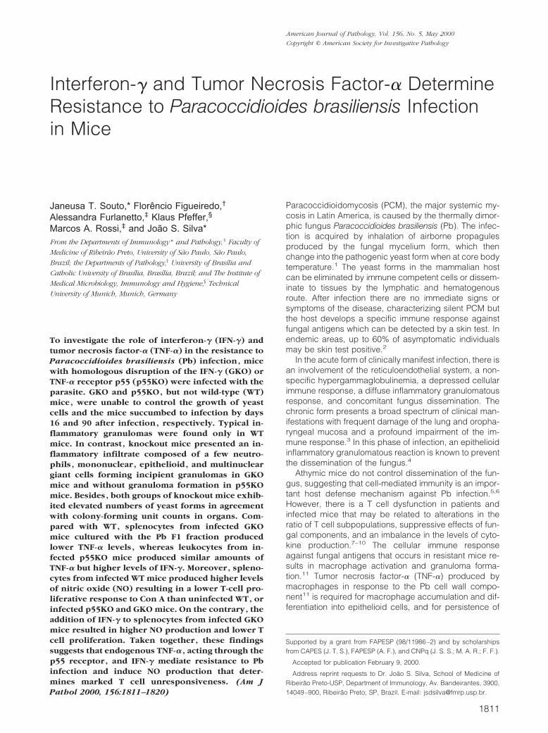
To determine the role of IFN-� and TNF-� in the resis-tance to Pb infection, IFN-� and TNF-� receptor p55knockout mice were infected with yeast cells and theirsurvival was compared with that of littermate controls. Wefound that whereas 100% of the control littermate micesurvived during the 90-day period of the experiment, theinfected GKO and p55KO mice succumbed by days 16and 90 after parasite inoculation, respectively (Figure 1).The infected p55KO mice started to die after day 60 ofinfection and the mortality rate at day 90 of infection was100% when compared with 100% of survival in infectedWT animals. Because infected GKO mice did not survivebeyond 16 days, the fungal loads in these animals weredetermined on days 4, 7, and 15 postinfection. The re-sults showed a significantly higher amount of fungus inGKO mice than that verified in infected WT mice (P �0.001; Figure 2, A–C). It is possible that the CFU numbers
at days 7 and 15 are underestimated, because the datarepresent the CFU load of mice that have survived. Thefungal loads obtained in the organs from infected p55KOmice (Figure 2, D–F) also were higher than those incontrol-infected WT mice (P � 0.001), except for thespleen at day 15 after infection, for which a statisticallysignificant difference could not be detected (Figure 2F).
Figure 1. Absence of IFN-� leads to high susceptibility to P. brasiliensisinfection. Survival of WT (triangles), p55KO (circles), and GKO (squares)mice after intravenous infection with 1 � 106 P. brasiliensis yeast cells. Eightmice were used in each group. Similar results were obtained in a secondexperiment.
Figure 2. Lack of resistance of infected GKO and p55KO mice to controlP. brasiliensis yeast cell proliferation and dissemination. Colony-formingunits (CFU) of lungs (A and D), liver (B and E), and spleen (C and F) of WT(open bars), GKO (black bars), and p55KO mice (hatched bars) weredetermined on different days, as described in Material and Methods. Thescale bars represent the mean � SE of CFU obtained of duplicates culturesperformed in groups of three animals and are representative of two inde-pendent experiments. *, P � 0.001 compared with infected WT mice.
IFN-� and TNF-� Control P. brasiliensis Infection 1813AJP May 2000, Vol. 156, No. 5
Similar fungal loads were also obtained at days 4 and 7postinfection in p55KO and WT mice (data not shown).
IFN-� and TNF-� Control Dissemination ofPb18 Yeast Cells in the Lung
Histopathological examination of lung tissue sections re-vealed marked differences between granulomatous lesionsdeveloped in WT mice compared with the lesions observedin both GKO and p55KO after infection with Pb18.
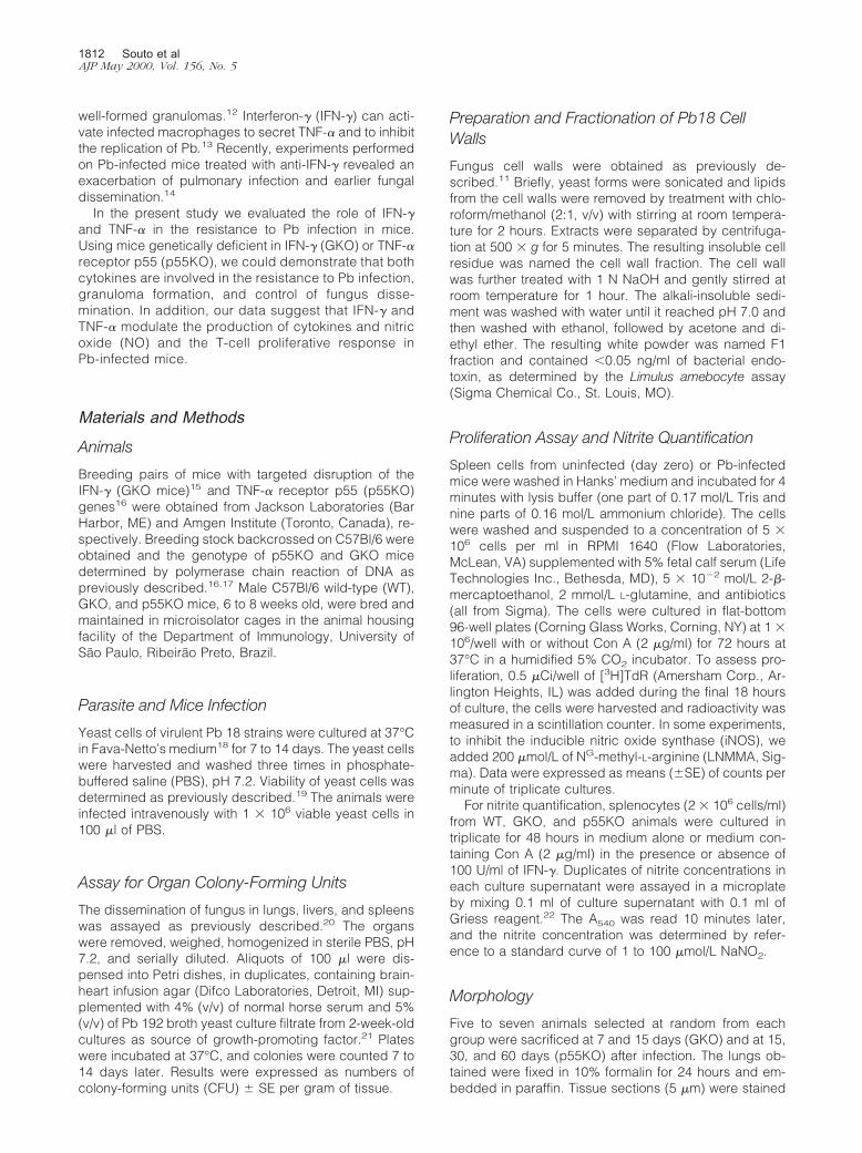
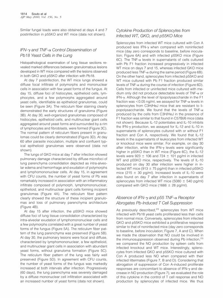
At day 7 postinfection, the WT mice lungs showed adiffuse focal infiltrate of polymorphs and mononuclearcells in association with few yeast forms of the fungus. Atday 15, diffuse foci of histiocytes, epithelioid cells, lym-phocytes, and a few polymorphs aggregated aroundyeast cells, identifiable as epithelioid granulomas, couldbe seen (Figure 3A). The reticulum fiber staining clearlydemonstrated the early structures of granuloma (Figure3B). At day 30, well-organized granulomas composed ofhistiocytes, epithelioid cells, and multinuclear giant cellsaggregated around yeast forms and surrounded by a rimof lymphocytes and fibroblasts, were formed (Figure 3C).The normal pattern of reticulum fibers present in granu-lomas could be clearly demonstrated (Figure 3D). At day60 after parasite inoculation, multiple and confluent typ-ical epithelioid granulomas were observed (data notshown).
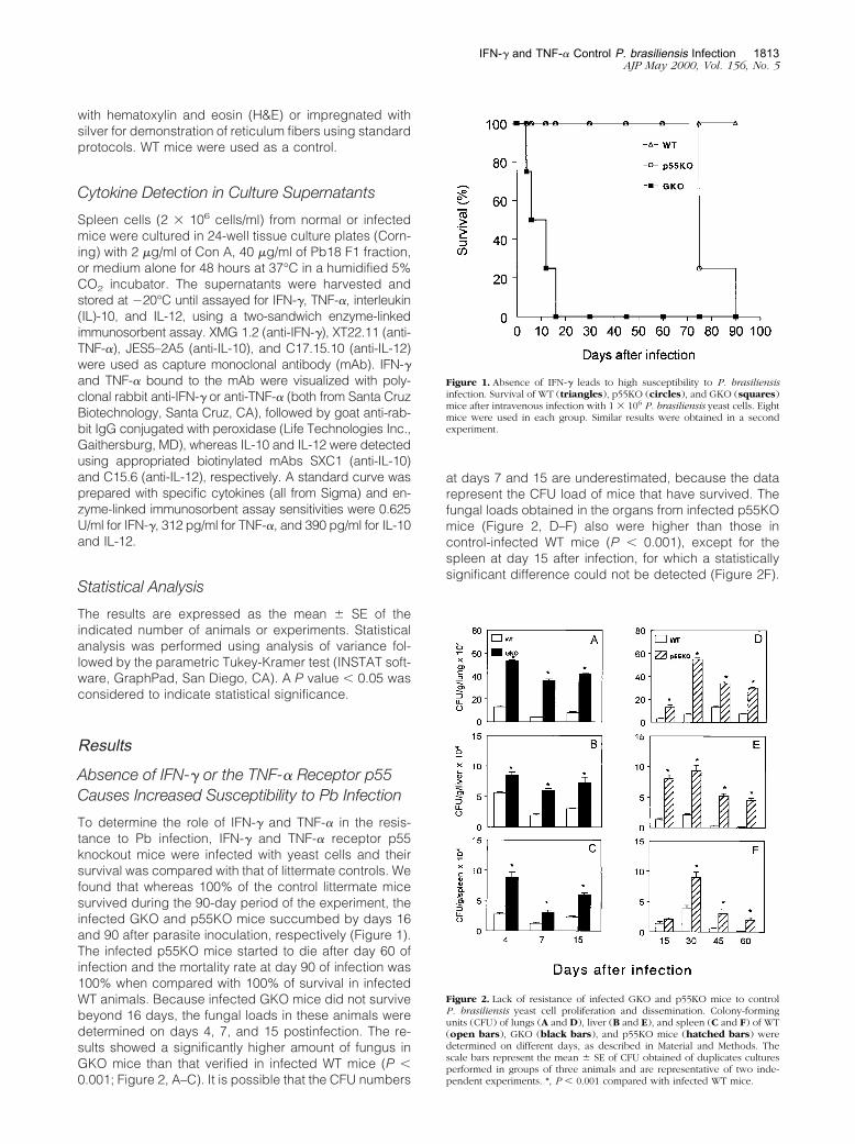
The lungs of GKO mice at day 7 after infection showedpulmonary damage characterized by diffuse microfoci oflung parenchyma consolidation depicted as intra-alveo-lar edema and hemorrhage and exudation of polymorphsand lymphomononuclear cells. At day 15, in agreementwith CFU counts, the number of yeast forms of Pb wasremarkably increased in association with an inflammatoryinfiltrate composed of polymorph, lymphomononuclear,epithelioid, and multinuclear giant cells forming incipientgranulomas (Figure 4A). The reticulum fiber patternclearly showed the structure of these incipient granulo-mas and loss of pulmonary parenchyma architecture(Figure 4B).
At day 15 after infection, the p55KO mice showeddiffuse foci of lung tissue consolidation characterized byintra-alveolar exudation of lymphomononuclear cells anda few polymorphs correlated with a great number of yeastforms of the fungus (Figure 5A). The reticulum fiber pat-tern of the lung parenchyma was preserved (Figure 5B).At day 30, the pulmonary lesions were focal and diffuse,characterized by lymphomononuclear, a few epithelioid,and multinuclear giant cells in association with abundantyeast forms, without granuloma formation (Figure 5C).The reticulum fiber pattern of the lung was fairly wellpreserved (Figure 5D). In agreement with CFU counts,the number of yeast forms of the fungus was markedlyincreased at both intervals after infection. Progressively(60 days), the lung parenchyma was severely damagedby a diffuse mononuclear cell infiltration associated withan increased number of yeast forms (data not shown).
Cytokine Production of Splenocytes fromInfected WT, GKO, and p55KO Mice
Splenocytes from infected WT mice cultured with Con Aproduced less IFN-� when compared with noninfectedmice (day zero corresponds to baseline, before inocula-tion; Figure 6A) and with infected p55KO mice (Figure6C). The TNF-� levels in supernatants of cells culturedwith Pb F1 fraction increased progressively in infectedWT mice on days 7 and 15, whereas infected GKO miceproduced less TNF-� during the same period (Figure 6B).On the other hand, splenocytes from infected p55KO andWT mice cultured with Pb F1 fraction produced similarlevels of TNF-� during the course of infection (Figure 6D).Cells from infected or uninfected mice cultured with me-dium only did not produce detectable levels of TNF-� orIFN-�. Although the level of lipopolysaccharide in the F1fraction was �0.05 ng/ml, we assayed for TNF-� levels insplenocytes from C3H/HeJ mice that are resistant to li-popolysaccharide. We found that the levels of TNF-�produced by the cells from C3H/HeJ in the presence ofF1 fraction was similar to that found in C57Bl/6 mice (datanot shown). Because IL-12 potentiates and IL-10 inhibitsthe IFN-� production, we assayed for IL-12 and IL-10 insupernatants of splenocytes cultured with or without F1fraction and Con A, respectively. We found that IL-12levels in the supernatants of leukocytes from infected WTor knockout mice were similar. For example, on day 30after infection, while the IFN-� levels were significantlyhigher in p55KO than in WT mice (Figure 6C), the IL-12levels were 923 � 106 and 724 � 101 pg/ml in infectedWT and p55KO mice, respectively. The levels of IL-10produced on day 30 after infection were higher in in-fected WT mice (913 � 75 pg/ml) than in infected p55KOmice (215 � 30 pg/ml). Increased levels of IL-10 werealso found on day 7 after infection in supernatants ofsplenocytes from infected WT mice (5080 � 540 pg/ml)compared with GKO mice (1666 � 28 pg/ml).
Absence of IFN-� and p55 TNF-� ReceptorAbrogates Pb-Induced T Cell Suppression
As previously described,23 splenocytes from WT miceinfected with Pb18 yeast cells proliferated less than cellsfrom normal mice. Conversely, splenocytes from infectedGKO and p55KO mice exhibited a proliferative responsesimilar to that of noninfected mice (day zero correspondsto baseline, before inoculation; Figure 7, A and C). Whenwe made the observation that NO could be involved inthe immunosuppression observed during Pb infection,23
we compared the NO production by spleen cells frominfected knockout and WT mice. Interestingly, spleno-cytes from infected GKO and p55KO mice cultured withCon A produced less NO when compared with theirinfected littermates (Figure 7, B and D). Considering thatabrogation of suppression of Con A-induced proliferateresponses are concomitant to absence of IFN-� and de-crease in NO production (Figure 7), we evaluated the roleof this cytokine in the control of T cell response and NOproduction by splenocytes of infected mice. We thus
1814 Souto et alAJP May 2000, Vol. 156, No. 5
Figure 3. Histopathology of lung sections from P. brasiliensis-infected WT mice. Lungs of mice at day 15 show diffuse foci of histiocytes, epithelioid cells(wide-open arrow), lymphocytes, and a few polymorphs aggregated around yeast cells (arrowheads) identifiable as epithelioid granulomas (A, arrows). Theearly structure of the granulomas are depicted by reticulum fiber silver impregnation (B, wide-open arrows). At day 30, well-organized granulomas withmultinuclear giant cells (wide-open arrow) are present (C, wide arrows) with a normal pattern of reticulum fibers (D, wide-open arrows). Lung sections werestained with H&E (A and C) and impregnated with silver for reticulum fibers (B and D). Magnification, �230.
IFN-� and TNF-� Control P. brasiliensis Infection 1815AJP May 2000, Vol. 156, No. 5

added IFN-� to splenocytes from normal or infected WTand GKO mice and evaluated the subsequent T-cell pro-liferative response and NO production. We found that theaddition of IFN-� to splenocytes from infected GKO micecultured with Con A restored NO production (Figure 8B)to the levels detected in infected WT mice (P � 0.001),and led to a significant inhibition of Con A-induced T cellproliferation (Figure 8A). The addition of IFN-� to cellcultures from noninfected GKO mice led only to a slightincrease in the nitrite synthesis (from 0.2 � 0.05 to 1.67 �0.49 �mol/L), that was not enough to modify the cellproliferation. To verify if NO was involved in the mecha-nism that mediates inhibition of T cell proliferative re-sponses, we added the iNOS inhibitor LNNMA to cellcultures. In the absence of Con A, the addition of LNMMAdid not modify the cell proliferation or nitrite production(data not shown). However, addition of LNMMA to spleencells from infected WT mice cultured with Con A abro-gated the NO production and, therefore, reversed thelevel of T cell proliferation to that found in cells frominfected GKO, as well as uninfected WT and GKO mice.On the contrary, the addition of LNMMA to cells fromnormal or infected GKO mice did not result in alteration incell proliferation, thus confirming the role of NO in sup-pression (Figure 8). Therefore, decreased NO productionleads to increased T-cell proliferative response.
DiscussionDisseminated PCM in GKO and p55KO mice as well asthe kinetics of lesion development in lungs after Pb infec-tion were described. The pattern of cytokine response inthe absence of IFN-� and TNF-� receptor p55 in infectedmice was also evaluated. Our results suggest that IFN-�and TNF-� are essential for the resistance and survival ofPb-infected mice. Although the role of IFN-� has beenextensively studied in fungal infections such as Candidaalbicans18,24,25 and Histoplasma capsulatum,26,27 the roleof this cytokine in mediating resistance or susceptibility toPb infection is poorly understood. In vitro, IFN-� plays apivotal role against several pathogens,28–30 including Pbyeast cells,13 through macrophage activation. Treatmentof infected mice with very high amounts of anti-IFN-�resulted in increased fungal burden, although the ani-mals did not die until 8 weeks after infection.14 In thepresent study the infection of GKO mice resulted in 100%mortality after only 16 days of infection as a consequenceof early fungal dissemination to liver, spleen, and lungs,whereas WT mice were resistant to infection. Histologicalexamination of lungs from infected GKO mice confirmedthe yeast cells were widespread throughout the majororgans. These data suggest that mice lacking a func-tional IFN-� gene were unable to contain and control avirulent Pb infection, besides the intense inflammatory
Figure 4. Histopathology of lung sections from P. brasiliensis-infected GKO mice. Lungs of mice at 15 days after infection show marked mononuclear cell infiltratewith incipient granuloma formation and a high number of yeast cells (A, arrows). The reticulum fiber staining shows the incipient pattern of granuloma (B, widearrows). Lung sections were stained with H&E (A) and impregnated with silver for reticulum fibers (B). Magnification, �230.
1816 Souto et alAJP May 2000, Vol. 156, No. 5

Figure 5. Histopathology of lung sections from P. brasiliensis-infected p55KO mice. Lungs of mice at day 15 after infection show foci of tissue consolidationcharacterized by intra-alveolar exudation of mononuclear cells and a few polymorphs correlated with a great number of yeast forms of the fungus (A, arrows).The reticulum fiber pattern of the lung parenchyma is preserved (B, arrows). At day 30, the pulmonary lesions are focal and diffuse characterized by mononuclearcells and a few epithelioid cells and multinuclear giant cells (wide arrow) in association with abundant yeast forms without granuloma formation (C). Thereticulum fiber pattern of the lung is fairly well preserved (D). Lung sections were stained with H&E (A and C) and impregnated with silver for reticulum fibers(B and D). Magnification, �230.
IFN-� and TNF-� Control P. brasiliensis Infection 1817AJP May 2000, Vol. 156, No. 5

reaction and formation of incipient granulomas associ-ated with a great number of yeast cells. Moreover, ourdata could also show that TNF-�, acting through the p55receptor, is also important to control parasite dissemina-tion, parasite growth, and the inflammatory response.Infection of p55KO mice, whose only abnormality is a lack
of p55 receptor,16 resulted in increased fungal burdenand absence of granuloma formation. In fact, the granu-lomatous inflammatory reaction, a specialized and effi-cient tissue response against certain parasites,31 re-quires TNF-�, which is supposed to be responsible forattracting and activating effector cells, as well as formacrophage accumulation and differentiation into epithe-lioid cells.32 Our results agree with recent studies show-ing that TNF-� activities mediated by the p55 receptorare important in granuloma formation in response to bac-teria16,33 and protozoa.34 Accordingly, we could not de-tect typical granuloma formation in either GKO or p55KO,suggesting that both TNF-�, signaling via the p55 recep-tor, and IFN-� are involved in the mediation of granulomaformation in response to Pb. The lesions observed in thelungs of infected p55KO mice showed a small number ofapparently typical epithelioid cells, in accordance with
Figure 6. TNF-� and IFN-� production by spleen cells from infected WT(open bars), GKO (black bars), and p55KO (hatched bars) mice. Spleno-cytes were harvested from GKO (0, 7, and 15 days after infection) and p55KOmice (0, 15, 30, and 60 days after infection), cultured for 48 hours with ConA (2 �g/ml, A and C) and with the F1 fraction (40 �g/ml, B and D) and thesupernatants assayed for IFN-� (A and C) and TNF-� (B and D). The amountof IFN-� and TNF-� found in splenocytes cultured without stimulus wasbelow the limit of detection of our enzyme-linked immunosorbent assay.Each scale bar represents the mean � SE of three mice per group in anexperiment representative of two separate experiments. *, P � 0.001 com-pared with infected WT mice.
Figure 7. Absence of the IFN-� and TNF-� receptor p55 results in lower NOproduction and normal T-cell proliferative response of Pb-infected mice.Spleen cells from noninfected (day zero) or infected WT (open bars), GKO(black bars), and p55KO (hatched bars) mice were cultivated with Con A(2 �g/ml) and T cell proliferation was evaluated (A and C). Nitrite (B and D)was assayed in supernatants of splenocytes (2 � 106 cells/ml) cultured withCon A for 48 hours. Each scale bar represents the mean � SE of three miceper group in an experiment representative of two separate experiments.*, P � 0.001 compared with infected WT mice.
Figure 8. Treatments that restore or abrogate NO production by splenocytesfrom Pb-infected mice inhibit and reverse T cell proliferation, respectively.Spleen cells from noninfected (NI) WT (striped bars) and GKO (hatchedbars) mice or WT (open bars) and GKO (black bars) mice at 15 days afterinfection (I) were cultured with Con A (2 �g/ml) with or without IFN-� (100U/ml) or LNMMA (200 �mol/L) and the T-cell proliferative response (A) andNO production (B) were evaluated (for details see Materials and Methods).Splenocytes cultured with medium only were used as a control. Each scalebar represents the mean � SE of three mice per group in an experimentrepresentative of two separate experiments. *, P � 0.001 compared withinfected WT mice. **, P� 0.01 compared with GKO infected mice in absenceof IFN-�.
1818 Souto et alAJP May 2000, Vol. 156, No. 5

observations in p55KO mice inoculated with liveCalmette-Guerin bacillus.35
The survival of infected p55KO mice was longer incomparison with infected GKO mice probably becauseTNF-�, acting through the p75 receptor, produces higheramounts of NO than infected GKO mice (Figure 7), and,therefore, are more resistant to infection (Figure 1), thusreinforcing the hypothesis that NO controls Pb infec-tion.23 Similarly, it has been shown that the TNF-� p55receptor-independent pathway involved TNF-� p75 re-ceptor signaling, because neutralization of TNF-� abro-gated the ability of p55KO macrophages to producesufficient NO to kill Leishmania major.36 Moreover, theelimination of L. major was delayed when compared withcontrol animals, suggesting that this pathway may beless efficient than signaling via the p55 receptor.36 Also,it was observed that mice genetically deficient in TNF-�p55 and p75 receptors succumbed significantly earlier tothe infection with Toxoplasma gondii as compared withTNF-� p55KO mice.34 Taken together, these observa-tions agree with our hypothesis that in the absence of theTNF-� p55 receptor, part of the function of this receptor istaken over by the TNF-� p75 receptor.
It is well known that IFN-� promotes TNF-� synthesisby murine macrophages37 and that IFN-� and TNF-� actsynergistically to induce antimicrobial activity in manyinfectious diseases.38,39 In our model, the greatly re-duced capacity of Pb-infected GKO mice to produceTNF-� may be an important factor in their inability to resistPb infection, because this cytokine is important in gran-uloma formation.12,34 The lungs of GKO mice showedformation of incipient granulomas probably related topartial production of TNF-�; whereas infected p55KOmice produced high levels of IFN-�, which may help tocontrol parasite growth and dissemination even in theabsence of the TNF p55 receptor, resulting in higherresistance to infection than that observed in GKO mice.The low IFN-� production by infected WT mice was notdue to decreased IL-12 synthesis, because it did notchange during the whole infection course. Also, low lev-els of IL-10 (data not shown) and high levels of IFN-�,known to inhibit and to activate the iNOS, respectively,29
may contribute to induce NO synthesis and to controlfungus replication.
We have previously shown that mice infected with theprotozoan Trypanosoma cruzi produce high levels ofIFN-� and TNF-�, which lead to activation of iNOS andNO production.40,41 Despite its importance as a microbi-cidal agent, NO has been shown to be involved in theestablishment and maintenance of lymphocyte unre-sponsiveness in mice infected with several parasites,30,40
including fungi.28 Similarly, in Pb infection associatedwith a classical granuloma reaction there is a markedsuppression of cell proliferative response to the mitogenCon A (Figure 7) and IFN-�, TNF-� (Figure 6), and NOproduction (Figure 7). In the absence of the IFN-� andTNF-� receptor p55, however, the proliferative responseto Con A was similar to that verified in cells from normalmice. Interestingly, production of high levels of NO sup-pressed the proliferative response to Con A. This obser-vation was reinforced by the addition of IFN-� to spleno-
cytes from infected GKO mice, which resulted indecreased proliferative response and increased NO pro-duction (Figure 8). Moreover, inhibition of the iNOS by theaddition of LNMMA to the cultures of splenocytes frominfected WT mice abrogated NO production and revertedthe proliferative response to values similar to those foundin cells from infected GKO and p55KO mice (Figure 8).Indeed, NO has been associated with a decreased pro-liferative response to Con A42,43 and a recent reportdemonstrated that treatment of Pb-infected mice with aspecific inhibitor of NO synthesis prevented failure of theproliferative capacity of spleen cells in response to Con Aand Pb antigen.23 The low NO production in GKO andp55KO mice may also contribute to the observed de-crease in granuloma formation, because inhibition of NOmetabolism caused a loss of granulomatous architecturein lesions induced by Calmette-Guerin bacillus.44 Takentogether, these data suggest that NO may modulate theimmune response to Pb antigens in infected mice andmay contribute to the organization of the granulomatouslesion.
In summary, a congenital deficiency in IFN-� or theTNF-� receptor p55 results in a pronounced loss of pro-tective immunity in mice infected with Pb. In associationwith this impairment of immunity, generation of NO iscompromised, suggesting that IFN-� and TNF-� partici-pate in the generation of this mediator that acts as animmunosuppressor in infections caused by Pb18 andalso in controlling dissemination of the fungus. In accor-dance with our results, patients with PCM have beendemonstrated to produce lower levels of IFN-� andTNF-�, suggesting that these cytokines are important tocontrol the development of the disease.45 The knowledgeof the regulatory mechanisms that lead to the control ofPb infection in resistant individuals may contribute to afuture immunotherapy for PCM that affects thousands ofpeople in South and Central America.
References
1. McEwen J, Bedoya V, Patino MM, Salazar ME, Restrepo AM: Exper-imental murine paracoccidioidomycosis induced by inhalation ofconidia. J Med Vet Mycol 1987, 25:165–167
2. Restrepo A: The ecology of Paracoccidioides brasiliensis, a puzzle stillunsolved. Sabouraudia 1985, 23:323–334
3. Franco M, Montenegro MR, Mendes RP, Marques AS, Dillon NL, MotaNS: Paracoccidioidomycosis: a recently proposed classification of itsclinical forms. Rev Soc Bras Med Trop 1987, 20:129–132
4. Franco M: Host-parasite relationships in paracoccidioidomycosis.J Med Vet Mycol 1986, 25:5–18
5. Miyaji M, Nishimura K: Granuloma formation and killing functions ofgranuloma in congenitally athymic nude mice infected with Blastomy-ces dermatitides and Paracoccidioides brasiliensis: Mycopathologia1983, 82:129–141
6. Burger E, Vaz CCA, Sano A, Calich VLG, Singer-Vermes LM, XidiehCF, Kashino SS, Nishimura K, Miyaji M: Paracoccidioides brasiliensisinfection in nude mice: studies with isolates differing in virulence anddefinition of their T cell-dependent and T cell-independent compo-nents. Am J Trop Med Hyg 1996, 55:391–398
7. Restrepo A, Restrepo M, Restrepo F, Aristizabal LH, Moncada LH,Velez H: Immune response in paracoccidioidomycosis. A controlledstudy of 16 patients before and after treatment. Sabouraudia 1978,16:151–163
8. Benard G, Hong MA, Del-Negro GMB, Batista L, Shikanai-Yasuda
IFN-� and TNF-� Control P. brasiliensis Infection 1819AJP May 2000, Vol. 156, No. 5

MA, Duarte AJS: Antigen-specific immunosuppression in paracoccid-ioidomycosis. Am J Trop Med Hyg 1996, 54:7–12
9. Benard G, Mendes-Giannini MJS, Juvenale M, Miranda ET, DuarteAJ: immunosuppression in paracoccidioidomycosis: T cell hypore-sponsiveness to two Paracoccidioides brasiliensis glycoproteins thatelicit strong humoral immune response. J Infect Dis 1997, 175:1263–1267
10. Silva M, Bocca AL, Ferracini R Jr, Figueiredo F, Silva CL: Cellularrequirements for immunomodulatory effects caused by cell wall com-ponents of Paracoccidioides brasiliensis on antibody production. ClinExp Immunol 1997, 109:261–271
11. Figueiredo F, Alves LMC, Silva CL: Tumor necrosis factor productionin vivo and in vitro in response to Paracoccidioides brasiliensis and thecell wall fractions thereof. Clin Exp Immunol 1993, 93:189–194
12. Kindler V, Sappino AP, Grau GE, Piguet PF, Vassali P: The inducingrole of tumor necrosis factor in the development of bactericidal gran-ulomas during BCG infection. Cell 1989, 56:731–740
13. Brummer E, Hanson LH, Restrepo A, Stevens DA: In vivo and in vitroactivation of pulmonary macrophage by IFN-� for enhanced killing ofParacoccidioides brasiliensis or Blastomyces dermatidis. J Immunol1988, 140:2786–2789
14. Cano LE, Kashino SS, Arruda C, Andre D, Xidieh CF, Singer-VermesL, Vaz CAC, Burger E, Calich VLG: Protective role of gamma inter-feron in experimental pulmonary paracoccidioidomycosis. Infect Im-mun 1998, 66:800–806
15. Dalton DK, Pitts-Meek S, Keshav S, Figari IS, Bradley A, Stewart TA:Multiple defects of immune cell function in mice with disrupted inter-feron-gamma genes. Science 1993, 259:1739–1742
16. Pfeffer K, Matsuyama T, Kundig TM, Wakeham A, Kishihara K, Sha-hinian A, Wiegmann K, Ohashi PS, Kronke M, Mak TW: Mice deficientfor the 55 kd tumor necrosis factor receptor are resistant to endotoxicshock, yet succumb to L. monocytogenes infection. Cell 1993, 73:457–467
17. Qian Q, Cutler JE: Gamma interferon is not essential in host defenseagainst disseminated candidiasis in mice. Infect Immun 1997, 65:1748–1753
18. Fava-Neto C, Vegas VS, Sciannamea IM, Guarnieri DB: Antıgenopolissacarıdico do Paracoccidioies brasiliensis. Estudo do tempo decultivo do P. brasiliensis necessario ao preparo do antigeno. Rev InstMed Top Sao Paulo 1969, 11:177–181
19. Calich VLG, Purchio A, Paula CRA: A new fluorescent viability test forfungi cell. Mycophatologia 1979, 66:175–177
20. Cano LE, Singer-Vermes LM, Vaz CAC, Russo M, Calich VLG: Pul-monary paracoccidioidomycosis in resistant and susceptible mice:relationship among progression of infection, bronchoalveolar cell ac-tivation, cellular immune response, and specific isotype patterns.Infect Immun 1995, 63:1777–1783
21. Singer-Vermes LM, Ciavaglia MC, Kashino SS, Burger E, Calich VLG:The source of the growth-promoting factor(s) affects the plating effi-ciency of Paracoccidioides brasiliensis. J Med Mycol 1992, 30:261–264
22. Green LC, Tannenbaum SR, Goldman P: Nitrate biosynthesis ingermfree and conventional rat. Science 1981, 212:56–68
23. Bocca AL, Hayashi EE, Pinheiro AG, Furlanetto AB, Campanelli AP,Cunha FQ, Figueiredo F: Treatment of Paracoccidioides brasiliensis-infected mice with a nitric oxide inhibitor prevents the failure ofcell-mediated immune response. J Immunol 1998, 161:3056–3063
24. Kaposzta R, Tree P, Marodi L, Gordon S: Characteristics of invasivecandidiasis in gamma interferon- and interleukin-4-deficient mice:role of macrophages in host defense against Candida albicans. InfectImmun 1998, 66:1708–1717
25. Balish E, Wagner RD, Vazquez-Torres A, Pierson C, Warner T: Can-didiasis in interferon-gamma knockout (IFN-gamma�/�) mice. J In-fect Dis 1998, 178:478–487
26. Cain JA, Deepe GS Jr: Evolution of the primary immune response toHistoplasma capsulatum in murine lung. Infect Immun 1998, 66:1473–1481
27. Allendoerfer R, Deepe GS Jr: Intrapulmonary response to His-toplasma capsulatum in gamma interferon knockout mice. Infect Im-mun 1997, 65:2564–2569
28. Nakamura LT, Wu-Hsieh BA, Howard DH: Recombinant murinegamma interferon stimulates macrophages of the RAW cell line toinhibit intracellular growth of Histoplasma capsulatum. Infect Immun1994, 62:680–684
29. Gazzinelli RT, Oswald IP, Hieny S, James SL, Sher A: The microbici-dal activity of interferon-gamma-treated macrophages againstTrypanosoma cruzi involves an L-arginine-dependent, nitrogen oxide-mediated mechanism inhibited by interleukin-10 and transforminggrowth factor-beta. Eur J Immunol 1992, 22:2501–2506
30. Denis M: Interferon-gamma-treated murine macrophages inhibitgrowth of tubercle bacilli via the generation of reactive nitrogenintermediates. Cell Immunol 1991, 132:150–157
31. Adams DO: The granulomatous inflammatory response. Am J Pathol1976, 84:164–191
32. Tracey KJ, Cerami A: Tumor necrosis factor, other cytokines anddisease. Annu Rev Cell Biol 1993, 9:317–343
33. Tsuji H, Harada A, Mukaida N, Nakanuma Y, Bluethmann H, KanekoS, Yamakawa K, Nakamura SI, Kobayashi KI, Matsushima K: Tumornecrosis factor receptor p55 is essential for intrahepatic granulomaformation and hepatocellular apoptosis in a murine model of bacte-rium-induced fulminant hepatitis. Infect Immun 1997, 65:1892–1898
34. Deckert-Schluter M, Bluethmann H, Rang A, Hof H, Schluter D: Cru-cial role of TNF receptor type 1 (p55), but not of TNF receptor type 2(p75), in murine toxoplasmosis. J Immunol 1998, 160:3427–3436
35. Senaldi G, Yin S, Shaklee CL, Piguet PF, Mak TW, Ulich TR: Coryne-bacterium parvum- and Mycobacterium bovis bacillus Calmette-Guerin-induced granuloma formation is inhibited in TNF receptor I(TNF-RI) knockout mice and by treatment with soluble TNF-RI. J Im-munol 1996, 157:5022–5026
36. Vieira LQ, Goldschmidt M, Nashleanas M, Pfeffer K, Mak T, Scott P:Mice lacking the TNF receptor p55 fail to resolve lesions caused byinfection with Leishmania major, but control parasite replication. J Im-munol 1996, 157:827–835
37. Beutler B, Tkacenko V, Milsark I, Krochin N, Cerami A: Effect ofgamma interferon on cachectin expression by mononuclear phago-cytes. Reversal of the lpsd (endotoxin resistance) phenotype. J ExpMed 1986, 164:1791–1796
38. Corradin SB, Buchmuller-Rouiller Y, Mauel J: Phagocytosis enhancesmurine macrophage activation by interferon-gamma and tumor ne-crosis factor-alpha. Eur J Immunol 1991, 21:2553–2558
39. Leenen PJ, Canono BP, Drevets DA, Voerman JS, Campbell PA:TNF-alpha and IFN-gamma stimulate a macrophage precursor cellline to kill Listeria monocytogenes in a nitric oxide-independent man-ner. J Immunol 1994, 153:5141–5147
40. Silva JS, Vespa GN, Cardoso MA, Aliberti JC, Cunha FQ: Tumornecrosis factor alpha mediates resistance to Trypanosoma cruzi in-fection in mice by inducing nitric oxide production in infected gammainterferon-activated macrophages. Infect Immun 1995, 63:4862–4867
41. Cardillo F, Voltarelli JC, Reed SG, Silva JS: Regulation of Trypano-soma cruzi infection in mice by gamma interferon and interleukin 10:role of NK cells. Infect Immun 1996, 64:128–134
42. Eisenstein TK, Huang D, Meissler JJ Jr, Al-Ramadi B: Macrophagesnitric oxide mediates immunosuppression in infectious inflammation.Immunobiology 1994, 191:493–502
43. Strengerg J, McGuigan F: Nitric oxide mediates suppression of T cellresponse in murine Trypanosoma brucei infection. Eur J Immunol1992, 22:2741–2744
44. Kreuger MR, Tames DR, Mariano M: Expression of NO-synthase incells of foreign-body and BCG-induced granulomata in mice: influ-ence of L-NAME on the evolution of the lesion. Immunology 1998,95:278–282
45. Bava AJ, Mistchenko AS, Palacios MF, Estevez ME, Tiraboschi NI,Sen L, Negroni R, Diez RA: Lymphocyte subpopulations and cytokineproduction in paracoccidioidomycosis patients. Microbiol Immunol1991, 35:167–174
1820 Souto et alAJP May 2000, Vol. 156, No. 5
Related Documents