CHAPTER 3 Interfacial positioning and stability of transmembrane peptides in lipid bilayers studied by combining hydrogen/deuterium exchange and mass spectrometry Jeroen A.A. Demmers 1,2 , Esther van Duijn 2 , Johan Haverkamp 2 , Denise V. Greathouse 3 , Roger E. Koeppe II 3 , Albert J.R. Heck 2 & J. Antoinette Killian 1 1 Department of Biochemistry of Membranes, Center for Biomembranes and Lipid Enzymology, Institute of Biomembranes, Utrecht University, Padualaan 8, 3584 CH Utrecht, The Netherlands 2 Department of Biomolecular Mass Spectrometry, Bijvoet Center for Biomolecular Research and Utrecht Institute for Pharmaceutical Sciences, Utrecht University, Sorbonnelaan 16, 3584 CA Utrecht, The Netherlands 3 Department of Chemistry and Biochemistry, University of Arkansas, Fayetteville, Arkansas 72701, USA Based on: J. Biol. Chem. 276 (37), 34501-34508 (2001)

Welcome message from author
This document is posted to help you gain knowledge. Please leave a comment to let me know what you think about it! Share it to your friends and learn new things together.
Transcript
CHAPTER 3
Interfacial positioning and stability of
transmembrane peptides in lipid bilayers studied by
combining hydrogen/deuterium exchange and
mass spectrometry
Jeroen A.A. Demmers1,2, Esther van Duijn2, Johan Haverkamp2, Denise V. Greathouse3,Roger E. Koeppe II3, Albert J.R. Heck2 & J. Antoinette Killian1
1 Department of Biochemistry of Membranes, Center for Biomembranes and Lipid Enzymology, Institute ofBiomembranes, Utrecht University, Padualaan 8, 3584 CH Utrecht, The Netherlands
2 Department of Biomolecular Mass Spectrometry, Bijvoet Center for Biomolecular Research and Utrecht Institutefor Pharmaceutical Sciences, Utrecht University, Sorbonnelaan 16, 3584 CA Utrecht, The Netherlands
3 Department of Chemistry and Biochemistry, University of Arkansas, Fayetteville, Arkansas 72701, USA
Based on: J. Biol. Chem. 276 (37), 34501-34508 (2001)

Chapter 3
58

Positioning and stability of membrane peptides
59
Abstract
Nano-electrospray ionization mass spectrometry (ESI-MS) was used to analyze
hydrogen/deuterium (H/D) exchange properties of transmembrane peptides with varying
length and composition. Synthetic transmembrane peptides were used with a general acetyl-
GW2(LA)nLW2A-ethanolamine sequence. These peptides were incorporated in large unilamellar
vesicles of 1,2-dimyristoyl-sn-glycero-3-phosphocholine. The vesicles were diluted in buffered
deuterium oxide and the H/D exchange after different incubation times was directly analyzed by
means of ESI-MS. First, the influence of the length of the hydrophobic Leu-Ala sequence on
exchange behavior was investigated. It was shown that longer peptide analogs are more
protected from H/D exchange than expected on basis of their length with respect to bilayer
thickness. This is explained by an increased protection from the bilayer environment, because of
stretching of the lipid acyl chains and/or tilting of the longer peptides. Next, the role of the
flanking tryptophan residues was investigated. The length of the transmembrane part that shows
very slow H/D exchange was found to depend on the exact position of the tryptophans in the
peptide sequence, suggesting that tryptophan acts as a strong determinant for positioning of
proteins at the membrane/water interface. Finally, the influence of putative helix breakers was
studied. It was shown that the presence of Pro in the transmembrane segment results in much
higher exchange rates as compared to Gly or Leu, suggesting a destabilization of the �-helix.
Tandem MS measurements suggested that the increased exchange takes place over the entire
transmembrane segment. The results show that ESI-MS is a convenient technique to gain
detailed insight into properties of peptides in lipid bilayers by monitoring H/D exchange
kinetics.
Introduction
The precise manner in which membrane proteins are embedded in a lipid bilayer is essential fortheir structure and function. Important structural and dynamic features, such as stability of thetransmembrane segments or their precise positioning at the lipid/water interface, will bedetermined not only by intrinsic properties of the transmembrane segments, but also by theirinteraction with surrounding lipids. A convenient way to gain insight into how the specialcharacteristics of transmembrane segments and their interaction with lipids may influence thebehavior of membrane proteins, is by studying model systems of artificial transmembranepeptides with desired properties in well-defined lipid bilayers.
Recently, we have described a new method using nano-ESI-MS [1] to study properties oftransmembrane protein segments in model systems by analyzing the kinetics ofhydrogen/deuterium (H/D) exchange. The results showed that various populations of amidehydrogen atoms can be distinguished that are characteristic for different regions of thetransmembrane segments. These populations are fast exchanging amide hydrogens located in thepeptide termini that are exposed to the aqueous phase, intermediately exchanging hydrogens ofthe residues located in the bilayer/water interface and slowly exchanging hydrogens located in

Chapter 3
60
the hydrophobic core of the lipid bilayer. The results suggested that measurement of exchangeproperties of peptides by ESI-MS is a convenient method to investigate factors that determineinterfacial positioning and/or stability of transmembrane protein segments. In the present study,we have investigated the influence of several of such factors. Hereby, special emphasis is given tothe length of the �-helical hydrophobic core with respect to the bilayer thickness, the role of
potential anchoring residues at the lipid/water interface, and the influence of �-helix breakingresidues in the transmembrane segment.
As models for protein transmembrane segments, we have used WALP peptides thatalready have been used successfully to investigate various aspects of peptide/lipid interactions [2-4]. These peptides have a hydrophobic core of alternating Leu and Ala, which is flanked on bothsides by Trp residues (see Table I), and they have been shown to form �-helical transmembranehelices [4]. Since in membrane proteins Trp residues are highly enriched near themembrane/water interface [5-8], they are thought to resemble a consensus sequence fortransmembrane �-helical segments of intrinsic membrane proteins.
The extent to which the hydrophobic length of transmembrane segments matches thehydrophobic bilayer thickness can significantly influence membrane protein structure andfunction [9]. To investigate whether this is related to changes in membrane protein interfacialpositioning and/or stability, we first analyzed the effects of increasing the hydrophobic length ofWALP peptides on the H/D exchange kinetics of peptides with different lengths of the Leu-Alacore in bilayers of DMPC. This lipid is chosen because it forms well-defined bilayers with allpeptides under the used experimental conditions [2,4,10] and has a hydrophobic thickness thatapproximately matches the hydrophobic length of the shortest peptide used [3].
Next, we investigated the importance of Trp as interfacial anchoring residues. It has beensuggested that Trp residues prefer to be positioned at a well-defined site in the lipid headgroups[3,11-15] and that thereby they can act as membrane anchors. Their abundance at the lipid/waterinterface in several membrane proteins [5-7] is therefore likely to be functionally important, e.g.for stabilization of transmembrane helices or precise positioning of such helices at the interface.The present mass spectrometric method offers the opportunity to investigate these effects byanalyzing the H/D exchange kinetics of peptides of identical total length in which the positionof Trp residues along the sequence is varied.
Finally, besides the length of the transmembrane helices and their interfacial anchoringbehavior, the stability of the transmembrane segment is also important for membrane proteinstructure and function. In water-soluble proteins, the stability of the backbone of regular �-
helices has been found to undergo large changes when potent breakers of both �-helical and �-sheet structures, like Pro and Gly, are inserted (e.g. see [16]). However, little is known about theeffect on stability of these residues in transmembrane segments. Therefore, H/D exchangekinetics in transmembrane segments containing Pro and Gly residues are compared to those ofpeptides without these residues.

Positioning and stability of membrane peptides
61
The results of this study show that peptides that are long with respect to thehydrophobic thickness of the bilayer are protected from H/D exchange to a relatively largeextent, which is explained in terms of induced adaptation (thickening) of the bilayer and/or atilting of the peptides. Furthermore, the positions of the Trp residues in the transmembranesequence are shown to be a critical factor for H/D exchange kinetics. These results suggest thatTrp side chains interact strongly with the membrane/water interface at specific sites. Finally, it isshown that peptides containing Pro, but not Gly, show a markedly different H/D exchangepattern than peptides lacking this residue. This suggests a significant effect of Pro on the stabilityof the transmembrane �-helix. The results are discussed in relation to existing literature data onrelated peptide/lipid interactions.
Materials and methods
Chemicals - Trifluoroacetic acid (TFA) was obtained from Merck (Darmstadt, Germany), 2,2,2-trifluoroethanol (TFE) from Sigma (St. Louis, MO). Deuterium oxide (>99.9 % D) was obtainedfrom Aldrich Chemical Company, Inc. (Milwaukee, WI, USA). D2O was stored under nitrogen at4�C. Sodium iodide was from OPG Farma Company (Utrecht, The Netherlands). Ammoniumacetate was from Fluka (Switzerland). The phospholipid DMPC was obtained from Avanti PolarLipids Inc. (Birmingham, AL). The peptides WALP16, WALP19, WALP21, WALP23,WALP23inner, WALP23outer, WALP23Pro and WALP23Gly were synthesized as described byKillian et al. [4], as modified by Greathouse et al. [17]. The W�ALP21 peptide was synthesized asdescribed by De Planque et al. [3] for related peptides. The peptides were tested for purity bynano-ESI-MS and found to be pure. Proteoliposome preparation - Peptide incorporation into phospholipid vesicles and massspectrometry measurements were performed essentially as described previously [1]. Shortly,peptides were dissolved in a small volume of TFA (10 �l per mg of peptide) and dried under anitrogen stream. To remove residual TFA, the peptides were subsequently dissolved in TFE (1mg/ml) followed by evaporation of the solvent in a rotavapor. Peptides were then againdissolved in TFE to a final concentration of 1 mg/ml. Dry mixed films of peptide and DMPC(peptide to lipid ratio1:25) were prepared as follows. Peptide solutions in TFE (1 ml; 0.46 mM)were added to DMPC solutions in methanol (1 ml; 12 mM) and vigorously vortexed. The solventwas removed by evaporation in a rotavapor. The mixed films were then dried for 24 hrs undervacuum. The films were hydrated at about 40ºC, well above the gel to liquid crystalline phasetransition temperature of the phospholipid (24�C [18]) in 0.5 ml 10 mM ammonium acetatebuffer (pH 7.5). Large unilamellar vesicles (LUVETs) were prepared by extrusion through a 400nm filter at room temperature and kept at 4�C until use.
Before the start of H/D exchange, LUVETs were preincubated at 30�C for at least 30min. For accurate comparison of the exchange data, LUVET suspensions with different peptidecomposition were mixed prior to the start of the exchange. LUVET suspensions were then 50

Chapter 3
62
times diluted in deuterated ammonium acetate buffer at 30�C (10 mM, pH 7.5), containing �1
mM NaI. At selected time points, 2 �l of this diluted suspension was transferred into a gold-coated glass capillary and the measurement was started as quickly as possible, whereby thepeptides were analyzed simultaneously. The dead-time between dilution and measurement was ata minimum 1 min. MS measurements - MS measurements were performed on an ESI quadrupole time-of-flightinstrument (Q-Tof; Micromass Ltd., Manchester, UK) or a an ESI time-of-flight instrument(LCT; Micromass Ltd., Manchester, UK), both operating in positive ion mode and equippedwith a Z-spray nano-ESI source. Nano-ESI needles with a relatively large tip opening (severaltens of �m) were prepared from borosilicate glass capillaries (Kwik-Fil�, World PrecisionInstruments Inc., Sarasota, Florida) on a P-97 puller (Sutter Instrument Co., Novato, CA). Theneedles were coated with a thin gold layer (�500 Å) using an Edwards Scancoat sputter-coater
501 (at 40 mV, 1 kV, for 200 sec). The nano-ESI capillary was positioned �5 mm before theorifice of the mass spectrometer. For MS experiments, the quadrupole was set in the RF-onlymode to act as an ion guide to efficiently link the ESI ion source with the reflectron time-of-flight analyzer. The potential between the nano-ESI capillary and the orifice of the massspectrometer was typically set to 1800 V, the cone voltage was 140 V. The nanospray needle wasconstantly kept at �30�C. In CID MS (MS/MS) mode on the Q-Tof instrument, the quadrupolewas used to select precursor ions, which were fragmented in the hexapole collision cell,generating product ions that were subsequently mass analyzed by the orthogonal time-of-flightmass analyzer. For CID MS measurements on the Q-Tof instrument, the collision energy was setto 150 V. Argon was used as collision gas. The quadrupole mass resolution parameters were setto a relatively large mass window to select the entire isotope envelope of the precursor ions. Thereflectron time-of-flight parameters were set such that the fragment ions were detected at morethan unit mass resolution, as required to obtain isotopically resolved H/D profiles. Increases indeuterium content (in Da) were calculated by using the average mass-to-charge (m/z) values ofthe isotope clusters of the undeuterated peptide and the (partly) deuterated peptides.
Results
To study effects of the hydrophobic length of transmembrane peptides on H/D exchangeproperties, a series of WALP/DMPC systems with varying peptide lengths (see Table I) wereprepared for nano-ESI-MS measurements.
To allow accurate detection of small differences in exchange properties of differentpeptides, all experiments were performed with mixtures of two or more peptide-lipid systemssynchronically. Therefore, LUVET suspensions with different peptide composition were mixedprior to the start of the exchange. Because such combination experiments are performed underidentical experimental conditions, the deuterium levels can be compared directly. Figure 1
Positioning and stability of membrane peptides
63
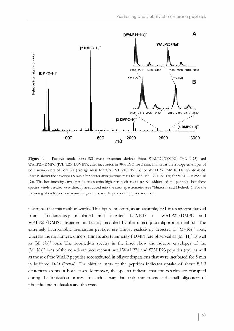
Figure 1 – Positive mode nano-ESI mass spectrum derived from WALP21/DMPC (P/L 1:25) andWALP23/DMPC (P/L 1:25) LUVETs, after incubation in 98% D2O for 5 min. In inset A the isotope envelopes ofboth non-deuterated peptides (average mass for WALP21: 2402.95 Da; for WALP23: 2586.18 Da) are depicted.Inset B shows the envelopes 5 min after deuteration (average mass for WALP21: 2411.59 Da; for WALP23: 2586.18Da). The low intensity envelopes 16 mass units higher in both insets are K+ adducts of the peptides. For thesespectra whole vesicles were directly introduced into the mass spectrometer (see “Materials and Methods”). For therecording of each spectrum (consisting of 30 scans) 10 pmoles of peptide was used.
illustrates that this method works. This figure presents, as an example, ESI mass spectra derivedfrom simultaneously incubated and injected LUVETs of WALP21/DMPC andWALP23/DMPC dispersed in buffer, recorded by the direct proteoliposome method. Theextremely hydrophobic membrane peptides are almost exclusively detected as [M+Na]+ ions,whereas the monomers, dimers, trimers and tetramers of DMPC are observed as [M+H]+ as wellas [M+Na]+ ions. The zoomed-in spectra in the inset show the isotope envelopes of the[M+Na]+ ions of the non-deuterated reconstituted WALP21 and WALP23 peptides (top), as wellas those of the WALP peptides reconstituted in bilayer dispersions that were incubated for 5 minin buffered D2O (bottom). The shift in mass of the peptides indicates uptake of about 8.5-9deuterium atoms in both cases. Moreover, the spectra indicate that the vesicles are disruptedduring the ionization process in such a way that only monomers and small oligomers ofphospholipid molecules are observed.

Chapter 3
64
Table I – Amino acid sequences of the peptides used and their number of exchangeable hydrogens.
Peptide Sequence Average No. of Mass labile(Da) H’s
WALP16 aAc-GWWLALALALALAWWA-Etnb 1897.31 22WALP19 Ac-GWWLALALALALALALWWA-Etn 2194.71 25WALP21 Ac-GWWLALALALALALALALWWA-Etn 2379.95 27WALP23 Ac-GWWLALALALALALALALALWWA-Etn 2563.18 29
W�ALP21d Ac-GW�W�LALALALALALALALW�W�A-Amc 2391.00 23
WALP23inner Ac-GAWLALALALALALALALALWAA-Etn 2332.91 27WALP23outer Ac-GWLALALALALALALALALALWA-Etn 2374.99 27
WALP23Pro Ac-GWWLALALALAPALALALALWWA-Etn 2547.14 28WALP23Gly Ac-GWWLALALALAGALALALALWWA-Etn 2507.07 29
aAc, acetylbEtn, ethanolaminecAm, amidedW´, Trp analog with N-methylindole side chain
Figure 2 – Time course for deuterium uptake of WALP16 (�), WALP19 (�), WALP21 (�) and WALP23 (�), allincorporated in DMPC bilayers, after dilution in deuterated buffer. Experiments were performed with twopeptides/lipid systems simultaneously. Data were normalized to the curve of WALP16.

Positioning and stability of membrane peptides
65
The effect of increasing the hydrophobic length of the peptides with respect to thehydrophobic thickness of the bilayer was tested using a series of WALP peptides with varyinghydrophobic core lengths (see Table I). Figure 2 shows the measured deuterium content as afunction of incubation time in deuterated buffer. All WALP peptides incorporated in DMPCshow a fast initial exchange. Within the experimental dead-time (�1.5 min) all peptides havetaken up about 7-9 deuterium atoms. After this fast exchange the deuterium content continues toincrease gradually with time, though at a much smaller exchange rate (defined as intermediateexchange rate, [1]). Figure 2 shows that all peptides have similar exchange kinetics curves in thefirst 20 min after incubation. All curves level off to a deuterium level, which is low as comparedto the total number of labile hydrogens present in each peptide, which varies from 22 to 29 (seeTable I). Furthermore, the deuterium content increases with increasing peptide length. However,the differences in deuterium levels are relatively small and represent only a fraction of thenumber of additional hydrogens that become available for exchange when the peptide length isincreased.
By fitting the kinetic curves using a non-linear squares fitting to multiexponentialfunctions in all peptides, three kinetic regions of exchange rates representing hydrogenpopulations were distinguished. In the short time range from 0 to 20 min, the fast andintermediate exchangeable hydrogens exchange, whereas the remaining hydrogens exchange onlyafter �20 min. The number of hydrogens in specific populations are shown in Table II. As wasshown previously [1], and will also be shown later in this work, the fast exchanging hydrogensare predominantly the hydrogens at the N- and C-termini of the peptides, sticking out of thebilayer, while the hydrogens with intermediate exchange rates are the ones close to the Trp in theinterfacial region. The remaining hydrogens, including the ones in the hydrophobic core haverate constants of (much) smaller than 0.10 min-1. These slow exchange rates are a result of boththe extremely low partition coefficient of D2O in a lipid bilayer [19] and the involvement of theamide protons in hydrogen bonding because of �-helix formation. The amount of fastexchanged hydrogens increases with the length of the peptides but does not increasequantitatively with the numbers of exchangeable hydrogens. For instance, only about one-thirdof the extra number of hydrogens of WALP23 compared to WALP16 exchanges rapidly and,therefore, the majority of the extra hydrogens of WALP23 are protected against exchange.
These results may be explained by several possible mechanisms. First, the number ofprotected hydrogens may be higher due to a larger core region of the �-helix in the longerpeptides. Second, the longer peptides may be more protected by the bilayer itself, as a result oftilting of the longer helices, or due to adaptation of the lipids to the peptide/lipid mismatch bystretching of the acyl chains [2]. These possibilities will be discussed in more detail below. Inprinciple, an alternative possible explanation for the protection of part of the peptides againstH/D exchange, would be the formation of peptide aggregates, which would also hamper accessof D2O molecules to the amide hydrogens. However, several observations argue against this.First, no restricted component was observed in electron spin resonance (ESR) measurements
Chapter 3
66
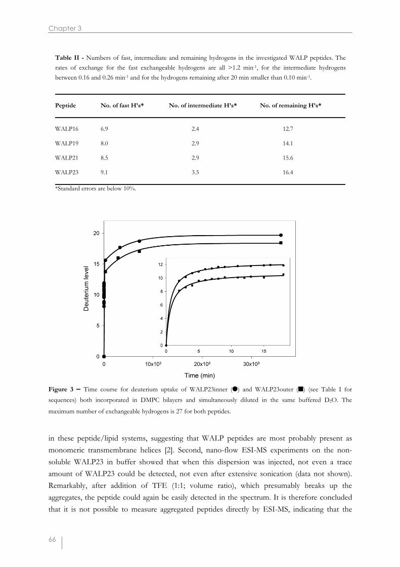
Figure 3 – Time course for deuterium uptake of WALP23inner (�) and WALP23outer (�) (see Table I forsequences) both incorporated in DMPC bilayers and simultaneously diluted in the same buffered D2O. The
maximum number of exchangeable hydrogens is 27 for both peptides.
in these peptide/lipid systems, suggesting that WALP peptides are most probably present asmonomeric transmembrane helices [2]. Second, nano-flow ESI-MS experiments on the non-soluble WALP23 in buffer showed that when this dispersion was injected, not even a traceamount of WALP23 could be detected, not even after extensive sonication (data not shown).Remarkably, after addition of TFE (1:1; volume ratio), which presumably breaks up theaggregates, the peptide could again be easily detected in the spectrum. It is therefore concludedthat it is not possible to measure aggregated peptides directly by ESI-MS, indicating that the
Peptide No. of fast H’s* No. of intermediate H’s* No. of remaining H’s* WALP16 6.9 2.4 12.7 WALP19 8.0 2.9 14.1 WALP21 8.5 2.9 15.6 WALP23 9.1 3.5 16.4 *Standard errors are below 10%.
Table II - Numbers of fast, intermediate and remaining hydrogens in the investigated WALP peptides. Therates of exchange for the fast exchangeable hydrogens are all >1.2 min-1, for the intermediate hydrogens between 0.16 and 0.26 min-1 and for the hydrogens remaining after 20 min smaller than 0.10 min-1.

Positioning and stability of membrane peptides
67
WALP peptides of which the mass spectra are analyzed in the present study do not originatefrom aggregates.
It has been proposed that Trp residues have strong interactions with the interface. Togain more insight into the potential anchoring role of the Trp residues, the positions of thisaromatic amino acid were varied in peptides of invariable length. Moreover, to make the putativeinteraction zone less broad, the number of Trp residues was reduced to one at each terminus ofthe peptide (see Table I). WALP23inner has Trp residues at the 3 and 21 positions, whereasWALP23outer has Trp residues at positions 2 and 22. Figure 3 shows the deuteriumincorporation of both peptides reconstituted in DMPC after synchronic dilution in deuteratedbuffer and subsequent mass spectrometric analysis. Although both peptides have the samenumber of exchangeable hydrogens, they clearly show different H/D exchange behavior. Thedeuterium level curve for WALP23inner lies well above the one of WALP23outer, both in theshort term region as well as after longer incubation periods. Comparing the average m/z valuesof the isotope clusters of the peptides in both undeuterated and deuterated buffer results in a 1.6(±0.2) Da higher deuterium content for WALP23inner. This suggests that WALP23inner has alarger population of fast exchangeable hydrogens. Consequently, WALP23inner incorporated ina DMPC bilayer seems to have a shorter protected region that could be due to a shorter inter-Trp distance. The deuteration levels of WALP23inner are also on the long term about 2 Dahigher than for WALP23outer. These results suggest that the position of the Trp side chaindetermines the extent of protection in the bilayer environment, consistent with the idea that Trpforms a strong interfacial anchor [3,12,14,15].
The time course for exchange of WALP23inner appears to match that of WALP23,when comparing the data in Figures 2 and 3. However, if one assumes that the tryptophan sidechain hydrogens exchange fast, then WALP23inner and WALP23outer would have two fastexchangeable hydrogens less than WALP23, and therefore the data would suggest that it is thebackbone exchange of WALP23outer that matches well that of WALP23. Measurements withW�ALP21, which contains methylated Trp residues (see Table I), and thus has no exchangeablehydrogen atom in the indole side chain moiety, suggested that these indole hydrogens indeedexchange rapidly. It was found that the H/D exchange curve of this peptide lies close to 4 Dalower than that of WALP21 after both short and longer incubation times (data not shown). Theapparent similarity in backbone exchange kinetics of WALP23 and WALP23outer is consistentwith the idea that in the unfavorable case when the peptide is long with respect to thehydrophobic thickness of the bilayer (as is the case for the WALP23/DMPC system), the outerTrp will be furthest away from the favorable interaction site and therefore will be moreimportant for determining the interfacial positioning of the peptide.
As shown previously, the proteoliposome spraying technique enables study of not onlyH/D exchange of the fast and intermediate exchangeable hydrogens, but also extremely slowlyexchanging transmembrane amide hydrogens [1]. The stability of the backbone of regular �-
helices has been found to undergo large changes when potent breakers of �-helical structures in

Chapter 3
68
Figure 4 – (A) Time dependent change in deuterium contents of WALP23 (�),WALP23Pro (▲) and WALP23Gly(�), all incorporated in a DMPC bilayer after exposure to deuterated buffer. The insert shows the uptake in the timeregion between 0 and 20 min. The plot in (B) shows the converted data in which the time dependent decrease in thenatural logarithm of the number of protected hydrogens is shown. Best-fit regression lines are shown, and hydrogenpopulations with similar exchange rates were probed from these plots (see text for details).
soluble (globular) proteins, like Pro and Gly, are inserted (e.g. [16]). In order to investigate theeffect of such amino acids on the stability of transmembrane �-helices, modified WALP23peptides with a Pro or Gly residue in the middle of the peptide (see Table I) were incorporatedin DMPC model membranes, and peptide mass increase was monitored. Generally, the time

Positioning and stability of membrane peptides
69
Table III – H/D exchange data for the sets of hydrogens exchanging after 100 min of WALP23, WALP23Glyand WALP23Pro.
Peptide No. of hydrogens Exchange rate � 107 s-1
WALP23 13.8 1.5
WALP23Gly 13.0 1.7
WALP23Pro 13.3 10
courses for exchange of WALP23, WALP23Gly and WALP23Pro shown in the inset in Figure 4A are quite similar at short incubation times (fast and intermediate exchange). This indicates thatthe positioning of the termini and the residues located in the interfacial region of all peptides issimilar. However, after longer incubation times the deuterium level of WALP23Pro increases at ahigher rate than that of WALP23 and WALP23Gly. In this time period, the amide hydrogens inthe transmembrane region exchange [1]. In order to probe H/D exchange rate constants the datawere converted to plots of the natural logarithm of the number of remaining exchangeablehydrogens (Ht) with time, showing linear segments representing populations of hydrogensexchanging at the same rate (see Figure 4 B). The rate of exchange for the remaining 13hydrogens in WALP23Pro as determined from these logarithmic plots is about seven timeshigher than in WALP23 (Table III), but for WALP23Gly this rate is almost similar to that ofWALP23. This suggests that the amide hydrogens in the transmembrane helix of WALP23Proare more accessible to exchange and thus less protected, probably due to destabilization of the�-helix. In our studies Gly does not significantly disturb the �-helix character of thetransmembrane part.
To further investigate the differences in exchange kinetics between WALP23Pro andWALP23, fragmentation studies were performed, which allow determination of the sites of theexchanged hydrogens [1]. Collision-induced fragmentation (CID) of Na+ cationized hydrophobicpeptides generates mostly N-terminal An fragment ions and C-terminal Y'n fragment ions (seeFigure 5 A). The deuterium levels in series of fragments were determined from differences in thecentroids of the isotope envelopes and are represented by bar diagrams (Figures 5 B & C).Briefly, the smaller the slope in these bar diagrams, the less deuterium uptake takes place in thispart of the peptide. Figure 5 B & C show a series of deuterium content plots of the fragmentions of WALP23 and WALP23Pro, respectively, both incorporated in DMPC and after variousincubation times in deuterated buffer. WALP23 (as well as WALP16, WALP19 and WALP21 –not shown) has a transmembrane part that is quite stable against exchange even after very longincubation times, indicated by the flat region in the bar diagrams, while both the C-terminal ends

Chapter 3
70
Figure 5 – Bar diagrams representing the deuterium levels of the partly deuterated A and Y' fragment ions [20] of(B) WALP23 and (C) WALP23Pro. The An fragment (panel A)comprises the amide and side chain deuteriums ofthe N-terminal n amino acid residues; the Y'n fragment comprises the amide and side chain deuteriums of the C-terminal n amino acid residues plus the ethanolamine terminal group. These bar plots were constructed using CIDMS spectra of the sodium cationized ion [M+Na]+ of the membrane peptides incorporated in DMPC bilayersincubated in buffered D2O for the indicated time periods. Blank entries indicate potential fragments whose signal-to-noise ratios were too weak to be analyzed accurately, except for Y'11 and A12 in WALP23Pro, which correspondsto the presence of Pro.
(higher A fragments) and the N-terminal ends (higher Y' fragments) of the peptide exchange fast.Also the termini of WALP23Pro (Figure 5 C) exchange fast, and, like in WALP23, after shortincubation times there is no exchange in the segment corresponding to the transmembrane part.However, after longer incubation times the flat region in the center of WALP23Pro disappears,indicating that a significant part of the H/D exchange also takes place in the transmembranepart.
These results suggest that Pro destabilizes the �-helical structure. From the CID MSexperiments it then can be concluded that destabilization is not centered around the Pro residue,but that it takes place over the entire transmembrane region.
Positioning and stability of membrane peptides
71
Discussion
In this study the recently developed proteoliposome nano-flow ESI-MS technique [1] has beenexploited to study the H/D exchange kinetics of a series of synthetic transmembrane peptides.These peptides were designed to gain insight into several crucial factors that can influence thepositioning and stability of transmembrane peptides in lipid bilayers.
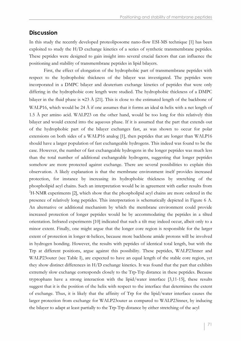
First, the effect of elongation of the hydrophobic part of transmembrane peptides withrespect to the hydrophobic thickness of the bilayer was investigated. The peptides wereincorporated in a DMPC bilayer and deuterium exchange kinetics of peptides that were onlydiffering in the hydrophobic core length were studied. The hydrophobic thickness of a DMPCbilayer in the fluid phase is �23 Å [21]. This is close to the estimated length of the backbone of
WALP16, which would be 24 Å if one assumes that it forms an ideal �-helix with a net length of1.5 Å per amino acid. WALP23 on the other hand, would be too long for this relatively thinbilayer and would extend into the aqueous phase. If it is assumed that the part that extends outof the hydrophobic part of the bilayer exchanges fast, as was shown to occur for polarextensions on both sides of a WALP16 analog [1], then peptides that are longer than WALP16should have a larger population of fast exchangeable hydrogens. This indeed was found to be thecase. However, the number of fast exchangeable hydrogens in the longer peptides was much lessthan the total number of additional exchangeable hydrogens, suggesting that longer peptidessomehow are more protected against exchange. There are several possibilities to explain thisobservation. A likely explanation is that the membrane environment itself provides increasedprotection, for instance by increasing its hydrophobic thickness by stretching of thephospholipid acyl chains. Such an interpretation would be in agreement with earlier results from2H-NMR experiments [2], which show that the phospholipid acyl chains are more ordered in thepresence of relatively long peptides. This interpretation is schematically depicted in Figure 6 A.An alternative or additional mechanism by which the membrane environment could provideincreased protection of longer peptides would be by accommodating the peptides in a tiltedorientation. Infrared experiments [10] indicated that such a tilt may indeed occur, albeit only to aminor extent. Finally, one might argue that the longer core region is responsible for the largerextent of protection in longer �-helices, because more backbone amide protons will be involvedin hydrogen bonding. However, the results with peptides of identical total length, but with theTrp at different positions, argue against this possibility. These peptides, WALP23inner andWALP23outer (see Table I), are expected to have an equal length of the stable core region, yetthey show distinct differences in H/D exchange kinetics. It was found that the part that exhibitsextremely slow exchange corresponds closely to the Trp-Trp distance in these peptides. Becausetryptophans have a strong interaction with the lipid/water interface [3,11-15], these resultssuggest that it is the position of the helix with respect to the interface that determines the extentof exchange. Thus, it is likely that the affinity of Trp for the lipid/water interface causes thelarger protection from exchange for WALP23outer as compared to WALP23inner, by inducingthe bilayer to adapt at least partially to the Trp-Trp distance by either stretching of the acyl

Chapter 3
72
Figure 6 – Model for the incorporation of different peptides in a DMPC bilayer, depicting adaptation of the bilayerthickness as one of the possible mechanisms to explain the observed data. When a too long peptide is incorporatedin the bilayer, the lipid acyl chains are able to partially adapt their hydrophobic thickness (A). The gray areas indicatethe parts of the peptide that are exchanging fast. Tryptophans are likely to be important for positioning in theinterface region, since WALP23inner exhibits more H/D exchange than does WALP23outer (B). Amide hydrogensin a transmembrane segment with a Pro residue are involved in hydrogen bonding to a lesser extent, and show faster
exchange over the whole transmembrane region. Therefore, the regular transmembrane �-helix is destabilized,perhaps involving introduction of a kink in the helix (C).

Positioning and stability of membrane peptides
73
chains, as schematically depicted in Figure 6 B, or by accommodating the peptide in a tiltedorientation.
Remarkably, WALP23 which contains two Trp residues at each side of the peptide,showed similar backbone amide exchange kinetics as WALP23outer. This suggests that the outerTrp residues are the determinants for interfacial positioning of this relatively long peptide andhence that localization of Trp residues further away from the interface towards the aqueousphase is unfavorable. Together, the results show that Trp has strong membrane anchoringproperties and interacts with a well-defined region in the bilayer interface. This supports the ideathat Trp residues, which have shown to be clustered at the border between the hydrophilic andhydrophobic zones both in single-spanning [8] and multispanning integral membrane proteins(e.g. [5,7]) are important for stabilizing integral membrane proteins in a lipid bilayer [3,11-15].The observation that Trp residues may determine the precise interfacial position oftransmembrane segments of proteins also is functionally relevant, since the interfacial positioningof for instance receptor proteins or channel-forming proteins can determine their accessibility toenzymes, substrates or ligands [22].
Finally, the effect of inserting specific amino acid residues that are generally known todestabilize a regular �-helix in water-soluble proteins, like Pro and Gly, was studied. Thefrequent occurrence of Pro in the putative transmembrane helices of integral membraneproteins, particularly transport proteins and G-coupled receptors, has led to the suggestion thatthe presence of this conserved amino acid residue might play a role in folding and/or assemblyof integral membrane proteins in general and that it may be essential for functional activity oftransport proteins and ion channels [16,23,24], because of its supposed ability to introduce kinksinto transmembrane �-helices. Our results show that the presence of a Pro residue in thetransmembrane helix of WALP23 leads to a significant increase in exchange after longerincubation times, whereas CID MS measurements demonstrated that exchange in thetransmembrane helix region is increased. The similar exchange kinetics in the short term regionof WALP23 peptides with and without Pro suggest that the peptides are anchored in the lipidbilayer interface in a similar way. Therefore, it is suggested that the presence of a Pro residue maycause a less stable helix fold of the hydrophobic core of WALP23, as schematically depicted inFigure 6 C.
There may be some other possibilities to interpret the increased H/D exchange rates forWALP23Pro as compared to WALP23. One such possibility would be that because of a Pro-induced kink in the peptide the lipid acyl chains in the vicinity of the transmembrane helix maybe perturbed. This would allow an increase of water permeation in the bilayer, and, therefore,could lead to somewhat higher exchange rates specifically in the transmembrane region. Inprinciple, an alternative possibility could be that the peptide does not have a transmembraneorientation, but instead is located on the surface of the bilayer, which also would result in higherexchange rates. However, in this case one would expect much faster exchange rates than theones observed, because the peptide would be in direct contact with bulk deuterated water.
Chapter 3
74
Indeed, control measurements on WALP23 in TFE/D2O buffer (1:1; volume ratio), in which thepeptide forms an �-helix (data not shown), demonstrated that under these conditions H/D
exchange was already complete within less than 60 min at 30�C. Another argument against asurface localization of WALP23Pro would be the observed similarity between the numbers offast exchangeable protons with those in WALP23, which by various techniques has been shownto adopt a transmembrane orientation [3,10]. Hence we exclude this possibility. Finally, onemight consider the possibility of a slow equilibrium between surface and bound states ofWALP23Pro. In this case, however, several populations of deuterated WALP23Pro (i.e. fast andslowly exchanging) should be present in the mass spectra at the same time. Since this is notobserved, we exclude this latter possibility as well.
In contrast to WALP23Pro, WALP23Gly shows similar exchange kinetics as WALP23,indicating that Gly does not have a strong effect on the stability of the transmembrane helix, atleast in the systems studied here. Gly has been suggested to play a role in environment-dependent �-helix modulation [25] and to mediate helix-helix interactions in membrane proteins[26]. Conformational studies on model peptides in a membrane mimetic environment [25] andanalysis of dihedral backbone angles in crystal structures of several membrane proteins [27]suggested that Gly does not disrupt the secondary structure of �-helical transmembranesegments. The results of the present study support this. In summary, our results show thatinsertion of a Pro residue but not of Gly results in significant destabilization of a transmembrane�-helix in a lipid bilayer. The structural changes in Pro-containing transmembrane segmentsmight be important for functional properties of membrane proteins.
This study has shown that the effect of variations in hydrophobic length, in the positionsof flanking residues and of insertion of putative helix breakers in transmembrane peptides, canbe studied in great detail by H/D exchange combined with mass spectrometry, whereby thepositions of the exchanged hydrogens can be derived from CID MS experiments. Moreover, itwas shown that it is possible to measure H/D exchange in two or more peptides simultaneously,allowing determination of very small differences in deuterium levels. Detailed information abouthelix stability and interfacial positioning, as obtained in the present study, not only providesinsight into the factors that determine the way in which membrane proteins are integrated into alipid bilayer, but also may be useful for predictions of transmembrane segments from amino acidsequences. In future studies we plan to extrapolate the method to larger integral membraneproteins and membrane-bound proteins.
References
1. Demmers, J.A.A., Haverkamp, J., Heck, A.J.R., Koeppe, R.E., II & Killian, J.A. (2000) Electrosprayionization mass spectrometry as a tool to analyze hydrogen/deuterium exchange kinetics oftransmembrane peptides in lipid bilayers. Proc. Natl. Acad. Sci. U. S. A. 97, 3189-94.
2. de Planque, M.R.R., Greathouse, D.V., Koeppe, R.E., II, Schafer, H., Marsh, D. & Killian, J.A. (1998)Influence of lipid/peptide hydrophobic mismatch on the thickness of diacylphosphatidylcholine bilayers. A
Positioning and stability of membrane peptides
75
2H NMR and ESR study using designed transmembrane alpha-helical peptides and gramicidin A.Biochemistry 37, 9333-45.
3. de Planque, M.R.R., Kruijtzer, J.A.W., Liskamp, R.M.J., Marsh, D., Greathouse, D.V., Koeppe, R.E., II, deKruijff, B. & Killian, J.A. (1999) Different membrane anchoring positions of tryptophan and lysine insynthetic transmembrane alpha-helical peptides. J. Biol. Chem. 274, 20839-46.
4. Killian, J.A., Salemink, I., de Planque, M.R.R., Lindblom, G., Koeppe, R.E., II & Greathouse, D.V. (1996)Induction of nonbilayer structures in diacylphosphatidylcholine model membranes by transmembranealpha-helical peptides: importance of hydrophobic mismatch and proposed role of tryptophans. Biochemistry35, 1037-45.
5. Doyle, D.A., Cabral, J.M., Pfuetzner, R.A., Kuo, A.L., Gulbis, J.M., Cohen, S.L., Chait, B.T. & MacKinnon,R. (1998) The Structure of the Potassium Channel Molecular Basis of K+ Conduction and Selectivity.Science 280, 69-77.
6. Kovacs, F., Quine, J. & Cross, T.A. (1999) Validation of the single-stranded channel conformation ofgramicidin A by solid-state NMR. Proc. Natl. Acad. Sci. U. S. A. 96, 7910-5.
7. Iwata, S., Ostermeier, C., Ludwig, B. & Michel, H. (1995) Structure at 2.8 A resolution of cytochrome coxidase from Paracoccus denitrificans. Nature 376, 660-9.
8. Landolt Marticorena, C., Williams, K.A., Deber, C.M. & Reithmeier, R.A. (1993) Non-random distributionof amino acids in the transmembrane segments of human type I single span membrane proteins. J. Mol.Biol. 229, 602-8.
9. Killian, J.A. (1998) Hydrophobic mismatch between proteins and lipids in membranes. Biochim. Biophys.Acta 1376, 401-15.
10. de Planque, M.R.R., Goormaghtigh, E., Greathouse, D.V., Koeppe, R.E., II, Kruijtzer, J.A.W., Liskamp,R.M.J., de Kruijff, B. & Killian, J.A. (2001) Sensitivity of single membrane-spanning alpha-helical peptidesto hydrophobic mismatch with a lipid bilayer: effects on backbone structure, orientation, and extent ofmembrane incorporation. Biochemistry 40, 5000-10.
11. Persson, S., Killian, J.A. & Lindblom, G. (1998) Molecular ordering of interfacially localized tryptophananalogs in ester- and ether-lipid bilayers studied by 2H-NMR. Biophys. J. 75, 1365-71.
12. Braun, P. & von Heijne, G. (1999) The aromatic residues Trp and Phe have different effects on thepositioning of a transmembrane helix in the microsomal membrane. Biochemistry 38, 9778-82.
13. Ridder, A.N.J.A., Morein, S., Stam, J.G., Kuhn, A., de Kruijff, B. & Killian, J.A. (2000) Analysis of the roleof interfacial tryptophan residues in controlling the topology of membrane proteins. Biochemistry 39, 6521-8.
14. Yau, W.M., Wimley, W.C., Gawrisch, K. & White, S.H. (1998) The preference of tryptophan formembrane interfaces. Biochemistry 37, 14713-8.
15. Wimley, W.C. & White, S.H. (1996) Experimentally determined hydrophobicity scale for proteins atmembrane interfaces. Nat. Struct. Biol. 3, 842-8.
16. Eyles, S.J. & Gierasch, L.M. (2000) Multiple roles of prolyl residues in structure and folding. J. Mol. Biol.301, 737-47.
17. Greathouse, D.V., Goforth, R.L., Crawford, T., van der Wel, P.C.A. & Killian, J.A. (2001) Optimizedaminolysis conditions for cleavage of N-protected hydrophobic peptides from solid-phase resins. J. PeptideRes. 57, 519-27.
18. Blume, A. (1983) Biochemistry 22, 5436-42.19. Jansen, M. & Blume, A. (1995) A comparative study of diffusive and osmotic water permeation across
bilayers composed of phospholipids with different head groups and fatty acyl chains. Biophys. J. 68, 997-1008.
20. Roepstorff, P. & Fohlman, J. (1984) Proposal for a common nomenclature for sequence ions in massspectra of peptides. Biomed. Mass Spectrom. 11, 601.
Chapter 3
76
21. Lewis, B.A. & Engelman, D.M. (1983) Lipid bilayer thickness varies linearly with acyl chain length in fluidphosphatidylcholine vesicles. J. Mol. Biol. 166, 211-7.
22. Killian, J.A. & von Heijne, G. (2000) How proteins adapt to a membrane-water interface. Trends Biochem.Sci. 25, 429-34.
23. Eilers, M., Shekar, S.C., Shieh, T., Smith, S.O. & Fleming, P.J. (2000) Internal packing of helical membraneproteins. Proc. Natl. Acad. Sci. U. S. A. 97, 5796-801.
24. Schiffer, M., Ainsworth, C.F., Deng, Y.L., Johnson, G., Pascoe, F.H. & Hanson, D.K. (1995) Proline in atransmembrane helix compensates for cavities in the photosynthetic reaction center. J. Mol. Biol. 252, 472-82.
25. Li, S.C. & Deber, C.M. (1992) Glycine and beta-branched residues support and modulate peptide helicity inmembrane environments. FEBS Lett. 311, 217-20.
26. Russ, W.P. & Engelman, D.M. (2000) The GxxxG motif: a framework for transmembrane helix-helixassociation. J. Mol. Biol. 296, 911-9.
27. Javadpour, M.M., Eilers, M., Groesbeek, M. & Smith, S.O. (1999) Helix packing in polytopic membraneproteins: role of glycine in transmembrane helix association. Biophys. J. 77, 1609-18.
Related Documents