Interactions of vanadium(V)–citrate complexes with the sarcoplasmic reticulum calcium pump Manuel Aureliano a, * , Teresa Tiago a , Ricardo M.C. Ga ˆndara a , Andrea Sousa a , A. Moderno a , M. Kaliva b , A. Salifoglou c,d , Rui O. Duarte e , Jose ´ J.G. Moura e a CBME, Department of Chemistry and Biochemistry, FCT, University of the Algarve, 8005-139 Faro, Portugal b Department of Chemistry, University of Crete, Heraklion 71409, Greece c Department of Chemical Engineering, Aristotle University of Thessaloniki, Thessaloniki 54124, Greece d Chemical Process Engineering Research Institute, Thermi, Thessaloniki 57001, Greece e REQUIMTE-CQFB, FCT, Universidade Nova de Lisboa, 2829-516 Caparica, Portugal Received 5 July 2005; received in revised form 5 September 2005; accepted 7 September 2005 Available online 10 October 2005 Abstract Among the biotargets interacting with vanadium is the calcium pump from the sarcoplasmic reticulum (SR). To this end, initial research efforts were launched with two vanadium(V)–citrate complexes, namely (NH 4 ) 6 [V 2 O 4 (C 6 H 4 O 7 ) 2 ] 6H 2 O and (NH 4 ) 6 [V 2 O 2 (O 2 ) 2 (C 6 H 4 O 7 ) 2 ] 4H 2 O, potentially capable of interacting with the SR calcium pump by combining kinetic studies with 51 V NMR spectroscopy. Upon dissolution in the reaction medium (concentration range: 4–0.5 mM), both vanadium(V):citrate (VC) and peroxovanadium(V):citrate (PVC) complexes are partially converted into vanadate oligomers. A 1 mM solution of the PVC com- plex, containing 184 lM of the PVC complex, 94 lM oxoperoxovanadium(V) (PV) species, 222 lM monomeric (V1), 43 lM dimeric (V2) and 53 lM tetrameric (V4) species, inhibits Ca 2+ accumulation by 75 %, whereas a solution of the VC complex of the same vana- dium concentration, containing 98 lM of the VC complex, 263 lM monomeric (V1), 64 lM dimeric (V2) and 92 lM tetrameric (V4) species inhibits the calcium pump activity by 33 %. In contrast, a 1 mM metavanadate solution, containing 460 lM monomeric (V1), 90.2 lM dimeric (V2) and 80 lM tetrameric (V4) species, has no effect on Ca 2+ accumulation. The NMR signals from the VC complex (548.0 ppm), PVC complex (551.5 ppm) and PV (611.1 ppm) are broadened upon SR vesicle addition (2.5 mg/ml total protein). The relative order for the half width line broadening of the NMR signals, which reflect the interaction with the protein, was found to be V4 > PVC > VC > PV > V2 = V1 = 1, with no effect observed for the V1 and V2 signals. Putting it all together the effects of two vana- dium(V)–citrate complexes on the modulation of calcium accumulation and ATP hydrolysis by the SR calcium pump reflected the observed variable reactivity into the nature of key species forming upon dissolution of the title complexes in the reaction media. Ó 2005 Elsevier Inc. All rights reserved. Keywords: Vanadium–citrate complex; Calcium pump 1. Introduction Almost 30 years after the discovery that vanadium was a muscle inhibiting factor and a Na + /K + -ATPase inhibitor present in commercially available ATP [1–3], it still remains to be clarified whether or not vanadium has a physiological role in the muscle. It is believed that the complex chemistry of vanadium itself, its interactions with small molecules of biological interest, and vanadate similarities with phos- phate constitute the rudiments of research emerging from the increased interest in vanadium in biological systems. Among the effects attributed to vanadium salts and com- plexes, the insulin mimetic action stands prominent [4,5]. However, vanadium toxicity and poor absorption in the gastrointestinal tract is still a major obstacle in the develop- ment of therapeutic applications [6]. The challenge of 0162-0134/$ - see front matter Ó 2005 Elsevier Inc. All rights reserved. doi:10.1016/j.jinorgbio.2005.09.002 * Corresponding author. Tel.: +351 965 536 476; fax: +351 289 819 403. E-mail address: [email protected] (M. Aureliano). www.elsevier.com/locate/jinorgbio Journal of Inorganic Biochemistry 99 (2005) 2355–2361 JOURNAL OF Inorganic Biochemistry

Welcome message from author
This document is posted to help you gain knowledge. Please leave a comment to let me know what you think about it! Share it to your friends and learn new things together.
Transcript
JOURNAL OF
www.elsevier.com/locate/jinorgbio
Journal of Inorganic Biochemistry 99 (2005) 2355–2361
InorganicBiochemistry
Interactions of vanadium(V)–citrate complexes with thesarcoplasmic reticulum calcium pump
Manuel Aureliano a,*, Teresa Tiago a, Ricardo M.C. Gandara a, Andrea Sousa a,A. Moderno a, M. Kaliva b, A. Salifoglou c,d, Rui O. Duarte e, Jose J.G. Moura e
a CBME, Department of Chemistry and Biochemistry, FCT, University of the Algarve, 8005-139 Faro, Portugalb Department of Chemistry, University of Crete, Heraklion 71409, Greece
c Department of Chemical Engineering, Aristotle University of Thessaloniki, Thessaloniki 54124, Greeced Chemical Process Engineering Research Institute, Thermi, Thessaloniki 57001, Greecee REQUIMTE-CQFB, FCT, Universidade Nova de Lisboa, 2829-516 Caparica, Portugal
Received 5 July 2005; received in revised form 5 September 2005; accepted 7 September 2005Available online 10 October 2005
Abstract
Among the biotargets interacting with vanadium is the calcium pump from the sarcoplasmic reticulum (SR). To this end, initialresearch efforts were launched with two vanadium(V)–citrate complexes, namely (NH4)6[V2O4(C6H4O7)2] Æ 6H2O and(NH4)6[V2O2(O2)2(C6H4O7)2] Æ 4H2O, potentially capable of interacting with the SR calcium pump by combining kinetic studies with51V NMR spectroscopy. Upon dissolution in the reaction medium (concentration range: 4–0.5 mM), both vanadium(V):citrate (VC)and peroxovanadium(V):citrate (PVC) complexes are partially converted into vanadate oligomers. A 1 mM solution of the PVC com-plex, containing 184 lM of the PVC complex, 94 lM oxoperoxovanadium(V) (PV) species, 222 lM monomeric (V1), 43 lM dimeric(V2) and 53 lM tetrameric (V4) species, inhibits Ca2+ accumulation by 75 %, whereas a solution of the VC complex of the same vana-dium concentration, containing 98 lM of the VC complex, 263 lM monomeric (V1), 64 lM dimeric (V2) and 92 lM tetrameric (V4)species inhibits the calcium pump activity by 33 %. In contrast, a 1 mM metavanadate solution, containing 460 lM monomeric (V1),90.2 lM dimeric (V2) and 80 lM tetrameric (V4) species, has no effect on Ca2+ accumulation. The NMR signals from the VC complex(�548.0 ppm), PVC complex (�551.5 ppm) and PV (�611.1 ppm) are broadened upon SR vesicle addition (2.5 mg/ml total protein). Therelative order for the half width line broadening of the NMR signals, which reflect the interaction with the protein, was found to beV4 > PVC > VC > PV > V2 = V1 = 1, with no effect observed for the V1 and V2 signals. Putting it all together the effects of two vana-dium(V)–citrate complexes on the modulation of calcium accumulation and ATP hydrolysis by the SR calcium pump reflected theobserved variable reactivity into the nature of key species forming upon dissolution of the title complexes in the reaction media.� 2005 Elsevier Inc. All rights reserved.
Keywords: Vanadium–citrate complex; Calcium pump
1. Introduction
Almost 30 years after the discovery that vanadium was amuscle inhibiting factor and a Na+/K+-ATPase inhibitorpresent in commercially available ATP [1–3], it still remainsto be clarified whether or not vanadium has a physiological
0162-0134/$ - see front matter � 2005 Elsevier Inc. All rights reserved.
doi:10.1016/j.jinorgbio.2005.09.002
* Corresponding author. Tel.: +351 965 536 476; fax: +351 289 819 403.E-mail address: [email protected] (M. Aureliano).
role in the muscle. It is believed that the complex chemistryof vanadium itself, its interactions with small molecules ofbiological interest, and vanadate similarities with phos-phate constitute the rudiments of research emerging fromthe increased interest in vanadium in biological systems.Among the effects attributed to vanadium salts and com-plexes, the insulin mimetic action stands prominent [4,5].However, vanadium toxicity and poor absorption in thegastrointestinal tract is still a major obstacle in the develop-ment of therapeutic applications [6]. The challenge of
2356 M. Aureliano et al. / Journal of Inorganic Biochemistry 99 (2005) 2355–2361
overcoming these obstacles has spurred considerable re-search activity, targeting the interaction of vanadium withbiomolecules and organic ligands, in order to enhanceabsorption and efficiency while concurrently reducing tox-icity. To that end, a number of ligands, such as citric acid,have been employed as potential ligand binders of vana-dium in biological systems [7–9].
It is known that vanadium ions and vanadium com-plexes inhibit or stimulate the activity of various enzymes.In the case of the membrane bound E1–E2 Ca2+-ATPase(calcium pump) from sarcoplasmic reticulum (SR), theinhibition is promoted by orthovanadate binding to theE2 conformation of the protein [10]. Moreover, the useof vanadate is related to the catalytic site of Ca2+ ATPase,which contains an aspartyl that is phosphorylated duringthe catalytic cycle thereby forming an acyl phosphate anhy-dride [11]. In addition to monomeric vanadate species, ithas been reported that other vanadate oligomers interactwith the SR calcium pump [12]. It has been demonstratedthat some of the interactions, e.g. decameric species, dis-rupt the energy coupling and enzyme turnover. Other inter-actions of vanadium, e.g. monomeric species, may have noimplicit effect or even improve the coupling to the Ca2+
pump [13]. The SR calcium pump was reported to have adecreased activity in some diabetic conditions, which couldresult in impaired cardiac relaxation [14]. Several studieshave been carried out using vanadium(V) for characteriza-tion of SR calcium pump [13,15]. Indeed, SR calcium pumphas proven to be an excellent model to study Ca2+ homeo-stasis affected by acute and cronic exposure of nitrosativestress [16].
In the present study, the effects of two vanadium(V)–citrate complexes namely (NH4)6[V2O4(C6H4O7)2] Æ 6H2Oand (NH4)6[V2O2(O2)2(C6H4O7)2] Æ 4H2O, on the modula-tion of calcium accumulation and ATP hydrolysis by theSR calcium pump were evaluated. The results demon-strated that it is of primary importance to precisely charac-terize the aqueous behaviour of the vanadium(V) speciesand their interactions with the investigated biosystem priorto attempting to understand the promoted effects. Bothvanadium(V)–citrate complexes interact and affect theactivity of the SR calcium pump, with the peroxovana-dium(V):citrate complex solutions being the most potentinhibitor.
2. Materials and methods
2.1. Reagents
Chemicals used to prepare buffers were of reagent grade.Ammonium metavanadate and citric acid were purchasedfrom Riedel-de Haen. ATP (vanadium free) was suppliedby Sigma. All other reagents were of biochemical analysisgrade from BDH, Merck or Sigma.
The synthesis of the vanadium(V) complexes(NH4)6[V2O4(C6H4O7)2] Æ 6H2O and (NH4)6[V2O2(O2)2(C6H4O7)2] Æ 4H2O was achieved according to published lit-
erature procedures [17,18]. In these articles, the character-ization of the complexes was carried out with analytical,spectroscopic techniques (UV–Visible, 13C NMR (in thecase of the ternary vanadium(V)–peroxo-citrate complex),FT-IR) and X-ray crystallographic techniques. Therefore,the structures of the two complexes are known along withtheir chemical reactivity. The 51V NMR, however, is notavailable for the two species and thus cannot be includedin this manuscript.
Vanadium–citrate complex stock solutions (9 mM) usedin the studies were prepared from vanadium(V)–citrate(NH4)6[V2O4(C6H4O7)2] Æ 6H2O and from vanadium(V)–peroxo-citrate (NH4)6[V2O2(O2)2(C6H4O7)2] Æ 4H2O saltsin a medium containing 25 mM Hepes (2-(4-(2-hydroxy-ethyl)-1-piperazinyl)ethanesulphonic acid), pH 7.0, and0.1 M KCl.
2.2. Preparation of sarcoplasmic reticulum vesicles
Sarcoplasmic reticulum vesicles (SRV) were preparedfrom white skeletal rabbit muscles as described elsewhere[13]. Isolated SRV suspended in KCl 0.1 M, tris(hydroxy-methyl)aminometane (Tris) 10 mM (pH 7.0) were diluted1:1 with sucrose 2 M and frozen in liquid nitrogen priorto storage at �80 �C. For SRV preparations used inNMR studies, no sucrose was added, once sucrose formscomplexes with vanadium and in the 51V NMR appearstwo signals at �540 and �550 ppm as described previously[13]. Protein concentration was determined by the biuretmethod, using bovine serum albumin as a standard [19].The percentage of each protein present in the SRV prepa-rations was determined trough densitometry analysis ofsodium dodecyl sulphate (SDS)–polyacrylamide gelelectrophoresis (7.5% acrylamide). The SR Ca2+-ATPaseanalysed by SDS–polyacrylamide gel electrophoresis wasat least 70% of the total protein in SRV.
2.3. NMR measurements
51V Nuclear magnetic resonance spectroscopy measure-ments on vanadium(V)–citrate complex solutions were ta-ken on a Bruker AM-400 MHz, by using a 90� pulseFourier transform technique. Chemical shift values are gi-ven with reference to VOCl3 (0 ppm) as a standard. The rel-ative areas of several free and bound vanadate resonanceswere integrated and the line widths were obtained by sub-tracting the value (20 Hz) used in line broadening. The con-centrations of vanadate oligomers Vx were calculated fromthe fractions of the total integrated areas observed in therecorded spectra as described in the equation below. Thesymbol A corresponds to the area measured for the x vana-dium(V) species (Vx) with n being the aggregation number(number of vanadium atoms), A(Vt), the sum of the mea-sured area and [Vt] the total vanadium(V) concentration
½Vx� ¼ AðVxÞ �1
AðVtÞ� ½Vt�nðVxÞ
.
Fig. 1. 105.2 MHz 51V NMR spectra, at 22 �C, of a 9 mM vanadium(V)–citrate complex solution prepared in water (a) or in the reaction medium(b). VC and V4 stands for vanadium(V)–citrate and tetrameric vanadatespecies, respectively.
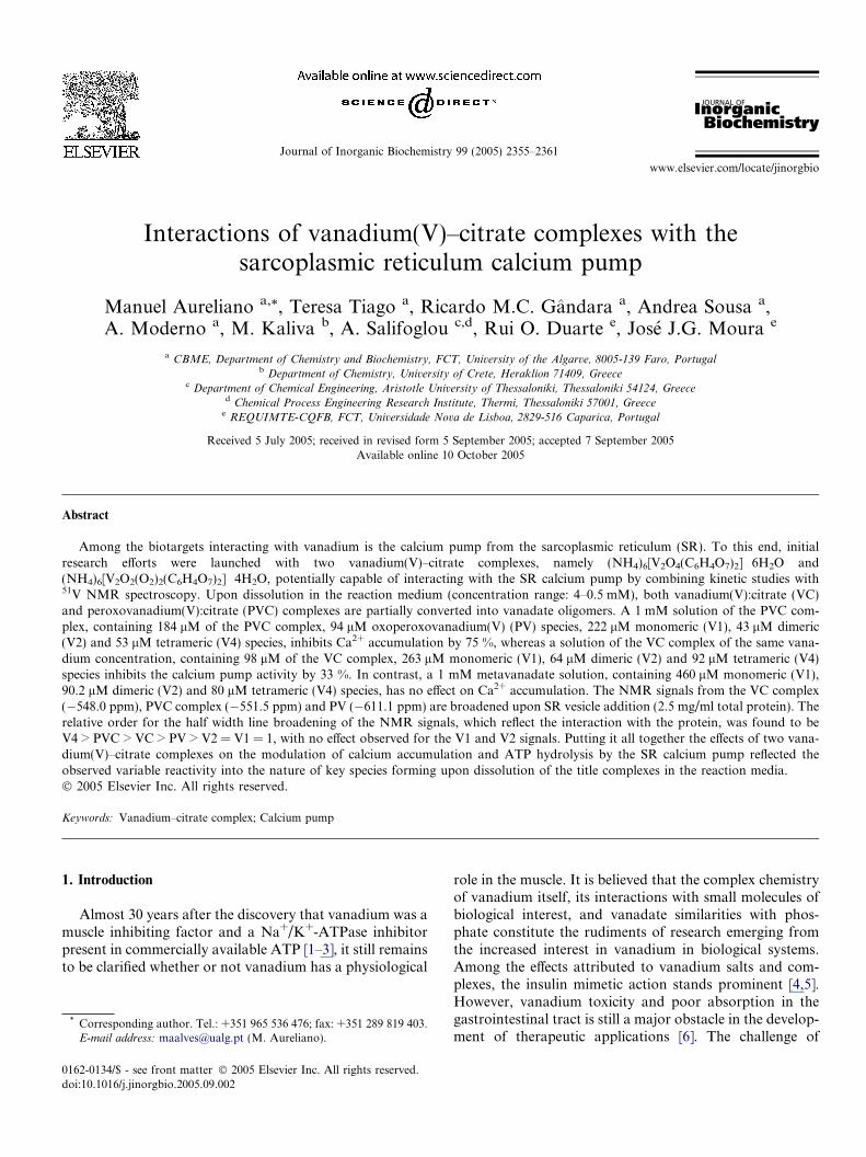
Fig. 2. 105.2 MHz 51V NMR spectra, at 22 �C, of different concentra-tions, namely 0.5 mM (a); 1 mM (b); 2 mM (c); 4 mM (d) and 9 mM (e)total vanadium, of a vanadium(V)–citrate (VC) complex in a mediumcontaining 25 mM Hepes, pH 7.0, and 0.1 M KCl. V1 and V2 NMRsignals correspond, respectively, to monomeric ðH2VO
�4 Þ and dimeric
ðHV2O3�7 and H2V2O
2�7 Þ vanadate species irrespective of the protonation
state, whereas V4 and V5 correspond to cyclic tetrameric ðV4O4�12 Þ and
pentameric ðV5O5�15 Þ vanadate species.
M. Aureliano et al. / Journal of Inorganic Biochemistry 99 (2005) 2355–2361 2357
The spectra of vanadium(V)–citrate complex solutions (VCand PVC complexes) were obtained at several concentra-tions in the reaction medium, containing KCl 0.1 M,MgCl2 5 mM, Hepes 25 mM (pH 7.0), CaCl2 50 lM andATP 500 lM in Hepes 25 mM (pH 7.0). Spectra of 2 mMvanadium(V)–citrate complex solutions were also obtainedin the same reaction medium, in the absence and presenceof 2.5 mg/ml of SR preparations. The spectra were ac-quired at room temperature, spectral with 40,355 Hz, accu-mulation time 0.05 s, number of transients between 12,000and 25,000 and relaxation delay 0.01 s. For quantitativemeasurements all of the spectral parameters were kept con-stant [12].
2.4. ATP hydrolysis by calcium pump
ATP hydrolysis was measured by colorimetric determi-nation of inorganic phosphate [20]. Kinetic experimentswere performed at 25 �C in a reaction medium containingKCl 0.1 M, Hepes 25 mM (pH 7.0), MgCl2 5 mM, CaCl250 lM, 0.285 g/ml of SRV preparations, in the absenceor presence of 1 mM vanadate, vanadium(V) complexesor citrate, according to the required experimental condi-tions. Vanadate, vanadium–citrate complexes or citratesolutions were added to the medium prior to protein addi-tion. The reaction was initiated by adding Mg-ATP500 lM and was subsequently monitored for up to 20 min.
2.5. Ca2+ uptake experiments
Ca2+ uptake experiments were performed as describedelsewhere [12]. The experiments were carried out at 25 �Cin a 4 ml assay medium, containing KCl 0.1 M, MgCl25 mM, Hepes 25 mM (pH 7.0), CaCl2 20 lM, and0.1 mg/ml SR preparations. The reaction was initiated byadding ATP 500 lM in Hepes 25 mM (pH 7.0). At theend of each experiment, an internal calibration was per-formed by addition of standard CaCl2 solutions. Vana-dium(V)–citrate complex or metavanadate solutions atseveral concentrations were added to the medium prior toATP addition.
3. Results and discussion
3.1. Spectroscopic characterization of the complexes
The 51V NMR spectra of 9 mM solutions of the vana-dium(V):citrate complex (VC) in water (pH 6.6) exhibit res-onances at �546 and at �549 ppm (Fig. 1). The relativeareas of the resonances reflect a 1:1 ratio, with the half linewidth of these signals being very similar (180 ± 10 Hz).When the VC complex solution was prepared in the reac-tion medium, another NMR signal was detected at�578 ppm. Upon dissolution, of the VC complex, in thereaction medium at lower concentrations (from 4 to0.5 mM), the VC complex is partially converted into vana-date oligomers (Fig. 2). NMR signals ascribed to mono
(V1) at �561 ppm, dimeric (V2) at �574 ppm andtetrameric (V4) at �578 ppm vanadate species wereobserved, whereas the VC NMR resonance at �546 ppm
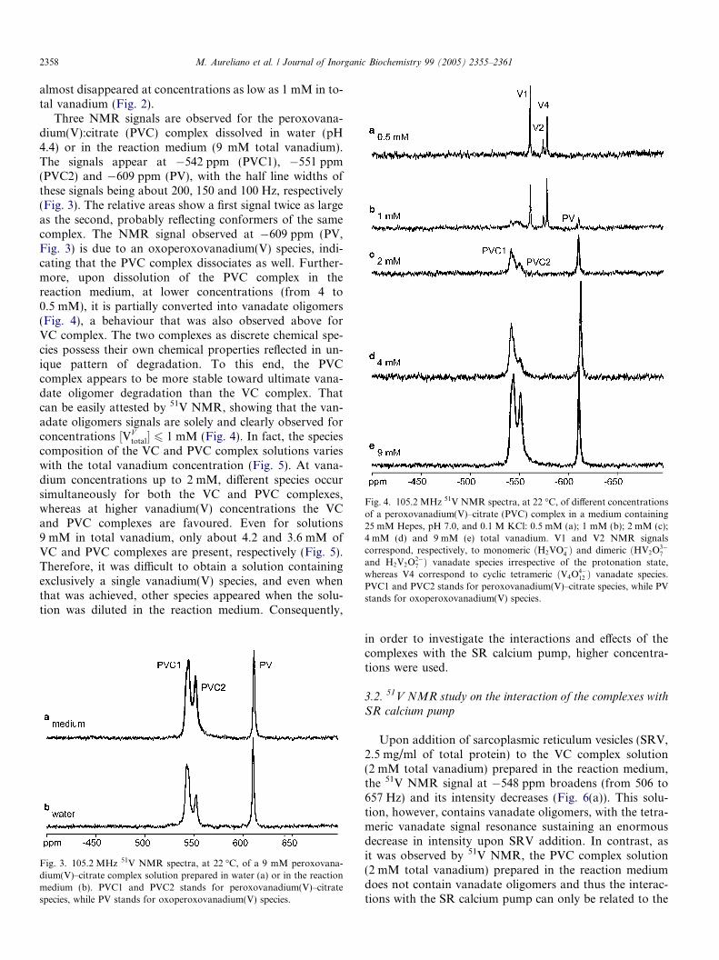
Fig. 4. 105.2 MHz 51V NMR spectra, at 22 �C, of different concentrationsof a peroxovanadium(V)–citrate (PVC) complex in a medium containing25 mM Hepes, pH 7.0, and 0.1 M KCl: 0.5 mM (a); 1 mM (b); 2 mM (c);4 mM (d) and 9 mM (e) total vanadium. V1 and V2 NMR signalscorrespond, respectively, to monomeric ðH2VO
�4 Þ and dimeric ðHV2O
3�7
and H2V2O2�7 Þ vanadate species irrespective of the protonation state,
whereas V4 correspond to cyclic tetrameric ðV4O4�12 Þ vanadate species.
PVC1 and PVC2 stands for peroxovanadium(V)–citrate species, while PVstands for oxoperoxovanadium(V) species.
2358 M. Aureliano et al. / Journal of Inorganic Biochemistry 99 (2005) 2355–2361
almost disappeared at concentrations as low as 1 mM in to-tal vanadium (Fig. 2).
Three NMR signals are observed for the peroxovana-dium(V):citrate (PVC) complex dissolved in water (pH4.4) or in the reaction medium (9 mM total vanadium).The signals appear at �542 ppm (PVC1), �551 ppm(PVC2) and �609 ppm (PV), with the half line widths ofthese signals being about 200, 150 and 100 Hz, respectively(Fig. 3). The relative areas show a first signal twice as largeas the second, probably reflecting conformers of the samecomplex. The NMR signal observed at �609 ppm (PV,Fig. 3) is due to an oxoperoxovanadium(V) species, indi-cating that the PVC complex dissociates as well. Further-more, upon dissolution of the PVC complex in thereaction medium, at lower concentrations (from 4 to0.5 mM), it is partially converted into vanadate oligomers(Fig. 4), a behaviour that was also observed above forVC complex. The two complexes as discrete chemical spe-cies possess their own chemical properties reflected in un-ique pattern of degradation. To this end, the PVCcomplex appears to be more stable toward ultimate vana-date oligomer degradation than the VC complex. Thatcan be easily attested by 51V NMR, showing that the van-adate oligomers signals are solely and clearly observed forconcentrations ½VV
total� 6 1 mM (Fig. 4). In fact, the speciescomposition of the VC and PVC complex solutions varieswith the total vanadium concentration (Fig. 5). At vana-dium concentrations up to 2 mM, different species occursimultaneously for both the VC and PVC complexes,whereas at higher vanadium(V) concentrations the VCand PVC complexes are favoured. Even for solutions9 mM in total vanadium, only about 4.2 and 3.6 mM ofVC and PVC complexes are present, respectively (Fig. 5).Therefore, it was difficult to obtain a solution containingexclusively a single vanadium(V) species, and even whenthat was achieved, other species appeared when the solu-tion was diluted in the reaction medium. Consequently,
Fig. 3. 105.2 MHz 51V NMR spectra, at 22 �C, of a 9 mM peroxovana-dium(V)–citrate complex solution prepared in water (a) or in the reactionmedium (b). PVC1 and PVC2 stands for peroxovanadium(V)–citratespecies, while PV stands for oxoperoxovanadium(V) species.
in order to investigate the interactions and effects of thecomplexes with the SR calcium pump, higher concentra-tions were used.
3.2. 51V NMR study on the interaction of the complexes with
SR calcium pump
Upon addition of sarcoplasmic reticulum vesicles (SRV,2.5 mg/ml of total protein) to the VC complex solution(2 mM total vanadium) prepared in the reaction medium,the 51V NMR signal at �548 ppm broadens (from 506 to657 Hz) and its intensity decreases (Fig. 6(a)). This solu-tion, however, contains vanadate oligomers, with the tetra-meric vanadate signal resonance sustaining an enormousdecrease in intensity upon SRV addition. In contrast, asit was observed by 51V NMR, the PVC complex solution(2 mM total vanadium) prepared in the reaction mediumdoes not contain vanadate oligomers and thus the interac-tions with the SR calcium pump can only be related to the
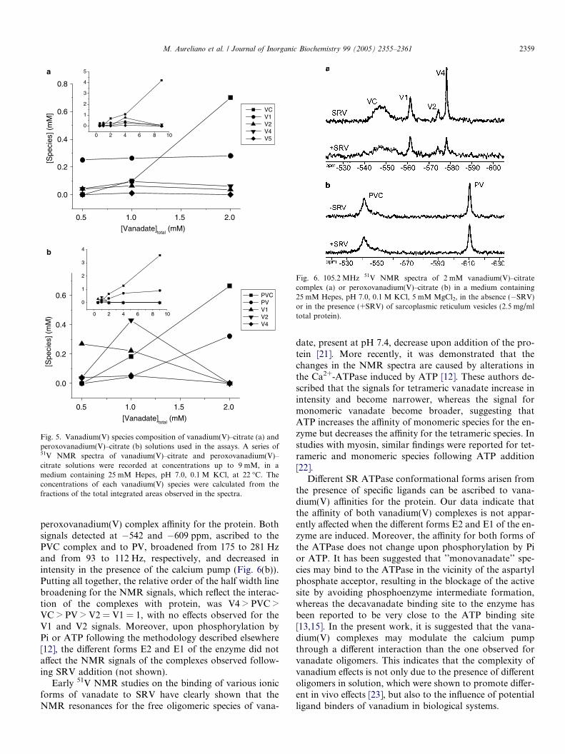
0 2 4 6 8 10
0
1
2
3
0.5 1.0 1.5 2.0
0.0
0.2
0.4
0.6
[Spe
cies
] (m
M)
[Vanadate]total
(mM)
PVC PV V1 V2 V4
0 2 4 6 8 10
0
1
2
3
4
5
0.5 1.0 1.5 2.0
0.0
0.2
0.4
0.6
0.8
[Spe
cies
] (m
M]
[Vanadate]total
(mM)
VC V1 V2 V4 V5
4
a
b
Fig. 5. Vanadium(V) species composition of vanadium(V)–citrate (a) andperoxovanadium(V)–citrate (b) solutions used in the assays. A series of51V NMR spectra of vanadium(V)–citrate and peroxovanadium(V)–citrate solutions were recorded at concentrations up to 9 mM, in amedium containing 25 mM Hepes, pH 7.0, 0.1 M KCl, at 22 �C. Theconcentrations of each vanadium(V) species were calculated from thefractions of the total integrated areas observed in the spectra.
Fig. 6. 105.2 MHz 51V NMR spectra of 2 mM vanadium(V)–citratecomplex (a) or peroxovanadium(V)–citrate (b) in a medium containing25 mM Hepes, pH 7.0, 0.1 M KCl, 5 mM MgCl2, in the absence (�SRV)or in the presence (+SRV) of sarcoplasmic reticulum vesicles (2.5 mg/mltotal protein).
M. Aureliano et al. / Journal of Inorganic Biochemistry 99 (2005) 2355–2361 2359
peroxovanadium(V) complex affinity for the protein. Bothsignals detected at �542 and �609 ppm, ascribed to thePVC complex and to PV, broadened from 175 to 281 Hzand from 93 to 112 Hz, respectively, and decreased inintensity in the presence of the calcium pump (Fig. 6(b)).Putting all together, the relative order of the half width linebroadening for the NMR signals, which reflect the interac-tion of the complexes with protein, was V4 > PVC >VC > PV > V2 = V1 = 1, with no effects observed for theV1 and V2 signals. Moreover, upon phosphorylation byPi or ATP following the methodology described elsewhere[12], the different forms E2 and E1 of the enzyme did notaffect the NMR signals of the complexes observed follow-ing SRV addition (not shown).
Early 51V NMR studies on the binding of various ionicforms of vanadate to SRV have clearly shown that theNMR resonances for the free oligomeric species of vana-
date, present at pH 7.4, decrease upon addition of the pro-tein [21]. More recently, it was demonstrated that thechanges in the NMR spectra are caused by alterations inthe Ca2+-ATPase induced by ATP [12]. These authors de-scribed that the signals for tetrameric vanadate increase inintensity and become narrower, whereas the signal formonomeric vanadate become broader, suggesting thatATP increases the affinity of monomeric species for the en-zyme but decreases the affinity for the tetrameric species. Instudies with myosin, similar findings were reported for tet-rameric and monomeric species following ATP addition[22].
Different SR ATPase conformational forms arisen fromthe presence of specific ligands can be ascribed to vana-dium(V) affinities for the protein. Our data indicate thatthe affinity of both vanadium(V) complexes is not appar-ently affected when the different forms E2 and E1 of the en-zyme are induced. Moreover, the affinity for both forms ofthe ATPase does not change upon phosphorylation by Pior ATP. It has been suggested that ’’monovanadate’’ spe-cies may bind to the ATPase in the vicinity of the aspartylphosphate acceptor, resulting in the blockage of the activesite by avoiding phosphoenzyme intermediate formation,whereas the decavanadate binding site to the enzyme hasbeen reported to be very close to the ATP binding site[13,15]. In the present work, it is suggested that the vana-dium(V) complexes may modulate the calcium pumpthrough a different interaction than the one observed forvanadate oligomers. This indicates that the complexity ofvanadium effects is not only due to the presence of differentoligomers in solution, which were shown to promote differ-ent in vivo effects [23], but also to the influence of potentialligand binders of vanadium in biological systems.
2360 M. Aureliano et al. / Journal of Inorganic Biochemistry 99 (2005) 2355–2361
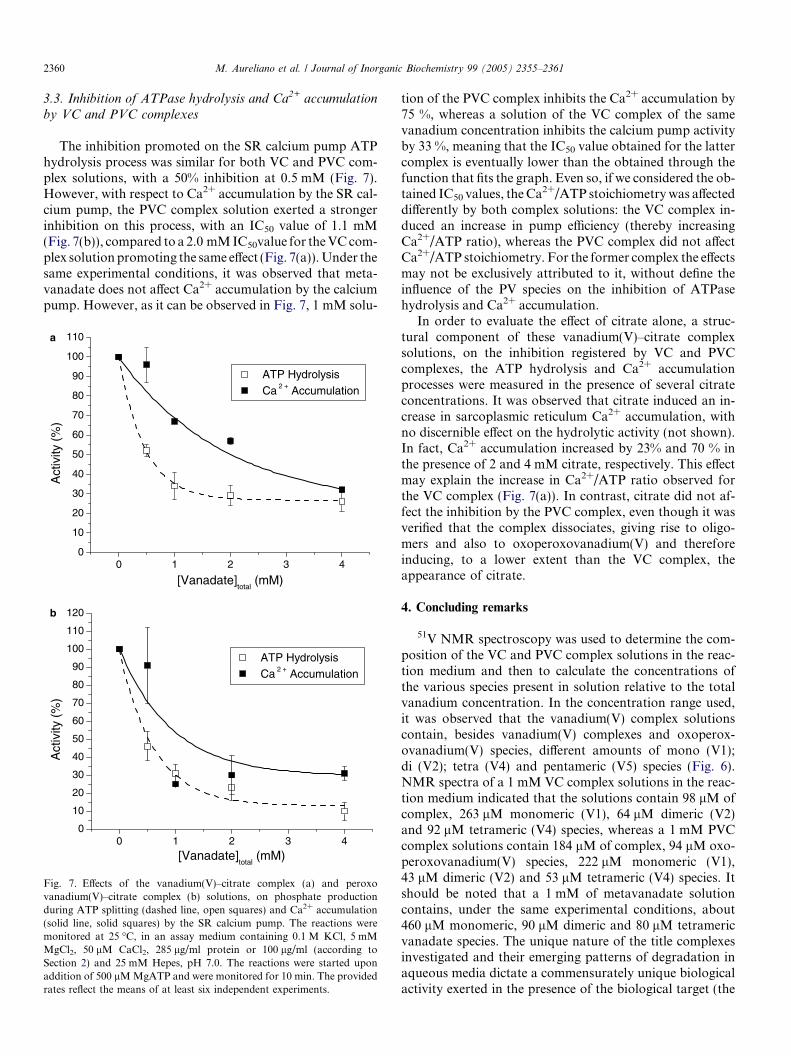
3.3. Inhibition of ATPase hydrolysis and Ca2+ accumulation
by VC and PVC complexes
The inhibition promoted on the SR calcium pump ATPhydrolysis process was similar for both VC and PVC com-plex solutions, with a 50% inhibition at 0.5 mM (Fig. 7).However, with respect to Ca2+ accumulation by the SR cal-cium pump, the PVC complex solution exerted a strongerinhibition on this process, with an IC50 value of 1.1 mM(Fig. 7(b)), compared to a 2.0 mMIC50value for theVCcom-plex solutionpromoting the same effect (Fig. 7(a)).Under thesame experimental conditions, it was observed that meta-vanadate does not affect Ca2+ accumulation by the calciumpump. However, as it can be observed in Fig. 7, 1 mM solu-
0 1 2 3 40
10
20
30
40
50
60
70
80
90
100
110
Act
ivity
(%
)
[Vanadate]total
(mM)
ATP Hydrolysis Ca 2 + Accumulation
0 1 2 3 40
10
20
30
40
50
60
70
80
90
100
110
120
ATP Hydrolysis Ca 2 + Accumulation
Act
ivity
(%
)
[Vanadate]total
(mM)
a
b
Fig. 7. Effects of the vanadium(V)–citrate complex (a) and peroxovanadium(V)–citrate complex (b) solutions, on phosphate productionduring ATP splitting (dashed line, open squares) and Ca2+ accumulation(solid line, solid squares) by the SR calcium pump. The reactions weremonitored at 25 �C, in an assay medium containing 0.1 M KCl, 5 mMMgCl2, 50 lM CaCl2, 285 lg/ml protein or 100 lg/ml (according toSection 2) and 25 mM Hepes, pH 7.0. The reactions were started uponaddition of 500 lMMgATP and were monitored for 10 min. The providedrates reflect the means of at least six independent experiments.
tion of the PVC complex inhibits the Ca2+ accumulation by75 %, whereas a solution of the VC complex of the samevanadium concentration inhibits the calcium pump activityby 33 %, meaning that the IC50 value obtained for the lattercomplex is eventually lower than the obtained through thefunction that fits the graph. Even so, if we considered the ob-tained IC50 values, theCa
2+/ATP stoichiometrywas affecteddifferently by both complex solutions: the VC complex in-duced an increase in pump efficiency (thereby increasingCa2+/ATP ratio), whereas the PVC complex did not affectCa2+/ATP stoichiometry. For the former complex the effectsmay not be exclusively attributed to it, without define theinfluence of the PV species on the inhibition of ATPasehydrolysis and Ca2+ accumulation.
In order to evaluate the effect of citrate alone, a struc-tural component of these vanadium(V)–citrate complexsolutions, on the inhibition registered by VC and PVCcomplexes, the ATP hydrolysis and Ca2+ accumulationprocesses were measured in the presence of several citrateconcentrations. It was observed that citrate induced an in-crease in sarcoplasmic reticulum Ca2+ accumulation, withno discernible effect on the hydrolytic activity (not shown).In fact, Ca2+ accumulation increased by 23% and 70 % inthe presence of 2 and 4 mM citrate, respectively. This effectmay explain the increase in Ca2+/ATP ratio observed forthe VC complex (Fig. 7(a)). In contrast, citrate did not af-fect the inhibition by the PVC complex, even though it wasverified that the complex dissociates, giving rise to oligo-mers and also to oxoperoxovanadium(V) and thereforeinducing, to a lower extent than the VC complex, theappearance of citrate.
4. Concluding remarks
51V NMR spectroscopy was used to determine the com-position of the VC and PVC complex solutions in the reac-tion medium and then to calculate the concentrations ofthe various species present in solution relative to the totalvanadium concentration. In the concentration range used,it was observed that the vanadium(V) complex solutionscontain, besides vanadium(V) complexes and oxoperox-ovanadium(V) species, different amounts of mono (V1);di (V2); tetra (V4) and pentameric (V5) species (Fig. 6).NMR spectra of a 1 mM VC complex solutions in the reac-tion medium indicated that the solutions contain 98 lM ofcomplex, 263 lM monomeric (V1), 64 lM dimeric (V2)and 92 lM tetrameric (V4) species, whereas a 1 mM PVCcomplex solutions contain 184 lM of complex, 94 lM oxo-peroxovanadium(V) species, 222 lM monomeric (V1),43 lM dimeric (V2) and 53 lM tetrameric (V4) species. Itshould be noted that a 1 mM of metavanadate solutioncontains, under the same experimental conditions, about460 lM monomeric, 90 lM dimeric and 80 lM tetramericvanadate species. The unique nature of the title complexesinvestigated and their emerging patterns of degradation inaqueous media dictate a commensurately unique biologicalactivity exerted in the presence of the biological target (the
M. Aureliano et al. / Journal of Inorganic Biochemistry 99 (2005) 2355–2361 2361
SR system). These solutions, with equivalent vanadium(V)concentrations, inhibit Ca2+ accumulation to variable de-grees, namely 75%, 33% and 2% for PVC, VC and meta-vanadate solutions, respectively. These results point outan interesting behaviour of the vanadate complexes. Specif-ically, based on total vanadium concentration of vanadiumpresent in the ternary PVC and binary VC complex solu-tions of the same 1 mM concentration, the PVC complexis more potent (2.3 times higher) than the VC complex.Thus, the peroxovanadate complex appears to be a morepotent inhibitor of the calcium pump.
It is considered to be of primary importance to preciselycharacterize the vanadium species that could interact withan enzyme system prior to attempting to investigate any ef-fects promoted by vanadium(V)-containing solutions. Tothat end, it was not possible to clearly define the effects pro-moted by each individual vanadium(V)–citrate complex.Concomitantly, due to the complexity of the system, whichcontains several vanadium(V) species interacting with mul-tiple sites on the Ca2+-ATPase, that can adopt several con-formations, it was not possible to completely define theeffects of the vanadium(V):citrate species on sarcoplasmicreticulum Ca2+-ATPase activity. It was clear, however,that enzyme activity inhibition increases in the presenceof the title complexes, suggesting an interaction of vana-dium with the sarcoplasmic reticulum Ca2+-ATPase differ-ent from that exhibited by other vanadate oligomers.
Acknowledgements
This work was supported by the POCTI programfunded through FEDER, research project POCTI/38191/QUI/2001. Rui O. Duarte thanks to PostDoc GrantSFRH/BPD/8912/2002 and T. Tiago thanks to Ph.D.Grant SFRH/BD/2924/2000, both from the PortugueseMinistry of Education, through ‘‘Fundacao para a Cienciae Tecnologia’’.
References
[1] L. Josephson, L.C. Cantley Jr., Biochemistry 16 (1977) 4572–4578.[2] L.C. Cantley Jr., L. Josephson, R. Warner, M. Yanagisawa, C.
Lechene, G. Guidotti, J. Biol. Chem. 252 (1977) 7421–7423.[3] L.A. Beauge, I.M. Glynn, Nature 272 (1978) 551–552.[4] Y. Shechter, Diabetes 39 (1990) 1–5.[5] D.C. Crans, J.J. Smee, E. Gaidamauskas, L. Yang, Chem. Rev. 104
(2004) 849–902.[6] A.K. Srivastava, M.Z. Mehdi, Diabet. Med. 22 (2005) 2–13.[7] M. Tsaramyrsi, D. Kavousanaki, C. Raptopoulou, A. Terzis, A.
Salifoglou, Inorg. Chim. Acta 320 (2001) 47–59.[8] M. Tsaramyrsi, M. Kaliva, A. Salifoglou, C. Raptopoulou, A. Terzis,
V. Tangoulis, J. Giapintzakis, Inorg. Chem. 40 (2001) 5772–5779.[9] A. Gorzsas, K. Getty, I. Andersson, L. Pettersson, Dalton Trans.
(2004) 2873–2882.[10] U. Pick, J. Biol. Chem. 257 (1982) 6111–6119.[11] L. De Meis, A.L. Vianna, Annu. Rev. Biochem. 48 (1979) 275–
292.[12] M. Aureliano, V.M.C. Madeira, Biochim. Biophys. Acta 1221 (1994)
259–271.[13] M. Aureliano, V.M.C. Madeira, in: J.O. Nriagu (Ed.), Vanadium in
the Environment, Part 1: Chemistry and Biochemistry, Wiley, NewYork, 1998, pp. 333–357.
[14] K.R. Bidasee, Y. Zhang, C.H. Shao, M. Wang, K.P. Patel, U.D.Dincer, H.R. Besch Jr., Diabetes 53 (2004) 463–473.
[15] S. Hua, G. Inesi, C. Toyoshima, J. Biol. Chem. 275 (2000) 30546–30550.
[16] Y. Gutierrez-Martin, F.J. Martın-Romero, F.A. Inesta-Vaquera, C.Gutierrz-Merino, F. Henao, Eur. J. Biochem. 271 (2004) 2647–2657.
[17] M. Kaliva, C.P. Raptopoulou, A. Terzis, A. Salifoglou, J. Inorg.Biochem. 93 (2003) 161–173.
[18] M. Kaliva, C.P. Raptopoulou, A. Terzis, A. Salifoglou, Inorg. Chem.43 (2004) 2895–2905.
[19] A.G. Gornall, C.J. Bardawill, M.M. Davis, J. Biol. Chem. 177 (1949)751–766.
[20] C.H. Fiske, Y. Subbarow, J. Biol. Chem. 66 (1925) 375–400.[21] P. Csermely, A. Martonosi, G.C. Levy, A.J. Ejchart, Biochem. J. 230
(1985) 807–815.[22] T. Tiago, M. Aureliano, R.O. Duarte, J.J. Moura, Inorg. Chim. Acta
339 (2002) 312–321.[23] R.M.C. Gandara, S.S. Soares, H. Martins, C. Gutierrez-Merino, M.
Aureliano, J. Inorg. Biochem. 99 (2005) 1238–1244.
Related Documents