Advances in Colloid and Interface Science, 25 (1986) l-57 Elsevier Science Publishers B.V., Amsterdam - Printed in The Netherlands INTERACTIONS OF NEUTRAL MOLECULES WITH IONIC MICELLES LUIS SEPULVEDA Department of Chemistry, Faculty of Sciences, University of Chile, Casilla 653, Las Palmeras 3425, Santiago, CHILE and EDUARDO LISSI Department of Chemistry, Faculty of Science, University of Santiago, Santiago, CHILE and FRANK QUINA Institute of Chemistry, University of Sao Paulo, Sao Paulo, BRAZIL CONTENTS 1. ABSTRACT ............................................................ 1 II. INTRODUCTION ........................................................ 2 A. The cell model ................................................... 4 B. The mass action model ............................................ 6 C. Standard free energies of transfer of solutes from water to micelles ......................................................... I.2 III. EXPERIMENTAL METHODS ................................................ 21 A. Solubilization methods ........................................... 21 6. Separation methods ............................................... 22 C. Spectroscopic methods ............................................ 24 D. Miscellaneous methods ............................................ 29 IV. SOLUBILIZATION DYNAMICS ............................................. 30 V. SOLUBILIZATION ENVIRONMENTS OF NEUTRAL MOLECULES INCORPORATED INTO MICELLES ............................................................ 36 A. The "model dependence" of the solubilization site ................ 38 B. General data trends .............................................. 42 VI. EFFECT OF NEUTRAL SOLUTES ON MICELLAR PROPERTIES .................... 48 VII. ACKNOWLEDGEMENTS .................................................... 51 VIII. REFERENCES .......................................................... 52 I. ABSTRACT The interactions of neutral molecules with ionic micelles are analyzed. The cell and mass action models are presented in order to provide a semi- quantitative description of the solubilization process. Both approaches are OOOl-8686/86/$19.95 0 1986 Elsevier Science Publishers B.V.

Welcome message from author
This document is posted to help you gain knowledge. Please leave a comment to let me know what you think about it! Share it to your friends and learn new things together.
Transcript
Advances in Colloid and Interface Science, 25 (1986) l-57 Elsevier Science Publishers B.V., Amsterdam - Printed in The Netherlands
INTERACTIONS OF NEUTRAL MOLECULES WITH IONIC MICELLES
LUIS SEPULVEDA
Department of Chemistry, Faculty of Sciences, University of Chile, Casilla 653,
Las Palmeras 3425, Santiago, CHILE
and
EDUARDO LISSI
Department of Chemistry, Faculty of Science, University of Santiago, Santiago,
CHILE
and
FRANK QUINA
Institute of Chemistry, University of Sao Paulo, Sao Paulo, BRAZIL
CONTENTS
1. ABSTRACT ............................................................ 1
II. INTRODUCTION ........................................................ 2
A. The cell model ................................................... 4
B. The mass action model ............................................ 6
C. Standard free energies of transfer of solutes from water to micelles ......................................................... I.2
III. EXPERIMENTAL METHODS ................................................ 21
A. Solubilization methods ........................................... 21
6. Separation methods ............................................... 22
C. Spectroscopic methods ............................................ 24
D. Miscellaneous methods ............................................ 29
IV. SOLUBILIZATION DYNAMICS ............................................. 30
V. SOLUBILIZATION ENVIRONMENTS OF NEUTRAL MOLECULES INCORPORATED INTO MICELLES ............................................................ 36
A. The "model dependence" of the solubilization site ................ 38
B. General data trends .............................................. 42
VI. EFFECT OF NEUTRAL SOLUTES ON MICELLAR PROPERTIES .................... 48
VII. ACKNOWLEDGEMENTS .................................................... 51
VIII. REFERENCES .......................................................... 52
I. ABSTRACT
The interactions of neutral molecules with ionic micelles are analyzed.
The cell and mass action models are presented in order to provide a semi-
quantitative description of the solubilization process. Both approaches are
OOOl-8686/86/$19.95 0 1986 Elsevier Science Publishers B.V.

2
discussed from a thermodynamic and kinetic point of view and the different
definitions of solute incorporation constants are also discussed and compared.
An extensive compilation of standard free energies of transfer from water to
micelles is provided and the basis of the employed methods to obtain them is
presented. Several aspects of the solubilization process such as its dynamics,
the effect of additives, the probe microenvironment and its dependence with
the solute mean occupation number are reviewed and critically discussed. The
effect of solute incorporation upon the micelle shape and size is also briefly
reviewed.
II. INTRODUCTION
Ionic micelles can interact with any kind of solute in aqueous solution and,
in general, those interactions could be classified in at least four types: 1)
interactions with apolar molecules; 2) interactions with polar or amphiphilic
molecules; 3) interactions with simple mono- or polivalent ions; and 4) inter-
actions with amphiphilic ions. This classification can be rationalized after
considering all the Dossible locations of a solute in a very crude micellar
aggregate such as that depicted in Fig. 1.
C
Fig. 1. Schematic representation of the different kinds of association of solutes to ionic micelles. a) surfactant monomer; b) simple counterion; c) amphiphilic molecule; d) amphiphilic counterion; e) hydrophobic molecule.
According to this picture, the chemical potential of any solute incorpor-
ated in the micellar aggregate will be strongly dependent, at a given intra-
micellar concentration, upon its location in the micelle and will be deter-
mined by hydrophobic, electrostatic and specific interactions between the
solute and the micelle (defined in its more general way). Furthermore, it
is evident that if a free energy of transfer from the water phase to the
micelle is considered, this value must include the thermodynamic properties
of the solutes in the aqueous phase.

The incorporation of neutral molecules in micellar aggregates (a process
known as "solubilization") is of practical importance in detergency, oil re-
covery, catalysis, etc. It can also serve as a basis to understand biologi-
cal ohenomena like those taking place in hydrophobic environments near a
water interface such as membranes or enzymes. The fundamental basis of mi-
cellar solubilization was established early by McBain and Hutchinson (ref. 1)
and by Elworthy (ref. 2).
The solubilization of apolar and polar molecules could in principle be con-
sidered as different. Apolar molecules will solubilize in the "micellar core"
and polar molecules will be "adsorbed" in the surface (ref. 3). Nevertheless,
in terms of the current models discussed below, they can be considered as ex-
tremes of a "continuum" and treated within the same framework.
The association of a solute to a micellar assembly (sometimes character-
ized by the solute properties or by the increase in its solubility in the
micellar solution in relation to that measured in pure water) can be treated
from a kinetic or a thermodynamic point of view. Furthermore, it has also
been treated as a statistical problem by characterizing the type of distribu-
tion of solute molecules among the micelles (ref. 4). These three approaches
are not independent but are closely related.
Any attempt to develop more general theoretical models for analyzing the
incorporation of solutes into micelles should, in principle, take into ac-
count: 1) the thermodynamic contributions of all of the components present
in the solution (surfactant, counterions, solute, cosolute, electrolyte and
solvent); 2) the intermicellar interaction (electrical repulsion and attrac-
tive dispersion forces); and 3) the smallness, dispersity and microheterogeneity
of the micellear aqgregates. Ideally, the model should provide a rigorous
framework for treating the individual electrical and non-electrical contribu-
tions to the free energy and should predict the most probable site(s) of
solute incorporation; the effects of solute incorporation on the aggregation
of the surfactant (micellar size, shape and aggregation number) and the ef-
fects of cosolutes, electrolyte or other additives on the solubilization
equilibrium. In addition, the model should allow quantitative or semi-
quantitative prediction of the efficiency of solute incorporation into the
micellar aggregate based on an analysis of the structures of the solute and
the surfactant. Evidently, no such model is yet available. Among the avail-
able alternatives, that which best seems to combine the necessary elements of
theoretical rigor with the conceptual clarity and simplicity required for the
formulation of meaningful (experimentally verificable!) predictive relation-
ships is the cell model approach of WennerstrGm and coworkers (ref. 4- 6).

4
A. The cell model
In this approach, the micellar solution is subdivided into a set of Nm
identical volume elements of cells, each of which contains a single micellar
aqgregate and its associated aqueous solution with the appropriate quantities
of water, aqueous electrolyte, counterions, etc. In comparison with other
approaches, this type of cell model provides a particularly convenient formal
framework for analyzing electrostatic effects, in particular, intermicellar
interactions and local concentration profiles of ionic species in the inter-
micellar aqueous region. Since each cell contains one micelle, the total num-
ber of cells in volume Vt of solution is given by:
Nm = (MIVt . N = ID,] . N/N. Vt , (1)
where N is the Avogadro's number, TM] is the molar concentration of micelles,
CD,] is the total concentration of surfactant monomers in micellar form and
N is the average aggregation number of micelles. Assuming (merely for con-
venience) a spherical micelle at the center of a spherical symmetric cell,
the outer radius or boundary of the cell can be readily known by simple geom-
etry to be: R= 0,735 ([Dm~/i)1’3, where R is expressed in nm. Thus, for
example, one liter of 0.025 M solution of a typical ionic detergent with a
milimolar CMC and a mean aggregation number of 80 would contain 1.8x 10 20
cells, corresponding to an average intermicellar distance (= 2R) of 22 nm.
The total free energy of a micellar solution containing Nm micelles will
be:
Gt = NmGo + Gmix ,
where Go is the free energy per cell and Gmix is the free energy of mixing
of the micelles in the solution. For ideal mixing:
G mix
= KT. Nm . (In XM- 1) ,
where X M
is the mole fraction of micelles in the solution. Within each cell,
the free energy Go should depend on (ref. 4- 6): 1) the quantities and stan-
dard chemical potentials of the components present in either the micelle
In. U0 1; . ) or the aqueous phase (n. . UT ‘W
) 2) a surface free energy contri-
bution $,) per cell due to the interfac! between the micellar components and
the aqueous phase; 3) an electrostatic contribution (Gel) per cell; and 4) a
free energy contribution due to the mixing of the components within the cell.
Thus, for Go we may write:
Go = Ii “ci * iii0 + Zi ni U!/ + GS + Gel + Grnix mm m w w w
(4)

Combining Eqs. Z- 4 to obtain Gt and taking the derivative provides a general
relationship for the chemical potential (ref. 6):
uj = ($$-) P,T,ni# j = ~9 + ($$.) + (z) + (!$.L) + simi;$Nrn . (5)
As shown by Wennerstrom et al. (ref. 4- 6), the following set of assumptions
permits evaluation of the derivatives on the right-hand side of Eq. 5 and
hence of the chemical potential of all species present in the solution:
(1): The Poisson-Boltzmann equation is assumed to provide a valid description
of the electrostatic effects within the cell.
If the electrostatic potential, JI,, at any point r in the aqueous region of -
the cell is taken to be zero at cell boundary (i.e., if JI (R)= 0) then it can be
shown quite generally that the chemical potential of the water is given by:
'Hz0 = p&,0 -i H20RTx Ciw(R) , (6)
where iH o is the partial molar volume of water and the suannation refers to
the loca concentrations at the cell boundary of all components except water. P
For all the other components, the chemical potential in the aqueous region
can be written as:
u. = 1w
usw + kT In Xiw(R), (7)
where Xiw(R) = Ciw(R)/55.5 is the mole fraction of component i at the cell
boundary (activity coefficient corrections can obviously be introduced when
necessary).
(2): The interfacial free energy contribution Gs (due largely to residual
contact between the apolar micellar core and water) is assumed to be di-
rectly proportional to the surface area A of the aggregate via the propor-
tionality constant y (the effective interfacial tension).
(3): The mixing of the components within the micelle is assumed to be ideal.
This implies that the standard chemical potential of a micellar component is
not a function of position within the micellar aggregate and implicitly de-
fines a unitary standard state for micellar components in terms of mole frac-
tions Xim in a "dry" micelle (water having been treated explicitly, via G,,
as a non-micellar component). Except for cases in which the solute is dis-
tinctly amphiphilic, this is potentially a quite bad assumption (correctable,
of course, by recourse to empirical activity coefficients). More detailed
models would be required to account for intramicellar equilibrium between
solubilization sites (e.g., explicit models for intramicellar variation of
6
$m) (ref. 7); likewise, other standard states such as volume fractions (ref.
8) or molarities (ref. 9,lO) of the solute in the aggregate may prove to be
more adequate.
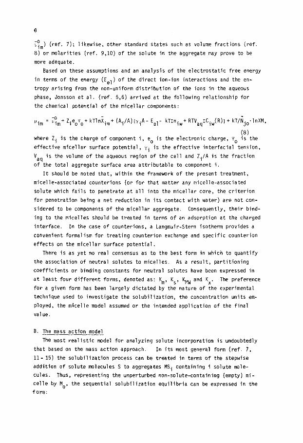
Based on these assumptions and an analysis of the electrostatic free energy
in terms of the energy (Eel) of the direct ion-ion interactions and the en-
tropy arising from the non-uniform distribution of the ions in the aqueous
phase, Jonsson et al. (ref. 5,6) arrived at the following relationship for
the chemical potential of the micellar components:
him = "s,t Zieoyo+ kTlnXim+ (Ai/A)[viA- Eel- kTzniw+ RTVaqcCiw(R)l+ kT/N jo*lnXM,
(81 where Zi is the charge of component i, e. is the electronic charge, y. is the
effective micellar surface potential, yi is the effective interfacial tension,
V aq
is the volume of the aqueous region of the cell and Zi/A is the fraction
of the total aggregate surface area attributable to component i.
It should be noted that, within the framework of the present treatment,
micelle-associated counterions (or for that matter any micelle-associated
solute which fails to penetrate at all into the micellar core, the criterion
for penetration being a net reduction in its contact with water) are not con-
sidered to be components of the micellar aggregate. Consequently, their bind-
ing to the micelles should be treated in terms of an adsorption at the charged
interface. In the case of counterions, a Langmuir?Stern isotherm provides a
convenient formalism for treating counterion exchange and specific counterion
effects on the micellar surface potential.
There is as yet no real consensus as to the best form in which to quantify
the association of neutral solutes to micelles. As a result, partitioning
coefficients or binding constants for neutral solutes have been expressed in
at least four different forms, denoted as: Km, KS, KMW and Kx. The preference
for a given form has been largely dictated by the nature of the experimental
technique used to investigate the solubilization, the concentration units em-
ployed, the micelle model assumed or the intended application of the final
value.
B. The mass action model -. -
The most realistic model for analyzing solute incorporation is undoubtedly
that based on the mass action approach. In its most general form (ref. 7,
ll- 15) the solubilization process can be treated in terms of the stepwise
addition of solute molecules S to aggregates MSi containing i solute mole-
cules. Thus, representing the unperturbed non-solute-containing (empty) mi-
celle by MO, the sequential solubilization equilibria can be expressed in the
form:
k +1
Mo + Sw ,* MS 1; (9)
k-l
k MSI + S w 3 MS2 ;
k-l
k MS
n-l + SW --=, MSn s
I,
(10)
(11) k-n
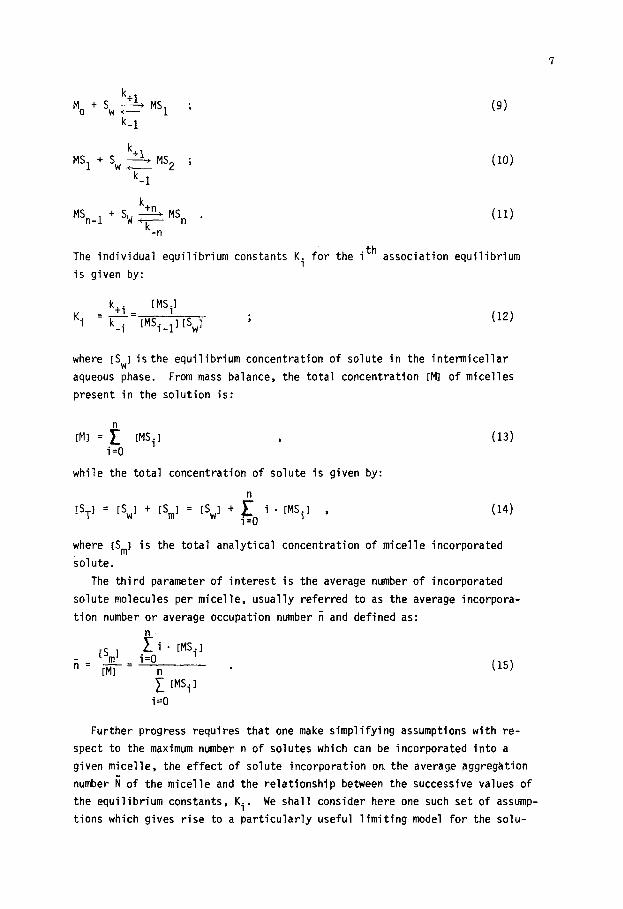
.th The individual equilibrium constants Ki for the 1 association equilibrium
is given by:
k. [MS11 Ki =p=iMS
i-I1 iswI ;
-i (12)
where [SW] isthe equilibrium concentration of solute in the intermicellar
aqueous phase. From mass balance, the total concentration [Ml of micelles
present in the solution is:
[MI = e IMSiI , (13) i=O
while the total concentration of solute is given by:
n
[ST1 = [SW1 + rSm1 = [SW1 + 1 i . [MSil , i=O
(14)
where [S,I is the total analytical concentration of micelle incorporated
solute.
The third parameter of interest is the average number of incorporated
solute molecules per micelle, usually referred to as the average incorpora-
tion number or average occupation number ii and defined as:
EmI f i - [MSil
ii= [Ml= i=O
~ [MSil
(15)
i=O
Further progress requires that one make simplifying assumptions with re-
spect to the maximum number n of solutes which can be incorporated into a
given micelle, the effect of solute incorporation on, the average aggregation
number i of the micelle and the relationship between the successive values of
the equilibrium constants, Ki. We shall consider here one such set of assump-
tions which gives rise to a particularly useful limiting model for the solu-

bilization process (ref. 7): 1) the micellar aggregation number is indepen-
dent of the presence of the solute (i constant for all aggregates); 2) the
solute entry rate is independent of the number of solute molecules present
in the micelle (k+i =k+ for all i); 3) the solute exit rate is directly pro-
portional to the number of solutes present in the micelle (i.e., k_i=i * k_);
and 4) the solubilization capacity of the micelle is "infinite" (n+-).
Based on these assumptions, the fraction Pi of micelles containing exactly
"i" solutes can readily be shown to be (ref. 7):
[MS+ _ a pi=--
[MI il exp (-6) 9 (16)
which corresponds to a Poisson distribution with an average occupation number
of:
n’= KmISwl ,
where:
k+ Km = F
(17)
(18)
is the equilibrium constant for incorporation of the first solute into an
empty micelle. Combining Eq. 17 with the definition of ti (Eq. 15) leads to
the extremely interesting relationship (ref. 7):
(19)
This last equation implies that, within the limits of this model, solute
incorporation can be treated as if it were governed by the following pseudo-
equilibrium:
Km SW + M 1 S, (20)
with a net solute entry rate of:
Vt = k+rM~ ISJ (21)
and a net exit rate of:
V = k_ [S,] = k_ii[M] . (22)
. Since k_ is a pseudo-first rate constant, l/k_ is in effect the mean life
time of a given solute molecule inside a micelle (ref. 7); k, is a bimolecular

rate constant which can be expressed as:
k+ = Bkdif 1 (23)
where kdif is the rate constant for diffusion-controlled encounters between
the aqueous solute and the micelles and B is the net efficiency of solute
incorporation per encounter.
Slightly different initial assumptions give rise to alternative statistical
distributions of the solute among the micelles. Virtually all of these alter-
native distributions (like the Poisson distribution itself) have a conmon
origin in the binomial distribution (ref. ll- 16) and hence reduce to a
Poisson distribution in the limit of low incorporation number (either ii<< 1
or tic< m). Indeed, despite considerations to the contrary (ref. 16), most of
the data obtained to date are compatible with a Poisson or Poisson-like dis-
tribution for neutral solutes (ref. 7,17). Perhaps the strongest evidence
for the applicability of a Poisson distribution is the fact (ref. 7,17) that
values of Km determined from saturation measurements at the solubility limit
are generally in reasonable agreement with those determined at very low incor-
poration numbers (or, alternatively, with those required to simulate kinetic
data at low substrate concentrations).
From a practical standpoint, the use of Km to describe solute incorporation
suffers from the inconvenience of requiring a knowledge of the concentration
of micelles. Although the micelle concentration [MI can in principle be
calculated by dividing the concentration of micellized surfactant ([D,]) by
the average aggregation number (i):
[MI = rD,l/F;r (24)
reliable aggregation numbers (in the absence of solutes!) are available for
only a few detergents. As a consequence, the more comnon form of expressing
the solute incorporation constant has been as KS, defined in terms of ID,]
and trivially related to Km:
KS = lS,l Km
IS,1 ID,1 = F (25)
This definition of KS, which in essence reduces the solubilization process
to a pseudophase equilibrium of the solute between a micellized surfactant
phase and the aqueous phase:
K
'W + Dm +--- m --s+S (26)
10
has played a central role in more recent pseudophase models for analyzing
micellar effects on the kinetics of ground state reactions (ref. 18- 20).
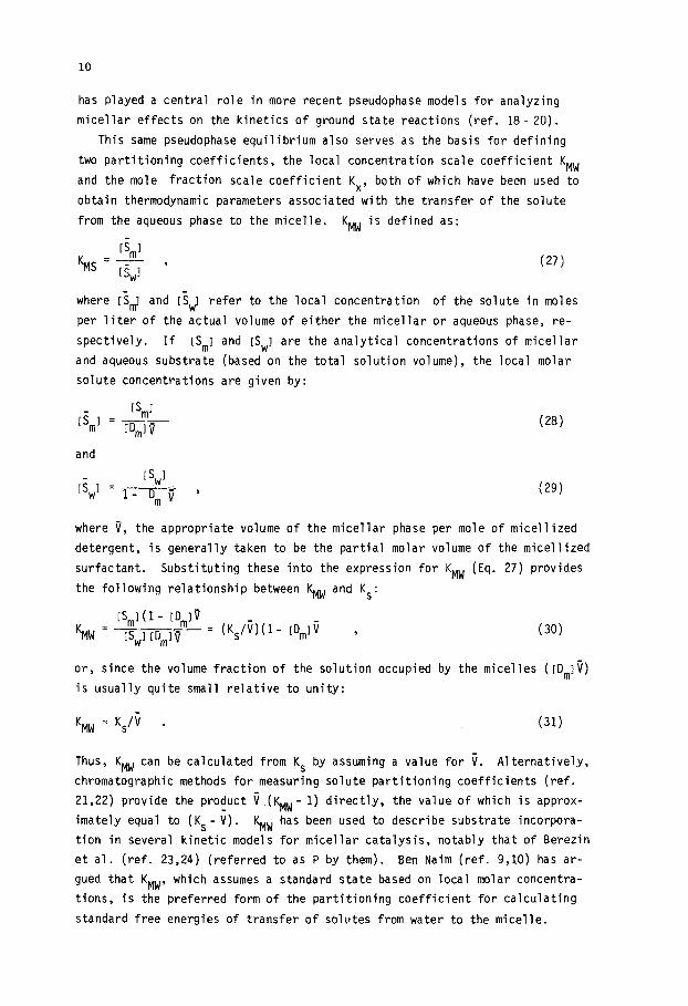
This same pseudophase equilibrium also serves as the basis for defining
two partitioning coefficients, the local concentration scale coefficient KMW
and the mole fraction scale coefficient Kx, both of which have been used to
obtain thermodynamic parameters associated with the transfer of the solute
from the aqueous phase to the micelle. KMW is defined as:
IS,1 KMS=Y-- I
IS,1 (27)
where I?$ and IsJ refer to the local concentration of the solute in moles
per liter of the actual volume of either the micellar or aqueous phase, re-
spectively. If IS,] and IS,] are the analytical concentrations of micellar
and aqueous substrate (based on the total solution volume), the local molar
solute concentrations are given by:
[S,l Ii,1 = _
IBmlV
and
IS 1 IS,1 = * ,
mv
(28)
(29)
where v, the appropriate volume of the micellar phase per mole of micellized
detergent, is generally taken to be the partial molar volume of the micellized
surfactant. Substituting these into the expression for KMW (Eq. 27) provides
the following relationship between KMW and KS:
KMW =
IS,l(I - ID,lil
IS,1 IBmlY = (K&(1- ID,I~ , (30)
or, since the volume fraction of the solution occupied by the micelles ([D,I~)
is usually quite small relative to unity:
'MW = KS/i . (31)
Thus, KMW can be calculated from KS by assuming a value for i. Alternatively,
chromatographic methods for measuring solute partitioning coefficients (ref.
21,22) provide the product i (KMW- 1) directly, the value of which is approx-
imately equal to (KS-i). KMW has been used to describe substrate incorpora-
tion in several kinetic models for micellar catalysis, notably that of Berezin
et al. (ref. 23,24) (referred to as P by them). Ben Naim (ref. 9,lO) has ar-
gued that KMW, which assumes a standard state based on local molar concentra-
tions, is the preferred form of the partitioning coefficient for calculating
standard free energies of transfer of solutes from water to the micelle.

11
The dimensionless mole fraction partitioning constant, Kx, is defined as:
X Kx=ji?l .
W
The expression for the solute mole
ward: IS..1 xw =
ISwGIC+ 55.5 *
(32)
fraction in the aqueous phase is straightfor-
(33)
As for Xm, it has become customary to express the mole fraction of the micelle-
incorporated solute in terms of its mole fraction in a "dry" micelle (ref. 25);
1 .e., to ignore water as a possible third micellar component and simply write:
xm = IS,1
[S,l+IDmI ’
Combining these equations provides an expression for Kx in terms
ytical concentrations of the detergent and solute:
Kx = 55.5IS,I 55.5 KS
ISw~(ISrn~ + ID,11 = 1+ KSISw~
(34)
of the anal-
(351
At low degrees of solute incorporation (KSISwl = lSml/IDrn~ << 11, Kx can be
interrelated to KMM and KS via:
Kx = 55.5 KM,,, i = 55.5 KS .
The principal application use of
(36)
Kx has been in the calculation of.standard
free energies of transfer of solutes from water to the micelle based on the
unitary scale (mole fraction standard state). Implicit in this use of Kx is
the supposition that the solute is distributed homogeneously within the mi-
celle, forming an ideal mixture with the detergent (vi& in&z).
In sumnary, the following relationships can be established between the dif-
ferent binding constants:
Kx = 55.5. KS ; (37)
K, = K&m.55..5 ; (38)
KS = sWjrn ; (39)
KM = KS * i . (40)
Thus, if the value of the partition coefficient is known on any given scale,
the corresponding values on the other scale can be calculated by assigning

12
values to the parameters irn and i. For the commonly used detergents SLS and
CTAB, the following values of Qrn and i (in the absence of added electrolytes)
are recommended: vrn (CTAB) = 0.363 dm3/mole (ref. 26,27); vrn (SLS) = 0.25
dm3/mole (ref. 28-30); I! (CTAB)=84 (ref. 31); m (SLS) = 58 (ref. 32,33).
C. Standard free energies of transfer of solutes from water to micelles
In order to transform experimental solute partitioning constants or incor-
poration coefficients into thermodynamically meaningful standard free ener-
gies of transfer (SFET) of the solute from water to the micellar environment,
one must adopt an appropriate reference state for the solute in the micellar
pseudophase. In addition, one should be aware of potential limitations inher-
ent in the model on which the partitioning constants or incorporation coeffi-
cients themselves are based. At present, the choice of the "best" standard
state is a matter of controversy. On the one hand, Ben Naim (ref. g,lO) advo-
cates on the basis of statistical mechanical arguments the use of the molarity
scale (total concentrations) for the calculation of the SFET values. On this
scale, the SFET would provide a measure of the difference in the solvation
properties of the two phases .with respect to the solute. On the other hand,
Tanford (ref. 34), following Gourney (ref. 35) and Kauzmann (ref. 36) sug-
gests that the SFET be expressed in the unitary system (mole fraction stan-
dard state). On this latter scale, the SFET should incorporate all interac-
tions of the solute with the micelle. In this context, however, it should be
re-emphasized that the solute mole fraction in the micellar pseudophase is
usually expressed in a form which ignores water as a potential component of
the micellar pseudophase.
As mentioned before, the mass action law probably provides the most mean-
ingful approach for treating the association of molecules to micelles. From
a practical standpoint, however, the two-phase of pseudophase model is a much
more convenient approximation, both for analyzing experimental data for solute
incorporation and for calculating SFET values. Thus, the respective chemical
potentials of the solute in the micellar and aqueous phases can be expressed
in the form:
u,=$,(T,P) + RTlnXmym ; (41)
u,=~i(i,P) + RTlnXwyw . (42)
At equilibrium, the SFET can be written as:
o_ 0 0 'mYm ut, - u~-F~~ = -RTln xwy~ = -RTlnKx , (43)

13
where xm and xw represent the mole fractions of the solute in the micellar
and water phases, respectively, and y, and yw are the corresponding activity
coefficients. For a neutral solute, yw can usually be assumed to be unity
and, when the partitioning coefficient is independent of the surfactant con-
centration or average solute incorporation number, ym can also safely be
taken to be unity. Departures from this ideal behavior can, of course, be
treated via inclusion of nonideality of mixing. The simplest approach is
probably that based on the so called regular solution theory (ref. 37,38)
which was used by Mukerjee (ref. 39) to interpret the non-ideal behavior of
the distribution of benzoic acid derivatives betweeen water and micelles of
nonionic surfactants. In this approach, the activity coefficient of the com-
oonent solubilized in the micelle is assumed to be given by:
Lny, = (l-x,)* * o/RT , (44)
where u is an adjustable interaction energy parameter which approaches RTlnv,
as xm tends to zero. In the present work, we have assumed the validity of
the pseudophase model, ideal behavior and a unitary standard state for all cal-
culations of SFET from experimental partitioning coefficient data.
Having established a basis on which to calculate SFET values from experi-
mental solute incorporation data, it is of interest to analyze these values
for trends which might provide insight into the nature of the intermolecular
forces which contribute to solute incorporation in the micellar pseudophase.
One potentially useful approach is to assumed that A,: is an additive-constitu-
tive property of the solute molecule; i.e., that the SFET of a molecule from
water to the micelle can be factored into individual group contributions from
its hydrophilic and hydrophobic constitutent molecular moieties. This assump-
tion, extensively used for the partitioning of solute in non-miscible solvents
(ref. 40) permits one to separate the total free energy (AU:) for transfer of
the molecule from one solvent to another (from water to micelles in this case)
into a hydrophilic Component (bEhy) and a hydrophobic component (Au:). If the
latter is further assumed to reflect individual contributions from "nC" hydro-
phobic groups of the molecule (ref. 41), AU: can be written in the form:
Aut ’ = Au0 + ncApF . hy
(45)
In agreement with this assumption, Au: for a set of related solutes (or re-
lated micelle-forming surfactants) is frequently found to be a linear func-
tion of the number of homologous hydrophobic groups present in the solute
(or surfactant). Similar linear free energy relationships have also been
found in virtually all studies of the distribution of solutes between water

14
and bulk nonaqueous solvents (ref. 40,41). In view of the uncertainties sur-
rounding the choice of the appropriate standard state for expressing A$ in
micellar systems, it should be noted that the slopes of correlations of AU:
vs. the number of homologous functional groups, which presumably reflect the
contribution of that group to the overall transfer free energy, are indepen-
dent of the standard state chosen. Thus, it is only the intercept, which in-
coporates the contributions from the remaining groups of the solute molecule,
that is dependent on the choice of the standard state (ref. 42).
Leo et al. (ref. 41) have also considered the case of a family of solute
molecules in which the number "n h' of hydrophilic groups is changed, in which
case Eq. 45 takes the form:
Ap”t = nhbEy + Au; .
When the solubilizate is a relatively hydrophobic ion, for example, a car-
boxylate, alkyl phenoxide or arylsulfonate ion, the total AU: also contains
an electrostatic contribution, APO ; i.e.: el
Aut ’ : Au0
b’ + ncAuF + Auzl , (47)
However, attempts to interpret the AuEl in terms of a straightforward
electrostatic contribution may be complicated by the fact that counterionic
orqanic solutes tend to form ion pairs with the surfactant monomer in the
aqueous phase, in which case it is the uncharged (if both solute and deter-
qent are monovalent) ion pair that is transferred from water to micelle (ref.
43,44).. In reality, ion pairinp of counterionic substrates is probably one
out of a variety of potentially unique interactions. The existence of such
(often unperceived) interactions merely emphasizes the fact that the solute
activity in the intermicellar aqueous phase cannot a ptiohi be presumed to
be equivalent to that in a bulk micelle-free water phase.
An important extension of the thermodynamic analysis of micellar solubili-
zation is the separation of the free energy of transfer into its constituent
enthalpic and entropic components. In principle, the standard enthalpy of
transfer (AH:) can be obtained from the temperature dependence of AU: (ref.
40). In practice, however, this method proves to be rather imprecise because
the changes in AU: with temperature are usually small. Furthermore, in view
of the possibility of temperature-dependent changes in micellar structure,
measurements over a wide temperature range are inherently undesirable. Di-
rect calorimetric measurements are therefore preferable whenever possible.
Since only very limited data of this type are presently available, compre-
hensive calorimetric determinations of standard enthalpies (AH:) for the

15
transfer of solutes from water to micelles as a function of the hydrophilic
or hydrophobic properties of the solute, the nature of the detergent and the
composition of the solution should be of inestimable value. Once the enthalpy
of transfer has been determined, the standard entropy of transfer can, of
course, be calculated from the relationship:
.Ci ; = (rH; - A+T . (48)
Like A,:, the magnitude of AS: is also a function of the choice of the refer-
ence state, the values of AS: calculated on the unitary (mole fraction scale)
being more positive than those calculated on the molarity scale.
A central question in the thermodynamic analysis of solute incorporation has
been (and continues to be) the interpretation of the origin and significance
of the incremental hydrophobic contribution (AU:) per methyl or ethylene group
to the overall free energy of transfer of the solute from water to the micelle.
Thus, using the unitary system and Wishnia's results (ref. 45), Tanford (ref.
34) found that the SFET values for transfer of alkanes from water to SLS mi-
celles obey the linear expression:
A,; = -1.934- 0.771 nc , (49)
where n c is the total number of alkyl carbon atoms in the alkane. The slope
of -0.771 Kcal/mole which corresponds to the contribution to Au: from each
of the carbon atoms of the alkane molecule is similar to that found for the
transfer of alkanes from water to hydrocarbon solvents (-0.88 Kcal/mole) (ref.
34), suggesting that alkanes are located in a micelle environment similar to
that of a nonaqueous hydrocarbon-like solvent. The non-zero value of the con-
stant term has been attributed by Tanford (ref. 34) and by Birdi (ref. 46) to
the difference in the contributions of -CH3 and -CH2 groups to the SFET. From
the data for transfer to hydrocarbon solvents, Tanford calculated a ALI: value
of -0.88 Kcal/mole for a -CH2 group versus a contribution of ca. -2.1 Kcal/mole
for each -CH3 group, the difference being attributed to the greater degree of
contact of the -CH3 group with water as compared to a -CH2 group. In addition,
it is also necessary to take into account that the calculation attributes most
of the differences in translational entropy cf the entire molecule to the ter-
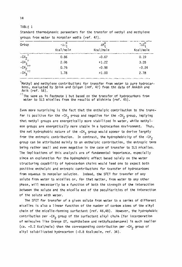
minal methyl group. According to Spink and Colgan (ref. 47), the contributions
of the methyl and methylene groups to the standard free energies, enthalpies
and entrooies of transfer of aliphatic molecules from water to micelles and
from water to hydrocarbon media (Table 1) are characterized by a more positive
enthalpy, a much more positive entropy and consequently, a larger incremental
contribution to AJJ: for the methyl group relative to the methylene group.
16
TABLE 1
Standard thermodynamic parameters for the transfer of methyl and methylene
groups from water to nonpolar media (ref. 47).
Group -&o, AH;
Kcal/mole Kcal/mole
TASF
Kcal/mole
-CH2+ 0.86 -0.67 0.19
-CH3+ 2.06 t1.22 3.28
-CH3'+ -CH2++
0.76 -0.98 -0.24
1.78 +1.00 2.78
'Methyl and methylene contributions for transfer from water to pure hydrocar- bons, evaluated by Spink and Colgan (ref. 47) from the data of Amidon and Anik (ref. 56).
'+The same as in Footnote 1 but based on the transfer of hydrocarbons from water to SLS micelles from the results of Wishinia (ref. 45).
Even more surprising is the fact that the enthalpic contribution to the trans-
fer is positive for the -CH3 group and negative for the -CH2 group, implying
that methyl groups are energetically more stabilized in water, while methyl-
ene groups are energetically more stable in a hydrocarbon environment. Thus,
the net hydrophobic nature of the -CH3 group would appear to derive largely
from the entropic contribution. In contrast, the hydrophobicity of the -CH2
group can be attributed mainly to an enthalpic contribution, the entropic term
being rather small and even negative in the case of transfer to SLS micelles.
The implications of this analysis are of fundamental importance, especially
since an explanation for the hydrophobic effect based solely on the water
structuring capability of hydrocarbon chains would lead one to expect both
positive enthalpic and entropic contributions for transfer of hydrocarbons
from aqueous to nonpolar solution. Indeed, the SFET for transfer of any
solute from water to micelles or, for that matter, from water to any other
phase, will necessarily be a function of both the strength of the interaction
between the solute and the micelle and of the peculiarities of the interaction
of the solute with water.
The SFET for transfer of a given solute from water to a series of different
micelles is also a linear function of the number of carbon atoms of the alkyl
chain of the micelle-forming surfactant (ref. 46,48). However, the hydrophobic
contribution per -CH2 group of the surfactant alkyl chain (for incorporation
of molecules like Orange OT, naphthalene and methylazobenzene) is much smaller
(ca. -0.2 Kcal/mole) than the corresponding contribution per -CH2 group of
alkyl solubilizated hydrocarbon (-0.8 Kcal;mole, ref. 34).

17
Of particular interest is the fact that Treiner (ref. 42) has found an
excellent correlation between the constants for partitioning of polar solutes
between water and ionic micelles and those for partioning between water and
n-octanol. The existence of such a correlation implies that solutes of re-
latively high polarity of the type studied by Treiner exhibit intrinsic dif-
ferences in hydrophilicity (probably related to the extent and the strength
of hydration) in the same manner that the hydrophobicity of a given molecule
varies with the number of constituent carbon atoms.
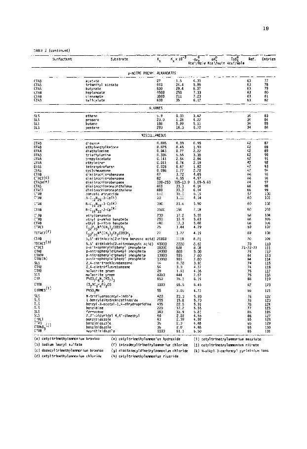
In Table 2, we have collected data for the incorporation of a large number
of substrates into a variety of different micelles. Since an extensive anal-
ysis of the individual data is beyond the scope of the present work, only the
major trends will be considered. In the cases where sufficient data were
available, values of A,: were determined for families of homologous molecules.
These values, together with analogous values for transfer from water to bulk
solvents, are collected in Table 3.
Table 2 also includes the corresponding directly measured enthalpies of
transfer for those (few) solutes for which data are available, along with
calculated values of the entropic contribution (TA.$) to the transfer. These
data are restricted to the studies by Larsen and Magid (ref. 49) and, more re-
cently, by Spink and coworkers (ref. 47,50). The data of Larsen and Magid
for the transfer of benzoic acids from water to the CTAB micelle (Table 2)
clearly suggest that both enthalpic and entropic factors contribute to the
overall free energy of transfer to the micelle. This is reinforced by the
work of Spink et al. (ref. 47,50), in which the thermodynamic functions for
transfer of aliphatic alcohols from water to deoxycholate micelles were mea-
sured (Table 2). Although the free energy of transfer was found to be a
linear function of the number of carbons in the aliphatic chain of the al-
cohol, neither the entropic nor the enthalpic contributions to the SFET ex-
hibit simple linear relationships when considered separately. Enthalpy-
entropy compensation resulting in linear correlations of the free energy
may prove to be the rule rather than the exception.
In general terms, the factors which tend to favor the transfer of a solute
from the aqueous phase to the micelle can be sumnarized as follows: 1) an
increase in the overall hydrophobicity of the solute; 2) an increase in the
length of the hydrocarbon chain of the surfactant; 3) a change in the relative
positions of hydrophilic substituents from a para or diametrical orientation
to an ortho or adjacent position; and 4) the presence of aryl moieties in the
solute.
These latter two factors are apparently indicative of additional contribu-
tions which arise from the interaction of certain types of aromatic residues

18
TABLE 2
Association constants (Ks and Kx), free energies (AU:). enthalpies (ntiy) and entropies (TdST) of transfer of molecules from
the +wxus to the micellar pseudophase
Surfactant Substrate Ks Kxx 1O-3 .PUO AHO TASO Kcal/kle Kcaljmole Kcal/&le
Ref. Entries
CTAB
P-naphtha1 1390 phenol 270 o-methvl ohenol 490
:TAB :TAE :TAB :TAB :TAB TAB TAB TAB :TAB :TAB TAB
b-ethyi phenol p-n-propyl phenol p-t-butyl phenol p-t-tmyl phenol p-set-butyl phenol m-t-butyl phenol phenoxide p-methyl phenaxlde p-ethyl phenaxide P-n-propyl phenoxide D-t-butvl ohenoxide
790 1400 1700 4300 1900 1700 IBOO 3300 5300 9600 10000
CTAB b-t-awi phenoxide 27000 CTAB benzoic acid 378 CTAB p-methyl benzoic acid 743 CTAB p-ethyl benroic acid 1727 CTAB p-t-butyl benzolc acid 3600 CTAB CTAB CTAB CTAB CTAB
:::!b) SLS SLS SLS SLS SLS SLS SLS SLS SLS SLS SLS SLS
benraate 2865 p-methyl benzoate 2865 p-ethyl benzoate 4015 p-t-butyl benzoate 5625 aniline 116 p-methyl aniline 193 phenol 50 p-methyl phenol 83 p-ethyl phenol 163 p-n-propyl phenol 270 p-t-butyl phenol 378 p-Gamy1 phenol 1041 aniline aniline H+
83 450
benzoic acid 193 p-methyl benzoic acid 228 p-ethyl benroic acid 448 p-t-butaxy benroic+acid 880 p-methyl aniline H 628
77.1 15.0 27.2 43.2 77.7 94.4 239 105 94.4 100 183 294 533 555 1500 21.0 41.2 95.8 200 159 159 223 313 6.44 10.7 2.78 4.61 9.0 15.0 21.0 57.0 4.61 25.0 10.7 12.7 24.9 44.8 34.9
6.6 58 5.7 57-58 6.0 57-58 6.3 6.7 6.8 7.3 6.9 6.8 6.8 7.2 7.5 7.8 7.9 8.4 5.9 6.3 6.8 7.2 7.1 7.1 7.3 7.5 5.5 5.5 4.7 5.0 5.4 5.7 5.9 6.5 5.0 6.0 5.5 5.6
::: 6.2
57-58 57-58 57-58 57-58 58 58 57-58 57-50 57-58 57-58 57-58 57-58 57 !3 :: 57 57
::
:: 57 57 ii 57
:: 57 57
:: 57 ii 57
: 3 4 5 6
: 9 10 11 12 13 14 15 16 17 18 19 20 21 22
2243 25 26 27
zi 30
:: 33 34 35
i7” 38
ALKYL ALCOHOLS
Sodium deoxycholate Sodium deaxycholate Sodium deoxycholate Sodium deoxycholate SLS SLS SLS
SLS SLS Sodium cholate Sodi m deoxycholate OTAB C) Y OTAB OTAB OTAB OTAB DTAB OTAB
butanol pentanol hexanol heptanol butanol pentanol hexanol
heptanol heptanol heptanol heptanol ethanol propanol P-propan butanol t-butanol methanol hexanol
0.95 0.053 2.34 6.60 a.94 2.20 0.122 2.73 3.70 6.51 :: :z 9.21 0.51 3.68 1.87 5.57 41 36.4 2.02 4.49 1.22 5.72 4": 42 5.4 0.30 3.38 61 13.0 0.72 3.90 61 4": 40.5 2.25 4.57 45
108.5 6.02 5.16 :: 111.7 6.20 5.15 62 :; 37.8 2.10 4.51 55.9 3.10 4.74 6"; 4": 0.010 0.18 1.36 42 0.033 0.59 2.06 42 :Y 0.027 0.49 1.95 52 0.094 1.69 2.68 :2' 53 0.045 0.81 2.25 42 54 0.29 5.19 3.34 42 55 0.87 15.7 3.99 42 56
- PHENONES
SLS acetophenone 35.2 1.95 4.47 84 SLS prapiophenone 81.0 4.50 4.86 84 :;: SLS isabutyrophenone 129.3 7.16 5.24 a4 59 SLS p-methoxiacetophenone S1.7 2.87 4.70 84 60 SLS xanthone 109.0 6.05 5.14 84 61
BENZOIC ACIOS
CTAB o-nitrobenzoic 28.7 1.59 4.35 -2.29 2.06 CTAB o-chlorobenzoic 36.3 2.01 4.49 -2.22 2.27 :; :: CTAB o-aminobenzaic 33.0 1.83 4.43 -5.09 -0.66 49 64 CTAB p-aminobenzoic 18.9 1.05 4.10 -1.78 2.32 CTAB o-hydroxybenzoic 59.2 3.29 4.77 -6.19 -1.42 4"; :6" CTAB p-hydroxybenzoic 12.0 0.67 3.83 -9.25 -5.42 49 67
p-NITRO PHENYL ALKYL CARBOXYLATES
N-Myristail-histidine, CTAB mixed micelles acetate N-Hyristoil-histldine... N-Myristoil-histldlne...
propionate butyrate
N-Myristoil-histidind... valerate N-Myristail-histidine... hexanoate CTAB acetate CTAB acetate
30.1
:E 769 2000
CTAB butyrate 530 CTAB heptanoate 4500
1.67 4.38 6.0 5.13 :; 2 19.2 5.82 42.7 6.29 :z :Y 111 6.85 59 72
3.0 4.72 79-80 1.5 4.31 81 :: 29.4 6.07 82 75 250 7.33 83 76
19
TABLE 2 (continued)
Surfactant Substrate KS Kx~10-3 ' Ati0 T&o Kc&,, Kca&le Kcal/r!,&
Ref. Entries
p-NIT!+0 PHENYL ALKANOATES
CTAB acetate CTAB trimethyl acetate CTAB butyrate CTAB heptanoate CTAB cinnamate CTAB salicvlate
27 1.5 4.31 440 24.4 5.96 :: :: 530 29.4 6.07 79 4500 250 7.33 :: 3800 211 7.23 630 35 6.17 ::
:; 82
ALKANES
SLS SLS SLS SLS
ethane 5.9 0.33 3.42 34 83 propane 23.0 1.28 4.22 34 butane 5.99 5.13 34 :z pentane 16.3 5.72 34 86
MISCELLANEOUS
DTAB dloxane 0.005 0.09 0.95 42
OTAB ethylmethylketone 0.025 0.45 1.90 OTAB diethvlamine 0.043 0.77 2.22 :: mm DTAB DTAB OTAB DTAB
$,(d) CTAOHce) CTAB CTACI CTAB CTAB
CTAB
CTAB
CTAB CTAB CTAB CTACI
TOTACl(f)
CTAB oocnci'"' CTACl CTAB CTAOH CTAF(h) SLS CTAB CTAB SLS CTA
CTAB
CTAMES("
SLS SLS SLS SLS SLS SLS CTACl
trietiylamine propy1acetate ethylether tetrahydrofura" cyclohexanone dinitrochlorobenzene dinitrachlarobenzene dinltrochlorobenzene dinitrochlaronaphthalene dinitrochloronaohthalene
0.306 5.55 3.38 0.141 2.54 2.94 0.041 0.74 2.19 0.026 0.47 1.92 0.098 1.77 2.72 67 3.72 4.85 82 4.55 4.97 100-250 555-13.9 5.09-5.63 600 600
benzoic anhydrihe 650 N-C12Hz5-3-Cpk) 20
N-C14Hzg-3-C@) 390
N-C,,",,-~-CP(~) 3500
ethylbenzoate 200 ethyl p-amino benzoate 250 ethyl p-nitro benroate 240 C,2H,,N+(CH3),COOCH3 26
C12H25N+(CH312CH2C00CH3, 22
5,5'-dithiobis(Z-nitro benroic acid) 20000 5.5'.dithiobis(Z-nitrobenzoic acid) 40000 p-nitrophenyldiphenyl phosphate 16000 p-nitrophenyldiphenyl phosphate 16000 p-nitraphenyldiphenyl phosphate 10000 p-nitrophenyldlphenyl phosphate 10000 2,4-dinitrochlorobenzene 2,4-dintrofluorobenzene :: malachite green 29 malachite green 8000 PhC02C6H3(N02)3 650
(02NC6H40)2~o 1000
PhS03Me 55
N-trifluomacetyl-indole 420 1-benzyldihydronicotinamide 285 benzyl-3-acetyl-1,4-dihydropridine 405 benzidine 220 ferracene 340 2,2'-bipyridyl 4,4'-dimethyl benzimidazole :: benzimidazole 36 benzimidazole 36 "aphthimidarole 1100
33.3 33.3 36.1 1.11
6.14 6.14 ii
21.6
194
12.2 13.9 13.3 1.44
6.19 4.14
5.90
7.18
5.55 5.63 5.66 4.29
1.22
1110
2220 888 888 555 555 0.78 3.0 1.61 444 36.1
4.19
8.21 8.62 8.08 8.08 7.80 7.80 3.93 4.72 4.36 7.67 6.19
55.5
3.05
6.45 67
4.73 66
23.3 5.93 15.8 5.70 22.5 5.91 12.2 5.55 18.9 5.81 2.22 4.55 2.39 4.59 2.0 4.48 2.0 4.48 61.1 6.50
42
:i 42 42
:: 64
67 60
60
60
6? 68 69
69
70
71-z-73 74
El‘! 74 74 75
:z
102
103
104 105 106 107
108
109
110 111 112 113 114 115 116 117 118 119
120
12L 123 124 125 126 127 128 129 130 131
(a) cetyltrimethylamvxium bromide (e) cetyltrimethylamnonium hydroxide (i) cetyltrimethylammnium mesylate
lb) sodium lauryl sulfate (f) tetradecyltrimethylrmnonlum chloride (j) cetyltrlmethylanrrmnium nitrate
(cl dodecyltrimethylammnium bromide (9) dioctadecyldimethylammnium chloride (k) N-alkyl-3-carbamoyl pyridinium ions
(d) cetyltrlmethylammonium chloride (h) cetyltrimethylawnlum fluoride

20
TABLE 3
Hydrophobic (AU:), hydrophilic (APE,) and electrostatic (APE) contributions to the free
energies of transfer of molecular moieties from water to micelles or to hydrophobic solvents
Surfactant or Solvent Family of Molecules o(d)
_AUC o (d)
-"uhy -Au;(e) Ref.
Kcal/mole Kcal/mole Kcal/mole
CTABLa' CTAB CTAB CTAB CTAB CTAB CTAB CTAB
SLS SLS SLS SLS
;:;B(c)
Lecithin Octanol Octane SLS n-heptane n-heptane n-heptane n-heptane p-alkyl benzenes
p-alkyl phenols 0.32 p-alkyl phenoxides 0.31 p-alkyl benzoic acids 0.34 p-alkyl benzoates 0.12 p.alkyl anilines 0.27 aromatic hydrocarbons 0.83 p-nitrophenyl alkyl carboxylates 0.63 N-alkyl-3-carbamoyl-pyridinium
ions 0.75 alk 1
r phenols 0.36
P-a kyl benzoic acids 0.29 p-alkyl anilines p-alkyl anilines x Ht
0.39 0.17
aromatic hydrocarbons 0.55 aliphatic alcohols 0.60 aliphatic alcohols 0.60 aliphatic alcohols 0.66 aliphatic alcohols 0.55 aliphatic alcohols 0.79 aliphatic alcohols 0.81 aliphatic hycrocarbons 0.77 alkyl phenols 0.78 p-alkyl benzoic acids 0.90 p-alkyl anilines 0.98 p-alkyl benzenes 0.80 p-alkyl benzenes 0.75 carboxilic acids 0.825 aliphatic alcohols 0.821
n-heptane aliphatic alcohols aliphatic hydrocarbons aliphatic hydrocarbons p-alkyl benzenes p-alkyl benzenes
0.884 1.00
5.7 6.9 ' 6.0 7.0 '
45:: 3.4
3.3 4.66
45::
i.2"
>
0:95 0.95 0.068
0.00 0.22 0.59
44.63 -3.9 -1.34
1.2 57-58
57-58
57
60 57-58 57
1.2 57 57
::
ti 42 42 42 34 57
:: 57 34 34
:1 34
(a), (b) and (c) as defined in Table 2.
(d) In terms of unitary system; i.e., using the molar fraction scale and from Eq. 45.
(e) Neglecting ion paring formation (Eq. 47).
with the micellar microenvironment. Thus, substitution of one of the protons
of benzene by a hydrophilic substituent (-OH, -COOH, -NH*) gives rise to a
much stronger interaction with both CTAB and SOS micelles. Due to its hydra-
tion, the hydrophilic group would tend to remain near the micelle surface,
enhancing the interaction of the r: electron cloud of the benzene ring with the
electric field produced by the charged surfactant head groups at the micelle
surface. This explanation is consistent with the heats of solubilization of
phenol and p-nitrophenol in CTAB as compared with other solvents (ref. 49)
and with the SFET data for transfer of phenols, benzoic acids, anilines and
benzene from water to n-heptane (Table 3). In fact, the transfer of benzene

21
from water to n-heptane is more favorable relative to the other solutes, a
trend which is opposite to that observed for transfer from water to CTAB mi-
celles. Finally, the presence of an aryl group in the solute seems to favor
a stronger interaction with cationic micelles than with anionic micelles, sug-
gesting the existence of a specific interaction between the cationic head
groups of the micelles and the aromatic ring of the solute molecule. Both
NMR and absorption spectroscopy (ref. 51- 55), as well as enthalpies for
transfer of phenols from water to 0.1 M CTAB (ref. 49) provide corroborative
evidence for this type of interaction.
III. EXPERIMENTAL METHODS
The experimental methods used to investigate the association of solutes to
micelles may be classified into three principal groups: solubilization, separa-
tion and spectroscopic methods. Other miscellaneous methods (e.g., kinetic
analysis of ground state reactions) will also be briefly considered. In gen-
eral, the method itself will be emphasized since the results obtained with
any of the different methods can be treated in a similar fashion to obtain
the required association constant. For nonionic substrates, this constant
usually expressed as KS (Eq. 25) which can in turn be transformed into KM,
KMH or Kx (Eqs. 37- 40).
A. Solubilization methods
The simplest and oldest of the methods are the solubilization methods,
is
which are based on the enhancement of the solubility of solutes in the pres-
ence of a surfactant at concentrations above its CMC. Excess pure solute,
in either its solid, liquid or vapor form (in the last case, the procedure
is known as the isopiestic method), must be in equilibrium with both the
micelle-associated solute and the free solute in the aqueous phase. In
essence, one determines the total amount of dissolved substrate in the pres-
ence ([St]) and absence ([Sol) of micelles. Assuming that the solubility
in the aqueous phase is unaffected by the presence of micelles, KS can be
written as:
$1 - [SoI
Ks=v[m .
Rearranging this equation, a plot of the saturation solubility ratio [St]/[Sol
versus the total detergent concentration [Dt] should be linear with slope KS:
w _ - - KSrDtl + I- K,CMC [SoI
. (51)
22
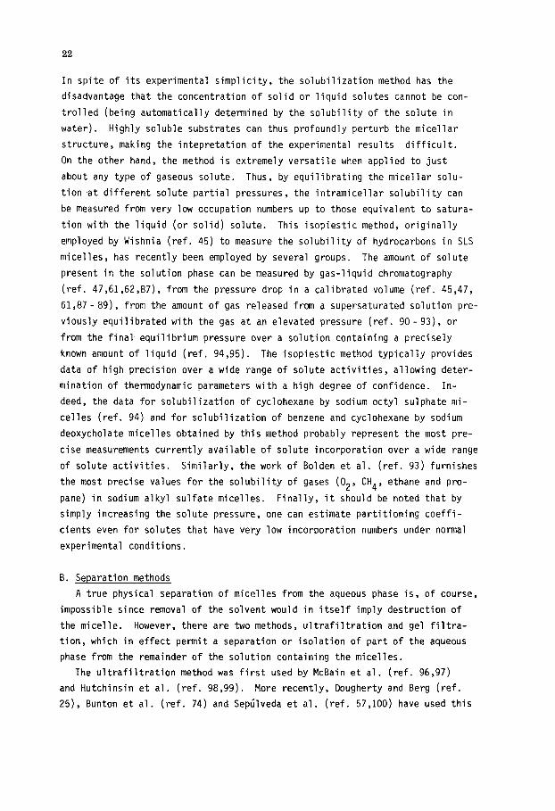
In spite of its experimental simplicity, the solubilization method has the
disadvantage that the concentration of solid or liquid solutes cannot be con-
trolled (being automatically determined by the solubility of the solute in
water). Highly soluble substrates can thus profoundly perturb the micellar
structure, making the intepretation of the experimental results difficult.
On the other hand, the method is extremely versatile when applied to just
about any type of gaseous solute. Thus, by equilibrating the micellar solu-
tion at different solute partial pressures, the intramicellar solubility can
be measured from very low occupation numbers up to those equivalent to satura-
tion with the liquid (or solid) solute. This isopiestic method, originally
employed by Wishnia (ref. 45) to measure the solubility of hydrocarbons in SLS
micelles, has recently been employed by several groups. The amount of solute
present in the solution phase can be measured by gas-liquid chromatography
(ref. 47,61,62,87), from the pressure drop in a calibrated volume (ref. 45,47,
61,87-89), from the amount of gas released from a supersaturated solution pre-
viously equilibrated with the gas at an elevated pressure (ref. 90 -93), or
from the final equilibrium pressure over a solution containing a precisely
known amount of liquid (ref. 94,95). The isopiestic method typically provides
data of high precision over a wide range of solute activities, allowing deter-
mination of thermodynamic parameters with a high degree of confidence. In-
deed, the data for solubilization of cyclohexane by sodium octyl sulphate mi-
celles (ref. 94) and for solubilization of benzene and cyclohexane by sodium
deoxycholate micelles obtained by this method probably represent the most pre-
cise measurements currently available of solute incorporation over a wide range
of solute activities. Similarly, the work of Bolden et al. (ref. 93) furnishes
the most precise values for the solubility of gases (02, CH4, ethane and pro-
pane) in sodium alkyl sulfate micelles. Finally, it should be noted that by
simply increasing the solute pressure, one can estimate partitioning coeffi-
cients even for solutes that have very low incorporation numbers under normal
experimental conditions.
B. Separation methods
A true physical separation of micelles from the aqueous phase is, of course,
impossible since removal of the solvent would in itself imply destruction of
the micelle. However, there are two methods, ultrafiltration and gel filtra-
tion, which in effect permit a separation or isolation of part of the aqueous
phase from the remainder of the solution containing the micelles.
The ultrafiltration method was first used by McBain et al. (ref. 96,97)
and Hutchinsin et al. (ref. 98,99). More recently, Dougherty and Berg (ref.
25), Bunton et al. (ref. 74) and Septilveda et al. (ref. 57,100) have used this

23
method to determine partitioning coefficients for a wide variety of solute
types. In a recent paper, Schechter et al. (ref. 101) have established the
conditions under which ultrafiltration experiments in micellar solution provide
the most reliable results. The method is based on the capacity of certain mem-
brane filters to retain species with molecular weights similar to, or greater
than, those of micelles. The technique requires a special stirrable and pres-
surizable filter cell fitted with an adequate (molecular weight retention,
solvent compatibility, solute adsorption) membrane filter. A small part of
the micellar solution (pre-equilibrated with the solute) is passed through the
membrane, the solute concentrations C, in the filtrate and filtrand solutions
measured and the fraction of micellar solute calculated from the equation:
EmI 'filtrand -c
-= filtrate $1 'filtrand
(52)
The method requires constant stirring of the solution and only a small
amount of filtrate should be collected for analysis in order to avoid a change
in the overall composition of the filtrand. Other problems may arise from ad-
sorption of surfactant or solute on to the membrane, from differences in the
rate of filtration of water relative to free monomer or solute and to stream-
ing potential effects. The principal advantages of the method are its range
of applicabiiity, relative simplicity and the fact that the micelle and sub-
strate concentrations can be varied over a wide range.
In the gel filtration method, the portion of the aqueous phase containing
the free solute is "separated" from the remainder of the solution by the use
of a cross-linked dextran gel (usually Sephadex) which excludes from its in-
terior species with molecular weights above a certain limit determined by the
characteristics of the gel (ref. 102). As long as the micelle has a molecular
weight larger than this exclusion limit of the gel, upon passage of a micelle-
containing mobile phase through a column of the gel the composition of the
solute within the gel phase should correspond to that of the aqueous phase
and hence contain only free surfactant monomers, non-micelle-bound solute
molecules and any low molecular weight electrolyte present. At surfactant
concentrations well above the CMC, the relative rate of migration (retention
volume) of an added low molecular weight solute will be a function of the par-
titioning coefficient of the solute between the micellar and aqueous phases.
Thus, given the parameters of the gel column, the degree of adsorption of the
solute in the gel matrix, the partial specific volume of the surfactant mole-
cule in the micelle and the molecular sieving constant, KMW can be evaluated
experimentally from the retention volume data.
The gel filtration method seems to be suitable for the detemination of
partitioning coefficients in the range of 10-1000. Although measurement of

24
values up to perhaps 10,000 may be possible, the method is less useful when
the partitioning coefficient is close to or less than unity because of the
small volume fraction occupied by the micelles.
In related work, Armstrong et al. (ref. 21,22,23) have shown in detail how
both high pressure liquid chromtagraphy (HPLC) and thin layer chromatography
(TLC) can be employed to determine partitioning coefficients using micellar
solutions as the mobile phase. The technique is based on the concept that a
solute which incorporates into the micelle must chromatograph at a different
rate in the presence of a micellar mobile phase than it would in the absence
of micelles. From the effect of the surfactant on the solute elution volume,
it is possible to obtain the solute partitioning coefficient, KMM, from HPLC
via the formula:
V,/(V, -Vm) = V(KfZW-l)
%W @,I + .
SW
In TLC, the relative solute migration (Rf) can be related to KMW via:
Rf/(l- Rf) = $. i%$w - 1) V
[Dm]++l
S KSW s KSW
(53)
(54)
In these equation, Vs is the volume of the stationary phase, V, is the volume
of the mobile phase, V, is the elution volume of the solute, [D,l is the con-
centration of micellized surfactant in the mobile phase, i is the partial
molar volume of the surfactant in the micelle and KSW is the coefficient for
partitioning of the solute between water and the stationary phase.
A plot of the term on the left-hand side of the equation against [D,I per-
mits the evaluation of KMW from the slope/intercept ratio. The method is
general, requires a minimum of equipment and, when applied with due care,
seems to be one of the potentially more useful methods for determining KMW.
One problem, for which corrections can be applied, is that even at moderate
detergent concentrations, binding of the surfactant to the stationary phase
may reduce [D,l significantly; such binding may, however, also cause varia-
tions in KSW. In addition, the method (in particular TLC) cannot be applied
to solutes that bind strongly to the stationary phase.
C. Spectroscopic methods
The spectroscopic methods take advantaqe of differences in the absorption
or emission of electromagnetic radiation between solute molecules bound to
micelles and those free in the aqueous phase. The discrimination between mole-
cules in the two solubilization environments permits quantification of the
amount of solute that is either free or micelle-bound. Some of these methods
25
also provide additional information as to the nature of the local solubiliza-
tion environment sensed by the micelle-incorporated substrate.
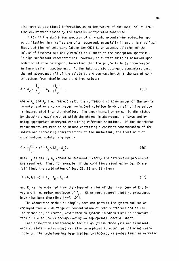
Shifts in the absorption spectrum of chromophore-containing molecules upon
solubilization in micelles are often observed, especially in cationic micelles.
Thus, addition of detergent (above the CMC) to an aqueous solution of the
solute of interest typically results in a shift of the absorption spectrum.
At high surfactant concentrations, however, no further shift is observed upon
addition of more detergent, indicating that the solute is fully incorporated
in the micellar pseudophase. At the intermediate detergent concentrations,
the net absorbance (A) of the solute at a given wavelength is the sum of con-
tributions from micelle-bound and free solute:
[SW1 Pm1 A=Aw.= +Arn*= ,
t t
where Aw and Am are, respectively, the corresponding absorbances of the solute
in water and in a concentrated surfactant solution in which all of the solute
is incorporated into the micelles. The experimental error can be diminished
by choosing a wavelength at which the change in absorbance is large and by
using appropriate detergent containing reference solutions. If the absorbance
measurements are made on solutions containing a constant concentration of the
solute and increasing concentrations of the surfactant, the fraction f of
micelle-bound solute is given by:
IS 1 f= fi= (A-Aa)/(+Aa).
t
When KS is small, A, cannot be measured directly and alternative procedures
are required. Thus, for example, if the conditions required by Eq. 55 are
fulfilled, the combination of Eqs. 25, 55 and 56 gives:
(A-Ao)/[ST~ = Ks.qn-Ks.~
and KS can be obtained from the slope of a plot of the first term of Eq. 57
vs. A with no prior knowledge of Am. Other more general plotting procedures
have also been described (ref. 104).
The absorption method is simple, does not perturb the system and can be
employed over a wide range of concentration of both surfactant and solute.
The method is, of course, restricted to systems in which micellar incorpora-
tion of the solute is accompanied by an appropriate spectral shift.
Fast absorption spectroscopic techniques (flash photolysis and transient
excited state spectroscopy) can also be employed to obtain partitioning coef-
ficients. The technique has been applied to photoactive probes (such as aromatic

26
ketones) in SLS micelles (ref. 84) and to a variety of compounds which quench
excited triplet states (such as mono- and diolefins) (ref. 105). An advantage
of this method is that it provides not only the partitioning constant KS, but
also the rates of micellar entry and exit of the probe. On the other hand, it
requires sophisticated instrumentation unavailable in most research centers.
Moreover, when the method is applied to the excited probe itself, rather than
a quencher species, one obtains the partitioning coefficient of the excited
triplet state rather than that of the ground state.
Luminescence measurements can also be employed to study the partitioning
of solutes between the micellar pseudophase and the surrounding aqueous phase.
The partitioning coefficient of a luminescent compound can be determined
either from modifications in its luminescence characteristics upon incorpora-
tion into the micelle or by selectively quenching its emission in either of
the pseudophases. Various modifications in the fluorescence characteristics
of the probe, including changes in the fluorescence intensity (resulting from
changes in the fluorescence quantum yield), changes in the excited probe life-
time, shifts in the position of the emission and changes in the fine structure
of the fluorescence spectra, have been employed to evaluate the micellar solu-
bility of probes (ref. 7, 105-110) ranging from aromatic compounds to singlet
oxygen. In most cases, the experimental approach is similar to that employed
in the absorbance method, the change in fluorescence characteristics being
monitored as a function of the surfactant concentration. The main advantage
of fluorescence is the sensitivity of the technique, which, for suitable sol-
utes, permits measurements down to -10 -8 M. On the other hand, it is somewhat
limited in that it can only be applied to fluorescent solutes or to solutes
that are efficient quenchers of the emission of an appropriate fluorescent
probe.
Time-resolved fluorescence permits the determination of KS from a single
fluorescence decay curve at a known surfactant concentration if the lifetimes
in both the micellar psuedophase and the aqueous phase can be adequately re-
solved (ref. 110). The fraction of solute incorporated into the micelles may
then be calculated from the relationship:
c!!Lq+ * ‘w
%J ,
Fm . E water *+F water *'w
where Fw and Fm are the amplitudes of the fluorescence signals from the aqueous
and micellar phases, ~~~~~~ and emit are the respective extinction coefficients,
$F represents the fluorescence yield under steady-state illumination and T is
the probe lifetime in each pseudophase.

27
The partitioning of a fluorescent probe can also be determined from selec-
tive quenching of its luminescence in one of the pseudophases. This has usu-
ally been done by selective quenching of the aqueous luminescence, using small
hydrophilic coions as quenchers (ref. 7,104,111,112). The method assumes that
the quencher is excluded from the micelle and that the excited probe does not
enter or leave the micelle during its lifetime. Since singlet lifetimes are
typically short (usually less than 20 nsec), this latter requirement is not
particularly serious; both conditions can be verified experimentally, for ex-
ample, by showinq that the lifetime of the micellar probe is unaffected upon
addition of the quencher (ref. 104). The data treatment initially proposed
by Quina and Toscano (ref. 104) leads to the following relationship for the
fluorescence yields in the presence (+ ) and absence (so ) of the quencher
Q (when the solution is excited at an !losbestic point f,'f absorption by the
aqueous and micellar probe):
(59)
A plot of the left-hand side of Eq. 59 against l/[Ql gives the ratio (S,,,I/LS~I;
division by the corresponding value of [D,,,] provides KS. A slightly modified
version of this equation, which takes into account the possibility of differ-
ent absorptivities in the two phases, has been employed by Lissi and Abuin
(ref. 111,112).
The partitioning coefficient of certain types of additives can be deter-
mined on the basis of the effect their addition provokes on the fluorescence
of a micelle-incorporated probe. The method has generally been applied to
fluorescence quenchers (ref. 113- 118). Encinas and Lissi (ref. 113) have
developed a method which can also be applied to solutes that do not interact
directly with the excited molecule (ref. 119). Their method, based on the
assumption that the quenching of the probe fluorescence is determined only
by the mean quencher occupation number, does not require knowledge of the
quenching mechanism (which may be either static of dynamic) or of the rela-
tionship between the probe lifetime and the quencher exit rate. Furthermore,
one can measure variations in the solute partitioning coefficient with increas-
ing mean occupation number. In essence, one measures the probe fluorescence
intensity ratio IF/IF in the absence and presence of quencher as a function of
quencher concentration at several surfactant concentrations. The quencher
concentration [Qli required to produce a given common value of I0 /I Fl Fl
at
each surfactant concentration is determined and the data are plotted according
to the relationship:
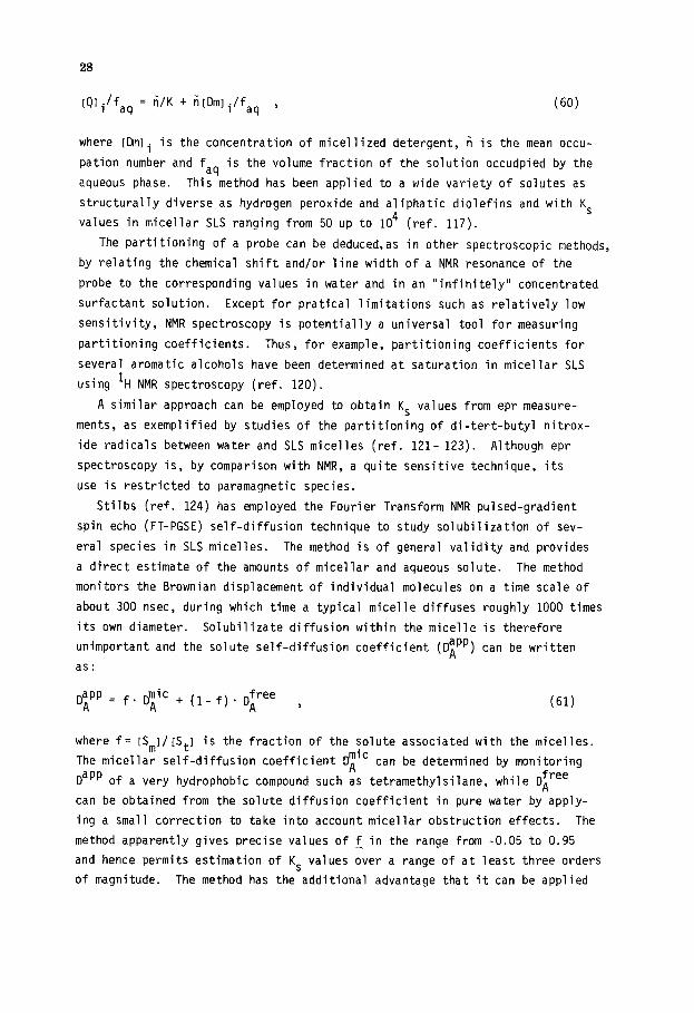
28
LQJi'faq = n/K + titDmli/f aq ’
where tDmli is the concentration of micellized detergent, I? is the mean occu-
pation number and faq is the volume fraction of the solution occudpied by the
aqueous phase. This method has been applied to a wide variety of solutes as
structurally diverse as hydrogen peroxide and aliphatic diolefins and with KS
values in micellar SLS ranging from 50 up to lo4 (ref. 117).
The Partitioning of a probe can be deduced,as in other spectroscopic methods,
by relating the chemical shift and/or line width of a NMR resonance of the
probe to the corresponding values in water and in an "infinitely" concentrated
surfactant solution. Except for pratical limitations such as relatively low
sensitivity, NMR spectroscopy is potentially a universal tool for measuring
partitioning coefficients. Thus, for example, partitioning coefficients for
several aromatic alcohols have been determined at saturation in micellar SLS
using 'H NMR spectroscopy (ref. 120).
A similar approach can be employed to obtain KS values from epr measure-
ments, as exemplified by studies of the partitioning of di-tert-butyl nitrox-
ide radicals between water and SLS micelles (ref. 121- 123). Although epr
spectroscopy is, by comparison with NMR, a quite sensitive technique, its
use is restricted to paramagnetic species.
Stilbs (ref. 124) has employed the Fourier Transform NMR pulsed-gradient
spin echo (FT-PGSE) self-diffusion technique to study solubilization of sev-
eral species in SLS micelles. The method is of general validity and provides
a direct estimate of the amounts of micellar and aqueous solute. The method
monitors the Brownian displacement of individual molecules on a time scale of
about 300 nsec, during which time a typical micelle diffuses roughly 1000 times
its own diameter. Solubilizate diffusion within the micelle is therefore
unimportant and the solute self-diffusion coefficient (Dipp) can be written
as:
jJ;pp = f. qy + (1-f). q-e , (61)
where f= [Sm]/[St] is the fraction of the solute associated with the micelles.
The micellar self-diffusion coefficient I$" can be determined by monitoring
DaPP of a very hydrop hobic compound such as tetramethylsilane, while DLree
can be obtained from the solute diffusion coefficient in pure water by apply-
ing a small correction to take into account micellar obstruction effects. The
method apparently gives precise values of z in the range from -0.05 to 0.95
and hence permits estimation of KS values over a range of at least three orders
of magnitude. The method has the additional advantage that it can be applied

29
to almost any solute-surfactant pair, as well as to mixtures of solubilizates,
since each compound can be determined independently.
D. Miscellaneous methods
Kaneshina Sal. (ref. 125) have employed the depression of the Krafft
point of the surfactant to quantify the incorporation of anesthetics and al-
cohols into ionic surfactant micelles. Thus, if the Krafft point is consid-
ered to be the melting temperature of the hydrated solid surfactant (ref.
126), the depression of the Krafft point in the presence of additives can be
treated as a colligative property. At low mole fractions of the additive in
the micellar pseudophase, simple thermodynamic considerations then lead to
the relationship (ref. 125):
KMW =
(-AU . AHf 1
where -AT is the Krafft point depression, To is the unperturbed Krafft point,
AH~ is the change in enthalpy upon going from the hydrated solid to the mi-
cellar state and Xw is the mole fraction of the solute in the aqueous phase.
The method gives values similar to those obtained by other techniques, but
its general validity may be limited to systems in which free energy contribu-
tions due to changes in counterion binding accompanying solute incorporation
are relatively unimportant.
Partitioning constants have also been evaluated from CMC depressions in-
duced by the solute (ref. 125). Thus, Shirahama and Kashiwabara (ref. 127)
proposed that:
(-e)KMW = v (63)
The (-e) has been referred to as the ISA (interaction of surfactant and addi-
tive) coefficient by Hayase and Hayano (ref. 128) and its physical meaning
has been discussed by Manabe et al. (ref. 129). The ISA coefficient seems to
be constant for a series of related compounds; e.g., -0.69 for alcohols (ref.
129) and -0.52 for anesthetics (ref. 126) in SLS. Treiner (ref. 42) employed
this approach to calculate partitioning coefficients_ for several polar mule-
cules in aqueous DTAB after correction of the CMC change for the contribution
from the Setchenov salting out constant. Although it gives results similar to
those obtained in other systems, this method depends heavily on the use of
empirically calculated Setchenov constants and "assumed" ISA coefficients.
De Lissi et al. (ref. 130) have recently proposed a method for obtaining
KS for association of alcohols to micelles on the basis of partial molar volume
measurements.
30
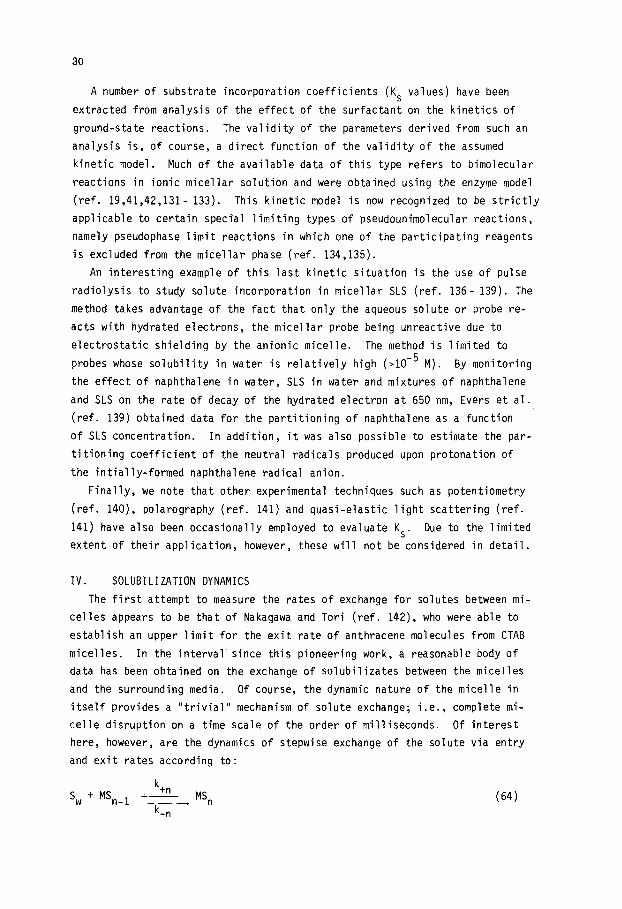
A number of substrate incorporation coefficients (KS values) have been
extracted from analysis of the effect of the surfactant on the kinetics of
ground-state reactions. The validity of the parameters derived from such an
analysis is, of course, a direct function of the validity of the assumed
kinetic model. Much of the available data of this type refers to bimolecular
reactions in ionic micellar solution and were obtained using the enzyme model
(ref. 19,41,42,131- 133). This kinetic model is now recognized to be strictly
applicable to certain special limiting types of pseudounimolecular reactions,
namely pseudophase limit reactions in which one of the participating reagents
is excluded from the micellar phase (ref. 134,135).
An interesting example of this last kinetic situation is the use of pulse
radiolysis to study solute incorporation in micellar SLS (ref. 136- 139). The
method takes advantage of the fact that only the aqueous solute or probe re-
acts with hydrated electrons, the micellar probe being unreactive due to
electrostatic shielding by the anionic micelle. The method is limited to
probes whose solubility in water is relatively high (,10e5 M). By monitoring
the effect of naphthalene in water, SLS in water and mixtures of naphthalene
and SLS on the rate of decay of the hydrated electron at 650 nm, Evers et al.
(ref. 139) obtained data for the partitioning of naphthalene as a function
of SLS concentration. In addition, it was also possible to estimate the par-
titioning coefficient of the neutral radicals produced upon protonation of
the intially-formed naphthalene radical anion.
Finally, we note that other experimental techniques such as potentiometry
(ref. 140), polarography (ref. 141) and quasi-elastic light scattering (ref.
141) have also been occasionally employed to evaluate KS. Due to the limited
extent of their application, however, these will not be considered in detail.
IV. SOLUBILIZATION DYNAMICS
The first attempt to measure the rates of exchange for solutes between mi-
celles appears to be that of Nakagawa and Tori (ref. 142), who were able to
establish an upper limit for the exit rate of anthracene molecules from CTAB
micelles. In the interval since this pioneering work, a reasonable body of
data has been obtained on the exchange of solubilizates between the micelles
and the surrounding media. Of course, the dynamic nature of the micelle in
itself provides a "trivial" mechanism of solute exchange; i.e., complete mi-
celle disruption on a time scale of the order of milliseconds. Of interest
here, however, are the dynamics of stepwise exchange of the solute via entry
and exit rates according to:
k SW + MSn_1 ++" ~ MSn
k -n
(64)
31
with n > 1. _
Under normal experimental conditions, this exchange of the probe takes
place on a time scale of between low3 and 10 -8
set and hence fast detection
techniques (stopped flow, relaxation and fast spectroscopic techniques) are
required to evaluate the individual rate constants.
Stopped flow techniques, being rather slow, are of utility only in select
cases (ref. 87). Although extensively exploited to study the dynamics of
monomer-micelle exchange, ultrasonic relaxation has been only sparingly ap-
plied to solute exchange. Wyn-Jones et al. (ref. 143,144) did, however, use
ultrasonic relaxation to evaluate exchange rates of n-alcohols in micellar
CTAB. The values of the entry and exit rate constants listed in Table 1
were obtained by combining the single relaxation time observed for the ex-
change process with partitioning constants determined by the saturation method
and thus refer to exchange in a strongly perturbed micelle.
Spectroscopic techniques such as electron paramagnetic resonance (ref. 145)
and transient time-resolved absorption (ref. 146- 149) and emission spectros-
copy (ref. 7,116,150,151) are particularly suitable for measuring exchange
rates on the time scale considered. A time-resolved flash CIDNP (chemically
induced dynamic nuclear polarization) method has also been used to evaluate
the rate of exit of benzyl radicals from SLS micelles (ref. 148). In at
least one system (ref. 149), exchange rate constants were extracted from
steady state photolysis data.
Various aspects of the use of photophysical techniques to investigate
micellar entry and exit of solutes and statistics of solute incorporation
have been reviewed (ref. 17,152). Of particular note are two key papers by
Tachiya (ref. 14,15) in which general closed form solutions are obtained for
the rate of decay of an excited micelle localized fluorescence probe in the
presence of a quencher species that distributes statistically among the avail-
able micelles in accord with either a Poisson (ref. 14) or a binomial (ref.
15) distribution. In principle, the Tachiya equations permit determination
of all of the relevant kinetic parameters associated with solubilization of
the quencher and/or detergent concentration. Frequently, by judicious choice
of the system and experimental conditions, it is often possible to measure
micellar entry and exit rates directly from the probe decay. Thus, the entry
rate of an excited water-soluble probe can be determined by measuring its
lifetime under conditions such that it is quenched upon entry into a micelle.
This method has been used by Ligh and Scaiano (ref. 153) to measure the rate
of entry of acetone into SLS and CTAC micelles. Conversely, the exit rate of
a micelle-incorporated chromophore can be determined directly by measuring
the lifetime of the excited species under conditions in which it is quenched

32
only upon exit into the aqueous phase. The following simple kinetic scheme
is appropriate for this latter situation:
MS + hv - MS* (65)
MS* -k-d-+ MS + hv (66)
MS* J=-+M+S* (67)
S* + Q, --kQ-+ quenching (68)
s* + M L MS*
where Q, is a micelle-excluded water-soluble quencher; i.e., a quencher coion.
If the following condition holds:
k_ " kd
kQIQwl " kt * M
kQtQwl " k-
(70)
(71)
(72)
the lifetime of S* is then equal to l/k_. This lifetime can be measured di-
rectly by time resolved fluorescence, by phosphorescence decay, or by transient
absorption spectroscopy. The first condition (k_>a k ) is the most restric- d
tive and is usually fulfilled only by the long-lived triplet states.
This method has been applied to triplet anthracene in CTAB micelles, using
C"++ ions as the quencher (ref. 150) and to several aromatic triplets in SLS,
employing NO; as quencher (ref. 7); triplet concentrations were monitored by
time-resolved emission spectroscopy. A similar approach, using transient ab-
sorption spectroscopy and NO; as quencher, was employed by Scaiano and Selwyn
(ref. 84) to measure the lifetime of several micelle-incorporated ketones.
One point which merits emphasis is that in all these systems, the exit rate
is that of the excited triplet state rather than that of the ground state
species; consequently, calculation of kt values using the partitioning coeffi-
cient of the ground state species (ref. 7,150) may lead to significant errors.
The approach of Scaiano and Selwyn (ref. 84) has the advantage that it permits
direct evaluation of both $ and k_ , and hence of k, (see Eq. 83) for the ex-
cited triplet. Values of k_ and k+ evaluated in this manner are collected in
Table 4.
A slightly modified approach can be employed to determine the micellar en-
trance rate of neutral water-soluble quenchers using a micelle-localized fluor-
escence probe. The reaction scheme can, by appropriate choice of the experimen-
tal conditions, be reduced to:
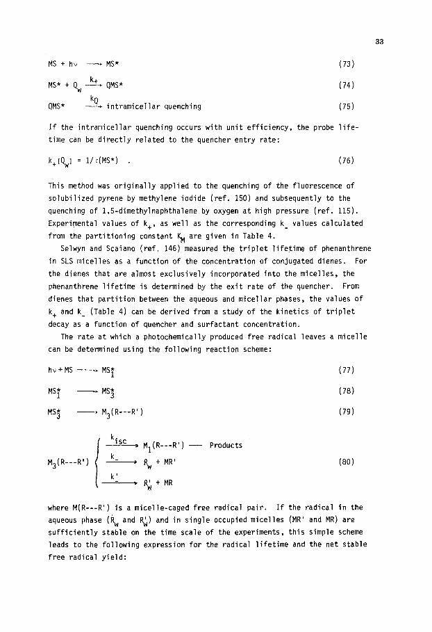
33
MS + hv - MS*
MS* + Qw A+ QMS*
QMS* :Q+ intramicellar quenching
(73)
(74)
(75)
If the intramicellar quenching occurs with unit efficiency, the probe life-
time can be directly related to the quencher entry rate:
kt[Qwl = 1/ dMS*) . (76)
This method was originally applied to the quenching of the fluorescence of
solubilized pyrene by methylene iodide (ref. 150) and subsequently to the
quenching of 1,5_dimethylnaphthalene by oxygen at high pressure (ref. 115).
Experimental values of k,, as well as the corresponding k_ values calculated
from the partitioning constant KM are given in Table 4.
Selwyn and Scaiano (ref. 146) measured the triplet lifetime of phenanthrene
in SLS micelles as a function of the concentration of conjugated dienes. For
the dienes that are almost exclusively incorporated into the micelles, the
phenanthrene lifetime is determined by the exit rate of the quencher. From
dienes that partition between the aqueous and micellar phases, the values of
k, and k_ (Table 4) can be derived from a study of the kinetics of triplet
decay as a function of quencher and surfactant concentration.
The rate at which a photochemically produced free radical leaves a micelle
can be determined using the following reaction scheme:
hv+MS - MS* 1 (77)
MST - MS* 3 (78)
MS? - M3(R---RI) (79)
k. 1sc
- M1(R---RI)
k M3(R---RI) --=--+
i-.
- Products
a + MR' (80)
k'
% ’ + MR
where M(R---RI) is a micelle-caged free radical ,pair. If the radical in the
aqueous phase (Rw and $) and in single occupied micelles (MR' and MR) are
sufficiently stable on the time scale of the experiments, this simple scheme
leads to the following expression for the radical lifetime and the net stable
free radical yield:
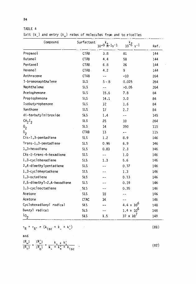
34
TABLE 4
Exit (k_) and entry (k,) rates of molecules from and to micelles
Compound Surfactant 10-9kgj-ls-l 10% ,-1 Ref.
Propanol
Butanol
Pentanol
Hexanol
Anthracene
l-bromonaphthalene
Naphthalene
Acetophenone
Propiophenone
Isobutyrophenone
Xanthone
di-terbutylnitroxide
CH2I2
O2
O2 Cis-1,3-pentadiene
Trans-1,3-pentadiene
1,3-hexadiene
Cis-2-trans-4-hexadiene
1,3-cyclohexadiene
2,4_dimethylpentadiene
1,3-cycloheptadiene
1,3-octadiene
2,5-dimethyl-2,4-hexadiene
1,3-cyclooctadiene
Acetone
Acetone
Cyclohexadienyl radical
Benzyl radical
lo*
CTAB
CTAB
CTAB
CTAB
CTAB
SLS
SLS
SLS
SLS
SLS
SLS
SLS
SLS
SLS
CTAB
SLS
SLS
SLS
SLS
SLS
SLS
SLS
SLS
SLS
SLS
SLS
CTAC
SLS
SLS
SLS
3.8
4.4
6.8
4.2
--
5-8
-_
15.6
14.1
12
17
1.4
25
14
13
1.2
0.95
0.83
-_
1.3
-_
-_
-_
-_
-_
10
34
__
__
1.5
81
58
26
9
<lo
0.025
>0.05
7.8
3.0
1.6
2.7
__
10
350
8.9
6.9
2.3
1.0
5.6
0.37
1.3
0.13
0.19
0.35
4.4 x 10⁶
1.4 x 10⁶
37 x 107
144
144
144
144
264
264
264
84
84
84
84
145
264
115
115
146
146
146
146
146
146
146
146
146
146
146
146
148
148
149
TR = TR’ = (kisc _ +k -+kJ) (81)
and
[R I m
[R'I =m= k_ t kl
[R,I IRol _ k + k; + kisc * (82)

35
Measurement of TR and RtZm/RtZO permits evaluation of kisc and of the sum
of the exit rates k t k'. Scaiano et al. (ref. 147) have applied this scheme __
to the photolysis of benzophenone in CTACl and SLS in the presence of 1,4-
cyclohexadiene, which gives benzydrol and cyclohexadienyl radicals. In CTACl
and SLS the values of k + kl are 1.6x lo6 s -1 and 4.4x lo6 s-l respectively. 3 These exit rates undoubtedly correspond to those of the cyclohexadienyl radi-
cals. By a similar method, Turro et al. (ref. 148) estimated a rate of 1.4 x
106 s-1 for the exit of benzyl radicals from SLS micelles.
In considering the trends for the entry and exit rate constants collected
in Table 4, several major shortcomings of the data obtained from quenching
experiments should be noted. First, if the excited probe is located at the
micelle-water interface (as is probably the case for most of the aromatic
probes employed), it can be quenched by a water-soluble quencher that has not
yet become "micelle bound". Thus, k, measures the time required for the
quencher to approach the excited molecule (within the effective quenching
radius) rather than the true entry rate associated with the partitioning
constant, KM= k,/k . Second, one must be extremely wary about the neglect
of intramicellar quenching by water-soluble quenchers, even when the poten-
tial quencher is a hydrophilic ionic species of the same charge as the mi-
celle (ref. 154). Finally, all of the above approaches assume, either impli-
citly or explicitly, that the exchange process is controlled by a simple bi-
molecular encounter involving the aqueous probe and micelles. Microscopic
reversibility then implies that the exit rate corresponds to a true uni-
molecular process of the micelle-incorporated probe. These assumptions
have been questioned, at least for the singlet oxygen (IO,) by Matheson and
Massoudi (ref. 149), who concluded that, at high O2 pressures, the entrance
(and exit) of the excited molecules is "assisted" by the ground state species;
these apparently anomalous results warrant further verification.
These factors may contribute to some of the peculiarities evident in the
entry rate data in Table 1.
From simple considerations of relative mobility, the rate constant for bi-
molecular encounter between a small molecule solubilized in the aqueous phase
and a micelle of radius Rmic should be approximately give by (ref. 155):
kdif = 4rr. N' - Rmic. Dprobe , (83)
where N'= N avog'looo and Dprobe
is the probe diffusion coefficient. The values
of kdif for typical solutes should then be of the order of 5x lOlo to 1011 M-1,-1 depending upon the probe diffusion coefficient and micellar size. The
k, values collected in Table 4 ranges from 0.8x 10' to 34x 10' M-'s-~ with
no clear relationship to either micellar size of solute properties. In this

36
regard, it is noteworthy that acetone, one of the mot-e hydrophobic solutes,
has the fastest rate of entry. This result is just the opposite of that ex-
pected from simple "thermodynamic" considerations. To what extent these entry
rate data reflect experimental errors and/or faulty assumptions is difficult
to evaluate, although they do, as a whole, tend to indicate that solute "incor-
poration" is considerably slower than "solute-micelle" collisions. A hypo-
thetical, though nonetheless appealing explanation is that a "salting out" of
the probe results in a significant reduction of the local concentration of
the probe in the immediate vicinity of the micelle surface.
In contrast to the entry rate data, micellar exit rates vary over an enor-
mous range, from =108 s-I down to less than ~10~ s-I (Table 4). Furthermore,
families of compounds exhibit a clear trend; i.e., k decreases as the solute
hydrophobicity increases. The dependence of Km on solute hydrophobicity thus
appears to be principally a function of k_ rather than of k,.
V. SOLUBILIZATION ENVIRONMENTS OF NEUTRAL MOLECULES INCORPORATED INTO
MICELLES
Lawrence (ref. 156) was the first to point out that the site of incorpor-
ation of a solubilized molecule should depend on its relative hydrophobic
and hydrophilic tendencies. The first direct physical evidence regarding
the solubilization environment of hydrocarbon derivatives in micellar sys-
tems was obtained by Hughes et al. (ref. 157), who found that the region
of solubilization had a low electron density, similar to that of liquid para-
ffins. In the intervening years, the nature of the microenvironment sensed
by the solute has been investigated by a variety of methods and several re-
views are available (ref. 158- 161).
A neutral molecule incorporated into (or solubilized in) a micelle can
be located in a variety of microenvironments, ranging (at least in principle)
between the two extremes of a hydrocarbon like solvent (the micellar "core")
and a completely aqueous medium (or at least a water-rich interface with a
high charge density). Indeed, the distinctive feature of the micelle as a
solvent is that it can provide not only different microenvironments for dif-
ferent molecules, but also different environments for the different parts of
the same molecule. Thus, polar molecules encounter a polar environment,
hydrophobic molecules have available a "hydrocarbon-like" medium and amphi-
phatic molecules should be able to orient themselves at the micelle-water
interface with their hydrophobic portion extending into the "hydrocarbon-
like core". "Oriented" solubilization of this type has in fact been demon-
strated conclusively by proton nuclear magnetic resonance spectroscopy (ref.
55,158,162). Based on these considerations alone, one can readily understand
31
why micelles are capable of dissolving such a wide variety of solutes (in-
cluding amphiphilic molecules) and exhibit solvent capacities larger than
those of either polar or nonpolar solvents (ref. I).
Additional aspects which should be considered in studies designed to probe
the micellar microenvironment are the dynamic nature of the solubilization
process and the fact that, even at very low occupation numbers, the presence
of the solute can significantly modify the micellar structure. The dynamics
of solute incorporation includes micellar entry and exit of the solute or
probe as well as the rotational and translational motion of the solute within
the micellar aggregate itself. As discussed above, the rate of solute entry
into the micelle is nearly diffusion controlled while the time scale for mi-
cellar exit is determined mainly by the hydrophobicity (ref. 163,164). Typi-
cal values of the pseudo-unimolecular exit rate constants fall in the range
of IO3 to IO8 set -' (see Table 4). The solute molecule can rapidly migrate
between different regions of the micelle and hence between different micro-
environments. Thus, a property such as the hmax of the absorption spectrum
of a probe will, in reality, be a weighted average over the accessible solu-
bilization environments. Other features of the absorption band, such as the
band width, may also be indicative of a multiplicity of environments. Sim-
milarly, the relative slow time scale of NMR spectroscopy (-10q4 set) implies
that the observed signals will be averaged over all possible intramicellar
displacements of the solute. On the other hand, the fast time scale of
fluorescence spectroscopy (down to less ~1 nanosecond) or deuterium magnetic
resonance and spin relaxation techniques (ref. 167), makes these methods par-
ticularly suitable for experiments on intermicellar migration and on reorien-
tation of probes. Thus, time correlated fluorescence measurements could be
employed to follow changes in the average microenvironments sensed by an ex-
cited probe during its lifetime, either by measuring the fluorescence spectrum
at different times after excitation or by tuning the excited probe lifetime,
through addition of appropriate quenchers. As in all studies which use probe
molecules to infer properties of the system, one cannot overlook the possibility
that the observed behavior reflects primarily probe-induced perturbations.
Incorporation of even a single solute molecule must perturb the original sys-
tem to some extent. Moreover, the microenvironment sensed by the probe neces-
sarily corresponds to that at the site of perturbation. The question of per-
turbation is particularly relevant in micellar systems since the aggregates
tend to be rather small (cl00 monomers) and the probes employed tend to be
rather large (e.g., polycyclic aromatic hydrocarbons). In one of the early
quantitative studies of solubilization sites, Erikson and Gilbert (ref. 55)
showed that addition of benzene to micellar CTAB shifts the CTAB proton NMR

38
resonances. At low incorporation of benzene, there is a rapid shift of the
N-CH3 and the c(-CH2 hydrogen resonances to higher fields, implying preferen-
tial adsorption close to the CX-CH~ groups. Since this process presumably in-
volves at least some rearrangement of water molecules at the adsorption sites,
the resultant solute-induced perturbation of the "solvent" properties of the
micelle should be magnified at even higher occupation numbers. Several of
the discrepancies between results obtained by different techniques or by the
same technique in different laboratories can be attributed to differences in
the degree of solute incorporation (ref. 165,166).
A. The "model dependence" of the solubilization site
The validity of attempts to draw definitive conclusions as to the nature
and location of an intramicellar solubilization site is seriously compromised
by the necessity of assuming (or presuming) a model for the micellar aggregate
itself. Thus, if the probe under investigation senses a polar microenvironment,
adepts of a tightly packed micelle model would infer that the probe must reside
at the micelle surface. On the other hand, proponents of an "open" micelle
could argue that the apparent polarity of the microenvironment is a consequence
of penetration of water down to a site in the micellar interior. The truth is
probably somewhere between these extremes. Thus, these are compelling evidences
for substantial residual water-hydrocarbon contact even at the terminus of
the hydrophobic moiety of the surfactant and several well designed experiments
(e.g., photo-abstraction of hydrogen atoms from the surfactant alkyl chain by
benzophenone derivatives) have failed to reveal the presence of dramatic re-
activity gradients along the surfactant chain (ref. 167). Indeed, this is
precisely what one might expect in retrospect on the basis of random inter-
twining and doubling back of the surfactant hydrophobic chains in a dynamic
micellar aggregate, independent of the degree of "openness" of the micellar
structure.
Mukerjee and Cardinal (ref. 3,168) have described the solubilization pro-
cess in terms of a simple model consisting of two distinct, but arbitrarily
(or "operationally") defined solubilization sites: the micellar "core" and
the "surface". Even assuming only two distinct solubilization sites, however,
the proportion of solubilizate in each zone will be determined by a variety
of factors, including the probe hydrophobicity (or surface activity), the
surface-to-volume ratio (determined by the micelle size and shape) and the
characteristics of the "core" and the "surface" (complex functions of the sur-
factant employed, its concentration, the counterions, other additive present
and the mean solute occupation number). The strong preference for the micell-
ar surface as a region of solubilization is in part a consequence of smallness

39
of the micellar aggregate. For a SLS micelle, at least half of the probe
would be within 4- 5 fi of the surface at any given time. Thus, even if the
solute has no affinity for the surface, it is capable of being strongly in-
fluenced by the surface and should be accessible to water and encounters with
water-soluble molecules. As a first approximation, Mukerjee and Cardinal
(ref. 3,168) assume that the solubility in the micellar core can be equated to
that in a hydrocarbon solvent if one corrects for effects (the Laplace pressure)
due to the small radius of the core. The solubilization in the surface is
treated as a surface adsorption obeying a Langmuir type adsorption isotherm,
for which the mean driving force is the capacity of the solute to reduce the
interfacial (hydrocarbon-water) tension.
With due consideration of the above factors, the simple two-site model can
rationalize, at least qualitatively, the results of most solubilization stud-
ies. Nonetheless, it whould be emphasized that this model is at best an over-
simplification which divides a "continuum" of environments into two extreme
types of sites. Thus, a change in the hydrophobicity of the probe (e.g.,
upon addition of alkyl groups to benzene) which alters the net probe-water
contact will be described by the two-site model as a change in the intramicellar
distribution of the solute between the two sites, regardless of the actual
mechanism. Finally, it should be reemphasized that the time scale for intra-
micellar migration of a solute is relatively short (10e7 to lo-' set), re-
quiring careful distinction between energetically accessible or "instantan-
eous" solubilization sites (which may determine chemical or photophysical re-
activity) and the time-weighted average solubilization site.
To date, the principal methods employed to investigate the nature of the
solute microenvironment are NMR spectroscopy and absorption and emission
spectroscopy; several other spectroscopic techniques have also been used,
but the results tend to be less readily interpretable; e.g., X-ray diffrac-
tion (ref. 158) or restricted to a limited number of solutes; e.g. epr tech-
nicques which can be used only with free radicals or spin-labelled surfac-
tants (ref. 158,169- 171). In addition to the spectroscopic methods, several
kinetic techniques provide information regarding the solute environment and/or
its accessibility to co-reactants which reside only in the aqueous phase (such
as simple ions) or are restricted to the micelle surface (such as ions with
hydrophobic groups). These kinetic techniques include: positron annihilation,
information related to the hydrophobicity of the probe location (ref. 172);
reactions of hydrated electrons, a function of the relative position of the
probe with respect to the micelle-water interface (ref. 173); quenching of
electronically excited micelle-incorporated solutes by counterions (ref. 106,
174,177) or by highly hydrophobic quenchers such as hydrogen peroxide (ref.
40
117); measurements of the rates of diffusion controlled processes such as
inter- or intramolecular excimer formation (ref. 152), sensitive to the
microviscosity of the medium; measurement of the rates of solvent-sensitive
unimolecular reactions such as the thermal cis-trans isomerization of p-nitro-
p'-dialkylaminoazobenzenes (ref. 178); measurements of rate constants for bi-
molecular reactions between micelleJocalized and water-soluble co-reactants;
e.g., reduction of steroids by borohydride (ref. 179) or oxidation of unde-
canal by chromic acid (ref. 180); measurement of quantum yields and/or pro-
duct distributions of photoreactions, type II photoelimination of ketones
(ref. 181- 182) or trans-cis photoisomerization of stilbene derivatives (ref.
183); and measurements of the apparent bimolecular rate constants for intra-
micellar reactions related to the average mutual positions of the reactants
inside the aggregate (ref. 184).
The nature of the medium sensed by the solute(s) has also been deduced
from thermodynamic measurements of equilibrium constants for complex forma-
tion; e.g., between hydrophobic compounds and viologen derivatives (ref. 185-
186) and from free energies and/or enthalpies of solubilization (ref. 49,57)
by comparison with the corresponding values for solubilization of the solute
in polar and nonpolar solvents.
Micelles influence the wavelength of maximum absorption and/or the vibra-
tional fine structure of the ultraviolet absorption band of many organic
molecules. Such changes have been extensively exploited to investigate the
local microenvironment of the solubilizate in the micelle (ref. 158,165,188-
197). Experimentally, one compares a spectral parameter of the solute which
exhibits a net solvent-sensitive variation (e.g., the "max) upon incorpora-
tion into the micelle with values obtained in a series of homogeneous sol-
vents. On the basis of this calculation, the solubilization environment in
the micelle may be said to correspond to that of an "equivalent solvent" or
"equivalent solvent mixture". This calibration approach has several impor-
tant and potentially severe limitations (ref. 192). Thus, when the spectral
variation (e.g., Amax) is plotted against a parameter related to the "polarity"
of the homogeneous solvent, protic and aprotic solvents frequently give two
distinctly different correlations. Furthermore, the spectral variations
for different probes may not correlate with the same solvent property (di-
electric constant, concentration of OH groups, the Kosower Z parameter, etc.)
of may give rise to a new empirical scale of solvent "polarity" (ref. 198,199).
The same considerations apply to the evaluation of micropolarities from fluor-
escence measurements (ref. 200).
Solvent effects in the excited state can alter the vibrational fine struc-
ture and position of the fluorescence band, the lifetime of the excited state,
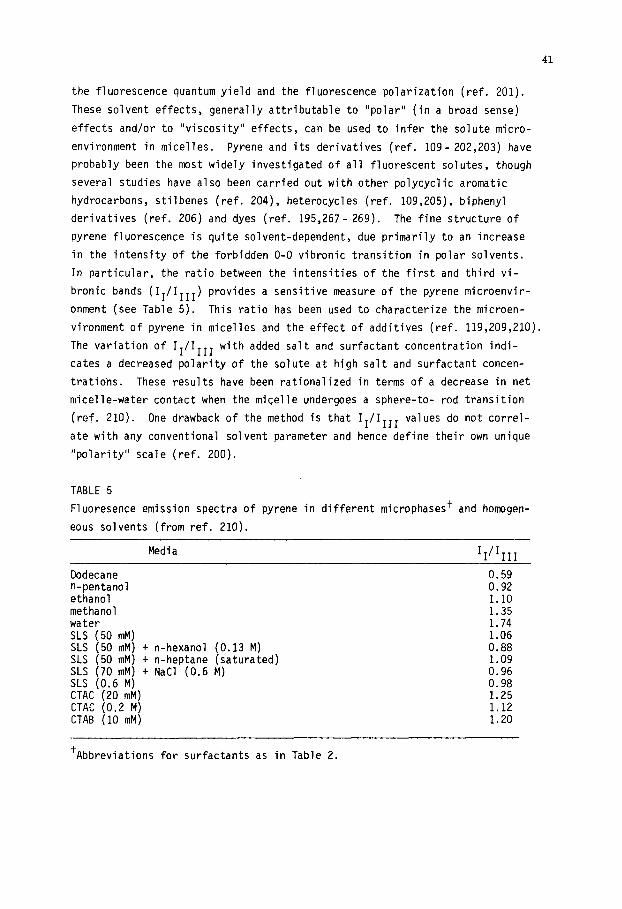
41
the fluorescence quantum yield and the fluorescence polarization (ref. 201).
These solvent effects, generally attributable to "polar" (in a broad sense)
effects and/or to "viscosity" effects, can be used to infer the solute micro-
environment in micelles. Pyrene and its derivatives (ref. 109- 202,203) have
probably been the most widely investigated of all fluorescent solutes, though
several studies have also been carried out with other polycyclic aromatic
hydrocarbons, stilbenes (ref. 204), heterocycles (ref. 109,205), biphenyl
derivatives (ref. 206) and dyes (ref. 195,267- 269). The fine structure of
pyrene fluorescence is quite solvent-dependent, due primarily to an increase
in the intensity of the forbidden O-O vibronic transition in polar solvents.
In particular, the ratio between the intensities of the first and third vi-
bronic bands (II/III1 ) provides a sensitive measure of the pyrene microenvir-
onment (see Table 5). This ratio has been used to characterize the microen-
vironment of pyrene in micelles and the effect of additives (ref. 119,209,210).
The variation of II/III1 with added salt and surfactant concentration indi-
cates a decreased polarity of the solute at high salt and surfactant concen-
trations. These results have been rationalized in terms of a decrease in net
micelle-water contact when the micelle undergoes a sphere-to- rod transition
(ref. 210). One drawback of the method is that II/III1 values do not correl-
ate with any conventional solvent parameter and hence define their own unique
"polarity" scale (ref. 200).
TABLE 5
Fluoresence emission spectra of pyrene in different microphases+ and homogen-
eous solvents (from ref. 210).
Media
Dodecane n-pentanol ethanol methanol water SLS (50 mM) SLS (50 mM) + n-hexanol (0.13 M) SLS (50 mM) + n-heptane (saturated) SLS (70 mM) + NaCl (0.6 M) SLS (0.6 M) CTAC (20 mM) CTAC (0.2 M) CTAB (10 mM)
WI1
0.59 0.92 1.10 1.35 1.74 1.06 0.88 1.09 0.96 0.98 1.25 1.12 1.20
'Abbreviations for surfactants as in Table 2.

42
Several aromatic compounds have fluorescence bands which exhibit solvent-
dependent wavelength shifts. A clear example of such a dependence is provided
by pyrenecarboxyaldehyde whose fluorescence exhibits a marked red shift with
increasing solvent polarity (ref. 158); thus, in homogeneous solvents, the
maximum wavelength of the fluorescence spectrum correlates with the bulk di-
electric constant of the solvent, providing an estimate of the effective di-
electric constant of the micellar environment at the solutilibization site.
From this type of study, it has been concluded that several pyrene derivatives
(pyrenecarboxyaldehyde aminopyrene) are located at the micellar surface (ref.
160). Exciplex emission,which is sensitive to the polarity of the medium, has
been used to investigate the intramicellar location of the pyrene-triethyl-
amine (ref. 211).
The net fluorescence polarization of a probe depends on the extent of rota-
tion and relaxation during its excited state lifetime, which is in turn related
to the viscosity of the medium (ref. 212). Several probes have been employed
to estimate the intramicellar viscosity by this method (ref. 201,213,214).
Other solvent-dependent properties of excited states (e.g., the solute life-
time) have also been used to infer the nature of the solute microenvironment
(ref. 210,215).
Incorporation sites of solutes in micellar systems have been extensively
investigated by proton (ref. 158) and l3 C (ref. 115,216) NMR spectroscopy.
Several features of this technique make it particularly attractive for the
evaluation of the intramicellar location of the solute. Thus, both nuclear
magnetic resonance frequencies (chemical shifts) and line widths are depen-
dent on the molecular environment of the nuclei; hence, comparison of the
values in a micellar system with those in polar and nonpolar solvents provides
information on the relative position of the solubilizate. Furthermore, since
the average environment of each nucleus can be evaluated independently (ref.
162,120,187,191,217,218), additional valuable information can be obtained re-
garding the relative position and orientation of the solute in the micelle at
different occupation numbers. Finally, it should be noted that the signals
of the surfactant are also affected by incorporation of the solute. The
changes induced in the resonances of the surfactant provide information on
the average solute localization, the manner in which the surfactant environ-
ment is modified by solute incorporation (ref. 187,219- 221) and effects
arising from changes in the solute mean occupation number.
B. General data trends
Although simple considerations might suggest that a highly hydrophobic mole-
cule such as a hydrocarbon would prefer to solubilize the micellar core, there
is no direct evidence to support this "prejudice"; in fact, the available data
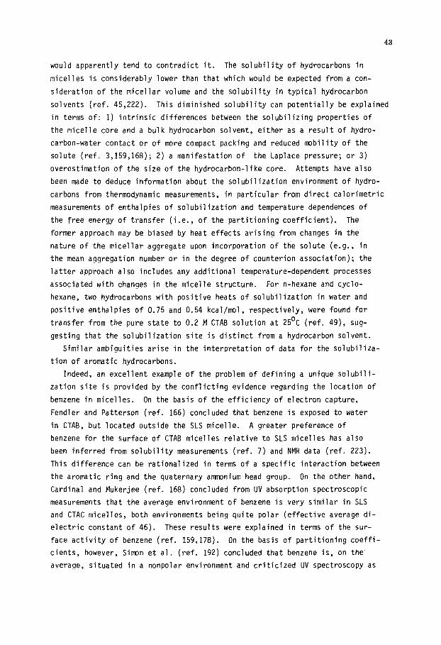
43
would apparently tend to contradict it. The solubility of hydrocarbons in
micelles is considerably lower than that which would be expected from a con-
sideration of the micellar volume and the solubility in typical hydrocarbon
solvents (ref. 45,222). This diminished solubility can potentially be explained
in terms of: 1) intrinsic differences between the solubilizing properties of
the micelle core and a bulk hydrocarbon solvent, either as a result of hydro-
carbon-water contact or of more compact packing and reduced mobility of the
solute (ref. 3,159,168); 2) a manifestation of the Laplace pressure; or 3)
overestimation of the size of the hydrocarbon-like core. Attempts have also
been made to deduce information about the solubilization environment of hydro-
carbons from thermodynamic measurements, in particular from direct calorimetric
measurements of enthalpies of solubilization and temperature dependences of
the free energy of transfer (i.e., of the partitioning coefficient). The
former approach may be biased by heat effects arising from changes in the
nature of the micellar aggregate upon incorporation of the solute (e.g., in
the mean aggregation number or in the degree of counterion association); the
latter approach also includes any additional temperature-dependent processes
associ,ated with changes in the micelle structure. For n-hexane and cyclo-
hexane, two hydrocarbons with positive heats of solubilization in water and
positive enthalpies of 0.75 and 0.54 kcal/mol, respectively, were found for
transfer from the pure state to 0.2 M CTAB solution at 25'C (ref. 49), sug-
gesting that the solubilization site is distinct from a hydrocarbon solvent.
Similar ambiguities arise in the interpretation of data for the solubiliza-
tion of aromatic hydrocarbons.
Indeed, an excellent example of the problem of defining a unique solubili-
zation site is provided by the conflicting evidence regarding the location of
benzene in micelles. On the basis of the efficiency of electron capture,
Fendler and Patterson (ref. 166) concluded that benzene is exposed to water
in CTAB, but located outside the SLS micelle. A greater preference of
benzene for the surface of CTAB micelles relative to SLS micelles has also
been inferred from solubility measurements (ref. 7) and NMR data (ref. 223).
This difference can be rationalized in terms of a specific interaction between
the aromatic ring and the quaternary ammonium head group. On the other hand,
Cardinal and Mukerjee (ref. 168) concluded from UV absorption spectroscopic
measurements that the average environment of benzene is very similar in SLS
and CTAC micelles, both environments being quite polar (effective average di-
electric constant of 46). These results were explained in terms of the sur-
face activity of benzene (ref. 159,178). On the basis of partitioning coeffi-
cients, however, Simon et al. (ref. 192) concluded that benzene is, on the'
average, situated in a nonpolar environment and criticized UV spectroscopy as

44
a method for determining solute location because of its dependence on the
choice of the "calibration scale". Free energies of transfer of benzene to
SLS and CTAB micelles (ref. 57) are also suggestive of a nonpolar environment
(at low occupation number).
Several works further indicate that the average location of benzene is a
function of the incorporation number. From ultrasonic spectroscopy measurements
on sodium octyl sulphate micelles, Jobe et al. (ref. 224) concluded that benzene
prefers the surface at low occupation numbers; however, once a few solute mole-
cules have been added per micelle, it tends to prefer the micellar core. Sim-
ilarly, using NMR spectroscopy, Eriksson and Gillberg (ref. 218) found that
benzene (as well as several more polar derivatives) resides mainly at the sur-
face of the CTAB micelle at low and intermediate concentrations; above a ben-
zene mole fraction of 0.45, however, solubilization in the core becomes signif-
icant. These data have since been confirmed by Matsuo et al. (ref. 187), who
found that the terminal methyl signal of CTAB is unaffected by incorporation
of benzene at low occupation numbers, while the N-methyl protons are signifi-
cantly affected. Furthermore, the chemical shift of benzene, extrapolated to
zero concentration, was found to be -164.2 Hz, a value close to that in water
(-166 Hz). Above a molar ratio of 0.6, benzene solubilized in the hydrocarbon
core; however, the size and/or shape of the micelle is so altered from its
original state thdt the concept of a "change in solubilization site" becomes
meaningless (ref. 218,221,225). Despite these differences, the bulk of the
data are compatible with an initial "adsorption" of benzene, at least in CTA
micelles. The conflicting interpretations appear to be associated with data
obtained at high occupation numbers (ref. 165,190,225) and attempts to deduce
solute locations from thermodynamic measurements (ref. 49,57).
Since the average position of a probe should depend (at least in part) on
its hydrophobicity, the degree of penetration of an aromatic derivative into
the micelle should be related to the number and polarity of the ring sub-
stituents. Indeed, UV spectroscopic measurements (ref. 159) indicate that
the polarity sensed by the aromatic rings of n-butyl benzene and p-di-ter-
butyl benzene is considerably lower than that sensed by benzene in the same
micellar system. The proximity of the aromatic ring to the surface and/or
its net exposure to water can also be deduced from quenching experiments with-
water-soluble quenchers (ref. 95,10,106,117,174,175,176). Thus, GonzB'lez et
al. (ref. 106) found that the ratio between the apparent rate constants for
quenching of a series of naphthalene derivatives by bromide ion in CTAB mi-
celles and in water correlates with the probe surface activity, as measured
by the micelle hydrocarbon solvent partitioning coefficient. The ratio of
quenching rate constants increases by a factor of nearly twenty in going from
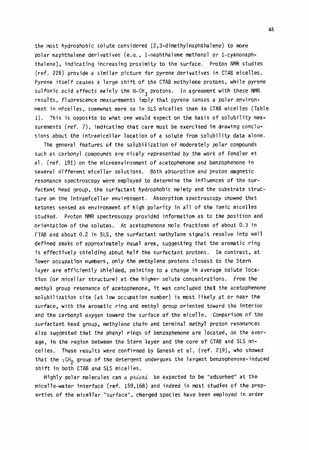
the most hydrophobic solute considered (2,3_dimethylnaphthalene) to more
polar naphthalene derivatives (e.a., 1-naphthalene methanol or l-cyanonaph-
thalene), indicating increasing proximity to the surface. Proton NMR studies
(ref. 226) provide a similar picture for pyrene derivatives in CTAB micelles.
Pyrene itself causes a large shift of the CTAB methylene protons, while pyrene
sulfonic acid affects mainly the N-CH3 protons. In agreement with these NMR
results, fluorescence measurements imply that pyrene senses a polar environ-
ment in micelles, somewhat more so in SLS micelles than in CTAB micelles (Table
I). This is opposite to what one would expect on the basis of solubility mea-
surements (ref. 7), indicating that care must be exercised in drawing conclu-
sions about the intramicellar location of a solute from solubility data alone.
The general features of the solubilization of moderately polar compounds
such as carbonyl compounds are nicely represented by the work of Fendler et
al. (ref. 191) on the microenvironment of acetophenone and benzophenone in
several different micellar solutions. Both absorption and proton magnetic
resonance spectroscopy were employed to determine the influences of the sur-
factant head group, the surfactant hydrophobic moiety and the substrate struc-
ture on the intramicellar environment. Absorption spectroscopy showed that
ketones sensed an environment of high polarity in all of the ionic micelles
studied. Proton NMR spectroscopy provided information as to the position and
orientation of the solutes. At acetophenone mole fractions of about 0.3 in
CTAB and about 0.2 in SLS, the surfactant methylene signals resolve into Well
defined peaks of approximately equal area, suggesting that the aromatic ring
is effectively shielding about half the surfactant protons. In contrast, at
lower occupation numbers, only the methylene protons closest to the Stern
layer are efficiently shielded, pointing to a change in average solute loca-
tion (or micellar structure) at the higher solute concentrations. From the
methyl group resonance of acetophenone, it was concluded that the acetophenone
solubilization site (at low occupation number) is most likely at or near the
surface, with the aromatic ring and methyl group oriented toward the interior
and the carbonyl oxygen toward the surface of the micelle. Comparison of the
surfactant head group, methylene chain and terminal methyl proton resonances
also suggested that the phenyl rings of benzophenone are located, on the aver-
age, in the region between the Stern layer and the core of CTAB and SLS mi-
celles. These results were confirmed by Ganesh et al. (ref. 219), who showed
that the yCH2 group of the detergent undergoes the largest benzophenone-induced
shift in both CTAB and SLS micelles.
Highly polar molecules can a p&&hi be expected to be "adsorbed" at the
micelle-water interface (ref. 159,168) and indeed in most studies of the prop-
erties of the micellar "surface", charged species have been employed in order

46
to avoid penetration inside the micelle. Even in this case, however, one
cannot completely exclude the possibility that the uncharged portion of the
probe molecule penetrates to some extent into the micelle and thus suffers an
influence from factors not directly related to the micelle-water interface.
One of the most comprehensive studies of the nature of the micellar micro-
environment sensed by adsorbed polar probes was carried out by Fernandez and
Fromherz (ref. 227). Employing coumarin dyes anchored into the micelle by a
long alkyl chain, the micropolarity was inferred indirectly from the shift in
the pK, of the probe relative to that in dioxane-water mixtures. In this
manner, they arrived at an effective dielectric constant of 32 for nonionic,
anionic and cationic micelles, irrespective of the charge of the head group.
Using the ET (30) of phenolbetaine as a measure of the polarity of the mi-
cellar environment, Zachariasse et al. (ref. 195) concluded that the probe
moleculeis invariably solubilized in the aqueous interface. The values ob-
tained for the effective dielectric constant were dependent on the solvent
system chosen to calibrate the polarity scale. Thus, for dodecyl-trinethyl-
ammonium chloride, the values ranged from 30 (on a scale calibrated vs. n-
alcohols) to 40 (on a scale vs. ethanol-water mixtures). Their values ob-
tained with water-dioxane mixtures as reference solvents are collected in
Table 6.
TABLE 6
Effective dielectric constants from ET (30) values (ref. 145)
Surfactant Cont. (M) E ef
OTAC 0.05 36 OTAB 0.05 TTAF 0.05 :93 TTAC 0.05 33
TTAB 0.05 CTAF 0.05 :: CTAC 0.05 31 CTAB BDHAC+ 0.05 0.05 ;: SDeS++ 0.1 55 SDS 0.1 55
tBenzildimethylhexadecylammonium chloride
++Sodiumdecyl sulphate; the rest as in Table 2
The results shown in Table 6 indicate that the probe senses a region of
higher average micropolarity in anionic surfactant solutions than in trimethyl-
ammonium detergent micelles and further suggest a dependence on the counterion
type and the surfactant chain length. A greater polarity (or a greater degree
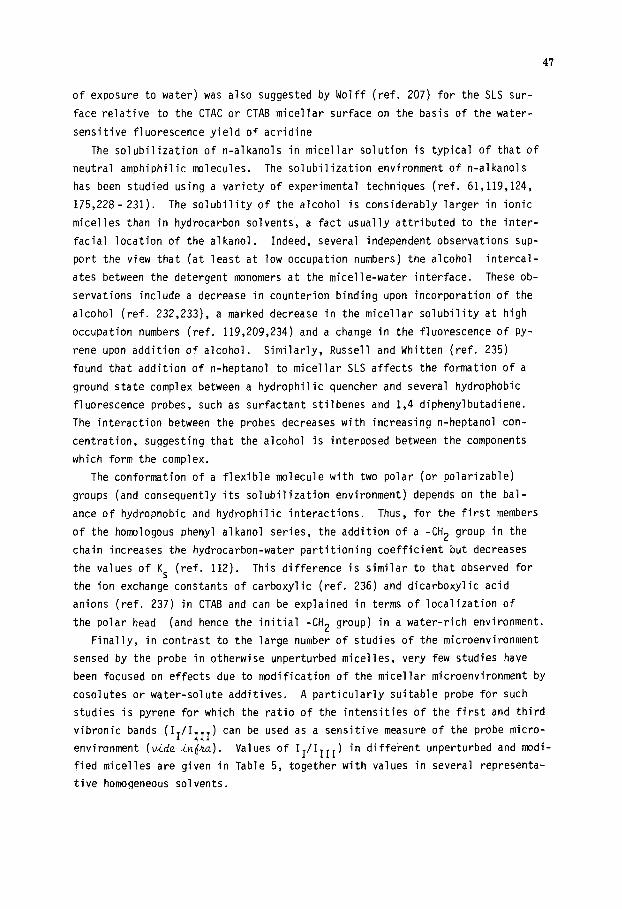
of exposure to water) was also suggested by Wolff (ref. 207) for the SLS sur-
face relative to the CTAC or CTAB micellar surface on the basis of the water-
sensitive fluorescence yield of acridine
The solubilization of n-alkanols in micellar solution is typical of that of
neutral amphiphilic molecules. The solubilization environment of n-alkanols
has been studied using a variety of experimental techniques (ref. 61,119,124,
175,228- 231). The solubility of the alcohol is considerably larger in ionic
micelles than in hydrocarbon solvents, a fact usually attributed to the inter-
facial location of the alkanol. Indeed, several independent observations sup-
port the view that (at least at low occupation numbers) the alcohol intercal-
ates between the detergent monomers at the micelle-water interface. These ob-
servations include a decrease in counterion binding upon incorporation of the
alcohol (ref. 232,233), a marked decrease in the micellar solubility at high
occupation numbers (ref. 119,209,234) and a change in the fluorescence of Py-
rene upon addition of alcohol. Similarly, Russell and Whitten (ref. 235)
found that addition of n-heptanol to micellar SLS affects the formation of a
ground state complex between a hydrophilic quencher and several hydrophobic
fluorescence probes, such as surfactant stilbenes and 1,4 diphenylbutadiene.
The interaction between the probes decreases with increasing n-heptanol con-
centration, suggesting that the alcohol is interposed between the components
which form the complex.
The conformation of a flexible molecule with two polar (or polarizable)
groups (and consequently its solubilization environment) depends on the bal-
ance of hydrophobic and hydrophilic interactions. Thus, for the first members
of the homologous phenyl alkanol series, the addition of a -CH2 group in the
chain increases the hydrocarbon-water partitioning coefficient but decreases
the values of KS (ref. 112). This difference is similar to that observed for
the ion exchange constants of carboxylic (ref. 236) and dicarboxylic acid
anions (ref. 237) in CTAB and can be explained in terms of localization of
the polar head (and hence the initial -CH2 group) in a water-rich environment.
Finally, in contrast to the large number of studies of the microenvironment
sensed by the probe in otherwise unperturbed micelles, very few studies have
been focused on effects due to modification of the micellar microenvironment by
cosolutes or water-solute additives. A particularly suitable probe for such
studies is pyrene for which the ratio of the intensities of the first and third
vibronic bands (II/IITI) can be used as a sensitive measure of the probe micro-
environment (vi& in&). Values of Ir/IIII) in different unperturbed and modi-
fied micelles are given in Table 5, together with values in several representa-
tive homogeneous solvents.
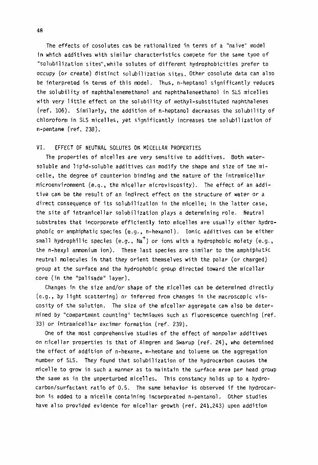
48
The effects of cosolutes can be rationalized in terms of a "naive" model
in which additives with similar characteristics compete for the same type of
"solubilization sites",while solutes of different hydrophobicities prefer to
occupy (or create) distinct solubilization sites. Other cosolute data can also
be interpreted in terms of this model. Thus, n-heptanol significantly reduces
the solubility of naphthalenemethanol and naphthaleneethanol in SLS micelles
with very little effect on the solubility of methyl-substituted naphthalenes
(ref. 106). Similarly, the addition of n-heptanol decreases the solubility of
chloroform in SLS micelles, yet significantly increases the solubilization of
n-pentane (ref. 238).
VI. EFFECT OF NEUTRAL SOLUTES ON MICELLAR PROPERTIES
The properties of micelles are very sensitive to additives. Both water-
soluble and lipid-soluble additives can modify the shape and size of the mi-
celle, the degree of counterion binding and the nature of the intramicellar
microenvironment (e.g., the micellar microviscosity). The effect of an addi-
tive can be the result of an indirect effect on the structure of water or a
direct consequence of its solubilization in the micelle; in the latter case,
the site of intramicellar solubilization plays a determining role. Neutral
substrates that incorporate efficiently into micelles are usually either hydro-
phobic or amphiphatic species (e.g., n-hexanol). Ionic additives can be either
small hydrophilic species (e.g., Nat) or ions with a hydrophobic moiety (e.g.,
the n-hexyl ammonium ion). These last species are similar to the amphiphatic
neutral molecules in that they orient themselves with the polar (or charged)
group at the surface and the hydrophobic group directed toward the micellar
core (in the "palisade" layer).
Changes in the size and/or shape of the micelles can be determined directly
(e.g., by light scattering) or inferred from changes in the macroscopic vis-
cosity of the solution. The size of the micellar aggregate can also be deter-
mined by "compartment counting" techniques such as fluorescence quenching (ref.
33) or intramicellar excimer formation (ref. 239).
One of the most comprehensive studies of the effect of nonpolar additives
on micellar properties is that of Almgren and Swarup (ref. 24), who determined
the effect of addition of n-hexane, n-heptane and toluene on the aggregation
number of SLS. They found that solubilization of the hydrocarbon causes the
micelle to grow in such a manner as to maintain the surface area per head group
the same as in the unperturbed micelles. This constancy holds up to a hydro-
carbon/surfactant ratio of 0.5. The same behavior is observed if the hydrocar-
bon is added to a micelle containing incorporated n-pentanol. Other studies
have also provided evidence for micellar growth (ref. 241,243) upon addition

49
of relatively nonpolar solutes. In general, the data lend support to the
proposal of Mukerjee (ref. 244) that solutes which penetrate into the core
should preferentially increase the size of the micelle without significantly
changing its shape.
In comparison with nonpolar solutes, the behavior of polar or amphiphatic
solutes is quite different in the sense that the latter tend to change the mi-
cellar shape by promoting the sphere-to-rod transition.
In a series of papers (ref. 229,234,245-258), Zana and co-workers have used
a variety of experimental techniques to study the effect of n-alcohols on the
micellar properties.
The system most thoroughly investigated was the pentanol-tetradecyl-trimethyl-
ammonium bromide system to which methods including conductivity and bromide ion
activity, elastic and quasi-elastic light scattering, osmometry, fluorescence
decay of micelle-solubilized pyrene, chemical relaxation and small neutron scat-
tering were applied to obtain information about the CMC, molecular weights (l$,)
and surfactant aggregation numbers, dynamics of micellar solutions and overall
radius of the hydrophobic core. In the initial paper of the series (ref. 145),
it was concluded that the addition of n-pentanol noticeably decreased the sur-
factant aggregation number and increased the bromide ion activity. At any
given alcohol concentration, the micelle molecular weight increased with in-
creasing surfactant concentration. The results, obtained both in the absence
and presence of added salt, were explained in terms of the effect of the
micelle-solubilized alcohol on the surface charge density and the local dielec-
tric constant in the micelle palisade layer. Subsequent work (ref. 246- 248)
showed that the apparent radius of gyration of the micelle increases with in-
creasing alcohol concentration. At low surfactant concentration, the micelles
apparently remain spherical in the presence of 1-pentanol; at large pentanol/
surfactant ratios, however, they may develop an inner core of I-pentanol in
addition to the I-pentanol already present in the palisade layer. As the
surfactant concentration is increased, the micelles grow, becoming ellipsoidal
(or perhaps rodlike).
The effect of a series of different alcohols on anionic SLS micelles has
been studied in detail by Almgren and Swarup (ref. 249). These authors
found that the radius of the micellar aggregate remains roughly constant upon
addition of the alcohols, resulting in an increase in the surface area per
detergent headgroup. The aggregation number typically decreased upon the init-
itial addition of the alcohols; further addition of the solute tended, however,
to produce an increase in micellar size. The general picture that emerges from
their data is that incorporation of an amphiphilic solute into the palisade
layer of the micelle causes a separation of the charges (an increase in the
50
area per charge), implying a decrease in the primary surface charge density.
The resulting decrease electrostatic repulsion between the headgroups out-
weighs any change in the hydrocarbon-water contact at the interface, favoring
micellar growth.
As pointed out by Manabe et al. (ref. 129), however, the release of counter-
ions from the micellar surface may conpensate for this decrease in surface
charge density, maintaining the electrical surface potential roughly constant.
The effect of both neutral and ionic amphiphiles on micellar properties
evidences itself in several systems as a change in the macroscopic viscosity
of the solute (ref. 250,251). In some cases, the viscosity of the solution be-
comes so high that it acquires the properties of a gel (ref. 252,253).
The viscosity effect produced by solutes seems to be related to a change
in the micellar shape from spherical to rodlike (ref. 254- 257). The associa-
tion of the solute to the micelle may facilitate the formation of large rod-
shaped micelles by decreasing the repulsion between the charged headgroups.
The increase in viscosity of a micellar solution is apparently quite solute-
specific. For example, Wan (ref. 252), investigating the effects of a variety
of substituted benzoic acids on the viscosity of some cationic micellar solu-
tions, found that salicyclic acid and its salts increased the viscosity of
dodecyl, tetradecyl and hexadecyl-trimethylammonium bromide solutions. In
contrast, m-hydroxybenzoic acid and p-hydroxybenzoic acid, as well as 0-,
m- and p-amino, chloro- and nitrobenzoic acids caused no viscosity change.
Larsen et al. (ref. 260) and Bunton et al. (ref. 261) reported similar re-
sults. These latter authors reported very high viscosities for CTAB solutions
containing sodium tosylate but not for those containing benzene-sulfonate;
disodium phenyl phosphate also has no effect on the viscosity of CTAB mi-
celles (ref. 251). The work of Wan (ref. 252) further indicates that the
nature of the micelle contributes to the specificity of the viscosity effect.
Thus, the same solutes that greatly increase the viscosity of cationic micelles
are without effect when added to anionic or nonionic micelles.
In all of the cases studied, the solutes that produce enhanced viscosity of
the micellar solution are aromatic molecules known to be strongly associated
to micelles and in particular to cationic micelles. This peculiar interaction
between positively charged micelles and aromatic molecules might reflect an
increased polarization of the II electron cloud of the aromatic ring under the
influence of the positive electric field at the micellar surface. However,
association of the solute to the micelle is not in itself a sufficient condi-
tion for producing the viscosity effect; the presence of a methyl group, an
alkyl chain, or some other appropriate functional group on the aromatic ring
is apparently also necessary. Likewise, there seems to be no clear correlation
between the magnitude of the solute incorporation coefficient and the viscosity
effect.

51
Finally, in many cases, the addition of the solute initially causes a sharp
increase in viscosity (ref. 261), followed by a sharp decrease at higher solute
concentrations (ref. 262). We believe that this behavior is a reflection of
the strong non-Newtonicity typical of very viscous solutions at finite flow
rates (ref. 253). Thus, the CTAB-sodium tosylate system exhibits a pronounced
maximum (ref. 261) when the apparent viscosities are plotted against tosylate
concentration. However, the maximum disappears if the viscosity data are deter-
mined at various flow rates and extrapolated to zero rate of flow (ref. 253).
Although the increase in viscosity is undoubtedly due to a solute-induced
change in the micellar shape from spherical to rodlike, the high solute speci-
ficity is far from understood and more work is required to identify the struc-
tural features of the solute responsible for the manifestation of these inter-
esting effects.
Using the Ru(bpy)12/methyl anthracene fluorescence quenching method, Almgren
and Swarup (ref. 249) measured the size of SLS micelles in the presence of
hydrophobic counterions and of nonionic surfactants. Hydrophobic counterions
of these kinds were tested: 1) cationic surfactants with one long alkyl chain
(from octylammonium chloride to CTAB); 2) symunetrical tetraalkylammonium (R=
C2H5, C4Hg, C5HII) ions; and 4) tetraphenylphosphonium salts. The first in-
duce a micelle growth (n increases from 67.5 to 87.9 at an octylammonium chlor-
ide mole fraction of 0.2, which would leave the surface area per SLS headgroup
nearly constant if the micelle were spherical (the large viscosity increase
observed when these surfactant counterions are incorporated suggests, however,
the occurence of a sphere-to-rod transition). In contrast, tetraalkylammonium
ions reduce the surfactant aggregation number (nsLs= 51.5 at a tetraalkyl-
ammonium chloride mole fraction of 0.37); the micellar volume remains almost
constant with the hydrophobic counterions acting as a spacer between the SLS
headgroups. Nonionic surfactants (tetra-, penta-, and octaethyleneglycol
mono-n-dodecyl ether) were also found to produce a decrease in the SLS aggre-
gation number, again consistent with a role as a spacer.
VII. ACKNOWLEDGEMENTS
Support of this work by the Departamento de Investigation y Bibliotecas de
la Universidad de Chile, the Comision National deInvestigaci6n Cientifica y
Tecnologica de Chile, the Proyecto de Fortalecimiento de1 Desarrolo
de las Ciencias Quimicas de1 PNUD, Programa CHI-84-006 and the Conselho National
de Desenvolvimento Cientifico e Tecnologico (CNPq) de Brasil is gratefully ack-
nowledged.

52
VI
1
2
: 9 10 11 12 13
:4s 16 I? 18
19
24
:: 27
28 29 30
31
36
33;
:: 41 42
. REFERENCES
M.E.L. McBain and E. Hutchinson, "Solubilization and Related Phenomena", Academic Press, New York, 1955. P.H. Elworthy, A.T. Florence and C.B. Macfarlane, "Solubilization by Surface- Active Agents and its Application in Chemistry and the Biological Sciences", Chapman and Hall, London, 1968. J.R. Cardinal and P. Mukerjee, J. Phys. Chem., 82(1978)1613. B. Jijnsson and H. Wennerstram, J. Colloid Interface Sci., 80(1981)482. G. Gunnarsson, B. Jijnsson and H. Wennerstrom, J. Phys. Chem., 84(1980)3114. B. Jiinsson, G. Gunnarsson and H. Wennerstrzm, in "Solution Chemistry of Surfactants". Vol. 1. K. L. Mittal and E.J. Fendler. eds.. Plenum Press. New York, 1982. . M. D. A. A. Y. Y.
;sI: M.
Algren, F. Grieser and J.K. Thomas, J. Am. Chem. Sot., 101(1979)279. Stinter. J. Colloid Interface Sci.. 47(1974)473. BenINaim, "Hydrophobic Interactions", Plenum Press, New York, 1980. Ben-Naim, J. Phys. Chem., 82(1978)792. Moroi, J. Phys. Chem., 84(1980)2186. Mardi, K. Sato and R. Matuura, J. Phys. Chem., 86(1982)2463. Mot-o?, H. Noma and R. Matuura, J. Phys. Chem., 87(1983)872. Tachiya, J. Chem. Phys. Lett.,_33(1975)289. _ Tachiya, J. Chem. Phys., 76(1982)340.
T.F. Hunter, Chem. Phys. Lett., 75(1980)152. A. Yecta, M. Aikawa and N.J. Turro, Chem. Phvs. Lett., 63(1979)543. H. Chaimovich, R.M.V. Aleixo, I. Cuccovia, K Zanette and.F.H..Quina, in "Solution Behavior of Surfactants", Vol. 2, K.L. Mittal and E.J. Fendler, eds., Plenum Press, New York, 1982. L. Romsted, in "Surfactants in Solution", Vol. 2, K.L. Mittal and B. Lindman, eds., Plenum Press, New York, 1984. C.A. Bunton, Pure Appl. Chem., 49(1977)969. D.W. Armstrong and G.Y. Stine, J. Am. Chem. Sot., 105(1983)2962. D.W. Armstrong and F. Nome, Anal. Chem., 53(1981)1662. I.V. Berezin, K. Martinek and A.K. Yatsimirsky, Russ. Chem. Revs. (English Transl.), 42(1973)487. K. Martinek, A.K. Yatsimirski, A.V. Levashov and I.V. Berezin, in "Micellization, Solubilization and Microemulsions", Vol. 2, K.L. Mittal, ed., Plenum Press, New York, 1977. S.J. Dougherty and J.C. Berg, J. Colloid Interface Sci., 48(1974 A.A. Bhalekar and J.B.F.N. Engberts, J. Am. Chem. Sot., loo)1978 5914. 1
110.
C6Fnford, Y. Nozaki, J.A. Reynolds and S. Marino, Biochemistry, 13(1974)
K. Shinoda and T. Soda, J. Phys. Chem., 67(1963)2072. P. Mukerjee, J. Phys. Chem., 66(1962)1733. T.S. Brun, H. Hoiland and E. Vikingstad, J. Colloid Interface Sci., 63(1978) 89. R. Dorshow, J. Briggs, C. A. Bunton and D.F. Nicoli, J. Phys. Chem., 86 (1982)2388. J.P. Kratohvil, J. Colloid Interface Sci., 75(1979)271. N.J. Turro and A. Yecta, J. Am. Chem. Sot., 100(1978)5951. C. Tanford, "The Hydrophobic Effect", Wiley, New York, N.Y., 1973. R.W. Gurney, "Ionic Processes in Solution", McGraw-Hill, New York, 1962 (reprinted by Dover Publications). W. Kauzmann, Adv. Protein Chem., 14(1959)1. S. Riegelman, J. Am. Pharm. Assoc., Sci. Ed., 49(1960)339. J. Hildebrand and R.L. Scott, "The Solubility of Nonelectrolytes", Reinhold, New York, 1950. P. Mukerjee, 3. Pharm. Sci., 60(1971)1531. P.D. Cratin, Ind. Eng. Chem., 60( 1968)14. A. Leo, C. Hansch and 0. Elkins, Chem. Rev., 71(1971)525. C. Treiner, J. Colloid Interface Sci., 93(1983)33.

53
55 56 57 58
Z 61 62
65
ii 70 71
L. Sepulveda and J. Perez-Cotapos, J. Colloid Interface Sci., in press. P. Mukerjee, in "Solution Chemistry of Surfactants", Vol. 1, K.L. Mittal, ed., Plenum Press, 1979. A. Wishnia, J. Phys. Chem., 67(1963)2079. K.S. Birdi, in "Micellization, Solubilization and Microemulsions", Vol. 1, K.L. Mittal, ed., Plenum Press, New York, 1977. C.H. Spink and S. Coloan, J. Phys. Chem., 87(1983)888. KS. Birdi and T. Magnusson, Colloid and Polymer Sci., 254(1976)1059. J.W. Larsen and L.J. Magid, J. Phys. Chem., 78(1974)834. C.H. Spink and R.E. Stedwell, J. Phys. Chem., 84(1980)2044. L. SepGlveda, J. Colloid Interface Sci., 46(1974)372. C.A. Bunton, M.J. Minch and L. Sepilveda, J. Phys. Chem., 75(1971)2707. C.A. Bunton and M.J. Minch, J. Phys. Chem., 78(1974)1480. J.H. Fendler and E.J. Fendler, "Catalysis in Micellar and Macromolecular Systems", Academic Press, New York, 1975. J.C. Erikson and G. Gillberg, Acta Chem. Stand., 20(1966)2019. G.L. Amidon and S.T. Anik, J. Phys.. Chem., 84(1980)970. C. Hirose and L. Sepfilveda, J. Phys. Chem., 85(1981)3689. C.A. Bunton and L. Septilveda, J. Phys. Chem., 83(1979)680. C. Gitler and A. Ochoa-Solano, J. Phys. Chem., 90(1968)5004. C.A. Bunton, L.S. Ronsted and C. Thamavit, J. Am. Chem. Sot., 102(1980)3900. K. Hayase and S. Hayano, Bull. Chem. Sot., Japan, 59(1977)83. C.H. Spink and S. Colgan, J. Colloid Interface Sci., 97(1984)41. A.K. Yatsimirski, K. Martinek and I.V. Berezin, Tetrahedron, 27(1971)2855. Ehcrn Bunton, L.H. Gan, J.R. Moffat, L.S. Romsted and G. Savelli, J. Phys.
85(1981)4118. C.A.'&nton, Y.S. Hong and L.S. Romsted, in "Symposium on Solution Behavior of Surfactants", Vol.‘2, K.L. Mittal and E.J. Fendler, eds., Plenum Press, New York, 1982. C.A. Bunton, J.R. Moffat and E. Rodenas, J. Am. Chem. Sot., 104(1982)2653. H. Al-Lohedan and C.A. Bunton, J. Org. Chem., 47(1982)1160. N. Funasaki and A. Murata, Chem. Pharm. Bull., 28(1980)805. H. Al-Lohedan, C.A. Bunton and L.S. Romsted, J. Phys. Chem., 85(1981)2123. J.H. Fendler and W.L. Hinze, J. Am. Chem. Sot., 103(1981)5439. C.A. Bunton, Y.S. Hong, L.S. Romsted and C. Quan, J. Am. Chem. Sot., 103 (1981)5788.
72 C.A. Bunton, S.E. Nelson and C. Quan, J. Org. Chem., 47(1982)1157. 73 C.A.Bunton and L. Sepirlveda, Israel J. Chem., 18(1975)298. 74 C.A. Bunton, G. Cerichelli, Y. Ihara and L. Septilveda, J. Am. Chem. Sot.,
101(1979)2429. 75
76
77 78 79
C.A: Bunton, N. Carrasco, S.K. Huang, C.H. Paik and L.S. Romsted, J. Am. Chem. Sot., 100(1978)5420. A. Cipiciani, P. Linda, G. Savelli and C.A. Bunton, J. Org. Chem., 46(1981) 911. C.A.Bunton, L.S. Romsted and H.J. Smith, J. Org. Chem., 43(1978)4299. C.A. Bunton, F. Rivera and L. Sepclveda, J. Org. Chem., 43(1978)1166. F.H. Quina, M.J. Politi, I. M. Cuccovia, E. Baumgarten, S.M. Martins- Francheti and H. Chaimovich, J. Phys. Chem., 84(1980)361. N. Funasaki, J. Phys. Chem., 83(1979)237. I.M. Cuccovia, E.H. Schroter, P.M. Monteiro and H. Chaimovich, J. Org. Chem., 43(1978)2248. N. Funasaki, J. Phys. Chem., 83(1979)1998. A.P. Osipov, K. Martinek, A.K. Yatsimirski and I.V. Berezin, Izv. Akad. Nauk. SSSR Ser. Kim., (1974)1984. J.C.Scaiano and J.C. Selwyn, Can. J. Chem., 59(1981)2368. C.A. Bunton and G. Cerichelli, Inter. J. Chem. Kin., 12(1980)519. V.C. Reinsborough and B.H. Robinson, J.C.S. Faraday I, 75(1979)2395. Y. Miyashita and S. Hayano, J. Colloid Interface Sci., 86(1982)344. L.W. Winters and E. Grunwald, J. Am. Chem. Sot., 87(1965)4608. E. Valenzuela, E. Abuin and E.A. Lissi, J. Colloid Interface Sci., 101 ( 1984)401.
54
90 91 92 93
94
95
;; 98
1.8.C. Matheson and A.D. King, J. Colloid Interface Sci., 101(1573)464. J.C. Hoskins and A.D. King, J. Colloid Interface Sci., 82(1981)264. J.C. Hoskins and A.D. King, J. Colloid Interface Sci., 82(1981)267. P.L. Bolden, J.C. Hoskins and A.D. King, J. Colloid Interface Sci., 91 (1983)454. S.D. Christian, E.E. Tucker and E.H. Lane, J. Colloid Interface Sci., 84(1981)423. S.D. Chritian, L.S. Smith, D.S. Bushong and E.E. Tucker, J. Colloid Inter- face Sci., 89(1982)514. J.W. MacBain, K. Yasota and H.P. Lucas, J. Am. Chem. Sot., 55(1933)2762. J.W. MacBain and W.J. Jenkins, J. Chem. Sot., 121(1922)2325. E. Hutchinson and P.M. Shaffer, Z. Phys. Chem. Neue Folge., 31(1962)397. -. _ ~~..
99 E. Hutchinson, Z. Phys. Chem. Neue Folge., 21(1959)38. 100 C. Gamboa, L. Sepulveda and R. Soto, J. Phvs. Chem., 85(1981)1429. 101
:8: 104 105
106
107 108 109
110 111 112 113 114 115 116 117 118
I.W. Osborne-Lee; R.S. Schechter and W.H. Wade, J. Colldid Interface Sci., 94(1983)179. D.G. Herries, W. Bishop and F.M. Richards, J. Phys. Chem., 68(1964)1842. W.L. Hinze and D.W. Armstrong, Anal. Lett., 13(1980)1093. F.H. Quina and V.G. Toscano, J. Phys. Chem., 81(1977)1750. M. van Borkstaelle, J. Gelan, H. Martens, J. Put, J.C. Dederen, J.N. Boens and F.C. Schryver, Chem. Phys. Lett., 58(1978)211. M. Gonzalez, J. Vera, E. Abuin and E.A. Lissi, J. Colloid Interface Sci., 98(1984)152. K.S. Birdi, H.N. Singh and S.V. Dalsager, J. Phys. Chem., 83(1979)2733. P.C. Lee and M.A. Rodoers. J. Phvs. Chem., 87(1983)4894. N.J. Turro, M.W. Geiger, R.R. Hautala and-N.E: Schore, in "Surfactants in Solution". K.L. Mittal. ed.. Plenum Press. New York. 1983. D. 75. R.R. Hautala, N.E. Schore and N.J. Turro,-J. Am. Chem. Sot.‘, 95(1973)5508. E. Lissi and E. Abuin, J. Phys. Chem., 84(1980)2605. E. Lissi, E. Abuin and A.M. Rocha, J. Phys. Chem., 84(1980)2406. M.V. Encinas and E. Lissi, Chem. Phys. Lett., 91(1982)55. M. Aikawa, A. Yecta and N.J. Turro, Chem. Phys. Lett., 68(1979)285. N.J. Turro, M. Aikawa and A.Yecta, Chem. Phys. Lett., 64(1979)473. H.W. Ziemiecki, R. Holland and W.R. Cherry, Chem. Phys. Lett., 73(1980)145. M.V. Encinas and E. Lissi, Photochem. and Photobiol., 37(1983)252. M.V. Encinas, M.A. Rubio and E. Lissi, Photochem. and Photobiol., 37(1983) 125.
119 120 121
E. Abuin and E. Lissi, J. Colloid Interface Sci., 95(1983)198. F. Tokiwa and K. Aigami, Kolloid-Z.Z. Polym., 246(1971)688. A.S. Waggoner, O.H. Griffith and C.R. Christensen, Proc. Nat. Acad. Sci., 57(1967)1198.
122 A.S. Waggoner, A.D. Keith and O.H. Griffith, J. Phys. Chem., 72(1968)4129. 123 NI;'. Atherton and S.J. Strach, J. Chem. Sot. Faraday Trans. II, 68(1972)
124 125 126
Stilbs, J. Colloid Interface Sci., 87(1982)385. Kaneshina, H. Kamaya and I.Ueda, J. Colloid Interface Sci., 83(1981)589. Shinoda, R. Nakagawa, B. Tamamushi and T. Isemura, "Colloid Surfactants", 7, Academic Press, N. Y., 1963.
127 128 129 130
s’: K. P. K.
;: R.
Shirahama and T. Kashiwabara. J. Colloid Interface Sci.. 36(1971)65. Kayase and S. Hayano, J. Colioid Interface Sci., 63(1978)446. Manabe, M. Koda and K. Shirahama, J. Colloid Interface Sci., 77(1980)189. De Lisi, C. Genova, R. Testa and V. Turco Liveri, J. Solution Chem.,
13( 1984)lZl. 131 132 133
134
F.M. Menger and C.E. Portnoy, J. Am. Chem. Sot., 89(1968)4698. L.S.Romsted, Ph.D.Thesis, Indiana University, 1975. C.A. Bunton, Progress in Solid State Chem., Vol. 8, J.O'McCaldin and G. Somorjai, eds., Pergamon, Oxford and New York, 1973. H. Chaimovich, R.M. Aleixo, I.M. Cuccovia, D. Zanette and F.H. Quina, in "Solution Behavior of Surfactants", Vol. 2, K.L. Mittal, ed., Plenum, New York, 1982.
135
136
137
138
139
140
141 142 143
144
145 146 147 148 149 150 151 152 153 154 155 156 157 158
159
160 161 162
163 164 165 166 167
168 169
170
C.A. Bunton and L.S. Romsted, in "Solution Behavior of Surfactants", Vol. 2, K. L. Mittal, ed., Plenum, New York, 1982. K.M. Bansal, L.K. Patterson, E.J. Fendler and J.H. Fendler, Int. J. Radia- tion Phys. Chem., 3(1971)321. P.P. Infelta, M. Gratzel and J.K. Thomas, J. Chem. Sot. Faraday Trans. I, 75(1979)1674. M. Algrem, F. Greiser and J.K. Thomas, J. Chem. Sot. Faraday Trans. I, 75(1979)1674. E.L. Evers, G.G. Jayson, I.D. Robb and A.J. Swallow, J. Chem. Sot. Faraday Trans. I, 76(1980)528. T. Miyaji, K. Kurono, K. Vekama and M. Ikeda, Chem. Pharm. Bull. Japan, 24( 19?6)115. C. Candau, E. Hirsch and R. Zana, J. Colloid Interface Sci., 88(1982)428. T. Nakagawa and K. Tori, Kolloid-Z.Z. Polym., 194(1961)143. 0. Hall, P.L. Jobling, J.E. Rassing and E. Wyn-Jones, J. Chem. Sot. Faraday II, 73(1977)1582. J. Getlings, D. Hall, P.L. Jobling, J.E. Rassing and E. Wyn-Jones, J. Chem. Sot. Faraday II, 74(1978)1957. M.F. Ottaviani, P. Baglioni and G. Martini, J. Phys. Chem., 87(1983)3146. J.C. Selwyn and J.C. Scaiano, Can. J. Chem., 59(1981)663. J.C. Scaiano and E.B. Abuin, Chem. Phys. Lett., 8(1981)209. N.J. Turro, M.T. Zimnt and I.R. Gould, J. Am. Chem. Sot., 105(1983)6347. I.B. Matheson and R. Massoudi, J. Am. Chem. Sot., 102(1980)1942. P.P. Intelta, M. Gratzel and J.K. Thomas, J. Phys. Chem., 78(1974)190. K. Gla'sle, U.K. Klein and M. Hauser, J. Mol. Struct., 84(1982)353. K. Kaliansundaram, Chem. Sot. Rev., 7(1978)453. M.J. Leigh and J.C. Scaiano, J. Am. Chem. Sot., 105(1983)5652. F. Grieser, J. Phys. Chem., 85(1981)928. A.F. Olea, M.V. Encina and E.A. Lissi, Macromolecules, 15(1982)1111. A.S.C. Lawrence, Trans. Faraday Sot., 33(1937)815. E.W. Hughes, W.M. Sawyer and J.R. Vinograd, J. Chem. Phys., 13(1945)131. J.H. Fendler and E.J. Fendler, "Catalysis in Micellar and Macromolecular Systems", Academic Press, N.Y., 1975. P. Mukerjee, I.R. Cardinal and N.R. Desai, in "Micellization, Solubiliza- tion and Microemulsions", Vol. 1, K.L. Mittal, ed., Plenum Press, New York, 1977. J.K. Thomas, Chem. Rev., 80(1980)283. H. Wennerstrb'm and B. Lindman, Phys. Reports, 52(1979)1. J.J. Jacobs, R. A. Anderson and T.R. Watson, J. Phar. Pharmacol.. 23(1971) 148. S. Onishi, T.J. Cyr and H. Fukushima, Bull. Chem. Sot., Japan, 43(1970)673. E.A.G. Anianson, J. Phys. Chem., 82(1978)2805. S.J. Rehfeld, J. Phys. Chem., 75(1971)3905. J.H. Fendler and L.K. Patterson, J. Phys. Chem., 75(1971)3907. A. Breslow, S. Kitasatake and J. Rothband, J. Am. Chem. Sot., lOO(1978) 8156. P. Mukerjee and J.R. Cardinal, J. Phys. Chem., 82( 1978)8156. N.M. Atherton and S.J. Strach, J. Chem. Sot. Faraday Trans. II, 68(1972) 374. M.J. Povich, J.A. Mann and A. Kawamoto, J. Colloid Interface Sci., 41(1972) 145.
171 172 173 174
H. Yoshioka, J. Am. Chem. Sot., 101(1979)28. Y.C. Jean and H.J.J. Ache, J. Am. Chem. Sot., 100(1978)6320. J.H. Fendler and L.K. Patterson, J. Phys. Chem., 74(1970)4609. M. Gratzel, K. Kalyanasundaran and J.K. Thomas, J. Amer. Chem. Sot., 96 (1974)7869.
175
176 177
JO, ,
M. Van Bockstaelle, J. Gelen, H. Martens, J. Put, F.C. de Schryver and J.C. Dederen, Chem. Phys. Lett., 70(1980)6135. D.E. Blatt, K.P. Ghiggino and W.H. Sawyer, J. Phys. Chem., 86(1982)4461. A.J. Rodgers, M.E. Da Silva and E. Wheeler, Chem. Phys. Lett., 43(1976) co7
55
56
178 K.S. Schanze, T.F. Mattox and D. Whitten, J. Am. Chem. Sot., 104(1982) 1733.
179 F.M. Menger and J.M. Bonicamp, J. Am. Chem. Sot., 103(1981)2140. 180 C.M. Paleos and A. Malliaris, J. Colloid Interface Sci., 82(1981)244. 181 N.J. Turro, K.C. Liv and M.F. Chow, Photochem. Photobiol., 26(1977)413. 182 J.R. Winkle, P.R. Worsham, K.S. Shanze and D.G. Whitten, J. Am. Chem. Sot.,
105(1983)3951. 183 J.C. Russell, S.B. Costa, R.P. Seiders and D.G. Wnitten, J. Am. Chem. Sot.,
102(1980)5679. 184 M.V: Encina, E. Guztin and E.A. Lissi, J. Phys. Chem., 87(1983)4770. 185 J.C. Russell, D.G. Whitten and A.M. Braun, J. Am. Chem. Sot., 103(1981)3219. 186 F.M. Martens and J.W. Verhoeven, J. Phys. Chem., 85(1981)1773. 187 T. Matsuo, K. Yudato and T.Nagamura, J. Colloid Interface Sci., 83(1981)
354. 188 B.A. Mulley and A.D. Metcalf, J. Pharm. Pharmacol., 8(1956)774. 189 S. Riegelman, N.A. Allawala, M.K. Hrenoff and L.A. Strait, J. Colloid Sci.,
13(1958)208. 190 S.J. Rehfeld, J. Phys. Chem., 74(1970)117. 191 J.H. Fendler, E.J. Fendler, G.A. Infante, Pong-Su Shih and L.K. Patterson,
J. Am. Chem. Sot., 97(1975)89. 192 S.A. Simon, R.V. McDaniel and T.J. McIntosh, J. Phys. Chem., 86(1982)1449. 193 C. Ramachandran, R.A. Pyter and P. Mukerjee, J. Phys. Chem., 86(1982)3198. 194 R.A. Pyter, C. Ramachandran dnd P. Mukerjee, J. Phys. Chem., 86(1982)3206 195 K.A. Zachariasse. N. van Phue and B. Kosonkiewicz. J. Phvs. Chem., 85(1981)
2676. 196 P. Plieminger and H. Banmgartel, Ber. Bunsenges Phys. Chem., 86(1982)161. 197 A. Goto and F. Endo, J. Colloid Interface Sci., 68(1979)163. 198 E.M. Kosower, J. Am. Chem. Sot., 80(1958)3253. 199 C. Reichardt, Angew Chem. Int. Ed. Engl., 18(1979)98. 200 D.C. Dang and M.A. Winnik, Photochem. Photobiol., 35(1982)17. ?I?, L" .-LI-IL-I... ” ,- ml--_.... c ,.‘L,_.. _-_I I. II-L_._ n:^^L^_:-L._.. ,il,,n7,\
202 K. Kalyanasundaram and J.K. Thomas, J. Am. Chem. Sot., 99(1977)2039. 203 K.A. Zachariasse, Chem. Phys. Lett., 57(1978)429. 204 J.C. Russell and D.G. Whitten, J. Am. Chem. Sot., 103(1981)3219. 205 S. Shinkai, Y. Ishikawa, 0. Manabe and T. Kunitake, Chem. Lett., (1981)
1523. 206 C. David, E. Szalai and D. Baeyen-Velant, Ber. Bunsenges Phys. Chem., 86
(1982)710. 207 T. Wolff, Ber. Bunsenges Phys. Chem., 85(1981)145. 208 M.A.J. Rodgers, Chem. Phys. Lett., 78(1981)509. 209 P. Lianos and R. Zana, Chem. Phys. Lett., 72(1980)171. 210 E. Abuin, E. Lissi and A. Datoli, Bol. Sot. Chilena de Quimica, in press. 211 M. Gratzel and J.K. Thomas, in "Modern Fluorescence Spectroscopy", H.L.
Whery, ed., Plenum Press, N.Y., 1976. 212 F.J. Perrin, Phys. Radium., 7(1936)1. 213 M. Gratzel and J.K. Thomas, J. Am. Chew Sot., 95(1973)6885. 214 P. Lianos and R. Zana, J. Colloid Interface Sci., 88(1982)594. 215 B.B. Craig, J. Kirk and M.A.J. Rodgers, Chem. Phys. Lett., 49(1977)437. 216 J. Ulmius, B. Lindman, G. Lindblom and T. Drakenberg, J. Colloid Interface
Sci., 65(1978)88. 217 J.C. Eriksson, Acta Chem. Stand., 17(1963)1478. 218 J.C. Eriksson and G. Gillberg, Acta Chem. Stand., 20(1966)2019. 219 K.N. Ganesh, P. Mitra and 0. Balasubramamion, J. Phys. Chem., 86(1982)
4291. 220 F.M. Menger and J.M. Jerkunica, J. Am. Chem. Sot., 100(1978)688. 221 F.M. Menger, J.M. Jerkunica and J.C. Johnston, J. Am. Chem. Sot., 100)1978)
4676. 222 E. Lissi, T. Ca'ceres and C. Veliz, Bol. Sot. Chilena de Quimica, 28(1983)
13.

57
223 E.J. Fendler, C.L. Day and J.H. Fendler, J. Phys. Chem., 76(1972)1460. 224 O.J. Jobe, V.C. Reinsborough and P.J. White, Can. J. Chem., 60(1982)279. 225 S.J. Rehfeld, J. Colloid Interface Sci., 34(1970)1518. 226 K. Kalyanasundaram and J.K. Thomas, J. Phys. Chem., 81(1977)2176. 227 M.S. Fernhndez and P. Fromherz, J. Phys. Chem., 81(1977)1755. 228 S. Yib. R. Zana, W. Ulbricht and H. Hoffmann, J. Colloid Interface Sci.,
80( 198i)224. 229 R. Zana, C. Picot and R. Duplessix, J. Colloid Interface Sci., 93(1983)43. 230 S. Kamshina. K. Kamava and I. Uedo, J. Colloid Interface Sci., 83(1981)589. 231 J. Gettins,.D. Hal1,"P.K. Job1ing;J.E. Rassing and E. Wyn-Jones,.J. Chem.
sot., Faraday II, 74(1978)1957. 232 M. Manabe, M. Koda and K. Shirahama, J. Colloid Interface Sci., 77(1980)189. 233 C. Treiner, J. Colloid Interface Sci., 90(1982)444. 234 P. Lianos, J. Lang, C. Strazielle and R. Zana, J. Phys. Chem., 86(1982)1019. 235 J.C. Russell and D.G. Whitten, J. Am. Chem. Sot., 104(1982)5937. 236 E. Lissi, E. Abuin, G. Ribot, E. Valenzuela, H. Chaimovich, P. Araujo,
R.M. Aleixo and I.M. Cuccovia, J. Colloid Interface Sci., in press. 237 E. Lissi, E. Abuin, H. Chaimovich and I.M. Cuccovia, unpublished results. 238 E. Valenzuela, E. Abuin and E.A. Lissi, J. Colloid Interface Sci., 102(1984)
401. 239 S. Atik,M. Man and L. Singer, Chem. Phys. Lett., 67(1979)75. 240 M. Almgren and S. Swarup, J. Phys. Chem., 86(1982)4212. 241 A. Hyde and D. Robb, Proceedings 4th International Congress on Surface
Active Substances, Brussels, Gordon and Breach, New York, 1964. 242 J. Nakagawa, K. Kuriyama and I. Inque, J. Colloid Sci., 15(1960)268. 243 D.A. Atwood, P.H. Elworthy and S.B. Kayne, J. Pharm. Pharmac., 23(1971)
775. 244 P. Mukerjee, Pure Applied Chem., 52(1980)1317. 245 R. Zana, S. Yiv, C. Strazielle and P. Lianos, J. Colloid Interface Sci.,
80(1981)208. 246 S. Yiv, R. Zana, W. Ulbricht and H. Hoffmann, J. Colloid Interface Sci.,
80(1981)224. 247 S. Candau and R. Zana, J. Colloid Interface Sci., 84(1981)206. 248 S. Candau, E. Hirsch and R. Zana, J. Colloid Interface Sci., 88(1982)428. 249 M. Algrem and S. Swarup, J. Colloid Interface Sci., 91(1983)256. 250 L. Sepirlveda, J. Colloid Interface Sci., 46(1974)372. 251 J.W. Larsen and L.B. Tepley, J. Org. Chem., 41(1976)2968. 252 L.J. Wan, J. Pharm. Sci., 55(1966)1395. 253 C. Gamboa and L. SepGlveda, J. Colloid Interface Sci., in ress. 254 N.A. Mazer, G.B. Benedek and M.C. Carey, J. Phys. Chem., 1 0(1976)1075. 255 G. Porte and J. Appell, J. Phys. Chem., 76(1972)565. 256 S. Ozeki and S. Ikeda, J. Colloid Interface Sci., 87(1982)424. 257 H. Hoffman, G. Platz, H. Rehage and W. Schorr, Adv. Colloid Interface Sci.,
ll( 1982)275. 258 C. Tanford, J. Phys. Chem., 76(1972)3020. 259 C. Tanford, J. Phys. Chem., 78(1974)2469. 260 J.W. Larsen, L.J. Magic and V. Payton, Tetrahedron Lett., 29(1973)2663. 261 C.A. Bunton, M.J. Minch, J. Hidaldo and L. Sepirlveda, J. Am. Chem. SOC.,
95(1973)3262. 262 A. Chau and L. SepGlveda, unpublished results. 263 J.K. Thomas, F. Grieser and M. Wong, Ber. Bunsenges. Phys. Chem., 82(1978)
937.
Related Documents