JOURNAL OF VIROLOGY, Mar. 2002, p. 2113–2122 Vol. 76, No. 5 0022-538X/02/$04.000 DOI: 10.1128/JVI.76.5.2113–2122.2002 Copyright © 2002, American Society for Microbiology. All Rights Reserved. Interaction of Translation Initiation Factor eIF4B with the Poliovirus Internal Ribosome Entry Site Kerstin Ochs, Lanja Saleh, Gergis Bassili, Volker H. Sonntag, Amandus Zeller, and Michael Niepmann* Institute of Biochemistry, 35392 Giessen, Germany Received 24 July 2001/Accepted 27 November 2001 Poliovirus translation is initiated at the internal ribosome entry site (IRES). Most likely involving the action of standard initiation factors, this highly structured cis element in the 5 noncoding region of the viral RNA guides the ribosome to an internal silent AUG. The actual start codon for viral protein synthesis further downstream is then reached by ribosomal scanning. In this study we show that two of the secondary structure elements of the poliovirus IRES, domain V and, to a minor extent, domain VI, are the determinants for binding of the eukaryotic initiation factor eIF4B. Several mutations in domain V which are known to greatly affect poliovirus growth also seriously impair the binding of eIF4B. The interaction of eIF4B with the IRES is not dependent on the presence of the polypyrimidine tract-binding protein, which also binds to the poliovirus IRES. In contrast to its weak interaction with cellular mRNAs, eIF4B remains tightly associated with the poliovirus IRES during the formation of complete 80S ribosomes. Binding of eIF4B to the IRES is energy dependent, and binding of the small ribosomal subunit to the IRES requires the previous energy-dependent association of initiation factors with the IRES. These results indicate that the interaction of eIF4B with the 3 region of the poliovirus IRES may be directly involved in translation initiation. When poliovirus, the paradigm of the family of picornavi- ruses, infects a susceptible cell, the positive-strand viral RNA can be used directly for translation of the viral polyprotein. In contrast to normal cellular mRNAs, translation of the picor- naviral RNA is facilitated by an internal region of the viral RNA, the internal ribosome entry site (IRES). This strategy of internal initiation allows some picornaviruses to take advan- tage of the obvious opportunity and shut down the cap-depen- dent cellular translation, while synthesis of the picornaviral polyprotein is initiated cap independently from the IRES ele- ment (for an overview, see reference 3). Picornavirus IRES elements are large cis-acting RNA re- gions that guide ribosomes to an internal site of the viral RNA (17, 37, 51). They are classified into three groups according to their primary and secondary structures: the type I elements of the enterovirus-rhinovirus group (including poliovirus), the type II elements of the cardiovirus-aphthovirus group, and the type III element of hepatitis A virus (HAV). The IRES ele- ments consist of highly conserved RNA secondary structure domains and have a characteristic oligopyrimidine tract fol- lowed by a conserved AUG triplet at their 3 borders (8, 42, 44). In poliovirus, this AUG is important but silent for trans- lation, while the actual initiation codon further downstream is reached by ribosomal scanning (20, 22, 25, 39). The small ribosomal subunit is guided to a starting window (43) contain- ing the conserved AUG triplet, most likely by the action of initiation factors. In addition to the standard initiation factors, picornavirus IRES elements recruit other cellular RNA-bind- ing proteins that enhance translation or modulate the balance between translation of the viral RNA and its replication, such as the 57-kDa polypyrimidine tract-binding protein (PTB) (13, 18, 31), the 52-kDa La protein (26), or the 39-kDa poly(rC) binding protein (6). For the internal initiation of translation on picornaviral RNA, almost the complete set of the canonical eukaryotic translation initiation factors (eIFs) appears to be essential (2, 35, 38), with the exception of the actual cap-binding protein eIF4E. In cap-dependent initiation of normal cellular mRNAs, the large adapter protein eIF4G associates with eIF4E and with the RNA helicase eIF4A to form the cap-binding protein complex eIF4F, which guides the small ribosomal subunit to the 5 end of the mRNA. eIF4B appears to be involved in several interactions during translation initiation. It stimulates the RNA helicase activity of eIF4A (35), it can bind to 18S rRNA (27), and it interacts with the ribosome-bound eIF3 (28). Moreover, eIF4B has two RNA-binding domains and can catalyze the hybridization of two complementary single- stranded RNAs by an RNA annealing activity (1). For the type II picornavirus IRES elements, the binding sites for these initiation factors have been roughly mapped, e.g., for encephalomyocarditis virus (EMCV) and for foot-and-mouth- disease virus (FMDV). A large Y-shaped RNA structure in the 3 region of these IRES elements (referred to as domain 4 in FMDV and J-K-L in EMCV) precedes the starting window with the conserved AUG. This RNA region is known to bind eIF4B (29, 33, 45) as well as eIF4G and eIF4A (19, 23, 38, 46). In contrast, for poliovirus as well as for the related rhinovi- ruses, the site of binding of the canonical initiation factors is not yet known. However, reasonable assumptions can be made about the IRES region possibly involved. In the poliovirus IRES, the domain V resembles the Y-shaped domain 4 (or J-K-L) of the type II IRES elements with regard to its relative position in the array of IRES secondary structures and its * Corresponding author. Mailing address: Institute of Biochem- istry, Faculty of Medicine, Friedrichstrasse 24, 35392 Giessen, Ger- many. Phone: 49-641-99-47471. Fax: 49-641-99-47429. E-mail: [email protected]. 2113 on December 24, 2015 by guest http://jvi.asm.org/ Downloaded from

Welcome message from author
This document is posted to help you gain knowledge. Please leave a comment to let me know what you think about it! Share it to your friends and learn new things together.
Transcript
JOURNAL OF VIROLOGY, Mar. 2002, p. 2113–2122 Vol. 76, No. 50022-538X/02/$04.00�0 DOI: 10.1128/JVI.76.5.2113–2122.2002Copyright © 2002, American Society for Microbiology. All Rights Reserved.
Interaction of Translation Initiation Factor eIF4B with the PoliovirusInternal Ribosome Entry Site
Kerstin Ochs, Lanja Saleh, Gergis Bassili, Volker H. Sonntag, Amandus Zeller,and Michael Niepmann*
Institute of Biochemistry, 35392 Giessen, Germany
Received 24 July 2001/Accepted 27 November 2001
Poliovirus translation is initiated at the internal ribosome entry site (IRES). Most likely involving the actionof standard initiation factors, this highly structured cis element in the 5� noncoding region of the viral RNAguides the ribosome to an internal silent AUG. The actual start codon for viral protein synthesis furtherdownstream is then reached by ribosomal scanning. In this study we show that two of the secondary structureelements of the poliovirus IRES, domain V and, to a minor extent, domain VI, are the determinants for bindingof the eukaryotic initiation factor eIF4B. Several mutations in domain V which are known to greatly affectpoliovirus growth also seriously impair the binding of eIF4B. The interaction of eIF4B with the IRES is notdependent on the presence of the polypyrimidine tract-binding protein, which also binds to the poliovirusIRES. In contrast to its weak interaction with cellular mRNAs, eIF4B remains tightly associated with thepoliovirus IRES during the formation of complete 80S ribosomes. Binding of eIF4B to the IRES is energydependent, and binding of the small ribosomal subunit to the IRES requires the previous energy-dependentassociation of initiation factors with the IRES. These results indicate that the interaction of eIF4B with the 3�region of the poliovirus IRES may be directly involved in translation initiation.
When poliovirus, the paradigm of the family of picornavi-ruses, infects a susceptible cell, the positive-strand viral RNAcan be used directly for translation of the viral polyprotein. Incontrast to normal cellular mRNAs, translation of the picor-naviral RNA is facilitated by an internal region of the viralRNA, the internal ribosome entry site (IRES). This strategy ofinternal initiation allows some picornaviruses to take advan-tage of the obvious opportunity and shut down the cap-depen-dent cellular translation, while synthesis of the picornaviralpolyprotein is initiated cap independently from the IRES ele-ment (for an overview, see reference 3).
Picornavirus IRES elements are large cis-acting RNA re-gions that guide ribosomes to an internal site of the viral RNA(17, 37, 51). They are classified into three groups according totheir primary and secondary structures: the type I elements ofthe enterovirus-rhinovirus group (including poliovirus), thetype II elements of the cardiovirus-aphthovirus group, and thetype III element of hepatitis A virus (HAV). The IRES ele-ments consist of highly conserved RNA secondary structuredomains and have a characteristic oligopyrimidine tract fol-lowed by a conserved AUG triplet at their 3� borders (8, 42,44). In poliovirus, this AUG is important but silent for trans-lation, while the actual initiation codon further downstream isreached by ribosomal scanning (20, 22, 25, 39). The smallribosomal subunit is guided to a starting window (43) contain-ing the conserved AUG triplet, most likely by the action ofinitiation factors. In addition to the standard initiation factors,picornavirus IRES elements recruit other cellular RNA-bind-ing proteins that enhance translation or modulate the balance
between translation of the viral RNA and its replication, suchas the 57-kDa polypyrimidine tract-binding protein (PTB) (13,18, 31), the 52-kDa La protein (26), or the 39-kDa poly(rC)binding protein (6).
For the internal initiation of translation on picornaviralRNA, almost the complete set of the canonical eukaryotictranslation initiation factors (eIFs) appears to be essential (2,35, 38), with the exception of the actual cap-binding proteineIF4E. In cap-dependent initiation of normal cellular mRNAs,the large adapter protein eIF4G associates with eIF4E andwith the RNA helicase eIF4A to form the cap-binding proteincomplex eIF4F, which guides the small ribosomal subunit tothe 5� end of the mRNA. eIF4B appears to be involved inseveral interactions during translation initiation. It stimulatesthe RNA helicase activity of eIF4A (35), it can bind to 18SrRNA (27), and it interacts with the ribosome-bound eIF3(28). Moreover, eIF4B has two RNA-binding domains and cancatalyze the hybridization of two complementary single-stranded RNAs by an RNA annealing activity (1).
For the type II picornavirus IRES elements, the binding sitesfor these initiation factors have been roughly mapped, e.g., forencephalomyocarditis virus (EMCV) and for foot-and-mouth-disease virus (FMDV). A large Y-shaped RNA structure in the3� region of these IRES elements (referred to as domain 4 inFMDV and J-K-L in EMCV) precedes the starting windowwith the conserved AUG. This RNA region is known to bindeIF4B (29, 33, 45) as well as eIF4G and eIF4A (19, 23, 38, 46).
In contrast, for poliovirus as well as for the related rhinovi-ruses, the site of binding of the canonical initiation factors isnot yet known. However, reasonable assumptions can be madeabout the IRES region possibly involved. In the poliovirusIRES, the domain V resembles the Y-shaped domain 4 (orJ-K-L) of the type II IRES elements with regard to its relativeposition in the array of IRES secondary structures and its
* Corresponding author. Mailing address: Institute of Biochem-istry, Faculty of Medicine, Friedrichstrasse 24, 35392 Giessen, Ger-many. Phone: 49-641-99-47471. Fax: 49-641-99-47429. E-mail:[email protected].
2113
on Decem
ber 24, 2015 by guesthttp://jvi.asm
.org/D
ownloaded from
position directly upstream of the conserved cryptic AUG. Con-sistent with this idea, domain V has been found to be the mostimportant structure of the poliovirus IRES, since nearly anymutation affecting its structure is deleterious for IRES activity(7, 11, 21, 36, 50).
In this study, we investigated the interaction of eIF4B withthe poliovirus IRES. We mapped the determinants that causethe binding of eIF4B in the poliovirus IRES to reside in do-mains V and VI. Moreover, we analyzed the association ofeIF4B with the IRES in the course of the formation of ribo-somal initiation complexes and the possible interaction ofeIF4B with a noncanonical RNA binding protein, PTB.
MATERIALS AND METHODS
Plasmids. pMPolio contains, downstream of an SP6 RNA polymerase pro-moter, the complete poliovirus type 1 (Mahoney) IRES sequence obtained fromplasmid pT7XLmyr(�) (kindly provided by C. U. T. Hellen). The authenticpoliovirus initiator AUG (nucleotides 743 to 745) was fused to the luciferasecoding sequence from plasmid pM12 (33). Plasmid pMPolio-Hind was derivedfrom pMPolio by PCR mutagenesis. The poliovirus sequence in domain VI(582-GCUUAUGU-589, with the conserved AUG at nucleotide 586 representedin bold face) was replaced by AAGCUUGG, mutating the silent AUG andintroducing an artificial HindIII site. In plasmids 5�-NC-X482, -X515, -X521,-X529, -X539, -X543, and -X585 (kindly provided by B.L. Semler), poliovirussequences of eight nucleotides are replaced by an eight-nucleotide linker se-quence (CCUCGAGG) (11). All mutations are in the context of the otherwisecomplete poliovirus IRES. For construction of pQE-PTB, the human PTB se-quence was excised from pGEX-2TKhuPTB (kindly provided by M. Garçia-Blanco) and inserted into pQE-9 (Qiagen).
Preparation of RNAs. To obtain full-length poliovirus IRES RNA, plasmidpMPolio was linearized with BsiWI 156 bp downstream of the luciferase ATG(compare Fig. 2A). The resulting RNA contained 9 nucleotides of linker se-quence, the complete poliovirus 5� nontranslated region from nucleotide 1 to742, and 156 nucleotides of the luciferase coding sequence. For obtaining shorterRNAs, pMPolio was linearized with AgeI (poliovirus IRES, nucleotide 343),BsmI (nucleotide 460), or MscI (nucleotide 629). pMPolio-Hind was linearized atthe artificial HindIII site at the beginning of domain VI. Plasmid 5�-NC-X482and related plasmids were linearized with MscI at nucleotide 629 downstream ofthe IRES domain VI sequence. Labeled RNAs were synthesized as describedpreviously (32) by using SP6 RNA polymerase in the presence of 2.5 �M[�-32P]CTP, -ATP, -GTP, or -UTP (400 Ci/mmol; Amersham) as indicated with15 �M nonradioactive labeling nucleotide added. RNAs were separated fromunincorporated nucleotides by gel filtration on Sephadex G-50 columns (Phar-macia).
UV cross-linking assays, immunoprecipitations, and immunoblots. UV cross-linking assays were performed with 3.3 �l of nuclease-treated rabbit reticulocytelysate (RRL) (Promega) and 0.2 pmol of labeled IRES RNA in a volume of 10�l. Unlabeled competitor RNAs were added if indicated. Reactions were incu-bated for 10 min at 30°C and were irradiated with UV light for 10 min. ExcessRNA was digested with RNase A at 0.1 mg/ml at 37°C for 60 min, and proteinswere separated on sodium dodecyl sulfate (SDS)-10% polyacrylamide gels andanalyzed by autoradiography. RRL depleted of endogenous PTB was preparedas described previously (31). Recombinant PTB with an N-terminal His6 tag wasexpressed and purified according to the supplier (Qiagen). Recombinant Laprotein was kindly supplied by M. Bachmann (16) and T. Heise. Anti-La anti-bodies 4B6 and 3B9 were kindly supplied by M. Bachmann. Anti-ribosomalprotein S6 antibody was purchased from Cell Signaling Technology, Beverly,Mass. Anti-eIF4B antibodies against recombinant human eIF4B were elicited inmice and used for immunoprecipitations (33) and for immunoblot detection ofeIF4B in sucrose gradient fractions. Immunoblotting was performed as describedpreviously (46). Cellular poly(A)� RNAs (capped mRNAs) used for competitionwere extracted from mouse liver using the RNeasy and Oligotex kits fromQiagen. Uncapped competitor RNA was transcribed from plasmid pGEM2-2�(34) and linearized with BfrI and contained nucleotides 1 to 417 of the eIF2 �subunit mRNA (29).
Initiation complex formation, sedimentation analyses, and UV cross-linking.Initiation complex formation and separation were performed essentially as de-scribed previously (32, 33). RRL devoid of ribosomes was prepared as describedpreviously (33). Briefly, RRL was adjusted to 250 mM K acetate and ribosomes
were pelleted for 2 h at 200,000 � g and 4°C. The supernatant was saved, andribosomes were resuspended in 80 �l of 15 mM Tris-HCl (pH 7.5)–0.5 mMMgCl2–1 mM dithiothreitol.
Binding reaction mixes contained 50 �l of RRL, 15 mM Tris-Cl (pH 7.5), 0.4mM MgCl2, 10 mM dithiothreitol, and 40 mM KCl in a volume of 150 �l. Inreactions containing translation inhibitors, either 4 mM 5�-guanylylimidodiphos-phate (GMP-PNP) or 0.17 mM anisomycin (Sigma) was added if indicated, andthe reaction was preincubated for 5 min at 30°C. To start the binding reaction,2 to 5 pmol of IRES RNA was added. The samples were incubated for 10 min at30°C for initiation complex formation and then irradiated for 10 min at 0°C withUV light (254 nm). RRL devoid of ribosomes was used as before in a bindingreaction for 10 min at the temperature indicated. After UV cross-linking at 0°Cfor 5 min, 4 �l of the resuspended ribosomes was added and incubated foranother 10 min at either 0 or 30°C as indicated.
After the binding reactions and irradiation, protease inhibitors were added asdescribed previously (32). The sample was loaded onto a 10 to 35% sucrosegradient, centrifuged for 4.5 h at 200,000 � g at 4°C in a Beckman SW 41 Tirotor, and fractionated into 24 fractions of 500 �l. Four-microliter aliquots of theresulting fractions were subjected to scintillation counting. The fractions weretreated with RNase A at 0.1 mg/ml at 37°C for 60 min. Proteins were precipitatedin the presence of 10% trichloroacetic acid (TCA) and 5 �g of gelatin for 30 min.After centrifugation, pellets were washed twice with ethanol, air dried, andredissolved in protein sample buffer containing 3 M urea. The samples wereseparated on SDS-10% polyacrylamide gels and analyzed by autoradiography.
For localization of ribosomal subunits in the gradients, proteins from aliquotsof the gradient fractions were separated on SDS-polyacrylamide gels and sub-jected to immunoblotting using a ribosomal protein S6 antibody. Another aliquotof each gradient fraction was phenol-chloroform extracted, and nucleic acidswere precipitated in the presence of 0.3 M Na acetate and 75% ethanol, sepa-rated on 1% agarose gels, and stained with ethidium bromide.
RESULTS
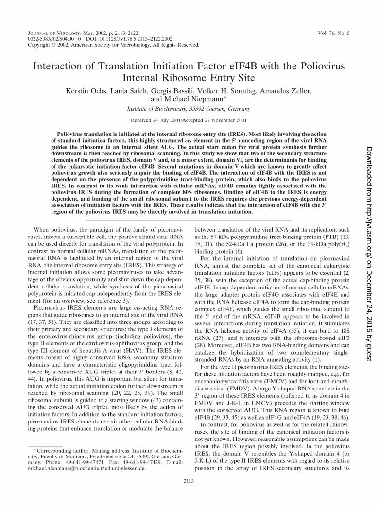
eIF4B binds to the poliovirus IRES. In order to detect thebinding of eIF4B to the poliovirus IRES, RNAs comprising thecomplete IRES of poliovirus type 1 (Mahoney) were tran-scribed in vitro and radioactively labeled with either[�-32P]ATP, -CTP, -GTP or -UTP. Proteins from RRL bindingto the IRES were then analyzed by the UV cross-linking assay.With all four differently labeled RNAs, a protein with an ap-parent molecular mass of about 80 kDa, as expected for eIF4B,was labeled (Fig. 1A). This 80-kDa protein was identified aseIF4B by immunoprecipitation. With an antiserum directedagainst human eIF4B, the 80-kDa band was immunoprecipi-tated after a UV cross-linking reaction with RRL (Fig. 1B, lane1), whereas no band was detected with preimmune serum (lane2).
The use of [�-32P]CTP-, GTP- and UTP-labeled IRES RNA(Fig. 1A, lanes 2 to 4) resulted in strong labeling of eIF4B. Inaddition, a band of about 50 kDa was labeled strongly, andseveral other proteins were labeled, including a 57-kDa protein(lanes 2 and 4) which is identical with the PTB (12, 13). For allfurther experiments, we used [�-32P]CTP-labeled IRES RNAs,since this label allowed the simultaneous detection of eIF4Band other bands, including that of the PTB (which is not welllabeled with GTP). UTP was not chosen because an additionalband migrating close to eIF4B was detected with this label.
The translation of poliovirus RNA in RRL has been re-ported to be stimulated and corrected by the addition of the52-kDa La protein (26). Therefore, we investigated whetherthe addition of recombinant La to the RRL could possiblyaffect the binding of eIF4B to the poliovirus IRES and thusreveal a molecular mechanism of the action of La. However,eIF4B binding was not affected when increasing amounts ofrecombinant La were added to the RRL (Fig. 1C, lanes 2 to 7;
2114 OCHS ET AL. J. VIROL.
on Decem
ber 24, 2015 by guesthttp://jvi.asm
.org/D
ownloaded from
compare with lane 1). The preparation of recombinant Laprotein used is functional in terms of RNA binding (T. Heise,personal communication), and the integrity of the recombinantLa protein was checked using two different anti-La antibodies(Fig. 1D and E). These immunoblots also showed that onlyminute amounts of endogenous La are present in the RRL.The level of 1 �M La protein which was added (Fig. 1C, lane7) is similar to the level (about 1.5 �M) at which La stimulatesthe correct translation of the poliovirus P1 precursor approx-imately tenfold (48). In the same way, the addition of a cyto-
plasmic HeLa extract to the RRL did not affect the binding ofeIF4B to the poliovirus IRES (data not shown).
Thus, irrespective of the reported effects of La protein onpoliovirus translation, the binding of eIF4B appears not to beaffected by La. Accordingly, normal RRL (i.e., not supple-mented with La protein) was used in all further experiments.
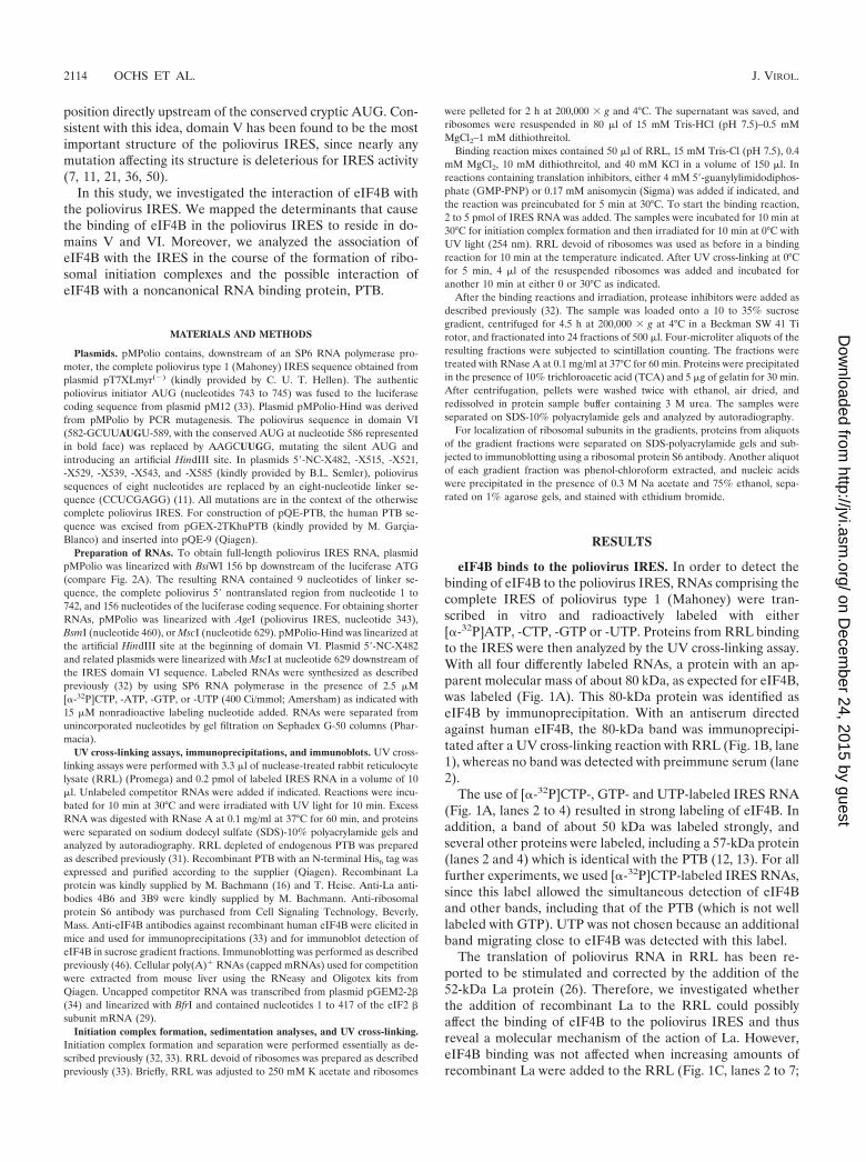
Mapping the eIF4B binding site. For delimiting the IRESregion required for the binding of eIF4B, we used RNAs cov-ering different regions of the poliovirus IRES (Fig. 2). Whensequences downstream of domain VI, including the nonstruc-tured spacer and the authentic initiator AUG, were removed(RNA 1–629), eIF4B still bound well to the IRES (Fig. 2B,lane 2), and the pattern of proteins detected did not changecompared with the full-length IRES RNA 1–745 (lane 1).Thus, the initiator AUG at nucleotide 743 as well as the pre-ceding nonstructured spacer sequence are not required forbinding of eIF4B. When most of domain VI, including theconserved silent AUG at nucleotide 586, was removed (RNA1–581), binding of eIF4B to the poliovirus IRES was slightlyweaker (lane 3). This indicates that domain VI and possibly thecryptic AUG may contribute to eIF4B binding.
In contrast, when the domain V and all downstream se-quences were removed (RNA 1–460), binding of eIF4B to theIRES was nearly completely abolished (Fig. 2B, lane 4),whereas binding of the 39-, 50-, and 68-kDa proteins was notaffected by this deletion. However, the 57-kDa band of PTBdisappeared, suggesting that domain V is also a major deter-minant for the binding of PTB. When further upstream se-quences, including the 3� half of the domain IV sequence, wereremoved (RNA 1–343), the same results were obtained (lane 5).
To exclude the possibility that the loss of detection of eIF4Bwas due only to the loss of a cross-link site and not to the lossof binding of the protein, we performed competition assays(Fig. 2C). Unlabeled competitor RNAs 1–745 and 1–629 com-peted efficiently with the binding of eIF4B and of other pro-teins to radiolabeled RNA 1–745 (lanes 3 to 4 and 7 to 8,respectively). RNA 1–581 competed only slightly more weakly(compare lane 11 with lane 3). However, the RNA regionscomprising nucleotides 1 to 460 and 1 to 343 (lanes 13 to 20)could not compete successfully with the binding of eIF4B tothe full-length IRES RNA (compare lanes 16 or 20 to lane 4).Thus, the loss of detection of eIF4B with the respective IRESdeletions in Fig. 2B is actually due to the loss of eIF4B bindingbut not due to the loss of cross-link sites alone.
Taken together, the above results indicate that the majordeterminants for the binding of eIF4B reside in domain V ofthe poliovirus IRES, whereas domain VI contributes less to thebinding of eIF4B.
Mutations within IRES domains V and VI affect eIF4B bind-ing. For a more detailed analysis of the eIF4B binding site, aseries of linker-scanning mutations covering most of domain Vand the beginning of domain VI (11) (Fig. 3A) in the contextof the otherwise complete IRES was used. In mutant X482, theapical stem of the domain V was affected. In mutants X515 andX521, the predicted large unpaired bulge was mutated, and inmutants X529, X539, and X543, the lower portion of the do-main V was affected.
All of the mutations in domain V severely affected the bind-ing of eIF4B to the poliovirus IRES (Fig. 3B), with thosemutations located in the upper and middle stem (X482 and
FIG. 1. eIF4B binds to the poliovirus IRES RNA. (A) Detection ofeIF4B in RRL. UV cross-linking assays were performed with poliovi-rus IRES RNA labeled with either [�-32P]ATP, -CTP, -GTP or -UTPas indicated. Proteins were resolved on SDS-10% polyacrylamide gelsand visualized by autoradiography. Molecular masses of marker pro-teins are indicated. (B) Immunoprecipitation of eIF4B. The bindingreaction was performed with RRL and [�-32P]CTP-labeled IRESRNA. After the UV cross-linking reaction, eIF4B was immunoprecipi-tated (IP) with anti-eIF4B antiserum (�-4B) (lane 1). Preimmuneserum was used as negative control (pre.) (lane 2). Ten percent of theproteins from the supernatants (sup.) (right panel) was concentratedby TCA-precipitation (lanes 3 and 4), resolved on SDS-10% polyacryl-amide gels, and visualized by autoradiography. 4B, eIF4B. (C) Noeffect of La protein on eIF4B binding. Poliovirus IRES RNA was usedin the UV cross-linking assay by using RRL either without (lane 1) orafter adding increasing amounts of recombinant La protein (rec. La) asindicated (lanes 2 to 7). (D and E) Immunoblots with anti-La antibod-ies 4B6 (D) and 3B9 (E). The bands representing endogenous La (end.La) and added recombinant La (rec. La) are indicated.
VOL. 76, 2002 INTERACTION OF eIF4B WITH THE POLIOVIRUS IRES 2115
on Decem
ber 24, 2015 by guesthttp://jvi.asm
.org/D
ownloaded from
X529) having the greatest effect. Mutations affecting the lowerpart of the stem (X543) or the bulge (X515 and X521) alsoseverely affected but did not completely abolish eIF4B binding.Thus, all parts of domain V are important for eIF4B binding,including the upper stem structure, which is predicted to be arelatively stable structure, the predicted unpaired bulge, and
the lower part of domain V. Together with that of eIF4B, thebinding of the 57-kDa protein PTB was also affected by allmutations, most severely by those affecting the stem. In con-trast, the binding of the 50-kDa protein was not only notreduced but was in some cases even enhanced, possibly due tothe loss of binding competition. When domain VI was mutated
FIG. 2. Mapping of the eIF4B binding site in the poliovirus IRES. (A) Schematic representation of the poliovirus IRES. The IRES domainsare marked with Roman numerals as described previously (8). Enzymes used for linearization of the templates for in vitro transcription areindicated together with the positions of the last nucleotide of the poliovirus sequence present in the respective RNAs. (B) UV cross-linking assayusing RRL and [�-32P]CTP-labeled poliovirus IRES RNAs. The nucleotide numbers of the poliovirus sequences are indicated on the top. RNA1–745 contains the authentic poliovirus initiator AUG at nucleotides 743 to 745 plus 156 additional nucleotides of luciferase sequence at the 3�end (up to the BsiWI site). Molecular masses of marker proteins are indicated. (C) Competitions. [�-32P]CTP-labeled poliovirus IRES RNA 1–745was used in all lanes. Unlabeled competitor RNAs are indicated on the top together with the molar concentrations used for competition. Molecularmasses of marker proteins are indicated. 4B, eIF4B.
2116 OCHS ET AL. J. VIROL.
on Decem
ber 24, 2015 by guesthttp://jvi.asm
.org/D
ownloaded from
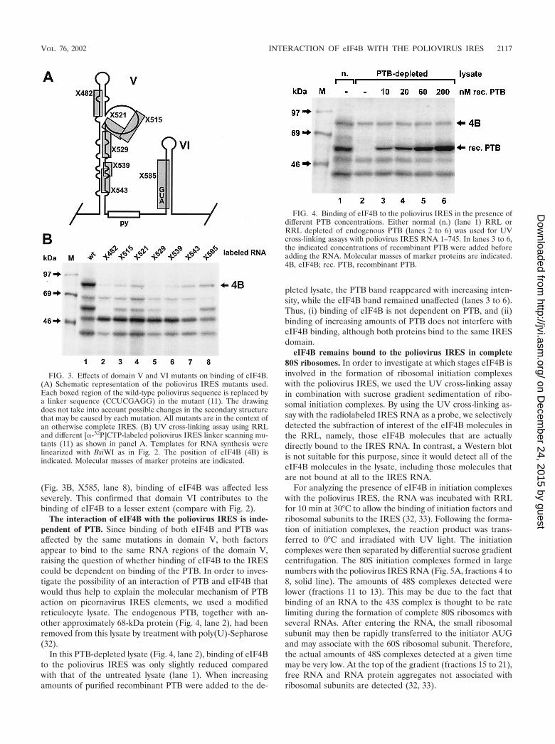
(Fig. 3B, X585, lane 8), binding of eIF4B was affected lessseverely. This confirmed that domain VI contributes to thebinding of eIF4B to a lesser extent (compare with Fig. 2).
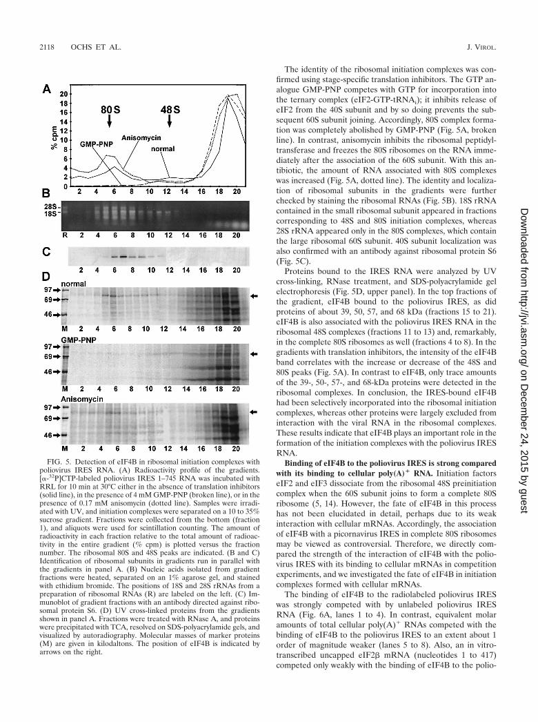
The interaction of eIF4B with the poliovirus IRES is inde-pendent of PTB. Since binding of both eIF4B and PTB wasaffected by the same mutations in domain V, both factorsappear to bind to the same RNA regions of the domain V,raising the question of whether binding of eIF4B to the IREScould be dependent on binding of the PTB. In order to inves-tigate the possibility of an interaction of PTB and eIF4B thatwould thus help to explain the molecular mechanism of PTBaction on picornavirus IRES elements, we used a modifiedreticulocyte lysate. The endogenous PTB, together with an-other approximately 68-kDa protein (Fig. 4, lane 2), had beenremoved from this lysate by treatment with poly(U)-Sepharose(32).
In this PTB-depleted lysate (Fig. 4, lane 2), binding of eIF4Bto the poliovirus IRES was only slightly reduced comparedwith that of the untreated lysate (lane 1). When increasingamounts of purified recombinant PTB were added to the de-
pleted lysate, the PTB band reappeared with increasing inten-sity, while the eIF4B band remained unaffected (lanes 3 to 6).Thus, (i) binding of eIF4B is not dependent on PTB, and (ii)binding of increasing amounts of PTB does not interfere witheIF4B binding, although both proteins bind to the same IRESdomain.
eIF4B remains bound to the poliovirus IRES in complete80S ribosomes. In order to investigate at which stages eIF4B isinvolved in the formation of ribosomal initiation complexeswith the poliovirus IRES, we used the UV cross-linking assayin combination with sucrose gradient sedimentation of ribo-somal initiation complexes. By using the UV cross-linking as-say with the radiolabeled IRES RNA as a probe, we selectivelydetected the subfraction of interest of the eIF4B molecules inthe RRL, namely, those eIF4B molecules that are actuallydirectly bound to the IRES RNA. In contrast, a Western blotis not suitable for this purpose, since it would detect all of theeIF4B molecules in the lysate, including those molecules thatare not bound at all to the IRES RNA.
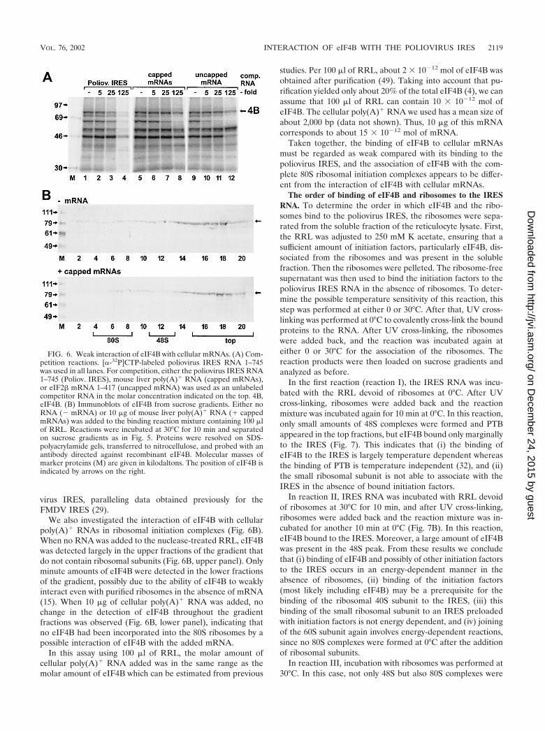
For analyzing the presence of eIF4B in initiation complexeswith the poliovirus IRES, the RNA was incubated with RRLfor 10 min at 30°C to allow the binding of initiation factors andribosomal subunits to the IRES (32, 33). Following the forma-tion of initiation complexes, the reaction product was trans-ferred to 0°C and irradiated with UV light. The initiationcomplexes were then separated by differential sucrose gradientcentrifugation. The 80S initiation complexes formed in largenumbers with the poliovirus IRES RNA (Fig. 5A, fractions 4 to8, solid line). The amounts of 48S complexes detected werelower (fractions 11 to 13). This may be due to the fact thatbinding of an RNA to the 43S complex is thought to be ratelimiting during the formation of complete 80S ribosomes withseveral RNAs. After entering the RNA, the small ribosomalsubunit may then be rapidly transferred to the initiator AUGand may associate with the 60S ribosomal subunit. Therefore,the actual amounts of 48S complexes detected at a given timemay be very low. At the top of the gradient (fractions 15 to 21),free RNA and RNA protein aggregates not associated withribosomal subunits are detected (32, 33).
FIG. 3. Effects of domain V and VI mutants on binding of eIF4B.(A) Schematic representation of the poliovirus IRES mutants used.Each boxed region of the wild-type poliovirus sequence is replaced bya linker sequence (CCUCGAGG) in the mutant (11). The drawingdoes not take into account possible changes in the secondary structurethat may be caused by each mutation. All mutants are in the context ofan otherwise complete IRES. (B) UV cross-linking assay using RRLand different [�-32P]CTP-labeled poliovirus IRES linker scanning mu-tants (11) as shown in panel A. Templates for RNA synthesis werelinearized with BsiWI as in Fig. 2. The position of eIF4B (4B) isindicated. Molecular masses of marker proteins are indicated.
FIG. 4. Binding of eIF4B to the poliovirus IRES in the presence ofdifferent PTB concentrations. Either normal (n.) (lane 1) RRL orRRL depleted of endogenous PTB (lanes 2 to 6) was used for UVcross-linking assays with poliovirus IRES RNA 1–745. In lanes 3 to 6,the indicated concentrations of recombinant PTB were added beforeadding the RNA. Molecular masses of marker proteins are indicated.4B, eIF4B; rec. PTB, recombinant PTB.
VOL. 76, 2002 INTERACTION OF eIF4B WITH THE POLIOVIRUS IRES 2117
on Decem
ber 24, 2015 by guesthttp://jvi.asm
.org/D
ownloaded from
The identity of the ribosomal initiation complexes was con-firmed using stage-specific translation inhibitors. The GTP an-alogue GMP-PNP competes with GTP for incorporation intothe ternary complex (eIF2-GTP-tRNAi); it inhibits release ofeIF2 from the 40S subunit and by so doing prevents the sub-sequent 60S subunit joining. Accordingly, 80S complex forma-tion was completely abolished by GMP-PNP (Fig. 5A, brokenline). In contrast, anisomycin inhibits the ribosomal peptidyl-transferase and freezes the 80S ribosomes on the RNA imme-diately after the association of the 60S subunit. With this an-tibiotic, the amount of RNA associated with 80S complexeswas increased (Fig. 5A, dotted line). The identity and localiza-tion of ribosomal subunits in the gradients were furtherchecked by staining the ribosomal RNAs (Fig. 5B). 18S rRNAcontained in the small ribosomal subunit appeared in fractionscorresponding to 48S and 80S initiation complexes, whereas28S rRNA appeared only in the 80S complexes, which containthe large ribosomal 60S subunit. 40S subunit localization wasalso confirmed with an antibody against ribosomal protein S6(Fig. 5C).
Proteins bound to the IRES RNA were analyzed by UVcross-linking, RNase treatment, and SDS-polyacrylamide gelelectrophoresis (Fig. 5D, upper panel). In the top fractions ofthe gradient, eIF4B bound to the poliovirus IRES, as didproteins of about 39, 50, 57, and 68 kDa (fractions 15 to 21).eIF4B is also associated with the poliovirus IRES RNA in theribosomal 48S complexes (fractions 11 to 13) and, remarkably,in the complete 80S ribosomes as well (fractions 4 to 8). In thegradients with translation inhibitors, the intensity of the eIF4Bband correlates with the increase or decrease of the 48S and80S peaks (Fig. 5A). In contrast to eIF4B, only trace amountsof the 39-, 50-, 57-, and 68-kDa proteins were detected in theribosomal complexes. In conclusion, the IRES-bound eIF4Bhad been selectively incorporated into the ribosomal initiationcomplexes, whereas other proteins were largely excluded frominteraction with the viral RNA in the ribosomal complexes.These results indicate that eIF4B plays an important role in theformation of the initiation complexes with the poliovirus IRESRNA.
Binding of eIF4B to the poliovirus IRES is strong comparedwith its binding to cellular poly(A)� RNA. Initiation factorseIF2 and eIF3 dissociate from the ribosomal 48S preinitiationcomplex when the 60S subunit joins to form a complete 80Sribosome (5, 14). However, the fate of eIF4B in this processhas not been elucidated in detail, perhaps due to its weakinteraction with cellular mRNAs. Accordingly, the associationof eIF4B with a picornavirus IRES in complete 80S ribosomesmay be viewed as controversial. Therefore, we directly com-pared the strength of the interaction of eIF4B with the polio-virus IRES with its binding to cellular mRNAs in competitionexperiments, and we investigated the fate of eIF4B in initiationcomplexes formed with cellular mRNAs.
The binding of eIF4B to the radiolabeled poliovirus IRESwas strongly competed with by unlabeled poliovirus IRESRNA (Fig. 6A, lanes 1 to 4). In contrast, equivalent molaramounts of total cellular poly(A)� RNAs competed with thebinding of eIF4B to the poliovirus IRES to an extent about 1order of magnitude weaker (lanes 5 to 8). Also, an in vitro-transcribed uncapped eIF2� mRNA (nucleotides 1 to 417)competed only weakly with the binding of eIF4B to the polio-
FIG. 5. Detection of eIF4B in ribosomal initiation complexes withpoliovirus IRES RNA. (A) Radioactivity profile of the gradients.[�-32P]CTP-labeled poliovirus IRES 1–745 RNA was incubated withRRL for 10 min at 30°C either in the absence of translation inhibitors(solid line), in the presence of 4 mM GMP-PNP (broken line), or in thepresence of 0.17 mM anisomycin (dotted line). Samples were irradi-ated with UV, and initiation complexes were separated on a 10 to 35%sucrose gradient. Fractions were collected from the bottom (fraction1), and aliquots were used for scintillation counting. The amount ofradioactivity in each fraction relative to the total amount of radioac-tivity in the entire gradient (% cpm) is plotted versus the fractionnumber. The ribosomal 80S and 48S peaks are indicated. (B and C)Identification of ribosomal subunits in gradients run in parallel withthe gradients in panel A. (B) Nucleic acids isolated from gradientfractions were heated, separated on an 1% agarose gel, and stainedwith ethidium bromide. The positions of 18S and 28S rRNAs from apreparation of ribosomal RNAs (R) are labeled on the left. (C) Im-munoblot of gradient fractions with an antibody directed against ribo-somal protein S6. (D) UV cross-linked proteins from the gradientsshown in panel A. Fractions were treated with RNase A, and proteinswere precipitated with TCA, resolved on SDS-polyacrylamide gels, andvisualized by autoradiography. Molecular masses of marker proteins(M) are given in kilodaltons. The position of eIF4B is indicated byarrows on the right.
2118 OCHS ET AL. J. VIROL.
on Decem
ber 24, 2015 by guesthttp://jvi.asm
.org/D
ownloaded from
virus IRES, paralleling data obtained previously for theFMDV IRES (29).
We also investigated the interaction of eIF4B with cellularpoly(A)� RNAs in ribosomal initiation complexes (Fig. 6B).When no RNA was added to the nuclease-treated RRL, eIF4Bwas detected largely in the upper fractions of the gradient thatdo not contain ribosomal subunits (Fig. 6B, upper panel). Onlyminute amounts of eIF4B were detected in the lower fractionsof the gradient, possibly due to the ability of eIF4B to weaklyinteract even with purified ribosomes in the absence of mRNA(15). When 10 �g of cellular poly(A)� RNA was added, nochange in the detection of eIF4B throughout the gradientfractions was observed (Fig. 6B, lower panel), indicating thatno eIF4B had been incorporated into the 80S ribosomes by apossible interaction of eIF4B with the added mRNA.
In this assay using 100 �l of RRL, the molar amount ofcellular poly(A)� RNA added was in the same range as themolar amount of eIF4B which can be estimated from previous
studies. Per 100 �l of RRL, about 2 � 10�12 mol of eIF4B wasobtained after purification (49). Taking into account that pu-rification yielded only about 20% of the total eIF4B (4), we canassume that 100 �l of RRL can contain 10 � 10�12 mol ofeIF4B. The cellular poly(A)� RNA we used has a mean size ofabout 2,000 bp (data not shown). Thus, 10 �g of this mRNAcorresponds to about 15 � 10�12 mol of mRNA.
Taken together, the binding of eIF4B to cellular mRNAsmust be regarded as weak compared with its binding to thepoliovirus IRES, and the association of eIF4B with the com-plete 80S ribosomal initiation complexes appears to be differ-ent from the interaction of eIF4B with cellular mRNAs.
The order of binding of eIF4B and ribosomes to the IRESRNA. To determine the order in which eIF4B and the ribo-somes bind to the poliovirus IRES, the ribosomes were sepa-rated from the soluble fraction of the reticulocyte lysate. First,the RRL was adjusted to 250 mM K acetate, ensuring that asufficient amount of initiation factors, particularly eIF4B, dis-sociated from the ribosomes and was present in the solublefraction. Then the ribosomes were pelleted. The ribosome-freesupernatant was then used to bind the initiation factors to thepoliovirus IRES RNA in the absence of ribosomes. To deter-mine the possible temperature sensitivity of this reaction, thisstep was performed at either 0 or 30°C. After that, UV cross-linking was performed at 0°C to covalently cross-link the boundproteins to the RNA. After UV cross-linking, the ribosomeswere added back, and the reaction was incubated again ateither 0 or 30°C for the association of the ribosomes. Thereaction products were then loaded on sucrose gradients andanalyzed as before.
In the first reaction (reaction I), the IRES RNA was incu-bated with the RRL devoid of ribosomes at 0°C. After UVcross-linking, ribosomes were added back and the reactionmixture was incubated again for 10 min at 0°C. In this reaction,only small amounts of 48S complexes were formed and PTBappeared in the top fractions, but eIF4B bound only marginallyto the IRES (Fig. 7). This indicates that (i) the binding ofeIF4B to the IRES is largely temperature dependent whereasthe binding of PTB is temperature independent (32), and (ii)the small ribosomal subunit is not able to associate with theIRES in the absence of bound initiation factors.
In reaction II, IRES RNA was incubated with RRL devoidof ribosomes at 30°C for 10 min, and after UV cross-linking,ribosomes were added back and the reaction mixture was in-cubated for another 10 min at 0°C (Fig. 7B). In this reaction,eIF4B bound to the IRES. Moreover, a large amount of eIF4Bwas present in the 48S peak. From these results we concludethat (i) binding of eIF4B and possibly of other initiation factorsto the IRES occurs in an energy-dependent manner in theabsence of ribosomes, (ii) binding of the initiation factors(most likely including eIF4B) may be a prerequisite for thebinding of the ribosomal 40S subunit to the IRES, (iii) thisbinding of the small ribosomal subunit to an IRES preloadedwith initiation factors is not energy dependent, and (iv) joiningof the 60S subunit again involves energy-dependent reactions,since no 80S complexes were formed at 0°C after the additionof ribosomal subunits.
In reaction III, incubation with ribosomes was performed at30°C. In this case, not only 48S but also 80S complexes were
FIG. 6. Weak interaction of eIF4B with cellular mRNAs. (A) Com-petition reactions. [�-32P]CTP-labeled poliovirus IRES RNA 1–745was used in all lanes. For competition, either the poliovirus IRES RNA1–745 (Poliov. IRES), mouse liver poly(A)� RNA (capped mRNAs),or eIF2� mRNA 1–417 (uncapped mRNA) was used as an unlabeledcompetitor RNA in the molar concentration indicated on the top. 4B,eIF4B. (B) Immunoblots of eIF4B from sucrose gradients. Either noRNA (� mRNA) or 10 �g of mouse liver poly(A)� RNA (� cappedmRNAs) was added to the binding reaction mixture containing 100 �lof RRL. Reactions were incubated at 30°C for 10 min and separatedon sucrose gradients as in Fig. 5. Proteins were resolved on SDS-polyacrylamide gels, transferred to nitrocellulose, and probed with anantibody directed against recombinant eIF4B. Molecular masses ofmarker proteins (M) are given in kilodaltons. The position of eIF4B isindicated by arrows on the right.
VOL. 76, 2002 INTERACTION OF eIF4B WITH THE POLIOVIRUS IRES 2119
on Decem
ber 24, 2015 by guesthttp://jvi.asm
.org/D
ownloaded from
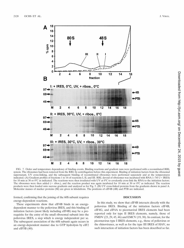
formed, confirming that the joining of the 60S subunit requiresenergy-dependent reactions.
These experiments show that eIF4B binds in an energy-dependent manner to the poliovirus IRES, and this binding ofinitiation factors (most likely including eIF4B) may be a pre-requisite for the entry of the small ribosomal subunit into thepoliovirus IRES, a step which is energy independent per se.The subsequent association of the 60S subunit again occurs inan energy-dependent manner due to GTP hydrolysis by eIF2and eIF5B (40).
DISCUSSION
In this study, we show that eIF4B interacts directly with thepoliovirus IRES. Binding of the initiation factors eIF4B,eIF4G, and eIF4A to picornaviral IRES elements had beenreported only for type II IRES elements, namely, those ofFMDV (23, 29, 45, 46) and EMCV (19, 38). In contrast, for thepicornavirus type I IRES elements, e.g., those of poliovirus orthe rhinoviruses, as well as for the type III IRES of HAV, nosuch interaction of initiation factors has been described so far.
FIG. 7. Order and temperature dependency of binding events. Binding reactions and gradient runs were performed with a reconstituted RRLsystem. The ribosomes had been removed from the RRL by centrifugation before this experiment. Binding of initiation factors from the ribosomalsupernatant, UV cross-linking, and the subsequent binding of reconstituted ribosomes were performed separately and at the temperaturesindicated. (A) Gradient profiles of fractions 1 to 16 of reactions I, II, and III. RRL devoid of ribosomes was incubated with RNA 1–745 (� IRES)for 10 min at 30 or 0°C as indicated. The reactions were then irradiated with UV at 0°C to covalently cross-link the RNA to the initiation factors.The ribosomes were then added (� ribos.), and the reaction product was again incubated for 10 min at 30 or 0°C as indicated. The reactionproducts were then loaded onto sucrose gradients and analyzed as for Fig. 5. (B) UV cross-linked proteins from the gradients shown in panel A.Molecular masses of marker proteins (M) are given in kilodaltons. The positions of eIF4B (4B) and PTB are indicated.
2120 OCHS ET AL. J. VIROL.
on Decem
ber 24, 2015 by guesthttp://jvi.asm
.org/D
ownloaded from
In cell-free reconstitution experiments, the more distantly re-lated IRES elements of hepatitis C virus and pestiviruses, twomembers of the genera Hepacivirus and Pestivirus, respectively,of the Flaviviridae family, bind the small ribosomal subunitcompletely independently of any initiation factors, even with-out eIF2 and eIF3 (41).
The binding site for eIF4B in the poliovirus IRES residesmainly in the IRES domain V, whereas domain VI sequences,including the silent AUG at nucleotide 586, contribute to thebinding of eIF4B to a lesser extent, which is a finding consis-tent with the observation that domain VI is not absolutelyessential for poliovirus translation (21, 25). In domain V, sev-eral linker scanning mutations seriously impaired the bindingof eIF4B (see Fig. 3). These mutations also seriously impairedpoliovirus translation and growth, leading to either lethal ortemperature-sensitive phenotypes (11). This correlation sug-gests that eIF4B may play an important role in the initiation oftranslation from the poliovirus IRES.
Even if these linker scanning mutations may alter the actualstructure of domain V, we can conclude that separate deter-minants located both in its lower part and in its upper part arerequired for eIF4B binding and for translation. Secondarystructure calculations (reference 24 and data not shown) forthe lower part of domain V and for the bulge predict a varietyof different possible secondary structures with similar energyminima. Accordingly, the RNA may easily change its confor-mation, possibly to meet different requirements for the bindingof various cellular proteins. All linker scanning mutations inthis region seriously impaired binding of eIF4B (this study) aswell as the viability of the corresponding virus (11). In contrast,all secondary structures for the upper part of domain V arepredicted to be similar, regardless of whether they are in thecontext of unmutated domain V or of variants with alteredpredicted secondary structures of the lower part. Thus, theserious effect seen of the X482 mutation implies that the upperpart of domain V must bear a determinant for the binding ofeIF4B (this study) and for virus growth (11) that is separatefrom those located in the lower part.
In the relative position of its eIF4B binding site within thearrangement of secondary structures, the poliovirus IRES re-sembles type II IRES elements like those of FMDV andEMCV (3, 8, 30). Also, in FMDV, the determinant for thebinding of eIF4B resides in the IRES 3�-region, mainly includ-ing the Y-shaped domain 4 (29, 45). A common feature of theeIF4B binding regions in both types of IRES elements is thatthey are located downstream of the large central domain struc-ture, which is referred to as domain 3 in FMDV and domainIV in poliovirus, respectively.
At a first glance, this arrangement of cis elements in thepicornaviral IRES elements may also reflect a segmentation ofthe IRES at a functional level. The 3� part of the IRES appearsto be the area for binding the canonical translation initiationfactors that probably guide the ribosome to the starting win-dow at the IRES 3� border. This process may also involveinteractions between the IRES-bound eIF4B and the 170-kDasubunit of the ribosome-bound eIF3 (28). In addition, theoligopyrimidine tract in the 3� region of the IRES may interactwith the ribosomal 18S RNA (44, 47), perhaps facilitated bythe RNA annealing activity of eIF4B (1) and the RNA helicaseactivity of eIF4A (35). In contrast, the upstream parts of the
IRES may bind regulatory proteins, like the 39-kDa poly(rC)binding protein 2 (6), which has been speculated to be involvedin a switch from translation to minus-strand synthesis later inthe poliovirus replication cycle (9).
However, this simple view of a clear segmentation of theIRES elements is complicated by the finding that the apicalparts of the poliovirus IRES domains V and VI bear determi-nants for neuropathogenicity (10). Moreover, PTB appears tobind to multiple sites of the poliovirus IRES, including domainV (reference 12 and this study). Thus, the determinants for thebinding of cellular proteins which may be involved in regula-tory phenomena appear to overlap with the binding site for atleast one of the canonical initiation factors, eIF4B. However,although binding of these factors appears to depend on closelyneighboring determinants in domain V, binding of PTB doesnot interfere with the binding of eIF4B. Further studies will berequired to investigate the possible interactions of known oryet unknown cellular factors with the assembled core of canonicalinitiation factors in the 3� region of the poliovirus IRES.
The events during the assembly of the complete 80S ribo-some on the poliovirus RNA can be clearly subdivided intodifferent steps that occur sequentially. The first step is thebinding of soluble initiation factors, including eIF4B, to theIRES. This step is energy dependent, consistent with the ob-servation that binding of eIF4B to the FMDV IRES is ATPdependent (29). This binding of initiation factors occurs inde-pendently of ribosomes, since eIF4B was UV cross-linked tothe IRES in the complete absence of ribosomes. Only after thisbinding does the small ribosomal subunit enter this RNA-proteincomplex. This second step is energy independent per se butrequires the previous binding of the initiation factors. The laststep in the assembly of complete 80S ribosomes, the joining ofthe ribosomal 60S subunit, is again energy dependent due tothe hydrolysis of the eIF2-bound GTP prior to subunit joining.
eIF4B remains bound to the poliovirus IRES not only afterthe association of the small ribosomal subunit. In contrast tothe weak interaction of eIF4B with capped cellular mRNAs,eIF4B remains still tightly associated with the poliovirus IRESeven after the formation of the complete 80S ribosome. Thisobservation parallels results obtained with the type II FMDVIRES (33) and points to a possible functional difference be-tween the initiation of translation mediated by picornaviralIRES elements and cap-dependent initiation. Probably eIF4Band also other initiation factors remain continuously bound tothe IRES 3� region after joining of the ribosomal 60S subunitand perhaps even during repeated events of ribosome entry.
ACKNOWLEDGMENTS
We thank B. L. Semler for kindly providing the poliovirus domain Vand VI mutants 5�-NC-X482, -X515, -X521, -X529, -X539, -X543, and-X585 (11); C. U. T. Hellen for plasmid pT7XLmyr(�); M. Garçia-Blanco for plasmid pGEX-2TKhuPTB; M. Bachmann for anti-La an-tibodies 4B6 and 3B9; M. Bachmann and T. Heise for recombinant Laprotein; and E. Beck for helpful discussions.
This work was supported by grants from the Deutsche Forschungs-gemeinschaft (SFB 535 and GK 370).
REFERENCES
1. Altmann, M., B. Wittmer, N. Méthot, N. Sonenberg, and H. Trachsel. 1995.The Saccharomyces cerevisiae translation initiation factor Tif3 and its mam-malian homologue, eIF-4B, have RNA annealing activity. EMBO J. 14:3820–3827.
2. Anthony, D. D., and W. C. Merrick. 1991. Eukaryotic initiation factor (eIF)-
VOL. 76, 2002 INTERACTION OF eIF4B WITH THE POLIOVIRUS IRES 2121
on Decem
ber 24, 2015 by guesthttp://jvi.asm
.org/D
ownloaded from
4F. Implications for a role in internal initiation of translation. J. Biol. Chem.266:10218–10226.
3. Belsham, G. J., and R. J. Jackson. 2000. Translation initiation on picorna-virus RNA, p. 869–900. In N. Sonenberg, J. W. B. Hershey, and M. B.Mathews (ed.), Translational control of gene expression. Cold Spring HarborLaboratory Press, Cold Spring Harbor, N.Y.
4. Benne, R., M. L. Brown Luedi, and J. W. Hershey. 1979. Protein synthesisinitiation factors from rabbit reticulocytes: purification, characterization, andradiochemical labeling. Methods Enzymol. 60:15–35.
5. Benne, R., and J. W. Hershey. 1978. The mechanism of action of proteinsynthesis initiation factors from rabbit reticulocytes. J. Biol. Chem. 253:3078–3087.
6. Blyn, L. B., J. S. Towner, B. L. Semler, and E. Ehrenfeld. 1997. Requirementof poly(rC) binding protein 2 for translation of poliovirus RNA. J. Virol.71:6243–6246.
7. Dildine, S. L., K. R. Stark, A. A. Haller, and B. L. Semler. 1991. Poliovirustranslation initiation: differential effects of directed and selected mutationsin the 5� noncoding region of viral RNAs. Virology 182:742–752.
8. Ehrenfeld, E., and B. L. Semler. 1995. Anatomy of the poliovirus internalribosome entry site. Curr. Top. Microbiol. Immunol. 203:65–83.
9. Gamarnik, A. V., and R. Andino. 1998. Switch from translation to RNAreplication in a positive-stranded RNA virus. Genes Dev. 12:2293–2304.
10. Gromeier, M., B. Bossert, M. Arita, A. Nomoto, and E. Wimmer. 1999. Dualstem loops within the poliovirus internal ribosomal entry site control neu-rovirulence. J. Virol. 73:958–964.
11. Haller, A. A., and B. L. Semler. 1992. Linker scanning mutagenesis of theinternal ribosome entry site of poliovirus RNA. J. Virol. 66:5075–5086.
12. Hellen, C. U., T. V. Pestova, M. Litterst, and E. Wimmer. 1994. The cellularpolypeptide p57 (pyrimidine tract-binding protein) binds to multiple sites inthe poliovirus 5� nontranslated region. J. Virol. 68:941–950.
13. Hellen, C. U., G. W. Witherell, M. Schmid, S. H. Shin, T. V. Pestova, A. Gil,and E. Wimmer. 1993. A cytoplasmic 57-kDa protein that is required fortranslation of picornavirus RNA by internal ribosomal entry is identical tothe nuclear pyrimidine tract-binding protein. Proc. Natl. Acad. Sci. USA90:7642–7646.
14. Hershey, J. W. B., and W. C. Merrick. 2000. Pathway and mechanism ofinitiation of protein synthesis, p. 33–88. In N. Sonenberg, J. W. B. Hershey,and M. B. Mathews (ed.), Translational control of gene expression. ColdSpring Harbor Laboratory Press, Cold Spring Harbor, N.Y.
15. Hughes, D. L., T. E. Dever, and W. C. Merrick. 1993. Further biochemicalcharacterization of rabbit reticulocyte eIF-4B. Arch. Biochem. Biophys. 301:311–319.
16. Hühn, P., G. J. Pruijn, W. J. van Venrooij, and M. Bachmann. 1997. Char-acterization of the autoantigen La (SS-B) as a dsRNA unwinding enzyme.Nucleic Acids Res. 25:410–416.
17. Jang, S. K., H. G. Kräusslich, M. J. Nicklin, G. M. Duke, A. C. Palmenberg,and E. Wimmer. 1988. A segment of the 5� nontranslated region of encepha-lomyocarditis virus RNA directs internal entry of ribosomes during in vitrotranslation. J. Virol. 62:2636–2643.
18. Kaminski, A., S. L. Hunt, J. G. Patton, and R. J. Jackson. 1995. Directevidence that polypyrimidine tract binding protein (PTB) is essential forinternal initiation of translation of encephalomyocarditis virus RNA. RNA1:924–938.
19. Kolupaeva, V. G., T. V. Pestova, C. U. Hellen, and I. N. Shatsky. 1998.Translation eukaryotic initiation factor 4G recognizes a specific structuralelement within the internal ribosome entry site of encephalomyocarditisvirus RNA. J. Biol. Chem. 273:18599–18604.
20. Kuge, S., N. Kawamura, and A. Nomoto. 1989. Genetic variation occurringon the genome of an in vitro insertion mutant of poliovirus type 1. J. Virol.63:1069–1075.
21. Kuge, S., and A. Nomoto. 1987. Construction of viable deletion and insertionmutants of the Sabin strain type 1 poliovirus: function of the 5� noncodingsequence in viral replication. J. Virol. 61:1478–1487.
22. La Monica, N., and V. R. Racaniello. 1989. Differences in replication ofattenuated and neurovirulent polioviruses in human neuroblastoma cell lineSH-SY5Y. J. Virol. 63:2357–2360.
23. Lopez de Quinto, S., and E. Martinez-Salas. 2000. Interaction of the eIF4Ginitiation factor with the aphthovirus IRES is essential for internal transla-tion initiation in vivo. RNA 6:1380–1392.
24. Mathews, D. H., J. Sabina, M. Zuker, and D. H. Turner. 1999. Expandedsequence dependence of thermodynamic parameters improves prediction ofRNA secondary structure. J. Mol. Biol. 288:911–940.
25. Meerovitch, K., R. Nicholson, and N. Sonenberg. 1991. In vitro mutationalanalysis of cis-acting RNA translational elements within the poliovirus type2 5� untranslated region. J. Virol. 65:5895–5901.
26. Meerovitch, K., Y. V. Svitkin, H. S. Lee, F. Lejbkowicz, D. J. Kenan, E. K.Chan, V. I. Agol, J. D. Keene, and N. Sonenberg. 1993. La autoantigenenhances and corrects aberrant translation of poliovirus RNA in reticulocytelysate. J. Virol. 67:3798–3807.
27. Méthot, N., G. Pickett, J. D. Keene, and N. Sonenberg. 1996. In vitro RNAselection identifies RNA ligands that specifically bind to eukaryotic transla-
tion initiation factor 4B: the role of the RNA recognition motif. RNA2:38–50.
28. Méthot, N., M. S. Song, and N. Sonenberg. 1996. A region rich in asparticacid, arginine, tyrosine, and glycine (DRYG) mediates eukaryotic initiationfactor 4B (eIF4B) self-association and interaction with eIF3. Mol. Cell. Biol.16:5328–5334.
29. Meyer, K., A. Petersen, M. Niepmann, and E. Beck. 1995. Interaction ofeukaryotic initiation factor eIF-4B with a picornavirus internal translationinitiation site. J. Virol. 69:2819–2824.
30. Niepmann, M. 1999. Internal initiation of translation of picornaviruses, hep-atitis C virus and pestiviruses. Recent Res. Dev. Virol. 1:229–250.
31. Niepmann, M. 1996. Porcine polypyrimidine tract-binding protein stimulatestranslation initiation at the internal ribosome entry site of foot-and-mouth-disease virus. FEBS Lett. 388:39–42.
32. Niepmann, M., A. Petersen, K. Meyer, and E. Beck. 1997. Functional in-volvement of polypyrimidine tract-binding protein in translation initiationcomplexes with the internal ribosome entry site of foot-and-mouth diseasevirus. J. Virol. 71:8330–8339.
33. Ochs, K., R. C. Rust, and M. Niepmann. 1999. Translation initiation factoreIF4B interacts with a picornavirus internal ribosome entry site in both 48Sand 80S initiation complexes independently of initiator AUG location. J. Vi-rol. 73:7505–7514.
34. Pathak, V. K., P. J. Nielsen, H. Trachsel, and J. W. Hershey. 1988. Structureof the beta subunit of translational initiation factor eIF-2. Cell 54:633–639.
35. Pause, A., N. Méthot, Y. Svitkin, W. C. Merrick, and N. Sonenberg. 1994.Dominant negative mutants of mammalian translation initiation factoreIF-4A define a critical role for eIF-4F in cap-dependent and cap-indepen-dent initiation of translation. EMBO J. 13:1205–1215.
36. Pelletier, J., G. Kaplan, V. R. Racaniello, and N. Sonenberg. 1988. Cap-independent translation of poliovirus mRNA is conferred by sequence ele-ments within the 5� noncoding region. Mol. Cell. Biol. 8:1103–1112.
37. Pelletier, J., and N. Sonenberg. 1988. Internal initiation of translation ofeukaryotic mRNA directed by a sequence derived from poliovirus RNA.Nature 334:320–325.
38. Pestova, T. V., C. U. Hellen, and I. N. Shatsky. 1996. Canonical eukaryoticinitiation factors determine initiation of translation by internal ribosomalentry. Mol. Cell. Biol. 16:6859–6869.
39. Pestova, T. V., C. U. Hellen, and E. Wimmer. 1994. A conserved AUG tripletin the 5� nontranslated region of poliovirus can function as an initiationcodon in vitro and in vivo. Virology 204:729–737.
40. Pestova, T. V., I. B. Lomakin, J. H. Lee, S. K. Choi, T. E. Dever, and C. U.Hellen. 2000. The joining of ribosomal subunits in eukaryotes requireseIF5B. Nature 403:332–335.
41. Pestova, T. V., I. N. Shatsky, S. P. Fletcher, R. J. Jackson, and C. U. Hellen.1998. A prokaryotic-like mode of cytoplasmic eukaryotic ribosome bindingto the initiation codon during internal translation initiation of hepatitis Cand classical swine fever virus RNAs. Genes Dev. 12:67–83.
42. Pilipenko, E. V., V. M. Blinov, B. K. Chernov, T. M. Dmitrieva, and V. I.Agol. 1989. Conservation of the secondary structure elements of the 5�-untranslated region of cardio- and aphthovirus RNAs. Nucleic Acids Res.17:5701–5711.
43. Pilipenko, E. V., A. P. Gmyl, S. V. Maslova, G. A. Belov, A. N. Sinyakov, M.Huang, T. D. Brown, and V. I. Agol. 1994. Starting window, a distinct elementin the cap-independent internal initiation of translation on picornaviralRNA. J. Mol. Biol. 241:398–414.
44. Pilipenko, E. V., A. P. Gmyl, S. V. Maslova, Y. V. Svitkin, A. N. Sinyakov, andV. I. Agol. 1992. Prokaryotic-like cis elements in the cap-independent inter-nal initiation of translation on picornavirus RNA. Cell 68:119–131.
45. Rust, R. C., K. Ochs, K. Meyer, E. Beck, and M. Niepmann. 1999. Interac-tion of eukaryotic initiation factor eIF4B with the internal ribosome entrysite of foot-and-mouth disease virus is independent of the polypyrimidinetract-binding protein. J. Virol. 73:6111–6113.
46. Saleh, L., R. C. Rust, R. Füllkrug, E. Beck, G. Bassili, K. Ochs, and M.Niepmann. 2001. Functional interaction of translation initiation factoreIF4G with the foot-and-mouth-disease virus internal ribosome entry site.J. Gen. Virol. 82:757–763.
47. Scheper, G. C., H. O. Voorma, and A. A. Thomas. 1994. Basepairing with 18Sribosomal RNA in internal initiation of translation. FEBS Lett. 352:271–275.
48. Svitkin, Y. V., K. Meerovitch, H. S. Lee, J. N. Dholakia, D. J. Kenan, V. I.Agol, and N. Sonenberg. 1994. Internal translation initiation on poliovirusRNA: further characterization of La function in poliovirus translation invitro. J. Virol. 68:1544–1550.
49. Thomas, A., H. Goumans, H. Amesz, R. Benne, and H. O. Voorma. 1979. Acomparison of the initiation factors of eukaryotic protein synthesis fromribosomes and from the postribosomal supernatant. Eur. J. Biochem. 98:329–337.
50. Trono, D., R. Andino, and D. Baltimore. 1988. An RNA sequence of hun-dreds of nucleotides at the 5� end of poliovirus RNA is involved in allowingviral protein synthesis. J. Virol. 62:2291–2299.
51. Trono, D., J. Pelletier, N. Sonenberg, and D. Baltimore. 1988. Translation inmammalian cells of a gene linked to the poliovirus 5� noncoding region.Science 241:445–448.
2122 OCHS ET AL. J. VIROL.
on Decem
ber 24, 2015 by guesthttp://jvi.asm
.org/D
ownloaded from
Related Documents


![Structure of eIF4E in Complex with an eIF4G Peptide ... · Structure of eIF4E in Complex with an eIF4G Peptide Supports a Universal Bipartite Binding Mode for Protein Translation1[OPEN]](https://static.cupdf.com/doc/110x72/5e5d1198ae86ce09fc4fef15/structure-of-eif4e-in-complex-with-an-eif4g-peptide-structure-of-eif4e-in-complex.jpg)


