ASIP Journal CME Program Immunopathology and Infectious Diseases Interaction of the Lyme Disease Spirochete Borrelia burgdorferi with Brain Parenchyma Elicits Inflammatory Mediators from Glial Cells as Well as Glial and Neuronal Apoptosis Geeta Ramesh,* Juan T. Borda, † Jason Dufour, ‡ Deepak Kaushal,* Ramesh Ramamoorthy,* Andrew A. Lackner, † and Mario T. Philipp* From the Divisions of Bacteriology and Parasitology,* Comparative Pathology, † and Veterinary Medicine, ‡ Tulane National Primate Research Center, Tulane University, Covington, Louisiana Lyme neuroborreliosis, caused by the spirochete Borrelia burgdorferi , often manifests by causing neu- rocognitive deficits. As a possible mechanism for Lyme neuroborreliosis , we hypothesized that B. burgdorferi induces the production of inflammatory mediators in the central nervous system with concomitant neuro- nal and/or glial apoptosis. To test our hypothesis , we constructed an ex vivo model that consisted of freshly collected slices from brain cortex of a rhesus ma- caque and allowed live B. burgdorferi to penetrate the tissue. Numerous transcripts of genes that regulate inflammation as well as oligodendrocyte and neuro- nal apoptosis were significantly altered as assessed by DNA microarray analysis. Transcription level in- creases of 7.43-fold (P 0.005) for the cytokine tumor necrosis factor- and 2.31-fold (P 0.016) for the chemokine interleukin (IL)-8 were also detected by real-time-polymerase chain reaction array analysis. The immune mediators IL-6, IL-8, IL-1 , COX-2, and CXCL13 were visualized in glial cells in situ by immu- nofluorescence staining and confocal microscopy. Concomitantly , significant proportions of both oligo- dendrocytes and neurons undergoing apoptosis were present in spirochete-stimulated tissues. IL-6 produc- tion by astrocytes in addition to oligodendrocyte ap- optosis were also detected, albeit at lower levels, in rhesus macaques that had received in vivo intraparen- chymal stereotaxic inoculations of live B. burgdorferi. These results provide proof of concept for our hy- pothesis that B. burgdorferi produces inflammatory mediators in the central nervous system, accompa- nied by glial and neuronal apoptosis. (Am J Pathol 2008, 173:1415–1427; DOI: 10.2353/ajpath.2008.080483) Lyme disease is a tick-borne infection caused by the spirochete Borrelia burgdorferi (sensu lato). 1,2 The re- markable organ pleiotropism of this spirochete results in diverse disease manifestations such as acute or chronic arthritis, myocarditis, and neuroborreliosis. 3 Lyme neu- roborreliosis affects 15 to 25% of patients with erythema migrans, the red skin-rash that in humans signals the point of entry of B. burgdorferi, and may involve both the peripheral and central nervous systems (CNS). Neuroborreliosis may manifest early, within the first few weeks to months of the appearance of erythema migrans, as a meningitis, often as part of the Garin-Bujadoux- Bannwarth syndrome, or, more seriously, as encephalo- myelitis. The latter is less common than the meningitis but shares with it the presence of lymphocytic pleocytosis in the cerebrospinal fluid (CSF) and intrathecal anti-B. burgdorferi antibody production. This disorder’s impor- tance is derived from the fact that it can lead to irrevers- ible CNS damage, 4 of the type that may be attributed to neuronal loss. Later in the course of disease, several months after infection, encephalopathies may appear. Patients with this late symptom complain of specific memory and/or intellectual impairment, often associated with incapacitating fatigue. 4 Brain lesions from Lyme neu- roborreliosis patients show vasculitis and subarachnoid hemorrhage 5–8 as well as multifocal encephalitis with Supported by the National Institutes of Health (grants NS048952, RR00164, G20 R018397-01, and G20 R019607-01) and the Centers for Disease Control and Prevention (grant UO1-CI000153). Accepted for publication August 5, 2008. Supplemental material for this article can be found on http://ajp.amjpathol.org. Address reprint requests to Mario T. Philipp, Division of Bacteriology and Parasitology, Tulane National Primate Research Center, Tulane University, 18703 Three Rivers Rd., Covington, LA 70433. E-mail: [email protected]. The American Journal of Pathology, Vol. 173, No. 5, November 2008 Copyright © American Society for Investigative Pathology DOI: 10.2353/ajpath.2008.080483 1415

Welcome message from author
This document is posted to help you gain knowledge. Please leave a comment to let me know what you think about it! Share it to your friends and learn new things together.
Transcript

ASIP
Journal
CME Program
Immunopathology and Infectious Diseases
Interaction of the Lyme Disease SpirocheteBorrelia burgdorferi with Brain ParenchymaElicits Inflammatory Mediators from Glial Cells asWell as Glial and Neuronal Apoptosis
Geeta Ramesh,* Juan T. Borda,† Jason Dufour,‡
Deepak Kaushal,* Ramesh Ramamoorthy,*Andrew A. Lackner,† and Mario T. Philipp*From the Divisions of Bacteriology and Parasitology,*
Comparative Pathology,† and Veterinary Medicine,‡ Tulane
National Primate Research Center, Tulane University,
Covington, Louisiana
Lyme neuroborreliosis , caused by the spirocheteBorrelia burgdorferi , often manifests by causing neu-rocognitive deficits. As a possible mechanism for Lymeneuroborreliosis, we hypothesized that B. burgdorferiinduces the production of inflammatory mediators inthe central nervous system with concomitant neuro-nal and/or glial apoptosis. To test our hypothesis, weconstructed an ex vivo model that consisted of freshlycollected slices from brain cortex of a rhesus ma-caque and allowed live B. burgdorferi to penetrate thetissue. Numerous transcripts of genes that regulateinflammation as well as oligodendrocyte and neuro-nal apoptosis were significantly altered as assessed byDNA microarray analysis. Transcription level in-creases of 7.43-fold (P � 0.005) for the cytokine tumornecrosis factor-� and 2.31-fold (P � 0.016) for thechemokine interleukin (IL)-8 were also detected byreal-time-polymerase chain reaction array analysis.The immune mediators IL-6, IL-8, IL-1� , COX-2, andCXCL13 were visualized in glial cells in situ by immu-nofluorescence staining and confocal microscopy.Concomitantly, significant proportions of both oligo-dendrocytes and neurons undergoing apoptosis werepresent in spirochete-stimulated tissues. IL-6 produc-tion by astrocytes in addition to oligodendrocyte ap-optosis were also detected, albeit at lower levels, inrhesus macaques that had received in vivo intraparen-chymal stereotaxic inoculations of live B. burgdorferi.These results provide proof of concept for our hy-pothesis that B. burgdorferi produces inflammatorymediators in the central nervous system, accompa-
nied by glial and neuronal apoptosis. (Am J Pathol2008, 173:1415–1427; DOI: 10.2353/ajpath.2008.080483)
Lyme disease is a tick-borne infection caused by thespirochete Borrelia burgdorferi (sensu lato).1,2 The re-markable organ pleiotropism of this spirochete results indiverse disease manifestations such as acute or chronicarthritis, myocarditis, and neuroborreliosis.3 Lyme neu-roborreliosis affects 15 to 25% of patients with erythemamigrans, the red skin-rash that in humans signals thepoint of entry of B. burgdorferi, and may involve both theperipheral and central nervous systems (CNS).
Neuroborreliosis may manifest early, within the first fewweeks to months of the appearance of erythema migrans,as a meningitis, often as part of the Garin-Bujadoux-Bannwarth syndrome, or, more seriously, as encephalo-myelitis. The latter is less common than the meningitis butshares with it the presence of lymphocytic pleocytosisin the cerebrospinal fluid (CSF) and intrathecal anti-B.burgdorferi antibody production. This disorder’s impor-tance is derived from the fact that it can lead to irrevers-ible CNS damage,4 of the type that may be attributed toneuronal loss. Later in the course of disease, severalmonths after infection, encephalopathies may appear.Patients with this late symptom complain of specificmemory and/or intellectual impairment, often associatedwith incapacitating fatigue.4 Brain lesions from Lyme neu-roborreliosis patients show vasculitis and subarachnoidhemorrhage5–8 as well as multifocal encephalitis with
Supported by the National Institutes of Health (grants NS048952,RR00164, G20 R018397-01, and G20 R019607-01) and the Centers forDisease Control and Prevention (grant UO1-CI000153).
Accepted for publication August 5, 2008.
Supplemental material for this article can be found onhttp://ajp.amjpathol.org.
Address reprint requests to Mario T. Philipp, Division of Bacteriologyand Parasitology, Tulane National Primate Research Center, TulaneUniversity, 18703 Three Rivers Rd., Covington, LA 70433. E-mail:[email protected].
The American Journal of Pathology, Vol. 173, No. 5, November 2008
Copyright © American Society for Investigative Pathology
DOI: 10.2353/ajpath.2008.080483
1415

large areas of demyelination in perivascular white matter,at times associated with the presence of B. burgdorferiDNA.9–12 Neurological disturbance limited to the spinalcord can manifest clinically as acute transverse myelitisand leptomeningitis.9,12
Cytokines and chemokines are key mediators in vari-ous kinds of inflammatory neurodegenerative diseasesand play an important role in promoting CNS injury.13–16
Various cytokines such as interleukin (IL)-1�, IL-6, tumornecrosis factor (TNF)-�, interferon-�, and transforminggrowth factor-� have been detected in the CSF of Lymeneuroborreliosis patients, indicating that they play a rolein the pathogenesis of this form of Lyme disease.17–20
Several chemokines also have been found in the CSF ofsuch patients, including the B-lymphocyte chemokineCXCL13 (BLC),21 interleukin 8, CXCL8 (IL-8), macro-phage inflammatory proteins, CCL3 (MIP-1�) and CCL4(MIP-1�),22 interferon-inducible T-cell chemoattractant,CXCL11 (I-TAC), and monocyte chemotactic proteinCCL2 (MCP-1).23 Soluble intercellular adhesion mole-cule-1, ICAM-1 (CD64), also has been observed.24
Lyme disease signs and disease progression in therhesus macaque are similar to those of human Lymedisease, which makes the use of this model both appro-priate and valuable.25–32 Observations from human Lymeneuroborreliosis patients as well as studies in rhesusmonkeys suggest that inflammation plays an importantrole in disease pathogenesis. We therefore hypothesizedthat B. burgdorferi spirochetes cause inflammation in theCNS by inducing cytokines, chemokines, and other im-mune mediators in glial cells. In this inflammatory milieu,both glial and neuronal function as well as survival couldbe affected, contributing eventually to cell death byapoptosis.
To test our hypothesis we set up an experimentaldesign aimed at facilitating the direct interaction of liveB. burgdorferi with brain parenchyma of rhesus monkeys.For our ex vivo model we obtained fresh brain slices fromthe frontal cortex of rhesus monkeys because this area isknown to control both motor and memory functions,33
and exposed them to live B. burgdorferi. The spirochetesreadily penetrated the tissue. Brain slices that were incu-bated with spirochetes or medium alone were surveyedwith rhesus-specific DNA microarrays. The up-regulationof genes encoding pro-inflammatory mediators or in-volved in apoptosis was specifically evaluated. A poly-merase chain reaction (PCR) array for common humancytokines and chemokines also was used to confirm theresults of the microarrays. We then identified the cellsproducing selected immune mediators in situ by multila-bel confocal microscopy and also determined the glialcell types undergoing apoptosis in situ. Further, we ex-tended our study into an in vivo model in which livespirochetes were injected stereotaxically into rhesusmonkey brain parenchyma. The results of these exper-iments document that live B. burgdorferi have the po-tential to induce immune mediators in glial cells, withconcomitant apoptosis of both oligodendrocytes andneurons.
Materials and Methods
Ex Vivo Experiments
Animal Information
Brain tissues for ex vivo experiments were collectedfrom five rhesus macaques (Macaca mulatta) of Indianorigin EI55 (female; age, 1.85 years), A434 (female; age,24.46 years), CL15 (female; age, 3.65 years), FI42 (male;age, 1.88 years), and DN55 (male; age, 6.00 years).These animals were uninfected with B. burgdorferi andwere slated for euthanasia because they suffered fromchronic idiopathic diarrhea. Animals were euthanized bya method consistent with the recommendations of theAmerican Veterinary Medical Association’s Panel onEuthanasia.
Incubation of Brain Slices with Live Spirochetes
Fresh brain tissue was obtained from the frontal corteximmediately after euthanasia in phosphate-buffered sa-line (PBS), pH 7.2 (Invitrogen, Grand Island, NY), at roomtemperature. The tissue was sliced into 2-mm sectionsusing a brain tissue slicer (Ted Pella Inc., Redding, CA)and tissue slicing blades (Thomas Scientific, Swedes-boro, NJ). The slices were placed in separate wells of12-well plates (Fisher Scientific, Fair Lawn, NJ), eachcontaining 2 ml of RPMI 1640 medium (BioWhittaker,Walkersville, MD), supplemented with 10% fetal bovineserum (Invitrogen). B. burgdorferi strain B31 clone 5A19spirochetes, passage 1 to 3 were grown in Barbour-Stoenner-Kelly medium, supplemented with 6% rabbitserum (Sigma, St. Louis, MO) to late logarithmic phaseunder microaerophilic conditions. Spirochetes were pel-leted at 2000 � g for 30 minutes at room temperature. Atthe end of the run the rotor was left to coast withoutbreaking so as to minimize damage to the live spiro-chetes. The culture was washed twice using PBS andresuspended in complete RPMI. Control slices were heldin medium alone. Some wells received spirochetes at afinal concentration of 1.0 � 107/ml in the presence orabsence of brefeldin A (Molecular Probes, Eugene, OR),a fungal metabolite that blocks protein transport34 at afinal concentration of 10 �g/ml. Corresponding controlslices were also held in medium plus brefeldin A withoutspirochetes. The dose of spirochetes was arrived at byincubating brain slices in the presence of brefeldin A withspirochete concentrations of 1.3, 1.7, 2.5, 5.0, and 10.0 �106/ml, to cover a wide range of inoculum sizes. Optimalproduction of inflammatory mediators was observed atthe highest spirochetal concentration, which was there-fore chosen for all of the ex vivo experiments. The brainslices were then incubated at 37°C for 4 or 8 hours in ahumidified 5% CO2 incubator. These time points wereselected based on viability results obtained in preliminaryexperiments that were done by incubating 2-mm brainslices in medium alone or medium containing 10 �g/ml ofbefeldin A for various time intervals, namely 2, 4, 6, 8, 12,and 24 hours. Cell viability was ascertained from fivetissue slices of 16 �m thickness made from each block of
1416 Ramesh et alAJP November 2008, Vol. 173, No. 5

tissue after fixation using 2% paraformaldehyde andcryopreservation as described below, using the terminaldeoxynucleotidyl transferase-mediated UTP nick-end la-beling (TUNEL)-ApopTagPlus fluorescein in situ apopto-sis assay (Chemicon, Temecula, CA) as per the manu-facturer’s instructions. No cell death was seen until 6hours of incubation in either medium alone or mediumcontaining brefeldin A. Cell death in control slices beganto be visible at 8 hours, and dramatically increased after12 hours and 24 hours of incubation. Thus brain slicesthat were used for RNA analysis as well as intracytoplas-mic cytokine staining described below were incubatedfor 4 hours, while apoptosis was quantified as describedbelow for both the 4-hour and 8-hour time points. Brainslices that were incubated with spirochetes alone as wellas corresponding control slices held in medium alonewere used for RNA analysis as well as for the detection ofapoptosis in situ. Brain slices held in medium containingbrefeldin A were used for the detection of intracytoplas-mic immune mediators in situ. A total of two tissue slicesper animal was subjected to each of the conditions de-scribed. A total of 10 cryosections was evaluated pertissue slice from animals EI55, A434, CL15, and FI42. Thedetection of immune mediators as well as apoptosis werequalitatively evaluated in these animals.
Extraction of Total RNA from Tissues
After stimulation, total RNA was extracted from dupli-cate tissue slices from three animals (EI55, FI42, andDN55) using the RNeasy lipid tissue mini kit designed fortissues such as brain, following the recommendations ofthe manufacturer (Qiagen Inc., Valencia, CA). PossibleDNA contamination was removed by subjecting the RNAto DNase treatment (DNA-free kit; Ambion, Austin, TX).Approximately 1 to 2 �g of each RNA sample was elec-trophoresed through a 1% agarose-0.1% sarkosyl gel toverify the quality of the preparations.35 The nucleic acidconcentration and purity of the preparations was moni-tored using a NanoDrop ND-1000 (V3.1) full-spectrumUV/Vis spectrophotometer (NanoDrop Technology Inc.,Wilmington, DE). RNA prepared from animal DN55 wasstocked as a backup.
Microarray Analysis
Total RNA extracted from two distinct brain slices fromone animal (FI42) exposed to spirochetes or mediumalone for 4 hours was used in this study. A quantity of 100 ngof total RNA was used to generate Cy-labeled cDNAsamples using the a low RNA input linear amplification kit(LRILAK) (Agilent Technologies, Inc., Foster City, CA).Control samples were labeled with Cy3, whereas exper-imental samples were labeled with Cy5. Labeled cDNAwas hybridized overnight to a 44,000-element 60-meroligonucleotide-based rhesus monkey microarray printedin a 4 � 44 k format (Agilent Technologies Inc.), whichcan interrogate the transcription of more than 18,000unique macaque genes at once. Hybridization was per-formed in a SciGene 4000 HybOven (SciGene Corp.,
Sunnyvale, CA) at 65°C for 18 hours, in a rotary chamberat 10 rpm. The slides were then washed using the man-ufacturer’s protocol (Agilent Technologies, Inc.) andscanned in a dual-confocal continuous microarray scan-ner (GenePix 4000B: Molecular Devices, Sunnyvale, CA),using GenePix Pro version 6.1 as the image acquisitionand extraction software. Microarray data were based onduplicate measurements made from two RNA samplesextracted from the two frontal cortex tissue slices.
The resulting text data were imported into SpotfireDecisionSite for Functional Genomics (Spotfire Inc., Som-erville, MA), filtered, and subjected to statistical analy-sis.36 Genes whose expression changed by at least 1.6-fold or more for up-regulated genes and �1.6-fold or lessfor down-regulated genes (with a corrected one-wayanalysis of variance, P � 0.05) were considered to bedifferentially expressed in a statistically significant man-ner. Pathway analysis was performed by uploading sig-nificant dataset(s) into Ingenuity Pathways Analysis algo-rithm (Ingenuity Systems, Redwood City, CA). Only thosepathways that were perturbed in a statistically significantmanner (P � 0.05) were included in the analysis.
Real Time-PCR Array Analysis
Total RNA extracted from two distinct brain slices fromtwo animals (EI55, FI42) exposed to spirochetes or me-dium alone for 4 hours was used in this study. TheRT2ProfilerPCR array APH-021 Human Common Cyto-kines (SuperArray Bioscience Corp., Frederick, MD) wasused according to the manufacturer’s instructions to de-tect the transcripts of common cytokine genes in the RNAsamples. PCR amplification was performed with an Ap-plied Biosystems ABI 7700 real-time PCR machine (Ap-plied Biosystems, Foster City, CA) using the appropriateprogram described in the instructions manual of theRT2ProfilerPCR array kit. The data were analyzed usingthe SuperArray (now SABiosciences, Frederick, MD) PCRArray Data Analysis software at (http://www.sabiosciences.com/pcr/arrayanalysis.php). Data were based on dupli-cate measurements made from RNA samples extractedfrom two distinct frontal cortex tissue slices obtained fromeach of the two animals.
Immunofluorescence Staining for the Detection ofIntracytoplasmic Immune Mediators
For in situ analysis, tissues were fixed in 2% parafor-maldehyde in PBS, pH 7.0 (USB, Cleveland, OH), cryo-preserved, and cryosectioned into 16-�m sections aspreviously described.37 Brain slices incubated with B.burgdorferi plus brefeldin A as well as correspondingcontrol slices held in medium plus brefeldin A were sub-jected to immunofluorescence staining as previously de-scribed.37 The primary antibodies against various im-mune mediators, B. burgdorferi, as well as phenotypicmarkers of cells used in this study are listed in Table 1.Relevant isotype controls (Sigma) at the concentration oftheir respective primary antibodies were also included.All primary antibodies at the appropriate concentrations
Lyme Disease: Inflammation and Apoptosis 1417AJP November 2008, Vol. 173, No. 5

were left on the slides for 1 hour at room temperature, ina humidifying chamber. The slides were then washedwith PBS-FT buffer (PBS, pH 7.4, containing 0.2% fishskin gelatin (Sigma) and 0.02% Triton X-100 (MP Bio-medicals, Solon, OH) and then held in this buffer for 5minutes, followed by a rinse with PBS-F buffer (PBS con-taining 0.2% fish skin gelatin).
The relevant secondary antibodies (either goat anti-mouse, goat anti-rabbit, or goat anti-rat) (MolecularProbes) at a dilution of 1:1000 in PBS, 10% normal goatserum, 0.2% FSG, and 0.02% sodium azide were appliedto the tissues and left in the humidified dark slide cham-ber at room temperature for 30 to 45 minutes. In somecases the Zenon rabbit IgG labeling kit was used (Mo-lecular Probes). Secondary antibodies were conjugatedto one of the Alexa fluorochromes, Alexa 488 (green),Alexa 568 (red), or Alexa 633 (blue) (Molecular Probes).Slides were washed and rinsed as described above andthen mounted in anti-quenching medium (Sigma). Thestained and mounted slides were stored in the dark at4°C until they were viewed.
The labeling scheme for double and triple labels wasas follows. First, primary antibodies to various cytokineswere applied to the slides followed by their correspond-ing secondary antibodies. This was followed by antibod-ies against specific brain cells, and finally by antibodiesagainst B. burgdorferi. Channel 1 was used for visualizingunlabeled primary antibodies to various cytokines orbrain cell markers that were bound with secondary anti-bodies conjugated with Alexa 488, or for visualizing spi-rochetes when these were stained with fluorescein iso-thiocyanate-conjugated antibody against B. burgdorferi.Channel 2 was used to visualize primary antibodies tovarious cytokines or cell markers whose respective sec-
ondary antibodies were conjugated to Alexa 568 or forprimary antibodies conjugated directly to cy3 (red) in thecase of GFAP. Channel 3 was used to visualize primaryantibody against cytokines or B. burgdorferi that had sec-ondary antibodies conjugated to Alexa 633 (Alexa 633fluoresces in the far red channel but the color is changedto blue at the time of data acquisition on the confocalmicroscope). The individual antibody-fluorochrome com-binations are specified in the respective figure legends.
Cell Types Undergoing Apoptosis
Tissues exposed to live spirochetes for 4 and 8 hoursas well as corresponding control tissues held in mediumalone for the same time were used for the evaluation ofapoptosis. Sections were stained for any one of the fol-lowing brain cell markers NeuN (neurons), IBA-1 (micro-glia), GFAP-cy3 (astrocytes), or S-100 (astrocytes andoligodendrocytes) by immunofluorescence staining fol-lowed by secondary antibodies conjugated with AlexaFlour 568 as described above. Sections were fixed in 2%paraformaldehyde for 15 minutes, followed by a 15-minute wash in PBS. Sections were then subjected to theTUNEL-ApopTagPlus fluorescein in situ apoptosis assay(Chemicon) as per the manufacturer’s instructions. Sec-tions were also stained with anti-B. burgdorferi antibodyas described above followed by secondary antibodyconjugated to Alexa Fluor 633. To confirm the identity ofS-100-staining cells as oligodendrocyte, S-100-stained(Alexa Fluor 568) sections were also stained with anti-GFAP followed by secondary antibody conjugated toAlexa Fluor 633 (blue), before doing the TUNEL assaybecause astrocytes are also known to express S-100.38
Cells that were only positive for S-100 (labeled red) were
Table 1. Primary Antibody and Antibody/Fluorochrome Conjugates against Various Immune Mediators, B. burgdorferi, andPhenotypic Markers of Cells
Mediator/cell type Primary Ab Isotype Dilution/ concentration Source
IL-1� Anti-human IL-1� Rabbit IgG 20 �g/ml RDI, Flanders, NJIL-6 Anti-human IL-6 Mouse IgG2a 5 �g/ml Chemicon, Temecula, CAIL-8 Anti-human IL-8 Rabbit IgG 10 �g/ml RDITNF-� Anti-human TNF-� Mouse IgG1 20 �g/ml BD Pharmingen,
San Diego, CAAnti-rhesus macaqueTNF-�
Rat IgG1 20 �g/ml R&D Systems,Minneapolis, MN
CXCL13 Anti-human CXCL13 Goat IgG 5 �g/ml R&DCOX-2 Anti-human COX-2 Rabbit IgG 10 �g/ml ChemiconActivated caspase-3 Anti-human Rabbit IgG 5 �g/ml Abcam, Inc.
Active caspase-3 Cambridge, MAAb clone 13847
Astrocyte Anti-human Mouse IgG1 1:200 Sigma, St Louis, MOGFAP-cy3Anti-human GFAP Mouse IgG1 1:200 Sigma
Microglia Anti-Iba1 synthetic Rabbit IgG 1:100 Wako Pure Chemicals,Richmond, VA
Oligodendrocyte Anti-human S-100 Rabbit IgG 1:500 SigmaAnti-human MAB 328 Mouse IgM 1:200 Chemicon
Neuron Anti-mouse NeuN Mouse IgG1 1:20 ChemiconB. burgdorferi Anti-whole cell preparation Rabbit IgG 1:250 Accurate Chemicals,
Westbury, NYAnti-Bb-FITC Goat 1:10 Kirkegaard and Perry,
Gaithersburg, MD
1418 Ramesh et alAJP November 2008, Vol. 173, No. 5
regarded as oligodendrocytes, whereas those that werelabeled pink because of an overlap of S-100 (red) andGFAP (blue) were considered to be astrocytes. Anti-MAB328, which is specific for oligodendrocytes and does notlabel astrocytes, was used as well, but S-100 stainingwas used for counting apoptotic oligodendrocytes be-cause it proved better for delineating discrete oligoden-drocytes. Anti-mAb 328 also stained myelin, resulting inlarge patches of myelin-stained areas making it unsuit-able for counting single cells. Slides were washed andmounted as described above and stored at 4°C in thedark until viewed. Apoptotic cells from 10 fields (morethan 500 cells in all cases) were counted from eachsection for the various incubation conditions and for the4- and 8-hour time points, respectively. All counts weremade by viewing slides under a fixed magnification of�63 (corresponding to an area of 0.05 mm2) using theconfocal microscope (see below).
Activated Caspase-3 Assay
Immunofluorescence staining for activated caspase-3was done on sections that were serial sections to thoseshowing cell death by the TUNEL assay. For this, sec-tions stained previously for brain cell markers as de-scribed above were stained with a rabbit polyclonalanti-active-caspase-3 primary antibody, clone ab13847(Abcam Inc., Cambridge, MA) at a concentration of 5 �g/ml,followed by goat anti-rabbit IgG Alexa Fluor 488 as thesecondary antibody.
Confocal Microscopy
Confocal microscopy was performed using a LeicaTCS SP2 confocal microscope equipped with three la-sers (Leica Microsystems, Exton, PA). Differential inter-ference contrast imaging for the observation of nonla-beled tissue during fluorescent confocal image collectionwas also used. Images of individual channels weremerged to obtain images containing all channels. Anaverage of 20 optical sections of 0.8 �m in thicknesswere examined for each tissue slice that was 16 �m inthickness at a resolution of 512 � 512 pixels. The � 63objective that was used in this study has a working dis-tance of 70 �m. Thus the entire thickness of the 16-�mtissue slices was effectively scanned. Further, as men-tioned, 10 fields were viewed per section. NIH Image(http://rsb.info.nih.gov/nih-image/; National Institutes ofHealth, Bethesda, MD) and Photoshop CS3 (Adobe Sys-tems Inc., San Jose, CA) were used to assign colors toeach fluorochrome and the differential contrast image(gray scale).
In vivo Experiments
Stereotaxic Inoculation of Live B. burgdorferi intoBrain Parenchyma
Four rhesus macaques of Chinese origin CB64 (male;age, 5.90 years), CV57 (male; age, 6.39 years), BV64
(male; age, 7.42 years), and CM60 (male; age, 6.19years) were used for this experiment. The protocol wasapproved by the Institutional Animal Care and Use Com-mittee of the Tulane National Primate Research Center,and the procedure used for euthanasia was consistentwith the recommendations of the American VeterinaryMedical Association’s Panel on Euthanasia.
B. burgdorferi was grown to late logarithmic phase asdescribed above. On the day of inoculation, spirocheteswere spun down gently and washed twice with endotox-in-free saline (Hospira, Inc., Lake Forest, IL) as describedand resuspended to the desired count (1 � 106/ml).Normal rhesus macaques were subjected to magneticresonance imaging of the brain a week before inoculationto monitor the normal status of the brain as well as forlocating reference points to help with identifying the ac-tual coordinates to be used for the stereotaxic inocula-tions. Physical examinations, the results of which werenormal, were also done at the time of magnetic reso-nance imaging and before inoculation. A total of 15 �l ofthe above bacterial suspension of live spirochetes wasstereotaxically inoculated into the right side of the brainafter routine surgical procedures as follows. The first site(A) was situated 5.7 mm lateral from the mid line at thelevel of the optic chiasma, the second (B) was 10 mmcaudal to the first at the level of the caudate nucleus, andthe third site (C) was 32 mm caudal to the first site. Theinoculations were made at two levels at each site, distrib-uting 2.5 �l of bacterial suspension into the gray matterand 2.5 �l into the white matter (a total of 5000 spiro-chetes per site). Inoculations on the left side of the brain(sham) mirrored the position of those on the right side,and consisted of the same volumes of endotoxin-freesaline but without spirochetes (sites D, E, and F). Twoanimals (CB64, CV57) received a dose of 5000 spiro-chetes per site whereas the other two (CM60, BV64)received 50,000 spirochetes per site. One animal fromeach set was euthanized 2 weeks after inoculation (CV57and CM60) whereas the other two were euthanized 1month after inoculation (CB64 and BV64). Brain tissuefrom the sites of inoculation (A, B, and C) as well as fromthe control side (D, E, and F), were obtained at necropsyimmediately after euthanasia. Tissue was transported inPBS at room temperature, and immediately processed forstimulation of intracytoplasmic cytokines as describedearlier.37 Tissues were also fixed directly in 2% parafor-maldehyde as described above and used for the analysisof apoptotic cells by the in situ TUNEL assay as de-scribed above.
Statistical Analysis
The statistical significance of the apoptosis data wasevaluated using the one-way analysis of variance non-parametric analysis, followed by the Tukey’s stringencytest using PRISM software (IBM, Armonk NY). A cor-rected one-way analysis of variance was used to analyzethe microarray data using Spotfire software (Spotfire De-cisionSite for Functional Genomics, Spotfire Inc.). Statis-tical analysis of the PCR array data was done using the
Lyme Disease: Inflammation and Apoptosis 1419AJP November 2008, Vol. 173, No. 5

SuperArray (now SABiosciences) PCR Array Data Anal-ysis software.
Results
Rhesus Monkey DNA Microarray Analyses andPCR Human Array Analyses of Genes thatRegulate Immune Function
A global overview of the response mounted by brain cellsagainst spirochetes as revealed by our rhesus microarrayanalyses suggests that spirochetes elicited an inflamma-tory milieu in brain slices. A large number of genes thatregulate the immune response in the CNS were per-turbed (Supplemental Table 1, http://ajp.amjpathol.org).The National Center for Biotechnology Information (NCBI)identification number, the fold change, as well as thereported functions of the gene transcripts that were per-turbed are listed. Two genes were up-regulated morethan a 100-fold, the chemokine (C-C motif)-like memberA1 (FAM19A1), which belongs to a family of brain-spe-cific chemokines that act as regulators of immune andnervous cells. This gene was up-regulated by 491-fold(NCBI NM_213609). In addition, the natural-killer cell re-ceptor (KIR2DL3) was up-regulated by 140-fold (NCBINM_014511). Genes that were up-regulated between 10-and 100-fold were corticotrophin-releasing hormone(NCBI NM_000756; 44.6-fold), which plays a key role inimmune modulation in the nervous system via the hypo-thalamus pituitary adrenal axis, the pyrimidinergic recep-tor P2Y, G-protein coupled 6 (NCBI NM_004154; 26.9-fold), which is involved in microglial immune activation,induction of the chemokine IL-8, and mitogen-activatedprotein kinase p38, and has important immunomodula-tory roles in the CNS; importantly, the pro-inflammatorycytokine TNF-� transcript also was up-regulated (NCBINM_000594; 21.7-fold). Finally, several immune re-sponse-related genes were significantly up-regulatedless than 10-fold, including the signal transducer andactivator of transcription 3 (STAT3, NCBI NM_213662;7.23-fold). This key transcription factor, which regulatesthe activation and function of IL-6, IL-8, as well as COX-2,also plays a key role in many cellular processes such asgrowth and apoptosis. The transcripts of various othergenes such as chemokines and transcription factors thataffect the immune response mounted by the brain werealso up-regulated (Supplemental Table 1, http://ajp.amjpathol.org).
In addition, real-time PCR human array analysis forcommon cytokines showed a mean positive fold increaseof 7.43 (P � 0.005) for the cytokine TNF-� and 2.31 (P �0.016) for the chemokine IL-8 in the tissues stimulatedwith live spirochetes for 4 hours as compared to that seenin tissue held in medium alone. This result validates thefold increase of TNF-� transcript that we observed inthe rhesus microarray analysis. Collectively, these datasupport our hypothesis that spirochetes can elicit aninflammatory environment on penetration of the brainparenchyma.
Assessment of the Production of InflammatoryMediators in Glial Cells by Confocal Microscopy
To further confirm the microarray and PCR data and toexamine the relationship between the presence of B.burgdorferi and cytokines produced by various cell types,we examined brain sections for the detection of immunemediators in glial cells by immunofluorescence stainingand confocal microscopy. We focused our attention onIL-6, IL-1�, TNF-�, IL-8, and CXCL13 because thesemediators have been reported to play a role in the acutestages of Lyme neuroborreliosis in humans.17–21 We alsolooked for the presence of the immune mediator COX-2because it is known to be involved in both inflammationand apoptosis in brain disorders.39 Intracytoplasmicstaining for cytokines in tissue sections stimulated withlive spirochetes in the presence of brefeldin A showedpresence of IL-6 in astrocytes (Figure 1A). Cells otherthan astrocytes also produced IL-6, as evidenced by theIL-6-specific staining of other areas of the tissue section.The highest amount of IL-6 was found along the section’sedge, where most of the spirochetes congregated. Nosignal for IL-6 was detected in control tissue slices held inmedium containing brefeldin A alone for 4 hours (Figure1B). IL-1� (in red) was produced by microglia (Figure1D). No signal for IL-1� was detected in control tissueslices held in medium containing brefeldin A alone for 4hours (Figure 1E).
We extended the observations made in our ex vivomodel into an in vivo setting by stereotaxically inoculatinglive spirochetes into the brain parenchyma of rhesusmacaques, as described in Materials and Methods. Im-munofluorescence staining and confocal microscopicevaluation of brain tissue slices obtained at postmortemfrom these animals and processed for intracytoplasmiccytokine detection showed presence of IL-6 in astrocytesin the three sites inoculated with live spirochetes but notin sections processed from the corresponding controlsites that received only saline (not shown). A represen-tative image from one animal showing the co-localizationof IL-6 and the astrocyte marker (GFAP) is shown inFigure 1C. The presence of traces of spirochetal antigenin the vicinity of the astrocyte can be seen in blue. Intactspirochetes were not detected in any of the inoculationsites.
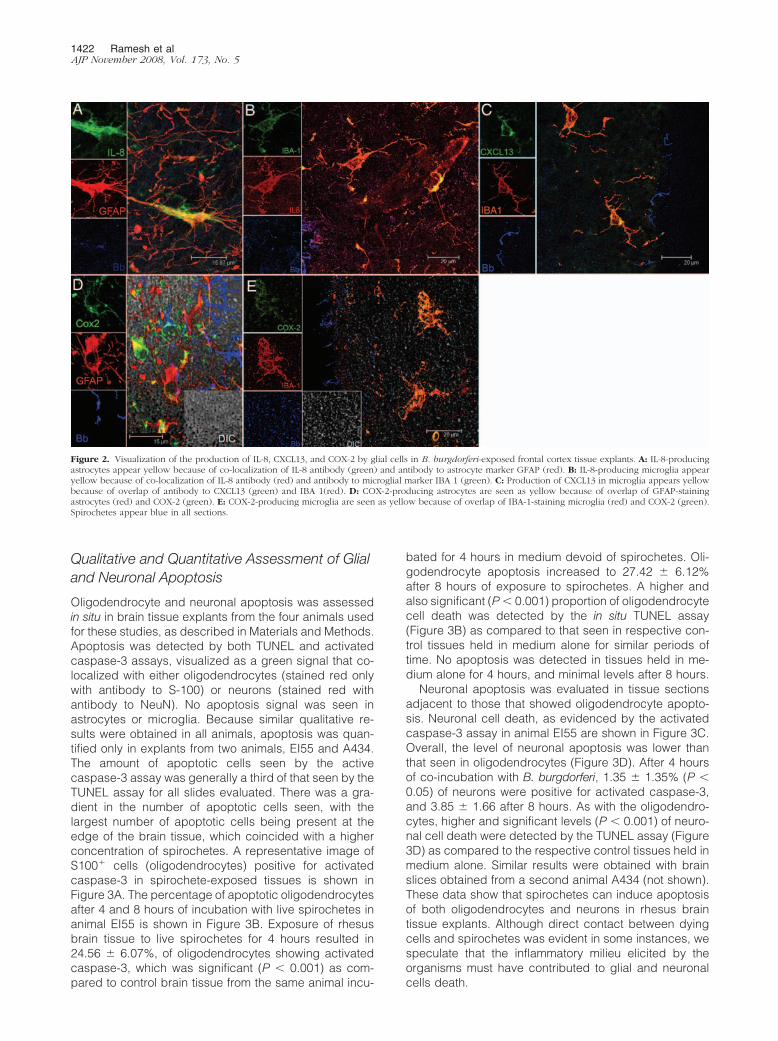
The chemokine IL-8 localized both to astrocytes andmicroglia (Figure 2, A and B, respectively). A positivesignal for IL-8 was also found in cells other than astro-cytes and microglia, possibly endothelial cells liningblood vessels (Figure 2B). The B-cell-attracting che-mokine CXCL13 was also detected in microglia in spi-rochete-stimulated tissues (Figure 2C). The immunemediator COX-2 was detected in both astrocytes (Fig-ure 2D) and microglia (Figure 2E). None of the pro-inflammatory mediators described above was de-tected in brain slices that were incubated for 4 hours inmedium that contained only brefeldin A (not shown).These data strongly support our hypothesis that glialcells elicit in the CNS an inflammatory milieu in re-sponse to B. burgdorferi.
1420 Ramesh et alAJP November 2008, Vol. 173, No. 5

DNA Microarray Survey of Genes Involved inApoptosis
We hypothesized that mediators elicited by spirochetescould induce apoptosis of both glial cells and neurons.Microarray analysis revealed that more than 30 apopto-sis-related gene transcripts were perturbed after expo-sure of rhesus monkey brain tissue explants to live B.burgdorferi for 4 hours. Importantly, many genes that spe-cifically induce apoptosis of oligodendrocytes as well asneurons were strongly up-regulated (Supplemental Table2, http://ajp.amjpathol.org). For example, the transcript ofthe gene coiled-coil-helix domain-containing six (NCBINM_032343), which regulates mitochondria-mediatedapoptosis was up-regulated 2048-fold. The transcript ofthe gene Cbp/p300-interacting transactivator, with Glu/Asp-rich carboxy-terminal domain 4 (NCBI NM_133467),which is an oligodendrocyte-specific tumor-repressorthat is silenced in oligodendrocyte tumors, was up-regu-lated 124.49-fold. The transcript of formin homology-2domain-containing 1 (NCBI NM_013241), which is asso-ciated with the caspase-3-mediated apoptosis pathway,
was up-regulated 43.40-fold. The transcript of the embry-onic lethal, abnormal vision, Drosophila-like 4 gene(NCBI NM_021952), which is associated with inflamma-tion and neuronal death and is up-regulated in Parkin-son’s disease, was up-regulated in spirochete-stimulatedtissues by 9.98-fold. The transcript of neuronal pentraxin1 (NCBI BX537550), which is involved in synapse loss,neurite damage, and apoptotic neuronal death and isalso overexpressed in Alzheimer’s brain was up-regu-lated by 8.39-fold. The transcript of glutamate receptor,ionotropic, delta 2 (NCBI NM_001510), which regulatesthe functions of glutamate, one of the predominant exci-tatory neurotransmitters in the mammalian brain that isknown to mediate neuronal apoptosis by excitotoxicity,was up-regulated by 4.40-fold. The transcripts of variousother genes that affect glial and neuronal survival werealso perturbed (Supplemental Table 2, http://ajp.amjpathol.org). These data support our notion that the global inflam-mation induced by B. burgdorferi as it enters the brain pa-renchyma also creates the conditions for apoptosis of glial
cells and neurons.
Figure 1. Visualization of the production of cytokines IL-6 and IL-1� by glial cells in B. burgdorferi-exposed frontal cortex tissue explants and brainsections from animals given stereotaxic inoculations with B. burgdorferi. A: IL-6-producing astrocytes appear pink because of co-localization of IL-6antibody (labeled blue) with antibody to the astrocyte marker GFAP (red). The spirochetes stained with fluorescein isothiocyanate-labeled B. burgdorferiantibody (Bb) appear green. Unstained tissue appears gray under differential interference contrast (DIC). B: Astrocytes labeled with GFAP (red) in braintissue slices incubated in medium plus brefeldin A in the absence of spirochetes had no detectable IL-6. C: Visualization of IL-6-producing astrocytes ina tissue section taken at the site of inoculation from an animal given stereotaxic inoculations with live B. burgdorferi. IL-6-producing astrocytes appearyellow because of co-localization of antibody to IL-6 (green) and antibody to the astrocyte marker GFAP (red) in the vicinity of B. burgdorferi antigenstained blue. D: IL-1�-producing microglia appear yellow because of co-localization of antibody to the microglial marker IBA 1 (green) and antibody toIL-1� (red). Spirochetes in this image appear blue. E: Microglia labeled with IBA-1 (green) in brain slices incubated in medium plus brefeldin A in theabsence of spirochetes had no detectable IL-1�.
Lyme Disease: Inflammation and Apoptosis 1421AJP November 2008, Vol. 173, No. 5
Qualitative and Quantitative Assessment of Glialand Neuronal Apoptosis
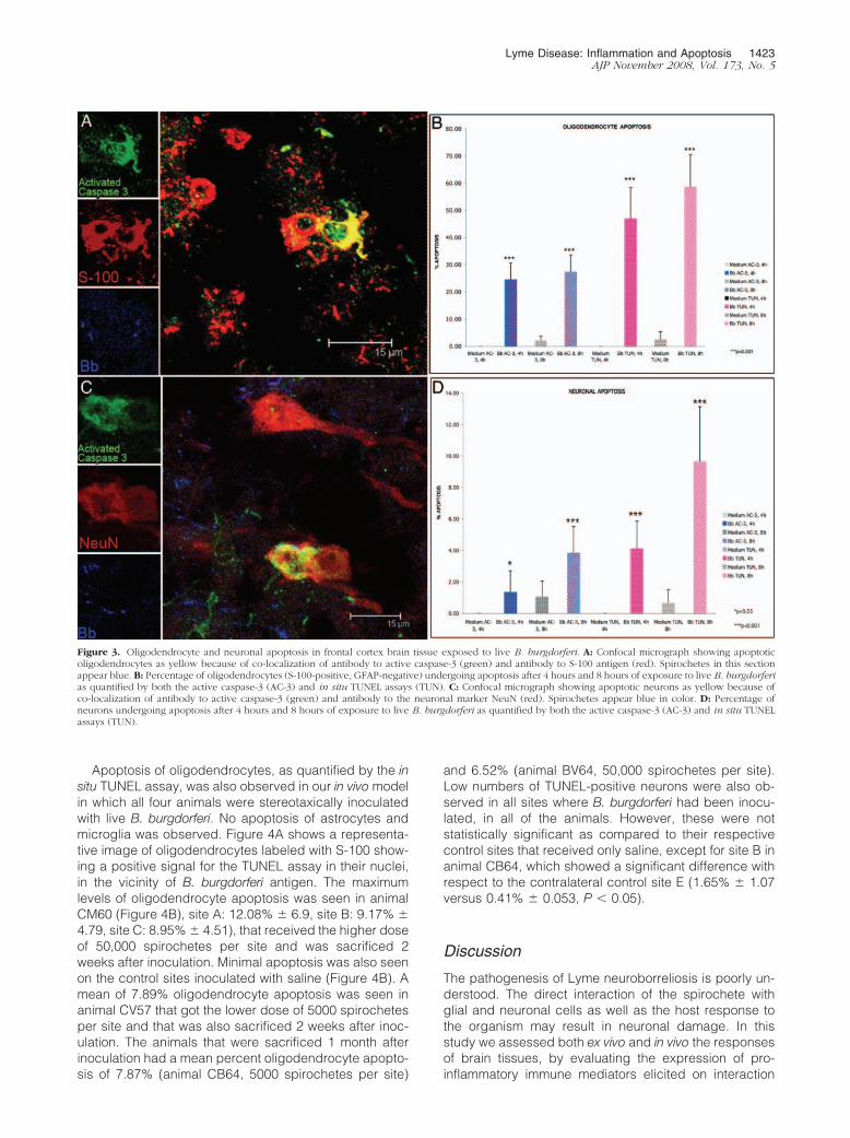
Oligodendrocyte and neuronal apoptosis was assessedin situ in brain tissue explants from the four animals usedfor these studies, as described in Materials and Methods.Apoptosis was detected by both TUNEL and activatedcaspase-3 assays, visualized as a green signal that co-localized with either oligodendrocytes (stained red onlywith antibody to S-100) or neurons (stained red withantibody to NeuN). No apoptosis signal was seen inastrocytes or microglia. Because similar qualitative re-sults were obtained in all animals, apoptosis was quan-tified only in explants from two animals, EI55 and A434.The amount of apoptotic cells seen by the activecaspase-3 assay was generally a third of that seen by theTUNEL assay for all slides evaluated. There was a gra-dient in the number of apoptotic cells seen, with thelargest number of apoptotic cells being present at theedge of the brain tissue, which coincided with a higherconcentration of spirochetes. A representative image ofS100� cells (oligodendrocytes) positive for activatedcaspase-3 in spirochete-exposed tissues is shown inFigure 3A. The percentage of apoptotic oligodendrocytesafter 4 and 8 hours of incubation with live spirochetes inanimal EI55 is shown in Figure 3B. Exposure of rhesusbrain tissue to live spirochetes for 4 hours resulted in24.56 � 6.07%, of oligodendrocytes showing activatedcaspase-3, which was significant (P � 0.001) as com-pared to control brain tissue from the same animal incu-
bated for 4 hours in medium devoid of spirochetes. Oli-godendrocyte apoptosis increased to 27.42 � 6.12%after 8 hours of exposure to spirochetes. A higher andalso significant (P � 0.001) proportion of oligodendrocytecell death was detected by the in situ TUNEL assay(Figure 3B) as compared to that seen in respective con-trol tissues held in medium alone for similar periods oftime. No apoptosis was detected in tissues held in me-dium alone for 4 hours, and minimal levels after 8 hours.
Neuronal apoptosis was evaluated in tissue sectionsadjacent to those that showed oligodendrocyte apopto-sis. Neuronal cell death, as evidenced by the activatedcaspase-3 assay in animal EI55 are shown in Figure 3C.Overall, the level of neuronal apoptosis was lower thanthat seen in oligodendrocytes (Figure 3D). After 4 hoursof co-incubation with B. burgdorferi, 1.35 � 1.35% (P �0.05) of neurons were positive for activated caspase-3,and 3.85 � 1.66 after 8 hours. As with the oligodendro-cytes, higher and significant levels (P � 0.001) of neuro-nal cell death were detected by the TUNEL assay (Figure3D) as compared to the respective control tissues held inmedium alone. Similar results were obtained with brainslices obtained from a second animal A434 (not shown).These data show that spirochetes can induce apoptosisof both oligodendrocytes and neurons in rhesus braintissue explants. Although direct contact between dyingcells and spirochetes was evident in some instances, wespeculate that the inflammatory milieu elicited by theorganisms must have contributed to glial and neuronalcells death.
Figure 2. Visualization of the production of IL-8, CXCL13, and COX-2 by glial cells in B. burgdorferi-exposed frontal cortex tissue explants. A: IL-8-producingastrocytes appear yellow because of co-localization of IL-8 antibody (green) and antibody to astrocyte marker GFAP (red). B: IL-8-producing microglia appearyellow because of co-localization of IL-8 antibody (red) and antibody to microglial marker IBA 1 (green). C: Production of CXCL13 in microglia appears yellowbecause of overlap of antibody to CXCL13 (green) and IBA 1(red). D: COX-2-producing astrocytes are seen as yellow because of overlap of GFAP-stainingastrocytes (red) and COX-2 (green). E: COX-2-producing microglia are seen as yellow because of overlap of IBA-1-staining microglia (red) and COX-2 (green).Spirochetes appear blue in all sections.
1422 Ramesh et alAJP November 2008, Vol. 173, No. 5
Apoptosis of oligodendrocytes, as quantified by the insitu TUNEL assay, was also observed in our in vivo modelin which all four animals were stereotaxically inoculatedwith live B. burgdorferi. No apoptosis of astrocytes andmicroglia was observed. Figure 4A shows a representa-tive image of oligodendrocytes labeled with S-100 show-ing a positive signal for the TUNEL assay in their nuclei,in the vicinity of B. burgdorferi antigen. The maximumlevels of oligodendrocyte apoptosis was seen in animalCM60 (Figure 4B), site A: 12.08% � 6.9, site B: 9.17% �4.79, site C: 8.95% � 4.51), that received the higher doseof 50,000 spirochetes per site and was sacrificed 2weeks after inoculation. Minimal apoptosis was also seenon the control sites inoculated with saline (Figure 4B). Amean of 7.89% oligodendrocyte apoptosis was seen inanimal CV57 that got the lower dose of 5000 spirochetesper site and that was also sacrificed 2 weeks after inoc-ulation. The animals that were sacrificed 1 month afterinoculation had a mean percent oligodendrocyte apopto-sis of 7.87% (animal CB64, 5000 spirochetes per site)
and 6.52% (animal BV64, 50,000 spirochetes per site).Low numbers of TUNEL-positive neurons were also ob-served in all sites where B. burgdorferi had been inocu-lated, in all of the animals. However, these were notstatistically significant as compared to their respectivecontrol sites that received only saline, except for site B inanimal CB64, which showed a significant difference withrespect to the contralateral control site E (1.65% � 1.07versus 0.41% � 0.053, P � 0.05).
Discussion
The pathogenesis of Lyme neuroborreliosis is poorly un-derstood. The direct interaction of the spirochete withglial and neuronal cells as well as the host response tothe organism may result in neuronal damage. In thisstudy we assessed both ex vivo and in vivo the responsesof brain tissues, by evaluating the expression of pro-inflammatory immune mediators elicited on interaction
Figure 3. Oligodendrocyte and neuronal apoptosis in frontal cortex brain tissue exposed to live B. burgdorferi. A: Confocal micrograph showing apoptoticoligodendrocytes as yellow because of co-localization of antibody to active caspase-3 (green) and antibody to S-100 antigen (red). Spirochetes in this sectionappear blue. B: Percentage of oligodendrocytes (S-100-positive, GFAP-negative) undergoing apoptosis after 4 hours and 8 hours of exposure to live B. burgdorferias quantified by both the active caspase-3 (AC-3) and in situ TUNEL assays (TUN). C: Confocal micrograph showing apoptotic neurons as yellow because ofco-localization of antibody to active caspase-3 (green) and antibody to the neuronal marker NeuN (red). Spirochetes appear blue in color. D: Percentage ofneurons undergoing apoptosis after 4 hours and 8 hours of exposure to live B. burgdorferi as quantified by both the active caspase-3 (AC-3) and in situ TUNELassays (TUN).
Lyme Disease: Inflammation and Apoptosis 1423AJP November 2008, Vol. 173, No. 5

with live B. burgdorferi spirochetes. We also addressedour hypothesis that this inflammatory response to B.burgdorferi could contribute to cell death by evaluatingthe presence of concomitant apoptosis of both glial andneuronal cells. The immune mediators IL-6, IL-8, IL-1�,COX-2, and CXCL13 were visualized in glial cells in situby immunofluorescence staining and confocal micros-copy. Concomitantly, a significant proportion of both oli-godendrocytes and neurons undergoing apoptosis werepresent in spirochete-exposed tissues. The observationthat the largest number of cells producing immune me-diators as well as those undergoing apoptosis coincidedwith the presence of large number of spirochetes alongthe edges of the brain tissue suggests, but does notprove, that these phenomena are related. The high num-ber of significantly perturbed transcripts of genes thatregulate immune function, as revealed in our microarrayanalysis of live spirochete-stimulated brain tissues (Sup-plemental Table 1, http://ajp.amjpathol.org), subscribes tothe notion that spirochetes can have a powerful effect onthe regulation of inflammation in the brain parenchyma.Indeed, transcription factors and immune activators thatregulate the synthesis and activation of cytokines andchemokines in the brain were seen to be perturbed as aresult of spirochete stimulation (Supplemental Table 1,http://ajp.amjpathol.org).
We focused on evaluating the cellular source of thepro-inflammatory cytokines IL-6, IL-1�, and TNF-�, andthe chemokines CXCL13 and IL-8 because these medi-ators have been documented to play a role in acute Lymeneuroborreliosis.17–19,21,22 IL-6 was produced by numer-ous cells and cell types in rhesus brain tissue that wasincubated with live spirochetes ex vivo. Of these we iden-tified the astrocyte as a major glial cell type involved.Importantly, stereotaxic intracerebral inoculation of livespirochetes in vivo also resulted in production of IL-6 byastrocytes. IL-6 mRNA has also been shown to be ex-
pressed at high levels in the CNS of the rhesus ma-caques infected with B. burgdorferi by others.40
Reactive astrogliosis has been shown to be a conse-quence of increased expression of IL-6 in the brain.41
Markers of astrogliosis have been detected in the CSF ofpatients with Lyme neuroborreliosis42,43 as well as in therhesus model.44 B. burgdorferi has been shown to induceIL-6 in rat glioma cells.45 Our findings expand on theseresults and indicate that astrocytes are among the cellsthat are a source of the IL-6 produced in the CNS inresponse to a B. burgdorferi infection.
Previously we reported on the production of the cyto-kine TNF-� in primary cultures of rhesus monkey astro-cytes when these cells were stimulated with lipidatedouter surface protein A (L-OspA, 1.0 �g/ml) in vitro.44
L-OspA is a major lipoprotein of B. burgdorferi. We hadalso found that primary cultures of rhesus monkey micro-glia can produce TNF-� in response to L-OspA.46 We didnot detect TNF-� in brain sections stimulated with livespirochetes by immunofluorescence staining. However,we did see a significant increase in TNF-� transcript inspirochete-stimulated tissues by microarray analysis(21.7-fold) as well as by real time PCR (7.43-fold). RhesusTNF-� is readily detectable with the anti-human TNF-�antibodies used in this study, as shown previously with arhesus macaque model of chronic gut inflammation inwhich the same antibodies were used.37 Therefore, it wasnot because of lack of antibody cross-reactivity that wefailed to observe TNF-� production in brain tissues.Rather, it is possible that there was a lag in the kinetics oftranslation of TNF-� transcript. Specific inhibitors ofTNF-� translation have been described.47
IL-1�, a cytokine that plays a key role in mediatinginflammation in the CNS,48 was produced by microglia inspirochete-stimulated brain tissues. Many different celltypes in the brain express the IL-1 receptor and respondto this cytokine by activating cell-type-specific signaling
Figure 4. Oligodendrocyte apoptosis observed in rhesus CM60 given stereotaxic inoculations with live B. burgdorferi. A: Apoptotic oligodendrocytes showTUNEL-positive nuclei stained green. Oligodendrocytes appear red because of staining with antibody to S-100. B. burgdorferi antigen appears blue in color. B:Percentage of oligodendrocytes (S-100-positive, GFAP-negative) undergoing apoptosis in brain sections from the sites of spirochetal inoculation (A–C) andcorresponding control sites (D–F) as quantified by the in situ TUNEL assay.
1424 Ramesh et alAJP November 2008, Vol. 173, No. 5

pathways leading to distinct functional responses.49 Neu-rons also express the IL-1 receptor, indicating that thiscytokine can influence neuronal function directly.50 Thereis evidence that IL-1� can cause synaptic inhibition51
and suppress long-term potentiation, two neuronal pro-cesses which are thought to be involved in learning andmemory.52 IL-1� is also involved in microglia-mediatedneurotoxicity.53 An inflammatory cascade of events trig-gered by IL-1� causes the decline of mental fitness inAlzheimer’s disease.54 IL-1�-mediated neuronal dys-function could similarly be a possible player in the patho-genesis of Lyme neuroborreliosis.
Spirochetes elicited both the up-regulation of IL-8 tran-script in brain tissues and production of this chemokineby astrocytes and microglia. The elevated levels of IL-8found in the CSF of patients with Lyme neuroborreliosis22
could indeed be caused by the interaction of spirocheteswith glial cells, and be an effector of the inflammatorylesions seen in this form of Lyme disease, and of theleukocyte pleocytosis that is characteristic of Lyme men-ingitis. We also demonstrated the presence of the B-cellchemokine CXCL13 in microglia. Increased presence ofCXCL13 or its transcript has been described in associa-tion with B. burgdorferi infection both in nonhuman pri-mates and in humans.55–57 CXCL13 also is produced bymonocytes in response to molecules with the Pam3Cysmotif of bacterial lipoproteins58; the B. burgdorferi ge-nome contains more than 150 open reading frames thatcould encode such molecules.59,60 Finally, COX-2, whichwe detected both in astrocytes and microglia, hasemerged as a major player in the inflammatory reactionsin the brain.39 COX-2 is expressed in apoptotic oligoden-drocytes in chronic active demyelinating lesions of pa-tients with multiple sclerosis.61 Activation of COX-2 con-tributes to motor and cognitive dysfunction,62 and hasbeen implicated in mediating neuropathic pain.63 Othershave shown B. burgdorferi to stimulate production ofCOX-2 in murine microglia,64 an observation that is inaccordance with ours in the rhesus model.
In addition to mediators elicited in glial cells, we ob-served the presence of IL-1�, IL-8, and COX-2 in glut-1-positive endothelial cells in blood vessels in tissue slicesincubated with spirochetes plus brefeldin A, but not incontrol tissues held in medium plus brefeldin A after 4hours of incubation (data not shown). Inflammatorychanges induced in the CNS blood vessels as a result ofinteraction with B. burgdorferi spirochetes could be majorplayers in the development of vasculitis seen in patientswith Lyme neuroborreliosis.5–8
Cytokines, chemokines, other immune mediators, aswell as glial and neuronal apoptosis have been docu-mented to play a major role in inflammatory neurodegen-erative diseases such as Alzheimer’s disease, AIDS de-mentia,65 and Parkinson’s disease66 in humans, and insimian immunodeficiency virus encephalitis in nonhumanprimates.67,68 The results of our microarray analysis em-phasizes the possible role of immune function-relatedgene products as well as that of apoptosis-regulatinggenes in contributing to the pathogenesis of Lyme neu-roborreliosis. Our observation of both neuronal and oli-godendrocyte apoptosis in situ in ex vivo-stimulated brain
tissues as well as in our stereotaxic in vivo model furtheraffirms this role. A recent report demonstrates thatphagocytosis of B. burgdorferi potentiates innate immuneactivation and induces apoptosis in human monocytes;this phenomenon is accompanied by the production ofcytokines such as TNF-�, IL-1�, and IL-6.69 Other in vitrostudies have shown that IL-1� promotes oligodendrocytedeath through glutamate excitotoxicity.70 Oligodendrocytedeath could also result in impaired neuronal functionbecause these glial cells are vital for maintaining thenormal nerve conduction.71 Further detailed studies toexplore the role of the various immune mediators andmechanisms involved in inducing glial and neuronaldeath will help answer these questions, and further ourunderstanding of the signaling pathways that contributeto the pathogenesis of Lyme neuroborreliosis.
The levels of apoptosis of both oligodendrocytes andneurons were more marked when quantified by theTUNEL assay as compared to when they were measuredvia activated caspase-3. This could be attributable to thefact that the active caspase-3 assay only detects cellsthat are in the early stages of apoptosis,72 whereas theTUNEL assay detects all nuclei showing fragmentedDNA.73 Importantly, the results obtained by both assayspoint in the same direction, with the percent apoptosis asdetected by the active caspase-3 assay being approxi-mately a third of the percent apoptosis as measured byTUNEL. The fact that we were also able to observe sig-nificant apoptosis of oligodendrocytes in our in vivo modelof animals that were sterotaxically inoculated with live B.burgdorferi further illustrates that B. burgdorferi spiro-chetes have the ability to elicit oligodendrocyte apoptosisand confirms the observations of both oligodendrocyteand neuronal apoptosis seen in our ex vivo model. Thereduced levels of oligodendrocyte and neuronal apopto-sis observed in vivo as compared to that seen in the exvivo specimens may be attributable to the action ofphagocytic cells that would not only affect spirochetalburdens but also diminish the number of residual apo-ptotic cells at any time after inoculation.74
The proinflammatory cytokines TNF-�, IL-1�, and IL-6have been reported to mediate apoptotic cell death ofneurons by signaling via the p38 MAP kinase pathway inneurodegenerative conditions.75 We have reported ear-lier that proliferation and apoptosis of astrocytes are me-diated by IL-6 and TNF-�, respectively, as induced bystimulation of glial cells with L-OspA. We also reportedthat the phosphorylation of mitogen-activated protein ki-nases Erk 1/2 and P38 was involved in these phenomena.76
These signaling mechanisms could also be operative inthe pathogenesis of Lyme neuroborreliosis.
The major symptoms of long-term Lyme neuroborrelio-sis of the CNS, namely fatigue and cognitive dysfunction,could be a consequence of impairment of glial and neu-ronal cell function and survival as a result of the directinteraction between these cells and B. burgdorferi, or withthe mediators elicited by the spirochete. The studiesreported herein have helped identify glial cells assources of immune mediators, as well as document thepresence of concomitant apoptosis of both oligodendro-cytes and neurons in spirochete-exposed parenchymal
Lyme Disease: Inflammation and Apoptosis 1425AJP November 2008, Vol. 173, No. 5

tissues. They provide proof of the concept that inflamma-tion with concomitant apoptosis are major factors in thepathogenesis of Lyme neuroborreliosis.
Acknowledgments
We thank Peter Mottram and Drs. Andrea Bernardino andXavier Avarez for technical help and advice, and RobinRodriguez for help with the confocal microscope images.
References
1. Steere AC, Grodzicki RL, Kornblatt AN, Craft JE, Barbour AG,Burgdorfer W, Schmid GP, Johnson E, Malawista SE: The spirochetaletiology of Lyme disease. N Engl J Med 1983, 308:733–740
2. Piesman J: Dynamics of Borrelia burgdoferi transmission by Ixodesdammini ticks. J Infect Dis 1993, 167:1082–1085
3. Steere AC: Lyme disease. N Engl J Med 2001, 345:115–1254. Gustaw K, Beltowska K, Studzinska MM: Neurological and psycho-
logical symptoms after severe acute neuroborreliosis. Ann Agric En-viron Med 2001, 8:91–94
5. Jacobi C, Schwark C, Kress B, Hug A, Storch-Hagenlocher B,Schwaninger M: Subarachnoid hemorrhage due to Borrelia burgdor-feri-associated vasculitis. Eur J Neurol 2006, 13:536–538
6. Chehrenama M, Zagardo MT, Koski CL: Subarachnoid hemorrhage ina patient with Lyme disease. Neurology 1997, 48:520–523
7. Schmitt AB, Kuker W, Nacimiento W: Neuroborreliosis with extensivecerebral vasculitis and multiple cerebral infarcts. Nervenarzt 1999,70:167–171
8. Seijo Martínez M, Grandes Ibanez J, García-Monoco JC: Spontane-ous brain hemorrhage associated with Lyme neuroborreliosis. Neu-rologia 2001, 16:43–45
9. Kohler J: Lyme borreliosis: a case of transverse myelitis with syrinxcavity. Neurology 1989, 39:1553–1554
10. Benach JL, Garcia-Monoco JC: Aspects of the pathogenesis of neu-roborreliosis. Lyme Disease: Molecular and Immunological Ap-proaches. Edited by Schutzer S. Cold Spring Harbor, Cold SpringHarbor Laboratory Press, 1992, pp. 1–10
11. Oksi J, Kalimo H, Marttila RJ, Marjamaki M, Sonninen P, Nikoskelainen J,Viljanen MK: Inflammatory brain changes in Lyme borreliosis. Areport on three patients and review of literature. Brain 1996,119:2143–2154
12. Dryden MS, O’Connell S, Samuel W, Iannotti F: Lyme myelitis mim-icking neurological malignancy. Lancet 1996, 348:624
13. Benveniste EN: Inflammatory cytokines within the central nervoussystem: sources, functions, and mechanisms of action. Am J Physiol1992, 263:C1–C5
14. Merrill JE, Benveniste EN: Cytokines in inflammatory brain lesions:helpful and harmful. TINS 1996, 19:331–338
15. Rothwell NJ, Hopkins SJ: Cytokines and nervous system II: actionsand mechanisms of action. Trends Neurosci 1995, 18:130–136
16. Raivich G, Jones LL, Werner A, Bluthmann, Doetschmann T,Kreutzberg GW: Molecular signals for glial activation: pro- and anti-inflammatory cytokines in the injured brain. Acta Neurochir Suppl1999, 73:21–30
17. Wang WZ, Fredrikson S, Sun JB, Link H: Lyme neuroborreliosis:evidence for persistent up-regulation of Borrelia burgorferi-reac-tive cells secreting interferon-gamma. Scand J Immunol 1995,42:694 –700
18. Ekerfelt C, Jarefors S, Tynngard N, Hedlund M, Sander B, BergstromS, Forsberg P, Ernerudh J: Phenotypes indicating cytolytic propertiesof Borrelia-specific interferon-� secreting cells on chronic Lyme neu-roborreliosis. J Neuroimmunol 2003, 145:115–126
19. Kondrusik M, Sweirzbinska R, Pancewicz S, Zajkowska J, GrygorczukS, Hermanowska-Szpakowicz T: Evaluation of proinflammatory cyto-kine (TNF-alpha, IL-1beta, IL-6, IFN-gamma) concentrations in serumand cerebrospinal fluid of patients with neuroborreliosis. Neurol Neu-rochir Pol 2004, 38:265–270
20. Widhe M, Grusell M, Ekerfelt C, Vrethem M, Forsberg P, Ernerudh J:Cytokines in Lyme borreliosis: lack of early tumor necrosis factor-�
and transforming growth factor-�1 responses are associated withchronic neuroborreliosis. Immunology 2002, 107:46–55
21. Rupprecht TA, Pfister HW, Angele B, Kastenbauer S, Wilske B, KoedelU: The chemokine CXCL13 (BLC): a putative diagnostic marker forneuroborreliosis. Neurology 2005, 65:448–450
22. Grygorczuk S, Pancewicz S, Zajkowska J, Kondrusik M, SwierzbinskaR, Hermanowska-Szpakowicz T: Concentration of macrophage in-flammatory proteins MIP-� and MIP-� and interleukin 8 (IL-8) in Lymeborreliosis. Infection 2004, 32:350–355
23. Grygorczuk S, Zajkowska J, Swierzbinska R, Pancewicz S, KondrusikM, Hermanowska-Szpakowicz T: Concentration of interferon-induc-ible T cell chemoattractant and monocyte chemotactic protein-1 withLyme borreliosis. Ann Acad Med Bialo 2005, 50:173–178
24. Lewczuk P, Reiber H, Korenke GC, Bollensen E, Dorta-Contreras AJ:Intrathecal release of sICAM-1 into CSF in neuroborreliosis increasedbrain-derived fraction. J Neuroimmunol 2000, 103:93–96
25. Philipp MT, Aydintug MK, Bohm Jr RP, Cogswell FB, Dennis VA,Lanners HN, Lowrie Jr RC, Roberts ED, Conway MD, Karacorlu M,Peyman GA, Gubler DJ, Johnson BJB, Piesman J, Gu Y: Early andearly-disseminated phases of Lyme disease in the rhesus monkey: amodel of infection in humans. Infect Immun 1993, 61:3047–3059
26. Roberts ED, Bohm Jr RP, Cogswell FB, Lanners HN, Lowrie Jr RC,Povinelli L, Piesman J, Philipp MT: Chronic Lyme disease in therhesus monkey. Lab Invest 1995, 72:146–160
27. Pachner AR, Delaney E, O’Neil T, Major E: Inoculation of non humanprimates with the N40 strain of Borrelia burgdorferi leads to a modelof Lyme neuroborreliosis faithful to the human disease. Neurology1995, 45:165–172
28. England JD, Bohm Jr R, Roberts ED, Philipp MT: Mononeuropathymultiplex in rhesus monkeys with chronic Lyme disease. Ann Neurol1997, 41:375–384
29. Roberts ED, Bohm Jr RP, Lowrie Jr RC, Habicht G, Katona L, PiesmanJ, Philipp MT: Pathogenesis of Lyme neuroborreliosis in the rhesusmonkey: the early disseminated and chronic phases of disease in theperipheral nervous system. J Infect Dis 1998, 178:722–732
30. Cadavid D, O’Neil T, Schaefer H, Pachner AR: Localization of Borreliaburgdorferi in the nervous system and other organs in a nonhumanprimate model of Lyme disease. Lab Invest 2000, 80:1043–1054
31. Pachner AR: The rhesus model of Lyme neuroborreliosis. ImmunolRev 2001, 183:186–204
32. Bai Y, Narayan K, Dail D, Sondey M, Hodzic E, Barthold SW, PachnerAR, Cadavid D: Spinal cord involvement in the nonhuman primatemodel of Lyme disease. Lab Invest 2004, 84:160–172
33. Haber SN: The primate basal ganglia: a parallel and integrativenetworks. J Chem Neuroanat 2003, 26:317–330
34. Jackson CL: Brefeldin A revealing fundamental principles governingmembrane dynamics and protein transport. Subcell Biochem 2000,34:233–272
35. Ramamoorthy R, McClain NA, Gautum A, Scholl-Meeker D: Ex-pression of the bmpB gene of Borelia burgdorferi is modulated bytwo distinct transcription termination events. J Bacteriol 2005,187:2592–2600
36. Kaushal D, Naeve CW: Analyzing and visualizing expression datawith Spotfire. Current Protocols in Bioinformatics. Edited by Baxeva-nis A. Hoboken, John Wiley and Sons Inc. 2004, pp 7.9.1–7.9.25
37. Ramesh G, Alvarez X, Borda JT, Aye PP, Lackner AA, Sestak K:Visualizing cytokine-secreting cells in situ in the rhesus macaquemodel of chronic gut inflammation. Clin Diag Lab Immunol 2005,12:192–197
38. Schmidt-Kastner R, Szymas J: Immunohistochemistry of glial fibrillaryacid protein, vimentin and S-100 protein for study of astrocytes inhippocampus of rat. J Chem Neuroanat 1990, 3:179–192
39. Minghetti L: Cyclooxygenase-2 (COX-2) in inflammatory and de-generative brain diseases. J Neuropathol Exp Neurol 2004,63:901–910
40. Pachner AR, Amemiya K, Delaney E, O’Neil T, Hughes CAN, ZhangWF: Interleukin-6 is expressed at high levels in the CNS in Lymeneuroborreliosis. Neurology 1997, 49:147–152
41. Chiang CS, Stalder A, Campbell IL: Reactive gliosis as a conse-quence of interleukin 6 expression in the brain: studies in transgenicmice. Dev Neurosci 1994, 16:212–221
42. Dotevall L, Hagberg L, Karlsson JL, Rosengren LE: Astroglial andneuronal proteins in cerebrospinal fluid as markers of CNS involve-ment in Lyme neuroborreliosis. Eur J Neurol 1999, 6:169–178
1426 Ramesh et alAJP November 2008, Vol. 173, No. 5
43. Dotevall L, Rosengren LE, Hagberg L: Increased cerebrospinal fluidlevels of glial fibrillary acidic protein (GFAP) in Lyme neuroborreliosis.Infection 1996, 24:125–129
44. Ramesh G, Alvarez AL, Roberts ED, Dennis VA, Lasater BL, AlvarezX, Philipp MT: Pathogenesis of Lyme neuroborreliosis: Borrelia burg-dorferi lipoproteins induce both proliferation and apoptosis in rhesusmonkey astrocytes. Eur J Immunol 2003, 33:2539–2550
45. Habicht GS, Katona LI, Benach JL: Cytokines and the pathogenesisof neuroborreliosis: Borrelia burgdorferi induces glioma cells to se-crete interleukin-6. J Infect Dis 1991, 164:568–574
46. Ramesh G: An in vitro model of the interaction of bacterial lipoproteinswith the central nervous system (Ph.D. thesis). [New Orleans (LA)]Tu-lane University
47. Piecyk M, Wax S, Beck AR, Kedersha N, Gupta M, Maritim B, Chen S,Gueydan C, Kruys V, Streuli M, Anderson P: TIA-1 is a translationalsilencer that selectively regulates the expression of TNF-alpha.EMBO J 2000, 19:4154–4163
48. Friedman WJ: Interactions of interleukin-1 with neurotropic factors inthe central nervous system: beneficial or detrimental? Mol Neurobiol2005, 32:133–144
49. Srinivasan D, Yen JH, Joseph DJ, Friedman W: Cell type-specific inter-leukin-1beta signaling in the CNS. J Neurosci 2004, 24:6482–6488
50. Friedman WJ: Cytokines regulate expression of the type 1 interleu-kin-1 receptor in rat hippocampal neurons and glia. Exp Neurol 2001,168:23–31
51. Zeise ML, Madamba S, Siggins GR: Interleukin-1� increases synapticinhibition in rat hippocampal pyramidal neurons in vitro. Regul Pept1992, 39:1–7
52. Katsuki H, Nakai S, Hirai Y, Akaji K, Kiso Y, Satoh M: Interleukin-1�
inhibits long-term potentiation in the CA3 region of mouse hippocam-pal slices. Eur J Pharmacol 1990, 181:323–326
53. Ma XC, Gottschall PE, Chen LT, Wiranowska M, Phelphs CP: Role andmechanisms of interleukin-1 in the modulation of neurotoxicity. Neu-roimmunomodulation 2002–2003, 10:199–207
54. Griffin WS: Inflammation and neurodegenerative diseases. Am J ClinNutr 2006, 83:470S–474S
55. Pachner AR, Dail D, Narayan K, Dutta K, Cadavid D: Increasedexpression of B-lymphocyte chemoattractant, but not pro-inflamma-tory cytokines, in muscle tissue in rhesus chronic Lyme borreliosis.Cytokine 2002, 19:297–307
56. Narayan K, Dail D, Li L, Cadavid D, Amrute S, Fitzgerald-Bocaesly P,Pachner AR: The nervous system as ectopic germinal center:CXCL13 and IgG in Lyme neuroborreliosis. Ann Neurol 2005,57:813–823
57. Cadavid D: The mammalian host response to borrelia infection. WienKlin Wochenschr 2006, 18:653–658
58. Rupprecht TA, Kirschning CJ, Popp B, Kastenbauer S, Fingerle V,Pfister HW, Koedel U: Borrelia garinii induces CXCL13 production inhuman monocytes through Toll-like receptor 2. Infect Immun 2007,75:4351–4356
59. Fraser CM, Casjens S, Huang WM, Sutton GG, Clayton R, Lathigra R,White O, Ketchum KA, Dodson R, Hickey EK, Gwinn M, Dougherty B,Tomb JF, Fleischmann RD, Richardson D, Peterson J, Kerlavage AR,Quackenbush J, Salzberg S, Hanson M, van Vugt R, Palmer N,Adams MD, Gocayne J, Weidman J, Utterback T, Watthey L,McDonald L, Artiach P, Bowman C, Garland S, Fuji C, Cotton MD,Horst K, Roberts K, Hatch B, Smith HO, Venter JC: Genomic se-quence of a Lyme disease spirochaete, Borrelia burgdorferi. Nature1997, 390:580–586
60. Casjens S, Palmer N, van Vugt R, Huang WM, Stevenson B, Rosa P,Lathigra R, Sutton G, Peterson J, Dodson RJ, Haft D, Hickey E, GwinnM, White O, Fraser CM: A bacterial genome in flux: the twelve linearand nine circular extrachromosomal DNAs in an infectious isolate ofthe Lyme disease spirochete Borrelia burgdorferi. Mol Microbiol2000, 35:490–516
61. Carlson NG, Hill KE, Tsunoda I, Fujinami RS, Rose JW: The patho-genic role for COX-2 in apoptotic oligodendrocytes in virus induceddemyelinating disease: implications for multiple sclerosis. J Neuro-immunol 2006, 174:21–31
62. Cernak I, O’Connor C, Vink R: Activation of cyclooxygenase-2 con-tributes to motor and cognitive dysfunction following diffuse traumaticbrain injury in rats. Clin Exp Pharmacol Physiol 2001, 28:922–925
63. Ma W, Quirion R: Up-regulation of interleukin-6 induced by prosta-glandin E from invading macrophages following nerve injury: an invivo and in vitro study. J Neurochem 2005, 93:664–673
64. Rasley A, Anguita J, Marriot I: Borrelia burgdorferi induces inflamma-tory mediator production of murine microglia. J Neuroimmunol 2002,130:22–31
65. Dickson DW, Lee SC, Mattiace LA, Yen SC, Brosnan C: Microglia andcytokines in neurological disease, with special reference to AIDS andAlzheimer’s disease. Glia 1993, 7:75–83
66. Nagatsu T, Sawada M: Inflammatory process in Parkinson’s disease:role for cytokines. Curr Pharm Des 2005, 11:999–1016
67. Orandle MT, MacLean AG, Sasseville VG, Alvarez X, Lackner AA:Enhanced expression of proinflammatory cytokines in the centralnervous system is associated with neuroinvasion by simian immuno-deficiency virus and the development of encephalitis. J Virol 2002,76:5797–5802
68. Adamson DC, Dawson TM, Zink MC, Clements JE, Dawson VL:Neurovirulent simian immunodeficiency virus infection induces neu-ronal, endothelial and glial apoptosis. Mol Med 1996, 2:417–428
69. Cruz AR, Moore MW, La Vake CJ, Eggers CH, Salazar JC, Radolf JD:Phagocytosis of B. burgdorferi potentiates innate immune activationand induces apoptosis in human monocytes. Infect Immun 2008,76:56–70
70. Takahashi JL, Giuliani F, Powe C, Imai Y, Young VW: Interleukin-betapromotes oligodendrocyte death through glutamate excitotoxicity.Ann Neurol 2003, 53:588–595
71. Compston A Zajicek J, Sussman J, Webb A, Hall G, Muir D, Shaw C,Wood A, Scolding N: Glial lineages and myelination in the centralnervous system. J Anat 1997, 190:161–200
72. Namura S, Zhu J, Fink K, Endres M, Srinivasan A, Tomaselli KJ, YuanJ, Moskowitz MA: Activation and cleavage of caspase-3 in apoptosisinduced by experimental cerebral ischemia. J Neurosci 1998,18:3659–3668
73. de Torres C, Munell F, Ferrer I, Reventos J, Macaya A: Identificationof necrotic cell death by the TUNEL assay in the hypoxic ischemicneonatal rat brain. Neurosci Lett 1997, 230:1–4
74. Maderna P, Godson C: Phagocytosis of apoptotic cells and resolutionof inflammation. Biochem Biophys Acta 2003, 1639:141–151
75. Chaparro-Huerta V, Rivera-Cervantes MC, Flores-Soto ME, Gomez-Pinedo U, Beas-Zarate C: Proinflammatory cytokines and apoptosisfollowing glutamate-induced excitotoxicity mediated by p38 MAPK inthe hippocampus of neonatal rats. J Neuroimmunol 2005, 165:53–62
76. Ramesh G, Philipp MT: Pathogenesis of Lyme neuroborreliosis: mi-togen-activated protein kinases Erk1, Erk2, and p38 in the responseof astrocytes to Borrelia burgdorferi lipoproteins. Neurosci Lett 2005,384:112–116
Lyme Disease: Inflammation and Apoptosis 1427AJP November 2008, Vol. 173, No. 5
Related Documents