Interaction of psychological, physiological and neuronal processes in functional dyspepsia Dissertation zur Erlangung des Grades eines Doktors der Naturwissenschaften der Mathematisch-Naturwissenschaftlichen Fakultät und der Medizinischen Fakultät der Eberhard-Karls-Universität Tübingen vorgelegt von In-Seon Lee aus Seoul, die Republik Korea August - 2017

Welcome message from author
This document is posted to help you gain knowledge. Please leave a comment to let me know what you think about it! Share it to your friends and learn new things together.
Transcript
Interaction of psychological, physiological and neuronal processes
in functional dyspepsia
Dissertation
zur Erlangung des Grades eines
Doktors der Naturwissenschaften
der Mathematisch-Naturwissenschaftlichen Fakultät
und
der Medizinischen Fakultät
der Eberhard-Karls-Universität Tübingen
vorgelegt
von
In-Seon Lee
aus Seoul, die Republik Korea
August - 2017

Tag der mündlichen Prüfung: .........................................
Dekan der Math.-Nat. Fakultät: Prof. Dr. W. Rosenstiel
Dekan der Medizinischen Fakultät: Prof. Dr. I. B. Autenrieth
1. Berichterstatter: Prof. Dr. Paul Enck
2. Berichterstatter: Prof. Dr. Hubert Preissl
Prüfungskommission: ...................................................................
................................................................…
................................................................…
................................................................…
Erklärung / Declaration:
Ich erkläre, dass ich die zur Promotion eingereichte Arbeit mit dem Titel:
„……………….…………….……………………………………………………...........................
..........................…...........................................................................................................….“
selbständig verfasst, nur die angegebenen Quellen und Hilfsmittel benutzt und wörtlich oder
inhaltlich übernommene Stellen als solche gekennzeichnet habe. Ich versichere an Eides statt,
dass diese Angaben wahr sind und dass ich nichts verschwiegen habe. Mir ist bekannt, dass die
falsche Abgabe einer Versicherung an Eides statt mit Freiheitsstrafe bis zu drei Jahren oder mit
Geldstrafe bestraft wird.
I hereby declare that I have produced the work entitled “……”, submitted for the award of a
doctorate, on my own (without external help), have used only the sources and aids indicated and
have marked passages included from other works, whether verbatim or in content, as such. I
swear upon oath that these statements are true and that I have not concealed anything. I am
aware that making a false declaration under oath is punishable by a term of imprisonment of up
to three years or by a fine.
Tübingen, den ......................................... .............................................................
Datum / Date Unterschrift /Signature
1
Contents
Abstract ......................................................................................................................................................................... 2
1. Introduction .............................................................................................................................................................. 4
1.1. Definition of functional dyspepsia ...................................................................................................................... 4
1.2. Diagnosis ............................................................................................................................................................ 5
1.3. Pathogenic factors ............................................................................................................................................... 7
1.4. Changes in the gastrointestinal tracts .................................................................................................................. 9
1.5. Psychological and cognitive characteristics ...................................................................................................... 15
1.6. The brain-gut axis ............................................................................................................................................. 16
1.7. Food, nutrition, and dietary behavior ................................................................................................................ 19
1.8. Treatment and placebo response ....................................................................................................................... 20
2. Functional neuroimaging studies in functional dyspepsia (Study I, II) ............................................................. 23
3. Physiological processing of and attentional bias to food images (Study III) ..................................................... 24
4. Neuronal processing of fat and fat label (Study IV) ............................................................................................ 26
5. Study I. Functional neuroimaging studies in functional dyspepsia patients: a systematic review .................. 28
6. Study II. How to perform and interpret functional magnetic resonance imaging studies in functional
gastrointestinal disorders ...................................................................................................................................... 42
7. Study III. Attentional and physiological processing of food images in functional dyspepsia patients .......... 54
8. Study IV. The effect of fat label on gastrointestinal symptoms and brain activity in functional dyspepsia
patients: an fMRI study ........................................................................................................................................ 84
9. Conclusion and future direction ......................................................................................................................... 116
10. Acknowledgements ............................................................................................................................................. 119
11. References ........................................................................................................................................................... 120
2
Abstract
Functional dyspepsia is characterized by postprandial fullness, early satiation, epigastric
pain, bloating, and nausea symptoms in the absence of structural changes in the gastrointestinal
tract. Numerous works have been performed to identify the peripheral characteristics of functional
dyspepsia and its association with dyspeptic symptoms, including changes of gastric motility,
visceral sensitivity, secretion of hormones, functions of immune system. However, the
pathophysiological mechanisms involved and standard treatment strategies are still lacking. The
role of the dysfunction of the brain-gut axis and the effect of the food ingestion in the
gastrointestinal symptoms of functional dyspepsia patients have therefore been attracting more
interest in recent years. How the food is processed differently in the peripheral and in the central
nervous system in functional dyspepsia has, however, received little attention in comparison to
other functional gastrointestinal disorders.
In this thesis, we used various approaches to examine the physiological and neuronal
mechanisms in functional dyspepsia patients. We commenced by summarizing previous functional
neuroimaging studies to establish their limitations. To bridge the resulting research gap, we
investigated physiological and attentional responses to visual food cues, and measured the altered
brain activity before and after the food ingestion in functional dyspepsia patients.
In the paper I, we reviewed the current status of brain research related to functional
dyspepsia and were able to clearly show a knowledge gap regarding neural mechanisms of food-
related factors in functional dyspepsia patients. In paper II, we introduced how to design the
neuroimaging study and interpret the results of it to clinicians. In paper III, we report findings of
an eyetracking and behavioral study on functional dyspepsia patients. The patients showed 1)
greater dyspeptic symptoms even after ingestion of a lower calorie and food intake from standard

3
breakfast; 2) decreased pleasantness ratings to food images; and 3) reduced visual attention to food
images in comparison to healthy controls. In paper IV, we report findings of a functional magnetic
resonance imaging study during meal ingestion (yoghurt with different fat content and label info)
in functional dyspepsia patients. The patients showed 1) greater abdominal pain, burning, and
discomfort after high fat labeled yogurt ingestion than after low fat labeled yogurt ingestion
irrespective of fat content, 2) increased activity in occipital areas before and after ingestion
irrespective of fat content and label and increased activity in the middle frontal gyrus before
ingestion, 3) increased functional connectivity between the insula and the precuneus after ingestion
of yogurt with low fat label, and 4) greater nausea-related increased functional connectivity
between the insula and the occipital gyrus after ingestion of high fat yogurt than of low fat yogurt.
Furthermore, bidirectional influences between quality of life and depression, as mediated by
dyspeptic symptoms and the impact of food craving on the amplitude of brain activity in the middle
frontal gyrus, as mediated by depression in functional dyspepsia patients were recorded. In
conclusion, the abnormal dietary behavior, reduced positive emotional response and visual
attention to food images, and the role of cognitive perception of fat on the aggravation of dyspeptic
symptoms should be considered in clinics and in research for functional dyspepsia.
4
1. Introduction
1.1. Definition of functional dyspepsia
Functional dyspepsia, the second most common functional gastrointestinal disorder after
irritable bowel syndrome, is defined as the presence of symptoms localized in the gastrointestinal
tracts without any structural or systemic diseases that might explain the symptoms [1]. Functional
dyspepsia patients have a relapsing-remitting course of postprandial fullness, early satiation,
epigastric pain, burning, nausea, and vomiting symptoms [2]. A large scale epidemiology study
showed that the prevalence of functional dyspepsia ranges between 11 and 29.2% in general
population [3] and a systematic review suggested that 20-70% of patients remain symptomatic by
the end of the follow-up period of 1.5-27 years [4]. Although functional dyspepsia does not increase
mortality, it should not be underestimated; its high prevalence and chronic nature cause a
considerable social and economic burden and reduce work productivity in patients [5]. An outsized
survey estimated that dyspepsia costs 0.5-1 billion pounds each year in the UK [6]. Furthermore,
functional dyspepsia reduces disease-related quality of life of patients, and somatization, abuse
history, and depression have been identified as the important risk factors for decreased quality of
life in patients [7].
According to the ROME IV criteria [8], the most recent diagnostic criteria for functional
gastrointestinal disorders, functional dyspepsia comprises postprandial distress syndrome and
epigastric pain syndrome patients. Postprandial distress syndrome is characterized by meal-related
dyspeptic complaints, and epigastric pain syndrome refers to epigastric pain and burning symptoms
which do not exclusively occur after meal ingestion. There is also a considerable overlap between
postprandial distress syndrome and epigastric pain syndrome patients in clinical practice. The
definition of postprandial distress syndrome was therefore adapted from the ROME III to the
5
ROME IV criteria to include epigastric pain or burning, belching, and nausea as supportive remarks
[8, 9]. Furthermore, a large overlap between gastroesophageal reflux disease [10-12], irritable
bowel syndrome [13, 14], and functional dyspepsia causes challenges in research and in practice.
1.2. Diagnosis
Diagnosis of functional dyspepsia is challenging since it depends predominantly on
subjective symptom reports by patients. Following a proposal for a classification for functional
gastrointestinal disorders in 1990 [15], the first ROME criteria (the ROME I) was developed for
irritable bowel syndrome in 1992 and for functional gastrointestinal disorders in 1994 [16]. Over
the past decades, the definition of functional dyspepsia has evolved (the ROME II in 1999 [17];
the ROME III in 2006 [9]; the ROME IV in 2016 [8]), and the current standard diagnosis of
functional dyspepsia is the ROME IV criteria. It comprises of a checklist of subjective symptoms
with onset, duration, and frequency of symptoms (criteria fulfilled for the last 3 months with
symptom onset at least 6 months before diagnosis, at least 1 or 3 days per week), and upper
gastrointestinal endoscopy is also required to locate any structural abnormalities [8]. Subgroups of
functional dyspepsia were defined as ulcer-like dyspepsia, dysmotility-like dyspepsia, and
unspecific (non-specific) dyspepsia in the ROME II criteria, and have been divided into
postprandial distress syndrome and epigastric pain syndrome from the ROME III criteria to this
day. Furthermore, since a relationship between meal and dyspeptic symptoms has been revealed
[18], it was described in the ROME III and IV criteria.
Although the standard criteria already existed, various issues also became the object of
controversy. First of all, the term functional dyspepsia is not easily understood by patients, and
clinicians also interpret it in different ways [1]. This may result in misdiagnosis of functional

6
dyspepsia patients as well as of other functional gastrointestinal disorders such as gastroesophageal
reflux disease or gastroparesis. Furthermore, the definition of dyspeptic symptoms varies in most
cultures and is also ambiguous. For instance, the term “discomfort” may or may necessarily be
pain-related [19].
Tests and questionnaires
Standard diagnosis is based on subjective reports and upper gastrointestinal tract endoscopy.
Nevertheless, clinicians and researchers have examined Helicobacter pylori infection, gastric
emptying time with scintigraphy or magnetic resonance imaging, gastric accommodation using
imaging techniques or drinking/nutrient challenge test, gastric sensitivity using barostat
(mechanical stimulation) or nutrient infusion (chemical stimulation), and gastric motility using
manometry or electrogastrography [20].
In addition to the ROME diagnostic questionnaire, several other questionnaires have been
developed and validated for functional dyspepsia. The Nepean dyspepsia index questionnaire is a
validated questionnaire for both functional dyspepsia symptoms and functional dyspepsia-specific
quality of life [21, 22]. The original version of Nepean dyspepsia index consists of 15 items of a
symptom checklist that measures the frequency, intensity and level of upper gastrointestinal
symptoms, 25 items measuring quality of life during the prior 2 weeks, and a further 11 items
measuring the importance of the above items using a 5-point Likert scale. Another two short forms
of Nepean dyspepsia index were developed and contain 25 [21] and 10 items [23], respectively.
Leeds Dyspepsia Questionnaire [24], Hong Kong index of dyspepsia [25], Functional dyspepsia-
Related Quality of Life Questionnaire [26], Leuven Postprandial Distress Scale for patients with
postprandial distress syndrome [27], Glasgow Dyspepsia Severity Score [28] have been developed
and validated to measure dyspeptic symptoms and disease-related quality of life. In addition,
7
questionnaires on anxiety, depression, somatization, stress, sleep behavior, eating behavior, and
other possible comorbidities have also been used depending on the research interests. Recently,
Fujikawa et al. proposed a new questionnaire – the Naniwa scale – which has not yet been validated.
It measures pain, burning, gastric acid reflux, fullness and bothersome nausea, belching, heaviness
(food remains in the stomach for several hours after meals), and bloating symptoms using a 7-point
Likert scale with an illustration of the eight upper abdominal regions and detailed descriptions of
each symptom [29]. Since patients might not be familiar with the upper gastrointestinal tract
anatomy and medical terms of symptoms, this approach would be an excellent opportunity to gather
more reliable data from patients.
1.3. Pathogenic factors
Some of the pathophysiological mechanisms involved in functional dyspepsia remain
unknown, suggesting that various physiological functions, pathogenic factors, and heterogeneous
symptoms are at work. Symptoms of functional dyspepsia do not affirmatively indicate inherent
pathophysiology, symptoms and gastric functions are even poorly correlated, and no physiological
measurements or psychological tests have been validated for functional dyspepsia. So far, our
knowledge of pathophysiological abnormalities in functional dyspepsia is practically limited to the
functional abnormalities in the gastrointestinal tract, such as delayed gastric emptying, impaired
gastric motility and intra-gastric meal distribution, visceral hypersensitivity to mechanical or
chemical stimuli, changed hormone secretions, and immune cell functions. However, the
prevalence of impaired gastric functions (particularly gastric accommodation and the gastric
emptying) did not differ between postprandial distress syndrome and epigastric pain syndrome
patient groups, nor did it explain symptom severity in patients with functional dyspepsia [30].
8
Since meal-related complaints, dietary behavior, and nutrition intake have become
interesting topics in functional dyspepsia, more recent studies investigated the role of dietary habits
in functional dyspepsia. Fat ingestion in particular is a potential factor in dyspeptic symptom
triggering [31-33]. For instance, in a recent study, the most symptom-related food was fatty food
(27.1%) followed by hot spices (26.4%) and carbonated drinks (21.8%) in patients with functional
dyspepsia [32]. However, evidence on the amount, frequency, and composition of nutrients, meals,
or snacks remains inconclusive.
Critics recently raised the issue that the stomach and the gastrointestinal system may not be
responsible for dyspeptic symptoms. Only a small number of studies have investigated the
psychological characteristics of functional dyspepsia patients and revealed the crucial role of
anxiety, depression, and somatization [34]. Furthermore, the abnormality of the brain-gut axis (the
mutual communication between the enteric nervous system and the central nervous system of
neuronal and hormonal signaling) may be one of the key mechanisms behind functional dyspepsia
[35]. Indeed, neuroimaging provided new findings on altered functional and anatomical changes in
the brain of patients. One recent systematic review [36] showed that abnormal brain activity was
frequently reported in somatosensory cortex, insula, thalamus, prefrontal cortex (sensory
processing regions), hippocampus, and amygdala (limbic regions) in functional dyspepsia patients
compared to healthy controls. Functional neuroimaging techniques now enable us to comprehend
brain activity generated by signals from the gastrointestinal tracts as well as the effect of emotion
and psychological factors in functional dyspepsia.
Furthermore, an earlier survey showed a significant effect of a family history in dyspepsia
patients [37]. The role of genetic factor (G-protein β3 genotypes) in upper gastric symptoms [38]
and in the impairment of the gastric emptying [39] in functional dyspepsia patients has also been
demonstrated. It was also proposed that the g-protein β3 and cholecystokinin-A receptor genotypes
9
were involved in the pathogenesis of functional dyspepsia [40]. These findings suggested that
genetic factors, dietary habits, and eating behavior of family contribute to the pathogenesis of
functional dyspepsia.
Gastrointestinal motility, secretions, perception, and immune responses are regulated by
the enteric nervous system. The latter receives considerable innervation from the autonomic
nervous system, which is one of the control centers of digestive function. Heart rate variability has
been measured extensively as a surrogate of sympathetic and parasympathetic activities to evaluate
autonomic nervous system in patients with functional dyspepsia, and decreased parasympathetic
activation [41] and vagal activity [42] were reported. However, we do not yet know whether the
altered autonomic nervous system in patients cause dyspeptic symptoms or impaired
gastrointestinal functions [43].
1.4. Changes in the gastrointestinal tracts
Impaired motor function, gastric accommodation, and emptying time
The gastrointestinal tract processes ingested food by motor functions of the proximal and
the distal part of the stomach. A dysfunction of the proximal stomach as well as disturbances of
gastric motor function, impaired gastric accommodation, and abnormal distribution of food in the
stomach have been studied from an early stage of research in functional dyspepsia patients. The
proximal stomach relaxes to allow an increase in intragastric volume without an increase of
pressure. Patients showed a lower antral motor response and gastric relaxation to a test meal than
healthy volunteers [44, 45]. The hypomotility of fundus may be involved in delaying the gastric
emptying [46] and impaired accommodation [47]. This remains a controversial issue. Impaired
gastric accommodation was associated with early satiation in the studies using barostat [48] and
10
scintigraphy [49]. However, other studies found neither impaired accommodation in patients [50]
nor any association with the symptoms [51].
Impaired gastric accommodation may be caused by abnormal vago-vagal reflex since the
accommodation reflex consists of a vago-vagal reflex pathway that affects smooth muscle tone in
the proximity of the stomach [52]. Since motor neurons within the enteric plexuses control gastric
motility, the inhibitory innervation may also be related to gastric accommodation. For instance,
activation of Nitroxidergic pathways and inhibition of cholinergic pathways both contribute to
gastric accommodation. Moreover, the central nervous system may affect gastric motility, for
example, anxiety negatively affects the accommodation reflex [53].
The distal part of the stomach regulates the gastric emptying of food in cooperation with
the proximal stomach and the small intestine. In a meta-analysis, the gastric emptying is slowed
down in almost 40% of functional dyspepsia patients [54]. Moreover, fat in the stomach releases
hormones such as cholecystokinin that increases pyloric sphincter tone and inhibits gastric
emptying [55]. However, inconsistent results have been reported with regard to the relationship
between dyspeptic symptoms and delayed gastric emptying in patients. Nevertheless, it is
conceivable that fullness, nausea, and vomiting are mainly related to gastric emptying [56, 57].
Delayed gastric emptying is more frequent in female and low-weighted patients.
For the assessment of gastric accommodation, the barostat was developed to evaluate
changes of pressure. Using single-photon emission-computed tomography, a three dimensional
image of the stomach and its volume could be obtained. Gastric accommodation is determined by
comparing fasting and postprandial volumes of the stomach. Magnetic resonance imaging and
ultrasound are also available. The standard method of measuring gastric emptying is using

11
radioactive isotope method (scintigraphy). However, acid breath tests are now more widely used
since these are non-invasive and without exposure to radiation [56].
Visceral hypersensitivity
Visceral hypersensitivity is an increased visceral sensation or a decreased threshold to
mechanical or chemical stimuli. In functional dyspepsia patients, visceral hypersensitivity has been
well established in gastric distension or nutrient infusion conditions. Expanding the balloon-type
barostat in the gastrointestinal tracts, and infusion tests of lipid or acid are the most frequently used
methods for mechanical and chemical stimuli, respectively. Both the volume of the meal
(mechanical stimuli) and the absorption of nutrients in the meal (chemical stimuli) may be the main
factors in meal-related dyspeptic symptoms, activating the mechanoreceptors and nutrient
receptors responsible for the distension of gastric muscles, feeling of hunger/fullness/satiation, and
secretion of hormones. A large-scale study using barostat distension showed that 34% of functional
dyspepsia patients were suffereing from gastric hypersensitivity which was associated with pain,
weight loss, belching [58] as well as with impaired accommodation [59].
Multiple studies have shown that functional dyspepsia patients showed higher visceral
symptoms to the balloon or barostat distension [59, 60], altered brain activities during the balloon
or barostat distension [61-63], higher nausea symptoms to the acid perfusion in duodenum [64, 65],
and increased sensitivity to gastric distension after lipid infusion in duodenum [66, 67] than healthy
controls.
Since barostat distension technique is invasive, it is unlikely to be used in clinics and is
more suitable for pre-clinical research. Another point is that the somatic hypersensitivity to
12
cutaneous heat pain stimuli applied to the hand and foot was demonstrated, as well as the visceral
hypersensitivity, in patients with irritable bowel syndrome [68]. Hyperalgesia to external pain
stimuli has never been studied in functional dyspepsia. However, it is conceivable that the
dysfunction of the central nervous system in pain processing leads to the somatic hypersensitivity
in the functional gastrointestinal disorders. Further studies with regard to the origin of
hypersensitivity in patients at the level of peripheral neurons in the gastrointestinal tracts, afferent
neurons in the spinal cord, and subcortical or cortical neurons involved in processing pain signal
may reveal the pathogenesis of visceral hypersensitivity in functional dyspepsia patients.
Lipid, carbohydrate, and acid have been infused in gastrointestinal tracts in functional
dyspepsia patients to measure changes of visceral symptoms and plasma hormone levels after
infusion. Functional dyspepsia patients showed more prevalent moderate to severe symptoms
(particularly abdominal pain and distress) during intra-duodenal lipid and dextrose infusions than
healthy controls, and they were associated with greater plasma level of Glucagon-like peptide-1
hormone [69]. Several studies have shown greater upper abdominal symptoms in response to lipid
infusion [66, 70, 71]. However, infusions of nutrient might not induce the same kind of
physiological responses as oral meal ingestion. Thus, a more recent study used standard meals of
high fat and high carbohydrate and demonstrated the increased pain and nausea after high fat meal
ingestion, as well as increased cholecystokinin and decreased peptide-YY in functional dyspepsia
patients compared to healthy controls [72]. Furthermore, higher nausea symptom and lower motor
response to duodenal infusion of hydrochloric acid were found in patients with functional
dyspepsia than in healthy controls [64, 73].
Secretion of hormones

13
In response to food, the gastrointestinal tracts produce several hormones and peptides which
are essential for the digestion of food. Ghrelin is a peptide secreted from the stomach mucosa.
Secretion of ghrelin is maximized in the fasted state and suppressed by fat and carbohydrate
ingestion, but not protein. Acylated ghrelin, a biologically active form of ghrelin, increases the
sensation of hunger and initiates eating behavior by accelerating gastric contraction and emptying
[74]. The relationship between the acylated ghrelin in plasma level and dyspeptic symptoms was
significantly correlated [75, 76]. Furthermore, intra-venous injection of ghrelin twice a day for two
weeks increased daily food intake in a small number of functional dyspepsia patients [77].
Ever since the fat-specific responses in functional dyspepsia have been revealed, scientists
have been showing increasing interest in the role of cholecystokinin. Cholecystokinin is released
from entero-endocrine cells by the presence of fat and protein in the small intestine and is regarded
as the satiety hormone which regulates food intake. Intra-venous injection of cholecystokinin
produced significantly higher bloating, fullness, and nausea symptoms in functional dyspepsia
patients than in healthy controls. Furthermore, oral administration of loxiglumide, a
cholecystokinin-A receptor antagonist, relieved dyspeptic symptoms by intravenous administration
of cholecystokinin in functional dyspepsia patients [78]. Plasma cholecystokinin level is
significantly higher before meal ingestion and also increases more significantly after high-fat meal
ingestion in functional dyspepsia patients than in healthy controls [72]. These findings suggest that
the enhanced cholecystokinin secretion at the fasted condition and increased release of
cholecystokinin in response to fat contributes to the pathophysiology of functional dyspepsia.
Infection and inflammation
Dysfunction of immune system has been investigated in functional dyspepsia due to the
fact that a small number of patients develop their symptoms after a gastrointestinal infection. This
14
is known as post-infectious functional dyspepsia. The potential role of an infectious agent in
functional dyspepsia initially focused on Helicobacter pylori. Although its role in the pathology of
functional dyspepsia is unclear, Helicobacter pylori infection [56, 79] is still under consideration.
It causes chronic inflammation in gastric mucosa and affects the production of ghrelin and mast
cells in infected functional dyspepsia patients [80]. However, the relationship between the infection
and gastric symptoms in functional dyspepsia patients does not seem to be significant [81].
Although the impact of Helicobacter pylori eradication in functional dyspepsia remains a
contentious issue, it provides symptomatic relief in a small number of patients [82]. A recent
systematic review reported small effect size of Helicobacter pylori eradication therapy which
showed no short term benefit. Histologic changes of chronic gastritis did, however, appear to be
relieved after therapy [83].
The prevalence of functional dyspepsia was significantly higher in patients with salmonella
gastroenteritis than in the non-infected population [84], and a recent systematic review showed that
diverse bacteria and viruses such as Salmonella spp., Escherichia coli O157, Campylobacter jejuni,
Giardia lamblia, and Norovirus were associated with post-infectious dyspeptic symptoms [85].
Post-infectious functional dyspepsia patients showed focal aggregates of T cells and CD8+,
reduced number of CD4+ T cells, and higher macrophage counts in the duodenum than functional
dyspepsia patients with unspecific onset [86]. Furthermore, epigastric burning symptom was
significantly correlated to the degree of histological duodenitis in post-infectious functional
dyspepsia patients [87]. Changes of inflammatory cells were also reported in non-infected
functional dyspepsia patients. Increased degranulation and clusters of eosinophils [87-90] and mast
cells [89, 91, 92] in the duodenum of functional dyspepsia patients have been reported consistently
in several studies. Investigation of immune cells in functional dyspepsia is a meaningful approach
15
as it shows the possibility of developing the objective measurement for the diagnosis and treatment
of functional dyspepsia in the future.
1.5. Psychological and cognitive characteristics
The psychological aspects of functional gastrointestinal disorders have been reported from
the mid-1980s and discussed vigorously since the 1990s. Of the many psychological factors
involved in functional dyspepsia, anxiety and depression have been studied most often. In almost
all studies, both were found to be more severe in functional dyspepsia patients than in healthy
controls. Moreover, stress and coping style, psychological distress, sleep dysfunction and
somatization, history of abuse, and traits such as perfectionism, hostility, and neuroticism have
been studied in functional dyspepsia [37, 93-100]. Physical abuse history and somatization were
associated with gastric discomfort threshold and gastric emptying time [101]. Moreover, both acute
and chronic comorbid anxiety were associated with impaired accommodation in functional
dyspepsia [102]. Epigastric pain was associated with neuroticism, somatization and abuse [103].
However, most of the studies used self-report questionnaires for assessment of psychosocial
characteristics or the presence of psychiatric disorders rather than structured interviews or clinical
decision process by well-trained psychologists.
The cognitive aspect is also involved in the development of dyspeptic symptoms. In an
early study with a small number of patients, dyspepsia patients were served different muffins with
or without high fat. Patients could not distinguish between the different muffins by taste and
dyspepsia did not differ either. [104]. A more recent study also showed the effect of information
about calorie (high or low calorie) on the level of plasma ghrelin and subjective satiety rating in
healthy controls [105]. Another study with functional dyspepsia patients showed that a low fat meal
16
– under the pretense that it was high fat meal –caused more severe fullness and bloating symptoms
than a low fat meal served with the correct fat information in FD patients [106]. This suggests that
modified information about fat plays a prominent role in causing perceptual dyspeptic symptoms.
These findings suggest that the effect of fat in gastric symptoms and functions in patients may be
psychologically mediated and affected by the perception of fat rather than the ingested amount of
fat. However, the size of impact of the cognitive perception of fat and the ingested amount of fat
on symptom development needs to be studied further.
1.6. The brain-gut axis
The enteric nervous system
The enteric nervous system, also known as the second brain, is located in the walls of the
gastrointestinal tracts and communicates with the central nervous system via autonomic nervous
system and vagus nerve. It contains 200-600 millions of sensory, interneurons, muscle motor, and
secreto-motor neurons [107, 108]. However, its function is highly independent of the central
nervous system and the autonomic nervous system. It regulates gastric motility [109], exocrine and
endocrine secretion, and immune system [108, 110]. More than 30 neurotransmitters comprised of
small molecules (norepinephrine, 5-hydroxytryptamine, etc.), peptides, nitric oxide, carbon
monoxide, and acetylcholine [108] are involved in this system. It is therefore one of the targets of
pharmacological treatments in functional dyspepsia. For example, acotiamide, an
acetylcholinesterase inhibitor that increases acetylcholine release in the enteric nervous system, is
efficacious for postprandial distress syndrome by enhancing gastric contractility and accelerating
delayed gastric emptying [111, 112]. Moreover, the gut microbiota, an ecological community of
commensal, symbiotic and pathogenic microorganisms with a great impact on the gut functions,
17
regulates neuronal functions of the enteric nervous system [113]. Paroxetine enhanced the meal-
induced relaxation of fundus, suggesting that selective serotonin reuptake inhibitor may be
beneficial to patients with impaired postprandial fundus relaxation [114]. In a recent study of
changes of neuronal function and structure of enteric nervous system, functional dyspepsia patients
showed impaired neuronal activity (decreased calcium responses and lower peak amplitude) while
healthy controls did not. FD also had a higher number of eosinophils and mast cells in submucosa
plexus than healthy controls [115].
The central nervous system
Neuroimaging techniques and a growing interest in the psychosocial factors in functional
disorders have accelerated the studies on the brain-gut axis in functional gastrointestinal disorders
[116]. In irritable bowel syndrome, the most prevalent functional gastrointestinal disorder, changes
of prefrontal cortex, somatosensory cortex, anterior cingulate cortex, insula, hippocampus, and
amygdala activities are known to be associated with clinical phenotypes and symptom severity
[117]. However, only very few studies have explored the structural and functional changes of the
brain in functional dyspepsia patients, and conflicting results prevent us from achieving an
integrative understanding [36]. Furthermore, the neuroimaging technique is an expansive, time-
consuming, labor-intensive experimental tool that requires profound knowledge in physiology,
pathology, neurology, physics, and program coding skills. As a matter of fact, the methods and
results of functional neuroimaging studies are practically incomprehensible to people outside the
field. Since it should provide novel methods of diagnosing and treating patients and improve our
understanding on the features of the central nervous system in functional dyspepsia patients, it is
18
vital that clinicians and scientists from various fields cooperate with each other to conduct and
interpret the results of neuroimaging studies [118].
The brain-gut interaction
A highly influential hypothesis to explain the functional gastrointestinal disorders is that
the dysfunction of brain-gut signaling may contribute to these problems. The brain-gut axis is part
of an interoceptive and homeostatic system and consists of the reward, affective, cognitive,
sensorimotor systems in the central nervous system, enteric nervous system, autonomic nervous
system, and vagus nerve. Ascending transmission of the information of visceral sensation and
environment from the gut through the afferent pathway and descending modulation signals of
psychological factors from the brain are responsible for gastrointestinal functions and symptoms.
For instance, satiety and eating behavior [119], and gastric motility [120] are controlled by brain-
gut axis.
In the neuronal pathways of brain-gut axis, the efferent pathway, consists of preganglionic
parasympathetic fibers, travels along vagus and pelvic nerves and projects to the smooth muscles
and enteroendocrine glands in the gut. The afferent pathway transmits the mechanical, chemical,
and thermal information from the gastrointestinal tracts to the hypothalamus. After the information
is integrated in hypothalamus, it is projected to several subcortical and cortical regions of brain
such as thalamus, anterior and posterior cingulate cortices, amygdala, insula, somatosensory cortex,
and frontal cortex [120].
The brain-gut pathway may explain how psychological states affect gastric symptoms and
vice versa. A large scale longitudinal population-based study with a follow-up of more than 10
years revealed that anxiety was associated with the new onset of functional dyspepsia at follow-up
19
[121], and depression at baseline in a population without functional dyspepsia independently
predicted dyspepsia symptoms at follow-up [122].
Recent studies have ascertained that the pathological changes of microbiota in the gut can
even affect immune system, mind, emotion (especially anxiety and depression, the most common
psychological problems in functional dyspepsia), cognitive development, and even human
behavior through the brain-gut axis [123]. The alterations in the microbiota compositions in
irritable bowel syndrome patients compared to healthy controls have been demonstrated. The
microbiota may synergistically interact with infection and inflammation and enhance abdominal
symptoms [124, 125] indicating the possible role of microbiota in functional dyspepsia. This theory
requires further investigation.
1.7. Food, nutrition, and dietary behavior
Food is responsible for diverse changes in gastrointestinal tracts including visceral
sensation, gastric motility, gastric volume, and hormonal release and also induces several
gastrointestinal symptoms. Furthermore, a long-term negative experience with certain foods in
functional dyspepsia patients may change the cognitive response to food by operant conditioning
of food and symptoms.
The effect of fat in the impaired gastrointestinal sensitivity and symptoms is one of the
well-known pathophysiological features in functional dyspepsia patients. Following ingestion of a
high fat meal, nausea and pain symptoms were greater than after a high carbohydrate meal [72].
Food diaries revealed that functional dyspepsia patients consumed less fat and that their bloating
symptoms were related to the amount of ingested fat [126].

20
Eating patterns of functional dyspepsia patients including size and frequency of meals,
energy intake, and food intolerance have received little attention so far. Evidence showed that a
smaller percentage of functional dyspepsia patients consumed three regular meals per day. They
had a lower prevalence of eating large meals, ate snacks more frequently, and had a lower
consumption of fiber and fat than healthy controls [126-129]. With regard to food intolerance,
functional dyspepsia patients reported that high fat meals induced or exacerbated their symptoms.
They exhibited more intolerance towards alcohol, fatty foods, fruits, spices, coffee, etc., than
healthy controls [128-130].
However, conflicting results, lack of consented definition of ‘meal’, ‘snack’, ‘frequency’,
and dyspeptic symptoms, and usage of diaries or questionnaires instead of in-depth interviews are
the limitations of previous studies. To overcome these limitations, a few studies served fixed
amounts of real meals to functional dyspepsia patients and investigated the gastric changes and
meal-related dyspeptic symptoms [18, 131-133]. Furthermore, visual food images are a validated
experimental tool that has been used to investigate food-related behavior in patients with obesity
[134], anorexia nervosa [135], and binge eating disorder [136]. In general, food images are
delivered as reward-related stimuli eliciting positive responses [137]. However, the evaluation of
the reward value of food and food images, emotional and physiological responses to food and food
images, and the effect of modification of eating behavior have yet to be demonstrated in functional
dyspepsia patients.
1.8. Treatment and placebo response
Treatment of functional dyspepsia is still unsatisfactory due to the insufficient awareness
of the disease on the part of both patients and physicians, difficulty in diagnosis, and lack of
standard treatment guidelines. Therapies for functional dyspepsia have focused mainly on gastric
21
functions and relief of symptoms. Current treatment options include an eradication of Helicobacter
pylori, prokinetic agents, histamine H2 receptor antagonists and proton pump inhibitors (acid
suppression medications), tricyclic antidepressants, selective serotonin reuptake inhibitors,
analgesics, complementary and alternative medicine (acupuncture and herbal medicine), and
psychotherapies [138].
Pharmacological treatments which have been tested with regard to their efficacy and safety
are currently not available for patients with impaired gastric accommodation. However, several
options may be worth considering. Administration of sublingual glyceryl trinitrate improved
proximal gastric accommodation and reduced pain, nausea, and total symptom score [139].
Sildenafil (used for smooth muscle relaxation) [140], paroxetine (a selective serotonin reuptake
inhibitor) [141], and buspirone (5-hydroxytryptamine 1A receptor agonist) [142] have been tested
and proved to increase gastric volume and enhance gastric accommodation, but only in healthy
controls.
Current treatment options for functional dyspepsia do not take into account that dyspeptic
symptoms are induced by food ingestion. To enhance the conventional therapies, a detailed
interview of their eating patterns should first be conducted by physicians. If required, physicians
might use the nutrient challenge test to measure meal-related symptoms in patients. On the basis
of these data, physicians and patients could then discuss their eating behavior and decide how to
modify it to alleviate their symptoms.
Placebo response in functional dyspepsia has been observed in clinical practice and clinical
trials show that a substantial number of patients, ranging from 13-73%, respond to placebo
treatment [143]. In an earlier study to determine predictors and contributing factors to the placebo
response in functional dyspepsia patients, body mass index and the consistency of the most
undesirable symptoms were found as predictors [1]. In a later study, lower baseline gastrointestinal

22
symptoms and increase of symptoms during the trial, and higher body mass index were found in
placebo responders than in non-responders [144]. The relatively high response rate to placebo
treatment in functional dyspepsia patients also shows the possibility of psychotherapies in symptom
relief.
23
2. Functional neuroimaging studies in functional dyspepsia (Paper I, II)
Only a small number of studies have addressed the functional brain alterations of functional
dyspepsia patients and conflicting results have been reported. We aimed to integrate the previous
neuroimaging results in functional dyspepsia patients and present the important technical and
practical issues of functional neuroimaging technique to clinicians. This might prompt functional
neuroimaging studies in functional dyspepsia patients.
The systematic review (paper I) aimed to 1) find the brain regions assumed to be related to
functional dyspepsia; and 2) establish a hypothesis of how altered brain activities are derived and
interact with various factors in functional dyspepsia.
Sixteen articles were reviewed, and we found functional abnormalities of frontal cortex,
somatosensory cortex, insula, anterior cingulate cortex, thalamus, hippocampus, and amygdala in
functional dyspepsia patients. With behavior results, it is conceivable that the changes of brain
activity of functional dyspepsia patients are induced from the repeated afferent signal from the gut
and failure of central pain modulation.
In a second technical review study (paper II), we introduced the basic understanding of
functional magnetic resonance imaging including the blood oxygen level dependent signal,
hemodynamic response function, design, analysis procedure and software, and the technical
terminology.
24
3. Physiological processing of and attentional bias to food images (paper III)
Chronic negative experience with food in functional dyspepsia patients may have a negative
influence on the reward value of food and alter the autonomic and emotional response to it.
Furthermore, food eating behavior and nutrient consumption have been studied in functional
dyspepsia patients using diaries and questionnaires and need to be examined with the meal
challenge test.
Visual food stimuli and the eye-tracking technique, which measures either the fixation of
gaze or the path of gaze [145], have been used to investigate food-related attentional bias.
Autonomic response and emotional state might change in functional dyspepsia patients: Attention
might also be distorted while watching visual food cues. Activity of the autonomic nervous system
and facial muscle contraction could be measured using skin conductance response, heart rate
variability, and electromyography. Skin conductance response refers to changes in skin resistance
in accordance with the activity of sweat glands. Since sweat glands are controlled by the
sympathetic nervous system, it refers to the activity of sympathetic nervous system. Heart rate
variability parameters are suitable for measuring different aspects of the autonomous nervous
system. Face muscles are related to emotional response and several studies have shown that the
pictures of positive and negative emotion are related to the greater activity of the zygomatic or
corrugator muscle, respectively [146, 147]. In general, food images are positive reward cues [137].
We therefore aimed to determine the physiological and emotional responses and visual
attention to food images after taking an ad-libitum meal. For this purpose, after a standard breakfast
at which the participants could eat as much as they wished, five sets of high fat food, low fat food,
positive, negative, and neutral images were presented with skin conductance response, heart rate,
and facial electromyography measurements. Gaze data was also obtained during the presentation
25
of pairs of images of food and non-food images in functional dyspepsia patients and in healthy
controls.
We observed that, in comparison to healthy controls, functional dyspepsia patients 1) had
a higher food craving, depression, and anxiety score, 2) consumed smaller amounts of food (bread)
and less calories and reported higher dyspeptic symptoms afterwards, 3) rated less pleasantness to
both high and low fat food images, 4) showed lower sympathetic activation (ratio between low and
high frequency components), and 5) fixated less time on food images than non-food images.
The results show that, despite the increased craving for food, functional dyspepsia patients
can tolerate only small amounts of food. Decreased visual attention and pleasantness rating to food
might reflect their disturbed perception of food.
26
4. Neuronal processing of fat and fat label (paper IV)
Due to the methodological difficulties of delivering a meal during scanning and matching the
central response with the slow digestive process, the central responses following regular food ingestion
have rarely been recorded [148]. In functional dyspepsia patients, fat content of food and modified
information of fat content [106] as well as psychological factors such as anxiety, depression, and abuse
history [34] influence dyspeptic symptoms. However, previous functional neuroimaging studies have
discussed the resting state brain activity, brain response to visceral pain stimulation or acupuncture [36],
and only very few of them examined the effects of anxiety, depression, and abuse history [62, 63, 149-
151]. To date, no neuroimaging studies have been conducted on how the brain processes food and food-
related information and how psychological/cognitive factors influence brain activity in functional
dyspepsia patients.
In this study, we used functional magnetic resonance imaging to investigate how cognitive
modulation of fat information and the amount of fat ingested influences the induction of dyspeptic
symptoms and brain activities in functional dyspepsia patients. The resting state blood oxygen level
dependent signal was recorded before and after the four types of yogurt ingestion. Functional dyspepsia
patients and healthy controls were given a 200ml of high fat yogurt labeled ‘high fat’ or ‘low fat’, low
fat yogurt labeled ‘low fat’ or ‘high fat’ during each visit (high fat=10%, low fat=0.1% fat). Dyspeptic
symptoms were measured 4 times using a visual analog scale (to what extent do you feel
fullness/satiety/epigastric pain/burning/nausea/vomiting).
We observed that 1) the low fat information relieved the abdominal pain, burning, and
discomfort symptoms, in both high fat or low fat yogurt condition, 2) the resting state brain activity
increased in the prefrontal, occipital and decreased in cingulate before yogurt ingestion, 3) resting
state activity increased after yogurt ingestion in the cerebellum and occipital cortices, 3) functional
connectivity of the insula-inferior occipital gyrus was higher in high fat condition than in low fat
27
condition and correlated with nausea symptom in functional dyspepsia patients, 4) functional
connectivity of the insula-precuneus was higher in low fat label condition in patients than in healthy
controls, 4) the bidirectional influences between the degree depression and disease-related quality
of life which are mediated by dyspeptic symptoms, 5) there is a mediation effect of depression on
the influence of food craving to the middle frontal gyrus activity in functional dyspepsia patients.
The results imply that the fat label has a significant effect on symptom aggravation, food
craving on the higher cognitive brain region mediated by depression, and symptom (nausea) related
functional connectivity from the insula to the occipital gyrus as well as on the reward context
involved in the functional connectivity from the insula to the precuenus. The role of expectation of
fat content in meals and psychological factors, particularly food craving and depression, may be
crucial in the somatic symptoms induction and in the altered brain activity in functional dyspepsia
patients.
28
5. Paper I. Functional neuroimaging studies in functional dyspepsia patients:
a systematic review
Author contributions
The material of this chapter was published in neurogastroenterology and motility (Lee et
al., 2016). All authors designed the study and interpreted the results. In-Seon Lee acquired and
summarized all the data. In-Seon Lee wrote the manuscript with the help of Hubert Preissl and
Paul Enck.
Acknowledgement
Writing of this review was funded by the People Programme of the European Union’s
Seventh Framework Programme under REA grant agreement No. 607652 (NeuroGUT).
REVIEW ARTICLE
Functional neuroimaging studies in functional dyspepsia
patients: a systematic review
I.-S. LEE,*,† H. WANG,*,† Y. CHAE,‡ H. PREISSL,§,¶,**,†† & P. ENCK*
*Psychosomatic Medicine and Psychotherapy Department, University of T€ubingen, T€ubingen, Germany
†Graduate Training Centre of Neuroscience, IMPRS for Cognitive and Systems Neuroscience, T€ubingen, Germany
‡Acupuncture and Meridian Science Research Center, College of Korean Medicine, Kyung Hee University, Seoul, Korea
§Institute for Diabetes Research and Metabolic Diseases, Helmholtz Center Munich at the University of T€ubingen, T€ubingen,
Germany
¶German Center for Diabetes Research, T€ubingen, Germany
**Division of Endocrinology, Diabetology, Angiology, Nephrology and Clinical Chemistry, Department of Internal Medicine,
University of T€ubingen, T€ubingen, Germany
††Department Pharmacy and Biochemistry, Faculty of Science, University of T€ubingen, T€ubingen, Germany
Key Points
• By summarizing earlier functional neuroimaging studies, this systematic review proposes the FD-related brain
regions and direction of future research.
• The functional abnormalities of frontal cortex, somatosensory cortex, insula, ACC, thalamus, hippocampus,
and amygdala were reported in FD.
• Various neuroimaging tasks, interventions, precise diagnosis, and measurement of psychological factors could
improve our understanding of FD.
Abstract
Background There is increasing evidence in support of
the presence of abnormal central changes (compared
to healthy controls) in functional dyspepsia (FD) in
addition to the peripheral changes in gastrointestinal
tract. Purpose This systematic review aims to provide
an integrative understanding of the abnormal func-
tional brain activity, visceral sensation, dyspeptic
symptoms, and psychological changes of FD. Elec-
tronic and hand searches were conducted to identify
functional neuroimaging studies involving FD
patients. Sixteen studies were selected and divided
into three categories: 10 resting state studies, three
visceral distention studies, and three acupuncture
studies. Changes were reported in several brain areas
in FD patients including the frontal cortex,
somatosensory cortex, insula, anterior cingulate cor-
tex, thalamus, hippocampus, and amygdala. These
brain activity changes were associated with visceral
hypersensitivity, dyspeptic symptoms, poorer quality
of life, anxiety, and depression. The results show that
FD is associated with functional abnormalities in
sensory and pain modulation, emotion, saliency, and
homeostatic processing regions. The diversity of con-
ditions, heterogeneous results, poorly standardized
diagnoses of FD, and various comorbidities may be
responsible for the variability in the results.
Keywords brain imaging, fMRI, functional dyspepsia,
PET, systematic review.
Address for Correspondence
Prof. Dr. Paul Enck, Dept. of Internal Medicine VI, UniversityHospital, Osianderstr. 5, 72076 T€ubingen, Germany.Tel: +49 7071 29-89118; fax: +49 7071 29-4382;e-mail: [email protected]: 20 July 2015Accepted for publication: 12 January 2016
© 2016 John Wiley & Sons Ltd 1
Neurogastroenterol Motil (2016) doi: 10.1111/nmo.12793
Neurogastroenterology & Motility
29
imlee1i1
Rectangle
INTRODUCTION
Functional dyspepsia (FD) is defined as the presence of
symptoms believed to originate in the gastroduodenal
regionwithout the evidence of any organic, systemic, or
metabolic disease that might explain the symptoms.1
Functional dyspepsia patients suffer from postprandial
fullness, early satiation, epigastric pain, and burning.2
This problem has now come into focus due to its high
prevalence in the general population (11–29.2%),3
unknown mechanism, heterogeneity of pathogenic
factors and symptoms, poorer quality of life (QOL),
and absence of management strategies. In addition to
the studies on peripheral abnormalities (hypersensitiv-
ity, abnormal accommodation, gastric dysmotility), a
hypothesis from the early 1990s proposed that abnor-
malities of the brain-gut axis (biochemical/neural
communication system between the gut and brain) are
one of the driving mechanisms behind FD.4 The
development of neuroimaging techniques and emerging
evidence of the importance of psychosocial factors have
also contributed to the study of the brain-gut axis
impairment in functional gastrointestinal diseases.5
The thalamus, secondary somatosensory cortex (SII),
prefrontal cortex (PFC), insula, and anterior cingulate
cortex (ACC) all receive signals from the gastrointesti-
nal tract via spinal or vagal afferents and process the
sensory, affective, and cognitive information of visceral
sensation.6 The thalamus receives signals from the
periphery and relays them to the insula, PFC, motor,
and somatosensory area, the so-called visceral pain
network.7 Unlike the somatic sensation with its clear
representation in the primary somatosensory cortex
(SI), the visceral sensation is vaguely localized and
diffused8 and may be more strongly associated with the
SII.6 Furthermore, visceral sensation is closely related
to the insula; a hub region responsible for the intero-
ceptive function.9,10 Insula, a monitoring center of our
cognitive, affective, and homeostatic systems, is also
considered to be a key region of salience network (the
brain network of identifying the item among several
stimuli to guide behavior11) with ACC.12 Anterior
cingulate cortex is involved in the motivation and
motor aspect of visceral sensation, while insula is
involved in the sensory part,10 and pain modulation.13–
15 Prefrontal cortex is implicated in the attention and
appraisal of stimuli and located in the highest hierarchy
of visceral sensory network.6,16 In short, thalamus and
somatosensory cortex (SI and SII) are mainly associated
with the first-order process of sensory information,
whereas PFC, insula, and ACC tend to be rather
associated with the higher order process of cognitive
evaluation, attention, sensory-motor integration, and
affective response.6,16 In irritable bowel syndrome (IBS),
one of the functional gastrointestinal disorders, changes
of PFC, somatosensory cortex, insula, hippocampus,
and amygdala activity are known to be associated with
clinical phenotypes and symptom severity,17 and vari-
ous brain networks, including sensory and salience
networks might be relevant.18 However, only a small
number of studies have addressed the functional brain
alterations of FD patients, and conflicting results hinder
the development of an integrative understanding.
This systematic review aims to (i) provide a com-
prehensive survey of the core brain regions assumed to
be related to FD, (ii) establish a brain-gut axis model of
how altered brain activities are derived and interact
with various factors and clinical changes, and (iii)
propose the direction of future research by summariz-
ing current functional neuroimaging studies.
METHODS
Paper search
We used a systematic search strategy that followed the PRISMAguidelines for systematic reviews. Electronic searches wereconducted in PubMed, EMBASE, MEDLINE, and CochraneLibrary using the keywords ‘FD’, ‘neuroimaging’, ‘functionalmagnetic resonance imaging (fMRI)’, and ‘positron emissiontomography (PET)’. Search terms and methods were modified forindividual databases (Table S1). Hand searching was performed byscreening the reference lists of articles that met the inclusioncriteria. The literature search was completed in October 2015.
Study selection and data extraction
Search results were screened on the basis of the title and abstractbefore the full text was assessed. Neuroimaging studies, includingFD patients regardless of their characteristics (e.g. diagnosis,symptoms, age, gender, etc.) and imaging conditions (e.g. resting,distention, medical intervention, etc.), were incorporated.
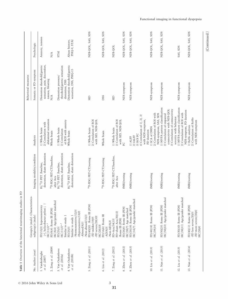
We retrieved the first author’s name, year of publication,characteristics and number of participants studied, subgroups ofFD patients, imaging modality and conditions, analysis methods,behavioral outcomes (Table 1), and results of the brain imagingdata (Tables 2 and S2). Results of behavioral and clinical out-comes are summarized in the text.
RESULTS
Study selection and description
Our research strategy retrieved a total of 314 articles,
104 of which were duplicates. These were discarded
together with a further 194 after screening the title and
abstract. Sixteen articles met the inclusion criteria and
were incorporated in the systematic review (Fig. 1).
All articles19–34 were published between 2007 and
October 2015 (Table 1). We distinguished two research
© 2016 John Wiley & Sons Ltd2
I.-S. Lee et al. Neurogastroenterology and Motility
30
imlee1i1
Rectangle
Tab
le1
Overview
ofthefunctional
neu
roim
agingstudiesin
FD
No.Author(year)
Groups/n(m
ale):Characteristics-
subgroup/n
(male)
Imagingmodality/condition
Analysis
Beh
avioralmeasu
res
Somatic
orFD
symptom
Psych
ologic
1.Van
den
bergh
e
etal.(2007)
FD/13(3):hypersensitivity
(+),helicobacterpylori
(�)
HC/11(5)*
,†
H215O
PET
/baseline,
distention,sh
amdistention
1)Whole
brain
2)Correlationwith
abdominal
sensation
Distentionthresh
old/gastric
sensation,pain,disco
mfort,
nau
sea,
bloating
Anxiety,tension
2.Zen
getal.(2009)
FD/8(4):RomeIII(PDS)
HC/8(4):Age/gen
der
match
ed
18F-FDG
PET-C
T/baseline,
afterAcu
Whole
brain
N/A
N/A
3.Van
Ouden
hove
etal.(2010a)
FD/25(5)
HC/11(5)*
,†
Sim
ilar
tostudy1
H215O
PET
/baseline,
distention,sh
amdistention
1)Whole
brain
2)Correlation
ofROIwithan
xiety
Distentionpressure/
thresh
old/gastric
sensation
distention,DSS
STAI
4.Van
Ouden
hove
etal.(2010b)
FD/25(5)
Sim
ilar
tostudy1
-Norm
osensitive/12(5)
-Hypersensitive/13(0)
-Abused/8(1)
-Non-abused/13(3)
H215O
PET
/baseline,
distention,sh
amdistention
Whole
brain
Distentionthresh
old/gastric
sensation,DSS,PHQ-15
Abuse
history,
PHQ-9,STAI
5.Zen
getal.(2011)
FD/40(20):RomeIII(PDS)
-FD
milder/19(9)
-FD
severe/20(10)
HC/20(10)
18F-FDG
PET-C
T/resting
1)Whole
brain
2)CorrelationofROI
withSID
,NDI-QOL
SID
NDI-QOL,SAS,SDS
6.Liu
etal.(2012)
FD/16(6):RomeIII
-AD/8(3)
-Non-A
D/8(3)
18F-FDG
PET-C
T/resting
Whole
brain
DSS
NDI-QOL,SAS,SDS
7.Zen
getal.(2012)
FD+Acu
/34(13)
FD+sh
amAcu
/30(12)
:RomeIII(PDS)
18F-FDG
PET-C
T/baseline,
afterAcu
1)Whole
brain
2)CorrelationofROI
withSID
,NDI-QOL
SID
NDI-QOL
8.Zhouetal.(2012)
FD/26(8):RomeIII(PDS)
HC/20(7):Age
match
ed
fMRI/resting
FC
NDI-symptom
NDI-QOL,SAS,SDS
9.Zhouetal.(2013)
FD/29(19):RomeIII(PDS)
HC/16(7):Age/gen
der
match
ed
fMRI/resting
1)ALFF
2)fA
LFF
3)ROIFC
4)Correlationof1),2),3)
withNDI-symptom,
FD
duration
NDI-symptom
NDI-QOL,SAS,SDS
10.Liu
etal.(2013)
FD/49(18):RomeIII(PDS)
HC/39(14)
fMRI/resting
1)IC
AofDMN
2)CorrelationofROIwith
NDI-symptom,SAS,SDS
NDI-symptom
NDI-QOL,SAS,SDS
11.Nan
etal.(2013)
FD/50(25):RomeIII(PDS)
HC/50(23):Age/gen
der
match
ed
fMRI/resting
1)MVPA
pattern
classification
2)Correlationofim
paired
connectivitywithNDI-QOL
3)Correlationofco
nnectivity
severitywithbeh
avior
NDI-symptom
NDI-QOL,SAS,SDS
12.Liu
etal.(2013)
FD/30(10):RomeIII(PDS)
HC/30(11):Age/gen
der
match
ed
fMRI/resting
1)MVPA
pattern
classification
2)CorrelationofROIwith
NDI-symptom,FD
duration
NDI-symptom
SAS,SDS
13.Nan
etal.(2014)
FD/40(11):RomeIII(PDS)
-FD
less
severe/20(5)
-FD
more
severe/20(6)
HC/20(8)
fMRI/resting
1)ReH
oan
alysis
2)CorrelationofReH
o
withNDI-symptom
NDI-symptom
NDI-QOL,SAS,SDS
(Continued
.)
© 2016 John Wiley & Sons Ltd 3
Functional imaging in functional dyspepsia
31
imlee1i1
Rectangle

groups (Group 1: Studies 1, 3, 4; Group 2: Studies 2, 5–16), on the basis of authors and affiliations. Group 1
focused on the central processing of visceral stimuli (by
distention of a gastric balloon) in FD patients using
PET in comparison to healthy controls (HC). The
influence of moderating variables (anxiety, gastric
sensitivity, and abuse history) on brain activity in FD
subgroups (normosensitive/hypersensitive and abused/
non-abused) was also investigated. Group 2 reported
resting state activity (n = 10) and brain activity fol-
lowing acupuncture (n = 3) with fMRI. Group 2 applied
several analysis methods for resting state activity,
including whole brain, region of interest, correlation
analysis with behavioral outcomes, functional connec-
tivity, (functional) amplitude of low-frequency fluctu-
ations ((f)ALFF), independent component analysis
(ICA), multivariate pattern analysis (MVPA), regional
homogeneity (ReHo), and topological brain network
analysis. They also measured the resting state brain
response before and after the acupuncture treatment,
and during the acupuncture stimulation.
Participants
A total of 504 FD patients (460 of whom participated in
the neuroimaging scan, 181 males) and 294 HC (120
males) were investigated. Twelve studies included FD
patients between 20 and 30 years of age only, and the
mean age of patients in the other four studies (in which
the inclusion criteria for the age was not stated) ranged
from 22.5 to 35.1 years. The mean duration of FD
symptoms or diagnosis ranged from 15.25 to
82.78 months. Thirteen studies (all by Group 2)
included FD patients who met the Rome III diagnostic
criteria for functional gastrointestinal disorders,2 and
10 of these studies contained postprandial distress
syndrome patients only (one of the subgroups of FD
patients in accordance with the Rome III criteria).
Five studies divided FD patients into subgroups
(Studies 4–6, 13, 15). Among the three gastric disten-
tion studies, Study 1 included FD patients with
visceral hypersensitivity, and Study 4 divided FD
patients into normo- and hypersensitive or abused
and non-abused groups. To identify the symptom-
related functional brain activity, patients were divided
into milder (or less severe) and severe (or more severe)
groups in Study 5 and 13. In Studies 6 and 15, patients
were divided by the score of anxiety and depression
(AD). In Study 7, FD patients were randomly assigned
into two groups for acupuncture and sham acupunc-
ture treatment.
With the exception of Studies 1, 3, 4, 6, and 7,
healthy volunteers were used in the other studies asTab
le1
(continued)
No.Author(year)
Groups/n(m
ale):Characteristics-
subgroup/n
(male)
Imagingmodality/condition
Analysis
Beh
avioralmeasu
res
Somatic
orFD
symptom
Psych
ologic
3)Seed-based
FC
4)Pattern
classification
14.Lietal.(2014)
FD/24(8):RomeIII(PDS)
HC/24(9)
fMRI/Acu
Whole
brain
N/A
N/A
15.Nan
etal.(2015a)
FD/40(8):RomeIII
-AD/18(3)
-non-A
D/22(5)
HC/20(6)
18F-FDG
PET-C
T/resting
1)Whole
brain
2)CorrelationwithSAS,SDS
Dyspep
siasymptom
NDI-QOL,SAS,SDS
16.Nan
etal.(2015b)
FD/25(6):RomeIII
HC/25(11):dem
ograp
hic
inform
ationmatch
ed
fMRI/resting
1)Smallworldproperties
2)Network
efficien
cy
3)Nodal
metrics
DSS
SAS,SDS
*Datafrom
another
study,†Overlappingsample.Acu
,acupuncture;AD,an
xiety
anddep
ression;(f)A
LFF,(functional)am
plitudeoflow-frequen
cyfluctuations;
CT,co
mputedtomograp
hy;DMN,
defau
ltmodenetwork;DSS,dyspep
siasymptom
score;FC,functional
connectivity;FD,functional
dyspep
siapatients;FDG,fluorodeo
xygluco
se;fM
RI,functional
magnetic
resonan
ceim
aging;
HC,healthy
controls;IC
A,indep
enden
tco
mponen
tan
alysis;
MVPA,multivariate
pattern
analysis;
n=
number;N/A
,no
answ
er;NDI,
nep
ean
dyspep
siaindex
;No.,
study
number;PDS,
postprandialdistresssyndrome;
PET,positronem
issiontomograp
hy;PHQ,patienthealthquestionnaire;QOL,qualityoflife;ReH
o,regional
homogeneity;ROI,regionofinterest;SAS,Zungself-
ratingan
xiety
scale;
SDS,Zungself-ratingdep
ressionscale;
SID
,symptom
index
ofdyspep
sia;
STAI,state-traitan
xiety
inven
tory;VAS,visual
analogu
escale.
© 2016 John Wiley & Sons Ltd4
I.-S. Lee et al. Neurogastroenterology and Motility
32
imlee1i1
Rectangle
the control group for FD patients. In Study 1 and 3, the
demographic, behavioral, and brain data of FD patients
were compared with the HC of a previous study.35 In
Studies 4, 6, and 7, the data of FD subgroups without
HC group were compared.
Imaging modality, analysis, and conditions
Functional magnetic resonance imaging is the most
frequently applied brain recording technology (n = 8).
This is followed by PET-CT (n = 5) and PET imaging
(n = 3). PET and PET-CT studies conducted whole
brain analysis and correlation analysis with behavioral
data. Functional magnetic resonance imaging studies
performed analyses of the whole brain, functional
connectivity, (f)ALFF, ICA, MVPA, ReHo, and topo-
logical brain network analysis.
Behavioral and clinical outcomes
Fourteen studies reported behavioral and clinical out-
comes, while two acupuncture studies (Studies 2, 14)
reported brain imaging data only. The behavioral
outcomes were classified into three categories: somatic
symptom, FD symptom, and psychological outcomes.
Somatic symptom outcomes Somatic symptom out-
comesweremeasured in three distention studies (Studies
1, 3, 4) as balloon distention threshold (pain or unpleas-
antness), gastric sensation, or on a visual analog scale for
pain, discomfort, nausea, and bloating during distention.
Gastric sensation during baseline, distention, and sham
distention were higher in FD patients than in HC in one
study, with lower distention pressure (Study 3). Gastric
sensation was higher in the hypersensitive and the
abused group than in the normosensitive and the non-
abused group, respectively (Study 4). Distention pressure
thresholdwasalso lower in thehypersensitive than in the
normosensitive group, but did not differ between the
abused and non-abused groups.
FD symptom outcomes Functional dyspepsia symp-
toms were measured in twelve studies. The Nepean
dyspepsia index (NDI) was reported in six studies (Study
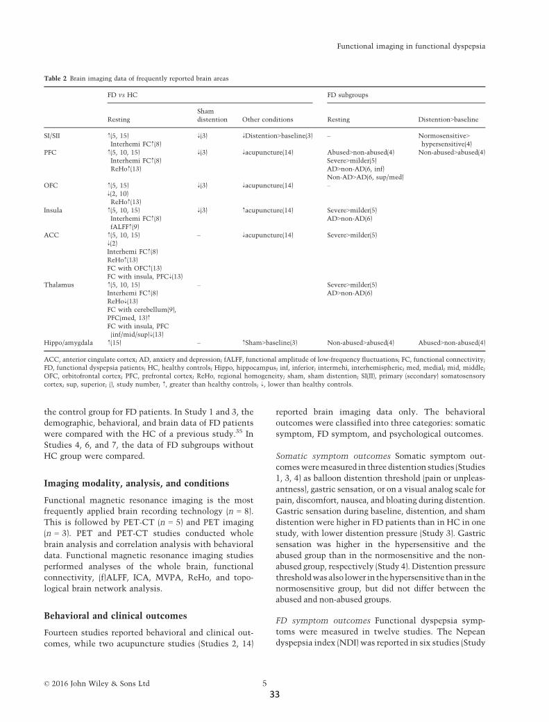
Table 2 Brain imaging data of frequently reported brain areas
FD vs HC FD subgroups
Resting
Sham
distention Other conditions Resting Distention>baseline
SI/SII ↑(5, 15)Interhemi FC↑(8)
↓(3) ↓Distention>baseline(3) – Normosensitive>hypersensitive(4)
PFC ↑(5, 10, 15)Interhemi FC↑(8)ReHo↑(13)
↓(3) ↓acupuncture(14) Abused>non-abused(4)Severe>milder(5)
AD>non-AD(6, inf)
Non-AD>AD(6, sup/med)
Non-abused>abused(4)
OFC ↑(5, 15)↓(2, 10)ReHo↑(13)
↓(3) ↓acupuncture(14) –
Insula ↑(5, 10, 15)Interhemi FC↑(8)fALFF↑(9)
↓(3) ↑acupuncture(14) Severe>milder(5)
AD>non-AD(6)
ACC ↑(5, 10, 15)↓(2)Interhemi FC↑(8)ReHo↑(13)FC with OFC↑(13)FC with insula, PFC↓(13)
– ↓acupuncture(14) Severe>milder(5)
Thalamus ↑(5, 10, 15)Interhemi FC↑(8)ReHo↓(13)FC with cerebellum(9),
PFC(med, 13)↑FC with insula, PFC
(inf/mid/sup)↓(13)
– Severe>milder(5)
AD>non-AD(6)
Hippo/amygdala ↑(15) – ↑Sham>baseline(3) Non-abused>abused(4) Abused>non-abused(4)
ACC, anterior cingulate cortex; AD, anxiety and depression; fALFF, functional amplitude of low-frequency fluctuations; FC, functional connectivity;
FD, functional dyspepsia patients; HC, healthy controls; Hippo, hippocampus; inf, inferior; intermehi, interhemispheric; med, medial; mid, middle;
OFC, orbitofrontal cortex; PFC, prefrontal cortex; ReHo, regional homogeneity; sham, sham distention; SI(II), primary (secondary) somatosensory
cortex; sup, superior; (), study number; ↑, greater than healthy controls; ↓, lower than healthy controls.
© 2016 John Wiley & Sons Ltd 5
Functional imaging in functional dyspepsia
33
imlee1i1
Rectangle
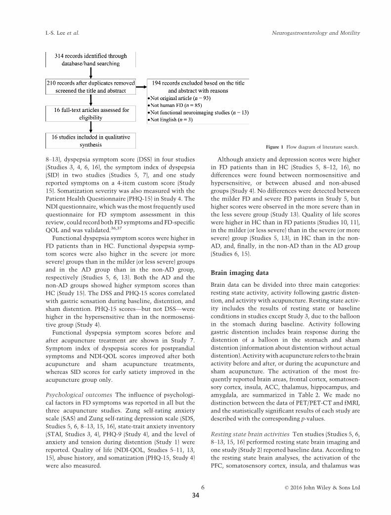
8–13), dyspepsia symptom score (DSS) in four studies
(Studies 3, 4, 6, 16), the symptom index of dyspepsia
(SID) in two studies (Studies 5, 7), and one study
reported symptoms on a 4-item custom score (Study
15). Somatization severity was also measured with the
Patient Health Questionnaire (PHQ-15) in Study 4. The
NDI questionnaire, whichwas themost frequently used
questionnaire for FD symptom assessment in this
review, could record both FD symptoms and FD-specific
QOL and was validated.36,37
Functional dyspepsia symptom scores were higher in
FD patients than in HC. Functional dyspepsia symp-
tom scores were also higher in the severe (or more
severe) groups than in the milder (or less severe) groups
and in the AD group than in the non-AD group,
respectively (Studies 5, 6, 13). Both the AD and the
non-AD groups showed higher symptom scores than
HC (Study 15). The DSS and PHQ-15 scores correlated
with gastric sensation during baseline, distention, and
sham distention. PHQ-15 scores—but not DSS—were
higher in the hypersensitive than in the normosensi-
tive group (Study 4).
Functional dyspepsia symptom scores before and
after acupuncture treatment are shown in Study 7.
Symptom index of dyspepsia scores for postprandial
symptoms and NDI-QOL scores improved after both
acupuncture and sham acupuncture treatments,
whereas SID scores for early satiety improved in the
acupuncture group only.
Psychological outcomes The influence of psychologi-
cal factors in FD symptoms was reported in all but the
three acupuncture studies. Zung self-rating anxiety
scale (SAS) and Zung self-rating depression scale (SDS,
Studies 5, 6, 8–13, 15, 16), state-trait anxiety inventory
(STAI, Studies 3, 4), PHQ-9 (Study 4), and the level of
anxiety and tension during distention (Study 1) were
reported. Quality of life (NDI-QOL, Studies 5–11, 13,15), abuse history, and somatization (PHQ-15, Study 4)
were also measured.
Although anxiety and depression scores were higher
in FD patients than in HC (Studies 5, 8–12, 16), no
differences were found between normosensitive and
hypersensitive, or between abused and non-abused
groups (Study 4). No differences were detected between
the milder FD and severe FD patients in Study 5, but
higher scores were observed in the more severe than in
the less severe group (Study 13). Quality of life scores
were higher in HC than in FD patients (Studies 10, 11),
in the milder (or less severe) than in the severe (or more
severe) group (Studies 5, 13), in HC than in the non-
AD, and, finally, in the non-AD than in the AD group
(Studies 6, 15).
Brain imaging data
Brain data can be divided into three main categories:
resting state activity, activity following gastric disten-
tion, and activity with acupuncture. Resting state activ-
ity includes the results of resting state or baseline
conditions in studies except Study 3, due to the balloon
in the stomach during baseline. Activity following
gastric distention includes brain response during the
distention of a balloon in the stomach and sham
distention (information about distention without actual
distention).Activitywith acupuncture refers to thebrain
activity before and after, or during the acupuncture and
sham acupuncture. The activation of the most fre-
quently reported brain areas, frontal cortex, somatosen-
sory cortex, insula, ACC, thalamus, hippocampus, and
amygdala, are summarized in Table 2. We made no
distinction between the data of PET/PET-CT and fMRI,
and the statistically significant results of each study are
described with the corresponding p-values.
Resting state brain activities Ten studies (Studies 5, 6,
8–13, 15, 16) performed resting state brain imaging and
one study (Study 2) reported baseline data. According to
the resting state brain analyses, the activation of the
PFC, somatosensory cortex, insula, and thalamus was
Figure 1 Flow diagram of literature search.
© 2016 John Wiley & Sons Ltd6
I.-S. Lee et al. Neurogastroenterology and Motility
34
imlee1i1
Rectangle
consistently greater in FD patients than in HC, while
brain activities of the orbitofrontal cortex (OFC) and
the ACC are inconsistent. The severe FD group showed
higher activity in ACC, midcingulate cortex (MCC),
insula, thalamus, and cerebellum than the milder FD
group (Study 5). The AD group showed higher activity
in SI, insula, thalamus, and parahippocampal gyrus,
and lower activity in frontal cortices and MCC than
the non-AD group (Study 6).
Functional dyspepsia patients showed higher inter-
hemispheric connectivity (synchronized activity
between the same brain areas in opposite hemispheres)
of ACC, insula, thalamus, and cerebellum than HC
(Study 8). Pattern classification analyses were also
applied to distinguish FD patients from HC. Classifica-
tion accuracy was sufficiently high, and discriminative
regions were the medial PFC, OFC, ACC, MCC, insula,
thalamus, hippocampus, and cerebellum based on
MVPA pattern classification. Anterior cingulate cortex
and thalamus distinguished FD from HC, or less severe
from more severe FD, respectively (Studies 11–13). In a
recent study (Study 16), a new approach to topological
changes of the brain network revealed a higher cluster-
ing coefficient and local efficiency in FDpatients than in
HC. Furthermore, nodal efficiency in the ACC was
found to be positively correlated with dyspeptic symp-
tom and duration.
Seven studies performed correlation analyses
between resting state brain activity and behavioral
measures (Studies 5, 9–13, 15). A positive correlation
between ACC activity and symptom score was
observed in four studies (Studies 5, 10, 12, 13). Anxiety
scores positive correlated with ACC, MCC, and insula
in Study 15 only. Depression score and FD duration
correlated poorly with brain activity in five studies
(Studies 9–12 and 15).
Distention-related brain activities The three disten-
tion studies were conducted by Group 1 and therefore
had similar balloon distention procedures (Studies 1, 3,
4). The ventral PFC, OFC, SI, and temporal lobe were
commonly activated during the balloon distention.
Significant correlations of upper abdominal sensations
with these areas were reported (Study 1). Study 3
reported a deactivation during distention in dorsal PFC,
medial OFC, ACC, hippocampus, amygdala, and sev-
eral regions in the parietal, temporal, and occipital
lobes in FD patients.
Group comparison of [distention>baseline] conditionrevealed that activity in the mid brain, cerebellum, and
dorsal pons was greater, and activity in SI and SII was
lower in FD patients than in HC (Study 3). The
normosensitive group showed greater activation in
SII, MCC, and precuneus than the hypersensitive
group. Functional dyspepsia patients with an abuse
history showed greater activation of the hippocampus,
parahippocampal gyrus, and amygdala, and lower acti-
vation of dorsal PFC, insula, caudate, and cerebellum
than the non-abused group (Study 4).
Sham distention did not elicit any brain (de)activa-
tion in FD patients (sham distention vs baseline), but
in comparison with HC, FD patients showed higher
hippocampus and amygdala activity under [sham dis-
tention>baseline] condition (Study 3).
Acupuncture-related brain activities The initial
acupuncture study (Study 2) compared the resting state
of FD patients before and after the acupuncture
treatment. The second study (Study 7) compared the
influence of acupuncture and sham acupuncture, while
the third study (Study 14) compared the brain response
during acupuncture stimulation in FD patients and
HC.
After five sessions of manual acupuncture, brain
activity in FD patients increased in PFC and pre-
cuneus, but decreased in SI, pons, and cerebellar tonsil
(Study 2). After 20 sessions of electro-acupuncture,
brain activity in FD patients increased in SI and
precuneus, but decreased in anterior/mid/posterior
cingulate cortex, insula, thalamus, putamen,
hippocampus, and cerebellum (Study 7). The inconsis-
tent results from these two studies might be due to the
different characteristics of FD patients, stimulation
type (manual or electro-), number of sessions, or
comparatively small sample size. Correlation analysis
showed that changes of ACC, insula, thalamus,
hypothalamus are positively correlated with changes
of symptom score, and negatively correlated with
changes of QOL score in the acupuncture group. In
the sham acupuncture group, changes of QOL score
were negatively correlated with fewer areas than the
acupuncture group that included thalamus and brain-
stem (Study 7).
During manual acupuncture stimulation at the
acupoint ST36, FD patients showed greater brain
activity in SI and insula, and lower activity in PFC,
OFC, and ACC than HC (Study 14).
DISCUSSION
Sixteen articles were taken into consideration in this
review and functional brain activity (resting state,
visceral distention, acupuncture conditions) and
behavioral/clinical outcomes were measured. The
abnormal brain activity was frequently found in SI,
PFC, insula, ACC, thalamus, hippocampus, and amyg-
© 2016 John Wiley & Sons Ltd 7
Functional imaging in functional dyspepsia
35
imlee1i1
Rectangle

dala. When compared to HC, FD patients showed
greater activation in PFC, insula, cingulate cortex, and
thalamus during the resting state, and altered activa-
tion in the somatosensory cortex, OFC, hippocampus,
and amygdala during distention and acupuncture-
related conditions. According to the pattern classifica-
tion analysis, FD patients and HC could be distin-
guished using the activity pattern of ACC and
thalamus. The behavioral data showed that FD
patients experienced visceral hypersensitivity (low
balloon distention threshold with higher gastric sen-
sation/pain) during the balloon distention. Further-
more, the anxiety and depression scores were higher in
FD patients than in HC, and QOL scores varied on the
FD symptom severity, anxiety, and depression scores.
Visceral sensations are involved in the ascending
visceral pain pathway and processed in the somatosen-
sory cortex.6,38,39 The somatosensory cortex activity
during both the resting state and visceral distention
was higher in FD patients than in HC. Moreover, brain
activity during acupuncture (external somatic stimu-
lation) also showed a greater increase in SI activity in
FD patients than in HC, implying that the hypersen-
sitive to somatic stimulation like IBS patients.40 The
increased activity of somatosensory cortex—even in
the absence of visceral stimulation—could support the
hypothesis of cortical sensitization in FD patients.41,42
Central sensitization, increased brain response to
various stimuli, may be one of the underlying patho-
physiologic features in fibromyalgia, migraine, IBS, and
FD patients.42 Since FD is a chronic disease,
somatosensory cortex receives afferent ascending vis-
ceral pain signals from internal organs repeatedly and
consequent sensitization of the brain could result in an
abnormal central modulation of sensory information
and peripheral abnormalities such as visceral hyper-
sensitivity. However, FD symptoms during resting
state measurement, which could affect the brain
activity, were not reported in any studies. The sensi-
tization hypothesis therefore still requires further
confirmation. Unlike the resting state activities, HC
showed a higher somatosensory cortex activation than
FD patients in [distention>baseline] and [sham disten-
tion>baseline] conditions. This could be due to the
increased activity in resting state (ceiling effect),
attenuated increase from chronic visceral sensation
in FD patients, or different visceral distention pressure
between groups (lower in FD patients). Although
visceral distention pressure was lower in FD patients
than in HC, this is not enough to explain the increase
in somatosensory cortex activation in HC in [sham
distention>baseline] condition. This is worth bearing
in mind as evidence of abnormal sensory processing in
FD patients. Further research on the sensitization or
attenuation of brain activity during resting or internal/
external stimuli is required to gain an understanding of
sensory processing in FD patients.
The frontal cortex is associated with executive and
integrative control functions. The integration of infor-
mation from peripheral, cognitive modulation of pain
(medial, dorsolateral), and appraise or response to
affective aspect of pain sensation (medial, ventrolat-
eral) are processed in the PFC.5,43,44 Orbitofrontal
cortex is also involved in cognitive pain modulation,
inhibition of pain-related emotional response, sensory
discrimination, and monitoring,45–47 and is closely
related to psychological disorders such as anxiety48
and depression.49 It is also associated with the
endogenous opioid analgesia systems in conjunction
with ACC.15 The activation pattern of PFC is similar
with somatosensory cortex during resting (FD>HC)
and sham distention conditions (HC>FD), whereas
PFC activity did not differ from HC during distention
despite low distention pressure in FD patients. One
may speculate that this is due to an overlapping
influence of chronic ascending sensory processing,
cognitive and descending pain modulation, attention,
and anticipation for visceral sensation on the frontal
cortex activity in FD patients. However, in contrast to
the previous studies which showed the close connec-
tion between OFC and anxiety or depression,48,49 the
relation between psychological factors and OFC activ-
ity was not observed in the current review.
Insula, ACC, and thalamus were already in the focus
of early functional gastrointestinal disease studies. The
insula is involved in interoceptive processing, homeo-
static function, emotion, affective state, and aware-
ness.10,50 In our review, the insula is activated during
visceral distention in FD patients ([distention>base-line]), where it showed greater activation than in HC
during baseline and sham distention in all but one
study. This implies that the abnormal excitement of
the insula could be derived by the residual influence of
chronic visceral sensation, psychological state, home-
ostatic imbalance (supported by greater brain activa-
tion in FD than in HC in the baseline condition), and
anticipation of distention (supported by comparison in
the sham distention condition) in FD patients. ACC,
one of the core regions of medial pain system, is
particularly important for cognitive pain modulation,
attention to pain, endogenous opioid system-related
placebo analgesia, and regulating the affective compo-
nent of pain experience.13–15 Various analyses, includ-
ing whole brain, interhemispheric functional
connectivity, topological brain network, ReHo, classi-
fication, and correlation analysis reported the abnor-
© 2016 John Wiley & Sons Ltd8
I.-S. Lee et al. Neurogastroenterology and Motility
36
imlee1i1
Rectangle
mal functional activity of ACC in FD patients. It is
feasible that the altered activity of ACC causes
diminished central pain modulation and subsequent
hypersensitivity in FD patients. Although various
functions and distinct neuroanatomical regions within
the insula and the ACC are not covered in this review,
it is still important to note that the insula is not a
homogeneous region,51 and neither is the ACC.52
The role of the limbic area (hippocampus, amygdala,
hypothalamus, parahippocampal gyrus, etc.) has been
discussed in termsof pain sensitivity, stress, andanxiety
in IBS patients.53,54 In particular, the studies evaluated
in this review reported a coactivation of hippocampus
and amygdala in FD patients. Amygdala is associated
with the emotional memories with hippocampus, emo-
tional evaluation of sensory stimuli, recognition of
emotion, and nociceptive pathway.55–58 In FD patients,
the activity of amygdala and hippocampus could be
interpreted as the anticipationor response to thevisceral
stimuli, and the recall of previous negative memories
(pain, unpleasant, anxiety, etc.). Furthermore, the vol-
ume and synaptic changes of hippocampus and amyg-
dala by chronic pain59,60 could also affect the functional
activity in FD patients. Greater activity is recorded in
these regions in FD patients than in HC during both
sham distention and [distention>baseline] condition
and in abused than in non-abused group, supporting
the hypothesis that negative emotional memory influ-
ences brain activity in FD patients.
On the basis of our results, it can be assumed that
the changes of brain activity, response to visceral
stimulation, and cognitive state of FD patients are due
to the repeated afferent signal from the periphery and
failure of central pain modulation leading to the
dysfunction of the pain (SI, SII, ACC, insula, thalamus,
amygdala) and salience network (ACC and insula). The
evaluation, integration, and response to salient stimuli
were altered in chronic pain and IBS patients.11,61–63
Moreover, a saliency of stimuli varies according to the
emotional state and pathological condition. We can
therefore also assume that the visceral sensation,
dyspeptic symptoms, emotion, and cognitive process-
ing of dyspeptic symptoms have different saliency in
FD patients than in HC. In summary, we propose that
FD, like IBS, can also be considered as the functional
chronic pain syndrome in which pain and salience
processing are impaired64,65 and that the ACC and
insula play critical roles in FD.66 Constant sensory
signal from the gut (bottom-up) and abnormal central
modulation (top-down) of pain and gut functions might
be key features of FD, showing that peripheral changes
could originate from abnormal brain activities through
the brain-gut axis (Fig. 2).
By summarizing previous neuroimaging studies, we
also suggest further research of FD. Since only two
research groups have performed functional neuroimag-
ing studies in FD patients, this review could poten-
tially be biased. In addition to the limited number of
research groups, there is still a lack of appropriate
tasks. In early studies, visceral distention was applied
to patients and healthy participants to investigate the
visceral sensitivity-related activation of the brain.
However, balloon distention is invasive and further
peripheral changes are also related to the FD. Differ-
ent kinds of tests such as water load test or real food
intake are therefore required. Although FD patients
have increased dyspeptic symptoms after eating
food,67 neuroimaging studies during or after food
ingestion are relatively rare compared to those on
obesity or IBS patients. New paradigms to overcome
the practical problems involved (e.g. discomfort and
dyspeptic symptoms of patients during food ingestion,
amount and type of food) could augment our under-
standing of FD. Furthermore, the only intervention in
which neuronal mechanism in FD has been investi-
gated is acupuncture, and careful interpretation is
necessary due to the poor reliability and validity of
sham acupuncture.68 Although acid-suppressive
drugs, prokinetic agents, antidepressants, and psy-
chotherapy are prescribed for FD patients, various
phenotypes of patients and unknown underlying
mechanisms often disrupt the standardized treatment
strategy. Neuronal mechanism studies could therefore
be helpful. Moreover, the improved and unified
methods of measuring the psychological factors in
FD, such as more specific definition (e.g. trait or state
anxiety, anxiety for symptom or experimental envi-
ronment, anxiety of present or previous week) and
well-structured interviews rather than self-rating
questionnaires are also important.69 It is also worth
investigating further psychological, behavioral, and
lifestyle factors, including somatization,42,70
stress,71,72 fatigue,73 food behavior,74 sleep behavior,
and comorbidities (IBS, other functional pain syn-
drome diseases, anxiety, depression). Finally, a more
representative sample of FD patients should be
included in further studies. In this review, although
diagnoses of FD usually depend on Rome III criteria, a
number of studies did not describe the diagnostic
procedure. Although the peak prevalence of FD is
distributed around the middle age,3 many studies
included only patients in their twenties. Representa-
tive and homogenous sample recruitment, where age,
symptom severity, comorbidities, and gender are
taken into consideration, could improve the reliability
of the research.75
© 2016 John Wiley & Sons Ltd 9
Functional imaging in functional dyspepsia
37
imlee1i1
Rectangle
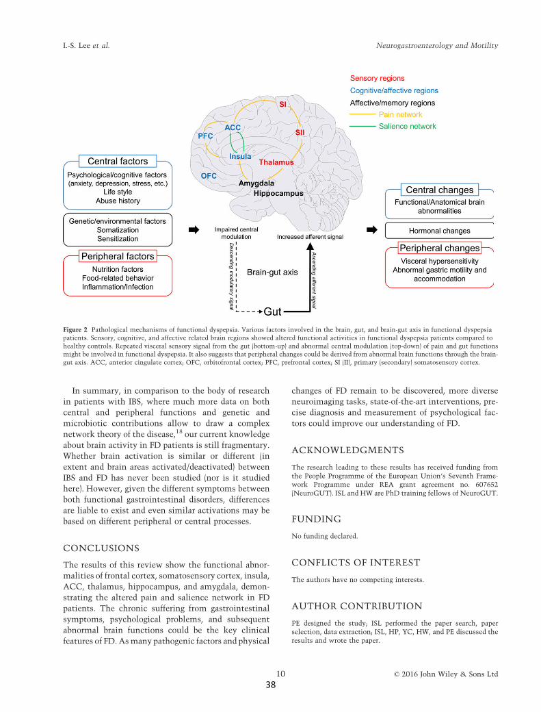
In summary, in comparison to the body of research
in patients with IBS, where much more data on both
central and peripheral functions and genetic and
microbiotic contributions allow to draw a complex
network theory of the disease,18 our current knowledge
about brain activity in FD patients is still fragmentary.
Whether brain activation is similar or different (in
extent and brain areas activated/deactivated) between
IBS and FD has never been studied (nor is it studied
here). However, given the different symptoms between
both functional gastrointestinal disorders, differences
are liable to exist and even similar activations may be
based on different peripheral or central processes.
CONCLUSIONS
The results of this review show the functional abnor-
malities of frontal cortex, somatosensory cortex, insula,
ACC, thalamus, hippocampus, and amygdala, demon-
strating the altered pain and salience network in FD
patients. The chronic suffering from gastrointestinal
symptoms, psychological problems, and subsequent
abnormal brain functions could be the key clinical
features of FD. Asmany pathogenic factors and physical
changes of FD remain to be discovered, more diverse
neuroimaging tasks, state-of-the-art interventions, pre-
cise diagnosis and measurement of psychological fac-
tors could improve our understanding of FD.
ACKNOWLEDGMENTS
The research leading to these results has received funding fromthe People Programme of the European Union’s Seventh Frame-work Programme under REA grant agreement no. 607652(NeuroGUT). ISL and HW are PhD training fellows of NeuroGUT.
FUNDING
No funding declared.
CONFLICTS OF INTEREST
The authors have no competing interests.
AUTHOR CONTRIBUTION
PE designed the study; ISL performed the paper search, paperselection, data extraction; ISL, HP, YC, HW, and PE discussed theresults and wrote the paper.
Figure 2 Pathological mechanisms of functional dyspepsia. Various factors involved in the brain, gut, and brain-gut axis in functional dyspepsia
patients. Sensory, cognitive, and affective related brain regions showed altered functional activities in functional dyspepsia patients compared to
healthy controls. Repeated visceral sensory signal from the gut (bottom-up) and abnormal central modulation (top-down) of pain and gut functions
might be involved in functional dyspepsia. It also suggests that peripheral changes could be derived from abnormal brain functions through the brain-
gut axis. ACC, anterior cingulate cortex; OFC, orbitofrontal cortex; PFC, prefrontal cortex; SI (II), primary (secondary) somatosensory cortex.
© 2016 John Wiley & Sons Ltd10
I.-S. Lee et al. Neurogastroenterology and Motility
38
imlee1i1
Rectangle
REFERENCES
1 Tack J, Talley NJ, Camilleri M, Holt-mann G, Hu P, Malagelada JR, Stan-ghellini V. Functional gastroduodenaldisorders. Gastroenterology 2006;130: 1466–79.
2 Drossman DA, Dumitrascu DL.Rome III: new standard for functionalgastrointestinal disorders. J Gastroin-
testin Liver Dis 2006; 15: 237–41.3 Mahadeva S, Goh KL. Epidemiology
of functional dyspepsia: a global per-spective. World J Gastroenterol 2006;12: 2661–6.
4 Mearin F, Cucala M, Azpiroz F,Malagelada JR. The origin of symp-toms on the brain-gut axis in func-tional dyspepsia. Gastroenterology
1991; 101: 999–1006.5 Mayer EA, Naliboff BD, Craig AD.
Neuroimaging of the brain-gut axis:from basic understanding to treat-ment of functional GI disorders. Gas-
troenterology 2006; 131: 1925–42.6 Aziz Q, Schnitzler A, Enck P. Func-
tional neuroimaging of visceral sen-sation. J Clin Neurophysiol 2000; 17:604–12.
7 Moisset X, Bouhassira D, Denis D,Dominique G, Benoit C, Sabate JM.Anatomical connections betweenbrain areas activated during rectaldistension in healthy volunteers: avisceral pain network. Eur J Pain
2010; 14: 142–8.8 McMahon SB. Are there fundamental
differences in the peripheral mecha-nisms of visceral and somatic pain?Behav Brain Sci 1997; 20: 381–91;discussion 435-513.
9 Dunckley P, Wise RG, Aziz Q, Pain-ter D, Brooks J, Tracey I, Chang L.Cortical processing of visceral andsomatic stimulation: differentiatingpain intensity from unpleasantness.Neuroscience 2005; 133: 533–42.
10 Craig AD. How do you feel? Intero-ception: the sense of the physiologicalcondition of the body. Nat Rev Neu-
rosci 2002; 3: 655–66.11 Seeley WW, Menon V, Schatzberg AF,
Keller J, Glover GH, Kenna H, ReissAL,GreiciusMD.Dissociable intrinsicconnectivity networks for salience pro-cessing and executive control. J Neu-rosci 2007; 27: 2349–56.
12 Menon V, Uddin LQ. Saliency,switching, attention and control: anetwork model of insula function.Brain structure & function 2010; 214:655–67.
13 Zubieta JK, Smith YR, Bueller JA, XuY, Kilbourn MR, Jewett DM, MeyerCR, Koeppe RA et al. Regional muopioid receptor regulation of sensoryand affective dimensions of pain.Science 2001; 293: 311–5.
14 Zubieta JK, Bueller JA, Jackson LR,Scott DJ, Xu Y, Koeppe RA, NicholsTE, Stohler CS. Placebo effects medi-ated by endogenous opioid activity onmu-opioid receptors. J Neurosci 2005;25: 7754–62.
15 Petrovic P, Kalso E, Petersson KM,Ingvar M. Placebo and opioid analge-sia– imaging a shared neuronal net-work. Science 2002; 295: 1737–40.
16 Van Oudenhove L, Coen SJ, Aziz Q.Functional brain imaging of gastroin-testinal sensation in health and dis-ease. World J Gastroenterol 2007; 13:3438–45.
17 Coss-Adame E, Rao SS. Brain and gutinteractions in irritable bowel syn-drome: new paradigms and newunderstandings. Curr Gastroenterol
Rep 2014; 16: 379.18 Mayer EA, Labus JS, Tillisch K, Cole
SW, Baldi P. Towards a systems viewof IBS. Nat Rev Gastroenterol Hepa-
tol 2015; 12: 592–605.19 Vandenberghe J, Dupont P, Van
Oudenhove L, Bormans G, Demytte-naere K, Fischler B, Geeraerts B,Janssens J et al. Regional cerebralblood flow during gastric balloon dis-tention in functional dyspepsia. Gas-troenterology 2007; 132: 1684–93.
20 Zeng F, Song WZ, Liu XG, Xie HJ,Tang Y, Shan BC, Liu ZH, Yu SGet al. Brain areas involved inacupuncture treatment on functionaldyspepsia patients: a PET-CT study.Neurosci Lett 2009; 456: 6–10.
21 Van Oudenhove L, Vandenberghe J,Dupont P, Geeraerts B, Vos R, DirixS, Bormans G, Vanderghinste D et al.
Abnormal regional brain activity dur-ing rest and (anticipated) gastric dis-tension in functional dyspepsia andthe role of anxiety: a H(2)(15)O-PETstudy. Am J Gastroenterol 2010; 105:913–24.
22 Van Oudenhove L, Vandenberghe J,Dupont P, Geeraerts B, Vos R, DirixS, Van Laere K, Bormans G et al.
Regional brain activity in functionaldyspepsia: a H(2)(15)O-PET study onthe role of gastric sensitivity andabuse history. Gastroenterology
2010; 139: 36–47.23 Zeng F, Qin W, Liang F, Liu J, Tang Y,
Liu X, Yuan K, Yu S et al. Abnormal
resting brain activity in patients withfunctional dyspepsia is related tosymptom severity. Gastroenterology
2011; 141: 499–506.24 Liu ML, Liang FR, Zeng F, Tang Y,
Lan L, Song WZ. Cortical-limbicregions modulate depression and anx-iety factors in functional dyspepsia: aPET-CT study. Ann Nucl Med 2012;26: 35–40.
25 Zeng F, Qin W, Ma T, Sun J, Tang Y,Yuan K, Li Y, Liu J et al. Influence ofacupuncture treatment on cerebralactivity in functional dyspepsiapatients and its relationship withefficacy. Am J Gastroenterol 2012;107: 1236–47.
26 Liu P, QinW,Wang J, Zeng F, ZhouG,WenH, vonDeneenKM, Liang F et al.Identifying neural patterns of func-tional dyspepsia using multivariatepattern analysis: a resting-state FMRIstudy. PLoS ONE 2013; 8: e68205.
27 Liu P, Zeng F, Zhou G, Wang J, WenH, von Deneen KM, Qin W, Liang Fet al. Alterations of the default modenetwork in functional dyspepsiapatients: a resting-state fmri study.Neurogastroenterol Motil 2013; 25:e382–8.
28 Nan J, Liu J, Li G, Xiong S, Yan X, YinQ, Zeng F, von Deneen KM et al.Whole-brain functional connectivityidentification of functional dyspepsia.PLoS ONE 2013; 8: e65870.
29 Zhou G, Liu P, Wang J, Wen H, ZhuM, Zhao R, von Deneen KM, Zeng Fet al. Fractional amplitude of low-frequency fluctuation changes infunctional dyspepsia: a resting-statefMRI study. Magn Reson Imaging
2013; 31: 996–1000.30 Zhou G, Liu P, Zeng F, Yuan K, Yu D,
von Deneen KM, Liang F, Qin Wet al. Increased interhemisphericresting-state functional connectivityin functional dyspepsia: a pilot study.NMR Biomed 2013; 26: 410–5.
31 Li Z, Zeng F, Yang Y, Chen Y, ZhangD, Sun J, Qin W, Yang J et al. Differ-ent cerebral responses to puncturingat ST36 among patients with func-tional dyspepsia and healthy subjects.Forsch Komplementmed 2014; 21:99–104.
32 Nan J, Liu J, Zhang D, Yang Y, Yan X,Yin Q, Xiong S, von Deneen KMet al. Altered intrinsic regional activ-ity and corresponding brain pathwaysreflect the symptom severity of func-tional dyspepsia. NeurogastroenterolMotil 2014; 26: 660–9.
© 2016 John Wiley & Sons Ltd 11
Functional imaging in functional dyspepsia
39
imlee1i1
Rectangle
33 Nan J, Liu J, Mu J, Dun W, Zhang M,Gong Q, Qin W, Tian J et al. Brain-based correlations between psycho-logical factors and functional dyspep-sia. J Neurogastroenterol Motil 2015;21: 103–10.
34 Nan J, Zhang L, Zhu F, Tian X, ZhengQ, Deneen KM, Liu J, Zhang M.Topological alterations of the intrin-sic brain network in functional dys-pepsia patients. J Neurogastroenterol
Motil 2016; 22: 118–28.35 Vandenbergh J, Dupont P, Fischler B,
Bormans G, Persoons P, Janssens J,Tack J. Regional brain activationduring proximal stomach distentionin humans: a positron emissiontomography study. Gastroenterology
2005; 128: 564–73.36 Talley NJ, Verlinden M, Jones M.
Validity of a new quality of life scalefor functional dyspepsia: a UnitedStates multicenter trial of the NepeanDyspepsia Index. Am J Gastroenterol
1999; 94: 2390–7.37 Talley NJ, Haque M, Wyeth JW, Stace
NH, Tytgat GN, Stanghellini V, Holt-mann G, Verlinden M et al. Develop-ment of a new dyspepsia impactscale: the Nepean Dyspepsia Index.Aliment Pharmacol Ther 1999; 13:225–35.
38 Jones MP, Dilley JB, Drossman D,Crowell MD. Brain-gut connectionsin functional GI disorders: anatomicand physiologic relationships. Neu-
rogastroenterol Motil 2006; 18: 91–103.
39 Van Oudenhove L, Dupont P, Van-denberghe J, Geeraerts B, van Laere K,Bormans G, Demyttenaere K, Tack J.The role of somatosensory corticalregions in the processing of painfulgastric fundic distension: an update ofbrain imaging findings. Neurogas-troenterol Motil 2008; 20: 479–87.
40 Verne GN, Robinson ME, Price DD.Hypersensitivity to visceral and cuta-neous pain in the irritable bowelsyndrome. Pain 2001; 93: 7–14.
41 Moshiree B, Zhou Q, Price DD, VerneGN. Central sensitisation in visceralpain disorders. Gut 2006; 55: 905–8.
42 Wilhelmsen I. Somatization, sensiti-zation, and functional dyspepsia.Scand J Psychol 2002; 43: 177–80.
43 Lu HC, Hsieh JC, Lu CL, NiddamDM, Wu YT, Yeh TC, Cheng CM,Chang FY et al. Neuronal correlatesin the modulation of placebo analge-sia in experimentally-induced eso-phageal pain: a 3T-fMRI study. Pain2010; 148: 75–83.
44 Wiech K, Ploner M, Tracey I. Neu-rocognitive aspects of pain perception.Trends Cogn Sci 2008; 12: 306–13.
45 Wager TD, Rilling JK, Smith EE,Sokolik A, Casey KL, Davidson RJ,Kosslyn SM, Rose RM et al. Placebo-induced changes in FMRI in theanticipation and experience of pain.Science 2004; 303: 1162–7.
46 Ochsner KN, Ludlow DH, Knierim K,Hanelin J, Ramachandran T, GloverGC, Mackey SC. Neural correlates ofindividual differences in pain-relatedfear and anxiety. Pain 2006; 120: 69–77.
47 Kringelbach ML. The human orbito-frontal cortex: linking reward tohedonic experience. Nat Rev Neu-
rosci 2005; 6: 691–702.48 Milad MR, Rauch SL. The role of the
orbitofrontal cortex in anxiety disor-ders. Ann N Y Acad Sci 2007; 1121:546–61.
49 Drevets WC. Orbitofrontal cortexfunction and structure in depression.AnnNYAcadSci2007;1121: 499–527.
50 Craig AD. How do you feel–now? Theanterior insula and human awareness.Nat Rev Neurosci 2009; 10: 59–70.
51 Nieuwenhuys R. The insular cortex:a review. Prog Brain Res 2012; 195:123–63.
52 Gasquoine PG. Localization of func-tion in anterior cingulate cortex: frompsychosurgery to functional neu-roimaging. Neurosci Biobehav Rev
2013; 37: 340–8.53 Lackner JM, Lou Coad M, Mertz HR,
Wack DS, Katz LA, Krasner SS, FirthR, Mahl TC et al. Cognitive therapyfor irritable bowel syndrome is asso-ciated with reduced limbic activity,GI symptoms, and anxiety. Behav Res
Ther 2006; 44: 621–38.54 Mertz H. Role of the brain and sen-
sory pathways in gastrointestinal sen-sory disorders in humans. Gut 2002;51 (Suppl. 1): i29–33.
55 Phelps EA. Human emotion andmemory: interactions of the amyg-dala and hippocampal complex. Curr
Opin Neurobiol 2004; 14: 198–202.56 Hamann SB, Ely TD, Grafton ST,
Kilts CD. Amygdala activity relatedto enhanced memory for pleasant andaversive stimuli. Nat Neurosci 1999;2: 289–93.
57 Zald DH. The human amygdala andthe emotional evaluation of sensorystimuli. Brain Res Brain Res Rev2003; 41: 88–123.
58 Adolphs R, Tranel D, Damasio H,Damasio A. Impaired recognition of
emotion in facial expressions follow-ing bilateral damage to the humanamygdala. Nature 1994; 372: 669–72.
59 Han JS, Neugebauer V. Synaptic plas-ticity in the amygdala in a visceralpain model in rats. Neurosci Lett
2004; 361: 254–7.60 Mutso AA, Radzicki D, Baliki MN,
Huang L, Banisadr G, Centeno MV,Radulovic J, Martina M et al. Abnor-malities in hippocampal functioningwith persistent pain. J Neurosci 2012;32: 5747–56.
61 Naliboff BD, Berman S, Suyenobu B,Labus JS, Chang L, Stains J, Mandelk-ern MA, Mayer EA. Longitudinalchange in perceptual and brain acti-vation response to visceral stimuli inirritable bowel syndrome patients.Gastroenterology 2006; 131: 352–65.
62 Legrain V, Iannetti GD, Plaghki L,Mouraux A. The pain matrixreloaded: a salience detection systemfor the body. Prog Neurobiol 2011; 93:111–24.
63 Borsook D, Edwards R, Elman I,Becerra L, Levine J. Pain and analge-sia: the value of salience circuits.Prog Neurobiol 2013; 104: 93–105.
64 Wessely S, Nimnuan C, Sharpe M.Functional somatic syndromes: oneor many? Lancet 1999; 354: 936–9.
65 Mayer EA, Bushnell MC. FunctionalPain Syndromes: Presentation andPathophysiology. Seattle, WA: IASPPress, 2009.
66 Uddin LQ. Salience processing andinsular cortical function and dysfunc-tion.NatRevNeurosci2015;16:55–61.
67 Bisschops R, Karamanolis G, Arts J,Caenepeel P, Verbeke K, Janssens J,Tack J. Relationship between symp-toms and ingestion of a meal infunctional dyspepsia. Gut 2008; 57:1495–503.
68 Moffet HH. Sham acupuncture maybe as efficacious as true acupuncture:a systematic review of clinical trials. JAltern Complement Med 2009; 15:213–6.
69 First MB, Spitzer RL, Gibbon M, Wil-liams J. Structured Clinical Interview
for Axis I DSM-IV Disorders: PatientEdition (SCID-I/P, vs 2.0). New York:Biometrics Research, New York StatePsychiatric Institute, 2002.
70 Anand P, Aziz Q, Willert R, vanOudenhove L. Peripheral and centralmechanisms of visceral sensitizationin man. Neurogastroenterol Motil2007; 19 (1 Suppl.): 29–46.
71 Monnikes H, Tebbe JJ, HildebrandtM, Arck P, Osmanoglou E, Rose M,
I.-S. Lee et al. Neurogastroenterology and Motility
© 2016 John Wiley & Sons Ltd12
40
imlee1i1
Rectangle
Klapp B, Wiedenmann B et al. Role ofstress in functional gastrointestinaldisorders. Evidence for stress-inducedalterations in gastrointestinal motil-ity and sensitivity. Dig Dis 2001; 19:201–11.
72 Camilleri M, Malagelada JR, Kao PC,Zinsmeister AR. Gastric and auto-nomic responses to stress in func-tional dyspepsia.Dig Dis Sci 1986; 31:1169–77.
73 Van Oudenhove L, Vandenberghe J,Vos R, Holvoet L, Tack J. Factorsassociated with co-morbid irritablebowel syndrome and chronic fatigue-like symptoms in functional dyspep-sia. Neurogastroenterol Motil 2011;23: 524–e202.
74 Feinle-Bisset C, Azpiroz F. Dietaryand lifestyle factors in functionaldyspepsia. Nat Rev Gastroenterol
Hepatol 2013; 10: 150–7.
75 Lee IS, Wang H, Chae Y, Preissl H,Braun C, Enck P. Functional neu-roimaging studies in functional dys-pepsia patients: a systematic review.Neurogastroenterol Motil 2015; 27(Suppl. 2): 80.
SUPPORTING INFORMATION
Additional supporting information may be found in the online version of this article at the publisher’s web site:
Table S1 Search terms used in each database.
Table S2 Brain imaging data of functional neuroimaging in FD
© 2016 John Wiley & Sons Ltd 13
Functional imaging in functional dyspepsia
41
imlee1i1
Rectangle

42
6. Paper II. How to perform and interpret functional magnetic resonance imaging
studies in functional gastrointestinal disorders
Author contributions
The material of this chapter was published in journal of neurogastroenterology and
motility (Lee et al., 2017). In-Seon Lee wrote the manuscript and Hubert Preissl and Paul Enck
revised the manuscript.
Acknowledgement
Writing of this review was funded by the People Programme of the European Union’s
Seventh Framework Programme under REA grant agreement No. 607652 (NeuroGUT).
197
How to Perform and Interpret Functional Magnetic Resonance Imaging Studies in Functional Gastrointestinal Disorders
In-Seon Lee,1,2 Hubert Preissl,3,4 and Paul Enck1*1Psychosomatic Medicine and Psychotherapy Department, University of Tübingen, Tübingen, Germany; 2Graduate Training Centre of Neuroscience, International Max Planck Research School, University of Tübingen, Tübingen, Germany; 3Institute for Diabetes Research and Metabolic Diseases of the Helmholtz Center Munich at the University of Tübingen, German Center for Diabetes Research (DZD e.V.), Tübingen, Germany; and 4Institute of Pharmaceutical Sciences, Department of Pharmacy and Biochemistry, University of Tübingen, Tübingen, Germany
Functional neuroimaging studies have revealed the importance of the role of cognitive and psychological factors and the dysregulation of the brain-gut axis in functional gastrointestinal disorder patients. Although only a small number of neuroimaging studies have been conducted in functional gastrointestinal disorder patients, and despite the fact that the neuroimaging technique requires a high level of knowledge, the technique still has a great deal of potential. The application of functional magnetic resonance imaging (fMRI) technique in functional gastrointestinal disorders should provide novel methods of diagnosing and treating patients. In this review, basic knowledge and technical/practical issues of fMRI will be introduced to clinicians.(J Neurogastroenterol Motil 2017;23:197-207)
Key WordsBrain; Functional magnetic resonance imaging; Functional neuroimaging; Gastrointestinal diseases
Received: November 8, 2016 Revised: None Accepted: December 19, 2016 This is an Open Access article distributed under the terms of the Creative Commons Attribution Non-Commercial License (http://creativecommons. org/licenses/by-nc/4.0) which permits unrestricted non-commercial use, distribution, and reproduction in any medium, provided the original work is properly cited.
*Correspondence: Paul Enck, PhD University Hospital, Department of Internal Medicine VI, Osianderstr. 5, 72076 Tübingen, Germany Tel: +49-07071-29-89118, Fax: +49-07071-29-4382, E-mail: [email protected]
JNMJ Neurogastroenterol Motil, Vol. 23 No. 2 April, 2017pISSN: 2093-0879 eISSN: 2093-0887https://doi.org/10.5056/jnm16196
Technique ReviewJournal of Neurogastroenterology and Motility
ⓒ 2017 The Korean Society of Neurogastroenterology and Motility
J Neurogastroenterol Motil, Vol. 23 No. 2 April, 2017www.jnmjournal.org
Introduction Functional gastrointestinal disorders (FGIDs) are associated
with functional and histological changes of gastrointestinal com-partments such as gastric motility, visceral sensitivity, and inflam-mation. Our understanding of the underlying pathophysiological mechanisms is, however, limited. The advent and development of functional neuroimaging techniques in humans has facilitated the investigation of bottom-up processes––brain activations generated by signals from the periphery––and top-down processes––the ef-
fect of cognitive and psychological factors––in healthy volunteers. Functional neuroimaging is now recognized as an objective and ac-curate tool in the exploration of the central mechanism of functional disorders. Over the past few years, evidence from functional neuro-imaging studies has endorsed the hypothesis that the dysregulation of the brain-gut axis (neuronal and hormonal interactions between the brain and the gut) is a key factor in FGIDs. According to previ-ous reviews,1,2 the functional alterations in sensory, emotional, pain-related, and homeostatic brain areas (changes of the brain function in frontal cortex, somatosensory cortex, insula, anterior cingulate cortex, thalamus, hippocampus, and amygdala) are the important
43
imlee1i1
Rectangle
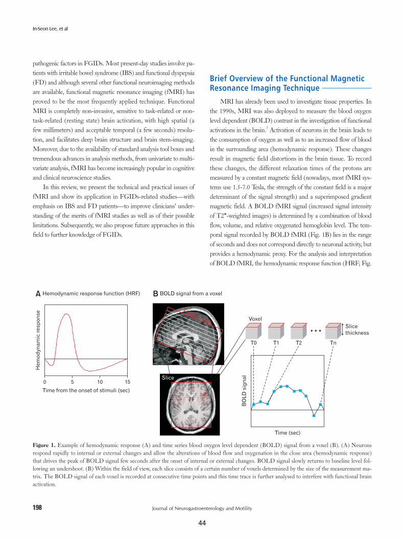
198
In-Seon Lee, et al
Journal of Neurogastroenterology and Motility
pathogenic factors in FGIDs. Most present-day studies involve pa-tients with irritable bowel syndrome (IBS) and functional dyspepsia (FD) and although several other functional neuroimaging methods are available, functional magnetic resonance imaging (fMRI) has proved to be the most frequently applied technique. Functional MRI is completely non-invasive, sensitive to task-related or non-task-related (resting state) brain activation, with high spatial (a few millimeters) and acceptable temporal (a few seconds) resolu-tion, and facilitates deep brain structure and brain stem-imaging. Moreover, due to the availability of standard analysis tool boxes and tremendous advances in analysis methods, from univariate to multi-variate analysis, fMRI has become increasingly popular in cognitive and clinical neuroscience studies.
In this review, we present the technical and practical issues of fMRI and show its application in FGIDs-related studies––with emphasis on IBS and FD patients––to improve clinicians’ under-standing of the merits of fMRI studies as well as of their possible limitations. Subsequently, we also propose future approaches in this field to further knowledge of FGIDs.
Brief Overview of the Functional Magnetic Resonance Imaging Technique
MRI has already been used to investigate tissue properties. In the 1990s, MRI was also deployed to measure the blood oxygen level dependent (BOLD) contrast in the investigation of functional activations in the brain.3 Activation of neurons in the brain leads to the consumption of oxygen as well as to an increased flow of blood in the surrounding area (hemodynamic response). These changes result in magnetic field distortions in the brain tissue. To record these changes, the different relaxation times of the protons are measured by a constant magnetic field (nowadays, most fMRI sys-tems use 1.5-7.0 Tesla, the strength of the constant field is a major determinant of the signal strength) and a superimposed gradient magnetic field. A BOLD fMRI signal (increased signal intensity of T2*-weighted images) is determined by a combination of blood flow, volume, and relative oxygenated hemoglobin level. The tem-poral signal recorded by BOLD fMRI (Fig. 1B) lies in the range of seconds and does not correspond directly to neuronal activity, but provides a hemodynamic proxy. For the analysis and interpretation of BOLD fMRI, the hemodynamic response function (HRF; Fig.
Figure 1. Example of hemodynamic response (A) and time series blood oxygen level dependent (BOLD) signal from a voxel (B). (A) Neurons respond rapidly to internal or external changes and allow the alterations of blood flow and oxygenation in the close area (hemodynamic response) that drives the peak of BOLD signal few seconds after the onset of internal or external changes. BOLD signal slowly returns to baseline level fol-lowing an undershoot. (B) Within the field of view, each slice consists of a certain number of voxels determined by the size of the measurement ma-trix. The BOLD signal of each voxel is recorded at consecutive time points and this time trace is further analysed to interfere with functional brain activation.
A BHemodynamic response function (HRF) BOLD signal from a voxel
Slice
thickness
0 15
He
mo
dyn
am
icre
sp
on
se
Time from the onset of stimuli (sec)
105Slice
Field of view
Slice thickness
Voxel
T0 T1 T2 Tn
Time (sec)
BO
LD
sig
na
l
44
imlee1i1
Rectangle
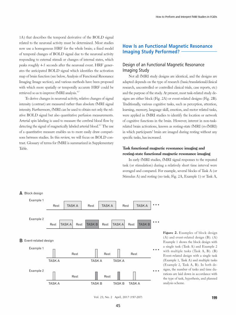
199
How to Perform and Interpret fMRI Studies in FGIDs
Vol. 23, No. 2 April, 2017 (197-207)
1A) that describes the temporal derivative of the BOLD signal related to the neuronal activity must be determined. Most studies now use a homogenous HRF for the whole brain; a fixed model of temporal changes of BOLD signal due to the neuronal activity responding to external stimuli or changes of internal states, which peaks roughly 4-5 seconds after the neuronal event. HRF gener-ates the anticipated BOLD signal which identifies the activation map of brain function (see below, Analysis of Functional Resonance Imaging Image section), and various methods have been proposed with which more spatially or temporally accurate HRF could be retrieved so as to improve fMRI analysis.4,5
To derive changes in neuronal activity, relative changes of signal intensity (contrast) are measured rather than absolute fMRI signal intensity. Furthermore, fMRI can be used to obtain not only the rel-ative BOLD signal but also quantitative perfusion measurements. Arterial spin labeling is used to measure the cerebral blood flow by detecting the signal of magnetically labeled arterial blood.6,7 The use of a quantitative measure enables us to more easily draw compari-sons between studies. In this review, we will focus on BOLD con-trast. Glossary of terms for fMRI is summarized in Supplementary Table.
How Is an Functional Magnetic Resonance Imaging Study Performed?
Design of an Functional Magnetic Resonance Imaging Study
Not all fMRI study designs are identical, and the designs are adapted depends on the type of research (basic/translational/clinical research, uncontrolled or controlled clinical trials, case reports, etc) and the purpose of the study. At present, most task-related study de-signs are either block (Fig. 2A) or event-related designs (Fig. 2B). Traditionally, various cognitive tasks, such as perception, attention, learning, memory, language skill, emotion, and motor related tasks, were applied in fMRI studies to identify the location or network of cognitive functions in the brain. However, interest in non-task-related brain activations, known as resting-state fMRI (rs-fMRI) in which participants’ brain are imaged during resting without any specific tasks, has increased.
Task functional magnetic resonance imaging and resting-state functional magnetic resonance imaging
In early fMRI studies, fMRI signal responses to the repeated task (or stimulation) during a relatively short time interval were averaged and compared. For example, several blocks of Task A (or Stimulus A) and resting (no task; Fig. 2A, Example 1) or Task A,
A
B
TASK A TASK ATASK ARest RestRest
Block design
TASK A TASK ATASK BRest RestRest TASK BRest
Event-related design
Example 1
Example 2
Example 1
Example 2
Rest Rest Rest
Rest Rest
TASK A TASK A TASK A
TASK A TASK B TASK B TASK A
Figure 2. Examples of block design (A) and event-related design (B). (A) Example 1 shows the block design with a single task (Task A) and Example 2 with multiple tasks (Task A, B). (B) Event-related design with a single task (Example 1, Task A) and multiple tasks (Example 2, Task A, B). In both de-signs, the number of tasks and time du-rations are laid down in accordance with the type of task, hypothesis, and planned analysis scheme.
45
imlee1i1
Rectangle
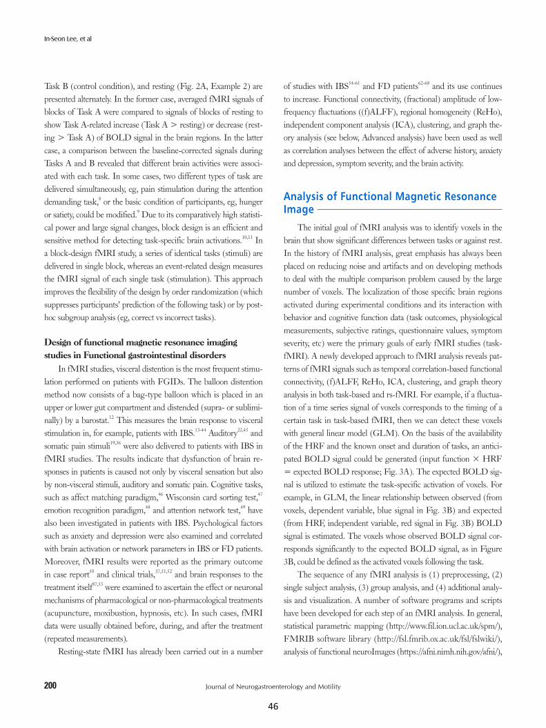
200
In-Seon Lee, et al
Journal of Neurogastroenterology and Motility
Task B (control condition), and resting (Fig. 2A, Example 2) are presented alternately. In the former case, averaged fMRI signals of blocks of Task A were compared to signals of blocks of resting to show Task A-related increase (Task A > resting) or decrease (rest-ing > Task A) of BOLD signal in the brain regions. In the latter case, a comparison between the baseline-corrected signals during Tasks A and B revealed that different brain activities were associ-ated with each task. In some cases, two different types of task are delivered simultaneously, eg, pain stimulation during the attention demanding task,8 or the basic condition of participants, eg, hunger or satiety, could be modified.9 Due to its comparatively high statisti-cal power and large signal changes, block design is an efficient and sensitive method for detecting task-specific brain activations.10,11 In a block-design fMRI study, a series of identical tasks (stimuli) are delivered in single block, whereas an event-related design measures the fMRI signal of each single task (stimulation). This approach improves the flexibility of the design by order randomization (which suppresses participants’ prediction of the following task) or by post-hoc subgroup analysis (eg, correct vs incorrect tasks).
Design of functional magnetic resonance imaging studies in Functional gastrointestinal disorders
In fMRI studies, visceral distention is the most frequent stimu-lation performed on patients with FGIDs. The balloon distention method now consists of a bag-type balloon which is placed in an upper or lower gut compartment and distended (supra- or sublimi-nally) by a barostat.12 This measures the brain response to visceral stimulation in, for example, patients with IBS.13-44 Auditory22,45 and somatic pain stimuli19,36 were also delivered to patients with IBS in fMRI studies. The results indicate that dysfunction of brain re-sponses in patients is caused not only by visceral sensation but also by non-visceral stimuli, auditory and somatic pain. Cognitive tasks, such as affect matching paradigm,46 Wisconsin card sorting test,47 emotion recognition paradigm,48 and attention network test,49 have also been investigated in patients with IBS. Psychological factors such as anxiety and depression were also examined and correlated with brain activation or network parameters in IBS or FD patients. Moreover, fMRI results were reported as the primary outcome in case report50 and clinical trials,37,51,52 and brain responses to the treatment itself37,53 were examined to ascertain the effect or neuronal mechanisms of pharmacological or non-pharmacological treatments (acupuncture, moxibustion, hypnosis, etc). In such cases, fMRI data were usually obtained before, during, and after the treatment (repeated measurements).
Resting-state fMRI has already been carried out in a number
of studies with IBS54-61 and FD patients62-68 and its use continues to increase. Functional connectivity, (fractional) amplitude of low-frequency fluctuations ((f)ALFF), regional homogeneity (ReHo), independent component analysis (ICA), clustering, and graph the-ory analysis (see below, Advanced analysis) have been used as well as correlation analyses between the effect of adverse history, anxiety and depression, symptom severity, and the brain activity.
Analysis of Functional Magnetic Resonance Image
The initial goal of fMRI analysis was to identify voxels in the brain that show significant differences between tasks or against rest. In the history of fMRI analysis, great emphasis has always been placed on reducing noise and artifacts and on developing methods to deal with the multiple comparison problem caused by the large number of voxels. The localization of those specific brain regions activated during experimental conditions and its interaction with behavior and cognitive function data (task outcomes, physiological measurements, subjective ratings, questionnaire values, symptom severity, etc) were the primary goals of early fMRI studies (task-fMRI). A newly developed approach to fMRI analysis reveals pat-terns of fMRI signals such as temporal correlation-based functional connectivity, (f)ALFF, ReHo, ICA, clustering, and graph theory analysis in both task-based and rs-fMRI. For example, if a fluctua-tion of a time series signal of voxels corresponds to the timing of a certain task in task-based fMRI, then we can detect these voxels with general linear model (GLM). On the basis of the availability of the HRF and the known onset and duration of tasks, an antici-pated BOLD signal could be generated (input function × HRF = expected BOLD response; Fig. 3A). The expected BOLD sig-nal is utilized to estimate the task-specific activation of voxels. For example, in GLM, the linear relationship between observed (from voxels, dependent variable, blue signal in Fig. 3B) and expected (from HRF, independent variable, red signal in Fig. 3B) BOLD signal is estimated. The voxels whose observed BOLD signal cor-responds significantly to the expected BOLD signal, as in Figure 3B, could be defined as the activated voxels following the task.
The sequence of any fMRI analysis is (1) preprocessing, (2) single subject analysis, (3) group analysis, and (4) additional analy-sis and visualization. A number of software programs and scripts have been developed for each step of an fMRI analysis. In general, statistical parametric mapping (http://www.fil.ion.ucl.ac.uk/spm/), FMRIB software library (http://fsl.fmrib.ox.ac.uk/fsl/fslwiki/), analysis of functional neuroImages (https://afni.nimh.nih.gov/afni/),
46
imlee1i1
Rectangle
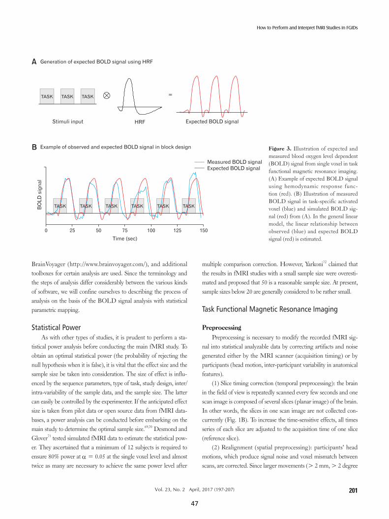
201
How to Perform and Interpret fMRI Studies in FGIDs
Vol. 23, No. 2 April, 2017 (197-207)
BrainVoyager (http://www.brainvoyager.com/), and additional toolboxes for certain analysis are used. Since the terminology and the steps of analysis differ considerably between the various kinds of software, we will confine ourselves to describing the process of analysis on the basis of the BOLD signal analysis with statistical parametric mapping.
Statistical PowerAs with other types of studies, it is prudent to perform a sta-
tistical power analysis before conducting the main fMRI study. To obtain an optimal statistical power (the probability of rejecting the null hypothesis when it is false), it is vital that the effect size and the sample size be taken into consideration. The size of effect is influ-enced by the sequence parameters, type of task, study design, inter/intra-variability of the sample data, and the sample size. The latter can easily be controlled by the experimenter. If the anticipated effect size is taken from pilot data or open source data from fMRI data-bases, a power analysis can be conducted before embarking on the main study to determine the optimal sample size.69,70 Desmond and Glover71 tested simulated fMRI data to estimate the statistical pow-er. They ascertained that a minimum of 12 subjects is required to ensure 80% power at α = 0.05 at the single voxel level and almost twice as many are necessary to achieve the same power level after
multiple comparison correction. However, Yarkoni72 claimed that the results in fMRI studies with a small sample size were overesti-mated and proposed that 50 is a reasonable sample size. At present, sample sizes below 20 are generally considered to be rather small.
Task Functional Magnetic Resonance Imaging
Preprocessing Preprocessing is necessary to modify the recorded fMRI sig-
nal into statistical analyzable data by correcting artifacts and noise generated either by the MRI scanner (acquisition timing) or by participants (head motion, inter-participant variability in anatomical features).
(1) Slice timing correction (temporal preprocessing): the brain in the field of view is repeatedly scanned every few seconds and one scan image is composed of several slices (planar image) of the brain. In other words, the slices in one scan image are not collected con-currently (Fig. 1B). To increase the time-sensitive effects, all times series of each slice are adjusted to the acquisition time of one slice (reference slice).
(2) Realignment (spatial preprocessing): participants’ head motions, which produce signal noise and voxel mismatch between scans, are corrected. Since larger movements (> 2 mm, > 2 degree
A
B
Measured BOLD signal
Expected BOLD signal
Generation of expected BOLD signal using HRF
Example of observed and expected BOLD signal in block design
BO
LD
sig
na
l
0 25 50 75 100 125 150
Time (sec)
Stimuli input HRF Expected BOLD signal
TASK TASKTASK =
TASKTASKTASK TASK TASK TASK
Figure 3. Illustration of expected and measured blood oxygen level dependent (BOLD) signal from single voxel in task functional magnetic resonance imaging. (A) Example of expected BOLD signal using hemodynamic response func-tion (red). (B) Illustration of measured BOLD signal in task-specific activated voxel (blue) and simulated BOLD sig-nal (red) from (A). In the general linear model, the linear relationship between observed (blue) and expected BOLD signal (red) is estimated.
47
imlee1i1
Rectangle
202
In-Seon Lee, et al
Journal of Neurogastroenterology and Motility
rotation) can produce significant non-amendable noise, slices with large head motion are usually discarded. Smaller movements can be corrected or the movement can be taken into consideration during the statistical analysis.
(3) Co-registration (spatial preprocessing): registration of an anatomical image to match the functional image is required for fur-ther analysis.
(4) Segmentation (spatial preprocessing): segmentation of an anatomical image to separate brain tissues, cerebral spinal fluid, white matter, and gray matter.
(5) Normalization: individual images are normalized into stan-dard space to correct between subject variability. This step increases sensitivity, and facilitates the generalization of results and compari-sons between studies.
(6) Smoothing: a smoothing filter, such as Gaussian kernel, is applied to blur the images and reduce the number of independent observations based on random field theory. This process suppresses noise, increases sensitivity, and makes images more appropriate for single-subject and group analysis.
Single subject and group analysisIn a single subject analysis, also known as subject level or first
level analysis, design and contrast of all experimental conditions are defined. In order to specify the experimental design, information about the onset and duration of each task is required. F-contrasts (effects of interest) or T-contrasts (the contrasts between tasks or task and resting condition) are defined according to the design and purpose of the analysis. Movement parameters and other regressors are also determined in case they are required.
In group analysis, also known as second level analysis, t tests, ANOVAs and other general linear model analyses with covariates or regressors can be performed. In the event of a specific hypothesis about the correlation between the clinical symptoms, task perfor-mance, personality, or duration of the disease and brain activation, multiple regression analysis using covariates could identify those brain regions that positively or negatively correlate with the covari-ates. Contrasts for group analysis must also be defined to report group level results. In general, the analysis is performed as a whole brain analysis. For region-of-interest (ROI) analysis, the equipped ROIs in the toolbox library (Automated Anatomical Labeling atlas73) or newly generated ROIs using center coordinates and radius or number of voxels are used. A ROI-based approach should be used only if clear hypotheses are available and the multi-comparison cor-rection should be taken into account if more than one ROI is used. Having set a statistical threshold and multiple comparison correc-
tion thresholds to correct false positives (family-wise error rate or false discovery rate is generally used), one can export the results into figures, tables, or time series signal data.
Resting-state Functional Magnetic Resonance ImagingOnce rs-fMRI data is preprocessed in a similar way to task-
fMRI, procedures of single subject and group analysis differ from task-fMRI. In resting state analysis, the spontaneous low frequency fluctuation (0.01-0.10 Hz) is of major interest. Several approaches, including ALFF and (f)ALFF, were developed specifically for rs-fMRI analysis in an effort to extract an amplitude or ratio of spontaneous low frequency fluctuation from the BOLD signal, indicative of a regional intensity of activation.74,75 Functional con-nectivity, ReHo, and ICA are also applicable in rs-fMRI as well as in task-fMRI. Further toolboxes and scripts for rs-fMRI were also developed.76,77
Advanced Analysis Various advanced analyses have been introduced in fMRI
analysis. Here, we briefly introduce the analysis technique which has been used of late in FGIDs studies.
Functional connectivity, one of the most widespread analysis techniques, is defined as ‘temporal correlation between the different parts (voxels, clusters, or ROIs) of the brain.38,44,56,57,78 It enables us to estimate the connection of brain regions and to compare its patterns between groups. Effective connectivity provides us with additional information as to which brain areas induce a direct causal influence over others.48,51,79 Dynamic causal modeling is an example of the effective connectivity analysis method and shows how the effective connectivity (causal influence) between brain regions is modulated by experimental conditions.47,80 Graph theory analysis, ie, the analysis of the properties of connections (edges) between func-tionally connected brain regions (nodes) to account for the complex characteristics of a network, is a further form of connectivity analy-sis.61,68,81 ReHo is basically a voxel-based connectivity analysis that measures the regional similarity of the signals between the specific voxel and its neighboring voxels.59,67,82
Of all the multivariate analyses applied in FGIDs studies, ICA pattern classification is the most familiar.29,38,58 ICA works on the assumption that an fMRI signal is linearly composed of several (spatially or temporally) independent signals, and that the original fMRI signal is separated into independent groups.83 Since ICA is one of the data-driven analysis methods, it can reveal an intrinsic structure of the original signal and can therefore also be utilized to generate hypotheses.
48
imlee1i1
Rectangle
203
How to Perform and Interpret fMRI Studies in FGIDs
Vol. 23, No. 2 April, 2017 (197-207)
Interpreting Functional Magnetic Resonance Imaging Results in Functional Gastrointesti-nal Disorders
In most studies, the list of brain regions (coordinates and sta-tistical information) displaying increased or decreased activity in certain conditions or groups is reported in a voxel-wise or a ROI-wise manner. In some instances, a group of the brain areas involved in the same function (eg, pain processing) is identified as a ‘network.’ For example, albeit opinions are deeply divided on this issue, so-matosensory cortex, insula, anterior cingulate cortex, and thalamus are termed a ‘pain network.’84 The most frequently reported brain regions in FGIDs studies are the prefrontal cortex, somatosensory cortex, insula, cingulate cortex, and thalamus. The contributory net-works to FGIDs are known as the sensory-motor network, salience network, autonomic network, and cognitive/affective network.1,85
Functional MRI data may allow us to elucidate the basic neuro-physiological and pathophysiological mechanisms in brains which is associated with clinical information. For example, the activation map following rectal balloon distention can indicate the altered neural processing of visceral pain in the somatosensory cortex, frontal cor-tex, cingulate cortex, insula, thalamus, and (pre)motor cortex with higher pain sensation (visceral hypersensitivity) in patients than in controls.15,17 Anxiety and depression were associated with the brain activation in the cingulate cortex and prefrontal cortex,28 and his-tory of abuse affected the brain activation in the cingulate cortex.27 Several studies have attempted to identify the specific mechanisms of treatment86 and neuroimaging biomarkers for further disorders.87 The inhibition effect of pain-related brain activation in IBS patients by amitriptyline (tricyclic antidepressants)20 identified the central mechanism of antidepressants in the reduction of rectal distention pain. The brain activity during acupuncture suggested the modula-tion of serotonin pathway at insula and the higher cortical regulation of affection as potential neural mechanisms of acupuncture treat-ment.34 Furthermore, correlation analysis between fMRI data and psychological indices such as anxiety and depression may demon-strate the influence of the psychological state on patients.28,35 When interpreting the fMRI results on interventions, the blinding issue, changes of symptoms, co-morbidities, quality of life, non-specific effect, and placebo response should also be taken into consideration carefully.
Limitations and Future Approaches of Functional Magnetic Resonance Imaging Studies in Functional Gastrointestinal Disorders
Functional MRI measurement is not only expensive and time consuming, but also requires extensive skills and resources. Re-searchers should be aware of the variety of factors which affect the brain imaging results before performing experiments, and it is only when valid tasks or stimuli, well-structured procedures, controlled populations of participants, and proper analyses come together that reliable data can be gained. The unusual environment of MRI must also be taken into consideration. Patients with a metal implant or with claustrophobia should not participate. No movement, par-ticularly no head movement, is permitted inside the scanner. Recent studies have demonstrated in both IBS and in healthy controls that visceral pain perception is higher within the MRI environment than outside.88 Investigators and participants must therefore adapt themselves to the MRI environment.
Until now, all neuroimaging studies in FGID have used a correlation approach. This does not permit us to make any causal inference about the direction of influence (central to peripheral, pe-ripheral to central, or both). At present, inconsistent study designs, analysis methods and statistical principles make it difficult to com-pare or integrate fMRI data in FGIDs across studies using meta-analysis. However, because FGIDs lack biomarkers such as neu-rohormones, cytokines, and genes, functional neuroimaging may provide further information to elucidate the symptoms in patients. Furthermore, fMRI studies may help us to better fathom the role of emotional feelings and cognitive functions by combined with other neuroimaging techniques or with autonomic response, genetic and epigenetic approaches, and neurotransmitter research to identify key components of the disease, or to differentiate between subtypes.
In summary, fMRI is a unique research tool that provides information on neuronal mechanisms of symptoms and treatment effects in the patient population, and physiological processing in healthy volunteers. It should, however, be utilized prudently in re-search, and its pros and cons should be weighed up carefully.
Since neuroimaging has been applied in FGIDs for less than twenty years and analysis methods are developing and improving rapidly, future approaches hold tremendous potential. As yet, only experimental pain stimulation and a few cognitive tasks have been implemented in FGIDs patients. Besides the pain and anxiety/de-pression scores, FGIDs patients may have many other pathological, behavioral and somatic characteristics; such as impaired affective
49
imlee1i1
Rectangle
204
In-Seon Lee, et al
Journal of Neurogastroenterology and Motility
memory, heightened vigilance, abnormal eating behavior, increased stress sensitivity, disordered autonomic regulation, dysbiosis of the gut microbiota, additional bowel symptoms such as nausea, bloat-ing, urgency, and autonomic and somatic co-morbidities. It may be advisable to examine the effects of pharmacological or non-phar-macological therapy, and the influence of such therapies on brain activity may help to establish novel treatment strategies. Albeit still a far cry from clinical application, neuroimaging data will neverthe-less one day be used to perform subgroup analyses in patients (eg, hypersensitive vs normosensitive or even hyposensitive patients) or to distinguish patients from healthy controls.89 The neuroimaging data with more numerous tasks, behavioral measurement, and ther-apies could improve our understanding of the pathophysiology of FGIDs and lead to more appropriate treatment options for patients in the future.
Conclusions The advent of the fMRI technique has not only provided in-
formation on regional brain activities and the interaction of different brain areas, but has also improved our understanding of the neuro-nal changes and its relationship with symptoms and cognitive/affec-tive changes in many patient groups. Although its usage in basic or clinical neuroscience research in FGIDs patients has been reported in only a limited number of studies, and despite its requiring an intensive level of knowledge in neurology, physiology, pathology, physics, and program coding, it does have considerable potential. An accurate understanding and application of fMRI technique in FGIDs will hopefully lead to new methods of diagnosing and treat-ing patients.
Supplementary Material Note: To access the supplementary table mentioned in this
article, visit the online version of Journal of Neurogastroenterol-ogy and Motility at http://www.jnmjournal.org/, and at https://doi.org/10.5056/jnm16196.
Acknowledgements: The data of this study were presented at the 6th Asian Postgraduate Course on Neurogastroenterology and Motility (APNM) in Seoul, Korea, 2016.
In-Seon Lee is a PhD training fellow of NeuroGUT.
Financial support: Writing of this review was funded by the People Programme of the European Union’s Seventh Framework
Programme under REA grant agreement No. 607652 (Neu-roGUT).
Conflicts of interest: None.
Author contributions: In-Seon Lee, Hubert Preissl, and Paul Enck planned the overall concept and frame work of the manu-script; In-Seon Lee wrote the manuscript; and Hubert Preissl and Paul Enck revised the manuscript.
References 1. Lee IS, Wang H, Chae Y, Preissl H, Enck P. Functional neuroimaging
studies in functional dyspepsia patients: a systematic review. Neurogastro-enterol Motil 2016;28:793-805.
2. Mayer EA, Naliboff BD, Craig AD. Neuroimaging of the brain-gut axis: from basic understanding to treatment of functional GI disorders. Gastroenterology 2006;131:1925-1942.
3. Ogawa S, Lee TM, Kay AR, Tank DW. Brain magnetic resonance im-aging with contrast dependent on blood oxygenation. Proc Natl Acad Sci USA 1990;87:9868-9872.
4. Maleki-Balajoo S, Hossein-Zadeh GA, Soltanian-Zadeh H, Ekhtiari H. Locally estimated hemodynamic response function and activation detection sensitivity in heroin-cue reactivity study. Basic Clin Neurosci 2016;7:299-314.
5. Puckett AM, Aquino KM, Robinson PA, Breakspear M, Schira MM. The spatiotemporal hemodynamic response function for depth-dependent functional imaging of human cortex. Neuroimage 2016;139:240-248.
6. Grade M, Hernandez Tamames JA, Pizzini FB, Achten E, Golay X, Smits M. A neuroradiologist’s guide to arterial spin labeling MRI in clinical practice. Neuroradiology 2015;57:1181-1202.
7. Wu WC, St Lawrence KS, Licht DJ, Wang DJ. Quantification issues in arterial spin labeling perfusion magnetic resonance imaging. Top Magn Reson Imaging 2010;21:65-73.
8. Bantick SJ, Wise RG, Ploghaus A, Clare S, Smith SM, Tracey I. Imag-ing how attention modulates pain in humans using functional MRI. Brain 2002;125(Pt 2):310-319.
9. Führer D, Zysset S, Stumvoll M. Brain activity in hunger and satiety: an exploratory visually stimulated fMRI study. Obesity (Silver Spring) 2008;16:945-950.
10. Buxton RB, Wong EC, Frank LR. Dynamics of blood flow and oxygen-ation changes during brain activation: the balloon model. Magn Reson Med 1998;39:855-864.
11. Friston KJ, Holmes AP, Price CJ, Büchel C, Worsley KJ. Multisubject fMRI studies and conjunction analyses. Neuroimage 1999;10:385-396.
12. Ritchie J. Pain from distension of the pelvic colon by inflating a balloon in the irritable colon syndrome. Gut 1973;14:125-132.
13. Mertz H, Morgan V, Tanner G, et al. Regional cerebral activation in ir-ritable bowel syndrome and control subjects with painful and nonpainful rectal distention. Gastroenterology 2000;118:842-848.
14. Bernstein CN, Frankenstein UN, Rawsthorne P, Pitz M, Summers R, McIntyre MC. Cortical mapping of visceral pain in patients with GI dis-
50
imlee1i1
Rectangle
205
How to Perform and Interpret fMRI Studies in FGIDs
Vol. 23, No. 2 April, 2017 (197-207)
orders using functional magnetic resonance imaging. Am J Gastroenterol 2002;97:319-327.
15. Bonaz B, Baciu M, Papillon E, et al. Central processing of rectal pain in patients with irritable bowel syndrome: an fMRI study. Am J Gastroen-terol 2002;97:654-661.
16. Verne GN, Himes NC, Robinson ME, et al. Central representation of visceral and cutaneous hypersensitivity in the irritable bowel syndrome. Pain 2003;103:99-110.
17. Yuan YZ, Tao RJ, Xu B, et al. Functional brain imaging in irritable bowel syndrome with rectal balloon-distention by using fMRI. World J Gastro-enterol 2003;9:1356-1360.
18. Sidhu H, Kern M, Shaker R. Absence of increasing cortical fMRI activ-ity volume in response to increasing visceral stimulation in IBS patients. Am J Physiol Gastrointest Liver Physiol 2004;287:G425-G435.
19. Wilder-Smith CH, Schindler D, Lovblad K, Redmond SM, Nirkko A. Brain functional magnetic resonance imaging of rectal pain and activation of endogenous inhibitory mechanisms in irritable bowel syndrome patient subgroups and healthy controls. Gut 2004;53:1595-1601.
20. Morgan V, Pickens D, Gautam S, Kessler R, Mertz H. Amitriptyline reduces rectal pain related activation of the anterior cingulate cortex in patients with irritable bowel syndrome. Gut 2005;54:601-607.
21. Kwan CL, Diamant NE, Pope G, Mikula K, Mikulis DJ, Davis KD. Abnormal forebrain activity in functional bowel disorder patients with chronic pain. Neurology 2005;65:1268-1277.
22. Andresen V, Bach DR, Poellinger A, et al. Brain activation responses to subliminal or supraliminal rectal stimuli and to auditory stimuli in irritable bowel syndrome. Neurogastroenterol Motil 2005;17:827-837.
23. Lawal A, Kern M, Sidhu H, Hofmann C, Shaker R. Novel evidence for hypersensitivity of visceral sensory neural circuitry in irritable bowel syndrome patients. Gastroenterology 2006;130:26-33.
24. Song GH, Venkatraman V, Ho KY, Chee MW, Yeoh KG, Wilder-Smith CH. Cortical effects of anticipation and endogenous modulation of vis-ceral pain assessed by functional brain MRI in irritable bowel syndrome patients and healthy controls. Pain 2006;126:79-90.
25. Price DD, Craggs J, Verne GN, Perlstein WM, Robinson ME. Placebo analgesia is accompanied by large reductions in pain-related brain activity in irritable bowel syndrome patients. Pain 2007;127:63-72.
26. Berman SM, Naliboff BD, Suyenobu B, et al. Reduced brainstem in-hibition during anticipated pelvic visceral pain correlates with enhanced brain response to the visceral stimulus in women with irritable bowel syn-drome. J Neurosci 2008;28:349-359.
27. Ringel Y, Drossman DA, Leserman JL, et al. Effect of abuse history on pain reports and brain responses to aversive visceral stimulation: an fMRI study. Gastroenterology 2008;134:396-404.
28. Elsenbruch S, Rosenberger C, Enck P, Forsting M, Schedlowski M, Gizewski ER. Affective disturbances modulate the neural processing of visceral pain stimuli in irritable bowel syndrome: an fMRI study. Gut 2010;59:489-495.
29. Hall GB, Kamath MV, Collins S, et al. Heightened central affective response to visceral sensations of pain and discomfort in IBS. Neurogas-troenterol Motil 2010;22:276, e80.
30. Elsenbruch S, Rosenberger C, Bingel U, Forsting M, Schedlowski M,
Gizewski ER. Patients with irritable bowel syndrome have altered emo-tional modulation of neural responses to visceral stimuli. Gastroenterology 2010;139:1310-1319.
31. Larsson MB, Tillisch K, Craig AD, et al. Brain responses to visceral stimuli reflect visceral sensitivity thresholds in patients with irritable bowel syndrome. Gastroenterology 2012;142:463-472, e3.
32. Tillisch K, Labus J, Nam B, et al. Neurokinin-1-receptor antagonism de-creases anxiety and emotional arousal circuit response to noxious visceral distension in women with irritable bowel syndrome: a pilot study. Aliment Pharmacol Ther 2012;35:360-367.
33. Lee HF, Hsieh JC, Lu CL, et al. Enhanced affect/cognition-related brain responses during visceral placebo analgesia in irritable bowel syn-drome patients. Pain 2012;153:1301-1310.
34. Chu WC, Wu JC, Yew DT, et al. Does acupuncture therapy alter activa-tion of neural pathway for pain perception in irritable bowel syndrome?: a comparative study of true and sham acupuncture using functional mag-netic resonance imaging. J Neurogastroenterol Motil 2012;18:305-316.
35. Rosenberger C, Thurling M, Forsting M, Elsenbruch S, Timmann D, Gizewski ER. Contributions of the cerebellum to disturbed central processing of visceral stimuli in irritable bowel syndrome. Cerebellum 2013;12:194-198.
36. Bouhassira D, Moisset X, Jouet P, Duboc H, Coffin B, Sabate JM. Changes in the modulation of spinal pain processing are related to sever-ity in irritable bowel syndrome. Neurogastroenterol Motil 2013;25:623-e468.
37. Lowén MB, Mayer EA, Sjöberg M, et al. Effect of hypnotherapy and educational intervention on brain response to visceral stimulus in the ir-ritable bowel syndrome. Aliment Pharmacol Ther 2013;37:1184-1197.
38. Letzen JE, Craggs JG, Perlstein WM, Price DD, Robinson ME. Func-tional connectivity of the default mode network and its association with pain networks in irritable bowel patients assessed via lidocaine treatment. J Pain 2013;14:1077-1087.
39. Craggs JG, Price DD, Robinson ME. Enhancing the placebo response: functional magnetic resonance imaging evidence of memory and seman-tic processing in placebo analgesia. J Pain 2014;15:435-446.
40. Schmid J, Langhorst J, Gaß F, et al. Placebo analgesia in patients with functional and organic abdominal pain: a fMRI study in IBS, UC and healthy volunteers. Gut 2015;64:418-427.
41. Zhu Y, Wu Z, Ma X, et al. Brain regions involved in moxibustion-induced analgesia in irritable bowel syndrome with diarrhea: a functional magnetic resonance imaging study. BMC Complement Altern Med 2014;14:500.
42. Icenhour A, Langhorst J, Benson S, et al. Neural circuitry of abdominal pain-related fear learning and reinstatement in irritable bowel syndrome. Neurogastroenterol Motil 2015;27:114-127.
43. Lowén MB, Mayer E, Tillisch K, et al. Deficient habituation to repeated rectal distensions in irritable bowel syndrome patients with visceral hyper-sensitivity. Neurogastroenterol Motil 2015;27:646-655.
44. Liu X, Silverman A, Kern M, et al. Excessive coupling of the salience network with intrinsic neurocognitive brain networks during rectal disten-sion in adolescents with irritable bowel syndrome: a preliminary report. Neurogastroenterol Motil 2016;28:43-53.
51
imlee1i1
Rectangle
206
In-Seon Lee, et al
Journal of Neurogastroenterology and Motility
45. Andresen V, Poellinger A, Tsrouya C, et al. Cerebral processing of audi-tory stimuli in patients with irritable bowel syndrome. World J Gastroen-terol 2006;12:1723-1729.
46. Kilpatrick LA, Labus JS, Coveleskie K, et al. The HTR3A polymor-phism c. -42C>T is associated with amygdala responsiveness in patients with irritable bowel syndrome. Gastroenterology 2011;140:1943-1951.
47. Aizawa E, Sato Y, Kochiyama T, et al. Altered cognitive function of pre-frontal cortex during error feedback in patients with irritable bowel syn-drome, based on fMRI and dynamic causal modeling. Gastroenterology 2012;143:1188-1198.
48. Labus JS, Gupta A, Coveleskie K, et al. Sex differences in emotion-re-lated cognitive processes in irritable bowel syndrome and healthy control subjects. Pain 2013;154:2088-2099.
49. Hubbard CS, Hong J, Jiang Z, et al. Increased attentional network functioning related to symptom severity measures in females with irritable bowel syndrome. Neurogastroenterol Motil 2015;27:1282-1294.
50. Drossman DA, Ringel Y, Vogt BA, et al. Alterations of brain activity as-sociated with resolution of emotional distress and pain in a case of severe irritable bowel syndrome. Gastroenterology 2003;124:754-761.
51. Hubbard CS, Labus JS, Bueller J, et al. Corticotropin-releasing factor receptor 1 antagonist alters regional activation and effective connectivity in an emotional-arousal circuit during expectation of abdominal pain. J Neurosci 2011;31:12491-12500.
52. Zhao JM, Lu JH, Yin XJ, et al. Comparison of electroacupuncture and moxibustion on brain-gut function in patients with diarrhea-predominant irritable bowel syndrome: a randomized controlled trial. Chin J Integr Med 2015;21:855-865.
53. Li Z, Zeng F, Yang Y, et al. Different cerebral responses to puncturing at ST36 among patients with functional dyspepsia and healthy subjects. Forsch Komplementmed 2014;21:99-104.
54. Hong JY, Kilpatrick LA, Labus J, et al. Patients with chronic visceral pain show sex-related alterations in intrinsic oscillations of the resting brain. J Neurosci 2013;33:11994-12002.
55. Gupta A, Kilpatrick L, Labus J, et al. Early adverse life events and rest-ing state neural networks in patients with chronic abdominal pain: evi-dence for sex differences. Psychosom Med 2014;76:404-412.
56. Hong JY, Kilpatrick LA, Labus JS, et al. Sex and disease-related altera-tions of anterior insula functional connectivity in chronic abdominal pain. J Neurosci 2014;34:14252-14259.
57. Ma X, Li S, Tian J, et al. Altered brain spontaneous activity and connec-tivity network in irritable bowel syndrome patients: a resting-state fMRI study. Clin Neurophysiol 2015;126:1190-1197.
58. Gupta A, Rapkin AJ, Gill Z, et al. Disease-related differences in resting-state networks: a comparison between localized provoked vulvodynia, ir-ritable bowel syndrome, and healthy control subjects. Pain 2015;156:809-819.
59. Ke J, Qi R, Liu C, et al. Abnormal regional homogeneity in patients with irritable bowel syndrome: a resting-state functional MRI study. Neuro-gastroenterol Motil 2015;27:1796-1803.
60. Qi R, Liu C, Ke J, et al. Intrinsic brain abnormalities in irritable bowel syndrome and effect of anxiety and depression. Brain Imaging Behav 2016;10:1127-1134.
61. Qi R, Ke J, Schoepf UJ, et al. Topological reorganization of the de-fault mode network in irritable bowel syndrome. Mol Neurobiol 2016;53:6585-6593.
62. Zhou G, Liu P, Zeng F, et al. Increased interhemispheric resting-state functional connectivity in functional dyspepsia: a pilot study. NMR Biomed 2013;26:410-415.
63. Zhou G, Liu P, Wang J, et al. Fractional amplitude of low-frequency fluctuation changes in functional dyspepsia: a resting-state fMRI study. Magn Reson Imaging 2013;31:996-1000.
64. Liu P, Zeng F, Zhou G, et al. Alterations of the default mode network in functional dyspepsia patients: a resting-state fmri study. Neurogastroen-terol Motil 2013;25:e382-e388.
65. Nan J, Liu J, Li G, et al. Whole-brain functional connectivity identifica-tion of functional dyspepsia. PLoS One 2013;8:e65870.
66. Liu P, Qin W, Wang J, et al. Identifying neural patterns of functional dyspepsia using multivariate pattern analysis: a resting-state fMRI study. PLoS One 2013;8:e68205.
67. Nan J, Liu J, Zhang D, et al. Altered intrinsic regional activity and cor-responding brain pathways reflect the symptom severity of functional dyspepsia. Neurogastroenterol Motil 2014;26:660-669.
68. Nan J, Zhang L, Zhu F, et al. Topological alterations of the intrinsic brain network in patients with functional dyspepsia. J Neurogastroenterol Mo-til 2016;22:118-128.
69. Mumford JA. A power calculation guide for fMRI studies. Soc Cogn Affect Neurosci 2012;7:738-742.
70. Joyce KE, Hayasaka S. Development of powermap: a software package for statistical power calculation in neuroimaging studies. Neuroinformat-ics 2012;10:351-365.
71. Desmond JE, Glover GH. Estimating sample size in functional MRI (fMRI) neuroimaging studies: statistical power analyses. J Neurosci Methods 2002;118:115-128.
72. Yarkoni T. Big correlations in little studies: inflated fMRI correlations reflect low statistical power-commentary on vul et al. (2009). Perspect Psychol Sci 2009;4:294-298.
73. Tzourio-Mazoyer N, Landeau B, Papathanassiou D, et al. Automated anatomical labeling of activations in SPM using a macroscopic anatomi-cal parcellation of the MNI MRI single-subject brain. Neuroimage 2002;15:273-289.
74. Zang YF, He Y, Zhu CZ, et al. Altered baseline brain activity in children with ADHD revealed by resting-state functional MRI. Brain Dev 2007;29:83-91.
75. Zou QH, Zhu CZ, Yang Y, et al. An improved approach to detection of amplitude of low-frequency fluctuation (ALFF) for resting-state fMRI: fractional ALFF. J Neurosci Methods 2008;172:137-141.
76. Chao-Gan Y, Yu-Feng Z. DPARSF: a MATLAB toolbox for “pipeline” data analysis of resting-state fMRI. Front Syst Neurosci 2010;4:13.
77. Song XW, Dong ZY, Long XY, et al. REST: a toolkit for resting-state functional magnetic resonance imaging data processing. PLoS One 2011;6:e25031.
78. Friston KJ, Frith CD, Liddle PF, Frackowiak RS. Functional connectiv-ity: the principal-component analysis of large (PET) data sets. J Cereb Blood Flow Metab 1993;13:5-14.
52
imlee1i1
Rectangle
207
How to Perform and Interpret fMRI Studies in FGIDs
Vol. 23, No. 2 April, 2017 (197-207)
79. Friston KJ. Functional and effective connectivity: a review. Brain Connect 2011;1:13-36.
80. Friston KJ, Harrison L, Penny W. Dynamic causal modelling. Neuroim-age 2003;19:1273-1302.
81. Wang J, Zuo X, He Y. Graph-based network analysis of resting-state functional MRI. Front Syst Neurosci 2010;4:16.
82. Zang Y, Jiang T, Lu Y, He Y, Tian L. Regional homogeneity approach to fMRI data analysis. Neuroimage 2004;22:394-400.
83. McKeown MJ, Makeig S, Brown GG, et al. Analysis of fMRI data by blind separation into independent spatial components. Hum Brain Mapp 1998;6:160-188.
84. Apkarian AV, Bushnell MC, Treede RD, Zubieta JK. Human brain mechanisms of pain perception and regulation in health and disease. Eur J Pain 2005;9:463-484.
85. Mayer EA, Labus JS, Tillisch K, Cole SW, Baldi P. Towards a systems
view of IBS. Nat Rev Gastroenterol Hepatol 2015;12:592-605.86. Duff EP, Vennart W, Wise RG, et al. Learning to identify CNS drug
action and efficacy using multistudy fMRI data. Sci Transl Med 2015;7:274ra16.
87. McGrath CL, Kelley ME, Holtzheimer PE, et al. Toward a neuroimag-ing treatment selection biomarker for major depressive disorder. JAMA Psychiatry 2013;70:821-829.
88. Wong RK, Van Oudenhove L, Li X, Cao Y, Ho KY, Wilder-Smith CH. Visceral pain perception in patients with irritable bowel syndrome and healthy volunteers is affected by the MRI scanner environment. United European Gastroenterol J 2016;4:132-141.
89. Sundermann B, Herr D, Schwindt W, Pfleiderer B. Multivariate clas-sification of blood oxygen level-dependent fMRI data with diagnostic intention: a clinical perspective. AJNR Am J Neuroradiol 2014;35:848-855.
53
imlee1i1
Rectangle
54
7. Paper III. Attentional and physiological processing of food images
in functional dyspepsia patients
Author contributions
The material of this chapter was submitted to Scientific reports (2017 July). All authors
designed the study and interpreted the results together. In-Seon Lee acquired and analyzed all the
data with the help of Katrin Giel and Kathrin Schag. Results were discussed with the help of Hubert
Preissl, Katrin Giel, Kathrin Schag, and Paul Enck. In-Seon Lee wrote the manuscript. Hubert
Preissl, Katrin Giel, Kathrin Schag, and Paul Enck revised the manuscript.
Acknowledgement
The research leading to these results received funding from the People Programme of the
European Union’s Seventh Framework Programme under REA grant agreement No. 607652
(NeuroGUT), the European Union’s Seventh Framework Programme (FP7/2007-2013) under grant
agreement No. 607310 (Nudge-it).
55
Attentional and physiological processing of food images
in functional dyspepsia patients
In-Seon Lee1,2, Hubert Preissl3,4, Katrin Giel1, Kathrin Schag1, Paul Enck1
1. Psychosomatic Medicine and Psychotherapy Department, University of Tübingen,
Tübingen, Germany
2. IMPRS for Cognitive and Systems Neuroscience, Tübingen, Germany
3. Institute for Diabetes Research and Metabolic Diseases of the Helmholtz Center Munich at
the University of Tübingen; German Center for Diabetes Research (DZD); Department of
Internal Medicine IV; Department of Pharmacy and Biochemistry, Institute of
Pharmaceutical Sciences, University of Tübingen, Tübingen, Germany
4. Institute for Diabetes and Obesity, Helmholtz Diabetes Center at Helmholtz Zentrum
München, German Research Center for Environmental Health (GmbH), Neuherberg,
Germany

56
Abstract
The food-related behavior of functional dyspepsia have been attracting more interest of late. This
study aims to provide evidence of the physiological, emotional, and attentional aspects of food
processing in functional dyspepsia patients. The study was performed in 15 functional dyspepsia
patients and 17 healthy controls after a standard breakfast. We measured autonomic nervous system
activity using skin conductance response and heart rate variability, emotional response using facial
electromyography, and visual attention using eyetracking during the visual stimuli of food/non-
food images after standard breakfast ingestion. In comparison to healthy controls, functional
dyspepsia patients showed a greater craving for food, a decreased intake of food, more dyspeptic
symptoms, lower pleasantness rating of food images (particularly of high fat), decreased low
frequency/high frequency ratio of heart rate variability, and suppressed total processing time of
food images. There were no significant differences of skin conductance response and facial
electromyography data between groups. The results suggest that high level cognitive functions
rather than autonomic and emotional mechanisms are more likely to function differently in
functional dyspepsia patients. Abnormal dietary behavior, reduced subjective rating of
pleasantness and visual attention to food should be considered as important pathophysiological
characteristics in functional dyspepsia.
Keywords functional dyspepsia; eye-tracking; food images; fat

57
1. Introduction
Functional dyspepsia (FD) is defined as a disorder that includes unexplained symptoms
originating from the gastroduodenal region such as postprandial fullness, early satiation, epigastric
pain and burning1, 2. So far, our knowledge of pathophysiological abnormalities in FD had been
limited to functional abnormalities in gastrointestinal tract (visceral hypersensitivity, abnormal
accommodation, delayed gastric emptying and gastric dysmotility), and only a small number of
studies had investigated the psychological characteristics of FD patients and revealed the crucial
role of anxiety, depression, and somatization3.
More recent studies investigating the role of dietary habit and nutritional intake in FD
patients suggest that fat ingestion is a potential factor in symptom triggering4-6. Although it is
already known that FD patients tolerate only small amounts of food, evidence on the extent of
nutritional intake of daily meals remains inconclusive7. One of the limitations of previous studies
in FD patients was that a food diary or questionnaire was used to measure their dietary habit, which
may have caused a recall bias8-10. In studies using real food in FD patients, specific amounts of
solid or liquid type meals were served to determine the meal-related dyspeptic symptom, gastric
accommodation, or hormonal changes11-14. Furthermore, the psychophysiological response and
cognitive processing of food stimuli in FD patients are not well established despite the fact that
these are important determinants in the pathophysiology of eating disorders such as anorexia
nervosa, binge eating disorder, and obesity15, 16. Since a close relationship between the exacerbated
FD symptoms and meal ingestion has been reported11-14, chronic negative experience of eating may
cause abnormal behavioral and cognitive response, i.e., avoidance or an aversive response rather
than a positive approach to food stimuli.
58
Visual food stimuli have been used to investigate food-related behavior in patients with
obesity17, anorexia nervosa18, and binge eating disorder19. Food images are generally used as
pleasure or incentive stimuli eliciting a positive response or causing an attentional bias20. The eye
tracking technique, which measures the gaze parameters such as initial fixation and total duration
of fixation on images21, is well suited to the investigation of initial saliency and the later cognitive
processing of images. In addition, autonomic nervous system function and facial movements can
provide further support for altered homeostatic and emotional changes during food image
processing in FD patients. Skin conductance response (SCR) and heart rate variability (HRV) have
been used as the parameter of the arousal level of the sympathetic branch of autonomic nervous
system and of the balance of sympathetic and parasympathetic activity, respectively.
Electromyography of facial muscles measuring the intensity of the contraction of the corrugator
supercilii and zygomaticus major muscle has been used to quantify negative and positive facial
emotional response22.
In the current study, we aimed to determine the physiological/emotional response and visual
attention to food images in FD patients. We evaluated the physiological, emotional, and attentional
response of FD patients to high fat food, low fat food, and non-food images after taking an ad-
libitum breakfast. We hypothesized that, in comparison to healthy controls, 1) FD patients consume
a smaller amount of food, but have higher dyspeptic symptoms afterwards; 2) FD patients show
negative emotional response and increased arousal level to food images, particularly to high fat
food images; 3) FD patients show decreased visual attention to food images, especially to high fat
food images.
59
2. Methods
2.1. Participants
15 FD patients (3 male, aged 41±4.72 years) and 17 age- and BMI-matched healthy controls
(HC, 5 male, aged 39.65±4.02 years) were included in the study. The age range was 18-75 years
and body mass index (BMI, weight/height2) range 19-29 kg/m2. FD patients were diagnosed on the
basis of ROME III criteria23 and an unsuspicious endoscopy documented in their medical records.
Participants with visual impairment, severe psychiatric illness, intake of antidepressants or
antipsychotics, and any food allergy or intolerance were excluded. The study was approved by the
ethics committee of the Medical Faculty, Tübingen University, Germany (041/2016BO2). All
participants provided informed consent and all experiments were conducted ethically according to
the principles of the Declaration of Helsinki.

60
2.2. Procedure
Study was conducted at the Universitätsklinikum Tübingen, Tübingen, Germany.
Participants were asked to fast from 10 pm of the evening prior to the study. The study commenced
at 8 a.m. the following morning. The participants began by rating their physical condition such as
hunger, appetite, abdominal fullness, satiation, nausea, vomiting, abdominal pain, abdominal
discomfort, burning, and bloating symptoms (baseline) on a visual analogue scale (VAS; 0=not at
all, 10=very much). They were then served a standard breakfast consisting of bread (2 slices, 110g),
butter (36g), jam (46g), milk (1.5% fat, 500ml), orange juice (500ml), and water (total calorie
402.09kcal, fat 14.52g, carbohydrate 53.61g, protein 12.98g). The participants could eat as much
as they wished within 10 minutes. VAS ratings were assessed again immediately after breakfast
(Post1), between Experiment 1 and Experiment 2 (Post2, 20-25 minutes after the meal), and at
the end of the experiment (Post3, 45-50 minutes after the meal, Figure 1A.). The remaining food
from each participant was weighed and calorie intake was calculated.
Experiment 1. Emotional and physiological response to food and non-food images
Skin conductance response was measured with two electrodes attached to the index and
middle finger of the left hand. Three electrodes were placed on the chest region to measure the
electrocardiography (ECG) signal. For facial electromyography (EMG) measurement, three
electrodes were attached on both the corrugator supercilii and zygomaticus major muscles on the
left side of the face24. The data were recorded with a Biopac MP36 system and Acknowledge
software ver. 4.1 (Biopac Systems Inc., Goleta, USA).
Five fixed-order sets of image stimuli (neutral, positive, negative, high fat, and low fat food
images, n=30, respectively), were each presented in a randomized order for 180 seconds (6 seconds
for each image) followed by 5-second rest with visual cross fixation between each set. Subjective
61
pleasantness to each image was measured by pressing a button from 1 to 10 on a keyboard (1=very
unpleasant, 10=very pleasant). Participants were also requested not to move or talk while the
measurements were being carried out (Figure 1.B.).
Experiment 2. Visual attention to food and non-food images
After Experiment 1, gaze data were recorded with the eye tracking system iView X Hi-
Speed (SensoMotoric Instruments GmbH, Berlin, Germany). Each participant received a
standardized 13-point calibration procedure to ensure optimal gaze data quality. Following
calibration, 24 pairs of images (different from those used in Experiment 1) composed of food
(high fat n=12, low fat n=12) and non-food images (household items, n=24), were randomly
presented. Each pair of stimuli images was presented for 3 seconds and a fixation cross at the center
of the screen was shown for 2 seconds between each pair. Participants were requested to freely
explore the presented pictures and to fixate the cross when shown (Figure 1.C.).
After the eye tracking experiment, the anticipated FD symptoms (postprandial fullness,
early satiation, abdominal pain, and burning sensation) at each food image (high fat n=12, low fat
n=12) was assessed using VAS (0=not at all, 10=very much). At the conclusion of the study, each
participant’s dyspepsia symptom intensity and disease-related quality of life were assessed using
Nepean Dyspepsia Index (NDI)25. Depression and anxiety levels were evaluated using Beck
Depression Inventory (BDI-II)26 and State-Trait Anxiety Inventory (STAI)27, respectively.
Furthermore, the Eating Disorders Examination questionnaire (EDE-Q)28, Food Craving
Questionnaire (FCQ)29, and Fat Preference Questionnaire (FPQ)30 were used to identify eating
behavior.
62
2.3. Materials and apparatus
Experiment 1. Positive and negative images were selected from the International affective picture
system (IAPS)31. Taking the diversity of food and color-matching between food and non-food
images into consideration, we selected neutral household items and food images from food image
databases32. Images were presented and subjective pleasantness rating was recorded with
Presentation® (version 16.5, www.neurobs.com). Physiological signals were recorded with a
Biopac MP36 system and Acknowledge software 4.1 (Biopac Systems Inc., Goleta, USA). SCR,
ECG and EMG signals were sampled at 1 kHz. For SCR, a low pass filter of 10Hz, for ECG a
bandpass filter between 0.5 and 35 Hz, and for EMG a bandpass filter between 30 and 250 Hz were
applied.
Experiment 2. A validated image set for the eye tracking experiment was used in this study33. The
food and non-food stimuli were matched in color, brightness, and contrast. The complexity, valence,
and arousal levels of the images were rated in a previous study34. Eye movements were recorded
with the IViewX Hi-Speed and IViewX 2.8 software (SensoMotoric Instruments, Berlin, Germany)
and sampling rate was set at 500 Hz.
63
2.4. Data processing
Experiment 1. We analyzed the raw SCR signal using Ledalab software (www.ledalab.de). Raw
SCR was smoothed and event-related activation was extracted when the signal exceeded 0.01muS.
For the computation of the standardized ratio, the total amplitude of SCR of each block (neutral,
positive, negative, high, and low fat cues) was divided by the total amplitude to normalize
individual differences.
Rectified EMG was derived from raw EMG data, while integrated EMG was defined as the
area under the curve of the rectified EMG signal. Muscle activation was located every 30ms
automatically and visually ascertained. To calculate the standardized ratio, we divided the whole
EMG signal from all muscle activations located in each block by the total amplitude (Acknowledge
software 4.1, Biopac Systems Inc., Goleta, USA).
ECG was analyzed with Kubios HRV software (version 2.2, http://kubios.uku.fi/)35.
Following QRS detection, a medium level of artifacts correction was applied and trend components
were removed using the smooth priors method (Lambda 500, f=0.035 Hz). Frequency bands were
set at 0.04-0.15 Hz for LF (low frequency) and at 0.15-0.4 Hz for HF (high frequency), and Fast
Fourier Transform-based power spectrum estimation was applied.
Experiment 2. The raw gaze data was analyzed using BeGaze 3.0 software (SensoMotoric
Instruments GmbH, Berlin, Germany). The areas of interest (AOIs) were defined for food and non-
food images and fixation cross. The initial fixation position was defined by the geographical
position of gaze on AOIs and fixation duration was calculated as the sum of the duration time of
fixation inside each AOI36. Any trials in which participants did not fixate on the cross at the onset
of the trial were ruled out. Two variables were defined to test the hypothesis: 1) the coefficient
percentage (%) of initial fixation on food versus non-food images: (number of initial fixations on

64
food images–number of initial fixations on non-food images)/(number of initial fixations on food
images+number of initial fixations on non-food images)*100 (%), 2) the coefficient percentage (%)
of total fixation duration on food versus non-food images: (fixation duration on food images–
fixation duration on non-food images)/(fixation duration on food images+fixation duration on non-
food images)*100 (%). All authors had access to the study data and reviewed and approved the
final manuscript.
65
2.5. Statistical analysis
All statistical analyses were performed with IBM SPSS statistics 24.0 (IBM Corp. New
York, USA). Independent two sample t-tests were used to assess any differences between study
groups (FD and HC) that were related to anthropometric data, food consumption data, and
questionnaire scores. A two-way repeated measures analysis of variance (ANOVA) with the groups
(FD and HC) and the time factors (baseline, Post1, Post2, Post3) was used to identify the changes
in meal-induced FD symptom ratings. A two-way ANOVA with the group (FD and HC) and type
of image (2x5: neutral, positive, negative, high fat food, low fat food; 2x2: high fat, low fat food)
factors were applied to the pleasantness rating, SCR, EMG, HRV variables, eye tracking data, and
anticipated FD symptom rating. Two-tailed partial Pearson correlation analysis was also computed
between BMI, fat and total energy intake, NDI_Symptom- and NDI_QOL (quality of life) scores,
BDI-II, STAI, FCQ, and FPQ scores, and the eye-tracking variables while controlling for age and
BMI. The statistical significance level was set at α=0.05 and Bonferroni correction was applied to
account for multiple testing if necessary.
66
3. Results
3.1. Sample characteristics
Sample characteristics and scores of questionnaires are presented in Table 1. No significant
differences in age and BMI between groups (p>0.5) was shown. Eleven FD patients showed both
postprandial distress syndrome (PDS) and epigastric pain syndrome (EPS) (9 females), 3 patients
(all females) had PDS only, and 1 patient (male) had EPS only. FD patients showed a significantly
higher NDI_Symptom score (P<.001) and a lower NDI_QOL score (P<.001) than the HC group.
FD patients also showed significantly higher depression and anxiety levels (P<.05 and P<.01,
respectively) and higher FCQ-state score (P<.05) than HC. The total and subscale scores in EDE-
Q and FPQ did not differ significantly between groups.
3.2. Food/energy intake and FD symptoms
Following overnight fasting, FD patients ate significantly less bread than HC group (FD:
61.6±5.07g; HC: 76.71±6.95g, P<.05). Albeit FD patients consumed less fat (FD: 9.25±1.12g; HC:
10.38±1.63g), carbohydrate (FD: 51.85±6.61g; HC: 62.53±6.60g), and protein (FD: 8.56±1.64g;
HC 9.27±1.36g) than the HC group, these differences were statistically not significant. Overall, FD
patients consumed significantly less total energy than the HC group (FD: 332.19±37.77kcal; HC
389.76±38.03kcal, P<.05).
FD symptom ratings of baseline, Post1, Post2, Post3 are described in Supplementary Table
1. Hunger rating decreased immediately after breakfast, and increased subsequently in both groups.
Appetite also decreased initially and increased gradually afterwards, but FD patients had
significantly lower appetite at Post3 than HC (P<.05). Abdominal fullness in the FD group was
only slightly higher than HC before breakfast, but significantly higher immediately after breakfast
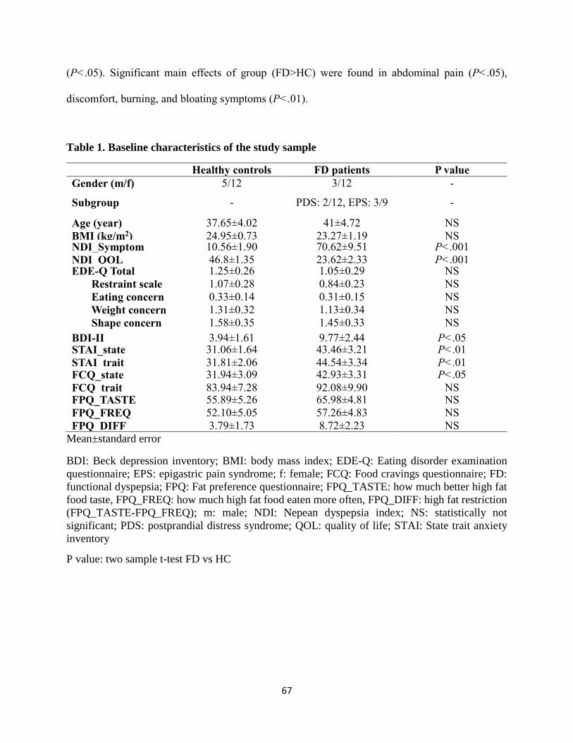
67
(P<.05). Significant main effects of group (FD>HC) were found in abdominal pain (P<.05),
discomfort, burning, and bloating symptoms (P<.01).
Table 1. Baseline characteristics of the study sample
Healthy controls FD patients P value
Gender (m/f) 5/12 3/12 -
Subgroup - PDS: 2/12, EPS: 3/9 -
Age (year) 37.65±4.02 41±4.72 NS
BMI (kg/m2) 24.95±0.73 23.27±1.19 NS NDI_Symptom 10.56±1.90 70.62±9.51 P<.001
NDI_QOL 46.8±1.35 23.62±2.33 P<.001 EDE-Q Total
Restraint scale
Eating concern
Weight concern
Shape concern
1.25±0.26
1.07±0.28
0.33±0.14
1.31±0.32
1.58±0.35
1.05±0.29
0.84±0.23
0.31±0.15
1.13±0.34
1.45±0.33
NS
NS
NS
NS
NS
BDI-II 3.94±1.61 9.77±2.44 P<.05 STAI_state
STAI_trait
31.06±1.64
31.81±2.06
43.46±3.21
44.54±3.34
P<.01
P<.01 FCQ_state
FCQ_trait
31.94±3.09
83.94±7.28
42.93±3.31
92.08±9.90
P<.05
NS FPQ_TASTE
FPQ_FREQ
FPQ_DIFF
55.89±5.26
52.10±5.05
3.79±1.73
65.98±4.81
57.26±4.83
8.72±2.23
NS
NS
NS
Mean±standard error
BDI: Beck depression inventory; BMI: body mass index; EDE-Q: Eating disorder examination
questionnaire; EPS: epigastric pain syndrome; f: female; FCQ: Food cravings questionnaire; FD:
functional dyspepsia; FPQ: Fat preference questionnaire; FPQ_TASTE: how much better high fat
food taste, FPQ_FREQ: how much high fat food eaten more often, FPQ_DIFF: high fat restriction
(FPQ_TASTE-FPQ_FREQ); m: male; NDI: Nepean dyspepsia index; NS: statistically not
significant; PDS: postprandial distress syndrome; QOL: quality of life; STAI: State trait anxiety
inventory
P value: two sample t-test FD vs HC
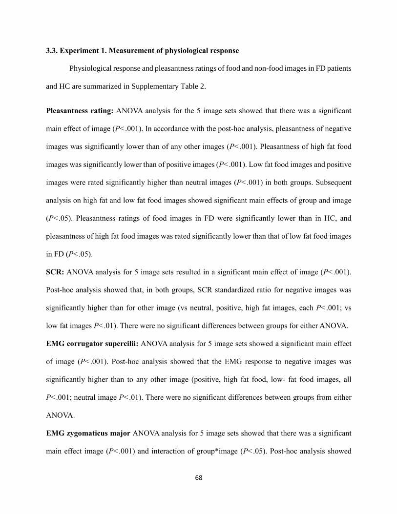
68
3.3. Experiment 1. Measurement of physiological response
Physiological response and pleasantness ratings of food and non-food images in FD patients
and HC are summarized in Supplementary Table 2.
Pleasantness rating: ANOVA analysis for the 5 image sets showed that there was a significant
main effect of image (P<.001). In accordance with the post-hoc analysis, pleasantness of negative
images was significantly lower than of any other images (P<.001). Pleasantness of high fat food
images was significantly lower than of positive images (P<.001). Low fat food images and positive
images were rated significantly higher than neutral images (P<.001) in both groups. Subsequent
analysis on high fat and low fat food images showed significant main effects of group and image
(P<.05). Pleasantness ratings of food images in FD were significantly lower than in HC, and
pleasantness of high fat food images was rated significantly lower than that of low fat food images
in FD (P<.05).
SCR: ANOVA analysis for 5 image sets resulted in a significant main effect of image (P<.001).
Post-hoc analysis showed that, in both groups, SCR standardized ratio for negative images was
significantly higher than for other image (vs neutral, positive, high fat images, each P<.001; vs
low fat images P<.01). There were no significant differences between groups for either ANOVA.
EMG corrugator supercilii: ANOVA analysis for 5 image sets showed a significant main effect
of image (P<.001). Post-hoc analysis showed that the EMG response to negative images was
significantly higher than to any other image (positive, high fat food, low- fat food images, all
P<.001; neutral image P<.01). There were no significant differences between groups from either
ANOVA.
EMG zygomaticus major ANOVA analysis for 5 image sets showed that there was a significant
main effect image (P<.001) and interaction of group*image (P<.05). Post-hoc analysis showed
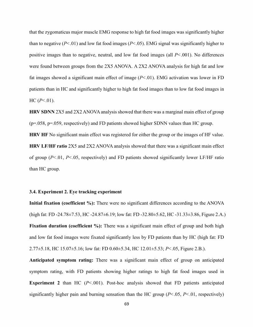
69
that the zygomaticus major muscle EMG response to high fat food images was significantly higher
than to negative (P<.01) and low fat food images (P<.05). EMG signal was significantly higher to
positive images than to negative, neutral, and low fat food images (all P<.001). No differences
were found between groups from the 2X5 ANOVA. A 2X2 ANOVA analysis for high fat and low
fat images showed a significant main effect of image (P<.01). EMG activation was lower in FD
patients than in HC and significantly higher to high fat food images than to low fat food images in
HC (P<.01).
HRV SDNN 2X5 and 2X2 ANOVA analysis showed that there was a marginal main effect of group
(p=.058, p=.059, respectively) and FD patients showed higher SDNN values than HC group.
HRV HF No significant main effect was registered for either the group or the images of HF value.
HRV LF/HF ratio 2X5 and 2X2 ANOVA analysis showed that there was a significant main effect
of group (P<.01, P<.05, respectively) and FD patients showed significantly lower LF/HF ratio
than HC group.
3.4. Experiment 2. Eye tracking experiment
Initial fixation (coefficient %): There were no significant differences according to the ANOVA
(high fat: FD -24.78±7.53, HC -24.87±6.19; low fat: FD -32.80±5.62, HC -31.33±3.86, Figure 2.A.)
Fixation duration (coefficient %): There was a significant main effect of group and both high
and low fat food images were fixated significantly less by FD patients than by HC (high fat: FD
2.77±5.18, HC 15.07±5.16; low fat: FD 0.60±5.34, HC 12.01±5.53; P<.05, Figure 2.B.).
Anticipated symptom rating: There was a significant main effect of group on anticipated
symptom rating, with FD patients showing higher ratings to high fat food images used in
Experiment 2 than HC (P<.001). Post-hoc analysis showed that FD patients anticipated
significantly higher pain and burning sensation than the HC group (P<.05, P<.01, respectively)
70
and there were no differences in fullness and satiation between groups. As for the low fat food
images, none of the symptoms differed between groups (Supplementary Table 3.).
3.5. Correlation analysis
Pearson correlation analysis revealed significant negative correlations between the fat
intake and BDI-II (r=-.88), fat intake and FCQ_DIFF (r=-.93), energy intake and FCQ_DIFF
(r=-.95), and STAI_state and FCQ_state score (r=-.91, P<.05) in FD patients.
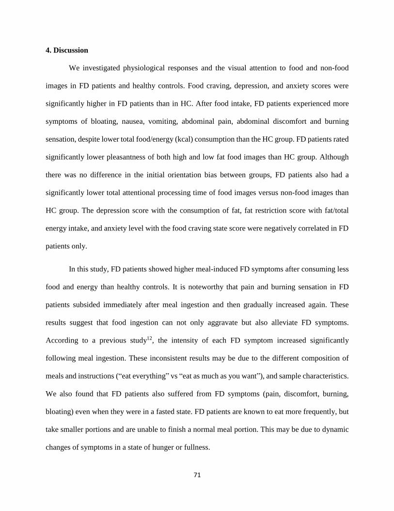
71
4. Discussion
We investigated physiological responses and the visual attention to food and non-food
images in FD patients and healthy controls. Food craving, depression, and anxiety scores were
significantly higher in FD patients than in HC. After food intake, FD patients experienced more
symptoms of bloating, nausea, vomiting, abdominal pain, abdominal discomfort and burning
sensation, despite lower total food/energy (kcal) consumption than the HC group. FD patients rated
significantly lower pleasantness of both high and low fat food images than HC group. Although
there was no difference in the initial orientation bias between groups, FD patients also had a
significantly lower total attentional processing time of food images versus non-food images than
HC group. The depression score with the consumption of fat, fat restriction score with fat/total
energy intake, and anxiety level with the food craving state score were negatively correlated in FD
patients only.
In this study, FD patients showed higher meal-induced FD symptoms after consuming less
food and energy than healthy controls. It is noteworthy that pain and burning sensation in FD
patients subsided immediately after meal ingestion and then gradually increased again. These
results suggest that food ingestion can not only aggravate but also alleviate FD symptoms.
According to a previous study12, the intensity of each FD symptom increased significantly
following meal ingestion. These inconsistent results may be due to the different composition of
meals and instructions (“eat everything” vs “eat as much as you want”), and sample characteristics.
We also found that FD patients also suffered from FD symptoms (pain, discomfort, burning,
bloating) even when they were in a fasted state. FD patients are known to eat more frequently, but
take smaller portions and are unable to finish a normal meal portion. This may be due to dynamic
changes of symptoms in a state of hunger or fullness.
72
As often reported in earlier studies, FD patients had significantly higher anxiety and
depression levels than HC group. In the current study we detected a negative correlation between
the food craving state score and the state anxiety score, and between the depression score and the
amount of fat intake in FD patients. Food craving is known to be less related to hunger than to the
restraint or deprivation of food37. Lower energy consumption in FD patients also suggests that food
craving may be induced by deprivation. A further explanation is that the food craving is more
related to a negative mood, such as anxiety38. Although a clear conclusion cannot be drawn from
correlation analyses, the results may show the mutual influences of a state of anxiety, food craving,
depression, and eating behavior in FD patients.
High HRV and decreased sympathetic activation in FD patients were observed regardless
of the type of pictures, which is akin to the results of previous studies 39, 40. The reduced HRV and
increased sympathetic activation may therefore be an intrinsic characteristic of FD patients rather
than a response to external stimuli. Furthermore, the emotional response during the visual
stimulation of food and non-food cues did not differ significantly between groups. This can be
interpreted along with the eye tracking results, which showed a similar tendency of initial attention
with HC group and a lower total attention processing time (fixation duration) to food images in FD
patients than in the HC group. While visual food images may not immediately induce negative
emotional and avoidance responses, a late cognitive processing of the images by higher cognitive
function may cause the avoidance response to food images in FD patients while processing food
images. These results suggest that high level cognitive functions rather than autonomic and
emotional mechanisms can operate differently in FD patients. Furthermore, a decreased fixation
duration on food images in FD patients is at variance with earlier findings in patients with obesity
and binge eating disorder33, 41 (where increased duration on food images was reported) and is
73
similar to the results in anorexia nervosa patients42 suggesting a positive and negative perception
of food cues in eating-related diseases.
The reduced pleasantness of and attentional bias to visual food stimuli in FD patients could
be a key to future psychotherapeutic intervention and research. Various treatment options have
been proposed for FD, such as H. pylori eradication, prokinetic agents, acid suppressive
medications, antidepressants. Nevertheless, a standardized treatment strategy for FD patients has
yet to be established and cognitive behavioral therapy remains an unexplored area43. A new therapy
that includes self-restraint response to food, emotional management, and eating behavior
modification could be considered for patients who do not respond to conventional therapies.
Furthermore, how FD patients perceive, encode, store, and recall the value of food and how food
memories influence their food-related decision making are interesting topics for future studies.
In the interviews conducted before the study, almost all FD patients complained about the
changes in their eating behavior and their poor quality of life. Most patients avoided symptom-
related foods, such as fatty foods, bread, pasta, or alcohol, which varied from person to person and
almost all patients requested advice as to what food they should be eating. Fatty foods aggravated
the symptoms in some patients, whereas others remained unaffected. Nevertheless, the high fat
restriction score was significantly related to the lower intake of fat and total energy in FD patients
only and they anticipated more severe symptoms to high fat food images than HC. Previous
negative memory of the aftereffect of eating could be extended to the restriction of food intake and
the attentional avoidance20. This fact needs to be better recognized in clinics and clinical studies,
and food consultation might be instrumental in improving the quality of life and establish healthier
eating guidelines for patients.
74
A limitation of this study was the difficulty in finding one particular item of food that might
be either symptom-related or symptom-unrelated to each patient. We therefore used standard
images for all participants. This may be the basis for the similar autonomic and emotional responses
to high fat and low fat images in our study. However, since this first-of-its-kind study investigates
the basic physiological response to food in FD patients, we tried to include various measurements
with diverse images from established databases. Moreover, our sample size was not large enough
to conduct further subgroup analysis and we did not examine any differences between PDS and
EPS, patients with severe and mild FD/depression/anxiety symptoms. Due to the lack of knowledge
on the food-related behavior, cognitive, emotional, and physiological responses of FD patients,
further studies with large sample size are necessary.

75
5. Conclusion
We observed an increased food craving, decreased amount of food intake, food ingestion-
induced aggravation of FD symptoms, and abnormal visual processing time and perception of food-
related pleasantness in FD patients. The effectiveness of conventional therapies in FD patients
might be enhanced by taking dietary consultation and modification of psychological response to
food as well as somatic symptoms, and future studies on the evaluation of food may identify the
underlying pathophysiology of FD.

76
6. Acknowledgement
The research leading to these results received funding from the People Programme of the European
Union’s Seventh Framework Programme under REA grant agreement No. 607652 (NeuroGUT),
the European Union’s Seventh Framework Programme (FP7/2007-2013) under grant agreement
No. 607310 (Nudge-it).
77
Reference
1. Drossman DA, Dumitrascu DL. Rome III: New standard for functional gastrointestinal disorders. J Gastrointestin Liver Dis 2006;15:237-41.
2. Tack J, Talley NJ. Functional dyspepsia--symptoms, definitions and validity of the Rome III criteria. Nat Rev Gastroenterol Hepatol 2013;10:134-41.
3. Van Oudenhove L, Aziz Q. The role of psychosocial factors and psychiatric disorders in functional dyspepsia. Nat Rev Gastroenterol Hepatol 2013;10:158-67.
4. Khodarahmi M, Azadbakht L. Dietary fat intake and functional dyspepsia. Adv Biomed Res 2016;5:76.
5. Goktas Z, Koklu S, Dikmen D, et al. Nutritional habits in functional dyspepsia and its subgroups: a comparative study. Scand J Gastroenterol 2016;51:903-7.
6. Fried M, Feinle C. The role of fat and cholecystokinin in functional dyspepsia. Gut 2002;51 Suppl 1:i54-7.
7. Feinle-Bisset C, Azpiroz F. Dietary and lifestyle factors in functional dyspepsia. Nat Rev Gastroenterol Hepatol 2013;10:150-7.
8. Pilichiewicz AN, Horowitz M, Holtmann GJ, et al. Relationship between symptoms and dietary patterns in patients with functional dyspepsia. Clin Gastroenterol Hepatol 2009;7:317-22.
9. Carvalho RV, Lorena SL, Almeida JR, et al. Food intolerance, diet composition, and eating patterns in functional dyspepsia patients. Dig Dis Sci 2010;55:60-5.
10. Mullan A, Kavanagh P, O'Mahony P, et al. Food and nutrient intakes and eating patterns in functional and organic dyspepsia. Eur J Clin Nutr 1994;48:97-105.
11. Kazemi M, Eshraghian A, Hamidpour L, et al. Changes in serum ghrelin level in relation to meal-time in patients with functional dyspepsia. United European Gastroenterol J 2015;3:11-6.
12. Bisschops R, Karamanolis G, Arts J, et al. Relationship between symptoms and ingestion of a meal in functional dyspepsia. Gut 2008;57:1495-503.
13. Yamawaki H, Futagami S, Kawagoe T, et al. Improvement of meal-related symptoms and epigastric pain in patients with functional dyspepsia treated with acotiamide was associated with acylated ghrelin levels in Japan. Neurogastroenterol Motil 2016;28:1037-47.
14. Amiriani T, Javadi H, Raiatnavaz T, et al. Assessment of Gastric Accommodation in Patients with Functional Dyspepsia by 99mTc-Pertechtenate Single Photon Emission Computed Tomography Imaging: Practical but not Widely Accepted. Mol Imaging Radionucl Ther 2015;24:105-9.
15. Stojek MMK, MacKillop J. Relative reinforcing value of food and delayed reward discounting in obesity and disordered eating: A systematic review. Clin Psychol Rev 2017;55:1-11.
16. Soussignan R, Jiang T, Rigaud D, et al. Subliminal fear priming potentiates negative facial reactions to food pictures in women with anorexia nervosa. Psychol Med 2010;40:503-14.
17. Nijs IM, Franken IH. Attentional Processing of Food Cues in Overweight and Obese Individuals. Curr Obes Rep 2012;1:106-113.
18. Scaife JC, Godier LR, Reinecke A, et al. Differential activation of the frontal pole to high vs low calorie foods: The neural basis of food preference in Anorexia Nervosa? Psychiatry Res 2016;258:44-53.
19. Wolz I, Sauvaget A, Granero R, et al. Subjective craving and event-related brain response to olfactory and visual chocolate cues in binge-eating and healthy individuals. Sci Rep 2017;7:41736.
20. Higgs S. Cognitive processing of food rewards. Appetite 2016;104:10-7. 21. Folkvord F, Anschutz DJ, Wiers RW, et al. The role of attentional bias in the effect of food
advertising on actual food intake among children. Appetite 2015;84:251-8. 22. Dimberg U. Facial electromyography and emotional reactions. Psychophysiology 1990;27:481-94.
78
23. Douglas A. Drossman, Enrico Corazziari M, Delvaux RCS, et al. ROME III: The Functional Gastrointestinal Disorders: Yale University Section of Digestive Disease: Degnon Associates, 2006.
24. Fridlund AJ, Cacioppo JT. Guidelines for human electromyographic research. Psychophysiology 1986;23:567-89.
25. Talley NJ, Haque M, Wyeth JW, et al. Development of a new dyspepsia impact scale: the Nepean Dyspepsia Index. Aliment Pharmacol Ther 1999;13:225-35.
26. Beck AT, Steer, R.A., & Brown, G. Manual for the Beck Depression Inventory-II: San Antonio, TX: Psychological Corporation., 1996.
27. Spielberger CD, Gorsuch, R. L. Lushene, R., Vagg, P. R., & Jacobs, G. A. . Manual for the State-Trait Anxiety Inventory. : Palo Alto, CA: Consulting Psychologists Press., 1983.
28. Fairburn CG, Beglin SJ. Assessment of eating disorders: interview or self-report questionnaire? Int J Eat Disord 1994;16:363-70.
29. Cepeda-Benito A, Gleaves, D. H., Williams, T. L., & Erath, S. A. The development and validation of the state and trait food-cravings questionnaires. Behavior Therapy 2000;1:151-173.
30. Ledikwe JH, Ello-Martin J, Pelkman CL, et al. A reliable, valid questionnaire indicates that preference for dietary fat declines when following a reduced-fat diet. Appetite 2007;49:74-83.
31. Lang PJ, Bradley MM, Cuthbert BN. International affective picture system (IAPS): Affective ratings of pictures and instruction manual. Technical Report A-8. . Gainesville, FL.: University of Florida, 2008.
32. Blechert J, Meule A, Busch NA, et al. Food-pics: an image database for experimental research on eating and appetite. Front Psychol 2014;5:617.
33. Schag K, Teufel M, Junne F, et al. Impulsivity in binge eating disorder: food cues elicit increased reward responses and disinhibition. PLoS One 2013;8:e76542.
34. Frank S, Laharnar N, Kullmann S, et al. Processing of food pictures: influence of hunger, gender and calorie content. Brain Res 2010;1350:159-66.
35. Tarvainen MP, Niskanen JP, Lipponen JA, et al. Kubios HRV--heart rate variability analysis software. Comput Methods Programs Biomed 2014;113:210-20.
36. Instruments S. BeGaze Manual version 3.0. Berlin: SensoMotoric Instruments, 2011. 37. Hill AJ. The psychology of food craving. Proc Nutr Soc 2007;66:277-85. 38. Hill AJ, Weaver CF, Blundell JE. Food craving, dietary restraint and mood. Appetite 1991;17:187-
97. 39. Dal K, Deveci OS, Kucukazman M, et al. Decreased parasympathetic activity in patients with
functional dyspepsia. Eur J Gastroenterol Hepatol 2014;26:748-52. 40. Tominaga K, Fujikawa Y, Tsumoto C, et al. Disorder of autonomic nervous system and its
vulnerability to external stimulation in functional dyspepsia. J Clin Biochem Nutr 2016;58:161-5. 41. Castellanos EH, Charboneau E, Dietrich MS, et al. Obese adults have visual attention bias for food
cue images: evidence for altered reward system function. Int J Obes (Lond) 2009;33:1063-73. 42. Giel KE, Friederich HC, Teufel M, et al. Attentional processing of food pictures in individuals with
anorexia nervosa--an eye-tracking study. Biol Psychiatry 2011;69:661-7. 43. Soo S, Moayyedi P, Deeks J, et al. Psychological interventions for non-ulcer dyspepsia. Cochrane
Database Syst Rev 2005:CD002301.

79
Figures and figure legends
Figure 1. Experimental protocol
A. Experimental procedure of the study. B. Illustration of the Experiment 1 including skin
conductance response, heart rate, electromyography measurements and pleasantness rating to food
and non-food images. Randomized order of 5 blocks of images (neutral, positive, negative, high
fat, and low fat food images, n=30, 6000ms for each image) with fixation cross (5000ms) between
each block were presented. C. Schematic presentation of the eye tracking experiment using free
exploration paradigm. Low fat food and non-food pairs and high fat food and non-food pairs (n=12,
respectively) were presented for 3000 ms with 2000 ms of fixation cross between pairs. Location
of the images (1st-4th quadrant) was balanced.
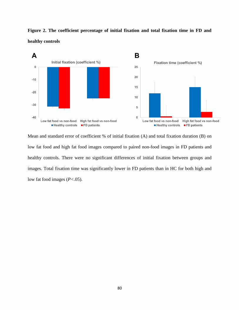
80
Figure 2. The coefficient percentage of initial fixation and total fixation time in FD and
healthy controls
Mean and standard error of coefficient % of initial fixation (A) and total fixation duration (B) on
low fat food and high fat food images compared to paired non-food images in FD patients and
healthy controls. There were no significant differences of initial fixation between groups and
images. Total fixation time was significantly lower in FD patients than in HC for both high and
low fat food images (P<.05).
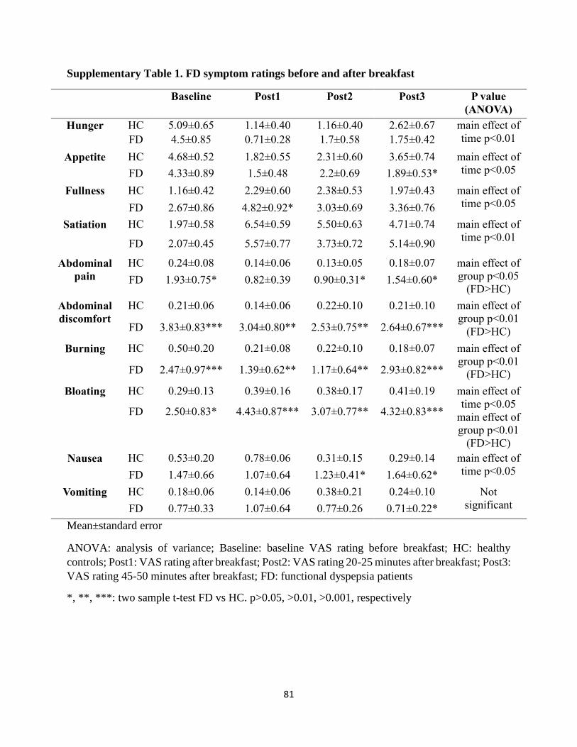
81
Supplementary Table 1. FD symptom ratings before and after breakfast
Baseline Post1 Post2 Post3 P value
(ANOVA)
Hunger HC 5.09±0.65 1.14±0.40 1.16±0.40 2.62±0.67 main effect of
time p<0.01
FD 4.5±0.85 0.71±0.28 1.7±0.58 1.75±0.42
Appetite HC 4.68±0.52 1.82±0.55 2.31±0.60 3.65±0.74 main effect of
time p<0.05 FD 4.33±0.89 1.5±0.48 2.2±0.69 1.89±0.53*
Fullness HC 1.16±0.42 2.29±0.60 2.38±0.53 1.97±0.43 main effect of
time p<0.05 FD 2.67±0.86 4.82±0.92* 3.03±0.69 3.36±0.76
Satiation HC 1.97±0.58 6.54±0.59 5.50±0.63 4.71±0.74 main effect of
time p<0.01 FD 2.07±0.45 5.57±0.77 3.73±0.72 5.14±0.90
Abdominal
pain
HC 0.24±0.08 0.14±0.06 0.13±0.05 0.18±0.07 main effect of
group p<0.05
(FD>HC) FD 1.93±0.75* 0.82±0.39 0.90±0.31* 1.54±0.60*
Abdominal
discomfort
HC 0.21±0.06 0.14±0.06 0.22±0.10 0.21±0.10 main effect of
group p<0.01
(FD>HC) FD 3.83±0.83*** 3.04±0.80** 2.53±0.75** 2.64±0.67***
Burning HC 0.50±0.20 0.21±0.08 0.22±0.10 0.18±0.07 main effect of
group p<0.01
(FD>HC) FD 2.47±0.97*** 1.39±0.62** 1.17±0.64** 2.93±0.82***
Bloating HC 0.29±0.13 0.39±0.16 0.38±0.17 0.41±0.19 main effect of
time p<0.05
main effect of
group p<0.01
(FD>HC)
FD 2.50±0.83* 4.43±0.87*** 3.07±0.77** 4.32±0.83***
Nausea HC 0.53±0.20 0.78±0.06 0.31±0.15 0.29±0.14 main effect of
time p<0.05 FD 1.47±0.66 1.07±0.64 1.23±0.41* 1.64±0.62*
Vomiting HC 0.18±0.06 0.14±0.06 0.38±0.21 0.24±0.10 Not
significant FD 0.77±0.33 1.07±0.64 0.77±0.26 0.71±0.22*
Mean±standard error
ANOVA: analysis of variance; Baseline: baseline VAS rating before breakfast; HC: healthy
controls; Post1: VAS rating after breakfast; Post2: VAS rating 20-25 minutes after breakfast; Post3:
VAS rating 45-50 minutes after breakfast; FD: functional dyspepsia patients
*, **, ***: two sample t-test FD vs HC. p>0.05, >0.01, >0.001, respectively
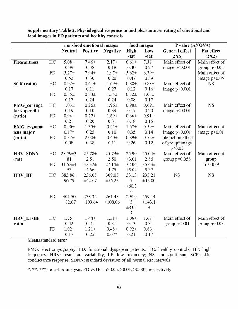
82
Supplementary Table 2. Physiological response to and pleasantness rating of emotional and
food images in FD patients and healthy controls
non-food emotional images food images P value (ANOVA)
Neutral Positive Negative High
-fat
Low
-fat
General effect
(2X5)
Fat effect
(2X2)
Pleasantness HC 5.08±
0.39
7.46±
0.38
2.17±
0.18
6.61±
0.40
7.38±
0.27
Main effect of
image p<0.001
Main effect of
group p<0.05
Main effect of
image p<0.05
FD 5.27±
0.52
7.94±
0.30
1.97±
0.20
5.62±
0.47
6.79±
0.39
SCR (ratio) HC 0.92±
0.17
0.61±
0.11
1.69±
0.27
0.88±
0.12
0.83±
0.16
Main effect of
image p<0.001
NS
FD 0.85±
0.17
0.83±
0.24
1.55±
0.24
0.72±
0.08
1.05±
0.17
EMG_corruga
tor supercilii
(ratio)
HC 1.03±
0.19
0.26±
0.10
1.96±
0.39
0.90±
0.17
0.69±
0.20
Main effect of
image p<0.001
NS
FD 0.94±
0.21
0.77±
0.20
1.69±
0.31
0.66±
0.18
0.91±
0.15
EMG_zygomat
icus major
(ratio)
HC 0.90±
0.17*
1.35±
0.25
0.41±
0.10
1.67±
0.35
0.59±
0.14
Main effect of
image p<0.001
Interaction effect
of group*image
p<0.05
Main effect of
image p<0.01
FD 0.37±
0.08
2.00±
0.38
0.40±
0.11
0.89±
0.26
0.52±
0.12
HRV_SDNN
(ms)
HC 28.79±3.
81
25.78±
2.51
25.79±
2.50
25.90
±3.01
25.04±
2.86
Main effect of
group p=0.058
Main effect of
group
p=0.059 FD 31.52±4.
53
32.32±
4.66
27.14±
4.75
32.06
±5.02
35.43±
5.37
HRV_HF HC 383.86±
96.79
236.05
±42.07
309.05
±56.23
331.3
7
±60.3
6
235.21
±42.00
NS NS
FD 401.50
±82.67
338.32
±109.64
261.48
±108.06
298.9
3
±83.3
7
459.14
±143.1
8
HRV_LF/HF
ratio
HC 1.75±
0.42
1.44±
0.21
1.38±
0.31
1.06±
0.13
1.67±
0.31
Main effect of
group p<0.01
Main effect of
group p<0.05
FD 1.02±
0.17
1.21±
0.25
0.48±
0.07*
0.92±
0.21
0.86±
0.17
Mean±standard error
EMG: electromyography; FD: functional dyspepsia patients; HC: healthy controls; HF: high
frequency; HRV: heart rate variability; LF: low frequency; NS: not significant; SCR: skin
conductance response; SDNN: standard deviation of all normal RR intervals
*, **, ***: post-hoc analysis, FD vs HC. p>0.05, >0.01, >0.001, respectively
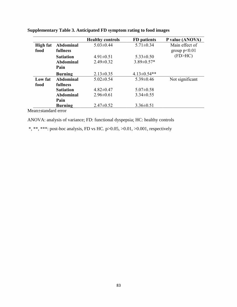
83
Supplementary Table 3. Anticipated FD symptom rating to food images
Healthy controls FD patients P value (ANOVA)
High fat
food
Abdominal
fullness
5.03±0.44 5.71±0.34 Main effect of
group p<0.01
(FD>HC) Satiation 4.91±0.51 5.33±0.50 Abdominal
Pain
2.49±0.32 3.89±0.57*
Burning 2.13±0.35 4.13±0.54**
Low fat
food
Abdominal
fullness
5.02±0.54 5.39±0.46 Not significant
Satiation 4.82±0.47 5.07±0.58 Abdominal
Pain
2.96±0.61 3.34±0.55
Burning 2.47±0.52 3.36±0.51 Mean±standard error
ANOVA: analysis of variance; FD: functional dyspepsia; HC: healthy controls
*, **, ***: post-hoc analysis, FD vs HC. p>0.05, >0.01, >0.001, respectively
84
8. Paper IV. The effect of fat label on gastrointestinal symptoms and brain activity
in functional dyspepsia patients: an fMRI study
Author contributions
The material of this chapter was submitted to Gastroenterology (2017 August). All authors
designed the study and interpreted the results together. In-Seon Lee acquired and analyzed all the
data. Results were discussed with the help of Hubert Preissl, Stephanie Kullmann, and Paul Enck.
In-Seon Lee wrote the manuscript and Hubert Preissl, Stephanie Kullmann, and Paul Enck revised
the manuscript.
Acknowledgement
The research leading to these results received funding from the People Programme of the
European Union’s Seventh Framework Programme under REA grant agreement No. 607652
(NeuroGUT), the European Union’s Seventh Framework Programme (FP7/2007-2013) under grant
agreement No. 607310 (Nudge-it).
85
Fat label versus fat content: Gastrointestinal symptoms and brain activity in
functional dyspepsia patients and healthy controls
In-Seon Lee1,2, Stephanie Kullmann3, Klaus Scheffler4,5, Hubert Preissl3, 6*, Paul Enck1*
1. Psychosomatic Medicine and Psychotherapy Department, University of Tübingen,
Tübingen, Germany
2. IMPRS for Cognitive and Systems Neuroscience, Tübingen, Germany
3. Institute for Diabetes Research and Metabolic Diseases of the Helmholtz Center Munich at
the University of Tübingen; German Center for Diabetes Research (DZD); Department of
Internal Medicine, Division of Endocrinology, Diabetology, Angiology, Nephrology and
Clinical Chemistry, University of Tübingen, Tübingen, Germany
4. Department of High-Field Magnetic Resonance, Max Planck Institute for Biological
Cybernetics, Tübingen, Germany
5. Department of Biomedical Magnetic Resonance, University of Tübingen, Tübingen,
Germany
6. Institute of Pharmaceutical Sciences; Interfaculty Centre for Pharmacogenomics and
Pharma Research, Department of Pharmacy and Biochemistry, University of Tübingen,
Tübingen, Germany
86
Abstract
High fat meals in particular are associated with dyspeptic symptoms in functional dyspepsia
(FD) patients. However, it is still unclear what neural processes are involved and how they can be
modulated by psychological factors such as expectation. We aimed to investigate brain activity by
functional magnetic resonance imaging (fMRI) after the ingestion of high and low fat food with
correct and incorrect fat information. Methods: We compared 12 FD patients and 14 age- and BMI-
matched healthy controls (5 males in each group). We recorded resting state fMRI on four different
days after an overnight fast before and after ingestion of one of four yogurts (200ml, either 10% or
0.1% fat, with ‘low fat’ or ‘high fat’ label (2x2 factorial design), sequence-randomized across
subjects). The statistical significance level was set at α=0.05 and multiple comparison correction
was applied. Results: FD patients showed more pronounced dyspeptic symptoms after consuming
high fat-labeled yoghurt than low fat-labeled yoghurt, irrespective of the actual fat content. This is
indicative of either a placebo effect of low fat information, or a nocebo effect of high fat
information on symptom expression. FD patients showed greater activity than healthy controls in
occipital areas before and after ingestion regardless of fat content and label as well as greater
activity in the middle frontal gyrus before ingestion. In addition, functional connectivity (FC) from
the insula to occipital cortex (I-O) increased after high fat and decreased after low fat ingestion in
FD patients. FC from the insula to the precuneus (I-P) was higher in FD patients than in healthy
controls after ingestion of yoghurt with a low fat label. In FD patients, I-O functional connectivity
negatively correlated with nausea and I-P functional connectivity with FD symptom intensity, food
craving, and depression. In summary, our results endorse the importance of psychological
perception of food on the incidence of dyspeptic symptoms and on the altered brain activities.
Taken together, these findings provide further evidence for the importance of cognitive

87
components in perception of fat, food craving, depression, and brain functions in
pathophysiological mechanisms of FD.
88
1. Introduction
Functional dyspepsia (FD) is characterized by postprandial fullness, early satiation,
epigastric pain, bloating, nausea symptoms after meals, particularly high fat food [1, 2], in the
absence of any structural abnormalities in the gastrointestinal tract [3, 4]. The effect of fat in the
altered gastrointestinal sensitivity and symptoms is a well-known pathophysiological feature in FD
patients. Intra-duodenal infusion of lipids, not glucose nor saline, were shown to induce nausea,
bloating, and vomiting symptoms in FD patients [5, 6]. After ingestion of a high fat meal, nausea
and pain symptoms were greater than after a high carbohydrate meal [1] and food diaries revealed
that FD patients consumed lower amounts of fat and that bloating symptoms were related to the
amount of fat ingested [2].
Feinle-Bisset et al. showed that a low fat meal, served to FD patients under the pretense
that it was high fat meal, caused more severe fullness and bloating symptoms than a low fat meal
served to with the correct fat information. [7]. In addition it has been shown in healthy volunteers
that ghrelin levels, as a physiological marker of satiation, varied after ingestion of identical
milkshakes when subjects were informed before that this was either a high fat, high calorie or a
low fat, low calorie milkshake. These findings suggest that the cognitive perception of fat at the
central nervous system level plays a prominent role in the secretion of hormones, altered perceptual
response to fat, and symptom reporting. The non-specific improvement (or worsening) of
symptoms by an inactive treatment or treatment-unrelated cue – the placebo (or nocebo) effect – is
due to the belief or expectation of symptom relief (or exacerbation). It is conceivable that if FD
patients were aware of a close association between their symptoms and high-fat diet, the
information on the amount of fat (more or less) could have an impact on their dyspeptic symptoms.
One hypothesis from the early 1990s proposed that abnormalities of the brain-gut axis are
one of the key mechanisms governing FD [8]. The presence of the food or nutrient in the
89
gastrointestinal tract is signaled to the central nervous system which, in turn, modulates
gastrointestinal function and eating behavior, and controls the gastrointestinal symptoms [9].
Furthermore, some of the brain’s many pathways for controlling the perception of internal and
external stimuli might be impaired in FD patients and cause somatic symptoms. The recent
development of functional magnetic resonance imaging (fMRI) technique has enabled scientists to
characterize the intrinsic brain activities and networks, and its intervention-related changes. A large
number functional neuroimaging studies have investigated the brain activities and networks during
the resting state (no-tasks) [10-17] and in reaction to the visceral distention [18-20]. They suggest
that there is an alteration in the activation of the cognitive and pain processing brain regions
(prefrontal cortex, somatosensory cortex, insula, cingulate cortex, thalamus, etc.) in FD patients
[21]. However, little is known about how fat or fat information is processed in the brain or how it
is mediated by pathological changes such as FD symptoms, decreased quality of life, increased
depression and anxiety, food craving, etc., in FD patients.
In the current study, we investigated the effect of fat ingestion and fat label, and
pathological factors on the resting state brain activities in FD patients and healthy controls (HC).
We hypothesized that i) the resting state brain activity in cognitive and pain processing networks
are mainly affected in FD patients, and ii) the functional connectivity (FC) emerging from the
middle-posterior insula is associated with the pathological variables in FD patients. Bilateral
middle-posterior insula was selected as a seed region since it is involved in signaling interoceptive
visceral sensation and homeostatic information. It responds to a wide variety of experimental
stimuli including pain/non-painful/salient/emotional stimuli as a region of the homeostatic afferent
network [22-27]. Moreover, the insula is activated during baseline condition (compared to healthy
controls) and in response to visceral distention (compared to baseline) [19, 20, 28], and correlates
with FD symptom intensity [10, 13], disease-related quality of life [29], and disease duration [16]

90
in FD patients. We tested our hypotheses by measuring resting state fMRI in the fasted and fed
state and performing seed-based FC analysis, correlation analysis, and mediation analysis.
91
2. Methods
2.1. Participants
12 FD patients (5 males, age 46.46±5.64 years, mean±standard error) and 14 for age- and
BMI controlled healthy subjects (HCs, 5 males, age 45.79±4.71 years) participated in the study.
Right-handed volunteers within the range of 18-75 years of age and with a body mass index (BMI,
weight/height2) of 19-29 kg/m2 were included. FD patients were diagnosed on the basis of the
ROME III criteria [30] as well as an unsuspicious endoscopy documented in their medical records.
Volunteers with non-removable metal implants, claustrophobia, severe psychiatric illness,
substance dependence and abuse, and any food allergy or intolerance were excluded from the study.
The ethics committee of the Medical Faculty, University of Tübingen, Germany (633/2015802)
approved the study and all participants gave their informed consent.
2.2. Test food
Two plain yogurts, low fat (0.1% fat, 200ml, 106kcal, 13.8g carbohydrate, 11g protein) and
high fat (10% fat, 200ml, 266kcal, 14g carbohydrate, 6g protein, Weihenstephan, Freising,
Germany) were used. Congruent or incongruent labels were attached to each yogurt (high fat yogurt
with ‘high fat’ label: HH, high fat yogurt with ‘low fat’ label: HL, low fat yogurt with ‘high fat’
label: LH, low fat yogurt with ‘low fat’ label: LL).
2.3. Study design
Each participant was examined in the morning (7-11 a.m.) on four separate occasions
following an overnight fast. Smoking and consumption of alcohol, coffee, or tea were prohibited

92
during the fasting period. Participants completed a visual analogue scale (VAS; 0=no symptoms at
all, 10=very severe symptoms) to assess hunger, appetite, abdominal fullness, satiation, nausea,
vomiting, abdominal pain and uncomfortable, burning, and bloating (baseline FD symptoms).
Identical VAS ratings were assessed again immediately (Post1), 10 minutes (Post2), and 20 minutes
(Post3) after the yogurt consumption. Between the pre-yogurt and post-yogurt fMRI sessions,
participants were permitted to exit the scanner and were served one of the 4 yogurts (HH, HL, LH,
LL) in randomized order. Participants were asked to sit on the MRI table and eat a whole portion
of yogurt within 5 minutes. At the end of the study, patients indicated their dyspepsia symptom
intensity, and disease-related quality of life was measured using Nepean Dyspepsia Index (NDI)
[31]. Depression and anxiety levels were evaluated using Beck Depression Inventory (BDI-II) [32]
and State-Trait Anxiety Inventory (STAI) [33]. Furthermore, the Eating Disorders Examination
Questionnaire (EDE-Q) [34], Food Craving Questionnaire (FCQ) [35], and Fat Preference
Questionnaire (FPQ) [36] were used to evaluate their eating behavior.
2.4. Imaging protocol
All images were obtained with a 3 Tesla scanner (Siemens MAGNETOM Prisma, Erlangen,
Germany). On the first day, a high resolution T1-weighted anatomical image (magnetization-
prepared rapid gradient-echo) was recorded (repetition time (TR)=2300ms, echo time
(TE)=4.18ms, 176 slices, matrix=256×256, voxel size=1×1×1cm3). Whole brain blood
oxygenation level-dependent (BOLD) data were obtained using standard T2*-weighted echo planar
sequence (160 volumes, TR=2000ms, TE=30ms, 30 slices, matrix=64×64, flip angle=80°, voxel
size=3×3×3.4cm3) before and after ingestion.
2.5. Imaging processing
93
Preprocessing of the BOLD signal was performed using Data Processing Assistant for
Resting-State fMRI (DPARSF, http://restfmri.net, v2.2) [37] and SPM8 (Statistical Parametric
Mapping, Wellcome Trust Centre for Neuroimaging, London, UK). Images for each subject were
assessed to identify any excessive movement (>2mm or 2º degree) and the first 4 volumes were
discarded for signal equilibrium and adaptation. Slice time-correction and head motion- correction
were applied to raw images, and functional images were realigned and co-registered with the
structural image. Images were normalized into the Montreal Neurological Institute (MNI) space
and smoothed with a Gaussian kernel full width at half maximum 6mm. Following preprocessing,
amplitude of low frequency fluctuations (ALFF) analysis within the low frequency band (0.01-
0.1Hz) was performed using the DPARSF. The time series data of each voxel was transformed into
the frequency domain, and the power spectrum amplitude was calculated. The square root was
calculated at each frequency of the power spectrum, and the average square root was then obtained
across 0.01-0.1 Hz at each voxel. This average square root was taken as the ALFF and mean ALFF
(mALFF) was calculated as the original ALFF value/averaged ALFF across all voxels.
For seed-based FC analysis, 8mm sphere ROIs of the left and right middle-posterior insula
were defined by peak coordinates (x=-42, y=-33, z=17; x=36, y=-15, z=13, respectively) of clusters
from a resting state ALFF map (family-wise error (FWE) corrected p<0.05, cluster dimension k>10
voxels). The averaged time course was then obtained from the ROIs and the correlation analysis
was performed in a voxel-wise fashion. Finally, the correlation coefficient map was converted into
z maps by Fisher’s r-to-z transform to improve the normality (zFC). For correlation and mediation
analysis, the first eigenvariate of each cluster that survived the threshold from mALFF and zFC
maps was extracted.
2.5 Statistical analysis
94
All statistical analyses were performed using IBM SPSS statistics 24.0 (IBM Corp. New
York, USA). An independent two sample t-test was used to compare sample characteristics
between the groups. For VAS ratings of FD symptoms, a two-way repeated ANOVA was conducted
with time (pre, post1, post2, post3) and the group (HC, FD) factors. For ALFF and FC maps, SPM
second level t-tests between the groups (HC, FD), fat content (high, low), and labels (high, low)
were performed. Two-tailed Pearson’s partial correlation analysis was also computed between
questionnaire variables and the intensity of ALFF and FC while controlling for age and BMI.
Mediation analysis was performed as described by Hayes using the PROCESS macro [38]. Age
and BMI were included as covariates in the simple mediation model with 1000 bootstrap samples.
The statistical significance level was set at α=0.05 while FWE correction for fMRI analysis and
Bonferroni correction for the analysis of behavior data were applied to account for multiple
comparison.
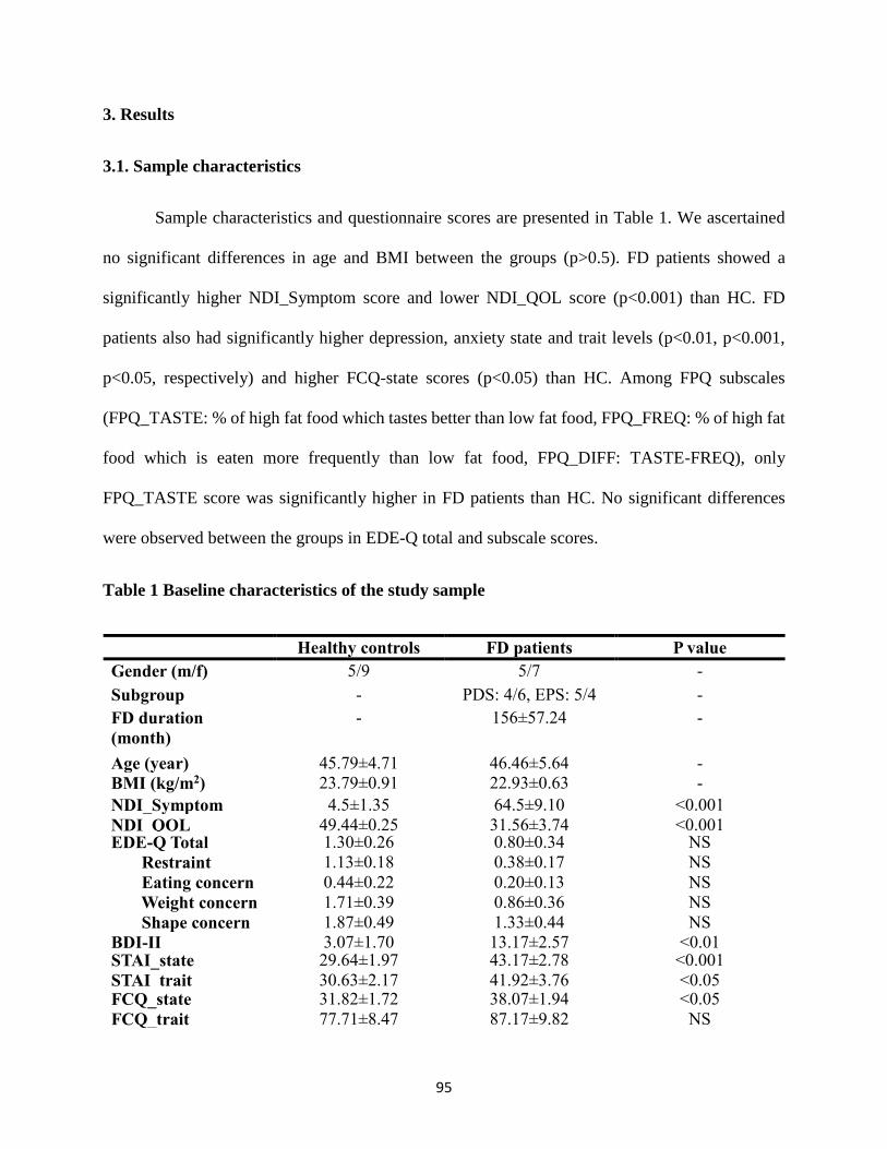
95
3. Results
3.1. Sample characteristics
Sample characteristics and questionnaire scores are presented in Table 1. We ascertained
no significant differences in age and BMI between the groups (p>0.5). FD patients showed a
significantly higher NDI_Symptom score and lower NDI_QOL score (p<0.001) than HC. FD
patients also had significantly higher depression, anxiety state and trait levels (p<0.01, p<0.001,
p<0.05, respectively) and higher FCQ-state scores (p<0.05) than HC. Among FPQ subscales
(FPQ_TASTE: % of high fat food which tastes better than low fat food, FPQ_FREQ: % of high fat
food which is eaten more frequently than low fat food, FPQ_DIFF: TASTE-FREQ), only
FPQ_TASTE score was significantly higher in FD patients than HC. No significant differences
were observed between the groups in EDE-Q total and subscale scores.
Table 1 Baseline characteristics of the study sample
Healthy controls FD patients P value
Gender (m/f) 5/9 5/7 -
Subgroup - PDS: 4/6, EPS: 5/4 -
FD duration
(month)
- 156±57.24
-
Age (year) 45.79±4.71
46.46±5.64
-
BMI (kg/m2) 23.79±0.91
22.93±0.63
-
NDI_Symptom 4.5±1.35
64.5±9.10
<0.001
NDI_QOL 49.44±0.25
31.56±3.74
<0.001 EDE-Q Total
Restraint
Eating concern
Weight concern
Shape concern
1.30±0.26
1.13±0.18
0.44±0.22
1.71±0.39
1.87±0.49
0.80±0.34
0.38±0.17
0.20±0.13
0.86±0.36
1.33±0.44
NS
NS
NS
NS
NS
BDI-II 3.07±1.70
13.17±2.57
<0.01 STAI_state
STAI_trait
29.64±1.97
30.63±2.17
43.17±2.78
41.92±3.76
<0.001
<0.05 FCQ_state
FCQ_trait
31.82±1.72
77.71±8.47
38.07±1.94
87.17±9.82
<0.05
NS
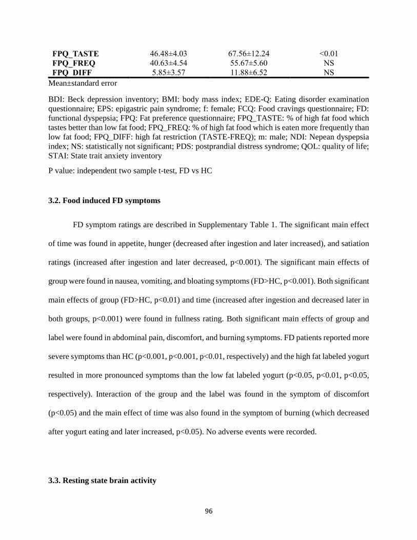
96
FPQ_TASTE
FPQ_FREQ
FPQ_DIFF
46.48±4.03
40.63±4.54
5.85±3.57
67.56±12.24
55.67±5.60
11.88±6.52
<0.01
NS
NS
Mean±standard error
BDI: Beck depression inventory; BMI: body mass index; EDE-Q: Eating disorder examination
questionnaire; EPS: epigastric pain syndrome; f: female; FCQ: Food cravings questionnaire; FD:
functional dyspepsia; FPQ: Fat preference questionnaire; FPQ_TASTE: % of high fat food which
tastes better than low fat food; FPQ_FREQ: % of high fat food which is eaten more frequently than
low fat food; FPQ_DIFF: high fat restriction (TASTE-FREQ); m: male; NDI: Nepean dyspepsia
index; NS: statistically not significant; PDS: postprandial distress syndrome; QOL: quality of life;
STAI: State trait anxiety inventory
P value: independent two sample t-test, FD vs HC
3.2. Food induced FD symptoms
FD symptom ratings are described in Supplementary Table 1. The significant main effect
of time was found in appetite, hunger (decreased after ingestion and later increased), and satiation
ratings (increased after ingestion and later decreased, p<0.001). The significant main effects of
group were found in nausea, vomiting, and bloating symptoms (FD>HC, p<0.001). Both significant
main effects of group (FD>HC, p<0.01) and time (increased after ingestion and decreased later in
both groups, p<0.001) were found in fullness rating. Both significant main effects of group and
label were found in abdominal pain, discomfort, and burning symptoms. FD patients reported more
severe symptoms than HC (p<0.001, p<0.001, p<0.01, respectively) and the high fat labeled yogurt
resulted in more pronounced symptoms than the low fat labeled yogurt (p<0.05, p<0.01, p<0.05,
respectively). Interaction of the group and the label was found in the symptom of discomfort
(p<0.05) and the main effect of time was also found in the symptom of burning (which decreased
after yogurt eating and later increased, p<0.05). No adverse events were recorded.
3.3. Resting state brain activity
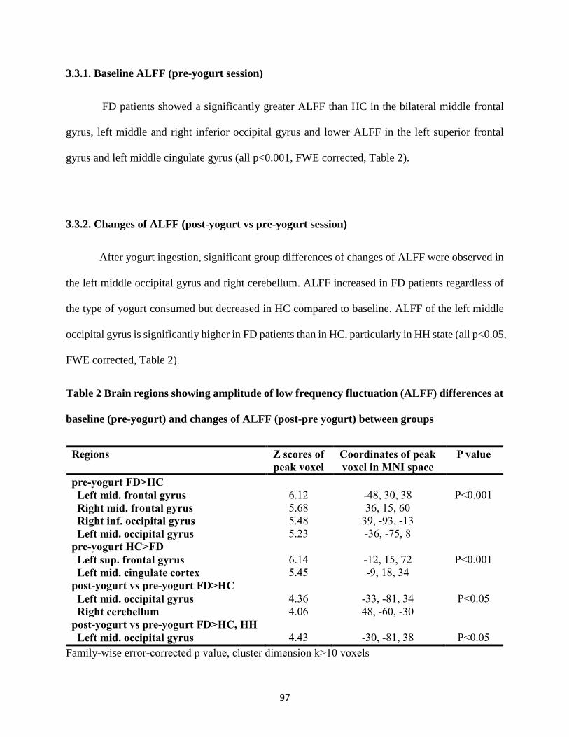
97
3.3.1. Baseline ALFF (pre-yogurt session)
FD patients showed a significantly greater ALFF than HC in the bilateral middle frontal
gyrus, left middle and right inferior occipital gyrus and lower ALFF in the left superior frontal
gyrus and left middle cingulate gyrus (all p<0.001, FWE corrected, Table 2).
3.3.2. Changes of ALFF (post-yogurt vs pre-yogurt session)
After yogurt ingestion, significant group differences of changes of ALFF were observed in
the left middle occipital gyrus and right cerebellum. ALFF increased in FD patients regardless of
the type of yogurt consumed but decreased in HC compared to baseline. ALFF of the left middle
occipital gyrus is significantly higher in FD patients than in HC, particularly in HH state (all p<0.05,
FWE corrected, Table 2).
Table 2 Brain regions showing amplitude of low frequency fluctuation (ALFF) differences at
baseline (pre-yogurt) and changes of ALFF (post-pre yogurt) between groups
Regions Z scores of
peak voxel
Coordinates of peak
voxel in MNI space
P value
pre-yogurt FD>HC
Left mid. frontal gyrus
Right mid. frontal gyrus
Right inf. occipital gyrus
Left mid. occipital gyrus
6.12
5.68
5.48
5.23
-48, 30, 38
36, 15, 60
39, -93, -13
-36, -75, 8
P<0.001
pre-yogurt HC>FD
Left sup. frontal gyrus
Left mid. cingulate cortex
6.14
5.45
-12, 15, 72
-9, 18, 34
P<0.001
post-yogurt vs pre-yogurt FD>HC
Left mid. occipital gyrus
Right cerebellum
4.36
4.06
-33, -81, 34
48, -60, -30
P<0.05
post-yogurt vs pre-yogurt FD>HC, HH
Left mid. occipital gyrus 4.43 -30, -81, 38 P<0.05
Family-wise error-corrected p value, cluster dimension k>10 voxels
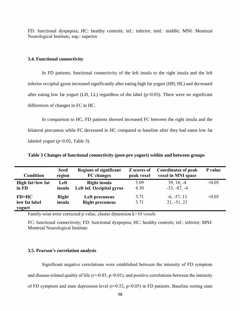
98
FD: functional dyspepsia; HC: healthy controls; inf.: inferior; mid.: middle; MNI: Montreal
Neurological Institute; sup.: superior
3.4. Functional connectivity
In FD patients, functional connectivity of the left insula to the right insula and the left
inferior occipital gyrus increased significantly after eating high fat yogurt (HH, HL) and decreased
after eating low fat yogurt (LH, LL) regardless of the label (p<0.05). There were no significant
differences of changes in FC in HC.
In comparison to HC, FD patients showed increased FC between the right insula and the
bilateral precuneus while FC decreased in HC compared to baseline after they had eaten low fat
labeled yogurt (p<0.05, Table 3).
Table 3 Changes of functional connectivity (post-pre yogurt) within and between groups
Condition
Seed
region
Regions of significant
FC changes
Z scores of
peak voxel
Coordinates of peak
voxel in MNI space
P value
High fat>low fat
in FD
Left
insula
Right insula
Left inf. Occipital gyrus
5.09
4.30
39, 18, -4
-33, -87, -4
<0.05
FD>HC
low fat label
yogurt
Right
insula
Left precuneus
Right precuneus
3.71
3.71
-6, -57, 13
21, -51, 21
<0.05
Family-wise error corrected p value, cluster dimension k>10 voxels
FC: functional connectivity; FD: functional dyspepsia; HC: healthy controls; inf.: inferior; MNI:
Montreal Neurological Institute
3.5. Pearson’s correlation analysis
Significant negative correlations were established between the intensity of FD symptom
and disease-related quality of life (r=-0.85, p<0.01), and positive correlations between the intensity
of FD symptom and state depression level (r=0.52, p<0.05) in FD patients. Baseline resting state
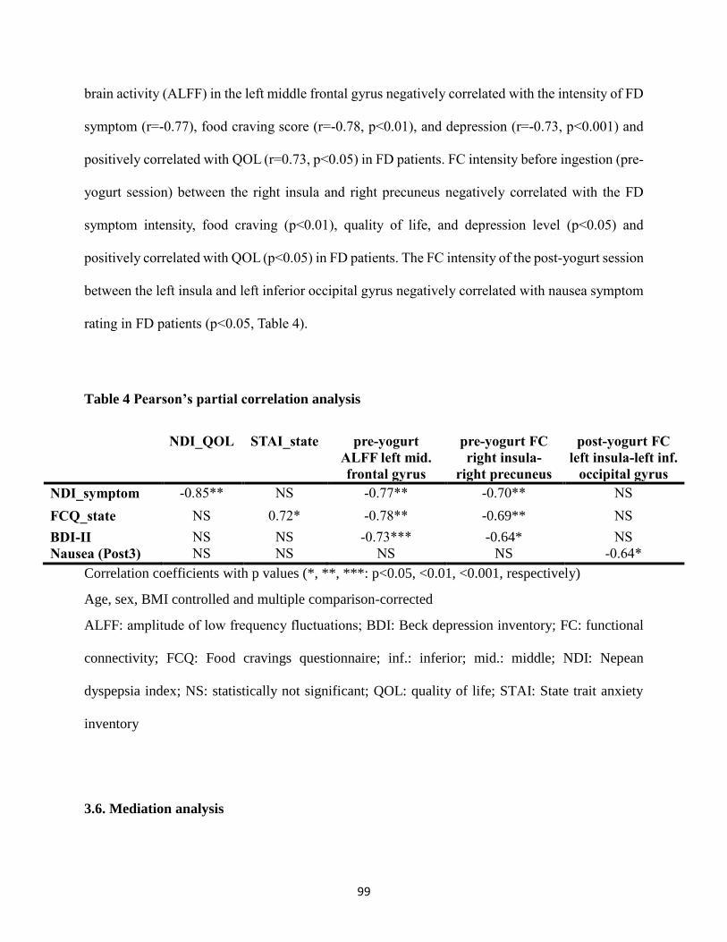
99
brain activity (ALFF) in the left middle frontal gyrus negatively correlated with the intensity of FD
symptom (r=-0.77), food craving score (r=-0.78, p<0.01), and depression (r=-0.73, p<0.001) and
positively correlated with QOL (r=0.73, p<0.05) in FD patients. FC intensity before ingestion (pre-
yogurt session) between the right insula and right precuneus negatively correlated with the FD
symptom intensity, food craving (p<0.01), quality of life, and depression level (p<0.05) and
positively correlated with QOL (p<0.05) in FD patients. The FC intensity of the post-yogurt session
between the left insula and left inferior occipital gyrus negatively correlated with nausea symptom
rating in FD patients (p<0.05, Table 4).
Table 4 Pearson’s partial correlation analysis
NDI_QOL STAI_state pre-yogurt
ALFF left mid.
frontal gyrus
pre-yogurt FC
right insula-
right precuneus
post-yogurt FC
left insula-left inf.
occipital gyrus
NDI_symptom -0.85** NS -0.77** -0.70** NS
FCQ_state NS 0.72* -0.78** -0.69** NS
BDI-II NS NS -0.73*** -0.64* NS
Nausea (Post3) NS NS NS NS -0.64*
Correlation coefficients with p values (*, **, ***: p<0.05, <0.01, <0.001, respectively)
Age, sex, BMI controlled and multiple comparison-corrected
ALFF: amplitude of low frequency fluctuations; BDI: Beck depression inventory; FC: functional
connectivity; FCQ: Food cravings questionnaire; inf.: inferior; mid.: middle; NDI: Nepean
dyspepsia index; NS: statistically not significant; QOL: quality of life; STAI: State trait anxiety
inventory
3.6. Mediation analysis
100
To assess the relationship of FD-related psychological symptoms, mediation analysis was
performed. The models and the investigated variables are described in Figure 3. The total effect of
the quality of life on depression was significant (path c p<0.05), and was fully mediated by FD
symptom (path a p<0.001; path b p<0.05; path c’ not significant; standardized indirect effect=-0.51,
95% confidence interval [-1.07, -0.25]) in FD patients (Figure 3.B., Model 1). The total effect of
depression on the quality of life was also significant (path c p<0.01) and fully mediated by FD
symptom (path a p<0.01; path b p<0.001; path c’ not significant; standardized indirect effect=-0.43,
95% confidence interval [-0.59, -0.26]) in FD patients (Figure 3.B., Model 2). We also found that
the total effect of food craving on the baseline resting state brain activity in the left middle frontal
gyrus (path c p<0.001) is fully mediated by depression (path a p<0.01; path b p<0.01; path c’ not
significant; standardized indirect effect=-0.17, 95% confidence interval [-0.29, -0.07]) in FD
patients (Figure 3.B., Model 3).
101
Discussion
Our data demonstrate I) an expectancy effect of the information about the fat content on
symptom severity, either in high fat or low fat yogurt condition, II) the altered resting state brain
activities in the prefrontal, occipital, cingulate, and cerebellum cortices, III) high fat-induced
changes in the FC of the insula-inferior occipital gyrus (vs low fat) and the group difference of the
changes in FC between the insula-precuneus in response to low fat label, IV) the negative
correlations between FD symptom, food craving, depression and the middle frontal gyrus activity,
nausea and the FC amplitude of the insula- inferior occipital gyrus, and V) the mediation effect of
depression on the influence of food craving to the middle frontal gyrus activity in FD patients.
Psychological factors in FD patients
Among the many psychological factors in functional dyspepsia, anxiety and depression
have been most frequently studied. In general, anxiety and depression are more severe in FD
patients than in healthy controls and correlate with various dyspeptic symptoms [39-42]. In this
study, anxiety, depressive, and also food craving state were more intense in FD patients than in
healthy controls. In a bid to understand the psychological processes in FD patients, mediation
analysis was performed. This enabled us to determine which independent variable affects another
(dependent variable) and which variable mediates it. We found that the bidirectional effect between
depression and disease-related QOL scores is mediated by FD symptom severity. This indicates
that increased depression, symptoms and decreased QOL in FD patients are influenced by each
other and that the role of dyspeptic symptoms is crucial in these psychological interactions.
Moreover, the inhibitory effect of craving for food on the amplitude of prefrontal brain activity is
also mediated by depression, leading to the plausible hypothesis that food craving enhances
depression and suppresses the brain activity involved in executive control in FD patients.
102
Expectancy effect of fat label on FD symptom
The effect of high fat food on symptom aggravation was not established in this study, albeit
high fat-labeled food induced more severe symptoms (abdominal pain, discomfort, and burning)
than low fat-labeled food. This result provides new knowledge on the pathophysiology of dyspeptic
symptoms since it demonstrates an expectancy effect of the information about fat content; these
may be called placebo or nocebo effects [43]. While other dyspeptic symptoms, including fullness,
nausea, vomiting, and bloating symptoms were higher in FD patients than in HC, these remained
unchanged for the different yogurts. This may indicate that some, but not all of the visceral
symptoms are subjective and can be modulated by cognitive factors. In particular, pain and burning
symptoms are mainly observed in patients with epigastric pain syndrome, a subtype of functional
dyspepsia known to be not exclusively meal-related. This may suggest that patients in different
subgroups of functional dyspepsia may have other underlying mechanisms of peripheral and
cognitive responses to food.
The behavior results are inconsistent with the previous study in which both a high fat
content and an information of high fat (HH, LH) caused higher fullness and bloating ratings than
low fat-labeled low fat yogurt (LL) in FD patients [7]. Furthermore, the effect of label was for both
high and low fat yogurt in our study while previous findings did not demonstrate the effect of low
fat label for high fat yogurt (no differences between HH and HL). This might be due to the total fat
amount in the high fat yogurt used in our study (18g vs 23.6g) and different sample characteristics.
The high fat yogurt used in this study may not suffice to provoke high fat effect on the symptom
reporting. The threshold of fat amount and varieties of symptoms which are affected by
psychological factors together with the role of expectation and previous experience of food in the
placebo/nocebo effect on visceral symptoms in FD patients will require further investigation.
103
Functional connectivity between the insula and the precuneus
Functional connectivity of the insula-precuneus negatively correlated to the FD symptom,
food craving, and depression in hunger state. This is enhanced in response to low fat-labeled yogurt
in FD patients compared to HC. The precuneus and insula are known to be functionally connected
during resting [44] and activated in response to smoking cues in smokers [45]. Insula is the core
region of the visceral sensory [25, 26] and interoceptive networks [22-24], and is believed to be
involved in ingestive behavior [46]. The precuneus is related to the episodic memory retrieval and
processing of self [47, 48], appetite control [49, 50], reward of food receipt [51], reappraisal of
benefits of eating the food [52], and comprises the default mode network [53]. Taken together, this
connection may be affected by visceral symptoms and psychological factors and strengthened by
the food signal processing in reward context (low fat label) by retrieving previous memories of
food.
Food craving
We isolated two hyper-sensitized brain regions; the middle frontal gyrus in the prefrontal
cortex (PFC) and the inferior occipital gyrus; which probably subserve different functions. We
found a higher craving for food in FD patients than in HC in a hunger state. Furthermore, food
craving influenced the middle frontal gyrus activity indirectly via depression. Food craving and
depression affect each other reciprocally and FD symptom mediates the influence. Food craving,
an intense urge to eat a particular food, is more related to the restraint or deprivation of food and
calories [54, 55] or negative emotional state [56] than to hunger. Although the role of the food
craving in obesity and eating disorders has been well established [57], it has not yet received
sufficient attention in FD patients. The PFC is well known for the executive functions (decision-
making, reward evaluation, associative learning, and control of eating behavior) and the inhibitory
104
regulation of craving for drug [58], smoking [59], and food [46]. In terms of craving, PFC has been
used for transcranial direct current stimulation to reduce food craving and calorie intake [60, 61].
Its activity increased more dramatically in the bulimia nervosa patients than in either healthy
controls or binge eating disorder patients [62]. With mediation analysis results, it is plausible that
the long-term experience of FD symptoms and consequent dietary restriction lead to higher food
craving, and that craving disrupts the functional demands of the PFC indirectly, with depression as
a mediator.
Nausea and the occipital cortex
The amplitude of functional connectivity between the insula and the inferior occipital gyrus
negatively correlated with the nausea ratings after food ingestion. FD patients suffered from higher
nausea symptom than HC and they reported more pronounced nausea after ingestion of high fat
yogurt than of low fat yogurt (statistically not significant). The occipital cortex is one of the most
frequently reported brain areas in other functional neuroimaging studies in FD patients [21].
However, the underlying cause of the functional change in the occipital cortex in patients remains
unclear. Previous studies showed that a visually induced nausea correlated with the occipital gyrus
activity [63] and that a gastric electrical stimulation with an anti-emesis effect increased the brain
activity in the occipital cortex [64]. The occipital gyrus is presumably affected by the food-induced
nausea as well as by visually induced nausea. A study on the food-induced nausea and the occipital
cortex activity would provide insight into the central mechanisms of nausea in patients.
In summary, our results showed the placebo/nocebo effect of fat information, the reward
cue- related change of functional connectivity of the insula-precuneus, the food craving-induced
activity in the PFC, and nausea-related functional connectivity of the insula-occipital cortex. These
results provide further important information about the underlying mechanism of brain activities
concerned with somatic symptoms and psychological factors in FD patients. Limitations are a
105
relatively small sample size and the food used in the study. Various food items were avoided or
preferred by FD patients and the unusual environment of MRI restricted the choice of the test meal.
Yogurt was selected since it had already proved successful in inducing FD symptoms in patients
in an earlier study, and because its fat composition is familiar to the participants and easily
modulated. However, patients suffering from lactose intolerance were unable to participate. Larger
sized studies are required to comprehend the central mechanisms of responses to food in FD
patients.

106
Conclusion
Individuals with FD have latent impairments in their cognitive perception of high fat food,
altered activity of the PFC, occipital cortex, and impaired connectivity between the insula and
occipital cortex, precuneus. Intensity of intrinsic FD symptom, food craving and depression, food-
induced nausea symptom correlated with abnormal brain activities in patients. Cognitive perception
of fat, food craving, depression, and altered brain functions as well as the somatic symptoms should
be deemed important pathological characteristics of FD.

107
Acknowledgement
The research leading to these results received funding from the People Programme of the European
Union’s Seventh Framework Programme under REA grant agreement no. 607652 (NeuroGUT)
and under Grant Agreement 607310 (Nudge-it). The authors declare that there is no conflict of
interest.
108
References
1. Pilichiewicz, A.N., et al., Functional dyspepsia is associated with a greater symptomatic response to fat but not carbohydrate, increased fasting and postprandial CCK, and diminished PYY. Am J Gastroenterol, 2008. 103(10): p. 2613-23.
2. Pilichiewicz, A.N., et al., Relationship between symptoms and dietary patterns in patients with functional dyspepsia. Clin Gastroenterol Hepatol, 2009. 7(3): p. 317-22.
3. Drossman, D.A. and D.L. Dumitrascu, Rome III: New standard for functional gastrointestinal disorders. J Gastrointestin Liver Dis, 2006. 15(3): p. 237-41.
4. Tack, J. and N.J. Talley, Functional dyspepsia--symptoms, definitions and validity of the Rome III criteria. Nat Rev Gastroenterol Hepatol, 2013. 10(3): p. 134-41.
5. Barbera, R., C. Feinle, and N.W. Read, Nutrient-specific modulation of gastric mechanosensitivity in patients with functional dyspepsia. Dig Dis Sci, 1995. 40(8): p. 1636-41.
6. Barbera, R., C. Feinle, and N.W. Read, Abnormal sensitivity to duodenal lipid infusion in patients with functional dyspepsia. Eur J Gastroenterol Hepatol, 1995. 7(11): p. 1051-7.
7. Feinle-Bisset, C., et al., Role of cognitive factors in symptom induction following high and low fat meals in patients with functional dyspepsia. Gut, 2003. 52(10): p. 1414-8.
8. Mearin, F., et al., The origin of symptoms on the brain-gut axis in functional dyspepsia. Gastroenterology, 1991. 101(4): p. 999-1006.
9. Feinle-Bisset, C., Upper gastrointestinal sensitivity to meal-related signals in adult humans - relevance to appetite regulation and gut symptoms in health, obesity and functional dyspepsia. Physiol Behav, 2016. 162: p. 69-82.
10. Zeng, F., et al., Abnormal resting brain activity in patients with functional dyspepsia is related to symptom severity. Gastroenterology, 2011. 141(2): p. 499-506.
11. Liu, M.L., et al., Cortical-limbic regions modulate depression and anxiety factors in functional dyspepsia: a PET-CT study. Ann Nucl Med, 2012. 26(1): p. 35-40.
12. Zhou, G., et al., Increased interhemispheric resting-state functional connectivity in functional dyspepsia: a pilot study. NMR Biomed, 2013. 26(4): p. 410-5.
13. Zhou, G., et al., Fractional amplitude of low-frequency fluctuation changes in functional dyspepsia: a resting-state fMRI study. Magn Reson Imaging, 2013. 31(6): p. 996-1000.
14. Liu, P., et al., Alterations of the default mode network in functional dyspepsia patients: a resting-state fmri study. Neurogastroenterol Motil, 2013. 25(6): p. e382-8.
15. Nan, J., et al., Whole-brain functional connectivity identification of functional dyspepsia. PLoS One, 2013. 8(6): p. e65870.
16. Liu, P., et al., Identifying neural patterns of functional dyspepsia using multivariate pattern analysis: a resting-state FMRI study. PLoS One, 2013. 8(7): p. e68205.
17. Nan, J., et al., Altered intrinsic regional activity and corresponding brain pathways reflect the symptom severity of functional dyspepsia. Neurogastroenterol Motil, 2014. 26(5): p. 660-9.
18. Vandenberghe, J., et al., Regional cerebral blood flow during gastric balloon distention in functional dyspepsia. Gastroenterology, 2007. 132(5): p. 1684-93.
19. Van Oudenhove, L., et al., Abnormal regional brain activity during rest and (anticipated) gastric distension in functional dyspepsia and the role of anxiety: a H(2)(15)O-PET study. Am J Gastroenterol, 2010. 105(4): p. 913-24.
20. Van Oudenhove, L., et al., Regional brain activity in functional dyspepsia: a H(2)(15)O-PET study on the role of gastric sensitivity and abuse history. Gastroenterology, 2010. 139(1): p. 36-47.
21. Lee, I.S., et al., Functional neuroimaging studies in functional dyspepsia patients: a systematic review. Neurogastroenterol Motil, 2016. 28(6): p. 793-805.
109
22. Cauda, F., et al., Functional connectivity of the insula in the resting brain. Neuroimage, 2011. 55(1): p. 8-23.
23. Craig, A.D., How do you feel? Interoception: the sense of the physiological condition of the body. Nat Rev Neurosci, 2002. 3(8): p. 655-66.
24. Craig, A.D., Interoception: the sense of the physiological condition of the body. Curr Opin Neurobiol, 2003. 13(4): p. 500-5.
25. Aziz, Q., A. Schnitzler, and P. Enck, Functional neuroimaging of visceral sensation. J Clin Neurophysiol, 2000. 17(6): p. 604-12.
26. Van Oudenhove, L., S.J. Coen, and Q. Aziz, Functional brain imaging of gastrointestinal sensation in health and disease. World J Gastroenterol, 2007. 13(25): p. 3438-45.
27. Mayer, E.A., B.D. Naliboff, and A.D. Craig, Neuroimaging of the brain-gut axis: from basic understanding to treatment of functional GI disorders. Gastroenterology, 2006. 131(6): p. 1925-42.
28. Nan, J., et al., Brain-based Correlations Between Psychological Factors and Functional Dyspepsia. J Neurogastroenterol Motil, 2015. 21(1): p. 103-10.
29. Zeng, F., et al., Influence of acupuncture treatment on cerebral activity in functional dyspepsia patients and its relationship with efficacy. Am J Gastroenterol, 2012. 107(8): p. 1236-47.
30. Douglas A. Drossman, et al., ROME III: The Functional Gastrointestinal Disorders. 3rd ed. 2006: Yale University Section of Digestive Disease: Degnon Associates.
31. Talley, N.J., et al., Development of a new dyspepsia impact scale: the Nepean Dyspepsia Index. Aliment Pharmacol Ther, 1999. 13(2): p. 225-35.
32. Beck, A.T., Steer, R.A., & Brown, G., Manual for the Beck Depression Inventory-II. 1996: San Antonio, TX: Psychological Corporation.
33. Spielberger, C.D., Gorsuch, R. L. Lushene, R., Vagg, P. R., & Jacobs, G. A. , Manual for the State-Trait Anxiety Inventory. . 1983: Palo Alto, CA: Consulting Psychologists Press.
34. Fairburn, C.G. and S.J. Beglin, Assessment of eating disorders: interview or self-report questionnaire? Int J Eat Disord, 1994. 16(4): p. 363-70.
35. Cepeda-Benito, A., Gleaves, D. H., Williams, T. L., & Erath, S. A., The development and validation of the state and trait food-cravings questionnaires. Behavior Therapy, 2000. 1(31): p. 151-173.
. 36. Ledikwe, J.H., et al., A reliable, valid questionnaire indicates that preference for dietary fat
declines when following a reduced-fat diet. Appetite, 2007. 49(1): p. 74-83. 37. Chao-Gan, Y. and Z. Yu-Feng, DPARSF: A MATLAB Toolbox for "Pipeline" Data Analysis of Resting-
State fMRI. Front Syst Neurosci, 2010. 4: p. 13. 38. Hayes, A.F., Introduction to mediation, moderation, and conditional process analysis: A
regression based approach. 2013, NewYork: NY: Guilford Press. 39. Lee, S., et al., Stress, coping, and depression in non-ulcer dyspepsia patients. J Psychosom Res,
2000. 49(1): p. 93-9. 40. Jones, M.P., L.K. Sharp, and M.D. Crowell, Psychosocial correlates of symptoms in functional
dyspepsia. Clin Gastroenterol Hepatol, 2005. 3(6): p. 521-8. 41. Locke, G.R., 3rd, et al., Psychosocial factors are linked to functional gastrointestinal disorders: a
population based nested case-control study. Am J Gastroenterol, 2004. 99(2): p. 350-7. 42. Koloski, N.A., N.J. Talley, and P.M. Boyce, A history of abuse in community subjects with irritable
bowel syndrome and functional dyspepsia: the role of other psychosocial variables. Digestion, 2005. 72(2-3): p. 86-96.
43. Elsenbruch, S. and P. Enck, Placebo effects and their determinants in gastrointestinal disorders. Nat Rev Gastroenterol Hepatol, 2015. 12(8): p. 472-85.
110
44. Zhang, S. and C.S. Li, Functional connectivity mapping of the human precuneus by resting state fMRI. Neuroimage, 2012. 59(4): p. 3548-62.
45. Engelmann, J.M., et al., Neural substrates of smoking cue reactivity: a meta-analysis of fMRI studies. Neuroimage, 2012. 60(1): p. 252-62.
46. Kahathuduwa, C.N., et al., Brain regions involved in ingestive behavior and related psychological constructs in people undergoing calorie restriction. Appetite, 2016. 107: p. 348-361.
47. Cavanna, A.E. and M.R. Trimble, The precuneus: a review of its functional anatomy and behavioural correlates. Brain, 2006. 129(Pt 3): p. 564-83.
48. Sajonz, B., et al., Delineating self-referential processing from episodic memory retrieval: common and dissociable networks. Neuroimage, 2010. 50(4): p. 1606-17.
49. Tuulari, J.J., et al., Neural circuits for cognitive appetite control in healthy and obese individuals: an fMRI study. PLoS One, 2015. 10(2): p. e0116640.
50. Scharmuller, W., et al., Appetite regulation during food cue exposure: a comparison of normal-weight and obese women. Neurosci Lett, 2012. 518(2): p. 106-10.
51. Winter, S.R., et al., Elevated reward response to receipt of palatable food predicts future weight variability in healthy-weight adolescents. Am J Clin Nutr, 2017. 105(4): p. 781-789.
52. Yokum, S. and E. Stice, Cognitive regulation of food craving: effects of three cognitive reappraisal strategies on neural response to palatable foods. Int J Obes (Lond), 2013. 37(12): p. 1565-70.
53. Utevsky, A.V., D.V. Smith, and S.A. Huettel, Precuneus is a functional core of the default-mode network. J Neurosci, 2014. 34(3): p. 932-40.
54. Hill, A.J., The psychology of food craving. Proc Nutr Soc, 2007. 66(2): p. 277-85. 55. Kahathuduwa, C.N., et al., Extended calorie restriction suppresses overall and specific food
cravings: a systematic review and a meta-analysis. Obes Rev, 2017. 56. Hill, A.J., C.F. Weaver, and J.E. Blundell, Food craving, dietary restraint and mood. Appetite, 1991.
17(3): p. 187-97. 57. Potenza, M.N. and C.M. Grilo, How Relevant is Food Craving to Obesity and Its Treatment? Front
Psychiatry, 2014. 5: p. 164. 58. Goldstein, R.Z. and N.D. Volkow, Dysfunction of the prefrontal cortex in addiction: neuroimaging
findings and clinical implications. Nat Rev Neurosci, 2011. 12(11): p. 652-69. 59. Kober, H., et al., Prefrontal-striatal pathway underlies cognitive regulation of craving. Proc Natl
Acad Sci U S A, 2010. 107(33): p. 14811-6. 60. Kekic, M., et al., The effects of prefrontal cortex transcranial direct current stimulation (tDCS) on
food craving and temporal discounting in women with frequent food cravings. Appetite, 2014. 78: p. 55-62.
61. Fregni, F., et al., Transcranial direct current stimulation of the prefrontal cortex modulates the desire for specific foods. Appetite, 2008. 51(1): p. 34-41.
62. Lee, J.E., K. Namkoong, and Y.C. Jung, Impaired prefrontal cognitive control over interference by food images in binge-eating disorder and bulimia nervosa. Neurosci Lett, 2017. 651: p. 95-101.
63. Farmer, A.D., et al., Visually induced nausea causes characteristic changes in cerebral, autonomic and endocrine function in humans. J Physiol, 2015. 593(5): p. 1183-96.
64. Yu, X., et al., Antiemesis effect and brain fMRI response of gastric electrical stimulation with different parameters in dogs. Neurogastroenterol Motil, 2014. 26(7): p. 1049-56.
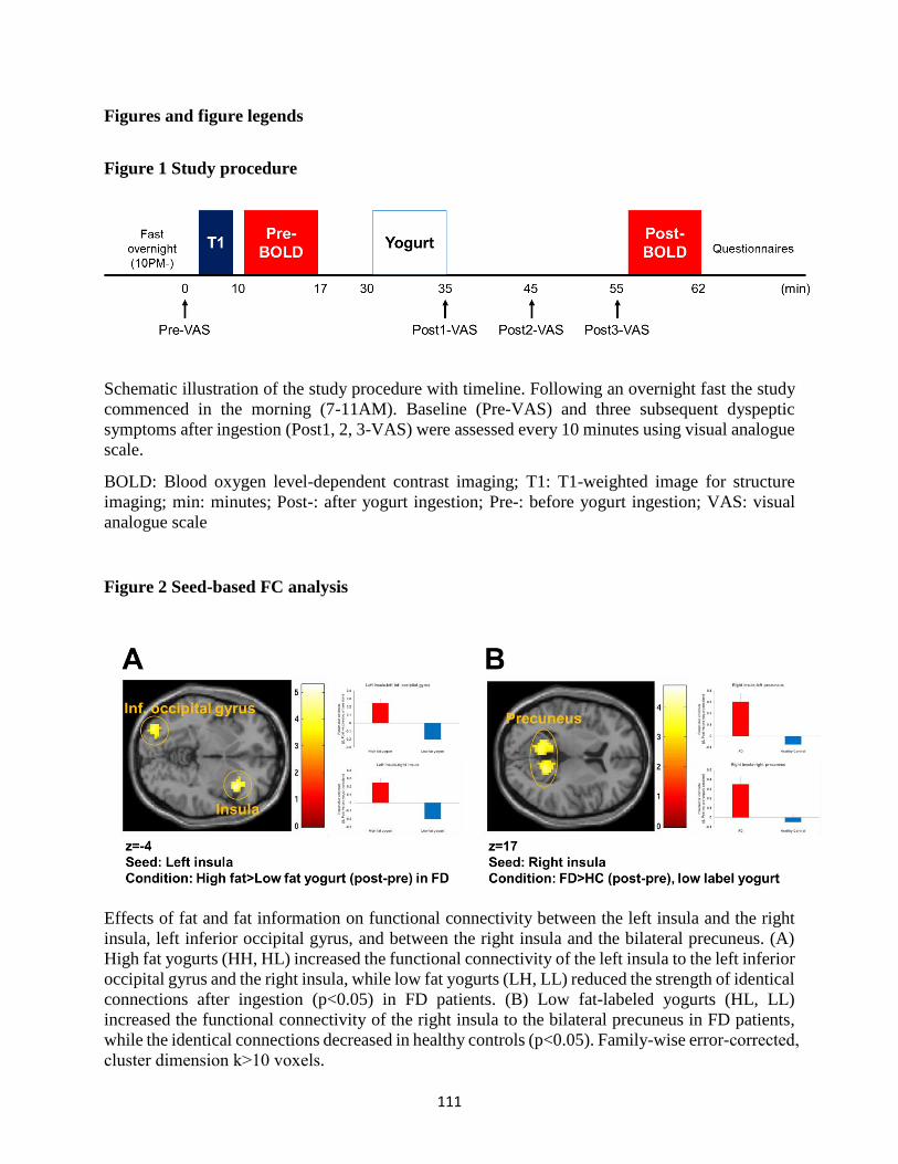
111
Figures and figure legends
Figure 1 Study procedure
Schematic illustration of the study procedure with timeline. Following an overnight fast the study
commenced in the morning (7-11AM). Baseline (Pre-VAS) and three subsequent dyspeptic
symptoms after ingestion (Post1, 2, 3-VAS) were assessed every 10 minutes using visual analogue
scale.
BOLD: Blood oxygen level-dependent contrast imaging; T1: T1-weighted image for structure
imaging; min: minutes; Post-: after yogurt ingestion; Pre-: before yogurt ingestion; VAS: visual
analogue scale
Figure 2 Seed-based FC analysis
Effects of fat and fat information on functional connectivity between the left insula and the right
insula, left inferior occipital gyrus, and between the right insula and the bilateral precuneus. (A)
High fat yogurts (HH, HL) increased the functional connectivity of the left insula to the left inferior
occipital gyrus and the right insula, while low fat yogurts (LH, LL) reduced the strength of identical
connections after ingestion (p<0.05) in FD patients. (B) Low fat-labeled yogurts (HL, LL)
increased the functional connectivity of the right insula to the bilateral precuneus in FD patients,
while the identical connections decreased in healthy controls (p<0.05). Family-wise error-corrected,
cluster dimension k>10 voxels.
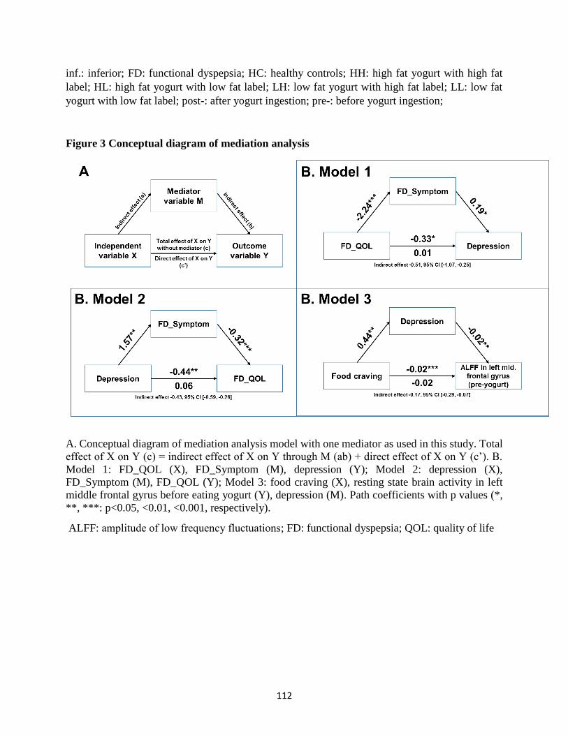
112
inf.: inferior; FD: functional dyspepsia; HC: healthy controls; HH: high fat yogurt with high fat
label; HL: high fat yogurt with low fat label; LH: low fat yogurt with high fat label; LL: low fat
yogurt with low fat label; post-: after yogurt ingestion; pre-: before yogurt ingestion;
Figure 3 Conceptual diagram of mediation analysis
A. Conceptual diagram of mediation analysis model with one mediator as used in this study. Total
effect of X on Y (c) = indirect effect of X on Y through M (ab) + direct effect of X on Y (c’). B.
Model 1: FD_QOL (X), FD_Symptom (M), depression (Y); Model 2: depression (X),
FD_Symptom (M), FD_QOL (Y); Model 3: food craving (X), resting state brain activity in left
middle frontal gyrus before eating yogurt (Y), depression (M). Path coefficients with p values (*,
**, ***: p<0.05, <0.01, <0.001, respectively).
ALFF: amplitude of low frequency fluctuations; FD: functional dyspepsia; QOL: quality of life
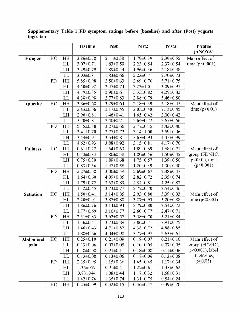
113
Supplementary Table 1 FD symptom ratings before (baseline) and after (Post) yogurts
ingestion
Baseline Post1 Post2 Post3 P value
(ANOVA)
Hunger HC HH 3.86±0.78 2.11±0.58 1.79±0.39 2.39±0.55 Main effect of
time (p<0.001) HL 3.07±0.71 1.83±0.59 2.23±0.54 2.37±0.54
LH 3.29±0.79 1.89±0.44 1.96±0.46 2.18±0.48
LL 3.03±0.81 1.83±0.66 2.23±0.71 2.70±0.73
FD HH 5.85±0.98 2.50±0.63 2.69±0.76 3.71±0.75
HL 4.50±0.92 2.45±0.74 3.23±1.01 3.09±0.95
LH 4.79±0.85 2.96±0.61 3.33±0.82 4.29±0.82
LL 4.38±0.98 2.77±0.83 2.88±0.79 3.46±0.80
Appetite HC HH 3.86±0.68 3.29±0.64 2.18±0.39 2.18±0.45 Main effect of
time (p<0.01) HL 2.83±0.66 2.17±0.55 2.03±0.48 2.13±0.45
LH 2.96±0.81 1.46±0.41 1.65±0.42 2.00±0.42
LL 3.70±0.81 2.40±0.71 2.64±0.72 2.67±0.66
FD HH 5.15±0.88 3.27±0.66 2.77±0.75 3.42±0.80
HL 3.41±0.70 2.77±0.72 3.14±1.00 3.59±0.96
LH 3.54±0.91 3.54±0.81 3.63±0.93 4.42±0.99
LL 4.62±0.93 3.88±0.92 3.15±0.81 4.17±0.76
Fullness HC HH 0.61±0.27 1.64±0.63 1.89±0.69 1.68±0.71 Main effect of
group (FD>HC,
p<0.01), time
(p<0.001)
HL 0.43±0.33 1.80±0.58 1.80±0.56 1.50±0.45
LH 0.75±0.39 1.89±0.68 1.75±0.57 1.39±0.50
LL 0.83±0.36 1.47±0.58 1.20±0.49 1.30±0.40
FD HH 2.27±0.68 3.00±0.59 2.69±0.67 2.38±0.47
HL 1.64±0.60 4.09±0.85 2.82±0.72 2.95±0.74
LH 1.79±0.72 3.63±0.89 4.54±0.81 4.25±0.87
LL 1.42±0.45 3.73±0.77 2.77±0.70 2.54±0.46
Satiation HC HH 1.50±0.41 3.14±0.85 2.93±0.80 3.39±0.93 Main effect of
time (p<0.001) HL 2.20±0.91 3.87±0.80 3.27±0.93 3.20±0.88
LH 1.86±0.76 3.14±0.94 2.79±0.80 2.54±0.72
LL 1.77±0.69 3.10±0.77 2.60±0.77 2.47±0.71
FD HH 2.31±0.83 3.62±0.57 3.58±0.70 3.21±0.84
HL 1.36±0.51 3.73±0.89 2.86±0.71 2.91±0.75
LH 1.46±0.43 4.71±0.82 4.38±0.72 4.88±0.85
LL 1.88±0.66 4.04±0.90 3.77±0.97 2.63±0.61
Abdominal
pain
HC HH 0.25±0.10 0.21±0.09 0.18±0.07 0.21±0.10 Main effect of
group (FD>HC,
p<0.001), label
(high>low,
p<0.05)
HL 0.13±0.06 0.07±0.05 0.10±0.05 0.07±0.05
LH 0.18±0.08 0.21±0.11 0.18±0.08 0.11±0.06
LL 0.13±0.08 0.13±0.06 0.17±0.06 0.13±0.08
FD HH 2.35±0.95 1.15±0.36 1.65±0.45 1.17±0.34
HL 1.36±057 0.91±0.41 1.27±0.61 1.45±0.62
LH 0.88±044 1.08±0.44 1.17±0.32 1.58±0.31
LL 1.42±0.76 1.35±0.74 1.31±0.75 0.54±0.24
HC HH 0.25±0.09 0.32±0.15 0.36±0.17 0.39±0.20
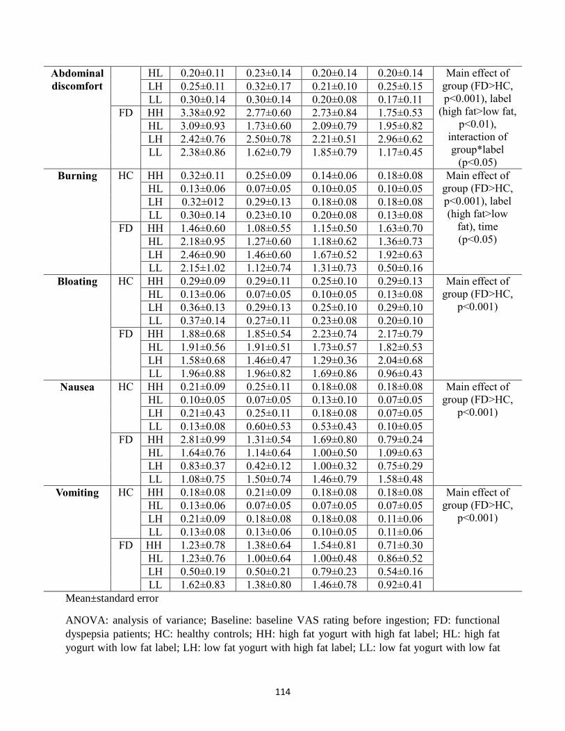
114
Abdominal
discomfort
HL 0.20±0.11 0.23±0.14 0.20±0.14 0.20±0.14 Main effect of
group (FD>HC,
p<0.001), label
(high fat>low fat,
p<0.01),
interaction of
group*label
(p<0.05)
LH 0.25±0.11 0.32±0.17 0.21±0.10 0.25±0.15
LL 0.30±0.14 0.30±0.14 0.20±0.08 0.17±0.11
FD HH 3.38±0.92 2.77±0.60 2.73±0.84 1.75±0.53
HL 3.09±0.93 1.73±0.60 2.09±0.79 1.95±0.82
LH 2.42±0.76 2.50±0.78 2.21±0.51 2.96±0.62
LL 2.38±0.86 1.62±0.79 1.85±0.79 1.17±0.45
Burning HC HH 0.32±0.11 0.25±0.09 0.14±0.06 0.18±0.08 Main effect of
group (FD>HC,
p<0.001), label
(high fat>low
fat), time
(p<0.05)
HL 0.13±0.06 0.07±0.05 0.10±0.05 0.10±0.05
LH 0.32±012 0.29±0.13 0.18±0.08 0.18±0.08
LL 0.30±0.14 0.23±0.10 0.20±0.08 0.13±0.08
FD HH 1.46±0.60 1.08±0.55 1.15±0.50 1.63±0.70
HL 2.18±0.95 1.27±0.60 1.18±0.62 1.36±0.73
LH 2.46±0.90 1.46±0.60 1.67±0.52 1.92±0.63
LL 2.15±1.02 1.12±0.74 1.31±0.73 0.50±0.16
Bloating HC HH 0.29±0.09 0.29±0.11 0.25±0.10 0.29±0.13 Main effect of
group (FD>HC,
p<0.001) HL 0.13±0.06 0.07±0.05 0.10±0.05 0.13±0.08
LH 0.36±0.13 0.29±0.13 0.25±0.10 0.29±0.10
LL 0.37±0.14 0.27±0.11 0.23±0.08 0.20±0.10
FD HH 1.88±0.68 1.85±0.54 2.23±0.74 2.17±0.79
HL 1.91±0.56 1.91±0.51 1.73±0.57 1.82±0.53
LH 1.58±0.68 1.46±0.47 1.29±0.36 2.04±0.68
LL 1.96±0.88 1.96±0.82 1.69±0.86 0.96±0.43
Nausea HC HH 0.21±0.09 0.25±0.11 0.18±0.08 0.18±0.08 Main effect of
group (FD>HC,
p<0.001) HL 0.10±0.05 0.07±0.05 0.13±0.10 0.07±0.05
LH 0.21±0.43 0.25±0.11 0.18±0.08 0.07±0.05
LL 0.13±0.08 0.60±0.53 0.53±0.43 0.10±0.05
FD HH 2.81±0.99 1.31±0.54 1.69±0.80 0.79±0.24
HL 1.64±0.76 1.14±0.64 1.00±0.50 1.09±0.63
LH 0.83±0.37 0.42±0.12 1.00±0.32 0.75±0.29
LL 1.08±0.75 1.50±0.74 1.46±0.79 1.58±0.48
Vomiting HC HH 0.18±0.08 0.21±0.09 0.18±0.08 0.18±0.08 Main effect of
group (FD>HC,
p<0.001) HL 0.13±0.06 0.07±0.05 0.07±0.05 0.07±0.05
LH 0.21±0.09 0.18±0.08 0.18±0.08 0.11±0.06
LL 0.13±0.08 0.13±0.06 0.10±0.05 0.11±0.06
FD HH 1.23±0.78 1.38±0.64 1.54±0.81 0.71±0.30
HL 1.23±0.76 1.00±0.64 1.00±0.48 0.86±0.52
LH 0.50±0.19 0.50±0.21 0.79±0.23 0.54±0.16
LL 1.62±0.83 1.38±0.80 1.46±0.78 0.92±0.41
Mean±standard error
ANOVA: analysis of variance; Baseline: baseline VAS rating before ingestion; FD: functional
dyspepsia patients; HC: healthy controls; HH: high fat yogurt with high fat label; HL: high fat
yogurt with low fat label; LH: low fat yogurt with high fat label; LL: low fat yogurt with low fat

115
label; Post1: VAS rating immediately after ingestion; Post2: VAS rating 10 minutes after ingestion;
Post3: VAS rating 20 minutes after ingestion
116
9. Conclusion and future direction
In functional dyspepsia, we observed 1) the absence of previous studies on neural
mechanisms of food-related tasks or food-related psychological factors, 2) higher food craving
scores but a reduced amount of food intake from standard breakfast compared to healthy controls,
3) lower pleasantness and total visual attention time to food images compared to healthy controls,
4) placebo/nocebo effects of fat label on dyspeptic symptoms, in both high fat or low fat food
ingestion sessions, and 5) altered resting state brain activities and functional connectivity in the
prefrontal cortex, occipital cortex, insula, and precuneus and their associations with dyspeptic
symptoms and psychological factors. The effectiveness of conventional treatments and basic
researches for functional dyspepsia might be improved by dietary consultation and modification of
their distorted perception of food. This will require the expansion of our conventional perspective
of functional dyspepsia, from the peripheral gastrointestinal tracts to the mental process of and
behavioral response to food.
Future studies
We propose future studies according to the following categories: studies on 1) the new
knowledge of basic mechanisms, 2) the understanding of psychology and placebo/nocebo effects,
and 3) clinical diagnosis and therapy in functional dyspepsia patients.
Although we did not find that the effect of high fat food triggered the dyspeptic symptom
in patients, the effect of nutritional factors on the dyspeptic symptom development needs to be
tested with further food types. Since symptoms related to different types of food or nutrients may
be different in each patient, individualized items should be tested in future studies to gain results
that are closer to the reality. To achieve this goal, we will require well-structured interviews and
validated questionnaires for clinicians and researchers. Future studies on the neuronal mechanisms
117
on the evaluation, perception, processing of food and behavioral responses to food are necessary
to unravel the pathophysiology of functional dyspepsia. How patients perceive, encode, store, and
recall the value of food and how negative experience of previous food-induced suffering influences
their food decision and eating behavior could be investigated using functional neuroimaging
techniques and physiological measurements during food-related tasks.
It would also be worthwhile to study the effect of the food consumption on the composition
of microbiota and their contribution to the gastric symptoms. The role of microbiota in the
abnormal function of the brain-gut axis is still unclear despite the wide use of probiotics or
antibiotics in irritable bowel syndrome patients. Gastric microbiota and fecal microbiota
transplantation have been investigated in functional dyspepsia only in the last few years [152, 153].
Various approaches including studies on microbiomics [154], efficacy and mechanisms of
probiotics, antibiotics, and fecal microbiota transplantation, changes of emotional, cognitive, and
behavioral response to food or other external stimuli (social stress, pain), individualized screening
and medication using microbiota may be a revolution in the functional dyspepsia research.
Another valuable approach is to investigate the efficacy and brain mechanisms of the
placebo treatment using low fat label on food to relieve the dyspeptic symptoms in patients. Apart
from the fact that there is no standard treatment guideline for functional dyspepsia patients, the
only intervention whose neuronal mechanism has been studied so far is acupuncture [155, 156]. If
the placebo treatment really works in functional dyspepsia patients, its peripheral and neuronal
mechanisms may promote the development of new treatment for functional dyspepsia.
We also discovered a new psychological factor which might be important in the
pathophysiology of functional dyspepsia: craving for food. Despite increased food craving when
fasting, functional dyspepsia patients ate smaller amounts of food compared to healthy controls 4.
Furthermore, food craving significantly affected the resting state activity in the middle frontal gyrus
118
in functional dyspepsia patients, but not in healthy controls. Besides the abnormal responses to
external painful stimuli, (e.g., barostat distension) and the effects of anxiety and depression, the
physiological cause and effect of increased food craving score in functional dyspepsia patients need
to be replicated and further investigated in large-scale studies.
Objective outcomes from functional neuroimaging studies, screening of the composition of
microbiota, well-structured interview about eating behavior and cognitive processes of food may
improve the current diagnosis. To improve existing treatments for functional dyspepsia, the
efficacy, safety, and protocol of psychotherapies with the manipulation of eating behavior,
consultation of food choice, and modification of negative response to food should be defined.
Moreover, placebo treatments using symptom relief cues, e.g., low fat label, symptom-independent
nutrients, elimination or supplement of microbiota, or placebo tools (which are similar or identical
to the treatment appliances without any actual effects) need to be tested.
119
10. Acknowledgements
I would like to express my gratitude to Prof. Dr. Paul Enck and Prof. Dr. Hubert Preissl for
their support and patient supervision. They guided me throughout my PhD and encouraged me
whenever I faced difficulties. I owe to them everything that I know about psychology and
neuroscience. I also learned how to write a manuscript, give a good scientific presentation, and
how to be patient and steady in research.
I am very thankful to the members of the Psychosomatic Medicine and Psychotherapy
department and fMEG center. They shared their ideas and experimental techniques, and also spent
time and labor for my PhD studies. Their considerable and motivating criticism improved the
studies enormously.
I am grateful to the Graduate Training Centre of Neuroscience in Tübingen for their
financial and administrative support. I am also extremely grateful to all the participants of my
studies. Particularly the functional magnetic resonance imaging study was challenging for me as
an experimenter, would not have been possible without their cooperation.
Lastly, I would like to thank my family for their encouragement and emotional support.
Their trust gave me confidence and allowed me to grow professionally. Most importantly, their
love made me love my life more than ever before.
120
11. References
1. Tack, J., et al., Functional gastroduodenal disorders. Gastroenterology, 2006. 130(5): p. 1466-79. 2. Drossman, D.A. and D.L. Dumitrascu, Rome III: New standard for functional gastrointestinal
disorders. J Gastrointestin Liver Dis, 2006. 15(3): p. 237-41. 3. Mahadeva, S. and K.L. Goh, Epidemiology of functional dyspepsia: a global perspective. World J
Gastroenterol, 2006. 12(17): p. 2661-6. 4. El-Serag, H.B. and N.J. Talley, Systemic review: the prevalence and clinical course of functional
dyspepsia. Aliment Pharmacol Ther, 2004. 19(6): p. 643-54. 5. Lacy, B.E., et al., Functional dyspepsia: the economic impact to patients. Aliment Pharmacol Ther,
2013. 38(2): p. 170-7. 6. Moayyedi, P. and J. Mason, Clinical and economic consequences of dyspepsia in the community.
Gut, 2002. 50 Suppl 4: p. iv10-2. 7. Van Oudenhove, L., et al., Risk factors for impaired health-related quality of life in functional
dyspepsia. Aliment Pharmacol Ther, 2011. 33(2): p. 261-74. 8. Drossman, D.A., et al., Rome IV Functional Gastrointestinal Disorders: Disorders of Gut-Brain
Interaction. 4th ed. 2017, Raleigh, NC: Rome Foundation. 9. Douglas A. Drossman, et al., ROME III: The Functional Gastrointestinal Disorders. 3rd ed. 2006:
Yale University Section of Digestive Disease: Degnon Associates. 10. Piessevaux, H., et al., Dyspeptic symptoms in the general population: a factor and cluster analysis
of symptom groupings. Neurogastroenterol Motil, 2009. 21(4): p. 378-88. 11. Keohane, J. and E.M. Quigley, Functional dyspepsia and nonerosive reflux disease: clinical
interactions and their implications. MedGenMed, 2007. 9(3): p. 31. 12. Pleyer, C., et al., Overdiagnosis of gastro-esophageal reflux disease and underdiagnosis of
functional dyspepsia in a USA community. Neurogastroenterol Motil, 2014. 26(8): p. 1163-71. 13. Corsetti, M., et al., Impact of coexisting irritable bowel syndrome on symptoms and
pathophysiological mechanisms in functional dyspepsia. Am J Gastroenterol, 2004. 99(6): p. 1152-9.
14. Ford, A.C., et al., Systematic review and meta-analysis of the prevalence of irritable bowel syndrome in individuals with dyspepsia. Clin Gastroenterol Hepatol, 2010. 8(5): p. 401-9.
15. Drossman, D.A., et al., Identification of subgroups of functional bowel disorders. Gastroenterol Int. 1990;3:159–172. Gastroenterol Int, 1990. 3: p. 159-172.
16. Drossman, D.A., et al., The Functional Gastrointestinal Disorders: Diagnosis, Pathophysiology, and Treatment. A Multinational Consensus. 1994, Boston: Little Brown & Co.
17. Drossman, D.A., et al., Rome II: A multinational consensus document on functional gastrointestinal disorders. Gut, 1999. 45(suppl II): p. 1–81.
18. Bisschops, R., et al., Relationship between symptoms and ingestion of a meal in functional dyspepsia. Gut, 2008. 57(11): p. 1495-503.
19. Spiegel, B.M., et al., Measuring symptoms in the irritable bowel syndrome: development of a framework for clinical trials. Aliment Pharmacol Ther, 2010. 32(10): p. 1275-91.
20. Brun, R. and B. Kuo, Functional dyspepsia. Therap Adv Gastroenterol, 2010. 3(3): p. 145-64. 21. Talley, N.J., M. Verlinden, and M. Jones, Validity of a new quality of life scale for functional
dyspepsia: a United States multicenter trial of the Nepean Dyspepsia Index. Am J Gastroenterol, 1999. 94(9): p. 2390-7.
22. Talley, N.J., et al., Development of a new dyspepsia impact scale: the Nepean Dyspepsia Index. Aliment Pharmacol Ther, 1999. 13(2): p. 225-35.
121
23. Talley, N.J., M. Verlinden, and M. Jones, Quality of life in functional dyspepsia: responsiveness of the Nepean Dyspepsia Index and development of a new 10-item short form. Aliment Pharmacol Ther, 2001. 15(2): p. 207-16.
24. Moayyedi, P., et al., The Leeds Dyspepsia Questionnaire: a valid tool for measuring the presence and severity of dyspepsia. Aliment Pharmacol Ther, 1998. 12(12): p. 1257-62.
25. Hu, W.H., et al., The Hong Kong index of dyspepsia: a validated symptom severity questionnaire for patients with dyspepsia. J Gastroenterol Hepatol, 2002. 17(5): p. 545-51.
26. Lee, E.H., et al., Development and validation of a functional dyspepsia-related quality of life (FD-QOL) scale in South Korea. J Gastroenterol Hepatol, 2006. 21(1 Pt 2): p. 268-74.
27. Carbone, F., et al., Validation of the Leuven Postprandial Distress Scale, a questionnaire for symptom assessment in the functional dyspepsia/postprandial distress syndrome. Aliment Pharmacol Ther, 2016. 44(9): p. 989-1001.
28. el-Omar, E.M., et al., The Glasgow Dyspepsia Severity Score--a tool for the global measurement of dyspepsia. Eur J Gastroenterol Hepatol, 1996. 8(10): p. 967-71.
29. Fujikawa, Y., et al., Postprandial Symptoms Felt at the Lower Part of the Epigastrium and a Possible Association of Pancreatic Exocrine Dysfunction with the Pathogenesis of Functional Dyspepsia. Intern Med, 2017. 56(13): p. 1629-1635.
30. Vanheel, H., et al., Pathophysiological Abnormalities in Functional Dyspepsia Subgroups According to the Rome III Criteria. Am J Gastroenterol, 2017. 112(1): p. 132-140.
31. Khodarahmi, M. and L. Azadbakht, Dietary fat intake and functional dyspepsia. Adv Biomed Res, 2016. 5: p. 76.
32. Goktas, Z., et al., Nutritional habits in functional dyspepsia and its subgroups: a comparative study. Scand J Gastroenterol, 2016. 51(8): p. 903-7.
33. Fried, M. and C. Feinle, The role of fat and cholecystokinin in functional dyspepsia. Gut, 2002. 51 Suppl 1: p. i54-7.
34. Van Oudenhove, L. and Q. Aziz, The role of psychosocial factors and psychiatric disorders in functional dyspepsia. Nat Rev Gastroenterol Hepatol, 2013. 10(3): p. 158-67.
35. Mearin, F., et al., The origin of symptoms on the brain-gut axis in functional dyspepsia. Gastroenterology, 1991. 101(4): p. 999-1006.
36. Lee, I.S., et al., Functional neuroimaging studies in functional dyspepsia patients: a systematic review. Neurogastroenterol Motil, 2016. 28(6): p. 793-805.
37. Gathaiya, N., et al., Novel associations with dyspepsia: a community-based study of familial aggregation, sleep dysfunction and somatization. Neurogastroenterol Motil, 2009. 21(9): p. 922-e69.
38. Holtmann, G., et al., G-protein beta 3 subunit 825 CC genotype is associated with unexplained (functional) dyspepsia. Gastroenterology, 2004. 126(4): p. 971-9.
39. Shimpuku, M., et al., G-protein beta3 subunit 825CC genotype is associated with postprandial distress syndrome with impaired gastric emptying and with the feeling of hunger in Japanese. Neurogastroenterol Motil, 2011. 23(12): p. 1073-80.
40. Singh, R., B. Mittal, and U.C. Ghoshal, Functional dyspepsia is associated with GNbeta3 C825T and CCK-AR T/C polymorphism. Eur J Gastroenterol Hepatol, 2016. 28(2): p. 226-32.
41. Dal, K., et al., Decreased parasympathetic activity in patients with functional dyspepsia. Eur J Gastroenterol Hepatol, 2014. 26(7): p. 748-52.
42. Lorena, S.L., et al., Autonomic function in patients with functional dyspepsia assessed by 24-hour heart rate variability. Dig Dis Sci, 2002. 47(1): p. 27-31.
43. Park, D.I., et al., Role of autonomic dysfunction in patients with functional dyspepsia. Dig Liver Dis, 2001. 33(6): p. 464-71.
44. Stanghellini, V., et al., Fasting and postprandial gastrointestinal motility in ulcer and non-ulcer dyspepsia. Gut, 1992. 33(2): p. 184-90.
122
45. Salet, G.A., et al., Responses to gastric distension in functional dyspepsia. Gut, 1998. 42(6): p. 823-9.
46. Stanghellini, V., et al., Risk indicators of delayed gastric emptying of solids in patients with functional dyspepsia. Gastroenterology, 1996. 110(4): p. 1036-42.
47. Gilja, O.H., et al., Impaired accommodation of proximal stomach to a meal in functional dyspepsia. Dig Dis Sci, 1996. 41(4): p. 689-96.
48. Tack, J., et al., Role of impaired gastric accommodation to a meal in functional dyspepsia. Gastroenterology, 1998. 115(6): p. 1346-52.
49. Piessevaux, H., et al., Intragastric distribution of a standardized meal in health and functional dyspepsia: correlation with specific symptoms. Neurogastroenterol Motil, 2003. 15(5): p. 447-55.
50. Castillo, E.J., et al., A community-based, controlled study of the epidemiology and pathophysiology of dyspepsia. Clin Gastroenterol Hepatol, 2004. 2(11): p. 985-96.
51. Bredenoord, A.J., et al., Gastric accommodation and emptying in evaluation of patients with upper gastrointestinal symptoms. Clin Gastroenterol Hepatol, 2003. 1(4): p. 264-72.
52. Kindt, S. and J. Tack, Impaired gastric accommodation and its role in dyspepsia. Gut, 2006. 55(12): p. 1685-91.
53. Geeraerts, B., et al., Influence of experimentally induced anxiety on gastric sensorimotor function in humans. Gastroenterology, 2005. 129(5): p. 1437-44.
54. Quartero, A.O., et al., Disturbed solid-phase gastric emptying in functional dyspepsia: a meta-analysis. Dig Dis Sci, 1998. 43(9): p. 2028-33.
55. Konturek, S.J., et al., Cholecystokinin in the inhibition of gastric secretion and gastric emptying in humans. Digestion, 1990. 45(1): p. 1-8.
56. Perri, F., et al., Patterns of symptoms in functional dyspepsia: role of Helicobacter pylori infection and delayed gastric emptying. Am J Gastroenterol, 1998. 93(11): p. 2082-8.
57. Sarnelli, G., et al., Symptoms associated with impaired gastric emptying of solids and liquids in functional dyspepsia. Am J Gastroenterol, 2003. 98(4): p. 783-8.
58. Tack, J., et al., Symptoms associated with hypersensitivity to gastric distention in functional dyspepsia. Gastroenterology, 2001. 121(3): p. 526-35.
59. Farre, R., et al., In functional dyspepsia, hypersensitivity to postprandial distention correlates with meal-related symptom severity. Gastroenterology, 2013. 145(3): p. 566-73.
60. Lemann, M., et al., Abnormal perception of visceral pain in response to gastric distension in chronic idiopathic dyspepsia. The irritable stomach syndrome. Dig Dis Sci, 1991. 36(9): p. 1249-54.
61. Vandenberghe, J., et al., Regional cerebral blood flow during gastric balloon distention in functional dyspepsia. Gastroenterology, 2007. 132(5): p. 1684-93.
62. Van Oudenhove, L., et al., Abnormal regional brain activity during rest and (anticipated) gastric distension in functional dyspepsia and the role of anxiety: a H(2)(15)O-PET study. Am J Gastroenterol, 2010. 105(4): p. 913-24.
63. Van Oudenhove, L., et al., Regional brain activity in functional dyspepsia: a H(2)(15)O-PET study on the role of gastric sensitivity and abuse history. Gastroenterology, 2010. 139(1): p. 36-47.
64. Samsom, M., et al., Abnormal clearance of exogenous acid and increased acid sensitivity of the proximal duodenum in dyspeptic patients. Gastroenterology, 1999. 116(3): p. 515-20.
65. Schwartz, M.P., M. Samsom, and A.J. Smout, Chemospecific alterations in duodenal perception and motor response in functional dyspepsia. Am J Gastroenterol, 2001. 96(9): p. 2596-602.
66. Barbera, R., C. Feinle, and N.W. Read, Abnormal sensitivity to duodenal lipid infusion in patients with functional dyspepsia. Eur J Gastroenterol Hepatol, 1995. 7(11): p. 1051-7.
67. Bjornsson, E., et al., Effects of duodenal lipids on gastric sensitivity and relaxation in patients with ulcer-like and dysmotility-like dyspepsia. Digestion, 2003. 67(4): p. 209-17.
123
68. Verne, G.N., M.E. Robinson, and D.D. Price, Hypersensitivity to visceral and cutaneous pain in the irritable bowel syndrome. Pain, 2001. 93(1): p. 7-14.
69. Bharucha, A.E., et al., Increased nutrient sensitivity and plasma concentrations of enteral hormones during duodenal nutrient infusion in functional dyspepsia. Am J Gastroenterol, 2014. 109(12): p. 1910-20; quiz 1909, 1921.
70. Barbera, R., C. Feinle, and N.W. Read, Nutrient-specific modulation of gastric mechanosensitivity in patients with functional dyspepsia. Dig Dis Sci, 1995. 40(8): p. 1636-41.
71. Feinle, C., et al., Role of duodenal lipid and cholecystokinin A receptors in the pathophysiology of functional dyspepsia. Gut, 2001. 48(3): p. 347-55.
72. Pilichiewicz, A.N., et al., Functional dyspepsia is associated with a greater symptomatic response to fat but not carbohydrate, increased fasting and postprandial CCK, and diminished PYY. Am J Gastroenterol, 2008. 103(10): p. 2613-23.
73. Lee, K.J., et al., A pilot study on duodenal acid exposure and its relationship to symptoms in functional dyspepsia with prominent nausea. Am J Gastroenterol, 2004. 99(9): p. 1765-73.
74. Quigley, E.M., Review article: gastric emptying in functional gastrointestinal disorders. Aliment Pharmacol Ther, 2004. 20 Suppl 7: p. 56-60.
75. Shinomiya, T., et al., Plasma acylated ghrelin levels correlate with subjective symptoms of functional dyspepsia in female patients. Scand J Gastroenterol, 2005. 40(6): p. 648-53.
76. Shindo, T., et al., Comparison of gastric emptying and plasma ghrelin levels in patients with functional dyspepsia and non-erosive reflux disease. Digestion, 2009. 79(2): p. 65-72.
77. Akamizu, T., et al., Repeated administration of ghrelin to patients with functional dyspepsia: its effects on food intake and appetite. Eur J Endocrinol, 2008. 158(4): p. 491-8.
78. Chua, A.S., et al., Cholecystokinin hyperresponsiveness in dysmotility-type nonulcer dyspepsia. Ann N Y Acad Sci, 1994. 713: p. 298-9.
79. Tucci, A., et al., Helicobacter pylori infection and gastric function in patients with chronic idiopathic dyspepsia. Gastroenterology, 1992. 103(3): p. 768-74.
80. Suzuki, H. and P. Moayyedi, Helicobacter pylori infection in functional dyspepsia. Nat Rev Gastroenterol Hepatol, 2013. 10(3): p. 168-74.
81. Sarnelli, G., et al., Symptom patterns and pathophysiological mechanisms in dyspeptic patients with and without Helicobacter pylori. Dig Dis Sci, 2003. 48(12): p. 2229-36.
82. Moayyedi, P., et al., An update of the Cochrane systematic review of Helicobacter pylori eradication therapy in nonulcer dyspepsia: resolving the discrepancy between systematic reviews. Am J Gastroenterol, 2003. 98(12): p. 2621-6.
83. Du, L.J., et al., Helicobacter pylori eradication therapy for functional dyspepsia: Systematic review and meta-analysis. World J Gastroenterol, 2016. 22(12): p. 3486-95.
84. Mearin, F., et al., Dyspepsia and irritable bowel syndrome after a Salmonella gastroenteritis outbreak: one-year follow-up cohort study. Gastroenterology, 2005. 129(1): p. 98-104.
85. Futagami, S., T. Itoh, and C. Sakamoto, Systematic review with meta-analysis: post-infectious functional dyspepsia. Aliment Pharmacol Ther, 2015. 41(2): p. 177-88.
86. Kindt, S., et al., Intestinal immune activation in presumed post-infectious functional dyspepsia. Neurogastroenterol Motil, 2009. 21(8): p. 832-e56.
87. Futagami, S., et al., Migration of eosinophils and CCR2-/CD68-double positive cells into the duodenal mucosa of patients with postinfectious functional dyspepsia. Am J Gastroenterol, 2010. 105(8): p. 1835-42.
88. Talley, N.J., et al., Non-ulcer dyspepsia and duodenal eosinophilia: an adult endoscopic population-based case-control study. Clin Gastroenterol Hepatol, 2007. 5(10): p. 1175-83.
89. Vanheel, H., et al., Impaired duodenal mucosal integrity and low-grade inflammation in functional dyspepsia. Gut, 2014. 63(2): p. 262-71.
124
90. Walker, M.M., et al., Duodenal mastocytosis, eosinophilia and intraepithelial lymphocytosis as possible disease markers in the irritable bowel syndrome and functional dyspepsia. Aliment Pharmacol Ther, 2009. 29(7): p. 765-73.
91. Wang, X., et al., Quantitative evaluation of duodenal eosinophils and mast cells in adult patients with functional dyspepsia. Ann Diagn Pathol, 2015. 19(2): p. 50-6.
92. Li, X., et al., The study on the role of inflammatory cells and mediators in post-infectious functional dyspepsia. Scand J Gastroenterol, 2010. 45(5): p. 573-81.
93. Lee, S., et al., Stress, coping, and depression in non-ulcer dyspepsia patients. J Psychosom Res, 2000. 49(1): p. 93-9.
94. Cheng, C., W.M. Hui, and S.K. Lam, Coping style of individuals with functional dyspepsia. Psychosom Med, 1999. 61(6): p. 789-95.
95. Jones, M.P., L.K. Sharp, and M.D. Crowell, Psychosocial correlates of symptoms in functional dyspepsia. Clin Gastroenterol Hepatol, 2005. 3(6): p. 521-8.
96. Locke, G.R., 3rd, et al., Psychosocial factors are linked to functional gastrointestinal disorders: a population based nested case-control study. Am J Gastroenterol, 2004. 99(2): p. 350-7.
97. Koloski, N.A., N.J. Talley, and P.M. Boyce, A history of abuse in community subjects with irritable bowel syndrome and functional dyspepsia: the role of other psychosocial variables. Digestion, 2005. 72(2-3): p. 86-96.
98. Ochi, M., et al., Perfectionism underlying psychological background correlated with the symptoms of functional dyspepsia. J Gastroenterol, 2008. 43(9): p. 699-704.
99. Talley, N.J., et al., Relation among personality and symptoms in nonulcer dyspepsia and the irritable bowel syndrome. Gastroenterology, 1990. 99(2): p. 327-33.
100. Jain, A.K., et al., Neuroticism and stressful life events in patients with non-ulcer dyspepsia. J Assoc Physicians India, 1995. 43(2): p. 90-1.
101. Van Oudenhove, L., et al., Abuse history, depression, and somatization are associated with gastric sensitivity and gastric emptying in functional dyspepsia. Psychosom Med, 2011. 73(8): p. 648-55.
102. Ly, H.G., et al., Acute Anxiety and Anxiety Disorders Are Associated With Impaired Gastric Accommodation in Patients With Functional Dyspepsia. Clin Gastroenterol Hepatol, 2015. 13(9): p. 1584-91 e3.
103. Fischler, B., et al., Heterogeneity of symptom pattern, psychosocial factors, and pathophysiological mechanisms in severe functional dyspepsia. Gastroenterology, 2003. 124(4): p. 903-10.
104. Taggart, D. and B.P. Billington, Fatty foods and dyspersia. Lancet, 1966. 2(7461): p. 465-6. 105. Crum, A.J., et al., Mind over milkshakes: mindsets, not just nutrients, determine ghrelin response.
Health Psychol, 2011. 30(4): p. 424-9; discussion 430-1. 106. Feinle-Bisset, C., et al., Role of cognitive factors in symptom induction following high and low fat
meals in patients with functional dyspepsia. Gut, 2003. 52(10): p. 1414-8. 107. Furness, J.B., et al., "The enteric nervous system and gastrointestinal innervation: integrated local
and central control." Microbial endocrinology: The microbiota-gut-brain axis in health and disease. 2014, New York: Springer
108. Hansen, M.B., The enteric nervous system I: organisation and classification. Pharmacol Toxicol, 2003. 92(3): p. 105-13.
109. Hansen, M.B., Neurohumoral control of gastrointestinal motility. Physiol Res, 2003. 52(1): p. 1-30.
110. Shanahan, F., Enteric neuropathophysiology and inflammatory bowel disease. Neurogastroenterol Motil, 1998. 10(3): p. 185-7.
111. Matsueda, K., et al., A placebo-controlled trial of acotiamide for meal-related symptoms of functional dyspepsia. Gut, 2012. 61(6): p. 821-8.
125
112. Xiao, G., et al., Efficacy and safety of acotiamide for the treatment of functional dyspepsia: systematic review and meta-analysis. ScientificWorldJournal, 2014. 2014: p. 541950.
113. Ochoa-Reparaz, J. and L.H. Kasper, The Second Brain: Is the Gut Microbiota a Link Between Obesity and Central Nervous System Disorders? Curr Obes Rep, 2016. 5(1): p. 51-64.
114. Tack, J., et al., Assessment of meal induced gastric accommodation by a satiety drinking test in health and in severe functional dyspepsia. Gut, 2003. 52(9): p. 1271-7.
115. Cirillo, C., et al., Evidence for neuronal and structural changes in submucous ganglia of patients with functional dyspepsia. Am J Gastroenterol, 2015. 110(8): p. 1205-15.
116. Mayer, E.A., B.D. Naliboff, and A.D. Craig, Neuroimaging of the brain-gut axis: from basic understanding to treatment of functional GI disorders. Gastroenterology, 2006. 131(6): p. 1925-42.
117. Coss-Adame, E. and S.S. Rao, Brain and gut interactions in irritable bowel syndrome: new paradigms and new understandings. Curr Gastroenterol Rep, 2014. 16(4): p. 379.
118. Lee, I.S., H. Preissl, and P. Enck, How to Perform and Interpret Functional Magnetic Resonance Imaging Studies in Functional Gastrointestinal Disorders. J Neurogastroenterol Motil, 2017. 23(2): p. 197-207.
119. Konturek, S.J., et al., Brain-gut axis and its role in the control of food intake. J Physiol Pharmacol, 2004. 55(1 Pt 2): p. 137-54.
120. Gaman, A. and B. Kuo, Neuromodulatory processes of the brain-gut axis. Neuromodulation, 2008. 11(4): p. 249-259.
121. Aro, P., et al., Anxiety Is Linked to New-Onset Dyspepsia in the Swedish Population: A 10-Year Follow-up Study. Gastroenterology, 2015. 148(5): p. 928-37.
122. Koloski, N.A., et al., The brain--gut pathway in functional gastrointestinal disorders is bidirectional: a 12-year prospective population-based study. Gut, 2012. 61(9): p. 1284-90.
123. Wiley, N.C., et al., The microbiota-gut-brain axis as a key regulator of neural function and the stress response: Implications for human and animal health. J Anim Sci, 2017. 95(7): p. 3225-3246.
124. De Palma, G., S.M. Collins, and P. Bercik, The microbiota-gut-brain axis in functional gastrointestinal disorders. Gut Microbes, 2014. 5(3): p. 419-29.
125. Simren, M., et al., Intestinal microbiota in functional bowel disorders: a Rome foundation report. Gut, 2013. 62(1): p. 159-76.
126. Pilichiewicz, A.N., et al., Relationship between symptoms and dietary patterns in patients with functional dyspepsia. Clin Gastroenterol Hepatol, 2009. 7(3): p. 317-22.
127. Feinle-Bisset, C. and F. Azpiroz, Dietary and lifestyle factors in functional dyspepsia. Nat Rev Gastroenterol Hepatol, 2013. 10(3): p. 150-7.
128. Kearney, J., et al., Dietary intakes and adipose tissue levels of linoleic acid in peptic ulcer disease. Br J Nutr, 1989. 62(3): p. 699-706.
129. Mullan, A., et al., Food and nutrient intakes and eating patterns in functional and organic dyspepsia. Eur J Clin Nutr, 1994. 48(2): p. 97-105.
130. Kaess, H., M. Kellermann, and A. Castro, Food intolerance in duodenal ulcer patients, non ulcer dyspeptic patients and healthy subjects. A prospective study. Klin Wochenschr, 1988. 66(5): p. 208-11.
131. Kazemi, M., et al., Changes in serum ghrelin level in relation to meal-time in patients with functional dyspepsia. United European Gastroenterol J, 2015. 3(1): p. 11-6.
132. Yamawaki, H., et al., Improvement of meal-related symptoms and epigastric pain in patients with functional dyspepsia treated with acotiamide was associated with acylated ghrelin levels in Japan. Neurogastroenterol Motil, 2016. 28(7): p. 1037-47.
133. Amiriani, T., et al., Assessment of Gastric Accommodation in Patients with Functional Dyspepsia by 99mTc-Pertechtenate Single Photon Emission Computed Tomography Imaging: Practical but not Widely Accepted. Mol Imaging Radionucl Ther, 2015. 24(3): p. 105-9.
126
134. Nijs, I.M. and I.H. Franken, Attentional Processing of Food Cues in Overweight and Obese Individuals. Curr Obes Rep, 2012. 1(2): p. 106-113.
135. Scaife, J.C., et al., Differential activation of the frontal pole to high vs low calorie foods: The neural basis of food preference in Anorexia Nervosa? Psychiatry Res, 2016. 258: p. 44-53.
136. Wolz, I., et al., Subjective craving and event-related brain response to olfactory and visual chocolate cues in binge-eating and healthy individuals. Sci Rep, 2017. 7: p. 41736.
137. Higgs, S., Cognitive processing of food rewards. Appetite, 2016. 104: p. 10-7. 138. Lacy, B.E., et al., Review article: current treatment options and management of functional
dyspepsia. Aliment Pharmacol Ther, 2012. 36(1): p. 3-15. 139. Gilja, O.H., et al., Effect of glyceryl trinitrate on gastric accommodation and symptoms in
functional dyspepsia. Dig Dis Sci, 1997. 42(10): p. 2124-31. 140. Sarnelli, G., et al., Influence of sildenafil on gastric sensorimotor function in humans. Am J Physiol
Gastrointest Liver Physiol, 2004. 287(5): p. G988-92. 141. Tack, J., et al., Influence of the selective serotonin re-uptake inhibitor, paroxetine, on gastric
sensorimotor function in humans. Aliment Pharmacol Ther, 2003. 17(4): p. 603-8. 142. Tack, J., et al., Efficacy of buspirone, a fundus-relaxing drug, in patients with functional dyspepsia.
Clin Gastroenterol Hepatol, 2012. 10(11): p. 1239-45. 143. Mearin, F., et al., Placebo in functional dyspepsia: symptomatic, gastrointestinal motor, and
gastric sensorial responses. Am J Gastroenterol, 1999. 94(1): p. 116-25. 144. Enck, P., et al., The placebo response in functional dyspepsia--reanalysis of trial data.
Neurogastroenterol Motil, 2009. 21(4): p. 370-7. 145. Folkvord, F., et al., The role of attentional bias in the effect of food advertising on actual food
intake among children. Appetite, 2015. 84: p. 251-8. 146. Schwartz, G.E., S.L. Brown, and G.L. Ahern, Facial muscle patterning and subjective experience
during affective imagery: sex differences. Psychophysiology, 1980. 17(1): p. 75-82. 147. Lang, P.J., et al., Looking at pictures: affective, facial, visceral, and behavioral reactions.
Psychophysiology, 1993. 30(3): p. 261-73. 148. Liu, Y., et al., The temporal response of the brain after eating revealed by functional MRI. Nature,
2000. 405(6790): p. 1058-62. 149. Liu, M.L., et al., Cortical-limbic regions modulate depression and anxiety factors in functional
dyspepsia: a PET-CT study. Ann Nucl Med, 2012. 26(1): p. 35-40. 150. Liu, P., et al., Alterations of the default mode network in functional dyspepsia patients: a resting-
state fmri study. Neurogastroenterol Motil, 2013. 25(6): p. e382-8. 151. Nan, J., et al., Brain-based Correlations Between Psychological Factors and Functional Dyspepsia.
J Neurogastroenterol Motil, 2015. 21(1): p. 103-10. 152. Nakae, H., et al., Gastric microbiota in the functional dyspepsia patients treated with probiotic
yogurt. BMJ Open Gastroenterol, 2016. 3(1): p. e000109. 153. Wang, J., et al., 16S rDNA Gene Sequencing Analysis in Functional Dyspepsia Treated With Fecal
Microbiota Transplantation. J Pediatr Gastroenterol Nutr, 2017. 64(3): p. e80-e82. 154. Wong, A.C., et al., Behavioral Microbiomics: A Multi-Dimensional Approach to Microbial Influence
on Behavior. Front Microbiol, 2015. 6: p. 1359. 155. Zeng, F., et al., Brain areas involved in acupuncture treatment on functional dyspepsia patients: a
PET-CT study. Neurosci Lett, 2009. 456(1): p. 6-10. 156. Zeng, F., et al., Influence of acupuncture treatment on cerebral activity in functional dyspepsia
patients and its relationship with efficacy. Am J Gastroenterol, 2012. 107(8): p. 1236-47.
Related Documents