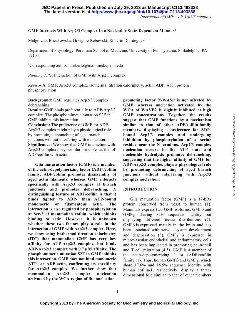
Interaction of GMF with Arp2/3 complex 1 GMF Interacts With Arp2/3 Complex In a Nucleotide State-Dependent Manner* Malgorzata Boczkowska, Grzegorz Rebowski, Roberto Dominguez 1 Department of Physiology, Perelman School of Medicine, University of Pennsylvania, Philadelphia, PA 19104 1 Corresponding author: [email protected] Running Title: Interaction of GMF with Arp2/3 complex Keywords: GMF, Arp2/3 complex, isothermal titration calorimetry, actin, ADP, ATP, protein phosphorylation Background: GMF regulates Arp2/3 complex debranching. Results: GMF binds preferentially to ADP-Arp2/3 complex. The phosphomimetic mutation S2E in GMF inhibits this interaction. Conclusion: The preference of GMF for ADP- Arp2/3 complex might play a physiological role by promoting debranching of aged branch junctions without interfering with nucleation. Significance: We show that GMF interaction with Arp2/3 complex obeys similar principles as that of ADF/cofilin with actin. Glia maturation factor (GMF) is a member of the actin-depolymerizing factor (ADF)/cofilin family. ADF/cofilin promotes disassembly of aged actin filaments, whereas GMF interacts specifically with Arp2/3 complex at branch junctions and promotes debranching. A distinguishing feature of ADF/cofilin is that it binds tighter to ADP- than ATP-bound monomeric or filamentous actin. The interaction is also regulated by phosphorylation at Ser-3 of mammalian cofilin, which inhibits binding to actin. However, it is unknown whether these two factors play a role in the interaction of GMF with Arp2/3 complex. Here, we show using isothermal titration calorimetry (ITC) that mammalian GMF has very low affinity for ATP-Arp2/3 complex, but binds ADP-Arp2/3 complex with 0.7 μ M affinity. The phosphomimetic mutation S2E in GMF inhibits this interaction. GMF does not bind monomeric ATP- or ADP-actin, confirming its specificity for Arp2/3 complex. We further show that mammalian Arp2/3 complex nucleation activated by the WCA region of the nucleation- promoting factor N-WASP is not affected by GMF, whereas nucleation activated by the WCA of WAVE2 is slightly inhibited at high GMF concentrations. Together, the results suggest that GMF functions by a mechanism similar to that of other ADF/cofilin-family members, displaying a preference for ADP- bound Arp2/3 complex and undergoing inhibition by phosphorylation of a serine residue near the N-terminus. Arp2/3 complex nucleation occurs in the ATP state and nucleotide hydrolysis promotes debranching, suggesting that the higher affinity of GMF for ADP-Arp2/3 complex plays a physiological role by promoting debranching of aged branch junctions without interfering with Arp2/3 complex nucleation. INTRODUCTION Glia maturation factor (GMF) is a 17-kDa protein conserved from yeast to human (1). Mammals express two GMF isoforms, GMFβ and GMFγ, sharing 82% sequence identity but displaying different tissue distributions (2). GMFβ is expressed mainly in the brain and has been associated with nervous system development and degeneration (3). GMFγ is expressed in microvascular endothelial and inflammatory cells and has been implicated in promoting neutrophil and T cell migration (4,5). GMF is a member of the actin-depolymerizing factor (ADF)/cofilin family (1). Thus, human GMFβ and GMFγ, which share 17.6% and 15.5% sequence identity with human cofilin-1, respectively, display a three- dimensional fold similar to that of other members http://www.jbc.org/cgi/doi/10.1074/jbc.C113.493338 The latest version is at JBC Papers in Press. Published on July 29, 2013 as Manuscript C113.493338 Copyright 2013 by The American Society for Biochemistry and Molecular Biology, Inc. by guest on April 6, 2018 http://www.jbc.org/ Downloaded from

Welcome message from author
This document is posted to help you gain knowledge. Please leave a comment to let me know what you think about it! Share it to your friends and learn new things together.
Transcript
Interaction of GMF with Arp2/3 complex
1
GMF Interacts With Arp2/3 Complex In a Nucleotide State-Dependent Manner* Malgorzata Boczkowska, Grzegorz Rebowski, Roberto Dominguez1 Department of Physiology, Perelman School of Medicine, University of Pennsylvania, Philadelphia, PA 19104 1Corresponding author: [email protected] Running Title: Interaction of GMF with Arp2/3 complex Keywords: GMF, Arp2/3 complex, isothermal titration calorimetry, actin, ADP, ATP, protein phosphorylation Background: GMF regulates Arp2/3 complex debranching. Results: GMF binds preferentially to ADP-Arp2/3 complex. The phosphomimetic mutation S2E in GMF inhibits this interaction. Conclusion: The preference of GMF for ADP-Arp2/3 complex might play a physiological role by promoting debranching of aged branch junctions without interfering with nucleation. Significance: We show that GMF interaction with Arp2/3 complex obeys similar principles as that of ADF/cofilin with actin.
Glia maturation factor (GMF) is a member of the actin-depolymerizing factor (ADF)/cofilin family. ADF/cofilin promotes disassembly of aged actin filaments, whereas GMF interacts specifically with Arp2/3 complex at branch junctions and promotes debranching. A distinguishing feature of ADF/cofilin is that it binds tighter to ADP- than ATP-bound monomeric or filamentous actin. The interaction is also regulated by phosphorylation at Ser-3 of mammalian cofilin, which inhibits binding to actin. However, it is unknown whether these two factors play a role in the interaction of GMF with Arp2/3 complex. Here, we show using isothermal titration calorimetry (ITC) that mammalian GMF has very low affinity for ATP-Arp2/3 complex, but binds ADP-Arp2/3 complex with 0.7 µM affinity. The phosphomimetic mutation S2E in GMF inhibits this interaction. GMF does not bind monomeric ATP- or ADP-actin, confirming its specificity for Arp2/3 complex. We further show that mammalian Arp2/3 complex nucleation activated by the WCA region of the nucleation-
promoting factor N-WASP is not affected by GMF, whereas nucleation activated by the WCA of WAVE2 is slightly inhibited at high GMF concentrations. Together, the results suggest that GMF functions by a mechanism similar to that of other ADF/cofilin-family members, displaying a preference for ADP-bound Arp2/3 complex and undergoing inhibition by phosphorylation of a serine residue near the N-terminus. Arp2/3 complex nucleation occurs in the ATP state and nucleotide hydrolysis promotes debranching, suggesting that the higher affinity of GMF for ADP-Arp2/3 complex plays a physiological role by promoting debranching of aged branch junctions without interfering with Arp2/3 complex nucleation. INTRODUCTION
Glia maturation factor (GMF) is a 17-kDa protein conserved from yeast to human (1). Mammals express two GMF isoforms, GMFβ and GMFγ, sharing 82% sequence identity but displaying different tissue distributions (2). GMFβ is expressed mainly in the brain and has been associated with nervous system development and degeneration (3). GMFγ is expressed in microvascular endothelial and inflammatory cells and has been implicated in promoting neutrophil and T cell migration (4,5). GMF is a member of the actin-depolymerizing factor (ADF)/cofilin family (1). Thus, human GMFβ and GMFγ, which share 17.6% and 15.5% sequence identity with human cofilin-1, respectively, display a three-dimensional fold similar to that of other members
http://www.jbc.org/cgi/doi/10.1074/jbc.C113.493338The latest version is at JBC Papers in Press. Published on July 29, 2013 as Manuscript C113.493338
Copyright 2013 by The American Society for Biochemistry and Molecular Biology, Inc.
by guest on April 6, 2018
http://ww
w.jbc.org/
Dow
nloaded from
Interaction of GMF with Arp2/3 complex
2
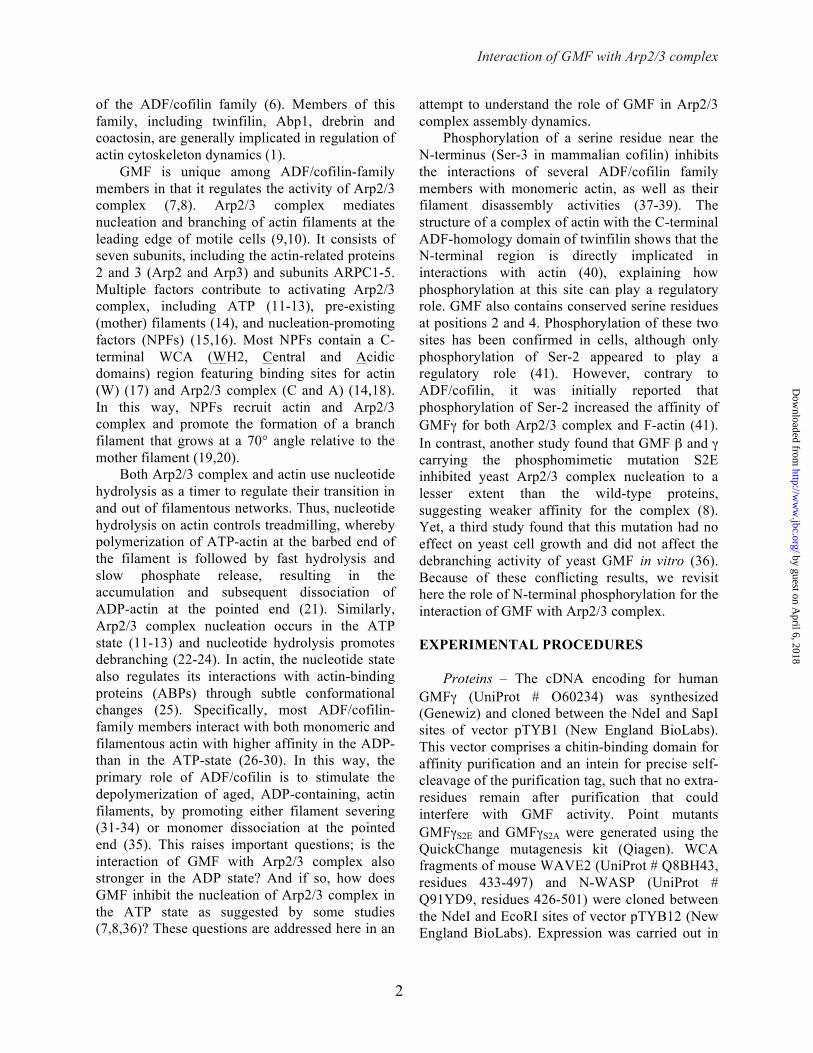
of the ADF/cofilin family (6). Members of this family, including twinfilin, Abp1, drebrin and coactosin, are generally implicated in regulation of actin cytoskeleton dynamics (1).
GMF is unique among ADF/cofilin-family members in that it regulates the activity of Arp2/3 complex (7,8). Arp2/3 complex mediates nucleation and branching of actin filaments at the leading edge of motile cells (9,10). It consists of seven subunits, including the actin-related proteins 2 and 3 (Arp2 and Arp3) and subunits ARPC1-5. Multiple factors contribute to activating Arp2/3 complex, including ATP (11-13), pre-existing (mother) filaments (14), and nucleation-promoting factors (NPFs) (15,16). Most NPFs contain a C-terminal WCA (WH2, Central and Acidic domains) region featuring binding sites for actin (W) (17) and Arp2/3 complex (C and A) (14,18). In this way, NPFs recruit actin and Arp2/3 complex and promote the formation of a branch filament that grows at a 70° angle relative to the mother filament (19,20).
Both Arp2/3 complex and actin use nucleotide hydrolysis as a timer to regulate their transition in and out of filamentous networks. Thus, nucleotide hydrolysis on actin controls treadmilling, whereby polymerization of ATP-actin at the barbed end of the filament is followed by fast hydrolysis and slow phosphate release, resulting in the accumulation and subsequent dissociation of ADP-actin at the pointed end (21). Similarly, Arp2/3 complex nucleation occurs in the ATP state (11-13) and nucleotide hydrolysis promotes debranching (22-24). In actin, the nucleotide state also regulates its interactions with actin-binding proteins (ABPs) through subtle conformational changes (25). Specifically, most ADF/cofilin-family members interact with both monomeric and filamentous actin with higher affinity in the ADP- than in the ATP-state (26-30). In this way, the primary role of ADF/cofilin is to stimulate the depolymerization of aged, ADP-containing, actin filaments, by promoting either filament severing (31-34) or monomer dissociation at the pointed end (35). This raises important questions; is the interaction of GMF with Arp2/3 complex also stronger in the ADP state? And if so, how does GMF inhibit the nucleation of Arp2/3 complex in the ATP state as suggested by some studies (7,8,36)? These questions are addressed here in an
attempt to understand the role of GMF in Arp2/3 complex assembly dynamics.
Phosphorylation of a serine residue near the N-terminus (Ser-3 in mammalian cofilin) inhibits the interactions of several ADF/cofilin family members with monomeric actin, as well as their filament disassembly activities (37-39). The structure of a complex of actin with the C-terminal ADF-homology domain of twinfilin shows that the N-terminal region is directly implicated in interactions with actin (40), explaining how phosphorylation at this site can play a regulatory role. GMF also contains conserved serine residues at positions 2 and 4. Phosphorylation of these two sites has been confirmed in cells, although only phosphorylation of Ser-2 appeared to play a regulatory role (41). However, contrary to ADF/cofilin, it was initially reported that phosphorylation of Ser-2 increased the affinity of GMFγ for both Arp2/3 complex and F-actin (41). In contrast, another study found that GMF β and γ carrying the phosphomimetic mutation S2E inhibited yeast Arp2/3 complex nucleation to a lesser extent than the wild-type proteins, suggesting weaker affinity for the complex (8). Yet, a third study found that this mutation had no effect on yeast cell growth and did not affect the debranching activity of yeast GMF in vitro (36). Because of these conflicting results, we revisit here the role of N-terminal phosphorylation for the interaction of GMF with Arp2/3 complex. EXPERIMENTAL PROCEDURES
Proteins – The cDNA encoding for human GMFγ (UniProt # O60234) was synthesized (Genewiz) and cloned between the NdeI and SapI sites of vector pTYB1 (New England BioLabs). This vector comprises a chitin-binding domain for affinity purification and an intein for precise self-cleavage of the purification tag, such that no extra-residues remain after purification that could interfere with GMF activity. Point mutants GMFγS2E and GMFγS2A were generated using the QuickChange mutagenesis kit (Qiagen). WCA fragments of mouse WAVE2 (UniProt # Q8BH43, residues 433-497) and N-WASP (UniProt # Q91YD9, residues 426-501) were cloned between the NdeI and EcoRI sites of vector pTYB12 (New England BioLabs). Expression was carried out in
by guest on April 6, 2018
http://ww
w.jbc.org/
Dow
nloaded from
Interaction of GMF with Arp2/3 complex
3
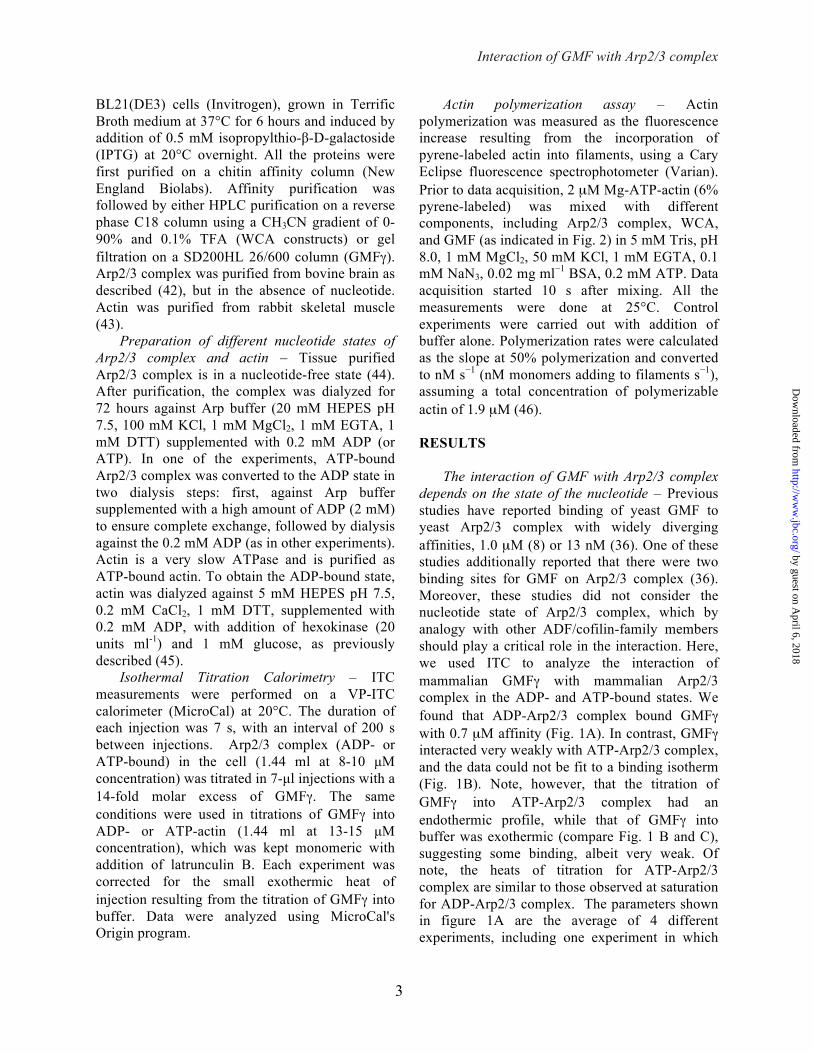
BL21(DE3) cells (Invitrogen), grown in Terrific Broth medium at 37°C for 6 hours and induced by addition of 0.5 mM isopropylthio-β-D-galactoside (IPTG) at 20°C overnight. All the proteins were first purified on a chitin affinity column (New England Biolabs). Affinity purification was followed by either HPLC purification on a reverse phase C18 column using a CH3CN gradient of 0-90% and 0.1% TFA (WCA constructs) or gel filtration on a SD200HL 26/600 column (GMFγ). Arp2/3 complex was purified from bovine brain as described (42), but in the absence of nucleotide. Actin was purified from rabbit skeletal muscle (43).
Preparation of different nucleotide states of Arp2/3 complex and actin – Tissue purified Arp2/3 complex is in a nucleotide-free state (44). After purification, the complex was dialyzed for 72 hours against Arp buffer (20 mM HEPES pH 7.5, 100 mM KCl, 1 mM MgCl2, 1 mM EGTA, 1 mM DTT) supplemented with 0.2 mM ADP (or ATP). In one of the experiments, ATP-bound Arp2/3 complex was converted to the ADP state in two dialysis steps: first, against Arp buffer supplemented with a high amount of ADP (2 mM) to ensure complete exchange, followed by dialysis against the 0.2 mM ADP (as in other experiments). Actin is a very slow ATPase and is purified as ATP-bound actin. To obtain the ADP-bound state, actin was dialyzed against 5 mM HEPES pH 7.5, 0.2 mM CaCl2, 1 mM DTT, supplemented with 0.2 mM ADP, with addition of hexokinase (20 units ml-1) and 1 mM glucose, as previously described (45).
Isothermal Titration Calorimetry – ITC measurements were performed on a VP-ITC calorimeter (MicroCal) at 20°C. The duration of each injection was 7 s, with an interval of 200 s between injections. Arp2/3 complex (ADP- or ATP-bound) in the cell (1.44 ml at 8-10 µM concentration) was titrated in 7-µl injections with a 14-fold molar excess of GMFγ. The same conditions were used in titrations of GMFγ into ADP- or ATP-actin (1.44 ml at 13-15 µM concentration), which was kept monomeric with addition of latrunculin B. Each experiment was corrected for the small exothermic heat of injection resulting from the titration of GMFγ into buffer. Data were analyzed using MicroCal's Origin program.
Actin polymerization assay – Actin polymerization was measured as the fluorescence increase resulting from the incorporation of pyrene-labeled actin into filaments, using a Cary Eclipse fluorescence spectrophotometer (Varian). Prior to data acquisition, 2 µM Mg-ATP-actin (6% pyrene-labeled) was mixed with different components, including Arp2/3 complex, WCA, and GMF (as indicated in Fig. 2) in 5 mM Tris, pH 8.0, 1 mM MgCl2, 50 mM KCl, 1 mM EGTA, 0.1 mM NaN3, 0.02 mg ml−1 BSA, 0.2 mM ATP. Data acquisition started 10 s after mixing. All the measurements were done at 25°C. Control experiments were carried out with addition of buffer alone. Polymerization rates were calculated as the slope at 50% polymerization and converted to nM s−1 (nM monomers adding to filaments s−1), assuming a total concentration of polymerizable actin of 1.9 µM (46). RESULTS
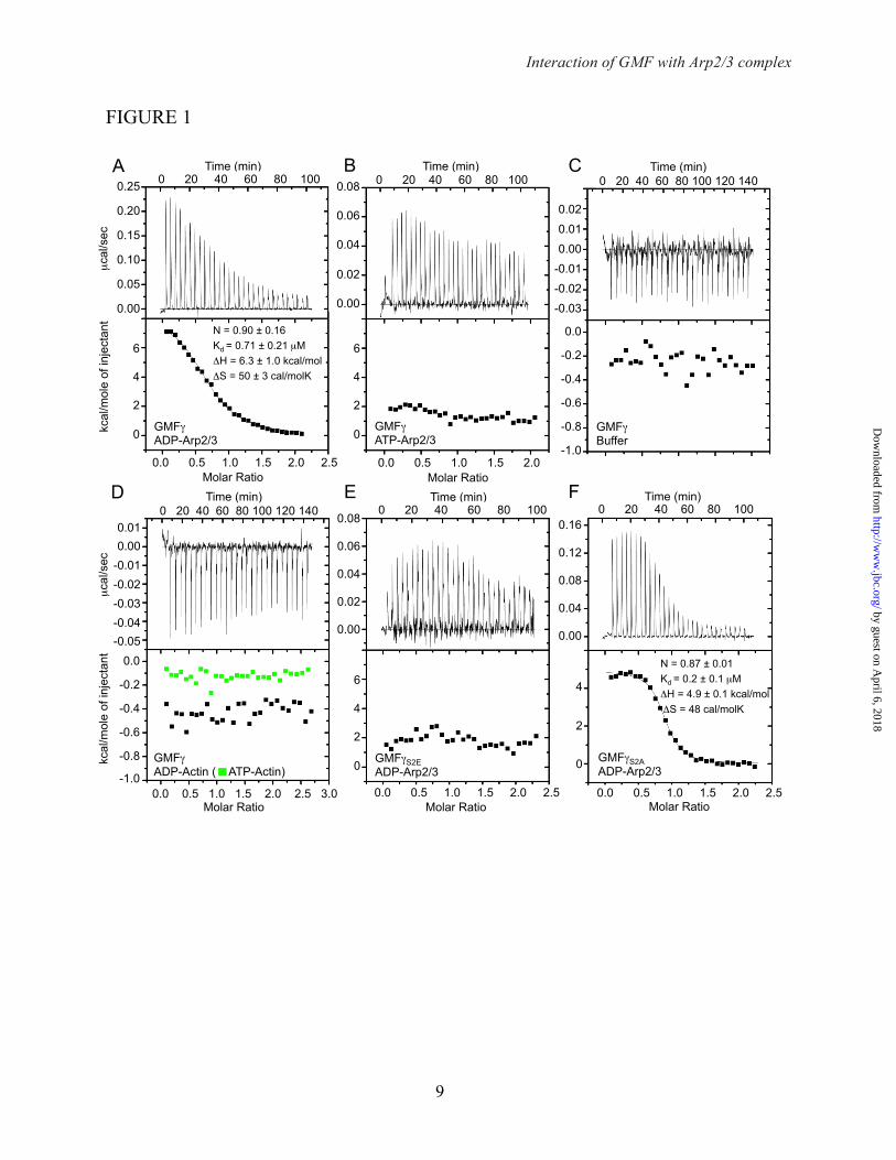
The interaction of GMF with Arp2/3 complex depends on the state of the nucleotide – Previous studies have reported binding of yeast GMF to yeast Arp2/3 complex with widely diverging affinities, 1.0 µM (8) or 13 nM (36). One of these studies additionally reported that there were two binding sites for GMF on Arp2/3 complex (36). Moreover, these studies did not consider the nucleotide state of Arp2/3 complex, which by analogy with other ADF/cofilin-family members should play a critical role in the interaction. Here, we used ITC to analyze the interaction of mammalian GMFγ with mammalian Arp2/3 complex in the ADP- and ATP-bound states. We found that ADP-Arp2/3 complex bound GMFγ with 0.7 µM affinity (Fig. 1A). In contrast, GMFγ interacted very weakly with ATP-Arp2/3 complex, and the data could not be fit to a binding isotherm (Fig. 1B). Note, however, that the titration of GMFγ into ATP-Arp2/3 complex had an endothermic profile, while that of GMFγ into buffer was exothermic (compare Fig. 1 B and C), suggesting some binding, albeit very weak. Of note, the heats of titration for ATP-Arp2/3 complex are similar to those observed at saturation for ADP-Arp2/3 complex. The parameters shown in figure 1A are the average of 4 different experiments, including one experiment in which
by guest on April 6, 2018
http://ww
w.jbc.org/
Dow
nloaded from

Interaction of GMF with Arp2/3 complex
4
ATP-Arp2/3 complex was recovered after the titration, converted to ADP-Arp2/3 complex (see Experimental Procedures), and re-titrated with GMFγ, which resulted in very similar binding. In contrast to a previous study (36), we only observed a single binding site for GMFγ on Arp2/3 complex, which is consistent with a recent crystal structure of this complex (47). The interaction was also specific for Arp2/3 complex, since GMFγ did not bind to either ADP- or ATP-bound actin (Fig. 1D).
The phosphomimetic mutation S2E inhibits binding of GMFγ to ADP-Arp2/3 complex – Previous studies have produced conflicting results regarding the role of N-terminal phosphorylation in the interaction of GMF with Arp2/3 complex (8,36,41). These studies used the phosphomimetic mutation S2E. By ITC, we found that this mutation strongly inhibited binding to ADP-Arp2/3 complex (Fig. 1E), resulting in a titration profile similar to that observed with ATP-Arp2/3 complex (compare Fig. 1 B and E). In contrast, mutant GMFγS2A bound ADP-Arp2/3 complex with similar affinity to wild type GMFγ (Fig. 1F), suggesting that the inhibitory effect of the S2E mutation was due to the additional charge and not to the removal of the serine side chain.
GMFγ does not interfere with the nucleation activity of bovine Arp2/3 complex – Previous studies have suggested that both yeast and mammalian GMF inhibit yeast and mammalian Arp2/3 complex nucleation (7,8,36). Arp2/3 complex requires ATP for nucleation (11-13), and polymerization assays are always conducted in the presence of ATP. In light of our finding that GMFγ has very low affinity for ATP-Arp2/3 complex, we questioned whether it could interfere with nucleation. We conducted pyrene-actin (6% pyrene-labeled) polymerization assays using mammalian Arp2/3 complex and the WCA regions of two NPFs, N-WASP and WAVE2, in the absence or the presence of increasing concentrations of GMFγ (Fig. 2). Compared to control experiments with actin alone, polymerization was strongly stimulated in the presence of 20 nM Arp2/3 complex and 200 nM WCAN-WASP. The addition of increasing concentrations of GMFγ or GMFγS2E (up to 4 µM) had no affect on this activity (Fig. 2A). In the case of WCAWAVE, we observed a small inhibitory
effect at high concentrations of GMFγ, as reflected by somewhat lower polymerization rates and increased lag times of polymerization (Fig. 2A).
DISCUSSION
Together, the results presented here show a parallel between the ways in which GMF interacts with Arp2/3 complex and ADF/cofilin interacts with actin. We have demonstrated for the first time that GMFγ has a clear preference for ADP- vs. ATP-bound Arp2/3 complex, which could have important physiological implications. Indeed, in vitro branches formed by Arp2/3 complex persist for hundreds of seconds (48), whereas branch turnover in cells occurs within a few seconds (49), implying the existence of cellular factors that accelerate debranching. One mechanism for debranching has been proposed to involve cofilin-mediated disassembly of the mother filament (50). Yet, by interacting directly with Arp2/3 complex, GMF acts in a more specific manner, inducing debranching at low concentrations (7,36). While Arp2/3 complex nucleation requires ATP (11-13), nucleotide hydrolysis occurs almost immediately after nucleation and promotes debranching (23,24). It thus appears that GMF’s higher affinity for ADP-Arp2/3 complex is specifically tailored for disassembly of older, ADP-containing branches. The weak affinity for ATP-Arp2/3 complex is equally important, as it reduces the likelihood of GMF interfering with the nucleation step, i.e. the formation of new branches.
Another way in which GMF might be prevented from interfering with the nucleation step is by competition with NPFs. Indeed, a recent crystal structure shows that GMF binds at the barbed end of Arp2 (47), whereas various studies have suggested that NPFs deliver an actin monomer at the barbed end of Arp2 during the first steps of nucleation (42,51,52). Thus, while our results generally contrast with previous reports of strong inhibition of nucleation by GMF (8,36), we observe some inhibition of nucleation induced by WCAWAVE but not WCAN-WASP, which can be explained by competition. Indeed, actin-WCAWAVE has 50-fold lower affinity for the site on Arp2 than actin-WCAN-WASP (Boczkowska et al., unpublished data). Similarly, differences in affinity between Arp2/3 complex and NPFs and/or GMF may also explain why the yeast system analyzed previously
by guest on April 6, 2018
http://ww
w.jbc.org/
Dow
nloaded from
Interaction of GMF with Arp2/3 complex
5
(8,36) is more susceptible to inhibition by GMF. It is important to point out, however, that the slight inhibition observed with mammalian WCAWAVE occurred at high GMF concentrations, which are probably irrelevant in the cellular context, particularly considering GMF’s marked preference for ADP-Arp2/3 complex.
We also found that GMFγ binds to ADP-Arp2/3 complex with 1:1 stoichiometry, and not 2:1 as suggested by a recent study (36). The crystal structure of GMFγ bound to Arp2/3 complex found a single binding site at the barbed end of Arp2, consistent with our results. The protein-protein contacts appear highly specific for GMF-Arp2, and cannot be reproduced on Arp3, where the corresponding binding interface is very different. We also note that this structure was determined in the ATP state, which we found binds very weakly to GMFγ. However, the
interaction is made possible by the high protein concentration used in crystallization. Curiously, the only existing structure of a complex of actin with a member of the ADF/cofilin family was also determined in the ATP-bound state (40). Therefore, there is a clear need for a structure of a complex showing the higher-affinity ADP-bound state.
We finally found through the study of the S2E phosphomimetic mutant (and a control S2A mutant) that phosphorylation at the N-terminus of GMF inhibits its interaction with ADP-Arp2/3 complex. We thus conclude that there exist striking parallels between GMF and ADF/cofilin, both in the way they select for their ADP-bound partners and in the way they are regulated by phosphorylation.
REFERENCES 1. Poukkula, M., Kremneva, E., Serlachius, M., and Lappalainen, P. (2011) Actin-depolymerizing
factor homology domain: a conserved fold performing diverse roles in cytoskeletal dynamics. Cytoskeleton 68, 471-490
2. Inagaki, M., Aoyama, M., Sobue, K., Yamamoto, N., Morishima, T., Moriyama, A., Katsuya, H., and Asai, K. (2004) Sensitive immunoassays for human and rat GMFB and GMFG, tissue distribution and age-related changes. Biochimica et biophysica acta 1670, 208-216
3. Stolmeier, D., Thangavel, R., Anantharam, P., Khan, M. M., Kempuraj, D., and Zaheer, A. (2013) Glia Maturation Factor Expression in Hippocampus of Human Alzheimer's Disease. Neurochemical research
4. Ikeda, K., Kundu, R. K., Ikeda, S., Kobara, M., Matsubara, H., and Quertermous, T. (2006) Glia maturation factor-gamma is preferentially expressed in microvascular endothelial and inflammatory cells and modulates actin cytoskeleton reorganization. Circulation research 99, 424-433
5. Aerbajinai, W., Lee, K., Chin, K., and Rodgers, G. P. (2013) Glia Maturation Factor-gamma Negatively Modulates TLR4 Signaling by Facilitating TLR4 Endocytic Trafficking in Macrophages. Journal of immunology
6. Goroncy, A. K., Koshiba, S., Tochio, N., Tomizawa, T., Sato, M., Inoue, M., Watanabe, S., Hayashizaki, Y., Tanaka, A., Kigawa, T., and Yokoyama, S. (2009) NMR solution structures of actin depolymerizing factor homology domains. Protein science : a publication of the Protein Society 18, 2384-2392
7. Gandhi, M., Smith, B. A., Bovellan, M., Paavilainen, V., Daugherty-Clarke, K., Gelles, J., Lappalainen, P., and Goode, B. L. (2010) GMF is a cofilin homolog that binds Arp2/3 complex to stimulate filament debranching and inhibit actin nucleation. Current biology : CB 20, 861-867
8. Nakano, K., Kuwayama, H., Kawasaki, M., Numata, O., and Takaine, M. (2010) GMF is an evolutionarily developed Adf/cofilin-super family protein involved in the Arp2/3 complex-mediated organization of the actin cytoskeleton. Cytoskeleton 67, 373-382
9. Pollard, T. D., and Beltzner, C. C. (2002) Structure and function of the Arp2/3 complex. Current opinion in structural biology 12, 768-774
by guest on April 6, 2018
http://ww
w.jbc.org/
Dow
nloaded from

Interaction of GMF with Arp2/3 complex
6
10. Mullins, R. D., Heuser, J. A., and Pollard, T. D. (1998) The interaction of Arp2/3 complex with actin: nucleation, high affinity pointed end capping, and formation of branching networks of filaments. Proc Natl Acad Sci U S A 95, 6181-6186
11. Dayel, M. J., Holleran, E. A., and Mullins, R. D. (2001) Arp2/3 complex requires hydrolyzable ATP for nucleation of new actin filaments. Proc Natl Acad Sci U S A 98, 14871-14876
12. Goley, E. D., Rodenbusch, S. E., Martin, A. C., and Welch, M. D. (2004) Critical conformational changes in the Arp2/3 complex are induced by nucleotide and nucleation promoting factor. Mol Cell 16, 269-279
13. Le Clainche, C., Didry, D., Carlier, M. F., and Pantaloni, D. (2001) Activation of Arp2/3 complex by Wiskott-Aldrich Syndrome protein is linked to enhanced binding of ATP to Arp2. J Biol Chem 276, 46689-46692
14. Higgs, H. N., Blanchoin, L., and Pollard, T. D. (1999) Influence of the C terminus of Wiskott-Aldrich syndrome protein (WASp) and the Arp2/3 complex on actin polymerization. Biochemistry 38, 15212-15222
15. Machesky, L. M., Mullins, R. D., Higgs, H. N., Kaiser, D. A., Blanchoin, L., May, R. C., Hall, M. E., and Pollard, T. D. (1999) Scar, a WASp-related protein, activates nucleation of actin filaments by the Arp2/3 complex. Proc Natl Acad Sci U S A 96, 3739-3744
16. Welch, M. D., Rosenblatt, J., Skoble, J., Portnoy, D. A., and Mitchison, T. J. (1998) Interaction of human Arp2/3 complex and the Listeria monocytogenes ActA protein in actin filament nucleation. Science 281, 105-108
17. Chereau, D., Kerff, F., Graceffa, P., Grabarek, Z., Langsetmo, K., and Dominguez, R. (2005) Actin-bound structures of Wiskott-Aldrich syndrome protein (WASP)-homology domain 2 and the implications for filament assembly. Proc Natl Acad Sci U S A 102, 16644-16649
18. Panchal, S. C., Kaiser, D. A., Torres, E., Pollard, T. D., and Rosen, M. K. (2003) A conserved amphipathic helix in WASP/Scar proteins is essential for activation of Arp2/3 complex. Nat Struct Biol 10, 591-598
19. Blanchoin, L., Amann, K. J., Higgs, H. N., Marchand, J. B., Kaiser, D. A., and Pollard, T. D. (2000) Direct observation of dendritic actin filament networks nucleated by Arp2/3 complex and WASP/Scar proteins. Nature 404, 1007-1011
20. Rouiller, I., Xu, X. P., Amann, K. J., Egile, C., Nickell, S., Nicastro, D., Li, R., Pollard, T. D., Volkmann, N., and Hanein, D. (2008) The structural basis of actin filament branching by the Arp2/3 complex. J Cell Biol 180, 887-895
21. Dominguez, R. (2010) Structural insights into de novo actin polymerization. Curr Opin Struct Biol 20, 217-225
22. Le Clainche, C., Pantaloni, D., and Carlier, M. F. (2003) ATP hydrolysis on actin-related protein 2/3 complex causes debranching of dendritic actin arrays. Proceedings of the National Academy of Sciences of the United States of America 100, 6337-6342
23. Martin, A. C., Welch, M. D., and Drubin, D. G. (2006) Arp2/3 ATP hydrolysis-catalysed branch dissociation is critical for endocytic force generation. Nature cell biology 8, 826-833
24. Ingerman, E., Hsiao, J. Y., and Mullins, R. D. (2013) Arp2/3 complex ATP hydrolysis promotes lamellipodial actin network disassembly but is dispensable for assembly. The Journal of cell biology 200, 619-633
25. Dominguez, R., and Holmes, K. C. (2011) Actin structure and function. Annual review of biophysics 40, 169-186
26. Maciver, S. K., and Weeds, A. G. (1994) Actophorin preferentially binds monomeric ADP-actin over ATP-bound actin: consequences for cell locomotion. FEBS letters 347, 251-256
27. Blanchoin, L., and Pollard, T. D. (1998) Interaction of actin monomers with Acanthamoeba actophorin (ADF/cofilin) and profilin. The Journal of biological chemistry 273, 25106-25111
28. Carlier, M. F., Laurent, V., Santolini, J., Melki, R., Didry, D., Xia, G. X., Hong, Y., Chua, N. H., and Pantaloni, D. (1997) Actin depolymerizing factor (ADF/cofilin) enhances the rate of filament turnover: implication in actin-based motility. J Cell Biol 136, 1307-1322
by guest on April 6, 2018
http://ww
w.jbc.org/
Dow
nloaded from

Interaction of GMF with Arp2/3 complex
7
29. Vartiainen, M. K., Mustonen, T., Mattila, P. K., Ojala, P. J., Thesleff, I., Partanen, J., and Lappalainen, P. (2002) The three mouse actin-depolymerizing factor/cofilins evolved to fulfill cell-type-specific requirements for actin dynamics. Molecular biology of the cell 13, 183-194
30. Yeoh, S., Pope, B., Mannherz, H. G., and Weeds, A. (2002) Determining the differences in actin binding by human ADF and cofilin. Journal of molecular biology 315, 911-925
31. Okreglak, V., and Drubin, D. G. (2007) Cofilin recruitment and function during actin-mediated endocytosis dictated by actin nucleotide state. The Journal of cell biology 178, 1251-1264
32. Andrianantoandro, E., and Pollard, T. D. (2006) Mechanism of actin filament turnover by severing and nucleation at different concentrations of ADF/cofilin. Molecular cell 24, 13-23
33. Michelot, A., Berro, J., Guerin, C., Boujemaa-Paterski, R., Staiger, C. J., Martiel, J. L., and Blanchoin, L. (2007) Actin-filament stochastic dynamics mediated by ADF/cofilin. Current biology : CB 17, 825-833
34. McCullough, B. R., Grintsevich, E. E., Chen, C. K., Kang, H., Hutchison, A. L., Henn, A., Cao, W., Suarez, C., Martiel, J. L., Blanchoin, L., Reisler, E., and De La Cruz, E. M. (2011) Cofilin-linked changes in actin filament flexibility promote severing. Biophysical journal 101, 151-159
35. Carlier, M. F., Ressad, F., and Pantaloni, D. (1999) Control of actin dynamics in cell motility. Role of ADF/cofilin. The Journal of biological chemistry 274, 33827-33830
36. Ydenberg, C. A., Padrick, S. B., Sweeney, M. O., Gandhi, M., Sokolova, O., and Goode, B. L. (2013) GMF Severs Actin-Arp2/3 Complex Branch Junctions by a Cofilin-like Mechanism. Current biology : CB
37. Morgan, T. E., Lockerbie, R. O., Minamide, L. S., Browning, M. D., and Bamburg, J. R. (1993) Isolation and characterization of a regulated form of actin depolymerizing factor. The Journal of cell biology 122, 623-633
38. Agnew, B. J., Minamide, L. S., and Bamburg, J. R. (1995) Reactivation of phosphorylated actin depolymerizing factor and identification of the regulatory site. The Journal of biological chemistry 270, 17582-17587
39. Ressad, F., Didry, D., Xia, G. X., Hong, Y., Chua, N. H., Pantaloni, D., and Carlier, M. F. (1998) Kinetic analysis of the interaction of actin-depolymerizing factor (ADF)/cofilin with G- and F-actins. Comparison of plant and human ADFs and effect of phosphorylation. The Journal of biological chemistry 273, 20894-20902
40. Paavilainen, V. O., Oksanen, E., Goldman, A., and Lappalainen, P. (2008) Structure of the actin-depolymerizing factor homology domain in complex with actin. J Cell Biol 182, 51-59
41. Ikeda, K., Kundu, R. K., Ikeda, S., Kobara, M., Matsubara, H., and Quertermous, T. (2006) Glia maturation factor-gamma is preferentially expressed in microvascular endothelial and inflammatory cells and modulates actin cytoskeleton reorganization. Circ Res 99, 424-433
42. Boczkowska, M., Rebowski, G., Petoukhov, M. V., Hayes, D. B., Svergun, D. I., and Dominguez, R. (2008) X-ray scattering study of activated Arp2/3 complex with bound actin-WCA. Structure 16, 695-704
43. Pardee, J. D., and Spudich, J. A. (1982) Purification of muscle actin. Methods Enzymol 85 Pt B, 164-181
44. Nolen, B. J., Littlefield, R. S., and Pollard, T. D. (2004) Crystal structures of actin-related protein 2/3 complex with bound ATP or ADP. Proc Natl Acad Sci U S A 101, 15627-15632
45. Pollard, T. D. (1986) Rate constants for the reactions of ATP- and ADP-actin with the ends of actin filaments. J Cell Biol 103, 2747-2754
46. Harris, E. S., and Higgs, H. N. (2006) Biochemical analysis of mammalian formin effects on actin dynamics. Methods Enzymol 406, 190-214
47. Luan, Q., and Nolen, B. J. (2013) Structural basis for regulation of Arp2/3 complex by GMF. Nature Structural & Molecular Biology in press, ?
48. Blanchoin, L., Pollard, T. D., and Mullins, R. D. (2000) Interactions of ADF/cofilin, Arp2/3 complex, capping protein and profilin in remodeling of branched actin filament networks. Current biology : CB 10, 1273-1282
by guest on April 6, 2018
http://ww
w.jbc.org/
Dow
nloaded from
Interaction of GMF with Arp2/3 complex
8
49. Lai, F. P., Szczodrak, M., Block, J., Faix, J., Breitsprecher, D., Mannherz, H. G., Stradal, T. E., Dunn, G. A., Small, J. V., and Rottner, K. (2008) Arp2/3 complex interactions and actin network turnover in lamellipodia. Embo J 27, 982-992
50. Chan, C., Beltzner, C. C., and Pollard, T. D. (2009) Cofilin dissociates Arp2/3 complex and branches from actin filaments. Curr Biol 19, 537-545
51. Padrick, S. B., Doolittle, L. K., Brautigam, C. A., King, D. S., and Rosen, M. K. (2011) Arp2/3 complex is bound and activated by two WASP proteins. Proceedings of the National Academy of Sciences of the United States of America 108, E472-479
52. Ti, S. C., Jurgenson, C. T., Nolen, B. J., and Pollard, T. D. (2011) Structural and biochemical characterization of two binding sites for nucleation-promoting factor WASp-VCA on Arp2/3 complex. Proceedings of the National Academy of Sciences of the United States of America 108, E463-471
FOOTNOTES *This work was supported by NIH grant R01 GM073791. 1To whom correspondence should be addressed: Roberto Dominguez, Department of Physiology,
Perelman School of Medicine, University of Pennsylvania, Philadelphia, PA 19104; [email protected]
FIGURE LEGENDS FIGURE 1. Analysis by isothermal titration calorimetry of the binding of GMFγ to Arp2/3 complex. Experiments were conducted at 20°C. Arp2/3 complex (or actin-latrunculin B) in the cell at 8-10 µM (or 13-15 µM) was titrated with a 14-fold molar excess of GMFγ in 7-µl injections (7 s per injection, with an interval of 200 s between injections). (A) Titration of GMFγ into ADP-Arp2/3 complex. The figure shows the fitting of the data to a binding isotherm derived from the integrated heats of binding plotted against the molar ratio of ligand (GMFγ) added to ADP-Arp2/3 complex in the cell, after subtracting the heat of dilution. The best-fit parameters (solid black line) correspond to a one-site binding model with dissociation constant of 0.7 µM. (B) Titration of GMFγ into ATP-Arp2/3 complex (the data could not be fit to a binding isotherm). (C) Titration of GMFγ into buffer (control experiment). (D) Titration of GMFγ into ADP- (black) and ATP-actin (green). Note that these two titrations look similar to that of GMFγ into buffer, indicating complete lack of interaction. (E) Titration of GMFγS2E into ADP-Arp2/3 complex (data could not be fit to a binding isotherm). (F) Titration of GMFγS2A into ADP-Arp2/3 complex. Each titration was repeated at least two times, and four times for that shown in part A. In part A, errors are reported as SEM, whereas for the other titrations errors are derived from curve fitting. FIGURE 2. GMFγ does not inhibit actin polymerization by Arp2/3 complex. Time-course of the fluorescence increase upon polymerization of 2 µM actin (6% pyrene-labeled) alone (black line) or with addition of the indicated proteins (color coded). (A) Effect of different concentrations of GMFγ on actin polymerization induced by 20 nM Arp2/3 complex activated by 200 nM WCAN-WASP. Polymerization rates at 50% polymerization are shown. Lag time (measured as the time to 10% polymerization) is 190 s. (B) Effect of different concentrations of GMFγ on actin polymerization induced by 20 nM Arp2/3 complex activated by 200 nM WCAWAVE. Lag times are 229, 245, 277, and 292 s for 0, 1, 2, and 4 µM GMF, respectively. Each measurement was performed three times (one representative curve is shown). Errors are reported as SEM.
by guest on April 6, 2018
http://ww
w.jbc.org/
Dow
nloaded from
Interaction of GMF with Arp2/3 complex
9
FIGURE 1
A
kcal
/mol
e of
inje
ctan
t
Time (min)
Molar Ratio
GMF!"ADP-Arp2/3"
N = 0.90 ± 0.16 Kd = 0.71 ± 0.21 µM #H = 6.3 ± 1.0 kcal/mol #S = 50 ± 3 cal/molK
0.0 0.5 1.0 1.5 2.0 2.5
0 20 40 60 80 100
6
4
2
0
0.25
0.20
0.15
0.10
0.05
0.00
µca
l/sec
Time (min)
Molar Ratio
GMF!S2E ADP-Arp2/3"
E 0 20 40 60 80 100
0.0 0.5 1.0 1.5 2.0 2.5
6
4
2
0
0.08
0.06
0.04
0.02
0.00
Time (min)
Molar Ratio
GMF!"ATP-Arp2/3"
B 0 20 40 60 80 100
0.0 0.5 1.0 1.5 2.0
6
4
2
0
0.08
0.06
0.04
0.02
0.00
Time (min)
GMF!"Buffer"
C
0.0
-0.2
-0.4
-0.6
-0.8
-1.0
0.02
0.01
0.00
-0.01
-0.02
-0.03
0 20 40 60 80 100 120 140
µca
l/sec
kc
al/m
ole
of in
ject
ant
N = 0.87 ± 0.01 Kd = 0.2 ± 0.1 µM #H = 4.9 ± 0.1 kcal/mol #S = 48 cal/molK
GMF!S2A ADP-Arp2/3"
4
2
0
0.16
0.12
0.08
0.04
0.00
0.0 0.5 1.0 1.5 2.0 2.5
0 20 40 60 80 100 Time (min)
Molar Ratio
F
Molar Ratio
GMF!"ADP-Actin ( ATP-Actin)"
Time (min) D
0.0 0.5 1.0 1.5 2.0 2.5 3.0
0.0
-0.2
-0.4
-0.6
-0.8
-1.0
0.01
0.00
-0.01
-0.02
-0.03
-0.04
-0.05
0 20 40 60 80 100 120 140
by guest on April 6, 2018
http://ww
w.jbc.org/
Dow
nloaded from

Interaction of GMF with Arp2/3 complex
10
FIGURE 2
Time (s)
B
0 200 400 600 800
500
450
400
350
300
250
200
150
100
50
0
Fluo
resc
ence
inte
nsity
(a.u
.)
+ 20 nM Arp2/3 + 4 µM GMF!"
+ 20 nM Arp2/3
+ buffer
+ 20 nM Arp2/3 + 200 nM WCAWAVE + 1 µM GMF! 12.2±1.7"
2 µM actin (6% pyrene)
+ 20 nM Arp2/3 + 200 nM WCAWAVE 11.4±0.2
+ 20 nM Arp2/3 + 200 nM WCAWAVE + 2 µM GMF! 10.3±0.3" + 20 nM Arp2/3 + 200 nM WCAWAVE + 4 µM GMF! 9.6±0.1 " + 20 nM Arp2/3 + 200 nM WCAWAVE + 4 µM GMF!S2E 9.5±0.7
Time (s)
A
0 200 400 600 800
500
450
400
350
300
250
200
150
100
50
0
Fluo
resc
ence
inte
nsity
(a.u
.)
+ 20 nM Arp2/3 + 4 µM GMF!"
+ 20 nM Arp2/3
+ buffer
+ 20 nM Arp2/3 + 200 nM WCAN-WASP + 1 µM GMF! 13.4±1.0
2 µM actin (6% pyrene)
+ 20 nM Arp2/3 + 200 nM WCAN-WASP 13.7±0.6
+ 20 nM Arp2/3 + 200 nM WCAN-WASP + 2 µM GMF! 12.7±1.3" + 20 nM Arp2/3 + 200 nM WCAN-WASP + 4 µM GMF! 13.0±1.3" + 20 nM Arp2/3 + 200 nM WCAN-WASP + 4 µM GMF!S2E 13.7±1.4
Polymerization rate nM/s
Polymerization rate nM/s
by guest on April 6, 2018
http://ww
w.jbc.org/
Dow
nloaded from

Malgorzata Boczkowska, Grzegorz Rebowski and Roberto DominguezGMF Interacts With Arp2/3 Complex In a Nucleotide State-Dependent Manner
published online July 29, 2013J. Biol. Chem.
10.1074/jbc.C113.493338Access the most updated version of this article at doi:
Alerts:
When a correction for this article is posted•
When this article is cited•
to choose from all of JBC's e-mail alertsClick here
by guest on April 6, 2018
http://ww
w.jbc.org/
Dow
nloaded from
Related Documents