Interaction between silicon amendment, bacterial wilt development and phenotype of Ralstonia solanacearum in tomato genotypes Elie Ayitondji Dannon, Kerstin Wydra * Institut fu ¨r Pflanzenkrankheiten und Pflanzenschutz, Universita ¨t Hannover, Herrenha ¨user Str. 2, 30419 Hannover, Germany Accepted 17 September 2004 Abstract Silicon amendment significantly reduced bacterial wilt incidence expressed as area under disease progress curve for tomato genotypes L390 (susceptible) by 26.8% and King Kong2 (moderately resistant) by 56.1% compared to non-treated plants grown in hydroponic culture. However, wilt incidence in silicon-treated plants of genotype L390 reached 100% at 13 days post-inoculation (dpi), while in genotype King Kong2, plant death was retarded by 6 days, with 20% reduction of final wilt incidence. Bacterial numbers were significantly lower in silicon- treated compared to non-treated plants in King Kong2 at 2 dpi in midstems and in all organs at 5 dpi, and in Hawaii 7998 (resistant) in all organs at 2 dpi. Differences between genotypes were obvious on midstem level (5 dpi), where bacterial populations were generally significantly lower compared to roots. Increased tolerance was observed in genotypes L390 and King Kong2 with silicon treatment. Silicon accumulated in roots and was low in stems and leaves. Inoculation with Ralstonia solanacearum did not significantly affect silicon uptake and distribution. Negative correlations between root silicon content and bacterial numbers of midstems in genotypes Hawaii 7998 and King Kong2 suggested an induced resistance. Indications for an influence of host genotype and silicon treatment on the phenotypic conversion of R. solanacearum strain To-udk2-sb from fluidal to non-fluidal colonies in planta were observed. This is the first report on the effect of silicon on a bacterial disease and in a silicon-non-accumulator plant. q 2004 Elsevier Ltd. All rights reserved. Keywords: Induced resistance; Mineral nutrition; Phenotypic conversion (PC); Tolerance 1. Introduction Bacterial wilt is widely distributed in tropical, subtropi- cal and some temperate regions of the world. The disease ranks as one of the most important if not the most important disease of bacterial origin in the world [22], causing sometimes total losses in tomato crops [33]. Causal agent is Ralstonia solanacearum (Smith) Yabuuchi et al. (1995) [45], a highly diverse and adaptive bacterium, that differs in host range, geographical distribution, pathogenicity, epide- miological interactions and physiological properties [4,18]. On the basis of host range, R. solanacearum strains have been traditionally divided into races [17], while physiologi- cal and genetic characterization resulted in the formation of biovars and divisions [13,17]. The bacterium invades the plant vascular tissues through wounded roots or natural openings, which occur after the emergence of secondary roots. It progresses through intercellular spaces into the xylem. Colonization of stems results in browning of the xylem, foliar epinasty and lethal generalized wilt [3]. Control of R. solanacearum is difficult due to its wide host range and its survival capacity in various environments such as irrigation water and soil. Host plant resistance breakdown due to high genotype x environment interactions was frequently observed [42]. Therefore, only integrated control combining host plant resistance and cultural and biological measures seems promising. Resistance against this pathogen is described as quanti- tative or polygenic [43]. In quantitative resistance, cell wall characteristics or modifications play a key role, directly as barriers against pathogens or indirectly through their effect on the increase of other active compounds [31]. Many soil elements found in cell walls have been reported to influence the susceptibility or resistance of plants to pathogen 0885-5765/$ - see front matter q 2004 Elsevier Ltd. All rights reserved. doi:10.1016/j.pmpp.2004.09.006 Physiological and Molecular Plant Pathology 64 (2004) 233–243 www.elsevier.com/locate/pmpp * Corresponding author. Fax: C49 511 7623 015. E-mail address: [email protected] (K. Wydra).

Welcome message from author
This document is posted to help you gain knowledge. Please leave a comment to let me know what you think about it! Share it to your friends and learn new things together.
Transcript
Interaction between silicon amendment, bacterial wilt development
and phenotype of Ralstonia solanacearum in tomato genotypes
Elie Ayitondji Dannon, Kerstin Wydra*
Institut fur Pflanzenkrankheiten und Pflanzenschutz, Universitat Hannover, Herrenhauser Str. 2, 30419 Hannover, Germany
Accepted 17 September 2004
Abstract
Silicon amendment significantly reduced bacterial wilt incidence expressed as area under disease progress curve for tomato genotypes
L390 (susceptible) by 26.8% and King Kong2 (moderately resistant) by 56.1% compared to non-treated plants grown in hydroponic culture.
However, wilt incidence in silicon-treated plants of genotype L390 reached 100% at 13 days post-inoculation (dpi), while in genotype King
Kong2, plant death was retarded by 6 days, with 20% reduction of final wilt incidence. Bacterial numbers were significantly lower in silicon-
treated compared to non-treated plants in King Kong2 at 2 dpi in midstems and in all organs at 5 dpi, and in Hawaii 7998 (resistant) in all
organs at 2 dpi. Differences between genotypes were obvious on midstem level (5 dpi), where bacterial populations were generally
significantly lower compared to roots. Increased tolerance was observed in genotypes L390 and King Kong2 with silicon treatment.
Silicon accumulated in roots and was low in stems and leaves. Inoculation with Ralstonia solanacearum did not significantly affect silicon
uptake and distribution. Negative correlations between root silicon content and bacterial numbers of midstems in genotypes Hawaii 7998 and
King Kong2 suggested an induced resistance. Indications for an influence of host genotype and silicon treatment on the phenotypic
conversion of R. solanacearum strain To-udk2-sb from fluidal to non-fluidal colonies in planta were observed.
This is the first report on the effect of silicon on a bacterial disease and in a silicon-non-accumulator plant.
q 2004 Elsevier Ltd. All rights reserved.
Keywords: Induced resistance; Mineral nutrition; Phenotypic conversion (PC); Tolerance
1. Introduction
Bacterial wilt is widely distributed in tropical, subtropi-
cal and some temperate regions of the world. The disease
ranks as one of the most important if not the most important
disease of bacterial origin in the world [22], causing
sometimes total losses in tomato crops [33]. Causal agent is
Ralstonia solanacearum (Smith) Yabuuchi et al. (1995)
[45], a highly diverse and adaptive bacterium, that differs in
host range, geographical distribution, pathogenicity, epide-
miological interactions and physiological properties [4,18].
On the basis of host range, R. solanacearum strains have
been traditionally divided into races [17], while physiologi-
cal and genetic characterization resulted in the formation of
biovars and divisions [13,17]. The bacterium invades
0885-5765/$ - see front matter q 2004 Elsevier Ltd. All rights reserved.
doi:10.1016/j.pmpp.2004.09.006
* Corresponding author. Fax: C49 511 7623 015.
E-mail address: [email protected] (K. Wydra).
the plant vascular tissues through wounded roots or natural
openings, which occur after the emergence of secondary
roots. It progresses through intercellular spaces into the
xylem. Colonization of stems results in browning of the
xylem, foliar epinasty and lethal generalized wilt [3].
Control of R. solanacearum is difficult due to its wide
host range and its survival capacity in various environments
such as irrigation water and soil. Host plant resistance
breakdown due to high genotype x environment interactions
was frequently observed [42]. Therefore, only integrated
control combining host plant resistance and cultural and
biological measures seems promising.
Resistance against this pathogen is described as quanti-
tative or polygenic [43]. In quantitative resistance, cell wall
characteristics or modifications play a key role, directly as
barriers against pathogens or indirectly through their effect
on the increase of other active compounds [31]. Many soil
elements found in cell walls have been reported to influence
the susceptibility or resistance of plants to pathogen
Physiological and Molecular Plant Pathology 64 (2004) 233–243
www.elsevier.com/locate/pmpp
E.A. Dannon, K. Wydra / Physiological and Molecular Plant Pathology 64 (2004) 233–243234
infections, among them silicon as part of the cell wall,
which is considered to be a ‘beneficial’ element for plants
and higher animals [10]. In nature, silicon is found in the
form of silica (SiO2), aluminium silicates, or iron or calcium
silicates and is absorbed by the plant as mono-silicic acid
(Si(OH)4) [6]. Ma et al. [26] classified plant species into
silicon accumulator species, intermediate types and silicon
non-accumulators. The uptake mode is active for the first
group, passive for the second and rejective for the third.
Application of silicon has been shown to increase leaf
chlorophyll and plant metabolism, enhance plant tolerance
to environmental stresses such as cold, heat and drought,
mitigate nutrient imbalance and metal toxicity in the plant,
reinforce cell walls and increase plant resistance against
pathogens and insects [10]. In rice, a silicon-accumulator
plant, silicon occurs in the wall of epidermic cells or
extracellular locations such as storage and vascular tissues,
intercellular spaces and cuticular layers [36] and has been
reported as important component of papilla [12]. Following
uptake and transport of silicon, the process of silicification,
the polymerisation of silicic acid into silica gel SiO2H2O,
begins, whereby silicon together with lignin contributes to
the rigidification of cell walls in leaves and xylem vessels
[26]. Therefore, it was proposed that silicon plays a role in
the formation of mechanical barriers restricting the
penetration of pathogens [1,9]. Silicon enhanced resistance
against fungal diseases such as Pythium and Sphaeroteca
fuliginea in cucumber [12,35] and blast and sheath blight
diseases in rice [9]. The role of a mechanical barrier could
not be confirmed, but induced resistance was suggested to
be involved [7,27,40]. The effect of silicon on bacterial
diseases and on pathogens in non-accumulator plants
remains so far unknown.
R. solanacearum possesses the ability to react to changes
in its environment with a wide range of physiological states.
Among them, the phenomenon of phenotypic conversion
(PC), has long been known [21]. The high variability of the
pathogen in phenotypic and genetic features is expressed in
response to varying environmental conditions. R. solana-
cearum shows a spontaneous shift from the mucoid (fluidal)
to the non-mucoid type under certain growth conditions
[23], whereby bacteria become avirulent, lose their ability to
produce exopolysaccharides, reduce their endoglucanase
activity, and increase motility and endo-polygalacturonase
activity [2]. Only recently the involvement of the host plant
in the reversion of the PC phenomenon has been shown with
a susceptible genotype [29]. A possible interaction between
host genotype and PC has not been described, nor is the
direct or indirect effect of silicon treatment on the state of
the pathogen known.
The goal of the study was to find out if silicon
amendment enhances the degree of resistance of tomato to
R. solanacearum, to elucidate whether direct or indirect
mechanisms are involved and to determine a possible effect
of silicon or genotype on PC of the pathogen.
2. Materials and methods
2.1. Plant materials and culture
Tomato genotypes L390 (susceptible to R. solana-
cearum), King Kong2 (moderately resistant) and Hawaii
7998 (resistant) were provided by the Genetic Resources
and Seeds Unit of the Asian Vegetable Research and
Development Center (AVRDC, Taiwan). Experiments were
performed in hydroponic culture. Tomato seeds were pre-
germinated in Petri dishes at 30 8C and transferred into a
plate system containing wet filter papers. About one week
old seedlings were transferred in black pots with lids
containing 4.5 L hydroponic solution: 0.5 M Ca (NO3)2,
0.2 M NH4NO3, 0.5 M K2SO4, 0.5 M MgSO4, 0.5 M
KH2PO4, 10 mM H3BO3, 0.1 M ZnSO4, 0.06 mM CuSO4,
1 mM MnSO4, 0.1 mM MoNaO4, 10 mM NaCl and 10 mM
FeEDTA; 20 ml monosilicic acid [1.4 mM Si(OH)4] was
added to 5 L of final nutrient solution; monosilicic acid was
obtained after exchange of potassium silicate solution
K2SiO2 with cation exchangers (20 ml volume, Biorad
Laboratories, Germany) (Hochmuth, 1999 [19], modified by
Institute for Plant Nutrition, University of Hannover). The
hydroponic solution was aerated through a tube system
connected to a pump (Hagen, Germany) and changed at
the day of inoculation of R. solanacearum. Plants were
grown in a climate chamber with 30/26 8C day/night
temperature, 80% RH and light for 12 h/day (photonflux
of 350 mmol/m2s).
2.2. Experimental design
Four treatments were arranged in a completely randomi
sed block design: (i) plants with silicon, inoculated with
R. solanacearum (RsCSi), (ii) plants without silicon,
inoculated with R. solanacearum (RsKSi), (iii) plants
with silicon, without R. solanacearum inoculation (KRsCSi, control) and (iv) plants without silicon, without
R. solanacearum inoculation (KRsKSi, control), with 16
plants per genotype and treatment. Four plants were
randomly selected per treatment and sampling date at two
and five days post-inoculation (dpi) for bacterial and silicon
quantification, while eight plants were kept for symptom
evaluation. Leaves, stems and roots of sampled plants were
subdivided in two parts, one for bacterial quantification and
the other for silicon analysis. Experiments were repeated
three times.
2.3. Inoculation and quantification of bacteria
The highly virulent R. solanacearum strain To-udk2-sb
from Thai origin belonging to race 1 biovar 3 (Leykun,
2003; Thaveechai, unpublished) was used for inoculation of
the tomato plants. Bacteria were grown on TTC medium
[21] for 48 h at 30 8C. Five hundred millilitre of inoculum
suspension (OD660 nm 0.06 corresponding to approximately
E.A. Dannon, K. Wydra / Physiological and Molecular Plant Pathology 64 (2004) 233–243 235
108 CFU/ml) were poured in each pot to obtain a final
inoculum concentration of approximately 107 CFU/ml.
R. solanacearum was quantified in leaves, midstems and
roots of four randomly selected plants two and five days
post-inoculation. Leaves, midstems (5 cm) and roots were
weighed, treated with 70% alcohol for 10 s and rinsed and
macerated with sterile demineralised water. The suspension
was centrifuged for 20 min at 5500!g at room temperature
and the pellet resuspended in 1.5 ml of sterile demineralised
water. Tenfold serial dilutions were prepared and 0.1 ml of
each dilution was plated with two replicates on TTC
medium. Bacterial colonies were counted after 48 h
incubation at 30 8C. Fluidal (O1 mm diameter) and non-
fluidal (!1 mm diameter) colony types were differentiated.
2.4. Symptom evaluation
Evaluation began with the appearance of leaf symptoms
and was continued daily until all plants had died or
symptoms became stable. Five disease severity classes
(modified after [44]) were used: 0Zno symptom, 1Zone
leaf wilted, 2Ztwo leaves wilted, 3Zthree leaves wilted,
4Zwilting of all leaves without the tip, 5Zwilting of the
whole plant. Wilt incidence was calculated as the percen-
tage of dead plants (class 5) at the evaluation date out of the
total number of plants in the treatment. The area under
disease progress curve (AUDPC) was calculated on the
basis of disease severity and of wilt incidence using the
following formula [20,39]:
AUDPC ZX
½ðxi CxiK1Þ=2�ðti K tiK1Þ;
where xi and xiK1 are disease severity or wilt incidence and
ti and tiK1 are consecutive evaluation dates (tiKtiK1 was
equal to 1).
2.5. Silicon quantification
Total silicon content of leaves, stems and roots of four
randomly selected plants was determined for organs of the
same plants which were used for bacterial quantification at
two and five days post-inoculation, following the method
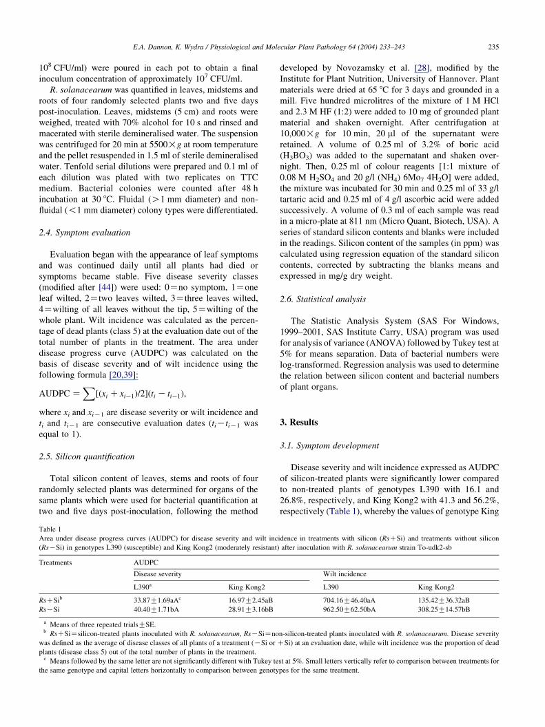
Table 1
Area under disease progress curves (AUDPC) for disease severity and wilt inc
(RsKSi) in genotypes L390 (susceptible) and King Kong2 (moderately resistant)
Treatments AUDPC
Disease severity
L390a King Kong2
RsCSib 33.87G1.69aAc 16.97G2.45aB
RsKSi 40.40G1.71bA 28.91G3.16bB
a Means of three repeated trialsGSE.b RsCSiZsilicon-treated plants inoculated with R. solanacearum, RsKSiZno
was defined as the average of disease classes of all plants of a treatment (KSi or Cplants (disease class 5) out of the total number of plants in the treatment.
c Means followed by the same letter are not significantly different with Tukey te
the same genotype and capital letters horizontally to comparison between genoty
developed by Novozamsky et al. [28], modified by the
Institute for Plant Nutrition, University of Hannover. Plant
materials were dried at 65 8C for 3 days and grounded in a
mill. Five hundred microlitres of the mixture of 1 M HCl
and 2.3 M HF (1:2) were added to 10 mg of grounded plant
material and shaken overnight. After centrifugation at
10,000!g for 10 min, 20 ml of the supernatant were
retained. A volume of 0.25 ml of 3.2% of boric acid
(H3BO3) was added to the supernatant and shaken over-
night. Then, 0.25 ml of colour reagents [1:1 mixture of
0.08 M H2SO4 and 20 g/l (NH4) 6Mo7 4H2O] were added,
the mixture was incubated for 30 min and 0.25 ml of 33 g/l
tartaric acid and 0.25 ml of 4 g/l ascorbic acid were added
successively. A volume of 0.3 ml of each sample was read
in a micro-plate at 811 nm (Micro Quant, Biotech, USA). A
series of standard silicon contents and blanks were included
in the readings. Silicon content of the samples (in ppm) was
calculated using regression equation of the standard silicon
contents, corrected by subtracting the blanks means and
expressed in mg/g dry weight.
2.6. Statistical analysis
The Statistic Analysis System (SAS For Windows,
1999–2001, SAS Institute Carry, USA) program was used
for analysis of variance (ANOVA) followed by Tukey test at
5% for means separation. Data of bacterial numbers were
log-transformed. Regression analysis was used to determine
the relation between silicon content and bacterial numbers
of plant organs.
3. Results
3.1. Symptom development
Disease severity and wilt incidence expressed as AUDPC
of silicon-treated plants were significantly lower compared
to non-treated plants of genotypes L390 with 16.1 and
26.8%, respectively, and King Kong2 with 41.3 and 56.2%,
respectively (Table 1), whereby the values of genotype King
idence in treatments with silicon (RsCSi) and treatments without silicon
after inoculation with R. solanacearum strain To-udk2-sb
Wilt incidence
L390 King Kong2
704.16G46.40aA 135.42G36.32aB
962.50G62.50bA 308.25G14.57bB
n-silicon-treated plants inoculated with R. solanacearum. Disease severity
Si) at an evaluation date, while wilt incidence was the proportion of dead
st at 5%. Small letters vertically refer to comparison between treatments for
pes for the same treatment.
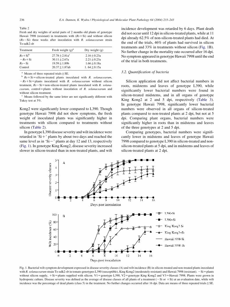
Table 2
Fresh and dry weights of aerial parts of 2 months old plants of genotype
Hawaii 7998 (resistant) in treatments with (RsCSi) and without silicon
(RsKSi) three weeks after inoculation with R. solanacearum strain
To-udk2-sb
Treatment Fresh weight (g)a Dry weight (g)
RsCSib 27.78G2.81ac 2.16G0.23a
KRsCSi 30.11G2.67a 2.21G0.23a
RsKSi 19.59G1.89b 1.66G0.18a
Control 20.37G1.87ab 1. 64G0.15a
a Means of three repeated trialsGSE.b RsCSiZsilicon-treated plants inoculated with R. solanacearum,
KRsCSiZplants inoculated with R. solanacearum without silicon
treatment, RsKSiZnon-silicon-treated plants inoculated with R. solana-
cearum, controlZplants without inoculation of R. solanacearum and
without silicon treatment.c Means followed by the same letter are not significantly different with
Tukey test at 5%.
E.A. Dannon, K. Wydra / Physiological and Molecular Plant Pathology 64 (2004) 233–243236
Kong2 were significantly lower compared to L390. Though
genotype Hawaii 7998 did not show symptoms, the fresh
weight of inoculated plants was significantly higher in
treatments with silicon compared to treatments without
silicon (Table 2).
In genotype L390 disease severity and wilt incidence were
retarded in ‘SiC’ plants by about two days and reached the
same level as in ‘SiK’ plants at day 12 and 13, respectively
(Fig. 1). In genotype King Kong2, disease severity increased
slower in silicon-treated than in non-treated plants, and wilt
Fig. 1. Bacterial wilt symptom development expressed in disease severity classes (A
with R. solanacearum strain To-udk2-sb in tomato genotypes L390 (susceptible), K
without silicon supply, CSiZplants supplied with silicon. V1Zgenotype L390,
hydroponic culture. Disease severity was defined as the average of disease classes
incidence was the percentage of dead plants (class 5) in the treatment. No further
incidence development was retarded by 6 days. Plant death
did not occur until 12 dpi in silicon-treated plants, while at 11
dpi already 62.5% of non-silicon-treated plants had died. At
the end of the trials, 46% of plants had survived in silicon
treatments and 33% in treatments without silicon (Fig. 1B).
No further change in the mortality rate occurred after 16 dpi.
No symptom appeared in genotype Hawaii 7998 until the end
of the trial in both treatments.
3.2. Quantification of bacteria
Silicon application did not affect bacterial numbers in
roots, midstems and leaves of genotype L390, while
significantly lower bacterial numbers were found in
silicon-treated midstems, and in all organs of genotype
King Kong2 at 2 and 5 dpi, respectively (Table 3).
In genotype Hawaii 7998, significantly lower bacterial
numbers were observed in all organs of silicon-treated
plants compared to non-treated plants at 2 dpi, but not at 5
dpi. Comparing plant organs, bacterial numbers were
significantly higher in roots than in midstems and leaves
of the three genotypes at 2 and 5 dpi.
Comparing genotypes, bacterial numbers were signifi-
cantly lower in midstems and leaves of genotype Hawaii
7998 compared to genotype L390 in silicon-treated and non-
silicon-treated plants at 5 dpi, and in midstems and leaves of
silicon-treated plants at 2 dpi.
) and wilt incidence (B) in silicon-treated and non-treated plants inoculated
ing Kong2 (moderately resistant) and Hawaii 7998 (resistant). KSiZplants
V2Zgenotype King Kong2 and V3ZHawaii 7998. Plants were grown in
of all plants of a treatment (KSi or CSi) at an evaluation date, while wilt
changes occurred after 16 dpi. Data are means of three repeated trialsGSE.

Table 3
Bacterial numbers of roots, midstems and leaves of silicon-treated and non-treated plants of tomato genotypes L390 (susceptible), King Kong2 (moderately
resistant) and Hawaii 7998 (resistant) at 2 and 5 days post-inoculation with R. solanacearum strain To-udk2-sb
Genotype Treatment Bacterial number [log10 (CFU/g)] 2 dpi Bacterial number [log (CFU/g)] 5 dpi
Rootsa Midstems Leaves Roots Midstems Leaves
L390 RsCSib 6.90G0.21aBac 4.18G0.19aAa 4.15G0.13aAad 7.92G0.20aBa 6.64G0.41aAa 6.21G0.40aAa
RsKSi 7.53G0.24aBa 4.61G0.26aAa 4.55G0.21aAa 7.89G0.09aBa 6.71G0.36 aABa 6.44G0.45aAa
King Kong2 RsCSi 7.42G0.18aBa 3.87G0.14aAa 3.69G0.26aAab 7.88G0.15aBa 5.14G0.40aAab 4.60G0.29 aAb
RsKSi 7.49G0.20aBa 4.56G0.18bAa 4.05G0.17aAa 8.91G0.49 bCb 7.69G0.07 bBb 6.14G0.46bAab
Hawaii 7998 RsCSi 6.27G0.43aBa 3.25G0.17 aAb 3.16G0.18 aAb 7.52G0.25aBa 4.69G0.21 aAb 4.38G0.24 aAb
RsKSi 7.66G0.29bBa 4.12G0.12bAa 4.28G0.09bAa 7.90G0.12aBa 4.67G0.22 aAl 4.82G0.24 aAb
a Means of three repeated trialsGSE. Counts of fluidal and non-fluidal colonies combined.b RsCSiZsilicon-treated plants inoculated with R. solanacearum, RsKSiZnon-silicon-treated plants inoculated with R. solanacearum.c Means followed by the same letter are not significantly different with Tukey test at 5%. Small letters vertically refer to comparison between treatments for
the same organ, capital letters horizontally refer to comparison between organs for the same treatment and genotype and Greek letters vertically refer to
comparison between genotypes for the same organ and treatment.d Early high level of bacterial numbers in leaves may be due to fast access to roots in hydroponic culture and high transpiration, and, thus, passive transport of
bacteria, under conditions in the climatic chamber.
Fig. 2. Distance between the three tomato genotypes L390 (susceptible),
King Kong2 (moderately resistant) and Hawaii 7998 (resistant) with regard
to the AUDPC based on wilt incidence and bacterial numbers in the
midstems at 5 dpi. Data are from means of the AUDPC based on wilt
incidence (see Table 1) and of bacterial numbers in the midstems at 5 dpi.
Wilt incidenceZpercentage of dead plants (class 5) in a treatment at an
evaluation date.
E.A. Dannon, K. Wydra / Physiological and Molecular Plant Pathology 64 (2004) 233–243 237
Comparing genotype Hawaii 7998 to genotype King
Kong2, bacterial numbers in Hawaii 7998 were signifi-
cantly lower in roots and midstems in non-silicon-treated
plants at 5 dpi and in midstems of silicon-treated plants
at 2 dpi.
Comparing genotype King Kong2 to L 390 in treatments
without silicon, no differences were observed at 2 dpi, but
bacterial numbers were higher in roots and stems of King
Kong2 than in L390 at 5 dpi. In silicon treatments, these
differences were not observed. Similar bacterial numbers
were generally observed in the roots of the three tomato
genotypes across treatments and evaluation dates. No
differences were found between bacterial numbers in the
nutrient solution with and without silicon (at 2 dpi: 7.70G0.13a and 7.67G0.11a, respectively; at 5 dpi 8.04G0.21a
and 7.58G0.10a log10 CFU/ml, respectively), and com-
pared to distilled water (at 2 dpi: 7.51G0.10a; at 5 dpi:
7.94G0.12a) (data not shown).
Comparing symptom development (Table 1) and bac-
terial populations (Table 3), symptom development
(AUDPC of disease severity and wilt incidence) and
bacterial numbers at 5 dpi were reduced in all organs of
genotype King Kong2 in silicon treatments. Although in
genotype L390 bacterial numbers were not reduced in
silicon treatments, symptom development was significantly
reduced. Bacterial numbers in roots and stems of silicon-
treated plants of genotypes L390 and King Kong2 were
similar at 5 dpi, but symptom development expressed as
AUDPC of disease severity and wilt incidence was
significantly reduced in genotype King Kong2 compared
to genotype L390 by 50 and 81%, respectively, in silicon
treatments.
Plotting wilt incidence against bacterial numbers at 5 dpi,
the effect of silicon application on the reduction of bacterial
numbers in genotype King Kong2 is demonstrated, whereas
a disease reducing effect occurred in both genotypes L390
and King Kong2 (Fig. 2).
3.3. Bacterial colony types
Dark-red colonies of R. solanacearum of the fluidal
(O1 mm diameter after 48 h incubation) and non-fluidal
type (!1 mm diameter) were isolated from the three tomato
genotypes at 2 and 5 dpi on TTC agar medium. Their
identity was confirmed by nitrocellulose membrane
enzyme-linked immuno-sorbent assay (NCM-ELISA). Sub-
culturing the non-fluidal colony type resulted in non-fludial
and fluidal colonies. Colonies similar to R. solanacearum
were never detected in non-inoculated plants.
Bacterial numbers in nutrient solution with and without
silicon at 2 and 5 dpi were similar (see above), while the
percentage of non-fluidal colonies increased in nutrient
solutions and in water from 2 to 5 dpi (Fig. 3). In the original
inoculum, the percentage of fluidal colonies (78.0G7.3b)
was significantly higher compared to non-fluidal colonies
(22.0G7.3a) (data not shown).
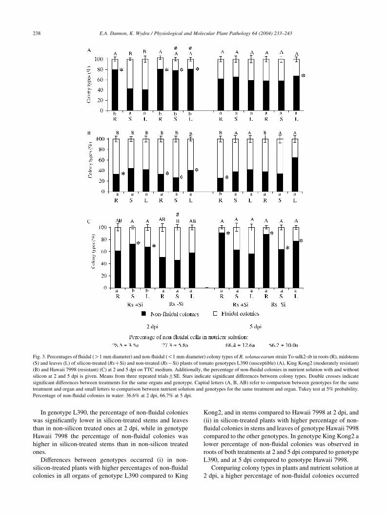
Fig. 3. Percentages of fluidal (O1 mm diameter) and non-fluidal (!1 mm diameter) colony types of R. solanacearum strain To-udk2-sb in roots (R), midstems
(S) and leaves (L) of silicon-treated (RsCSi) and non-treated (RsKSi) plants of tomato genotypes L390 (susceptible) (A), King Kong2 (moderately resistant)
(B) and Hawaii 7998 (resistant) (C) at 2 and 5 dpi on TTC medium. Additionally, the percentage of non-fluidal colonies in nutrient solution with and without
silicon at 2 and 5 dpi is given. Means from three repeated trialsGSE. Stars indicate significant differences between colony types. Double crosses indicate
significant differences between treatments for the same organs and genotype. Capital letters (A, B, AB) refer to comparison between genotypes for the same
treatment and organ and small letters to comparison between nutrient solution and genotypes for the same treatment and organ. Tukey test at 5% probability.
Percentage of non-fluidal colonies in water: 36.6% at 2 dpi, 66.7% at 5 dpi.
E.A. Dannon, K. Wydra / Physiological and Molecular Plant Pathology 64 (2004) 233–243238
In genotype L390, the percentage of non-fluidal colonies
was significantly lower in silicon-treated stems and leaves
than in non-silicon treated ones at 2 dpi, while in genotype
Hawaii 7998 the percentage of non-fluidal colonies was
higher in silicon-treated stems than in non-silicon treated
ones.
Differences between genotypes occurred (i) in non-
silicon-treated plants with higher percentages of non-fluidal
colonies in all organs of genotype L390 compared to King
Kong2, and in stems compared to Hawaii 7998 at 2 dpi, and
(ii) in silicon-treated plants with higher percentage of non-
fluidal colonies in stems and leaves of genotype Hawaii 7998
compared to the other genotypes. In genotype King Kong2 a
lower percentage of non-fluidal colonies was observed in
roots of both treatments at 2 and 5 dpi compared to genotype
L390, and at 5 dpi compared to genotype Hawaii 7998.
Comparing colony types in plants and nutrient solution at
2 dpi, a higher percentage of non-fluidal colonies occurred
E.A. Dannon, K. Wydra / Physiological and Molecular Plant Pathology 64 (2004) 233–243 239
in all non-silicon-treated organs and in silicon-treated roots
of genotype L390, and in stems and leaves of silicon-treated
plants of genotype Hawaii 7998. No influence of plants on
colony type was observed across genotypes and treatments
at 5 dpi except in roots of genotype King Kong2, where a
lower percentage of non-fluidal colonies occurred in silicon-
treated plants.
3.4. Silicon distribution
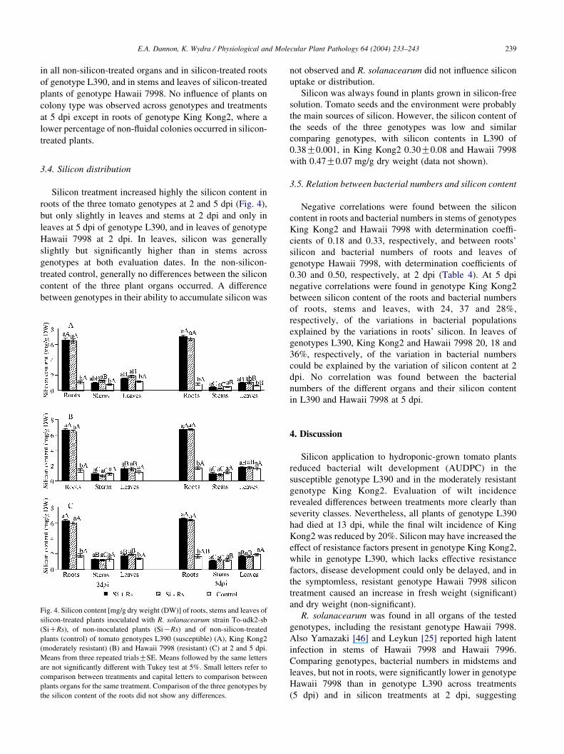
Silicon treatment increased highly the silicon content in
roots of the three tomato genotypes at 2 and 5 dpi (Fig. 4),
but only slightly in leaves and stems at 2 dpi and only in
leaves at 5 dpi of genotype L390, and in leaves of genotype
Hawaii 7998 at 2 dpi. In leaves, silicon was generally
slightly but significantly higher than in stems across
genotypes at both evaluation dates. In the non-silicon-
treated control, generally no differences between the silicon
content of the three plant organs occurred. A difference
between genotypes in their ability to accumulate silicon was
Fig. 4. Silicon content [mg/g dry weight (DW)] of roots, stems and leaves of
silicon-treated plants inoculated with R. solanacearum strain To-udk2-sb
(SiCRs), of non-inoculated plants (SiKRs) and of non-silicon-treated
plants (control) of tomato genotypes L390 (susceptible) (A), King Kong2
(moderately resistant) (B) and Hawaii 7998 (resistant) (C) at 2 and 5 dpi.
Means from three repeated trialsGSE. Means followed by the same letters
are not significantly different with Tukey test at 5%. Small letters refer to
comparison between treatments and capital letters to comparison between
plants organs for the same treatment. Comparison of the three genotypes by
the silicon content of the roots did not show any differences.
not observed and R. solanacearum did not influence silicon
uptake or distribution.
Silicon was always found in plants grown in silicon-free
solution. Tomato seeds and the environment were probably
the main sources of silicon. However, the silicon content of
the seeds of the three genotypes was low and similar
comparing genotypes, with silicon contents in L390 of
0.38G0.001, in King Kong2 0.30G0.08 and Hawaii 7998
with 0.47G0.07 mg/g dry weight (data not shown).
3.5. Relation between bacterial numbers and silicon content
Negative correlations were found between the silicon
content in roots and bacterial numbers in stems of genotypes
King Kong2 and Hawaii 7998 with determination coeffi-
cients of 0.18 and 0.33, respectively, and between roots’
silicon and bacterial numbers of roots and leaves of
genotype Hawaii 7998, with determination coefficients of
0.30 and 0.50, respectively, at 2 dpi (Table 4). At 5 dpi
negative correlations were found in genotype King Kong2
between silicon content of the roots and bacterial numbers
of roots, stems and leaves, with 24, 37 and 28%,
respectively, of the variations in bacterial populations
explained by the variations in roots’ silicon. In leaves of
genotypes L390, King Kong2 and Hawaii 7998 20, 18 and
36%, respectively, of the variation in bacterial numbers
could be explained by the variation of silicon content at 2
dpi. No correlation was found between the bacterial
numbers of the different organs and their silicon content
in L390 and Hawaii 7998 at 5 dpi.
4. Discussion
Silicon application to hydroponic-grown tomato plants
reduced bacterial wilt development (AUDPC) in the
susceptible genotype L390 and in the moderately resistant
genotype King Kong2. Evaluation of wilt incidence
revealed differences between treatments more clearly than
severity classes. Nevertheless, all plants of genotype L390
had died at 13 dpi, while the final wilt incidence of King
Kong2 was reduced by 20%. Silicon may have increased the
effect of resistance factors present in genotype King Kong2,
while in genotype L390, which lacks effective resistance
factors, disease development could only be delayed, and in
the symptomless, resistant genotype Hawaii 7998 silicon
treatment caused an increase in fresh weight (significant)
and dry weight (non-significant).
R. solanacearum was found in all organs of the tested
genotypes, including the resistant genotype Hawaii 7998.
Also Yamazaki [46] and Leykun [25] reported high latent
infection in stems of Hawaii 7998 and Hawaii 7996.
Comparing genotypes, bacterial numbers in midstems and
leaves, but not in roots, were significantly lower in genotype
Hawaii 7998 than in genotype L390 across treatments
(5 dpi) and in silicon treatments at 2 dpi, suggesting
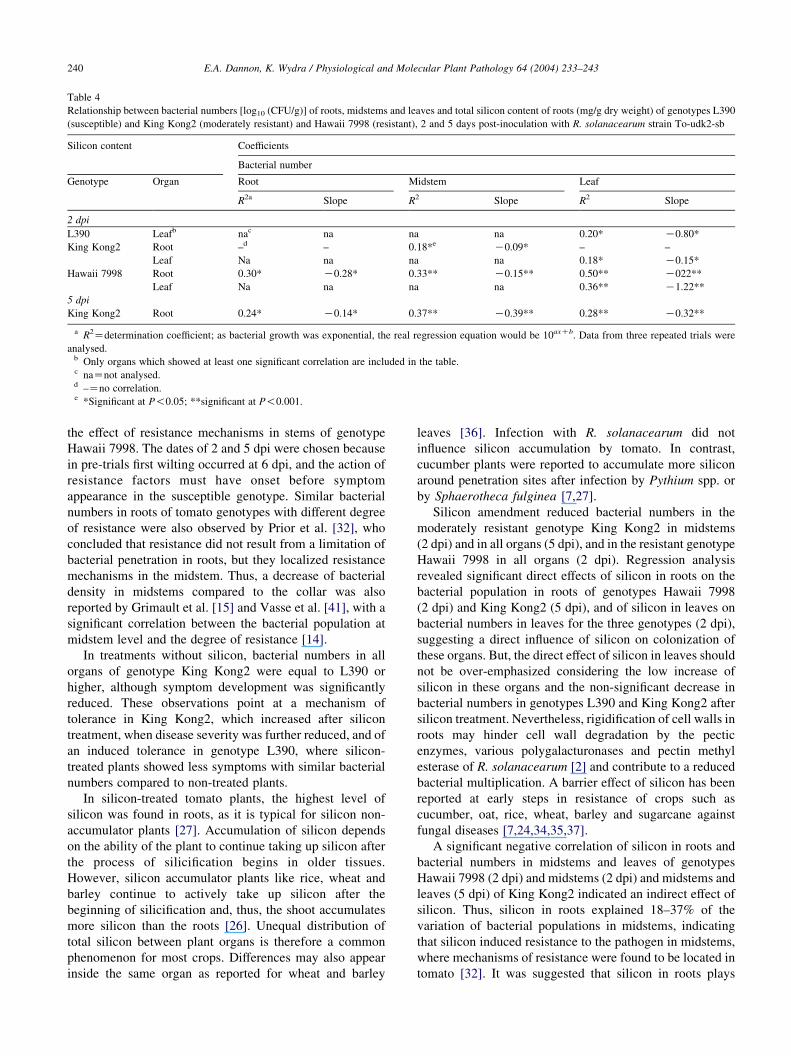
Table 4
Relationship between bacterial numbers [log10 (CFU/g)] of roots, midstems and leaves and total silicon content of roots (mg/g dry weight) of genotypes L390
(susceptible) and King Kong2 (moderately resistant) and Hawaii 7998 (resistant), 2 and 5 days post-inoculation with R. solanacearum strain To-udk2-sb
Silicon content Coefficients
Bacterial number
Genotype Organ Root Midstem Leaf
R2a Slope R2 Slope R2 Slope
2 dpi
L390 Leafb nac na na na 0.20* K0.80*
King Kong2 Root –d – 0.18*e K0.09* – –
Leaf Na na na na 0.18* K0.15*
Hawaii 7998 Root 0.30* K0.28* 0.33** K0.15** 0.50** K022**
Leaf Na na na na 0.36** K1.22**
5 dpi
King Kong2 Root 0.24* K0.14* 0.37** K0.39** 0.28** K0.32**
a R2Zdetermination coefficient; as bacterial growth was exponential, the real regression equation would be 10axCb. Data from three repeated trials were
analysed.b Only organs which showed at least one significant correlation are included in the table.c naZnot analysed.d –Zno correlation.e *Significant at P!0.05; **significant at P!0.001.
E.A. Dannon, K. Wydra / Physiological and Molecular Plant Pathology 64 (2004) 233–243240
the effect of resistance mechanisms in stems of genotype
Hawaii 7998. The dates of 2 and 5 dpi were chosen because
in pre-trials first wilting occurred at 6 dpi, and the action of
resistance factors must have onset before symptom
appearance in the susceptible genotype. Similar bacterial
numbers in roots of tomato genotypes with different degree
of resistance were also observed by Prior et al. [32], who
concluded that resistance did not result from a limitation of
bacterial penetration in roots, but they localized resistance
mechanisms in the midstem. Thus, a decrease of bacterial
density in midstems compared to the collar was also
reported by Grimault et al. [15] and Vasse et al. [41], with a
significant correlation between the bacterial population at
midstem level and the degree of resistance [14].
In treatments without silicon, bacterial numbers in all
organs of genotype King Kong2 were equal to L390 or
higher, although symptom development was significantly
reduced. These observations point at a mechanism of
tolerance in King Kong2, which increased after silicon
treatment, when disease severity was further reduced, and of
an induced tolerance in genotype L390, where silicon-
treated plants showed less symptoms with similar bacterial
numbers compared to non-treated plants.
In silicon-treated tomato plants, the highest level of
silicon was found in roots, as it is typical for silicon non-
accumulator plants [27]. Accumulation of silicon depends
on the ability of the plant to continue taking up silicon after
the process of silicification begins in older tissues.
However, silicon accumulator plants like rice, wheat and
barley continue to actively take up silicon after the
beginning of silicification and, thus, the shoot accumulates
more silicon than the roots [26]. Unequal distribution of
total silicon between plant organs is therefore a common
phenomenon for most crops. Differences may also appear
inside the same organ as reported for wheat and barley
leaves [36]. Infection with R. solanacearum did not
influence silicon accumulation by tomato. In contrast,
cucumber plants were reported to accumulate more silicon
around penetration sites after infection by Pythium spp. or
by Sphaerotheca fulginea [7,27].
Silicon amendment reduced bacterial numbers in the
moderately resistant genotype King Kong2 in midstems
(2 dpi) and in all organs (5 dpi), and in the resistant genotype
Hawaii 7998 in all organs (2 dpi). Regression analysis
revealed significant direct effects of silicon in roots on the
bacterial population in roots of genotypes Hawaii 7998
(2 dpi) and King Kong2 (5 dpi), and of silicon in leaves on
bacterial numbers in leaves for the three genotypes (2 dpi),
suggesting a direct influence of silicon on colonization of
these organs. But, the direct effect of silicon in leaves should
not be over-emphasized considering the low increase of
silicon in these organs and the non-significant decrease in
bacterial numbers in genotypes L390 and King Kong2 after
silicon treatment. Nevertheless, rigidification of cell walls in
roots may hinder cell wall degradation by the pectic
enzymes, various polygalacturonases and pectin methyl
esterase of R. solanacearum [2] and contribute to a reduced
bacterial multiplication. A barrier effect of silicon has been
reported at early steps in resistance of crops such as
cucumber, oat, rice, wheat, barley and sugarcane against
fungal diseases [7,24,34,35,37].
A significant negative correlation of silicon in roots and
bacterial numbers in midstems and leaves of genotypes
Hawaii 7998 (2 dpi) and midstems (2 dpi) and midstems and
leaves (5 dpi) of King Kong2 indicated an indirect effect of
silicon. Thus, silicon in roots explained 18–37% of the
variation of bacterial populations in midstems, indicating
that silicon induced resistance to the pathogen in midstems,
where mechanisms of resistance were found to be located in
tomato [32]. It was suggested that silicon in roots plays
E.A. Dannon, K. Wydra / Physiological and Molecular Plant Pathology 64 (2004) 233–243 241
a role in the signalling network and can induce resistance
systemically in other organs [12]. Silicon has been reported
to stimulate host defence mechanisms in crops such as
cucumber and barley by increasing the level of inhibitory
phenolic compounds and the activity of chitinases,
b-1,3-glucanases, peroxides, phenylalanine ammonia-
lyase and polyphenoloxidase [5,11,27]. Furthermore,
silicon was involved in the increased resistance of cucumber
by enhancing the antifungal activity attributed to a
phytoalexin [11]. Information on possible resistance
mechanisms in tomato to R. solanacearum is limited, but
bacterial spread was suggested to be limited by production
of tyloses forming physical barriers, and gums and other
deposits [14].
The PC of R. solanacearum was frequently reported in
stored cultures [30,38], but reasons for the transformation
are unknown. Non-fluidal colonies, present in low percen-
tage in the inoculum suspension, were increased in
isolations from genotype L390 (2 dpi). Quorum sensing
was found to determine the PC, directed by the phcA
confinement system [8]. PC was recently reported to be
reversible in presence of a susceptible host plant [29].
Interactions with genotypes of different resistance were to
date not investigated.
Grimault and Prior [16] also found that root invasion
was not a limiting factor for bacterial multiplication, when
high numbers of bacteria occurred in tomato stems
without producing wilt symptoms and suggested that in
planta plant–bacteria interactions are involved in reduced
bacterial multiplication, and that resistance in tomato is
expressed on stem level. They speculated that the resistant
host might be involved in impairing exopolysaccharide
production of R. solanacearum in planta resulting in
higher numbers of non-fluidal cells in the resistant plant.
Thus, our results indicated an influence of the plant,
comparing the percentage of non-fluidal cells in planta in
the susceptible genotype with the cells in nutrient
solution, where the silicon amendment did not have an
effect on the phenotype. But, we observed significantly
higher numbers of non-fluidal cells in stems of untreated
plants of the susceptible genotype, compared to the
nutrient solution and to King Kong2 and Hawaii 7998
at 2 dpi, while bacterial numbers in stems were similar
across genotypes. After silicon treatment, the number of
non-fluidal cells was reduced in the susceptible genotype
and increased in the resistant genotype at 2 dpi.
Thus, plant factors might have an influence on the loss
of the ability to produce high quantities of extracellular
polysaccharides, without or prior to a suppressing effect
on bacterial multiplication. Before a difference in bacterial
number in stems between non-silicon-treated genotypes
occurred, an interaction between the plant and the
pathogen seems to take place and becomes obvious in
the PC of R. solanacearum. Although the PC phenomenon
has been generally described for R. solanacearum strains
in culture, it may be that our strain has a higher tendency
to change its phenotype than other strains. Also
Thaveechaii (personal communication) confirms, that
after subculturing each colony type of strain To-udk2-sb,
both types may occur again. The non-fluidal type was
observed to be still virulent, and inoculation of fluidal and
non-fluidal types each in a susceptible tomato plant
resulted in both types after re-isolation. Also Poussier et
al. [29] described a host plant-dependent PC of
R. solanacearum from non-pathogenic forms. Neverthe-
less, in an ongoing trial performed in substrate culture,
strain To-udk2-sb did not show the two colony types in
culture nor after re-isolation, indicating the high varia-
bility of the strain after some months of storage in sterile
water at room temperature (unpublished data).
On the reasons for the PC in planta it can only be
speculated that in the susceptible plant extracellular
polysaccharides as a protection from resistance factors are
not yet needed at 2 dpi and bacterial cells lose their fluidal
character for some time. Under silicon treatment, which
might induce resistance factors, bacteria in stems of the
susceptible genotype showed lower percentages of non-
fluidal colonies and behaved similar as in the untreated
resistant genotypes. The reasons for the higher number of
non-fluidal colonies in stems of genotype Hawaii 7998 after
silicon treatment at 2 dpi compared to non-treated plants and
to the nutrient solution remain unclear. Further investi-
gations with more genotypes and strains are ongoing.
Resistance of tomato against R. solanacearum was
enhanced by silicon treatment in the three genotypes, and
accumulated silicon in roots was negatively correlated to
bacterial numbers in other organs. Therefore, silicon is
suggested to be involved in induced resistance and increased
tolerance, interacting with resistance factors of the plants,
with an indirect effect on bacterial growth and the
physiological status of the bacteria. The role of the genotype
and the molecular basis of resistance induction as well as the
interaction between silicon, host and pathogen are being
studied. The silicon effect should be verified in various
substrates and soils, and integrated in a bacterial wilt
management system.
Acknowledgements
We thank Prof. Dr W. Horst and G. Heine, Institute of
Plant Nutrition, University of Hannover, for support in
silicon analysis.
References
[1] Belanger RR, Bowen PA, Ehret DL, Menzies JG. Soluble silicon: its
role in crop and disease management of greenhouse crops. Plant Dis
1995;79:329–36.
E.A. Dannon, K. Wydra / Physiological and Molecular Plant Pathology 64 (2004) 233–243242
[2] Brumbley SM, Denny TP. Cloning of wild-type Pseudomonas
solanacearum phcA, a gene that when mutated alters expression of
multiple traits that contribute to virulence. J Bacteriol 1999;172:
5677–85.
[3] Buddenhagen I, Kelman A. Biological and physiological aspects of
bacterial wilt caused by Pseudomonas solanacearum. Ann Rev
Phytopathol 1964;2:203–30.
[4] Buddenhagen I, Sequeira L, Kelman A. Designation of races in
Pseudomonas solanacearum. Phytopathology 1962;52:726.
[5] Carver TLW, Zeyen RJ, Ahlstrand GG. The relation between
insoluble silicon and success or failure of attempted penetration by
powdery mildew (Erysiphe graminis) germlings on barley. Physiol
Plant Pathol 1987;31:133–48.
[6] Chen J, Caldwell RD, Robinson CA, Steinkamp R. Silicon: the
estranged medium element. Environmental Horticulture, Florida;
2000. Bulletin 341.
[7] Cherif M, Benhamou N, Belanger RR. Ultrastructural and cytochem-
ical studies of fungal development and host relations in cucumber
plants infected by Pythium ultimum. Physiol Mol Plant Pathol 1992;
39:353–75.
[8] Clough SJ, Lee KE, Schell MA, Denny TP. A two-component system
in Ralstonia (Pseudomonas solanacearum) modulates production of
PhcA-regulated virulence factors in response to 3-hydroxypalmitic
acid methyl ester. J Bacteriol 1997;179:3639–48.
[9] Datnoff LE, Seebold KW, Correa VFJ. The use of silicon for
integrated disease management: reducing fungicide applications and
enhancing host plant resistance. In: Datnoff LE, Snyder GH,
Korndorfer GH, editors. Silicon in agriculture. The Netherlands:
Elsevier Science; 2001. p. 171–83.
[10] Epstein E. Silicon in plants: facts vs concepts. In: Datnoff LE,
Snyder GH, Korndorfer GH, editors. Silicon in agriculture. The
Netherlands: Elsevier Science; 2001. p. 1–15.
[11] Fawe A, Abou-Zaid M, Menzies JG, Belanger RR. Silicon-mediated
accumulation of flavonoid phytoalexins in cucumber. Phytopathology
1998;88:396–401.
[12] Fawe A, Menzies JG, Cherif M, Belanger RR. Silicon and disease
resistance in dicotyledons. In: Datnoff LE, Snyder GH,
Korndorfer GH, editors. Silicon in agriculture. The Netherlands:
Elsevier Science; 2001. p. 159–69.
[13] Genin S, Boucher C. Ralstonia solanaceraum: secrets of a major
pathogen unveiled by analysis of its genome. Mol Plant Pathol 2002;
3:111–8.
[14] Grimault V, Anais G, Prior P. Distribution of Pseudomonas
solanacearum in the stem tissues of tomato plants with different
levels of resistance to bacterial wilt. Plant Pathol 1994;43:663–8.
[15] Grimault V, Prior P. Bacterial wilt resistance in tomato associated
with tolerance of vascular tissues to Pseudomonas solanacearum.
Plant Pathol 1993;42:589–94.
[16] Grimault V, Prior P, Anais G. A monogenic dominant resistance of
tomato to bacterial wilt in Hawaii 7996 is associated with plant
colonization by Pseudomonas solanacearum. J Phytopathol 1995;
143:349–52.
[17] Hayward AC. Characterisation of Pseudomonas solanacearum.
J Appl Bacteriol 1964;27:265–77.
[18] Hayward AC. Biology and epidemiology of bacterial wilt caused by
Pseudomonas solanacearum. Ann Rev Phytopathol 1991;29:65–87.
[19] Hochmuth G. Nutrient solution formulation for hydroponic (rock-
wood and NFT) tomatoes in Florida. Florida Cooperative Extension
Service, Report 44. Florida, USA: Service for Suwannee Valley
Education; 1999.
[20] Jeger MJ, Viljanen-Rollinson SLH. The use of the area under the
disease-progress curve (AUDPC) to assess quantitative disease
resistance in crop cultivars. Theor Appl Genet 2001;102:32–40.
[21] Kelman A. The relationship of pathogenicity in Pseudomonas
solanacearum to colony appearance on a Tetrazolium medium.
Phytopathology 1954;51:158–61.
[22] Kelman, A. One hundred and one years of research on bacterial wilt.
In: Second international bacterial wilt symposium, 22–27 June, 1997,
Guadeloupe. Book of Abstracts; 1998. p. 10.
[23] Kelman A, Hruschka J. The role of motility and aerotaxis in the
selective increase of avirulent bacteria in still broth culture of
Pseudomonas solanacearum. J Gen Microbiol 1973;76:177–88.
[24] Kunoh H, Ishizaki H. Silicon levels near penetration sites of fungi on
wheat, barley, cucumber and morning glory leaves. Physiol Plant
Pathol 1975;5:283–7.
[25] Leykun, Z. Latent infection of resistant tomato genotypes with
Ralstonia solanacearum and the viable but non-cuturable state of the
pathogen in tomato tissue. MSc Thesis, University of Hannover,
Germany; 2003.
[26] Ma JF, Miyake Y, Takahashi E. Silicon as a beneficial element for
crop plants. In: Datnoff LE, Snyder GH, Korndorfer GH, editors.
Silicon in agriculture. The Netherlands: Elsevier Science; 2001. p.
17–39.
[27] Menzies JG, Ehret DL, Glass ADM, Helmer T, Koch C, Seywerd F.
Effects of soluble silicon on the parasitic fitness of Sphaerotheca
fuliginea on Cucumis sativus. Phytopathology 1991;81:84–8.
[28] Novozamsky I, Van Eck R, Houba VJG. A rapid determination of
silicon in plant material. Commun Soil Sci Plant Anal 1984;15:
205–11.
[29] Poussier S, Thoquet P, Trigalet-Demery D, Matthieu A, Trigalet A.
Host plant-dependent phenotypic conversion of Ralstonia solana-
cearum from non-pathogenic forms via alteration in the phcA gene.
Mol Microbiol 2003;49:991–1003.
[30] Pradhanang PM, Elphinstone JG, Fox RTV. Sensitive detection of
Ralstonia solanacearum in soil: a comparison of different detection
techniques. Plant Pathol 2000;49:414–22.
[31] Prell HH. Interaktionen von Pflanzen und phytopathogenen Pilzen.
Stuttgart, Germany: Gustav Fischer; 1996.
[32] Prior P, Grimault V, Schmit J. Resistance to bacterial wilt
(Pseudomonas solanacearum) in tomato: present status and prospects.
In: Hayward AC, Hartman GL, editors. Bacterial wilt: the disease and
its causative agent Pseudomonas solanacearum. Wallingfor, United
Kingdom: International Centre for Agriculture and Bioscience
(CAB); 1994. p. 209–34.
[33] Ram-Kishun S, Kishun R. Loss in yield of tomato due to bacterial wilt
caused by Pseudomonas solanacearum. Indian Phytopathol 1987;40:
152–5.
[34] Rodrigues FA, Vale FXR, Korndorfer GH, Prabhu AS, Datnoff LE,
Oliveira AMA, et al. Influence of silicon on sheath blight of rice in
Brazil. Crop Prot 2003;22:23–9.
[35] Samuels AL, Glass ADM, Menzies JG, Ehret DL. Silicon in cell walls
and papillae of Cucumus sativus during infection by Sphaerotheca
fuliginea. Physiol Mol Plant Pathol 1994;44:237–42.
[36] Sangster AG, Hodson MJ, Tubb HJ. Silicon deposition in higher
plants. In: Datnoff LE, Snyder GH, Korndorfer GH, editors.
Silicon in agriculture. The Netherlands: Elsevier Science; 2001. p.
85–113.
[37] Savant NK, Korndorfer GH, Datnoff LE, Snyder GH. Silicon
nutrition and sugarcane production: a review. J Plant Nutr 1999;
22:1853–903.
[38] Schell MA. Control of virulence and pathogenicity genes of Ralstonia
solanacearum by an elaborate sensory network. Ann Rev Phytopathol
2000;38:263–92.
[39] Shaner G, Finney RE. The effect of nitrogen fertilization on the
expression of slow-mildewing resistance in Knox wheat. Phytopathol-
ogy 1977;67:1051–6.
[40] Stumpf MA, Heath MC. Cytological studies of the interactions
between the cowpea rust fungus and silicon-depleted French bean
plants. Physiol Plant Pathol 1985;27:369–85.
[41] Vasse J, Danoun S, Trigalet A. Cytological and biochemical analysis
of roots infection of the resistant tomato line Hawaii 7996 by R.
solanacaearum, Abstract. Third international bacterial wilt

E.A. Dannon, K. Wydra / Physiological and Molecular Plant Pathology 64 (2004) 233–243 243
symposium, February 4–8, 2002. http:/ibws.nexenservices.com/
Talks/talk_23Vasse.htm.
[42] Wang J-F, Hanson P, Barnes JA. Worldwide evaluation of
an international set of resistance sources to bacterial wilt in
tomato. In: Prior P, Allen C, Elphinstone J, editors. Bacterial wilt
disease. Molecular and ecological aspects. Second international
bacterial wilt symposium, Gossier, Guadeloupe, France, 22–27 June
1997. Germany: Springer; 1998. p. 269–79.
[43] Wang JF, Olivier J, Thoquet P, Mangin B, Sauviac L, Grimsley NH.
Resistance of tomato line Hawaii 7996 to Ralstonia solanacearum
Pss4 in Taiwan is controlled mainly by a major strain-specific locus.
Mol Plant Microbe Interact 2000;13:6–13.
[44] Winstead NN, Kelman A. Inoculation technique for evaluating
resistance to Pseudomonas solanacearum. Phytopathology 1952;42:
628–34.
[45] Yabuuchi E, Kosako Y, Yano I, Hotta H, Nishiuchi Y. Transfer of two
Burkholderia and an Alcaligenes species to Ralstonia gen. Nov.:
proposal of Ralstonia picketti (Ralston, Palleroni and Doudoroff 1973)
comb. Nov., Ralstonia solanacearum (Smith 1896) comb. Nov. and
Ralstonia eutropha (Davis 1969) comb Nov. Microbiol Immunol
1995;39:897–904.
[46] Yamazaki H. Relation between resistance to bacterial wilt and
calcium nutrition in tomato seedlings. Jpn Agric Res Quart
2001;163–9.
Related Documents