Interaction between Nbp35 and Cfd1 Proteins of Cytosolic Fe-S Cluster Assembly Reveals a Stable Complex Formation in Entamoeba histolytica Shadab Anwar 1 , Manas Ranjan Dikhit 2 , Krishn Pratap Singh 1 , Rajiv Kumar Kar 2 , Amir Zaidi 1 , Ganesh Chandra Sahoo 2 , Awadh Kishore Roy 3 , Tomoyoshi Nozaki 4 , Pradeep Das 5 , Vahab Ali 1 * 1 Laboratory of Molecular Biochemistry and Cell Biology, Department of Biochemistry, Rajendra Memorial Research Institute of Medical Sciences, Agam-kuan, Patna, India, 2 Department of Biomedical Informatics Centre, Rajendra Memorial Research Institute of Medical Sciences, Agam-kuan, Patna, India, 3 Department of Botany, T. M. Bhagalpur University, Bhagalpur, India, 4 Department of Parasitology, National Institute of Infectious Diseases, Shinjuku-ku, Tokyo, Japan, 5 Department of Molecular Biology, Rajendra Memorial Research Institute of Medical Sciences, Agam-kuan, Patna, India Abstract Iron-Sulfur (Fe-S) proteins are involved in many biological functions such as electron transport, photosynthesis, regulation of gene expression and enzymatic activities. Biosynthesis and transfer of Fe-S clusters depend on Fe-S clusters assembly processes such as ISC, SUF, NIF, and CIA systems. Unlike other eukaryotes which possess ISC and CIA systems, amitochondriate Entamoeba histolytica has retained NIF & CIA systems for Fe-S cluster assembly in the cytosol. In the present study, we have elucidated interaction between two proteins of E. histolytica CIA system, Cytosolic Fe-S cluster deficient 1 (Cfd1) protein and Nucleotide binding protein 35 (Nbp35). In-silico analysis showed that structural regions ranging from amino acid residues (P33-K35, G131-V135 and I147-E151) of Nbp35 and (G5-V6, M34-D39 and G46-A52) of Cfd1 are involved in the formation of protein-protein complex. Furthermore, Molecular dynamic (MD) simulations study suggested that hydrophobic forces surpass over hydrophilic forces between Nbp35 and Cfd1 and Van-der-Waal interaction plays crucial role in the formation of stable complex. Both proteins were separately cloned, expressed as recombinant fusion proteins in E. coli and purified to homogeneity by affinity column chromatography. Physical interaction between Nbp35 and Cfd1 proteins was confirmed in vitro by co-purification of recombinant Nbp35 with thrombin digested Cfd1 and in vivo by pull down assay and immunoprecipitation. The insilico, in vitro as well as in vivo results prove a stable interaction between these two proteins, supporting the possibility of its involvement in Fe-S cluster transfer to target apo-proteins through CIA machinery in E. histolytica. Our study indicates that initial synthesis of a Fe-S precursor in mitochondria is not necessary for the formation of Cfd1-Nbp35 complex. Thus, Cfd1 and Nbp35 with the help of cytosolic NifS and NifU proteins can participate in the maturation of non-mitosomal Fe-S proteins without any apparent assistance of mitosomes. Citation: Anwar S, Dikhit MR, Singh KP, Kar RK, Zaidi A, et al. (2014) Interaction between Nbp35 and Cfd1 Proteins of Cytosolic Fe-S Cluster Assembly Reveals a Stable Complex Formation in Entamoeba histolytica. PLoS ONE 9(10): e108971. doi:10.1371/journal.pone.0108971 Editor: Tracey Rouault, National Institute of Child Health and Human Development, United States of America Received March 8, 2014; Accepted August 29, 2014; Published October 1, 2014 Copyright: ß 2014 Anwar et al. This is an open-access article distributed under the terms of the Creative Commons Attribution License, which permits unrestricted use, distribution, and reproduction in any medium, provided the original author and source are credited. Data Availability: The authors confirm that all data underlying the findings are fully available without restriction. All data are included within the paper. Funding: This work was supported by a grant from Indian Council of Medical Research (ICMR), Ministry of Health and Family Welfare, and Department of Science & Technology (DST/INT/JSPSP-117), New Delhi, India. The funder had no role in study design, data collection and analysis decision to publish, or preparation of the manuscript. Competing Interests: The authors have declared that no competing interests exist. * Email: [email protected] Introduction Entamoeba histolytica is one of the most widespread and clinically important protozoan parasite causing both intestinal (amoebic colitis) and extra intestinal (amoebic liver abscess) disease throughout the world, resulting to an estimated 40,000 to 110,000 deaths annually. World Health Organisation estimate (WHO, 1998) places E. histolytica second after Plasmodium falciparum in causing abundant annual death among protozoan parasites. E. histolytica lacks a defined structure of mitochondria and its functions [1]. However, mitochondrion residual organelle known as mitosome [2] is present in this parasite. Mitochondria performs many crucial roles in various biochemical and iron-requiring biosynthetic processes; namely, heme formation, Iron-Sulfur (Fe-S) clusters biogenesis and cellular iron regulation [3,4]. Among them, Iron-Sulfur clusters biogenesis is essential for the maturation of Fe- S proteins which are biologically functional and ubiquitous components that orchestrate a wide range of biochemical machinery and efficiently regulate the metabolic cascades in living organisms for sustainable and fundamental life processes [4–8]. Mitochondria assemble Fe-S clusters for their own set of mitochondrial Fe-S proteins as well as crucially involved in the biogenesis and maturation of Fe-S proteins located in the cytosol and nucleus [9–11]. Despite the chemical simplicity of Fe-S clusters, Fe-S clusters biogenesis is a complex process involving three types of systems, viz, Iron Sulfur Clusters (ISC), Sulfur Utilization Factors (SUF) and Nitrogen Fixation (NIF) systems. The ISC system is a house- keeping system involving ,30 protein components [8,12–14] and among them 10 proteins have been conserved from bacteria to human [15,16]. The majority of protozoan parasites have retained PLOS ONE | www.plosone.org 1 October 2014 | Volume 9 | Issue 10 | e108971

Welcome message from author
This document is posted to help you gain knowledge. Please leave a comment to let me know what you think about it! Share it to your friends and learn new things together.
Transcript
Interaction between Nbp35 and Cfd1 Proteins ofCytosolic Fe-S Cluster Assembly Reveals a StableComplex Formation in Entamoeba histolyticaShadab Anwar1, Manas Ranjan Dikhit2, Krishn Pratap Singh1, Rajiv Kumar Kar2, Amir Zaidi1,
Ganesh Chandra Sahoo2, Awadh Kishore Roy3, Tomoyoshi Nozaki4, Pradeep Das5, Vahab Ali1*
1 Laboratory of Molecular Biochemistry and Cell Biology, Department of Biochemistry, Rajendra Memorial Research Institute of Medical Sciences, Agam-kuan, Patna, India,
2 Department of Biomedical Informatics Centre, Rajendra Memorial Research Institute of Medical Sciences, Agam-kuan, Patna, India, 3 Department of Botany, T. M.
Bhagalpur University, Bhagalpur, India, 4 Department of Parasitology, National Institute of Infectious Diseases, Shinjuku-ku, Tokyo, Japan, 5 Department of Molecular
Biology, Rajendra Memorial Research Institute of Medical Sciences, Agam-kuan, Patna, India
Abstract
Iron-Sulfur (Fe-S) proteins are involved in many biological functions such as electron transport, photosynthesis, regulation ofgene expression and enzymatic activities. Biosynthesis and transfer of Fe-S clusters depend on Fe-S clusters assemblyprocesses such as ISC, SUF, NIF, and CIA systems. Unlike other eukaryotes which possess ISC and CIA systems,amitochondriate Entamoeba histolytica has retained NIF & CIA systems for Fe-S cluster assembly in the cytosol. In thepresent study, we have elucidated interaction between two proteins of E. histolytica CIA system, Cytosolic Fe-S clusterdeficient 1 (Cfd1) protein and Nucleotide binding protein 35 (Nbp35). In-silico analysis showed that structural regionsranging from amino acid residues (P33-K35, G131-V135 and I147-E151) of Nbp35 and (G5-V6, M34-D39 and G46-A52) ofCfd1 are involved in the formation of protein-protein complex. Furthermore, Molecular dynamic (MD) simulations studysuggested that hydrophobic forces surpass over hydrophilic forces between Nbp35 and Cfd1 and Van-der-Waal interactionplays crucial role in the formation of stable complex. Both proteins were separately cloned, expressed as recombinant fusionproteins in E. coli and purified to homogeneity by affinity column chromatography. Physical interaction between Nbp35 andCfd1 proteins was confirmed in vitro by co-purification of recombinant Nbp35 with thrombin digested Cfd1 and in vivo bypull down assay and immunoprecipitation. The insilico, in vitro as well as in vivo results prove a stable interaction betweenthese two proteins, supporting the possibility of its involvement in Fe-S cluster transfer to target apo-proteins through CIAmachinery in E. histolytica. Our study indicates that initial synthesis of a Fe-S precursor in mitochondria is not necessary forthe formation of Cfd1-Nbp35 complex. Thus, Cfd1 and Nbp35 with the help of cytosolic NifS and NifU proteins canparticipate in the maturation of non-mitosomal Fe-S proteins without any apparent assistance of mitosomes.
Citation: Anwar S, Dikhit MR, Singh KP, Kar RK, Zaidi A, et al. (2014) Interaction between Nbp35 and Cfd1 Proteins of Cytosolic Fe-S Cluster Assembly Reveals aStable Complex Formation in Entamoeba histolytica. PLoS ONE 9(10): e108971. doi:10.1371/journal.pone.0108971
Editor: Tracey Rouault, National Institute of Child Health and Human Development, United States of America
Received March 8, 2014; Accepted August 29, 2014; Published October 1, 2014
Copyright: � 2014 Anwar et al. This is an open-access article distributed under the terms of the Creative Commons Attribution License, which permitsunrestricted use, distribution, and reproduction in any medium, provided the original author and source are credited.
Data Availability: The authors confirm that all data underlying the findings are fully available without restriction. All data are included within the paper.
Funding: This work was supported by a grant from Indian Council of Medical Research (ICMR), Ministry of Health and Family Welfare, and Department of Science& Technology (DST/INT/JSPSP-117), New Delhi, India. The funder had no role in study design, data collection and analysis decision to publish, or preparation of themanuscript.
Competing Interests: The authors have declared that no competing interests exist.
* Email: [email protected]
Introduction
Entamoeba histolytica is one of the most widespread and
clinically important protozoan parasite causing both intestinal
(amoebic colitis) and extra intestinal (amoebic liver abscess) disease
throughout the world, resulting to an estimated 40,000 to 110,000
deaths annually. World Health Organisation estimate (WHO,
1998) places E. histolytica second after Plasmodium falciparum in
causing abundant annual death among protozoan parasites. E.histolytica lacks a defined structure of mitochondria and its
functions [1]. However, mitochondrion residual organelle known
as mitosome [2] is present in this parasite. Mitochondria performs
many crucial roles in various biochemical and iron-requiring
biosynthetic processes; namely, heme formation, Iron-Sulfur (Fe-S)
clusters biogenesis and cellular iron regulation [3,4]. Among them,
Iron-Sulfur clusters biogenesis is essential for the maturation of Fe-
S proteins which are biologically functional and ubiquitous
components that orchestrate a wide range of biochemical
machinery and efficiently regulate the metabolic cascades in living
organisms for sustainable and fundamental life processes [4–8].
Mitochondria assemble Fe-S clusters for their own set of
mitochondrial Fe-S proteins as well as crucially involved in the
biogenesis and maturation of Fe-S proteins located in the cytosol
and nucleus [9–11].
Despite the chemical simplicity of Fe-S clusters, Fe-S clusters
biogenesis is a complex process involving three types of systems,
viz, Iron Sulfur Clusters (ISC), Sulfur Utilization Factors (SUF)
and Nitrogen Fixation (NIF) systems. The ISC system is a house-
keeping system involving ,30 protein components [8,12–14] and
among them 10 proteins have been conserved from bacteria to
human [15,16]. The majority of protozoan parasites have retained
PLOS ONE | www.plosone.org 1 October 2014 | Volume 9 | Issue 10 | e108971
ISC system either in mitochondrion or mitochondria like
organelles; mitosomes, hydrogenosomes, and mitochondria related
organelles (MROs) [17–20]. However, Plasmodium spp. &
Blastocystis hominis possess SUF system or some of its components
in addition to the canonical ISC system which is functional under
oxidative stress and iron deficient conditions [19,21–23]. Nitro-
gen-fixation (NIF) system is present in nitrogen fixing bacteria,
cyanobacteria and microaerophilic bacteria but absent in eukary-
otes and protozoan parasites except E. histolytica and free living
amoeba (Mastigamoeba balamuthi). Thus, Fe-S cluster biogenesis
in E. histolytica solely depends on NIF system [24]. It has already
been proved that the NIF system alone is required for the
biosynthesis of Fe-S cluster in E. histolytica under anaerobic
conditions [24]. This organism possesses two components of NIF
machinery: NifS and NifU [24,25] that are responsible for Fe-S
cluster assembly. Surprisingly, M. balamuthi possesses two types of
NifS and NifU components, of which one of them has retained
targeting signal and localized in the mitosomes [26]. Contrary to
the mitosomes of M. balamuthi, E. histolytica mitosomes have no
evidence of classic Fe-S cluster machinery. Therefore, cytosolic
NIF machinery predominantly regulates the cellular requirements
for Fe-S cluster biogenesis in this organism. E. histolytica possess
mitosomes that do not generate ATP unlike other protozoan
parasites harbouring MROs (Blastocystis sp.) or hydrogenosomes
which are involved in both ATP generation and Fe-S cluster
biogenesis [27]. However, neither amoebic mitosomes possesses
the ISC machinery, nor any component of ISC or SUF machinery
has been identified in E. histolytica genome [23]. It has not been
resolved till-date how NIF system works in connection with
Cytosolic Iron-sulfur protein Assembly (CIA) in the absence of true
mitochondria. It also remains unknown whether NIF and CIA
system interact with each other for biogenesis and subsequent
transfer of Fe-S clusters to apoproteins, as both systems co-exist in
the cytoplasm.
In eukaryotes, the ISC system assists for the maturation of
cytosolic/nuclear Fe-S proteins including CIA machinery compo-
nents. The CIA system is limited to cytoplasm and core protein
assembly consists of Cfd1, Nbp35, Nar1, Cia1, Dre2, Tah18 [28–
34] and some additional components (MMS19, MIP18 and
ANT2) which are exclusively present in mammalian system [35].
MMS19 function as part of the CIA machinery which interacts
and facilitate Fe-S cluster targeting to apo-proteins involved in
methionine biosynthesis, DNA replication, DNA repair, and
telomerase maintenance [36]. In addition, MMS19 forms a
complex with CIA proteins (CIAO1, IOP1, & MIP18) involved in
DNA metabolism and its presence is necessary for DNA
replication and repair [37]. The CIA1 (CIAO1) associates with
either CIA2A (FAM96A) or CIA2B (FAM96B) and MMS19
proteins. It has been reported recently that CIA2B-CIA1-MMS19
complex binds to and facilitates assembly of most cytosolic/
nuclear Fe-S proteins but CIA2A is specially required for the
maturation of iron regulatory protein 1 (IRP1), which is involved
in cellular iron homeostasis [38]. The CIA2A is also involved in
stabilization of IRP2 through its interaction with IRP2. However,
E. histolytica, comprised of a NIF system has also retained CIA
components namely; Cfd1, Nbp35, Nar1, Dre2, and Cia1 [23].
Cfd1 and Nbp35 belong to a subfamily of deviant P-loop
NTPases, often referred to as the MRP/NBP35 sub-family, which
appear to function in Fe-S clusters biogenesis in all kingdoms. The
first component of the CIA machinery to be identified was Cfd1
which is an essential and highly conserved P-loop NTPase [29]. Invitro reconstitution study has shown that Nbp35 forms an
oligomeric complex with Cfd1 and both can assemble labile Fe-
S cluster on their conserved cysteine residues of C- terminal
domain. They may serve as transient scaffold for Fe-S cluster
before transfer to apo-proteins in yeast [39,40]. The class of
NTPases typically form homodimer involving a signature lysine
(Lys26 in Cfd1 and Lys81 in Nbp35) residue within the walker A
(Nucleotide binding) motif which also plays a role in ATP binding
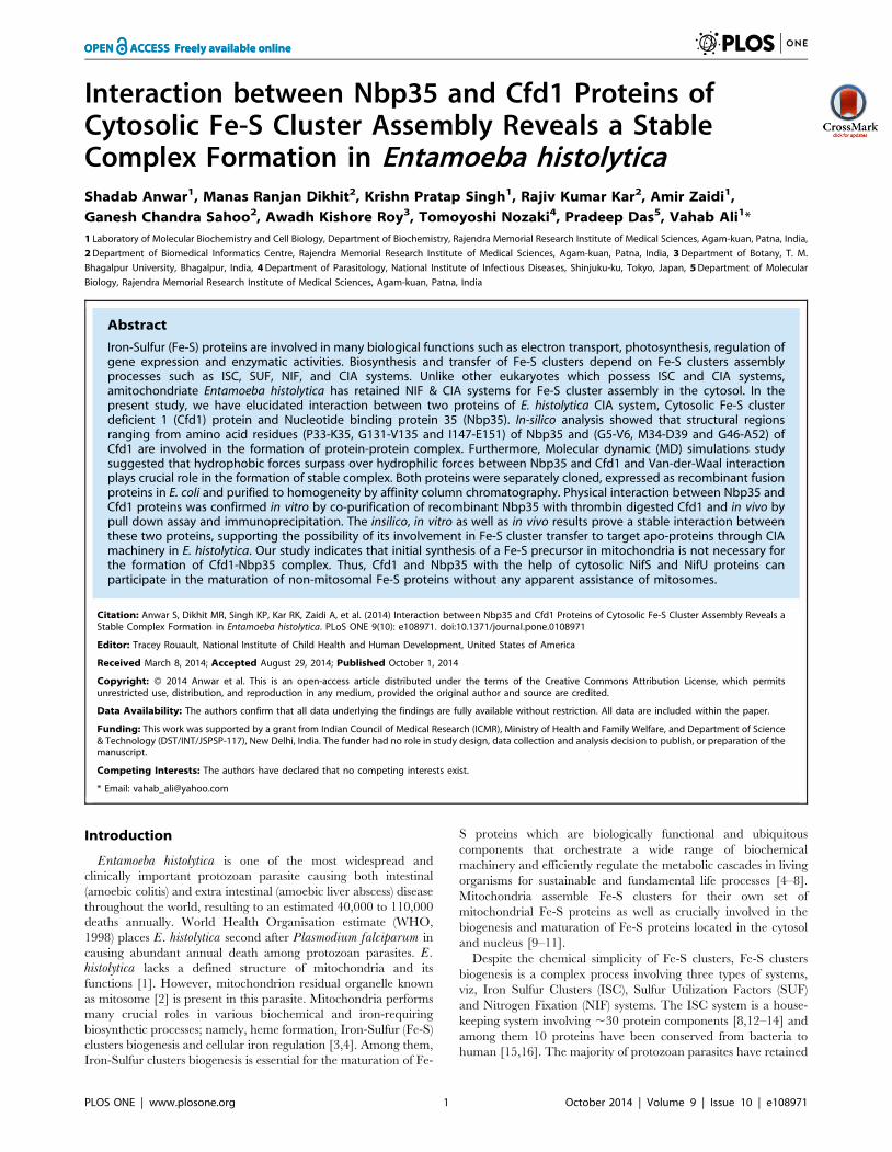
Figure 1. RMSDs time scan for Nbp35 and Cfd1. Backbone RMSDs are shown as a function of time for Nbp35 (red) and Cfd1 (green) protein at3 ns.doi:10.1371/journal.pone.0108971.g001
Interaction between Nbp and Cfd Proteins of E. histolytica
PLOS ONE | www.plosone.org 2 October 2014 | Volume 9 | Issue 10 | e108971

and/or hydrolysis [41]. However, in plantae including Arabidopsisthaliana, Cfd1 is absent and Npb35 works alone as the scaffold
component [42,43]. Although, Cfd1 gene is absent in some
organisms such as Caenorhabditis elegans (metazoa) and plantae
[43]; the specific functional role of Cfd1 is its interaction with
Nbp35 which alters the character of Nbp35-bound Fe-S clusters,
making it more labile and enhancing transfer to apo-target Fe-S
proteins [41].
In the present study, we have attempted to investigate the
interaction between Cfd1 and Nbp35 in E. histolytica in vivo by
co-purification and immunoprecipitation and insilico using mo-
lecular dynamics simulation tool. Our results show that Npb35
and Cfd1 of E. histolytica interacts with each other to form a stable
complex and each protein has the potential to coordinate a 4Fe-4S
cluster on it similar to the earlier report in yeast [30,40]. This
would be the first report showing interaction and stable complex
formation between two components of CIA machinery Nbp35 and
Cfd1 in an amitochondriate protozoan parasite possessing a
cytosolic NIF system for Fe-S cluster assembly.
Materials and Methods
Chemicals and reagentsAll chemicals of analytical grade were purchased and used from
Sigma-Aldrich, Amresco (USA), Merck, and USB (USA) unless
otherwise stated. Chromatography column was purchased from
Bio-Rad. Ni+2-NTA agarose was purchased from Qiagen. Adult
bovine serum from Hyclone, Yeast extract and Casitone were
purchased from BD Biosciences.
Microorganism and cultivationE. histolytica trophozoites clonal strain (HM-1: IMMS cl 6) was
maintained axenically in TYIS-33 medium supplemented with
15% adult bovine serum at 35.5uC [44]. Trophozoites were
harvested in the late-logarithmic growth phase 2–3 days after the
inoculation of medium with one- thirtieth to one-twelfth of the
total culture volume. After the cultures were chilled on ice for
5 mins, trophozoites were collected by centrifugation at 5006g for
10 mins at 4uC and washed twice with ice-cold phosphate-
Figure 2. RMSD and RMSF profile of Nbp35-Cfd1 complex. (A) RMSD profile of Nbp35-Cfd1 complex for all-atoms (red), backbone atoms(blue) and side chain (green) plotted as a function of time (B) Root mean square fluctuation for Nbp35 and Cfd1 plotted against time from MDsimulation.doi:10.1371/journal.pone.0108971.g002
Interaction between Nbp and Cfd Proteins of E. histolytica
PLOS ONE | www.plosone.org 3 October 2014 | Volume 9 | Issue 10 | e108971
buffered saline (PBS), pH 7.4 [45]. Cell pellets were stored at
230uC until use.
PCR amplification and cloning of Nbp35 & Cfd1 genesBased on the nucleotide sequence of the protein-encoding
region of the putative E. histolytica Nucleotide binding proteins
genes (Nbp35, accession number XP_650593, EHI_047750; Cfd1
accession number XP_653192; EHI_000610); primers (shown
below) were designed to clone Nbp35 & Cfd1 in vector pET15b
with a histidine tag at the amino terminus. The Nbp35 & Cfd1ORFs were amplified from cDNA of E. histolytica with a sense
(59CCTCATATGAGTTGTTCTCATAATTGTTCA-39) and an
antisense (59-CCAGGATCCTTAAAGATTTGTTATTATTT-
CCTT-39) primers for Nbp35 and a sense (59CCTCATATGACT-
GAACTTAACTCTGATCGT-39) and an antisense (59-CCAG-GATCCTTAAGCAAAAGTTTTAGCAAGATCG-39) primers
for Cfd1, where NdeI and BamHI-sites are underlined and the
translation initiation and termination codons are italicized. PCR
was performed in a 50 ml volume containing 0.25 mM each
dNTPs, 2.0 mM MgCl2 1.0 mM each primer, 1 mg cDNA (E.histolytica) and 1.0 U Pfu DNA polymerase. The conditions used
to amplify the Nbp35 & Cfd1 genes were hot start at 94uC for
5 mins, denaturation at 94uC for 30 s, annealing at 55uC for 30 s,
elongation at 68uC for 1.0 min and subjected to 30 cycles with a
final extension for 10 mins at 68uC. A ,1.0 & 0.8 kb PCR
products were observed on 1.0% agarose gel electrophoresis.
These PCR products were double digested with NdeI and BamHI,
electrophoresed, purified with gel extraction kit (Qiagen), and
cloned into NdeI and BamHI- digested pET-15b (Novagen) in the
same orientation as the T7 promoter. The ligated mixture was
transformed in competent DH5a cells (Novagen) which produced
the pET-15b-Nbp35 & pET-15b-Cfd1 plasmids. The insert and
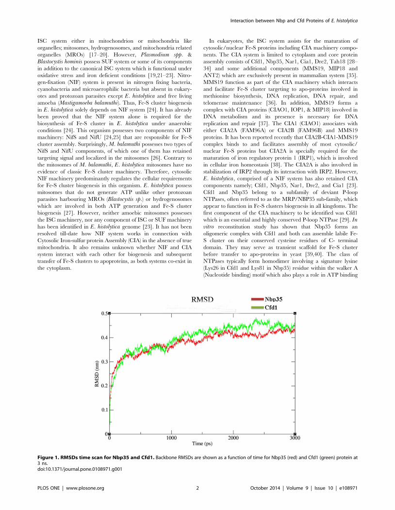
Figure 3. Nbp35-Cfd1 complex structure showing the regions involved in interaction. (A) Complex structure of Nbp35-Cfd1 where themolecular contacts are highlighted (blue–Nbp35) and (green-Cfd1) with mentioned residues involved in interaction. (B) Porcupine plots for Nbp35-Cfd1 complex showing backbone fluctuation from simulation time course. The regions involved in contact are highlighted with dotted region.doi:10.1371/journal.pone.0108971.g003
Interaction between Nbp and Cfd Proteins of E. histolytica
PLOS ONE | www.plosone.org 4 October 2014 | Volume 9 | Issue 10 | e108971
ORF orientation were confirmed by colony PCR. Construct
plasmids were isolated using Qiagen miniprep kit as per
manufacturer’s instructions. The pET-15b-Nbp35 & pET-15b-
Cfd1 constructs were transformed into competent E. coli BL21
(DE3) (Novagen) cells by heat shock at 42uC for 45 s, and the cells
were grown at 37uC on Luria Bertani (LB) agar medium in the
presence of 50 mg/ml ampicillin (Amp).
Expression and purification of recombinant Nbp35 andCfd1 Proteins
The pET-Nbp35 and pET-Cfd1 expression constructs were
introduced into competent cells and the resulting single colony
picked up from LB-agar plate were grown at 37uC in 5 ml of LB
medium in the presence of 50 mg/ml ampicillin. The overnight
culture was used to inoculate 500 ml of fresh medium and cultured
at 37uC with shaking at 200 rpm. When the A600 reached 0.6,
0.4 mM of isopropyl b-D- thiogalactopyranoside was added to
induce protein expression for 12 h at 25uC. E. coli cells were
harvested by centrifugation at 5000 rpm for 10 mins at 4uC, the
resulting cell pellet washed with PBS (pH 7.4) and resuspended in
25 ml lysis buffer [46] containing 100 mg/ml lysozyme and 1 mM
Phenylmethylsulfonyl fluoride (PMSF). After 45 mins of incuba-
tion at 30uC, the cells were sonicated on ice and centrifuged at
13000 rpm for 20 mins at 4uC. The supernatant was mixed with
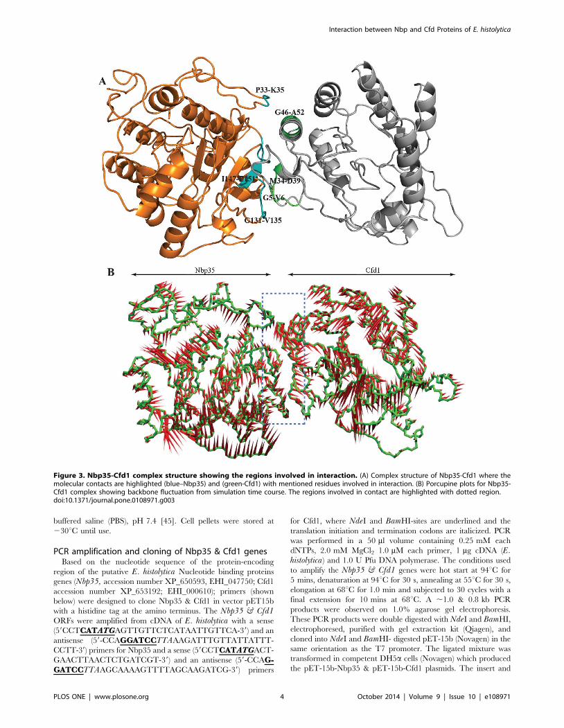
Figure 4. Hydrogen bonding profile & solvent accessibility. (A) Hydrogen bonding profile indicating numbers of hydrogen bonds formedwithin protein atoms and with water molecules. (B) Solvent accessible surface area computed over the simulation time course.doi:10.1371/journal.pone.0108971.g004
Interaction between Nbp and Cfd Proteins of E. histolytica
PLOS ONE | www.plosone.org 5 October 2014 | Volume 9 | Issue 10 | e108971

500 ml of Ni+2-NTA His-tag slurry (Qiagen) and incubated for
1.0 hr at 4uC with gentle shaking. The recombinant Nbp35 and
Cfd1 protein bound resin was washed with 8–10 column volumes
of buffer A (50 mM Tris-HCl, pH 8.0, 300 mM NaCl, and 0.1%
Triton X-100, v/v) containing 10–50 mM of imidazole and bound
proteins were eluted in 2–3 ml with buffer A containing 100–
300 mM imidazole. The quality and purity of the rNbp35 and
rCfd1 proteins were confirmed by 12% SDS-PAGE analysis. The
proteins were extensively dialyzed against a 300 fold volume of
50 mM Tris-HCl, 150 mM NaCl, pH 8.0, containing 10%
glycerol (V/V) and the complete Mini protease inhibitor cocktail
(Calbiochem). The concentration of the dialyzed proteins was
determined by Bradford method using bovine serum albumin as
standard (U3900, Hitachi, Japan). The rNbp35 and rCfd1
proteins were stored at 230uC in 10% glycerol in small aliquots
until used.
Chemical reconstitution of the Fe-S cluster on Cfd1 andNbp35
The purified Nbp35 and Cfd1 proteins were dialyzed in 50 mM
Tris-HCl, 150 mM NaCl, pH 8.0 to remove glycerol and
rechecked their integrity and purity. Chemical reconstitution of
Fe-S cluster on Nbp35 and Cfd1 was performed as described
previously with some modifications [40–42]. Briefly, Nbp35 and
Cfd1 (100 mM each protein) were reduced in reconstitution buffer
(50 mM Tris-HCl, pH 8.0, 150 mM NaCl, 10 mM DTT) for 1 h
at 25uC. Fresh anaerobic stocks solution of ferric ammonium
citrate and Li2S were prepared in water containing10 mM DTT
before use and reconstitution of both proteins was started by the
addition of 100 mM ferric ammonium citrate and 100 mM Li2S
with stirring. To avoid precipitation, the reconstitution mixture
was incubated with ferric ammonium citrate for 5 min before Li2S
was added slowly drop wise to the reaction mixture followed by
incubation of 2–3 hrs at 25uC. To remove non bound iron and
sulphide, reconstituted proteins were desalted by PD-10 column
equilibrated with reconstitution buffer containing 2 mM DTT.
The assembly of Fe-S clusters into apo-proteins was monitored by
UV-Vis spectroscopy (U3900, Hitachi, Japan).
Iron estimationThe iron content of Nbp35 and Cfd1 proteins were determined
by the O- phenanthroline method as described previously [24,47].
Briefly, the Nbp35 and Cfd1 samples (80–100 ml) were acidified by
the addition of 3–5 ml of concentrated HCl and then diluted with
distilled water up to 0.2 ml. The mixtures were heated to 80uC for
10 mins and cooled down to room temperature. The reaction
mixture was diluted with 0.6 ml of water, and 40 ml of 10%
hydroxylamine hydrochloride, 0.2 ml of 0.1% O-phenanthroline
Figure 5. Contact map of Nbp35-Cfd1 complex. The contact map of Nbp35-Cfd1 complex is indicating the cross residue region involved incontact of these two proteins.doi:10.1371/journal.pone.0108971.g005
Interaction between Nbp and Cfd Proteins of E. histolytica
PLOS ONE | www.plosone.org 6 October 2014 | Volume 9 | Issue 10 | e108971
were added. Finally, reaction mixtures were incubated at room
temperature for 30 mins, and absorbance was measured at
512 nm.
Sulfide estimationSulfide content of purified rNbp35 and rCfd1 proteins was
determined by measuring the absorbance of a blue colour complex
treated with N,N-dimethyl-p-phenylenediamine dihydrochloride
(DPD) as described previously [33]. Protein samples (50 mg) were
diluted up to 775 ml with water and mixed with 50 ml of 6%
NaOH. The DPD reagent (0.1%) dissolved in 5 N HCl (125 ml)
and 30 mM FeCl3 (50 ml) solutions were added to the reaction
mixture and incubated for 30 min at 30uC. Finally, reaction
mixture was vortexed, centrifuged for 5 mins at 13000 rpm to
remove precipitate, and absorbance of supernatant was measured
at 670 nm by UV-Vis spectroscopy (U3900, Hitachi, Japan). Na2S
(0–100 mM) was used as standard.
Production of E. histolytica Nbp35 & Cfd1 antibodies andimmunoblot analysis
Polyclonal antisera against recombinant E. histolytica Nbp35 or
Cfd1 was raised in adult rabbit by three repeated subcutaneous
injection. Pre-immune sera was collected before immunization and
first dose of 300 mg Nbp35 or Cfd1 proteins emulsified in complete
Freund’s adjuvant was injected 8–10 places subcutaneously;
followed by three booster doses of 250 mg of proteins emulsified
in Freund’s incomplete adjuvant. Anti-Nbp35 & Cfd1 titres were
checked by ELISA after three weeks of final immunization.
Finally, serum was collected from rabbits and stored at 230uC in
small aliquots. Working antibodies were stored at 4uC. Prior
animal ethical committee approval was taken and recommenda-
tions were strictly followed.
Cell lysate of E. histolytica (,16107 cells/ml) was prepared
using lysis buffer (100 mM Tris-HCl, pH 8.0, 300 mM NaCl,
1.0 mM EDTA, 1.0 mM DTT, 10% glycerol) as described
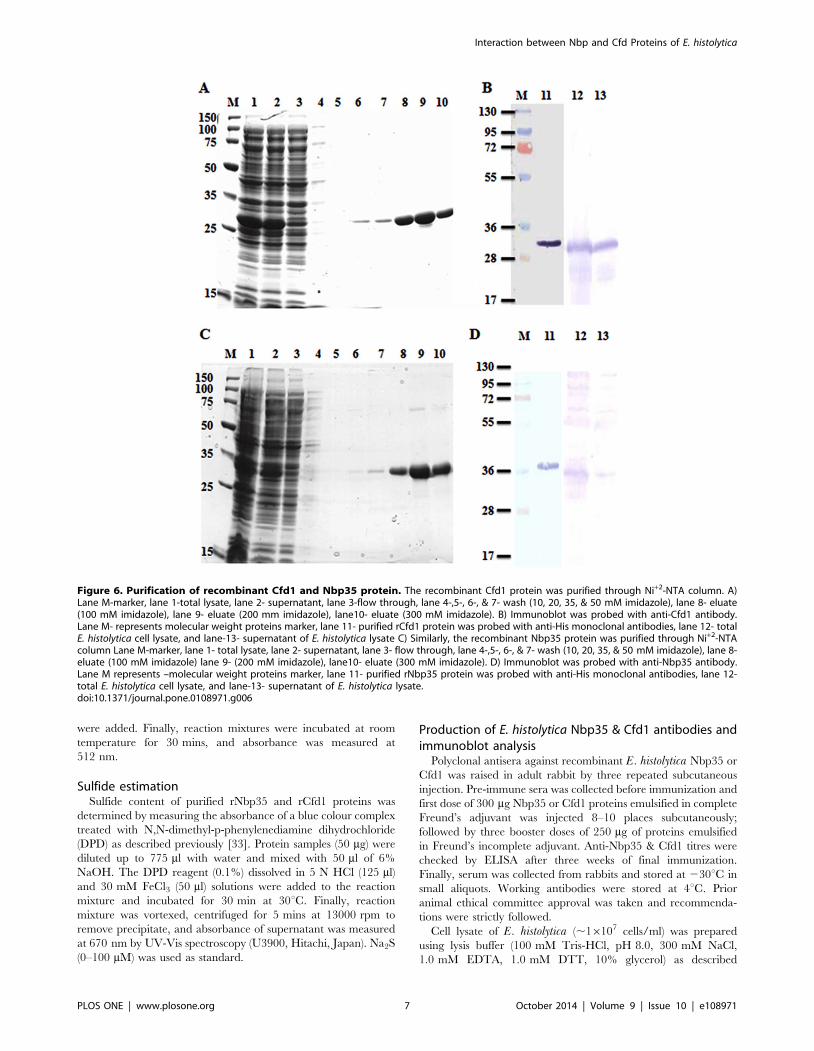
Figure 6. Purification of recombinant Cfd1 and Nbp35 protein. The recombinant Cfd1 protein was purified through Ni+2-NTA column. A)Lane M-marker, lane 1-total lysate, lane 2- supernatant, lane 3-flow through, lane 4-,5-, 6-, & 7- wash (10, 20, 35, & 50 mM imidazole), lane 8- eluate(100 mM imidazole), lane 9- eluate (200 mm imidazole), lane10- eluate (300 mM imidazole). B) Immunoblot was probed with anti-Cfd1 antibody.Lane M- represents molecular weight proteins marker, lane 11- purified rCfd1 protein was probed with anti-His monoclonal antibodies, lane 12- totalE. histolytica cell lysate, and lane-13- supernatant of E. histolytica lysate C) Similarly, the recombinant Nbp35 protein was purified through Ni+2-NTAcolumn Lane M-marker, lane 1- total lysate, lane 2- supernatant, lane 3- flow through, lane 4-,5-, 6-, & 7- wash (10, 20, 35, & 50 mM imidazole), lane 8-eluate (100 mM imidazole) lane 9- (200 mM imidazole), lane10- eluate (300 mM imidazole). D) Immunoblot was probed with anti-Nbp35 antibody.Lane M represents –molecular weight proteins marker, lane 11- purified rNbp35 protein was probed with anti-His monoclonal antibodies, lane 12-total E. histolytica cell lysate, and lane-13- supernatant of E. histolytica lysate.doi:10.1371/journal.pone.0108971.g006
Interaction between Nbp and Cfd Proteins of E. histolytica
PLOS ONE | www.plosone.org 7 October 2014 | Volume 9 | Issue 10 | e108971

previously [24]. The protein fractions were resolved by 10% SDS-
PAGE and electro-blotted on to nitrocellulose membrane. The
membrane was probed with polyclonal anti-Nbp35 or Cfd1 sera
(1:2000) raised in rabbit as mentioned above. ALP-conjugated
goat anti-rabbit IgG (1:2000) was used as secondary antibody and
blot developed with BCIP/NBT (Santa Cruz), as per manufac-
turer’s instructions [48].
Homology modelling and Structure ValidationPrediction of an interacting complex was proceeded with
homology modelling, since the crystal structures for Nbp35 and
Cfd1 are not available. Full length amino acid sequences of both
the proteins were retrieved from Universal Protein Resource
Database (http://www.uniprot.org/). The low percentage of
sequence identity in the homologues PDB structures may not
fetch robust models for protein structure. Thus, we have relied
upon Zhang’s I-TASSER server (http://www.zhanglab.ccmb.
med.umich.edu/I-TASSER/), which gives the best protein models
at the Critical Assessment of Structure Prediction (CASP 7 and
CASP 8), a community-wide, worldwide experiment designed to
obtain an objective assessment of the state-of-the-art in structure
prediction [49]. Five models of Nbp35 and Cfd1 were computa-
tionally generated using the I-TASSER algorithm. The models
were selected with lowest DOPE Score and verified using the
Profile 3D profile analysis method [50]. The stereo-chemical
properties of both the models were investigated with Ramachan-
dran plot using PROCHECK [51]. The quality of final model was
checked using Verify-3D program [52]. In addition to this, stereo
chemical qualities of 3D model were analyzed using WHATIF and
ProSA web server. Molecular dyanamics (MD) simulations were
conducted for the Nbp35 and Cfd1 models in explicit solvent using
the GROMACS 4.0.3 (The Groningen Machine for Chemical
Simulations) package. The model was solvated by water molecules
in an octahedron box having edges at a distance of 0.9 nm from
the molecule’s periphery. The solvated system was subjected to
further energy minimization to remove the steric conflicts between
the atoms of protein and water molecules having a maximum step
of 2000 with steepest descent integrator that converge the energy
minimization when the maximum force is smaller than
1000 kJ.mol21.nm21. The energy minimized model was subjected
to position-restrained MD with NPT ensemble keeping number of
particles (N), system pressure (P) and temperature (T) as constant
parameters. This was carried out for 50,000 steps for a total of
Figure 7. UV-Visible spectra of purified and reconstitutedrNbp35 and rCfd1 proteins. A) As purified rNbp-35 and rNbp35 afterchemical reconstitution was scanned in the UV-Vis range to detect Fe-Scluster. Solid and dashed lines represent reconstituted Nbp35 andpurified Nbp35, respectively. B) UV-visible spectra of purified rCfd1 andreconstituted rCfd1protein in which, solid and dashed lines representreconstituted rCfd1 and purified rCfd1, respectively.doi:10.1371/journal.pone.0108971.g007
Figure 8. Interaction study between Cfd1 and Nbp35 proteins.(A) Co-purification using recombinant Nbp35 and thrombin digestedCfd1; Immunoblots of fractions obtained from co-purification experi-ment as described in material and methods probed with differentantibodies (i) Anti-Nbp35 (ii) Anti-Cfd1 (iii) Anti-His antibodies forinteraction between recombinant Nbp35 and thrombin digested Cfd1proteins. Lane M- represents molecular weight proteins marker, lane 1-E. coli lysate overexpressing rNbp35, lane 2- purified rNbp35, lane 3-undigested rCfd1, lane 4- digested rCfd1, lane 5- wash (60 mMimidazole), lane 6 final wash did not show any proteins in all blots,lane 7- eluate (200 mM imidazole), lane 8- eluate (500 mM imidazole).(B) Pull down assay using rNbp35 and E. histolytica lysate: Ni+2-NTAbound rNbp35 was incubated with E. histolytica lysate as described inmaterial and methods for pull down assay and eluate (500 mMimidazole) fraction was subjected to immunoblot analysis proveddifferent antibodies (upper panel) Anti-His (middle panel) Anti-Nbp35(lower panel) Anti-Cfd1 antibodies for rNbp35 and endogenousamoebic Cfd1 interaction. Lane M- represents molecular weightproteins marker, lane 1- total E. coli lysate expressed rNbp35, lane 2-supernatant rNbp35, lane 3- total amoebic lysate, lane 4- amoebiclysate flow through, lane 5 final wash did not show any proteins in allblots, lane 6- eluate (500 mM imidazole).doi:10.1371/journal.pone.0108971.g008
Interaction between Nbp and Cfd Proteins of E. histolytica
PLOS ONE | www.plosone.org 8 October 2014 | Volume 9 | Issue 10 | e108971

100 ps time period. Final MD was carried out for 3000 ps (3 ns)
under Particle Mesh Ewald (PME), electrostatics in NPT condition
[53].
Protein Protein Docking and complex simulationIt would be interesting to investigate whether Nbp35 and Cfd1
interact to each other and form complex in E. histolytica. Protein
docking between Nbp35 and Cfd1 was done using PatchDock
server (http://bioinfo3d.cs.tau.ac.il/PatchDock/) [54] with default
settings. This server is widely used for the protein-protein docking
studies [54–56] and its algorithm based on rigid docking with
surface flexibility addressed by intermolecular penetration.
PatchDock calculation resulted in three-dimensional transforma-
tions of the protein’s Cartesian coordinate, which were further
refined and scored based on protein-protein interaction energy
using Fire Dock algorithm. Complex structure with the lowest
interaction energy was taken as the final model for further analysis.
Molecular dynamics simulations of Nbp35-Cfd1 complex was
performed using the GROMACS 4.0.3 under gromos43a1 force
field. The whole simulation experiment was done for 10 ns with
36527 water molecules and 1 Na+ ion. The same methodology
was adopted for simulation of this complex as done in the MD of
individual proteins Nbp35 and Cfd1.
Co-purification by affinity chromatographyIn vitro interaction between recombinant Nbp35 and Cfd1
proteins was investigated by co-elution from an affinity column
[57]. The bacterial cell pellet overexpressing recombinant Nbp35
protein was dissolved in 10 ml lysis buffer (50 mM Tris-HCl,
pH 8.0, 300 mM NaCl, 0.1% triton X-100) supplemented with
1.0 mM PMSF, 1 X protease inhibitor cocktail (100 ml) and
100 mg/ml lysozyme, and incubated at 30uC for 45 mins. The cell
suspension was sonicated on ice and lysate centrifuged at
13000 rpm for 20 min at 4uC. The supernatant containing
Nbp35 with N-terminal His-tag was loaded onto a Ni2+–NTA
column and washed with buffer A (50 mM Tris–HCl, pH 8.0,
300 mM NaCl, 0.1% Triton X 100) containing 10–50 mM
imidazole to remove unbound proteins. Purified recombinant
Cfd1 protein was digested with thrombin to remove N-terminal-
His-tag and eluted from column followed by desalting as described
earlier [24]. The digested Cfd1 protein was loaded on the Nbp35
bound Ni2+–NTA column and incubated for 1.0 hr at RT
followed by washing with 15 ml each of buffer A containing 10–
60 mM imidazole. Finally, column-bound proteins complex was
eluted with buffer A containing 200 and 500 mM of imidazole.
Eluate proteins complex was analyzed by 10% SDS–PAGE and
western blotting [48] with anti-His (1:2000 Santa cruz), anti-
Nbp35 (1:2000), and anti-Cfd1 (1:2000) antibodies raised in rabbit
as described previously.
Interaction between recombinant protein and amoebiccell lysate
The bacterial cell pellet over-expressing recombinant Nbp35
protein was processed to obtain Nbp35 bound to Ni2+-NTA
column as described in the above section. Amoebic cell lysate
(,56107 cells) was mixed with Ni2+-NTA matrix bound Nbp35 in
column and incubated for 1 hr at 25uC for complex formation.
The column was washed rigorously with buffer A containing 10–
50 mM imidazole to remove the unbound amoebic proteins and
bound proteins complex was eluted with buffer A containing 200
& 500 mM imidazole. All the fractions were electrophoresed in
12% SDS-PAGE and transferred to nitrocellulose membrane. The
blots were probed with different antibodies; Anti-His (Santa cruz)
mouse monoclonal antibody, anti-Nbp35 & anti-Cfd1 antibodies
raised in rabbit at 1:2000 dilution. The alkaline phosphatase
conjugated anti-rabbit secondary antibody or anti mouse were also
used at 1:2000 dilution and finally blots were developed by adding
BCIP/NBT solution as per manufacturer’s instruction.
Interaction study by immunoprecipitationThe immunoprecipitation experiments were performed using
standard protocol [48] with minor modifications. The anti-Cfd1 or
anti-Nbp35 antibody (10 ml) was incubated with protein A
sepharose beads (100 ml) overnight at 4uC. The beads suspension
was centrifuged at 5006g for 2 mins at 4uC, supernatant
discarded and pellet fraction washed twice with 1.0 ml PBS.
Amoebic cell lysate (,56107 cells) was then mixed with antibody
bound protein A sepharose matrix in a 1.5 ml tube with gentle
mixing of the sample on a suitable shaker and incubated for
90 mins at RT or overnight at 4uC. The mixture was centrifuged
at 5006g for 2 mins at 4uC, supernatant discarded and matrix
washed twice with 1 ml PBS to remove loosely bound proteins.
The bound complex was eluted from beads by boiling in 100 ml of
16 SDS-loading dye for 10 mins at 95uC. Finally, immunopre-
cipitated complex (I.P.) was collected by centrifugation at
13000 rpm for 30 sec at RT and supernatant applied on 12%
SDS-PAGE and transferred to nitrocellulose membrane. The blots
were probed with different antibodies as described above &
developed by BCIP/NBT solution [48].
Results and Discussion
Modelling of Nbp35 proteinBLASTP search against Protein Data Bank (PDB) using Nbp35
as query sequence did not give any suitable template. The crystal
structure of nucleotide-binding protein from Archaeoglobusfulgidus (PDB ID: 2PH1_A) shared only 36% sequence identity
and 70% query coverage. Large portion of the target sequence
remained unaligned which suggested that not so many nucleotide-
binding protein structures have been experimentally resolved to
date. Five model of Nbp35 were generated using the I-TASSER
algorithm which directly modelled the aligned regions from the
template structures (2ph1_A &3vx3_A) and the unaligned regions
modelled with ab-initio simulations. These models of Nbp35 were
generated with C-scores ranging from 21.99 to 23.04. The C-
Figure 9. In-vivo interaction study Nbp35 and Cfd1 byimmunoprecipitation. The anti-Nbp35 antibody bound with pro-tein-A sepharose was incubated with amoebic lysate and immunopre-cipitate was analysed by immunoblots were developed separately withanti-Cfd1 (upper panel) and anti-Nbp35 (lower panel) antibodies. IgGheavy chain (55 kda) and Cfd1 (29 Kda) were found in I.P. complexfraction and IgG heavy chain (55 kda) and Nbp35 (35 kda) bands foundwhen probed with anti-Nbp35 antibody. Lane M- represents molecularweight proteins marker, lane 1- total amoebic lysate, lane 2-supernatant of amoebic lysate, lane 3- amoebic lysate flow through,lane 4- final wash did not show any proteins, lane 5- I.P. complex.doi:10.1371/journal.pone.0108971.g009
Interaction between Nbp and Cfd Proteins of E. histolytica
PLOS ONE | www.plosone.org 9 October 2014 | Volume 9 | Issue 10 | e108971
score is a confidence score and ranges from 25 to 2, with higher
scores representing higher confidence in the model [49]. Best three
models were selected (M1, M2 and M3) based on their C-scores
(21.99, 22.65 and 22.84, respectively). Discrete optimized
protein energy (DOPE) suggested that the second model (M2)
revealed the lowest energy (233600.3349) whereas the other two
models DOPE score, i.e., M1 and M3 revealed 233217.539063
and 232761.363281, respectively. The W/Y distribution of the
backbone conformational angle for each residue of the selected
structures was calculated. The M1 model revealed the highest
96.7% residues in the most favoured region and additional allowed
region. 2.2% residues were found in generously allowed region
whereas only 1.1% residues (Leu232, Thr274 and His275) fell in
disallowed region. Based on the DOPE score and the Ramachan-
dran plot, the M2 model was considered to be a reliable model
used for rest of the study. After loop refinement, final structure
with the lowest energy was checked by Profile-3D (DS 2.5). The
self-compatibility score for this protein is 133.09 which is much
higher than the Verify expected lowest score (68.24) and very close
to the Verify expected high score (151.65). Validation of the
modelled protein with ProSA-web revealed that the Z score
(26.74) is within the range of native conformations of the crystal
structures. The plot showed the energy remains negative for
almost all amino acid residues indicating the acceptability of the
predicted model. The Verify-3D result of the model showed that
98.80% amino acids had an average 3D-1D score, and thus
indicated the reliability of the proposed model. The monomeric
Nbp35 is an a/b type protein containing P-loop nucleoside
triphosphate hydrolase type domain (70–314). The modelled
structure composed of 12 a-helices (a1-a12) and four-stranded
parallel b-sheets (b1, b2, b4, b5) with one anti-parallel b-sheet (b3).
One b/a/b motif (b1/a4/b2) is present in between two a-helices
(a2 and a5). ATPase-like, ParA/MinD signature motif (191–272) is
formed by a central a-helix (a8) joined with a b-sheet (b5) and a-
helix (a9) on either side.
Modelling of Cfd1 proteinIn order to proceed for an accurate search of interacting
residues of Cfd1, it is indeed necessary to have a robust 3D model
of the protein structure. BLASTp search against PDB database did
not return any suitable template. Five model were generated
(2ph1_A & 3vx3_A as template) using I-TASSER web server with
C-score ranging from 21.61 to 23.52. The fourth loop refined
model (C-score = 23.26) revealed the lowest DOPE score
(223306.49) and only three residues; Lys20, Val153 and Asn91
fell in disallowed region. The Ramachandran map of the model
revealed 95.2% residues in the most favoured region and
additional allowed region. The self-compatibility score for this
protein is 87.68, which is much higher than the Verify expected
lowest score (55.85). The further precision of the model was
checked using the ProSA server, where the Z-score (26.46) depicts
the model to be within the domicile of reported X-ray crystal
structures till date. Moreover, the Verify-3D results illustrated the
compatibility score of ,84% of amino acid residues which assesses
the favourability of residue environments within the folded
structure. All these validations suggested the robustness and
reliability of the proposed model. The modelled Cfd1 is an a-
helices dominated protein (8 a-helices and 2 b-sheets) containing
P-loop nucleoside triphosphate hydrolase type domain (21–211). A
central a-helix is present in CobQ/CobB/MinD/ParA nucleotide
binding domain, joined by one a-helix (a4) and one 310 helix on
either side.
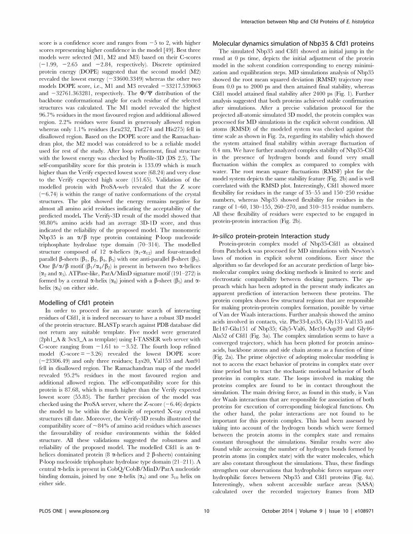
Molecular dynamics simulation of Nbp35 & Cfd1 proteinsThe simulated Nbp35 and Cfd1 showed an initial jump in the
rmsd at 0 ps time, depicts the initial adjustment of the protein
model in the solvent condition corresponding to energy minimi-
zation and equilibration steps. MD simulations analysis of Nbp35
showed the root mean squared deviation (RMSD) trajectory rose
from 0.0 ps to 2000 ps and then attained final stability, whereas
Cfd1 model attained final stability after 2400 ps (Fig. 1). Further
analysis suggested that both proteins achieved stable confirmation
after simulations. After a precise validation protocol for the
projected all-atomic simulated 3D model, the protein complex was
processed for MD simulations in the explicit solvent condition. All
atoms (RMSD) of the modeled system was checked against the
time scale as shown in Fig. 2a, regarding its stability which showed
the system attained final stability within average fluctuation of
0.4 nm. We have further analyzed complex stability of Nbp35-Cfd
in the presence of hydrogen bonds and found very small
fluctuation within the complex as compared to complex with
water. The root mean square fluctuations (RMSF) plot for the
model system depicts the same stability feature (Fig. 2b) and is well
correlated with the RMSD plot. Interestingly, Cfd1 showed more
flexibility for residues in the range of 35–55 and 150–250 residue
numbers, whereas Nbp35 showed flexibility for residues in the
range of 1–60, 130–155, 260–270, and 310–315 residue numbers.
All these flexibility of residues were expected to be engaged in
protein-protein interaction (Fig. 2b).
In-silico protein-protein Interaction studyProtein-protein complex model of Nbp35-Cfd1 as obtained
from Patchdock was processed for MD simulations with Newton’s
laws of motion in explicit solvent conditions. Ever since the
algorithm so far developed for an accurate prediction of large bio-
molecular complex using docking methods is limited to steric and
electrostatic compatibility between docking partners. The ap-
proach which has been adopted in the present study indicates an
apparent prediction of interaction between these proteins. The
protein complex shows few structural regions that are responsible
for making protein-protein complex formation, possible by virtue
of Van der Waals interactions. Further analysis showed the amino
acids involved in contacts, viz. Phe33-Lys35, Gly131-Val135 and
Ile147-Glu151 of Nbp35; Gly5-Val6, Met34-Asp39 and Gly46-
Ala52 of Cfd1 (Fig. 3a). The complex simulation seems to have a
converged trajectory, which has been plotted for protein amino-
acids, backbone atoms and side chain atoms as a function of time
(Fig. 2a). The prime objective of adopting molecular modeling is
not to access the exact behavior of proteins in complex state over
time period but to tract the stochastic motional behavior of both
proteins in complex state. The loops involved in making the
proteins complex are found to be in contact throughout the
simulation. The main driving force, as found in this study, is Van
der Waals interactions that are responsible for association of both
proteins for execution of corresponding biological functions. On
the other hand, the polar interactions are not found to be
important for this protein complex. This had been assessed by
taking into account of the hydrogen bonds which were formed
between the protein atoms in the complex state and remains
constant throughout the simulations. Similar results were also
found while accessing the number of hydrogen bonds formed by
protein atoms (in complex state) with the water molecules, which
are also constant throughout the simulations. Thus, these findings
strengthen our observations that hydrophobic forces surpass over
hydrophilic forces between Nbp35 and Cfd1 proteins (Fig. 4a).
Interestingly, when solvent accessible surface areas (SASA)
calculated over the recorded trajectory frames from MD
Interaction between Nbp and Cfd Proteins of E. histolytica
PLOS ONE | www.plosone.org 10 October 2014 | Volume 9 | Issue 10 | e108971
simulations as a function of time, showed a decrease of almost 15–
20 nm.S2.N21 as shown in (Fig. 4b). This observation demon-
strates that the globular folding of proteins get more compact,
thereby preserving the hydrophobic residues from getting exposed
to the solvent in complex state.
The conformational changes of protein complex were also
analyzed by MD simulations in terms of Principal components
analysis (PCA), where we have taken into consideration of the first
eigenvectors, to trace down the movement of macromolecule.
Pczdump was used to prepare the average coordinates from
simulation and was used to show the projections of eigenvectors.
The approach helps us in correlating the related fluctuations of
protein atoms with that of the total fluctuation in the course of
simulation. Fig. 3b represents a porcupine plot for backbone
atoms (represented in green color) and the red spikes represent the
directionality regarding the motional behavior. The structural
regions those are in contacts at the interface both of the proteins
are shown an enclosed box in Fig. 3b. We also analyzed the polar
contacts formed between these two proteins, by analyzing the
coordinate files at regular interval from MD trajectory, though the
numbers of hydrogen bond remains same but the residues
participating varies. An overview of the residue pairs that are in
contact, had been generated based upon the average structure and
is represented by Contact-map in Fig. 5. Thus, the fundamental
observation as found from the simulation study, being the
hydrophobic forces, i.e. the Van der Waals interaction plays
crucial role in making the protein complex stable, even in the
explicit solvent conditions.
Expression and purification of recombinant Cfd1 andNbp35
The Nbp35 ORF of 1002 nucleotides encodes a 333 amino
acids protein with predicted molecular weight ,36 kDa and an
iso-electric point (pI) value 6.05. The Nbp35 protein, related to
Cfd1 show 49% amino acid identity with yeast Nbp35 protein
[30]. Similarly, the Cfd1 ORF of 822 nucleotides encodes a 273
amino acids protein with predicted molecular weight ,29 kDa
and an iso-electric point (pI) value 4.98. Neither the MITO-
PROT II program nor Signal IP 4.1 Server, which predicts
protein localization in cells, and a Kyte-Doolittle hydropathy plot,
suggested any possible cellular localization other than cytosolic
distribution for Nbp35 and Cfd1 proteins. The recombinant
(rNbp35 and rCfd1) proteins were expressed in E. coli BL21 (DE3)
and purified to homogeneity using Ni2+-NTA affinity chromatog-
raphy as shown in Fig. 6. It was observed that rCfd1 expression
was higher in soluble form and eluted between 100–400 mM
imidazole with high protein yield of ,5 mg/ml. The purified
rCfd1 protein gave a single band of ,29 kDa when examined on
SDS-PAGE and immunoblot using anti-Cfd1 antibody in parasite
lysates (Fig. 6a). Similarly, Nbp35 was also expressed and purified
to homogeneity using Ni2+-NTA affinity chromatography
(Fig. 6b). It revealed an apparently homogeneous band of
38 kDa on 12% SDS-PAGE that correlates well with predicted
molecular mass 36 kDa. The purified rNbp35 and rCfd1 proteins
concentration determined by Bradford’s method was found to be
0.212 mg/ml (rCfd1), 0.187 mg/ml (rNbp35) and were stable at
280uC for more than six months in the presence of 10% glycerol.
Polyclonal antisera against purified rNbp35 & rCfd1 proteins
were raised in adult rabbit showed high titre at final bleed
(1:16000 dilution). Antibody against either rNbp35 or rCfd1
recognised a specific and single band both in E. histolytica lysate
proteins and purified recombinant protein suggesting that a single
homologue is present in the parasite (Fig. 6a & 6b). By
immunoblot analysis of a series of diluted recombinant proteins
(data not shown), we roughly estimated that the E. histolyticacontains a significant amount of Nbp35 & Cfd1, which consists of
approximately 0.01–0.02% of the total proteins.
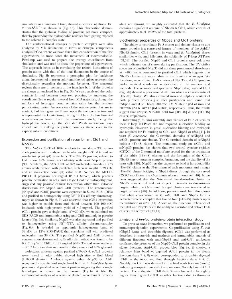
Biochemical properties of Nbp35 and Cfd1 proteinsThe ability to coordinate Fe-S cluster and donate cluster to apo
target proteins is a conserved feature of members of the ApbC/
Nbp35 family. Cfd1 (present in yeast and E. histolytica) shares
similarities with, and falls into, the subfamily of P-loop ATPases
[58,59]. The purified Nbp35 and Cfd1 proteins were colourless
which indicates loss of cluster during purification. The UV-visible
spectrum of purified Nbp35 did not show pronounced absorbance
at ,400 nm as compared to purified Cfd1 which suggests that
Nbp35 clusters are more labile in the presence of oxygen. We
therefore, reconstituted Fe-S clusters of Npb35 and Cfd1proteins
under reduced conditions as described in the material and
methods. The reconstituted spectra of Nbp35 (Fig. 7a) and Cfd1
(Fig. 7b) showed a peak around 410 nm which is characteristic of
[4Fe-4S] cluster. We also estimated iron and sulphide content of
both purified proteins just after purification and found that
rNbp35 and rCfd1 holds 200–253 mM & 56–87 mM of iron and
209606 mM & 78612 mM sulfide, respectively. Thus, the results
suggest that rNbp35 & rCfd1 hold two [4Fe-4S] & one [4Fe-4S]
cluster, respectively.
Interestingly, in vitro assembly and transfer of Fe-S clusters on
these P-loop NTPases did not required nucleotide binding or
hydrolysis. However, in yeast, nucleotide binding and hydrolysis
are required for Fe binding to Cfd1 and Nbp35 in vivo [41]. In
yeast (S. cereviseae), the C-terminal domains of scNbp35 and
scCfd1 proteins are similar. The C-terminal domain of scNbp35
holds a 4Fe-4S cluster. The mutational study on scCfd1 and
scNbp35 proteins has shown that two central cysteine residues
(CPXC) of the C-terminal motif are crucial for the co-ordination
of the labile [4Fe-4S] clusters and the formation of the Cfd1-
Nbp35 hetero-tetramer complex formation, and the viability of the
yeast cells [40]. Nbp35 has the capacity to bind a ferrodoxin-like
[4Fe-4S] cluster at the N-terminus of each monomer and a single
[4Fe-4S] cluster bridging a Nbp35 dimer through the conserved
CX2C motif near the C-terminus of each monomer [40]. It has
been suggested that the N-terminal ferrodoxin-like cluster on
Nbp35 is structural and not subject to transfer to downstream
targets, while the C-terminal bridged clusters are transferred to
target proteins [40]. In addition, previous work had also shown
that when co-expressed in E. coli, Cfd1 and Nbp35 form a
heterotetrameric complex that bound four [4Fe-4S] clusters upon
reconstitution in vitro [41]. Above all, the functional relevance of
the Cfd1 and Nbp35 lies in the ability to assemble and deliver Fe-S
clusters in the cytosol [34,41].
In-vitro and in-vivo protein-protein interaction studyTo prove in-silico interaction, we performed co-purification and
immunoprecipitation experiments. Co-purification using E. colirNbp35 lysate and thrombin digested rCfd1 was performed as
described in materials and methods and immunoblot analysis of
different fractions with anti-Nbp35 and anti-Cfd1 antibodies
confirmed the presence of the Nbp35-Cfd1 protein complex in the
eluate fractions. Anti-Cfd1 probed blot (Fig. 8a, ii) showed a
relatively faint band of digested rCfd1 protein in the eluate
fractions (lane 7 & 8) which corresponded to thrombin digested
rCfd1 in the input and flow through fractions (lane 4 & 5).
Notably, no Cfd1 was detected in the final wash fraction (lane 6)
indicating complete removal of any unbound/loosely bound Cfd1
protein. The undigested rCfd1 (lane 3) was observed to be slightly
higher than digested rCfd1 in other fractions due to thrombin
Interaction between Nbp and Cfd Proteins of E. histolytica
PLOS ONE | www.plosone.org 11 October 2014 | Volume 9 | Issue 10 | e108971
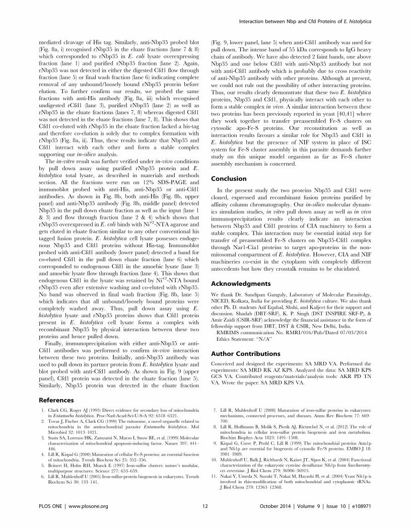
mediated cleavage of His tag. Similarly, anti-Nbp35 probed blot
(Fig. 8a, i) recognised rNbp35 in the eluate fractions (lane 7 & 8)
which corresponded to rNbp35 in E. coli lysate overexpressing
fraction (lane 1) and purified rNbp35 fraction (lane 2). Again,
rNbp35 was not detected in either the digested Cfd1 flow through
fraction (lane 5) or final wash fraction (lane 6) indicating complete
removal of any unbound/loosely bound rNbp35 protein before
elution. To further confirm our results, we probed the same
fractions with anti-His antibody (Fig. 8a, iii) which recognised
undigested rCfd1 (lane 3), purified rNbp35 (lane 2) as well as
rNbp35 in the eluate fractions (lanes 7, 8) whereas digested Cfd1
was not detected in the eluate fractions (lane 7, 8). This shows that
Cfd1 co-eluted with rNbp35 in the eluate fraction lacked a his-tag
and therefore co-elution is solely due to complex formation with
rNbp35 (Fig. 8a, ii). Thus, these results indicate that Nbp35 and
Cfd1 interact with each other and form a stable complex
supporting our in-silico analysis.
The in-vitro result was further verified under in-vivo conditions
by pull down assay using purified rNbp35 protein and E.histolytica total lysate, as described in materials and methods
section. All the fractions were run on 12% SDS-PAGE and
immunoblot probed with anti-His, anti-Nbp35 or anti-Cfd1
antibodies. As shown in Fig. 8b, both anti-His (Fig. 8b, upper
panel) and anti-Nbp35 antibody (Fig. 8b, middle panel) detected
Nbp35 in the pull down eluate fraction as well as the input (lane 1
& 3) and flow through fraction (lane 2 & 4) which shows that
rNbp35 overexpressed in E. coli binds with Ni+2-NTA agarose and
gets eluted in eluate fraction similar to any other conventional his
tagged fusion protein. E. histolytica cell lysate possesses endoge-
nous Nbp35 and Cfd1 proteins without His-tag. Immunoblot
probed with anti-Cfd1 antibody (lower panel) detected a band for
co-eluted Cfd1 in the pull down eluate fraction (lane 6) which
corresponded to endogenous Cfd1 in the amoebic lysate (lane 3)
and amoebic lysate flow through fraction (lane 4). This shows that
endogenous Cfd1 in the lysate was retained by Ni+2-NTA bound
rNbp35 even after extensive washing and co-eluted with rNbp35.
No band was observed in final wash fraction (Fig. 8b, lane 5)
which indicates that all unbound/loosely bound proteins were
completely washed away. Thus, pull down assay using E.histolytica lysate and rNbp35 proteins shows that Cfd1 protein
present in E. histolytica cell lysate forms a complex with
recombinant Nbp35 by physical interaction between these two
proteins and hence pulled down.
Finally, immunoprecipitation with either anti-Nbp35 or anti-
Cfd1 antibodies was performed to confirm in-vivo interaction
between these two proteins. Initially, anti-Nbp35 antibody was
used to pull down its partner protein from E. histolytica lysate and
blot probed with anti-Cfd1 antibody. As shown in Fig. 9 (upper
panel), Cfd1 protein was detected in the eluate fraction (lane 5).
Similarly, Nbp35 protein was detected in the eluate fraction
(Fig. 9, lower panel, lane 5) when anti-Cfd1 antibody was used for
pull down. The intense band of 55 kDa corresponds to IgG heavy
chain of antibody. We have also detected 2 faint bands, one above
Nbp35 and one below Cfd1 with anti-Nbp35 antibody but not
with anti-Cfd1 antibody which is probably due to cross reactivity
of anti-Nbp35 antibody with other proteins. Although at present,
we could not rule out the possibility of other interacting proteins.
Thus, our results clearly demonstrate that these two E. histolyticaproteins, Nbp35 and Cfd1, physically interact with each other to
form a stable complex in vivo. A similar interaction between these
two proteins has been previously reported in yeast [40,41] where
they work together to transfer preassembled Fe-S clusters on
cytosolic apo-Fe-S proteins. Our reconstitution as well as
interaction results favours a similar role for Nbp35 and Cfd1 in
E. histolytica but the presence of NIF system in place of ISC
system for Fe-S cluster assembly in this parasite demands further
study on this unique model organism as far as Fe-S cluster
assembly mechanism is concerned.
Conclusion
In the present study the two proteins Nbp35 and Cfd1 were
cloned, expressed and recombinant fusion proteins purified by
affinity column chromatography. Our in-silico molecular dynam-
ics simulation studies, in vitro pull down assay as well as in vivoimmunoprecipitation results clearly indicate an interaction
between Nbp35 and Cfd1 proteins of CIA machinery to form a
stable complex. This interaction may be essential initial step for
transfer of preassembled Fe-S clusters on Nbp35-Cfd1 complex
through Nar1-Cia1 proteins to target apo-proteins in the non-
mitosomal compartment of E. histolytica. However, CIA and NIF
machineries co-exist in the cytoplasm with completely different
antecedents but how they crosstalk remains to be elucidated.
Acknowledgments
We thank Dr. Sandipan Ganguly, Laboratory of Molecular Parasitolgy,
NICED, Kolkata, India for providing E. histolytica culture. We also thank
other Ph. D. students Asif Equbal, Shshi, and Kuljeet for their support and
discussion. Shadab (DBT-SRF), K. P. Singh (DST INSPIRE SRF-P), &
Amir Zaidi (CSIR-SRF) acknowledge the financial assistance in the form of
fellowship support from DBT, DST & CSIR, New Delhi, India.
RMRIMS communication No. RMRI/016/Pub/Dated 07/03/2014
Ethics Statement: ‘‘N/A’’
Author Contributions
Conceived and designed the experiments: SA MRD VA. Performed the
experiments: SA MRD RK AZ KPS. Analyzed the data: SA MRD KPS
GCS VA. Contributed reagents/materials/analysis tools: AKR PD TN
VA. Wrote the paper: SA MRD KPS VA.
References
1. Clark CG, Roger AJ (1995) Direct evidence for secondary loss of mitochondria
in Entamoeba histolytica. Proc-Natl-Acad-Sci-U-S-A 92: 6518–6521.
2. Tovar J, Fischer A, Clark CG (1999) The mitosome, a novel organelle related to
mitochondria in the amitochondrial parasite Entamoeba histolytica. Mol
Microbiol 32: 1013–1021.
3. Susin SA, Lorenzo HK, Zamzami N, Marzo I, Snow BE, et al. (1999) Molecular
characterization of mitochondrial apoptosis-inducing factor. Nature 397: 441–
446.
4. Lill R, Kispal G (2000) Maturation of cellular Fe-S proteins: an essential function
of mitochondria. Trends Biochem Sci 25: 352–356.
5. Beinert H, Holm RH, Munck E (1997) Iron-sulfur clusters: nature’s modular,
multipurpose structures. Science 277: 653–659.
6. Lill R, Muhlenhoff U (2005) Iron-sulfur-protein biogenesis in eukaryotes. Trends
Biochem Sci 30: 133–141.
7. Lill R, Muhlenhoff U (2008) Maturation of iron-sulfur proteins in eukaryotes:
mechanisms, connected processes, and diseases. Annu Rev Biochem 77: 669–
700.
8. Lill R, Hoffmann B, Molik S, Pierik AJ, Rietzschel N, et al. (2012) The role of
mitochondria in cellular iron-sulfur protein biogenesis and iron metabolism.
Biochim Biophys Acta 1823: 1491–1508.
9. Kispal G, Csere P, Prohl C, Lill R (1999) The mitochondrial proteins Atm1p
and Nfs1p are essential for biogenesis of cytosolic Fe/S proteins. EMBO J 18:
3981–3989.
10. Muhlenhoff U, Balk J, Richhardt N, Kaiser JT, Sipos K, et al. (2004) Functional
characterization of the eukaryotic cysteine desulfurase Nfs1p from Saccharomy-ces cerevisiae. J Biol Chem 279: 36906–36915.
11. Nakai Y, Umeda N, Suzuki T, Nakai M, Hayashi H, et al. (2004) Yeast Nfs1p is
involved in thio-modification of both mitochondrial and cytoplasmic tRNAs.
J Biol Chem 279: 12363–12368.
Interaction between Nbp and Cfd Proteins of E. histolytica
PLOS ONE | www.plosone.org 12 October 2014 | Volume 9 | Issue 10 | e108971
12. Lill R (2009) Function and biogenesis of iron-sulphur proteins. Nature 460: 831–
838.13. Balk J, Pilon M (2010) Ancient and essential: the assembly of iron-sulfur clusters
in plants. Trends Plant Sci 16: 218–226.
14. Rouault TA (2012) Biogenesis of iron-sulfur clusters in mammalian cells: newinsights and relevance to human disease. Dis Model Mech 5: 155–164.
15. Craig EA, Voisine C, Schilke B (1999) Mitochondrial iron metabolism in theyeast Saccharomyces cerevisiae. Biol Chem 380: 1167–1173.
16. Muhlenhoff U, Lill R (2000) Biogenesis of iron-sulfur proteins in eukaryotes: a
novel task of mitochondria that is inherited from bacteria. Biochim Biophys Acta1459: 370–382.
17. Tovar J, Leon-Avila G, Sanchez LB, Sutak R, Tachezy J, et al. (2003)Mitochondrial remnant organelles of Giardia function in iron-sulphur protein
maturation. Nature 426: 172–176.18. Dolezal P, Smid O, Rada P, Zubacova Z, Bursac D, et al. (2005) Giardia
mitosomes and trichomonad hydrogenosomes share a common mode of protein
targeting. Proc Natl Acad Sci U S A 102: 10924–10929.19. Tsaousis AD, Ollagnier de Choudens S, Gentekaki E, Long S, Gaston D, et al.
(2012) Evolution of Fe/S cluster biogenesis in the anaerobic parasite Blastocystis.Proc Natl Acad Sci U S A 109: 10426–10431.
20. Changmai P, Horakova E, Long S, Cernotikova-Stribrna E, McDonald LM, et
al. (2013) Both human ferredoxins equally efficiently rescue ferredoxin deficiencyin Trypanosoma brucei. Mol Microbiol 89: 135–151.
21. Seeber F (2002) Biogenesis of iron-sulphur clusters in amitochondriate andapicomplexan protists. Int J Parasitol 32: 1207–1217.
22. Kumar B, Chaubey S, Shah P, Tanveer A, Charan M, et al. (2011) Interactionbetween sulphur mobilisation proteins SufB and SufC: evidence for an iron-
sulphur cluster biogenesis pathway in the apicoplast of Plasmodium falciparum.
Int J Parasitol 41: 991–999.23. Ali V, Nozaki T (2013) Iron-sulphur clusters, their biosynthesis, and biological
functions in protozoan parasites. Adv Parasitol 83: 1–92.24. Ali V, Shigeta Y, Tokumoto U, Takahashi Y, Nozaki T (2004) An Intestinal
Parasitic Protist, Entamoeba histolytica, Possesses a Non-redundant Nitrogen
Fixation-like System for Iron-Sulfur Cluster Assembly under AnaerobicConditions. J Biol Chem 279: 16863–16874.
25. Gill EE, Diaz-Trivino S, Barbera MJ, Silberman JD, Stechmann A, et al. (2007)Novel mitochondrion-related organelles in the anaerobic amoeba Mastigamoebabalamuthi. Mol Microbiol 66: 1306–1320.
26. Nyvltova E, Sutak R, Harant K, Sedinova M, Hrdy I, et al. (2013) NIF-type
iron-sulfur cluster assembly system is duplicated and distributed in the
mitochondria and cytosol of Mastigamoeba balamuthi. Proc Natl AcadSci U S A 110: 7371–7376.
27. Muller M, Mentel M, van Hellemond JJ, Henze K, Woehle C, et al. (2012)Biochemistry and evolution of anaerobic energy metabolism in eukaryotes.
Microbiol Mol Biol Rev 76: 444–495.
28. Sheftel A, Stehling O, Lill R (2010) Iron-sulfur proteins in health and disease.Trends Endocrinol Metab 21: 302–314.
29. Roy A, Solodovnikova N, Nicholson T, Antholine W, Walden WE (2003) Anovel eukaryotic factor for cytosolic Fe-S cluster assembly. EMBO J 22: 4826–
4835.30. Hausmann A, Aguilar Netz DJ, Balk J, Pierik AJ, Muhlenhoff U, et al. (2005)
The eukaryotic P loop NTPase Nbp35: an essential component of the cytosolic
and nuclear iron-sulfur protein assembly machinery. Proc Natl Acad Sci U S A102: 3266–3271.
31. Balk J, Pierik AJ, Netz DJ, Muhlenhoff U, Lill R (2004) The hydrogenase-likeNar1p is essential for maturation of cytosolic and nuclear iron-sulphur proteins.
EMBO J 23: 2105–2115.
32. Balk J, Lobreaux S (2005) Biogenesis of iron-sulfur proteins in plants. TrendsPlant Sci 10: 324–331.
33. Zhang Y, Lyver ER, Nakamaru-Ogiso E, Yoon H, Amutha B, et al. (2008)Dre2, a conserved eukaryotic Fe/S cluster protein, functions in cytosolic Fe/S
protein biogenesis. Mol Cell Biol 28: 5569–5582.
34. Netz DJ, Stumpfig M, Dore C, Muhlenhoff U, Pierik AJ, et al. (2010) Tah18transfers electrons to Dre2 in cytosolic iron-sulfur protein biogenesis. Nat Chem
Biol 6: 758–765.35. Van Wietmarschen N, Moradian A, Morin GB, Lansdorp PM, Uringa EJ (2012)
The mammalian proteins MMS19, MIP18, and ANT2 are involved incytoplasmic iron-sulfur cluster protein assembly. J Biol Chem 287: 43351–
43358.
36. Stehling O, Vashisht AA, Mascarenhas J, Jonsson ZO, Sharma T, et al. (2012)
MMS19 assembles iron-sulfur proteins required for DNA metabolism andgenomic integrity. Science 337: 195–199.
37. Gari K, Leon Ortiz AM, Borel V, Flynn H, Skehel JM, et al. MMS19 links
cytoplasmic iron-sulfur cluster assembly to DNA metabolism. Science 337: 243–245.
38. Stehling O, Mascarenhas J, Vashisht AA, Sheftel AD, Niggemeyer B, et al.(2013) Human CIA2A-FAM96A and CIA2B-FAM96B integrate iron homeo-
stasis and maturation of different subsets of cytosolic-nuclear iron-sulfur proteins.
Cell Metab 18: 187–198.39. Broderick JB (2007) Assembling iron-sulfur clusters in the cytosol. Nat Chem
Biol 3: 243–244.40. Netz DJ, Pierik AJ, Stumpfig M, Muhlenhoff U, Lill R (2007) The Cfd1-Nbp35
complex acts as a scaffold for iron-sulfur protein assembly in the yeast cytosol.Nat Chem Biol 3: 278–286.
41. Netz DJ, Pierik AJ, Stumpfig M, Bill E, Sharma AK, et al. (2012) A bridging
[4Fe-4S] cluster and nucleotide binding are essential for function of the Cfd1-Nbp35 complex as a scaffold in iron-sulfur protein maturation. J Biol Chem 287:
12365–12378.42. Bych K, Netz DJ, Vigani G, Bill E, Lill R, et al. (2008) The essential cytosolic
iron-sulfur protein Nbp35 acts without Cfd1 partner in the green lineage. J Biol
Chem 283: 35797–35804.43. Basu S, Leonard JC, Desai N, Mavridou DA, Tang KH, et al. (2013) Divergence
of Erv1-associated mitochondrial import and export pathways in trypanosomesand anaerobic protists. Eukaryot Cell 12: 343–355.
44. Diamond LS, Clark CG, Cunnick CC (1995) YI-S, a casein-free medium foraxenic cultivation of Entamoeba histolytica, related Entamoeba, Giardiaintestinalis and Trichomonas vaginalis. J Eukaryot Microbiol 42: 277–278.
45. Ali V, Hashimoto T, Shigeta Y, Nozaki T (2004) Molecular and biochemicalcharacterization of D-phosphoglycerate dehydrogenase from Entamoeba histo-lytica. A unique enteric protozoan parasite that possesses both phosphorylatedand nonphosphorylated serine metabolic pathways. Eur J Biochem 271: 2670–
2681.
46. Ali V, Shigeta Y, Nozaki T (2003) Molecular and structural characterization ofNADPH-dependent d-glycerate dehydrogenase from the enteric parasitic protist
Entamoeba histolytica. Biochem J 375: 729–736.47. Olson JW, Agar JN, Johnson MK, Maier RJ (2000) Characterization of the NifU
and NifS Fe-S cluster formation proteins essential for viability in Helicobacterpylori. Biochemistry 39: 16213–16219.
48. Sambrook J, Fritsch EF, Maniatis T (2001) Molecular Cloning: a Laboratory
Manual, 3nd edn. Cold Spring Harbor: Cold Spring Harbor Laboratory Press,2001.
49. Roy A, Kucukural A, Zhang Y (2010) I-TASSER: a unified platform forautomated protein structure and function prediction. Nat Protoc 5: 725–738.
50. Eswar N, Webb B, Marti-Renom MA, Madhusudhan MS, Eramian D, et al.
(2007) Comparative protein structure modeling using MODELLER. CurrProtoc Protein Sci Chapter 2: Unit 2 9.
51. Laskowski RA, MacArthur MW, Moss DS, Thornton JM (1993) ‘‘PRO-CHECK: a program to check the stereochemical quality of protein structures.’’
J Appl Crystallog 26: 283–291.52. Eramian D, Shen MY, Devos D, Melo F, Sali A, et al. (2006) A composite score
for predicting errors in protein structure models. Protein Sci 15: 1653–1666.
53. Ewald PP (1921) ‘‘Die Berechnung optischer und elektrostatischer gitterpoten-tiale.’’ Annals of Physics 369: 253–287.
54. Srinivasan K, Stalin T, Sivakumar K (2012) Spectral and electrochemical studyof host-guest inclusion complex between 2,4-dinitrophenol and beta-cyclodex-
trin. Spectrochim Acta A Mol Biomol Spectrosc 94: 89–100.
55. Sharma A, Nigam A (2010) Structure modeling of novel DNA glycosylaseenzyme from oral pathogen Streptococcus sanguinis. Bioinformation 5: 136–140.
56. Subramaniam S, Mohmmed A, Gupta D (2009) Molecular modeling studies ofthe interaction between Plasmodium falciparum HslU and HslV subunits.
J Biomol Struct Dyn 26: 473–479.
57. Nachin L, Loiseau L, Expert D, Barras F (2003) SufC: an unorthodoxcytoplasmic ABC/ATPase required for [Fe-S] biogenesis under oxidative stress.
EMBO J 22: 427–437.58. Dean DR, Bolin JT, Zheng L (1993) Nitrogenase metalloclusters: structures,
organization, and synthesis. J Bacteriol 175: 6737–6744.59. Leipe DD, Wolf YI, Koonin EV, Aravind L (2002) Classification and evolution
of P-loop GTPases and related ATPases. J Mol Biol 317: 41–72.
Interaction between Nbp and Cfd Proteins of E. histolytica
PLOS ONE | www.plosone.org 13 October 2014 | Volume 9 | Issue 10 | e108971
Related Documents


![Cytosolic [Ca]](https://static.cupdf.com/doc/110x72/56814e3f550346895dbbac79/cytosolic-ca.jpg)


