1 Inter-individual differences in empathy are reflected in human brain structure Michael J Banissy 1, 2 , Ryota Kanai 2 , Vincent Walsh 2 , and Geraint Rees 2,3 1. Department of Psychology, Goldsmiths, University of London, London, SE14 6NW 2. UCL Institute of Cognitive Neuroscience, 17 Queen Square, London, U.K. 3. Wellcome Trust Centre for Neuroimaging, University College London, 12 Queen Square, London WC1N 3BG Running head: Structural brain correlates of empathy Main text word count: 4427 words Address correspondence to :- Dr Michael Banissy Department of Psychology Goldsmiths University of London New Cross London SE14 6NW [email protected]

Welcome message from author
This document is posted to help you gain knowledge. Please leave a comment to let me know what you think about it! Share it to your friends and learn new things together.
Transcript
1
Inter-individual differences in empathy are reflected in human brain structure
Michael J Banissy1, 2, Ryota Kanai2, Vincent Walsh2, and Geraint Rees2,3
1. Department of Psychology, Goldsmiths, University of London, London, SE14 6NW
2. UCL Institute of Cognitive Neuroscience, 17 Queen Square, London, U.K.
3. Wellcome Trust Centre for Neuroimaging, University College London, 12 Queen Square,
London WC1N 3BG
Running head: Structural brain correlates of empathy
Main text word count: 4427 words
Address correspondence to :- Dr Michael Banissy Department of Psychology Goldsmiths University of London New Cross London SE14 6NW [email protected]
2
Abstract
Empathy is a multi-faceted concept consisting of our ability not only to share emotions but
also to exert cognitive control and perspective taking in our interactions with others. Here we
examined whether inter-individual variability in different components of empathy were
related to differences in brain structure assessed using voxel-based morphometry. Following
a magnetic resonance imaging (MRI) scan, participants completed the Interpersonal
Reactivity Index (IRI). Multiple regression was then used to assess the relationship between
individual differences in gray matter volume and individual differences in empathy traits.
We found that individual differences in affective empathic abilities oriented towards another
person were negatively correlated with gray matter volume in the precuneus, inferior frontal
gyrus, and anterior cingulate. Differences in self-oriented affective empathy were negatively
correlated with gray matter volume of the somatosensory cortex, but positively correlated
with volume in the insula; cognitive perspective taking abilities were positively correlated
with gray matter volume of the anterior cingulate; and the ability to empathise with fictional
characters was positively related to gray matter changes in the right dorsolateral prefrontal
cortex. These findings are discussed in relation to neurocognitive models of empathy.
Keywords: Empathy; Voxel Based Morphometry; Interpersonal Reactivity Index; Structure;
Individual Differences; Social Neuroscience
3
1. Introduction
Empathy is a psychological construct, which describes not only our ability to share
the emotions of others but also to exert cognitive control and perspective taking in our
interactions. Accordingly, models of empathy highlight that the construct is multifaceted and
comprises at least two key components: cognitive empathy – predicting and understanding
another’s mental state by using cognitive processes, and affective empathy – experiencing an
appropriate emotional response as a consequence of another’s state (Baron-Cohen and
Wheelwright, 2004; Batson, 2009; Decety and Jackson, 2004; Preston and de Waal, 2002).
The neural mechanisms that contribute to empathy have been a topic of recent debate.
Some models highlight the importance of perceptually driven activity in neural regions
corresponding to observed experiences (Preston and de Waal, 2002; Gallese, 2003; Goldman,
2006). For example, simulation models of empathy contend that the observer understands the
observed experience by activating similar regions of their brain that are engaged when they
experience the perceived state themselves (e.g. Gallese, 2003; Goldman, 2006). Whereas,
others describe empathy as an outcome of several inter-related mechanisms, including shared
activations between the observer and observed, mechanisms of regulation, contextual
appraisal, and mechanisms of control (Decety and Sommerville, 2003; Decety and Jackson,
2004; Decety and Lamm, 2006). For example, Decety and colleagues contend that there are
three inter-related mechanisms, which give rise to cognitive (i.e. thoughts) and affective (i.e.
feelings) aspects of empathy: i) shared perception-action mechanisms (Preston and de Waal,
2002), which are involved in mapping another person’s experience onto the same
sensorimotor and affective representations as when we experience the state ourselves; ii)
regulatory mechanisms which mediate whether our empathic reactions are self (e.g. personal
distress/self-oriented aversive emotional responses) or other-oriented (e.g. sympathy or
4
compassion) (Decety and Jackson, 2006); and iii) mechanisms that aid our ability to share
another’s perspective (Decety and Jackson, 2004).
Functional brain imaging and transcranial magnetic stimulation studies have provided
evidence in line with the notion that there are multiple mechanisms that may contribute to
empathy. These include ‘shared’ neural systems in which common brain areas are activated
during both self-experience and passive observation of the experiences of others (see Keysers
and Gazzola, 2006; Singer, 2006 for a review); neural systems that are associated with
perspective taking (Ruby and Decety, 2004; Jackson et al., 2006); and mechanisms involved
in regulating distinctions between the source of one’s affective response (i.e. whether our
response to someone else’s emotions is personal distress or is a shared feeling with the target;
see Singer and Lamm, 2009 for review).
The role that these mechanisms play in different aspects of empathy (e.g. cognitive,
affective empathy) has also been examined. For example, lesions to sensorimotor cortices
result in impairments in affective but not cognitive empathy, whereas lesions to ventromedial
prefrontal cortex result in a disruption of cognitive but not affective empathy (Shamay-
Tsoory, Aharon-Peretz and Perry, 2009). Furthermore, regions involved in affect sharing
appear to be related more closely with self-oriented empathy, but can be distinguished from
the neural systems that are associated with the cognitive capacity to adopt the mental states of
another person (Dosch et al., 2010; Jackson et al., 2006). For example, trait levels of
personal distress (measuring self-oriented aversive reactions) have been shown to positively
correlate with the level of neural activity in anterior cingulate (Lawrence et al., 2006;
Cheetham, Pedroni, Antley, Slater, and Jäncke, 2009), anterior insula (Cheetham, Pedroni,
Antley, Slater, and Jäncke, 2009), and sensorimotor cortices (Yang, Decety, Lee, Chen, and
Cheng,, 2009) during empathy for pain. Similarly, Jackson and colleagues (2006) report that
self-oriented empathy towards the pain of another person (in which participants were asked to
5
imagine observed pain/non-pain from their own perspective) leads to greater neural activity
in the neural network involved in experiencing pain (e.g. secondary somatosensory cortex,
anterior cingulate cortex, the insula), whereas other-oriented empathy (in which participants
were asked to imagine observed pain/non-pain from another’s’ perspective) is associated with
increases in neural activity within the inferior parietal cortex, posterior cingulate cortex,
precuneus cortex, temporal-parietal junction (TPJ), and medial prefrontal cortex (also see
David et al., 2006; Ruby and Decety, 2001, 2003, 2004; Vogeley et al., 2001).
In addition to personal distress, a number of other trait empathic disposition have been
linked to neural activity in brain regions involved in perspective taking and sensorimotor
resonance. For example, the level of functional activation in the precuneus (Chakrabarti et
al., 2006), inferior parietal cortex (Chakrabarti et al., 2006), dorsolateral prefrontal cortex
(Chakrabarti et al., 2006), medial prefrontal cortex (Chakrabarti et al., 2006), inferior frontal
gyrus / premotor cortex (Chakrabarti et al., 2006; Dapretto et al., 2006; Gazzola et al., 2006;
Nishitani et al., 2004; Sculte-Rüther et al., 2007), somatosensory cortex (Yang, Decety, Lee,
Chen, and Cheng,, 2009; Cheng, Yang, Lin, Lee, and Decety, 2008; Gazzola et al., 2006),
insula (Chakrabarti et al., 2006; Jabbi et al., 2007; Lamm, Decety, and Singer, 2011; Singer et
al., 2004), anterior cingulate cortex (Singer and Lamm, 2009; Decety, 2010) and superior
temporal regions (Chakrabarti et al., 2006; Sculte-Rüther et al., 2007) have all been
associated with trait empathy levels.
Although functional imaging studies have examined the relationship between brain
activation and empathy, few studies have sought to investigate whether underlying brain
structure is related to empathy in healthy adults. Moreover, the majority of work examining
measures of brain structure that contribute to empathy has focused on structural properties
related to empathic deficits in a variety of pathological conditions (e.g., schizophrenia -
Hooker et al, 2011; conduct disorder - Sterzer et al., 2007; frontotemporal lobar degeneration
6
- Rankin et al., 2006). However, reverse inference from patients to healthy individuals carries
a number of difficulties (Robertson & Murre, 1999), and thus the extent to which these
structural differences are evident in the healthy adult brain remains an open question. To
address this, here we sought to establish whether there was a relationship between trait
empathy (measured using the Interpersonal Reactivity Index; Davis, 1980) and gray matter
density using voxel-based morphometry (VBM) in healthy adults.
Based on previous studies highlighting the involvement of several brain regions in
different facets of empathy, we expected to observe a relationship between brain structure
and regions that are associated with mechanisms that support cognitive (i.e. thoughts) and
affective (i.e. feelings) aspects of empathy in activation studies. For example, we expected to
observe a relationship between the precuneus and perspective taking abilities (Mar, 2011;
Ruby and Decety, 2001; Farrer and Frith, 2002, Vogeley et al., 2004, Ochsner et al., 2004),
and between areas commonly involved in affect sharing and self-oriented empathic responses
(e.g. anterior cingulate, anterior insula, inferior frontal gyrus, somatosensory cortex;
Cheetham, Pedroni, Antley, Slater, and Jäncke, 2009; Lawrence, Shaw, Giampietro,
Surguladze, Brammer, and David, 2006; Chakrabarti et al., 2006; Dapretto et al., 2006;
Gazzola et al., 2006; Nishitani et al., 2004; Sculte-Rüther et al., 2007; Yang, Decety, Lee,
Chen, and Cheng,, 2009; Shamay-Tsoory, Aharon-Peretz and Perry, 2009).
2. Methods
2.1. Participants
One hundred and eighteen healthy participants (age mean 22.9 + 4.2 (s.d.) years old;
66 female) gave written informed consent to take part in the experiment that was approved by
the local ethics committee.
7
2.2. Materials and procedure
All participants completed the Interpersonal Reactivity Index (IRI; Davis, 1980); a
widely used multi-dimensional measure of trait empathy, based on self-report. It consists of
four subscales: perspective taking; personal distress; empathic concern; and fantasy (Davis,
1980; Davis, Luce, and Kraus, 1994). Empathic concern and personal distress measure
affective reactions but differ in their targets. Personal distress is self-oriented and associated
to aversive emotional responses in the observer (e.g. feelings of fear or discomfort at
witnessing negative experiences of others). Empathic concern is other-oriented and related to
feelings of compassion and sympathy for the observed individual. Perspective taking
examines the tendency to think from another perspective (i.e. cognitive responses). Fantasy
examines participants’ abilities to transpose themselves into fictional situations (e.g. books,
movies, daydreams).
Each subscale contained seven items. They were measured on a five point Likert
scale ranging from 0 (“Does not describe me well”) to 4 (“Describes me very well”). For
each subscale, a minimum score of 0 or maximum score of 28 was possible.
2.3. MRI acquisition and analyses
High-resolution anatomical images were acquired using a T1-weighted 3-D Modified
Driven Equilibrium Fourier Transform (MDEFT) sequence for each participant on a Siemens
1.5T Sonata scanner (TR = 12.24, TE=3.56, flip angle = 23deg, field of view = 256 x 256,
176 slices, resolution = 1 x 1 x 1 mm).
The images were first segmented into gray matter and white matter using
segmentation tools in Statistical Parametric Mapping software (SPM8)
(http://www.fil.ion.ucl.ac.uk/spm) running on MATLAB (MathWorks, Natick, MA).
Coregistration of gray matter images across participants was achieved using the DARTEL
8
(Diffeomorphic Anatomical Registration Through Exponentiated Lie Algebra) algorithm
(Ashburner, 2007). The resulting template image was transformed to MNI stereotactic space
using affine and non-linear spatial normalisation with intensity modulation by the Jacobian
determinant of the deformation flow field computed for each image. Then the images were
smoothed with a Gaussian kernel (full-with at half-maximum, FWHM =12mm).
The pre-processed images were entered into a multiple regression model in SPM8 to
identify cortical regions that showed a correlation with the subscales of the IRI (with all
subscales included in the same design matrix). We included age, gender and total gray matter
volume as covariates of no interest in the design matrix to regress out any effects attributable
to them. The inclusion of gender as a covariate of no interest was of particular importance, as
sex differences have been reported for all subscales of the IRI (Davis, 1980). In line with
previous studies (e.g. Rankin et al., 2006), our decision to focus our analysis on IRI subscales
rather than total IRI score, was because some components of the IRI have been shown to
negatively correlate with social competence (e.g. Personal Distress Scale; Davis, 1983).
We conducted region of interest analyses using previous functional brain imaging
studies of empathy to constrain our anatomical hypotheses. In particular, we focused our
analysis on the anterior cingulate (MNI coordinates: ± x=3, y=24, z=33; Singer et al., 2004),
inferior frontal gyrus (x=60, y=14, z=24; Lamm et al., 2011), precuneus (± x=10, y=-50,
z=36; Mar, 2011), anterior insula (± x=39, y=9, z=-21; Singer et al., 2004), somatosensory
cortex (± x=48, y=-16, z=54; Hooker, Verosky, Germine, Knight, and D’Esposito, 2008), and
dorsolateral prefrontal cortex (± x=42, y=39, z=24; Lamm et al., 2011). These regions were
selected based on previous functional brain imaging studies demonstrating their involvement
in affective and cognitive empathy tasks, and meta-analyses of brain regions involved in
affect sharing and metalizing. In situations where regions of interest were reported in more
than one study we choose the coordinate from the study with a higher number of citations.
9
Statistical significance was assessed using small volume correction (Worsley et al., 1996) at a
threshold of P<0.05 (corrected) for those clusters that also passed a whole-brain uncorrected
threshold of P<0.001 within a sphere (10mm radius) centred at each of the coordinates
identified by our prior hypotheses. Outside these pre-defined regions, we used a statistical
threshold of P < 0.05 corrected for the whole-brain volume at a cluster level using non-
stationary correction (Hayasaka et al., 2004).
3. Results
3.1 IRI Scores
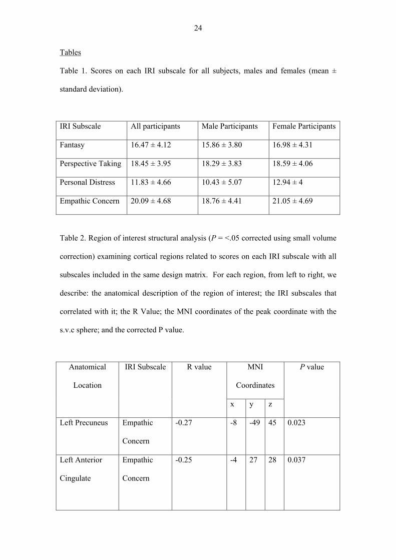
Scores on each subscale of the IRI were consistent with previously published norms
for this measure (Davis, 1980; Table 1). Previous findings have also reported sex differences
on each subscale of the IRI (Davis, 1980), with females displaying higher scores than males
in each case. We observed a similar pattern of results (Table 1), with females scoring higher
on the Empathic Concern scale [t(116) = 2.55, p = .012] and the Personal Distress scale
[t(95.23) = 2.93, p = .004]. There was also a trend for females to score higher than males on
the Fantasy Scale, t(116) = 1.55, p = .123. We were unable to replicate differences between
male and female scores on the Perspective Taking subscale, t(116) = .438, p = .662, however
it is of note that this represents the smallest identified sex difference on all four subscales of
the IRI (Davis, 1980).
In addition, scores on the Fantasy Scale were positively correlated with scores on the
Perspective Taking (R = .307, p = .001) and Empathic Concern (R = .224, p = .015) scales.
Scores on Perspective Taking were also positively correlated with scores on Empathic
Concern (R = .464, p = < .001). No other correlations were found across the subscales.
Insert Table 1 Here
10
3.2 Region of interest structural analysis
Each region-of-interest was defined according to our prior hypotheses (see Methods)
and statistical correction undertaken for the small volume examined. This analysis revealed
that inter-individual variability on the fantasy scale showed a significant positive correlation
with brain volume in the right dorsolateral prefrontal cortex (R = 0.29, T(110) = 3.15,
PFWE_corr = 0.012; peak MNI coordinate within the s.v.c. sphere x = 40, y= 47, z = 30). No
other regions of interest showed a significant correlation with the scale of fantasy (p > 0.05,
corrected; Table 2).
Inter-individual variability in participants’ empathic concern negatively correlated
with gray matter volume in left precuneus (R = -0.27, T(110) = 2.89, PFWE_corr = 0.023; peak
MNI coordinate within the s.v.c. sphere x = -8, y=-49, z = 45); and left anterior cingulate (R
= -0.25, T(110) = 2.69, PFWE_corr = 0.037; peak MNI coordinate within the s.v.c. sphere x = -
4, y= 27, z = 28. We also found a significant negative relationship with empathic concern at
our ROI on the anterior insula. The peak coordinate was found within the inferior frontal
gyrus (R = -0.35, T(110) = 3.98, PFWE_corr = 0.001; peak MNI coordinate within the s.v.c.
sphere x = -48, y= 6, z = 18), but this is consistent with the cluster from the original study
used to define the ROI extending to this region. No other regions of interest showed a
significant correlation with the scale of empathic concern (p > 0.05, corrected; Table 2).
Inter-individual variability in perspective taking showed a significant positive
correlation with gray matter volume of left anterior cingulate (R = 0.25, T(110) = 2.69,
PFWE_corr = 0.037; the coordinate of the peak x = -6 y = 33, z = 31). No other regions of
interest including precuneus, which showed a significant correlation with empathic concern,
11
showed a significant correlation with the perspective taking scale (p > 0.05, corrected; Table
2).
As for personal distress, we found a significant positive correlation with gray matter
volume of the left anterior insula (R = 0.27, T(110) = 2.92, PFWE_corr = 0.022; the coordinate
of the peak x = -32, y = 9, z = -18). We also found a negative correlation with the gray
matter volume of somatosensory cortex (R = -0.32, T(110) = 3.57, PFWE_corr = 0.004; the
coordinate of the peak x = -45, y = -19, z = 61). No other regions of interest showed a
significant correlation with the personal distress scale (p > 0.05, corrected; Table 2).
INSERT TABLE 2 HERE
3.3. Whole brain analysis
At a threshold of p <.05 corrected for the whole-brain volume at a cluster level using
non-stationary correction, we found a significant negative relationship between scores on the
empathic concern subscale and gray matter volume in the left inferior frontal gyrus (R = -
0.36, T = 4.06, P = < 0.05, corrected for multiple comparisons across the whole brain
volume; the coordinate of the peak x = -51, y= 8, z = 18). We did not observe any other
regions at this corrected threshold.
4. Discussion
This study examined whether individual differences in trait empathy dimensions were
related to morphological differences in human brain structure. Our results suggest that inter-
individual variability in different aspects of empathy were linked with distinct morphological
changes in separate brain regions. Affective empathic abilities that are oriented towards
another person (i.e. empathic concern subscale) were linked with reduced gray matter volume
within the left precuneus, left inferior frontal gyrus, and left anterior cingulate; a tendency
12
towards self-oriented affective empathy was linked with reduced gray matter volume in the
left somatosensory cortex, but increased volume in the left insula; cognitive perspective
taking abilities were linked to increased gray matter volume in the left anterior cingulate; and
the ability to empathise with / place oneself into fictional situations (fantasy subscale) was
associated with increased gray matter volume in the right dorsolateral prefrontal cortex .
To our knowledge, our study is the first to examine the relationship between regional
gray matter volume and different facets of empathy in healthy adults. Our findings show that
the gray matter structure in brain regions implicated in previous studies of functional
commonalities in empathy can also account for inter-individual variability in empathic traits.
For example, the involvement of the precuneus cortex, insula, anterior cingulate and
dorsolateral prefrontal cortex are consistent with recent meta-analyses highlighting these
regions as components of core networks involved in affect sharing (Lamm et al., 2011) and
perspective taking (Mar, 2011). The relationship between changes in brain structure of the
inferior frontal gyrus and empathic abilities is consistent with findings showing a relationship
between trait empathy and functional levels of neural activity in this brain region
(Chakrabarti et al., 2006; Gazzola et al., 2006; Jabbi et al., 2007; Hooker et al., 2008; Hooker
et al., 2010), and neuropsychological patient data showing that lesions to the inferior frontal
gyrus result in impairments in affective empathy (Shamay-Tsoory, Aharon-Peretz and Perry,
2009; Rankin et al., 2006). Finally, the association with changes in somatosensory cortex and
levels of personal distress is in line with growing evidence pointing to a role for
somatosensation in social perception (Keysers, Kaas, and Gazzola, 2010; Hooker et al., 2008;
Banissy et al., 2010; Banissy et al., 2011; Hooker et al., 2010; Pitcher et al., 2008).
The relationship between structural variations in each brain region and specific
empathy traits is also interesting. For example, our finding that decreased brain volume in the
inferior frontal gyrus was associated with increased scores on the empathic concern subscale
13
of the IRI is consistent with previous work examining the neural correlates of dispositional
measures of empathy. The empathic concern subscale of the IRI is other-oriented and related
to affective empathy. In healthy adults, functional neural activity in the inferior frontal gyrus
has been linked to trait levels of empathic concern in a number of studies (e.g. Schulte-Ruther
et al., 2007). Previous findings have also indicated, that lesions to the inferior frontal gyrus
result in impairments in affective but not cognitive empathy (Shamay-Tsoory, Aharon-Peretz
and Perry, 2009) and damage to the inferior frontal gyrus has been linked to scores on the
empathic concern subscale of the IRI (Rankin et al., 2006). Our evidence that reduced brain
volume in the inferior frontal gyrus was related to increased levels of empathic concern is
therefore consistent with this data. The specific direction of the relationship also implies that
in healthy adults “less is more”. While this may first seem paradoxical to the typical “more is
better” notion, such a relationship is not uncommon (e.g. see Jung et al., 2010; Kanai et al.,
2011) and may reflect differences in maturation during adolescence and cortical thinning.
Moreover, it has been postulated that cortical thinning during maturation reflects changes in
functional activation during skill acquisition, with plasticity decreasing as efficiency
increases (Durston and Casey, 2006; Durston et al., 2006). In this context, less gray matter
volume in the inferior frontal gyrus showing a relationship with increased empathic concern
scores would seem consistent with the patient data and functional imaging studies described
above.
The trade off between increased local gray matter volume in the anterior cingulate
being related to increased scores of the perspective taking subscale of the IRI, but decreased
gray matter volume in the anterior cingulate being associated with increased scores on the
empathic concern subscale of the IRI is also intriguing. A recent meta-analysis of empathy
for pain highlights the anterior cingulate as part of a core network in this process (Lamm et
al., 2011). In relation to trait levels of empathy, previous functional brain imaging studies
14
have associated levels of neural activity in anterior cingulate with trait levels of perspective
taking (measured on IRI) (Montag, Schubert, Heinz, and Gallinat, 2008) and personal distress
(measured on IRI) (Lawrence et al., 2006; Cheetham, Pedroni, Antley, Slater, and Jäncke,
2009). Our data relating brain volume in the anterior cingulate to perspective taking abilities
is therefore consistent with some of the studies of functional correlates of empathy. They
extend them by indicating that the anterior cingulate may also be important in other empathic
capacities including more affective empathic dispositions that are other-oriented. Precisely
why less gray matter volume in the anterior cingulate may facilitate affective empathy (i.e.
empathic concern), but more gray matter volume in the same brain region may facilitate
cognitive perspective taking is difficult to disentangle. This is because the links between
macroscopic volumetric measures such as regional grey matter volume and functional
differences are barely understood. The idea of cortical thinning and maturation, postulated
above (in the case of empathic concern), is one of many possible explanations. To reach a full
understanding of this issue, it will be important to investigate how differences in
microstructure measures (e.g., the number of neurons, the degree of myelination, the degree
of dendritic arborization and so on) are associated with functional differences and how they
contribute to differences in the aggregate measure of grey matter volume across tasks.
We found a negative relationship between scores on the personal distress subscale of
the IRI and structural brain changes in and the somatosensory cortex. Recent findings have
implicated a prominent role for the somatosensory system in social perception, with studies
indicating that somatosensory-related cortices play a key in role in using social cues to
understand mental and emotional states of others (e.g. Keysers, Kaas, and Gazzola, 2010;
Hooker et al., 2008; Banissy et al., 2010; Banissy et al., 2011; Hooker et al., 2010; Pitcher et
al., 2008). Previous findings have shown that scores on the personal distress subscale of the
IRI are negatively correlated with social competence and social perception abilities (Davis,
15
1983). In this regard, a negative relationship between scores on the personal distress subscale
and brain volume in the somatosensory system may indicate that less grey matter volume in
the somatosensory system is related to poorer social perception abilities and is therefore
consistent with prior studies highlight the role of somatosensation in our ability to use social
cues to infer emotions and mental states. In contrast, the positive relationship between brain
volume in the anterior insula and personal distress implies that more brain volume in the
anterior insula is negative for social perception abilities.
Our findings are also interesting in a broader context of the neurobiology of
individual differences. A number of recent functional neuroimaging studies have begun to go
beyond considering functional activation that is common to all subjects, to examine
predictive links between brain activity and individual trait-behaviours (see Hariri, 2009;
Kanai and Rees, 2011 for review). This has led to a variety of developments in our
understanding of how trait-like behaviours modulate variability in brain function (e.g. trait
anxiety modulates amygdala reactivity to affective stimuli – Dickie and Armony, 2008;
ventral striatum responsiveness is associated with individual differences in impulsivity –
Hariri et al., 2006) and to the suggestion that differential patterns of brain activity may act as
markers for individual differences in personality and liability for disease (Hariri, 2009).
While a limited number of studies have examined the influence of trait empathy on functional
brain activity, none have considered the relationship between structural variation and inter-
individual differences in trait empathy in healthy adults. Our findings therefore provide
novel predictive links between trait-empathy and region-specific structural variation. They
indicate that brain regions previously identified in studies of functional commonalities in the
neuroscience of empathy are susceptible to individual variation in brain structure and that this
contributes to inter-individual differences in specific components of trait-empathy. An
important next step will be to determine the mechanisms driving this variability and to
16
consider the extent to which individual variation in these components may provide predictive
markers for vulnerability towards social perception deficits. In this context, it is also
important to consider the extent to which inter-individual differences in empathy are a
consequence of, or contribute to, the structural differences that we observe. Moreover, while
levels of trait empathy are enduring and show continuity across time and situations from early
childhood (Volbrecht et al., 2007; Zahn-Waxler, Robinson and Emde, 1992; Zahn-Waxler et
al., 2001), the relative contributions of environmental and biological factors in the
development of empathic abilities have been a topic of much interest (e.g. Knafo et al., 2008;
Volbrecht et al., 2007; Zahn-Waxler et al., 2001). Our findings add a new dimension to this
by implicating specific intra-individual differences in brain structure that may determine
empathic cognition or vice versa.
Taken together, we examined how individual variability in different components of
empathy related to volumetric differences in brain structure. Our findings demonstrate that
different aspects of empathy are linked with distinct morphological changes in separate brain
regions and suggest that multiple mechanisms are associated with increases in specific
empathic skills. This implies that empathy is multi-faceted and that structural variation in
brain regions that support affect sharing and cognitive perspective taking act to facilitate self
and other related empathic processes in different ways.
Acknowledgements
This work was supported by a British Academy Postdoctoral Fellowship (M.J.B.), the ESRC
(M.J.B.), the Human Frontier Science Program (RK), the Japan Society for the Promotion of
Science (R.K.) and the Wellcome Trust (G.R.)
17
References
Ashburner, J. (2007). A fast diffeomorphic image registration algorithm. Neuroimage, 38, 95-
113.
Banissy, M.J., Garrido, L., Kusnir, F., Duchaine, B., Walsh, V., and Ward, J. (2011).
Superior facial expression, but not identity recognition, in mirror-touch synesthesia. J
Neurosci, 31, 1820-1824.
Banissy, M.J., Sauter, D.A., Ward, J., Warren, J.E., Walsh, V., and Scott, S.K. (2010).
Suppressing sensorimotor activity modulates the discrimination of auditory emotions
but not speaker identity. J Neurosci., 30, 13552-13557.
Baron-Cohen, S., and Wheelwright, S. (2004). The empathy quotient: An investigation of
adults with Asperger syndrome or high functioning autism and normal sex
differences. Journal of Autism and Developmental Disorders, 34, 163-175.
Batson, C. D. (2009). These things called empathy: Eight related but distinct phenomena. In
J. Decety and W. J. Ickes (Eds.), The Social Neuroscience of Empathy. Cambridge,
MA: MIT Press.
Chakrabarti, B., Bullmore, E., and Baron-Cohen, S. (2006). Empathizing with basic
emotions: Common and discrete neural substrates. Social Neuroscience, 1, 364-384.
Cheetham, M., Pedroni, A.F., Antley, A., Slater, M., and Jäncke, L. (2009). Virtual milgram:
Empathic concern or personal distress? Evidence from functional MRI and
dispositional measures. Front Hum Neurosci., 3, 1-13.
Cheng, Y., Yang, C.Y., Lin, C.P., Lee, P.L., and Decety, J. (2008). The perception of pain in
others suppresses somatosensory oscillations: A magnetoencephalography study.
NeuroImage, 40, 1833-1840.
Dapretto, M., Davies, M. S., Pfeifer, J. H., Scott, A. A., Sigman, M., Bookheimer, S.
Y., et al. (2006). Understanding emotions in others: Mirror neuron dysfunction
18
in children with autism spectrum disorders. Nature Neuroscience, 9, 28-30.
David, N., Bewernick, B. H., Cohen, M. X., Newen, A., Lux, S., Fink, G. R., et al. (2006).
Neural representation of self versus other: Visual spatial perspective taking and
agency in a virtual ball tossing game. Journal of Cognitive Neuroscience, 18, 898-
910.
Davis, M. H. (1980). A multi-dimensional approach to individual differences in empathy.
JCAS Catalog of Selected Documents in Psychology, 75, 989-1015.
Davis, M. H. (1983). Measuring individual differences in empathy: A multidimensional
approach. J Per Soc Psychol., 44, 113-126.
Davis, M. H., Luce, C., and Kraus, S. J. (1994). The heritability of characteristics associated
with dispositional empathy. Journal of Personality, 62(3), 369-391.
Decety, J. (2010). The neurodevelopment of empathy in humans. Developmental Science,
Decety, J., and Jackson, P. J. (2004). The functional architecture of human empathy.
Behavioral and Cognitive Neuroscience Reviews, 3, 71-100.
Decety, J., and Jackson, P. L. (2006). A social-neuroscience perspective on empathy. Current
Directions in Psychological Science, 15(2), 54-58.
Decety, J., and Lamm, C. (2006). Human empathy through the lens of social neuroscience.
Sci World J., 6, 1145-1163.
Decety, J., and Sommerville, J. A. (2003). Shared representations between self and other: a
social cognitive neuroscience view. Trends in Cognitive Sciences, 17, 527-533.
Dickie, E. W., and Armony, J. L. (2008). Amygdala responses to unattended fearful faces:
Interaction between sex and trait anxiety. Psychiatry Res., 162, 51-57.
Dosch, M., Loenneker, T., Bucher, K., Martin, E., Klaver, P. (2010). Learning to appreciate
others: Neural development of cognitive perspective taking. Neuroimage, 50, 837-
846.
19
Durston, S., and Casey, B. J. (2006). What have we learned about cognitive development
from neuroimaging. Neuropsychologia, 44, 2149-2157.
Durston, S., Davidson, M. C., Tottenham, N., Galvan, A., Spicer, J., Fossella, J. A., et al.
(2006). A shift from diffuse to focal cortical activity with development.
Developmental Science, 9, 1-8.
Farrer C., and Frith, C.D. (2002). Experiencing oneself vs. another person as being the cause
of an action: The neural correlates of the experience of agency. NeuroImage, 15, 596-
603.
Gallese, V. (2003). The roots of empathy: The shared manifold hypothesis and the neural
basis of intersubjectivity. Psychopathology, 36, 171-180.
Gazzola, V., Aziz-Zadeh, L., and Keysers, C. (2006). Empathy and the somatotopic
auditory mirror system in humans. Current Biology, 16, 1824-1829.
Goldman, A. (2006). The simulating mind. Oxford University Press, New York.
Hariri, A. R. (2009). The neurobiology of individual differences in complex behavioral traits.
Annual Review Neuroscience, 32, 225-247.
Hariri, A. R., Brown, S. M., Williamson, D. E., Flory, J. D., de Wit, H., and Manuck, S. B.
(2006). Preference for immediate over delayed rewards is associated with magnitude
of ventral striatal activity. Journal of Neuroscience, 26, 13213-13217.
Hayasaka, S., Phan, K.L., Liberzon, I., Worsley, K.J. and Nichols, T.E. (2004). Nonstationary
cluster-size inference with random field and permutation methods. NeuroImage, 22,
676-687.
Hooker, C., I., Bruce, L., Lincoln, S.H., Fisher, M., and Vinogradov, S. (2011). Theory of
mind skills are related to gray matter volume in the ventromedial prefrontal cortex in
schizophrenia. Biol Psychiatry, 70, 1169-1178.
Hooker, C.I., Verosky, S.C., Germine, L.T., Knight, R.T., and D’Esposito, M. (2008).
20
Mentalizing about emotion and its relationship to empathy. Soc Cogn Affect
Neurosci., 3, 204-217.
Hooker, C.I., Verosky, S.C., Germine, L.T., Knight, R.T., and D’Esposito, M. (2010). Neural
activity during social signal perception correlates with self-reported empathy. Brain
Res., 1308, 110-113.
Jabbi, M., Swart, M., and Keysers, C. (2007). Empathy for positive and negative emotions in
the gustatory cortex. Neuroimage, 34, 1744-1753.
Jackson, P. L., Brunet, E., Meltzoff, A. N., and Decety, J. (2006). Empathy examined through
the neural mechanisms involved in imagining how I feel versus how you feel pain.
Neuropsychologia, 44, 752-761.
Jung, R. E., Segall, J. M., Bockholt, J., Flores, R. A., Smith, S. M., Chavez, R. S. (2010).
Neuroanatomy of creativity. Hum Brain Map, 31, 398-409.
Kanai, R., Dong, M.Y., Bahrami, B. & Rees, G. (2011). Distractibility in daily life is
reflected in the structure and function of human parietal cortex. J. Neurosci., 31,
6620-6626.
Keysers, C., and Gazzola, V. (2006). Towards a unifying theory of social cognition. Prog
Brain Res, 156, 379-401.
Keysers, C., Kaas, J.H., and Gazzola, V. (2010). Somatosensation in social perception. Nat
Rev Neurosci., 11, 417-428.
Knafo, A., Zahn-Waxler, C., Van Hulle C., Robinson, J. L., and Rhee, S. H. (2008). The
developmental origins of a disposition towards empathy: Genetic and environmental
contributions. Emotion, 8, 737-752.
Lamm, C., Decety, J., and Singer, T. (2011). Meta-analytic evidence for common and distinct
neural networks associated with directly experienced pain and empathy for pain.
Neuroimage, 54, 2492-2502.
21
Lawwrence, E. J., Shaw, P., Giampietro, V. P., Surguladze, S., Brammer, M.J., and David, A.
S. (2006). The role of ‘shared representations’ in social perception and empathy: an
fMRI study. NeuroImage, 29, 1173-1184.
Mar, R.A. (2011). The neural basis of social cognition and story comprehension. Ann. Rev.
Psychol., 62, 103-134.
Montag, C., Schuber, F., Heinz, A., and Gallinat, J. (2008). Prefrontal cortex glutamate
correlates with mental perspective-taking. PLoS One, 3, e3890.
Nishitani, N., Avikainen, S., and Hari, R. (2004). Abnormal imitation-related cortical
activation sequences in Asperger’s syndrome. Annals of Neurology, 55, 558-562.
Pitcher, D., Garrido, L., Walsh, V., and Duchaine, B.C. (2008). Transcranial magnetic
stimulation disrupts the perception and embodiment of facial expressions. J Neurosci.,
28, 8929-8933.
Preston, S. D., and de Waal, F. B. M. (2002). Empathy: Its ultimate and proximate bases.
Behavioral and Brain Sciences, 25(1), 1-+.
Rankin, K. P., Gorno-Tempini, M.L., Allison, S. C., Stanley, C. M., Glenn, S., Weiner, M.
W., and Miller, B. L. (2006). Structural anatomy of empathy in neurodegenerative
disease. Brain, 129, 2945-2956.
Robertson, I. H., and Murre, J. M. J. (1999). Rehabilitation of brain damage: Brain
plasticity and principles of guided recovery. Psychological Bulletin, 125, 544-
575.
Ruby, P., and Decety, J. (2001). Effect of subjective perspective taking during simulation of
action: A PET investigation of agency. Nature Neuroscience, 4, 546-550.
Ruby, P., and Decety, J. (2003). What you believe versus what you think they believe: A
neuroimaging study of conceptual perspective taking. European Journal of
Neuroscience, 17, 2475-2480.
22
Ruby, P., and Decety, J. (2004). How do you feel versus how do you think she would feel? A
neuroimaging study of perspective taking with social emotions. Journal of Cognitive
Neuroscience, 16, 988-999.
Schulte-Rüther, M., Markowitsch, H. J., Fink, G. R., and Piefke, M. (2007). Mirror neuron
and theory of mind mechanisms involed in face-to-face interactions: A functional
magnetic resonance imaging approach to empathy. Journal of Cognitive
Neuroscience, 19, 1354-1372.
Shamay-Tsoory, S. G., Aharon-Peretz, J., and Perry, D. (2009). Two systems for empathy: a
double dissociation between emotional and cognitive empathy in inferior frontal gyrus
versus ventromedial prefrontal lesions. Brain, 132, 617-627.
Singer, T. (2006). The neuronal basis and ontology of empathy and mind reading: Review of
the literature and implications for future research. Neurosci Biobehav Rev, 30, 855-
863.
Singer, T., and Lamm, C. (2009). The social neuroscience of empathy. Ann. NY Acad. Sci.,
1156, 81-96.
Singer, T., Seymour, B., O’Doherty, J., Kaube, H., Dolan, R. J., and Frith, C. D. (2004).
Empathy for pain involves the affective but not sensory components of pain. Science,
303, 1157-1162.
Sterzer, P., Stadler, C., Poustka, F., and Kleinschmidt, A. (2007). A structural neural deficit
in adolescents with conduct disorder and its association with lack of empathy.
NeuroImage, 37, 335-342.
Vogeley, K., Bussfeld, P., Newen, A., Herrmann, S., Happe, F., Falkai, P., et al. (2001). Mind
reading: Neural mechanisms of theory of mind and self-perspective. Neuroimage, 14,
170-181.
Vogeley, K., May, M., Ritzl, A., Falkai, P., Zilles K., and Fink, G.R. (2004). Neural
23
correlates of first-person perspective as one constituent of human self-consciousness.
J Cogn Neuroscience, 16, 817-827.
Volbrecht, M. M., Lemery-Chalfant, K., Aksan, N., Zahn-Waxler, C., and Goldsmith, H. H.
(2007). Examining the familial link between positive affect and empathy development
in the second year. Journal of Genetic Psychology, 168, 105-129.
Worsley, K.J., Marrett, S., Neelin, P., Vandal, A.C., Friston, K.J. and Evans, A.C. (1996). A
unified statistical approach for determining significant signals in images of cerebral
activation. Hum. Brain Map. 4, 58-73.
Yang, C.Y., Decety, J., Lee, S., Chen, C., and Cheng, Y. (2009). Gender differences in the
mu rhythm during empathy for pain: An electroencephalographic study. Brain Res,
1251, 176-184.
Zahn-Waxler, C., Robinson, J., and Emde, R. N. (1992). The development of empathy in
twins. Developmental Psychology, 28, 1038-1047.
Zahn-Waxler, C., Schiro, K., Robinson, J. L., Emde, R. N., and Schmitz, S. (2001). Empathy
and prosocial patterns in young MZ and DZ twins: Development and genetic and
environmental influences. In R. N. Emde and J. K. Hewitt, (Eds.) Infancy to early
childhood: Genetic and environmental influences on developmental change. New
York: Oxford University Press.
24
Tables
Table 1. Scores on each IRI subscale for all subjects, males and females (mean ±
standard deviation).
IRI Subscale All participants Male Participants Female Participants
Fantasy 16.47 ± 4.12 15.86 ± 3.80 16.98 ± 4.31
Perspective Taking 18.45 ± 3.95 18.29 ± 3.83 18.59 ± 4.06
Personal Distress 11.83 ± 4.66 10.43 ± 5.07 12.94 ± 4
Empathic Concern 20.09 ± 4.68 18.76 ± 4.41 21.05 ± 4.69
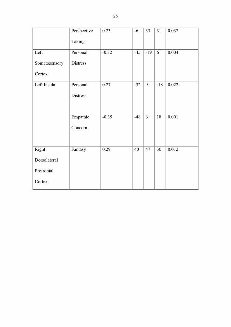
Table 2. Region of interest structural analysis (P = <.05 corrected using small volume
correction) examining cortical regions related to scores on each IRI subscale with all
subscales included in the same design matrix. For each region, from left to right, we
describe: the anatomical description of the region of interest; the IRI subscales that
correlated with it; the R Value; the MNI coordinates of the peak coordinate with the
s.v.c sphere; and the corrected P value.
Anatomical
Location
IRI Subscale R value MNI
Coordinates
P value
x y z
Left Precuneus Empathic
Concern
-0.27 -8 -49 45 0.023
Left Anterior
Cingulate
Empathic
Concern
-0.25
-4
27
28
0.037
25
Perspective
Taking
0.23 -6 33 31 0.037
Left
Somatosensory
Cortex
Personal
Distress
-0.32 -45 -19 61 0.004
Left Insula Personal
Distress
Empathic
Concern
0.27
-0.35
-32
-48
9
6
-18
18
0.022
0.001
Right
Dorsolateral
Prefrontal
Cortex
Fantasy 0.29 40 47 30 0.012
Related Documents