Integrin-Linked Kinase Controls Microtubule Dynamics Required for Plasma Membrane Targeting of Caveolae Sara A. Wickström 1,7,∗ , Anika Lange 1 , Michael W. Hess 2 , Julien Polleux 1,3 , Joachim P. Spatz 3 , Marcus Krüger 4 , Kristian Pfaller 2 , Armin Lambacher 1 , Wilhelm Bloch 5 , Matthias Mann 4 , Lukas A. Huber 6 , and Reinhard Fässler 1 1 Department of Molecular Medicine, Max-Planck Institute of Biochemistry, Am Klopferspitz 18, 82152 Martinsried, Germany 2 Division of Histology and Embryology, Innsbruck Medical University, Müllerstrasse 59, 6020 Innsbruck, Austria 3 Department of New Materials and Biosystems, Max Planck Institute for Metals Research, 70569 Stuttgart, Germany 4 Department of Proteomics and Signal Transduction, Max-Planck Institute of Biochemistry, Am Klopferspitz 18, 82152 Martinsried, Germany 5 Department of Molecular and Cellular Sport Medicine, 50933 Cologne, Germany 6 Biocenter, Division of Cell Biology, Innsbruck Medical University, Fritz-Pregl Strasse 3, 6020 Innsbruck, Austria Summary Caveolae are specialized compartments of the plasma membrane that are involved in signaling, endocytosis, and cholesterol transport. Their formation requires the transport of caveolin-1 to the plasma membrane, but the molecular mechanisms regulating the transport are largely unknown. Here, we identify a critical role for adhesion-mediated signaling through β1 integrins and integrin-linked kinase (ILK) in caveolae formation. Mice lacking β1 integrins or ILK in keratinocytes have dramatically reduced numbers of plasma membrane caveolae in vivo, which is due to impaired transport of caveolin-1-containing vesicles along microtubules (MT) to the plasma membrane. Mechanistically, ILK promotes the recruitment of the F-actin binding protein IQGAP1 to the cell cortex, which, in turn, cooperates with its effector mDia1 to locally stabilize MTs and to allow stable insertion of caveolae into the plasma membrane. Our results assign an important role to the integrin/ ILK complex for caveolar trafficking to the cell surface. Abstract Graphical Abstract— © 2010 ELL & Excerpta Medica. ∗ Corresponding author [email protected]. 7 Present address: Max Planck Instutute for Biology of Ageing, Gleueler Strasse 50a, D-50931 Cologne, Germany This document was posted here by permission of the publisher. At the time of deposit, it included all changes made during peer review, copyediting, and publishing. The U.S. National Library of Medicine is responsible for all links within the document and for incorporating any publisher-supplied amendments or retractions issued subsequently. The published journal article, guaranteed to be such by Elsevier, is available for free, on ScienceDirect. Sponsored document from Developmental Cell Published as: Dev Cell. 2010 October 19; 19(4): 574–588. Sponsored Document Sponsored Document Sponsored Document

Welcome message from author
This document is posted to help you gain knowledge. Please leave a comment to let me know what you think about it! Share it to your friends and learn new things together.
Transcript
Integrin-Linked Kinase Controls Microtubule Dynamics Requiredfor Plasma Membrane Targeting of Caveolae
Sara A. Wickström1,7,∗, Anika Lange1, Michael W. Hess2, Julien Polleux1,3, Joachim P.Spatz3, Marcus Krüger4, Kristian Pfaller2, Armin Lambacher1, Wilhelm Bloch5, MatthiasMann4, Lukas A. Huber6, and Reinhard Fässler11Department of Molecular Medicine, Max-Planck Institute of Biochemistry, Am Klopferspitz 18,82152 Martinsried, Germany2Division of Histology and Embryology, Innsbruck Medical University, Müllerstrasse 59, 6020Innsbruck, Austria3Department of New Materials and Biosystems, Max Planck Institute for Metals Research, 70569Stuttgart, Germany4Department of Proteomics and Signal Transduction, Max-Planck Institute of Biochemistry, AmKlopferspitz 18, 82152 Martinsried, Germany5Department of Molecular and Cellular Sport Medicine, 50933 Cologne, Germany6Biocenter, Division of Cell Biology, Innsbruck Medical University, Fritz-Pregl Strasse 3, 6020Innsbruck, Austria
SummaryCaveolae are specialized compartments of the plasma membrane that are involved in signaling,endocytosis, and cholesterol transport. Their formation requires the transport of caveolin-1 to theplasma membrane, but the molecular mechanisms regulating the transport are largely unknown. Here,we identify a critical role for adhesion-mediated signaling through β1 integrins and integrin-linkedkinase (ILK) in caveolae formation. Mice lacking β1 integrins or ILK in keratinocytes havedramatically reduced numbers of plasma membrane caveolae in vivo, which is due to impairedtransport of caveolin-1-containing vesicles along microtubules (MT) to the plasma membrane.Mechanistically, ILK promotes the recruitment of the F-actin binding protein IQGAP1 to the cellcortex, which, in turn, cooperates with its effector mDia1 to locally stabilize MTs and to allow stableinsertion of caveolae into the plasma membrane. Our results assign an important role to the integrin/ILK complex for caveolar trafficking to the cell surface.
AbstractGraphical Abstract—
© 2010 ELL & Excerpta Medica.∗Corresponding author [email protected] address: Max Planck Instutute for Biology of Ageing, Gleueler Strasse 50a, D-50931 Cologne, GermanyThis document was posted here by permission of the publisher. At the time of deposit, it included all changes made during peer review,copyediting, and publishing. The U.S. National Library of Medicine is responsible for all links within the document and for incorporatingany publisher-supplied amendments or retractions issued subsequently. The published journal article, guaranteed to be such by Elsevier,is available for free, on ScienceDirect.
Sponsored document fromDevelopmental Cell
Published as: Dev Cell. 2010 October 19; 19(4): 574–588.
Sponsored Docum
ent Sponsored D
ocument
Sponsored Docum
ent
Highlights—► Formation of plasma membrane caveolae in keratinocytes requires β1 integrin andILK ► β1 integrin/ILK recruit IQGAP1/mDia to nascent focal adhesions at the cell cortex ►IQGAP1/mDia stabilizes MTs and promotes caveolin trafficking to the plasma membrane ► Thus,the ILK/mDia/MT pathway drives caveolin availability for caveolae formation
IntroductionCaveolae represent a specialized subclass of plasma membrane lipid domains present in severalmammalian cell types. They play fundamental roles in the compartmentalization andorganization of signaling pathways regulating cell growth and differentiation (Parton andSimons, 2007). The expression of caveolin-1, a transmembrane protein and main structuralcomponent of caveolae, is required for their formation, as mice lacking caveolin-1 also lackcaveolae in all nonmuscle tissues (Drab et al., 2001). Caveolin-1 is synthesized in theendoplasmic reticulum, from where it enters the Golgi complex and caveolae assembly isbelieved to start. During its exit from the Golgi, caveolin binds cholesterol and forms detergent-insoluble oligomers (Pol et al., 2005), which are transported to the plasma membrane. Althoughthese vesicles, termed caveolar carriers, appear to be distinct from other exocytotic vesicles,their exact molecular composition is still unknown (Parton et al., 2006; Tagawa et al., 2005).Exocytotic carriers in general move on microtubule (MT) tracks from the Golgi to the plasmamembrane (Lippincott-Schwartz, 1998; Toomre et al., 1999), but whether this is also true forcaveolar carriers remains to be shown. Once at the plasma membrane, caveolae are rather stableand become internalized upon specific stimuli, such as during viral entry or loss of cell adhesion(Pelkmans et al., 2002; Pelkmans and Zerial, 2005; del Pozo et al., 2005). Internalization ofcaveolae depends on the cortical F-actin network, which also constrains caveolae at the plasmamembrane (Mundy et al., 2002; Pelkmans et al., 2002). Upon internalization, caveolae movealong MT tracks and fuse with early and recycling endosomes, from where they can be recycledback to the plasma membrane (Mundy et al., 2002; Pelkmans et al., 2004; Tagawa et al.,2005).
We made the serendipitous observation that caveolar invaginations are absent from the plasmamembrane of keratinocytes of mice lacking the expression of β1 integrin, or its key bindingpartner, integrin-linked kinase (ILK). Integrins are heterodimeric cell surface receptors thatenable cell adhesion to extracellular matrix proteins. Upon ligand binding, integrins clusterand recruit adaptor and signaling proteins to their cytoplasmic domains, resulting in formationof small, immature adhesion sites called focal complexes (FCs) that eventually mature intolarge signaling hubs called focal adhesions (FAs) (Legate et al., 2009). An important integrin-binding protein is ILK, which can bind β1 integrin tails and regulate actin reorganizationdownstream of integrins (Wickström et al., 2010). Deletion of ILK in mice results in earlyembryonic lethality due to defects in epiblast polarity (Sakai et al., 2003). Mice with a targeteddeletion of ILK in skin suffer from epidermal defects characterized by impaired polarity,attachment and migration of keratinocytes (Lorenz et al., 2007; Nakrieko et al., 2008).
We chose to investigate why keratinocytes lacking β1 integrin or ILK fail to develop normalnumbers of caveolae. We found that ILK regulates the local stabilization of MTs, which, inturn, is critical for proper trafficking of caveolin-1-containing vesicles. ILK controls thisprocess by regulating MT stability through the recruitment of the scaffold protein IQGAP1and its downstream effector mDia1 to nascent, cortical adhesion sites. In the absence of ILK,
Wickström et al. Page 2
Published as: Dev Cell. 2010 October 19; 19(4): 574–588.
Sponsored Docum
ent Sponsored D
ocument
Sponsored Docum
ent
caveolae remain associated with dynamic MTs, fail to stably fuse with the plasma membrane,and subsequently accumulate in intracellular structures.
ResultsThe β1 Integrin/ILK Signaling Complex Is Required for Caveolae Formation
To delete the Ilk gene specifically in keratinocytes we intercrossed floxed ILK mice with akeratin 5 (K5) promoter-driven Cre recombinase strain (ILK-K5 mice). As describedpreviously, ILK-K5 epidermis exhibits defects in keratinocyte polarity, adhesion, andbasement membrane (BM) integrity (Lorenz et al., 2007; data not shown). We also observed,by electron microscopy (EM), that the number of caveolae was dramatically reduced at thebasal plasma membrane of ILK-K5 keratinocytes when compared with controls (Figures 1Aand 1B).
As ILK functions downstream of β1 integrins, we assessed whether the effect of ILK on plasmamembrane caveolae is integrin dependent. We deleted the β1 integrin gene in the epidermis ofmice (β1-K5) (Brakebusch et al., 2000) and analyzed their skin. ILK was no longer enrichedat the basal side of basal keratinocytes (see Figure S1A available online). Primary β1 nullkeratinocytes were unable to form FAs, and displayed a diffuse ILK distribution, whilekeratinocytes lacking ILK still localized β1 integrin to FAs (Figure S1B and S1C). Similar toILK-K5 skin, EM analysis also revealed a significantly reduced number of caveolae in basalkeratinocytes of β1-K5 skin (Figure S1D and S1E). These results indicate that formation ofplasma membrane caveolae depends on the β1 integrin/ILK complex.
Loss of ILK Decreases Plasma Membrane Caveolin-1 in KeratinocytesThe caveolin-1 protein is essential for the formation of caveolae (Drab et al., 2001). Westernblot analyses excluded changes in the expression levels of caveolin-1 in lysates from ILK-K5epidermis (Figure 1C). Next, we compared the distribution of caveolin-1 in epidermalkeratinocytes from control and ILK-K5 mice. Normal back skin contained one layer ofpolarized, basal keratinocytes and one layer of postmitotic, suprabasal keratinocytes. In normalskin, the expression of caveolin-1 was restricted to the basal cell layer, where it localizedpredominantly to the basolateral surface of keratinocytes facing the BM (Figure 1D). Asreported previously, ILK-K5 epidermis consists of a basal layer of keratinocytes, which losttheir polar morphology and adopted a flattened shape, and multiple suprabasal layers ofkeratinocytes, which continue to express basal markers, such as K14, and β1 and β4 integrins(Lorenz et al., 2007). The expression of caveolin-1 was prominent in basal keratinocytes andalso frequently observed in suprabasal cells of ILK-K5 epidermis (Figure 1D). Furthermore,the normal distribution of caveolin-1 was lost in basal ILK-K5 keratinocytes, and, instead,appeared throughout the cytoplasm (Figure 1D). The absence of caveolae, as well as theabnormal distribution of caveolin-1, was observed in areas with absent BM, as well as in areaswith intact BM and hemidesmosomes, excluding the possibility that loss of BM integrityrepresents the trigger for the defective targeting of caveolin 1 (Figures 1A and 1D).
The abnormal distribution of caveolin-1 in ILK-K5 skin was confirmed by immuno-EM, whichrevealed dramatically reduced caveolin-1 staining at the basal plasma membrane and increasedstaining in the cytoplasm, while, in control skin, caveolin-1 staining was found almostexclusively associated with the basal plasma membrane (Figure 1E).
To confirm that the abnormal subcellular distribution of caveolin-1 in ILK-K5 keratinocytesis cell autonomous, we isolated primary keratinocytes from ILK-K5 mice and performedimmuno-EM on these cells. These analyses confirmed the reduction of plasma membrane-associated caveolin-1 in ILK-K5 cells (Figure 1F). Semiquantitative estimation of immunogoldlabeling on keratinocyte cryosections showed that caveolin-1 labeling localized predominantly
Wickström et al. Page 3
Published as: Dev Cell. 2010 October 19; 19(4): 574–588.
Sponsored Docum
ent Sponsored D
ocument
Sponsored Docum
ent
at and in close vicinity to the plasma membrane of control cells (∼66% of total label peripheral,∼33% intracellular), whereas ILK-K5 cells showed an increased intracellular caveolin-1labeling (∼33% peripheral, ∼66% intracellular). The altered distribution of caveolin-1 wasfurther substantiated by immunofluorescence analyses where primary keratinocytes werestained for caveolin-1 and F-actin. In control keratinocytes caveolin-1 was targeted to the cellperiphery, while, in ILK-K5 keratinocytes, caveolin-1 localized preferentially around thenucleus (Figure 1G; Figure S2A). The same was observed in β1 null keratinocytes(Figure S2B).
To exclude that the altered localization of caveolin-1 could be an indirect effect caused byreduced cell spreading and increased rounding of ILK-K5 keratinocytes, we used deep UVlithography to micropattern circular fibronectin-coated islands with a diameter of 20 μm(Azioune et al., 2009; Blummel et al., 2007). Cells adhering to the islands had a definedspreading area, and the cell shape and surface area of control and ILK-K5 cells were found tobe comparable (Figure 2A). Immunofluorescence analysis of these cells revealed that caveolinwas still targeted to the cell periphery in control cells, whereas perinuclear accumulation wasobserved in ILK-K5 cells (Figure 2B).
To determine the subcellular localization of caveolin-1 in ILK-K5 cells, we performed doublestaining of caveolin-1 with the early endosomal antigen 1, a marker for early endosomes, aswell as transferrin receptor, a marker for recycling endosomes, and found that the cytoplasmiccaveolin in the ILK-K5 cells partially colocalized with both markers (Figures 2C–2F). Nocolocalization was observed with lysosomal markers (data not shown).
Loss of cell-matrix adhesion has been shown to result in rapid internalization of caveolarmembrane domains, associated with the removal of Rac1 from the plasma membrane anddownregulation of Akt and Erk activities (del Pozo et al., 2004, 2005). However, Akt and Erkphosphorylation (Lorenz et al., 2007), as well as the levels of membrane-associated Rac1(Figure S2C), are all unchanged in ILK-K5 cells. Moreover, ligand internalization assays withfluorescent cholera toxin subunit B (Ctx), which binds the cell surface ganglioside GM1 andis internalized primarily through caveolae (Parton et al., 1994), revealed that binding of Ctx tothe plasma membrane of ILK-K5 cells was significantly reduced, and that internalization ofCtx was decreased proportionally to the total cell surface levels (Figures 2G and 2H). Thebinding of Ctx to cell extracts was slightly increased in ILK-K5 cells (Figure 2I), excludingthe possibility that the reduction in cell surface binding and internalization were due to adecrease in total GM1 levels. These experiments strongly indicate that the absence of plasmamembrane caveolae in ILK-K5 cells is not due to increased internalization rates.
The formation of lipid domains upon caveolin-1 binding to cholesterol represents the initialstep of caveolae formation, which most likely occurs shortly before or during the exit from theGolgi apparatus (Parton et al., 2006). This results in the partitioning of caveolin into detergent-resistant membranes (DRMs), which can be separated by density centrifugation. In lipid raftsisolated from ILK-K5 cells, caveolin-1 still floated with DRMs, indicating that the formationof caveolar lipid domains is normal, and that initial biogenesis of caveolae occursindependently of ILK (Figure S2D). In addition, we observed no obvious colocalization orinteraction between ILK and caveolin-1, indicating that ILK regulates caveolin-1 through anindirect mechanism (Figures S2E–S2G).
ILK Controls Dynamics of CaveolaeTo test whether the abnormal caveolin-1 distribution in ILK-K5 keratinocytes is a result ofaltered caveolar dynamics, we expressed EGFP-tagged caveolin-1 and performed live-cellimaging by total internal reflection fluorescence (TIRF) microscopy. In line with a previousstudy (Pelkmans and Zerial, 2005), the majority of EGFP-caveolin containing vesicles (87 ±
Wickström et al. Page 4
Published as: Dev Cell. 2010 October 19; 19(4): 574–588.
Sponsored Docum
ent Sponsored D
ocument
Sponsored Docum
ent
4%) in control cells remained stable for the duration of imaging. In contrast, the pool of stablecaveolae was significantly reduced in ILK-K5 cells and 52 ± 5% of EGFP-caveolin vesiclesdisplayed an extremely dynamic behavior, characterized by frequent appearing anddisappearing of EGFP-caveolin-1 from the TIRF field (Figures 3A amd 3B; Movie S1). Stableplasma membrane caveolae could be restored by transient re-expression of ILK in ILK-K5cells (Figures 3A and 3B). The total number of caveolin-1-positive vesicles in the TIRF fieldremained constant over time in both control and ILK-K5 cells (Figure 3C). The absence of anet loss of EGFP-caveolin in ILK-K5 cells over time indicates bidirectional motility ofcaveolin-1 vesicles. Importantly, reduced cell spreading did not influence caveolar dynamics,as control cells adhering on 20 μm-sized micropatterns retained the stationary phenotype ofcaveolar motility despite reduced spreading, whereas caveolae in ILK-K5 cells displayedincreased motility irrespective of whether plated on micropatterns or not (Figure S3A).
As cortical actin has been shown to mediate the motility of caveolae close to the plasmamembrane (Mundy et al., 2002; Pelkmans et al., 2002), we addressed whether the alteredpattern of caveolar motility in ILK-K5 cells results from altered actin-based motility. To thisend, we transfected keratinocytes with EGFP-caveolin-1 and the actin marker Lifeact (Riedlet al., 2008), and analyzed them by spinning-disc microscopy. These experiments revealed thata fraction of the peripheral, static caveolin-containing vesicles of control cells colocalized withcortical F-actin fibers, whereas no colocalization with actin was observed in ILK-K5 cells(Figures S3B and S3C). The experiments also showed that the increased caveolar dynamics inILK-K5 cells were not an indirect effect of increased lamellipodial dynamics, as the lamellaremained static over the duration of the imaging, whereas the caveolin-containing vesiclesunderwent extensive motility (Figures S3B and S3C).
Increased MT-Based Motility of Peripheral Caveolae in ILK-K5 CellsSince caveolae recycle along MT tracks between the plasma membrane and endosomalcompartments (Mundy et al., 2002; Tagawa et al., 2005), we imaged MT-based motility ofcaveolae by transfecting keratinocytes with EGFP-caveolin-1 and Cherry-tubulin. Theexperiments revealed that peripheral caveolae in control cells were static, and only a minorpopulation underwent motility along MTs. In contrast, caveolae in ILK-K5 cells were highlydynamic and frequently trafficked on growing MTs to the periphery, and, upon MT shortening,moved back toward the cell center (Figure 3D; Movie S2). We quantified this change in motilityby tracking individual caveolae. The tracks of motile caveolae had a high degree of correlationwith the directionality vectors of their nearest MTs when compared with the correlation withmathematically modeled, random isotropic motility (Figure 3E), supporting the visualobservation that caveolae in ILK-K5 cells move along MTs. In addition, peripheral caveolaein ILK-K5 cells moved with higher velocity and over longer distances compared with controlcells (Figures 3F and 3G). In contrast, fluorescent recovery after photobleaching assays andindividual tracking of caveolar carriers revealed that the velocity of central caveolae wassimilar in control and ILK-K5 cells (Figures S3D–S3F).
The dependence of caveolar motility on MTs was further confirmed by disrupting MTs with1 μM nocadozole and subsequently analyzing EGFP-caveolin-1 motility by TIRF. Nocadozoletreatment resulted in a marked decrease of caveolar vesicles visible in the TIRF field of ILK-K5 cells, while it had little effect on plasma membrane motility of caveolae in control cells(data not shown). As caveolae moved preferentially along dynamic MTs, we next determinedwhether the dynamic instability of MTs affects caveolar trafficking, as has been observed forother vesicular carriers previously (Lomakin et al., 2009). First, we treated ILK-K5 cells witha combination of low concentrations of nocodazole (50 nM) and taxol (1 μM) to inhibit MTgrowth and catastrophe without disrupting the MTs themselves (Buck and Zheng, 2002). Underthese conditions, caveolae detached from the stable MTs and underwent rapid, random motility
Wickström et al. Page 5
Published as: Dev Cell. 2010 October 19; 19(4): 574–588.
Sponsored Docum
ent Sponsored D
ocument
Sponsored Docum
ent
in the cytoplasm (Figure 3H; Movie S3), indicating that caveolae associate with dynamic MTs.Next, we treated cells with low concentrations of taxol (3 nM) to inhibit MT catastrophe, butstill allowing MT growth (Buck and Zheng, 2002). Under these conditions, caveolae stillmoved along MTs to the cell periphery where they were subsequently stabilized. Furthermore,multiple stable caveolae were detected in the cell periphery (Figure 3I; Movie S3). Kymographsfrom the movies confirmed the enhanced motility of caveolae in ILK-K5 cells and restorationof stable caveolae upon taxol treatment (Figure 3J). To correlate the stabilization of peripheralcaveolin-positive vesicles to the ability of the cells to form plasma membrane caveolae, weperformed quantitative analyses of plasma membrane caveolae in untreated and taxol-treatedILK-K5 cells by EM (Figure 3K). These experiments suggest that both plus- and minus-endedtraffic of caveolin-containing vesicles occur on dynamic MTs, and inhibition of MTcatastrophe leads to the stabilization of peripheral caveolae.
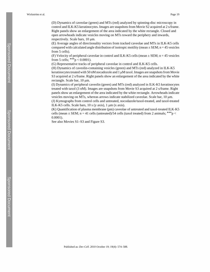
ILK Regulates MT Distribution and StabilityThe increased MT-based dynamics and concomitant decrease in actin-based motility ofcaveolae suggest that cytoskeletal defects might underlie the abnormal caveolar targeting inILK-K5 cells. Therefore, we examined the MT and F-actin networks in more detail. Inagreement with our previous report (Lorenz et al., 2007), ILK-K5 keratinocytes showed F-actin defects and reduced spreading (Figure 4A; Figure S1C). In addition, the MT network ofILK-K5 cells was abnormal, with compact and dense packs of MTs in the center andsignificantly decreased density in the periphery. Furthermore, peripheral MTs failed to reachthe cell cortex (Figure 4A). The altered distribution of MTs was not due to the decreasedspreading of ILK-K5 cells, as the same defect was observed on 20 μm-sized micropatterns(data not shown).
MT stabilization requires the interaction of MT plus-end-binding proteins with the cell cortex.In control cells, a large proportion of MTs stained positive for acetylated tubulin, aposttranslational modification associated with stabilized MTs. Strikingly, only low levels ofacetylated tubulin were detected in ILK-K5 cells (Figure 4B), which was confirmed by westernblotting (Figures 4C and 4D). Treatment of cells with taxol, which increases the levels ofacetylated tubulin by stabilizing MTs (Yvon et al., 1999), induced comparable levels ofacetylated tubulin in control and ILK-K5 cells (Figure 4E), indicating that tubulin acetylationitself can be induced in the absence of ILK, and that ILK regulates tubulin acetylation indirectlyby regulating MT stability. The reduction in MT stability of ILK-K5 cells was furtherconfirmed by treating cells with nocodazole, which revealed a reduced MT resistance in theabsence of ILK expression (Figure 4F). Live imaging of EGFP-tubulin-transfectedcells revealed that the peripheral MTs in control keratinocytes were extremely stable, whilethe majority of MTs in ILK-K5 cells were dynamic, characterized by increased catastrophefrequency and decreased duration of pauses between phases of growth and shortening(Figure 4G; Table S1A and Movie S4).
MT Dynamics Regulate Trafficking of CaveolaeAs we observed defects both in the actin and MT cytoskeletons, we sought to determine thepotential contribution of these defects to the altered caveolar trafficking in ILK-K5 cells. Theinternalization of caveolae requires an intact and dynamic actin cytoskeleton, as inhibition ofactin polymerization by latrunculin B (LatB) blocks this process (Pelkmans et al., 2002). Onthe other hand, cortical actin was shown to stabilize plasma membrane caveolae (Mundy et al.,2002). To determine whether the loss of stable plasma membrane caveolae was a result ofaltered actin polymerization, we treated EGFP-caveolin-1-transfected cells with LatB andanalyzed caveolar dynamics by spinning-disc and TIRF microscopy. LatB treatment increasedthe peripheral density of caveolae in control cells, but not in ILK-K5 cells (Figures 5A and5B). Furthermore, LatB treatment of ILK-K5 cells did not significantly stabilize peripheral
Wickström et al. Page 6
Published as: Dev Cell. 2010 October 19; 19(4): 574–588.
Sponsored Docum
ent Sponsored D
ocument
Sponsored Docum
ent
caveolae, or increase the motility of caveolae in control cells (Figures 5A–5C), suggesting thataberrant actin polymerization was not causing increased caveolar dynamics in ILK-K5 cells.
To address whether increased caveolar dynamics was due to the observed increased catastrophefrequency of MTs, we transfected control cells with the C-terminal fragment of the plus-end-binding protein EB1 (EB1-CΔAC), which lacks the MT-binding domain. EB1-CΔAC acts asa dominant negative EB by forming heterodimers with wild-type EB proteins, reducing theiraffinity to MTs, thereby inhibiting their ability to protect MTs from catastrophe (Komarovaet al., 2009). Expression of EB1-CΔAC led to increased MT dynamics (data not shown) andincreased caveolar trafficking along MTs in the periphery of control keratinocytes (Figure 5D;Movie S5) resembling the behavior of caveolin in ILK-K5 cells (Figure 3D). The effect ofEB1-CΔAC on caveolar motility was further confirmed by TIRF microscopy (Figures 5E and5F; Movie S6). Collectively, these results demonstrate that MT dynamics regulate caveolartraffic, and that increased catastrophe frequency of MTs leads to the loss of stable plasmamembrane caveolae.
ILK Binds and Regulates the Subcellular Distribution of IQGAP1To test whether ILK is part of an MT capture complex that binds cortical F-actin and MT tipsand links them together (Rodriguez et al., 2003), we screened for novel ILK interaction partnersby utilizing stable isotope labeling by amino acids in cell culture (SILAC; Ong et al., 2002),followed by ILK-FLAG immunoprecipitation and mass spectrometry. Among proteins withhigh light-to-heavy isotope ratios, indicative of specific binding, were known ILK-interactingproteins, such as PINCH or parvin, as well as poorly characterized ILK-binding partners, suchas IQGAP1 (Figure 6A), which has also been identified in a previous proteomic screen(Dobreva et al., 2008). IQGAP1 is an adaptor protein that localizes to the cell cortex, where itcan link the MT plus ends to cortical actin by binding actin and the MT tip protein CLIP 170(Fukata et al., 2002; Watanabe et al., 2004; Brown and Sacks, 2006). In addition, IQGAP1 canlocalize the activity of the formin mDia1, which can also stabilize MTs, to the cell cortex(Brandt et al., 2007).
Consistent with the proteomic data, IQGAP1 was detected by immunoprecipitation of ILK-FLAG (Figure 6B) or endogenous ILK (Figure 6C) from keratinocyte lysates. Conversely,antibodies against IQGAP1 coprecipitated ILK (Figure 6D), confirming the interaction of theendogenous proteins. In addition, mDia1 was also detected in IQGAP1 immunoprecipitates(Figure 6D). Next, we performed pulldown experiments with recombinant GST-taggedIQGAP1 in ILK-deficient fibroblasts re-expressing ILK-FLAG. These experiments confirmedthe specific association of ILK with IQGAP1, and showed that deletion of IQGAP1's WWdomain allowed ILK binding, while deletion of either its IQ domain or ILK's kinase domainabolished the interaction (Figure 6E). Moreover, immunostaining showed that IQGAP1 andILK colocalized at the keratinocyte cell cortex (Figure 6F). These findings demonstrate thatthe interaction between ILK and IQGAP1 is specific and mediated by the kinase domain ofILK and the IQ domain of IQGAP1.
Next, we tested whether ILK regulates IQGAP1 localization. IQGAP1 was found at the cortexof control keratinocytes, where it colocalized with cortical actin (Figure 6G) and MT tips(Figure 6H). In ILK-K5 cells, the MT tips failed to extend to the cell cortex (Figure 6H), andthe colocalization of IQGAP1 with cortical actin was almost completely lost (Figure 6G).Analysis of IQGAP1 localization on 20 μm-sized micropatterns confirmed that its alteredlocalization in ILK-K5 cells was not due to reduced cell spreading (Figure S4A). Furthermore,transient re-expression of ILK-FLAG in the ILK-K5 cells rescued both IQGAP1 localizationand the architecture of the MT network (Figures 6I and 6J). In addition, an IQGAP1 mutantlacking the IQ domain localized diffusely in the cytoplasm and did not colocalize with ILK,whereas a mutant lacking the C terminus, including the Ras-GAP, APC, CLIP 170, and mDia-
Wickström et al. Page 7
Published as: Dev Cell. 2010 October 19; 19(4): 574–588.
Sponsored Docum
ent Sponsored D
ocument
Sponsored Docum
ent
binding domains, localized extensively in ILK-positive adhesions (Figure 6K). These resultsindicate that ILK interacts with IQGAP1, and that both ILK expression and the IQ domain arerequired to localize IQGAP1 to the cell cortex.
IQGAP1 Acts in Concert with mDia1 to Regulate Caveolin-1 TraffickingThe results suggest that ILK regulates MT dynamics via IQGAP1 and possibly also mDia1.In agreement with previous reports (Watanabe et al., 2004), downregulation of IQGAP1 withsiRNA resulted in decreased stability and increased dynamics of MTs in otherwise normalkeratinocytes (Figures 7A–7C; Table S1B). The F-actin organization was also slightly alteredin IQGAP1-depleted cells, while the localization and cell surface expression of β1 integrin, aswell as the localization of ILK, were unaffected (Figures S4B and S4C and data not shown).These findings suggest that IQGAP1 acts downstream of ILK in regulating the MT network.
Next, we investigated the dynamics of caveolin-1 in IQGAP1-depleted cells. TIRF imaging ofEGFP-caveolin showed that caveolae were static in control cells and highly dynamic inIQGAP1-depleted cells (Figures 7D and 7E; Movie S7). Furthermore, downregulation ofIQGAP1 induced a relocalization of caveolin-1 from the periphery to the perinuclear region(Figure 7F). These findings resemble those of the abnormal behavior of caveolin-1 in ILK-K5cells (Figures 1 and 3).
IQGAP1 has been shown to localize mDia1 activity to the cell cortex to regulate local actinpolymerization (Brandt et al., 2007). Since mDia has also been shown to stabilize MTs (Palazzoet al., 2001), we analyzed the subcellular localization of mDia1 in ILK-K5 cells, and found itscortical localization (Figure S4D) and overall proteins levels (Figure S4E) to be reducedcompared with control cells. Moreover, RNAi-resistent EGFP-IQGAP1 constructs(Figure S4F) lacking either ILK-binding (IQ domain mutant) or mDia-binding (C-terminaltruncation; Brandt et al., 2007) activities failed to rescue IQGAP1 siRNA-induced defects inMT acetylation (Figure S4G).
The C terminus of IQGAP1 contains a Ras-GAP domain that has been shown to lack activityand thereby enhance Rac GTPase activity (Noritake et al., 2004; Weissbach et al., 1994). Thisregion of the protein also serves as the CLIP-170, APC, and mDia interacting domain(Watanabe et al., 2004). We have previously analyzed Rac activity in ILK-K5 cells, and didnot find it altered (Lorenz et al., 2007), pointing to mDia1 as the likely downstream target ofIQGAP1 in these cells. To analyze this, we depleted mDia1 in keratinocytes with siRNA(Figure 8A). As expected, an increase in MT dynamics was observed in mDia1-depleted cells(Figure 8B; Table S1B). In addition, an increase in caveolar dynamics close to the plasmamembrane was observed in these cells (Figures 8C and 8D). Furthermore, depletion of mDia1resulted in a perinuclear localization of caveolin 1 (Figure S4H). Importantly, as in IQGAP1-depleted cells, surface expression and localization of β1 integrin were unchanged by mDia1depletion (Figures S4B and S4C).
To further confirm that the increased caveolar dynamics in the ILK-K5 cells results fromimpaired MT-actin crosstalk downstream of the IQGAP1-mDia1 pathway, we overexpressedconstitutively active mDia1 transiently in ILK-K5 keratinocytes. Analysis of MT dynamicsrevealed increased stability of peripheral MTs in the presence of active mDia1 (Figure 8E;Table S1C). Expression of constitutively active mDia1 also rescued the altered mobility ofEGFP-caveolin-1 near the plasma membrane (Figures 8F and 8G; Movie S8), and resulted inthe redistribution of caveolin-1, with high levels of cortical staining and colocalization withcortical F-actin (Figure 8H). These data indicate that the IQGAP1-mDia1 pathway regulatesMT dynamics in keratinocytes, which is required for the trafficking of caveolin-1 to the plasmamembrane.
Wickström et al. Page 8
Published as: Dev Cell. 2010 October 19; 19(4): 574–588.
Sponsored Docum
ent Sponsored D
ocument
Sponsored Docum
ent
DiscussionIn the present study, we show that signaling through the β1 integrin/ILK complex is requiredfor plasma membrane caveolae formation in vivo and in vitro. In the absence of β1 integrin orILK expression in keratinocytes, caveolae are not stably inserted into the plasma membranedue to a profound defect in MT stability. Mechanistically, ILK is required for localizingIQGAP1 to the cell cortex, which then, together with mDia1, regulates local stability of MTs.Consequently, instability of MTs is increased in cells lacking ILK, IQGAP, or mDia1, whichresults in increased caveolar dynamics close to the plasma membrane.
The absence of cell surface caveolae and decreased plasma membrane caveolin-1 in our EMpreparations, together with increased long-range motility of caveolin-1, suggests that the β1integrin/ILK signaling complex promotes the shuttling of caveolae between the cytoplasm andthe plasma membrane. Since internalization of Ctx is not increased despite increasedbidirectional trafficking of caveolae, we conclude that ILK regulates their exocytosis ratherthan their endocytosis. There is a strong correlation between the expression level of caveolin-1and the presence of plasma membrane caveolae, indicating that exocytosis of caveolin-1dictates the formation of peripheral caveolae (Parton et al., 2006). Interestingly, caveolae aremaintained as stable structures throughout cycles of endo- and exocytosis, during which theyare internalized and shuttled through caveosomes and endosomes back to the plasma membrane(Pelkmans et al., 2004). Since we observed both increased trafficking of caveolae, as well asaccumulation of caveolin-1, in endosomal structures in our ILK-K5 keratinocytes, it remainsopen whether the main defect is in the initial exocytotic transport or in the recycling of caveolaefrom endosomes back to the plasma membrane. This question is very difficult to tackle inprimary keratinocytes, as their MT density is extremely high.
Our findings confirm previous studies showing that caveolar transport between the plasmamembrane and intracellular compartment occurs on MTs (Mundy et al., 2002). It is not clear,however, whether MTs are also essential for the initial exocytosis of caveolar carriers. Otherexocytotic carriers move on MT tracks when they traffic from the Golgi to the plasmamembrane (Toomre et al., 1999), or when they are transported to basolateral or apicalcompartments in polarized cells (Lafont et al., 1994). Interestingly, exocytosis of the Ctx-binding GM1 requires intact MTs and also integrin-mediated adhesion (Balasubramanian et al.,2007). In line with these findings, ILK-K5 cells display strongly reduced cell surface bindingof Ctx, pointing to decreased plasma membrane levels of GM1, which strongly corroboratesthe link between integrin signaling, MTs, and exocytosis. Some reports have also shown thatsecretion can occur in the absence of MTs (Rindler et al., 1987; Salas et al., 1986). However,nocadozole-mediated disruption of MTs induces dispersion of the Golgi into cytoplasmicministacks, and thus exocytotic carriers need to cover only short distances to the plasmamembrane, making random diffusion sufficient for efficient delivery (Bloom and Goldstein,1998). Therefore, it is very likely that all exocytotic carriers, including caveolae, utilize MTtracks for long-range transport when MTs are present in cells, and MT-based transport isrequired to ensure the fidelity of polarized transport. In line with this, caveolin-1 transport isnot completely abolished in ILK-K5 cells, as some caveolin-1 can still be detected on theplasma membrane.
The increase in bidirectional motility of caveolae near the plasma membrane is associated withloss of MT stability in ILK-deficient cells. A similar requirement of stable MTs for peripheraltargeting of caveolae was also observed in wild-type cells, in which MT stability was disruptedby expressing a dominant-negative EB1 mutant. Moreover, inhibition of MT catastrophe bytaxol allowed caveolae to be released from MTs also in the absence of ILK, identifying MTstability as an important determinant of caveolar trafficking. Local stabilization of MTs,regulated by spatial cues provided from adhesion-mediated signaling, has been shown to lead
Wickström et al. Page 9
Published as: Dev Cell. 2010 October 19; 19(4): 574–588.
Sponsored Docum
ent Sponsored D
ocument
Sponsored Docum
ent
to stabilizing ganglioside-containing plasma membrane rafts in fibroblasts (Palazzo et al.,2004). On the other hand, dynamic MTs have been shown to capture melanophores and initiatetheir minus-end-directed transport (Lomakin et al., 2009). Why caveolar vesicles are notreleased from the unstable MTs is not clear. Vesicle and organelle transport along MTs is drivenby two families of molecular motors, dyneins and kinesins, which mediate plus- and minus-ended transport of cargo along MTs. A single cargo can employ multiple motors, but themechanisms that regulate cargo loading and unloading at specific cellular locations are poorlyunderstood (Verhey and Hammond, 2009). Interestingly, secretory and endocytic cargo canuse a combination of MT- and actin-based transport systems (Ross et al., 2008). Outwardmovement of melanosomes, for example, has been shown to be regulated by crosstalk betweendynein and the actin motor myosin V, where engagement of myosin V terminates dynein-drivenminus-ended motility, enabling the transfer of the vesicles to actin-based motility andsubsequent dispersion (Gross et al., 2002). Thus, one could, on the one hand, envisage thatcaveolae use distinct motors for plus- and minus-ended transport, and that the activity of thesemotors is differentially regulated by the dynamic instability and/or posttranslationalmodification status of the MT. The local stabilization of peripheral MTs could thus allow therelease of the caveolar carrier, whereas dynamic MTs would capture the caveolar cargo andinitiate minus-ended transport. On the other hand, decreased interactions of unstable MTs withthe actin cortex, combined with the observed defects in actin organization of ILK-K5 cells,could affect the ability of caveolae to engage the myosin motor, initiate actin-based transport,and to suppress minus-ended MT transport.
It has been shown that MTs target adhesion complexes, and that repetitive targeting ofadhesions by MTs can lead to the disassembly of these structures (Kaverina et al., 1998).However, adhesions can also capture MTs and stabilize them against disassembly (Kaverinaet al., 1999; Krylyshkina et al., 2003). This stabilization is known to involve MT plus-end-binding proteins, which regulate the interaction between MTs and the cell cortex (Akhmanovaand Steinmetz, 2008). The precise mechanisms by which adhesions attract and stabilize MTshave not been elucidated. We now identify IQGAP1 as a binding partner for ILK that linksintegrins in FCs directly to the MT capture complex to stabilize the MT network. FA targetingof MT tips was reduced, but not totally absent (data not shown), so it is not clear whether thisfunction involves direct targeting of MTs to FAs as well. Our finding that ILK and IQGAP1colocalize only in FCs suggests that this is not the case.
In agreement with previous studies, we observed plasma membrane caveolae exclusively onthe basal and basolateral membranes of basal keratinocytes in vivo. The formation of a distinctlipid microenvironment at the basolateral membrane is one important mechanism to establishcell polarity. The role of actin and actin-associated proteins, as well as MTs, in the regulationof cell polarity is also well established (Siegrist and Doe, 2007; Wodarz, 2002). Thus, it seemsthat the β1 integrin/ILK complex functions at the interphase of these three polarity pathways,where it integrates extrinsic, ECM-derived, as well as intrinsic, MT-, actin-, and membrane-derived, polarity cues. This is consistent with the essential role of ILK in maintaining epithelialpolarity in vivo (Lorenz et al., 2007; Sakai et al., 2003). Whether this mechanism applies forall cell types, however, will await further investigation.
Experimental ProceduresMouse Strains
To obtain a keratinocyte-restricted deletion of the Ilk gene, mice expressing Cre under thecontrol of the keratin 5 promoter were crossed with floxed ILK mice (Lorenz et al., 2007).Offspring were genotyped as described previously (Grashoff et al., 2003).
Wickström et al. Page 10
Published as: Dev Cell. 2010 October 19; 19(4): 574–588.
Sponsored Docum
ent Sponsored D
ocument
Sponsored Docum
ent
Cell Culture and TransfectionPrimary keratinocytes were obtained and cultured as described previously (Lorenz et al.,2007). Transfections were carried out with Fugene 6 (Roche). The following expressionconstructs were used: EGFP-caveolin-1, mDia1 FH1FH2 (Böttcher et al., 2009), EGFP/Cherry-tubulin (from R.Y. Tsien), EB1-CΔAC (from A. Akhmanova), EGFP-IQGAP1, EGFP-ΔIQ (lacking amino acids 763–863), EGFP-ΔIQ (lacking amino acids 864–1657) (from D.B.Sacks), and RFP-Lifeact (Riedl et al., 2008). ILK-Cherry was cloned by inserting human ILKcDNA (from E. Van Obberghen-Schilling) into the pCherry-N1 vector (from R.Y. Tsien).
Biofunctionalized Micropatterned SubstratesMicropatterns were generated on PEG-coated coverslips with deep UV lithography (adaptedfrom Azioune et al., 2009). For details, see Supplemental Experimental Procedures.
ImmunofluorescenceImmunohistochemical staining from skin cryosections was carried out as described previously(Brakebusch et al., 2000). For cellular immunostaining, primary keratinocytes were fixed in3% formaldehyde or in 100% methanol, followed by incubation with primary and secondaryantibodies (see Supplemental Experimental Procedures). The fluorescent images werecollected by laser scanning confocal microscopy (DMIRE2; Leica) with Leica ConfocalSoftware version 2.5, Build 1227, with 63× or 100× oil objectives.
Drug TreatmentsFor nocodazole resistance assays, cells were treated with 1 μM nocodazole for 20–60 min,fixed, and stained with antibodies against α tubulin. Quantifications represent threeindependent experiments, where 100 cells per time point were analyzed for MT content.
For inhibition of MT catastrophe, cells were treated with 3 nM taxol for 45 min prior to EMor live cell imaging analyses. For inhibition of MT growth and catastrophe, cells were treatedwith a combination of 50 nM nocodazole and 1 μM taxol 45 min prior to live cell imaging. Forinhibition of actin polymerization, cells were treated with 0.2 μM LatB 30 min prior to livecell imaging.
EMSkin and primary keratinocytes were processed for resin section EM and cryosection immuno-EM, essentially as described previously (Hess et al., 2010). Basal caveolae were quantified onresin sections cut perpendicularly to the BM or culturing plane. The relative distribution ofcaveolin-1 immunogold label was estimated by comparing gold label within a distance of <100 nm to the plasma membrane (classified as “peripheral”) with the residual gold (consideredas “intracellular”). Pre-embedding immuno-EM of primary keratinocytes was performedaccording to standard protocols (for details, see Supplemental Experimental Procedures).
Ctx InternalizationCells were allowed to bind fluorescently labeled Ctx (20 μg/ml; Invitrogen) for 5 or 15 min at+37°C, fixed, and analyzed by confocal microscopy. Alternatively, cells were allowed to bindCtx for 5 or 15 min at +37°C, after which they were washed three times with 0.5 M HOAc and0.5 M NaCl (pH 4.1) to remove plasma membrane-associated Ctx, and analyzed by flowcytometry (FACSCalibur; Becton Dickinson). For total binding of Ctx, cells were incubatedwith Ctx for 15 min at +4°C, washed, and analyzed by flow cytometry. The flow cytometrydata were analyzed with FlowJo software (Tree Star). For the analyses of GM1 expression,cell extracts were plotted on nitrocellulose, which was then incubated with horseradishperoxidase-coupled Ctx (Jackson ImmunoResearch) and analyzed by chemiluminescence.
Wickström et al. Page 11
Published as: Dev Cell. 2010 October 19; 19(4): 574–588.
Sponsored Docum
ent Sponsored D
ocument
Sponsored Docum
ent
Immunoprecipitation and Western BlottingFLAG immunoprecipitation was carried out according to the manufacturers' protocol (Sigma).For immunoprecipitation of IQGAP1 or ILK, cells were lysed in 50 mM Tris-HCl (pH 8.0),150 mM NaCl, 1% Triton X-100, 0.05% sodium deoxycholate, and 10 mM EDTA. Lysateswere incubated with IQGAP1 (Santa Cruz Biotechnology) or ILK (BD Biosciences) antibodiesfor 30 min on ice. Isotype-matched IgG was used as a negative control. Lysates weresubsequently incubated with protein G Sepharose for 1 hr and washed repeatedly with lysisbuffer. Proteins were eluted from the beads with Laemmli buffer and analyzed by westernblotting. Quantification of band intensities was performed with MultiGauge software(Fujifilm).
TIRF MicroscopyTIRF images were captured with an Axiovert 200M inverted microscope (Zeiss) with a 100×oil objective and a CoolSnap HQ CCD camera (Photometrics). Acquisition was controlled byMetamorph Software (Molecular Devices). Images were collected at 37°C and processed byGaussian low-pass or median filtering and linear contrast enhancement. Motile caveolae werequantified by subtracting consecutive frames from movies.
Spinning-Disc MicroscopyImages were captured with an Axiovert 200 (Zeiss) microscope, a CSU10 spinning-discconfocal scanhead (Yokogawa), and a Cascade II EMCCD camera (Photometrics), with a 100×oil objective. Acquisition was controlled by Metamorph. Images were collected at 37°C, 5%CO2, and processed as described above.
Quantification of MT dynamics was performed with Metamorph by creating kymographs fromindividual MTs. For quantification of vesicle dynamics, the positions of individual vesicleswere tracked with the Track points application of Metamorph. For each vesicle, the positionof the nearest MT was simultaneously tracked. The difference angle between the motility vectorof a vesicle and the directionality vector of its closest MT was then calculated (403 angles intotal). Angle distribution for random isotropic motility was assumed from a uniformdistribution of angles between 0° and 90°. An average of 45° ± 12.99 (SD) can be obtainedfrom this assumption.
SILACSILAC labeling, followed by mass spectrometry, was performed essentially as describedpreviously (Mann, 2006). Briefly, ILK−/− fibroblasts (Sakai et al., 2003) expressing ILK-FLAGwere cultured in medium containing heavy lysine isotope for five cell doublings. ILK f/f cellscultured in unlabeled medium were used as a negative control. The cells were subsequentlylysed, pooled, and subjected to FLAG immunoprecipitation. The immunoprecipitates wereseparated by SDS-PAGE, followed by in-gel trypsin digestion and reversed-phase liquidchromatography (Proxeon Biosystems) coupled online to an LTQ-Orbitrap mass spectrometer(Thermo Fisher Scientific). Data were acquired with Xcalibur software and analyzed withMaxQuant software.
GST PulldownsThe cDNA constructs encoding for GST-IQGAP, ΔIQ, and ΔWW were a gift from Dr. D. B.Sacks. Recombinant proteins were expressed and purified from Escherichia coli. Recombinantprotein (5 μg) was incubated with 500 μg cell lysate from ILK-FLAG or ANK-FLAG, afterwhich proteins were precipitated with glutathione beads (Novagen) and analyzed by westernblotting.
Wickström et al. Page 12
Published as: Dev Cell. 2010 October 19; 19(4): 574–588.
Sponsored Docum
ent Sponsored D
ocument
Sponsored Docum
ent
siRNAsiRNA duplexes for mDia1, IQGAP1, and scrambled control were from Sigma (for sequences,see Supplemental Experimental Procedures). siRNAs were transfected with Lipofectamine(Invitrogen), and experiments were carried out 48 hr after transfection.
Statistical AnalysisStatistical evaluation was performed with GraphPad Prism software (version 5.0; GraphPad).Statistical significance was determined by the Mann-Whitney U test, except for Figure 3B andFigure 5B, where analysis of variance in combination with a post hoc Tuckey test was used,and Figure 2H, where a paired t test was used. Normal distribution was confirmed with theKolomogorov-Smirnov test (alpha = 0.05).
ReferencesAkhmanovaA.SteinmetzM.O.Tracking the ends: a dynamic protein network controls the fate of
microtubule tipsNat. Rev. Mol. Cell Biol.9200830932218322465AziouneA.StorchM.BornensM.TheryM.PielM.Simple and rapid process for single cell micro-
patterningLab Chip920091640164219458875BalasubramanianN.ScottD.W.CastleJ.D.CasanovaJ.E.SchwartzM.A.Arf6 and microtubules in adhesion-
dependent trafficking of lipid raftsNat. Cell Biol.920071381139118026091BloomG.S.GoldsteinL.S.Cruising along microtubule highways: how membranes move through the
secretory pathwayJ. Cell Biol.1401998127712809508761BlummelJ.PerschmannN.AydinD.DrinjakovicJ.SurreyT.Lopez-GarciaM.KesslerH.SpatzJ.P.Protein
repellent properties of covalently attached PEG coatings on nanostructured SiO(2)-basedinterfacesBiomaterials2820074739474717697710
BöttcherR.T.WiesnerS.BraunA.WimmerR.BernaA.EladN.MedaliaO.PfeiferA.AszodiA.CostellM.FässlerR.Profilin 1 is required for abscission during late cytokinesis of chondrocytesEMBO J.2820091157116919262563
BrakebuschC.GroseR.QuondamatteoF.RamirezA.JorcanoJ.L.PirroA.SvenssonM.HerkenR.SasakiT.TimplR.Skin and hair follicle integrity is crucially dependent on beta 1 integrin expression onkeratinocytesEMBO J.1920003990400310921880
BrandtD.T.MarionS.GriffithsG.WatanabeT.KaibuchiK.GrosseR.Dia1 and IQGAP1 interact in cellmigration and phagocytic cup formationJ. Cell Biol.178200719320017620407
BrownM.D.SacksD.B.IQGAP1 in cellular signaling: bridging the GAPTrends Cell Biol.16200624224916595175
BuckK.B.ZhengJ.Q.Growth cone turning induced by direct local modification of microtubule dynamicsJ.Neurosci.2220029358936712417661
del PozoM.A.BalasubramanianN.AldersonN.B.KiossesW.B.Grande-GarciaA.AndersonR.G.SchwartzM.A.Phospho-caveolin-1 mediates integrin-regulated membranedomain internalizationNat. Cell Biol.7200590190816113676
del PozoM.A.AldersonN.B.KiossesW.B.ChiangH.AndersonR.G.W.SchwartzM.A.Integrins regulateRac targeting by internalization of membrane domainsScience303200483984214764880
DobrevaI.FieldingA.FosterL.J.DedharS.Mapping the integrin-linked kinase interactome using SILACJ.Proteome Res.720081740174918327965
DrabM.VerkadeP.ElgerM.KasperM.LohnM.LauterbachB.MenneJ.LindschauC.MendeF.LuftF.C.Lossof caveolae, vascular dysfunction, and pulmonary defects in caveolin-1 gene-disruptedmiceScience29320012449245211498544
FukataM.WatanabeT.NoritakeJ.NakagawaM.YamagaM.KurodaS.MatsuuraY.IwamatsuA.PerezF.KaibuchiK.Rac1 and Cdc42 capture microtubules through IQGAP1 andCLIP-170Cell109200287388512110184
GrashoffC.AszodiA.SakaiT.HunzikerE.B.FässlerR.Integrin-linked kinase regulates chondrocyte shapeand proliferationEMBO Rep.4200343243812671688
Wickström et al. Page 13
Published as: Dev Cell. 2010 October 19; 19(4): 574–588.
Sponsored Docum
ent Sponsored D
ocument
Sponsored Docum
ent
GrossS.P.TumaM.C.DeaconS.W.SerpinskayaA.S.ReileinA.R.GelfandV.I.Interactions and regulation ofmolecular motors in Xenopus melanophoresJ. Cell Biol.156200285586511864991
HessM.W.PfallerK.EbnerH.L.BeerB.HeklD.SeppiT.3D versus 2D cell culture: implications for electronmicroscopyMethods Cell Biol.96201064967020869542
KaverinaI.RottnerK.SmallJ.V.Targeting, capture, and stabilization of microtubules at early focaladhesionsJ. Cell Biol.14219981811909660872
KaverinaI.KrylyshkinaO.SmallJ.V.Microtubule targeting of substrate contacts promotes their relaxationand dissociationJ. Cell Biol.14619991033104410477757
KomarovaY.DeGrootC.O.GrigorievI.GouveiaS.M.MunteanuE.L.SchoberJ.M.HonnappaS.BueyR.M.HoogenraadC.C.DogteromM.Mammalian end binding proteins control persistent microtubule growthJ. Cell Biol.184200969170619255245
KrylyshkinaO.AndersonK.I.KaverinaI.UpmannI.MansteinD.J.SmallJ.V.ToomreD.K.Nanometertargeting of microtubules to focal adhesionsJ. Cell Biol.161200385385912782685
LafontF.BurkhardtJ.K.SimonsK.Involvement of microtubule motors in basolateral and apical transportin kidney cellsNature37219948018037997271
LegateK.R.WickströmS.A.FässlerR.Genetic and cell biological analysis of integrin outside-insignalingGenes Dev.23200939741819240129
Lippincott-SchwartzJ.Cytoskeletal proteins and Golgi dynamicsCurr. Opin. Cell Biol.10199852599484595
LomakinA.J.SemenovaI.ZaliapinI.KraikivskiP.NadezhdinaE.SlepchenkoB.M.AkhmanovaA.RodionovV.CLIP-170-dependent capture of membrane organelles by microtubules initiates minus-enddirected transportDev. Cell17200932333319758557
LorenzK.GrashoffC.TorkaR.SakaiT.LangbeinL.BlochW.AumailleyM.FässlerR.Integrin-linked kinaseis required for epidermal and hair follicle morphogenesisJ. Cell Biol.177200750151317485490
MannM.Functional and quantitative proteomics using SILACNat. Rev. Mol. Cell Biol.7200695295817139335
MundyD.I.MachleidtT.YingY.S.AndersonR.G.BloomG.S.Dual control of caveolar membrane traffic bymicrotubules and the actin cytoskeletonJ. Cell Sci.11520024327433912376564
NakriekoK.A.WelchI.DupuisH.BryceD.PajakA.St ArnaudR.DedharS.D'SouzaS.J.DagninoL.Impairedhair follicle morphogenesis and polarized keratinocyte movement upon conditional inactivation ofintegrin-linked kinase in the epidermisMol. Biol. Cell1920081462147318234842
NoritakeJ.FukataM.SatoK.NakagawaM.WatanabeT.IzumiN.WangS.FukataY.KaibuchiK.Positive roleof IQGAP1, an effector of Rac1, in actin-meshwork formation at sites of cell-cell contactMol. Biol.Cell1520041065107614699063
OngS.E.BlagoevB.KratchmarovaI.KristensenD.B.SteenH.PandeyA.MannM.Stable isotope labeling byamino acids in cell culture, SILAC, as a simple and accurate approach to expression proteomicsMol.Cell. Proteomics1200237638612118079
PalazzoA.F.CookT.A.AlbertsA.S.GundersenG.G.mDia mediates Rho-regulated formation andorientation of stable microtubulesNat. Cell Biol.3200172372911483957
PalazzoA.F.EngC.H.SchlaepferD.D.MarcantonioE.E.GundersenG.G.Localized stabilization ofmicrotubules by integrin- and FAK-facilitated Rho signalingScience303200483683914764879
PartonR.G.Hanzal-BayerM.HancockJ.F.Biogenesis of caveolae: a structural model for caveolin-induceddomain formationJ. Cell Sci.119200678779616495479
PartonR.G.SimonsK.The multiple faces of caveolaeNat. Rev. Mol. Cell Biol.8200718519417318224PartonR.G.JoggerstB.SimonsK.Regulated internalization of caveolaeJ. Cell Biol.
1271994119912157962085PelkmansL.ZerialM.Kinase-regulated quantal assemblies and kiss-and-run recycling of
caveolaeNature436200512813316001074PelkmansL.PuntenerD.HeleniusA.Local actin polymerization and dynamin recruitment in SV40-induced
internalization of caveolaeScience296200253553911964480PelkmansL.BurliT.ZerialM.HeleniusA.Caveolin-stabilized membrane domains as multifunctional
transport and sorting devices in endocytic membrane trafficCell118200476778015369675
Wickström et al. Page 14
Published as: Dev Cell. 2010 October 19; 19(4): 574–588.
Sponsored Docum
ent Sponsored D
ocument
Sponsored Docum
ent
PolA.MartinS.FernandezM.A.Ingelmo-TorresM.FergusonC.EnrichC.PartonR.G.Cholesterol and fattyacids regulate dynamic caveolin trafficking through the Golgi complex and between the cell surfaceand lipid bodiesMol. Biol. Cell1620052091210515689493
RiedlJ.CrevennaA.H.KessenbrockK.YuJ.H.NeukirchenD.BistaM.BradkeF.JenneD.HolakT.A.WerbZ.Lifeact: a versatile marker to visualize F-actinNat. Methods5200860560718536722
RindlerM.J.IvanovI.E.SabatiniD.D.Microtubule-acting drugs lead to the nonpolarized delivery of theinfluenza hemagglutinin to the cell surface of polarized Madin-Darby canine kidney cellsJ. Cell Biol.10419872312412879845
RodriguezO.C.SchaeferA.W.MandatoC.A.ForscherP.BementW.M.Waterman-StorerC.M.Conservedmicrotubule-actin interactions in cell movement and morphogenesisNat. Cell Biol.5200359960912833063
RossJ.L.AliM.Y.WarshawD.M.Cargo transport: molecular motors navigate a complex cytoskeletonCurr.Opin. Cell Biol.202008414718226515
SakaiT.LiS.DochevaD.GrashoffC.SakaiK.KostkaG.BraunA.PfeiferA.YurchencoP.D.FässlerR.Integrin-linked kinase (ILK) is required for polarizing the epiblast, cell adhesion, and controlling actinaccumulationGenes Dev.17200392694012670870
SalasP.J.MisekD.E.Vega-SalasD.E.GundersenD.CereijidoM.Rodriguez-BoulanE.Microtubules andactin filaments are not critically involved in the biogenesis of epithelial cell surface polarityJ. CellBiol.1021986185318672871031
SiegristS.E.DoeC.Q.Microtubule-induced cortical cell polarityGenes Dev.21200748349617344411TagawaA.MezzacasaA.HayerA.LongattiA.PelkmansL.HeleniusA.Assembly and trafficking of caveolar
domains in the cell: caveolae as stable, cargo-triggered, vesicular transportersJ. Cell Biol.170200576977916129785
ToomreD.KellerP.WhiteJ.OlivoJ.C.SimonsK.Dual-color visualization of trans-Golgi network to plasmamembrane traffic along microtubules in living cellsJ. Cell Sci.112199921339841901
VerheyK.J.HammondJ.W.Traffic control: regulation of kinesin motorsNat. Rev. Mol. Cell Biol.10200976577719851335
WatanabeT.WangS.NoritakeJ.SatoK.FukataM.TakefujiM.NakagawaM.IzumiN.AkiyamaT.KaibuchiK.Interaction with IQGAP1 links APC to Rac1, Cdc42, and actin filaments during cell polarization andmigrationDev. Cell7200487188315572129
WeissbachL.SettlemanJ.KaladyM.F.SnijdersA.J.MurthyA.E.YanY.X.BernardsA.Identification of ahuman rasGAP-related protein containing calmodulin-binding motifsJ. Biol. Chem.269199420517205218051149
WickströmS.A.LangeA.MontanezE.FässlerR.The ILK/PINCH/parvin complex: the kinase is dead, longlive the pseudokinase!EMBO J.29201028129120033063
WodarzA.Establishing cell polarity in developmentNat. Cell Biol.42002E39E4411835058YvonA.M.WadsworthP.JordanM.A.Taxol suppresses dynamics of individual microtubules in living
human tumor cellsMol. Biol. Cell10199994795910198049
Supplemental InformationRefer to Web version on PubMed Central for supplementary material.
AcknowledgmentsWe thank M. Grzejszczyk, K. Gutleben, A. Flörl, and B. Witting for excellent technical assistance; Dr. A. Akhmanova(Erasmus Medical Center, the Netherlands) for constructs and helpful discussions; Drs. D.B. Sacks (Brigham andWomen's Hospital), R.Y. Tsien (University of California), E. Van Obberghen-Schilling (University of Nice-SophiaAntiopolis, France), and M. Aumailley (University of Cologne, Germany) for providing reagents; M. Selbach for theinitial SILAC experiment; and A. Elischer for the EGFP-caveolin construct. This work was supported by the SigridJuselius Foundation, the Finnish Cultural Foundation, and the Academy of Finland (to S.A.W.), the Marie CurieFellowship for Career Development within the 7th European Community Framework Programme (to J.P.), theAustrian Science Funds (grants P19486-B12 to M.W.H. and SFB021 to R.F. and L.A.H.), and the Max Planck Society(to J.P.S. and R.F.).
Wickström et al. Page 15
Published as: Dev Cell. 2010 October 19; 19(4): 574–588.
Sponsored Docum
ent Sponsored D
ocument
Sponsored Docum
ent
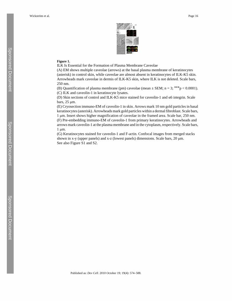
Figure 1.ILK Is Essential for the Formation of Plasma Membrane Caveolae(A) EM shows multiple caveolae (arrows) at the basal plasma membrane of keratinocytes(asterisk) in control skin, while caveolae are almost absent in keratinocytes of ILK-K5 skin.Arrowheads mark caveolae in dermis of ILK-K5 skin, where ILK is not deleted. Scale bars,250 nm.(B) Quantification of plasma membrane (pm) caveolae (mean ± SEM; n = 3; ∗∗∗p < 0.0001).(C) ILK and caveolin-1 in keratinocyte lysates.(D) Skin sections of control and ILK-K5 mice stained for caveolin-1 and α6 integrin. Scalebars, 25 μm.(E) Cryosection immuno-EM of caveolin-1 in skin. Arrows mark 10 nm gold particles in basalkeratinocytes (asterisk). Arrowheads mark gold particles within a dermal fibroblast. Scale bars,1 μm. Insert shows higher magnification of caveolae in the framed area. Scale bar, 250 nm.(F) Pre-embedding immuno-EM of caveolin-1 from primary keratinocytes. Arrowheads andarrows mark caveolin-1 at the plasma membrane and in the cytoplasm, respectively. Scale bars,1 μm.(G) Keratinocytes stained for caveolin-1 and F-actin. Confocal images from merged stacksshown in x-y (upper panels) and x-z (lowest panels) dimensions. Scale bars, 20 μm.See also Figure S1 and S2.
Wickström et al. Page 16
Published as: Dev Cell. 2010 October 19; 19(4): 574–588.
Sponsored Docum
ent Sponsored D
ocument
Sponsored Docum
ent
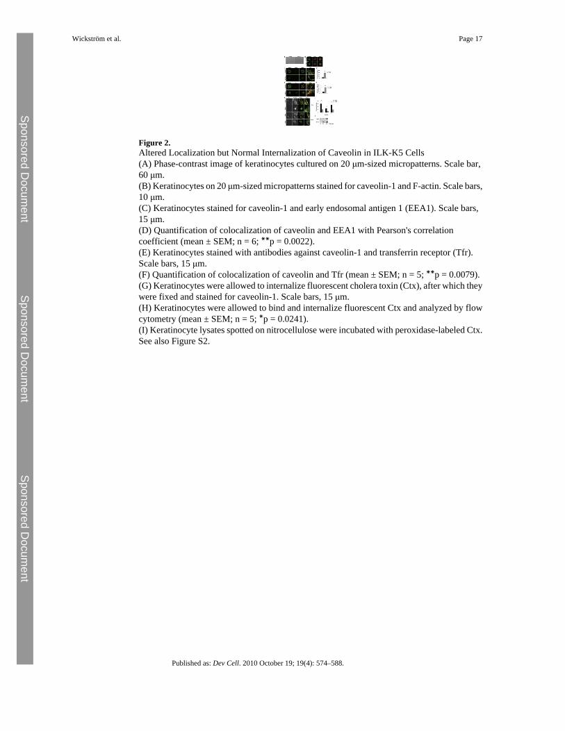
Figure 2.Altered Localization but Normal Internalization of Caveolin in ILK-K5 Cells(A) Phase-contrast image of keratinocytes cultured on 20 μm-sized micropatterns. Scale bar,60 μm.(B) Keratinocytes on 20 μm-sized micropatterns stained for caveolin-1 and F-actin. Scale bars,10 μm.(C) Keratinocytes stained for caveolin-1 and early endosomal antigen 1 (EEA1). Scale bars,15 μm.(D) Quantification of colocalization of caveolin and EEA1 with Pearson's correlationcoefficient (mean ± SEM; n = 6; ∗∗p = 0.0022).(E) Keratinocytes stained with antibodies against caveolin-1 and transferrin receptor (Tfr).Scale bars, 15 μm.(F) Quantification of colocalization of caveolin and Tfr (mean ± SEM; n = 5; ∗∗p = 0.0079).(G) Keratinocytes were allowed to internalize fluorescent cholera toxin (Ctx), after which theywere fixed and stained for caveolin-1. Scale bars, 15 μm.(H) Keratinocytes were allowed to bind and internalize fluorescent Ctx and analyzed by flowcytometry (mean ± SEM; n = 5; ∗p = 0.0241).(I) Keratinocyte lysates spotted on nitrocellulose were incubated with peroxidase-labeled Ctx.See also Figure S2.
Wickström et al. Page 17
Published as: Dev Cell. 2010 October 19; 19(4): 574–588.
Sponsored Docum
ent Sponsored D
ocument
Sponsored Docum
ent
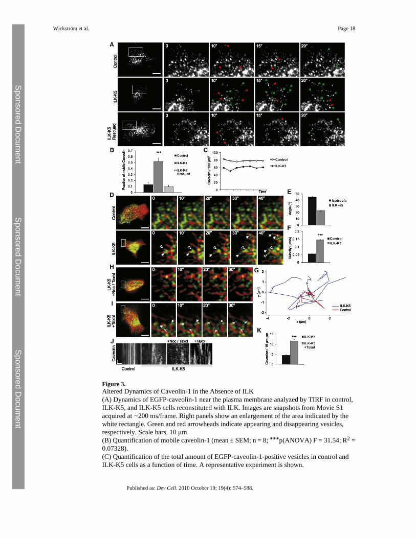
Figure 3.Altered Dynamics of Caveolin-1 in the Absence of ILK(A) Dynamics of EGFP-caveolin-1 near the plasma membrane analyzed by TIRF in control,ILK-K5, and ILK-K5 cells reconstituted with ILK. Images are snapshots from Movie S1acquired at ∼200 ms/frame. Right panels show an enlargement of the area indicated by thewhite rectangle. Green and red arrowheads indicate appearing and disappearing vesicles,respectively. Scale bars, 10 μm.(B) Quantification of mobile caveolin-1 (mean ± SEM; n = 8; ∗∗∗p(ANOVA) F = 31.54; R2 =0.07328).(C) Quantification of the total amount of EGFP-caveolin-1-positive vesicles in control andILK-K5 cells as a function of time. A representative experiment is shown.
Wickström et al. Page 18
Published as: Dev Cell. 2010 October 19; 19(4): 574–588.
Sponsored Docum
ent Sponsored D
ocument
Sponsored Docum
ent
(D) Dynamics of caveolae (green) and MTs (red) analyzed by spinning-disc microscopy incontrol and ILK-K5 keratinocytes. Images are snapshots from Movie S2 acquired at 2 s/frame.Right panels show an enlargement of the area indicated by the white rectangle. Closed andopen arrowheads indicate vesicles moving on MTs toward the periphery and inwards,respectively. Scale bars, 10 μm.(E) Average angles of directionality vectors from tracked caveolae and MTs in ILK-K5 cellscompared with calculated angle distribution of isotropic motility (mean ± SEM; n = 45 vesiclesfrom 5 cells).(F) Velocity of peripheral caveolae in control and ILK-K5 cells (mean ± SEM; n = 45 vesiclesfrom 5 cells; ∗∗∗p < 0.0001).(G) Representative tracks of peripheral caveolae in control and ILK-K5 cells.(H) Dynamics of caveolin-containing vesicles (green) and MTs (red) analyzed in ILK-K5keratinocytes treated with 50 nM nocadozole and 1 μM taxol. Images are snapshots from MovieS3 acquired at 2 s/frame. Right panels show an enlargement of the area indicated by the whiterectangle. Scale bar, 10 μm.(I) Dynamics of peripheral caveolin (green) and MTs (red) analyzed in ILK-K5 keratinocytestreated with taxol (3 nM). Images are snapshots from Movie S3 acquired at 2 s/frame. Rightpanels show an enlargement of the area indicated by the white rectangle. Arrowheads indicatevesicles moving on MTs, whereas arrows indicate stabilized caveolae. Scale bar, 10 μm.(J) Kymographs from control cells and untreated, nocodazole/taxol-treated, and taxol-treatedILK-K5 cells. Scale bars, 10 s (y axis), 1 μm (x axis).(K) Quantification of plasma membrane (pm) caveolae of untreated and taxol-treated ILK-K5cells (mean ± SEM; n = 41 cells (untreated)/54 cells (taxol treated) from 2 animals; ∗∗∗p <0.0001).See also Movies S1–S3 and Figure S3.
Wickström et al. Page 19
Published as: Dev Cell. 2010 October 19; 19(4): 574–588.
Sponsored Docum
ent Sponsored D
ocument
Sponsored Docum
ent
Figure 4.ILK Regulates Organization and Stability of the MT Network(A) Control and ILK-K5 keratinocytes stained for tubulin and F-actin. Note disorganized MTnetwork and lack of MT tip association with cortical actin (arrow) in ILK-K5 cells. Scale bars,10 μm.(B) Control and ILK-K5 keratinocytes stained for acetylated tubulin and α tubulin. Scale bars,50 μm.(C) Reduced tubulin acetylation confirmed by western blotting.(D) Quantification of acetylated tubulin levels (average ± SD; n = 3).(E) Control and ILK-K5 keratinocytes treated with taxol and analyzed for acetylated tubulin.(F) Control and ILK-K5 were treated with 1 μM nocodazole for time points indicated, afterwhich cells containing more than 10 MTs were quantified (mean ± SEM; n = 3).(G) Dynamics of MTs analyzed by spinning-disc microscopy in control and ILK-K5keratinocytes expressing EGFP-tubulin. Images are snapshots from Movie S4 acquired at 2 s/frame. Right panels show an enlargement of the area indicated by the white rectangle.Arrowheads indicate stable MTs. Scale bars, 10 μm.See also Movie S4 and Table S1.
Wickström et al. Page 20
Published as: Dev Cell. 2010 October 19; 19(4): 574–588.
Sponsored Docum
ent Sponsored D
ocument
Sponsored Docum
ent
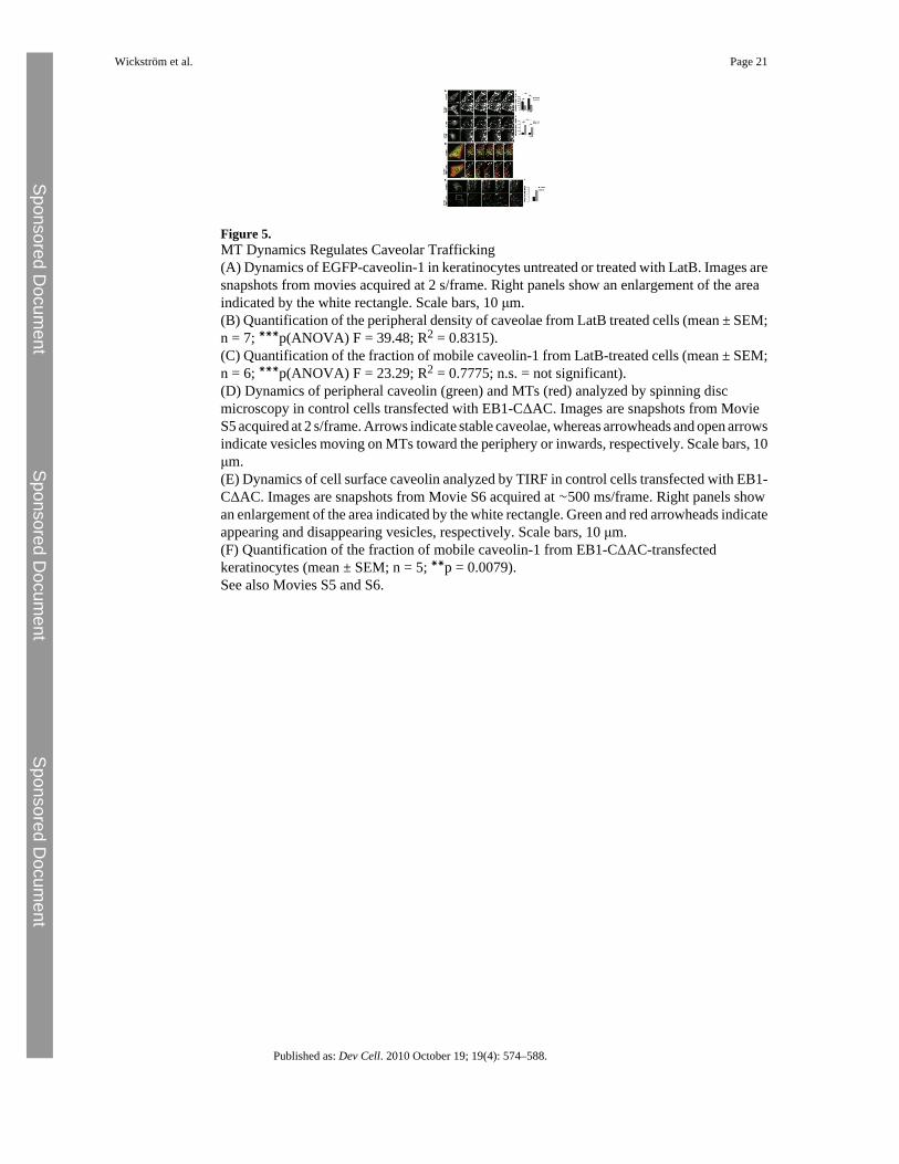
Figure 5.MT Dynamics Regulates Caveolar Trafficking(A) Dynamics of EGFP-caveolin-1 in keratinocytes untreated or treated with LatB. Images aresnapshots from movies acquired at 2 s/frame. Right panels show an enlargement of the areaindicated by the white rectangle. Scale bars, 10 μm.(B) Quantification of the peripheral density of caveolae from LatB treated cells (mean ± SEM;n = 7; ∗∗∗p(ANOVA) F = 39.48; R2 = 0.8315).(C) Quantification of the fraction of mobile caveolin-1 from LatB-treated cells (mean ± SEM;n = 6; ∗∗∗p(ANOVA) F = 23.29; R2 = 0.7775; n.s. = not significant).(D) Dynamics of peripheral caveolin (green) and MTs (red) analyzed by spinning discmicroscopy in control cells transfected with EB1-CΔAC. Images are snapshots from MovieS5 acquired at 2 s/frame. Arrows indicate stable caveolae, whereas arrowheads and open arrowsindicate vesicles moving on MTs toward the periphery or inwards, respectively. Scale bars, 10μm.(E) Dynamics of cell surface caveolin analyzed by TIRF in control cells transfected with EB1-CΔAC. Images are snapshots from Movie S6 acquired at ∼500 ms/frame. Right panels showan enlargement of the area indicated by the white rectangle. Green and red arrowheads indicateappearing and disappearing vesicles, respectively. Scale bars, 10 μm.(F) Quantification of the fraction of mobile caveolin-1 from EB1-CΔAC-transfectedkeratinocytes (mean ± SEM; n = 5; ∗∗p = 0.0079).See also Movies S5 and S6.
Wickström et al. Page 21
Published as: Dev Cell. 2010 October 19; 19(4): 574–588.
Sponsored Docum
ent Sponsored D
ocument
Sponsored Docum
ent
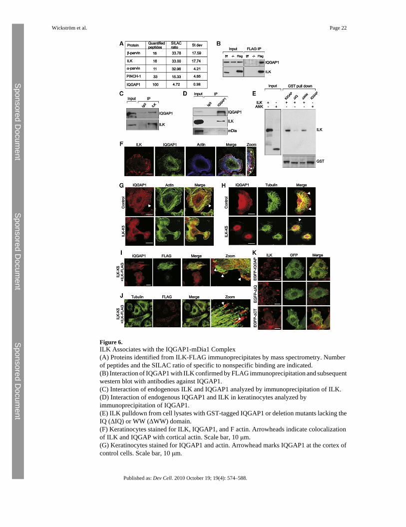
Figure 6.ILK Associates with the IQGAP1-mDia1 Complex(A) Proteins identified from ILK-FLAG immunoprecipitates by mass spectrometry. Numberof peptides and the SILAC ratio of specific to nonspecific binding are indicated.(B) Interaction of IQGAP1 with ILK confirmed by FLAG immunoprecipitation and subsequentwestern blot with antibodies against IQGAP1.(C) Interaction of endogenous ILK and IQGAP1 analyzed by immunoprecipitation of ILK.(D) Interaction of endogenous IQGAP1 and ILK in keratinocytes analyzed byimmunoprecipitation of IQGAP1.(E) ILK pulldown from cell lysates with GST-tagged IQGAP1 or deletion mutants lacking theIQ (ΔIQ) or WW (ΔWW) domain.(F) Keratinocytes stained for ILK, IQGAP1, and F actin. Arrowheads indicate colocalizationof ILK and IQGAP with cortical actin. Scale bar, 10 μm.(G) Keratinocytes stained for IQGAP1 and actin. Arrowhead marks IQGAP1 at the cortex ofcontrol cells. Scale bar, 10 μm.
Wickström et al. Page 22
Published as: Dev Cell. 2010 October 19; 19(4): 574–588.
Sponsored Docum
ent Sponsored D
ocument
Sponsored Docum
ent
(H) Keratinocytes stained for IQGAP1 and tubulin. MT tips align with cortical IQGAP1 incontrol cells (arrowheads). Scale bars, 10 μm.(I) ILK-K5 keratinocytes transiently transfected with ILK-FLAG and stained for IQGAP1 andFLAG. Arrowheads mark cortical localization of IQGAP1. Scale bar, 10 μm.(J) ILK-K5 keratinocytes transiently transfected with ILK-FLAG and stained for tubulin andFLAG. Arrowheads mark dense peripheral MTs. Scale bar, 10 μm.(K) Localization of EGFP-tagged full-length IQGAP1 or deletion mutants lacking either theIQ domain (ΔIQ) or the C terminus (ΔCT). Scale bars, 10 μm.See also Figure S4.
Wickström et al. Page 23
Published as: Dev Cell. 2010 October 19; 19(4): 574–588.
Sponsored Docum
ent Sponsored D
ocument
Sponsored Docum
ent
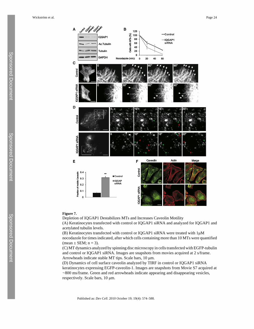
Figure 7.Depletion of IQGAP1 Destabilizes MTs and Increases Caveolin Motility(A) Keratinocytes transfected with control or IQGAP1 siRNA and analyzed for IQGAP1 andacetylated tubulin levels.(B) Keratinocytes transfected with control or IQGAP1 siRNA were treated with 1μMnocodazole for times indicated, after which cells containing more than 10 MTs were quantified(mean ± SEM; n = 3).(C) MT dynamics analyzed by spinning disc microscopy in cells transfected with EGFP-tubulinand control or IQGAP1 siRNA. Images are snapshots from movies acquired at 2 s/frame.Arrowheads indicate stable MT tips. Scale bars, 10 μm.(D) Dynamics of cell surface caveolin analyzed by TIRF in control or IQGAP1 siRNAkeratinocytes expressing EGFP-caveolin-1. Images are snapshots from Movie S7 acquired at∼800 ms/frame. Green and red arrowheads indicate appearing and disappearing vesicles,respectively. Scale bars, 10 μm.
Wickström et al. Page 24
Published as: Dev Cell. 2010 October 19; 19(4): 574–588.
Sponsored Docum
ent Sponsored D
ocument
Sponsored Docum
ent
(E) Quantification of the fraction of mobile caveolin-1 from control and IQGAP1 siRNAkeratinocytes (mean ± SEM; n = 5/6; ∗∗p = 0.0043).(F) Control and IQGAP1 siRNA-transfected cells stained for caveolin-1 and actin. Scale bars,15 μm.See also Movie S7, Figure S4, and Table S1.
Wickström et al. Page 25
Published as: Dev Cell. 2010 October 19; 19(4): 574–588.
Sponsored Docum
ent Sponsored D
ocument
Sponsored Docum
ent
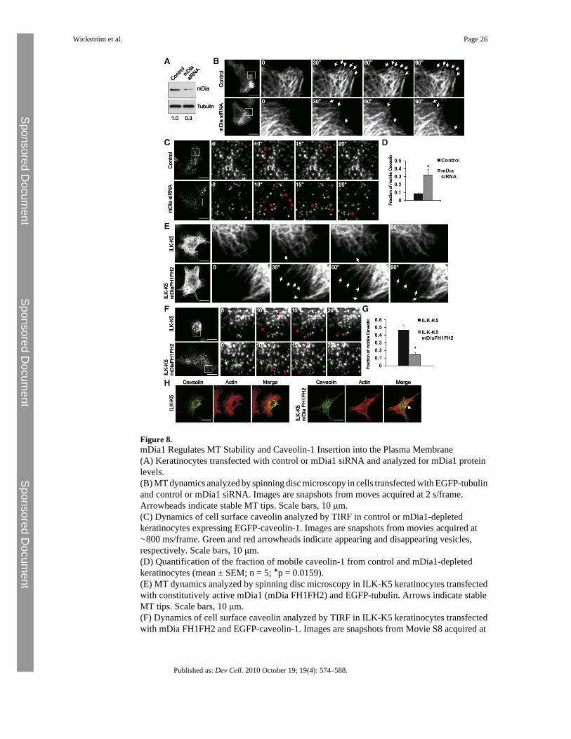
Figure 8.mDia1 Regulates MT Stability and Caveolin-1 Insertion into the Plasma Membrane(A) Keratinocytes transfected with control or mDia1 siRNA and analyzed for mDia1 proteinlevels.(B) MT dynamics analyzed by spinning disc microscopy in cells transfected with EGFP-tubulinand control or mDia1 siRNA. Images are snapshots from moves acquired at 2 s/frame.Arrowheads indicate stable MT tips. Scale bars, 10 μm.(C) Dynamics of cell surface caveolin analyzed by TIRF in control or mDia1-depletedkeratinocytes expressing EGFP-caveolin-1. Images are snapshots from movies acquired at∼800 ms/frame. Green and red arrowheads indicate appearing and disappearing vesicles,respectively. Scale bars, 10 μm.(D) Quantification of the fraction of mobile caveolin-1 from control and mDia1-depletedkeratinocytes (mean ± SEM; n = 5; ∗p = 0.0159).(E) MT dynamics analyzed by spinning disc microscopy in ILK-K5 keratinocytes transfectedwith constitutively active mDia1 (mDia FH1FH2) and EGFP-tubulin. Arrows indicate stableMT tips. Scale bars, 10 μm.(F) Dynamics of cell surface caveolin analyzed by TIRF in ILK-K5 keratinocytes transfectedwith mDia FH1FH2 and EGFP-caveolin-1. Images are snapshots from Movie S8 acquired at
Wickström et al. Page 26
Published as: Dev Cell. 2010 October 19; 19(4): 574–588.
Sponsored Docum
ent Sponsored D
ocument
Sponsored Docum
ent
∼800 ms/frame. Green and red arrowheads indicate appearing and disappearing vesicles,respectively. Scale bars, 10 μm.(G) Quantification of the fraction of mobile caveolin-1 from ILK-K5 and mDia1 FH1FH2-expressing keratinocytes (mean ± SEM; n = 4; ∗∗p = 0.0286).(H) ILK-K5 cells expressing mDia1 FH1FH2 stained for caveolin and F-actin. Arrow indicatesperipheral caveolin-1 in mDia FH1FH2-expressing cells. Scale bars, 10 μm.See also Movie S8, Figure S4, and Table S1.
Wickström et al. Page 27
Published as: Dev Cell. 2010 October 19; 19(4): 574–588.
Sponsored Docum
ent Sponsored D
ocument
Sponsored Docum
ent
Related Documents