Integrated biophysical studies implicate partial unfolding of NBD1 of CFTR in the molecular pathogenesis of F508del cystic fibrosis Chi Wang, 1 Irina Protasevich, 2 Zhengrong Yang, 2 Derek Seehausen, 1 Timothy Skalak, 1 Xun Zhao, 3 Shane Atwell, 3 J. Spencer Emtage, 3 Diana R. Wetmore, 4 Christie G. Brouillette, 2 and John F. Hunt 1 * 1 Department of Biological Sciences, 702A Fairchild Center, Columbia University, New York, New York 10027 2 Department of Chemistry, University of Alabama, Birmingham, Alabama 35294-4400 3 SGX Pharmaceuticals, 10505 Roselle Street San Diego, California 92121 4 Cystic Fibrosis Foundation Therapeutics, 6931 Arlington Road, Bethesda, Maryland 20872 Received 15 April 2010; Revised 21 July 2010; Accepted 22 July 2010 DOI: 10.1002/pro.480 Published online 4 August 2010 proteinscience.org Abstract: The lethal genetic disease cystic fibrosis is caused predominantly by in-frame deletion of phenylalanine 508 in the cystic fibrosis transmembrane conductance regulator (CFTR). F508 is located in the first nucleotide-binding domain (NBD1) of CFTR, which functions as an ATP-gated chloride channel on the cell surface. The F508del mutation blocks CFTR export to the surface due to aberrant retention in the endoplasmic reticulum. While it was assumed that F508del interferes with NBD1 folding, biophysical studies of purified NBD1 have given conflicting results concerning the mutation’s influence on domain folding and stability. We have conducted isothermal (this paper) and thermal (accompanying paper) denaturation studies of human NBD1 using a variety of biophysical techniques, including simultaneous circular dichroism, intrinsic fluorescence, and static light-scattering measurements. These studies show that, in the absence of ATP, NBD1 unfolds via two sequential conformational transitions. The first, which is strongly influenced by F508del, involves partial unfolding and leads to aggregation accompanied by an increase in tryptophan fluorescence. The second, which is not significantly influenced by F508del, involves full unfolding of NBD1. Mg-ATP binding delays the first transition, thereby offsetting the effect of F508del on domain stability. Evidence suggests that the initial partial unfolding transition is partially responsible for the poor in vitro solubility of human NBD1. Second-site mutations that increase the solubility of isolated F508del-NBD1 in vitro and suppress the trafficking defect of intact F508del-CFTR in vivo also stabilize the protein against this transition, supporting the hypothesize that it is responsible for the pathological trafficking of F508del-CFTR. Keywords: cystic fibrosis; cystic fibrosis transmembrane conductance regulator (CFTR); protein thermodynamics; circular dichroism; fluorescence; static light-scattering Additional supporting information can be found in the online version of this article. Xun Zhao, Shane Atwell, and J. Spencer Emtage’s current address is Eli Lilly and Company, Lilly Biotechnology Center, 1300 Campus Point Drive, Suite 200, San Diego, CA 92121. Diana R. Wetmore’s current address is Emerald Biostructures, Bainbridge Island, WA 981110. Grant sponsor: Cystic Fibrosis Foundation Therapeutics, Inc. *Correspondence to: John F. Hunt, Department of Biological Sciences, 702A Fairchild Center, MC2434, Columbia University, New York, NY 10027, United States. E-mail: [email protected]. 1932 PROTEIN SCIENCE 2010 VOL 19:1932—1947 Published by Wiley-Blackwell. V C 2010 The Protein Society

Welcome message from author
This document is posted to help you gain knowledge. Please leave a comment to let me know what you think about it! Share it to your friends and learn new things together.
Transcript

Integrated biophysical studies implicatepartial unfolding of NBD1 of CFTR in themolecular pathogenesis of F508delcystic fibrosis
Chi Wang,1 Irina Protasevich,2 Zhengrong Yang,2 Derek Seehausen,1
Timothy Skalak,1 Xun Zhao,3 Shane Atwell,3 J. Spencer Emtage,3
Diana R. Wetmore,4 Christie G. Brouillette,2 and John F. Hunt1*
1Department of Biological Sciences, 702A Fairchild Center, Columbia University, New York, New York 100272Department of Chemistry, University of Alabama, Birmingham, Alabama 35294-44003SGX Pharmaceuticals, 10505 Roselle Street San Diego, California 921214Cystic Fibrosis Foundation Therapeutics, 6931 Arlington Road, Bethesda, Maryland 20872
Received 15 April 2010; Revised 21 July 2010; Accepted 22 July 2010
DOI: 10.1002/pro.480Published online 4 August 2010 proteinscience.org
Abstract: The lethal genetic disease cystic fibrosis is caused predominantly by in-frame deletion of
phenylalanine 508 in the cystic fibrosis transmembrane conductance regulator (CFTR). F508 is
located in the first nucleotide-binding domain (NBD1) of CFTR, which functions as an ATP-gatedchloride channel on the cell surface. The F508del mutation blocks CFTR export to the surface due
to aberrant retention in the endoplasmic reticulum. While it was assumed that F508del interferes
with NBD1 folding, biophysical studies of purified NBD1 have given conflicting results concerningthe mutation’s influence on domain folding and stability. We have conducted isothermal (this
paper) and thermal (accompanying paper) denaturation studies of human NBD1 using a variety of
biophysical techniques, including simultaneous circular dichroism, intrinsic fluorescence, andstatic light-scattering measurements. These studies show that, in the absence of ATP, NBD1
unfolds via two sequential conformational transitions. The first, which is strongly influenced by
F508del, involves partial unfolding and leads to aggregation accompanied by an increase intryptophan fluorescence. The second, which is not significantly influenced by F508del, involves full
unfolding of NBD1. Mg-ATP binding delays the first transition, thereby offsetting the effect of
F508del on domain stability. Evidence suggests that the initial partial unfolding transition ispartially responsible for the poor in vitro solubility of human NBD1. Second-site mutations that
increase the solubility of isolated F508del-NBD1 in vitro and suppress the trafficking defect ofintact F508del-CFTR in vivo also stabilize the protein against this transition, supporting the
hypothesize that it is responsible for the pathological trafficking of F508del-CFTR.
Keywords: cystic fibrosis; cystic fibrosis transmembrane conductance regulator (CFTR); proteinthermodynamics; circular dichroism; fluorescence; static light-scattering
Additional supporting information can be found in the online version of this article.
Xun Zhao, Shane Atwell, and J. Spencer Emtage’s current address is Eli Lilly and Company, Lilly Biotechnology Center, 1300Campus Point Drive, Suite 200, San Diego, CA 92121.
Diana R. Wetmore’s current address is Emerald Biostructures, Bainbridge Island, WA 981110.
Grant sponsor: Cystic Fibrosis Foundation Therapeutics, Inc.
*Correspondence to: John F. Hunt, Department of Biological Sciences, 702A Fairchild Center, MC2434, Columbia University, NewYork, NY 10027, United States. E-mail: [email protected].
1932 PROTEIN SCIENCE 2010 VOL 19:1932—1947 Published by Wiley-Blackwell. VC 2010 The Protein Society
IntroductionCystic fibrosis (CF) is a genetic disease caused by
mutations in an ATP-gated chloride channel called
the cystic fibrosis transmembrane conductance regu-
lator (CFTR).1–6 CF is the most common fatal
genetic disease among Caucasians and is prevalent
in many other populations.7 CF causes pervasive
defects in secretory processes, including most impor-
tantly water secretion in the epithelial tissues of the
lung. This defect leads to insufficient hydration,
which impairs bacterial clearance and leads to per-
sistent cycles of infection/inflammation followed by
eventual lung failure.8–11 While many advances
have been made in treating CF during the past 20
years, most patients still die before age 30.8 There is
intense interest in applying understanding of the
molecular etiology of the disease to developing more
efficacious pharmacological treatments.12–14
Population genetics shows that a single muta-
tion, an in-frame deletion of phenylalanine 508
(F508del), accounts for �70% of the mutant CFTR
alleles present in the human population.7 Therefore,
�50% of CF patients have two copies and �90%
have at least one copy of this specific mutation.
Therefore, a substantial proportion of CF drug-dis-
covery efforts have focused on correction of the mo-
lecular defect caused by the F508del mutation.12,13
F508del is located in the first nucleotide-binding
domain (NBD1) of CFTR,15–19 which is homologous
to proteins in the ABC Transporter superfamily.20–22
This name derives from the stereotyped nucleotide-
binding domains (NBDs), or ATP-binding cassettes
(ABCs), that are conserved among superfamily mem-
bers. While CFTR is the only member known to
function as an ATP-gated ion channel rather than
an ATP-fueled transmembrane pump, its overall do-
main organization and ATP-dependent mechano-
chemistry are equivalent to that of ABC Transport-
ers.4,6,18 These all contain a pair of transmembrane
domains (TMDs) that interact with a pair of cyto-
plasmic ABC domains, which control protein
conformation by binding ATP at their mutual inter-
face.23–25 In CFTR, these domains are encoded in a
single polypeptide along with a regulatory (R) do-
main, in the order TMD1, NBD1, R, TMD2, NBD2.
F508del-CFTR displays a severe temperature-
dependent defect in protein biogenesis.26–29 While
�10% of it is properly exported to the plasma mem-
brane in cells growing at 25�C, less than 1% is prop-
erly exported at 37�C. The remainder is retained in
the endoplasmic reticulum (ER) and eventually
degraded via retrograde transport to the cytoplasmic
proteasome complex. Furthermore, F508del-CFTR
channels exported to the plasma membrane at 25�C
are relatively stable at that temperature but are
destabilized and degraded much more rapidly at
37�C.28,30 The obvious temperature-dependent
defects in the biogenesis and stability of F508del-
CFTR have led to a widespread assumption that the
mutation interferes with protein folding.31 However,
structural and thermodynamic studies have led to
conflicting conclusions regarding the exact molecular
defect caused by the F508del mutation.15,18,19,32–34
Previously published isothermal denaturation
studies have failed to reveal a perturbation in the
major chemical unfolding transition of human NBD1
(hNBD1).15,19 Moreover, extensive crystallographic
analyses demonstrated that F508del only perturbs
the equilibrium structure of Mg-ATP-bound hNBD1
locally at the site of the mutation, which is the
region likely to mediate interaction with the trans-
membrane domains of CFTR.18 High-resolution
hydrogen/deuterium-exchange mass spectrometry
analyses of Mg-ATP-bound hNBD1 have measured
the influence of the mutation on protein backbone
dynamics, which are modestly enhanced near the
site of the mutation but not elsewhere in the do-
main.18 These results have led to the hypothesis
that F508del may interfere with biogenesis by block-
ing proper interdomain interaction in nascent
CFTR15,35 or by promoting adventitious chaperone
interactions near the site of the mutation,18 rather
than by destabilizing hNBD1 itself. On the other
hand, other results provide evidence that F508del
does perturb hNBD1 stability. First, the mutation
lowers the thermal melting temperature of hNBD1
by �6 to 7�C (see Ref. 36). Moreover, three second-
site mutations, selected by Teem and coworkers to
reverse the in vivo trafficking defect caused by
F508del,37 appeared to stabilize hNBD1 against
chemical unfolding.15 Because this ‘‘Teem suppressor
triplet’’ (G550E, R553Q, and R555K) does not signifi-
cantly alter the structure of F508del-hNBD1,18 its
effect increasing the thermodynamic stability of
hNBD1 seems likely to be responsible for reversing
the deleterious effects of the F508del mutation.
While the F508del mutation has complex
effects on the structure and stability of hNBD1,
this mutation very clearly impairs the in vitro solu-
bility of the domain compared to matched protein
constructs containing F508.15 Even wild-type
hNBD1 has such low solubility in aqueous buffers
that various ‘‘solubilizing’’ mutations were needed
to obtain well-behaved protein preparations for bio-
physical studies.15 These mutations included the
Teem suppressor triplet as well as substitutions of
surface-exposed hydrophobic residues with more
hydrophilic residues found at equivalent positions
in other vertebrate CFTR sequences. Surprisingly,
several of these solubilizing surface mutations in
hNBD1, identified in a screen focused exclusively
on the in vitro solubility of hNBD1, were shown to
suppress the in vivo trafficking defect of F508del-
CFTR more strongly than the best existing pharma-
cological agents.32,38 Notably, the mutated residues
Wang et al. PROTEIN SCIENCE VOL 19:1932—1947 1933

(e.g., F429S, F494N, and Q637R) are not in direct
contact with F508 and do not appear to be allosteri-
cally coupled.18 A similar hydrophobic-to-hydro-
philic substitution in the immediate vicinity of
F508, the V510D mutation, also strongly sup-
presses the in vivo trafficking defect of F508del-
CFTR.39,40 It was proposed that these substitutions
could block adventitious chaperone interactions
that prevent proper ER export.18 However, there is
as yet no concrete evidence explaining the tight cor-
relation between the effects of mutations on the
in vitro solubility properties of hNBD1 and the
in vivo trafficking properties of human CFTR.
To clarify the relationship between the folding,
stability, and solubility properties of hNBD1 and the
in vivo trafficking of CFTR, we have undertaken
coordinated isothermal (this paper) and thermal (see
Ref. 36) denaturation studies of human NBD1
(hNBD1), using a variety of biophysical techniques.
These studies produce a thermodynamic and struc-
tural model for hNBD1 unfolding and importantly
identify a partial unfolding transition that is desta-
bilized by F508del and stabilized by second-site
mutations suppressing the in vivo trafficking defect
of F508del-CFTR. This transition, which precedes
the major chemical unfolding transition and seems
likely to include unfolding of the a-helical (ABCa)subdomain, leads to aggressive self-association
in vitro. The observed biophysical properties of
hNBD1 variants harboring different mutations sug-
gest that this initial unfolding transition is responsi-
ble for both the poor in vitro solubility of F508del-
hNBD1 and the pathological ER retention of
F508del-CFTR in vivo. This hypothesis, along with
our model for hNBD1 unfolding, should help guide
development of more efficacious drugs to treat the
�90% of CF patients carrying the F508del mutation.
Results
Overview of Design of Isothermal DenaturationExperiments
Figure 1 shows isothermal urea denaturation pro-
files at 25�C for F508 and F508del-hNBD1-
D(RI,RE). This protein construct has the wild-type
human sequence except for deletion of most of the
regulatory insertion (RI) and regulatory extension
(RE).16,17,41 These protein segments, 37 and 39 resi-
dues in length, respectively, are disordered in
hNBD1 in solution in vitro18,42 and dispensable for
normal gating by intact CFTR in vivo.43 Their
removal substantially improves both F508del
CFTR biogenesis in vivo41 and hNBD1 solubility
in vitro,17 enabling the F508del domain to be puri-
fied in sufficient yield for biophysical characteriza-
tion in the absence of additional solubility-enhanc-
ing point-mutations. The F508del mutation in the
full-length domain reduces its solubility to the point
where such solubility-enhancing mutations are
required to obtain a practically useful yield of puri-
fied protein.16 While these mutations represent nat-
ural sequence variations found in some vertebrate
CFTR orthologues, their influence on the biophysi-
cal properties of hNBD1 are controversial.18,32
Therefore, we exploited the improved solubility of
hNBD1-D(RI,RE) to explore the folding properties
of the domain in the absence of solubility-enhancing
mutations and compare its behavior to the full-
length domain containing solubilizing mutations.
Furthermore, we conducted experiments in a
Standard Stabilizing Buffer, containing 10% (v/v)
glycerol and 10% (v/v) ethylene glycol, that was
developed to inhibit hNBD1 aggregation.15
Denaturation of hNBD1-D(RI,RE) was per-
formed using an autotitrator protocol and followed
simultaneously by circular dichroism (CD) spectros-
copy to monitor protein secondary structure (Fig.
1A,D), fluorescence spectroscopy to monitor the envi-
ronment of the two tryptophan (trp) residues in
hNBD1 (Fig. 1B,E), and static light-scattering
(SLS)44 to monitor its aggregation state (Fig. 1C,F).
The low solubility of hNBD1 has raised questions
concerning the relationship between unfolding and
self-association processes.18,32 This relationship can
be addressed explicitly by monitoring protein aggre-
gation state using SLS44 simultaneously with moni-
toring of protein conformation using the traditional
spectroscopic methods. Supporting Information Figure
S1 for this paper presents data validating our SLS
methods, by showing minimal changes in SLS during
chemical denaturation of a well behaved soluble pro-
tein that undergoes a two-state unfolding transition
without alteration of its aggregation state.
The hNBD1-D(RI,RE) variants without or with
F508del both display significantly different isothermal
denaturation properties at a nominally saturating
Mg-ATP concentration (430 lM) compared to a lower
concentration (30 lM) giving only partial occupancy of
the binding site (Fig. 1 and Supporting Information
Fig. S2). The latter concentration is the lowest that is
readily achievable because a high concentration of
Mg-ATP (�2 mM) must be maintained during purifica-
tion to obtain high protein yield.15 Surface plasmon
resonance studies have demonstrated that hNBD1-
D(RI,RE) binds Mg-ATP with 0.8 lM affinity at 4�C.17
Calorimetric studies of this construct presented in Ref.
36 indicate that the enthalpy of Mg-ATP binding is
20–25 kcal/mole, so binding-affinity will be reduced
�10-fold at 25�C versus 4�C. Therefore, the affinity for
Mg-ATP should be �8 lM at the temperature of the
chemical denaturation experiments shown in Figure 1,
producing �80% occupancy of the binding site at 30
lM Mg-ATP compared to �98% at 430 lM. Most
importantly, irrespective of the exact binding affinity,
the ratio of Mg-ATP-free versus Mg-ATP-bound pro-
tein should decrease �14-fold (30/430) between the
1934 PROTEINSCIENCE.ORG Partial Unfolding of NBD1 Implicated in CF Pathogenesis
experiments in Figure 1 conducted at a high concen-
tration of Mg-ATP compared to those conducted at a
low concentration (because [hNBD1free]/[hNBD1bound] ¼Kd/[ATPfree]). The qualitatively different behavior in iso-
thermal denaturation at different Mg-ATP concen-
trations indicates that hNBD1 has substantially dif-
ferent conformational behavior with versus without
bound Mg-ATP (see also Ref. 36).
F508del Facilitates the First of Two SequentialIsothermal Unfolding Transitions in hNBD1
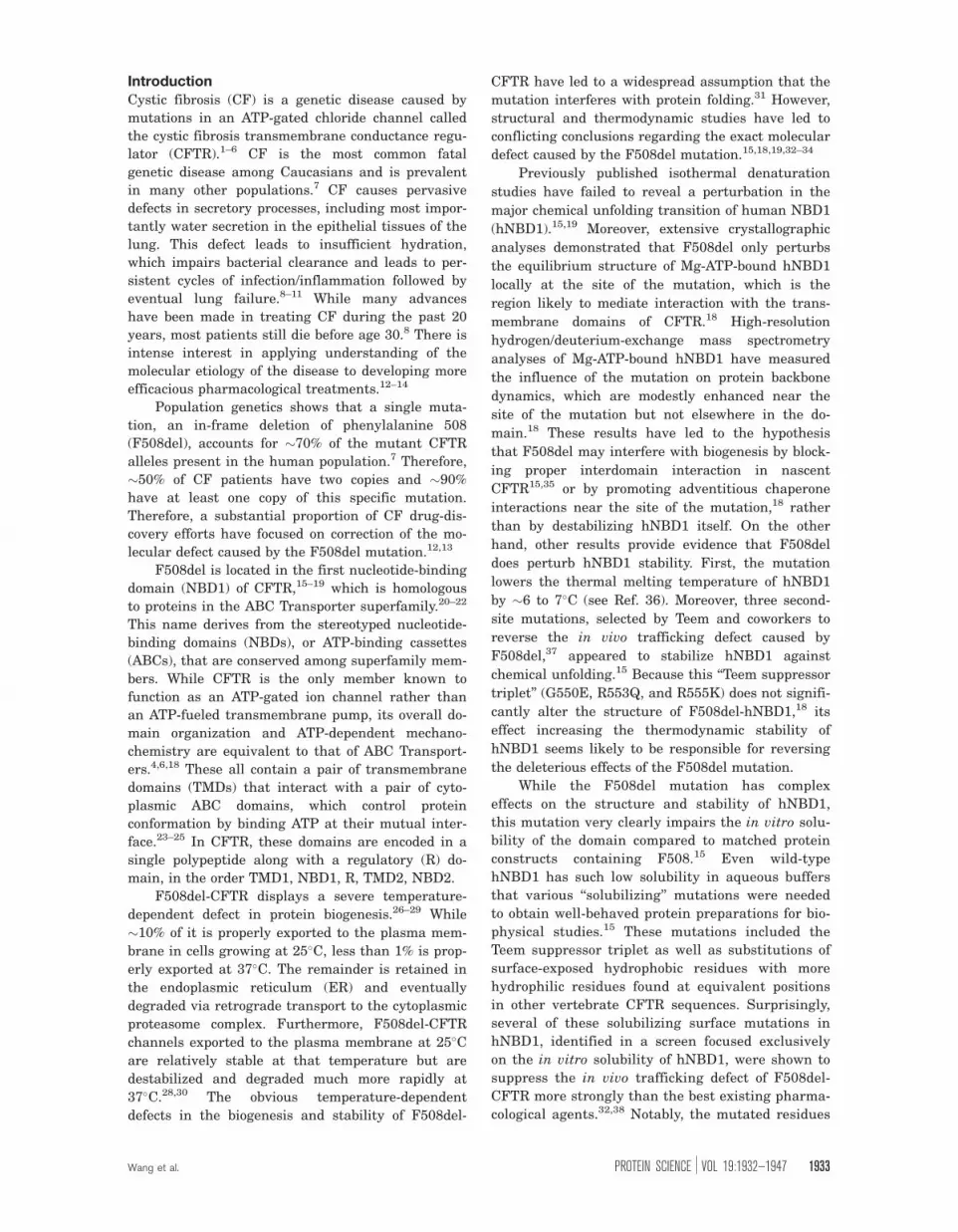
At a subsaturating 30 lM concentration of Mg-ATP,
hNBD1-D(RI,RE) denatures in two successive steps
at 25�C, the first resulting in �25% loss of secondary
structure and the second resulting in �75% loss of
secondary structure as monitored by CD (Fig. 1A).
Note that the second transition is not completed at
the highest urea concentration attainable in these
experiments (8M) conducted in Standard Stabilizing
Buffer, so there is no post-denaturation baseline visi-
ble in the CD spectra in Figure 1A or D. However,
experiments conducted in Simple Buffer with urea
(Supporting Information Fig. S3) or in Standard Sta-
bilizing buffer with Gdn-HCl (data not shown),
which is more denaturing than urea, demonstrate
that the later phase of the denaturation profile does
represent a discrete unfolding transition. The bipha-
sic nature of the unfolding process is also clearly
visible in the fluorescence traces in Figure 1B,E and
the SLS traces in Figure 1C,F.
Figure 1. Simultaneous monitoring of CD, trp fluorescence, and SLS during isothermal urea denaturation at 25�C of hNBD1-
D(RI,RE) 6 F508del. Protein samples (2 lM) in the absence or presence of 8 M urea were progressively mixed every 800
seconds to achieve the indicated concentration of urea, using an autotitrator for CD measurements or an equivalent kinetic
protocol for fluorescence and SLS measurements. The buffer contained 150 mM NaCl, 0.5 mM MgCl2 10% glycerol, 10%
ethylene glycol, 0.8 mM TCEP, 20 mM Na-HEPES, pH 7.5. Data for F508 and F508del constructs are shown in black and
gray, respectively. The buffer used to purify and store the protein, which is catalytically inactive,15–19 introduces 30 lM Mg-
ATP. Additional Mg-ATP was added to the indicated final concentration. CD (top) was monitored at 230 nm instead of 222
nm to enable use of a higher concentration of Mg-ATP without interference from nucleotide absorbance. Intrinsic trp
fluorescence (middle) was excited at 297 nm and monitored at 340 nm. SLS at 297 nm (bottom) was measured at a 90� angle
simultaneously with trp fluorescence in a T-format fluorimeter (see Methods). SLS counts are background-subtracted for
scattering from the protein-free 0 M urea buffer for measurements in the fluorimeter, so the ratio of the SLS signal at a given
urea concentration to that at the start of the titration gives a rough estimate of weight-averaged aggregation state. Note,
however, that this calculation underestimates the aggregation state at higher urea concentrations because it ignores the
effect of urea in reducing protein contrast.
Wang et al. PROTEIN SCIENCE VOL 19:1932—1947 1935
The F508del mutation moves the midpoint of
the initial unfolding transition to substantially lower
urea concentration (�1.8 M vs. �3.6 M), while the
second unfolding transition (with midpoint at �6.5
M) is unaffected. This pattern suggests that the
ABCa subdomain45 of hNBD1,16,18 which contains
F508, unfolds during the initial unfolding transition
to produce a partially folded intermediate. The ob-
servation that F508del does not influence the second
transition15 indicates that this transition does not
produce an energetically significant conformational
change in the vicinity of F508 in the ABCa subdo-
main and suggests that it is likely to involve pre-
dominantly conformational changes in hNBD1’s nu-
cleotide-binding core (which comprises an F1
ATPase-like core subdomain and the ABCb subdo-
main16,18,45). It is difficult to obtain a reliable esti-
mate of the free energy of the initial unfolding tran-
sition (DG0unfolding) in F508-hNBD1 due to the lack
of a clear baseline between the two successive tran-
sitions (although a crude estimate is presented in
Supporting Information Fig. S9 later). For F508del-
hNBD1, it is �þ3.3 kcal/mole at 25�C in the pres-
ence of 30 lM Mg-ATP, based on extrapolation of the
equilibrium constant to zero urea concentration
(Supporting Information Fig. S9 later).46 This esti-
mate is only a rough approximation due to partial
occupancy of the Mg-ATP binding site and the
incomplete reversibility of the transition in these
experiments (see below). However, it is generally
consistent with the value of þ1.8 kcal/mole for Mg-
ATP-free hNBD1 at 25�C determined via detailed
mathematical modeling of DSC data in Ref. 36.
The initial unfolding transition observed by CD
spectroscopy is followed by an increase in trp fluo-
rescence (Fig. 1B) and subsequently a strong
increase in SLS (Fig. 1C). The fluorescence transi-
tion occurs at a slightly higher urea concentration
than the CD transition, and the SLS transition
occurs at a significantly higher urea concentration
than the fluorescence transition. These offsets indi-
cate that, rather than a simple two-state transition,
the protein is undergoing sequential changes in its
physical state resulting in the formation of protein
aggregates. These aggregates probably correspond to
the ‘‘N*’’ species previous identified by Thomas and
coworkers.47 Their elevated fluorescence seems
likely to be attributable to burial of the one or both
of the trp residues in hNBD1 in intersubunit interfa-
ces in the aggregates. The inference that aggrega-
tion follows an initial unfolding transition is strongly
supported by a lag between the CD and fluores-
cence/SLS changes in kinetic studies of the denatu-
ration process (Fig. 2 later). Moreover, although the
forward titration behavior is reproducible, reverse
titrations show significant hysteresis in the region of
the initial transition (data not shown), and the mag-
nitude of the increase in trp fluorescence varies
somewhat in experimental replicates even though
the qualitative features of the curve are reproducible
(data not shown). These observations are consistent
with irreversible aggregation influencing behavior in
this range of urea concentration. Such aggregation
is consistent with the kinetic trapping of partially
unfolded intermediates inferred in Ref. 36 to occur
during thermal denaturation. Both the trp fluores-
cence and SLS changes that follow the initial unfold-
ing transition are reversed by the second unfolding
transition that results in the loss of most protein
secondary structure as monitored by CD. Therefore,
full denaturation of hNBD1 reverses aggregation of
the partially unfolded intermediate produced by the
initial unfolding transition.
Increasing Mg-ATP to a more highly saturating
concentration of 430 lM shifts the first unfolding
transition to higher urea concentration, and it sup-
presses the differences in the fluorescence and SLS
signals observed during urea denaturation of the
F508 versus F508del domains at 30 lM Mg-ATP
(Fig. 1D–F compared to Fig. 1A–C; see Supporting
Information Fig. S2 for direct overlay of the experi-
ments in Fig. 1 conducted at high versus low Mg-
ATP concentrations). The SLS changes during urea
denaturation of hNBD1-D(RI,RE) are reduced in
magnitude at the higher Mg-ATP concentration,
especially in the F508del construct (Fig. 1F and
Supporting Information Fig. S2C,F), indicating that
aggregation is suppressed by greater saturation of
the Mg-ATP binding site. This effect is presumably
due to inhibition of the initial unfolding transition,
which limits the concentration of the aggregation-
prone, partially unfolded intermediate until the urea
concentration is high enough to inhibit aggregation.
The observed effect of Mg-ATP in suppressing aggre-
gation is likely to explain the beneficial influence of
super-saturating Mg-ATP concentrations on the
yield of the domain during purification.15
Characterization of the Second IsothermalChemical Unfolding Transition
Rigorous characterization of the second unfolding
transition in urea is not possible in Standard Stabi-
lizing Buffer because the domain is not fully dena-
tured at the highest accessible denaturant concen-
tration. However, this transition is completed at
accessible urea concentrations in buffers that do not
contain stabilizing osmolytes, for example, the Sim-
ple Buffer used in earlier chemical denaturation
experiments.15 Supporting Information Figure S3
shows the results of autotitrator denaturation
experiments conducted on hNBD1-D(RI,RE) con-
structs at 25�C in this buffer. All qualitative features
of the unfolding behavior are the same as in Stand-
ard Stabilizing Buffer, although the lower stability
of the domain in Simple Buffer shifts all conforma-
tional transitions 1–2 M lower in urea concentration.
1936 PROTEINSCIENCE.ORG Partial Unfolding of NBD1 Implicated in CF Pathogenesis

Figure 2. Kinetics of isothermal urea denaturation at 25�C of hNBD1-D(RI,RE) 6 F508del. CD (top), trp fluorescence (middle),
and SLS (bottom) signals were monitored using identical methods to Fig. 1 except that fluorescence emission at 340 nm and
90� SLS were measured in the Jasco spectropolarimeter using 230 nm incident light. At �100 seconds, 2 lM hNBD1-
D(RI,RE) with (black) or without (gray) F508del was added from urea-free storage buffer to the Standard Stabilizing Buffer
containing 30 lM Mg-ATP and the indicated concentration of urea.
Wang et al. PROTEIN SCIENCE VOL 19:1932—1947 1937

(The significant differences in these results com-
pared to those reported earlier in the same buffer15
are likely to be attributable to the faster denatura-
tion/observation protocol used here, as explained in
the Discussion section below.) Similar glycerol
concentrations to those in Standard Stabilizing
Buffer have been observed to suppress the protein
trafficking defect caused by the F508del mutation in
full-length CFTR in vivo in tissue culture cells,48,49
consistent with the hypothesis that unfolding of the
domain contributes to aberrant ER retention of
F508del-CFTR. Increasing Mg-ATP concentration
has at most a minor effect on the progression of the
second unfolding transition in Simple Buffer (Sup-
porting Information Fig. S3), in contrast to the
obvious inhibition of the first unfolding transition
observed at the higher Mg-ATP concentration in ei-
ther buffer (Supporting Information Figs. S2–S3).
Additional investigations of the influence of adenine
nucleotides on the progression of the second unfold-
ing transition in several buffers have failed to detect
a reproducible effect (data not shown).
These results indicate that there is a substantial
reduction in Mg-ATP binding affinity during the ini-
tial urea unfolding transition but not during the
ensuing transition that yields fully unfolded
hNBD1-D(RI,RE). Therefore, the partially unfolded
intermediate produced by the initial unfolding tran-
sition is unlikely to have significant affinity for Mg-
ATP. While aggregation of the intermediate or the
elevated urea concentrations (>4M) needed to drive
the second unfolding transition could potentially
interfere with Mg-ATP binding, it is also possible
that the intermediate does not retain a functional
Mg-ATP binding site, even though it retains 75% of
the native secondary structure. Such characteristics
would be consistent with formation of a ‘‘molten
globule’’ conformation,50,51 which has significant sec-
ondary structure but lacks stable tertiary structure.
The accompanying paper36 demonstrates that ther-
mal denaturation produces an aggregation-prone in-
termediate with generally similar biophysical prop-
erties that seems likely to have molten globule
properties based on its minimal enthalpy of unfold-
ing. Therefore, thermal denaturation and isothermal
chemical denaturation of hNBD1-D(RI,RE) may pro-
ceed via similar aggregation-prone molten globule
intermediates. (See Discussion section.)
Evidence for Multiple Self-Association PathwaysIt is noteworthy that, as opposed to the order of
events observed during urea denaturation in the
presence of 30 lM Mg-ATP (Fig. 1A–C), the fluores-
cence and SLS changes precede the CD changes dur-
ing urea denaturation of F508-hNBD1-D(RI,RE) in
the presence of 430 lM Mg-ATP (Fig. 1D–F). These
observations raise the possibility that self-associa-
tion of the domain leading to an increase in trp fluo-
rescence can also occur via a distinct pathway
involving a native-like conformation that has not
lost significant secondary structure. Kinetic studies
(Fig. 2G later) show that aggregation of the partially
unfolded intermediate leads to a small increase in
secondary structure content as monitored by CD,
meaning that aggregation tightly coupled to unfold-
ing could potentially obscure a secondary structural
change. However, equivalent denaturation experi-
ments conducted on constructs harboring suppressor
and solubilizing mutations (Fig. 4 later) strongly
support the inference that hNBD1-D(RI,RE) can ag-
gregate in a native-like conformational state to pro-
duce a species with elevated trp fluorescence. In
addition, the SLS data demonstrate that the domain
has a tendency to self-associate in the native confor-
mational state to form aggregates with unaltered trp
fluorescence (i.e., at the points below 1.5 M urea in
Fig. 1C,F). Therefore, hNBD1-D(RI,RE) displays
three distinct aggregation pathways in vitro. How-
ever, the biggest aggregates (i.e., those giving the
largest SLS signals in Fig. 1), which also have the
most strongly elevated trp fluorescence, are formed
subsequent to the initial unfolding transition, and
this pathway clearly dominates the aggregation
behavior of the domain harboring the F508del
mutation.
Similar Chemical Unfolding Behavior ofhNBD1-D(RI,RE) at Higher Temperature
Isothermal urea denaturation of hNBD1-D(RI,RE)using an autotitrator protocol at 35�C (top of Sup-
porting Information Fig. S4) yields qualitatively sim-
ilar results to those obtained at 25�C. Most impor-
tantly, the F508del mutation clearly promotes
unfolding (lowering the midpoint of the transition by
�1.5 M in urea concentration), while raising the
Mg-ATP concentration clearly inhibits unfolding
(raising the midpoint of the transition by �1.5 M in
urea concentration). Therefore, F508del and Mg-ATP
have equivalently strong and opposing influences on
domain stability at 35� and 25�C. Furthermore,
there is evidence of self-association of the F508del
domain in a native-like conformational state at both
temperatures in the presence of 430 lM Mg-ATP,
because the increase in trp fluorescence precedes the
secondary structure transition monitored by CD
spectroscopy. However, there is one noteworthy dif-
ference in the biophysical behavior of the domain at
the higher temperature. Only a single CD transition
is observed at 35� for either the F508 or F508del do-
main in the presence of 30 lM Mg-ATP (Supporting
Information Fig. S4A,B), compared to two successive
transitions under the same conditions at 25�C
(Fig. 1 and Supporting Information Fig. S4C,D). While
additional analyses would be required for rigorous
mechanistic dissection of this behavior, the thermal
denaturation studies presented in Ref. 36 indicate a
1938 PROTEINSCIENCE.ORG Partial Unfolding of NBD1 Implicated in CF Pathogenesis

two-step unfolding pathway for the same protein
constructs in the absence of urea in this tempera-
ture range, with the relative secondary structure
content of the thermal unfolding intermediate
closely matching that of the chemical unfolding in-
termediate observed at 25�C. Therefore, the simplest
explanation for the observed behavior is that unfold-
ing of hNBD1-D(RI,RE) still proceeds through a par-
tially unfolded intermediate but that this intermedi-
ate is less stable to urea denaturation at 35�C and
therefore does not accumulate at this temperature,
at least on the kinetic timescale of isothermal chemi-
cal denaturation experiments (�5 minutes per
point). Increasing urea concentration above that
required for full denaturation of the domain at 35�C
continues to reduce trp fluorescence, likely reflecting
dissociation of unfolded protein aggregates by urea
(Supporting Information Fig. S4B,F).
Notably, chemical denaturation of the domain
occurs at substantially lower urea concentrations at
35 versus 25�C (by �1.5 M), consistent with the calo-
rimetric studies in Ref. 36 showing substantially
reduced stability at the higher temperature. Consist-
ent with the results of these studies, the data in
Supporting Information Fig. S4A suggest that the
unfolding transition in the F508del domain is al-
ready underway at 35�C in the absence of urea,
while the initial unfolding transition in this con-
struct is delayed at 25� to urea concentrations in
excess of 1 M. The lower stability of hNBD1-
D(RI,RE) at 35� versus, especially in the presence of
the F508del mutation, parallels the dramatic exacer-
bation of the in vivo trafficking defect produced by
this mutation during cell growth at 37� versus
25�C.26–29 These results are consistent with the ini-
tial unfolding transition contributing to the patho-
logical ER retention of F508del-CFTR.
Equivalent Two-Step Chemical Unfolding of
Full-Length hNBD1
Isothermal urea denaturation of full-length hNBD1
(i.e., containing both the RI and RE) using an autoti-
trator protocol at 25�C yields essentially equivalent
results to those obtained with the D(RI,RE) con-
struct under the same solution conditions (Support-
ing Information Fig. S5). Solubilizing surface muta-
tions need to be introduced into full-length hNBD1
to obtain sufficient material for biophysical stud-
ies.15 Supporting Information Figure S5 compares
the behavior of matched full-length and D(RI,RE)constructs containing F429S, F494N, and Q637R
mutations15 in the absence or presence of the
F508del mutation. Both constructs show two sequen-
tial unfolding transitions equivalent to those
observed for the hNBD1-D(RI,RE) construct without
any solubilizing mutations; the first of these transi-
tions is inhibited by a higher concentration of
Mg-ATP and facilitated by the F508del mutation.
Supporting Information Figure S6 replots the
same data to compare directly the equivalent con-
structs with and without the RI and RE segments.
The presence of the RI and RE destabilize the domain,
moving the initial unfolding transition to lower urea
concentration (by 1.5–2.5 M). This observation sug-
gests that one or both of these disordered protein seg-
ments interact with and stabilize the partially
unfolded conformation of hNBD1, thereby reducing
the magnitude of the free energy change during the
initial unfolding transition.17 The thermal denatura-
tion studies in Ref. 36 which examine a DRI constructin addition to D(RI,RE) and full-length constructs,
demonstrate that the increase in domain stability in
the D(RI,RE) construct compared to the full-length
construct is attributable primarily to deletion of the
RI rather than the RE. Recently, it has been demon-
strated that deletion of the RI strongly suppresses the
trafficking defect in F508del-CFTR in vivo in tissue
culture cells.41 These observations provide further evi-
dence that the initial unfolding transition of hNBD1
contributes to aberrant trafficking of F508del-CFTR
in vivo. Supporting Information Figure S6 shows that
the trp fluorescence changes, which likely reflect do-
main aggregation, track the initial unfolding transi-
tion in all experiments conducted on full-length
hNBD1 constructs. Therefore, aggregation subsequent
to the initial unfolding transition also contributes to
the low solubility of the domain in vitro, suggesting
that the improved solubility of the D(RI,RE) domain17
is also attributable at least in part to the enhanced
stability of the truncated domain.
Kinetic Studies Demonstrate that the Initial
Unfolding Transition Induces AggregationFigure 2 shows CD, intrinsic trp fluorescence, and
SLS signals as a function of time after introducing
hNBD1-D(RI,RE) into solutions containing a low con-
centration of Mg-ATP (30 lM) and 0, 2, 4, or 8 M urea.
Without urea (Fig. 2A–C), no significant changes are
observed during the 13 minutes of the experiment
except for a very small increase in SLS at the latest
time points, likely reflecting a minor degree of protein
self-association. Similarly, under denaturing condi-
tions at 8 M urea (Fig. 2J–L), the signals are all con-
stant after rapid completion of the unfolding transi-
tion (which occurs more slowly in the F508 domain,
with a half-time of �20 seconds).
In contrast, multiphase kinetic processes are
observed for the F508 and F508del domains at urea
concentrations leading to accumulation of partially
unfolded intermediates. At 2 M urea, the initial
unfolding transition is mostly completed for F508del
but only beginning for F508 hNBD1-D(RI,RE) (Fig.
1A). The F508 domain remains stable under these
conditions but the F508del domain shows an initial
loss of secondary structure as monitored by CD fol-
lowed �3 minutes later by a gradual increase in trp
Wang et al. PROTEIN SCIENCE VOL 19:1932—1947 1939

fluorescence and SLS (Fig. 2D–F). The similarity in
the kinetics of the fluorescence and SLS changes
suggests the alteration in trp environment leading
to increased fluorescence quantum yield occurs dur-
ing aggregation of the domain (presumably due to
solvent-shielding of at least one of its two trps in
interprotein interfaces in the aggregates, as
explained above). In any event, the different kinetics
observed for the SLS versus CD changes demon-
strate that aggregation of the partially unfolded in-
termediate occurs on the time-scale of minutes only
after occurrence of the initial unfolding transition.
In 4 M urea (Fig. 2G–I), the F508 domain shows
CD, trp fluorescence, and SLS changes with very sim-
ilar magnitudes and kinetics to those observed for
the F508del domain in 2 M urea. These observations
suggest that F508-hNBD1 adopts a partially unfolded
conformation in 4M urea similar to that adopted by
F508del-hNBD1 in 2 M urea, consistent with the
data in Figure 1. Note that the F508del domain
unfolds more rapidly than the F508 domain in 4 M
urea (as monitored by CD – Fig. 2G) and then aggre-
gates more rapidly and aggressively (as monitored by
trp fluorescence and SLS – Fig. 2H–I). Under these
conditions, aggregation of F508del-hNBD1 (Fig. 2I) is
accompanied by a small increase in its secondary
structural content (Fig. 2G,I). When aggregation is
very rapid, this increase might potentially obscure
the loss of CD signal occurring during the initial
unfolding transition. Therefore, caution must be exer-
cised in interpreting results from individual isother-
mal chemical denaturation experiments.
Distinct Effects of Mg-ADP Versus Mg-ATP on
the Initial Unfolding TransitionFigure 3 examines the influence of Mg-ADP versus
Mg-ATP on the isothermal urea denaturation of
hNBD1-D(RI,RE) at 25�C in the absence (Fig. 3A,B)
or presence (Fig. 3C,D) of the F508del mutation. In
all cases, unfolding occurs in two successive steps,
as observed in Figure 1 above. The addition of 400
lM Mg-ADP (in addition to 30 lM Mg-ATP) stabil-
izes the domain in the native conformational state
and increases the midpoint of the initial unfolding
transition in urea, although to a lesser extent than
addition of the same concentration of Mg-ATP. Sur-
face plasmon resonance measurements indicate that
both nucleotides have similar binding and release
rates and affinities for hNBD1-D(RI,RE).17 To verify
that they exert somewhat different effects on the
unfolding of the domain, we monitored CD and trp
fluorescence as a function of time after dilution of
F508del-hNBD1-D(RI,RE) into buffers containing 0,
2, or 4 M urea (Supporting Information Fig. S7).
These kinetic studies verify the qualitative features
of the data in Figure 3. Notably, Mg-ATP retards the
unfolding and subsequent self-association of the do-
main substantially more than the equivalent
Figure 3. Effects of Mg-ADP versus Mg-ATP on isothermal urea denaturation of hNBD1-D(RI,RE). Denaturation of the F508
(left) or F508del (right) domain was conducted at 25�C under conditions identical to Fig. 1 and monitored using CD (top) and
trp fluorescence (bottom, with 230 nm excitation and 340 nm emission). Proteins were diluted into Standard Stabilizing Buffer
containing 400 lM Mg-ADP (crosses), 400 lM Mg-ATP (gray diamonds), or no additional nucleotide (gray closed circles).
Because purified proteins are stored in Standard Stabilizing Buffer containing Mg-ATP, they introduce 30 lM Mg-ATP into
every experimental sample in addition to any nucleotide present in the measurement buffer. The final Mg-ATP concentration
in each experiment is indicated in the legends.
1940 PROTEINSCIENCE.ORG Partial Unfolding of NBD1 Implicated in CF Pathogenesis

concentration of Mg-ADP (Supporting Information
Fig. S7C–F). While ADP interacts exclusively with
the nucleotide-binding core of hNBD1, the c-phos-phate of ATP makes a bridging contact between the
ABCa subdomain and the nucleotide-binding core
via the c-phosphate switch (or Q-loop),18,23,52 a short
loop at the N-terminus of the ABCa subdomain.18,45
This subdomain packs tightly against the core in a
defined orientation when the c-phosphate of Mg-ATP
is bound,18,23,52 but it is otherwise flexibly
attached,45,53 including when Mg-ADP is bound. Mg-
ATP thus stabilizes the interaction of the core of
hNBD1 with the ABCa subdomain, an interaction
that would be disrupted by unfolding of this subdo-
main. The more rapid unfolding and aggregation of
Mg-ADP-bound versus Mg-ATP-bound F508del-
hNBD1-D(RI,RE) suggests that the ABCa subdomain
plays a critical role in controlling the kinetics of
unfolding and provides additional evidence that its
conformation is altered significantly during the ini-
tial unfolding transition.
Second-Site Solubilizing/Suppressor Mutations
Delay the Initial Unfolding Transition
Figure 4 shows the isothermal denaturation behav-
ior of F508del-hNBD1-D(RI,RE) constructs harboring
additional point mutations known to suppress the
trafficking defect caused by the F508del muta-
tion.32,37,39,40 The G550E, R553Q, and R555K muta-
tions in the Teem suppressor triplet were isolated
in vivo using a selection for mutations restoring the
export of a yeast CFTR homolog bearing the equiva-
lent of the F508del mutation. They were subse-
quently introduced into intact F508del-CFTR and
demonstrated to suppress its aberrant trafficking in
vivo in tissue culture cells.37 Introducing all three
mutations simultaneously into full-length hNBD1
led to improved yield and stability of the purified
soluble domain after overexpression in E. coli.15 The
V510D mutation was made to explore the molecular
pathology caused by the F508del mutation after
crystallographic studies demonstrated that F508del
produces a large change in the conformation of
val-510.15 The hydrophobic sidechain of val-510 is
mostly buried on the surface of hNBD1 in the
absence of the F508del mutation,18 at a site where it
is likely to make direct packing interactions to the
transmembrane domains of CFTR.35 However, its
backbone conformation is altered so that it projects
from the surface of hNBD1 and is completely solvent-
exposed in the presence of the F508del mutation.18
Surprisingly, the V510D mutation strongly suppresses
the trafficking defect caused by the F508del muta-
tion in vivo in tissue culture cells.39,40 Finally, the
Figure 4. Trafficking suppressor mutations stabilize hNBD1-D(RI,RE)-F508del against the initial unfolding transition. Urea
denaturation at 25�C in the presence of 30 lM Mg-ATP was conducted under conditions identical to Fig. 1 and monitored
using CD spectroscopy (top), trp fluorescence spectroscopy (middle, with 297 nm excitation and 340 nm emission), and SLS
(bottom, at 297 nm). The denaturation of hNBD1-D(RI,RE)-F508del is compared to that of the same construct containing in
addition the Teem suppressor triplet37 (left), V510D39 (center), or F494N/Q637R15 (right).
Wang et al. PROTEIN SCIENCE VOL 19:1932—1947 1941

F494N/Q637R mutations were identified in a screen
for surface substitutions that improve the solubility
of purified hNBD1 in vitro.15 This screen focused on
replacement of hydrophobic residues with more
hydrophilic residues present at equivalent sites in
other vertebrate CFTR sequences. These mutations
also show significant efficacy in suppressing the
trafficking defect caused by the F508del mutation in
vivo in tissue culture cells,32 although they are less
effective that the Teem suppressors,37 the V510D
mutation,39,40 or deletion of the RI.41
The Teem suppressor triplet (Fig. 4A–C), the
V510D mutation (Fig. 4D–F), and the F494N/Q637R
mutations (Fig. 4G–I) all stabilize hNBD1 against
the initial unfolding transition, shifting its midpoint
�0.25–0.50 M higher in urea concentration. The
magnitude of the SLS increase following the initial
unfolding transition is greatly reduced by the Teem
suppressor triplet and the V510D mutation and sig-
nificantly reduced by the F494N/Q637R mutations.
Therefore, the suppressor and solubilizing mutations
all reduce the extent of aggregation of the partially
unfolded intermediate, possibly by delaying its
formation until a higher concentration of urea is
present.
These second-site mutations have generally sim-
ilar effects improving the stability and reducing the
aggregation of hNBD1-D(RI,RE) bearing the native
phe residue at position 508 (Supporting Information
Fig. S8). In the presence of the Teem suppressor
triplet or the V510D mutation, which significantly
stabilize the domain, the urea-induced increase in
trp fluorescence (Supporting Information Fig. S8B,E)
precedes the initial unfolding transition as monitored
by CD spectroscopy (Supporting Information Fig.
S8A,D). This observation provides further evidence
that the domain can also self-associate in a native-like
conformation before formation of the partially unfolded
intermediate, as suggested by the analysis of Figure
1D–F above.
While the different suppressor and solubilizing
mutations studied here all have similar biophysical
effects, some minor differences in behavior are worth
noting. First, the SLS data in Figure 4I demonstrate
that the F494N/Q637R mutation pair in the F508del
domain is unique in suppressing the self-association
that occurs without an increase in trp fluorescence
before the onset of the initial unfolding transition.
Therefore, this mutation pair may influence the sol-
ubility of hNBD1 via more than one mechanism.
Second, the F494N/Q637R mutation pair appears to
decrease the secondary structure content in the par-
tially unfolded intermediate formed by both the
F508del (Fig. 4G) and F508 (Supporting Information
Fig. S8G) domains, while the Teem suppressor tri-
plet may increase the secondary structure content in
this state just in the F508del domain (Fig. 4A).
Therefore, the second-site suppressor mutations may
alter the structure of the partially unfolded interme-
diate produced by the initial unfolding transition, as
predicted in computational studies of the folding
pathway of hNBD1,54 although aggregation of the
intermediate could also influence its secondary
structure content (as shown in Fig. 2G). Conforma-
tional changes in the partially unfolded intermediate
could produce complex behavior in some
experiments.
DiscussionThe isothermal denaturation studies in this paper
combined with the thermal denaturation studies in
Ref. 36 provide a consistent model for the unfolding
of NBD1 from human CFTR (Fig. 5) and its pertur-
bation by F508del, the predominant mutation caus-
ing cystic fibrosis. This model and the data used to
develop it lead to a coherent hypothesis concerning
the thermodynamic and structural effects of F508del
that cause pathogenic retention of CFTR in the ER.
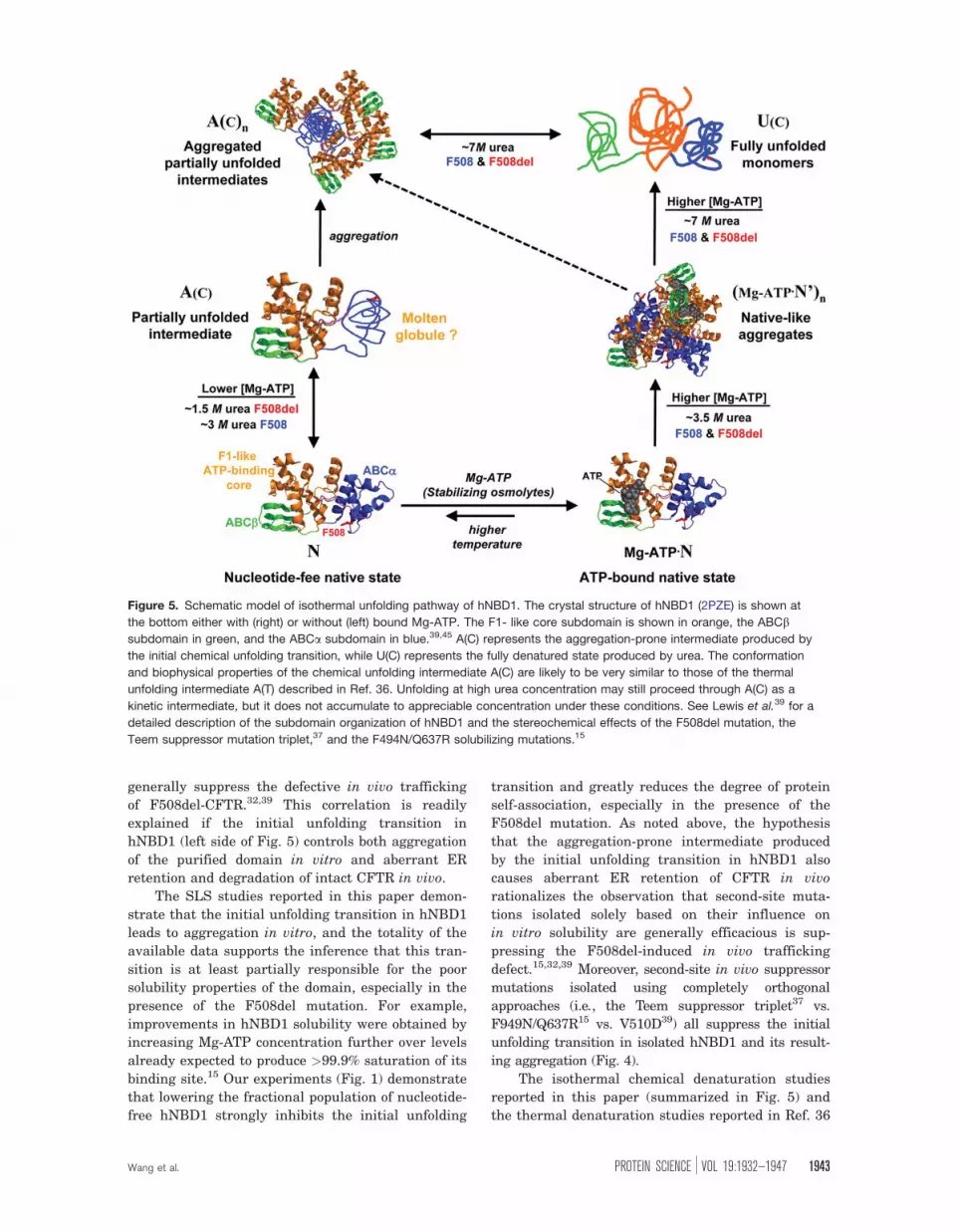
Specifically, we hypothesize that F508del promotes
the initial step in the unfolding of hNBD1, which
produces an aggregation-prone, partially unfolded
intermediate (species A(c) in Fig. 5) that is targeted
for degradation rather than exported to the cell sur-
face. Several lines of evidence support this hypothe-
sis, including the observations that that the initial
unfolding transition in hNBD1 is facilitated by
F508del and inhibited by the second-site mutations
that suppress the trafficking defect caused by
F508del in vivo32,37,39 (Supporting Information Fig.
S9).
Beyond agreement with a wide variety of bio-
physical and physiological studies, our model linking
partial unfolding of hNBD1 with the molecular etiol-
ogy of CF explains several enigmatic observations.
Most importantly, this model explains why muta-
tions improving the in vitro solubility of hNBD1
have consistently shown efficacy in suppressing the
in vivo trafficking defect caused by the F508del
mutation in intact CFTR.32,39 To obtain constructs of
full-length hNBD1-F508del sufficiently soluble for in
vitro purification and characterization, an extensive
screening program was carried out to identify sec-
ond-site mutations that improve solubility.15 Many
candidate mutations were considered, which gener-
ally involved substitution of surface-exposed hydro-
phobic residues with more polar residues naturally
occurring at the equivalent position in other verte-
brate CFTR sequences. This research program led to
the identification of several point mutations that
improve the solubility and yield of purified hNBD1,
including the Teem suppressor triplet (G550E,
R553Q, and R555K) isolated as suppressors of the in
vivo trafficking defect of F508del-CFTR.37 However,
surprisingly, the other efficacious solubilizing muta-
tions chosen exclusively on the basis of polarity and
consistency with the CFTR sequence profile also
1942 PROTEINSCIENCE.ORG Partial Unfolding of NBD1 Implicated in CF Pathogenesis
generally suppress the defective in vivo trafficking
of F508del-CFTR.32,39 This correlation is readily
explained if the initial unfolding transition in
hNBD1 (left side of Fig. 5) controls both aggregation
of the purified domain in vitro and aberrant ER
retention and degradation of intact CFTR in vivo.
The SLS studies reported in this paper demon-
strate that the initial unfolding transition in hNBD1
leads to aggregation in vitro, and the totality of the
available data supports the inference that this tran-
sition is at least partially responsible for the poor
solubility properties of the domain, especially in the
presence of the F508del mutation. For example,
improvements in hNBD1 solubility were obtained by
increasing Mg-ATP concentration further over levels
already expected to produce >99.9% saturation of its
binding site.15 Our experiments (Fig. 1) demonstrate
that lowering the fractional population of nucleotide-
free hNBD1 strongly inhibits the initial unfolding
transition and greatly reduces the degree of protein
self-association, especially in the presence of the
F508del mutation. As noted above, the hypothesis
that the aggregation-prone intermediate produced
by the initial unfolding transition in hNBD1 also
causes aberrant ER retention of CFTR in vivo
rationalizes the observation that second-site muta-
tions isolated solely based on their influence on
in vitro solubility are generally efficacious is sup-
pressing the F508del-induced in vivo trafficking
defect.15,32,39 Moreover, second-site in vivo suppressor
mutations isolated using completely orthogonal
approaches (i.e., the Teem suppressor triplet37 vs.
F949N/Q637R15 vs. V510D39) all suppress the initial
unfolding transition in isolated hNBD1 and its result-
ing aggregation (Fig. 4).
The isothermal chemical denaturation studies
reported in this paper (summarized in Fig. 5) and
the thermal denaturation studies reported in Ref. 36
Figure 5. Schematic model of isothermal unfolding pathway of hNBD1. The crystal structure of hNBD1 (2PZE) is shown at
the bottom either with (right) or without (left) bound Mg-ATP. The F1- like core subdomain is shown in orange, the ABCbsubdomain in green, and the ABCa subdomain in blue.39,45 A(C) represents the aggregation-prone intermediate produced by
the initial chemical unfolding transition, while U(C) represents the fully denatured state produced by urea. The conformation
and biophysical properties of the chemical unfolding intermediate A(C) are likely to be very similar to those of the thermal
unfolding intermediate A(T) described in Ref. 36. Unfolding at high urea concentration may still proceed through A(C) as a
kinetic intermediate, but it does not accumulate to appreciable concentration under these conditions. See Lewis et al.39 for a
detailed description of the subdomain organization of hNBD1 and the stereochemical effects of the F508del mutation, the
Teem suppressor mutation triplet,37 and the F494N/Q637R solubilizing mutations.15
Wang et al. PROTEIN SCIENCE VOL 19:1932—1947 1943
reveals that hNBD1 unfolds via two sequential tran-
sitions which have similar spectroscopic and aggre-
gation properties whether unfolding is driven by
urea or by heat. The first transition, which results
in loss of 20–25% of the overall secondary structure
content (as assessed by CD spectroscopy), is facili-
tated by the F508del mutation and inhibited by sec-
ond-site suppressors of the in vivo and in vitro path-
ologies caused by this mutation. The second
transition, which results in loss of the remaining
secondary structure, is not strongly influenced by
these sequence variations. The F508del mutation
destabilizes the native conformational state of the
domain compared to the partially unfolded interme-
diate produced by the initial unfolding transition,
while the suppressor mutations tested so far stabi-
lize it. Mg-ATP binding inhibits the initial unfolding
transition in isothermal urea denaturation experi-
ments (Fig. 1 and Supporting Information Fig. S1),
consistent with the conclusion from thermal unfold-
ing studies that the initial unfolding transition
occurs from the nucleotide-free state (based on non-
linear least squares fitting of differential scanning
calorimetry (DSC) data in Ref. 36). This transition
leads to an aggregation-prone intermediate, as
revealed explicitly by the SLS data collected during
our chemical denaturation experiments and inferred
indirectly from the observation that thermal denatu-
ration is kinetically controlled and largely irreversi-
ble (see Ref. 36).
The initial unfolding transition may produce a
‘‘molten globule’’50,51 conformation that retains sig-
nificant secondary structure but lacks stable tertiary
structure (Fig. 5). This possibility is supported by a
series of different observations including the mini-
mal enthalpy of unfolding of the resulting intermedi-
ate (see Ref. 36), its enhanced binding of the hydro-
phobic reporter dye bis-anilinonaphthalene-disulfonic
acid (data not shown), and its minimal near-UV circu-
lar dichroism from the two trp residues in the domain
(data not shown and Ref. 36). The minimal influence
of Mg-ATP on the second unfolding transition
(Supporting Information Fig. S3 and Ref. 36) is also
consistent with formation of a molten globule during
the initial unfolding transition, because this observa-
tion indicates that the partially unfolded intermedi-
ate does not possess a high-affinity nucleotide bind-
ing site.
While further investigation will be required for
definitive characterization of the global conforma-
tional properties of the partially unfolded intermedi-
ate, it clearly involves significant conformational
changes in the ABCa subdomain (Fig. 5) compared
to the native-state conformation of hNBD1. This in-
ference is supported by the location in the ABCasubdomain of the F508del mutation that strongly
facilitates the initial unfolding transition and all but
Q637R among the second-site suppressor mutations
demonstrated in Figures 4 and Supporting Informa-
tion Figure S8 to inhibit this transition. The Teem
suppressor triplet and the F494N mutation are
located on the opposite face of the ABCa subdomain
from the V510D mutation, which is adjacent to the
F508del site, and crystallographic studies indicate
that the direct stereochemical effects of these differ-
ent mutations are likely to be unrelated.18 The influ-
ence of all of these mutations on the initial unfolding
transition suggests that it involves a global change
in ABCa subdomain conformation. This inference is
further supported by the inhibition of the transition
by Mg-ATP (Figs. 1 and Supporting Information S1),
as well as by the greater stability and slower unfold-
ing kinetics of the domain in the presence of Mg-
ATP compared to Mg-ADP (Fig. 3), because the c-phosphate of ATP specifically bridges the interface
between the nucleotide-binding core of hNBD1 and
the ABCa subdomain at a site on that subdomain
that is distal to the location of the F508del
mutation.18
The multimode spectroscopic studies reported in
this paper demonstrate that hNBD1 self-associates
via three distinct pathways. The first precedes
unfolding of the domain as monitored by CD and
occurs without an increase in intrinsic trp fluores-
cence. The second also precedes the unfolding of the
domain but produces a substantial increase in trp
fluorescence, presumably due to burial of at least
one of the two trp residues in the domain in inter-
protein interfaces in the aggregates. The third path-
way, which produces the largest aggregates (as
assessed by SLS) and dominates the behavior of the
F508del domain, involves aggregation of the par-
tially folded intermediate concomitant with a sub-
stantial increase in trp fluorescence. Note that the
influences of protein mutations, nucleotide-binding,
stabilizing osmolytes, and temperature on this last
aggregation pathway correlate consistently with
their influence on CFTR biogenesis in vivo. The
other two aggregation pathways, which seem likely
to involve distinct conformational phenomena, do
not have any clear relationship to the physiological
properties of CFTR. However, these pathways could
readily be confused with the physiologically relevant
unfolding/aggregation pathway in studies employing
only a single spectroscopic probe rather than the
multimode spectroscopic methods employed here
(e.g., in Fig. 1).
An earlier isothermal chemical denaturation
study15 showed minimal differences in the behavior
of full-length hNBD1 in the presence or absence of
the F508del mutation. There are several technical
differences between the current study and the previ-
ously reported study that could potentially account
for the different observations. Supporting Informa-
tion Figure S3 shows that the equivalent denatura-
tion behavior is obtained in either the Standard
1944 PROTEINSCIENCE.ORG Partial Unfolding of NBD1 Implicated in CF Pathogenesis

Stabilizing Buffer developed to optimize hNBD1 sol-
ubility or in the Simple Buffer not containing glyc-
erol or ethylene glycol used in the earlier study.15
Therefore, the divergent results cannot be due to dif-
ferences in buffer composition and are likely to be
attributable to the different protocols used to per-
form isothermal chemical denaturation in the two
studies. In contrast to the autotitrator protocol used
here, the earlier study used a standard titration pro-
tocol involving lengthy pre-incubation of parallel
samples at each denaturant concentration.46 There-
fore, except at fully denaturing urea concentrations,
all samples in the earlier study were likely to be
aggregated at the time of measurement (i.e., kineti-
cally trapped in an aggregated state after under-
going the initial unfolding transition). Even though
that study employed a widely used protocol for
chemical denaturation experiments,46 aggregation
went undetected because parallel SLS measure-
ments were not performed, and protein aggregation
likely masked the influence of the F508del mutation
on domain stability. Furthermore, the stabilizing
influence of the Teem suppressor triplet reported in
the earlier study differs in detail from that reported
in Figure 4 above, so even those results seem likely
to have been distorted by protein aggregation. The
results presented here highlight the greater power
of chemical denaturation studies monitored simulta-
neously by SLS.
Materials and Methods
BuffersStandard Stabilizing Buffer, developed to optimize
yield of purified hNBD1,15,16 contains 150 mM NaCl,
0.5 mM MgCl2, 10% (v/v) glycerol, 10% (v/v) ethylene
glycol, 0.8 mM TCEP, 20 mM Na-HEPES, pH 7.5.
The Simple Buffer used in previously published
chemical denaturation experiments15 contains 0.8
mM TCEP, 20 mM sodium phosphate, pH 8.0.
Protein Expression and Purification
The hNBD1-D(RI,RE) construct comprises residues
387–646 of human CFTR with residues 405–436
deleted. All hNBD1 constructs were expressed in
E. coli at 18�C and purified using previously pub-
lished methods.15–17 In brief, domains with an N-ter-
minal His6-Smt3 fusion55 were purified by Ni-NTA
chromatography, cleaved by Ulp1 protease, purified
by S200 gel filtration chromatography, recovered
from the flow-through of a second Ni-NTA column
(to remove residual His6-Smt3 tag), and concen-
trated to 2–10 mg in Standard Stabilizing Buffer
containing 3 mM MgCl2 and 2 mM ATP.
Chemical Denaturation Protocol
Titrations were monitored in a 1 cm path-length
Hellma quartz fluorescence cuvette (Thermo, Wal-
tham, MA) filled initially with 1.6 mL of protein in
urea-free buffer and continuously mixed with a mag-
netic stir-bar. The reservoir solution contained 8–10
mL of an identical concentration of protein in the
same buffer with 8.5–9.0 M spectroscopic grade urea
(Fluka, St. Louis, MO). The urea-containing protein
solution in the reservoir was progressively mixed
with the protein sample in the cuvette to achieve 0.4
M steps in urea concentration, with 800 second
equilibration time between successive concentration
steps and spectroscopic measurements, using a
computer-controlled autotitrator on the Jasco CD
instrument or by hand on the PTI fluorimeter.
Urea concentration was determined using http://
sosnick.uchicago.edu/gdmcl.html ([urea] ¼ 118�Dg þ29.8�Dg2 þ 186�Dg3, with Dg being the difference in
refractive index relative to urea-free buffer). Refrac-
tive index was measured at room temperature using
a refractometer (Baush & Lomb, catalogue no.
33.46.10). Control titrations of protein-free buffers
with or without 400 lM ATP showed no significant
changes in the spectroscopic signals monitored in
experiments reported in this paper (data not shown).
CD, Fluorescence, and SLS Measurements onthe Jasco Spectropolarimeter
Measurements were conducted using a J-815 spec-
tropolarimeter (Jasco, Easton, MD) equipped with a
PFD-425 Peltier temperature-controlled cell, an
FMO-427 fluorescence detector, and an ATS-429
autotitrator. Wavelength scans were conducted from
220 to 300 or 340 nm at 100 nm/second. The mono-
chromator of the FMO-427 detector was set to 340
nm for fluorescence and 230 or 340 nm for SLS44
with sensitivity of 600–650 Volts. Either fluorescence
or SLS were acquired simultaneously with CD data,
which were collected at 230 nm rather than 222 nm
to allow use of a higher Mg-ATP concentration with-
out interference from nucleotide absorbance. At
lower nucleotide concentrations, equivalent CD
results were observed at either wavelength (data not
shown).
Fluorescence and SLS Measurements on the
PTI Fluorimeter
Measurements were alternatively conducted in a T-
format PTI QuantaMaster C-61 spectrofluorimeter
(Monmouth Junction, NJ), using 5 nm slits and pho-
tomultipliers in digital photon-counting mode. The
temperature of the jacketed cell-holder was main-
tained by a circulating water bath and monitored by
a Digi-Sense type T thermocouple thermometer.
Unpolarized fluorescence and 90� SLS44 signals
were acquired simultaneously with emission mono-
chromators set to 340 nm and 297 nm, respectively,
using 297 nm excitation light passing through a ver-
tical Glan-Thompson polarizer.
Wang et al. PROTEIN SCIENCE VOL 19:1932—1947 1945

Data AnalysisPrism 5 (GraphPad, San Diego, CA) was used for
plotting and least-squares curve fitting. Background-
subtraction used protein-free buffer at 0 M urea for
CD and 90� SLS measurements in the fluorimeter or
protein-free buffer containing the same urea concen-
tration for fluorescence measurements. CD values
were normalized to mean residue ellipticity using
protein concentration measured in a Bradford assay;56
for the constructs with suppressor mutations in
Figure 4 and Supporting Information Figure S8 and
for the full-length F508 construct in Supporting
Information Figures S5 and S6, an additional linear
normalization factor was applied to produce equiva-
lent CD signals at 0 M urea, to correct for likely
inaccuracy in protein concentration determination.
Fluorescence was normalized to the value in 0 M
urea. While only two of the three possible measure-
ments could be conducted simultaneously, combined
CD-fluorescence-SLS datasets were assembled from
pairs of experiments showing equivalent results for
one redundant measurement.
Acknowledgment
The authors thank the Cystic Fibrosis Foundation for
their long-term commitment to basic research. They
also thank Scott Banta and members of his lab for
access to their Jasco spectrometer. They acknowledge
an anonymous referee for a critical review that led to
significant improvements in this manuscript.
References
1. Riordan JR, Rommens JM, Kerem B, Alon N, Rozma-hel R, Grzelczak Z, Zielenski J, Lok S, Plavsic N, ChouJL, et al. (1989) Identification of the cystic fibrosisgene: cloning and characterization of complementaryDNA. Science 245:1066–1073.
2. Anderson MP, Gregory RJ, Thompson S, Souza DW,Paul S, Mulligan RC, Smith AE, Welsh MJ (1991)Demonstration that CFTR is a chloride channel byalteration of its anion selectivity. Science 253:202–205.
3. Anderson MP, Rich DP, Gregory RJ, Smith AE, WelshMJ (1991) Generation of cAMP-activated chloride cur-rents by expression of CFTR. Science 251:679–682.
4. Gadsby DC, Vergani P, Csanady L (2006) The ABC pro-tein turned chloride channel whose failure causescystic fibrosis. Nature 440:477–483.
5. Hwang TC, Sheppard DN (2009) Gating of the CFTRCl- channel by ATP-driven nucleotide-binding domaindimerisation. J Physiol 587:2151–2161.
6. Riordan JR (2008) CFTR function and prospects fortherapy. Annu Rev Biochem 77:701–726.
7. Bobadilla JL, Macek M Jr, Fine JP, Farrell PM (2002)Cystic fibrosis: a worldwide analysis of CFTR muta-tions–correlation with incidence data and applicationto screening. Hum Mutat 19:575–606.
8. Cystic Fibrosis Foundation Patient Registry 2008Annual Report (2008) Bethesda, MD:Cystic FibrosisFoundation.
9. Zemanick ET, Harris JK, Conway S, Konstan MW,Marshall B, Quittner AL, Retsch-Bogart G, Saiman L,Accurso FJ (2010) Measuring and improving respira-tory outcomes in cystic fibrosis lung disease: opportuni-ties and challenges to therapy. J Cyst Fibros 9:1–16.
10. Kirkby S, Novak K, McCoy K (2009) Update on antibi-otics for infection control in cystic fibrosis. Expert RevAnti Infect Ther 7:967–980.
11. Cleveland RH, Zurakowski D, Slattery D, Colin AA(2009) Cystic fibrosis genotype and assessing rates ofdecline in pulmonary status. Radiology 253:813–821.
12. Amaral MD, Kunzelmann K (2007) Molecular targetingof CFTR as a therapeutic approach to cystic fibrosis.Trends Pharmacol Sci 28:334–341.
13. Ashlock MA, Beall RJ, Hamblett NM, Konstan MW,Penland CM, Ramsey BW, Van Dalfsen JM, WetmoreDR, Campbell PW 3rd. (2009) A pipeline of therapiesfor cystic fibrosis. Semin Respir Crit Care Med 30:611–626.
14. Frerichs C, Smyth A (2009) Treatment strategies forcystic fibrosis: what’s in the pipeline? Expert OpinPharmacother 10:1191–1202.
15. Lewis HA, Zhao X, Wang C, Sauder JM, Rooney I,Noland BW, Lorimer D, Kearins MC, Conners K, Con-don B, et al. (2005) Impact of the deltaF508 mutationin first nucleotide-binding domain of human cysticfibrosis transmembrane conductance regulator on do-main folding and structure. J Biol Chem 280:1346–1353.
16. Lewis HA, Buchanan SG, Burley SK, Conners K,Dickey M, Dorwart M, Fowler R, Gao X, Guggino WB,Hendrickson WA, et al. (2004) Structure of nucleotide-binding domain 1 of the cystic fibrosis transmembraneconductance regulator. EMBO J 23:282–293.
17. Atwell S, Brouillette CG, Conners K, Emtage S, Gheyi T,Guggino WB, Hendle J, Hunt JF, Lewis HA, Lu F, et al.(2010) Structures of a minimal human CFTR first nucle-otide-binding domain as a monomer, head-to-tail homo-dimer, and pathogenic mutant. Protein Eng Des Sel.
18. Lewis HA, Wang C, Zhao X, Hamuro Y, Conners K,Kearins MC, Lu F, Sauder JM, Molnar KS, Coales SJ,et al. (2010) Structure and dynamics of NBD1 fromCFTR characterized using crystallography and hydro-gen/deuterium exchange mass spectrometry. J Mol Biol396:406–430.
19. Thibodeau PH, Brautigam CA, Machius M, Thomas PJ(2005) Side chain and backbone contributions ofPhe508 to CFTR folding. Nat Struct Mol Biol 12:10–16.
20. Higgins CF (1992) ABC transporters: from microorgan-isms to man. Ann Rev Cell Biol 8:67–113.
21. Linton KJ, Higgins CF (1998) The Escherichia coliATP-binding cassette (ABC) proteins. Mol Microbiol 28:5–13.
22. Dassa E, Bouige P (2001) The ABC of ABCS: a phyloge-netic and functional classification of ABC systems inliving organisms. Res Microbiol 152:211–229.
23. Smith PC, Karpowich N, Millen L, Moody JE, Rosen J,Thomas PJ, Hunt JF (2002) ATP binding to the motordomain from an ABC transporter drives formation of anucleotide sandwich dimer. Mol Cell 10:139–149.
24. Vergani P, Lockless SW, Nairn AC, Gadsby DC (2005)CFTR channel opening by ATP-driven tight dimeriza-tion of its nucleotide-binding domains. Nature 433:876–880.
25. Mense M, Vergani P, White DM, Altberg G, Nairn AC,Gadsby DC (2006) In vivo phosphorylation of CFTRpromotes formation of a nucleotide-binding domain het-erodimer. EMBO J 25:4728–4739.
1946 PROTEINSCIENCE.ORG Partial Unfolding of NBD1 Implicated in CF Pathogenesis

26. Cheng SH, Gregory RJ, Marshall J, Paul S, Souza DW,White GA, O’Riordan CR, Smith AE (1990) Defectiveintracellular transport and processing of CFTR is themolecular basis of most cystic fibrosis. Cell 63:827–834.
27. Denning GM, Anderson MP, Amara JF, Marshall J,Smith AE, Welsh MJ (1992) Processing of mutantcystic fibrosis transmembrane conductance regulator istemperature-sensitive. Nature 358:761–764.
28. Sharma M, Benharouga M, Hu W, Lukacs GL (2001)Conformational and temperature-sensitive stabilitydefects of the delta F508 cystic fibrosis transmembraneconductance regulator in post- endoplasmic reticulumcompartments. J Biol Chem 276:8942–8950.
29. Lukacs GL, Chang XB, Kartner N, Rotstein OD, Rior-dan JR, Grinstein S (1992) The cystic fibrosis trans-membrane regulator is present and functional inendosomes. Role as a determinant of endosomal pH.J Biol Chem 267:14568–14572.
30. Cholon DM, O’Neal WK, Randell SH, Riordan JR,Gentzsch M (2010) Modulation of endocytic traffickingand apical stability of CFTR in primary human airwayepithelial cultures. Am J Physiol Lung Cell Mol Physiol298:L304–314.
31. Qu BH, Thomas PJ (1996) Alteration of the cystic fibro-sis transmembrane conductance regulator folding path-way. J Biol Chem 271:7261–7264.
32. Pissarra LS, Farinha CM, Xu Z, Schmidt A, ThibodeauPH, Cai Z, Thomas PJ, Sheppard DN, Amaral MD(2008) Solubilizing mutations used to crystallize oneCFTR domain attenuate the trafficking and channeldefects caused by the major cystic fibrosis mutation.Chem Biol 15:62–69.
33. Brouillette C, Protasevich I, Yang Z, Atwell S, Zhao X,Emtage S (2008) (conference abstract). Pediatric Pul-monology 43:227.
34. Protasevich I, Yang Z, Wang C, Atwell S, Zhao X, Emt-age S, Wetmore D, Hunt J, Brouillette C (2009) (confer-ence abstract). Pediatric Pulmonology 44:227.
35. Serohijos AW, Hegedus T, Aleksandrov AA, He L, Cui L,Dokholyan NV, Riordan JR (2008) Phenylalanine-508mediates a cytoplasmic-membrane domain contact inthe CFTR 3D structure crucial to assembly and channelfunction. Proc Natl Acad Sci U S A 105:3256–3261.
36. Protasevich I, Yang Z, Wang C, Zhao X, Atwell S, Emt-age JS, Wetmore DR, Hunt JF, Brouillette CG (2010)Thermal unfolding studies show the disease causingF508 deletion mutation in cystic fibrosis transmem-brane conductance regulator (CFTR) thermodynami-cally destabilizes nucleotide-binding domain 1. ProteinSci 19:1917–1931.
37. DeCarvalho AC, Gansheroff LJ, Teem JL (2002) Muta-tions in the nucleotide binding domain 1 signaturemotif region rescue processing and functional defects ofcystic fibrosis transmembrane conductance regulatordelta f508. J Biol Chem 277:35896–35905.
38. He L, Aleksandrov LA, Cui L, Jensen TJ, Nesbitt KL,Riordan JR (2010) Restoration of domain folding andinterdomain assembly by second-site suppressors of the{Delta}F508 mutation in CFTR. FASEB J.
39. Wang Y, Loo TW, Bartlett MC, Clarke DM (2007) Correc-tors promote maturation of cystic fibrosis transmem-brane conductance regulator (CFTR)-processing mutantsby binding to the protein. J Biol Chem 282:33247–33251.
40. Loo TW, Bartlett MC, Clarke DM (2010) The V510DSuppressor Mutation Stabilizes DeltaF508-CFTR at theCell Surface. Biochemistry.
41. Aleksandrov AA, Kota P, Aleksandrov LA, He L, Jen-sen T, Cui L, Gentzsch M, Dokholyan NV, Riordan JR(2010) Regulatory Insertion Removal Restores Matura-tion, Stability and Function of DeltaF508 CFTR. J MolBiol.
42. Baker JM, Hudson RP, Kanelis V, Choy WY, ThibodeauPH, Thomas PJ, Forman-Kay JD (2007) CFTR regula-tory region interacts with NBD1 predominantly viamultiple transient helices. Nat Struct Mol Biol 14:738–745.
43. Csanady L, Chan KW, Nairn AC, Gadsby DC (2005)Functional roles of nonconserved structural segmentsin CFTR’s NH2-terminal nucleotide binding domain.J Gen Physiol 125:43–55.
44. Hayashi Y, Matsui H, Takagi T (1989) Membrane pro-tein molecular weight determined by low-angle laserlight-scattering photometry coupled with high-perform-ance gel chromatography. Methods Enzymol 172:514–528.
45. Karpowich N, Martsinkevich O, Millen L, Yuan YR,Dai PL, MacVey K, Thomas PJ, Hunt JF (2001) Crystalstructures of the MJ1267 ATP binding cassette revealan induced-fit effect at the ATPase active site of anABC transporter. Structure 9:571–586.
46. Pace CN (1986) Determination and analysis of ureaand guanidine hydrochloride denaturation curves.Methods Enzymol 131:266–280.
47. Richardson JM, Thibodeau PH, Watson J, Thomas PJ(2007) Identificatin of a non-native state of NBD1 thatis affected by F508del (conference abstract). PediatricPulmonology 42:203.
48. Brown CR, Hong-Brown LQ, Biwersi J, Verkman AS,Welch WJ (1996) Chemical chaperones correct the mu-tant phenotype of the delta F508 cystic fibrosis trans-membrane conductance regulator protein. Cell StressChaperones 1:117–125.
49. Sato S, Ward CL, Krouse ME, Wine JJ, Kopito RR(1996) Glycerol reverses the misfolding phenotype ofthe most common cystic fibrosis mutation. J Biol Chem271:635–638.
50. Krishna MM, Hoang L, Lin Y, Englander SW (2004)Hydrogen exchange methods to study protein folding.Methods 34:51–64.
51. Kelly SM, Price NC (2000) The use of circular dichro-ism in the investigation of protein structure and func-tion. Curr Protein Pept Sci 1:349–384.
52. Hung LW, Wang IX, Nikaido K, Liu PQ, Ames GF, KimSH (1998) Crystal structure of the ATP-binding subunitof an ABC transporter. Nature 396:703–707.
53. Diederichs K, Diez J, Greller G, Muller C, Breed J,Schnell C, Vonrhein C, Boos W, Welte W (2000) Crystalstructure of MalK, the ATPase subunit of the treha-lose/maltose ABC transporter of the archaeon thermo-coccus litoralis. EMBO J 19:5951–5961.
54. Serohijos AW, Hegedus T, Riordan JR, Dokholyan NV(2008) Diminished self-chaperoning activity of the Del-taF508 mutant of CFTR results in protein misfolding.PLoS Comput Biol 4:e1000008.
55. Mossessova E, Lima CD (2000) Ulp1-SUMO crystalstructure and genetic analysis reveal conserved inter-actions and a regulatory element essential for cellgrowth in yeast. Mol Cell 5:865–876.
56. Bradford MM (1976) A rapid and sensitive method forthe quantitation of microgram quantities of protein uti-lizing the principle of protein-dye binding. Anal Bio-chem 72:248–254.
Wang et al. PROTEIN SCIENCE VOL 19:1932—1947 1947
Related Documents


![Cystic Fibrosis Agents - Texas Health and Human Services · 3. Manual step – Does the client have at least one F508del mutation in the CFTR gene? [ ] Yes (Go to #4)](https://static.cupdf.com/doc/110x72/5f105f3f7e708231d448c89f/cystic-fibrosis-agents-texas-health-and-human-services-3-manual-step-a-does.jpg)
