6 Genetic Factors Affecting Facial Growth James K. Hartsfield Jr. 1 , Lorri Ann Morford 1 and Liliana M. Otero 1,2 1 University of Kentucky 2 Pontificia Universidad Javeriana 1 USA 2 Colombia 1. Introduction Malocclusion is the manifestation of complex genetic and environmental interactions on the development of the oral-facial region. Historically, orthodontists have been interested in genetics as a means to better understand why a patient has a particular occlusion, and to determine the best course of treatment for the malocclusion. The application of genetic information in treatment, however, has been hampered by several factors including: 1) the presumption that heritability studies have some clinical relevance to the individual patient, which they do not (Harris, 2008); 2) the presumption that whatever genetic factors may have contributed to the occlusion will also affect how the patient responds to treatment, which they may not; and 3) a lack of understanding to the extent at which genetic factors may interact with environmental factors (such as those created during orthodontic and dentofacial orthopedic treatments) to influence single gene (Mendelian) traits versus “Complex” traits which are more frequently observed in the clinic. (Hartsfield, 2011) While it is essential to consider genetic factors when diagnosing the underlying cause for virtually all oral-facial anomalies and developmental variations, the importance of how genetic factors will affect the outcome of treatment is often not appreciated. Understanding the etiology of a malocclusion is important, e.g., if the patient is a thumb sucker, then that habit must stop. But in terms of etiology, the factors that influenced a malocclusion to develop may not be the same ones that will influence how the patient responds to treatment of that malocclusion. In addition, the patient’s developmental stage during treatment is typically a later stage then when the basis of the malocclusion first formed. Although an environmental modification may alter the development of the phenotype at a particular moment, gross structural morphology, already present, may not change readily unless the environmental modification is sufficient to alter preexisting structure.(Buschang & Hinton, 2005) As every orthodontist knows, the ability of the practitioner to affect a change is dependent both on the time of intervention (treatment) and the patient’s stage of development. Knowing whether the cause of the problem is genetic has been cited as a factor in eventual outcome; that is, if the problem is genetic, then orthodontists may be limited in what they can do (or change), because of an intrinsic “predestination.” This is a misapplication of genetics to clinical practice since most malocclusions we treat appear to not be the result of a single dominant (Mendelian) gene.(Mossey, 1999b) There are inappropriate uses of www.intechopen.com

Welcome message from author
This document is posted to help you gain knowledge. Please leave a comment to let me know what you think about it! Share it to your friends and learn new things together.
Transcript

6
Genetic Factors Affecting Facial Growth
James K. Hartsfield Jr.1, Lorri Ann Morford1 and Liliana M. Otero1,2 1University of Kentucky
2Pontificia Universidad Javeriana 1USA
2Colombia
1. Introduction
Malocclusion is the manifestation of complex genetic and environmental interactions on the development of the oral-facial region. Historically, orthodontists have been interested in genetics as a means to better understand why a patient has a particular occlusion, and to determine the best course of treatment for the malocclusion. The application of genetic information in treatment, however, has been hampered by several factors including: 1) the presumption that heritability studies have some clinical relevance to the individual patient, which they do not (Harris, 2008); 2) the presumption that whatever genetic factors may have contributed to the occlusion will also affect how the patient responds to treatment, which they may not; and 3) a lack of understanding to the extent at which genetic factors may interact with environmental factors (such as those created during orthodontic and dentofacial orthopedic treatments) to influence single gene (Mendelian) traits versus “Complex” traits which are more frequently observed in the clinic. (Hartsfield, 2011)
While it is essential to consider genetic factors when diagnosing the underlying cause for
virtually all oral-facial anomalies and developmental variations, the importance of how
genetic factors will affect the outcome of treatment is often not appreciated. Understanding
the etiology of a malocclusion is important, e.g., if the patient is a thumb sucker, then that
habit must stop. But in terms of etiology, the factors that influenced a malocclusion to
develop may not be the same ones that will influence how the patient responds to treatment of
that malocclusion. In addition, the patient’s developmental stage during treatment is typically
a later stage then when the basis of the malocclusion first formed. Although an environmental
modification may alter the development of the phenotype at a particular moment, gross
structural morphology, already present, may not change readily unless the environmental
modification is sufficient to alter preexisting structure.(Buschang & Hinton, 2005) As every
orthodontist knows, the ability of the practitioner to affect a change is dependent both on the
time of intervention (treatment) and the patient’s stage of development.
Knowing whether the cause of the problem is genetic has been cited as a factor in eventual outcome; that is, if the problem is genetic, then orthodontists may be limited in what they can do (or change), because of an intrinsic “predestination.” This is a misapplication of genetics to clinical practice since most malocclusions we treat appear to not be the result of a single dominant (Mendelian) gene.(Mossey, 1999b) There are inappropriate uses of
www.intechopen.com

Orthodontics – Basic Aspects and Clinical Considerations
126
heritability estimates in the literature as a proxy for evaluating whether a malocclusion or some anatomic morphology is “genetic.” This however has no relevance to the question. Regardless of the heritability estimate, there is not a yes or no answer. Heritability estimates only apply to the group that was studied and the environmental factors that they were exposed to up to that time. They do not necessarily apply to an individual at the time of the study, and are not predictive for an individual or the group in the future.(Harris, 2008) How genetic factors will influence the response to environmental factors, including treatment, and the long-term stability of its outcome as determined by genetic linkage or association studies, should be the greatest concern for the clinician as they are the only way leading to a better understanding of the genetic background of the individual patient in terms of their malocclusion and response to treatment.(Hartsfield, 2008) It is of critical importance in clinical practice to understand how genetic factors and their interaction with environmental factors may affect facial growth. The aim of this chapter is to review what is known about the genetic factors that affect facial growth with an emphasis on human studies involving malocclusion.
2. Genome, genotype, phenotype, modes of inheritance and epigenetics
An individual’s genome is defined as the genetic information inherited from both of their parents. The information encoded in a patient’s genome can influence growth and development when the coded information is converted into the form of protein (and/or regulatory molecules such as microRNAs (miRNAs)). This information is encoded by ~3.2 billion nucleotide base pairs (bps), comprised of adenine (A), thymine (T) , cytosine (C) and guanine (G) residues, that are organized into sequences on 23 pairs of chromosomes. Each individual has 22 pairs of autosomal chromosomes (chromosomes that exhibit the same copy number in both males and females) and 1 pair of sex chromosomes (XX or XY). One chromosome of each pair is inherited from the individual’s mother and the other pair from their father. Collectively this genetic information is often referred to as a person’s DNA or genetic code. Amazingly, the genetic sequences of all humans appear to be ~99.9% identical, and hence it is a mere 0.1% of the sequence information which codes for our individual differences.
It is estimated that the human genome is comprised of 25,000 genes (accounting for only ~2% of the entire genome), with the average gene length being ~3,000 bps of information. A gene is a specific sequence of information that provides the instructions for making a unique protein or set of related proteins. The location or “address” for any gene within a genome is called its locus (plural loci: i.e., referring to the physical location of more than one gene). A determination of the actual DNA code (A, T, C or G) for a specific location within a person’s genome describes their genotype for that location. Since there is natural variation in the sequence of DNA, a specific gene at a locus can still vary among individuals and homologous chromosomes in the same individual. These different forms of the “same” gene are called alleles. When the alleles on homologous chromosome pairs are the same, they are said to be homozygous. When the alleles on homologous chromosome pairs are different, they are said to be heterozygous. The mode of inheritance describes how the genetic information is passed down one generation to the next.
Within a single individual, the majority of cells in the body will contain a complete copy of the genome the individual inherited from their parents. Only a small number of specialized
www.intechopen.com

Genetic Factors Affecting Facial Growth
127
cell types (e.g. mature erythrocytes, mature T- and B-cells of the immune system, sperm, and egg cells) eliminate a portion of inherited DNA to facilitate the cell’s ability to perform a specialized function. Aside from these specialized cell types, most cells within an individual’s body become (or differentiate into) a particular kind of cell (e.g. a muscle, nerve, or skin cell, etc…) or become part of a larger tissue or organ based upon the pattern of genes that are turned “on” or “off” within each cell. The process of turning a gene “on” is referred to as “gene expression” and most forms of gene expression lead to the production a protein or set of related proteins. Hence, a well differentiated cell like an osteoblast, does not become an osteoblast due to the presence of unique DNA codes found only is osteoblast cells or due to the loss of non-osteoblast related genetic information. An osteoblast becomes an osteoblast due to the genes and related proteins (or regulatory molecules) being expressed within the cell combined with the influence of any environmental factors that can alter these expression pattern(s).
The visible or measurable characteristics of an individual is their phenotype. A phenotype is determined based on the combination of: (1) the inherited genetic information being expressed by cells within the individual (e.g., the individual’s genotype); (2) the environment in which the proteins (or regulatory molecules) are being expressed; and (3) any genotype-environment interactions that could influence protein (or regulatory molecule) expression or function. In contrast, a trait is a particular aspect or characteristic of the overall phenotype. An inherited trait is one that has the ability to be transferred from one generation to the next generation. A syndrome is a combination of traits that occur together in non random pattern that is different from the usual pattern.(Hartsfield & Bixler, 2011)
When the information in a single gene locus is essentially responsible for the development of a trait or syndrome, this trait or syndrome is said to be monogenic. If the gene locus is located on one of 22 autosomal chromosome pairs (chromosomes other than the X or Y sex chromosomes), and only one copy of a specific gene allele on the autosomal pair is sufficient to lead to the production of the trait or syndrome, then the individual is typically heterozygous for that allele and the effect on the inheritance pattern of the trait or syndrome is autosomal dominant. If the production of the trait or syndrome does not occur when only one copy of a particular allele is present at the locus on a paired set of autosomes, but does occur when two copies of that particular allele are present at the locus of a paired set of autosomes, then the inheritance pattern of the trait or syndrome is autosomal recessive. In this situation the “recessive” alleles are said to be homozygous.(Mossey, 1999a) This may be the case by having a common ancestor (inbreeding) in which the alleles are presumed to be identical, or by the random combination of alleles that although may not be of identical DNA sequence, still are operationally recessive.
The following are characteristic for autosomal dominant inheritance: (1) the trait or syndrome occurs in successive generations; (2) when an individual has the gene allele that results in the trait or syndrome, each child of theirs has a 50% chance of inheriting that gene allele; (3) males and females are equally likely to have the trait or syndrome; and (4) parents who do not have the trait or syndrome have offspring who do not have the trait or syndrome (see figure 1). However, there are notable caveats to these characteristics. Just because an individual has the “dominant” gene allele that would usually lead to the development of some particular trait or syndrome, such as Class III malocclusion, Treacher
www.intechopen.com

Orthodontics – Basic Aspects and Clinical Considerations
128
Collins syndrome or Crouzon syndrome (a common craniosynostosis condition), the appearance of the trait or syndrome may “skip a generation” in what is called non-
penetrance in the individual, or incomplete penetrance in a group of individuals who have the genotype but don’t manifest the trait or syndrome.(Cruz et al., 2008; Everett et al., 1999; Hennekam et al., 2010) In addition, traits and syndromes with autosomal dominant inheritance typically have varying degrees of severity in individuals who show any evidence of the condition, which is termed variable expressivity of the phenotype. Thus analyzing the genome/genotype of even traits or syndromes with autosomal dominant may not “precisely” predict the phenotype, but certainly can often indicate there will be a major effect on growth and development to some degree. Variable expressivity also may apply to the pleiotropic effect of a particular genotype: that is the expression of a gene resulting in seemingly disparate traits in an individual. Thus even dominant traits that are said to be due to a change in a single gene can be influenced by the proteins from other genes and environmental factors (see figure 2).
Fig. 1. Autosomal dominant Inheritance.
Fig. 2. Interaction of Genetic and Environmental Factors on an “Monogenic Dominant” Trait.
www.intechopen.com

Genetic Factors Affecting Facial Growth
129
In autosomal recessive inheritance the transmission of the pedigree is typically horizontal (present only in siblings, see figure 3). Parents of a child with a trait or syndrome that has an autosomal recessive mode of inheritance are typically heterozygous (“carriers”). The heterozygous parents would then have a 25% of each child of theirs having the autosomal recessive trait or syndrome.
Fig. 3. Autosomal recessive inheritance.
For X linked traits, recessive genes on the one male X chromosome express themselves
phenotypically as if they were dominant genes because a male usually only has one X
chromosome (hemizygous). In this case the males with the genotype are affected in the
pedigree, although in some cases the females can be affected as well. Females who are
heterozygous for the gene associated with the X linked recessive phenotype may show some
expression of the phenotype. This is because most of the genes on one of the X chromosomes
in each cell of a female normally will be inactivated by a process called lyonization (or X
chromosome inactivation). Early in fetal development (at approximately the 16-cell morula
stage), each cell of the developing female fetus inactivates almost all the genes on one of her
two X chromosomes, and all cells that develop from that cell will show the inactivation of
the same X chromosome. Depending on the ratio of cells with the X chromosome that has
the recessive gene on it versus the X chromosome that does not have the recessive gene, the
female may show some variable manifestation of the condition.
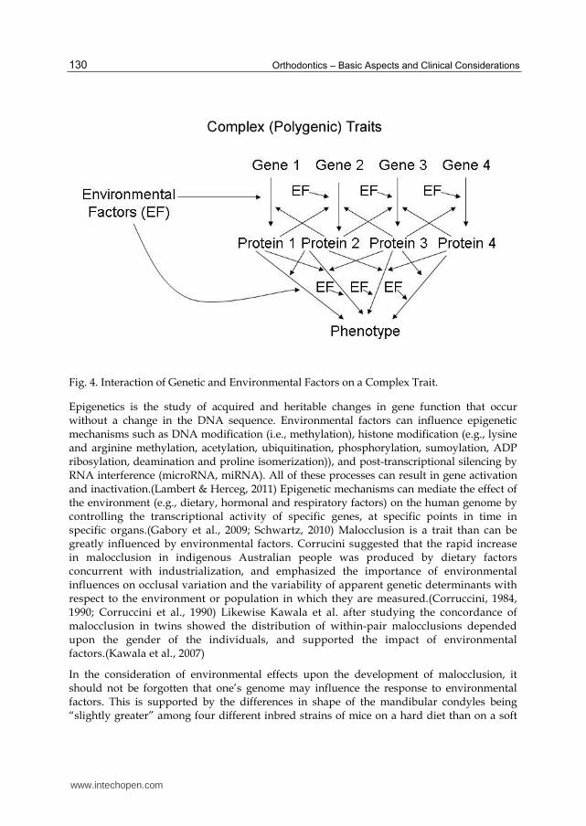
Most traits do not adhere to patterns of Mendelian inheritance. These traits are referred to as
complex or common diseases and traits, and reflect their complex interaction between genes
from more than one locus and environmental factors. Polygenic traits infer the effect of
multiple genes on the phenotype, but can be affected by environmental factors also (see
figure 4). The distinction between polygenic traits and multifactorial traits (both are traits
influenced by environmental and multiple genetic factors) has been made for some
multifactorial traits that are discrete (dichotomous) and that occur in an individual once a
developmental threshold of genetic and environmental factors to produce the phenotype
has been reached.
www.intechopen.com
Orthodontics – Basic Aspects and Clinical Considerations
130
Fig. 4. Interaction of Genetic and Environmental Factors on a Complex Trait.
Epigenetics is the study of acquired and heritable changes in gene function that occur without a change in the DNA sequence. Environmental factors can influence epigenetic mechanisms such as DNA modification (i.e., methylation), histone modification (e.g., lysine and arginine methylation, acetylation, ubiquitination, phosphorylation, sumoylation, ADP ribosylation, deamination and proline isomerization)), and post-transcriptional silencing by RNA interference (microRNA, miRNA). All of these processes can result in gene activation and inactivation.(Lambert & Herceg, 2011) Epigenetic mechanisms can mediate the effect of the environment (e.g., dietary, hormonal and respiratory factors) on the human genome by controlling the transcriptional activity of specific genes, at specific points in time in specific organs.(Gabory et al., 2009; Schwartz, 2010) Malocclusion is a trait than can be greatly influenced by environmental factors. Corrucini suggested that the rapid increase in malocclusion in indigenous Australian people was produced by dietary factors concurrent with industrialization, and emphasized the importance of environmental influences on occlusal variation and the variability of apparent genetic determinants with respect to the environment or population in which they are measured.(Corruccini, 1984, 1990; Corruccini et al., 1990) Likewise Kawala et al. after studying the concordance of malocclusion in twins showed the distribution of within-pair malocclusions depended upon the gender of the individuals, and supported the impact of environmental factors.(Kawala et al., 2007)
In the consideration of environmental effects upon the development of malocclusion, it should not be forgotten that one’s genome may influence the response to environmental factors. This is supported by the differences in shape of the mandibular condyles being “slightly greater” among four different inbred strains of mice on a hard diet than on a soft
www.intechopen.com
Genetic Factors Affecting Facial Growth
131
diet for six weeks. When the environment changed sufficiently, the response was different among animals with different genotypes that were not different before the environmental change.(Lavelle, 1983) Siblings may often have similar malocclusions not just because of common genetic or environmental factors, but also because of their shared genetic factors affecting how they respond to the shared environmental factors.(King et al., 1993) However, none of these studies on the effect of environmental factors were focused on epigenetic modifications as a result of environmental factors influencing malocclusion. As the exploration of epigenetics continues throughout biology and medicine, it may also be an interesting area to explore in facial growth.
3. Heritability and malocclusion
Most problems in orthodontics (or any outcome of growth), unless acquired by trauma, are
not strictly the result of only genetic or only environmental factors. The ideal occlusion
condition shows a proportional growth between the cranial base, the maxilla and the
mandible; and involves the harmonious relation between skeletal bases and soft tissues
(perioral musculature, lips and tongue).(Mossey, 1999b) The general morphology of
craniofacial bones and teeth are largely genetically determined, although clearly variation is
partly attributable to environmental factors.(Corruccini et al., 1990; Harris, 2008;
Klingenberg et al., 2004; Kraus & Lufkin, 2006; Thesleff, 2006; Townsend et al., 2003) Genetic
mechanisms predominate during embryonic craniofacial morphogenesis and in the etiology
of many craniofacial abnormalities, therefore genetic factors must be considered in the
etiology of malocclusion. However environment is also thought to influence dentofacial
morphology postnatally, particularly during facial growth. In response to the presumption
of the genome being the predetermining force for facial development and by inference
skeletal malocclusion, the Functional Matrix Hypothesis by Moss theorized the primary role
of function in craniofacial growth and development. Still, Moss did conclude that both
genomic and environmental/epigenetic factors are necessary causes, that neither alone is a
sufficient cause and that only the two interacting together furnish both the necessary and
sufficient cause(s) of growth and development.(Moss, 1997b, 1997a)
One method employed to estimate this relative contribution of genetic and environmental factors is by calculating the heritability of a trait. Heritability in the broad since (H2) includes all additive, interactive and other types of genetic and environmental influences. This is impossible to derive, since all the factors and how they interact is not known. Therefore heritability estimates in the literature are in the narrow sense (h2), and represent the proportion of the total phenotypic variance in a sample that is contributed by additive genetic variance. However, the estimated ratio of genetic variation does not take into account gene-gene or gene-environment interaction.(Hartsfield, 2011) Numerous studies have examined how genetic variation contributes to either or both occlusal and skeletal variation among family members. It is difficult to estimate the influence of environmental (treatment) factors in craniofacial growth because the heritability studies of occlusion are typically based on twins and siblings who did not receive orthodontic treatment. Twin pairs and other groups of siblings containing one or more treated patients (with moderate to severe malocclusion) may have been excluded from most studies. Moreover the twin studies have not included extensive analysis of the parents, nor familial, and nutritional habits; and usually have not compared the twin group with a control group to ascertain environmental
www.intechopen.com

Orthodontics – Basic Aspects and Clinical Considerations
132
covariance (similarity due to twins and other siblings being in a common environment). Therefore, estimates of genetic and environmental contributions may have been affected by lack of accounting for a common environmental effect(Corruccini & Potter, 1980) and ascertainment bias.(King et al., 1993)
The cause of most skeletal- and dentoalveolar based malocclusions is essentially multifactorial in the sense that many diverse causes converge to produce the observed outcome.(King et al., 1993) Numerous studies have examined how genetic variation contributes to either or both occlusal and skeletal variation among family members.(Arya et al., 1973; Boraas et al., 1988; Byard et al., 1985; Cassidy et al., 1998; Chung & Niswander, 1975; Corruccini et al., 1986; Devor, 1987; Fernex et al., 1967; Gass et al., 2003; Harris et al., 1973; Harris et al., 1975; Harris & Smith, 1980; Harris & Johnson, 1991; Hauspie et al., 1985; Horowitz et al., 1960; Hunter et al., 1970; Johannsdottir et al., 2005; King et al., 1993; Kraus et al., 1959; Litton et al., 1970; Lobb, 1987; Lundstrom & McWilliam, 1987; Manfredi et al., 1997; Nakata et al., 1973; Nikolova, 1996; Proffit, 1986; Saunders et al., 1980; Susanne & Sharma, 1978; Watnick, 1972) In most studies (particularly those that try to account for bias from the effect of shared environmental factors, unequal means, and unequal variances in monozygotic and dizygotic twin samples),(Harris & Potter, 1997) variations in cephalometric skeletal dimensions are associated in general with a moderate to high degree of genetic variation, whereas in general, variation of occlusal relationships has little or no association with genetic variation.(Harris, 2008)
Although the heritability estimates are low, most of the studies that looked at occlusal traits found that genetic variation is positively correlated with phenotypic variation for arch width and arch length more than for overjet, overbite, and molar relationship. Still, arch size and shape are associated more with environmental variation than with genetic variation.(Cassidy et al., 1998) Because many occlusal variables reflect the combined variations of tooth position and basal and alveolar bone development, these variables (e.g., overjet, overbite, and molar relationship) cannot be less variable than the supporting structures. They will vary because of their own variation in position and those of the basilar structures.(Harris & Johnson, 1991) Heritability studies must be supplemented and to some degree superseded by studies linking or associating specific traits with variation in genetic markers such as single nucleotide polymorphisms (SNPs), variable number of tandem repeats, or other types of specific DNA variation.
For example, SNPs in the EDA gene and the gene for its receptor XEDAR, were found to be associated with dental crowding greater than 5 mm in a Hong Kong Chinese Class I malocclusion sample. It was thought that this may at least be due in part to variation in tooth size as the gene product of EDA is involved in tooth development, and mutations in EDA cause X-linked Hypohydrotic Ectodermal Dysplasia.(Ting et al., 2011) A possible affect on tooth size is consistent with the findings that in skeletal Class I crowding cases tooth size variation may more often play a role than skeletal growth.(Bernabe & Flores-Mir, 2006; Hashim & Al-Ghamdi, 2005; Poosti & Jalali, 2007; Ting et al., 2011) Although these genes are located on the X chromosome, the associations remained after adjustment for sex. This type of investigation is thought to help get around the problem of confounding environmental factors, although an increased analysis of epigenetic markers may show this is not that simple. Still these studies are the only way in which possible predictive data will be collected and tested.
www.intechopen.com
Genetic Factors Affecting Facial Growth
133
4. Use of family data to predict growth
Siblings have been noted as often showing similar types of malocclusion. Examination of parents and older siblings has been suggested as a way to gain information regarding the treatment need for a child, including early treatment of malocclusion.(Harris & Kowalski, 1976; Litton et al., 1970; Niswander, 1975; Saunders et al., 1980) Niswander noted that the frequency of malocclusion is decreased among siblings of index cases with normal occlusion, whereas the siblings of index cases with malocclusion tend to have the same type of malocclusion more often. (Niswander, 1975) There are high correlation coefficient values between parents and their offspring for Class II and Class III malocclusions.(Nakasima et al., 1982) It has been shown that the craniofacial skeletal patterns of children with Class II (division 1) malocclusions are familial (i.e., occur more often in multiple members of some families), and that a high resemblance to the skeletal patterns occurs in their siblings with normal occlusion.(Harris et al., 1975) Although this was ascribed to the Class II (division 1) being “heritable,” common environmental factors were not taken into account. From this it was concluded that the genetic basis for this resemblance is probably polygenic, and family skeletal patterns were used as predictors for the treatment prognosis of the child with a Class II malocclusion, although it was acknowledged that the current morphology of the patient is the primary source of information about future growth.(Harris & Kowalski, 1976)
Each child receives half of his or her genes from each parent, but not likely the same combination of genes as a sibling unless the children are monozygotic twins. When looking at parents with a differing skeletal morphology, knowing which of the genes in what combination from each parent is present in the child is difficult until the child’s phenotype matures under the continuing influence of environmental factors. When considering polygenic traits, the highest phenotypic correlation that can be expected based on genes in common by inheritance from one parent to a child, or between siblings, is 0.5. Because the child’s phenotype is likely to be influenced by the interaction of genes from both parents, the “mid-parent” value may increase the correlation with their children to 0.7 because of the regression to the mean of parental dimensions in their children. Squaring the correlation between the two variables derives the amount of variation predicted for one variable in correlation with another variable. Therefore, at best, using mid-parent values, only 49% of the variability of any facial dimension in a child can be predicted by consideration of the average of the same dimension in the parents. Only 25% of the variability of any facial dimension in a child can be predicted, at best, by considering the same dimension in a sibling or one parent. Because varying effects of environmental factors interact with the multiple genetic factors, the usual correlation for facial dimensions between parents and their children is about 30%, yielding even less predictive power.(Hunter, 1990)
In most patients, the mode of inheritance for the craniofacial skeleton is polygenic (complex). However, in some families (e.g., with a relatively prognathic mandible compared with the maxilla), the mode of inheritance is not polygenic. Future research may investigate the genetic factors that do not fit a polygenic mode that may be present in some families. Identification of those factors will increase the ability to predict the likelihood of a particular resulting morphology. Unfortunately, orthodontists do not have sufficient information to make accurate predictions about the development of occlusion simply by studying the frequency of its occurrence in parents or even siblings. Admittedly, family patterns of resemblance are frequently obvious, and observed family tendencies should not be ignored.
www.intechopen.com
Orthodontics – Basic Aspects and Clinical Considerations
134
Nonetheless, predictions must be made cautiously because genetic and environmental factors and their interaction are unknown and difficult to evaluate and predict with precision.(Hartsfield, 2011)
5. Genetic markers associated with variations in growth of complex etiology
5.1 Growth hormone receptor
Growth hormone is an important factor in craniofacial and skeletal growth. A variant in the growth hormone receptor and its gene (GHR), when there is a proline amino acid instead of threonine at the 561st residue in the protein, is referred to as the GHR P56IT allele. Of a normal Japanese sample of 50 men and 50 women, those who did not have the GHR P56IT allele had a significantly greater mandibular ramus length (condylion-gonion) than did those with the GHR P56IT allele. The average mandibular ramus height in those with the GHR P56IT allele was 4.65 mm shorter than the average for those without the GHR P56IT allele. This significant correlation between the GHR P56IT allele and shorter mandibular ramus height was confirmed in an additional 80 women.(Yamaguchi et al., 2001) Interestingly, the association was with the mandibular ramus height but not mandibular body length, maxillary length, or anterior cranial base length. This suggests a site-, area-, or region-specific effect. The study concluded that the GHR P56IT allele may be associated with mandibular height growth and can be a genetic marker for it. Still, whether the effect is directly on the mandible or some other nearby tissue or on another matrix is not clear. It has been suggested that GHR variants P561T and C422F are associated with mandibular ramus height in Japanese population and that the SNPs of the GHR gene associated with differences in mandibular ramus height in the Japanese are likely to be different in other ethnic groups. (Tomoyasu et al., 2009)
This is supported by the finding that although there is a possible association between the GHR polymorphisms P561T, C422F and “haplotype 4” in a Korean population, there was not significant association between these markers and mandibular height in African-Americans, European-Americans, and Hispanics.(Kang et al., 2009) This group suggested that this finding might partly explain the differing craniofacial morphology among different ethnicities. Analysis of the possible association between the P561T variant in the GHR gene and mandibular growth during early childhood did not find a difference between mandibular protrusion and normal occlusion. (Sasaki et al., 2009) To see what effect different diets would have on individuals with and without the GHR P56IT allele would be interesting as a means of looking at genetic and environmental factor interaction. Undoubtedly many other genes that may influence craniofacial structure, including ramus height, could be identified, and their variation could be studied along with different environmental factors (e.g., orthodontic treatment) and the resulting phenotype.
5.2 Growth differences during puberty
Increased accuracy in the estimation of pubertal facial growth would be of great benefit prior to the utilization of different therapeutic modalities including orthodontics, orthopedic growth modification and surgery. Research and discussion about facial growth and treatment in the literature have focused either on the timing of the greatest amount of facial growth, particularly for the mandible(Gu & McNamara, 2007; Hunter et al., 2007; Verma et
www.intechopen.com
Genetic Factors Affecting Facial Growth
135
al., 2009); or the estimated extent of facial growth to be attained.(Chvatal et al., 2005; Turchetta et al., 2007) As useful as average facial growth predictions based upon expected growth curves may be, more valid prediction must incorporate and account for the variation associated with individual genetic factors, particularly those that are highly pertinent to the pubertal growth spurt. The pubertal growth spurt response is mediated by the combination of sex steroids, growth hormone, insulin-like growth factor (IGF-I) and other endocrine, paracrine and autocrine factors. Testosterone and estradiol in mice have a direct, sex-specific stimulatory activity on male and female derived chondroprogenitor cell proliferation. Testosterone stimulated growth and local production of IGF-I and IGF-I-R in chondrocyte cell layers of an isolated organ culture of mice mandibular condyle.(Maor et al., 1999) Investigation into the effects of neonatal surgical castration and prepubertal chemical castration on craniofacial growth in rats showed that craniofacial growth was related to testosterone concentration. Administration of low doses of testosterone in boys with delayed puberty not only accelerates their statural growth rate, but their craniofacial growth rate as well.(Verdonck et al., 1998; Verdonck et al., 1999)
Ovariectomized and orchiectomized mice that sex hormone levels influenced condylar morphogenesis changed the internal structure of the mandibular condyle.(Fujita et al., 2001) It has been suggested that the suppression of sex hormone secretion in the growth phase might inhibit craniofacial growth and result in poor craniofacial development, particularly nasomaxillary bone and mandible, in new born and pubertal rats.(Fujita et al., 2004; Fujita et al., 2006) It has been demonstrated using administration of sex hormone specific receptor antagonists that growth of the mandible and femur is induced in response to the stimulation of the estrogen receptor beta (ER┚) in chondrocytes before and during early puberty in mice. In late and after puberty, the growth is induced by the stimulation of estrogen receptor alpha (ER┙) in male and female mice. From this it was proposed that a screen of sex hormones could be used as an indicator of bone maturity to accurately predict the beginning and end of growth in orthodontic treatment.
CYP19A1 is the gene that encodes aromatase. This enzyme catalyzes the rate limiting step in estrogen biosynthesis by converting androgens. In order to best diagnose and treat the child or adolescent patient, the orthodontist needs to know as much as possible about the patient’s growth potential. As useful as predictions based upon expected growth models starting from early in the patient’s life may be, prediction must incorporate and account for the variation associated with individual genetic factors, especially those that are highly pertinent to the pubertal growth spurt.
Estrogens are a group of hormones involved in growth and development.(Honjo et al., 1992) Estrogen stimulates chondrogenesis, promotes the progressive closure of the epiphyseal growth plate, has an anabolic effect on the osteoblast and an apoptotic effect on the osteoclast, and increases bone mineral acquisition in axial and appendicular bone during adolescence and into the third decade.(Grumbach, 2000) Aromatase (also known as estrogen synthetase) is a key cytochrome P450 enzyme involved in estrogen biosynthesis.(Bulun et al., 2003) This steroidogenic enzyme catalyzes the final step of estrogen biosynthesis by converting testosterone and androstenedione to estradiol and estrone, respectively.(Guo et al., 2006) Regulation of this gene’s transcription is critical for the testosterone/estrogen (T/E) ratio in the body since aromatase plays an important role in the conversion of androgens to estrogens. Some studies have shown that the T/E ratio is critical in the
www.intechopen.com

Orthodontics – Basic Aspects and Clinical Considerations
136
development of sex-indexed facial characteristics such as the growth of cheekbones, the mandible and chin, the prominence of eyebrow ridges and the lengthening of the lower face. (Schaefer et al., 2005; Schaefer et al., 2006)
The difference in the average sagittal jaw growth between the two groups of Caucasian
males with different CYP19A1 alleles with the greatest differences in growth per year was
just over 1.5 mm per year during treatment for the maxilla, and 2.5 mm per year for the
mandible. (Hartsfield Jr. et al., 2010) There was no statistical difference for the particular
CYP19A1 alleles in females. This is particularly impressive since at the beginning of
treatment there was no significant difference among the males based upon the CYP19A1
genotype. The significant difference only expressed itself over the time of treatment during
the cervical vertebral stage associated with increased growth velocity.(Hartsfield et al., 2010)
Interestingly the same result was found in a group of Chinese males and females, strongly
suggesting that this variation in the CYP19A1 gene may be a multi-ethnic marker for sagittal
facial growth. (He et al., 2011) Although the difference in average annual sagittal
mandibular and maxillary growth based upon this CYP19A1 genotype were significant, as
one factor in a complex trait (sagittal jaw growth), they account for only part of the variation
seen, and therefore by itself has little predictive power. Further investigation of this and
other genetic factors, their interactions with each other and with environmental factors will
help to explain what has up to now been an unknown component of individual variations in
facial growth.
5.3 Class II division 2 (Class II/2) malocclusion
There is evidence that Class II division 2, and particularly Class III malocclusions, can have
a strong genetic component. The Class II division 2 (II/2) malocclusion is a relatively rare
type of malocclusion, representing between 2.3% and 5% of all malocclusions in the western
white population.(Ast et al., 1965; Mills, 1966) In one study 100% of 20 monozygotic (MZ)
twin pairs were concordant for II/2 malocclusion, while only 10.7% of 28 dizygotic (DZ)
twin pairs demonstrated concordance for the Class II/2 malocclusion. (Markovic, 1992)
These findings suggest the effect of common genetic or environmental factors; however, the
much lower concordance for DZ twins would suggest that multiple genetic factors rather
than a single gene contribute to the risk for Class II/2. This was reinforced by Ruf et al.
concluding that the etiology of Class II/2 malocclusion was unclear, with neither form nor
function the sole controlling factor.(Ruf & Pancherz, 1999)
From a developmental viewpoint it is interesting that there is a strong association of Class
II/2 malocclusion with dental developmental anomalies, more so than for other Angle
malocclusion classes.(Basdra et al., 2001) Excluding 3rd molars, agenesis of other teeth
was at least three times more common in Class II/2 subjects than in the general
population. In addition, there were a significantly greater number of dental
developmental anomalies present in Class II/2 subjects as compared to the general
population. They found 56.6% of Class II/2 patients exhibited developmental tooth
anomalies including hypodontia as compared to as many as 35% of the general
population having agenesis of one or more third molar.(Basdra et al., 2000) In addition
Peck et al. showed a statistically significant reduction in permanent maxillary incisor
mesial-distal width associated with Class II/2.(Peck et al., 1998)
www.intechopen.com
Genetic Factors Affecting Facial Growth
137
Further evidence for a polygenic complex etiology for Class II/2 was found in a study of 68
subjects (67 self reported as white and 1 white/African-American who was a child of one of
the 18 probands). (Morrison, 2008) A proband is the affected individual through whom a
family is first seen or studied for a genetic trait, syndrome or disorder. In this study,
researchers included 50 reported first-degree relatives of each proband, with a minimum of
2 first-degree relatives of each proband. The findings showed a marked increase in the
number of females affected with Class II/2 in both the probands and their first-degree
relatives than affected males. Of the 36 first-degree relatives whose occlusion was analyzed,
6 (16.7%) were found to be Class II/2. The relative risk (RR) of first-degree relatives to have
a Class II/2 was found to be 3.3 – 7.3. The confidence interval (CI) was 1.1-10.3 if the RR was
3.3 and 1.7-31.6 if the RR was 7.3.
Agenesis of one or more permanent teeth (excluding 3rd molars) was found in 2 (11.1%) of
the 18 probands and 7 (14.0%) of the 50 first-degree relatives. Agenesis of one or more 3rd
molars was found in 4 (22.2%) of the 18 probands and 12 (24.0%) of the 50 first-degree
relatives. Agenesis of one or both permanent maxillary incisors was found in none of the 18
probands and 2 (4.0%) of the 50 first-degree relatives. One or more small teeth (excluding
3rd molars) were found in 4 (22.2%) of the 18 probands and 15 (30.0%) of the 50 first-degree
relatives. Small maxillary permanent incisors were found in none of the 18 probands and 4
(8.0%) of the 50 first-degree relatives. Agenesis of one or more permanent teeth in
combination with the presence of one or more small permanent teeth was found in 2 (11.1%)
of the 18 probands and 7 (14.0%) of the 50 first-degree relatives. Of the 36 first-degree
relatives evaluated for malocclusion, 6 (16.67%) were found to be Class II/2. The RR for
first-degree relatives of the probands to have a Class II/2 malocclusion was 3.3 –
7.35.(Morrison, 2008)
These results indicate that first-degree relatives of Class II/2 probands have a significantly
increased risk of having a Class II/2 malocclusion as compared with individuals from the
general population. Were Class II/2 malocclusion to be the result of variation in a single
gene, acting in either a dominant or recessive fashion, the relative risk would be expected to
be much higher. Rather, the modest, albeit significant increase in risk appears consistent
with results from previous studies, which suggest a multifactorial etiology for Class II/2
malocclusion.
The question could be raised as to whether or not anomalous maxillary lateral incisors are
associated with the Class II/2 malocclusion phenotype, and therefore share common
etiological factors. Basdra et al. showed that 13.9% of Class II/2 subjects had agenesis of
maxillary lateral incisors and 7.5% had peg-shaped or small maxillary lateral incisors. In
contrast, Morrison found none of the probands had agenesis of or small maxillary lateral
incisors, although first-degree relatives of the Class II/2 probands showed similar
frequencies of hypodontia and microdontia of other teeth as the II/2 probands. However,
the frequencies of these dental anomalies in the probands and first degree relatives were not
significantly greater than those in the general population.(Morrison, 2008) Thus it is unclear
if Class II/2 probands and their first-degree relatives are at an increased risk of developing
hypodontia and/or microdontia. Investigations of a larger sample of Class II/2 subjects and
relatives to address that question and possible common etiological factors, including genes
associated with tooth development and hypodontia, are needed.
www.intechopen.com
Orthodontics – Basic Aspects and Clinical Considerations
138
A start on this was made when DNA markers (single nucleotide polymorphisms, also referred to as SNPs) in two genes associated with dental development and or hypodontia, MSX1, PAX9, AXIN2. RUNX2 and RUNX3 were investigated in 94 Class II/2 Caucasian subjects (31 with hypodontia) compared to 89 non-Class II/2 Caucasian subjects without hypodontia. (Morford et al., 2010b; Morford et al., 2010a) A borderline-association of all Class II/2 subjects with the PAX9 SNP (rs8004560) was identified (p=0.06). A borderline-association of the same rs8004560 PAX9 SNP was also identified for subjects with Class II/2 with hypodontia of any permanent tooth, excluding third-molars, when compared to non-Class II/2 without hypodontia (p=0.08) but not when compared to Class II/2 without hypodontia (p=0.46). No associations of Class II/2 with the PAX9 rs1955734, MSX1 rs3821949, RUNX2 (rs1406846), RUNX3 (rs6672420), or AXIN2 (rs7591, rs2240308) genotypes were identified. There was a significant association (p=0.0286) for Class II/2 subjects (with or without hypodontia) and the RUNX2 rs6930053 SNP. However, there was no association of RUNX2 rs6930053 for subjects with Class II/2 that had hypodontia of any permanent tooth, including third-molars, when compared to Class II/2 subjects without hypodontia (p=0.3858). This suggests a mild impact of PAX9 (or a locus in linkage-disequilibrium with it) on the development of Class II/2 with hypodontia, and that RUNX2 (or genetic loci in linkage-disequilibrium with RUNX2) plays a role in Class II/2 development but not in the occasionally-associated hypodontia. These findings and other DNA markers should be investigated in a larger Caucasian and other ethnic groups.(Malinowski, 1983; Strohmayer, 1937; Suzuki, 1961)
5.4 Class III malocclusion
Although all Angle occlusion types a Class III malocclusion were initially only based on the sagittal relationship of the permanent first molars, it has generally been recognized that this dental relationship is often observed with a corresponding skeletal relationship as well. Thus, the Class III malocclusion is a complex disorder characterized by a combination of dental and skeletal features that characteristically result in the appearance of a prominent lower jaw. Often referred to as mandibular prognathism (taken from the Greek pro =forward and gnathos =jaw), skeletal aspects of this disorder can be a result of pure mandibular prognathism, maxillary hypoplasia/retrognathism, or a combination of the two. These phenotypic variations create a significant heterogeneity among Class III subjects that can vary according to sex and ethnicity, and account for some of the difficulty encountered when investigating the condition.(Singh, 1999) The familial nature of mandibular prognathism was first reported by Strohmayer (1937) as noted by Wolff et al (1993) in their analysis of the pedigree of the Hapsburg family.(Wolff et al., 1993)
The highest prevalence of Class III malocclusion is observed in East Asian populations such as Korean, Chinese, and Japanese (8%-40%).(Allwright, 1964; Ishii et al., 2002) By comparison, African populations exhibit a reduced prevalence rate (3-8%) compared to Asian samples(Emrich et al., 1965; Garner & Butt, 1985), as do individuals of European or European-American (Caucasian) decent (reports varying between 0.48%-9.5%, with most in the 3-5% range)(Davidov et al., 1961; Emrich et al., 1965; Goose et al., 1957; Helm, 1968; Horowitz, 1970; Ingervall, 1974; Laine & Hausen, 1983; Luffingham & Campbell, 1974; Massler & Frankel, 1951; Solow & Helm, 1968; Tipton & Rinchuse, 1991) While the prevalence in a sample of Native American Chippewa Indian children is relatively low (2.6-3.1%),(Grewe et al., 1968) North American Eskimos in Labrador, Canada have a class III prevalence of approximately 16%.(Zammit et al., 1995) (Zammit, Hans, et al. 1995)
www.intechopen.com

Genetic Factors Affecting Facial Growth
139
Populations in South America are often a mixture of Caucasian/European, African and Amerindian decent. While the percentage of children in Bogotá, Colombia with Class III has been reported as 3.7%, Brazilian children exhibited a frequency between 4 -10%.(Grando et al., 2008; Martins Mda & Lima, 2009; Thilander et al., 2001) In areas of the Middle East, the prevalence of class III also displays variation with the highest prevalence in Egypt at 10.6%,(El-Mangoury & Mostafa, 1990) followed by 7.8% in Iran,(Borzabadi-Farahani et al., 2009) and 5.1% in Lebanon.(Saleh, 1999)
Several studies have suggested the existence of multiple patterns or sub-phenotypes of the Class III malocclusion based on anatomical appearance. For example, Ellis and McNamara reported considerable variation among class III patients. The most common combination of variables was a retrusive maxilla, protrusive maxillary incisors, retrusive mandibular incisors, a protrusive mandible, and a long lower facial height.(Ellis & McNamara, 1984) Although they did not find significant sex differences, Baccetti et al. showed a significant degree of sexual dimorphism in craniofacial features in subjects with class III malocclusion.(Baccetti et al., 2005) The female Class III subjects presented smaller linear dimensions in the maxilla, mandible, and anterior facial heights when compared with male subjects. The increase in mandibular growth was three times greater in males with class III than in subjects with normal occlusion.(Baccetti et al., 2007) Martone and colleagues suggested that craniofacial growth generates several head form types resulting in anatomic sub-groupings of Classes III.(Martone et al., 1992) Mackay et al (1992) identified five Class III subgroups, all of which exhibited mandibular prognathism.(Mackay et al., 1992) English children with Class III malocclusions divided into groups (normal anteroposterior positioned mandibles and protruded mandibles) according to their SNB angle were found to have significant differences in both groups relating to sagittal position of the maxilla and mandibular rotation.(Hashim & Sarhan, 1993)
Bui et al (2006) found five clusters representing distinct subphenotypes of class III malocclusion. The groupings of variables reflected anteroposterior and vertical dimensions rather than specific craniofacial structures, suggesting that different genes are involved in controlling dimension versus structure. The five subgroupings or “Prototype Clusters” were described as follows: (1) prognathic mandible with long face, (2) maxillary deficiency with decreased vertical dimension (low angle), (3) maxillary deficiency with increased vertical dimension (high angle), (4) mild prognathic mandible with normal vertical dimension, and (5) a combination of prognathic mandible and maxillary deficiency with normal vertical dimension.(Bui et al., 2006) Further studies of the variation of the subtypes of the Class III phenotype within families should facilitate increased understanding of the genetic and non-genetic factors involved.
The genetic factors appear to be heterogeneous, with monogenic (usually autosomal dominant with incomplete penetrance and variable expressivity) influences in some families and multifactorial (polygenic complex) influences in others.(Cruz et al., 2008; Downs, 1927 ; El-Gheriani et al., 2003; Krauss et al., 1959; Litton et al., 1970; Niswander, 1975; Stiles & Luke, 1953; Strohmayer, 1937; Thompson & Winter, 1988; Wolff et al., 1993) This contributes to the variety of anatomical changes in the cranial base, maxilla, and mandible that may be associated with “mandibular prognathism” or a Class III malocclusion.(Bui et al., 2006; Singh, 1999) The prevalence of Class III malocclusion varies among races and can show different anatomic characteristics between races.(Ishii et al., 2002) Considering this
www.intechopen.com

Orthodontics – Basic Aspects and Clinical Considerations
140
heterogeneity, and possible epistasis (the interaction between or among gene products on their expression) and even epigenetics, it is not surprising that genetic linkage and candidate gene studies to date have indicated the possible location of genetic loci influencing this trait in several chromosomal locations (see figure 5).(Falcão-Alencar et al., 2010; Frazier-Bowers et al., 2009; Jang et al., 2010; Li et al., 2010; Li et al., 2011; Tassopoulou-Fishell et al., 2011; Xue et al., 2010; Yamaguchi et al., 2005)
Fig. 5. Chromosome location of markers linked or associated with Class III malocclusion in humans.
6. Personalized orthdontics
In summary, “Personalized Medicine” is a new buzz phrase, based initially upon pharmacogenetics and now exploding as genome-wide association and pathway studies are undertaken. The understanding of the combination and interaction of genetic and environmental (including treatment) factors (nature and nurture together) that influence the growth treatment response of our patients is fundamental to the evidence based practice of orthodontics. Conclusions from retrospective studies must be evaluated by prospective testing to truly evaluate their value in practice. Genome-wide association studies, metabolic pathway analysis and candidate gene studies are necessary to further the evidence base for the practice of orthodontics to determine what the best treatment plan is for each patient in the era of truly personalized orthodontics.(Hartsfield, 2008)
7. References
Allwright, W.C. (1964). A survey of handicapping dentofacial anomalies among Chinese in
Hong Kong. Int Dent J, Vol. 14, No. 1964), pp. 505-519,
www.intechopen.com
Genetic Factors Affecting Facial Growth
141
Arya, B.S., Savara, B.S., Clarkson, Q.D. & Thomas, D.R. (1973). Genetic variability of
craniofacial dimensions. The Angle orthodontist, Vol. 43, No. 2, (Apr 1973), pp. 207-
215, 0003-3219 (Print)
Ast, D.B., Carlos, J.P. & Cons, N.C. (1965). The Prevalence and Characteristics of
Malocclusion among Senior High School Students in Upstate New York. American
journal of orthodontics, Vol. 51, No. (Jun 1965), pp. 437-445, 0002-9416
Baccetti, T., Reyes, B.C. & McNamara, J.A., Jr. (2005). Gender differences in Class III
malocclusion. The Angle orthodontist, Vol. 75, No. 4, (Jul 2005), pp. 510-520, 0003-
3219 (Print) 0003-3219 (Linking)
Baccetti, T., Reyes, B.C. & McNamara, J.A., Jr. (2007). Craniofacial changes in Class III
malocclusion as related to skeletal and dental maturation. American journal of
orthodontics and dentofacial orthopedics : official publication of the American Association of
Orthodontists, its constituent societies, and the American Board of Orthodontics, Vol. 132,
No. 2, (Aug 2007), pp. 171 e171-171 e112, 1097-6752 (Electronic) 0889-5406 (Linking)
Basdra, E.K., Kiokpasoglou, M. & Stellzig, A. (2000). The Class II Division 2 craniofacial type
is associated with numerous congenital tooth anomalies. European journal of
orthodontics, Vol. 22, No. 5, (Oct 2000), pp. 529-535, 0141-5387
Basdra, E.K., Kiokpasoglou, M.N. & Komposch, G. (2001). Congenital tooth anomalies and
malocclusions: a genetic link? European journal of orthodontics, Vol. 23, No. 2, (Apr
2001), pp. 145-151, 0141-5387
Bernabe, E. & Flores-Mir, C. (2006). Dental morphology and crowding. A multivariate
approach. The Angle orthodontist, Vol. 76, No. 1, (Jan 2006), pp. 20-25, 0003-3219 (Print)
0003-3219 (Linking)
Boraas, J.C., Messer, L.B. & Till, M.J. (1988). A genetic contribution to dental caries,
occlusion, and morphology as demonstrated by twins reared apart. Journal of dental
research, Vol. 67, No. 9, (Sep 1988), pp. 1150-1155, 0022-0345 (Print)
Borzabadi-Farahani, A., Borzabadi-Farahani, A. & Eslamipour, F. (2009). Malocclusion and
occlusal traits in an urban Iranian population. An epidemiological study of 11- to
14-year-old children. European journal of orthodontics, Vol. 31, No. 5, (Oct 2009), pp.
477-484, 1460-2210 (Electronic) 0141-5387 (Linking)
Bui, C., King, T., Proffit, W. & Frazier-Bowers, S. (2006). Phenotypic characterization of Class
III patients. The Angle orthodontist, Vol. 76, No. 4, (Jul 2006), pp. 564-569, 0003-3219
(Print) 0003-3219 (Linking)
Bulun, S.E., Sebastian, S., Takayama, K., Suzuki, T., Sasano, H. & Shozu, M. (2003). The
human CYP19 (aromatase P450) gene: update on physiologic roles and genomic
organization of promoters. J Steroid Biochem Mol Biol, Vol. 86, No. 3-5, (Sep 2003),
pp. 219-224, 0960-0760 (Print)
Buschang, P.H. & Hinton, R.J. (2005). A Gradient of Potential for Modifying Craniofacial
Growth. Semin Orthod, Vol. 11, No. 2005), pp. 219-226,
Byard, P.J., Poosha, D.V., Satyanarayana, M. & Rao, D.C. (1985). Family resemblance for
components of craniofacial size and shape. Journal of craniofacial genetics and
developmental biology, Vol. 5, No. 3, 1985), pp. 229-238, 0270-4145 (Print) 0270-4145
(Linking)
www.intechopen.com

Orthodontics – Basic Aspects and Clinical Considerations
142
Cassidy, K.M., Harris, E.F., Tolley, E.A. & Keim, R.G. (1998). Genetic influence on dental
arch form in orthodontic patients. The Angle orthodontist, Vol. 68, No. 5, (Oct 1998),
pp. 445-454, 0003-3219 (Print)
Chung, C.S. & Niswander, J.D. (1975). Genetic and epidemiologic studies of oral
characteristics in Hawaii's schoolchildren: V. Sibling correlations in occlusion traits.
Journal of dental research, Vol. 54, No. 2, (Mar-Apr 1975), pp. 324-329, 0022-0345
(Print)
Chvatal, B.A., Behrents, R.G., Ceen, R.F. & Buschang, P.H. (2005). Development and testing
of multilevel models for longitudinal craniofacial growth prediction. American
journal of orthodontics and dentofacial orthopedics : official publication of the American
Association of Orthodontists, its constituent societies, and the American Board of
Orthodontics, Vol. 128, No. 1, (Jul 2005), pp. 45-56, 0889-5406 (Print)
Corruccini, R.S. & Potter, R.H. (1980). Genetic analysis of occlusal variation in twins.
American journal of orthodontics, Vol. 78, No. 2, (Aug 1980), pp. 140-154, 0002-9416
(Print)
Corruccini, R.S. (1984). An epidemiologic transition in dental occlusion in world
populations. American journal of orthodontics, Vol. 86, No. 5, (Nov 1984), pp. 419-426,
0002-9416 (Print)
Corruccini, R.S., Sharma, K. & Potter, R.H. (1986). Comparative genetic variance and
heritability of dental occlusal variables in U.S. and Northwest Indian twins.
American journal of physical anthropology, Vol. 70, No. 3, (Jul 1986), pp. 293-299, 0002-
9483 (Print)
Corruccini, R.S. (1990). Australian aboriginal tooth succession, interproximal attrition, and
Begg's theory. American journal of orthodontics and dentofacial orthopedics : official
publication of the American Association of Orthodontists, its constituent societies, and the
American Board of Orthodontics, Vol. 97, No. 4, (Apr 1990), pp. 349-357, 0889-5406
(Print) 0889-5406 (Linking)
Corruccini, R.S., Townsend, G.C., Richards, L.C. & Brown, T. (1990). Genetic and
environmental determinants of dental occlusal variation in twins of different
nationalities. Hum Biol, Vol. 62, No. 3, 1990), pp. 353-367.,
Cruz, R.M., Krieger, H., Ferreira, R., Mah, J., Hartsfield, J., Jr. & Oliveira, S. (2008). Major
gene and multifactorial inheritance of mandibular prognathism. American journal of
medical genetics. Part A, Vol. 146A, No. 1, (Jan 1 2008), pp. 71-77, 1552-4833
(Electronic) 1552-4825 (Linking)
Davidov, S., Geseva, N., Donveca, T. & Dehova, L. (1961). Incidence of prognathism in
Bulgaria. Dental Abstracts, Vol. 6, No. 1961), pp. 240,
Devor, E.J. (1987). Transmission of human craniofacial dimensions. Journal of craniofacial
genetics and developmental biology, Vol. 7, No. 2, 1987), pp. 95-106, 0270-4145 (Print)
Downs, W.G. (1927 ). Studies in the Causes of Dental Anomalies. . Genetics Vol. 12, No. 6,
(Nov 1927 1927 ), pp. 570-580, 0016-6731 (Print) 0016-6731 (Linking)
El-Gheriani, A.A., Maher, B.S., El-Gheriani, A.S., Sciote, J.J., Abu-Shahba, F.A., Al-Azemi, R.
& Marazita, M.L. (2003). Segregation analysis of mandibular prognathism in Libya.
Journal of dental research, Vol. 82, No. 7, (Jul 2003), pp. 523-527, 0022-0345 (Print)
0022-0345 (Linking)
www.intechopen.com

Genetic Factors Affecting Facial Growth
143
El-Mangoury, N.H. & Mostafa, Y.A. (1990). Epidemiologic panorama of dental occlusion.
The Angle orthodontist, Vol. 60, No. 3, (Fall 1990), pp. 207-214, 0003-3219 (Print) 0003-
3219 (Linking)
Ellis, E., 3rd & McNamara, J.A., Jr. (1984). Components of adult Class III malocclusion. J Oral
Maxillofacial Surg, Vol. 42, No. 5, (May 1984), pp. 295-305, 0278-2391 (Print) 0278-
2391 (Linking)
Emrich, R.E., Brodie, A.G. & Blayney, J.R. (1965). Prevalence of Class 1, Class 2, and Class 3
malocclusions (Angle) in an urban population. An epidemiological study. Journal of
dental research, Vol. 44, No. 5, (Sep-Oct 1965), pp. 947-953, 0022-0345 (Print) 0022-
0345 (Linking)
Everett, E.T., Britto, D.A., Ward, R.E. & Hartsfield, J.K., Jr. (1999). A novel FGFR2 gene
mutation in Crouzon syndrome associated with apparent nonpenetrance. The Cleft
palate-craniofacial journal : official publication of the American Cleft Palate-Craniofacial
Association, Vol. 36, No. 6, (Nov 1999), pp. 533-541, 1055-6656 (Print)
Falcão-Alencar, G., Ortero, L., Cruz, R.M., Foroud, T.M., Dongbing, L., Koller, D.,
Morford, L.A., Ferrari, I., Oliveira, S.F. & Hartsfield, J.K. (2010). Evidence for
Genetic Linkage of the Class III Craniofacial Phenotype With Human Chromosome 7 in
36 South American Families, (abstract/program #975). Presented at the 60th
Annual Meeting of The American Society of Human Genetics, November 3, 2010,
Washington, DC).
Fernex, E., Hauenstein, P. & Roche, M. (1967). [Heredity and craniofacial morphology].
Report of the congress. European Orthodontic Society, Vol. No. 1967), pp. 239-257,
Frazier-Bowers, S., Rincon-Rodriguez, R., Zhou, J., Alexander, K. & Lange, E. (2009).
Evidence of linkage in a Hispanic cohort with a Class III dentofacial phenotype.
Journal of dental research, Vol. 88, No. 1, (Jan 2009), pp. 56-60, 1544-0591 (Electronic)
0022-0345 (Linking)
Fujita, T., Kawata, T., Tokimasa, C., Kohno, S., Kaku, M. & Tanne, K. (2001). Breadth of the
mandibular condyle affected by disturbances of the sex hormones in
ovariectomized and orchiectomized mice. Clinical orthodontics and research, Vol. 4,
No. 3, (Aug 2001), pp. 172-176, 1397-5927 (Print) 1397-5927 (Linking)
Fujita, T., Ohtani, J., Shigekawa, M., Kawata, T., Kaku, M., Kohno, S., Tsutsui, K., Tenjo, K.,
Motokawa, M., Tohma, Y. & Tanne, K. (2004). Effects of sex hormone disturbances
on craniofacial growth in newborn mice. Journal of dental research, Vol. 83, No. 3,
(Mar 2004), pp. 250-254, 0022-0345 (Print) 0022-0345 (Linking)
Fujita, T., Ohtani, J., Shigekawa, M., Kawata, T., Kaku, M., Kohno, S., Motokawa, M.,
Tohma, Y. & Tanne, K. (2006). Influence of sex hormone disturbances on the
internal structure of the mandible in newborn mice. European journal of
orthodontics, Vol. 28, No. 2, (Apr 2006), pp. 190-194, 0141-5387 (Print) 0141-5387
(Linking)
Gabory, A., Attig, L. & Junien, C. (2009). Sexual dimorphism in environmental epigenetic
programming. Molecular and cellular endocrinology, Vol. 304, No. 1-2, (May 25 2009),
pp. 8-18, 1872-8057 (Electronic) 0303-7207 (Linking)
www.intechopen.com

Orthodontics – Basic Aspects and Clinical Considerations
144
Garner, L.D. & Butt, M.H. (1985). Malocclusion in black Americans and Nyeri Kenyans. An
epidemiologic study. The Angle orthodontist, Vol. 55, No. 2, (Apr 1985), pp. 139-146,
0003-3219 (Print) 0003-3219 (Linking)
Gass, J.R., Valiathan, M., Tiwari, H.K., Hans, M.G. & Elston, R.C. (2003). Familial
correlations and heritability of maxillary midline diastema. American journal of
orthodontics and dentofacial orthopedics : official publication of the American Association of
Orthodontists, its constituent societies, and the American Board of Orthodontics, Vol. 123,
No. 1, (Jan 2003), pp. 35-39, 0889-5406 (Print)
Goose, D.H., Thomson, D.G. & Winter, F.C. (1957). Malocclusion in schoolchildren of the
west Midlands. British Dental Journal, Vol. 102, No. 1957), pp. 174-178,
Grando, G., Young, A.A., Vedovello Filho, M., Vedovello, S.A. & Ramirez-Yanez, G.O.
(2008). Prevalence of malocclusions in a young Brazilian population. International
journal of orthodontics, Vol. 19, No. 2, (Summer 2008), pp. 13-16, 1539-1450 (Print)
1539-1450 (Linking)
Grewe, J.M., Cervenka, J., Shapiro, B.L. & Witkop, C.J., Jr. (1968). Prevalence o f
malocclusion in Chippewa Indian children. Journal of dental research, Vol. 47, No. 2,
(Mar-Apr 1968), pp. 302-305, 0022-0345 (Print) 0022-0345 (Linking)
Grumbach, M.M. (2000). Estrogen, bone, growth and sex: a sea change in conventional
wisdom. J Pediatr Endocrinol Metab, Vol. 13 Suppl 6, No. 2000), pp. 1439-1455, 0334-
018X (Print)
Gu, Y. & McNamara, J.A. (2007). Mandibular growth changes and cervical vertebral
maturation. a cephalometric implant study. The Angle orthodontist, Vol. 77, No. 6,
(Nov 2007), pp. 947-953, 0003-3219 (Print)
Guo, Y., Xiong, D.H., Yang, T.L., Guo, Y.F., Recker, R.R. & Deng, H.W. (2006).
Polymorphisms of estrogen-biosynthesis genes CYP17 and CYP19 may influence
age at menarche: a genetic association study in Caucasian females. Human molecular
genetics, Vol. 15, No. 16, (Aug 15 2006), pp. 2401-2408, 0964-6906 (Print)
Harris, E.F. & Smith, R.J. (1980). A study of occlusion and arch widths in families.
American journal of orthodontics, Vol. 78, No. 2, (Aug 1980), pp. 155-163, 0002-9416
(Print)
Harris, E.F. & Johnson, M.G. (1991). Heritability of craniometric and occlusal variables: a
longitudinal sib analysis. American journal of orthodontics and dentofacial orthopedics :
official publication of the American Association of Orthodontists, its constituent societies,
and the American Board of Orthodontics, Vol. 99, No. 3, (Mar 1991), pp. 258-268, 0889-
5406 (Print)
Harris, E.F. & Potter, R.H. (1997). Sources of bias in heritability studies. American journal of
orthodontics and dentofacial orthopedics : official publication of the American Association of
Orthodontists, its constituent societies, and the American Board of Orthodontics, Vol. 112,
No. 3, (Sep 1997), pp. 17A-21A, 0889-5406 (Print)
Harris, E.F. (2008). Interpreting Heritability Estimates in the Orthodontic Literature. Semin
Orthod, Vol. 14, No. 2008), pp. 125-134,
Harris, J.E., Kowalski, C.J. & Watnick, S.S. (1973). Genetic factors in the shape of the
craniofacial complex. The Angle orthodontist, Vol. 43, No. 1, (Jan 1973), pp. 107-111,
0003-3219 (Print)
www.intechopen.com
Genetic Factors Affecting Facial Growth
145
Harris, J.E., Kowalski, C.J. & Walker, S.J. (1975). Intrafamilial dentofacial associations for
Class II, Division 1 probands. American journal of orthodontics, Vol. 67, No. 5, (May
1975), pp. 563-570, 0002-9416 (Print)
Harris, J.E. & Kowalski, C.J. (1976). All in the family: use of familial information in
orthodontic diagnosis, case assessment, and treatment planning. American journal of
orthodontics, Vol. 69, No. 5, (May 1976), pp. 493-510, 0002-9416 (Print)
Hartsfield, J.K., Jr. (2008). Personalized Orthodontics, The Future of Genetics in Practice.
Semin Orthod, Vol. 14, No. 2008), pp. 166-171,
Hartsfield, J.K., Jr., Zhou, J. & Chen, S. (2010).In: Surgical Enhancement of Orthodontic
Treatment, Craniofacial Growth Series, Department of Orthodontics and Pediatric
Dentistry and Center for Human Growth and Development, J.A. McNamara & S.D.
Kapila, pp. 267-281, The University of Michigan,Ann Arbor
Hartsfield, J.K., Jr. (2011).5, In: Orthodontics : current principles and techniques, L.W. Graber,
R.L. Vanarsdall & K.W.L. Vig, pp. 139-156, Elsevier Mosby,0323066410 St Louis
Hartsfield, J.K., Jr. & Bixler, D. (2011).6, In: McDonald's and Avery's dentistry for the child and
adolescent, J.A. Dean, D.R. Avery & R.E. McDonald, pp. 64-84,
Mosby/Elsevier,9780323057240, St. Louis, Mo.
Hartsfield Jr., J.K., Zhou, J. & Chen, S. (2010). Genetic Variation in the CYP19A1/Aromatase
Gene and Facial Growth. Journal of dental research, Vol. 89, No. Special Issue A,
2010), pp. Abstract 63, (www.dentalresearch.org).
Hashim, H.A. & Sarhan, O.A. (1993). Dento-skeletal components of class III malocclusions
for children with normal and protruded mandibles. The Journal of clinical pediatric
dentistry, Vol. 18, No. 1, (Fall 1993), pp. 12-16, 1053-4628 (Print) 1053-4628
(Linking)
Hashim, H.A. & Al-Ghamdi, S. (2005). Tooth width and arch dimensions in normal and
malocclusion samples: an odontometric study. J Contemp Dent Pract, Vol. 6, No. 2,
(May 15 2005), pp. 36-51, 1526-3711 (Electronic) 1526-3711 (Linking)
Hauspie, R.C., Susanne, C. & Defrise-Gussenhoven, E. (1985). Testing for the presence of
genetic variance in factors of face measurements of Belgian twins. Annals of human
biology, Vol. 12, No. 5, (Sep-Oct 1985), pp. 429-440, 0301-4460 (Print)
He, S., Hartsfield, J., Guo, Y., Cao, Y., Wang, S. & Chen, S. (2011). Association between
CYP19A1 SNPs and haplotypes and pubertal sagittal jaw growth, Submitted to the
Journal of Dental Research.
Helm, S. (1968). Malocclusion in Danish children with adolescent dentition: an
epidemiologic study. American journal of orthodontics, Vol. 54, No. 5, (May 1968), pp.
352-366, 0002-9416 (Print) 0002-9416 (Linking)
Hennekam, R.C.M., Allanson, J.E., Krantz, I.D. & Gorlin, R.J. (2010). Gorlin's syndromes of
the head and neck (edition), Oxford University Press, 9780195307900 (alk. paper),
Oxford; New York
Honjo, H., Tamura, T., Matsumoto, Y., Kawata, M., Ogino, Y., Tanaka, K., Yamamoto, T.,
Ueda, S. & Okada, H. (1992). Estrogen as a growth factor to central nervous cells.
Estrogen treatment promotes development of acetylcholinesterase-positive basal
forebrain neurons transplanted in the anterior eye chamber. J Steroid Biochem Mol
Biol, Vol. 41, No. 3-8, (Mar 1992), pp. 633-635, 0960-0760 (Print)
www.intechopen.com

Orthodontics – Basic Aspects and Clinical Considerations
146
Horowitz, H.S. (1970). A study of occlusal relations in 10 to 12 year old Caucasian and
Negro children--summary report. International dental journal, Vol. 20, No. 4, (Dec
1970), pp. 593-605, 0020-6539 (Print) 0020-6539 (Linking)
Horowitz, S.L., Osborne, R.H. & DeGeorge, F.V. (1960). A Cephalometric Study Of
Craniofacial Variation In Adult Twins. The Angle orthodontist, Vol. 30, No. 1, 1960),
pp. 1-5,
Hunter, W.S., Balbach, D.R. & Lamphiear, D.E. (1970). The heritability of attained growth in
the human face. American journal of orthodontics, Vol. 58, No. 2, (Aug 1970), pp. 128-
134, 0002-9416 (Print) 0002-9416 (Linking)
Hunter, W.S. (1990). Heredity in the craniofacial complex. (edition), Saunders, Philadelphia
Hunter, W.S., Baumrind, S., Popovich, F. & Jorgensen, G. (2007). Forecasting the timing of
peak mandibular growth in males by using skeletal age. American journal of
orthodontics and dentofacial orthopedics : official publication of the American Association of
Orthodontists, its constituent societies, and the American Board of Orthodontics, Vol. 131,
No. 3, (Mar 2007), pp. 327-333, 1097-6752 (Electronic)
Ingervall, B. (1974). Prevalence of dental and occlusal anomalies in Swedish conscripts. Acta
odontologica Scandinavica, Vol. 32, No. 2, 1974), pp. 83-92, 0001-6357 (Print) 0001-
6357 (Linking)
Ishii, N., Deguchi, T. & Hunt, N.P. (2002). Craniofacial differences between Japanese and
British Caucasian females with a skeletal Class III malocclusion. European journal of
orthodontics, Vol. 24, No. 5, (Oct 2002), pp. 493-499, 0141-5387 (Print) 0141-5387
(Linking)
Jang, J.Y., Park, E.K., Ryoo, H.M., Shin, H.I., Kim, T.H., Jang, J.S., Park, H.S., Choi, J.Y. &
Kwon, T.G. (2010). Polymorphisms in the Matrilin-1 gene and risk of mandibular
prognathism in Koreans. Journal of dental research, Vol. 89, No. 11, (Nov 2010), pp.
1203-1207, 1544-0591 (Electronic) 0022-0345 (Linking)
Johannsdottir, B., Thorarinsson, F., Thordarson, A. & Magnusson, T.E. (2005). Heritability of
craniofacial characteristics between parents and offspring estimated from lateral
cephalograms. American journal of orthodontics and dentofacial orthopedics : official
publication of the American Association of Orthodontists, its constituent societies, and the
American Board of Orthodontics, Vol. 127, No. 2, (Feb 2005), pp. 200-207; quiz 260-201,
0889-5406 (Print) 0889-5406 (Linking)
Kang, E.H., Yamaguchi, T., Tajima, A., Nakajima, T., Tomoyasu, Y., Watanabe, M.,
Yamaguchi, M., Park, S.B., Maki, K. & Inoue, I. (2009). Association of the growth
hormone receptor gene polymorphisms with mandibular height in a Korean
population. Archives of oral biology, Vol. 54, No. 6, (Jun 2009), pp. 556-562, 1879-1506
(Electronic) 0003-9969 (Linking)
Kawala, B., Antoszewska, J. & Necka, A. (2007). Genetics or environment? A twin-method
study of malocclusions. World journal of orthodontics, Vol. 8, No. 4, (Winter 2007),
pp. 405-410, 1530-5678 (Print) 1530-5678 (Linking)
King, L., Harris, E.F. & Tolley, E.A. (1993). Heritability of cephalometric and occlusal
variables as assessed from siblings with overt malocclusions. American journal of
orthodontics and dentofacial orthopedics : official publication of the American Association of
www.intechopen.com
Genetic Factors Affecting Facial Growth
147
Orthodontists, its constituent societies, and the American Board of Orthodontics, Vol. 104,
No. 2, (Aug 1993), pp. 121-131, 0889-5406 (Print)
Klingenberg, C.P., Leamy, L.J. & Cheverud, J.M. (2004). Integration and modularity of
quantitative trait locus effects on geometric shape in the mouse mandible. Genetics,
Vol. 166, No. 4, (Apr 2004), pp. 1909-1921, 0016-6731 (Print) 0016-6731 (Linking)
Kraus, B.S., Wise, W.J. & Frei, R.H. (1959). Heredity and the craniofacial complex. American
journal of orthodontics, Vol. 45, No. 1959), pp. 172,
Kraus, P. & Lufkin, T. (2006). Dlx homeobox gene control of mammalian limb and
craniofacial development. American journal of medical genetics. Part A, Vol. 140, No.
13, (Jul 1 2006), pp. 1366-1374, 1552-4825 (Print) 1552-4825 (Linking)
Krauss, B., Wise, W. & Frei, R. (1959). Heredity and the craniofacial complex. American
journal of orthodontics, Vol. 45, No. 1959), pp. 172-217,
Laine, T. & Hausen, H. (1983). Occlusal anomalies in Finnish students related to age, sex,
absent permanent teeth and orthodontic treatment. European journal of orthodontics,
Vol. 5, No. 2, (May 1983), pp. 125-131, 0141-5387 (Print) 0141-5387 (Linking)
Lambert, M.-P. & Herceg, Z. (2011). Mechanisms of Epigenetic Gene Silencing, Epigenetic
Aspects of Chronic Diseases. H.I. Roach, F. Bronner & R.O.C. Oreffo. Springer,
London. 41-53
Lavelle, C.L. (1983). Study of mandibular shape in the mouse. Acta Anat (Basel), Vol. 117, No.
4, 1983), pp. 314-320, 0001-5180 (Print)
Li, Q., Zhang, F., Li, X. & Chen, F. (2010). Genome scan for locus involved in mandibular
prognathism in pedigrees from China. PLoS One, Vol. 5, No. 9, 2010), pp. 1932-6203
(Electronic) 1932-6203 (Linking)
Li, Q., Li, X., Zhang, F. & Chen, F. (2011). The identification of a novel locus for mandibular
prognathism in the Han Chinese population. Journal of dental research, Vol. 90, No. 1,
(Jan 2011), pp. 53-57, 1544-0591 (Electronic) 0022-0345 (Linking)
Litton, S.F., Ackermann, L.V., Isaacson, R.J. & Shapiro, B.L. (1970). A genetic study of Class 3
malocclusion. American journal of orthodontics, Vol. 58, No. 6, (Dec 1970), pp. 565-
577, 0002-9416 (Print) 0002-9416 (Linking)
Lobb, W.K. (1987). Craniofacial morphology and occlusal variation in monozygous and
dizygous twins. The Angle orthodontist, Vol. 57, No. 3, (Jul 1987), pp. 219-233, 0003-
3219 (Print) 0003-3219 (Linking)
Luffingham, J.K. & Campbell, H.M. (1974). The need for orthodontic treatment. A pilot
survey of 14 year old school children in Paisley, Scotland. Transactions. European
Orthodontic Society, Vol. No. 1974), pp. 259-267,
Lundstrom, A. & McWilliam, J.S. (1987). A comparison of vertical and horizontal
cephalometric variables with regard to heritability. European journal of orthodontics,
Vol. 9, No. 2, (May 1987), pp. 104-108, 0141-5387 (Print)
Mackay, F., Jones, J.A., Thompson, R. & Simpson, W. (1992). Craniofacial form in class III
cases. British journal of orthodontics, Vol. 19, No. 1, (Feb 1992), pp. 15-20, 0301-228X
(Print) 0301-228X (Linking)
Malinowski, A. (1983). Changes in dimensions and proportions of the human mandible
during fetal period. Collegium antropologicum, Vol. 7, No. 1983), pp. 65-70,
www.intechopen.com
Orthodontics – Basic Aspects and Clinical Considerations
148
Manfredi, C., Martina, R., Grossi, G.B. & Giuliani, M. (1997). Heritability of 39 orthodontic
cephalometric parameters on MZ, DZ twins and MN-paired singletons. American
journal of orthodontics and dentofacial orthopedics : official publication of the American
Association of Orthodontists, its constituent societies, and the American Board of
Orthodontics, Vol. 111, No. 1, (Jan 1997), pp. 44-51, 0889-5406 (Print) 0889-5406
(Linking)
Maor, G., Segev, Y. & Phillip, M. (1999). Testosterone stimulates insulin-like growth factor-I
and insulin-like growth factor-I-receptor gene expression in the mandibular
condyle--a model of endochondral ossification. Endocrinology, Vol. 140, No. 4, (Apr
1999), pp. 1901-1910, 0013-7227 (Print) 0013-7227 (Linking)
Markovic, M.D. (1992). At the crossroads of oral facial genetics. European journal of
orthodontics, Vol. 14, No. 6, (Dec 1992), pp. 469-481, 0141-5387 (Print) 0141-5387
(Linking)
Martins Mda, G. & Lima, K.C. (2009). Prevalence of malocclusions in 10- to 12-year-old
schoolchildren in Ceara, Brazil. Oral health & preventive dentistry, Vol. 7, No. 3,
2009), pp. 217-223, 1602-1622 (Print) 1602-1622 (Linking)
Martone, V.D., Enlow, D.H., Hans, M.G., Broadbent, B.H., Jr. & Oyen, O. (1992). Class I and
Class III malocclusion sub-groupings related to headform type. The Angle orthodontist,
Vol. 62, No. 1, (Spring 1992), pp. 35-42; discussion 43-34, 0003-3219 (Print) 0003-3219
(Linking)
Massler, M. & Frankel, J.M. (1951). Prevalence of malocclusion in children aged 14 to 18
years. American journal of orthodontics, Vol. 37, No. 10, (Oct 1951), pp. 751-768, 0002-
9416 (Print) 0002-9416 (Linking)
Mills, L.F. (1966). Epidemiologic studies of occlusion. IV. The prevalence of malocclusion in
a population of 1,455 school children. Journal of dental research, Vol. 45, No. 2, (Mar-
Apr 1966), pp. 332-336, 0022-0345
Morford, L.A., Coles, T.J., Fardo, D.W., Wall, M.D., Morrison, M.W., Kula, K.S. & Hartsfield,
J.K., Jr. (2010a). Assocaiton analysis of Class II division 2 (CII/D2) with RUNX2 and
RUNX3 Journal of dental research. 89 (Special Iss B) abstract #1948:
Morford, L.A., Wall, M.D., Morrison, M.W., Kula, K.S. & Hartsfield, J.K., Jr. (2010b).
Association Analysis of Class-II Division-2 (CII/D2) with PAX9 and MSX1, Journal of
dental research. 89 (Special Iss A) abstract #633:
Morrison, M.W. (2008). Relative Risk of Class II division 2 Malocclusion in First-Degree Relatives
of Probands with Class II division 2 Malocclusion, Department of Orthodontics and
Oral Facial Genetics. Indiana University School of Dentistry, Master of Science in
Dentistry degree:
Moss, M.L. (1997a). The functional matrix hypothesis revisited. 4. The epigenetic antithesis
and the resolving synthesis. American journal of orthodontics and dentofacial
orthopedics : official publication of the American Association of Orthodontists, its
constituent societies, and the American Board of Orthodontics, Vol. 112, No. 4, 1997a),
pp. 410-417.,
Moss, M.L. (1997b). The functional matrix hypothesis revisited. 3. The genomic thesis.
American journal of orthodontics and dentofacial orthopedics : official publication of the
www.intechopen.com
Genetic Factors Affecting Facial Growth
149
American Association of Orthodontists, its constituent societies, and the American Board of
Orthodontics, Vol. 112, No. 3, 1997b), pp. 338-342.,
Mossey, P.A. (1999a). The heritability of malocclusion: Part 1--Genetics, principles and
terminology. British journal of orthodontics, Vol. 26, No. 2, (Jun 1999a), pp. 103-113,
0301-228X (Print)
Mossey, P.A. (1999b). The heritability of malocclusion: part 2. The influence of genetics in
malocclusion. British journal of orthodontics, Vol. 26, No. 3, (Sep 1999b), pp. 195-203,
0301-228X (Print) 0301-228X (Linking)
Nakasima, A., Ichinose, M., Nakata, S. & Takahama, Y. (1982). Hereditary factors in the
craniofacial morphology of Angle's Class II and Class III malocclusions. American
journal of orthodontics, Vol. 82, No. 2, (Aug 1982), pp. 150-156, 0002-9416 (Print)
0002-9416 (Linking)
Nakata, N., Yu, P.I., Davis, B. & Nance, W.E. (1973). The use of genetic data in the prediction
of craniofacial dimensions. American journal of orthodontics, Vol. 63, No. 5, (May
1973), pp. 471-480, 0002-9416 (Print) 0002-9416 (Linking)
Nikolova, M. (1996). Similarities in anthropometrical traits of children and their parents in a
Bulgarian population. Annals of human genetics, Vol. 60, No. Pt 6, (Nov 1996), pp.
517-525, 0003-4800 (Print) 0003-4800 (Linking)
Niswander, J.D. (1975). Genetics of common dental disorders. Dent Clin North Am, Vol. 19,
No. 1, (Jan 1975), pp. 197-206, 0011-8532 (Print)
Peck, S., Peck, L. & Kataja, M. (1998). Class II Division 2 malocclusion: a heritable pattern of
small teeth in well-developed jaws. The Angle orthodontist, Vol. 68, No. 1, (Feb 1998),
pp. 9-20, 0003-3219
Poosti, M. & Jalali, T. (2007). Tooth size and arch dimension in uncrowded versus crowded
Class I malocclusions. J Contemp Dent Pract, Vol. 8, No. 3, 2007), pp. 45-52, 1526-
3711 (Electronic) 1526-3711 (Linking)
Proffit, W.R. (1986). On the aetiology of malocclusion. The Northcroft lecture, 1985
presented to the British Society for the Study of Orthodontics, Oxford, April 18,
1985. British journal of orthodontics, Vol. 13, No. 1, (Jan 1986), pp. 1-11, 0301-228X
(Print)
Ruf, S. & Pancherz, H. (1999). Class II Division 2 malocclusion: genetics or environment? A
case report of monozygotic twins. The Angle orthodontist, Vol. 69, No. 4, (Aug 1999),
pp. 321-324, 0003-3219
Saleh, F.K. (1999). Prevalence of malocclusion in a sample of Lebanese schoolchildren: an
epidemiological study. Eastern Mediterranean health journal = La revue de sante de la
Mediterranee orientale = al-Majallah al-sihhiyah li-sharq al-mutawassit, Vol. 5, No. 2,
(Mar 1999), pp. 337-343, 1020-3397 (Print) 1020-3397 (Linking)
Sasaki, Y., Satoh, K., Hayasaki, H., Fukumoto, S., Fujiwara, T. & Nonaka, K. (2009). The
P561T polymorphism of the growth hormone receptor gene has an inhibitory effect
on mandibular growth in young children. European journal of orthodontics, Vol. 31,
No. 5, (Oct 2009), pp. 536-541, 1460-2210 (Electronic) 0141-5387 (Linking)
Saunders, S.R., Popovich, F. & Thompson, G.W. (1980). A family study of craniofacial
dimensions in the Burlington Growth Centre sample. American journal of
www.intechopen.com
Orthodontics – Basic Aspects and Clinical Considerations
150
orthodontics, Vol. 78, No. 4, (Oct 1980), pp. 394-403, 0002-9416 (Print) 0002-9416
(Linking)
Schaefer, K., Fink, B., Mitteroecker, P., Neave, N. & Bookstein, F.L. (2005). Visualizing facial
shape regression upon 2nd to 4th digit ratio and testosterone. Collegium
antropologicum, Vol. 29, No. 2, (Dec 2005), pp. 415-419, 0350-6134 (Print) 0350-6134
(Linking)
Schaefer, K., Fink, B., Grammer, K., Mitteroecker, P., Gunz, P. & Bookstein, F.L. (2006).
Female appearance: facial and bodily attractiveness as shape. Psych Sci, Vol. 48, No.
2006), pp. 187-204,
Schwartz, D.A. (2010). Epigenetics and environmental lung disease. Proceedings of the
American Thoracic Society, Vol. 7, No. 2, (May 2010), pp. 123-125, 1943-5665
(Electronic) 1546-3222 (Linking)
Singh, G.D. (1999). Morphologic determinants in the etiology of class III malocclusions: a
review. Clinical anatomy, Vol. 12, No. 5, 1999), pp. 382-405, 0897-3806 (Print) 0897-
3806 (Linking)
Solow, B. & Helm, S. (1968). A method for tabulation and statistical evaluation of
epidemiological malocclusion data. Acta odontologica Scandinavica, Vol. 26, No. 1,
(May 1968), pp. 63-88, 0001-6357 (Print) 0001-6357 (Linking)
Stiles, K. & Luke, J. (1953). The inheritance of malocclusion due to mandibular prognathism.
J of Hereditary, Vol. 44, No. 1953), pp. 241-245,
Strohmayer, W. (1937). Die Vereburg des Hapsburger Familientypus. Nova Acta Leopoldina,
Vol. 5, No. 1937), pp. 219-296,
Susanne, C. & Sharma, P.D. (1978). Multivariate analysis of head measurements in Punjabi
families. Annals of human biology, Vol. 5, No. 2, (Mar 1978), pp. 179-183, 0301-4460
(Print)
Suzuki, S. (1961). Studies on the so-called reverse occlusion. The Journal of Nihon University
School of Dentistry, Vol. 3, No. 1961), pp. 51-58,
Tassopoulou-Fishell, M., Deeley, K., Harvey, E., Sciote, J. & Viera, A. (2011). MYO1H
contributes to Class III malocclusion. Journal of dental research, Vol. 90 (Special Iss A)
abstract #248, No. 2011), pp.
Thesleff, I. (2006). The genetic basis of tooth development and dental defects. American
journal of medical genetics. Part A, Vol. 140, No. 23, (Dec 1 2006), pp. 2530-2535, 1552-
4825 (Print) 1552-4825 (Linking)
Thilander, B., Pena, L., Infante, C., Parada, S.S. & de Mayorga, C. (2001). Prevalence of
malocclusion and orthodontic treatment need in children and adolescents in
Bogota, Colombia. An epidemiological study related to different stages of dental
development. European journal of orthodontics, Vol. 23, No. 2, (Apr 2001), pp. 153-
167, 0141-5387 (Print) 0141-5387 (Linking)
Thompson, E.M. & Winter, R.M. (1988). Another family with the 'Habsburg jaw'. Journal of
medical genetics, Vol. 25, No. 12, (Dec 1988), pp. 838-842, 0022-2593 (Print) 0022-2593
(Linking)
Ting, T.Y., Wong, R.W. & Rabie, A.B. (2011). Analysis of genetic polymorphisms in skeletal
Class I crowding. American journal of orthodontics and dentofacial orthopedics : official
publication of the American Association of Orthodontists, its constituent societies, and the
www.intechopen.com
Genetic Factors Affecting Facial Growth
151
American Board of Orthodontics, Vol. 140, No. 1, (Jul 2011), pp. e9-15, 1097-6752
(Electronic) 0889-5406 (Linking)
Tipton, R.T. & Rinchuse, D.J. (1991). The relationship between static occlusion and
functional occlusion in a dental school population. The Angle orthodontist, Vol. 61,
No. 1, (Spring 1991), pp. 57-66, 0003-3219 (Print) 0003-3219 (Linking)
Tomoyasu, Y., Yamaguchi, T., Tajima, A., Nakajima, T., Inoue, I. & Maki, K. (2009). Further
evidence for an association between mandibular height and the growth hormone
receptor gene in a Japanese population. American journal of orthodontics and
dentofacial orthopedics : official publication of the American Association of Orthodontists,
its constituent societies, and the American Board of Orthodontics, Vol. 136, No. 4, (Oct
2009), pp. 536-541, 1097-6752 (Electronic) 0889-5406 (Linking)
Townsend, G., Richards, L. & Hughes, T. (2003). Molar intercuspal dimensions: genetic
input to phenotypic variation. Journal of dental research, Vol. 82, No. 5, (May 2003),
pp. 350-355, 0022-0345 (Print)
Turchetta, B.J., Fishman, L.S. & Subtelny, J.D. (2007). Facial growth prediction: a comparison
of methodologies. American journal of orthodontics and dentofacial orthopedics : official
publication of the American Association of Orthodontists, its constituent societies, and the
American Board of Orthodontics, Vol. 132, No. 4, (Oct 2007), pp. 439-449, 1097-6752
(Electronic)
Verdonck, A., De Ridder, L., Verbeke, G., Bourguignon, J.P., Carels, C., Kuhn, E.R., Darras,
V. & de Zegher, F. (1998). Comparative effects of neonatal and prepubertal
castration on craniofacial growth in rats. Archives of oral biology, Vol. 43, No. 11,
(Nov 1998), pp. 861-871, 0003-9969 (Print) 0003-9969 (Linking)
Verdonck, A., Gaethofs, M., Carels, C. & de Zegher, F. (1999). Effect of low-dose testosterone
treatment on craniofacial growth in boys with delayed puberty. European journal of
orthodontics, Vol. 21, No. 2, (Apr 1999), pp. 137-143, 0141-5387 (Print) 0141-5387
(Linking)
Verma, D., Peltomaki, T. & Jager, A. (2009). Reliability of growth prediction with hand-wrist
radiographs. European journal of orthodontics, Vol. 31, No. 4, (Aug 2009), pp. 438-442,
1460-2210 (Electronic)
Watnick, S.S. (1972). Inheritance of craniofacial morphology. The Angle orthodontist, Vol. 42,
No. 4, (Oct 1972), pp. 339-351, 0003-3219 (Print) 0003-3219 (Linking)
Wolff, G., Wienker, T.F. & Sander, H. (1993). On the genetics of mandibular prognathism:
analysis of large European noble families. Journal of medical genetics, Vol. 30, No. 2,
(Feb 1993), pp. 112-116, 0022-2593 (Print) 0022-2593 (Linking)
Xue, F., Wong, R.W. & Rabie, A.B. (2010). Genes, genetics, and Class III malocclusion.
Orthodontics & craniofacial research, Vol. 13, No. 2, (May 2010), pp. 69-74, 1601-6343
(Electronic) 1601-6335 (Linking)
Yamaguchi, T., Maki, K. & Shibasaki, Y. (2001). Growth hormone receptor gene variant and
mandibular height in the normal Japanese population. American journal of
orthodontics and dentofacial orthopedics : official publication of the American Association of
Orthodontists, its constituent societies, and the American Board of Orthodontics, Vol. 119,
No. 6, (Jun 2001), pp. 650-653, 0889-5406 (Print)
www.intechopen.com

Orthodontics – Basic Aspects and Clinical Considerations
152
Yamaguchi, T., Park, S.B., Narita, A., Maki, K. & Inoue, I. (2005). Genome-wide linkage
analysis of mandibular prognathism in Korean and Japanese patients. Journal of
dental research, Vol. 84, No. 3, (Mar 2005), pp. 255-259, 0022-0345 (Print) 0022-0345
(Linking)
Zammit, M.P., Hans, M.G., Broadbent, B.H., Johnsen, D.C., Latimer, B.M. & Nelson, S.
(1995). Malocclusion in Labrador Inuit youth: a psychosocial, dental and
cephalometric evaluation. Arctic medical research, Vol. 54, No. 1, (Jan 1995), pp. 32-
44, 0782-226X (Print) 0782-226X (Linking)
www.intechopen.com
Orthodontics - Basic Aspects and Clinical ConsiderationsEdited by Prof. Farid Bourzgui
ISBN 978-953-51-0143-7Hard cover, 446 pagesPublisher InTechPublished online 09, March, 2012Published in print edition March, 2012
InTech EuropeUniversity Campus STeP Ri Slavka Krautzeka 83/A 51000 Rijeka, Croatia Phone: +385 (51) 770 447 Fax: +385 (51) 686 166www.intechopen.com
InTech ChinaUnit 405, Office Block, Hotel Equatorial Shanghai No.65, Yan An Road (West), Shanghai, 200040, China
Phone: +86-21-62489820 Fax: +86-21-62489821
The book reflects the ideas of nineteen academic and research experts from different countries. The differentsections of this book deal with epidemiological and preventive concepts, a demystification of cranio-mandibulardysfunction, clinical considerations and risk assessment of orthodontic treatment. It provides an overview ofthe state-of-the-art, outlines the experts' knowledge and their efforts to provide readers with quality contentexplaining new directions and emerging trends in Orthodontics. The book should be of great value to bothorthodontic practitioners and to students in orthodontics, who will find learning resources in connection withtheir fields of study. This will help them acquire valid knowledge and excellent clinical skills.
How to referenceIn order to correctly reference this scholarly work, feel free to copy and paste the following:
James K. Hartsfield Jr., Lorri Ann Morford and Liliana M. Otero (2012). Genetic Factors Affecting FacialGrowth, Orthodontics - Basic Aspects and Clinical Considerations, Prof. Farid Bourzgui (Ed.), ISBN: 978-953-51-0143-7, InTech, Available from: http://www.intechopen.com/books/orthodontics-basic-aspects-and-clinical-considerations/genetic-factors-affecting-facial-growth
Related Documents