Insights into complement convertase formation based on the structure of the factor B-cobra venom factor complex Bert JC Janssen 1,5 , Lucio Gomes 1 , Roman I Koning 2 , Dmitri I Svergun 3 , Abraham J Koster 2 , David C Fritzinger 4 , Carl-Wilhelm Vogel 4 and Piet Gros 1, * 1 Crystal and Structural Chemistry, Bijvoet Center for Biomolecular Research, Department of Chemistry, Faculty of Science, Utrecht University, Utrecht, The Netherlands, 2 Department of Molecular Cell Biology, Section Electron Microscopy, Leiden University Medical Center, Leiden, The Netherlands, 3 European Molecular Biology Laboratory, Hamburg Outstation, Hamburg, Germany and 4 Cancer Research Center of Hawaii, University of Hawaii at Manoa, Honolulu, HI, USA Immune protection by the complement system critically depends on assembly of C3 convertases on the surface of pathogens and altered host cells. These short-lived protease complexes are formed through pro-convertases, which for the alternative pathway consist of the comple- ment component C3b and the pro-enzyme factor B (FB). Here, we present the crystal structure at 2.2-A ˚ resolution, small-angle X-ray scattering and electron microscopy (EM) data of the pro-convertase formed by human FB and cobra venom factor (CVF), a potent homologue of C3b that generates more stable convertases. FB is loaded onto CVF through its pro-peptide Ba segment by specific contacts, which explain the specificity for the homologous C3b over the native C3 and inactive products iC3b and C3c. The protease segment Bb binds the carboxy terminus of CVF through the metal-ion dependent adhesion site of the Von Willebrand factor A-type domain. A possible dynamic equilibrium between a ‘loading’ and ‘activation’ state of the pro-convertase may explain the observed difference between the crystal structure of CVFB and the EM struc- ture of C3bB. These insights into formation of convertases provide a basis for further development of complement therapeutics. The EMBO Journal (2009) 28, 2469–2478. doi:10.1038/ emboj.2009.184; Published online 2 July 2009 Subject Categories: membranes & transport; structural biology Keywords: complement system; convertase; immunology; protein complex; structure Introduction The complement system is a key part of the innate and adaptive immune system and is critical for the resistance to infection and clearance of altered host cells. This intricate host defence system consists of over 30 plasma and cell- surface proteins that enables the host to recognize pathogens or immunogenic particles and eliminates them from the host’s system (Muller-Eberhard, 1988; Walport, 2001). In the central step of the proteolytic cascade of the complement system, cells are covalently labelled, or opsonized, for B-cell stimulation, clearance by phagocytosis and cell lysis. On activation of the recognition pathways, protease complexes called C3 convertases form on the target cell surface that cleave and activate C3 into the large fragment C3b and a small fragment C3a that mediates inflammation (Walport, 2001). C3b molecules react indiscriminately with hydroxyls and hence bind covalently to the targeted surface, in which they act as labels for recognition by macrophages and B-cells (Muller-Eberhard, 1988). Two homologous surface-bound C3 convertases are formed. One through the antibody-mediated classical and lectin-binding pathways; and, one through the alternative pathway formed by C3b and pro-enzyme factor B (FB) that is used in the central amplification step of the complement response (Muller-Eberhard, 1988) (see Figure 1A). Control over the activity of the complement system is of critical importance to the homeostasis of the organism and depends on formation and dissociation of the central con- vertases. Uncontrolled complement activity may lead to host tissue damage and is associated with several pathological conditions such as age-related macular degeneration, atypical haemolytic uraemic syndrome (aHUS) and rejection of trans- plants (Ricklin and Lambris, 2007). Recently, mutations in both C3 and FB have been associated with aHUS (Goicoechea de Jorge et al, 2007; Fremeaux-Bacchi et al, 2008). On the other hand, lack of function, due to deficiencies or mutations in complement proteins, may predispose individuals to in- fectious diseases. Formation of the convertase complexes depends on a proteolytic assembly process, which starts with proteolytic activation of C3 into C3b. Next, FB binds surface-bound C3b forming the pro-convertase C3bB. When bound to C3b, FB becomes susceptible to proteolysis by factor D (FD). Cleavage by FD removes the pro-peptide fragment Ba (residues 1–234) and yields the active and labile convertase C3bBb (consisting of C3b and the protease frag- ment Bb (residues 235–739)), which amplifies the comple- ment response by cleaving C3 into C3b (Muller-Eberhard, 1988). Similarly, the venom of the Indian cobra contains a C3 homologue called cobra venom factor (CVF) (49% identical in sequence to C3) (Fritzinger et al, 1994), which is processed by proteases in the venom gland into a three-chain molecule, which has C3b-like activity and forms soluble convertases Received: 8 April 2009; accepted: 8 June 2009; published online: 2 July 2009 *Corresponding author. Department of Crystal and Structural Chemistry, Utrecht University, Padualaan 8, Utrecht 3584, The Netherlands. Tel.: þ 31 30 253 3127; Fax: þ 31 30 253 3940; E-mail: [email protected] 5 Present address: Division of Structural Biology, University of Oxford, Henry Wellcome Building of Genomic Medicine, Roosevelt Drive, Oxford OX3 7BN, UK The EMBO Journal (2009) 28, 2469–2478 | & 2009 European Molecular Biology Organization | All Rights Reserved 0261-4189/09 www.embojournal.org & 2009 European Molecular Biology Organization The EMBO Journal VOL 28 | NO 16 | 2009 EMBO THE EMBO JOURNAL THE EMBO JOURNAL 2469

Welcome message from author
This document is posted to help you gain knowledge. Please leave a comment to let me know what you think about it! Share it to your friends and learn new things together.
Transcript

Insights into complement convertase formationbased on the structure of the factor B-cobra venomfactor complex
Bert JC Janssen1,5, Lucio Gomes1,Roman I Koning2, Dmitri I Svergun3,Abraham J Koster2, David C Fritzinger4,Carl-Wilhelm Vogel4 and Piet Gros1,*1Crystal and Structural Chemistry, Bijvoet Center for BiomolecularResearch, Department of Chemistry, Faculty of Science, UtrechtUniversity, Utrecht, The Netherlands, 2Department of Molecular CellBiology, Section Electron Microscopy, Leiden University Medical Center,Leiden, The Netherlands, 3European Molecular Biology Laboratory,Hamburg Outstation, Hamburg, Germany and 4Cancer Research Centerof Hawaii, University of Hawaii at Manoa, Honolulu, HI, USA
Immune protection by the complement system critically
depends on assembly of C3 convertases on the surface
of pathogens and altered host cells. These short-lived
protease complexes are formed through pro-convertases,
which for the alternative pathway consist of the comple-
ment component C3b and the pro-enzyme factor B (FB).
Here, we present the crystal structure at 2.2-A resolution,
small-angle X-ray scattering and electron microscopy
(EM) data of the pro-convertase formed by human FB
and cobra venom factor (CVF), a potent homologue of
C3b that generates more stable convertases. FB is loaded
onto CVF through its pro-peptide Ba segment by specific
contacts, which explain the specificity for the homologous
C3b over the native C3 and inactive products iC3b and C3c.
The protease segment Bb binds the carboxy terminus of
CVF through the metal-ion dependent adhesion site of the
Von Willebrand factor A-type domain. A possible dynamic
equilibrium between a ‘loading’ and ‘activation’ state of
the pro-convertase may explain the observed difference
between the crystal structure of CVFB and the EM struc-
ture of C3bB. These insights into formation of convertases
provide a basis for further development of complement
therapeutics.
The EMBO Journal (2009) 28, 2469–2478. doi:10.1038/
emboj.2009.184; Published online 2 July 2009
Subject Categories: membranes & transport;
structural biology
Keywords: complement system; convertase; immunology;
protein complex; structure
Introduction
The complement system is a key part of the innate and
adaptive immune system and is critical for the resistance to
infection and clearance of altered host cells. This intricate
host defence system consists of over 30 plasma and cell-
surface proteins that enables the host to recognize pathogens
or immunogenic particles and eliminates them from the
host’s system (Muller-Eberhard, 1988; Walport, 2001). In
the central step of the proteolytic cascade of the complement
system, cells are covalently labelled, or opsonized, for B-cell
stimulation, clearance by phagocytosis and cell lysis. On
activation of the recognition pathways, protease complexes
called C3 convertases form on the target cell surface that
cleave and activate C3 into the large fragment C3b and a
small fragment C3a that mediates inflammation (Walport,
2001). C3b molecules react indiscriminately with hydroxyls
and hence bind covalently to the targeted surface, in which
they act as labels for recognition by macrophages and B-cells
(Muller-Eberhard, 1988). Two homologous surface-bound C3
convertases are formed. One through the antibody-mediated
classical and lectin-binding pathways; and, one through the
alternative pathway formed by C3b and pro-enzyme
factor B (FB) that is used in the central amplification step
of the complement response (Muller-Eberhard, 1988) (see
Figure 1A).
Control over the activity of the complement system is of
critical importance to the homeostasis of the organism and
depends on formation and dissociation of the central con-
vertases. Uncontrolled complement activity may lead to host
tissue damage and is associated with several pathological
conditions such as age-related macular degeneration, atypical
haemolytic uraemic syndrome (aHUS) and rejection of trans-
plants (Ricklin and Lambris, 2007). Recently, mutations in
both C3 and FB have been associated with aHUS (Goicoechea
de Jorge et al, 2007; Fremeaux-Bacchi et al, 2008). On the
other hand, lack of function, due to deficiencies or mutations
in complement proteins, may predispose individuals to in-
fectious diseases. Formation of the convertase complexes
depends on a proteolytic assembly process, which starts
with proteolytic activation of C3 into C3b. Next, FB binds
surface-bound C3b forming the pro-convertase C3bB. When
bound to C3b, FB becomes susceptible to proteolysis by
factor D (FD). Cleavage by FD removes the pro-peptide
fragment Ba (residues 1–234) and yields the active and labile
convertase C3bBb (consisting of C3b and the protease frag-
ment Bb (residues 235–739)), which amplifies the comple-
ment response by cleaving C3 into C3b (Muller-Eberhard,
1988). Similarly, the venom of the Indian cobra contains a C3
homologue called cobra venom factor (CVF) (49% identical
in sequence to C3) (Fritzinger et al, 1994), which is processed
by proteases in the venom gland into a three-chain molecule,
which has C3b-like activity and forms soluble convertasesReceived: 8 April 2009; accepted: 8 June 2009; published online:2 July 2009
*Corresponding author. Department of Crystal and StructuralChemistry, Utrecht University, Padualaan 8, Utrecht 3584, TheNetherlands. Tel.: þ 31 30 253 3127; Fax: þ 31 30 253 3940;E-mail: [email protected] address: Division of Structural Biology, University of Oxford,Henry Wellcome Building of Genomic Medicine, Roosevelt Drive,Oxford OX3 7BN, UK
The EMBO Journal (2009) 28, 2469–2478 | & 2009 European Molecular Biology Organization | All Rights Reserved 0261-4189/09
www.embojournal.org
&2009 European Molecular Biology Organization The EMBO Journal VOL 28 | NO 16 | 2009
EMBO
THE
EMBOJOURNAL
THE
EMBOJOURNAL
2469

(Vogt et al, 1974) (see Figure 1B). The CVF-containing con-
vertases are far more stable (with a half lifetime of B7 h)
(Vogel and Muller-Eberhard, 1982) than C3bBb convertases
(half lifetime of B90 s) (Fishelson et al, 1984) and cleave C3
and in some instances also C5 to consume complement
components of the prey (von Zabern et al, 1980). This
prolonged convertase activity underpins the putative thera-
peutic use of humanized CVF in pathological conditions in
which tissue damage may be prevented by complement
depletion (Vogel and Fritzinger, 2007).
Like C3bBb, the CVFBb convertase assembles in two steps,
which are (i) Mg2þ -dependent binding of FB to CVF (Kd of
1mM) (Hensley et al, 1986) and (ii) subsequent cleavage of
FB by FD (Figure 1A). In recent years, crystal structures have
been reported of C3b (Janssen et al, 2006; Wiesmann et al,
2006), pro-enzyme FB (Milder et al, 2007) and of the isolated
fragment Bb (Ponnuraj et al, 2004). C3b consists of 12
domains (see Figure 1B). The structure of FB is formed by
five domains, three N-terminal complement-control protein
domains (CCP1–3; also called short consensus repeat or
SCR), a Von Willebrand factor A-type (VWA) domain and a
C-terminal serine protease (SP) domain. Mutagenesis and
binding studies located putative binding sites for FB on the
C345C domain and the a0 chain N-terminal tail (a0NT) of C3b
(Taniguchi-Sidle and Isenman, 1994; Kolln et al, 2005;
Fritzinger et al, 2009) and for C3b or CVF at or near the
metal-ion dependent adhesion site (MIDAS) of the VWA
domain and on the CCP domains of FB (Hourcade et al,
1995, 1999; Tuckwell et al, 1997; Hinshelwood et al, 1999;
Thurman et al, 2005). An allosteric model for the activation
of the pro-enzyme FB was proposed based on 1H NMR
spectroscopy studies (Hinshelwood and Perkins, 2000a, b).
The crystal structure of the pro-enzyme FB (Milder et al,
2007) allowed a more detailed hypothesis for FB binding to
C3b or CVF and exposure of the scissile loop in FB for
cleavage by FD. Putatively, binding of C3b or CVF to the
Mg2þ ion of the MIDAS in the VWA domain of FB relocates
the CCP1–3 domains and the linker helix aL (which together
form the Ba pro-peptide segment). Dislocation of helix aL
putatively allows docking of the activation helix a7 of the
VWA domain into its canonical groove as observed in the
structure of Bb and related integrin inserted domains. In the
pro-enzyme FB, the scissile bond (Arg234–Lys235) is par-
tially occluded with the Arg234 (the P1 residue of the scissile
bond) interacting with both helices aL and a7. Alterations in
the aL–a7 arrangement may disrupt this interaction leading
to exposure of the scissile loop for FD cleavage. A very recent
electron microscopy (EM) model of C3bB at B27-A resolu-
tion is consistent with the predicted C3b–B binding sites and
supports the rearrangement of the CCP1–3 domains (Torreira
et al, 2009). However, details of the C3b–FB or CVF–FB
interactions and possible induced structural changes are
unknown. Here, we study the CVFB complex at 2.2-A resolu-
tion to determine the CVF–FB interaction sites, the conforma-
tional state of the MIDAS and the associated allosteric
changes, which addresses the composite roles of the multiple
domains of CVF and FB that underlie convertase formation
and activation.
Figure 1 Structure of the CVFB complex at 2.2-A resolution. (A) Ribbon representation of CVFB with FB coloured by domain and CVFcoloured cyan (left) and of CVF coloured by domain with FB in wheat surface representation (right). The proteolytic assembly process of the C3convertase is shown schematically. (B) Domain compositions, including disulphide bridges and glycan positions, of FB and CVF are indicated,together with the topology of C3b and C3c for clarity. (C) Comparison of CVF (cyan) with C3b (Janssen et al, 2006) (red) (see alsoSupplementary Table IIA).
Structure of the complement pro-convertase CVFBBJC Janssen et al
The EMBO Journal VOL 28 | NO 16 | 2009 &2009 European Molecular Biology Organization2470
Results
Structure determination of the CVFB complex
We determined the crystal structure of the pro-convertase
complex using CVF (1266 residues) purified from cobra
venom and recombinant human FB (739 residues). To en-
hance complex formation and increase the chances of suc-
cessful crystallization, we used a double gain-of-function
mutation (D254G/N260D) that increases stability of the
pro-convertase (Hourcade et al, 1999) and eliminates the
glycan moiety on N260 (see Materials and methods and
Supplementary Figure 1; all amino acids are numbered
according to mature, secreted protein thus excluding the 22
and 25 residue long signal peptides of CVF and FB, respec-
tively). First, we solved the structure successfully at 8.5-A
resolution using glycosylated CVF; and, later at 3.0 and
2.2-A resolution using deglycosylated CVF (Figure 1; Supple-
mentary Figure 2; Supplementary Table I). Crystal structures
were validated at low resolution by small-angle X-ray scatter-
ing (SAXS) and negative stain EM. SAXS data were collected
on the CVFB complex consisting of glycosylated CVF and
wild-type FB in solution (see Materials and methods and
Figure 2). The experimental molecular mass of the solute
(220±20 kDa) agreed with the expected molecular mass of
the complex (230 kDa), proving homogeneous complex for-
mation in the samples. The radius of gyration of the crystal
structure of CVFB accounting for the hydration shell (45.4 A)
matched the experimental SAXS value (45.8±0.5 A).
Moreover, the computed SAXS curve from the crystal struc-
ture neatly fitted the measured scattering of CVFB indicating
that the quaternary crystal structure is preserved in solution
up to a resolution of ca 25 A (Figure 2B). Furthermore, single-
particle reconstruction using EM negative stained images of
CVFB also correlated well with the crystal structures of CVFB
(Figure 2A). Overall, the structure of the pro-convertase
CVFB is characterized by an extensive interface that involves
four out of five domains of pro-enzyme FB and five out of 11
domains of CVF.
CVF structurally resembles C3b
The domain composition of mature, three-chain CVF falls in
between that of the biologically active C3b (12 domains) and
inactive C3c (10 domains) molecules. Mature CVF contains
macroglobulin (MG) domains 1–8, a linker domain, a ‘com-
plement C1r/C1s, Uegf, Bmp1’ (CUB) domain and a C-term-
inal C345C domain, but lacks a thioester-containing domain
(TED). Overall, the 11 domains of CVF are arranged very
similar to the corresponding domains in C3b (Janssen et al,
2006) and C3c (Janssen et al, 2005) (see Figure 1C). After
submission of this study, the crystal structure of free CVF was
published (Krishnan et al, 2009). The structures of free CVF
and CVF bound to FB are very similar, with domain arrange-
ments and conformations more similar between the two CVF
structures than between CVF and C3b (Supplementary Table
IIA). An exception is formed by the C345C domain, which has
adopted an orientation closer to the CUB domain in free CVF
(Krishnan et al, 2009). In the structure of FB bound CVF, this
domain has an orientation more similar to that observed in
structures of C3b. Reorientations of the CUB and C345C
domains (Supplementary Table IIA) likely reflect inherent
flexibility of the molecules (Janssen et al, 2007; Krishnan
et al, 2009).
Five domains of CVF contact FB
FB binds to the ‘top’ part of CVF, away from the cell-surface
attachment site in C3b. Domains MG2, MG6, MG7, CUB,
C345C and the a0NT region (using C3 chain names) of CVF
form a concave ‘clasp’ that grabs around domains CCP1–3
and VWA of FB (Figure 1). This observation is supported by
C3/CVF chimera studies in which more stable C3bBb com-
plexes were generated by replacing the C345C domain of C3b
with that of CVF (Kolln et al, 2005; Fritzinger et al, 2009). In
addition, mutagenesis studies in the a0NT of C3b that affect
the ability of FB to bind to C3b underscore the role for the
central a0NT in this interaction (Taniguchi-Sidle and Isenman,
1994). Proteolytic activation of C3 into C3b induces large
Figure 2 The CVFB crystal structures correlate well with EM and SAXS of CVFB in solution. (A) EM class averages of the pro-convertase CVFB(I–III) correlate well with the crystal structure of CVFB, shown in surface representation (cyan and wheat, respectively, with the FB scissilebond black) and as a low-resolution projection (P). (B) The computed scattering curve of the 2.2-A crystal structure of CVFB (red line) fittedwith CRYSOL (Svergun et al, 1995) to the measured scattering data of 2 mg/ml CVFB (black dots with experimental errors) with goodcorrelation (w2 of 1.2). The computed scattering curves of the 3.0-A crystal structures of CVFB gave good correlation (both w2 of 1.2) with themeasured scattering data as well (not shown). Inset; Guinier plot for CVFB from X-ray scattering.
Structure of the complement pro-convertase CVFBBJC Janssen et al
&2009 European Molecular Biology Organization The EMBO Journal VOL 28 | NO 16 | 2009 2471

rearrangements of a0NT, MG7 and CUB (Janssen et al, 2005,
2006) that are required to form the observed FB-binding site,
which explains that FB binds to C3b and not to native C3.
Inactivation of C3b is caused by cleavages in the CUB domain
by factor I (FI) yielding iC3b and finally C3c, which do not
bind FB and cannot form convertases (Ross et al, 1983). The
FB-binding site is virtually present in C3c except for the CUB
domain, which is missing in C3c. Furthermore, FB contacts
Arg1262–Glu1263 of the CUB domain in CVF, which corre-
spond to Arg1281–Ser1282 in C3b that is the first scissile
bond cleaved by FI when forming iC3b (Figure 3A). The
structural data, therefore, indicate that pro-convertase forma-
tion depends on an arrangement of five domains in CVF or
C3b with a critical role for an intact CUB domain, which is
used in the regulation of complement activity.
The FB interface consists of two distinct functional
patches
The FB interface is divided into a large contact site formed by
the pro-peptide segment Ba and a small contact site formed
by the protease segment Bb (B3600 and B1300 A2 buried
surface areas, respectively). The anti-parallel arranged CCP2–
3 domains of the Ba segment contact a0NT and MG2, MG6,
MG7 and CUB domains of CVF (see Figures 3 and 4). This
binding site includes epitopes of antibodies that block pro-
convertase formation (Hourcade et al, 1995; Thurman et al,
2005), and explains the effects of FB/C2 chimeras in which
replacement of several short parts in Ba of FB with those of
C2 increased the binding of FB to C3b (Hourcade et al, 1995)
(Figure 4E). The orientation of CCP1 is variable in the
structures determined at 3.0- and 2.2-A; CCP1 contributes
only 30–600 A2 buried surface area, respectively, to the CVF–
B interface (Supplementary Figures 2 and 3). The Bb segment
contacts the C345C domain of CVF through its VWA domain
(Figure 3B). This is supported by previous biochemical and
mutagenesis studies in the VWA domain of FB that identified
the VWA domain to be involved in pro-convertase formation
(Tuckwell et al, 1997; Hinshelwood et al, 1999; Hourcade
et al, 1999). In contrast, no contacts are made to CVF by the
SP domain of FB, as predicted earlier (Smith et al, 1982;
Pryzdial and Isenman, 1987). A positive charged patch on FB,
centred on VWA, complements a negative charged patch on
C345C of CVF, in an otherwise largely neutral interface
(Figure 4D). In conclusion, the CCP domains of the Ba
segment and the VWA domain of the Bb segment form two
distinct functional interfaces with CVF in which Ba makes
specific contacts that discriminate C3b from native C3 and
inactive cleavage products iC3b and C3c and in which the
VWA-C345C interface is likely important for the activity of the
active convertase.
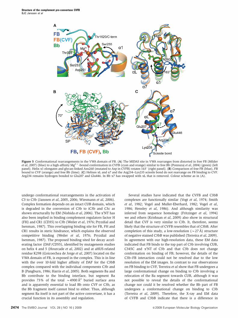
The FB MIDAS adopts a high-affinity state
On binding to CVF, the distorted MIDAS in free FB has
rearranged into a canonical high-affinity ligand-bound state,
as in fragment Bb (Ponnuraj et al, 2004) and the isolated
VWA domain (Bhattacharya et al, 2004) (Figure 5). MIDAS
residues Ser255 and Asp364 move up to 7.4 A and together
with residues Asp251, Ser253, Thr328 and two water mole-
cules coordinate the Mg2þ ion. The COO� terminus
(Thr1620) of CVF is the sixth chelating ligand of the Mg2þ
ion (Figure 5A). Thus, the Bb segment binds CVF through its
MIDAS, in which the carboxy terminus of CVF completes the
coordination sphere of the Mg2þ ion. These details confirm
the prominent role for Mg2þ -dependent MIDAS-mediated
complex formation, which has been shown earlier by muta-
genesis studies in which replacement of two MIDAS loops
(252–259 and 366–372) of FB with those of C2 decreased the
binding of FB to CVF (Tuckwell et al, 1997), by a combined
affinity and mass spectrometry approach that identified two
segments that contain the MIDAS (229–265 and 355–381) to
be involved in pro-convertase formation (Hinshelwood et al,
1999), and by gain-of-function mutations (D254G and
N260D) near the MIDAS of FB that increased stability of the
pro-convertase (Hourcade et al, 1999) (Figure 4E).
Furthermore, C3/CVF chimera studies underscore the role
for the C345C domain in this interaction (Kolln et al, 2005;
Fritzinger et al, 2009) (Figure 4E). Reduction of steric hin-
drance explains the D254G gain-of-function mutation in FB.
Deletion of the glycan in the N260D gain-of-function mutant
possibly facilitates rotation by 1631 and elongation of VWA
helix a1 that is coupled to the MIDAS loop rearrangements
(Figure 5A). Similarly, mutation F261L, which is located in
the refolding region of helix a1, may favour this rearrange-
ment and hence enhance pro-convertase formation causing
atypical haemolytic uremic syndrome (Goicoechea de Jorge
et al, 2007). In conclusion, FB binding to CVF induces a local
but functionally important rearrangement in the MIDAS and
Figure 3 The CVFB interface consists of two patches. (A) Ribbonrepresentation of CCP2–3 (coloured orange and red, respectively) ofthe Ba segment interacting with MG2, MG6, MG7, CUB (all colouredcyan) and a0NT (coloured black) of CVF. Glu182 of FB interacts withArg1262 and Glu1263 of CVF (ball-and-stick representation), whichcorrespond to the first FI-cleavage site in C3b. (B) VWA of the Bbsegment, shown in green surface representation, interacts withC345C of CVF shown in cyan ribbon representation. The C-terminus(Thr1620) of CVF binds to the Mg2þ ion (purple sphere) in FB.
Structure of the complement pro-convertase CVFBBJC Janssen et al
The EMBO Journal VOL 28 | NO 16 | 2009 &2009 European Molecular Biology Organization2472
surrounding loops from a distorted to a high-affinity ligand-
binding state.
CVF does not induce domain rearrangements in FB
We observe no further large conformational changes in FB on
binding to CVF neither in the crystal structures (Figure 5B;
Supplementary Table IIB), SAXS data or EM classes
(Figure 2). Apparently, FB provides sufficient space for the
C-terminal tail of CVF to access the MIDAS without dislocat-
ing the CCP1 domain. Correspondingly, helix aL and a7 do
not relocate and the scissile bond 234–235 remains partially
occluded, when FB binds CVF (Figure 5C). Detailed compar-
ison of the VWA domain in CVFB and in free Bb (Ponnuraj
et al, 2004) shows that activation of FB into Bb repositions
helix a7 into the groove previously occupied by helix aL,
away from the elongated helix a1. This additional rearrange-
ment may further stabilize the high-affinity MIDAS config-
uration that is observed in Bb (and the isolated VWA domain
(Bhattacharya et al, 2004)). Possibly the VWA domain of FB
adopts this conformation in the active convertase CVFBb and
C3bBb. This rearrangement may prevent release of Mg2þ
from C3bBb (in which the MIDAS is locked into a stable
configuration) and allows Mg2þ release and dissociation of
C3bB or CVFB, when treated with EDTA (Harris et al, 2005).
We observe no structural changes in the active site of the SP
domain between free FB, the pro-convertase and free Bb
(Supplementary Figure 4), indicating that enzyme activity is
possibly controlled by quaternary changes in the enzyme
complex; that is by substrate (C3) binding to the convertase.
Thus, except for the MIDAS and surrounding loops, FB
binding to CVF does not induce major conformational
changes in either FB or CVF.
Discussion
The central amplification step of the complement system is
crucial for the defence against invading pathogens and the
homeostasis of the host. Lack of activity may result in
recurrent bacterial infections, whereas unregulated activation
may lead to tissue damage. Convertase formation and activity
is, therefore, a tightly regulated process and reduced control,
for example caused by mutations or deficiencies in proteins
that form the convertase (C3b and FB) or in complement
regulators that dissociate convertases, have been associated
with several immune-related diseases (Ricklin and Lambris,
2007). Detailed understanding of the formation, activity and
regulation of the convertase will be instrumental in the
development of therapeutics to control complement related
diseases. Here, we show for the first time in atomic detail the
interactions that underlie pro-convertase formation.
Restricting the assembly of the convertase in place and
time is an important regulatory mechanism of the comple-
ment system. FB only binds to the activated form C3b and not
to C3, iC3b or C3c. The data presented here reveal that the Ba
segment of FB determines this specificity. Segment Ba has
specific interactions with a0NT, MG7 and CUB, which
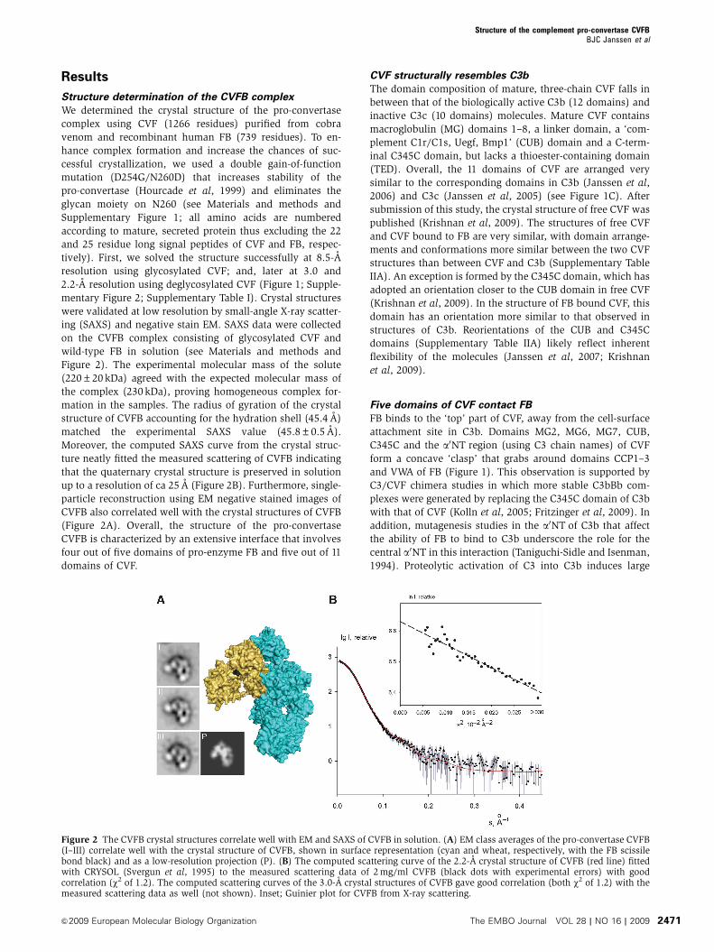
Figure 4 Surface representation of the CVFB interface coloured functionally. (A) An opened view of the 4900 A2 footprint of the FB–CVFinterface is highlighted in green. (B) Domains of FB and CVF coloured according to Figure 1. (C) FB and CVF colour-coded to residueconservation; from non-conserved (white) to conserved (black). Figure is produced using CONSURF (Glaser et al, 2005). (D) FB and CVFcoloured by electrostatic potential from red (�10 kbT/ec) to blue (�10 kbT/ec). The VWA:C345C interface consists of conserved complementaryelectrostatic patches. (E) Previously proposed sites involved in complex formation. The yellow coloured patches are epitopes to which antibodybinding decreases complex formation (Hourcade et al, 1995; Thurman et al, 2005). The other patches are based on FB to C2 chimeras thatincrease binding of FB to C3b 4150% (Hourcade et al, 1995) (blue) or decrease binding o10% (Tuckwell et al, 1997) (green); on C3 to CVFchimeras that increase C3bBb complex stability (Kolln et al, 2005; Fritzinger et al, 2009) (lime); on an alternative proteolytic product of C3, thatsupports activation of FB (O’Keefe et al, 1988) (orange) or on single site mutants (single numbers) that increase complex formation (Hourcadeet al, 1999) (dark red) or decrease complex formation (Taniguchi-Sidle and Isenman, 1994) (red). Legend for colour-coding and residuenumbers are presented in the table. CVF residue numbering is according to human C3.
Structure of the complement pro-convertase CVFBBJC Janssen et al
&2009 European Molecular Biology Organization The EMBO Journal VOL 28 | NO 16 | 2009 2473
undergo conformational rearrangements in the activation of
C3 to C3b (Janssen et al, 2005, 2006; Wiesmann et al, 2006).
Complex formation depends on an intact CUB domain, which
is degraded in the conversion of C3b to iC3b and C3c as
shown structurally by EM (Nishida et al, 2006). The a0NT has
also been implied in binding complement regulators factor H
(FH) and CR1 (CD35) to C3b (Weiler et al, 1976; Pryzdial and
Isenman, 1987). This overlapping binding site for FB, FH and
CR1 results in steric hindrance, which explains the observed
competitive binding (Weiler et al, 1976; Pryzdial and
Isenman, 1987). The proposed binding sited for decay accel-
erating factor (DAF/CD55), identified by mutagenesis studies
on helix 4 and 5 (Hourcade et al, 2002) and at aHUS-related
residue K298 (Goicoechea de Jorge et al, 2007) located on the
VWA domain of FB, is exposed in the complex. This is in line
with the over 10-fold higher affinity of DAF for the C3bB
complex compared with the individual components C3b and
B (Pangburn, 1986; Harris et al, 2005). Both segments Ba and
Bb contribute to the binding interface, but segment Ba
provides 73% of the total B4900 A2 buried surface area
and is apparently essential to load Bb onto CVF or C3b, as
the Bb fragment itself cannot bind to either. Thus, although
segment Ba itself is not part of the active convertase, it has a
crucial function in its assembly and regulation.
Several studies have indicated that the CVFB and C3bB
complexes are functionally similar (Vogt et al, 1974; Smith
et al, 1982; Vogel and Muller-Eberhard, 1982; Vogel et al,
1984; Hensley et al, 1986). And although similarity was
inferred from sequence homology (Fritzinger et al, 1994)
we and others (Krishnan et al, 2009) also show in structural
detail that CVF is very similar to C3b. It, therefore, seems
likely that the structure of CVFB resembles that of C3bB. After
completion of this study, a low-resolution (B27 A) structure
of negative stained C3bB was published (Torreira et al, 2009).
In agreement with our high-resolution data, these EM data
indicated that FB binds to the top part of C3b involving CUB,
C345C and a0NT of C3b and that C3b does not change
conformation on binding of FB; however, the details of the
C3b–FB interaction could not be resolved due to the low
resolution of the EM images. In contrast to our observations
on FB binding to CVF, Torreira et al show that FB undergoes a
large conformational change on binding to C3b involving a
relocation of the Ba segment towards CUB, although it was
not possible to reveal the details of the conformational
change nor could it be resolved whether the Bb part of FB
undergoes a conformational change on binding to C3b
(Torreira et al, 2009). Therefore, the X-ray and EM data
of CVFB and C3bB indicate that there is a difference in
Figure 5 Conformational rearrangements in the VWA domain of FB. (A) The MIDAS site in VWA rearranges from distorted in free FB (Milderet al, 2007) (blue) to a high-affinity Mg2þ -bound conformation in CVFB (cyan and orange) similar to free Bb (Ponnuraj et al, 2004) (green) (leftpanel). Helix a1 elongates and glycan-linked Asn260 (mutated to Asp in CVFB) rotates 1631 (right panel). (B) Comparison of free FB (blue), FBbound to CVF (orange) and free Bb (lime). (C) Helixes aL and a7 and the Arg234–Lys235 scissile bond do not rearrange on FB binding to CVF.Arg234 remains hydrogen bonded to Glu207 and Glu446. In Bb a7 has swapped with aL that is removed. Colour scheme as in (A).
Structure of the complement pro-convertase CVFBBJC Janssen et al
The EMBO Journal VOL 28 | NO 16 | 2009 &2009 European Molecular Biology Organization2474
conformation of FB when bound to CVF and bound to C3b.
This difference in conformation is unlikely to arise from
crystal packing events as the CVFB crystal structure is very
similar to the structure determined by solution SAXS and EM
studies. Possibly, this difference illustrates two functional
different states of the pro-convertase structure, which may
exist in a dynamic equilibrium.
In this equilibrium, the CVFB structure may represent the
‘loading’ state of FB to the cofactor, whereas the C3bB
structure indicates the form that is activated for cleavage by
FD. Activation of FB by FD occurs solely when FB forms a
pro-convertase. In the CVFB structure, the scissile bond 234–
235 has remained partially occluded similar to its position in
free FB. In the C3bB structure, relocation of the Ba segment
may have exposed the scissile bond and helix a7 has possibly
relocated to the position previously occupied by helix aL.
Unfortunately, these details could not be resolved due to the
low resolution of the EM data (Torreira et al, 2009). Our data
suggest that FB bound to CVF remains predominantly in the
initial ‘loading’ state, whereas FB bound to C3b adopts more
frequently an ‘activation’ state with an exposed scissile loop
that can be cleaved by FD. In support of this hypothesis, we
observe a much slower activation rate of FB by FD in CVFB
than in C3bB (Supplementary Figure 5) and Harris et al
demonstrate a conformation change in the C3bB complex in
surface plasmon resonance experiments (Harris et al, 2005).
Although a difference in the initial ‘loading’ of FB to CVF and
C3b cannot be excluded at present, the data presented here
and the observation that FB in the CVFB complex is very
similar in domain arrangements to free FB indicate that FB is
likely to bind initially in a similar manner to C3b as it binds to
CVF. Thus, there may be two distinct, ‘loading’ and ‘activa-
tion’, states of the pro-convertase structure represented by
the CVFB and C3bB structures. However, the details of the
differences between the CVFB and C3bB structures can only
be resolved with high-resolution data of the C3bB complex.
In conclusion, the CVFB complex indicates that the CCP
domains of the Ba segment of FB provide a scaffold for the
protease segment Bb to be loaded onto the C-terminal C345C
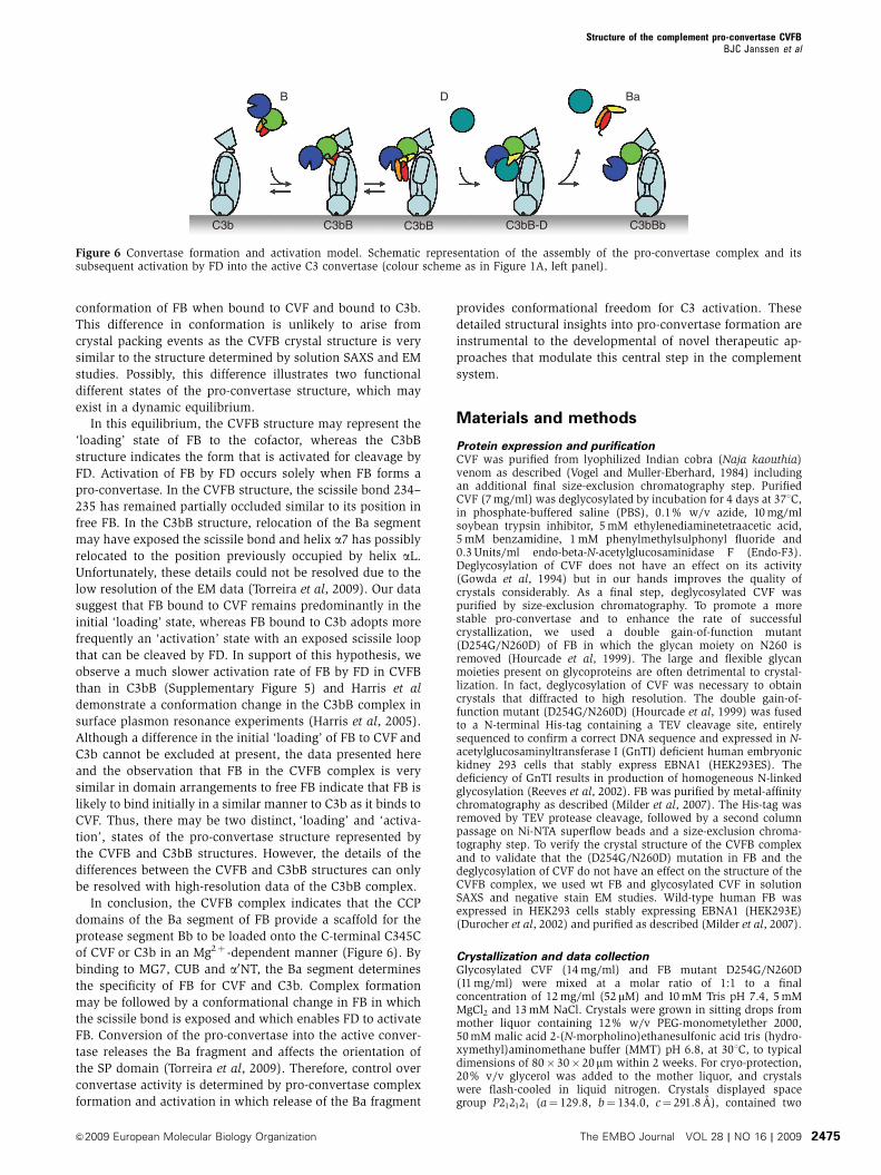
of CVF or C3b in an Mg2þ -dependent manner (Figure 6). By
binding to MG7, CUB and a0NT, the Ba segment determines
the specificity of FB for CVF and C3b. Complex formation
may be followed by a conformational change in FB in which
the scissile bond is exposed and which enables FD to activate
FB. Conversion of the pro-convertase into the active conver-
tase releases the Ba fragment and affects the orientation of
the SP domain (Torreira et al, 2009). Therefore, control over
convertase activity is determined by pro-convertase complex
formation and activation in which release of the Ba fragment
provides conformational freedom for C3 activation. These
detailed structural insights into pro-convertase formation are
instrumental to the developmental of novel therapeutic ap-
proaches that modulate this central step in the complement
system.
Materials and methods
Protein expression and purificationCVF was purified from lyophilized Indian cobra (Naja kaouthia)venom as described (Vogel and Muller-Eberhard, 1984) includingan additional final size-exclusion chromatography step. PurifiedCVF (7 mg/ml) was deglycosylated by incubation for 4 days at 371C,in phosphate-buffered saline (PBS), 0.1% w/v azide, 10 mg/mlsoybean trypsin inhibitor, 5 mM ethylenediaminetetraacetic acid,5 mM benzamidine, 1 mM phenylmethylsulphonyl fluoride and0.3 Units/ml endo-beta-N-acetylglucosaminidase F (Endo-F3).Deglycosylation of CVF does not have an effect on its activity(Gowda et al, 1994) but in our hands improves the quality ofcrystals considerably. As a final step, deglycosylated CVF waspurified by size-exclusion chromatography. To promote a morestable pro-convertase and to enhance the rate of successfulcrystallization, we used a double gain-of-function mutant(D254G/N260D) of FB in which the glycan moiety on N260 isremoved (Hourcade et al, 1999). The large and flexible glycanmoieties present on glycoproteins are often detrimental to crystal-lization. In fact, deglycosylation of CVF was necessary to obtaincrystals that diffracted to high resolution. The double gain-of-function mutant (D254G/N260D) (Hourcade et al, 1999) was fusedto a N-terminal His-tag containing a TEV cleavage site, entirelysequenced to confirm a correct DNA sequence and expressed in N-acetylglucosaminyltransferase I (GnTI) deficient human embryonickidney 293 cells that stably express EBNA1 (HEK293ES). Thedeficiency of GnTI results in production of homogeneous N-linkedglycosylation (Reeves et al, 2002). FB was purified by metal-affinitychromatography as described (Milder et al, 2007). The His-tag wasremoved by TEV protease cleavage, followed by a second columnpassage on Ni-NTA superflow beads and a size-exclusion chroma-tography step. To verify the crystal structure of the CVFB complexand to validate that the (D254G/N260D) mutation in FB and thedeglycosylation of CVF do not have an effect on the structure of theCVFB complex, we used wt FB and glycosylated CVF in solutionSAXS and negative stain EM studies. Wild-type human FB wasexpressed in HEK293 cells stably expressing EBNA1 (HEK293E)(Durocher et al, 2002) and purified as described (Milder et al, 2007).
Crystallization and data collectionGlycosylated CVF (14 mg/ml) and FB mutant D254G/N260D(11 mg/ml) were mixed at a molar ratio of 1:1 to a finalconcentration of 12 mg/ml (52 mM) and 10 mM Tris pH 7.4, 5 mMMgCl2 and 13 mM NaCl. Crystals were grown in sitting drops frommother liquor containing 12% w/v PEG-monometylether 2000,50 mM malic acid 2-(N-morpholino)ethanesulfonic acid tris (hydro-xymethyl)aminomethane buffer (MMT) pH 6.8, at 301C, to typicaldimensions of 80�30�20mm within 2 weeks. For cryo-protection,20% v/v glycerol was added to the mother liquor, and crystalswere flash-cooled in liquid nitrogen. Crystals displayed spacegroup P212121 (a¼ 129.8, b¼ 134.0, c¼ 291.8 A), contained two
B D Ba
C3bB bBb3CD-Bb3Cb3C C3bB
Figure 6 Convertase formation and activation model. Schematic representation of the assembly of the pro-convertase complex and itssubsequent activation by FD into the active C3 convertase (colour scheme as in Figure 1A, left panel).
Structure of the complement pro-convertase CVFBBJC Janssen et al
&2009 European Molecular Biology Organization The EMBO Journal VOL 28 | NO 16 | 2009 2475
complexes per asymmetric unit and diffracted to 8.5-A resolution atEuropean synchrotron radiation facility (ESRF) beamline ID23-2.Purified deglycosylated CVF (12 mg/ml) and FB mutant D254G/N260D (15 mg/ml) were mixed to a molar ratio of 1:1 and dialyzedagainst 10 mM Tris pH 7.4, 5 mM MgCl2 and 10 mM NaCl resultingin a final concentration of 12 mg/ml (50mM). This sample yieldedtwo different crystal forms. Crystals with space group C2221
(a¼ 128.9, b¼ 283.5, c¼ 134.4 A) were grown in sitting drops frommother liquor containing 6.6% w/v PEG 3350, 16 mM Bis-Trispropane pH 6.5, 66 mM Na/K Phosphate, at 181C, to typicaldimensions of 400�100� 80mm within 1 week. For cryo-protec-tion, 30% v/v PEG 400 was added to the mother liquor, and crystalswere flash-cooled in liquid nitrogen. These crystals contained onecomplex per asymmetric unit and diffracted to 2.2-A resolution atESRF beamline ID23-1. Crystals with space group P212121
(a¼ 134.0, b¼ 137.0, c¼ 283.7 A) were grown in sitting drops frommother liquor containing 8.25% w/v PEG 1500, 33 mM MMT pH9.0, at 181C, to typical dimensions of 200� 60� 40mm within 1week. For cryo-protection, 12% v/v 2,3 butanediol was added to themother liquor, and crystals were flash-cooled in liquid nitrogen.These crystals contained two complexes per asymmetric unit anddiffracted to 3.0-A resolution at ESRF beamline ID23-1. Alldiffraction data was integrated and scaled with MOSFLM (Leslie,1992) and SCALA (Evans, 2006) in CCP4 (CCP4, 1994).
Structure determinationInitially the 8.5-A resolution structure was solved by molecularreplacement with PHASER (McCoy et al, 2007) using the isolatedstructures of C3b (Janssen et al, 2006) (pdb code: 2I07) with theTED domain omitted and FB (Milder et al, 2007) (pdb code: 2OK5)as the search models. Owing to limited resolution, only rigid bodyrefinement (using 18 groups) was performed in REFMAC (Mur-shudov et al, 1997) resulting in Rwork/Rfree values of 34.4/34.9%. Ata later stage, the 2.2-A resolution structure was solved by molecularreplacement in PHASER (McCoy et al, 2007) starting with the b-chain and MG6 part of the a-chain from C3b followed step-by-stepby SP, VWA and CCP2–3 from FB and a0NT-MG7-anchor, MG8, CUBand C345C from C3b and CCP1 from FB, respectively, usingmolecular graphics in COOT (Emsley and Cowtan, 2004) with rigid-body refinement in PHASER. This partial model was re-builtautomatically by ARP/wARP (Perrakis et al, 1999) and completedby several cycles of manual rebuilding in COOT and refinement inPHENIX (Adams et al, 2002) to a final Rwork/Rfree value of 18.0/22.6%. The 3.0-A structure was solved by molecular replacement inPHASER using the refined 2.2-A structure and was completed byseveral cycles of manual rebuilding in COOT and refinement inPHENIX to a final Rwork/Rfree value of 18.9/24.3%. All moleculargraphics figures were generated with pymol (W Delano; http://www.pymol.org/).
EM and image classificationFB (from HEK293E cells) and CVF were mixed to a molar ratio of1.2:1 and a final concentration of 1 mg/ml in PBS, 5 mM MgCl2 and1 mM NiCl2 and incubated for 15 min at room temperature. Thesample was diluted to 5 mg/ml in water and immediately applied toa freshly glow-discharged carbon layer supported by an EM grid andnegatively stained with 0.75% uranyl formate as described (Ohiet al, 2004). Micrographs were recorded under low-dose conditionson a 4k� 4k CCD camera with a FEI Tecnai 12 transmission electronmicroscope operating at 120 kV and a magnification of 39 000�resulting in a pixel size of 3.8 A. A total of 10 943 particles wereselected from the micrographs, respectively, using the program‘Boxer’ from the EMAN software package (Ludtke et al, 1999). The
contrast transfer function of the microscope for each micrographwas estimated and corrected using XMIPP (Sorzano et al, 2004).The extracted particles were classified into 20 classes with themaximum-likelihood multireference refinement protocol in XMIPPas described (Scheres et al, 2008). The three most populous orrepresentative classes are shown in Figure 2. The projection wasgenerated from the crystal structure of CVFB after low-pass filteringto 25-A resolution in EMAN (Ludtke et al, 1999).
Small angle X-ray scatteringFB (from HEK293E cells) and CVF were mixed to a molar ratio of1:1 in Tris-buffered saline and 5 mM MgCl2 and incubated for atleast 15 min at room temperature. Synchrotron X-ray scattering datafrom a 1 mg/ml and a 2 mg/ml solution of the CVFB complex werecollected following standard procedures at the European MolecularBiology Laboratory (EMBL) beamline X33 (Roessle et al, 2007)(storage ring DORIS-III, Deutsches Elektronen-Synchrotron (DESY),Hamburg) using a Pilatus 500K detector (DECTRIS, Switzerland).To monitor for radiation damage, four successive 30 s exposures onthe same sample were compared, and no changes were detected.The data were processed using PRIMUS (Konarev et al, 2003). Themolecular mass of the solute was calculated by normalizationagainst the scattering from a reference solution of bovine serumalbumin. The SAXS curves were computed from the 2.2- and 3.0-Acrystal structures of CVFB by CRYSOL (Svergun et al, 1995).
Accession codesCoordinates and structure factors of the CVFB complex have beendeposited in the Protein Data Bank with succession numbers 3HRZ(2.2-A resolution) and 3HS0 (3.0-A resolution).
Supplementary dataSupplementary data are available at The EMBO Journal Online(http://www.embojournal.org).
Acknowledgements
We thank Catharina Verheij for help with initial crystallizationexperiments, Hans Meeldijk (Cellular Architecture and Dynamics,Utrecht) for help with EM grid preparation and testing, SjorsScheres (National Center for Biotechnology, Madrid) for suggestionson EM image processing in XMIPP and Roland Romijn (U-ProteinExpress, Utrecht) for preparation of FB for SAXS analysis. Weacknowledge the ESRF and the EMBL/DESY for the provision ofsynchrotron radiation facilities and thank David Flot (ESRF,Grenoble) and Adam Round (EMBL, Hamburg) for beamline assis-tance. This work was supported by the Council for ChemicalSciences of the Netherlands Organization for Scientific Research(NWO-CW) and the US National Institutes of Health (to PG).
Author contributions: DCF purified CVF. LG cloned, expressedand purified FB and deglycosylated CVF. BJCJ crystallized thecomplex, determined and analysed the structures. PG, LG and DIScollected, processed and analysed the SAXS data. RIK and BJCJprepared and tested the EM grids. RIK collected the EM data undersupervision of AJK. BJCJ processed the EM data. RIK and BJCJanalysed the EM data. BJCJ, C-WV and PG conceived the project.BJCJ and PG wrote the paper.
Conflict of interest
The authors declare that they have no conflict of interest.
References
Adams PD, Grosse-Kunstleve RW, Hung LW, Ioerger TR, McCoy AJ,Moriarty NW, Read RJ, Sacchettini JC, Sauter NK, Terwilliger TC(2002) PHENIX: building new software for automated crystal-lographic structure determination. Acta Crystallogr D BiolCrystallogr 58: 1948–1954
Bhattacharya AA, Lupher Jr ML, Staunton DE, Liddington RC (2004)Crystal structure of the A domain from complement factor Breveals an integrin-like open conformation. Structure (Camb) 12:371–378
CCP4 (1994) The CCP4 suite: programs for protein crystallography.Acta Crystallogr D Biol Crystallogr 50: 760–763
Durocher Y, Perret S, Kamen A (2002) High-level and high-through-put recombinant protein production by transient transfection ofsuspension-growing human 293-EBNA1 cells. Nucleic Acids Res30: E9
Emsley P, Cowtan K (2004) Coot: model-building tools formolecular graphics. Acta Crystallogr D Biol Crystallogr 60:2126–2132
Structure of the complement pro-convertase CVFBBJC Janssen et al
The EMBO Journal VOL 28 | NO 16 | 2009 &2009 European Molecular Biology Organization2476
Evans P (2006) Scaling and assessment of data quality. ActaCrystallogr D Biol Crystallogr 62: 72–82
Fishelson Z, Pangburn MK, Muller-Eberhard HJ (1984)Characterization of the initial C3 convertase of the alternativepathway of human complement. J Immunol 132: 1430–1434
Fremeaux-Bacchi V, Miller EC, Liszewski MK, Strain L, Blouin J,Brown AL, Moghal N, Kaplan BS, Weiss RA, Lhotta K, Kapur G,Mattoo T, Nivet H, Wong W, Gie S, Hurault de Ligny B, FischbachM, Gupta R, Hauhart R, Meunier V et al (2008) Mutations incomplement C3 predispose to development of atypical hemolyticuremic syndrome. Blood 112: 4948–4952
Fritzinger DC, Bredehorst R, Vogel CW (1994) Molecular cloningand derived primary structure of cobra venom factor. Proc NatlAcad Sci USA 91: 12775–12779
Fritzinger DC, Hew BE, Thorne M, Pangburn MK, Janssen BJ, GrosP, Vogel CW (2009) Functional characterization of human C3/cobra venom factor hybrid proteins for therapeutic complementdepletion. Dev Comp Immunol 33: 105–116
Glaser F, Rosenberg Y, Kessel A, Pupko T, Ben-Tal N (2005)The ConSurf-HSSP database: the mapping of evolutionaryconservation among homologs onto PDB structures. Proteins58: 610–617
Goicoechea de Jorge E, Harris CL, Esparza-Gordillo J, Carreras L,Arranz EA, Garrido CA, Lopez-Trascasa M, Sanchez-Corral P,Morgan BP, Rodriguez de Cordoba S (2007) Gain-of-functionmutations in complement factor B are associated with atypicalhemolytic uremic syndrome. Proc Natl Acad Sci USA 104: 240–245
Gowda DC, Petrella EC, Raj TT, Bredehorst R, Vogel CW (1994)Immunoreactivity and function of oligosaccharides in cobravenom factor. J Immunol 152: 2977–2986
Harris CL, Abbott RJ, Smith RA, Morgan BP, Lea SM (2005)Molecular dissection of interactions between components of thealternative pathway of complement and decay accelerating factor(CD55). J Biol Chem 280: 2569–2578
Hensley P, O’Keefe MC, Spangler CJ, Osborne Jr JC, Vogel CW(1986) The effects of metal ions and temperature on the interac-tion of cobra venom factor and human complement factor B.J Biol Chem 261: 11038–11044
Hinshelwood J, Perkins SJ (2000a) Conformational changes duringthe assembly of factor B from its domains by (1)H NMR spectro-scopy and molecular modelling: their relevance to the regulationof factor B activity. J Mol Biol 301: 1267–1285
Hinshelwood J, Perkins SJ (2000b) Metal-dependent conformationalchanges in a recombinant vWF-A domain from human factor B: asolution study by circular dichroism, fourier transform infraredand (1)H NMR spectroscopy. J Mol Biol 298: 135–147
Hinshelwood J, Spencer DI, Edwards YJ, Perkins SJ (1999)Identification of the C3b binding site in a recombinant vWF-Adomain of complement factor B by surface-enhanced laser deso-rption-ionisation affinity mass spectrometry and homology mod-elling: implications for the activity of factor B. J Mol Biol 294:587–599
Hourcade DE, Mitchell L, Kuttner-Kondo LA, Atkinson JP, MedofME (2002) Decay-accelerating factor (DAF), complement receptor1 (CR1), and factor H dissociate the complement AP C3 con-vertase (C3bBb) via sites on the type A domain of Bb. J Biol Chem277: 1107–1112
Hourcade DE, Mitchell LM, Oglesby TJ (1999) Mutations of the typeA domain of complement factor B that promote high-affinity C3b-binding. J Immunol 162: 2906–2911
Hourcade DE, Wagner LM, Oglesby TJ (1995) Analysis of the shortconsensus repeats of human complement factor B by site-directedmutagenesis. J Biol Chem 270: 19716–19722
Janssen BJ, Christodoulidou A, McCarthy A, Lambris JD, Gros P(2006) Structure of C3b reveals conformational changes thatunderlie complement activity. Nature 444: 213–216
Janssen BJ, Halff EF, Lambris JD, Gros P (2007) Structure ofcompstatin in complex with complement component C3c revealsa new mechanism of complement inhibition. J Biol Chem 282:29241–29247
Janssen BJ, Huizinga EG, Raaijmakers HC, Roos A, Daha MR,Nilsson-Ekdahl K, Nilsson B, Gros P (2005) Structures of comple-ment component C3 provide insights into the function andevolution of immunity. Nature 437: 505–511
Kolln J, Bredehorst R, Spillner E (2005) Engineering of humancomplement component C3 for catalytic inhibition of comple-ment. Immunol Lett 98: 49–56
Konarev PV, Volkov VV, Sokolova AV, Koch MH, Svergun DI (2003)PRIMUS: a Windows PC-based system for small-angle scatteringdata analysis. J Appl Cryst 36: 1277–1282
Krishnan V, Ponnuraj K, Xu Y, Macon K, Volanakis JE, Narayana SV(2009) The crystal structure of cobra venom factor, a cofactor forC3- and C5-convertase CVFBb. Structure 17: 611–619
Leslie AG (1992) Recent changes to the MOSFLM package forprocessing film and image plate data. Joint CCP4+ESF-EAMCBNewsletter on Protein Crystallography 26
Ludtke SJ, Baldwin PR, Chiu W (1999) EMAN: semiautomatedsoftware for high-resolution single-particle reconstructions. JStruct Biol 128: 82–97
McCoy AJ, Grosse-Kunstleve RW, Adams PD, Winn MD, Storoni LC,Read RJ (2007) Phaser crystallographic software. J Appl Cryst 40:658–674
Milder FJ, Gomes L, Schouten A, Janssen BJ, Huizinga EG, RomijnRA, Hemrika W, Roos A, Daha MR, Gros P (2007) Factor Bstructure provides insights into activation of the central proteaseof the complement system. Nat Struct Mol Biol 14: 224–228
Muller-Eberhard HJ (1988) Molecular organization and function ofthe complement system. Annu Rev Biochem 57: 321–347
Murshudov GN, Vagin AA, Dodson EJ (1997) Refinement of macro-molecular structures by the maximum-likelihood method. ActaCrystallogr D Biol Crystallogr 53: 240–255
Nishida N, Walz T, Springer TA (2006) Structural transitions ofcomplement component C3 and its activation products. Proc NatlAcad Sci USA 103: 19737–19742
O’Keefe MC, Caporale LH, Vogel CW (1988) A novel cleavageproduct of human complement component C3 with structuraland functional properties of cobra venom factor. J Biol Chem 263:12690–12697
Ohi M, Li Y, Cheng Y, Walz T (2004) Negative staining and imageclassification—powerful tools in modern electron microscopy.Biol Proced Online 6: 23–34
Pangburn MK (1986) Differences between the binding sites of thecomplement regulatory proteins DAF, CR1, and factor H on C3convertases. J Immunol 136: 2216–2221
Perrakis A, Morris R, Lamzin VS (1999) Automated protein modelbuilding combined with iterative structure refinement. Nat StructBiol 6: 458–463
Ponnuraj K, Xu Y, Macon K, Moore D, Volanakis JE, Narayana SV(2004) Structural analysis of engineered Bb fragment of comple-ment factor B: insights into the activation mechanism of thealternative pathway C3-convertase. Mol Cell 14: 17–28
Pryzdial EL, Isenman DE (1987) Alternative complement pathwayactivation fragment Ba binds to C3b. Evidence that formation ofthe factor B-C3b complex involves two discrete points of contact.J Biol Chem 262: 1519–1525
Reeves PJ, Callewaert N, Contreras R, Khorana HG (2002) Structureand function in rhodopsin: high-level expression of rhodopsinwith restricted and homogeneous N-glycosylation by a tetracy-cline-inducible N-acetylglucosaminyltransferase I-negativeHEK293S stable mammalian cell line. Proc Natl Acad Sci USA99: 13419–13424
Ricklin D, Lambris JD (2007) Complement-targeted therapeutics.Nat Biotechnol 25: 1265–1275
Roessle MW, Klaering R, Ristau U, Robrahn B, Jahn D, Gehrmann T,Konarev PV, Round A, Friedler S, Hermes S, Svergun DI (2007)Upgrade of the small angle X-ray scattering Beamline X33 at theEuropean Molecular Biology Laboratory, Hamburg. J Appl Cryst40: s190–s194
Ross GD, Newman SL, Lambris JD, Devery-Pocius JE, Cain JA,Lachmann PJ (1983) Generation of three different fragments ofbound C3 with purified factor I or serum. II. Location of bindingsites in the C3 fragments for factors B and H, complementreceptors, and bovine conglutinin. J Exp Med 158: 334–352
Scheres SH, Nunez-Ramirez R, Sorzano CO, Carazo JM, Marabini R(2008) Image processing for electron microscopy single-particleanalysis using XMIPP. Nat Protoc 3: 977–990
Smith CA, Vogel CW, Muller-Eberhard HJ (1982) Ultrastructureof cobra venom factor-dependent C3/C5 convertase and itszymogen, factor B of human complement. J Biol Chem 257:9879–9882
Sorzano CO, Marabini R, Velazquez-Muriel J, Bilbao-Castro JR,Scheres SH, Carazo JM, Pascual-Montano A (2004) XMIPP: anew generation of an open-source image processing package forelectron microscopy. J Struct Biol 148: 194–204
Structure of the complement pro-convertase CVFBBJC Janssen et al
&2009 European Molecular Biology Organization The EMBO Journal VOL 28 | NO 16 | 2009 2477
Svergun DI, Barberato C, Koch MH (1995) CRYSOL—a program toevaluate X-ray solution scattering of biological macromoleculesfrom atomic coordinates. J Appl Cryst 28: 768–773
Taniguchi-Sidle A, Isenman DE (1994) Interactions of human com-plement component C3 with factor B and with complementreceptors type 1 (CR1, CD35) and type 3 (CR3, CD11b/CD18)involve an acidic sequence at the N-terminus of C3 alpha’-chain.J Immunol 153: 5285–5302
Thurman JM, Kraus DM, Girardi G, Hourcade D, Kang HJ, Royer PA,Mitchell LM, Giclas PC, Salmon J, Gilkeson G, Holers VM (2005)A novel inhibitor of the alternative complement pathwayprevents antiphospholipid antibody-induced pregnancy loss inmice. Mol Immunol 42: 87–97
Torreira E, Tortajada A, Montes T, Rodriguez de Cordoba S,Llorca O (2009) 3D structure of the C3bB complex providesinsights into the activation and regulation of the complementalternative pathway convertase. Proc Natl Acad Sci USA 106:882–887
Tuckwell DS, Xu Y, Newham P, Humphries MJ, Volanakis JE(1997) Surface loops adjacent to the cation-binding siteof the complement factor B von Willebrand factor type Amodule determine C3b binding specificity. Biochemistry 36:6605–6613
Vogel CW, Fritzinger DC (2007) Humanized cobra venom factor:experimental therapeutics for targeted complement activationand complement depletion. Curr Pharm Des 13: 2916–2926
Vogel CW, Muller-Eberhard HJ (1982) The cobra venom factor-dependent C3 convertase of human complement. A kinetic and
thermodynamic analysis of a protease acting on its natural highmolecular weight substrate. J Biol Chem 257: 8292–8299
Vogel CW, Muller-Eberhard HJ (1984) Cobra venom factor: im-proved method for purification and biochemical characterization.J Immunol Methods 73: 203–220
Vogel CW, Smith CA, Muller-Eberhard HJ (1984) Cobra venomfactor: structural homology with the third component of humancomplement. J Immunol 133: 3235–3241
Vogt W, Dieminger L, Lynen R, Schmidt G (1974) Alternativepathway for the activation of complement in human serum.Formation and composition of the complex with cobra venomfactor that cleaves the third component of complement. HoppeSeylers Z Physiol Chem 355: 171–183
von Zabern I, Hinsch B, Przyklenk H, Schmidt G, Vogt W (1980)Comparison of Naja n. naja and Naja h. haje cobra-venomfactors: correlation between binding affinity for the fifth compo-nent of complement and mediation of its cleavage.Immunobiology 157: 499–514
Walport MJ (2001) Complement. First of two parts. N Engl J Med344: 1058–1066
Weiler JM, Daha MR, Austen KF, Fearon DT (1976) Control of theamplification convertase of complement by the plasma proteinbeta1H. Proc Natl Acad Sci USA 73: 3268–3272
Wiesmann C, Katschke KJ, Yin J, Helmy KY, Steffek M, FairbrotherWJ, McCallum SA, Embuscado L, DeForge L, Hass PE, vanLookeren Campagne M (2006) Structure of C3b in complexwith CRIg gives insights into regulation of complement activa-tion. Nature 444: 217–220
Structure of the complement pro-convertase CVFBBJC Janssen et al
The EMBO Journal VOL 28 | NO 16 | 2009 &2009 European Molecular Biology Organization2478
Related Documents