Insight into the Putative Specific Interactions between Cholesterol, Sphingomyelin, and Palmitoyl-Oleoyl Phosphatidylcholine Jussi Aittoniemi,* Perttu S. Niemela ¨,* Marja T. Hyvo ¨ nen,* y Mikko Karttunen, z and Ilpo Vattulainen* §{ *Laboratory of Physics and Helsinki Institute of Physics, Helsinki University of Technology, Helsinki, Finland; y Wihuri Research Institute, Helsinki, Finland; z Department of Applied Mathematics, the University of Western Ontario, Middlesex College, London, Ontario, Canada; § Institute of Physics, Tampere University of Technology, Tampere, Finland; and { MEMPHYS-Center for Biomembrane Physics, Physics Department, University of Southern Denmark, Odense, Denmark ABSTRACT The effects of cholesterol (Chol) on phospholipid bilayers include ordering of the fatty acyl chains, condensing of the lipids in the bilayer plane, and promotion of the liquid-ordered phase. These effects depend on the type of phospholipids in the bilayer and are determined by the nature of the underlying molecular interactions. As for Chol, it has been shown to interact more favorably with sphingomyelin than with most phosphatidylcholines, which in given circumstances leads to formation of lateral domains. However, the exact origin and nature of Chol-phospholipid interactions have recently been subjects of speculation. We examine interactions between Chol, palmitoylsphingomyelin (PSM) and palmitoyl-oleoyl-phosphatidylcholine (POPC) in hydrated lipid bilayers by extensive atom-scale molecular dynamics simulations. We employ a tailored lipid configuration: Individual PSM and Chol monomers, as well as PSM-Chol dimers, are embedded in a POPC lipid bilayer in the liquid crystalline phase. Such a setup allows direct comparison of dimeric and monomeric PSMs and Chol, which ultimately shows how the small differences in PSM and POPC structure can lead to profoundly different interactions with Chol. Our analysis shows that direct hydrogen bonding between PSM and Chol does not provide an adequate explanation for their putative specific interaction. Rather, a combination of charge-pairing, hydrophobic, and van der Waals interactions leads to a lower tilt in PSM neighboring Chol than in Chol with only POPC neighbors. This implies improved Chol-induced ordering of PSM’s chains over POPC’s chains. These findings are discussed in the context of the hydrophobic mismatch concept suggested recently. INTRODUCTION The lipid bilayer is responsible for some remarkable physical properties of cellular membranes, for they are as tight and robust as they are thin and flexible (1,2). Membrane proteins in turn are responsible for specific membrane functions such as signaling, channeling, or cell recognition (1,2). Based on these fundamental roles of lipids and proteins in biological membranes, our views on their detailed molecular organi- zation have been changing for the past decade. The classical Singer-Nicolson model of membrane struc- ture of 1972 (3) has proven to be highly useful but incom- plete. It describes the lipid bilayer part of cell membranes as a uniform fluid phase, in which all membrane proteins dis- solve and diffuse evenly (3). Yet, studies of model mem- branes show that bilayer mixtures of already a few different physiological lipids exhibit rather complex phase behavior (1,4). In particular, sphingolipids and other phospholipids with mostly saturated fatty acid residues can form a liquid- ordered (l o ) phase that may coexist in the bilayer with a conformationally more disordered (l d ) phase (1,5). The formation of the l o phase is greatly facilitated by the presence of cholesterol (Chol), which partitions rather into an l o than an l d lipid environment (6–8). The coexistence of l o and l d phases inflict lateral fine structure on a lipid bilayer: Separate l o domains are known to form in the l d matrix of model membranes that mimic physiological conditions (6–8). Understandably, the phase behavior of real cell mem- brane lipid bilayers, which contain hundreds of different lipids (2,9), is even more complex and domain formation in them is not fully established (10,11). In the case of lipid domains in cell membranes, as so often in biology, structural aspects are closely related to function: Their possible physiological consequences were first de- scribed in the lipid raft model, introduced by Simons and Ikonen in 1997 (12). In this model, certain membrane pro- teins were suggested to segregate in l o lipid domains, or rafts, while others are excluded from them (2,8,9,12–14). The earliest functions connected to lipid rafts were protein traf- ficking and cell signaling (12). Later on, raft lipids have been associated also with viral budding, prion diseases, and can- cer, though clearcut evidence is lacking (11,13,14). Nev- ertheless, it is nowadays largely accepted that the functioning of proteins in membranes depends on their local membrane composition, which highlights the role of lipid membranes in the understanding of various cellular functions. For example, insulin receptor activity is greatly inhibited in kidney cells grown with desmosterol instead of Chol (15). Full understanding of lipid domains and in particular raftlike ordered patches in cell membranes requires detailed knowledge of their properties in model membranes (6). Yet, the exact mechanisms of l o domain formation are largely unknown (6,7,14). While the presence of Chol is generally reckoned to be a necessary requirement for l o phase formation, Chol interactions with phospholipids have not been resolved Submitted May 3, 2006, and accepted for publication October 6, 2006. Address reprint requests to Ilpo Vattulainen, E-mail: ilpo.vattulainen@csc.fi. Ó 2007 by the Biophysical Society 0006-3495/07/02/1125/13 $2.00 doi: 10.1529/biophysj.106.088427 Biophysical Journal Volume 92 February 2007 1125–1137 1125

Welcome message from author
This document is posted to help you gain knowledge. Please leave a comment to let me know what you think about it! Share it to your friends and learn new things together.
Transcript

Insight into the Putative Specific Interactions between Cholesterol,Sphingomyelin, and Palmitoyl-Oleoyl Phosphatidylcholine
Jussi Aittoniemi,* Perttu S. Niemela,* Marja T. Hyvonen,*y Mikko Karttunen,z and Ilpo Vattulainen*§{
*Laboratory of Physics and Helsinki Institute of Physics, Helsinki University of Technology, Helsinki, Finland; yWihuri Research Institute,Helsinki, Finland; zDepartment of Applied Mathematics, the University of Western Ontario, Middlesex College, London, Ontario, Canada;§Institute of Physics, Tampere University of Technology, Tampere, Finland; and {MEMPHYS-Center for Biomembrane Physics, PhysicsDepartment, University of Southern Denmark, Odense, Denmark
ABSTRACT The effects of cholesterol (Chol) on phospholipid bilayers include ordering of the fatty acyl chains, condensing ofthe lipids in the bilayer plane, and promotion of the liquid-ordered phase. These effects depend on the type of phospholipids in thebilayer and are determined by the nature of the underlying molecular interactions. As for Chol, it has been shown to interact morefavorably with sphingomyelin than with most phosphatidylcholines, which in given circumstances leads to formation of lateraldomains. However, the exact origin and nature of Chol-phospholipid interactions have recently been subjects of speculation. Weexamine interactions between Chol, palmitoylsphingomyelin (PSM) and palmitoyl-oleoyl-phosphatidylcholine (POPC) inhydrated lipid bilayers by extensive atom-scale molecular dynamics simulations. We employ a tailored lipid configuration:Individual PSM and Chol monomers, as well as PSM-Chol dimers, are embedded in a POPC lipid bilayer in the liquid crystallinephase. Such a setup allows direct comparison of dimeric and monomeric PSMs and Chol, which ultimately shows how the smalldifferences in PSM and POPC structure can lead to profoundly different interactions with Chol. Our analysis shows that directhydrogen bonding between PSM and Chol does not provide an adequate explanation for their putative specific interaction.Rather, a combination of charge-pairing, hydrophobic, and van der Waals interactions leads to a lower tilt in PSM neighboringChol than in Chol with only POPC neighbors. This implies improved Chol-induced ordering of PSM’s chains over POPC’s chains.These findings are discussed in the context of the hydrophobic mismatch concept suggested recently.
INTRODUCTION
The lipid bilayer is responsible for some remarkable physical
properties of cellular membranes, for they are as tight and
robust as they are thin and flexible (1,2). Membrane proteins
in turn are responsible for specific membrane functions such
as signaling, channeling, or cell recognition (1,2). Based on
these fundamental roles of lipids and proteins in biological
membranes, our views on their detailed molecular organi-
zation have been changing for the past decade.
The classical Singer-Nicolson model of membrane struc-
ture of 1972 (3) has proven to be highly useful but incom-
plete. It describes the lipid bilayer part of cell membranes as
a uniform fluid phase, in which all membrane proteins dis-
solve and diffuse evenly (3). Yet, studies of model mem-
branes show that bilayer mixtures of already a few different
physiological lipids exhibit rather complex phase behavior
(1,4). In particular, sphingolipids and other phospholipids
with mostly saturated fatty acid residues can form a liquid-
ordered (lo) phase that may coexist in the bilayer with a
conformationally more disordered (ld) phase (1,5).
The formation of the lo phase is greatly facilitated by the
presence of cholesterol (Chol), which partitions rather into
an lo than an ld lipid environment (6–8). The coexistence of loand ld phases inflict lateral fine structure on a lipid bilayer:
Separate lo domains are known to form in the ld matrix
of model membranes that mimic physiological conditions
(6–8). Understandably, the phase behavior of real cell mem-
brane lipid bilayers, which contain hundreds of different
lipids (2,9), is even more complex and domain formation in
them is not fully established (10,11).
In the case of lipid domains in cell membranes, as so often
in biology, structural aspects are closely related to function:
Their possible physiological consequences were first de-
scribed in the lipid raft model, introduced by Simons and
Ikonen in 1997 (12). In this model, certain membrane pro-
teins were suggested to segregate in lo lipid domains, or rafts,
while others are excluded from them (2,8,9,12–14). The
earliest functions connected to lipid rafts were protein traf-
ficking and cell signaling (12). Later on, raft lipids have been
associated also with viral budding, prion diseases, and can-
cer, though clearcut evidence is lacking (11,13,14). Nev-
ertheless, it is nowadays largely accepted that the functioning
of proteins in membranes depends on their local membrane
composition, which highlights the role of lipid membranes in
the understanding of various cellular functions. For example,
insulin receptor activity is greatly inhibited in kidney cells
grown with desmosterol instead of Chol (15).
Full understanding of lipid domains and in particular
raftlike ordered patches in cell membranes requires detailed
knowledge of their properties in model membranes (6). Yet,
the exact mechanisms of lo domain formation are largely
unknown (6,7,14). While the presence of Chol is generally
reckoned to be a necessary requirement for lo phase formation,
Chol interactions with phospholipids have not been resolvedSubmitted May 3, 2006, and accepted for publication October 6, 2006.
Address reprint requests to Ilpo Vattulainen, E-mail: [email protected].
� 2007 by the Biophysical Society
0006-3495/07/02/1125/13 $2.00 doi: 10.1529/biophysj.106.088427
Biophysical Journal Volume 92 February 2007 1125–1137 1125

in satisfying detail. In particular, Chol is thought to interact
preferentially with sphingolipids such as sphingomyelin
(SM)—a major component of lipid rafts—rather than with
comparable glycerophospholipids. For instance, Chol frac-
tions of 0.3 or more have been shown to reduce the bilayer
lateral elasticity by up to 25% more in palmitoyl (16:0) or
stearoyl (18:0) SM than in their chain-matched PC analogs
myristoyl (14:0)-palmitoyl PC and myristoyl-stearoyl PC
(16). The same study showed similar observations also in a
monounsaturated case. What is more, the rate of Chol desorp-
tion from SM monolayers into a water-b-cyclodextrin solu-
tion is significantly slower than that from dipalmitoyl PC
(17,18). In addition, several partitioning experiments indicate
a preference of Chol for SM over acyl-chain matching PC (7).
The origin of this putative specific Chol-SM interac-
tion has remained unresolved despite different attempts of
explanation, including direct hydrogen bonding, hydropho-
bic mismatch, and lipid packing. SM features two potential
hydrogen bond (H-bond) donors in the bilayer interfacial
region, while PC features none. Consequently, the formation
of direct H-bonds between SM donors and the Chol oxygen
is a commonly proposed mechanism of SM–Chol interaction
(8,16,19,20). An infrared spectroscopy study hinted that,
after inclusion of Chol, SM H-bonding patterns change more
than those of PC. These changes included a change in
H-bonding of the SM amide group with water (21), possibly
involving direct SM–Chol H-bonds. Other experiments
suggest that Chol association with phospholipids is driven
by minimization of hydrophobic mismatch (22,23) instead of
any specific interactions. In that line of thought, Chol gathers
at the interfaces of lipid bilayer regions of different hydro-
phobic thickness, regardless of the involved phospholipids.
In this way, Chol would promote the formation of ordered
patches by significantly reducing the associated line tension
between the thicker (more ordered) and thinner (less ordered)
bilayer regions. Another idea based on hydrophobic inter-
actions is that phospholipid headgroups may help to shield
bilayer Chols from unwanted water contact (the so-called
umbrella model) (24). An experimental NMR comparison of
Chol interactions with synthetic DPPC and natural brain SM
(two phospholipid mixtures with similar main-chain transi-
tion temperatures) found only minor differences between
both two-component systems (25). This lead the authors to
the conclusion that, in actual cellular membranes, Chol pref-
erence for SM probably arises mainly from the higher sat-
uration levels of SM compared to other phospholipids (25).
The case of Chol–SM interactions highlights why a
thorough understanding of lipid-lipid interactions is crucial
to the understanding of the basic principles of membrane func-
tion. In the past 10 years simulation methods have developed
far enough to help tackle lipid membrane problems. Espe-
cially the atomic-level picture provided by molecular dynam-
ics (MD) has become an important complementary means to
the understanding of soft-matter systems (26–30). MD sim-
ulations should be well suited to shed light on some open
questions about lo phases and lipid rafts, particularly regarding
the atom-level mechanisms of phospholipid-Chol interactions
and their implications on domain formation. First steps have
already been taken in that direction (31–38).
This study continues to develop this idea: Atomic level
MD simulations are analyzed for specific interactions of
Chol, palmitoyl sphingomyelin (PSM, a typical raft lipid),
and palmitoyl-oleoyl phosphatidylcholine (POPC). A special
lipid setup was chosen to address the problem from a novel
perspective: the dilute limit of only few SM and Chol mol-
ecules in a matrix of monosaturated glycerophospholipids.
The analysis presented in this work focuses on specific, atomic
level mechanisms of the interactions between the involved
lipids. Therefore, it provides a foundation for further studies
on actual lipid raft formation.
SIMULATION DETAILS
We have simulated a three-component lipid bilayer com-
prised of palmitoyl-sphingomyelin (PSM), palmitoyl-oleoyl-
phosphatidylcholine (POPC), and cholesterol (Chol) in
explicit water using the GROMACS package (39,40). Chem-
ical structures of the involved lipids are shown in Fig. 1.
We employ a special lipid configuration tailored to study
the problem of specific interactions: a matrix of 992 POPC
lipids (496 per monolayer) embedded with PSM and Chol
monomers (four each per monolayer) as well as PSM-Chol
dimers (four dimers per monolayer). Thus, in total, this sys-
tem features 1024 lipids in molar fractions of POPC/PSM/
Chol 62:1:1. A snapshot of one monolayer of this system is
shown in Fig. 2.
Starting coordinates for the system were obtained by
expanding a previously equilibrated POPC bilayer (41) to a
total number of 1024 lipids. We replaced 32 selected POPC
molecules to result in a POPC matrix with eight Chol–PSM
dimers and 16 monomers that are as far away as possible
from each other. The force-field parameters for POPC (42),
PSM (35), and Chol (43) were obtained from previously
published works. The system was fully hydrated with 27.8
SPC water molecules per lipid (44), resulting in a total of
138,147 atoms. Using GROMACS (39,40) for integrating
the equations of motion with a 2 fs time step, the system was
initially equilibrated with a Langevin thermostat in NVT-
ensemble (for 50 ps) and then in NpT ensemble (for 500 ps).
The first 5 ns of the actual simulation were run in NpTensemble (T ¼ 310 K, p ¼ 1 atm) using Berendsen ther-
mostat and barostat (45), after which we switched to the
Nose-Hoover thermostat (46,47) and Parrinello-Rahman
(48,49) barostat to produce the correct ensemble. The chosen
temperature of 310 K is well above the main phase transition
temperature of POPC (Tm¼ 268 K (50)), the lipid that forms
the bulk of the simulated bilayer. The pressure coupling was
applied in a semi-isotropic way to result in zero surface ten-
sion. Long-range electrostatic interactions were accounted
for by the reaction-field technique (with rc ¼ 2.0 nm and
1126 Aittoniemi et al.
Biophysical Journal 92(4) 1125–1137

er ¼ 80), whereas 1.0 nm cutoff was used for the Lennard-
Jones interactions. Reaction-field has been shown to be a
reliable method for simulating noncharged lipid bilayers,
giving results that are comparable to those of the particle-
mesh Ewald method (51). The total simulation time was
50 ns. For the analysis, we have considered the last 40 ns of
the trajectory.
RESULTS
Structural and dynamic features
Before going into detailed analysis, we collect some overall
properties of the system. Calculating them separately for
PSMs and POPCs with and without Chol neighbor reveals
first differences in phospholipid-Chol interactions.
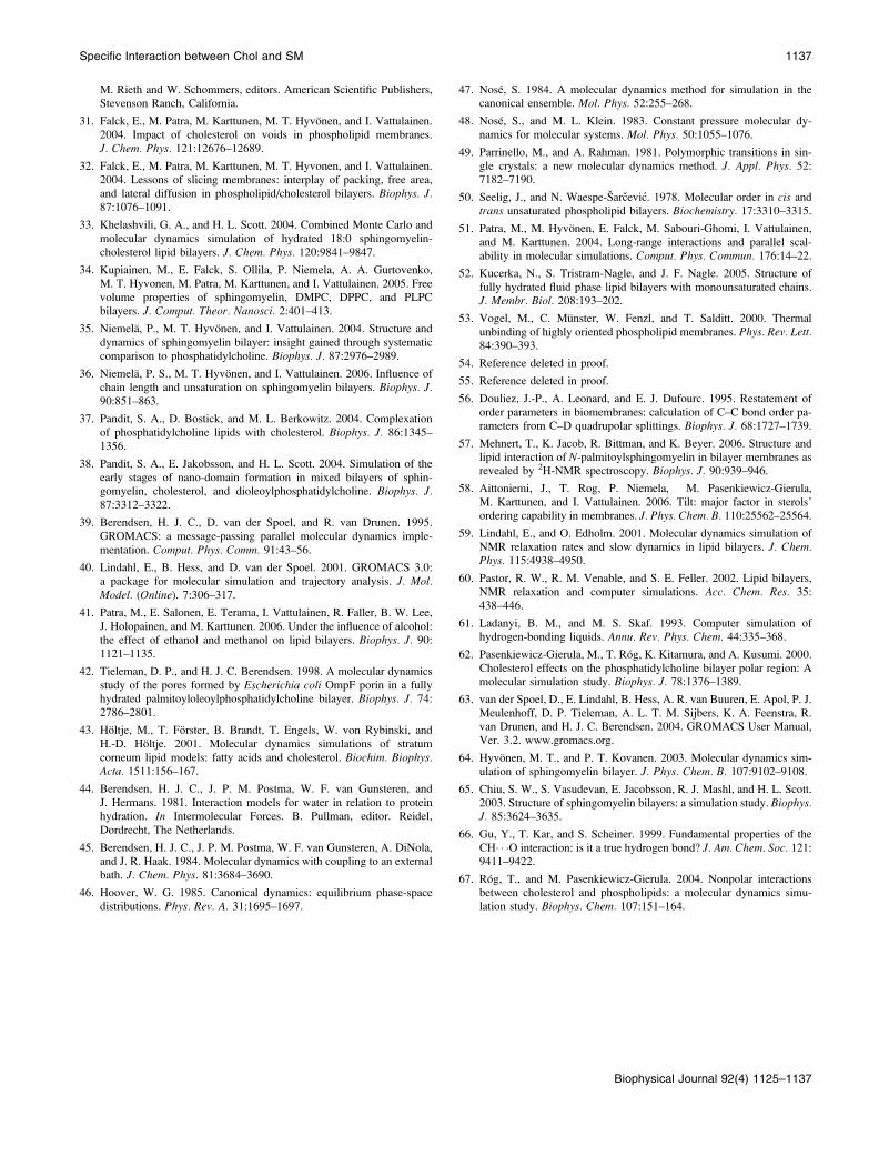
Equilibration and area per molecule
The area per molecule is a central structural quantity for lipid
bilayers. In simulations, it is often used as a measure of
system equilibration. Fig. 3 shows this quantity, calculated
as the quotient of simulation box size over number of lipids
per monolayer. Since the simulated composition is very close
to the all-POPC system used as the starting structure, the
system reached a dynamic equilibrium rather quickly, within
10 ns. After equilibration, the mean area per molecule attains
a value of A ¼ 0.66 6 0.01 nm2. The original all-POPC
structure featured an area per lipid of A ¼ 0.69 nm2, a value
that decreased quickly during the initial 50 ps stochastic
simulation. A recent experimental study of pure POPC
bilayers resulted in an area per lipid of A ¼ 0.683 6 0.015
nm2 at a temperature of 303 K (52). Thus, the POPC force
field we use in this simulation reproduces the experimental
area per lipid very well.
Density profile
The electron density profile across the bilayer is shown in
Fig. 4 separately for the different lipid species with or with-
out specific nearest neighbors. More specifically, the density
FIGURE 1 Structures of palmitoyl-
oleoyl-phosphatidylcholine (POPC, left),
cholesterol (Chol, middle), and palmitoyl-
sphingomyelin (PSM, right). Despite their
broad structural similarities, POPC and
PSM interact differently with Chol.
Specific Interaction between Chol and SM 1127
Biophysical Journal 92(4) 1125–1137
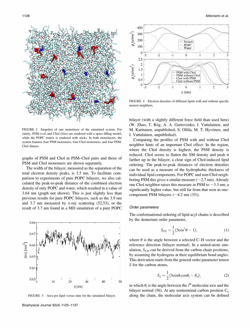
graphs of PSM and Chol in PSM–Chol pairs and those of
PSM and Chol monomers are shown separately.
The width of the bilayer, measured as the separation of the
total electron density peaks, is 3.5 nm. To facilitate com-
parison to experiments of pure POPC bilayers, we also cal-
culated the peak-to-peak distance of the combined electron
density of only POPC and water, which resulted in a value of
3.64 nm (graph not shown). This is just slightly less than
previous results for pure POPC bilayers, such as the 3.9 nm
and 3.7 nm measured by x-ray scattering (52,53), or the
result of 3.7 nm found in a MD simulation of a pure POPC
bilayer (with a slightly different force field than used here)
(W. Zhao, T. Rog, A. A. Gurtovenko, I. Vattulainen, and
M. Karttunen, unpublished; S. Ollila, M. T. Hyvonen, and
I. Vattulainen, unpublished).
Comparing the profiles of PSM with and without Chol
neighbor hints of an important Chol effect: In the region,
where the Chol density is highest, the PSM density is
reduced. Chol seems to flatten the SM density and push it
farther up in the bilayer, a clear sign of Chol-induced lipid
ordering. The peak-to-peak distances of electron densities
can be used as a measure of the hydrophobic thickness of
individual lipid components. For POPC and non-Chol neigh-
boring PSM this gives a similar measure (;2.7 nm). Already
one Chol neighbor raises this measure in PSM to ;3.3 nm, a
significantly higher value, but still far from that seen in one-
component PSM bilayers (;4.2 nm (35)).
Order parameters
The conformational ordering of lipid acyl chains is described
by the deuterium order parameter,
SCD ¼1
2Æ3cos
2u� 1æ; (1)
where u is the angle between a selected C–H vector and the
reference direction (bilayer normal). In a united-atom sim-
ulation, SCD can be derived from the carbon chain positions,
by assuming the hydrogens at their equilibrium bond angles.
This derivation starts from the general order parameter tensor
S for the carbon atoms,
Sij ¼1
2Æ3cosuicosuj � dijæ; (2)
in which ui is the angle between the ith molecular axis and the
bilayer normal (56). At any nonterminal carbon position Cn
along the chain, the molecular axis system can be defined
FIGURE 2 Snapshot of one monolayer of the simulated system. For
clarity, PSM (red) and Chol (blue) are rendered with a space-filling model,
while the POPC matrix is rendered with sticks. In both monolayers, the
system features four PSM monomers, four Chol monomers, and four PSM-
Chol dimers.
FIGURE 3 Area per lipid versus time for the simulated bilayer.
FIGURE 4 Electron densities of different lipids with and without specific
nearest neighbors.
1128 Aittoniemi et al.
Biophysical Journal 92(4) 1125–1137

using neighboring carbon atoms of the chain: The z axis is
taken to be the (normalized) vector Cn�1Cn11������!
, the x axis is
the unit vector perpendicular to Cn�1Cn����!
and CnCn11����!
, and the
y axis is the cross product of the z and x axes. Now, for
hydrogens in a sp3 hybridized CH2 group, this reduces to
�SCD ¼2
3Sxx 1
1
3Syy: (3)
For a sp2 hybridized CH group with an in-plane hydrogen
at angle 120� we get
�SCD ¼1
4Szz 1
3
4Syy7
ffiffiffi3p
2Syz: (4)
The SCD order parameters for the individual acyl chains are
shown in Fig. 5 separately for those chains that interact with
Chol and for those that do not. More specifically, the whole
POPC matrix represents POPC without Chol contact, the
monomeric PSM represents PSM without Chol contact, and
the PSM in PSM–Chol pairs represents PSM with Chol con-
tact. To establish POPC lipids with Chol contact, the order
profile was collected for those POPCs, whose center-of-mass
is the closest neighbor of a Chol center-of-mass, in terms of
distance in the two dimensions of the bilayer plane (i.e., in
the x,y plane).
Except for the unsaturated carbon positions, POPC and
PSM without Chol neighbor are about equally ordered. Since
the PSM ordering falls short of that found in pure PSM-
bilayers (35,57), it seems reasonable to state that the PSM
monomers adopt the (lesser) ordering of the POPC matrix.
Interaction with Chol changes this: Being nearest neighbor
to a Chol increases the order of PSM chains more than it
increases the order of POPC chains. While the mean POPC
order grows by 0.041 for palmitoyl (to 0.201) and by 0.042
for oleoyl (to 0.173), the mean PSM order rises by 0.067 for
the sphingosine base (to 0.224) and 0.066 for the palmitoyl
residue (to 0.230). These numbers underline two general
properties of Chol also seen in experiments: its ordering
effect, and a preference for SM over a monounsaturated PC.
A recent 2H-NMR study of a pure POPC bilayer at 321 K
found a mean order parameter value of SCD¼ 0.146 6 0.003
for the whole palmitoyl chain (57), and a mean value of
SCD ¼ 0.185 6 0.004 for the plateau region of the palmitoyl
chain. The plateau region chosen in (57) corresponds to chain
positions 3–5 in Fig. 5. This experiment allows for a com-
parison with our simulated system, which consists mostly of
POPC. In the simulation, the mean order of POPC-palmitoyl
without contact to neither a Chol nor a PSM is SCD ¼ 0.152
6 0.005 for the whole chain and SCD ¼ 0.184 6 0.005 for
carbon positions 3–5. The agreement with the experimental
values is very good.
Cholesterol tilt and ordering
We define the Chol tilt as the angle that the vector across the
steroid nucleus (from C21 to C5 in Fig. 1) forms with the
bilayer normal. Distributions of this tilt are shown in Fig. 6
separately for the Chol in PSM-Chol pairs and the mono-
meric Chol. The averaged tilt angles are 25� for Chol with
PSM neighbor and 33� for Chol without.
Recently, we established a relationship of a sterol’s tilt
angle to its local ordering effect: A higher sterol tilt weakens
the ordering effect on the sterol’s nearest neighbors (58). The
same relationship applies in this system as well: The mean
order parameters of the acyl chains of Chol nearest neighbors
as a function of Chol’s tilt are shown in Fig. 7. The clear
downward slopes indicate a weakened ordering with higher
Chol tilt. In a more detailed analysis (data not shown), we
found that the observed effect splits into two contributions.
First, an increased tilt of cholesterol decreases the trans/
gauche fraction of the neighboring acyl chain, which is
reflected as a decrease in the order parameter values. Second,
a more tilted cholesterol also increases the overall tilt of the
neighboring acyl chains, causing an additional decrease in
the order parameter values.
FIGURE 5 SCD order parameters of the acyl chains of phospholipids
without contact to a Chol molecule (upper graph) and of those that have a
Chol as a nearest neighbor (lower graph). SPbase refers to the sphingosine
chain of PSM.
Specific Interaction between Chol and SM 1129
Biophysical Journal 92(4) 1125–1137

PSM and POPC both carry palmitoyl residues, which can
be compared as suggested by the lines in Fig. 7. It seems as if
the different tilts of PSM neighboring and non-PSM neigh-
boring Chol explain a major part of the observation, that
Chol orders PSM better than POPC. Now, having estab-
lished the importance of the Chol tilt, we should put special
emphasis on the effects of different interaction mechanisms
on the Chol tilt.
Headgroup orientations
A revealing quantity for studying phosphocholine headgroup
structure and interactions is the headgroup orientation dis-
tribution, as measured using the angle of the P-N vector
(from the phosphate to the nitrogen) with the bilayer normal.
Such distributions are plotted in Fig. 8. Neighborship to
Chol was determined similarly as in the calculation of the
order profiles (i.e., using the POPC matrix, the monomeric
PSM, the paired PSM, and POPCs that are closest neighbors
to Chol). The headgroup orientations of non-Chol neighbor-
ing PSMs and that of all (i.e., mostly non-Chol neighboring)
POPCs have similar shapes, with the PSM distribution
peaking at a lower angle than the POPC distribution (70� vs.
85�). As a reference for pure POPC, a simulation of DPPC
(with a similar force field than used here for POPC) resulted
in a monomodal P-N angle distribution peaking at 90� (35).
Comparing that result to Fig. 8 shows that POPC headgroup
orientation is very similar to that of DPPC. Since a sim-
ulation of a hydrated PSM-only bilayer with the same force
field showed a bimodal P-N vector angular distribution, with
peaks at 55� and 105� (35), the appearance of a monomodal
distribution in non-Chol neighboring PSM suggests that the
POPC headgroups inflict their preferred orientation also onto
the headgroups of the PSM monomers.
Interactions with Chol bring remarkable changes to the
headgroup orientations. The angular distribution of the
headgroups of those PSMs that are in contact with Chol
becomes bimodal, with maxima at 60� and 115�. The POPC
distribution remains monomodal but with a narrower maxi-
mum at an increased angle of 100�. Thus, when a PSM has
just one Chol as nearest neighbor, the neighboring POPC
headgroups no longer inflict their orientation on its head-
group. Presumably the PSM choline moves into the free
space above the Chol, which has hardly any headgroup of its
own. In this way, the headgroup-free space of Chol allows a
neighboring PSM to adopt headgroup orientations similar to
those in the pure PSM bilayer. To visualize different possible
headgroup orientations, snapshots of a PSM molecule with
and without Chol neighbor are shown in Fig. 9.
Other factors that cause shifts toward higher angles for
both POPC and PSM headgroups are charge-pair interac-
tions between the choline nitrogen moiety and Chol oxygens,
FIGURE 7 Order parameter of Chol neighboring chains (mean SCD
averaged over all carbons in a chain) plotted against that Chol’s tilt angle.
The added lines indicate the average tilts of PSM neighboring Chol (25�)
and monomeric Chol (33�). Neighborship to cholesterol was established
using a 0.7 nm cutoff for the center-of-mass distances in the bilayer plane
(the x,y plane).
FIGURE 8 Orientation distributions of choline headgroups, measured as
the angle uPN of the P-N vector with the outward bilayer normal. Of all
POPCs, only a small fraction is in contact with Chol. SM without chol refers
to the monomeric non-Chol neighboring PSMs. POPC/PSM with chol nn
contains only those lipids that actually have a Chol as nearest neighbor.
FIGURE 6 Distribution of Chol tilt angles for those Chols with a PSM
neighbor and those without.
1130 Aittoniemi et al.
Biophysical Journal 92(4) 1125–1137

as well as hydrophobic interactions in the sense of Chol
water shielding. These aspects will be revisited later.
Rotational motions
Along with lateral translation, rotation around a molecular
axis is a major degree of freedom of bilayer lipids. In a liquid-
ordered (lo) phase, rotational motion should be restricted just
as acyl chain conformational changes are. Thus, Chol—an
efficient promoter of the lo phase—should affect rotational
motions of neighboring lipids. We study rotational motions
using second rank reorientational autocorrelation functions
C2(t),
C2ðtÞ ¼1
2Æ3½m/ðtÞ � m/ð0Þ�2 � 1æ; (5)
where m/ðtÞ is a unit vector that defines the chosen rota-
tional mode. We examine two rotational modes, one in the
headgroup and one in the interfacial region, separately for
the POPC matrix and the Chol paired and non-Chol paired
PSM. The headgroup mode was defined as the vector from
phosphate to nitrogen (P–N vector), while the interfacial
vector was taken from carbons G1 to G3 in POPC and SPH-3
to SPH-1 in SM (see Fig. 1).
The resulting autocorrelation functions are shown in Fig.
10. Since the motions of the examined lipid parts are limited,
the autocorrelation functions do not decay to zero. Instead,
their plateau values represent inherent ordering of the studied
vectors (59,60). It seems that our simulation is not long
enough for the autocorrelation functions to reach their
plateau values. Yet, Fig. 10 contains interesting information
about the mobility of the studied lipid parts in different local
lipid compositions.
The POPC matrix is mostly without Chol contact, so its
rotations should be compared to those of PSMs without a
Chol neighbor. But, since ;10% of POPCs have a Chol
neighbor, the rotations of non-Chol neighboring POPC are
probably a bit faster than indicated in Fig. 10. Keeping that in
mind, headgroup reorientations in POPC and PSM without
Chol contact seem about equally fast (a bit slower in POPC),
whereas interfacial reorientations are slower in (non-Chol
neighboring) PSM than in POPC. The latter finding is prob-
ably due to PSM intermolecular hydrogen bonding (see next
section).
Interaction with Chol slows down PSM rotations remark-
ably, both in the headgroup as well as the interfacial regions.
In both regions, the C2(t) decay half-times grow approxi-
mately fourfold. A less stringent analysis of reorientations of
Chol-neighboring POPCs shows that Chol contact slows
POPC rotations, but far less than it does in PSM (data not
shown): Decay half-times in the interfacial region roughly
double, while those in the headgroup region grow only
slightly.
Hydrogen bonding
Although classical MD simulations fail to include quantum
effects (such as proton sharing) entirely, many such simu-
lations have been able to predict the correct qualitative static
and dynamic features of water and other hydrogen-bonding
liquids (61). In this work, we employ geometric criteria to
define a hydrogen bond (H-bond): the acceptor-hydrogen
distance dAH , 0.25 nm and the donor-hydrogen-acceptor
angle uDHA , 90�. Geometric conditions like these are a
commonly used operational definition in the field (35,62,63).
The average hydrogen-bond numbers for the different lipids
are gathered in Table 1. In the following sections, we will
analyze them in more detail.
PSM H-bonding
In the simulation of the dilute system, hardly any direct
H-bonds form between PSM and Chol (0.08 6 0.02 H-bonds
FIGURE 9 Simulation snapshots representing typical PSM orientations,
for a PSM without Chol neighbor (left) and a PSM with Chol neighbor
(right). The PSM and Chol in the right image form a charge pair between the
headgroup positive charge and the Chol oxygen (see Charge-Pairing).
FIGURE 10 C2 reorientational autocorrelation functions of vectors in the
headgroup (P-N) and in the interfacial region for POPC/PSM molecules with
(w) or without (no) Chol contact.
Specific Interaction between Chol and SM 1131
Biophysical Journal 92(4) 1125–1137

on average per Chol-PSM pair). Thus, direct H-bonding
of PSM and Chol is too rare to be considered of importance for
the lipid-lipid interactions. Yet, the simulations show a change
in the PSM amide group’s H-bonding to water as a con-
sequence of Chol interactions: The H–N–C¼O entity of a
PSM without a Chol neighbor forms on average 0.45 6 0.03
H-bonds to water, while that of a PSM with a Chol neighbor
forms only 0.34 6 0.12 of them. Therefore, just as in the IR
spectroscopy experiment (21), interactions with Chol change
the SM amide group’s H-bonding to water. However, at least
under the conditions of this simulation, this is not due to
direct SM–Chol H-bonds.
Instead of binding to Chol, the PSM donors are occupied
with forming H-bonds to POPC (through the N–H group)
and intramolecular H-bonds (the O–H group). The intramo-
lecular PSM H-bond forms between the hydroxyl group and
oxygens of the phosphate group, mostly oxygen atom OPa
(see Fig. 1), and is virtually always present in every PSM
molecule. This intramolecular H-bond has been found to be
very stable and popular also in other simulations of all-PSM
bilayers, using both the same PSM force field as here (35) as
well as an all-atom CHARMM force field in an earlier study
(64). The four oxygens and net negative charge of the phos-
phate group probably stabilize this H-bond. PSM intramo-
lecular H-bonding is increased in those PSMs with Chol
contact: While the number of O–H� � � OPa contacts remain
virtually one, the number of O–H� � �OPb H-bond-like con-
tacts increases from 0.08 6 0.01 per non-Chol neighboring
PSM to 0.12 6 0.02 per Chol-neighboring PSM. The re-
sulting case of a proton being shared between three oxygens
is probably unphysical. Nonetheless, the increase in OH� � �OPb
H-bonds is indicative of a strengthening interaction. This
increase in intramolecular H-bonding is related to a change
in headgroup orientations (see below).
It is important to remember that the studied three-
component system features merely pairs of Chol and PSM.
It does not allow conclusions about PSM-Chol H-bonding in
a two-component (or mostly two-component) phase. A
simulation study of SM-Chol bilayers found diverse hydro-
gen bonding between these lipids (33). Thus, the virtually
complete absence of direct SM-Chol H-bonds probably
results from the dilute PSM and Chol concentrations, so that
PSM has to compete with POPC about the popularity of
H-bonds to Chol. Chol prefers H-bonds to POPC, while
PSM prefers intramolecular H-bonds (O–H group) and those
to POPC (N–H group).
PSM and POPC form, on average, 0.93 H-bonds per PSM
molecule through the N–H donor of PSM. All POPC ester-
bond oxygens participate as acceptors. H-bonding of the SM
amide group to neighboring phospholipids has been estab-
lished in other studies as well (35,65). Such H-bonds prob-
ably stabilize the PSM molecule orientation with respects
to its neighbors. It seems reasonable to assume that PSM
intermolecular H-bonding explains its slower reorientations
in the interfacial region (see Rotational Motions).
POPC-Chol
POPC and Chol form on average 0.85 H-bonds per Chol, i.e.,
far more than Chol forms with PSM. In these H-bonds, the
Chol hydroxyl group acts as donor while POPC ester bond
oxygens (in 90% of bonds) and phosphate oxygen OPb (in
10% of bonds) act as acceptors. Notably, oxygen Ob2 of the
oleoyl chain is the by far most common acceptor of POPC-
Chol H-bonds, acting as acceptor in two-thirds of the bonds.
Thus, Ob2 is better suited to accept Chol H-bonds than the
other POPC oxygens, including those of the saturated fatty
acid residue.
H-bonding to POPC has interesting effects on the Chol tilt.
Chol without a PSM neighbor has an average tilt of 34� when
H-bonded to POPC, but only 28� when not H-bonded to
POPC. The equivalent numbers for Chol with PSM neighbor
are 25� (with H-bond to POPC) and 24� (without H-bond to
POPC). H-bonding to oxygens in the interfacial region might
force Chol to keep rather low in the bilayer, which in turn
would cause a higher Chol tilt. In addition, PSM neighboring
Chol has significantly lower tilts when H-bonded to POPC
oxygens Oa2 and Ob2 (;25�) than when H-bonded to POPC
oxygens Oa1 and Ob1 (;30�).
Direct H-bonding is a rather favorable interaction. It is
especially pronounced between Chol and POPC. Yet, such
H-bonds enforce a high tilt on the Chol, especially if that
Chol is not neighboring a PSM. The higher Chol tilt in turn
weakens the ordering and condensing effects of Chol, as well
as other forms of interactions, which are analyzed next.
Charge-pairing
Both phospholipids in the simulation feature a positive
charge in the amide moiety of their choline headgroups.
Thus, they interact favorably with oxygens, all of which
carry negative partial charges. In the context of this study,
the most relevant oxygen for a choline group to pair with is
the cholesterol oxygen.
The fundamental nature of such N1(CH3)3� � �O interac-
tions is ambiguous: Some consider them to be hydrogen
bonds, with the partially charged methyl groups acting as
donors (37). Others prefer to talk simply about charge-pair
TABLE 1 Average numbers of hydrogen bonds per
corresponding pair for different molecules
POPC PSM Water
POPC — 0.93 6.99
PSM without Chol 0.93 1.08* 6.39
PSM with Chol 0.93 1.12* 6.2
Chol without PSM 0.88 — 0.54
Chol with PSM 0.82 0.08 0.44
*PSM intramolecular H-bonds, including both OH� � �OPa and OH� � �OPb
contacts.
1132 Aittoniemi et al.
Biophysical Journal 92(4) 1125–1137

interactions (62). Quantum chemical calculations indeed
suggest that a carbon with an electronegative substituent can
donate a proton to an oxygen, forming a H-bond-like in-
teraction (66). Yet, this interaction was found to be ener-
getically weaker than a classical OH� � �O H-bond, and to
react less sensitively to changes in donor-hydrogen-acceptor
angles and distances (66). The CH� � �O interaction decays
more slowly with increasing H� � �O distance, so, at certain
distances, it can be effectively stronger than a OH� � �OH-bond (66).
In the united atom descriptions of the simulations at hand,
no choline group hydrogens are included explicitly. This
forbids a similar geometric analysis of the N1(CH3)3� � �Ointeraction than was used above for OH� � �O and NH� � �OH-bonds. As an alternative, we employ an approach based on
groupwise Coulomb energies to analyze these interactions.
After all, the CH� � �O interaction is mainly of electrostatic
nature (66). In this work, the N1(CH3)3� � �O interaction is
referred to as a charge-pair interaction, because, as outlined
above, it is different from ‘‘classical’’ H-bonds. This choice
also distinguishes it from the OH� � �O and NH� � �O H-bonds
in the interfacial region of the bilayer, and reminds the reader
that, in this analysis, N1(CH3)3� � �O interactions are defined
energetically, not geometrically.
Charge-pairing criterion
To study charge-pairing of choline groups to the polar atoms
of Chol, the Coulomb energy between the involved groups
(the nitrogen and the three adjacent methyl groups for the
phospholipid, and the C–O–H moiety for the cholesterol) is
calculated as a sum of atom-pairwise interactions. A charge-
pair binding mode is identified as a local maximum in the
negative tail of the interaction energy histograms (graphs not
shown). These choline-cholesterol-COH histograms have
local minima at �2.8 kcal/mol (POPC) and �2.5 kcal/mol
(PSM), with local maxima at �3.5 kcal/mol (POPC) and
�4.0 kcal/mol (PSM). Thus, energy cutoffs of�2.8 kcal/mol
(POPC) and �2.5 kcal/mol (PSM) give an operational def-
inition for charge-pair interactions. In a similar simulation,
Pandit et al. found a cutoff energy of �2.8 kcal/mol for the
interaction of the DPPC choline group and the Chol polar
atoms (37), which is exactly the same as observed here for
the POPC-Chol charge pairing. For comparison, the histo-
gram of the Chol OH to POPC Ob2 hydrogen bond has a
maximum at �11 kcal/mol (graph not shown). Thus, in this
simulation, the charge-pair interactions have roughly one-
third of the nominal strength of actual hydrogen bonds.
Charge-pairing occurrence
On average, there are 0.17 6 0.02 charge-pair bonds to a
PSM choline group per (PSM neighboring) Chol. To POPC
choline groups, there are on average 0.36 6 0.05 charge
pairs per PSM neighboring Chol and 0.44 6 0.06 charge
pairs per non-PSM neighboring Chol. Thus, between Chol
and PSM, charge pairs are more frequent than conventional
H-bonds. To put these numbers into proper relation, the
different configuration numbers should be noted: Chol has
typically six closest POPC neighbors, but only at most one
PSM neighbor. Thus, if Chol formed charge pairs to both
phospholipids with equal probability, we should see 5–6
times more Chol-POPC charge pairs than Chol-PSM charge
pairs. This is obviously not the case, since Chol seems rather
eager to charge-pair to PSM instead of POPC.
To bind to a Chol oxygen, the phospholipid headgroup has
to bend low, deep into the interfacial region of the bilayer.
This can be clearly seen in the P-N vector angular distribu-
tions. Headgroups that are charge-pair-bonded have almost
exclusively P-N vector angles .90�, with maxima at 110�(for POPC) and 125� (for PSM) (graphs not shown). Thus,
headgroup pairing with Chol oxygens helps to explain Chol-
induced shifts in P-N angle histogram maxima (see Fig. 8).
PSM structure and charge-pairing
PSM’s preference for higher headgroup angles and Chol’s
preference for charge pairs to PSM are probably caused by
certain structural differences in PSM and POPC. The intra-
molecular H-bonds between PSM hydroxyl groups and phos-
phate oxygens—without match in POPC—seem to pull the
headgroups down and stabilize their higher angles. Indeed,
as mentioned earlier, the prevalence of PSM intramolecular
OH� � �OPb H-bonds increases from 0.08 in Chol without a
PSM neighbor to 0.12 in Chol with a PSM neighbor, an in-
crease that is mostly explained by an increase to 0.18 in those
PSM with a charge-pair bond to Chol. In a simulation of pure
SM and SM-Chol bilayers (with other force fields than those
used here), the frequency of the SM OH��OPa intramolecular
bond rose remarkably in the bilayer system with Chol (33). It
seems reasonable to assume that this increase is connected to
SM choline charge-pairing to Chol oxygens.
The PSM choline also forms intramolecular charge pairs
with the carbonyl oxygen OPA of the same molecule. The
occurrence of this interaction is 0.14 charge pairs per PSM in
those PSM without a Chol neighbor, and 0.24 in those PSM
with a Chol neighbor. Again, it seems reasonable to believe
that this interaction stabilizes headgroup interactions with
Chol. In POPC, no charge pairs form between the choline
group and the corresponding carbonyl oxygen Ob2.
Other forms of interaction
Hydrophobic interactions
Hydrophobic interactions are the main factor that drives
structural lipids into bilayer form. Being of such overall
importance, they might be involved in lipid-lipid interactions
as well. After all, Chol is mostly hydrophobic, with only a
small polar headgroup.
Unfortunately, hydrophobic interactions are difficult to
quantify energetically in molecular dynamics simulations.
Specific Interaction between Chol and SM 1133
Biophysical Journal 92(4) 1125–1137

Water contact may be easily derived from a simulation, but the
free energies related to it are hard to establish. Therefore, in
this analysis, hydrophobic interactions are examined through
changes in Chol-water contact. To this end, density profiles of
water and the Chol nonpolar parts are calculated over small
cylinders centered around Chol molecules. In other words,
only atoms that are, in the x,y (bilayer) plane, within 0.7 nm of
the Chol center of mass are included in the density profile. The
overlap of the densities of water and Chol nonpolar carbons
indicates unfavorable water contacts (see Fig. 11).
Upon visual inspection, no differences are visible in the
graphs of Fig. 11. As a measure for the water contact, the
area overlap of the density profiles of water and Chol carbons
(i.e., the striped area of the minimum of the two curves) can
be related to the total Chol carbon density area. This dimen-
sionless fraction j is then a measure for the unfavorable hy-
dration of the Chol nonpolar part,
j ¼R
minðrwater; rcarbonÞRrcarbon
; (6)
where r denotes electron density of the water or the cho-
lesterol carbon molecules.
For Chol with no PSM neighbor, this fraction is j ¼0.17 6 0.03. For those with a PSM neighbor, this fraction is
j ¼ 0.15 6 0.03. Keeping in mind the bad statistics implied
by the dilute Chol (and PSM) concentration, these numbers
could be interpreted to point toward a better Chol water
shielding by PSM.
A more interesting point is the effect on the water contact
of choline headgroup charge-pairing to Chol. When the
cylindrical density calculations are restricted only to those
Chols, whose oxygen forms a charge pair to a PSM choline
group, the density overlap fraction drops to j ¼ 0.11 6 0.01.
However, for those non-PSM neighboring Chols that form
the same bond to a POPC choline group, this fraction
decreases merely to j ¼ 0.16 6 0.03. Thus, in these sim-
ulations, choline group charge-pairing to the Chol oxygen
(forming on average 0.17 6 0.02 charge pairs per Chol) is
responsible for a great part of the PSM-caused reduction in
Chol water contact.
When forming a charge pair with Chol oxygen, the PSM
headgroup folds down, after which it should be able to
accommodate the Chol underneath itself and protect it from
unwanted water contact (a snapshot of a charge-paired PSM-
Chol pair is shown in Fig. 9). The POPC headgroup does not
shield a neighboring Chol from water as well as the PSM
headgroup. This is probably related to the Chol tilt, which is
significantly higher for Chols without PSM neighbor. A
more tilted Chol should be more accessible to water.
The differences in steric shielding by the choline headgroup
might explain the observed variation in Chol efflux rates from
SM and PC monolayers to cyclodextrin subphase (accelerat-
ing efflux with decreasing SM content (23)), and the reduced
accessibility of Chol oxidase to Chol in SM monolayers than
in PC monolayers (7,23).
Van der Waals interactions and lipid packing
Direct calculation of van der Waals energies is out of scope
of classical MD force fields. Those few lipid bilayer simula-
tion articles that analyze van der Waals interactions usually
examine only order parameters or atom packing, and relate
those directly to van der Waals interactions (e.g., in (67)). In
that spirit, since this work found Chol to order PSM more
than POPC (in the sense of the SCD order parameter), van der
Waals interactions between PSM and Chol can be seen as
more favorable than those between POPC and Chol. This
line of thought also means that a lower Chol tilt improves
van der Waals interactions and lipid packing.
Steric hindrance issues affect Chol-phospholipid orienta-
tions. To study lipid arrangements with reference to the Chol
a (smooth) and b (two protruding methyl groups) faces,
center-of-mass trajectories were produced for the Chols,
the protruding Chol methyl groups, and the individual
FIGURE 11 Hydrophobic interac-
tion calculation. Densities of water
and Chol are calculated inside small
cylinders (of radius 0.7 nm), that are
centered on Chol molecules (illustrated
above on the purple-colored choles-
terol molecule). Density overlap of
water and Chol nonpolar carbons (high-
lighted by stripes) indicates unfavor-
able water contacts. The left profile is of
non-PSM-paired Chol and the right one
is of Chol that is paired to a PSM.
1134 Aittoniemi et al.
Biophysical Journal 92(4) 1125–1137

phospholipid chains. In such a trajectory, the two-dimen-
sional angle (angle in the x,y-plane) formed by the centers of
mass of the phospholipid chain, the Chol, and the Chol
methyl groups, indicates on which face the chain is located
(angle . 90�: a-face; angle , 90�: b-face). This analysis
shows a clear preference (of ;60:40) of PSM to reside on a
neighboring Chol’s smooth a-face, while POPC shows no
face preference. In analogous simulations, Pandit et al. found
similar preferences of PSM and DOPC for the Chol faces (38).
The Chol face affects acyl-chain ordering: The saturated
acyl chains (both PSM chains and the palmitoyl in POPC)
are more ordered when next to a Chol a-face than when next
to a Chol b-face. For the POPC-oleoyl chain, there is no sig-
nificant change in ordering between the Chol faces. The dif-
ferences in chain ordering between Chol a- and b-faces are
greater in PSM chains (with changes in the mean SCD order
of ;0.020) than in POPC-palmitoyl (changes of ;0.010).
This difference is probably related to the differences in the
tilt angles of Chols neighboring these phospholipids.
The changes in order on the two Chol faces are minor
when compared to the overall order increment of having a
Chol neighbor at all (which increases the mean SCD order by
0.030 – 0.070).
DISCUSSION
Cholesterol favors PSM over POPC
As established in the density and order profiles, already sin-
gle Chol molecules order and condense the bilayer locally.
Thus, no large phospholipid-Chol complexes or networks
are needed to initiate these ultimately macroscopic effects of
Chol. Moreover, just one Chol neighbor is enough to promote
significant changes in PSM: Its hydrophobic thickness in-
creases, its acyl-chain order increases, its headgroup orienta-
tion becomes bimodal and resembles more closely that in an
all-PSM bilayer, and its rotational motions become slower.
Most of these changes are also visible in Chol neighboring
POPC, but with significantly smaller magnitudes. In addi-
tion, having just one PSM neighbor significantly reduces the
tilt of Chol. Taken together, these pieces of evidence con-
vincingly assert a preference, in the simulations, of Chol
for saturated SM over monounsaturated PC. The existence
of such a preferential interaction is in line with many
experiments.
Yet, the simulations do not support one of the main lines
of speculation on the origin of the SM-Chol interaction
specificity: direct H-bonding of SM donors to the Chol
oxygen. Instead, in the simulations, the PSM hydroxyl group
is constantly H-bonded to a phosphate oxygen of the same
molecule, while the PSM amide group forms H-bonds
almost exclusively to neighboring POPCs. Chol forms
hydrogen bonds through its hydroxyl group mainly to
POPC oxygens in the interfacial region, a consequence of the
higher number, greater partial charge, and greater flexibility
of the POPC acceptors over the SM acceptors in the inter-
facial region.
Since the simulated system includes only monomers and
dimers of PSM and Chol, no conclusions should be drawn
from it on the PSM-Chol hydrogen bonding in a phase rich in
PSM and Chol. Still, the unique approach of this study, with
its dilute PSM and Chol concentration, shows that direct
PSM-Chol hydrogen bonds are of little importance in the
pairing of these molecules. Consequently, the initial forma-
tion of PSM-Chol enriched phases must be driven by alter-
native factors.
Alternatives to hydrogen bonding
Alternatives to hydrogen bonding include charge-pairing of
the phospholipid headgroup nitrogen moieties with Chol
oxygens, hydrophobic interactions, and van der Waals inter-
actions. The differences in PSM and POPC van der Waals
interactions with Chol are manifested in a greater Chol-
induced increase in PSM ordering than in POPC ordering, as
well as the clear preference of PSM for the smooth a-face of
Chol, which is not seen for POPC. Some evidence suggests
that charge pairs of the Chol polar group to the PSM choline
group are more stable than those to the POPC choline group.
This seems to originate from features related to structural
differences of POPC and PSM, namely the formation of the
PSM intramolecular hydrogen bonds and PSM choline group
intramolecular charge pairs to the PSM carbonyl oxygen.
What is more, the headgroup-free space above Chol allows
the PSM headgroup to adopt a bimodal orientation distri-
bution, which is also seen in one-component PSM bilayers.
Analysis of the water contact of the nonpolar parts of Chol
shows a small decrease in water contact for SM-neighboring
Chol versus Chol with only POPC neighbors. To a great part,
this decrease is explained by the PSM choline charge-pairing
to Chol, which provides clearly better water shielding than
similar charge-pairing of POPC choline.
Of these mechanisms, at least van der Waals and hydro-
phobic interactions should benefit from small Chol tilts. The
ordering capability of Chol clearly decreases with increasing
tilt, and a more tilted Chol seemingly exposes more of its
nonpolar carbons to water. H-bonding of Chol to POPC ester
bond oxygens was found to increase the Chol tilt in com-
parison to Chol without such H-bonds. This raises the idea
that Chol H-bonding to POPC, an energetically very favorable
bond, competes with the other interactions mentioned above.
PSM has fewer and (at least in our force field) weaker
H-bond acceptors in the interfacial region, and its peptide
bond renders the PSM less flexible in the interfacial region.
Therefore, between PSM and Chol, H-bonding is weakened
and the other interactions become stronger, which leads to a
lower Chol tilt and improved ordering of surrounding lipids.
A central role of hydrophobicity in phospholipid-Chol in-
teractions has been suggested in connection with the concept
of hydrophobic mismatch (22). In that concept, Chol
Specific Interaction between Chol and SM 1135
Biophysical Journal 92(4) 1125–1137

positions itself preferentially at boundaries of more and less
ordered patches not because of specific interactions but to
smooth the mismatch in hydrophobic thickness between the
regions. In our system, the monomeric (non-Chol neigh-
boring) PSM features a hydrophobic thickness similar to that
of the POPC matrix, but already a single Chol neighbor
raises the hydrophobic thickness of a PSM molecule signif-
icantly. Thus, a Chol molecule in a lipid bilayer allows for
differences in hydrophobic thickness to form. What is more,
due to differences in Chol interaction mechanisms with PSM
and POPC, Chol not only allows but also promotes differ-
ences in PSM and POPC hydrophobic thickness. The change
in PSM hydrophobic thickness is accompanied by a lowering
of the tilt angle of the neighboring Chol. It seems that Chol
can adapt to different hydrophobic environments by adjust-
ing its tilt angle.
In conclusion, this work suggests that the initial phases of
raft formation are not driven by direct H-bonding between
PSM and Chol. Rather, the ‘‘specific’’ nature of the inter-
action between these molecules is more subtle and comprises
a shift in interactions away from H-bonding toward electro-
static (charge-pair) interactions between PSM headgroups
and Chol oxygens, together with improved van der Waals
interactions and better water-shielding of Chol. Unlike direct
H-bonding, these latter interactions benefit from a lower
Chol tilt, which in turn promotes higher ordering of hydro-
carbon chains. In addition, the concept of hydrophobic mis-
match seems to hold, in the sense that Chol smoothens a
difference in hydrophobic thickness that is itself created in
the first place. In a bilayer of Chol and phospholipids with
different acyl-chain lengths, the role of hydrophobic mis-
match is probably more pronounced.
We thank Juha M. Holopainen for discussions. We acknowledge the Finnish
IT Center for Science and the HorseShoe (DCSC) supercluster computing
facility at the University of Southern Denmark for computer resources.
This work has, in part, been supported by the Academy of Finland (to I.V.,
M.T.H., P.S.N., and M.K.), the Academy of Finland Center of Excellence
Program (to P.S.N. and I.V.), the Jenny and Antti Wihuri Foundation (to
M.T.H.), the Finnish Academy of Science and Letters (to P.S.N.), the Emil
Aaltonen foundation (M.K.), and the Natural Sciences and Engineering
Council (NSERC) of Canada (to M.K.).
REFERENCES
1. Bloom, M., E. Evans, and O. G. Mouritsen. 1991. Physical propertiesof the fluid lipid-bilayer component of cell membranes: a perspective.Q. Rev. Biophys. 24:293–397.
2. Nelson, D. L., and M. M. Cox. 2004. Lehninger Principles of Bio-chemistry, 4th Ed. W.H. Freeman and Company, New York.
3. Singer, S. J., and G. L. Nicolson. 1972. The fluid mosaic model of thestructure of cell membranes. Science. 175:720–731.
4. Vist, M. R., and J. H. Davis. 1990. Phase equilibria of cholesterol/dipalmitoylphosphatidylcholine mixtures: 2H nuclear magnetic reso-nance and differential scanning calorimetry. Biochemistry. 29:451–464.
5. Ipsen, J. H., G. Karlstrom, O. G. Mouritsen, H. Wennerstrom, andM. J. Zuckermann. 1987. Phase equilibria in the phosphatidylcholine-cholesterol system. Biochim. Biophys. Acta. 905:162–172.
6. London, E. 2002. Insights into lipid raft structure and formationfrom experiments in model membranes. Curr. Opin. Struct. Biol. 12:480–486.
7. Silvius, J. R. 2003. Role of cholesterol in lipid raft formation: lessonsfrom lipid model systems. Biochim. Biophys. Acta. 1610:174–183.
8. Simons, K., and W. L. C. Vaz. 2004. Model systems, lipid rafts, andcell membranes. Annu. Rev. Biophys. Biomol. Struct. 33:269–295.
9. Alberts, B., A. Johnston, J. Lewis, M. Raff, K. Roberts, and P. Walter.2002. Molecular Biology of the Cell, 4th Ed. Garland Science, New York.
10. Cottingham, K. 2004. Do you believe in lipid rafts? Anal. Chem. 76:403A–406A.
11. Munro, S. 2003. Lipid rafts: elusive or illusive? Cell. 115:377–388.
12. Simons, K., and E. Ikonen. 1997. Functional rafts in cell membranes.Nature. 387:569–572.
13. Brown, D. A., and E. London. 1998. Functions of lipid rafts in bio-logical membranes. Annu. Rev. Cell Dev. Biol. 14:111–136.
14. Edidin, M. 2003. The state of lipid rafts: from model membranes tocells. Annu. Rev. Biophys. Biomol. Struct. 32:257–283.
15. Vainio, S., M. Jansen, M. Koivusalo, T. Rog, M. Karttunen, I.Vattulainen, and E. Ikonen. 2006. Significance of sterol structuralspecificity: desmosterol cannot replace cholesterol in lipid rafts. J. Biol.Chem. 281:348–355.
16. Li, X.-M., M. M. Momsen, J. M. Smaby, H. L. Brockman, and R. E.Brown. 2001. Cholesterol decreases the interfacial elasticity and deter-gent solubility of sphingomyelins. Biochemistry. 40:5954–5963.
17. Ohvo, H., and J. P. Slotte. 1996. Cyclodextrin-mediated removal ofsterols from monolayers: effects of sterol structure and phospholipidson desorption rate. Biochemistry. 35:8018–8024.
18. Ramstedt, B., and J. P. Slotte. 1999. Interaction of cholesterol withsphingomyelins and acyl-chain-matched phosphatidylcholines: a com-parative study of the effect of the chain length. Biophys. J. 76:908–915.
19. Bittman, R., C. R. Kasireddy, P. Mattjus, and J. P. Slotte. 1994. Inter-action of cholesterol with sphingomyelin in monolayers and vesicles.Biochemistry. 33:11776–11781.
20. Sankaram, M. B., and T. E. Thompson. 1990. Interaction of cholesterolwith various glycerophospholipids and sphingomyelin. Biochemistry.29:10670–10675.
21. Veiga, M. P., J. L. R. Arrondo, F. M. Goni, A. Alonso, and D. Marsh.2001. Interaction of cholesterol with sphingomyelin in mixed mem-branes containing phosphatidylcholine, studied by spin-label ESR andIR spectroscopies. A possible stabilization of gel-phase sphingolipiddomains by cholesterol. Biochemistry. 40:2614–2622.
22. Holopainen, J. M., A. J. Metso, J.-P. Mattila, A. Jutila, and P. K. J.Kinnunen. 2004. Evidence for the lack of a specific interaction betweencholesterol and sphingomyelin. Biophys. J. 86:1510–1520.
23. Slotte, J. P. 1999. Sphingomyelin-cholesterol interactions in biologicaland model membranes. Chem. Phys. Lipids. 102:13–27.
24. Huang, J., and G. W. Feigenson. 1999. A microscopic interactionmodel of maximum solubility of cholesterol in lipid bilayers. Biophys.J. 76:2142–2157.
25. Guo, W., V. Kurze, T. Huber, N. H. Afdhal, K. Beyer, and J. A.Hamilton. 2002. A solid-state NMR study of phospholipid-cholesterolinteractions: sphingomyelin-cholesterol binary systems. Biophys. J. 83:1465–1478.
26. Feller, S. E. 2000. Molecular dynamics simulations of lipid bilayers.Curr. Op. Coll. Interface Sci. 5:217–223.
27. Saiz, L., and M. L. Klein. 2002. Computer simulation studies of modelbiological membranes. Acc. Chem. Res. 35:482–489.
28. Scott, H. L. 2002. Modeling the lipid component of membranes. Curr.Opin. Struct. Biol. 12:495–502.
29. Tieleman, D. P., S. J. Marrink, and H. J. C. Berendsen. 1997. Acomputer perspective of membranes: molecular dynamics studies oflipid bilayer systems. Biochim. Biophys. Acta. 1331:235–270.
30. Vattulainen, I., and M. Karttunen. 2004. Modeling of biologicallymotivated soft matter systems. In Computational Nanotechnology.
1136 Aittoniemi et al.
Biophysical Journal 92(4) 1125–1137
M. Rieth and W. Schommers, editors. American Scientific Publishers,Stevenson Ranch, California.
31. Falck, E., M. Patra, M. Karttunen, M. T. Hyvonen, and I. Vattulainen.2004. Impact of cholesterol on voids in phospholipid membranes.J. Chem. Phys. 121:12676–12689.
32. Falck, E., M. Patra, M. Karttunen, M. T. Hyvonen, and I. Vattulainen.2004. Lessons of slicing membranes: interplay of packing, free area,and lateral diffusion in phospholipid/cholesterol bilayers. Biophys. J.87:1076–1091.
33. Khelashvili, G. A., and H. L. Scott. 2004. Combined Monte Carlo andmolecular dynamics simulation of hydrated 18:0 sphingomyelin-cholesterol lipid bilayers. J. Chem. Phys. 120:9841–9847.
34. Kupiainen, M., E. Falck, S. Ollila, P. Niemela, A. A. Gurtovenko,M. T. Hyvonen, M. Patra, M. Karttunen, and I. Vattulainen. 2005. Freevolume properties of sphingomyelin, DMPC, DPPC, and PLPCbilayers. J. Comput. Theor. Nanosci. 2:401–413.
35. Niemela, P., M. T. Hyvonen, and I. Vattulainen. 2004. Structure anddynamics of sphingomyelin bilayer: insight gained through systematiccomparison to phosphatidylcholine. Biophys. J. 87:2976–2989.
36. Niemela, P. S., M. T. Hyvonen, and I. Vattulainen. 2006. Influence ofchain length and unsaturation on sphingomyelin bilayers. Biophys. J.90:851–863.
37. Pandit, S. A., D. Bostick, and M. L. Berkowitz. 2004. Complexationof phosphatidylcholine lipids with cholesterol. Biophys. J. 86:1345–1356.
38. Pandit, S. A., E. Jakobsson, and H. L. Scott. 2004. Simulation of theearly stages of nano-domain formation in mixed bilayers of sphin-gomyelin, cholesterol, and dioleoylphosphatidylcholine. Biophys. J.87:3312–3322.
39. Berendsen, H. J. C., D. van der Spoel, and R. van Drunen. 1995.GROMACS: a message-passing parallel molecular dynamics imple-mentation. Comput. Phys. Comm. 91:43–56.
40. Lindahl, E., B. Hess, and D. van der Spoel. 2001. GROMACS 3.0:a package for molecular simulation and trajectory analysis. J. Mol.Model. (Online). 7:306–317.
41. Patra, M., E. Salonen, E. Terama, I. Vattulainen, R. Faller, B. W. Lee,J. Holopainen, and M. Karttunen. 2006. Under the influence of alcohol:the effect of ethanol and methanol on lipid bilayers. Biophys. J. 90:1121–1135.
42. Tieleman, D. P., and H. J. C. Berendsen. 1998. A molecular dynamicsstudy of the pores formed by Escherichia coli OmpF porin in a fullyhydrated palmitoyloleoylphosphatidylcholine bilayer. Biophys. J. 74:2786–2801.
43. Holtje, M., T. Forster, B. Brandt, T. Engels, W. von Rybinski, andH.-D. Holtje. 2001. Molecular dynamics simulations of stratumcorneum lipid models: fatty acids and cholesterol. Biochim. Biophys.Acta. 1511:156–167.
44. Berendsen, H. J. C., J. P. M. Postma, W. F. van Gunsteren, andJ. Hermans. 1981. Interaction models for water in relation to proteinhydration. In Intermolecular Forces. B. Pullman, editor. Reidel,Dordrecht, The Netherlands.
45. Berendsen, H. J. C., J. P. M. Postma, W. F. van Gunsteren, A. DiNola,and J. R. Haak. 1984. Molecular dynamics with coupling to an externalbath. J. Chem. Phys. 81:3684–3690.
46. Hoover, W. G. 1985. Canonical dynamics: equilibrium phase-spacedistributions. Phys. Rev. A. 31:1695–1697.
47. Nose, S. 1984. A molecular dynamics method for simulation in thecanonical ensemble. Mol. Phys. 52:255–268.
48. Nose, S., and M. L. Klein. 1983. Constant pressure molecular dy-namics for molecular systems. Mol. Phys. 50:1055–1076.
49. Parrinello, M., and A. Rahman. 1981. Polymorphic transitions in sin-gle crystals: a new molecular dynamics method. J. Appl. Phys. 52:7182–7190.
50. Seelig, J., and N. Waespe-Sarcevic. 1978. Molecular order in cis andtrans unsaturated phospholipid bilayers. Biochemistry. 17:3310–3315.
51. Patra, M., M. Hyvonen, E. Falck, M. Sabouri-Ghomi, I. Vattulainen,and M. Karttunen. 2004. Long-range interactions and parallel scal-ability in molecular simulations. Comput. Phys. Commun. 176:14–22.
52. Kucerka, N., S. Tristram-Nagle, and J. F. Nagle. 2005. Structure offully hydrated fluid phase lipid bilayers with monounsaturated chains.J. Membr. Biol. 208:193–202.
53. Vogel, M., C. Munster, W. Fenzl, and T. Salditt. 2000. Thermalunbinding of highly oriented phospholipid membranes. Phys. Rev. Lett.84:390–393.
54. Reference deleted in proof.
55. Reference deleted in proof.
56. Douliez, J.-P., A. Leonard, and E. J. Dufourc. 1995. Restatement oforder parameters in biomembranes: calculation of C–C bond order pa-rameters from C–D quadrupolar splittings. Biophys. J. 68:1727–1739.
57. Mehnert, T., K. Jacob, R. Bittman, and K. Beyer. 2006. Structure andlipid interaction of N-palmitoylsphingomyelin in bilayer membranes asrevealed by 2H-NMR spectroscopy. Biophys. J. 90:939–946.
58. Aittoniemi, J., T. Rog, P. Niemela, M. Pasenkiewicz-Gierula,M. Karttunen, and I. Vattulainen. 2006. Tilt: major factor in sterols’ordering capability in membranes. J. Phys. Chem. B. 110:25562–25564.
59. Lindahl, E., and O. Edholm. 2001. Molecular dynamics simulation ofNMR relaxation rates and slow dynamics in lipid bilayers. J. Chem.Phys. 115:4938–4950.
60. Pastor, R. W., R. M. Venable, and S. E. Feller. 2002. Lipid bilayers,NMR relaxation and computer simulations. Acc. Chem. Res. 35:438–446.
61. Ladanyi, B. M., and M. S. Skaf. 1993. Computer simulation ofhydrogen-bonding liquids. Annu. Rev. Phys. Chem. 44:335–368.
62. Pasenkiewicz-Gierula, M., T. Rog, K. Kitamura, and A. Kusumi. 2000.Cholesterol effects on the phosphatidylcholine bilayer polar region: Amolecular simulation study. Biophys. J. 78:1376–1389.
63. van der Spoel, D., E. Lindahl, B. Hess, A. R. van Buuren, E. Apol, P. J.Meulenhoff, D. P. Tieleman, A. L. T. M. Sijbers, K. A. Feenstra, R.van Drunen, and H. J. C. Berendsen. 2004. GROMACS User Manual,Ver. 3.2. www.gromacs.org.
64. Hyvonen, M. T., and P. T. Kovanen. 2003. Molecular dynamics sim-ulation of sphingomyelin bilayer. J. Phys. Chem. B. 107:9102–9108.
65. Chiu, S. W., S. Vasudevan, E. Jacobsson, R. J. Mashl, and H. L. Scott.2003. Structure of sphingomyelin bilayers: a simulation study. Biophys.J. 85:3624–3635.
66. Gu, Y., T. Kar, and S. Scheiner. 1999. Fundamental properties of theCH� � �O interaction: is it a true hydrogen bond? J. Am. Chem. Soc. 121:9411–9422.
67. Rog, T., and M. Pasenkiewicz-Gierula. 2004. Nonpolar interactionsbetween cholesterol and phospholipids: a molecular dynamics simu-lation study. Biophys. Chem. 107:151–164.
Specific Interaction between Chol and SM 1137
Biophysical Journal 92(4) 1125–1137
Related Documents


![Sphingomyelin Liposomes Containing Porphyrin phospholipid ...phosphoethanolamine-N-[methoxy(polyethylene glycol)-2000] (DSPE-PEG-2K, Avanti #880120P), and Sphingomyelin (SPM, # Coatsome](https://static.cupdf.com/doc/110x72/5f3f9b782f336f6958157d47/sphingomyelin-liposomes-containing-porphyrin-phospholipid-phosphoethanolamine-n-methoxypolyethylene.jpg)