© 2012 Mansour et al, publisher and licensee Dove Medical Press Ltd. This is an Open Access article which permits unrestricted noncommercial use, provided the original work is properly cited. Clinical Ophthalmology 2012:6 343–363 Clinical Ophthalmology Insight into 144 patients with ocular vascular events during VEGF antagonist injections Ahmad M Mansour 1 Maha Shahin 2 Peter K Kofoed 3 Maurizio B Parodi 4 Michel Shami 5 Stephen G Schwartz 6 Collaborative Anti- VEGF Ocular Vascular Complications Group Department of Ophthalmology, 1 American University of Beirut, Beirut, Lebanon, Rafic Hariri University Hospital, Beirut, Lebanon; 2 Mansoura University, Mansoura City, Egypt; 3 Glostrup Hospital, University of Copenhagen, Denmark, National Eye Clinic, Kennedy Center, Glostrup, Denmark; 4 University Vita-Salute, Scientific Institute San Raffaele, Milan, Italy; 5 Texas Tech University Health Sciences Center, Lubbock, TX, USA; 6 Bascom Palmer Eye Institute, University of Miami Miller School of Medicine, Naples and Miami, FL, USA Correspondence: Ahmad Mansour, Dept of Ophthalmology, AUB, 3 Daghammarskjold Plaza, 8th floor, New York, NY 10017-2303, USA Tel +1 9611374625 Fax +1 9611370837 Email [email protected] Aim: To record ocular vascular events following injections of vascular endothelium growth factor (VEGF) antagonists. Methods: Collaborative multicenter case series (48 cases), literature reviews (32 cases), and reports to the FDA (64 cases) of patients that had vascular occlusions during anti-VEGF therapy were collected and analyzed. Results: A total of 144 cases of ocular vascular events were identified, with these diagnosed a median of 15 days after anti-VEGF injection. The majority of patients had pre-existing risk factors for cardiovascular events and nine patients had a prior history of glaucoma. Mean visual acuity dropped by 6.4 lines with severe visual loss after injection to NLP (five eyes), LP (six eyes), and HM (two eyes). The overall risk of ocular vascular events following a VEGF antagonist injection was 0.108% in the general population and 2.61% in the diabetic population. Mean retinal arterial constriction after intravitreal bevacizumab in 13 eyes was 21% (standard deviation = 27%), and mean retinal venous constriction was 8% (standard deviation = 30%). Conclusion: Ocular vascular events are rare during anti-VEGF therapy, but can lead to severe visual loss and may be caused by a number of factors including the vasoconstrictor effect of the drug, a post-injection rise of intraocular pressure, patient stress as a result of the procedure, and the patient’s natural history of underlying ocular or systemic diseases. The diabetic population appears to have a tendency towards ocular vascular occlusions. Keywords: Bevacizumab, retinal artery occlusion, retinal vein occlusion, retinal capillary occlusion, ranibizumab Introduction Vascular endothelial growth factor (VEGF) has vasodilatory effects so is used by vascular surgeons to treat ischemic diseases, 1 and intravitreal VEGF antagonists are now being used by ophthalmologists to treat various ischemic retinal disorders. 2,3 Several studies report that fluorescein angiographic findings are absent following the administration of intravitreal bevacizumab or ranibizumab. 4–9 Preliminary case series reported by some researchers suggest the possibility of a temporal link between these injections and subsequent retinal vascular events. 10–21 In the current study additional data that was contributed by various collaborators and supplemented by the literature 22–44 is presented to further analyze the possible relationship between anti-VEGF injections and ocular vascular accidents. Additionally, the current study provides information regarding the characterization of patients developing ocular vascular complications after intravitreal injections of anti-VEGF drugs. Dovepress submit your manuscript | www.dovepress.com Dovepress 343 ORIGINAL RESEARCH open access to scientific and medical research Open Access Full Text Article http://dx.doi.org/10.2147/OPTH.S29075

Welcome message from author
This document is posted to help you gain knowledge. Please leave a comment to let me know what you think about it! Share it to your friends and learn new things together.
Transcript
© 2012 Mansour et al, publisher and licensee Dove Medical Press Ltd. This is an Open Access article which permits unrestricted noncommercial use, provided the original work is properly cited.
Clinical Ophthalmology 2012:6 343–363
Clinical Ophthalmology
Insight into 144 patients with ocular vascular events during VEGF antagonist injections
Ahmad M Mansour1
Maha Shahin2
Peter K Kofoed3
Maurizio B Parodi4
Michel Shami5
Stephen G Schwartz6
Collaborative Anti-VEGF Ocular Vascular Complications GroupDepartment of Ophthalmology, 1American University of Beirut, Beirut, Lebanon, Rafic Hariri University Hospital, Beirut, Lebanon; 2Mansoura University, Mansoura City, Egypt; 3Glostrup Hospital, University of Copenhagen, Denmark, National Eye Clinic, Kennedy Center, Glostrup, Denmark; 4University Vita-Salute, Scientific Institute San Raffaele, Milan, Italy; 5Texas Tech University Health Sciences Center, Lubbock, TX, USA; 6Bascom Palmer Eye Institute, University of Miami Miller School of Medicine, Naples and Miami, FL, USA
Correspondence: Ahmad Mansour, Dept of Ophthalmology, AUB, 3 Daghammarskjold Plaza, 8th floor, New York, NY 10017-2303, USA Tel +1 9611374625 Fax +1 9611370837 Email [email protected]
Aim: To record ocular vascular events following injections of vascular endothelium growth
factor (VEGF) antagonists.
Methods: Collaborative multicenter case series (48 cases), literature reviews (32 cases), and
reports to the FDA (64 cases) of patients that had vascular occlusions during anti-VEGF therapy
were collected and analyzed.
Results: A total of 144 cases of ocular vascular events were identified, with these diagnosed
a median of 15 days after anti-VEGF injection. The majority of patients had pre-existing risk
factors for cardiovascular events and nine patients had a prior history of glaucoma. Mean
visual acuity dropped by 6.4 lines with severe visual loss after injection to NLP (five eyes),
LP (six eyes), and HM (two eyes). The overall risk of ocular vascular events following a
VEGF antagonist injection was 0.108% in the general population and 2.61% in the diabetic
population. Mean retinal arterial constriction after intravitreal bevacizumab in 13 eyes was
21% (standard deviation = 27%), and mean retinal venous constriction was 8% (standard
deviation = 30%).
Conclusion: Ocular vascular events are rare during anti-VEGF therapy, but can lead to severe
visual loss and may be caused by a number of factors including the vasoconstrictor effect of the
drug, a post-injection rise of intraocular pressure, patient stress as a result of the procedure, and
the patient’s natural history of underlying ocular or systemic diseases. The diabetic population
appears to have a tendency towards ocular vascular occlusions.
Keywords: Bevacizumab, retinal artery occlusion, retinal vein occlusion, retinal capillary
occlusion, ranibizumab
IntroductionVascular endothelial growth factor (VEGF) has vasodilatory effects so is used by
vascular surgeons to treat ischemic diseases,1 and intravitreal VEGF antagonists are
now being used by ophthalmologists to treat various ischemic retinal disorders.2,3
Several studies report that fluorescein angiographic findings are absent following the
administration of intravitreal bevacizumab or ranibizumab.4–9 Preliminary case series
reported by some researchers suggest the possibility of a temporal link between these
injections and subsequent retinal vascular events.10–21 In the current study additional
data that was contributed by various collaborators and supplemented by the literature22–44
is presented to further analyze the possible relationship between anti-VEGF injections
and ocular vascular accidents. Additionally, the current study provides information
regarding the characterization of patients developing ocular vascular complications
after intravitreal injections of anti-VEGF drugs.
Dovepress
submit your manuscript | www.dovepress.com
Dovepress 343
O R I G I N A L R E S E A R C H
open access to scientific and medical research
Open Access Full Text Article
http://dx.doi.org/10.2147/OPTH.S29075
Clinical Ophthalmology 2012:6
MethodsThe current study is a retrospective survey among members of
the American Society of Retinal Specialists who were invited
to contribute a detailed protocol of cases that had vascular
occlusions (central retinal artery occlusion [CRAO], branch
retinal artery occlusion [BRAO], capillary occlusion, central
retinal vein occlusion [CRVO], branch retinal vein occlusion
[BRVO], anterior ischemic optic neuropathy [AION], and
ocular ischemic syndrome) following anti-VEGF therapy.
This research was approved by the Institutional Research
Board (Rafic Hariri University Hospital, an affiliate of the
American University of Beirut). Each center received Ethical
Committee approval for the use of anti-VEGF for specific use
and data analyses. The data collected included risk factors for
vascular occlusion (carotid disease, coronary artery disease,
systemic hypertension, diabetes mellitus, migraine, smoking,
and glaucoma), the intraocular pressure on discharge, and
the time period from intravitreal injection to detection of the
vascular event. The total number of injections per investiga-
tor was also recorded.
A 14-month prospective study was also performed at
Mansoura University using intravitreal bevacizumab. This
included 42 patients, 33 of whom had proliferative diabetic
retinopathy, seven with age-related macular degeneration,
and two with central retinal vein occlusion. The study
was approved by the Ethical Committee of Mansoura
University.
Additionally, all studies in the literature regarding treat-
ments with ranibizumab, bevacizumab, pegaptanib, and
aflibercept as listed in PubMed and Scopus prior to August
2011 were searched for reports of adverse effects. As well as
this, detailed reports of adverse effects of anti-VEGF medi-
cations sent to the FDA prior to April 2011 were retrieved
via patientsville.com (for reports submitted from 2006 to
2009) and eHealthme.com (for reports submitted from 2010
to 2011), and retinal vascular events were selected for the
current study.
Digital fundus photography and computerized deter-
mination of retinal trunk vessel diameters were performed
using the previously described software.3,45,46 For each case
the pre- and post-anti-VEGF treatment fundus photographs
were analyzed using custom computer software. For each
case a grader (PKK) chose at least two artery segments
and two vein segments that were deemed the most suitable
for analysis based on image quality, contrast, straightness
of the vessel, absence of branching, and absence of vessel
crossings. Images of these vessel segments taken before
and after anti-VEGF treatment were analyzed for each case.
Images were considered non-gradable if the image was of
poor quality (low contrast), as judged by the grader. When
necessary, images were calibrated by scaling them so that
they were of equal size. Results were presented as the relative
change in vessel diameter following anti-VEGF treatment.
ResultsA total of 144 cases were available for this study, which
included 32 cases retrieved from the literature, 64 reports
to the Food and Drug Administration (FDA), and 48 cases
contributed from 22 centers across Africa, America, Asia,
and Europe (Tables 1 and 2). Eight of these cases were part
of a prospective study at Mansoura University (Mansoura
City, Egypt) of 42 patients treated with intravitreal beva-
cizumab (33 patients with advanced proliferative diabetic
retinopathy, seven with choroidal neovascularization, and
two with central retinal vein occlusion). From 1665 reports of
adverse effects following treatment with ranibizumab, 7167
reports of adverse effects following treatment with bevaci-
zumab, 355 reports of adverse effects following treatment
with pegaptanib, and 74 reports of adverse effects following
treatment with aflibercept (VEGF Trap), the current study
collected data on twelve ranibizumab-, 28 bevacizumab-, and
six pegaptanib-related retinal vascular events.
Overall, 30 received ranibizumab, 9 pegaptanib and 106
bevacizumab (of which 13 received systemic bevacizumab,
one received intracameral bevacizumab, one received
0.625 mg intravitreal bevacizumab, six received 2.5 mg
intravitreal bevacizumab, and 55 received 1.25 mg intrav-
itreal bevacizumab). The patient’s gender was not always
specified, but of those patients for whom this was specified
there were 53 males and 55 females. In 95 patients, the
median age was 67 (range = 0–95 years; mean = 64.5 years).
Vascular events were diagnosed a median of 15 days after
treatment (n = 56; range = 0–60 days). The median number
of prior injections was one (range = 0–34). The right eye
was involved in 30 patients, and the left eye in 28 patients
(five patients had bilateral events, while the side was not
specified for the remainder).
A majority of patients had preexisting risk factors for
cardiovascular events. More specifically, diabetes mellitus
was documented in a total of 44 patients. There were 37
diabetic patients in the combined group of 80 patients from
the collaborative study and the literature review, ie, 46.3%
of the combined group had diabetes mellitus. Other systemic
disorders of the whole series included systemic hypertension
submit your manuscript | www.dovepress.com
Dovepress
Dovepress
344
Mansour et al
Clinical Ophthalmology 2012:6
in 31 patients, coronary heart disease in 16, and carotid
artery disease in eight. Moreover, nine patients had a prior
history of glaucoma. Mean initial intraocular pressure was
15.5 mm Hg (range = 7–24 mm Hg), and on discharge this was
21.5 mm Hg (range = 11–50 mm Hg) (n = 32). Paracentesis
was performed in only three cases after the injection to reverse
post-injection ocular hypertension and to facilitate retinal per-
fusion as assessed by indirect ophthalmoscopy (two eyes had
neovascular glaucoma, and one eye had central retinal artery
occlusion at a post-injection pressure of 21 mm Hg).
The major ocular conditions under therapy included
diabetic retinopathy in 39 patients (21 with proliferative
retinopathy and twelve with background retinopathy), wet
age-related macular degeneration in 25, and retinal venous
occlusion in 18 (13 central and five branch varieties). The
ocular vascular events registered were ocular vascular occlu-
sions (of an unspecified type in 30 cases), ipsilateral central
retinal artery occlusion (19 cases), contralateral central retinal
artery occlusion (one case), branch retinal artery occlusion
(four cases), unspecified retinal artery occlusion (14 cases),
ophthalmic artery occlusion (two cases), choroidal ischemia
(one case), retinal capillary occlusion (31 cases, 19 of which
were causing macular ischemia), central retinal vein occlu-
sion (three cases), branch retinal vein occlusion (four cases),
unspecified retinal vein occlusion (twelve cases), retinal
artery spasm (two cases), anterior ischemic optic neuropathy
(16 cases), ischemic optic neuropathy (four cases), and one
case of vision loss of unspecified origin (Tables 1 and 2).
The median follow-up time in 48 patients was 3 months
(average = 8 months; range = 1–36 months). Mean visual
acuity (log Mar) dropped by 6.4 lines from 0.85 (20/142;
median = 0.7) to 1.49 (20/618; median = 1.0) (Student’s
t-test n = 62; P = 0.0002). 40 eyes lost vision, ten eyes main-
tained vision, and 15 eyes gained vision at the last carried
examination. Severe visual loss after injection to no light
perception (NLP) occurred in five eyes, light perception
(LP) in six eyes, and hand motion (HM) in three eyes.
Ocular vascular events occurred during anti-VEGF
therapy in eight of 42 of patients (19.0%) in this selective
prospective study. Overall in 26 centers, 55 ocular vascu-
lar events were reported among a total of 51,152 patients
(0.108%) that received intravitreal anti-VEGF therapy
(Tables 1 and 2). Eight ocular vascular events were reported
in five centers among a total of 5340 patients (0.149%) that
received intravitreal bevacizumab therapy. In the subset of
the population who were diabetic, 15 ocular vascular events
were reported in four centers from a total of 575 patients
(2.61%; Tables 1 and 2). In one center, two cases of retinal
vascular occlusions followed intravitreal VEGF antagonists
from a total of 300 retinovascular occlusion cases exam-
ined. In a double blind randomized prospective study, two
patients (2%) developed retinovascular events among 102
diabetics with macular edema treated with intravitreal ranibi-
zumab, while there were no events reported in the control
group.30 Terui et al37 described the occurrence of capillary
nonperfusion in four out of 58 eyes (6.9%) with branch retinal
vein occlusion 1 month after intravitreal bevacizumab (note
that this was minimal in three eyes and marked in one); it
is unknown if this is related to the injection or part of the
natural history of the ocular disease.
Retinal vasoconstriction was observed after both beva-
cizumab and ranibizumab injections. More specifically,
vasoconstriction analyses were available in 13 of the submit-
ted 20 eyes (seven eyes did not meet the requirements for a
paired comparison; Table 3). Vasoconstriction was measured
between 7 and 30 days (median = 14 days) after injection
of bevacizumab (1.25 mg) in 13 eyes. Mean retinal arterial
constriction was 21% (standard deviation = 27%) and mean
venous constriction was 8% (standard deviation = 30%).
Four cases had prominent retinal arterial vasoconstriction of
78%, 57%, 54%, and 28%, while a fifth eye had 33% retinal
venous constriction. Vasoconstriction was also measured
in one eye that had intravitreal ranibizumab (0.5 mg), with
42% retinal arterial constriction and 16% retinal venous
constriction reported.
DiscussionThe adverse events associated with systemic bevacizumab
include hypertension, proteinuria, and thromboembolism.44,47
Mourad et al used intravital video microscopy to measure der-
mal capillary densities in the dorsum of the fingers of patients
receiving systemic bevacizumab and showed endothelial dys-
function and rarefaction by laser Doppler flowmetry.48 The
ocular vascular effects of VEGF antagonists are still unclear.
Costa et al evaluated the safety of intravitreal bevacizumab
injections for the management of macular edema due to
ischemic central or hemicentral retinal vein occlusion, with
no complications noted at the 25-week follow-up in seven
patients.49 Neubauer et al tried to assess peripheral perfusion
before and after intravitreal bevacizumab and described a
significant improvement in retinal perfusion post injection
in 19 patients with nonproliferative diabetic macular edema.9
Chung et al found no visual improvement in eyes with dia-
betic macular ischemia after intravitreal bevacizumab, and
submit your manuscript | www.dovepress.com
Dovepress
Dovepress
345
Vascular events during VEGF antagonist injections

Clinical Ophthalmology 2012:6
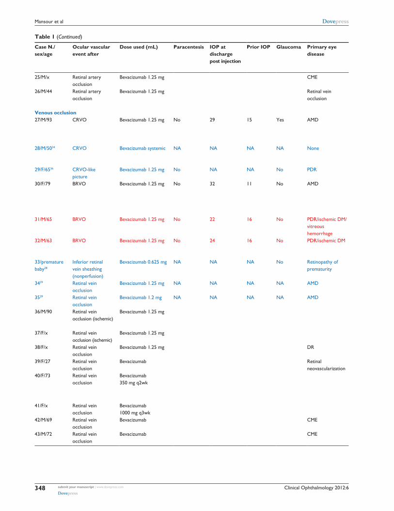
Table 1 Collaborative and literature review of 106 cases of ocular vascular complications of the VEGF antagonist bevacizumab: clinical profile
Case N./ sex/age
Ocular vascular event after
Dose used (mL) Paracentesis IOP at discharge post injection
Prior IOP Glaucoma Primary eye disease
Interval injection to detection of vascular occlusion (days)
N. prior injections
OD or OS Visual acuity prior to vascular event (log MAR)
Visual acuity after vascular event (log MAR)
Follow up after ocular event (months)
Systemic disease and risk of the vascular event per submitting author (new cases per total number of injected patients)
Arterial occlusion1/F/60 CRAO Bevacizumab 2.5 mg No 30 15 No Ischemic CRVO/
PDR/serous macular elevation
5 1 OD CF3m (1.6) 20/60 (0.5) 4 HTNDMcarotid stenosis1/19,158
2/F/74 CRAO Bevacizumab 1.25 mg No NA 10 No Ischemic CRVO/ serous macular elevation
14 1 OD CF0.3m (2.5) CF0.3m (2.5) 1 Smoker 1/19,158
3/F/95 CRAO Bevacizumab 1.25 mg No NA 7 No AMD 28 4 OD 20/50 (0.4) LP (3.3) 5 HTNCAD 1/19,158
4/M/49 CRAO Bevacizumab 1.25 mg Yes 21 before tap 14 No PDR/DM 0 1 OS 20/160 (0.9) NLP (3.6) DM 1/19,1585/F/47 CRAO Bevacizumab 1.25 mg No 14 16 No PDR/vitreous
hemorrhage45 1 OD 20/200 (1) HM (3) DM
1/2,0006/M/70 CRAO Bevacizumab 1.25 mg No Nl Nl No CRVO 30 1 NA 20/200 (1) CF0.3m (2.5) HTN
1/2,400 bevacizumab7/F/56 CRAO Bevacizumab 1.25 mg No Nl Nl No CRVO 30 1 NA 20/80 (0.6) HM (3) None
1/2,400 bevacizumab8/F/60 CRAO Bevacizumab 2.5 mg No NA 12 Yes CRVO 14 0 OD 20/20 (0.0) HM (3) 69/M/73 CRAO Bevacizumab 1.25 mg No 16 17 No AMD 15 1 OD 20/100 (0.7) 20/400 (1.3) 15 HTN
Smoker1/6,478 anti-VEGF
10/F/72 CRAO Bevacizumab 1.25 mg No 19 20 No AMD 10 1 OD 20/160 (0.9) 20/250 (1.1) 18 HTNSmokerCAD1/6,478 anti-VEGF
11/74/F CRAO Bevacizumab 1.25 mg No ,25 ,25 No DM 2 3 NA CF (1.6) NLP (3.6) DM/CAD12/52/F CRAO Bevacizumab 1.25 mg No 20 mmHg 20 mmHg Yes NVG 1 1 NA 20/200 (1) NLP (3.6) DM/HTN1331 CRAO Bevacizumab NA NA NA NA NA NA NA 20/1000 (1.7) LP (3.3) 1/400 bevacizumab14/F/6022 CRAO Bevacizumab1.25 mg
intracameralYes 50 20 Yes PDR/NVG 30 1 OD 20/200 (1) 20/200 (1) 12r DM
1527 CRAO Bevacizumab NA NA NA NA NA 7 NA NA NA NA 1/5,22816/M/78 Contralateral
CRAOBevacizumab 1.25 mg NA NA NA NA AMD 21 0 NA CF0.3m (2.5) CF1m (2) 3 Hypercholesterolemia, CAD post
coronary bypass 3/2,400 bevacizumab17/M/44 BRAO Bevacizumab 1.25 mg No Nl NA No Ischemic CRVO 2 1 OD 20/125 (0.8) CF2m (1.8) 9 HTN 1/2,400 anti-VEGF18/M/76 BRAO Bevacizumab 1.25 mg No 28 12 No AMD NA 13 OD 20/400 (1.3) 20/50 (0.4) 24 HTN
DMCADSmoker1/19,158
19/M/45 BRAO Bevacizumab systemic
No 16 16 No None 1 1 OS 20/100 (0.7) 20/50 (0.4) 3 HTNcancer
20/F/5332 BRAO Bevacizumab 1.25 mg NA NA NA NA PDR 14 1 OS 20/50 (0.4) 20/600 (1.5) 1 DM1/12 PDR patients
21/M/65 Retinal artery occlusion
Bevacizumab Avastin Side Effects Report: 5096382-0
22/M/80 Retinal artery occlusion
Bevacizumab 2.5 mg DR DMAvastin Side Effects Report: 5105228-35105228-3
23/F/60 Retinal artery occlusion
Bevacizumab Yes Avastin Side Effects Report: 5536025-2
24/F/x Retinal artery occlusion
Bevacizumab 15mg/kg q 3wk
Lung cancer on NavelbineAvastin Side Effects Report: 5593981-4
(Continued)
submit your manuscript | www.dovepress.com
Dovepress
Dovepress
346
Mansour et al

Clinical Ophthalmology 2012:6
Table 1 Collaborative and literature review of 106 cases of ocular vascular complications of the VEGF antagonist bevacizumab: clinical profile
Case N./ sex/age
Ocular vascular event after
Dose used (mL) Paracentesis IOP at discharge post injection
Prior IOP Glaucoma Primary eye disease
Interval injection to detection of vascular occlusion (days)
N. prior injections
OD or OS Visual acuity prior to vascular event (log MAR)
Visual acuity after vascular event (log MAR)
Follow up after ocular event (months)
Systemic disease and risk of the vascular event per submitting author (new cases per total number of injected patients)
Arterial occlusion1/F/60 CRAO Bevacizumab 2.5 mg No 30 15 No Ischemic CRVO/
PDR/serous macular elevation
5 1 OD CF3m (1.6) 20/60 (0.5) 4 HTNDMcarotid stenosis1/19,158
2/F/74 CRAO Bevacizumab 1.25 mg No NA 10 No Ischemic CRVO/ serous macular elevation
14 1 OD CF0.3m (2.5) CF0.3m (2.5) 1 Smoker 1/19,158
3/F/95 CRAO Bevacizumab 1.25 mg No NA 7 No AMD 28 4 OD 20/50 (0.4) LP (3.3) 5 HTNCAD 1/19,158
4/M/49 CRAO Bevacizumab 1.25 mg Yes 21 before tap 14 No PDR/DM 0 1 OS 20/160 (0.9) NLP (3.6) DM 1/19,1585/F/47 CRAO Bevacizumab 1.25 mg No 14 16 No PDR/vitreous
hemorrhage45 1 OD 20/200 (1) HM (3) DM
1/2,0006/M/70 CRAO Bevacizumab 1.25 mg No Nl Nl No CRVO 30 1 NA 20/200 (1) CF0.3m (2.5) HTN
1/2,400 bevacizumab7/F/56 CRAO Bevacizumab 1.25 mg No Nl Nl No CRVO 30 1 NA 20/80 (0.6) HM (3) None
1/2,400 bevacizumab8/F/60 CRAO Bevacizumab 2.5 mg No NA 12 Yes CRVO 14 0 OD 20/20 (0.0) HM (3) 69/M/73 CRAO Bevacizumab 1.25 mg No 16 17 No AMD 15 1 OD 20/100 (0.7) 20/400 (1.3) 15 HTN
Smoker1/6,478 anti-VEGF
10/F/72 CRAO Bevacizumab 1.25 mg No 19 20 No AMD 10 1 OD 20/160 (0.9) 20/250 (1.1) 18 HTNSmokerCAD1/6,478 anti-VEGF
11/74/F CRAO Bevacizumab 1.25 mg No ,25 ,25 No DM 2 3 NA CF (1.6) NLP (3.6) DM/CAD12/52/F CRAO Bevacizumab 1.25 mg No 20 mmHg 20 mmHg Yes NVG 1 1 NA 20/200 (1) NLP (3.6) DM/HTN1331 CRAO Bevacizumab NA NA NA NA NA NA NA 20/1000 (1.7) LP (3.3) 1/400 bevacizumab14/F/6022 CRAO Bevacizumab1.25 mg
intracameralYes 50 20 Yes PDR/NVG 30 1 OD 20/200 (1) 20/200 (1) 12r DM
1527 CRAO Bevacizumab NA NA NA NA NA 7 NA NA NA NA 1/5,22816/M/78 Contralateral
CRAOBevacizumab 1.25 mg NA NA NA NA AMD 21 0 NA CF0.3m (2.5) CF1m (2) 3 Hypercholesterolemia, CAD post
coronary bypass 3/2,400 bevacizumab17/M/44 BRAO Bevacizumab 1.25 mg No Nl NA No Ischemic CRVO 2 1 OD 20/125 (0.8) CF2m (1.8) 9 HTN 1/2,400 anti-VEGF18/M/76 BRAO Bevacizumab 1.25 mg No 28 12 No AMD NA 13 OD 20/400 (1.3) 20/50 (0.4) 24 HTN
DMCADSmoker1/19,158
19/M/45 BRAO Bevacizumab systemic
No 16 16 No None 1 1 OS 20/100 (0.7) 20/50 (0.4) 3 HTNcancer
20/F/5332 BRAO Bevacizumab 1.25 mg NA NA NA NA PDR 14 1 OS 20/50 (0.4) 20/600 (1.5) 1 DM1/12 PDR patients
21/M/65 Retinal artery occlusion
Bevacizumab Avastin Side Effects Report: 5096382-0
22/M/80 Retinal artery occlusion
Bevacizumab 2.5 mg DR DMAvastin Side Effects Report: 5105228-35105228-3
23/F/60 Retinal artery occlusion
Bevacizumab Yes Avastin Side Effects Report: 5536025-2
24/F/x Retinal artery occlusion
Bevacizumab 15mg/kg q 3wk
Lung cancer on NavelbineAvastin Side Effects Report: 5593981-4
(Continued)
submit your manuscript | www.dovepress.com
Dovepress
Dovepress
347
Vascular events during VEGF antagonist injections
Clinical Ophthalmology 2012:6
Table 1 (Continued)
Case N./ sex/age
Ocular vascular event after
Dose used (mL) Paracentesis IOP at discharge post injection
Prior IOP Glaucoma Primary eye disease
Interval injection to detection of vascular occlusion (days)
N. prior injections
OD or OS Visual acuity prior to vascular event (log MAR)
Visual acuity after vascular event (log MAR)
Follow up after ocular event (months)
Systemic disease and risk of the vascular event per submitting author (new cases per total number of injected patients)
25/M/x Retinal artery occlusion
Bevacizumab 1.25 mg CME Avastin Side Effects Report: 5736856-X, 5746319-3
26/M/44 Retinal artery occlusion
Bevacizumab 1.25 mg Retinal vein occlusion
Avastin Side Effects Report: 6237313-7, 6237504-5, 6253539-0, 6253542-0, 6341872-3; 6358564-7
Venous occlusion27/M/93 CRVO Bevacizumab 1.25 mg No 29 15 Yes AMD 10 1 OD 20/60 (0.5) 20/400 (1.3) 18 HTN
CAD (stent; pacemaker)Left carotid artery disease1/19,158
28/M/5034 CRVO Bevacizumab systemic NA NA NA NA None 1 day after each cycle
NA OD 20/120 (0.8) NA Metastatic adenocarcinoma of colon after 2 cycles of capecitabine, oxaliplatin and bevacizumab
29/F/6536 CRVO-like picture
Bevacizumab 1.25 mg No NA NA No PDR 7 0 OD 20/400 (1.3) 20/200 (1) 9 DM
30/F/79 BRVO Bevacizumab 1.25 mg No 32 11 No AMD 55 1 OS 20/30 (0.2) 20/30 (0.2) 18 HTNCAD MigraineCVA1/19,158
31/M/65 BRVO Bevacizumab 1.25 mg No 22 16 No PDR/ischemic DM/ vitreous hemorrhage
7 0 OS CF2m (1.8) 20/200 (1) 3 HTNDM1/42 prospective study
32/M/63 BRVO Bevacizumab 1.25 mg No 24 16 No PDR/ischemic DM 7 0 OD CF4m (1.5) 20/80 (0.6) 3 HTNDM1/42 prospective study
33/premature baby28
Inferior retinal vein sheathing (nonperfusion)
Bevacizumab 0.625 mg NA NA NA No Retinopathy of prematurity
3 0 OU NA NA Retinopathy of prematurity1/40 patients with retinopathy of prematurity
3439 Retinal vein occlusion
Bevacizumab 1.25 mg NA NA NA NA AMD NA NA NA NA NA 1/300 of wet AMD
3539 Retinal vein occlusion
Bevacizumab 1.2 mg NA NA NA NA AMD NA NA NA NA NA 1/300 of wet AMD
36/M/90 Retinal vein occlusion (ischemic)
Bevacizumab 1.25 mg HTN, CAD, dyslipidemiaAvastin Side Effects Report: 5197845-X, 5197968-5
37/F/x Retinal vein occlusion (ischemic)
Bevacizumab 1.25 mg Avastin Side Effects Report: 5508336-8, 5532270-0
38/F/x Retinal vein occlusion
Bevacizumab 1.25 mg DR DMAvastin Side Effects Report: 5706126-4
39/F/27 Retinal vein occlusion
Bevacizumab Retinal neovascularization
Side Effects Report: 6054515-3
40/F/73 Retinal vein occlusion
Bevacizumab 350 mg q2wk
Colon cancer on capecitabine, oxaliplatinAvastin Side Effects Report: 4839872-5, 4865570-8
41/F/x Retinal vein occlusion
Bevacizumab 1000 mg q3wk
Lung cancerAvastin Side Effects Report: 6209258-X
42/M/69 Retinal vein occlusion
Bevacizumab CME Avastin Side Effects Report: 6440612-7
43/M/72 Retinal vein occlusion
Bevacizumab CME Avastin Side Effects Report: 6440613-9
(Continued)
submit your manuscript | www.dovepress.com
Dovepress
Dovepress
348
Mansour et al

Clinical Ophthalmology 2012:6
Table 1 (Continued)
Case N./ sex/age
Ocular vascular event after
Dose used (mL) Paracentesis IOP at discharge post injection
Prior IOP Glaucoma Primary eye disease
Interval injection to detection of vascular occlusion (days)
N. prior injections
OD or OS Visual acuity prior to vascular event (log MAR)
Visual acuity after vascular event (log MAR)
Follow up after ocular event (months)
Systemic disease and risk of the vascular event per submitting author (new cases per total number of injected patients)
25/M/x Retinal artery occlusion
Bevacizumab 1.25 mg CME Avastin Side Effects Report: 5736856-X, 5746319-3
26/M/44 Retinal artery occlusion
Bevacizumab 1.25 mg Retinal vein occlusion
Avastin Side Effects Report: 6237313-7, 6237504-5, 6253539-0, 6253542-0, 6341872-3; 6358564-7
Venous occlusion27/M/93 CRVO Bevacizumab 1.25 mg No 29 15 Yes AMD 10 1 OD 20/60 (0.5) 20/400 (1.3) 18 HTN
CAD (stent; pacemaker)Left carotid artery disease1/19,158
28/M/5034 CRVO Bevacizumab systemic NA NA NA NA None 1 day after each cycle
NA OD 20/120 (0.8) NA Metastatic adenocarcinoma of colon after 2 cycles of capecitabine, oxaliplatin and bevacizumab
29/F/6536 CRVO-like picture
Bevacizumab 1.25 mg No NA NA No PDR 7 0 OD 20/400 (1.3) 20/200 (1) 9 DM
30/F/79 BRVO Bevacizumab 1.25 mg No 32 11 No AMD 55 1 OS 20/30 (0.2) 20/30 (0.2) 18 HTNCAD MigraineCVA1/19,158
31/M/65 BRVO Bevacizumab 1.25 mg No 22 16 No PDR/ischemic DM/ vitreous hemorrhage
7 0 OS CF2m (1.8) 20/200 (1) 3 HTNDM1/42 prospective study
32/M/63 BRVO Bevacizumab 1.25 mg No 24 16 No PDR/ischemic DM 7 0 OD CF4m (1.5) 20/80 (0.6) 3 HTNDM1/42 prospective study
33/premature baby28
Inferior retinal vein sheathing (nonperfusion)
Bevacizumab 0.625 mg NA NA NA No Retinopathy of prematurity
3 0 OU NA NA Retinopathy of prematurity1/40 patients with retinopathy of prematurity
3439 Retinal vein occlusion
Bevacizumab 1.25 mg NA NA NA NA AMD NA NA NA NA NA 1/300 of wet AMD
3539 Retinal vein occlusion
Bevacizumab 1.2 mg NA NA NA NA AMD NA NA NA NA NA 1/300 of wet AMD
36/M/90 Retinal vein occlusion (ischemic)
Bevacizumab 1.25 mg HTN, CAD, dyslipidemiaAvastin Side Effects Report: 5197845-X, 5197968-5
37/F/x Retinal vein occlusion (ischemic)
Bevacizumab 1.25 mg Avastin Side Effects Report: 5508336-8, 5532270-0
38/F/x Retinal vein occlusion
Bevacizumab 1.25 mg DR DMAvastin Side Effects Report: 5706126-4
39/F/27 Retinal vein occlusion
Bevacizumab Retinal neovascularization
Side Effects Report: 6054515-3
40/F/73 Retinal vein occlusion
Bevacizumab 350 mg q2wk
Colon cancer on capecitabine, oxaliplatinAvastin Side Effects Report: 4839872-5, 4865570-8
41/F/x Retinal vein occlusion
Bevacizumab 1000 mg q3wk
Lung cancerAvastin Side Effects Report: 6209258-X
42/M/69 Retinal vein occlusion
Bevacizumab CME Avastin Side Effects Report: 6440612-7
43/M/72 Retinal vein occlusion
Bevacizumab CME Avastin Side Effects Report: 6440613-9
(Continued)
submit your manuscript | www.dovepress.com
Dovepress
Dovepress
349
Vascular events during VEGF antagonist injections

Clinical Ophthalmology 2012:6
Table 1 (Continued)
Case N./ sex/age
Ocular vascular event after
Dose used (mL) Paracentesis IOP at discharge post injection
Prior IOP Glaucoma Primary eye disease
Interval injection to detection of vascular occlusion (days)
N. prior injections
OD or OS Visual acuity prior to vascular event (log MAR)
Visual acuity after vascular event (log MAR)
Follow up after ocular event (months)
Systemic disease and risk of the vascular event per submitting author (new cases per total number of injected patients)
Retinal vascular occlusion (unspecified)44/M/43 Retinal vascular
disorderBevacizumab 1 mg DR DM
Avastin Side Effects Report: 5959710-745/M/41 Retinal vascular
disorderBevacizumab 1 mg DR DM
Avastin Side Effects Report: 5961890-446/F/x Retinal vascular
disorderBevacizumab 1 mg DR DM
Avastin Side Effects Report: 6033375-047/M/x Retinal vascular
disorderBevacizumab 1 mg maculopathy HTN
Avastin Side Effects Report: 6159169-348/F/x Retinal vascular
disorderBevacizumab 1 mg CAD, unstable angina
Avastin Side Effects Report: 6291768-049/M/75 Retinal vascular
disorderBevacizumab 1 mg DM DM
Avastin Side Effects Report: 6438164-01i
50/F/33 Retinal vascular disorder
Bevacizumab Vitreous hemorrhage
Avastin Side Effects Report: 5724031-4
51-59/9 cases above 40 years
Retinal vascular disorder
Bevacizumab mixed 2010 events from eHealthMe drug outcomes from FDA and community
Optic neuropathy60/F/72 AION Bevacizumab 1.25 mg No NA 12 No AMD; fellow eye
AION7 1 OS CF2m (1.8) LP (3.3) 0.5 none 1/2,100 bevacizumab
61/F/71 AION Bevacizumab 1.25 mg No 16 20 No AMD 60 1 OS 20/70 (0.55) 20/70 (0.55) 6 HTN 1/33362/M/51 AION Bevacizumab 1.25 mg No NA NA No AMD 15 1 OD 20/25 (0.1) 20/25 (0.1) 12r Pseudoxanthoma elasticum63/F/38 AION Bevacizumab 1.25 mg No 21 23 Yes DM 21 0 OS 20/40 (0.3) 20/25 (0.1) 14 DM
1/150 bevacizumab64/F/70 AION Bevacizumab 1.25 mg No Nl Nl No AMD 28 3 OD 20/60 (0.5) 20/120 (0.8) 6 None
1/500 bevacizumab65/M/86 AION Bevacizumab 2.5 mg No No AMD 30 14 OD 20/70 (0.55) 20/100 (0.7) 12 HTN, prostate cancer, esophageal
cancer, on amlodipine1/6000 injection anti-VEGF
66/F/92 AION Bevacizumab 2.5 mg AMD 8 OS 20/70 (0.55) 20/100 (0.7) 48 No significant past medical history, on no medications
67/M/70 AION Bevacizumab 1.25 mg AMD 34 OS 20/70 (0.55) 20/200 (1.0) 6 No significant past medical history, on aspirin
68/M/x AION Bevacizumab 10 mg/kg Visual acuity reduced
Renal cancer on interferonAvastin Side Effects Report: 5863726-9, 5872556-3
69/F/x AION Bevacizumab 394 mg days 1 and 15
Breast cancer on capecitabineAvastin Side Effects Report: 5927943-1
70/F/x AION Bevacizumab 1.25 mg DR DMAvastin Side Effects Report: 6155052-8
71/F/72 AION Bevacizumab Avastin Side Effects Report: 6367854-372/F/47 Optic neuropathy Bevacizumab systemic
monthlyNA NA NA NA Glioblastoma right
frontotemporal2 years after initial injection
.20 (monthly)
OU 20/200 (1) (amblyopia) OD 20/70 (0.55) OS
LP OD (3.3) NLP (3.6) OS
30
73/M42 Optic neuropathy Bevacizumab systemic NA NA NA NA Glioblastoma NA 8 OU NA NA74/F/6742 Optic neuropathy Bevacizumab systemic NA NA NA NA Glioblastoma NA 6 OS NA NA75/F/5942 Optic neuropathy Bevacizumab systemic NA NA NA NA Glioblastoma NA 7 OU NA NACapillary occlusion76/F/58 Macular ischemia 1.25 mg Bevacizumab No Nl Nl No Background DR 2 1 OD 20/60 (0.5) 20/400 (1.3) 12 DM 1/2,350 anti-VEGF77/F/73 Macular ischemia Bevacizumab 1.25 mg No NA NA No Pre-PDR 42 0 OS 20/80 (0.6) 20/80 (0.6) 3 DM
HTN1/53 retrospective study of BRVO and diabetic maculopathy
(Continued)
submit your manuscript | www.dovepress.com
Dovepress
Dovepress
350
Mansour et al

Clinical Ophthalmology 2012:6
Table 1 (Continued)
Case N./ sex/age
Ocular vascular event after
Dose used (mL) Paracentesis IOP at discharge post injection
Prior IOP Glaucoma Primary eye disease
Interval injection to detection of vascular occlusion (days)
N. prior injections
OD or OS Visual acuity prior to vascular event (log MAR)
Visual acuity after vascular event (log MAR)
Follow up after ocular event (months)
Systemic disease and risk of the vascular event per submitting author (new cases per total number of injected patients)
Retinal vascular occlusion (unspecified)44/M/43 Retinal vascular
disorderBevacizumab 1 mg DR DM
Avastin Side Effects Report: 5959710-745/M/41 Retinal vascular
disorderBevacizumab 1 mg DR DM
Avastin Side Effects Report: 5961890-446/F/x Retinal vascular
disorderBevacizumab 1 mg DR DM
Avastin Side Effects Report: 6033375-047/M/x Retinal vascular
disorderBevacizumab 1 mg maculopathy HTN
Avastin Side Effects Report: 6159169-348/F/x Retinal vascular
disorderBevacizumab 1 mg CAD, unstable angina
Avastin Side Effects Report: 6291768-049/M/75 Retinal vascular
disorderBevacizumab 1 mg DM DM
Avastin Side Effects Report: 6438164-01i
50/F/33 Retinal vascular disorder
Bevacizumab Vitreous hemorrhage
Avastin Side Effects Report: 5724031-4
51-59/9 cases above 40 years
Retinal vascular disorder
Bevacizumab mixed 2010 events from eHealthMe drug outcomes from FDA and community
Optic neuropathy60/F/72 AION Bevacizumab 1.25 mg No NA 12 No AMD; fellow eye
AION7 1 OS CF2m (1.8) LP (3.3) 0.5 none 1/2,100 bevacizumab
61/F/71 AION Bevacizumab 1.25 mg No 16 20 No AMD 60 1 OS 20/70 (0.55) 20/70 (0.55) 6 HTN 1/33362/M/51 AION Bevacizumab 1.25 mg No NA NA No AMD 15 1 OD 20/25 (0.1) 20/25 (0.1) 12r Pseudoxanthoma elasticum63/F/38 AION Bevacizumab 1.25 mg No 21 23 Yes DM 21 0 OS 20/40 (0.3) 20/25 (0.1) 14 DM
1/150 bevacizumab64/F/70 AION Bevacizumab 1.25 mg No Nl Nl No AMD 28 3 OD 20/60 (0.5) 20/120 (0.8) 6 None
1/500 bevacizumab65/M/86 AION Bevacizumab 2.5 mg No No AMD 30 14 OD 20/70 (0.55) 20/100 (0.7) 12 HTN, prostate cancer, esophageal
cancer, on amlodipine1/6000 injection anti-VEGF
66/F/92 AION Bevacizumab 2.5 mg AMD 8 OS 20/70 (0.55) 20/100 (0.7) 48 No significant past medical history, on no medications
67/M/70 AION Bevacizumab 1.25 mg AMD 34 OS 20/70 (0.55) 20/200 (1.0) 6 No significant past medical history, on aspirin
68/M/x AION Bevacizumab 10 mg/kg Visual acuity reduced
Renal cancer on interferonAvastin Side Effects Report: 5863726-9, 5872556-3
69/F/x AION Bevacizumab 394 mg days 1 and 15
Breast cancer on capecitabineAvastin Side Effects Report: 5927943-1
70/F/x AION Bevacizumab 1.25 mg DR DMAvastin Side Effects Report: 6155052-8
71/F/72 AION Bevacizumab Avastin Side Effects Report: 6367854-372/F/47 Optic neuropathy Bevacizumab systemic
monthlyNA NA NA NA Glioblastoma right
frontotemporal2 years after initial injection
.20 (monthly)
OU 20/200 (1) (amblyopia) OD 20/70 (0.55) OS
LP OD (3.3) NLP (3.6) OS
30
73/M42 Optic neuropathy Bevacizumab systemic NA NA NA NA Glioblastoma NA 8 OU NA NA74/F/6742 Optic neuropathy Bevacizumab systemic NA NA NA NA Glioblastoma NA 6 OS NA NA75/F/5942 Optic neuropathy Bevacizumab systemic NA NA NA NA Glioblastoma NA 7 OU NA NACapillary occlusion76/F/58 Macular ischemia 1.25 mg Bevacizumab No Nl Nl No Background DR 2 1 OD 20/60 (0.5) 20/400 (1.3) 12 DM 1/2,350 anti-VEGF77/F/73 Macular ischemia Bevacizumab 1.25 mg No NA NA No Pre-PDR 42 0 OS 20/80 (0.6) 20/80 (0.6) 3 DM
HTN1/53 retrospective study of BRVO and diabetic maculopathy
(Continued)
submit your manuscript | www.dovepress.com
Dovepress
Dovepress
351
Vascular events during VEGF antagonist injections
Clinical Ophthalmology 2012:6
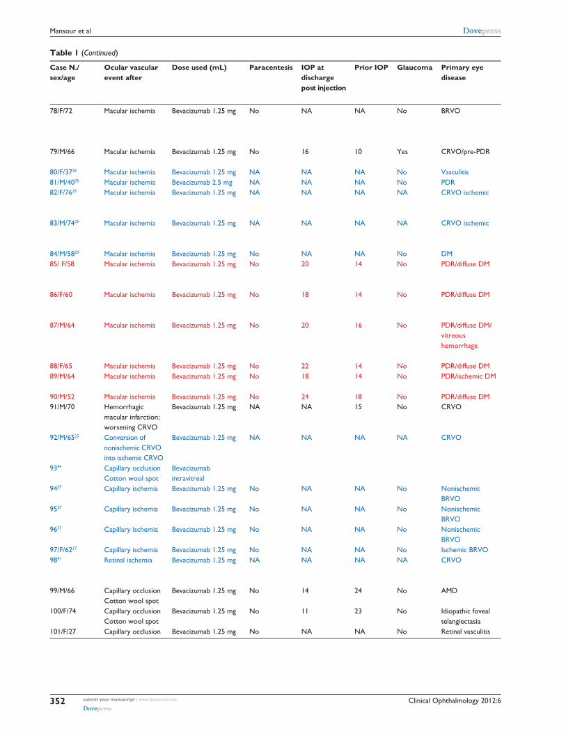
Table 1 (Continued)
Case N./ sex/age
Ocular vascular event after
Dose used (mL) Paracentesis IOP at discharge post injection
Prior IOP Glaucoma Primary eye disease
Interval injection to detection of vascular occlusion (days)
N. prior injections
OD or OS Visual acuity prior to vascular event (log MAR)
Visual acuity after vascular event (log MAR)
Follow up after ocular event (months)
Systemic disease and risk of the vascular event per submitting author (new cases per total number of injected patients)
78/F/72 Macular ischemia Bevacizumab 1.25 mg No NA NA No BRVO 28 0 OS 20/60 (0.5) 20/80 (0.6) 2 DMHTN1/53 retrospective study of BRVO and diabetic maculopathy
79/M/66 Macular ischemia Bevacizumab 1.25 mg No 16 10 Yes CRVO/pre-PDR 4 1 OS 20/100 (0.7) 20/220 (1.04) 30 DM1 of 1,500 anti-VEGF
80/F/3726 Macular ischemia Bevacizumab 1.25 mg NA NA NA No Vasculitis 7 1 OS 20/50 (0.4) 20/125 (0.8) 1 None81/M/4035 Macular ischemia Bevacizumab 2.5 mg NA NA NA No PDR NA 0 OS 20/400 (1.3) 20/400 (1.3) DM82/F/7625 Macular ischemia Bevacizumab 1.25 mg NA NA NA NA CRVO ischemic 28 1 OD 20/200 (1) 20/200 (1) 1 DM
CVA1/300 of retinal vascular occlusion cases
83/M/7425 Macular ischemia Bevacizumab 1.25 mg NA NA NA NA CRVO ischemic 28 2 OS 20/100 (0.7) 20/200 (1) 1 DMMI1/300 of retinal vascular occlusion cases
84/M/5829 Macular ischemia Bevacizumab 1.25 mg No NA NA No DM 21 0 OD 20/80 (0.6) 20/200 (1) 6 DM85/ F/58 Macular ischemia Bevacizumab 1.25 mg No 20 14 No PDR/diffuse DM 7 0 OD 20/200 (1) 20/80 (0.6) 3 HTN
DM1/42 prospective study
86/F/60 Macular ischemia Bevacizumab 1.25 mg No 18 14 No PDR/diffuse DM 7 0 OS 20/200 (1) 20/120 (0.8) 3 HTNDM1/42 prospective study
87/M/64 Macular ischemia Bevacizumab 1.25 mg No 20 16 No PDR/diffuse DM/ vitreous hemorrhage
7 0 OD CF3m (1.6) 20/80 (0.6) 3 HTNDMHepatic disease1/42 prospective study
88/F/65 Macular ischemia Bevacizumab 1.25 mg No 22 14 No PDR/diffuse DM 7 0 OD 20/120 (0.8) 20/80 (0.6) 3 DM 1/42 prospective study89/M/64 Macular ischemia Bevacizumab 1.25 mg No 18 14 No PDR/ischemic DM 7 0 OS 20/200 (1) 20/80 (0.6) 3 HTN
DM 1/42 prospective study90/M/52 Macular ischemia Bevacizumab 1.25 mg No 24 18 No PDR/diffuse DM 7 0 OD 20/200 (1) 20/120 (0.8) 3 DM 1/42 prospective study91/M/70 Hemorrhagic
macular infarction; worsening CRVO
Bevacizumab 1.25 mg NA NA 15 No CRVO 21 0 OS 20/100 (0.7) 20/320 (1.2) 1 None 1/2,000
92/M/6523 Conversion of nonischemic CRVO into ischemic CRVO
Bevacizumab 1.25 mg NA NA NA NA CRVO 21 1 OD 20/50 (0.4) 20/800 (1.6) 6 DM
9344 Capillary occlusion Cotton wool spot
Bevacizumab intravitreal
9437 Capillary ischemia Bevacizumab 1.25 mg No NA NA No Nonischemic BRVO
1 month 0 NA NA NA 1 1/37 nonischemic branch retinal vein occlusion
9537 Capillary ischemia Bevacizumab 1.25 mg No NA NA No Nonischemic BRVO
1 month 0 NA NA NA 1 1/37 nonischemic branch retinal vein occlusion
9637 Capillary ischemia Bevacizumab 1.25 mg No NA NA No Nonischemic BRVO
1 month 0 NA NA NA 1 1/37 nonischemic branch retinal vein occlusion
97/F/6237 Capillary ischemia Bevacizumab 1.25 mg No NA NA No Ischemic BRVO 1 month 0 OS 20/120 (0.8) 20/200 (1.0) 1 1 of 21 with ischemic BVO9841 Retinal ischemia Bevacizumab 1.25 mg NA NA NA NA CRVO NA NA NA NA NA 1/186 total patients in 1 center
(1/9 eyes with CRVO, 0/173 eyes with AMD)
99/M/66 Capillary occlusion Cotton wool spot
Bevacizumab 1.25 mg No 14 24 No AMD 30 1 OS 20/200 (1) 20/200 (1) 36 Gout 1/2,500
100/F/74 Capillary occlusion Cotton wool spot
Bevacizumab 1.25 mg No 11 23 No Idiopathic foveal telangiectasia
60 1 OS 20/80 (0.6) 20/70 (0.55) 36 HTN 1/2,500
101/F/27 Capillary occlusion Bevacizumab 1.25 mg No NA NA No Retinal vasculitis 14 1 OU 20/20 (0) 20/20 (0) 1 No 1/19,158
(Continued)
submit your manuscript | www.dovepress.com
Dovepress
Dovepress
352
Mansour et al

Clinical Ophthalmology 2012:6
Table 1 (Continued)
Case N./ sex/age
Ocular vascular event after
Dose used (mL) Paracentesis IOP at discharge post injection
Prior IOP Glaucoma Primary eye disease
Interval injection to detection of vascular occlusion (days)
N. prior injections
OD or OS Visual acuity prior to vascular event (log MAR)
Visual acuity after vascular event (log MAR)
Follow up after ocular event (months)
Systemic disease and risk of the vascular event per submitting author (new cases per total number of injected patients)
78/F/72 Macular ischemia Bevacizumab 1.25 mg No NA NA No BRVO 28 0 OS 20/60 (0.5) 20/80 (0.6) 2 DMHTN1/53 retrospective study of BRVO and diabetic maculopathy
79/M/66 Macular ischemia Bevacizumab 1.25 mg No 16 10 Yes CRVO/pre-PDR 4 1 OS 20/100 (0.7) 20/220 (1.04) 30 DM1 of 1,500 anti-VEGF
80/F/3726 Macular ischemia Bevacizumab 1.25 mg NA NA NA No Vasculitis 7 1 OS 20/50 (0.4) 20/125 (0.8) 1 None81/M/4035 Macular ischemia Bevacizumab 2.5 mg NA NA NA No PDR NA 0 OS 20/400 (1.3) 20/400 (1.3) DM82/F/7625 Macular ischemia Bevacizumab 1.25 mg NA NA NA NA CRVO ischemic 28 1 OD 20/200 (1) 20/200 (1) 1 DM
CVA1/300 of retinal vascular occlusion cases
83/M/7425 Macular ischemia Bevacizumab 1.25 mg NA NA NA NA CRVO ischemic 28 2 OS 20/100 (0.7) 20/200 (1) 1 DMMI1/300 of retinal vascular occlusion cases
84/M/5829 Macular ischemia Bevacizumab 1.25 mg No NA NA No DM 21 0 OD 20/80 (0.6) 20/200 (1) 6 DM85/ F/58 Macular ischemia Bevacizumab 1.25 mg No 20 14 No PDR/diffuse DM 7 0 OD 20/200 (1) 20/80 (0.6) 3 HTN
DM1/42 prospective study
86/F/60 Macular ischemia Bevacizumab 1.25 mg No 18 14 No PDR/diffuse DM 7 0 OS 20/200 (1) 20/120 (0.8) 3 HTNDM1/42 prospective study
87/M/64 Macular ischemia Bevacizumab 1.25 mg No 20 16 No PDR/diffuse DM/ vitreous hemorrhage
7 0 OD CF3m (1.6) 20/80 (0.6) 3 HTNDMHepatic disease1/42 prospective study
88/F/65 Macular ischemia Bevacizumab 1.25 mg No 22 14 No PDR/diffuse DM 7 0 OD 20/120 (0.8) 20/80 (0.6) 3 DM 1/42 prospective study89/M/64 Macular ischemia Bevacizumab 1.25 mg No 18 14 No PDR/ischemic DM 7 0 OS 20/200 (1) 20/80 (0.6) 3 HTN
DM 1/42 prospective study90/M/52 Macular ischemia Bevacizumab 1.25 mg No 24 18 No PDR/diffuse DM 7 0 OD 20/200 (1) 20/120 (0.8) 3 DM 1/42 prospective study91/M/70 Hemorrhagic
macular infarction; worsening CRVO
Bevacizumab 1.25 mg NA NA 15 No CRVO 21 0 OS 20/100 (0.7) 20/320 (1.2) 1 None 1/2,000
92/M/6523 Conversion of nonischemic CRVO into ischemic CRVO
Bevacizumab 1.25 mg NA NA NA NA CRVO 21 1 OD 20/50 (0.4) 20/800 (1.6) 6 DM
9344 Capillary occlusion Cotton wool spot
Bevacizumab intravitreal
9437 Capillary ischemia Bevacizumab 1.25 mg No NA NA No Nonischemic BRVO
1 month 0 NA NA NA 1 1/37 nonischemic branch retinal vein occlusion
9537 Capillary ischemia Bevacizumab 1.25 mg No NA NA No Nonischemic BRVO
1 month 0 NA NA NA 1 1/37 nonischemic branch retinal vein occlusion
9637 Capillary ischemia Bevacizumab 1.25 mg No NA NA No Nonischemic BRVO
1 month 0 NA NA NA 1 1/37 nonischemic branch retinal vein occlusion
97/F/6237 Capillary ischemia Bevacizumab 1.25 mg No NA NA No Ischemic BRVO 1 month 0 OS 20/120 (0.8) 20/200 (1.0) 1 1 of 21 with ischemic BVO9841 Retinal ischemia Bevacizumab 1.25 mg NA NA NA NA CRVO NA NA NA NA NA 1/186 total patients in 1 center
(1/9 eyes with CRVO, 0/173 eyes with AMD)
99/M/66 Capillary occlusion Cotton wool spot
Bevacizumab 1.25 mg No 14 24 No AMD 30 1 OS 20/200 (1) 20/200 (1) 36 Gout 1/2,500
100/F/74 Capillary occlusion Cotton wool spot
Bevacizumab 1.25 mg No 11 23 No Idiopathic foveal telangiectasia
60 1 OS 20/80 (0.6) 20/70 (0.55) 36 HTN 1/2,500
101/F/27 Capillary occlusion Bevacizumab 1.25 mg No NA NA No Retinal vasculitis 14 1 OU 20/20 (0) 20/20 (0) 1 No 1/19,158
(Continued)
submit your manuscript | www.dovepress.com
Dovepress
Dovepress
353
Vascular events during VEGF antagonist injections
Clinical Ophthalmology 2012:6
Table 1 (Continued)
Case N./ sex/age
Ocular vascular event after
Dose used (mL) Paracentesis IOP at discharge post injection
Prior IOP Glaucoma Primary eye disease
Interval injection to detection of vascular occlusion (days)
N. prior injections
OD or OS Visual acuity prior to vascular event (log MAR)
Visual acuity after vascular event (log MAR)
Follow up after ocular event (months)
Systemic disease and risk of the vascular event per submitting author (new cases per total number of injected patients)
Miscellaneous102/M/55 Ophthalmic artery
occlusionBevacizumab 1.25 mg Yes NA NA Yes PDR/NVG 3 1 OS 20/200 (1) NLP (3.6) 3 DM, carotid artery occlusion
1/256 bevacizumab103/F/40 Choroidal infarction,
HTN retinopathyBevacizumab 15 mg/kg q3wk
Glioma, HTN Avastin Side Effects Report: 4969093-7
10424 Visual loss Bevacizumab NA NA NA NA PDR 21 NA NA NA NA DM105/M/78 Retinal artery
spasmBevacizumab 5 mg/kg q2wk
Colon cancer on oxaliplatin avastin Side Effects Report: 5407594-8
106/M/x Retinal artery spasm
Bevacizumab 5 mg/kg q2wk
Tunnel vision Colon cancer; obesity on oxaliplatin Avastin Side Effects Report: 5442353-1
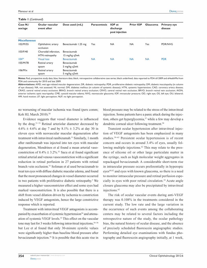
Notes: Red, prospective study data; blue, literature data; black, retrospective collaborative case series; black underlined, data reported to FDA till 2009 and eHealthMe from FDA and community for 2010 and late 2009.Abbreviations: AMD, wet age-related macular degeneration; DR, diabetic retinopathy; PDR, proliferative diabetic retinopathy; DM, diabetic maculopathy (in column of eye disease); NA, not assessed; Nl, normal; DM, diabetes mellitus (in column of systemic disease); HTN, systemic hypertension; CAD, coronary artery disease; CRAO, central retinal artery occlusion; BRAO, branch retinal artery occlusion; CRVO, central retinal vein occlusion; BRVO, branch retinal vein occlusion; AION, anterior ischemic optic neuropathy; CME, cystoid macular edema; NVG, neovascular glaucoma; IOP, intraocular pressure; OD, right eye; OS, left eye; OU, bilateral; HM, hand motion; LP, light perception; NLP, no light perception.
no worsening of macular ischemia was found (pers comm;
Koh HJ, March 2010).50
Evidence suggests that vessel diameter is influenced
by the drug.51–53 Retinal arteriolar diameter decreased by
4.6% ± 4.6% at day 7 and by 8.1% ± 3.2% at day 30 in
eleven eyes with neovascular macular degeneration after
treatment with intravitreal ranibizumab.51 Similarly, 1 month
after ranibizumab was injected into ten eyes with macular
degeneration, Mendrinos et al found a mean arterial vaso-
constriction of 8.4% ± 3.2%.52 Sacu et al found significant
retinal arterial and venous vasoconstriction with a significant
reduction in retinal perfusion in 27 patients with retinal
branch vein occlusion.53 Soliman et al used bevacizumab to
treat ten eyes with diffuse diabetic macular edema, and found
that the most pronounced changes in vessel diameter occurred
in two patients with proliferative diabetic retinopathy.3 We
measured a higher vasoconstrictor effect and some eyes had
marked vasoconstriction. It is also possible that there is a
shift from vessel dilation driven by ischemia to constriction
induced by VEGF antagonists, hence the large constrictive
response which is reported.
Treatment with intravitreal VEGF antagonists is accom-
panied by exacerbation of systemic hypertension54 and attenu-
ation of systemic VEGF levels.55 This effect on the vascular
tone may last for 3 weeks following intravitreal injections,54–56
but Lee et al found that only 30-minute systolic values
were significantly higher than baseline blood pressure after
bevacizumab injection.56 It is possible that this acute rise in
blood pressure may be related to the stress of the intravitreal
injection. Some patients have a panic attack during the injec-
tion, others get hyperglycemia,57 while a few may develop a
dendritic corneal ulcer following treatment.58
Transient ocular hypertension after intravitreal injec-
tion of VEGF antagonists has been emphasized in many
studies.59–62 Persistent ocular hypertension is of recent
concern and occurs in around 3.4% of eyes, usually fol-
lowing multiple injections.62 This may relate to the pres-
ence of silicone oil or other large particulate matter in
the syringe, such as high molecular weight aggregates in
repackaged bevacizumab. A considerable short-term rise
in intraocular pressure occurs preferentially in hyperopic
eyes60,62 and eyes with known glaucoma, so there is a need
to monitor intraocular pressure and retinal perfusion espe-
cially in eyes with poor retinal circulation.18 Acute angle
closure glaucoma may also be precipitated by intravitreal
injections.62
The risk of ocular vascular events during anti-VEGF
therapy was 0.108% in the treatments considered in the
current study. The low rate and the large variation in
the occurrence of such events among the collaborating
centers may be related to several factors including the
retrospective nature of the study, the ocular pathology
bias, the natural history of ocular disease, and the absence
of precisely scheduled fluorescein angiographic studies.
Performing detailed eye examinations with fundus pho-
tography and fluorescein angiography initially, at 1 week,
submit your manuscript | www.dovepress.com
Dovepress
Dovepress
354
Mansour et al

Clinical Ophthalmology 2012:6
Table 1 (Continued)
Case N./ sex/age
Ocular vascular event after
Dose used (mL) Paracentesis IOP at discharge post injection
Prior IOP Glaucoma Primary eye disease
Interval injection to detection of vascular occlusion (days)
N. prior injections
OD or OS Visual acuity prior to vascular event (log MAR)
Visual acuity after vascular event (log MAR)
Follow up after ocular event (months)
Systemic disease and risk of the vascular event per submitting author (new cases per total number of injected patients)
Miscellaneous102/M/55 Ophthalmic artery
occlusionBevacizumab 1.25 mg Yes NA NA Yes PDR/NVG 3 1 OS 20/200 (1) NLP (3.6) 3 DM, carotid artery occlusion
1/256 bevacizumab103/F/40 Choroidal infarction,
HTN retinopathyBevacizumab 15 mg/kg q3wk
Glioma, HTN Avastin Side Effects Report: 4969093-7
10424 Visual loss Bevacizumab NA NA NA NA PDR 21 NA NA NA NA DM105/M/78 Retinal artery
spasmBevacizumab 5 mg/kg q2wk
Colon cancer on oxaliplatin avastin Side Effects Report: 5407594-8
106/M/x Retinal artery spasm
Bevacizumab 5 mg/kg q2wk
Tunnel vision Colon cancer; obesity on oxaliplatin Avastin Side Effects Report: 5442353-1
Notes: Red, prospective study data; blue, literature data; black, retrospective collaborative case series; black underlined, data reported to FDA till 2009 and eHealthMe from FDA and community for 2010 and late 2009.Abbreviations: AMD, wet age-related macular degeneration; DR, diabetic retinopathy; PDR, proliferative diabetic retinopathy; DM, diabetic maculopathy (in column of eye disease); NA, not assessed; Nl, normal; DM, diabetes mellitus (in column of systemic disease); HTN, systemic hypertension; CAD, coronary artery disease; CRAO, central retinal artery occlusion; BRAO, branch retinal artery occlusion; CRVO, central retinal vein occlusion; BRVO, branch retinal vein occlusion; AION, anterior ischemic optic neuropathy; CME, cystoid macular edema; NVG, neovascular glaucoma; IOP, intraocular pressure; OD, right eye; OS, left eye; OU, bilateral; HM, hand motion; LP, light perception; NLP, no light perception.
and 1 month post-injection in a prospective setting (such
as in the prospective study from Mansoura University)
yielded higher rates of ocular events than were reported
following retrospective quick screening examinations
at the time of repeated injections. Many of the reported
events were asymptomatic, such as capillary occlusion
outside the fovea, and minor branch retinal artery or vein
occlusion. In the RESOLVE study, a total of 102 cases
having ranibizumab injections for diabetic maculopa-
thy resulted in two cases with retinal vascular events
(capillary and arterial occlusions).30 In the ROCC study,
one of the 16 patients with central retinal vein occlusion
developed central retinal artery occlusion.63 Branch retinal
artery occurred in one out of twelve consecutive patients
with proliferative diabetic retinopathy following intrav-
itreal bevacizumab.32 In the ANCHOR64 and MARINA65
studies (280 and 477 patients, respectively), no retinal
vascular events were noted after 2 years of repeated intra-
vitreal ranibizumab for the wet form of age-related macular
degeneration. Prior prospective studies and the current survey
found that eyes with wet age-related macular degeneration
had the lowest frequencies of vascular events (0%–0.3%)5,65
while eyes with a greater number of ischemic vascular
diseases such as proliferative diabetic retinopathy yielded
a higher frequency of retinal vascular events (2%–19%,
as in the current prospective study).30 The occurrence of
ocular vascular occlusions after anti-VEGF medications
was 2.61% in the diabetic population (Tables 1 and 2),
almost 24 times the occurrence in the general population
receiving VEGF antagonists (Tables 1 and 2).
Three studies show choroidal or retinal vaso-occlusion
after intravitreal bevacizumab injections in experimental
animals. Peters et al analyzed the acute intravitreal effects
of bevacizumab in four cynomolgus monkeys and found that
choriocapillaris endothelial cell fenestrations were signifi-
cantly reduced, and that densely packed thrombocytes and
leukocytes regionally occluded the choriocapillaris lumen
of treated eyes.66 Schraermeyer et al found that bevacizumab
immune complexes activate platelets and cause thrombo-
sis in choroidal vessels of primate eyes.67,68 Ameri et al
evaluated the effects of intravitreal bevacizumab in a rabbit
retinal neovascularization model. An intravitreal VEGF
injection was administered and intravitreal bevacizumab
was then injected at day 2 and at week 1, and it was found
that administration of intravitreal bevacizumab at week 1
resulted in severe capillary nonperfusion at week 2.69 Bonnin
et al demonstrated ocular hypoperfusion after intravitreal
bevacizumab in humans. In 15 patients with wet age-related
macular degeneration, mean blood flow velocities were
measured by ultrasound imaging before, and 4 weeks after,
a single intravitreal injection of bevacizumab. Velocities
decreased significantly in the central retinal, temporal
posterior ciliary, and ophthalmic arteries by 10%, 20%,
and 20% respectively.60,70 Sacu et al found significant vaso-
constriction of retinal arteries and veins outside the area
of nonischemic retinal branch vein occlusions as well as a
submit your manuscript | www.dovepress.com
Dovepress
Dovepress
355
Vascular events during VEGF antagonist injections

Clinical Ophthalmology 2012:6
Table 2 Collaborative and literature review of 38 cases of ocular vascular complications of VEGF antagonists excluding bevacizumab (ranibizumab and pegaptanib): clinical profile
Case N./ sex/age
Ocular vascular event after
Dose used (mL) Paracentesis IOP at discharge post injection
Prior IOP Glaucoma Primary eye disease
Interval injection to detection of vascular occlusion (days)
N. prior injections
OD or OS Visual acuity prior to vascular event (log MAR)
Visual acuity after vascular event (log MAR)
Follow up after ocular event (months)
Systemic disease and risk of the vascular event per submitting author (new cases per total number of injected patients)
Arterial occlusion1/M/75 CRAO Ranibizumab 0.5 mg No Nl NA No Ischemic
CRVO30 1 OS 20/400 (1.3) LP (3.3) 2 DM
CAD1/2,400 anti-VEGF1/16 ROCC study63
2/M/67 CRAO Ranibizumab 0.5 mg No 15 15 No DM 30 4 OS 20/100 (0.7) 20/400(1.3)
12 DMHTN1/6,478 anti-VEGF
330 CRAO Ranibizumab NA NA NA NA DM NA NA NA NA NA 12 DM 1/102 eyes prospective study (RESOLVE)
4/M/85 (Reimao*)
CRAO Ranibizumab 0.5 mg No 38 mmHg NA Yes NVG 2d 0 OD 20/25 (0.1) 20/80 (0.6) HTN COPD ex-smoker bilateral carotid stenosis
5/F/81 Retinal artery occlusion
Ranibizumab Lucentis Side Effects Report: 6109626-0
6/F/x Retinal artery occlusion
Ranibizumab 0.5 mg HTN, CADLucentis Side Effects Report: 6184843-2
7/M/84 Retinal artery occlusion
Ranibizumab 0.5 mg High IOP AMD Lucentis Side Effects Report: 6210113-X
8/F/70 Retinal artery occlusion
Ranibizumab Lucentis Side Effects Report: 6480905-0, 6496635-5
9/F/70 Retinal artery occlusion
Ranibizumab 0.5 mg Lucentis Side Effects Report: 6207699-8
10/F/86 Retinal artery occlusion
Pegaptanib HTN, dyslipidemia Macugen Side Effects Report: 5248582-4, 5224175-X
11/M/67 Retinal artery occlusion
Pegaptanib Macugen Side Effects Report: 6108967-0
12/F/above 60 years
Retinal artery occlusion
Ranibizumab AMD ,1 month 2010 events from eHealthMe drug outcomes from FDA and community
Venous occlusion13/M/84 Retinal vein
occlusionRanibizumab 0.5 mg Lucentis Side Effects Report:
5216324-4/5889807-114/M/74 Retinal vein
occlusionRanibizumab Lucentis Side Effects Report:
5253885-3/5259058-2Retinal vascular occlusion (unspecified)1537c Ocular vascular
occlusionRanibizumab 0.5 mg NA NA NA NA DM NA NA NA NA NA DM
1/375 for diabetic CME1637c Ocular vascular
occlusionRanibizumab 0.5 mg NA NA NA NA DM NA NA NA NA NA DM
1/375 for diabetic CME1737c Ocular vascular
occlusionRanibizumab 0.5 mg NA NA NA NA DM NA NA NA NA NA DM
1/375 for diabetic CME18/M/47 Retinal vascular
disorderRanibizumab 0.3 mg CME Lucentis Side Effects Report:
5896098-419/M/x Retinal vascular
disorderRanibizumab Lucentis Side Effects Report:
6180863-220/F/66 Retinal vascular
disorderPegaptanib AMD Macugen Side Effects Report:
6409650-4
(Continued)
submit your manuscript | www.dovepress.com
Dovepress
Dovepress
356
Mansour et al

Clinical Ophthalmology 2012:6
Table 2 Collaborative and literature review of 38 cases of ocular vascular complications of VEGF antagonists excluding bevacizumab (ranibizumab and pegaptanib): clinical profile
Case N./ sex/age
Ocular vascular event after
Dose used (mL) Paracentesis IOP at discharge post injection
Prior IOP Glaucoma Primary eye disease
Interval injection to detection of vascular occlusion (days)
N. prior injections
OD or OS Visual acuity prior to vascular event (log MAR)
Visual acuity after vascular event (log MAR)
Follow up after ocular event (months)
Systemic disease and risk of the vascular event per submitting author (new cases per total number of injected patients)
Arterial occlusion1/M/75 CRAO Ranibizumab 0.5 mg No Nl NA No Ischemic
CRVO30 1 OS 20/400 (1.3) LP (3.3) 2 DM
CAD1/2,400 anti-VEGF1/16 ROCC study63
2/M/67 CRAO Ranibizumab 0.5 mg No 15 15 No DM 30 4 OS 20/100 (0.7) 20/400(1.3)
12 DMHTN1/6,478 anti-VEGF
330 CRAO Ranibizumab NA NA NA NA DM NA NA NA NA NA 12 DM 1/102 eyes prospective study (RESOLVE)
4/M/85 (Reimao*)
CRAO Ranibizumab 0.5 mg No 38 mmHg NA Yes NVG 2d 0 OD 20/25 (0.1) 20/80 (0.6) HTN COPD ex-smoker bilateral carotid stenosis
5/F/81 Retinal artery occlusion
Ranibizumab Lucentis Side Effects Report: 6109626-0
6/F/x Retinal artery occlusion
Ranibizumab 0.5 mg HTN, CADLucentis Side Effects Report: 6184843-2
7/M/84 Retinal artery occlusion
Ranibizumab 0.5 mg High IOP AMD Lucentis Side Effects Report: 6210113-X
8/F/70 Retinal artery occlusion
Ranibizumab Lucentis Side Effects Report: 6480905-0, 6496635-5
9/F/70 Retinal artery occlusion
Ranibizumab 0.5 mg Lucentis Side Effects Report: 6207699-8
10/F/86 Retinal artery occlusion
Pegaptanib HTN, dyslipidemia Macugen Side Effects Report: 5248582-4, 5224175-X
11/M/67 Retinal artery occlusion
Pegaptanib Macugen Side Effects Report: 6108967-0
12/F/above 60 years
Retinal artery occlusion
Ranibizumab AMD ,1 month 2010 events from eHealthMe drug outcomes from FDA and community
Venous occlusion13/M/84 Retinal vein
occlusionRanibizumab 0.5 mg Lucentis Side Effects Report:
5216324-4/5889807-114/M/74 Retinal vein
occlusionRanibizumab Lucentis Side Effects Report:
5253885-3/5259058-2Retinal vascular occlusion (unspecified)1537c Ocular vascular
occlusionRanibizumab 0.5 mg NA NA NA NA DM NA NA NA NA NA DM
1/375 for diabetic CME1637c Ocular vascular
occlusionRanibizumab 0.5 mg NA NA NA NA DM NA NA NA NA NA DM
1/375 for diabetic CME1737c Ocular vascular
occlusionRanibizumab 0.5 mg NA NA NA NA DM NA NA NA NA NA DM
1/375 for diabetic CME18/M/47 Retinal vascular
disorderRanibizumab 0.3 mg CME Lucentis Side Effects Report:
5896098-419/M/x Retinal vascular
disorderRanibizumab Lucentis Side Effects Report:
6180863-220/F/66 Retinal vascular
disorderPegaptanib AMD Macugen Side Effects Report:
6409650-4
(Continued)
submit your manuscript | www.dovepress.com
Dovepress
Dovepress
357
Vascular events during VEGF antagonist injections

Clinical Ophthalmology 2012:6
Table 2 (Continued)
Case N./ sex/age
Ocular vascular event after
Dose used (mL) Paracentesis IOP at discharge post injection
Prior IOP Glaucoma Primary eye disease
Interval injection to detection of vascular occlusion (days)
N. prior injections
OD or OS Visual acuity prior to vascular event (log MAR)
Visual acuity after vascular event (log MAR)
Follow up after ocular event (months)
Systemic disease and risk of the vascular event per submitting author (new cases per total number of injected patients)
21/M/60 Retinal vascular disorder
Pegaptanib 0.3 mg AMD Macugen Side Effects Report: 6463543-5
22/Above 60 years Retinal vascular disorder
Pegaptanib AMD Late 2009 events from eHealthMe drug outcomes from FDA and community
23–28/6 cases above 60 years
Retinal vascular disorder
Ranibizumab mixed 2010 events from eHealthMe drug outcomes from FDA and community
Optic neuropathy29/M/75 AION Ranibizumab 0.5 mg AMD 4 OS 20/40 20/60 1 HTN, hypothyroidism, BPH, angina,
on amlodipine, levothyroxine, temazepam, nitroglycerin1/4500 antiVEGF injection
30/F/7037a AION OU Pegaptanib No NA NA No AMD ODDiabetic prophylaxis for cataract surgery OS
7d OD4d OS
0 ODOS
20/40 (0.3)NA
20/4000 (2.2)20/200 (1)
33
DMHTN
31/M/93 AION 0.3 mg Pegaptanib Visual acuity reduced
Macugen Side Effects Report: 4825003-4
32/M/72 AION Pegaptanib AMD Macugen Side Effects Report: 4982605-2
Capillary occlusion33/F/x Retinal ischemia
(macular)Ranibizumab Lucentis Side Effects Report:
5889807-134/F/x Retinal ischemia
(macular)Ranibizumab 0.5 mg Lucentis Side Effects Report:
6454819-63530 Capillary occlusion
(peripheral)Ranibizumab NA NA NA NA DM NA NA NA NA NA 12 DM 1/102 eyes prospective study
(RESOLVE)36/F/x Retinal ischemia
(peripheral)Ranibizumab 0.5 mg 9 Lucentis Side Effects Report:
6037721-3; patient died 9 months after injection
37/above 60 years Retinal ischemia Ranibizumab Unspecified 2010 events from eHealthMe drug outcomes from FDA and community
Miscellaneous38/M/85 Diffuse vascular
occlusionRanibizumab 0.5 mg No NA 15 No Ocular
ischemic syndrome
14 1 OD 20/100 (0.7) LP (3.3) 10 Carotid stenosis
Notes: Red, prospective study data; blue, literature data; black, retrospective collaborative case series; black underlined, data reported to FDA till 2009 and eHealthMe from FDA and community for 2010 and late 2009; *Reimao reference refers to eposter EP-GLA-405 SOE 2011 Geneve presented by Reimao P, Macedo M, Gomes M, Maia S, Santos M, Meneres MJ, from Portugal.Abbreviations: AMD, wet age-related macular degeneration; DR, diabetic retinopathy; PDR, proliferative diabetic retinopathy; DM, diabetic maculopathy (in column of eye disease); NA, not assessed; Nl, normal; DM, diabetes mellitus; HTN, systemic hypertension; CAD, coronary artery disease; CRAO, central retinal artery occlusion; BRAO, branch retinal artery occlusion; CRVO, central retinal vein occlusion; BRVO, branch retinal vein occlusion; AION, anterior ischemic optic neuropathy; IOP, intraocular pressure; OD, right eye; OS, left eye; CME, cystoid macular edema; NVG, neovascular glaucoma.
significant reduction in the flow velocity of the retrobulbar
central retinal artery.53
The vascular events reported during VEGF antagonist
therapies could be part of the natural history of the underlying
ocular disease. A rise in blood pressure, stress of the proce-
dure, the underlying systemic disease, and a sharp rise in
intraocular pressure are variables that can be involved in some
cases of ocular vascular events, and these variables can be
detected and treated. A majority of the patients discussed in
the current study had systemic diseases, particularly diabe-
tes mellitus. VEGF antagonism could play a leader role in
some cases that demonstrated vasoconstriction by analysis
of vessel caliber. VEGF acts as a vessel dilator by stimulat-
ing nitric oxide synthesis, and influences the autoregulation
submit your manuscript | www.dovepress.com
Dovepress
Dovepress
358
Mansour et al

Clinical Ophthalmology 2012:6
Table 2 (Continued)
Case N./ sex/age
Ocular vascular event after
Dose used (mL) Paracentesis IOP at discharge post injection
Prior IOP Glaucoma Primary eye disease
Interval injection to detection of vascular occlusion (days)
N. prior injections
OD or OS Visual acuity prior to vascular event (log MAR)
Visual acuity after vascular event (log MAR)
Follow up after ocular event (months)
Systemic disease and risk of the vascular event per submitting author (new cases per total number of injected patients)
21/M/60 Retinal vascular disorder
Pegaptanib 0.3 mg AMD Macugen Side Effects Report: 6463543-5
22/Above 60 years Retinal vascular disorder
Pegaptanib AMD Late 2009 events from eHealthMe drug outcomes from FDA and community
23–28/6 cases above 60 years
Retinal vascular disorder
Ranibizumab mixed 2010 events from eHealthMe drug outcomes from FDA and community
Optic neuropathy29/M/75 AION Ranibizumab 0.5 mg AMD 4 OS 20/40 20/60 1 HTN, hypothyroidism, BPH, angina,
on amlodipine, levothyroxine, temazepam, nitroglycerin1/4500 antiVEGF injection
30/F/7037a AION OU Pegaptanib No NA NA No AMD ODDiabetic prophylaxis for cataract surgery OS
7d OD4d OS
0 ODOS
20/40 (0.3)NA
20/4000 (2.2)20/200 (1)
33
DMHTN
31/M/93 AION 0.3 mg Pegaptanib Visual acuity reduced
Macugen Side Effects Report: 4825003-4
32/M/72 AION Pegaptanib AMD Macugen Side Effects Report: 4982605-2
Capillary occlusion33/F/x Retinal ischemia
(macular)Ranibizumab Lucentis Side Effects Report:
5889807-134/F/x Retinal ischemia
(macular)Ranibizumab 0.5 mg Lucentis Side Effects Report:
6454819-63530 Capillary occlusion
(peripheral)Ranibizumab NA NA NA NA DM NA NA NA NA NA 12 DM 1/102 eyes prospective study
(RESOLVE)36/F/x Retinal ischemia
(peripheral)Ranibizumab 0.5 mg 9 Lucentis Side Effects Report:
6037721-3; patient died 9 months after injection
37/above 60 years Retinal ischemia Ranibizumab Unspecified 2010 events from eHealthMe drug outcomes from FDA and community
Miscellaneous38/M/85 Diffuse vascular
occlusionRanibizumab 0.5 mg No NA 15 No Ocular
ischemic syndrome
14 1 OD 20/100 (0.7) LP (3.3) 10 Carotid stenosis
Notes: Red, prospective study data; blue, literature data; black, retrospective collaborative case series; black underlined, data reported to FDA till 2009 and eHealthMe from FDA and community for 2010 and late 2009; *Reimao reference refers to eposter EP-GLA-405 SOE 2011 Geneve presented by Reimao P, Macedo M, Gomes M, Maia S, Santos M, Meneres MJ, from Portugal.Abbreviations: AMD, wet age-related macular degeneration; DR, diabetic retinopathy; PDR, proliferative diabetic retinopathy; DM, diabetic maculopathy (in column of eye disease); NA, not assessed; Nl, normal; DM, diabetes mellitus; HTN, systemic hypertension; CAD, coronary artery disease; CRAO, central retinal artery occlusion; BRAO, branch retinal artery occlusion; CRVO, central retinal vein occlusion; BRVO, branch retinal vein occlusion; AION, anterior ischemic optic neuropathy; IOP, intraocular pressure; OD, right eye; OS, left eye; CME, cystoid macular edema; NVG, neovascular glaucoma.
in the microcirculation. If we block this rescuer, the retina
may be damaged due to decreased retinal perfusion in the
presence of a low ophthalmic systolic pressure. Because
retinal vessel diameter is a useful surrogate for retinal per-
fusion, changes in the diameter of the retinal arterioles may
indicate changes in retinal capillary blood flow. Thus, these
findings suggest that VEGF antagonists may reduce retinal
capillary blood flow, and caution should be exercised in
the use of intravitreal VEGF inhibitors in eyes with severe
ocular ischemia such as ocular ischemic syndrome with low
ophthalmic systolic pressure or severe proliferative diabetic
retinopathy.11,15 Further studies are needed to evaluate the
incidence of vascular events during VEGF antagonist therapy
in such high-risk patients.11
submit your manuscript | www.dovepress.com
Dovepress
Dovepress
359
Vascular events during VEGF antagonist injections

Clinical Ophthalmology 2012:6
Tab
le 3
Ret
inal
vas
ocon
stri
ctio
n va
lues
in s
ubje
cts
with
ocu
lar
vasc
ular
eve
nts
duri
ng b
evac
izum
ab t
hera
py in
13
eyes
, and
intr
avitr
eal r
anib
izum
ab t
hera
py in
one
eye
Cas
e/se
x/
age
Ocu
lar
vasc
ular
ev
ent
afte
rB
evac
izum
ab
(mg)
Pri
mar
y ey
e
dise
ase
Inte
rval
inje
ctio
n
to la
st fl
uore
scei
n
angi
ogra
phy
(day
s)
N. p
rior
in
ject
ions
Syst
emic
di
seas
eA
rter
ial v
asoc
on-
stri
ctio
n fr
om
base
line
1.0
Ven
ous
vaso
con-
st
rict
ion
from
ba
selin
e 1.
0
1/F/
74C
RA
O1.
25Is
chem
ic C
RV
O14
1Sm
oker
(he
avy)
0.93
0.68
*2/
F/27
Cap
illar
y oc
clus
ion
1.25
Ret
inal
vas
culit
is14
1N
o0.
46*
0.73
3/M
/93
CR
VO
1.25
CN
V10
1H
TN
C
AD
car
otid
ar
tery
dis
ease
0.90
1.35
+
4/M
/66
Cap
illar
y oc
clus
ion
CW
S1.
25 C
NV
301
Gou
t0.
960.
845/
M/5
1A
ION
1.25
CN
V15
1Ps
eudo
xant
hom
a el
astic
um0.
72*
0.88
6/F/
76M
acul
ar is
chem
ia1.
25C
RV
O is
chem
ic28
1D
M
CV
A1.
030.
95
7/M
/74
Mac
ular
isch
emia
1.25
CR
VO
isch
emic
282
DM
M
I1.
030.
93
8/M
/65
BRV
O1.
25PD
R7
0H
TN
D
M0.
22*
0.67
*
9/M
/63
BRV
O1.
25PD
R7
0H
TN
D
M 0.
43*
0.85
10/F
/ 60
Mac
ular
isch
emia
1.25
PDR
70
HT
N
DM
0.83
1.82
11/M
/64
Mac
ular
isch
emia
1.25
PDR
70
HT
N
DM
H
epat
ic d
isea
se
1.01
0.88
12/M
/ 64
Mac
ular
isch
emia
1.25
PDR
70
HT
N
DM
0.95
0.85
13/M
/52
Mac
ular
isch
emia
1.25
PDR
70
DM
Not
mea
sura
ble
0.97
14/M
/85
Diff
use
vasc
ular
occ
lusi
onLu
cent
is 0
.5 m
gO
cula
r is
chem
ic
synd
rom
e14
1C
arot
id s
teno
sis
com
plet
e0.
58*
0.84
Not
es: R
ed, i
ntra
vitr
eal r
anib
izum
ab; *
refe
rs t
o m
arke
d co
nstr
ictio
n; + in
dica
tes
that
thi
s va
lue
not
coun
ted
beca
use
it w
as p
art
of C
RV
O.
Abb
revi
atio
ns:
PDR
, pr
olife
rativ
e di
abet
ic r
etin
opat
hy;
NA
, no
t as
sess
ed;
CN
V,
chor
oida
l ne
ovas
cula
riza
tion;
DM
, di
abet
es m
ellit
us;
HT
N,
syst
emic
hyp
erte
nsio
n; C
AD
, co
rona
ry a
rter
y di
seas
e; M
I, m
yoca
rdia
l in
farc
tion;
CV
A,
cere
brov
ascu
lar
acci
dent
; CR
AO
, cen
tral
ret
inal
art
ery
occl
usio
n; B
RA
O, b
ranc
h re
tinal
art
ery
occl
usio
n; C
RV
O, c
entr
al r
etin
al v
ein
occl
usio
n; B
RV
O, b
ranc
h re
tinal
vei
n oc
clus
ion;
AIO
N, a
nter
ior
isch
emic
opt
ic n
euro
path
y.
submit your manuscript | www.dovepress.com
Dovepress
Dovepress
360
Mansour et al

Clinical Ophthalmology 2012:6
AcknowledgmentsThe Collaborative Anti-VEGF Ocular Vascular Complica-
tions Group: K Bailey Freund, Ninel Z Gregori, Nikolaus
Feucht, Alay Banker, Therese von Hanno, Claudio Furino,
Sabine Aisenbrey, Wael Soliman, Rong-Kung Tsai, Hamid
Hosseini, Eric Chen, Hee-Seung Chin, Jane Y Huang, Ali A
Bodla, Ozgur Artunay, Vladimir Poposki, Daniel Vilaplana,
Michael Larsen, M Tariq Bhatti, Hana A Mansour, Ihab
Saab, Hasan Shahine, Zohar Yehoshua, Kara Dellatorre,
Shree Kurup.
The authors acknowledge the contribution of Professor
FT Fraunfelder who supplied the data from the National
Registry of Drug-Induced Ocular Side Effects.
DisclosureThe authors have no financial interests in any product men-
tioned in the manuscript.
References 1. Liu MH, Jin H, Floten HS, Ren Z, Yim AP, He GW. Vascular endothe-
lial growth factor-mediated, endothelium-dependent relaxation in human internal mammary artery. Ann Thorac Surg. 2002;73(3): 819–824.
2. Arevalo JF, Fromow-Guerra J, Quiroz-Mercado H, et al. Primary intra-vitreal bevacizumab (avastin) for diabetic macular edema. Results from the Pan-American Collaborative Study Group at 6-month follow-up. Ophthalmology. 2007;114(4):743–750.
3. Soliman W, Vinten M, Sander B, et al. Optical coherence tomography and vessel diameter changes after intravitreal bevacizumab in diabetic macular oedema. Acta Ophthalmol. 2008;86(4):365–371.
4. Michaelides M, Fraser-Bell S, Hamilton R, et al. Macular perfusion determined by fundus fluorescein angiography at the 4-month time point in a prospective randomized trial of intravitreal bevacizumab or laser therapy in the management of diabetic macular edema (Bolt Study): Report 1. Retina. 2010;30(5):781–786.
5. Kook D, Wolf A, Kreutzer T, et al. Long-term effect of intravitreal bevacizumab (avastin) in patients with chronic diffuse diabetic macular edema. Retina. 2008;28(8):1053–1060.
6. Bonini-Filho M, Costa RA, Calucci D, Jorge R, Melo LA Jr, Scott IU. Intravitreal bevacizumab for diabetic macular edema associated with severe capillary loss: one-year results of a pilot study. Am J Ophthalmol. 2009;147(6):1022–1030.
7. Wu L, Martínez-Castellanos MA, Quiroz-Mercado H, et al. Twelve-month safety of intravitreal injections of bevacizumab (Avastin®): results of the Pan-American Collaborative Retina Study Group (PACORES). Graefe’s Archive Clin Exp Ophthalmol. 2008;246(1):81–87.
8. Ikuno Y, Soga K, Wakabayashi T, Gomi F. Angiographic changes after bevacizumab. [Letter.] Ophthalmology. 2009;116(11):2263.
9. Neubauer AS, Kook D, Haritoglou D, et al. Bevacizumab and retinal ischemia. [Letter.] Ophthalmology. 2007;114(11):2096.
10. von Hanno T, Kinge B, Fossen K. Retinal artery occlusion follow-ing intravitreal anti-VEGF therapy. Acta Ophthalmol. 2010;88(2): 263–266.
11. Huang ZL, Lin KH, Lee YC, Sheu MM, Tsai RK. Acute vision loss after intravitreal injection of bevacizumab (avastin) associated with ocular ischemic syndrome. Ophthalmologica. 2009;224(2):86–89.
12. Artunay O, Yuzbasioglu E, Rasier R, Sengul A, Bahcecioglu H. Acute retinal arterial occlusion after intravenous administration of bevacizumab. BMJ Case Rep. 2009;1:1478.
13. Ganssauge M, Wilhelm H, Bartz-Schmidt KU, Aisenbrey S. Non-arteritic anterior ischemic optic neuropathy (NA-AION) after intravitreal injection of bevacizumab (Avastin) for treatment of angioid streaks in pseudoxanthoma elasticum. Graefes Arch Clin Exp Ophthalmol. 2009;247(12):1707–1710.
14. Hosseini H, Razeghinejad MR. Anterior ischemic optic neuropathy after intravitreal injection of bevacizumab. J Neuroophthalmol. 2009;29(2):160–161.
15. Kofoed PK, Christine Munch IC, Larsen M. Profound retinal ischae-mia after ranibizumab administration in an eye with ocular ischaemic syndrome. Acta Ophthalmologica. 2010;88(7):808–810.
16. Furino C, Boscia F, Cardascia N, Alessio G, Sborgia C. Hemorrhagic macular infarction after intravitreal bevacizumab for central retinal vein occlusion. Ophthalmic Surg Lasers Imaging. 2010;9:1–2.
17. Chen E, Hsu J, Park CH. Acute visual acuity loss following intravitreal bevacizumab for diabetic macular edema. Ophthalmic Surg Lasers Imaging. 2009;40(1):68–70.
18. Mansour AM, Bynoe LA, Welch JC, et al. Retinal vascular events after intravitreal bevacizumab. Acta Ophthalmol. 2010;88(7):730–735.
19. Huang JY, Ozaki H, Hayashi H, Uchio E. Anterior ischemic optic neuropathy following intravitreal bevacizumab. Jpn J Ophthalmol. 2010;54(3):252–254.
20. Bodla AA, Rao P. Non-arteritic ischemic optic neuropathy followed by intravitreal bevacizumab injection: is there an association? Indian J Ophthalmol. 2010;58(4):349–350.
21. Kim NR, Chin HS. Progression of impending central retinal vein occlu-sion to the ischemic variant following intravitreal bevacizumab. Korean J Ophthalmol. 2010;24(3):179–181.
22. Yokoyama K, Chochi T, Kimoto K, Shinoda K, Nakatsuka K. Retinal circulatory disturbances following intracameral injection of bevacizumab for neovascular glaucoma. Acta Ophthalmologica. 2008;86(8):927–928.
23. Kim KS, Chang HR, Song S. Ischaemic change after intravitreal bevacizumab (Avastin) injection for macular oedema secondary to non-ischaemic central retinal vein occlusion. Acta Ophthalmol. 2008;86(8):925–927.
24. Shima C, Sakaguchi H, Gomi F, et al. Complications in patients after intravitreal injection of bevacizumab. Acta Ophthalmol. 2008;86(4): 372–376.
25. Shimura M, Yasuda K. Macular ischaemia after intravitreal bevaci-zumab injection in patients with central retinal vein occlusion and a history of diabetes and vascular disease. Br J Ophthalmol. 2010;94(3): 381–383.
26. Sabet-Peyman EJ, Heussen FM, Thorne JE, Casparis H, Patel SJ, Do DV. Progression of macular ischemia following intravitreal bevacizumab. Ophthalmic Surg Lasers Imaging. 2009;40(3):316–318.
27. Fung AE, Rosenfeld PJ, Reichel E. The International Intravitreal Bevacizumab Safety Survey: using the internet to assess drug safety worldwide. Br J Ophthalmol. 2006;90(11):1344–1349.
28. Wu WC, Yeh PT, Chen SN, Yang CM, Lai CC, Kuo HK. Effects and complications of bevacizumab use in patients with retinopathy of prematurity: a multicenter study in Taiwan. Ophthalmology. 2011;118(1):176–183.
29. Goel N, Kumar V, Ghosh B. Ischemic maculopathy following intravitreal bevacizumab for refractory diabetic macular edema. Int Ophthalmol. 2011;31(1):39–42.
30. Massin P, Bandello F, Garweg J, et al. Safety and efficacy of ranibi-zumab in diabetic macular edema (RESOLVE study): a 12-month, randomized, controlled, double-masked, multicenter phase II study. Diabetes Care. 2010;33(11):2399–2405.
31. Yu XR, Wang XZ. Clinical effect for intravitreal injection of avastin for 400 cases fundus disease. International Journal of Ophthalmology. 2010;10:1913–1915.
32. Shin YW, Lee YJ, Lee BR, Cho HY. Effects of an intravitreal bevacizumab injection combined with panretinal photocoagulation on high-risk proliferative diabetic retinopathy. Korean J Ophthalmol. 2009;23(4):266–272.
submit your manuscript | www.dovepress.com
Dovepress
Dovepress
361
Vascular events during VEGF antagonist injections

Clinical Ophthalmology 2012:6
33. Battaglia Parodi M, Iacono P, Cascavilla ML, et al. Sequential anterior ischemic optic neuropathy and central retinal artery and vein occlu-sion after ranibizumab for diabetic macular edema. Eur J Ophthalmol. 2010;20(6):1076–1078.
34. Gilbar P, Sorour N. Retinal vein thrombosis in a patient with metastatic colon cancer receiving XELOX chemotherapy combined with bevaci-zumab pre-hepatic resection. J Oncol Pharm Pract. March 22, 2011. [Epub ahead of print.]
35. Lee SJ, Koh HJ. Enlargement of the foveal avascular zone in dia-betic retinopathy after adjunctive intravitreal bevacizumab (avastin) with pars plana vitrectomy. J Ocul Pharmacol Ther. 2009;25(2): 173–174.
36. Lee CS, Koh HJ. Multiple retinal haemorrhages in diabetic retinopathy after adjunctive intravitreal bevacizumab (Avastin) with pars plana vitrectomy. Acta Ophthalmologica. 2008;86(7):812–813.
37. Terui T, Kondo M, Sugita T, et al. Changes in area of capillary nonper-fusion after intravitreal injection of bevacizumab in eyes with branch retinal vein occlusion. Retina. 2011;31(6):1068–1074.
38. Kimakura M, Oishi A, Mandai M, Kurimoto Y. Bilateral nonarteritic anterior ischemic optic neuropathy following intravitreal injection of pegaptanib. Clin Exp Ophthalmol. 2011;2:2–6.
39. CATT Research Group, Martin DF, Maguire MG, et al. Ranibizumab and bevacizumab for neovascular age-related macular degeneration. New Eng J Med. 2011;364(20):1897–1908.
40. lman MJ, Bressler NM, Qin H, et al. Expanded 2-year follow-up of ranibizumab plus prompt or deferred laser or triamcinolone plus prompt laser for diabetic macular edema. Ophthalmology. 2011;118(4):609–614.
41. Wong LJ, Desai RU, Jain A, et al. Surveillance for potential adverse events associated with the use of intravitreal bevacizumab for retinal and choroidal vascular disease. Retina. 2008;28(8):1151–1158.
42. Sherman JH, Aregawi DG, Lai A, et al. Optic neuropathy in patients with glioblastoma receiving bevacizumab. Neurology. 2009;73(22): 1924–1926.
43. Tolentino M. Systemic and ocular safety of intravitreal anti-VEGF therapies for ocular neovascular disease. Surv Ophthalmol. 2011;56(2): 95–113.
44. Van der Reis M, La Heij EC, De Jong-Hesse Y, Ringens PJ, Hendrikse F, Schouten AG. A systematic review of the adverse events of intra-vitreal anti-vascular endothelial growth factor injections. Retina. 2011;31(8):1449–1469.
45. Larsen M, Colmorn LB, Bønnelycke M, et al. Retinal artery and vein diameters during pregnancy in diabetic women. Invest Ophthalmol Vis Sci. 2005;46(2):709–713.
46. Kofoed PK, Sander B, Zubieta-Calleja G, Kessel L, Larsen M. Retinal vessel diameters in relation to hematocrit variation during acclimati-zation of highlanders to sea level altitude. Invest Ophthalmol Vis Sci. 2009;50(8):3960–3963.
47. Schutz FA, Je Y, Azzi GR, Nguyen PL, Choueiri TK. Bevacizumab increases the risk of arterial ischemia: a large study in cancer patients with a focus on different subgroup outcomes. Ann Oncol. 2010;22(6): 1404–1412.
48. Mourad JJ, des Guetz G, Debbabi H, Levy BI. Blood pressure rise following angiogenesis inhibition by bevacizumab. A crucial role for microcirculation. Ann Oncol. 2008;19(5):927–934.
49. Costa RA, Jorge R, Calucci D, Melo LA Jr, Cardillo JA, Scott IU. Intravitreal bevacizumab (avastin) for central and hemicentral retinal vein occlusions: IBeVO study. Retina. 2007;27(2):141–149.
50. Chung EJ, Roh MI, Kwon OW, Koh YJ. Effects of macular ischemia on the outcome of intravitreal bevacizumab therapy for diabetic macular edema. Retina. 2008;28(7):957–963.
51. Papadopoulou DN, Mendrinos E, Mangioris G, Donati G, Pournaras CJ. Intravitreal ranibizumab may induce retinal arteriolar vasoconstric-tion in patients with neovascular age-related macular degeneration. Ophthalmology. 2009;116(9):1755–1761.
52. Mendrinos E, Mangioris G, Papadopoulou D, Donati G, Pournaras C. One year results of the effect of intravitreal ranibizumab on the retinal arteriolar diameter in patients with neovascular age-related macular degeneration. Invest Ophthalmol Vis Sci. December 13, 2011. [Epub ahead of print.]
53. Sacu S, Pemp B, Weigert G, et al. Response of retinal vessels and retrobulbar hemodynamics to intravitreal anti-VEGF treatment in eyes with branch retinal vein occlusion. Inv Ophthalmol Vis Sci. 2011;52(6): 3046–3050.
54. Rasier R, Artunay O, Yuzbasioglu E, Sengul A, Bahcecioglu H. The effect of intravitreal bevacizumab (avastin) administration on systemic hypertension. Eye. 2009;23(8):1714–1718.
55. Matsuyama K, Ogata N, Matsuoka M, Wada M, Takahashi K, Nishimura T. Plasma levels of vascular endothelial growth factor and pigment epithelium-derived factor before and after intravitreal injection of bevacizumab. Br J Ophthalmol. 2010;94(9):1215–1218.
56. Lee K, Yang H, Lim H, Lew HM. A prospective study of blood pressure and intraocular pressure changes in hypertensive and non-hypertensive patients after intravitreal bevacizumab injection. Retina. 2009;29(10):1409–1417.
57. Feldman-Billard S, Chibani A, Héron E. Intravitreal triamcinolone and blood glucose (letter). Ophthalmology. 2008;115(5):917.
58. Khalili MR, Mehdizadeh M, Mehryar M. Herpetic epithelial keratitis after intravitreal injection of bevacizumab (avastin). Cornea. 2009;28(3):360–361.
59. Adelman RA, Zheng Q, Mayer HR. Persistent ocular hypertension following intravitreal bevacizumab and ranibizumab injections. J Ocul Pharmacol Ther. 2010;26(1):105–110.
60. Gismondi M, Salati C, Salvetat ML, Zeppieri M, Brusini P. Short-term effect of intravitreal injection of ranibizumab (Lucentis) on intraocular pressure. J Glaucoma. 2009;18(9):658–661.
61. Sharei V, Höhn F, Köhler T, Hattenbach LO, Mirshahi A. Course of intraocular pressure after intravitreal injection of 0.05 mL ranibizumab (Lucentis). Eur J Ophthalmol. 2010;20(1):174–179.
62. Semoun O, Blumen-Ohana E, de Preobrajensky N, Nordmann JP. Acute angle-closure glaucoma complicating an intravitreal injection of bevacizumab [Glaucome aigu par fermeture de l’angle compliquant une injection intravitréenne de bevacizumab]. J Fr Ophtalmol. 2009;32(1):58.e1–e4. [French.]
63. Kinge B, Stordahl PB, Forsaa V, et al. Efficacy of ranibizumab in patients with macular edema secondary to central retinal vein occlu-sion: results from the sham-controlled ROCC study. Am J Ophthalmol. 2010;150(3):310–314.
64. Brown DM, Michels M, Kaiser PK, et al. Ranibizumab versus verte-porfin photodynamic therapy for neovascular age-related macular degeneration: Two-year results of the ANCHOR study. Ophthalmology. 2009;116(1):57–65.
65. Rosenfeld PJ, Brown DM, Heier JS, et al. Ranibizumab for neovascular age-related macular degeneration. New Engl J Med. 2006;355(14):1419–1431.
66. Peters S, Heiduschka P, Julien S, et al. Ultrastructural findings in the primate eye after intravitreal injection of bevacizumab. Am J Ophthalmol. 2007;143(6):995–1002.
67. Schraermeyer U, Schultheiss S, Hofmeister S, Julien S. Bevacizumab immune complexes activate platelets and cause thrombosis in choroidal vessels of primate eyes. Invest Ophthalmol Vis Sci. 2011 (suppl), ARVO Paper No. 5271.
68. Meyer T, Robles-Carrillo L, Robson T, et al. Bevacizumab immune complexes activate platelets and induce thrombosis in FCGR2 A trans-genic mice. J Thromb Haemost. 2009;7(1):171–181.
69. Ameri H, Chader GJ, Kim JG, Sadda SR, Rao NA, Humayun MS. The effects of intravitreous bevacizumab on retinal neovascular mem-brane and normal capillaries in rabbits. Invest Ophthalmol Vis Sci. 2007;48(12):5708–5715.
70. Bonnin P, Pournaras J-AC, Lazrak Z, et al. Ultrasound assessment of short-term ocular vascular effects of intravitreal injection of bevaci-zumab (Avastin®) in neovascular age-related macular degeneration. Acta Ophthalmol. 2010;88(6):641–645.
submit your manuscript | www.dovepress.com
Dovepress
Dovepress
362
Mansour et al

Clinical Ophthalmology
Publish your work in this journal
Submit your manuscript here: http://www.dovepress.com/clinical-ophthalmology-journal
Clinical Ophthalmology is an international, peer-reviewed journal covering all subspecialties within ophthalmology. Key topics include: Optometry; Visual science; Pharmacology and drug therapy in eye diseases; Basic Sciences; Primary and Secondary eye care; Patient Safety and Quality of Care Improvements. This journal is indexed on
PubMed Central and CAS, and is the official journal of The Society of Clinical Ophthalmology (SCO). The manuscript management system is completely online and includes a very quick and fair peer-review system, which is all easy to use. Visit http://www.dovepress.com/ testimonials.php to read real quotes from published authors.
Clinical Ophthalmology 2012:6 submit your manuscript | www.dovepress.com
Dovepress
Dovepress
Dovepress
363
Vascular events during VEGF antagonist injections
Related Documents