Cerebral Cortex October 2009;19:2308--2320 doi:10.1093/cercor/bhn247 Advance Access publication February 4, 2009 Input Specificity and Dependence of Spike Timing--Dependent Plasticity on Preceding Postsynaptic Activity at Unitary Connections between Neocortical Layer 2/3 Pyramidal Cells Misha Zilberter 1,2 , Carl Holmgren 3,4 , Isaac Shemer 1 , Gilad Silberberg 1 , Sten Grillner 1 , Tibor Harkany 2,5 and Yuri Zilberter 1,4 1 Department of Neuroscience, Karolinska Institutet, SE-17177 Stockholm, Sweden, 2 Division of Molecular Neurobiology, Department of Medical Biochemistry and Biophysics, Karolinska Institutet, SE-17177 Stockholm, Sweden, 3 Department of Experimental Neurophysiology, CNCR, Vrije Universiteit, NL-1081HV Amsterdam, the Netherlands, 4 Institut de Neurobiologie de la Mediterranee (INMED), F-13273 Marseille Cedex 09, France and 5 Institute of Medical Sciences, College of Life Sciences and Medicine, University of Aberdeen, Aberdeen AB25 2ZD, UK Misha Zilberter and Carl Holmgren have contributed equally to this work. Dr. Harkany and Dr. Zilberter share senior authorship. Layer 2/3 (L2/3) pyramidal cells receive excitatory afferent input both from neighbouring pyramidal cells and from cortical and subcortical regions. The efficacy of these excitatory synaptic inputs is modulated by spike timing--dependent plasticity (STDP). Here we report that synaptic connections between L2/3 pyramidal cell pairs are located proximal to the soma, at sites overlapping those of excitatory inputs from other cortical layers. Nevertheless, STDP at L2/3 pyramidal to pyramidal cell connections showed fundamental differences from known STDP rules at these neighbouring contacts. Coincident low-frequency pre- and postsynaptic activation evoked only LTD, independent of the order of the pre- and postsynaptic cell firing. This symmetric anti-Hebbian STDP switched to a typical Hebbian learning rule if a postsynaptic action potential train occurred prior to the presynaptic stimulation. Receptor dependence of LTD and LTP induction and their pre- or postsynaptic loci also differed from those at other L2/3 pyramidal cell excitatory inputs. Overall, we demonstrate a novel means to switch between STDP rules dependent on the history of postsynaptic activity. We also highlight differences in STDP at excitatory synapses onto L2/3 pyramidal cells which allow for input specific modulation of synaptic gain. Keywords: neocortex, pyramidal cells, synaptic plasticity Introduction Neocortical pyramidal cells receive and process information from a wide variety of cortical and subcortical regions. In neocortical layer 2/3 (L2/3), information processing occurs in subnetworks of adjacent pyramidal cells embedded within larger local neuronal networks (Yoshimura et al. 2005; Feldmeyer et al. 2006). Consequently, it is important to determine how temporally coordinated neuronal activity affects plasticity at synaptic connections between neighboring L2/3 pyramidal cells. Spike timing--dependent plasticity (STDP), in which the precise timing between action potentials (APs) in pre- and postsynaptic neurons determines changes in synaptic gain, is an extensively studied form of synaptic modification due to its possible significance in vivo (Mehta et al. 1997; Lambert et al. 1998; Froemke and Dan 2002; Zhou et al. 2003). A narrow transition-window between maximal potentiation and maximal depression has been demonstrated in several STDP studies (Aizenman et al. 1998; Lambert et al. 1998; Froemke and Dan 2002; Celikel et al. 2004; Tzounopoulos et al. 2004). This striking switch between the induction of synaptic potentiation or depression provides the basis for spike-based, temporally asymmetric Hebbian learning rules (Bi and Poo 2001; Roberts and Bell 2002; Rubin et al. 2005). Following the definition by Roberts and Bell (2002), the term ‘‘Hebbian’’ is used here to describe synaptic plasticity in which potentiation of an excitatory postsynaptic potential [EPSP] occurs if a presynaptic spike is accompanied by an increase in the probability of a postsynaptic spike during the period of association, and the term ‘‘anti-Hebbian’’ is used to describe synaptic plasticity in which depression of the EPSP occurs under such conditions. The term ‘‘symmetric’’ refers to the phenomenon when the direction of the change in the synaptic gain is the same independent of the pairing order (pre--post vs. post--pre). Consequently, ‘‘asymmetric’’ repre- sents plasticity where depression switches into potentiation if the pairing order is reversed. However, asymmetric anti-Hebbian STDP has been observed in the dorsal cochlear nucleus of the brainstem (Tzounopoulos et al. 2004; Tzounopoulos et al. 2007), whereas symmetric anti- Hebbian learning rules operate at intralaminar L4 spiny stellate cell (Egger et al. 1999) and L2/3 to L5 pyramidal cell unitary connections (Letzkus et al., 2006; Sjo¨stro¨m and Ha¨usser 2006), indicating the cellular specificity and spatial diversity of STDP rules in different brain structures. In studies of STDP, backpropagating APs (bAPs) provide the crucial associative link between synaptic activation, elevation of postsynaptic dendritic spine Ca 2+ concentration ([Ca 2+ ] post ), and synaptic plasticity (Magee and Johnston 1997; Markram et al. 1997; Bi and Poo 1998; Debanne et al. 1998; Ko¨ster and Sakmann 1998; Feldman 2000; Sjo¨stro¨m et al. 2001, 2003; Froemke and Dan 2002; Celikel et al. 2004; Tzounopoulos et al. 2004; Sjo¨stro¨m and Ha¨usser 2006). A key function of bAPs in this process is the depolarization-induced relief of N-methyl-D- aspartate receptor (NMDAR) channels from Mg 2+ block and subsequent increase in synaptic Ca 2+ influx. However, attenu- ation of the bAP as it travels into the dendrites means that its ability to modulate synaptic strength at distal synapses may be reduced both in slices and in vivo; and other forms of synaptic plasticity based on dendritic spikes may operate at these sites (Goldberg et al. 2002; Golding et al. 2002; Mehta 2004; Lisman and Spruston 2005; Gordon et al. 2006; Kampa et al. 2006). This phenomenon has been suggested to be a mechanism for input Ó The Author 2009. Published by Oxford University Press. All rights reserved. For permissions, please e-mail: [email protected]

Welcome message from author
This document is posted to help you gain knowledge. Please leave a comment to let me know what you think about it! Share it to your friends and learn new things together.
Transcript

Cerebral Cortex October 2009;19:2308--2320
doi:10.1093/cercor/bhn247
Advance Access publication February 4, 2009
Input Specificity and Dependence of SpikeTiming--Dependent Plasticity on PrecedingPostsynaptic Activity at UnitaryConnections between Neocortical Layer2/3 Pyramidal Cells
Misha Zilberter1,2, Carl Holmgren3,4, Isaac Shemer1,
Gilad Silberberg1, Sten Grillner1, Tibor Harkany2,5 and
Yuri Zilberter1,4
1Department of Neuroscience, Karolinska Institutet, SE-17177
Stockholm, Sweden, 2Division of Molecular Neurobiology,
Department of Medical Biochemistry and Biophysics,
Karolinska Institutet, SE-17177 Stockholm, Sweden,3Department of Experimental Neurophysiology, CNCR, Vrije
Universiteit, NL-1081HV Amsterdam, the Netherlands, 4Institut
de Neurobiologie de la Mediterranee (INMED), F-13273
Marseille Cedex 09, France and 5Institute of Medical Sciences,
College of Life Sciences and Medicine, University of Aberdeen,
Aberdeen AB25 2ZD, UK
Misha Zilberter and Carl Holmgren have contributed equally to
this work. Dr. Harkany and Dr. Zilberter share senior authorship.
Layer 2/3 (L2/3) pyramidal cells receive excitatory afferent inputboth from neighbouring pyramidal cells and from cortical andsubcortical regions. The efficacy of these excitatory synaptic inputsis modulated by spike timing--dependent plasticity (STDP). Here wereport that synaptic connections between L2/3 pyramidal cell pairsare located proximal to the soma, at sites overlapping those ofexcitatory inputs from other cortical layers. Nevertheless, STDP atL2/3 pyramidal to pyramidal cell connections showed fundamentaldifferences from known STDP rules at these neighbouring contacts.Coincident low-frequency pre- and postsynaptic activation evokedonly LTD, independent of the order of the pre- and postsynaptic cellfiring. This symmetric anti-Hebbian STDP switched to a typicalHebbian learning rule if a postsynaptic action potential trainoccurred prior to the presynaptic stimulation. Receptor dependenceof LTD and LTP induction and their pre- or postsynaptic loci alsodiffered from those at other L2/3 pyramidal cell excitatory inputs.Overall, we demonstrate a novel means to switch between STDPrules dependent on the history of postsynaptic activity. We alsohighlight differences in STDP at excitatory synapses onto L2/3pyramidal cells which allow for input specific modulation ofsynaptic gain.
Keywords: neocortex, pyramidal cells, synaptic plasticity
Introduction
Neocortical pyramidal cells receive and process information
from a wide variety of cortical and subcortical regions. In
neocortical layer 2/3 (L2/3), information processing occurs in
subnetworks of adjacent pyramidal cells embedded within
larger local neuronal networks (Yoshimura et al. 2005;
Feldmeyer et al. 2006). Consequently, it is important to
determine how temporally coordinated neuronal activity
affects plasticity at synaptic connections between neighboring
L2/3 pyramidal cells.
Spike timing--dependent plasticity (STDP), in which the
precise timing between action potentials (APs) in pre- and
postsynaptic neurons determines changes in synaptic gain, is an
extensively studied form of synaptic modification due to its
possible significance in vivo (Mehta et al. 1997; Lambert et al.
1998; Froemke and Dan 2002; Zhou et al. 2003). A narrow
transition-window between maximal potentiation and maximal
depression has been demonstrated in several STDP studies
(Aizenman et al. 1998; Lambert et al. 1998; Froemke and Dan
2002; Celikel et al. 2004; Tzounopoulos et al. 2004). This
striking switch between the induction of synaptic potentiation
or depression provides the basis for spike-based, temporally
asymmetric Hebbian learning rules (Bi and Poo 2001; Roberts
and Bell 2002; Rubin et al. 2005).
Following the definition by Roberts and Bell (2002), the
term ‘‘Hebbian’’ is used here to describe synaptic plasticity
in which potentiation of an excitatory postsynaptic potential
[EPSP] occurs if a presynaptic spike is accompanied by an
increase in the probability of a postsynaptic spike during the
period of association, and the term ‘‘anti-Hebbian’’ is used
to describe synaptic plasticity in which depression of the EPSP
occurs under such conditions. The term ‘‘symmetric’’ refers
to the phenomenon when the direction of the change in the
synaptic gain is the same independent of the pairing order
(pre--post vs. post--pre). Consequently, ‘‘asymmetric’’ repre-
sents plasticity where depression switches into potentiation if
the pairing order is reversed.
However, asymmetric anti-Hebbian STDP has been observed
in the dorsal cochlear nucleus of the brainstem (Tzounopoulos
et al. 2004; Tzounopoulos et al. 2007), whereas symmetric anti-
Hebbian learning rules operate at intralaminar L4 spiny stellate
cell (Egger et al. 1999) and L2/3 to L5 pyramidal cell unitary
connections (Letzkus et al., 2006; Sjostrom and Hausser 2006),
indicating the cellular specificity and spatial diversity of STDP
rules in different brain structures.
In studies of STDP, backpropagating APs (bAPs) provide the
crucial associative link between synaptic activation, elevation
of postsynaptic dendritic spine Ca2+concentration ([Ca2
+]post),
and synaptic plasticity (Magee and Johnston 1997; Markram
et al. 1997; Bi and Poo 1998; Debanne et al. 1998; Koster and
Sakmann 1998; Feldman 2000; Sjostrom et al. 2001, 2003;
Froemke and Dan 2002; Celikel et al. 2004; Tzounopoulos et al.
2004; Sjostrom and Hausser 2006). A key function of bAPs in
this process is the depolarization-induced relief of N-methyl-D-
aspartate receptor (NMDAR) channels from Mg2+block and
subsequent increase in synaptic Ca2+influx. However, attenu-
ation of the bAP as it travels into the dendrites means that its
ability to modulate synaptic strength at distal synapses may be
reduced both in slices and in vivo; and other forms of synaptic
plasticity based on dendritic spikes may operate at these sites
(Goldberg et al. 2002; Golding et al. 2002; Mehta 2004; Lisman
and Spruston 2005; Gordon et al. 2006; Kampa et al. 2006). This
phenomenon has been suggested to be a mechanism for input
� The Author 2009. Published by Oxford University Press. All rights reserved.
For permissions, please e-mail: [email protected]

specificity in cortical pyramidal cells. Additionally, activation
of particular signaling pathways including those downstream
from metabotropic glutamate receptors (mGluRs) (Bender et al.
2006; Nevian and Sakmann 2006) and CB1 cannabinoid
receptors (CB1R) (Sjostrom et al. 2003; Tzounopoulos et al.
2007) can contribute to STDP induction, resulting in input-
specific STDP rules (for review, see Kampa et al. 2007).
We studied STDP induction at unitary synaptic connections
between neocortical L2/3 pyramidal cells. We show that
although synaptic contacts at these connections appear prox-
imal to the soma, pairing single EPSPs with single postsynaptic
bAPs induces LTD irrespective whether the presynaptic
activation precedes or follows the postsynaptic activation,
resulting in a symmetric, anti-Hebbian learning rule at these
synapses. Additional postsynaptic depolarization or even
complete relief of the NMDAR Mg2+block does not change
the outcome of this standard spike-pairing protocol, suggesting
that the failure to induce LTP is not location-dependent, as
previously suggested for excitatory inputs onto L5 and L2/3
pyramidal cells. However, if single presynaptic EPSPs are
preceded by a train of 8 or more bAPs (at 50 Hz), the plasticity
rule switches to a typical Hebbian timing dependence for both
LTP and LTD. This switch relies on the elevation of the
[Ca2+]post prior to synaptic activation, predominantly via L-type
voltage-gated Ca2+channels (VGCCs). Thus, in L2/3 pyramidal
cell pairs, the postsynaptic activity occurring shortly before the
synaptic input can determine which synaptic plasticity rule will
govern the strength of the unitary connection. In addition, we
report that the requirement for STDP rules in L2/3 pyramidal-
to-pyramidal (P--P) connections is accompanied by synaptic
properties that differ from those reported previously for other
excitatory inputs onto cortical pyramidal cells. Namely, we
show that at these connections LTP is presynaptic, CB1R
independent, mGluR independent but NMDAR dependent;
whereas LTD is CB1R independent, NMDAR independent but
mGluR dependent. Altogether, our data suggest that single L2/3
pyramidal cells are able to distinguish between different
presynaptic sources even when input locations overlap, and
form physiologically distinct synapses accordingly.
Materials and Methods
ElectrophysiologyParasagittal cortical slices (300 lm) were prepared from 14- to 21-
day-old Sprague--Dawley rat pups, with slice orientation chosen to
minimize axonal cutting (Holmgren et al. 2003). Extracellular solution
contained (in mM): 125 NaCl, 2.5 KCl, 2 CaCl2, 1 MgCl2, 25 NaHCO3,
1.25 NaH2PO4, and 25 glucose. Pipette solution contained (in mM): 135
K-gluconate, 20 KCl, 4 ATP-Mg, 10 Na-phosphocreatine, 0.3 GTP, and
10 4-(2-hydroxyethyl)-1-piperazineethanesulfonic acid, pH 7.3 (with
100 lM fura-2 (Molecular Probes, Leiden, The Netherlands) in
fluorescence imaging experiments; fura-2 was not included in any
experiments where STDP has been recorded). All experiments were
performed at 32--34 �C. In cases where antagonists or agonists were
applied, drugs were present in the solution throughout the experiment.
Pyramidal cells located in L2/3 of the visual cortex, identified using
infrared differential interference contrast microscopy, were selected
on the basis of morphology and the subsequent characterization of
their firing patterns. XOhm seals were obtained on 2 or 3 pyramidal
cells typically within 25 lm from each other. Recordings were
performed on independent pyramidal cell pairs or triplets. If no
connection was found a new pair or triplet was used instead.
Connectivity was assessed by averaging 10--15 traces and connections
with low release probability were discarded. In Mg2+-free experiments,
slices were superfused with nominally Mg2+-free external solution for
at least 20 min prior to initiating the experiment to achieve stability
without hyperactivity in the slice.
Recordings were made using Axopatch 200B and Axoclamp 2B
amplifiers (Axon Instruments, Foster City, CA), sampled at 50- or 100-lsintervals, digitized by an ITC-18 interface (Instrutech, Port Washington,
NY) and analyzed off-line (Igor Wavemetrics, Lake Oswego, OR).
Borosilicate glass patch pipettes had a resistance of 3--5 MX. Seriesresistance was not compensated. Cell input resistance (average = 157 ±11 MX) was monitored throughout the experiments by applying a
11 pA, 300-ms hyperpolarizing pulse at the end of each sweep.
Experiments were excluded if the resting membrane potential deviated
by more than 5 mV, input resistance deviated by more than 30%, or if
baseline recording changed significantly (Supplementary Fig. 1). In
each experiment, mean EPSPs measured in control were averaged from
at least 50 sweeps (7-s intersweep intervals). During conditioning
protocols for induction of plasticity, pre- and postsynaptic pyramidal
cells were stimulated 40 times, every 5 s. Postinduction measurements
were started immediately after completion of the conditioning
protocol. Synaptic change was estimated for the period 5 min after
the conditioning until the end of the experiment.
Paired-pulse ratios (PPRs) were calculated as EPSP2/EPSP1, where
EPSP1 and EPSP2 were the average postsynaptic potential amplitudes in
response to the first and second APs in a presynaptic cell (100-ms
interpulse interval).
In experiments where the single pre- and postsynaptic AP protocol
was combined with an additional EPSP evoked by extracellular
stimulation (0.2-ms pulse duration, 7--8 mV), the stimulating electrode
was placed in lower L1 (L2/3 experiments) or lower L4 (L5 experi-
ments). For experiments with VGCC blockade by the intracellular
L-type channel antagonist methoxyverapamil (D890), connected cell
pairs were first identified using pipettes with normal intracellular
solution. The postsynaptic cell was then repatched with a pipette
containing 200 lM D890. Fifteen minutes were allowed for drug
diffusion before the start of the baseline recordings.
During experiments in which the calcium chelator 1,2-bis(o-
aminophenoxy) ethane-N,N,N#,N#-tetraacetic acid (BAPTA, Sigma) was
introduced postsynaptically via the patch pipette, at least 5 min were
allowed for buffer diffusion. This period corresponds to a mean-squared
displacement of 270 lm (calculated for a cytoplasmic diffusion
coefficient of 200 lm2/s described by Naraghi and Neher 1997), which
is 7.5 times the average distance from the soma to L2/3 pyramidal cell
synaptic contacts (36.5 ± 5.4 lm).
Induction of LTP Using Extracellular StimulationBaseline, conditioning and postconditioning durations, stimulus fre-
quencies and conditions were as described above for unitary L2/3 P--P
cell connections. EPSPs were evoked using an extracellular stimulation
electrode positioned in L2/3 at a distance of 50--100 lm lateral to the
recorded pyramidal cell. Initial EPSP amplitudes were between 1 and
3 mV. The initial EPSP slope was measured to ensure that data reflected
monosynaptic input in each experiment. Cl–concentration in the
intracellular solution was adjusted so that the calculated Cl–reversal
potential was close to the resting membrane potential. No significant
difference was observed in the degree of LTP induction in the presence
(1.33 ± 0.15; n = 5), or absence (1.34 ± 0.12; n = 10; p > 0.5) of gabazine
(1 lM; Sigma), c-aminobutyric acid receptor A (GABAAR) antagonist
that does not affect GABA-transaminase or glutamate-decarboxylase
activities, and data were consequently pooled. During the induction
protocol spike timings were measured from the onset of the evoked
EPSP to the peak of the postsynaptic AP.
Morphometric Analysis of Pyramidal Cell ConnectionsPre- and postsynaptic neurons in connected pyramidal cell pairs were
intracellularly labeled with biocytin (0.5 mg/mL; Sigma) and Alexa Fluor
488 (0.5--1.0 mM; Molecular Probes), respectively. The presynaptic
neuron was always filled with biocytin for at least 20 min, as this gave
the strongest signal when fluorochromated streptavidin (Jackson
ImmunoResearch, West Grove, PA) was used and allowed ready
identification and visualization of presynaptic boutons. Brain slices
Cerebral Cortex October 2009, V 19 N 10 2309

were fixed by immersion in 4% paraformaldehyde and 0.1% glutaral-
dehyde in phosphate buffer (PB, 0.1 M, pH 7.4) overnight. Following
repeat washes in PB, slices were preincubated in PB containing 1%
Triton X-100 in PB for 1 h. The tissue was then extensively rinsed in PB
and the cellular distribution of biocytin was revealed by carbocyanine
(Cy)3-tagged streptavidin (0.25 lg/mL; Jackson) in 2% bovine serum
albumin (BSA) and 0.5% Triton X-100 in PB overnight at 4 �C.Analysis of our specimens was performed using a confocal laser-
scanning microscope (Model 510, Zeiss, Jena, Germany) equipped with
argon (488 nm) and helium-neon (543 nm) lasers and appropriate
excitation and emission filters for maximum separation of Alexa Fluor
488 and Cy3 signals. Emission wavelengths were limited to 505--530 nm
(bandpass filter, Alexa Fluor 488), and 560--610 nm (bandpass filter,
Cy3). Identification of synapses was carried out by capturing
consecutive images with an 85-lm pinhole size at 633 primary
magnification (0.8 lm optical slice thickness, Fig. 1) and 1.33 optical
zoom as previously described (Harkany et al. 2004). Confocal imaging
was always performed shortly after the pairs were filled and the slices
fixed, to avoid problems with fading or a reduction in signal of the
Alexa Fluor 488 dye over time. Intersections of biocytin-filled
presynaptic axons and Alexa Fluor 488--labeled postsynaptic dendritic
spines were only considered as putative sites of synaptic contacts when
no spatial signal separation between pre- and postsynaptic profiles in
3-dimensionally reconstructed orthogonal image stacks was evident
(Fig. 1A,B). Subsequently, the distances of putative synapses from the
soma were measured from images of 6 connected pyramidal cell pairs,
and a map of synaptic locations was then generated (Fig. 1C). The
locations of synaptic contacts were displayed on a generic postsynaptic
pyramidal cell (Fig. 1C) with the distances and dendritic branch orders
being preserved. Distances of putative synapses measured from the
projection images are assumed to be correct, as the calculated
correction factor (in the x--y plane) for postfixed and processed tissue
was 1.04, based on measurements of cortical thickness pre- and
postfixation/processing (n = 20 slices from 2 rats). Images were
processed and off-line analyzed by using Zeiss LSM Viewer software
(v. 3.2.0.115, Zeiss, Germany). After conversion to high-resolution TIFF
format, exported images were processed using CorelDraw X3 (Corel
Corp., Ottawa, Canada). Data were expressed as means ± SEM. Statistical
significance was determined by the paired Student’s t-test.
Calcium ImagingImaging was performed using a MicroMax CCD camera (Roper
Scientific, Tucson, AZ) fitted onto an upright microscope equipped
with a 603 water immersion objective (BX50WI, Olympus Optical,
Hamburg, Germany). During measurements, the cell was illuminated
by a polychromatic illumination system (TILL Photonics, Munich,
Germany). Regions of interest (ROIs) were placed on the oblique
dendritic shafts 50--100 lm from the soma and the combined average
Fura-2 fluorescence intensity (F) of enclosed pixels was sampled at
100 Hz. A separate ROI was placed in the neighboring region to provide
background fluorescence subtraction (B). Data were then used to
calculate the fluorescence ratio, R = (F356 – B356)/(F380 – B380).
Traces are given as averages of 5--10 sweeps.
Results
Synaptic Contacts between L2/3 Pyramidal Cells Maponto Proximal Dendrites
To determine the precise location of synaptic contacts between
neighboring L2/3 pyramidal--pyramidal cells, we mapped the
locations of putative synapses between presynaptic axonal
boutons and postsynaptic dendrites (Fig. 1). A putative synapse
was defined by 1) a lack of spatial signal separation (<0.2 lm)
between Cy3-tagged biocytin (presynaptic label) and Alexa Fluor
Figure 1. Layer 2/3 pyramidal-to-pyramidal cell synaptic connections. (A, B) Synaptically connected pyramidal cell pairs. Presynaptic neurons are in red (biocytin/Cy3-streptavidin), whereas postsynaptic cells appear in green (Alexa Fluor 488). Open circles denote the location of putative synaptic contacts shown in (A1--B2). (A1--B1) Imagestacks of synaptic contacts were rotated to provide maximal spatial resolution between pre- and postsynaptic structures. Putative synaptic boutons (arrows) formed by pyramidalcell axons (a) target dendritic (d) spines (arrowheads) on postsynaptic pyramidal cells. Figure B2 shows orthogonal views of consecutive planar images (z-stack) to unequivocallyidentify a synaptic contact (arrow) on a dendritic spine (arrowheads) of a proximal basal dendrite segment. (C) Schematic map of the location of synaptic contacts, from 6identified pyramidal cell pairs. Somatic locations of presynaptic neurons are presented by preserving their distances in slices, whereas postsynaptic neurons (green) weresuperimposed. Colors of postsynaptic spines correspond with the color of each presynaptic neuron. (D) Morphometric parameters of individual neurons used to map synapselocations in (C). Scale bars 5 30 lm (A, B), 10 lm (C), 2 lm (A1-B2).
2310 STDP at Synapses between Layer 2/3 Pyramidal Cells d Zilberter et al.

488 (postsynaptic marker) as defined by high-resolution laser-
scanning microscopy; 2) varicose expansion of the presynaptic
axon reminiscent of a synaptic bouton; 3) contact with
a postsynaptic dendritic spine, the preferred site of excitatory
innervation (Fig. 1A,B). The long time of intracellular dye
application (>20 min) together with the above criteria
prevented oversampling the number of putative synapses
between synaptically connected pyramidal cell pairs. In agree-
ment with Feldmeyer et al. (2006), connections were found
primarily on proximal apical and/or basal dendrites (Fig. 1A--C).
Overall, a postsynaptic pyramidal cell received 5 ± 1 synaptic
contacts (Fig. 1C,D). Putative synaptic contacts on basal dendrites
mapped markedly closer (25.4 ± 2.7 lm; n = 21) to neuronal
somata than apical contacts (62.5 ± 13.6 lm; n = 9) (Fig. 1C,D).
Although L2/3 pyramidal cells displayed a variety of apical tuft
morphologies (Fig. 1A,B), the number, intralaminar distribution,
and lengths of their basal dendrites appeared largely uniform
(Fig. 1D). Given that the location of identified synaptic contacts
arising from neighboring pyramidal cells mapped onto proximal
dendrites, it is likely that bAPs reliably reach active synapses both
in acute brain slice preparations (Koster and Sakmann 1998) and
in vivo (Svoboda et al. 1999; Waters et al. 2003).
Single EPSP-Postsynaptic bAP Protocols Induce LTD atSynapses between L2/3 Pyramidal Cells
A simple asymmetric Hebbian learning rule has been shown to
regulate synaptic plasticity at excitatory synapses onto L2/3
pyramidal cells (Feldman 2000; Froemke and Dan 2002;
Froemke et al. 2005): pairing a single extracellularly evoked
EPSP with a single postsynaptic bAP induces LTP if the EPSP
precedes the bAP by a short (ms) time interval (Figs 2A and 3;
synaptic gain: 1.34 ± 0.09, n = 15; P < 0.01).
Using this simple pre-before-post pairing protocol we tested
whether a similar rule governs synaptic plasticity specifically at
unitary L2/3 P--P synaptic connections. However, following the
same single pre-before-postsynaptic AP pairing protocol in
synaptically connected L2/3 pyramidal cell pairs (Dt = 10 ms),
LTD and not LTP was induced (synaptic gain: 0.64 ± 0.07; n = 6,
P < 0.05; see Figs 2B and 3). To ensure that this observation was
not just the consequence of an even narrower coincidence
detection window for LTP induction at these unitary con-
nections, we reduced the time interval between the pre- and
postsynaptic APs (Dt within 2--5 ms), which resulted in a
similar LTD outcome (synaptic gain: 0.77 ± 0.09 of control;
n = 5, P < 0.05; data not shown). To confirm that the changes in
synaptic plasticity were not the result of rundown during the
recording period or instability in the experimental set-up, we
stimulated the presynaptic cell alone, at the same frequency as
during the pairing protocol. The lack of change in synaptic gain
(0.98 ± 0.08 of control, n = 5; Supplementary Fig. 1B) excluded
the possibility that the changes observed were due to the
above methodological artifacts.
An important role of the bAP in STDP induction is the relief
of NMDARs from Mg2+
block. This can be facilitated by
a depolarization of the postsynaptic cell, or by using Mg2+free
extracellular solution. Neither somatic subthreshold depolar-
ization (to –45.5 ± 1.77 mV), nor synaptically induced
depolarization (concurrent extracellularly evoked EPSP; 8.3 ±0.65 mV) during the single pre- and postsynaptic AP pairing
period induced LTP (synaptic gain with somatic depolarization:
0.84 ± 0.05 of control; n = 8, P < 0.05; Figs 2C and 3; synaptic
gain with concurrent extracellularly evoked EPSP: 0.82 ± 0.06
of control, n = 5, P < 0.05; Figs 2D and 3; Supplementary Fig. 2).
Importantly, under the same experimental conditions, the
latter stimulation protocol effectively induced LTP at L5 P--P
synaptic connections (in agreement with Sjostrom et al. 2001)
(1.34 ± 0.01 of control, n = 5, P < 0.05, with a 7.2 ± 0.6 mV
concurrent extracellularly evoked EPSP, Figs 2E and 3;
Supplementary Fig. 2).
We subsequently tested the single pre- and postsynaptic AP
protocol (Dt = 10 ms) in Mg2+-free extracellular solution,
effectively removing NMDAR’s voltage dependence, which still
resulted in the induction of LTD (0.72 ± 0.07 of control; n = 5,
P < 0.02; Figs 2F and 3).
Thus, none of the standard single EPSP-postsynaptic bAP
stimulation protocols induce LTP at L2/3 P--P unitary con-
nections, but instead induce LTD, even when 1) Dt is decreasedto 2--5 ms, 2) coupled with additional postsynaptic depolariza-
tion, or 3) in the absence of extracellular Mg2+. 4) LTD is also
induced when the order in which single post- and presynaptic
APs occurs is reversed (post--pre pairing, Dt = –10 ms; synaptic
gain: 0.56 ± 0.06 of control, n = 4, P < 0.01; data not shown).
Therefore, a symmetric anti-Hebbian rule governs synaptic
plasticity at L2/3 P--P synapses when single EPSPs are paired
with single postsynaptic APs.
Pairing Low-Frequency Pre- and Postsynaptic AP Bursts
At L5 P--P synaptic connections, an increase in the number of
coincident pre- and postsynaptic APs, using trains of 5 pre- and
5 postsynaptic APs, allows for reliable induction of LTP even
with low-frequency (10 Hz) stimulation (Markram et al. 1997;
Sjostrom et al. 2001). At L2/3 P--P synaptic connections, LTD
was induced with this protocol (0.76 ± 0.07 of control; n = 19,
p < 0.01; Figs 2G and 3). However, an increase in the train
frequency to 20 Hz (pre--post) shifted the gain towards LTP,
abolishing LTD induction (1.07 ± 0.11 of control, n = 6, Fig. 2H,
Fig. 3). Additionally, with a 5--5 post--pre AP protocol at 20 Hz
(postsynaptic APs occurring 10 ms prior to the presynaptic
APs) LTD was not induced (0.93 ± 0.07; n = 5, data not shown).
Postsynaptic AP Train Permits LTP Induction andChanges the Synaptic Learning Rule
Further increases in the frequency of the 5--5 pre--post AP
trains should increase synaptic gain and induce LTP (Markram
et al. 1997; Egger et al. 1999; Sjostrom et al. 2001) at L2/3 P--P
synaptic connections. However, with this stimulation protocol
there are multiple spike-timings as 5 presynaptic activations are
interacting with 5 postsynaptic APs, producing multiple Dtvalues. Additionally, short-term synaptic plasticity affects the
contribution of each presynaptic AP to the synaptic plasticity
outcome. Presynaptic failures can occur at any time during the
train, thus coinciding with different postsynaptic APs and
resulting in different and unpredictable postsynaptic Ca2+
levels. These nonlinear interactions complicate the process of
dissecting out the contribution of any one presynaptic AP to
the resultant change in synaptic plasticity.
We therefore modified our stimulation protocol. In partic-
ular, we tested the effect of the pattern of activity in the
postsynaptic neuron prior to synaptic activation on both
the change in synaptic gain and the simple STDP rule. The
postsynaptic firing pattern was changed to a train of APs
(10 APs at 50 Hz; Fig. 4) to evoke dendritic Ca2+influx through
Cerebral Cortex October 2009, V 19 N 10 2311

VGCCs and lead to Ca2+accumulation in dendrites. However, in
order to observe the effects of the timing of presynaptic ac-
tivation on synaptic gain we retained the single presynaptic AP.
If a single presynaptic AP was evoked 3--5 ms prior to the
10th AP in the 10 AP train, synaptic potentiation occurred in all
cases (summed average of synaptic gain: 1.49 ± 0.12; n = 11, P <
0.01; Fig. 4A,D). However, if the order was reversed such that
the presynaptic stimulation preceded the postsynaptic AP train,
LTP was not induced (single presynaptic AP evoked 5 ms prior
to the first AP in the bAP train; synaptic gain: 0.97 ± 0.06 of
control, n = 4, P > 0.5; Fig. 4B). Additionally, the postsynaptic
train alone (no presynaptic activation) was insufficient to
induce LTP (1.03 ± 0.04 of control, n = 4, Supplementary
Fig. 1C). Therefore, at L2/3 unitary P--P synaptic connections,
single presynaptic stimuli can induce LTP, provided they are
preceded by a postsynaptic bAP train.
To test whether a spike timing rule still operates when
a postsynaptic bAP train precedes the presynaptic stimulation
we evoked a single presynaptic AP after the 10th AP in the train
(Fig. 4C,D), effectively making it a post--pre protocol. With a 3-
to 5-ms time interval between the 10th AP and the presynaptic
AP there was no significant change in synaptic gain (0.99 ± 0.09
of control, n = 6, P > 0.5; Fig. 4D). However, if the interval
between the 10th AP in the train and the presynaptic AP was
5--12 ms, depression was reliably induced (summed average,
0.72 ± 0.05 of control, n = 13, P < 0.01; Fig. 4C,D).
Figure 2. LTD induced by pre-before-postsynaptic stimulation at synapses between L2/3 pyramidal cells. (A) Pre--post pairing (Dt 5 10 ms); extracellularly induced EPSP pairedwith a single postsynaptic AP. (B) Stimulation with single pre- and postsynaptic APs (pre--post pairing). (C) Pre--post pairing (Dt 5 10 ms) with additional subthresholdpostsynaptic depolarization. (D) Pre--post pairing (Dt5 10 ms); unitary EPSP coincident with a large extracellularly induced EPSP. (E) Pre--post pairing (Dt5 10 ms); unitary EPSPcoincident with a large extracellularly induced EPSP in pairs of connected L5 pyramidal neurons. Extracellularly induced EPSP was elicited during the induction period only and notfor baseline or postinduction measurements in both D and E. (F) Single pre- and postsynaptic APs (pre--post pairing) in the absence of Mg2þ. (G) Stimulation with trains of 5 pre-and 5 postsynaptic APs at 10 Hz. (H) Stimulation with trains of 5 pre- and 5 postsynaptic APs at 20 Hz. The graphs show the average of experiments (n5 15 for (A), n 5 10 for(B); n 5 8 for (C), n 5 5 for (D), n 5 5 for (E), n 5 5 for (F), n 5 19 for (G), and n 5 6 for (H)). Each data point represents mean ± SEM values binned over a period of 3 min.Graphs of corresponding sample experiments for each of the protocols introduced here can be found in Supplementary Figure 2.
2312 STDP at Synapses between Layer 2/3 Pyramidal Cells d Zilberter et al.

postpre ext.
EPSPonly
+ext.EPSP
+ext.EPSP
Layer 5
Mg2+-free
∆t = 10 msext. EPSP
only
∆t = 10 ms+ depolarization
∆t = 10 ms+ ext. EPSP
∆t = 10 ms10 Hz
∆t = 10 ms20 Hz
Layer 5∆t = 10 ms
+ ext. EPSP
∆t = 10 msMg2+-free
∆t = 10 ms
gain
0.0
0.4
0.8
1.2
1.6
Figure 3. Effect of different stimulation paradigms on STDP induction at L2/3 P--P connections. Each open circle shows the change in synaptic gain in an individual experimentfollowing the conditioning protocol shown above each group. Mean change in synaptic gain within each group is indicated by a horizontal bar. Significance in change from 1 (1being no change) is represented in red bars, and black bar denotes absence of significant change.
a
b
post
pre10 ms
after
before
1mV
200 ms
a
b
post
pre
4 ms
4 ms
after
before
1mV
200 ms
A
0
0
20
20
40
40
60
60
80
80
100
Time (min) Time (min)
Time (min) Time (min)
Time (min)
Time (min)
0.0
1.0
2.0
3.0
EP
SP
, mV
0.0
0.5
1.0
1.5
EP
SP
(no
rmal
ized
)
a
b
post
pre
after
before
1mV
20 ms
B
0
0
10
10
20
20
30
30
40
40
0.0
1.0
2.0
3.0
EP
SP
, mV
0.0
0.5
1.0
1.5
2.0
EP
SP
(no
rmal
ized
)
C
D
0
0
20
20
40
40
60
60
80
0.0
1.0
2.0E
PS
P, m
V
0.0
0.4
1.8
1.2
EP
SP
(no
rmal
ized
)
post post
pre pre∆t ∆t
-12 -8 -4 0 4
∆t (ms)
gain
0.0
1.0
2.0
Figure 4. A postsynaptic train of bAPs rescues synaptic potentiation and establishes Hebbian plasticity at pyramidal-to-pyramidal cell synapses. (A) Reliable synaptic potentiationwith a preceding train of bAPs (train-LTP protocol; 10 APs, 50 Hz). (B) No significant change in gain with ‘‘postconditioning’’ with an AP train. Insets (a) schematic representationsof stimulation paradigms; (b) mean EPSPs pre- and poststimulation. Bottom graphs; average of experiments (n 5 6 for (A), n 5 7 for (B), n 5 4 for (C)). Each data point representsmean ± SEM data averaged within a period of 3 min. (C) Synaptic depression with the train-LTD protocol. (D) Summary of train-LTP and -LTD protocols, showing an asymmetricHebbian rule.
Cerebral Cortex October 2009, V 19 N 10 2313

In addition, we investigated the effect of both preceding-
train induction paradigms in the absence of extracellular Mg2+.
In Mg2+-free solution, both pre--post and post--pre pairing
protocols with a preceding bAP train resulted in LTP induction
(synaptic gain ranging from 2.0 to 5.0 of control, n = 3 and
1.39 ± 0.13 of control, n = 6, P < 0.01, respectively; data not
shown), indicating a switch between an asymmetric Hebbian to
a symmetric Hebbian rule. This highlights the importance of
Ca2+kinetics following synaptic activation and indicates that the
failure to induce LTPwith a single pre--post pairing in absence of
Mg2+is not due to saturation of LTP under such conditions.
At unitary connections between L2/3 pyramidal cells, a burst of
postsynaptic bAPs shortly preceding synaptic activation can
therefore switch the STDP rule from a symmetric anti-Hebbian to
an asymmetric Hebbian one. Without the burst, coincidence of
single pre- and postsynaptic APs induces LTD, independent of the
order in which pre- and postsynaptic stimulation occurs. Mean-
while, following the burst, stimulation with pre--post and post--pre
pairing protocols can induce both LTP and LTD, respectively.
Further in the text, stimulation protocols utilizing preceding
postsynaptic AP trains are referred to as train-LTP or
train-LTD.
Ca2+ Provided by VGCC Controls the Induction of LTP
To study the role of VGCCs in the induction of LTP we used
D890, a permanently charged and membrane impermeant
verapamil analogue that predominantly inhibits L-type VGCCs
(200 lM), which has the advantage that it can be applied via
the patch pipette to the postsynaptic cell alone. When applying
D890, the amplitude of dendritic Ca2+transients during the 10
AP train was reduced to 0.37 ± 0.04 of control (n = 4, Fig. 5A).
Figure 5. Regulation of basal Ca2þ levels by VGCCs controls LTP induction. (A) Dendritic Ca2þ transients in response to a 10 AP train (50 Hz) measured in oblique dendrites incontrol and after repatching with 200 lM D890. (B) Blockade of VGCC by 200 lM D890 prevents the induction of LTP by the train-LTP protocol, resulting in LTD instead; (a)schematic of the stimulation paradigm; (b) mean EPSPs pre- and poststimulation. Lower graph; average of 5 experiments. Each data point represents data averaged within 3 min.(C) Dendritic Ca2þ transients in response to AP trains consisting of 1, 4, 8, and 10 APs. (D) Summary of experiments; effect of varying dendritic basal Ca2þ levels on STDP. Eachdata point represents an individual experiment (Dt 5 4 ms in all experiments). (E) Effect of different postsynaptic BAPTA concentrations on STDP, using a train-LTP inductionprotocol. Note that zero postsynaptic BAPTA point comes from Figure 4A. Each point shows the average change in synaptic gain from 3 to 11 experiments. Error bars show SEM.(F) Summary of different train-LTP protocol outcomes. Blue circles represent individual experiments with the use of standard train-LTP or train-LTD protocols, with the presynapticactivation occurring around the 10th AP in the 50 Hz train. Red circles represent individual experiments with the use of a modified stimulation protocol with a presynaptic APshifted to the vicinity of eighth AP in the train (see inset).
2314 STDP at Synapses between Layer 2/3 Pyramidal Cells d Zilberter et al.

As a result, the train-LTP stimulation protocol induced
prominent LTD (Fig. 5B), which was 0.57 ± 0.07 of control
(n = 5, Dt = 4 ms, P < 0.01). Meanwhile, in control experiments
(without D890) a prolonged waiting period after patching but
prior to conditioning did not prevent LTP induction (1.34 ±0.11; n = 5, P < 0.05, Supplementary Fig. 1A). Therefore, the
failure to induce LTP in the presence of D890 was not due to
washout of key signaling molecules during the loading
protocol. Although D890 has been shown to inhibit CaMKII,
a molecule important for LTP induction, the concentration we
used in this study was less than that required for 20% inhibition
of CaMKII in vitro, and the actual concentration at the
dendritic spine is likely to be significantly lower than this
(Conti and Lisman 2002). Thus, the effect of D890 on LTP
induction in our study is not likely to be due to inhibition of
CaMKII activity.
As an alternative means of changing [Ca2+]post in proximal
dendrites we varied the number of postsynaptic APs in the
train-LTP protocol. Figure 5C shows the dendritic Ca2+
transients corresponding to trains of 1, 4, 8, and 10 APs.
Although affected by the presence of the exogenous buffer
(100 lM fura-2), these transients reflect the relative change in
dendritic [Ca2+]post with the change in bAP number. Figure 5D
shows that a train-LTP protocol consisting of only 4 APs still
results in LTD (0.76 ± 0.07 of control, n = 4, Dt = 4 ms, P <
0.05). Increasing the number of bAPs to 8, however, already
induces LTP (1.15 ± 0.08 of control, n = 8, Dt = 4 ms, P < 0.05).
LTP induction was blocked by addition of the Ca2+chelator
BAPTA (0.01 mM) to the postsynaptic recording pipette (train-
LTP conditioning protocol; Fig. 5E). Using the same train-LTP
protocol but with a higher concentration of BAPTA (0.05 mM)
LTD was induced. With 0.25 mM BAPTA, this LTD induction
was also blocked.
Thus, enhancing the basal [Ca2+]post by increasing the
number of postsynaptic bAPs prior to synaptic activation is
paralleled by an increased probability for LTP induction. VGCCs
(L-type more specifically) play a critical role in this process,
as their blockade prevents the rescue of LTP induction by the
bAP train. We suggest that LTP induction at L2/3 P--P unitary
synaptic connections depends on the interplay between the
basal [Ca2+]post preceding synaptic stimulation and the level and
dynamics of [Ca2+]post at dendritic spines during synaptic
activity.
Effect of Presynaptic Stimuli Occurring during thePostsynaptic bAP Train
A progressive increase in the number of APs in the postsynaptic
train increases the probability for LTP induction and induces
a switch in the STDP rules. However, if the presynaptic
stimulation occurs during, rather than at the very end of the
train, multiple bAPs will occur after the presynaptic stimulus.
This may result in 1) the induction of LTP, irrespective of
whether the presynaptic stimulus occurred before or after
the nearest postsynaptic bAP, 2) increased LTP due to a higher
Ca2+influx caused by additional bAPs arriving during NMDAR
activation (in the 50 Hz train, the additional bAPs will arrive
close to the peak of the NMDAR current, should substantially
enhance spine Ca2+influx, and therefore might be expected to
increase the amount of LTP), or 3) no additional effect on
synaptic plasticity. To test this we used the 50 Hz, 10AP
postsynaptic train stimulation protocol, but induced synaptic
stimulation in the vicinity of the eighth AP (–5 < Dt < 5 ms)
instead. When compared with the standard train-protocol, 3
bAPs, rather than one, now followed the synaptic activation.
However, the change in synaptic gain following this stimulation
protocol was the same as synaptic stimulation in the vicinity of
the 10th AP (Fig. 5F). Therefore, the switch in STDP rules
occurs even if the presynaptic stimulus arrives during, and not
just at the end of, the period of postsynaptic activity.
The Expression Sites of LTP and LTD in L2/3 PyramidalCells
To assess the expression site of LTP we measured the PPR in
cell pairs in which more than 10% potentiation was obtained.
The PPR was significantly reduced after LTP induction in all
connections measured (Fig. 6A/a): 1.1 ± 0.04 in control,
compared with 0.87 ± 0.05 (n = 26, p < 0.01) postconditioning,
indicating a presynaptic locus of expression. This suggestion
was supported by CV analysis (Fig. 6A/b), in which a distribu-
tion characteristic for entirely presynaptic effects was ob-
served. Strong dependence of LTP on the postsynaptic Ca2+
concentration and the presynaptic site of its expression
suggest that a retrograde messenger is required for LTP
initiation at L2/3 P--P cell synapses.
To test whether the target of a retrograde messenger is the
CB1R, we applied the train-LTP protocol in the presence of
AM251, a CB1R inverse agonist (2 lM; Fig. 6C). In all
experiments, LTP was induced (1.73 ± 0.24 of control, n = 4)
indicating that CB1Rs are likely not involved in LTP induction.
We did not address the identity of a retrograde messenger or
other probable cannabinoid receptors any further in the
present study.
To assess the expression site of LTD we measured the PPR in
cell pairs displaying more than 10% synaptic depression (n =41). PPR was 0.95 ± 0.04 in control, and 0.95 ± 0.05 following
the conditioning train (Fig. 6B/a). This indicates a postsynaptic
locus of LTD expression, and CV analysis confirmed that, in
contrast to L5 pyramidal cells unitary connections and those
from L4 spiny stellate to L2/3 pyramids (Sjostrom et al. 2004;
Bender et al. 2006), synaptic depression is expressed post-
synaptically (Fig. 6B/b). Moreover, AM251 did not inhibit LTD
at L2/3 P--P unitary connections (0.73 ± 0.07 of control, n = 7;
Fig. 6C). Meanwhile, in L5 pyramidal cell pairs, AM251
prevented LTD induction (1.07 ± 0.08 of control, n = 3) using
a standard LTD conditioning protocol (trains of 5 presynaptic
and 5 postsynaptic APs; 10 Hz; Dt = –10 ms) previously utilized
by Sjostrom et al. (2003).
LTP Depends on NMDAR Activation, whereas LTDRequires Activation of mGluRs
Application of the NMDAR antagonist APV (50 lM) not only
inhibited LTP (train-LTP protocol) but actually induced LTD
instead (0.7 ± 0.07 of control, n = 4; Fig. 6D). Meanwhile, LTD
induction (train-LTD protocol) was unaffected by APV appli-
cation (0.73 ± 0.08 of control, n = 7; Fig. 6D).
Because LTD was NMDAR independent and could not be
induced by merely postsynaptic bAPs, we hypothesized that
mGluRs might be involved. Indeed, the stimulation protocol
(train-LTD protocol) which reliably induced LTD in control
conditions (see Fig. 4) did not evoke synaptic depression
during coapplication of the group 1 and 2 mGluRs antagonists,
CPCCOEt (25 lM) and EGLU (50 lM), respectively. Synaptic
Cerebral Cortex October 2009, V 19 N 10 2315
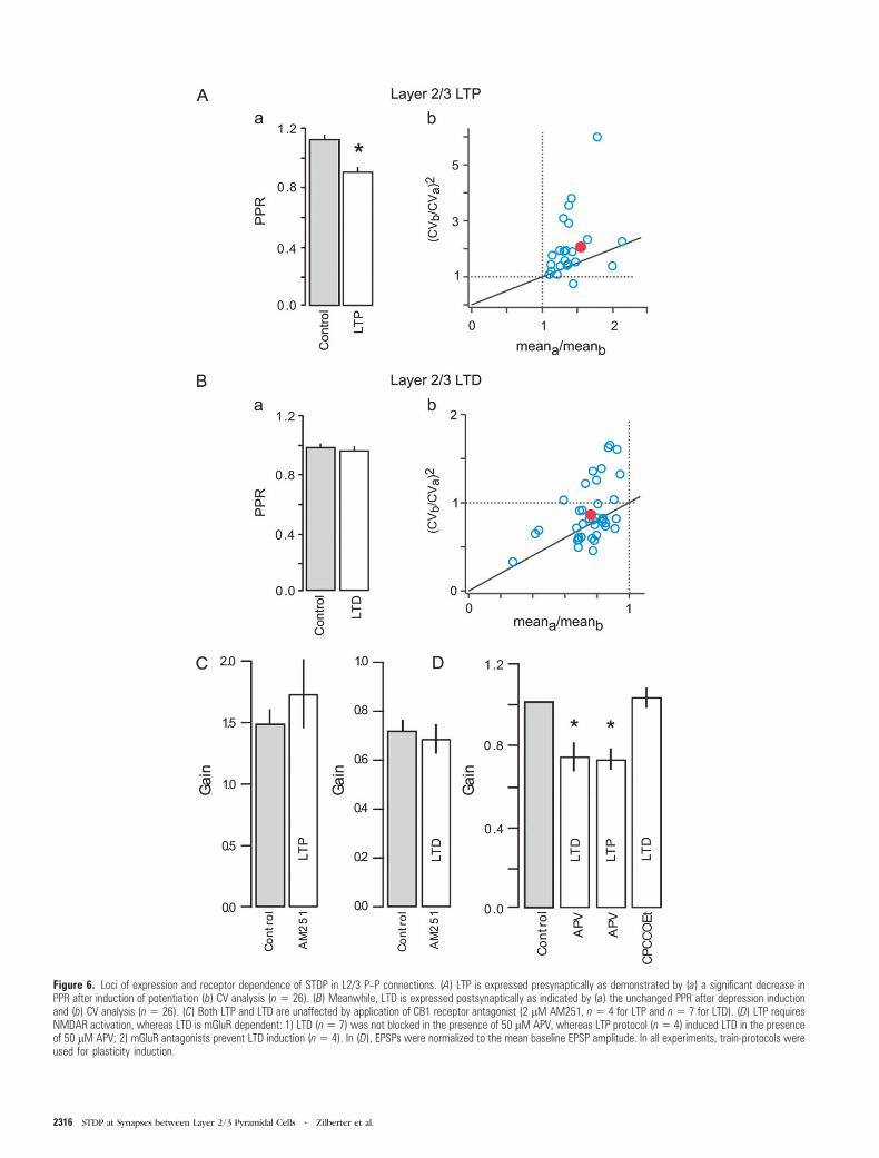
Figure 6. Loci of expression and receptor dependence of STDP in L2/3 P--P connections. (A) LTP is expressed presynaptically as demonstrated by (a) a significant decrease inPPR after induction of potentiation (b) CV analysis (n 5 26). (B) Meanwhile, LTD is expressed postsynaptically as indicated by (a) the unchanged PPR after depression inductionand (b) CV analysis (n 5 26). (C) Both LTP and LTD are unaffected by application of CB1 receptor antagonist (2 lM AM251, n 5 4 for LTP and n 5 7 for LTD). (D) LTP requiresNMDAR activation, whereas LTD is mGluR dependent: 1) LTD (n5 7) was not blocked in the presence of 50 lM APV, whereas LTP protocol (n5 4) induced LTD in the presenceof 50 lM APV; 2) mGluR antagonists prevent LTD induction (n5 4). In (D), EPSPs were normalized to the mean baseline EPSP amplitude. In all experiments, train-protocols wereused for plasticity induction.
2316 STDP at Synapses between Layer 2/3 Pyramidal Cells d Zilberter et al.

gain was 1.02 ± 0.05 of control (n = 4, Fig. 6D). It was however
possible to induce LTP in the presence of mGluR antagonists
(1.23 ± 0.12 of control, n = 4, data not shown). These results
demonstrate that mGluRs play a critical role in the induction of
LTD at L2/3 pyramidal cell unitary connections.
Discussion
At L2/3 P--P synapses, the rule of STDP can be converted from
one mode (symmetric anti-Hebbian) to another (asymmetric
Hebbian) depending on the postsynaptic activity that takes
place prior to synaptic activation. Thus, the history of the
postsynaptic cell firing shortly before the synaptic input
determines which STDP plasticity rule will govern the strength
of the unitary connection. This activity-dependent switch
depends on the interplay between the basal [Ca2+]post pre-
ceding synaptic stimulation and the level and dynamics of
Ca2+ at dendritic spines during synaptic activity. LTP in-
duction at these connections is NMDAR dependent and
presynaptically expressed, whereas LTD is mGluR dependent
and postsynaptically expressed. These data suggest a novel
mechanism for regulating which synaptic plasticity rule
governs plasticity induction at L2/3 pyramidal cell unitary
connections and highlight differences in synaptic plasticity at
excitatory synaptic inputs onto L2/3 pyramidal cells.
Location of L2/3 P--P Synapses
Synapse location plays an important role in determining
whether bAPs or local signaling is likely to regulate its synaptic
plasticity. Differences in STDP time-windows (Froemke et al.
2005), the requirement for NMDAR spikes (Gordon et al. 2006;
Kampa et al. 2006) and even a complete inversion of the STDP
rule (Letzkus et al. 2006; Sjostrom and Hausser 2006) have
been observed dependent on whether the synaptic input lies
on proximal or distal dendrites. Thus, location-dependent
modification of plasticity rules can result in input specificity
and play a part in dendritic processing (for reviews see
Goldberg et al. 2002; Kampa et al. 2007; Sjostrom et al. 2008).
Synapses between L2/3 pyramidal cells are situated mainly
on proximal basal dendritic sites (Feldmeyer et al. 2006), at
locations which are readily reachable by bAPs in vitro (Koster
and Sakmann 1998) and in vivo (Svoboda et al. 1999; Waters
et al. 2003). Differences in the exact number and location of
synaptic contacts in our study and (Feldmeyer et al. 2006)
could be due to differences in interneuronal distances (smaller
in our study), and/or differences in local P--P microcircuitry
(visual cortex vs. barrel cortex). However, synaptic location
does not seem to be a major factor contributing to the ‘‘LTD
only’’ induction we observed with low-frequency pre--post
pairing.
Simple Spike Timing--Dependent Plasticity Rules atExcitatory Synapses
The simplest STDP protocol consists of single EPSPs paired
with single postsynaptic bAPs (Bi and Poo 1998; Froemke and
Dan 2002). It has been shown that LTP can be induced by
precisely timed pre-before-postsynaptic pairing, whereas post-
before-presynaptic pairing can induce LTD, resulting in an
asymmetric Hebbian learning rule (Bi and Poo 1998; Froemke
and Dan 2002).
At L2/3 P--P synapses however, when neuronal activity is
low, a symmetric anti-Hebbian rule governs synaptic plasticity.
A similar spike-timing, ‘‘LTD only,’’ induction pattern with
low neuronal activity has also been observed at CA3-CA1
(Wittenberg and Wang 2006) and L4-L4 spiny stellate synapses
(Egger et al. 1999). Indeed, the same single pre-before-single
postsynaptic stimulation induces no change (Markram et al.
1997; Pike et al. 1999; Sjostrom et al. 2001; Kampa et al. 2006;
Nevian and Sakmann 2006), reliable LTP induction (Bi and Poo
1998; Feldman 2000; Froemke and Dan 2002), or the induction
of LTD (Tzounopoulos et al. 2004; Zhou et al. 2005; Wittenberg
and Wang 2006) dependent on the identity of the synaptic
connection. Whether LTP or LTD are induced with single EPSP
before single postsynaptic bAP pairing at a particular excitatory
synapse will depend on a number of factors including;
developmental age (Meredith et al. 2003), dendritic location
of the synapses (Letzkus et al. 2006; Sjostrom and Hausser
2006), synaptic strength (Bi and Poo 1998), concurrent
synaptic inhibition (Meredith et al. 2003), multiple coincidence
detectors (Karmarkar and Buonomano 2002; Bender et al. 2006;
Nevian and Sakmann 2006), synaptic cooperativity (Sjostrom
et al. 2001), SK channels (Ngo-Anh et al. 2005), or the width of
the bAP (Zhou et al. 2005; Wittenberg and Wang 2006).
Although one simple STDP rule does not ‘‘fit all’’ excitatory
synaptic connections, the differences in many, though not all,
cases can be explained by differences in levels of postsynaptic
depolarization and subsequent Ca2+ influx during the pairing
protocol. Indeed, at L5 P--P and CA3-CA1 synapses, synaptic
cooperativity (Sjostrom et al. 2001) or an increase in the width
of the postsynaptic bAP (Wittenberg and Wang 2006), re-
spectively provide the necessary additional conditions for LTP
induction.
Given the proximity of L2/3 P--P synaptic connections to the
soma it was therefore surprising that additional depolarization
or stimulation in Mg2+free extracellular solution (which should
cause a dramatic increase in [Ca2+]post; Sabatini et al. 2002) did
not induce a shift in synaptic gain in our study. The
explanations for this could include a low affinity calcium
sensor for LTP induction at these contacts and/or insufficient
postsynaptic dendritic calcium influx. We therefore increased
the amount of postsynaptic activity during the pairing protocol.
bAP Bursts and STDP
Postsynaptic cell bAP burst firing facilitates communication
between somatic and distal dendritic sites and can modulate
STDP rules (Pike et al. 1999; Meredith et al. 2003; Gordon et al.
2006; Letzkus et al. 2006; Nevian and Sakmann 2006; Sjostrom
and Hausser 2006; Wittenberg and Wang 2006). It permits LTP
induction with single presynaptic stimulation in CA1 pyramidal
cells (Pike et al. 1999; Meredith et al. 2003), allows induction of
LTP independent of pre--post spike order (Kampa et al. 2006),
rescues LTP at distal L2/3 inputs onto L5 pyramidal cells
(Letzkus et al. 2006; Sjostrom and Hausser 2006) and allows
LTP induction at L2/3 proximal (Gordon et al. 2006; Nevian and
Sakmann 2006) and distal (Gordon et al. 2006) basal dendrites.
At L5 P--P connections, low-frequency stimulation does not
induce a change in synaptic gain, but with a 5 pre- 5-post AP
burst protocol (10 Hz or higher) LTP is reliably induced
(Markram et al. 1997; Sjostrom et al. 2001). Likewise, at L2/3
P--P connections in the barrel cortex, 20 Hz 5 pre- 5 post AP
bursts reliably induce LTP (Egger et al. 1999). A low-frequency
burst protocol (5 pre- 5 postsynaptic AP burst at 10 Hz)
induced LTD at L2/3 visual cortical P--P connections. However
Cerebral Cortex October 2009, V 19 N 10 2317

an increase in the burst frequency (pre-before-post) to 20 Hz
caused a shift towards LTP, (although this burst frequency was
not sufficient to actually induce LTP). This suggests that the
relationship between pre--post burst frequency and changes in
synaptic gain, whereas similar to that at L2/3 P--P connections
in the barrel cortex or L5, is shifted to favor LTD induction
with low-frequency pre--post-burst stimulation at L2/3 visual
cortical P--P contacts.
Our results suggest that at visual cortex L2/3 P--P con-
nections LTP should be induced with a higher ( >20 Hz) 5 pre-
5 postsynaptic burst pairing protocol. Alternatively, single
EPSPs paired with a high frequency (100--200 Hz) postsynaptic
burst protocol could also induce LTP (Gordon et al. 2006;
Kampa et al. 2006). However, in this study we focused on the
simplest burst paradigm which would retain spike-timing,
permit a clear distinction between the contribution of pre- and
postsynaptic activity to LTD and LTP induction, and yet provide
the requisite postsynaptic depolarization for LTP induction. We
therefore used a single presynaptic AP paired with a post-
synaptic AP train. We found that a ‘‘preconditioning’’ post-
synaptic AP train fundamentally modified the pre--post spike
interaction rule and evoked a switch from anti-Hebbian to
Hebbian STDP.
Role of Dendritic [Ca2+]post in the Regulation of STDPRules
The importance of [Ca2+]post elevation in the regulation of
STDP has been well documented (for review see Sjostrom and
Nelson 2002). Burst firing (Pike et al. 1999), Ca2+
spikes
(Kampa et al. 2006), the distance of synapses from the soma
(Froemke et al. 2005; Sjostrom and Hausser 2006), and bAP
width (Zhou et al. 2005; Wittenberg and Wang 2006) can all
regulate the form of STDP rules, by affecting dendritic
[Ca2+]post directly during the peristimulus period.
At L2/3 P--P connections we found separate thresholds for
LTD and LTP induction. Low-frequency single or burst pairing
protocols resulted in ‘‘LTD only’’ induction. Although LTP was
NMDAR dependent and was inhibited even by low concen-
trations of BAPTA, LTD could be induced when NMDARs were
blocked and required higher BAPTA concentrations for
blockade. Interestingly, an intermediate region where neither
LTD nor LTP occurred was also observed with BAPTA
application.
A simple peak Ca2+concentration threshold model, how-
ever, does not explain the induction of LTD with the preceding
10 AP train, when [Ca2+]post is high, suggesting that additional
factors play a role in STDP induction at L2/3 P--P contacts. We
found that an additional requirement for the switch in STDP
rules is a rise in basal VGCC-dependent [Ca2+]post prior to
synaptic stimulation. If VGCCs are blocked or the number of
APs in the postsynaptic train is decreased, LTP is no longer
induced. The order of these events is however important, as no
LTP was induced when the presynaptic stimulus was followed
by a postsynaptic 10 AP train.
Finally, spike-timing dependent LTP and LTD could be
induced if presynaptic stimulation occurred around the 8th AP
or the 10th AP in a 10AP postsynaptic train. Moreover,
removing NMDAR’s Mg2+block in both train-LTP and train-
LTD protocols seemed to abolish the coincidence timing
dependence, as LTP was induced in all cases. This suggests that
the basal [Ca2+]post preceding synaptic stimulation and the
calcium dynamics during synaptic stimulation (with possible
supra- or sublinear postsynaptic summation of Ca2+signals
(Koster and Sakmann 1998) act in concert to determine the
form of the STDP rule at L2/3 P--P connections.
LTP and LTD Induction Mechanisms at L2/3 P--PConnections
Induction of LTP and LTD at L2/3 P--P connections required
the activation of 2 different receptor pathways, NMDAR
mediated for LTP, and mGluR activated for LTD. Although it
is widely accepted that STDP is NMDAR dependent (Magee and
Johnston 1997; Bi and Poo 1998; Debanne et al. 1998; Feldman
2000; Sjostrom et al. 2001, 2003), an NMDAR-independent
component of LTD has also been observed at synapses onto L2/
3 pyramidal cells (Feldman 2000; Nevian and Sakmann 2006).
mGluR-dependent LTD has also been reported to occur in
a wide variety of neurons in different brain regions (Linden
et al. 1991; Shigemoto et al. 1994; Hensch and Stryker 1996;
Oliet et al. 1997; Egger et al. 1999).
The presence of distinct biochemical signaling cascades for
LTP and LTD induction suggest the possibility of 2 separate
coincidence detectors for STDP (Karmarkar and Buonomano
2002). This is indeed the case at L4 (Bender et al. 2006) and
L2/3 (Nevian and Sakmann 2006) excitatory synaptic con-
nections onto L2/3 pyramidal cells. However, at L2/3 P--P
connections a key prediction of the global Ca2+, 2 co-
incidence detector model, namely that LTD should not be
induced at positive Dt intervals with a single EPSP-single
postsynaptic AP protocol (Karmarkar and Buonomano 2002),
is not met. This suggests that other factors, in this case
postsynaptic activity and postsynaptic Ca2+dynamics, play
a key part in the induction of bidirectional synaptic plasticity
at L2/3 P--P contacts.
LTD is presynaptic and is mediated by retrograde endocan-
nabinoid signaling at L4 (Bender et al. 2006) and L2/3 (Nevian
and Sakmann 2006) afferent excitatory inputs onto L2/3
pyramidal cells and at L5 P--P synaptic connections (Sjostrom
et al. 2003). In contrast, LTD is postsynaptic, and LTP displays
a presynaptic expression locus at L2/3 P--P connections.
Neither LTD nor LTP are CB1R dependent, although the
postsynaptic VGCC dependence together with the presynaptic
expression locus indicate that LTP is mediated by release of
a retrograde messenger at L2/3 P--P connections. The differ-
ences in LTP and LTD expression loci and signaling pathways at
excitatory contacts onto L2/3 pyramidal cells could reflect
fundamental differences in properties of excitatory synapses
originating in different cortical layers (L2/3-L2/3 vs. L4-L2/3;
Brasier and Feldman 2008) or regions (visual cortex vs. barrel
cortex). Additionally, they may reflect differences in pre-
synaptic stimulation methods (unitary connections vs. extra-
cellular stimulation) with the possible activation of excitatory
afferents whose origins lie outside the local network in the
latter case.
Differences in expression locus, retrograde signaling path-
ways, and calcium dependence at different excitatory synapses
onto L2/3 suggest that single L2/3 pyramidal neurons are able
to distinguish input sources and use different learning rules
based on the origin of input. This input-specific tuning of
synaptic gain should greatly enhance the computational
capabilities of each individual pyramidal cell within the local
neuronal network.
2318 STDP at Synapses between Layer 2/3 Pyramidal Cells d Zilberter et al.

Functional Implications
‘‘Preconditioning’’ with a postsynaptic spike train can evoke
a switch in STDP rule from symmetric anti-Hebbian rule to
asymmetric Hebbian. Dependent on the activity of the
network, pyramidal cells can therefore determine not only
whether LTP or LTD will be induced at a particular synapse, but
also which learning rule will govern that change. For this rule
switch to be physiologically relevant, L2/3 pyramidal cells
should display periods of sparse activity (where the governing
rule would be symmetric anti-Hebbian) as well as periods of
increased activity (with asymmetric Hebbian rule in effect).
Pyramidal cells in vivo show a range of firing rates in
response to sensory stimuli; from low firing rates ( <1 Hz) in
which ‘‘sparse coding’’ is used to encode information (reviewed
in Olshausen and Field 2004), to higher rates 3-- >100 Hz
(Parnavelas 1984; Softky and Koch 1993; Holt et al. 1996;
Shadlen and Newsome 1998; Steriade 2001). Therefore, the
required conditions for the rule switch to occur appear to be
fulfilled in vivo.
Dynamic functional columns have been suggested to be
a means of improving information processing and storage in the
cerebral cortex (Diamond et al. 2003). The capability of
pyramidal cells to switch between STDP rules suggests
a possible mechanism for their formation. Thalamic input is
relayed to L2/3 via excitatory afferents from L4 as well as from
thalamus itself (Bruno and Sakmann 2006). Input from L4 is
reliable and diffuse, and provides an effective lateral spread of
excitation for a local population of neurons in L2/3 (Feldmeyer
et al. 2002; Shepherd and Svoboda 2005). Following thalamic
input, for example, during processing of a sensory task, local
pyramidal cells enter an active state, firing trains of APs. LTP at
pyramidal cell synapses then becomes possible, allowing the
formation of a functional local network by potentiating certain
connections and depressing others, according to their relative
discharge patterns. In the absence of the thalamic input,
pyramidal cells enter a period of sparse activity and LTD is the
dominant plasticity outcome. This ability to control the input
gain of a limited number of synapses, allows the signal-to-noise
ratio of the network to be increased.
Recent studies have shown that the cortex is a dynamic
entity; previously potentiated synapses can be ‘‘de-potentiated’’
(weakened), depressed ones can be ‘‘de-depressed’’ (restored
or repotentiated) and existing connections constantly form and
dissolve over a period of hours (Turrigiano and Nelson 2004; Le
Be and Markram 2006). Our results suggest a new way in which
‘‘wandering,’’ task-specific functional columns might transiently
take shape in the neocortex.
Supplementary Material
Supplementary material can be found at: http://www.cercor.
oxfordjournals.org/
Funding
Swedish Medical Research Council funded T.H., Y.Z.; European
Molecular Biology Organization Young Investigator Programme
funded T.H.; European Commission (HEALTH-F2-2007-201159)
to T.H., Y.Z.; National Institutes of Health (DA023214) to T.H.;
and the Alzheimer’s Association funded T.H.
Notes
We thank M. Hausser, P. J. Sjostrom, and G. Stuart for their critical
comments on an earlier version of this manuscript. The permanently
charged and membrane impermeant verapamil analogue D890 was
provided by Abbott Laboratories, Inc. (Abbott Park, IL). Conflict of
Interest : None declared.
Address correspondence to Dr Yuri Zilberter, Institut de Neuro-
biologie de la Mediterranee (INMED), Inserm U29, Parc Scientifique de
Luminy, 13273 Marseille Cedex 09, France. Email: zilberter@inmed.
univ-mrs.fr.
References
Aizenman CD, Manis PB, Linden DJ. 1998. Polarity of long-term synaptic
gain change is related to postsynaptic spike firing at a cerebellar
inhibitory synapse. Neuron. 21:827--835.
Bender VA, Bender KJ, Brasier DJ, Feldman DE. 2006. Two coincidence
detectors for spike timing-dependent plasticity in somatosensory
cortex. J Neurosci. 26:4166--4177.
Bi G-Q, Poo M-M. 1998. Synaptic modifications in cultured hippocampal
neurons: dependence on spike timing, synaptic strength, and
postsynaptic cell type. J Neurosci. 18:10464--10472.
Bi G-Q, Poo M-M. 2001. Synaptic modification by correlated activity:
Hebb’s postulate revisited. Annu Rev Neurosci. 24:139--166.
Brasier DJ, Feldman DE. 2008. Synapse-specific expression of functional
presynaptic NMDA receptors in rat somatosensory cortex.
J Neurosci. 28:2199--2211.
Bruno RM, Sakmann B. 2006. Cortex is driven by weak but synchronously
active thalamocortical synapses. Science. 312:1622--1627.
Celikel T, Szostak VA, Feldman DE. 2004. Modulation of spike timing by
sensory deprivation during induction of cortical map plasticity. Nat
Neurosci. 7:534--541.
Conti R, Lisman J. 2002. A large sustained Ca2+elevation occurs in
unstimulated spines during the LTP pairing protocol but does not
change synaptic strength. Hippocampus. 12:667--679.
Debanne D, Gahwiler BH, Thompson SM. 1998. Long-term synaptic
plasticity between pairs of individual CA3 pyramidal cells in rat
hippocampal slice cultures. J Physiol. 507:237--247.
Diamond ME, Petersen RS, Panzeri S. 2003. Investigations into the
organization of information in sensory cortex. J Physiol (Paris).
97:529--536.
Egger V, Feldmeyer D, Sakmann B. 1999. Coincidence detection and
changes of synaptic efficacy in spiny stellate neurons in rat barrel
cortex. Nat Neurosci. 2:1098--1105.
Feldman DE. 2000. Timing-based LTP and LTD at vertical inputs to layer
II/III pyramidal cells in rat barrel cortex. Neuron. 27:45--56.
Feldmeyer D, Lubke J, Sakmann B. 2006. Efficacy and connectivity of
intracolumnar pairs of layer 2/3 pyramidal cells in the barrel cortex
of juvenile rats. J Physiol. 575:583--602.
Feldmeyer D, Lubke J, Silver RA, Sakmann B. 2002. Synaptic connections
between layer 4 spiny neurone-layer 2/3 pyramidal cell pairs in
juvenile rat barrel cortex: physiology and anatomy of interlaminar
signalling within a cortical column. J Physiol. 538:803--822.
Froemke RC, Dan Y. 2002. Spike-timing-dependent synaptic modifica-
tion induced by natural spike trains. Nature. 416:433--438.
Froemke RC, Poo M-M, Dan Y. 2005. Spike-timing-dependent synaptic
plasticity depends on dendritic location. Nature. 434:221--225.
Goldberg JK, Holthoff K, Yuste R. 2002. A problem with Hebb and local
spikes. Trends Neurosci. 25:433--435.
Golding NL, Staff NP, Spruston N. 2002. Dendritic spikes as a mechanism
for cooperative long-term potentiation. Nature. 418:326--331.
Gordon U, Polsky A, Schiller J. 2006. Plasticity compartments in basal
dendrites of neocortical pyramidal neurons. J Neurosci. 26:
12717--12726.
Harkany T, Holmgren C, Hartig W, Qureshi T, Chaudhry FA, Storm-
Mathisen J, Dobszay MB, Berghuis P, Schulte G, Fremeau RT, Jr, et al.
2004. Endocannabinoid-independent retrograde signaling at in-
hibitory synapses in layer 2/3 of neocortex: involvement of vesicular
glutamate transporter 3. J Neurosci. 24:4978--4988.
Hensch TK, Stryker MP. 1996. Ocular dominance plasticity under
metabotropic glutamate receptor blockade. Science. 272:554--557.
Cerebral Cortex October 2009, V 19 N 10 2319

Holmgren C, Harkany T, Svennenfors B, Zilberter Y. 2003. Pyramidal cell
communication within local networks in layer 2/3 of rat neocortex.
J Physiol. 551:139--153.
Holt GR, Softky WR, Koch C, Douglas RJ. 1996. Comparison of
discharge variability in vitro and in vivo in cat visual cortex neurons.
J Neurophysiol. 75:1806--1814.
Kampa BM, Letzkus JJ, Stuart G. 2006. Requirement of dendritic
calcium spikes for induction of spike-timing-dependent synaptic
plasticity. J Physiol. 574:283--290.
Kampa BM, Letzkus JJ, Stuart GJ. 2007. Dendritic mechanisms
controlling spike-timing-dependent synaptic plasticity. Trends
Neurosci. 30:456--463.
Karmarkar UR, Buonomano DV. 2002. A model of spike-timing
dependent plasticity: one or two coincidence detectors? J Neuro-
physiol. 88:507--513.
Koster HJ, Sakmann B. 1998. Calcium dynamics in single spines during
coincident pre- and postsynaptic activity depend on relative timing
of back-propagating action potentials and subthreshold excitatory
postsynaptic potentials. Proc Natl Acad Sci USA. 95:9596--9601.
Lambert MP, Barlow AK, Chromy BA, Edwards C, Freed R, Liosatos M,
Morgan TE, Rozovsky I, Trommer B, Viola KL, et al. 1998. Diffusible,
nonfibrillar ligands derived from Ab1--42 are potent central nervous
system neurotoxins. Proc Natl Acad Sci USA. 95:6448--6453.
Le Be JV, Markram H. 2006. Spontaneous and evoked synaptic rewiring
in the neonatal neocortex. Proc Natl Acad Sci USA. 103:
13214--13219.
Letzkus JJ, Kampa BM, Stuart GJ. 2006. Learning rules for spike timing-
dependent plasticity depend on dendritic synapse location.
J Neurosci. 26:10420--10429.
Linden DJ, Dickinson MH, Smeyne M, Connor JA. 1991. A long-term
depression of AMPA currents in cultured cerebellar Purkinje
neurons. Neuron. 7:81--89.
Lisman J, Spruston N. 2005. Postsynaptic depolarization requirements
for LTP and LTD: a critique of spike timing dependent plasticity. Nat
Neurosci. 8:839--841.
Magee JC, Johnston D. 1997. A synaptically controlled, associative signal
for Hebbian plasticity in hippocampal neurons. Science. 275:
209--213.
Markram H, Lubke J, Frotscher M, Sakmann B. 1997. Regulation of
synaptic efficacy by coincidence of postsynaptic APs and EPSPs.
Science. 275:213--215.
Mehta MR. 2004. Cooperative LTP can map memory sequences on
dendritic branches. Trends Neurosci. 27:69--72.
Mehta MR, Barnes CA, McNaughton BL. 1997. Experience-dependent,
asymmetric expansion of hippocampal place fields. Proc Natl Acad
Sci USA. 94:8918--8921.
Meredith RM, Floyer-Lea AM, Paulsen O. 2003. Maturation of long-term
potentiation induction rules in rodent hippocampus: role of
GABAergic inhibition. J Neurosci. 23:11142--11146.
Nevian T, Sakmann B. 2006. Spine Ca2+signaling in spike-timing-
dependent plasticity. J Neurosci. 26:11001--11013.
Ngo-Anh TJ, Bloodgood BL, Lin M, Sabatini BL, Maylie J, Adelman JP.
2005. SK channels and NMDA receptors form a Ca2+-mediated
feedback loop in dendritic spines. Nat Neurosci. 8:642--649.
Oliet SHR, Malenka RC, Nicoll RA. 1997. Two distinct forms of long-
term depression coexist in CA1 hippocampal pyramidal cells.
Neuron. 18:969--982.
Olshausen BA, Field DJ. 2004. Sparse coding of sensory inputs. Curr
Opin Neurobiol. 14:481--487.
Parnavelas JG. 1984. Physiological properties of identified cortical
neurons. In: Peters A, Jones EG, editors. Cerebral cortex. New York:
Plenum Press. p. 205--239.
Pike FG, Meredith RM, Olding AW, Paulsen O. 1999. Rapid report:
postsynaptic bursting is essential for ‘Hebbian’ induction of
associative long-term potentiation at excitatory synapses in rat
hippocampus. J Physiol. 518:571--576.
Roberts PD, Bell CC. 2002. Spike timing dependent synaptic plasticity in
biological systems. Biol Cybern. 87:392--403.
Rubin JE, Gerkin RC, Bi GQ, Chow CC. 2005. Calcium time course as
a signal for spike-timing dependent plasticity. J Neurophysiol.
93:2600--2613.
Sabatini BL, Oertner TG, Svoboda K. 2002. The life cycle of Ca2+ions in
dendritic spines. Neuron. 33:439--452.
Shadlen MN, Newsome WT. 1998. The variable discharge of cortical
neurons: implications for connectivity, computation, and informa-
tion coding. J Neurosci. 18:3870--3896.
Shepherd GMG, Svoboda K. 2005. Laminar and columnar organization
of ascending excitatory projections to layer 2/3 pyramidal neurons
in rat barrel cortex. J Neurosci. 25:5670--5679.
Shigemoto R, Abe T, Nomura S, Nakanishi S, Hirano T. 1994. Antibodies
inactivating mGluR1 metabotropic glutamate receptor block long-
term depression in cultured Purkinje cells. Neuron. 12:1245--1255.
Sjostrom PJ, Hausser M. 2006. A cooperative switch determines the sign
of synaptic plasticity in distal dendrites of neocortical pyramidal
neurons. Neuron. 51:227--238.
Sjostrom PJ, Nelson SB. 2002. Spike timing, calcium signals and synaptic
plasticity. Curr Opin Neurobiol. 12:305--314.
Sjostrom PJ, Rancz EA, Roth A, Hausser M. 2008. Dendritic excitability
and synaptic plasticity. Physiol Rev. 88:769--840.
Sjostrom PJ, Turrigiano GG, Nelson SB. 2001. Rate, timing, and
cooperativity jointly determine cortical synaptic plasticity. Neuron.
32:1149--1164.
Sjostrom PJ, Turrigiano GG, Nelson SB. 2003. Neocortical LTD via
coincident activation of presynaptic NMDA and cannabinoid
receptors. Neuron. 39:641--654.
Sjostrom PJ, Turrigiano GG, Nelson SB. 2004. Endocannabinoid-
dependent neocortical layer-5 LTD in the absence of postsynaptic
spiking. J Neurophysiol. 43:3338--3343.
Softky WR, Koch C. 1993. The highly irregular firing of cortical cells is
inconsistent with temporal integration of random EPSPs. J Neurosci.
13:334--350.
Steriade M. 2001. Impact of network activity on neuronal properties in
corticothalamic systems. J Neurophysiol. 86:1--39.
Svoboda K, Helmchen F, Denk W, Tank DW. 1999. Spread of dendritic
excitation in layer 2/3 pyramidal neurons in rat barrel cortex in
vivo. Nat Neurosci. 2:65--73.
Turrigiano GG, Nelson SB. 2004. Homeostatic plasticity in the
developing nervous system. Nat Rev Neurosci. 5:97--107.
Tzounopoulos T, Kim Y, Oertel D, Trussell LO. 2004. Cell-specific, spike
timing-dependent plasticities in the dorsal cochlear nucleus. Nat
Neurosci. 7:719--725.
Tzounopoulos T, Rubio ME, Keen JE, Trussell LO. 2007. Coactivation of
pre- and postsynaptic signaling mechanisms determines cell-specific
spike-timing-dependent plasticity. Neuron. 54:291--301.
Waters J, Larkum ME, Sakmann B, Helmchen F. 2003. Supralinear Ca2+
influx into dendritic tufts of layer 2/3 neocortical pyramidal
neurons in vitro and in vivo. J Neurosci. 23:8558--8567.
Wittenberg GM, Wang SS. 2006. Malleability of spike-timing-dependent
plasticity at the CA3-CA1 synapse. J Neurosci. 26:6610--6617.
Yoshimura Y, Dantzker JLM, Callaway EM. 2005. Excitatory cortical
networks form fine-scale functional networks. Nature. 433:868--873.
Zhou Q, Tao HW, Poo M-M. 2003. Reversal and stabilization of synaptic
modifications in a developing visual system. Science. 300:
1953--1957.
Zhou YD, Acker CD, Netoff TI, Sen K, White JA. 2005. Increasing Ca2+
transients by broadening postsynaptic action potentials enhances
timing-dependent synaptic depression. Proc Natl Acad Sci USA.
102:19121--19125.
2320 STDP at Synapses between Layer 2/3 Pyramidal Cells d Zilberter et al.
Related Documents