Inositol Hexakisphosphate-Mediated Regulation of Glutamate Receptors in Rat Brain Sections Barbara Valastro, 1,2 Martine Girard, 1 Joe ¨l Gagne ´, 1 Fre ´de ´ric Martin, 1 Ange `le T. Parent, 3 Michel Baudry, 4 and Guy Massicotte 1,2 * 1 De ´partement de Chimie-Biologie, Universite ´ du Que ´bec a ` Trois-Rivie `res, Trois-Rivie `res, Que ´bec, Canada 2 De ´partement de Pharmacologie, Universite ´ de Montre ´al, Montre ´al, Que ´bec, Canada 3 Department of Neurobiology, Pharmacology and Physiology, University of Chicago, Chicago, Illinois 4 Neuroscience Program, University of Southern California, Los Angeles, California ABSTRACT: D-myo-inositol 1,2,3,4,5,6-hexakisphosphate (InsP6), one of the most abundant inositol phosphates within cells, has been proposed to play a key role in vesicle trafficking and receptor compartmentaliza- tion. In the present study, we used in vitro receptor autoradiography, subcellular fractionation, and immunoblotting to investigate its effects on a-amino-3-hydroxy-5-methylisoxazole-4-propionate (AMPA) and N- methyl-D-aspartate (NMDA) receptors. Qualitative and quantitative anal- ysis of 3 H-AMPA binding indicated that incubation of frozen-thawed brain sections with InsP6 at 35°C enhanced AMPA receptor binding in several brain regions, with maximal increases in the hippocampus and cerebel- lum. Moreover, saturation kinetics demonstrated that InsP6-induced aug- mentation of AMPA binding was due to an increment in the maximal number of AMPA binding sites. At the immunological level, Western blots performed on crude mitochondrial/synaptic (P2) fractions revealed that InsP6 (but not InsP5 and InsP3) treatment increased glutamate receptor (GluR)1 and GluR2 subunits of AMPA receptors, an effect that was associated with concomitant reductions in microsomal (P3) fractions. Interestingly, the InsP6-induced modulation of AMPA receptor binding was blocked at room temperature, and pretreatment with heparin also dampered its action on both AMPA receptor binding and GluR subunits. These effects of InsP6 appear to be specific to AMPA receptors, as neither 3 H-glutamate binding to NMDA receptors nor levels of NR1 and NR2A subunits in P2 and P3 fractions were affected. Taken together, our data strongly suggest that InsP6 specifi- cally regulates AMPA receptor distribution, possibly through a clathrin-dependent process. Hippocampus 2001;11:673– 682. © 2001 Wiley-Liss, Inc. KEY WORDS: inositol phosphate; phytic acid; AMPA receptor; NMDA receptor; adaptin; clathrin INTRODUCTION D-myo-inositol 1,2,3,4,5,6-hexakisphosphate (InsP6), one of the most abundant inositol phosphates, is formed via complex pathways of inositol phosphate metabolism. In some cell types, its concentration is rapidly altered upon cell stimulation (Sasakawa et al., 1993, 1995), and experimental evidence indicates that it is involved in the control of several cellular functions. For instance, InsP6 has been shown to reduce light-induced desensitization of rhodopsin (Palczewski et al., 1992), presumably through direct binding to arrestin (Palczewski et al., 1991), a specialized class of regulatory proteins. In the brain, a number of high-affinity binding sites for InsP6 have been found (Hawkins et al., 1990; Nicoletti et al., 1990; Theibert et al., 1991, 1992), and one of them has been identified as the C2B domain of synaptotagmin (Fukada et al., 1994), a major calcium-sensor for regulat- ing exocytosis in neurons. InsP6 also binds with high affinity to clathrin assembly proteins, the adaptor pro- teins (AP)-2 and AP-3 (Beek and Keen, 1991; Theibert et al., 1992; Timerman et al., 1992; Voglmaier et al., 1992), thereby inhibiting clathrin assembly (Beek and Keen, 1991; Norris et al., 1995; Ye et al., 1995). In fact, InsP6 has been proposed to participate in vesicle trafficking and Abbreviations: AP, adaptor protein; AMPA, (RS)-a-amino-3-hydroxy-5- methylisoxazole-propionate; DPGA, 2,3-diphospho-D-glyceric acid; GluR, glutamate receptor; IGF II, insulin-like growth factor receptor II; InsPs, inositol phosphates; InsP3, D-myo-inositol 1,4,5-trisphosphate; InsP5, D- myo-inositol 1,3,4,5,6-pentakisphosphate; InsP6, D-myo-inositol 1,2,3,4,- 5,6-hexakisphosphate; InsS6, D-myo-inositol 1,2,3,4,5,6-hexakissulfate; LTP, long-term potentiation; NMDA, N-methyl-D-aspartate; OD, optical density; PBS, phosphate-buffered saline; PMSF, phenylmethylsulfonyl flu- oride; PSD, postsynaptic density; SITS, 4-acetanido-49-isothiocyanato-stilbene- 2,29-disulfonic acid; TPCK, N-tosyl-L-phenylalanine chloromethyl ketone. Grant sponsor: Natural Sciences and Engineering Research Council of Canada. B. Valastro and M. Girard contributed equally to this work. *Correspondence to: Guy Massicotte, De ´partement de Chimie-Biologie, Universite ´ du Que ´bec a ` Trois-Rivie `res, C.P. 500, Trois-Rivie `res, Que ´bec G9A 5H7, Canada. E-mail: [email protected] Accepted for publication 9 April 2001 HIPPOCAMPUS 11:673– 682 (2001) © 2001 WILEY-LISS, INC. DOI 10.1002/hipo.1082

Welcome message from author
This document is posted to help you gain knowledge. Please leave a comment to let me know what you think about it! Share it to your friends and learn new things together.
Transcript
Inositol Hexakisphosphate-Mediated Regulation ofGlutamate Receptors in Rat Brain Sections
Barbara Valastro,1,2 Martine Girard,1 Joel Gagne,1
Frederic Martin,1 Angele T. Parent,3 Michel Baudry,4
and Guy Massicotte1,2*
1Departement de Chimie-Biologie, Universite du Quebec aTrois-Rivieres, Trois-Rivieres, Quebec, Canada2Departement de Pharmacologie, Universite de Montreal,Montreal, Quebec, Canada3Department of Neurobiology, Pharmacology andPhysiology, University of Chicago, Chicago, Illinois4Neuroscience Program, University of SouthernCalifornia, Los Angeles, California
ABSTRACT: D-myo-inositol 1,2,3,4,5,6-hexakisphosphate (InsP6), oneof the most abundant inositol phosphates within cells, has been proposedto play a key role in vesicle trafficking and receptor compartmentaliza-tion. In the present study, we used in vitro receptor autoradiography,subcellular fractionation, and immunoblotting to investigate its effectson a-amino-3-hydroxy-5-methylisoxazole-4-propionate (AMPA) and N-methyl-D-aspartate (NMDA) receptors. Qualitative and quantitative anal-ysis of 3H-AMPA binding indicated that incubation of frozen-thawed brainsections with InsP6 at 35°C enhanced AMPA receptor binding in severalbrain regions, with maximal increases in the hippocampus and cerebel-lum. Moreover, saturation kinetics demonstrated that InsP6-induced aug-mentation of AMPA binding was due to an increment in the maximalnumber of AMPA binding sites. At the immunological level, Western blotsperformed on crude mitochondrial/synaptic (P2) fractions revealed thatInsP6 (but not InsP5 and InsP3) treatment increased glutamate receptor(GluR)1 and GluR2 subunits of AMPA receptors, an effect that wasassociated with concomitant reductions in microsomal (P3) fractions.Interestingly, the InsP6-induced modulation of AMPA receptor bindingwas blocked at room temperature, and pretreatment with heparin alsodampered its action on both AMPA receptor binding and GluR subunits.These effects of InsP6 appear to be specific to AMPA receptors, as neither3H-glutamate binding to NMDA receptors nor levels of NR1 and NR2A
subunits in P2 and P3 fractions were affected. Takentogether, our data strongly suggest that InsP6 specifi-cally regulates AMPA receptor distribution, possiblythrough a clathrin-dependent process. Hippocampus2001;11:673–682. © 2001 Wiley-Liss, Inc.
KEY WORDS: inositol phosphate; phytic acid; AMPAreceptor; NMDA receptor; adaptin; clathrin
INTRODUCTION
D-myo-inositol 1,2,3,4,5,6-hexakisphosphate (InsP6),one of the most abundant inositol phosphates, is formedvia complex pathways of inositol phosphate metabolism.In some cell types, its concentration is rapidly alteredupon cell stimulation (Sasakawa et al., 1993, 1995), andexperimental evidence indicates that it is involved in thecontrol of several cellular functions. For instance, InsP6has been shown to reduce light-induced desensitizationof rhodopsin (Palczewski et al., 1992), presumablythrough direct binding to arrestin (Palczewski et al.,1991), a specialized class of regulatory proteins. In thebrain, a number of high-affinity binding sites for InsP6have been found (Hawkins et al., 1990; Nicoletti et al.,1990; Theibert et al., 1991, 1992), and one of them hasbeen identified as the C2B domain of synaptotagmin(Fukada et al., 1994), a major calcium-sensor for regulat-ing exocytosis in neurons. InsP6 also binds with highaffinity to clathrin assembly proteins, the adaptor pro-teins (AP)-2 and AP-3 (Beek and Keen, 1991; Theibert etal., 1992; Timerman et al., 1992; Voglmaier et al., 1992),thereby inhibiting clathrin assembly (Beek and Keen,1991; Norris et al., 1995; Ye et al., 1995). In fact, InsP6has been proposed to participate in vesicle trafficking and
Abbreviations: AP, adaptor protein; AMPA, (RS)-a-amino-3-hydroxy-5-methylisoxazole-propionate; DPGA, 2,3-diphospho-D-glyceric acid; GluR,glutamate receptor; IGF II, insulin-like growth factor receptor II; InsPs,inositol phosphates; InsP3, D-myo-inositol 1,4,5-trisphosphate; InsP5, D-myo-inositol 1,3,4,5,6-pentakisphosphate; InsP6, D-myo-inositol 1,2,3,4,-5,6-hexakisphosphate; InsS6, D-myo-inositol 1,2,3,4,5,6-hexakissulfate;LTP, long-term potentiation; NMDA, N-methyl-D-aspartate; OD, opticaldensity; PBS, phosphate-buffered saline; PMSF, phenylmethylsulfonyl flu-oride; PSD, postsynaptic density; SITS, 4-acetanido-49-isothiocyanato-stilbene-2,29-disulfonic acid; TPCK, N-tosyl-L-phenylalanine chloromethyl ketone.Grant sponsor: Natural Sciences and Engineering Research Council ofCanada.B. Valastro and M. Girard contributed equally to this work.*Correspondence to: Guy Massicotte, Departement de Chimie-Biologie,Universite du Quebec a Trois-Rivieres, C.P. 500, Trois-Rivieres, QuebecG9A 5H7, Canada. E-mail: [email protected] for publication 9 April 2001
HIPPOCAMPUS 11:673–682 (2001)
© 2001 WILEY-LISS, INC.DOI 10.1002/hipo.1082
receptor compartmentalization (Sasakawa et al., 1995), and thedemonstration that microinjection of InsP6 into cells can inhibitvesicle trafficking is consistent with the involvement of an inosit-ide-binding site in this process (Llinas et al., 1994). The conceptwas also recently strengthened by the observation that increasedInsP6 in response to G protein-coupled receptor signals blocks therecruitment of AP-2 and clathrin-dependent endocytosis of Na1,K1-ATPase activity in renal epithelial cells (Ogimito et al., 2000).
Glutamate receptors (GluRs), which are major excitatory recep-tors within the central nervous system, are the object of particularattention, since their regulation appears to be crucial for control-ling synaptic function. For instance, GluRs have been proposed tocontrol fast excitatory neurotransmission at glutamatergic syn-apses, and their excessive activation has been demonstrated to bepotently neurotoxic (Seeburg, 1993; Hollmann and Heinemann,1994; Lipton and Rosenberg, 1994). N-methyl-D-aspartate(NMDA) and non-NMDA (kainate and a-amino-3-hydroxy-5-methylisoxazole-propionate; AMPA) receptors are two families ofionotropic receptors activated by glutamate (Dingledine et al.,1999). Among several important functions, the NMDA receptorhas been implicated in the induction of long-term potentiation(LTP), an electrophysiological model of learning and memory(Bliss and Collingridge, 1993). However, alterations in AMPAreceptors appear to be primarily responsible for the enhanced syn-aptic current observed during expression of LTP (Kauer et al.,1988; Muller and Lynch., 1988; Davies et al., 1989). In this con-text, studies have shown that LTP expression may involve theuncovering of functional AMPA receptors in hippocampal slicesthat, prior to LTP, were either not present in postsynaptic mem-branes or were electrophysiologically silent (Isaac et al., 1995; Liaoet al., 1995). Consistently, recent reports have proposed that reg-ulation in the surface expression of AMPA receptors at individualsynapses may contribute to the changes in synaptic strength thatoccur during LTP (Shi et al., 1999; Hayashi et al., 2000).
Although the detailed molecular mechanisms by which AMPAreceptors are regulated during synaptic plasticity remain intenselydebated, it was suggested recently that the surface expression ofthese receptors might undergo endocytosis through a clathrin-de-pendent process (Man et al., 2000; Wang and Linden, 2000). Itwas also reported that GluR2 subunits coimmunoprecipitate withadaptor protein 2 (AP-2) (Man et al., 2000), indicating that ino-sitol lipids could play a role in the regulation of AMPA receptors.Here, we used in vitro receptor autoradiography, subcellular frac-tionation, and immunoblotting to study the effect of InsP6 onboth AMPA and NMDA receptors. Our results suggest that InsP6treatment of rat brain sections selectively regulates the subcellulardistribution of AMPA receptors.
MATERIALS AND METHODS
Tissue Preparation and Chemicals
Male Sprague-Dawley rats (Anilab, Inc., St. Foy, Quebec, Can-ada) were kept in a temperature- and humidity-controlled environ-
ment and exposed to a 12-h light/12-h dark cycle, with standardrat chow and water ad libitum. The animals were housed in afacility that met Canadian Council of Animal Care guidelines. Forbinding studies, 4-month-old rats were sacrificed by decapitationafter methoxyflurane anesthesia. Their brains were quickly re-moved and frozen in isopentane (methyl butane) at 220°C, andthen kept at 270°C. Frontal or horizontal 10-mm-thick sectionswere cut in a cryostat, thaw-mounted onto chrome-alum gelatin-coated slides, and stored at 270°C until the day of use. 2,3-diphos-pho-D-glyceric acid (DPGA), D-myo-inositol 1,4,5-trisphosphate(InsP3), D-myo-inositol 1,3,4,5,6-pentakisphosphate (InsP5), D-myo-inositol 1,2,3,4,5,6-hexakissulfate (InsS6), and InsP6 were allobtained from Sigma (St. Louis, MO).
Quantitative Glutamate ReceptorAutoradiography
Adjacent sections were preincubated at 35°C for 60 min in50 ml of Tris-acetate buffer (100 mM, pH 7.4) containing 100mM EGTA with and without InsP6 (or other compounds). Insome experiments, preincubation with InsP6 was also per-formed in the presence of heparin (2.0 U/ml). After washing in50 ml of Tris-acetate buffer (100 mM, pH 7.4) containing 100mM EGTA, the sections were processed for AMPA receptorbinding, as described previously (Tocco et al., 1992). In brief,sections were incubated for 45 min at 0 – 4°C in Tris-acetatebuffer (50 mM, pH 7.4; 100 mM EGTA) containing 50 mMpotassium thiocyanate and 75 nM 3H-AMPA (specific activity53 Ci mmol21, NEN-Dupont, Wilmington, DE). Nonspecificbinding was defined as binding measured in the presence of 1mM quisqualate. At the end of incubation, the sections wererinsed twice in cold incubation buffer for 10 s and once for 5 sin 50% of the same buffer, followed by three dips in distilledwater. They were rapidly either wiped off the slides with GF/Cfilters and filter radioactivity was counted in a liquid scintilla-tion counter, or dried under a stream of warm air and used forautoradiography. Saturation kinetics were performed at variousconcentrations of 3H-AMPA (20 nM to 6 mM) in area CA1 ofthe hippocampus, and the autoradiographs were analyzed bythe Inplot program from GrafPad to generate Kd and Bmaxvalues.
For 3H-glutamate binding to NMDA receptors, sections wereincubated with 100 nM of 3H-glutamate (51 Ci mmol21, NEN-Dupont) for 45 min at 0–4°C in Tris-acetate buffer (50 mM, pH7.4; 50 mM EGTA) containing 5 mM AMPA, 1 mM kainic acid,and 10 mM quisqualate to eliminate glutamate binding to non-NMDA sites, and 100 mM SITS (4-acetanido-49-isothiocyanato-stilbene-2,29-disulfonic acid) to block glutamate uptake sites.Nonspecific binding was defined as binding measured in the pres-ence of 1 mM glutamate. The sections were rinsed twice in incu-bation buffer for 15 s and once for 5 s in 50% of the same buffer,followed by three dips in distilled water. They were dried as de-scribed above for AMPA binding. Dried sections as well as tritiumstandards (ARC, St. Louis, MO) were exposed to tritium-sensitivefilm (Amersham Hyperfilm) for 7 days (3H-AMPA) or 14 days(3H-glutamate). The films were processed in Sigma developer and
674 VALASTRO ET AL.

fixer. The optical densities of different brain regions were con-verted to radioactive units, using tritium standards after measure-ment with an image analysis system (Imaging Research, MCID, St.Catherines, Ontario, Canada). ANOVA was followed by Scheffe’spost hoc analysis with the conventional criterion for statisticalsignificance.
Electrophoresis and Immunoblotting
Horizontal 30-mm sections were cut in a cryostat and thaw-mounted in chrome-alum gelatin-coated slides. Adjacent sectionswere preincubated for 60 min in 50 ml of Tris-acetate buffer (100mM, pH 7.4) containing 100 mM EGTA at 35°C with or without200 mM InsP6. Tissue was collected in 0.32 M sucrose containingseveral protease inhibitors (leupeptin 5 mM, phenylmethylsulfonylfluoride (PMSF) 200 mM, and N-tosyl-L-phenylalanine chlorom-ethyl ketone (TCPK) 1 mg/ml), and homogenized with a glass-Teflon homogenizer. The homogenates were centrifuged to obtaincrude mitochondrial/synaptic (P2) and microsomal (P3) mem-brane fractions (Henley, 1995). Briefly, homogenates were centri-fuged at 1,000g for 10 min, and the supernatants were centrifugedat 11,500g for 20 min. The resulting pellet, P2, was defined as thesynaptosomal fraction. The pellet P3, obtained by supernatantcentrifugation at 100,000g for 1 h, was defined as the microsomalfraction. The P2 and P3 fractions were resuspended in Tris-acetatebuffer (50 mM, pH 7.4, with the same concentrations of inhibitorsas above), and protein concentrations in each fraction were mea-sured by Bio-Rad protein assay. In some experiments, TritonX-100 extraction of crude synaptic membrane fractions was per-formed to isolate postsynaptic densities (PSDs). Aliquots of P2fractions (about 100 mg proteins) were treated with 1 ml of Tritonextraction buffer (1% Triton X-100, 40 mM Tris, 150 mM NaCl,1 mM EDTA, 50 mM leupeptin, 100 mM PMSF, and 2 mg/mlaprotinin, pH 7.4) on ice for 15 min with intermittent vortexing.The extraction was centrifuged at 16,000g for 20 min, and theresulting pellet was defined as the Triton-insoluble fraction.
Western blot analysis was conducted on membrane fractionsobtained from control and InsP6-treated sections. Aliquots of P2and P3 fractions were subjected to SDS-PAGE (8% polyacryl-amide), according to the method of Laemmli (1970). Proteinswere transferred onto nitrocellulose membranes, as described pre-viously (Towbin et al., 1979). To block nonspecific sites, the mem-branes were first incubated for 1 h at room temperature in phos-phate-buffered saline (PBS) containing 5% dry nonfat milk. Themembranes were incubated with primary antibodies againstGluR1 (0.5 mg/ml) or GluR2 (0.5 mg/ml) (Upstate Biotechnol-ogy, Lake Placid, NY) in PBS containing 5% dry nonfat milk.Bands corresponding to GluR1 or 2 were detected with analkaline phosphatase-conjugated secondary antibody (RocheMolecular Biochemicals). The optical density (OD) of the im-munoreactive bands was quantified with a computer imagingsystem (Imaging Research). Here again, ANOVA was followedby Scheffe’s post hoc analysis with the conventional criterionfor statistical significance.
RESULTS
InsP6-Induced Changes in GluR Binding in RatBrain Sections
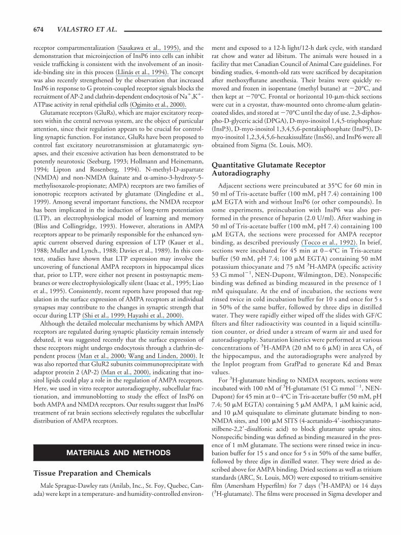
We first assessed the effects of incubating rat brain sections withincreasing InsP6 concentrations on the binding properties ofAMPA receptors. Horizontal sections were preincubated in Tris-acetate buffer in the presence of various InsP6 concentrations (1–300 mM), and 3H-AMPA binding was then measured by wipingoff the sections to evaluate the amount of specifically bound radio-activity. InsP6 produced a dose-dependent increase of specific 3H-AMPA binding in these rat brain sections, with a maximal eleva-tion of about 50% above control values at 100 mM InsP6 (solidcircles, Fig. 1A). Its effect was temperature-dependent, as it wasmarkedly reduced at room temperature (22°C; open circles, Fig.1A). At a concentration of 200 mM, InsP6 caused a 50 6 4%increase of specific 3H-AMPA binding in sections preincubated at35°C, while it had no action in sections preincubated at roomtemperature (4 6 4%; n 5 6, P , 0.05, Student’s t-test).
Brain sections were next preincubated with 200 mM InsP6 andsubjected to quantitative 3H-AMPA ligand binding autoradiogra-phy. Visual examination of autoradiography clearly showed amarked increase of 3H-AMPA binding in the hippocampus, cere-bellum, and other brain structures after InsP6 treatment (Fig. 1B).The rise in 3H-AMPA binding elicited by InsP6 preincubation wasdistributed uniformly in the various regions of the hippocampus(Fig. 1C). InsP6 produced a 20–30% augmentation of 3H-AMPAbinding in CA1 and CA3 strata radiatum and oriens as well as in thedentate gyrus. It also elicited a moderate increase of about 15% in3H-AMPA binding in the internal cortex and, among all the re-gions analyzed, the cerebellum showed the highest elevation (88%;see Fig. 1D). Saturation experiments at equilibrium were per-formed to determine whether the increase in 3H-AMPA bindingelicited by InsP6 was the result of alterations in receptor affinity ormaximal number. The saturation kinetics of binding in the strataradiatum of the CA1 region yielded a linear Scatchard plot, sug-gesting the existence of a single binding site (Fig. 2). In this region,the InsP6-induced increase in 3H-AMPA binding was not due to achange in affinity of AMPA receptors (Kd 5 145 6 26 nM incontrol vs. Kd 5 158 6 14 nM in InsP6-treated sections; means 6SEM of six experiments), but to a rise in the maximal number ofsites (Bmax 5 7.5 6 0.34 in control vs. 11.6* 6 0.3 pmol/mg ofprotein in InsP6-treated sections; means 6 SEM of six experi-ments, P , 0.01, Student’s t-test). It should be mentioned that, inthe present study, we failed to detect any high-affinity sites (Fig. 2).This was probably due to the preincubation of sections at 35°C;high-affinity AMPA-binding sites have been shown to representcytoplasmic receptors, and to be easily washed out by preincuba-tion of tissue sections at 35°C (Standley et al., 1998).
Ligand-binding autoradiography with 3H-glutamate was alsoused to test the effects of InsP6 on the binding properties ofNMDA receptors in various brain regions (Fig. 3A). As illustratedin Figure 3B,C, quantification and averaging of the data obtainedfrom several sections indicated that no significant changes in 3H-
__________________________________________________ INSP6 MODULATION OF AMPA RECEPTORS 675
glutamate binding were produced by InsP6 treatment in the vari-ous brain regions analyzed. Of course, these results suggest thatchanges in AMPA receptors are not the result of a chemical artifactof tissue preparation.
Subcellular Localization of GluR Subunits AfterInsP6 Treatment of Rat Brain Sections
Frozen-thawed brain sections (30 mm thick) were incubated at35°C in the presence and absence of 200 mM InsP6 for 60 min.After preincubation, they were homogenized and subjected to sub-cellular fractionation. Samples from crude mitochondrial/synaptic(P2) and microsomal (P3) fractions were immunoblotted withantibodies against the C-terminal domains of GluR1 or GluR2, asreported previously (Bi et al., 1997, 1998). Figure 4A shows thatincubation of rat brain sections with InsP6 resulted in an increaseof 105-kDa species of GluR1 and GluR2 subunits in the mito-chondrial/synaptic (P2) fraction. The mean (6 SEM) OD valuesfor both GluR1 (0.64 6 0.05 in control vs. 0.84 6 0.06 in InsP6-treated sections; n 5 8, P , 0.01) and GluR2 (0.47 6 0.05 incontrol vs. 0.61 6 0.05 in InsP6-treated sections; n 5 8, P , 0.01)
subunits were elevated by about 30% after InsP6 treatment of ratbrain sections. However, while the levels of these two subunitswere enhanced in the P2 fraction, they were reduced (also by about30%) in the microsomal (P3) fraction (Fig. 4A). It should bementioned that, in homogenates containing both P2 and P3 frac-tions, we failed to detect any changes in GluR1 subunits afterInsP6 treatment (0.52 6 0.06 in control vs. 0.55 6 0.04 in InsP6-treated sections; n 5 4), reinforcing the notion that apparent in-creases in GluR subunits observed in P2 fractions are due to cor-responding reductions in P3 fractions. Another set of experimentswas conducted using anti-NR1 and anti-NR2A antibodies to testwhether NMDA receptors were affected by InsP6 treatment of ratbrain sections. Consistent with the binding experiments, no sig-nificant differences in the intensity of NR1 or NR2A staining weredetected in mitochondrial/synaptic as well as microsomal fractionsprepared from control and InsP6-treated sections (Fig. 4B).
The cellular mechanisms for the regulation of AMPA recep-tor properties by InsP6 remain to be understood. InsP6 isknown to interact with adaptor proteins in the brain, and InsP6binding has been demonstrated to be inhibited by heparin
FIGURE 1. Modulation of AMPA receptor binding by InsP6. A:Adjacent horizontal sections at the level of the dorsal hippocampuswere preincubated at 35°C (solid circles) or 22°C (open circles) for 1 hwith increasing concentrations (1–300 mM) of InsP6. The resultsrepresent InsP6-induced modulation of 3H-AMPA binding and areexpressed as percent of basal binding determined in sections preincu-bated with buffer alone. The data are means 6 SEM of values ob-tained from at least 5 rats with three different measurements per rat.B: Computer-generated images of autoradiograms of 3H-AMPAbinding in rat brain sections preincubated at 35°C for 1 h in theabsence or presence of 200 mM InsP6. C: Autoradiographs like thoseshown in B were analyzed with an image analysis system for the
various hippocampal regions (OR, stratum oriens; RAD, stratum ra-diatum; DG, molecular layer of the dentate gyrus). The results ob-tained for the different regions of control (open bars) and InsP6-treated (200 mM; solid bars) sections are expressed in pmol/mg ofprotein. D: Same as C, but representing 3H-AMPA binding to non-hippocampal structures (CTX, inner layers III–VI of the parieto-temporal cortex; STRIA, striatum; MOL, molecular layer of the cer-ebellum; GRAN, granular layer of the cerebellum; THAL, thalamicnuclei). The data are means 6 SEM of values obtained from at least 6rats with at least eight different measurements per rat for each of thecerebral regions examined. *P < 0.05, **P < 0.01 (Scheffe’s test).InsP6-treated vs. control sections.
676 VALASTRO ET AL.

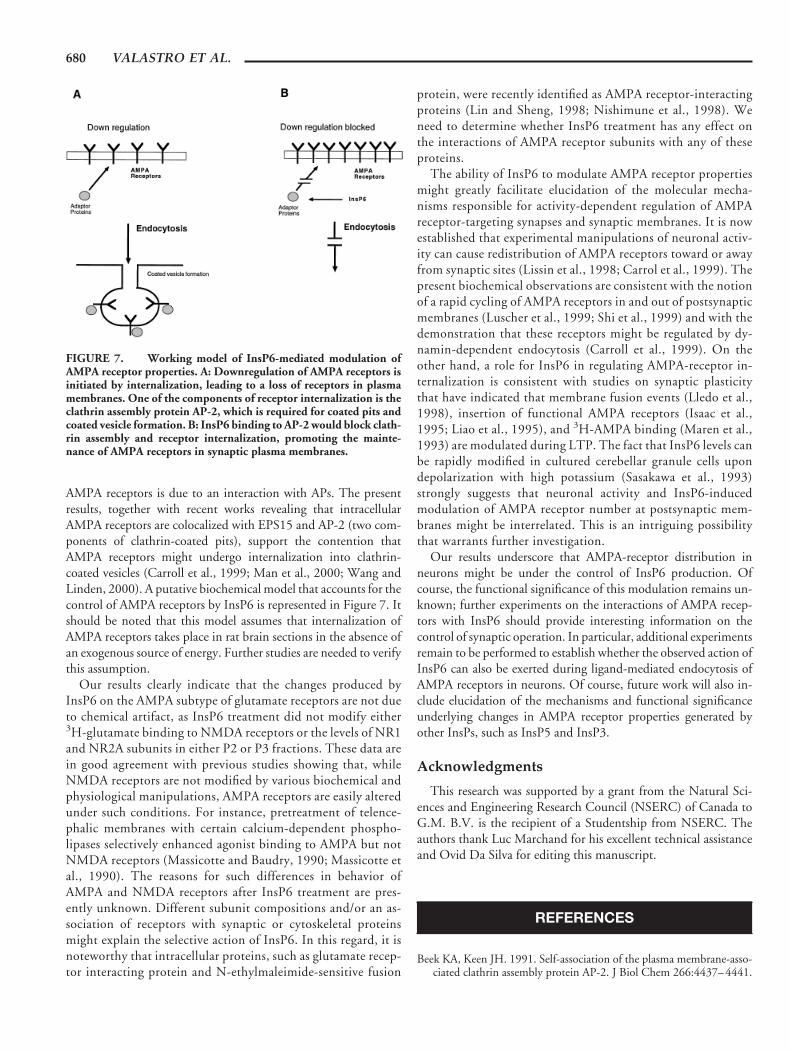
(Theibert et al., 1991). Therefore, we examined the effect ofheparin on InsP6-induced modulation of AMPA receptor prop-erties. Preincubation of horizontal sections with heparin (2U/ml) significantly reduced the InsP6-mediated increase of 3H-AMPA binding (Fig. 5A). In addition, heparin prevented theInsP6-induced elevation of GluR1 subunits of AMPA receptorsin synaptic membranes (Fig. 5B). The effect of InsP6 on GluR1subunits was also abolished by pretreatment of rat sections witha hypertonic medium (350 mM glucose) and was not repro-duced by a similar concentration of InsS6 that binds with lowaffinity to clathrin adaptor protein, suggesting that it requiresclathrin-dependent processes (see Fig. 6A).
We also examined the effects of InsP6 treatment of rat brainsections on Triton X-100 solubility of GluR1 subunits in crudesynaptic membrane (P2) preparations. In agreement with previousreports (Wenthold et al., 1996; Lu et al., 1999), levels of TritonX-100-insoluble GluR1 subunits represented about 20% of thoseoccurring in P2 fractions (Fig. 5C). Triton-insoluble AMPA re-ceptors possibly represent receptors incorporated in PSDs, as thepattern of proteins observed with Coomassie blue staining of Tri-ton-insoluble fractions was very similar to that reported by othersusing standard PSD preparation procedures, with clear identifica-tion of spectrin, actin, and CaMKII (data not shown). InsP6-induced elevation of GluR1 subunit levels was also observed inTriton-insoluble fractions (Fig. 5C), suggesting that InsP6 plays arole in regulating AMPA receptors at PSDs.
Effects of Other Inositol Phosphates on AMPAReceptors
It might be argued that InsPs produced by InsP6 dephosphor-ylation during the preincubation period can account for the ob-served modulation of AMPA receptors by InsP6. Therefore, weexamined the effect of several InsPs (InsP5, InsP3, and DPGA) onGluR1 subunit levels in P2 fractions. As shown in Figure 6, West-ern blotting experiments indicated a dose-dependent increase ofGluR1 immunoreactivity in P2 fractions, with a maximal augmen-tation to about 40% of control values at 200 mM InsP6. This effectof InsP6 on GluR1 appeared to be relatively specific, since it wasnot reproduced by InsS6 and other phosphate compounds such asInsP5, InsP3, and DPGA. In fact, our immunoblot experimentsrevealed that both InsP5 and InsP3 are capable of reducing GluR1
FIGURE 2. Effect of InsP6 on binding properties of AMPA re-ceptors in the hippocampus formation. Scatchard plots of 3H-AMPAbinding in the stratum radiatum of the CA1 region of sections prein-cubated without (open squares) and with (solid squares) 200 mMInsP6. 3H-AMPA binding was determined using concentrations from20 nM to 6 mM, and autoradiography was performed as described inMaterials and Methods. The data are presented as Scatchard plots(B/F, bound/free) and are analyzed by the Inplot program from Graf-Pad to generate Kd and Bmax; Scatchard plots are the results ofexperiments performed in 5 different animals.
FIGURE 3. Effect of InsP6 on NMDA receptor binding. A: Com-puter-generated images of autoradiograms of 3H-glutamate bindingto NMDA receptor sites in rat brain sections preincubated at 35°C for1 h in the absence or presence of 200 mM InsP6. B: Autoradiographslike those shown in A were analyzed with an image analysis system forthe various hippocampal regions (OR, stratum oriens; RAD, stratumradiatum; DG, molecular layer of dentate gyrus). The results obtainedfor the different regions of control (open bars) and InsP6-treated (200mM; solid bars) sections are expressed in pmol/mg of protein. C: Sameas B but represent 3H-glutamate binding to nonhippocampal struc-tures (CTX, inner layers III–VI of the parieto-temporal cortex;STRIA, striatum; MOL, molecular layer of the cerebellum; GRAN,granular layer of the cerebellum; THAL, thalamic nuclei). The dataare means 6 SEM of values obtained from at least 6 rats with at leasteight different measurements per rat for each of the cerebral regionsexamined.
__________________________________________________ INSP6 MODULATION OF AMPA RECEPTORS 677
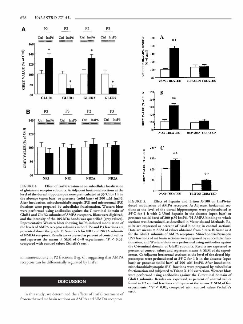
immunoreactivity in P2 fractions (Fig. 6), suggesting that AMPAreceptors can be differentially regulated by InsPs.
DISCUSSION
In this study, we determined the effects of InsP6 treatment offrozen-thawed rat brain sections on AMPA and NMDA receptors.
FIGURE 4. Effect of InsP6 treatment on subcellular localizationof glutamate receptor subunits. A: Adjacent horizontal sections at thelevel of the dorsal hippocampus were preincubated at 35°C for 1 h inthe absence (open bars) or presence (solid bars) of 200 mM InsP6.After incubation, mitochondrial/synaptic (P2) and microsomal (P3)fractions were prepared by subcellular fractionation. Western blotswere performed using antibodies against the C-terminal domain ofGluR1 and GluR2 subunits of AMPA receptors. Blots were digitized,and the intensity of the 105-kDa bands was quantified (grey values).Representative Western blots showing InsP6-induced modulation ofthe levels of AMPA receptor subunits in both P2 and P3 fractions arepresented above the graph. B: Same as A for NR1 and NR2A subunitsof NMDA receptors. Results are expressed as percent of control valuesand represent the means 6 SEM of 6–8 experiments. *P < 0.05,compared with control values (Scheffe’s test).
FIGURE 5. Effect of heparin and Triton X-100 on InsP6-in-duced modulation of AMPA receptors. A: Adjacent horizontal sec-tions at the level of the dorsal hippocampus were preincubated at35°C for 1 h with 2 U/ml heparin in the absence (open bars) orpresence (solid bars) of 200 mM InsP6. 3H-AMPA binding to wholesections was determined, as described in Materials and Methods. Re-sults are expressed as percent of basal binding in control sections.Data are means 6 SEM of values obtained from 5 rats. B: Same as Afor the GluR1 subunits of AMPA receptors. Mitochondrial/synaptic(P2) fractions of rat brain sections were prepared by subcellular frac-tionation, and Western blots were performed using antibodies againstthe C-terminal domain of GluR1 subunits. Results are expressed aspercent of control values and represent means 6 SEM of six experi-ments. C: Adjacent horizontal sections at the level of the dorsal hip-pocampus were preincubated at 35°C for 1 h in the absence (openbars) or presence (solid bars) of 200 mM InsP6. After incubation,mitochondrial/synaptic (P2) fractions were prepared by subcellularfractionation and subjected to Triton X-100 extraction. Western blotswere performed using antibodies against the C-terminal domain ofGluR1 subunits. Results are expressed as percent of control valuesfound in P2 control fractions and represent the means 6 SEM of fiveexperiments. **P < 0.01, compared with control values (Scheffe’stest).
678 VALASTRO ET AL.

Western blot and ligand-binding analysis revealed that InsP6 treat-ment of brain sections selectively modulated AMPA receptor prop-erties. In particular, InsP6 produced an increase in the levels ofGluR1 and GluR2 subunits of AMPA receptors in crude mito-chondrial/synaptic (P2) fractions prepared from rat brain sections.In contrast, the amounts of these subunits were reduced in micro-somal (P3) fractions, suggesting that AMPA receptor distributionbetween intracellular and plasma membrane compartments wasaltered by InsP6 treatment. Quantitative autoradiography of 3H-AMPA binding similarly revealed that InsP6 treatment enhanced3H-AMPA binding sites in several regions, although modulation ofAMPA binding appeared to be different in various brain structures.Whether regional differences in the effects of InsP6 are due to
heterogeneity of AMPA receptors or to regional variations in theability of tissues to generate the InsP6 effect is not clear. Neverthe-less, the observation that AMPA receptor modulation by InsP6 wasmaximal in the hippocampus and cerebellum is entirely consistentwith the distribution of InsP6 binding sites in these brain regions(Hawkins et al., 1990; Parent and Quirion, 1994), raising thepossibility that endogenous InsP6 plays an important role in theregulation of AMPA receptors.
There are relatively few studies on the regulation of surfacereceptors by InsP6. With regard to the present investigation, Kar etal. (1994) reported an interaction between InsP6 and insulin-likegrowth factor receptor II (IGF II) in rat brain sections. InsP6 alsoreduced light-induced desensitization of rhodopsin (Palczewski etal., 1992), presumably through direct binding to arrestin (Palczew-ski et al., 1991), suggesting that InsP6 participates in the control ofG-protein-coupled receptors. In agreement with this prediction,agonist-induced desensitization of substance P receptors wasblocked by InsP6 in Xenopus oocytes (Sasakawa et al., 1994).AMPA receptors, originally identified as classical ionotropic recep-tors, were recently found to exhibit metabotropic activity. In par-ticular, AMPA-receptor stimulation reduced both pertussis toxin-induced ADP ribolysation of Gai1 and forskolin-induced cAMPelevation in cultured cortical neurons and membrane preparations(Wang et al., 1997). Interestingly, Gai1 coimmunoprecipitatedwith GluR1 in AMPA-treated cultures but not in control cultures,suggesting the involvement of ionotropic AMPA receptors inmetabotropic signaling cascades. The mechanism of interactionbetween AMPA receptors and G proteins is still unclear, althoughan AP might be involved. One indication that this idea might betrue is the recent observation that GluR2 subunits coimmunopre-cipitated with AP (Man et al., 2000).
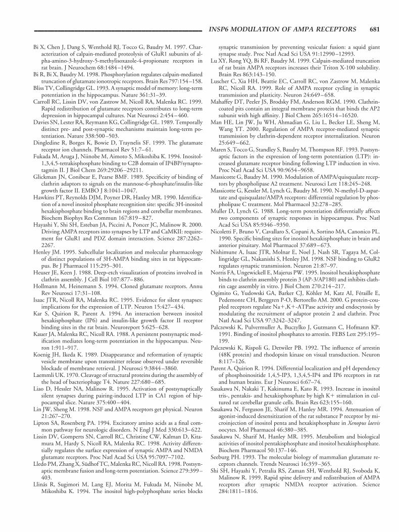
The recognized interaction of InsP6 with AP-2 calls attention tothe possibility that InsP6 might contribute to the regulation ofAMPA receptor levels in synaptic membranes. It is well-recognizedthat AP-2 binds to the cytoplasmic tail of a number of membranereceptors and is involved in the internalization of neurotransmitterreceptors. In particular, AP-2 is known to interact with receptorsfor IGF II and to mediate the endocytosis of IGF II as well as othersignaling peptides, such as transforming growth factor b (Glick-man et al., 1989; Kar et al., 1994). Once recruited to the innersurface of the plasma membrane, AP-2 is likely to initiate theformation of clathrin-coated pits by triggering the assembly ofclathrin triskelion subunits into a polygonal lattice that causesbending of the membrane into the coated pit structure (Heuserand Keen, 1988; Mahaffey et al., 1990). Clathrin-coated pits de-tach from the plasma membrane by a GTP-dependent fission re-action mediated by the GTPase dynamin, and the resulting coatedmembrane vesicles become internalized (Koenig and Ikeda, 1989).InsP6, by interacting with AP-2, inhibits clathrin assembly andvesicle trafficking, suggesting its influence on receptor compart-mentalization (Sasakawa et al., 1995). In the present investigation,the levels of some GluR subunits were increased in crude mito-chondrial/synaptic (P2) fractions and decreased in microsomal(P3) fractions following InsP6 treatment. As predicted, heparinwas found to block the InsP6-induced change of AMPA receptorcompartmentalization, suggesting that InsP6 modulation of
FIGURE 6. Effects of various phosphate compounds and InsS6on GluR1 subunits. A: Adjacent horizontal sections at the level of thedorsal hippocampus were preincubated at 35°C for 1 h in the absenceor presence of increasing concentrations (3–300 mM) of InsP6 (solidcircles), InsS6 (closed squares), and DPGA (solid triangles). B: Sameas A for sections incubated with 100 mM of various phosphate com-pounds and InsS6. Mitochondrial/synaptic (P2) fractions of rat brainsections were prepared by subcellular fractionation, and Westernblots were performed using antibodies against the C-terminal domainof GluR1 subunits. Results are expressed as percent of control valuesfound in P2 control fractions and represent the means 6 SEM of fiveexperiments.
__________________________________________________ INSP6 MODULATION OF AMPA RECEPTORS 679
AMPA receptors is due to an interaction with APs. The presentresults, together with recent works revealing that intracellularAMPA receptors are colocalized with EPS15 and AP-2 (two com-ponents of clathrin-coated pits), support the contention thatAMPA receptors might undergo internalization into clathrin-coated vesicles (Carroll et al., 1999; Man et al., 2000; Wang andLinden, 2000). A putative biochemical model that accounts for thecontrol of AMPA receptors by InsP6 is represented in Figure 7. Itshould be noted that this model assumes that internalization ofAMPA receptors takes place in rat brain sections in the absence ofan exogenous source of energy. Further studies are needed to verifythis assumption.
Our results clearly indicate that the changes produced byInsP6 on the AMPA subtype of glutamate receptors are not dueto chemical artifact, as InsP6 treatment did not modify either3H-glutamate binding to NMDA receptors or the levels of NR1and NR2A subunits in either P2 or P3 fractions. These data arein good agreement with previous studies showing that, whileNMDA receptors are not modified by various biochemical andphysiological manipulations, AMPA receptors are easily alteredunder such conditions. For instance, pretreatment of telence-phalic membranes with certain calcium-dependent phospho-lipases selectively enhanced agonist binding to AMPA but notNMDA receptors (Massicotte and Baudry, 1990; Massicotte etal., 1990). The reasons for such differences in behavior ofAMPA and NMDA receptors after InsP6 treatment are pres-ently unknown. Different subunit compositions and/or an as-sociation of receptors with synaptic or cytoskeletal proteinsmight explain the selective action of InsP6. In this regard, it isnoteworthy that intracellular proteins, such as glutamate recep-tor interacting protein and N-ethylmaleimide-sensitive fusion
protein, were recently identified as AMPA receptor-interactingproteins (Lin and Sheng, 1998; Nishimune et al., 1998). Weneed to determine whether InsP6 treatment has any effect onthe interactions of AMPA receptor subunits with any of theseproteins.
The ability of InsP6 to modulate AMPA receptor propertiesmight greatly facilitate elucidation of the molecular mecha-nisms responsible for activity-dependent regulation of AMPAreceptor-targeting synapses and synaptic membranes. It is nowestablished that experimental manipulations of neuronal activ-ity can cause redistribution of AMPA receptors toward or awayfrom synaptic sites (Lissin et al., 1998; Carrol et al., 1999). Thepresent biochemical observations are consistent with the notionof a rapid cycling of AMPA receptors in and out of postsynapticmembranes (Luscher et al., 1999; Shi et al., 1999) and with thedemonstration that these receptors might be regulated by dy-namin-dependent endocytosis (Carroll et al., 1999). On theother hand, a role for InsP6 in regulating AMPA-receptor in-ternalization is consistent with studies on synaptic plasticitythat have indicated that membrane fusion events (Lledo et al.,1998), insertion of functional AMPA receptors (Isaac et al.,1995; Liao et al., 1995), and 3H-AMPA binding (Maren et al.,1993) are modulated during LTP. The fact that InsP6 levels canbe rapidly modified in cultured cerebellar granule cells upondepolarization with high potassium (Sasakawa et al., 1993)strongly suggests that neuronal activity and InsP6-inducedmodulation of AMPA receptor number at postsynaptic mem-branes might be interrelated. This is an intriguing possibilitythat warrants further investigation.
Our results underscore that AMPA-receptor distribution inneurons might be under the control of InsP6 production. Ofcourse, the functional significance of this modulation remains un-known; further experiments on the interactions of AMPA recep-tors with InsP6 should provide interesting information on thecontrol of synaptic operation. In particular, additional experimentsremain to be performed to establish whether the observed action ofInsP6 can also be exerted during ligand-mediated endocytosis ofAMPA receptors in neurons. Of course, future work will also in-clude elucidation of the mechanisms and functional significanceunderlying changes in AMPA receptor properties generated byother InsPs, such as InsP5 and InsP3.
Acknowledgments
This research was supported by a grant from the Natural Sci-ences and Engineering Research Council (NSERC) of Canada toG.M. B.V. is the recipient of a Studentship from NSERC. Theauthors thank Luc Marchand for his excellent technical assistanceand Ovid Da Silva for editing this manuscript.
REFERENCES
Beek KA, Keen JH. 1991. Self-association of the plasma membrane-asso-ciated clathrin assembly protein AP-2. J Biol Chem 266:4437–4441.
FIGURE 7. Working model of InsP6-mediated modulation ofAMPA receptor properties. A: Downregulation of AMPA receptors isinitiated by internalization, leading to a loss of receptors in plasmamembranes. One of the components of receptor internalization is theclathrin assembly protein AP-2, which is required for coated pits andcoated vesicle formation. B: InsP6 binding to AP-2 would block clath-rin assembly and receptor internalization, promoting the mainte-nance of AMPA receptors in synaptic plasma membranes.
680 VALASTRO ET AL.
Bi X, Chen J, Dang S, Wenthold RJ, Tocco G, Baudry M. 1997. Char-acterization of calpain-mediated proteolysis of GluR1 subunits of al-pha-amino-3-hydroxy-5-methylisoxazole-4-propionate receptors inrat brain. J Neurochem 68:1484–1494.
Bi R, Bi X, Baudry M. 1998. Phosphorylation regulates calpain-mediatedtruncation of glutamate ionotropic receptors. Brain Res 797:154–158.
Bliss TV, Collingridge GL. 1993. A synaptic model of memory: long-termpotentiation in the hippocampus. Nature 361:31–39.
Carroll RC, Lissin DV, von Zastrow M, Nicoll RA, Malenka RC. 1999.Rapid redistribution of glutamate receptors contributes to long-termdepression in hippocampal cultures. Nat Neurosci 2:454–460.
Davies SN, Lester RA, Reymann KG, Collingridge GL. 1989. Temporallydistinct pre- and post-synaptic mechanisms maintain long-term po-tentiation. Nature 338:500–503.
Dingledine R, Borges K, Bowie D, Traynelis SF. 1999. The glutamatereceptor ion channels. Pharmacol Rev 51:7–61.
Fukada M, Aruga J, Niinobe M, Aimoto S, Mikoshiba K. 1994. Inositol-1,3,4,5-tetrakisphosphate binding to C2B domain of IP4BP/synapto-tagmin II. J Biol Chem 269:29206–29211.
Glickman JN, Conibear E, Pearse BMF. 1989. Specificity of binding ofclathrin adaptors to signals on the mannose-6-phosphate/insulin-likegrowth factor II. EMBO J 8:1041–1047.
Hawkins PT, Reynolds DJM, Poyner DR, Hanley MR. 1990. Identifica-tion of a novel inositol phosphate recognition site: specific 3H-inositolhexakisphosphate binding to brain regions and cerebellar membranes.Biochem Biophys Res Commun 167:819–827.
Hayashi Y, Shi SH, Esteban JA, Piccini A, Poncer JC, Malinow R. 2000.Driving AMPA receptors into synapses by LTP and CaMKII: require-ment for GluR1 and PDZ domain interaction. Science 287:2262–2267.
Henley JM. 1995. Subcellular localization and molecular pharmacologyof distinct populations of 3H-AMPA binding sites in rat hippocam-pus. Br J Pharmacol 115:295–301.
Heuser JE, Keen J. 1988. Deep-etch visualization of proteins involved inclathrin assembly. J Cell Biol 107:877–886.
Hollmann M, Heinemann S. 1994. Cloned glutamate receptors. AnnuRev Neurosci 17:31–108.
Isaac JTR, Nicoll RA, Malenka RC. 1995. Evidence for silent synapses:implications for the expression of LTP. Neuron 15:427–434.
Kar S, Quirion R, Parent A. 1994. An interaction between inositolhexakisphosphate (IP6) and insulin-like growth factor II receptorbinding sites in the rat brain. Neuroreport 5:625–628.
Kauer JA, Malenka RC, Nicoll RA. 1988. A persistent postsynaptic mod-ification mediates long-term potentiation in the hippocampus. Neu-ron 1:911–917.
Koenig JH, Ikeda K. 1989. Disappearance and reformation of synapticvesicle membrane upon transmitter release observed under reversibleblockade of membrane retrieval. J Neurosci 9:3844–3860.
Laemmli UK. 1970. Cleavage of structural proteins during the assembly ofthe head of bacteriophage T4. Nature 227:680–685.
Liao D, Hessler NA, Malinow R. 1995. Activation of postsynapticallysilent synapses during pairing-induced LTP in CA1 region of hip-pocampal slice. Nature 375:400–404.
Lin JW, Sheng M. 1998. NSF and AMPA receptors get physical. Neuron21:267–270.
Lipton SA, Rosenberg PA. 1994. Excitatory amino acids as a final com-mon pathway for neurologic disorders. N Engl J Med 330:613–622.
Lissin DV, Gomperts SN, Carroll RC, Christine CW, Kalman D, Kita-mura M, Hardy S, Nicoll RA, Malenka RC. 1998. Activity differen-tially regulates the surface expression of synaptic AMPA and NMDAglutamate receptors. Proc Natl Acad Sci USA 95:7097–7102.
Lledo PM, Zhang X, Sudhof TC, Malenka RC, Nicoll RA. 1998. Postsyn-aptic membrane fusion and long-term potentiation. Science 279:399–403.
Llinas R, Sugimori M, Lang EJ, Morita M, Fukuda M, Niinobe M,Mikoshiba K. 1994. The inositol high-polyphosphate series blocks
synaptic transmission by preventing vesicular fusion: a squid giantsynapse study. Proc Natl Acad Sci USA 91:12990–12993.
Lu XY, Rong YQ, Bi RF, Baudry M. 1999. Calpain-mediated truncationof rat brain AMPA receptors increases their Triton X-100 solubility.Brain Res 863:143–150.
Luscher C, Xia HH, Beattie EC, Carroll RC, von Zastrow M, MalenkaRC, Nicoll RA. 1999. Role of AMPA receptor cycling in synaptictransmission and plasticity. Neuron 24:649–658.
Mahaffey DT, Peeler JS, Brodsky FM, Anderson RGM. 1990. Clathrin-coated pits contain an integral membrane protein that binds the AP2subunit with high affinity. J Biol Chem 265:16514–16520.
Man HE, Lin JW, Ju WH, Ahmadian G, Liu L, Becker LE, Sheng M,Wang YT. 2000. Regulation of AMPA receptor-mediated synaptictransmission by clathrin-dependent receptor internalization. Neuron25:649–662.
Maren S, Tocco G, Standley S, Baudry M, Thompson RF. 1993. Postsyn-aptic factors in the expression of long-term potentiation (LTP): in-creased glutamate receptor binding following LTP induction in vivo.Proc Natl Acad Sci USA 90:9654–9658.
Massicotte G, Baudry M. 1990. Modulation of AMPA/quisqualate recep-tors by phospholipase A2 treatment. Neurosci Lett 118:245–248.
Massicotte G, Kessler M, Lynch G, Baudry M. 1990. N-methyl-D-aspar-tate and quisqualate/AMPA receptors: differential regulation by phos-pholipase C treatment. Mol Pharmacol 32:278–285.
Muller D, Lynch G. 1988. Long-term potentiation differentially affectstwo components of synaptic responses in hippocampus. Proc NatlAcad Sci USA 85:9346–9350.
Nicoletti F, Bruno V, Cavallaro S, Copani A, Sortino MA, Canonico PL.1990. Specific binding sites for inositol hexakisphosphate in brain andanterior pituitary. Mol Pharmacol 37:689–673.
Nishimune A, Isaac JTR, Molnar E, Noel J, Nash SR, Tagaya M, Col-lingridge GL, Nakanishi S, Henley JM. 1998. NSF binding to GluR2regulates synaptic transmission. Neuron 21:87–97.
Norris FA, Ungewickell E, Majerus PW. 1995. Inositol hexakisphosphatebinds to clathrin assembly protein 3 (AP-3/AP180) and inhibits clath-rin cage assembly in vitro. J Biol Chem 270:214–217.
Ogimito G, Yudowski GA, Barker CJ, Kohler M, Katz AI, Feraille E,Pedemonte CH, Berggren P-O, Bertorello AM. 2000. G protein-cou-pled receptors regulate Na1,K1-ATPase activity and endocytosis bymodulating the recruitment of adaptor protein 2 and clathrin. ProcNatl Acad Sci USA 97:3242–3247.
Palczewski K, Pulvermuller A, Buczylko J, Gutmann C, Hofmann KP.1991. Binding of inositol phosphates to arrestin. FEBS Lett 295:195–199.
Palczewski K, Rispoli G, Detwiler PB. 1992. The influence of arrestin(48K protein) and rhodopsin kinase on visual transduction. Neuron8:117–126.
Parent A, Quirion R. 1994. Differential localization and pH dependencyof phosphoinositide 1,4,5-IP3, 1,3,4,5-IP4 and IP6 receptors in ratand human brains. Eur J Neurosci 6:67–74.
Sasakawa N, Nakaki T, Kakinuma E, Kato R. 1993. Increase in inositoltris-, pentakis- and hexakisphosphate by high K1 stimulation in cul-tured rat cerebellar granule cells. Brain Res 623:155–160.
Sasakawa N, Ferguson JE, Sharif M, Hanley MR. 1994. Attenuation ofagonist-induced desensitization of the rat substance P receptor by mi-croinjection of inositol penta and hexakisphosphate in Xenopus laevisoocytes. Mol Pharmacol 46:380–385.
Sasakawa N, Sharif M, Hanley MR. 1995. Metabolism and biologicalactivities of inositol pentakisphosphate and inositol hexakisphosphate.Biochem Pharmacol 50:137–146.
Seeburg PH. 1993. The molecular biology of mammalian glutamate re-ceptors channels. Trends Neurosci 16:359–365.
Shi SH, Hayashi Y, Petralia RS, Zaman SH, Wenthold RJ, Svoboda K,Malinow R. 1999. Rapid spine delivery and redistribution of AMPAreceptors after synaptic NMDA receptor activation. Science284:1811–1816.
__________________________________________________ INSP6 MODULATION OF AMPA RECEPTORS 681
Standley S, Georges T, Naveed W, Baudry M. 1998. High- and low-affinity alpha-[3H] amino-3-hydroxy-5-methylisoxazole-4-propionicacid ([3H] AMPA) binding sites represent immature and mature formsof AMPA receptors and are composed of differentially glycosylatedsubunits. J Neurochem 70:2434–2445.
Theibert AB, Estevez VA, Ferris CD, Danoff SK, Barrow RK, PrestwichGD, Snyder SH. 1991. Inositol 1,3,4,5-tetrakisphosphate and inositolhexakisphosphate receptor proteins: isolation and characterizationfrom rat brain. Proc Natl Acad Sci USA 88:3165–3169.
Theibert AB, Estevez VA, Mourey RJ, Marecek JF, Barrow RK,Prestwich GD, Snyder SH. 1992. Photoaffinity labeling and char-acterization of isolated inositol 1,3,4,5-tetrakisphosphate- and ino-sitol hexakisphosphate-binding proteins. J Biol Chem 267:9071–9079.
Timerman AP, Mayrleitner MM, Lukas TJ, Chadwick CC, Saito A,Watterson DM, Schindler H, Fleischer S. 1992. Inositol polyphos-phate receptor and clathrin assembly protein AP-2 are related proteinsthat form potassium-selective ion channels in planar lipid bilayers.Proc Natl Acad Sci USA 89:8976–8980.
Tocco G, Massicotte G, Standley S, Thompson RF, Baudry M. 1992.Effect of temperature and calcium on the binding properties of the
AMPA receptor in frozen rat brain sections. Eur J Neurosci 4:1093–1103.
Towbin H, Staehelin T, Gordon J. 1979. Electrophoretic transfer of pro-teins from polyacrylamide gels to nitrocellulose sheet: procedure andsome applications. Proc Natl Acad Sci USA 76:4350–4354.
Voglmaier SM, Keen JH, Murphy JE, Ferris CD, Prestwich GD, SnyderSH, Theibert AB. 1992. Inositol hexakisphosphate receptor identifiedas the clathrin assembly protein AP-2. Biochem Biophys Res Commun187:158–163.
Wang YT, Linden DJ. 2000. Expression of cerebellar long-term depres-sion requires postsynaptic clathrin-mediated endocytosis. Neuron 25:635–647.
Wang YT, Small DL, Stanimirovic DB, Morley P, Durkin JP. 1997.AMPA receptor-mediated regulation of a Gi-protein in cortical neu-rons. Nature 389:502–504.
Wenthold RJ, Petralia RS, Blahos J II, Niedzielski AS. 1996. Evidence formultiple AMPA receptor complexes in hippocampal CA1/CA2 neu-rons. J Neurosci 16:1982–1989.
Ye W, Ali N, Bembenek ME, Shears SB, Lafer EM. 1995. Inhibition ofclathrin assembly by high affinity binding of specific inositol polyphos-phates to the synapse-specific clathrin assembly protein AP-3. J BiolChem 270:1564–1568.
682 VALASTRO ET AL.
Related Documents