Pharmaceutics 2022, 14, 770. https://doi.org/10.3390/pharmaceutics14040770 www.mdpi.com/journal/pharmaceutics Review Inorganic Nanoparticles in Bone Healing Applications Alexandra‐Cristina Burdușel 1 , Oana Gherasim 1,2 , Ecaterina Andronescu 1,3, *, Alexandru Mihai Grumezescu 1,3,4 and Anton Ficai 1,3 1 Department of Science and Engineering of Oxide Materials and Nanomaterials, Faculty of Applied Chemistry and Materials Science, University Politehnica of Bucharest, 1–7 Gheorghe Polizu Street, 011061 Bucharest, Romania; [email protected] (A.‐C.B.); [email protected] (O.G.); [email protected] (A.M.G.); [email protected] (A.F.) 2 Lasers Department, National Institute for Lasers, Plasma and Radiation Physics, 409 Atomiștilor Street, 077125 Magurele, Romania 3 Academy of Romanian Scientists, 3 Ilfov Street, 050044 Bucharest, Romania 4 Research Institute of the University of Bucharest—ICUB, University of Bucharest, 90–92 Panduri Road, 050657 Bucharest, Romania * Correspondence: [email protected] Abstract: Modern biomedicine aims to develop integrated solutions that use medical, biotechno‐ logical, materials science, and engineering concepts to create functional alternatives for the specific, selective, and accurate management of medical conditions. In the particular case of tissue engi‐ neering, designing a model that simulates all tissue qualities and fulfills all tissue requirements is a continuous challenge in the field of bone regeneration. The therapeutic protocols used for bone healing applications are limited by the hierarchical nature and extensive vascularization of osseous tissue, especially in large bone lesions. In this regard, nanotechnology paves the way for a new era in bone treatment, repair and regeneration, by enabling the fabrication of complex nanostructures that are similar to those found in the natural bone and which exhibit multifunctional bioactivity. This review aims to lay out the tremendous outcomes of using inorganic nanoparticles in bone healing applications, including bone repair and regeneration, and modern therapeutic strategies for bone‐related pathologies. Keywords: bone regeneration; inorganic nanoparticles; bioceramic nanoparticles; oxide nanopar‐ ticles; metallic nanoparticles 1. Introduction Bone is a dynamic tissue that is constantly renewed and repaired through its intrin‐ sic remodeling process, which involves interactions between resident cells (osteoclasts and osteoblasts) and signaling factors, that remove old and damaged tissue and create new bone, respectively [1,2]. This fine‐tuned synergy is responsible for the preservation of bone balance. The healing of bone fractures and the restoration of critical bone anom‐ alies are difficult tasks for orthopedics, traumatologists, and maxillofacial surgeons [3]. Given the specific patient‐related requirements and limitations in bone regeneration, the clinical use of synthetic bone substitutes represents one of the most important updates in bone regenerative therapy [4,5]. The current progress in nanotechnology‐derived bio‐ materials enables the development of bone implants that are osteoconductive and oste‐ oinductive, as well as biocompatible, biodegradable, and bioresorbable [6–8]. Nanobiomaterials include nanometer‐sized and nanostructured bioactive materials, which peculiar behavior and new properties strongly impact the emerging trends of modern biomedicine and biotechnology [9,10]. Nanostructured biomaterials possess improved and superior bone regeneration ability thanks to their particular physico‐ chemical properties and biological behavior, which are quite different from their bulk Citation: Burdușel, A.‐C.; Gherasim, O.; Andronescu, E.; Grumezescu, A.M.; Ficai, A. Inorganic Nanoparticles in Bone Healing Applications. Pharmaceutics 2022, 14, 770. https://doi.org/ 10.3390/pharmaceutics14040770 Academic Editors: Denis V. Voronin, Tatiana N. Borodina and Yulia I. Svenskaya Received: 24 February 2022 Accepted: 28 March 2022 Published: 31 March 2022 Publisher’s Note: MDPI stays neu‐ tral with regard to jurisdictional claims in published maps and insti‐ tutional affiliations. Copyright: © 2022 by the authors. Submitted for possible open access publication under the terms and conditions of the Creative Commons Attribution (CC BY) license (https://creativecommons.org/license s/by/4.0/).

Welcome message from author
This document is posted to help you gain knowledge. Please leave a comment to let me know what you think about it! Share it to your friends and learn new things together.
Transcript
Pharmaceutics 2022, 14, 770. https://doi.org/10.3390/pharmaceutics14040770 www.mdpi.com/journal/pharmaceutics
Review
Inorganic Nanoparticles in Bone Healing Applications
Alexandra‐Cristina Burdușel 1, Oana Gherasim 1,2, Ecaterina Andronescu 1,3,*, Alexandru Mihai Grumezescu 1,3,4
and Anton Ficai 1,3
1 Department of Science and Engineering of Oxide Materials and Nanomaterials,
Faculty of Applied Chemistry and Materials Science, University Politehnica of Bucharest,
1–7 Gheorghe Polizu Street, 011061 Bucharest, Romania; [email protected] (A.‐C.B.);
[email protected] (O.G.); [email protected] (A.M.G.); [email protected] (A.F.) 2 Lasers Department, National Institute for Lasers, Plasma and Radiation Physics, 409 Atomiștilor Street,
077125 Magurele, Romania 3 Academy of Romanian Scientists, 3 Ilfov Street, 050044 Bucharest, Romania 4 Research Institute of the University of Bucharest—ICUB, University of Bucharest, 90–92 Panduri Road,
050657 Bucharest, Romania
* Correspondence: [email protected]
Abstract: Modern biomedicine aims to develop integrated solutions that use medical, biotechno‐
logical, materials science, and engineering concepts to create functional alternatives for the specific,
selective, and accurate management of medical conditions. In the particular case of tissue engi‐
neering, designing a model that simulates all tissue qualities and fulfills all tissue requirements is a
continuous challenge in the field of bone regeneration. The therapeutic protocols used for bone
healing applications are limited by the hierarchical nature and extensive vascularization of osseous
tissue, especially in large bone lesions. In this regard, nanotechnology paves the way for a new era
in bone treatment, repair and regeneration, by enabling the fabrication of complex nanostructures
that are similar to those found in the natural bone and which exhibit multifunctional bioactivity.
This review aims to lay out the tremendous outcomes of using inorganic nanoparticles in bone
healing applications, including bone repair and regeneration, and modern therapeutic strategies
for bone‐related pathologies.
Keywords: bone regeneration; inorganic nanoparticles; bioceramic nanoparticles; oxide nanopar‐
ticles; metallic nanoparticles
1. Introduction
Bone is a dynamic tissue that is constantly renewed and repaired through its intrin‐
sic remodeling process, which involves interactions between resident cells (osteoclasts
and osteoblasts) and signaling factors, that remove old and damaged tissue and create
new bone, respectively [1,2]. This fine‐tuned synergy is responsible for the preservation
of bone balance. The healing of bone fractures and the restoration of critical bone anom‐
alies are difficult tasks for orthopedics, traumatologists, and maxillofacial surgeons [3].
Given the specific patient‐related requirements and limitations in bone regeneration, the
clinical use of synthetic bone substitutes represents one of the most important updates in
bone regenerative therapy [4,5]. The current progress in nanotechnology‐derived bio‐
materials enables the development of bone implants that are osteoconductive and oste‐
oinductive, as well as biocompatible, biodegradable, and bioresorbable [6–8].
Nanobiomaterials include nanometer‐sized and nanostructured bioactive materials,
which peculiar behavior and new properties strongly impact the emerging trends of
modern biomedicine and biotechnology [9,10]. Nanostructured biomaterials possess
improved and superior bone regeneration ability thanks to their particular physico‐
chemical properties and biological behavior, which are quite different from their bulk
Citation: Burdușel, A.‐C.;
Gherasim, O.; Andronescu, E.;
Grumezescu, A.M.; Ficai, A.
Inorganic Nanoparticles in Bone
Healing Applications. Pharmaceutics
2022, 14, 770. https://doi.org/
10.3390/pharmaceutics14040770
Academic Editors: Denis V. Voronin,
Tatiana N. Borodina and Yulia I.
Svenskaya
Received: 24 February 2022
Accepted: 28 March 2022
Published: 31 March 2022
Publisher’s Note: MDPI stays neu‐
tral with regard to jurisdictional
claims in published maps and insti‐
tutional affiliations.
Copyright: © 2022 by the authors.
Submitted for possible open access
publication under the terms and
conditions of the Creative Commons
Attribution (CC BY) license
(https://creativecommons.org/license
s/by/4.0/).
Pharmaceutics 2022, 14, 770 2 of 41
counterparts [11,12]. During the last decade, various nanoparticle‐based protocols have
been successfully evaluated for the diagnosis and targeted treatment of orthotopic and
metastatic bone cancers [13,14]. The size of nanoparticles (NPs, 1–100 nm size range)
permits their passage through biological barriers, while their size‐related features (in‐
cluding a high surface area‐to‐volume ratio, surface energy and reactivity, mechanical,
thermal, optical, electrical and magnetic properties governed by quantum effects, and
intrinsic biological activity) enable them to attain significant therapeutic efficacy [15,16].
Moreover, nanoengineered platforms may increase drug solubility and improve drug
bioavailability, but also enhance pharmacokinetics and pharmacodynamics, and provide
specific and selective targeted and/or controlled therapeutic effects [17,18].
With the aim to overcome the drawbacks of classical restorative and replacement
procedures of hard tissues (including herein the limited bioavailability and increased
immunogenicity of autografts and allografts, but also the bioinertness and limited bioac‐
tivity of clinically approved biomaterials) [19,20], an impressive amount of progress has
been reported in the development of bone regeneration materials during the last few
decades. Biomaterials for hard tissue engineering applications include the following
categories: (i) first‐generation biomaterials—prosthetic devices made from biologically
inert materials, such as metals and alloys, certain synthetic polymers, and bioceramics;
(ii) second‐generation biomaterials—osteoconductive and osteoinductive devices made
from bioactive, biodegradable, and bioresorbable materials, such as calcium phosphates,
bioactive glasses, and polyesters; and (iii) third‐generation biomaterials—advanced and
multifunctional biomaterials with osteogenic properties and the ability to regulate the
body’s functions [21–23].
As the size‐related behavior of NPs is also responsible for the occurrence of circum‐
stantial toxic effects, a real challenge consists in maximizing their therapeutic effects by
properly tuning the biocompatibility/multifunctionality balance. Nanosized particles can
invade surrounding cells or tissues, and they frequently cluster or migrate inside blood
vessels, causing additional damage to distant tissues or organs [24,25]. The toxicity of
nanoparticles is determined by various parameters, including shape, size, composition,
porosity, surface chemistry and coating, but other factors—such as the aggregation state
and interactions with biomolecules—may influence their toxicity in humans [26,27].
Nanoparticle‐based delivery systems have many advantages over conventional
pharmaceutical formulations. These include reduced side effects, enhanced therapeutic
effects, prolonged circulation half‐life, improved permeability, and patient compliance
[28,29]. Designing and developing performance‐enhanced platforms for targeted or
non‐targeted drug delivery generally implies the precise selection of the nanocarrier,
which can be (i) inorganic, including quantum dots (semiconductor‐based nanoparticles),
metallic (noble metals) and oxide nanoparticles, or (ii) organic, including carbon‐based
nanostructures, such as polymers, dendrimers, exosomes, micelles, liposomes, and solid
lipid NPs [30,31].
Thanks to their high surface reactivity, unique surface physics and chemistry, in‐
creased chemical stability and photostability, facile surface modification, quantum yields,
improved bioavailability, reduced or absent intrinsic toxicity, extended lifetime, great
drug‐loading capacity, and controlled drug release ability, inorganic NPs have indis‐
putable advantages as active therapeutic carriers [32,33]. Moreover, by coating the inor‐
ganic NPs with additional surface ligands (i.e., proteins, peptides, carbohydrates, etc.),
higher reactivity and enhanced functionality can be achieved [34,35]. In general,
nanocarriers based on inorganic NPs consist of an inorganic core (metal‐/oxide‐based
nanostructures) and an organic shell (carbon‐based compounds, which serve as sub‐
strates for bio‐macromolecular conjugation and/or as shields that protect the inner core
from undesirable physicochemical interactions with the biological microenvironment)
[36,37]. Biocompatible nanomaterials based on pristine and metal‐doped calcium phos‐
phates [38–40], bioceramics [41,42] and vitroceramics [43,44], oxides (such as alumina,
Pharmaceutics 2022, 14, 770 3 of 41
ceria, silica, titania, and zirconia) [45–49], and metallic nanostructures [50–52] are exten‐
sively investigated for the unconventional management of bone tissue injuries.
This review aims to point out the significance of inorganic nanoparticles in bone
healing by including relevant and recent data on the NP‐based repair and regeneration of
bone tissue.
2. Bioceramic Nanoparticles
2.1. Hydroxyapatite
The conventional therapeutic strategy in bone grafting mainly includes the use of
allografts and autogenous grafts, and also different isolated or combined substitutes
based on calcium phosphate (CaP) materials [53,54]. CaP‐based nanoparticles have been
extensively investigated in preclinical and clinical studies as bone graft alternatives
[55,56]. The use of CaP nanoparticles can be expanded towards cell‐/tissue‐specific drug
delivery platforms owing to their intrinsic features, such as unique biocompatibility and
bioactivity, high adsorptive capacity, composition‐/microstructure‐related tunable
properties, and application‐related adjustable biodegradability [57,58].
Particularly successful and promising outcomes in designing biomaterials for hard
tissue repair and replacement are related to synthetic hydroxyapatite (HA),
Ca10(PO4)6(OH)2 [58,59]. Naturally, HA is present in metamorphic and igneous rocks as a
natural mineral, but it is also present in teeth and bones as the major inorganic compo‐
nent [60,61]. Tremendous interest has been lately oriented towards the revaluation of
naturally‐derived HA, which can be extracted from sustainable biogenic sources or
wastes [62–65]. Representative sources for extracting natural HA include: (i) mammalian
sources—bovine [66–68], ovine [69,70], and swine [71,72] bones; (ii) marine or aquatic
sources—fish bones [72–74], cuttlefish bones [75,76], and corals [77,78]; (iii) shells—cockle
shell [79,80], clam shell [81,82], mussel shell [83,84], snail shell [85,86], and egg shell
[87,88]; and (iv) mineral sources [89,90].
Nanosized HA particles have more unique properties than micro‐sized HA parti‐
cles. For example, it has been reported that nanosized HA exhibits greater protein ad‐
sorption, improved cell adhesion, and superior bioactivity when compared to mi‐
cro‐sized HA [60,91]. It also possesses a significant capability to decrease apoptotic death
in healthy cells and, therefore, improve cell proliferation and cell activity related to bone
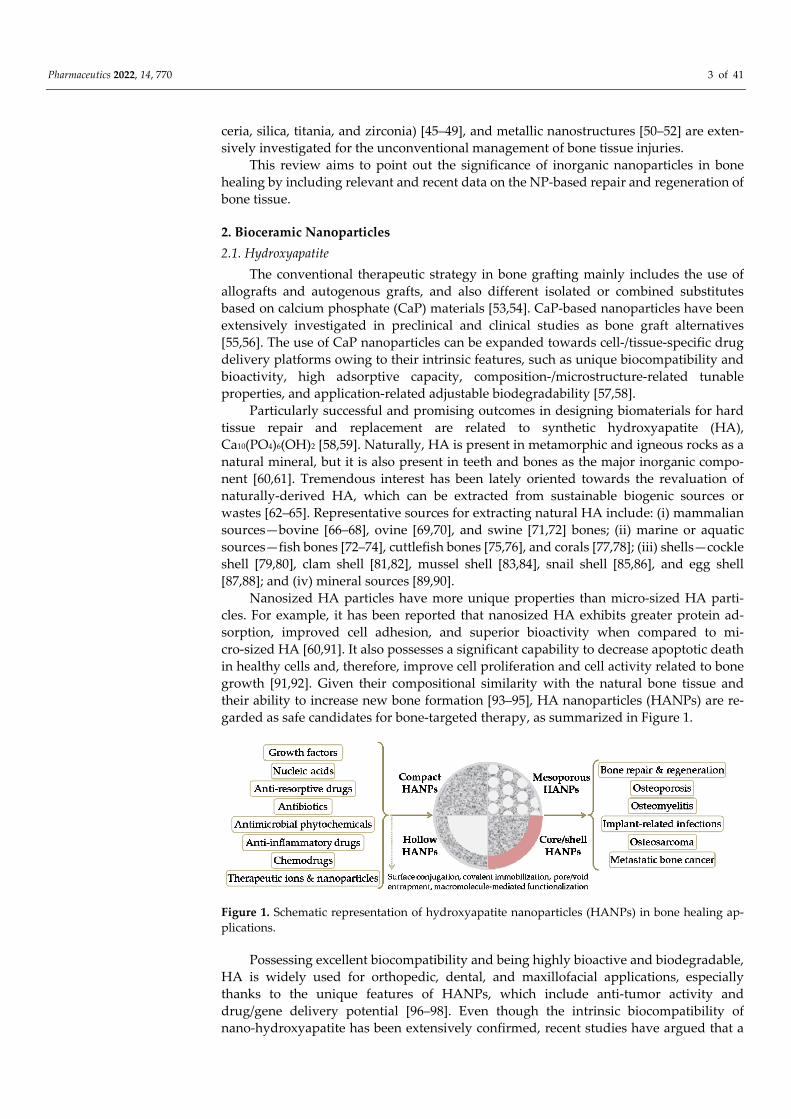
growth [91,92]. Given their compositional similarity with the natural bone tissue and
their ability to increase new bone formation [93–95], HA nanoparticles (HANPs) are re‐
garded as safe candidates for bone‐targeted therapy, as summarized in Figure 1.
Figure 1. Schematic representation of hydroxyapatite nanoparticles (HANPs) in bone healing ap‐
plications.
Possessing excellent biocompatibility and being highly bioactive and biodegradable,
HA is widely used for orthopedic, dental, and maxillofacial applications, especially
thanks to the unique features of HANPs, which include anti‐tumor activity and
drug/gene delivery potential [96–98]. Even though the intrinsic biocompatibility of
nano‐hydroxyapatite has been extensively confirmed, recent studies have argued that a
Pharmaceutics 2022, 14, 770 4 of 41
thorough screening of HANPs’ toxicity should be conducted to assess their biological
effects, as the potential biotoxicity of HANPs (affected by particle diameter, exposure
dose, and contact method) was reported [91,99].
Although HA is considered to be a suitable material for bone tissue repair and re‐
generation, its osteoinductive qualities are insufficient to allow large bone defects to
mend. To circumvent these drawbacks, several bioactive compounds including growth
factors that play a key role during the bone remodeling process, have been employed in
bone tissue engineering [100–104]. Osteoinductive growth factors have been utilized in
restorative and regenerative procedures for dental [7,105] and orthopedic (craniofacial,
spinal fusion and non‐union deformities) [54,106,107] pathologies, either alone or com‐
bined with ceramic and polymeric or composite materials, with little indication that they
are superior to autografts. Bone morphogenetic protein‐2 (BMP‐2) is the gold standard
growth factor for enhancing bone healing, and it has been successfully used in various
research studies. In terms of osteogenic activity and augmented bone healing, superior
results were reported for BMP‐2‐modified nanostructured formulations based on
HA/natural polymers [108,109] and HA/synthetic polymers [110,111]. However, due to
its short half‐life in vivo, the clinical applicability of BMP‐2 is limited, as a suitable
BMP‐2‐loaded bone substitute should accurately provide initial large doses and subse‐
quent constant therapeutic concentrations [112]. Promising HANP‐based formulations
for orthopedic and orthodontic applications have also been developed via modification
with other bone morphogenetic proteins (BMPs) [113,114], fibroblast growth factor (FGF)
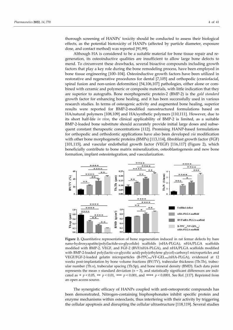
[101,115], and vascular endothelial growth factor (VEGF) [116,117] (Figure 2), which
beneficially contribute to bone matrix mineralization, osteoblastogenesis and new bone
formation, implant osteointegration, and vascularization.
Figure 2. Quantitative representation of bone regeneration induced in rat femur defects by bare
nano‐hydroxyapatite/poly(lactide‐co‐glycolide) scaffolds (nHA‐PLGA), nHA/PLGA scaffolds
modified with BMP‐2, VEGF, and FGF‐2 (BVF/nHA‐PLGA), and nHA/PLGA scaffolds modified
with BMP‐2‐loaded poly(lactic‐co‐glycolic acid)‐poly(ethylene glycol)‐carboxyl microparticles and
VEGF/FGF‐2‐loaded gelatin microparticles (B‐PPCmp/VF‐GELmp/nHA‐PLGA), evidenced at 12
weeks post‐implantation by bone volume fractions (BV/TV), trabecular thickness (Tb.Th), trabec‐
ular number (Tb.n), trabecular spacing (Tb.Sp), and bone mineral density (BMD). Each data point
represents the mean ± standard deviation (n = 3), and statistically significant differences are indi‐
cated as ∗ p < 0.05, ∗∗ p < 0.01, ∗∗∗ p < 0.001, and ∗∗∗∗ p < 0.0001. See Ref. [117]. Reprinted from
an open access source.
The synergistic efficacy of HANPs coupled with anti‐osteoporotic compounds has
been demonstrated. Nitrogen‐containing bisphosphonates inhibit specific protein and
enzyme mechanisms within osteoclasts, thus interfering with their activity by triggering
the cellular apoptosis and disrupting the cellular ultrastructure [118,119]. Several studies
Pharmaceutics 2022, 14, 770 5 of 41
evidenced the anti‐osteoporotic efficiency of HANP‐based materials modified with
alendronate [97,120], risedronate [121,122], and zoledronate [123,124]. By inhibiting os‐
teoclast‐mediated bone resorption, bisphosphonate‐modified HA‐based con‐
structs—such as coatings [125,126], scaffolds [109,127], and injectable formulations
[128,129]—determine a net improvement in osteogenic processes. Recently, HANPs
loaded with salmon calcitonin polypeptide were proposed for the sublingual manage‐
ment of osteoporosis [130]. Promising results were also evidenced for HA‐based bio‐
materials loaded with an anti‐resorptive agent (denosumab) [131] or an anabolic agent
(teriparatide) [132].
Following the development of the promising strontium ranelate (SR) (Prote‐
los®/Protos®, Servier Laboratories, Surene, France) anti‐osteoporotic drug, a variety of
studies have been conducted, ranging from strontium (Sr) mapping in bones and teeth to
investigating Sr incorporation into bone mineral (in particular, in the crystal surface and
lattice) and a decrease in calcium content, and to evaluating Sr effects in synthetic HA. Sr
has a dual positive effect during osteogenesis and bone remodeling, by boosting the de‐
velopment of pre‐osteoblastic cells, while suppressing the generation and functionality of
osteoclastic cells [133,134]. By gathering the distinctive advantages of HA and Sr, their
composites represent a suitable choice for the controlled and targeted therapy of bone
tissue [135–137].
Other studies revealed the significance of zinc (Zn)‐enriched HA nanomaterials for
the repair and regeneration of traumatic and osteoporotic bone tissues, as it has been
evidenced that Zn addition is beneficial for enhanced osteogenesis and the prevention of
osteoclast‐mediated resorption [138–140].
Selenium (Se) is a vital micronutrient for human health, as it plays an important role
in disease prevention and cellular pathophysiological balance maintenance. In this re‐
spect, Se‐modified HA nanomaterials proved to be promising alternatives for bone tissue
therapy, since the presence of Se synergistically determines enhanced cellular processes
in healthy cells (adhesion, migration, proliferation, and osteogenic differentiation)
[141,142] and significant apoptotic damage in cancerous cells [143,144].
In order to increase the structural integrity and to modulate interactions between the
biological microenvironment and inorganic nanostructures, the surface modification of
HA‐based nanomaterials was explored [145,146]. The hydroxyl‐abundant surface of HA
is responsible for beneficial interactions with organic compounds, resulting in surface
silanization and covalent bonding [147–149], immobilization and grafting [150–152].
Coupling natural [153,154] or synthetic [155,156] polymers onto the surface of HANPs
has been shown to improve the NPs’ colloidal stability and mechanical qualities, together
with their biofunctional outcomes. When used as bone‐filling materials, such composite
or hybrid structures can additionally act as active depots for the long‐term release of
pharmaceuticals, including drugs [157–159] and biomolecules [160–162].
Particular attention was oriented towards the fabrication of HANP‐modified poly‐
meric scaffolds, given the fact that a higher amount of nanoparticles triggers and accel‐
erates the nucleation of biomimetic apatite, finally resulting in increased bone formation
[146,163]. Designing HA/polymer constructs for bone tissue engineering requires ful‐
filling some essential aspects: (i) structural requirements: tissue‐mimicking composition
and architecture, adequate mechanical behavior, and highly porous interconnected
structure, which are responsible for the osteoconductive and osteoinductive outcomes, as
well as for proper cellular migration and normal development, oxygenation and nutri‐
tion, and vascularization; and (ii) biological requirements: biocompatibility, nontoxicity,
non‐immunogenicity, and biodegradability, which are vital aspects for enhanced osteo‐
genesis and host integration [164–166].
Owing to their excellent biodegradability and nontoxicity, and particular resem‐
blance with the natural extracellular matrix, natural polymers—such as proteins (e.g.,
collagen [109,167], gelatin [168,169], silk fibroin [170,171]) and polysaccharides (e.g., chi‐
tosan [172,173], cellulose [174,175], alginate [176,177])—are indisputable candidates for
Pharmaceutics 2022, 14, 770 6 of 41
bone healing applications. The modification of such scaffolds with HA‐based formula‐
tions represents an attractive strategy to overcome their intrinsic restrictions (improper
mechanical properties, uncontrollable degradability, immunogenicity, and microbial
contamination susceptibility).
In comparison with natural polymers, synthetic polyesters (e.g., polylactide (PLA)
[178,179], poly(lactide‐co‐glycolide) (PLGA) [180,181], polycaprolactone (PCL) [182,183],
and polyhydroxyalkanoates [184,185]) provide superior mechanical performance, in‐
creased chemical and structural stability, and tunable biodegradability. However, due to
their intrinsic limitations (including hydrophobicity, slower degradation rate, and prob‐
lematical metabolization/excretion of their byproducts), additional alterations are re‐
quired to fabricate superior HA‐modified bioactive scaffolds for bone healing.
As particular representatives of HANPs, mesoporous nanostructures have gained
great attention regarding the development of nanostructured platforms for the controlled
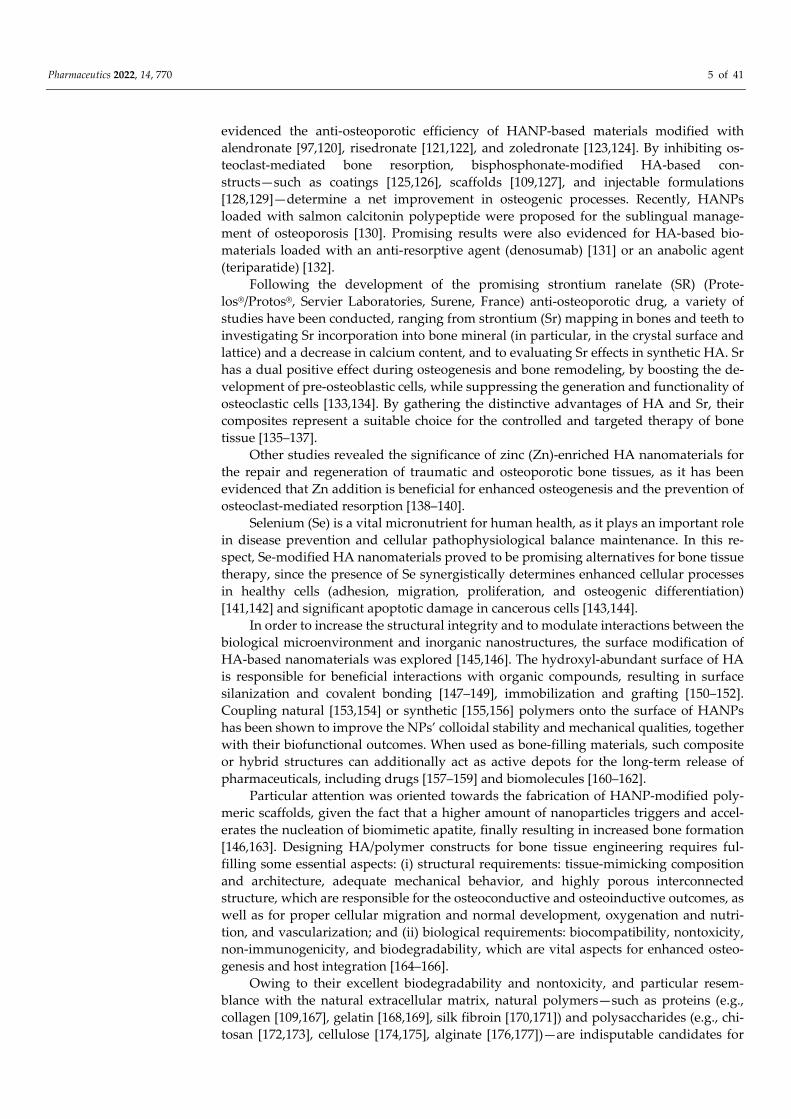
therapy of bone tissue [186,187]. It has been demonstrated that mesoporous HANPs
represent efficient nanocarriers for growth factors [188–190], antimicrobial ions [191,192]
(Figure 3), antibiotics [193,194], and anti‐tumor drugs [195–197], as a result of their uni‐
form, accessible, and highly organized porous microstructure.
Figure 3. Quantitative representation of mortality (death rate, %) in zebrafish embryos treated with
mesoporous fluoride‐doped nano‐hydroxyapatite (0.6, 1.2, and 3.2 at.% for FHAp‐1, FHAp‐2, and
FHAp‐3, respectively) with respect to time and concentration. The as‐developed FHAp nanorods
also exhibited important concentration‐dependent antibacterial effects against Pseudomonas aeru‐
ginosa and Bacillus subtilis. See Ref. [192]. Reprinted from an open access source.
HANP‐based therapeutic strategies have a lot of promise for bone tissue engineer‐
ing, which represents a complex and challenging research field of modern biomedicine
[198]. The characteristics of HA‐based nanomaterials can be accurately optimized during
the synthesis, in order to fabricate low‐cost and performance‐enhanced advanced bio‐
materials for therapeutic usage [199,200]. Nanofabrication techniques can provide precise
control over the physicochemical and microstructural features of HANPs, which are
mandatory for achieving spatial control over cell behavior, while imparting the necessary
structural properties [201,202].
2.2. Bioactive Glass
Bioactive glasses (BGs), with their indisputable and versatile silica‐based represent‐
atives, are amorphous solids which compositional and structural characteristics have
been proved beneficial for the development of bioactive substitutes and platforms for
bone tissue repair and regeneration [112,203]. BGs, firstly introduced in the early 1970s,
opened up a new direction towards bone tissue therapy, as their intrinsic features (rapid
and stable bonding with living tissues and surface‐mediated reactions that encourage
Pharmaceutics 2022, 14, 770 7 of 41
biomimetic apatite formation under physiological conditions) became prototypical re‐
quirements for designing bioactive materials [203,204].
An increased SiO2 content in silica‐based BGs (of maximum 60%) is responsible for
their strong bonding with the bone tissue (i.e., direct BG/bone interface, without fibrous
connective tissue), which further provides enhanced interactions between sur‐
face‐generated bone‐like apatite layer and collagen fibers [203,205]. Besides the intrinsic
osteostimulative characteristics of silicon‐containing bioceramics [206,207], it has been
evidenced that subsidiary ions released by the dissolution of BGs (calcium, sodium, and
phosphorous) contribute to bone repair and regeneration by accelerating mineralization,
stimulating cellular processes (proliferation, migration, and differentiation), and regu‐
lating the molecular mechanisms (protein and gene expression) involved in osteogenesis
and angiogenesis [208–210]. The bioactivity of silica‐based BGs can be further boosted by
incorporating other ions that provide additional immunomodulatory and/or antimicro‐
bial functions, such as magnesium [211,212], zinc [213,214], copper [215,216], silver
[217,218], and strontium [219,220]. In addition to conventional BGs, phosphate‐based
[221–223] and borate‐based [224–226] bioactive glasses have been explored for bone
healing applications, but they require extensive composition‐related control over their
stability, dissolution, and biological activity [227,228].
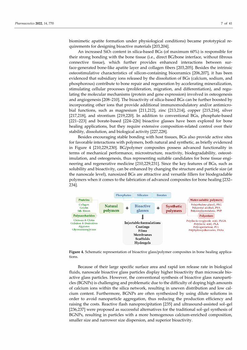
Besides encouraging stable bonding with host tissues, BGs also provide active sites
for favorable interactions with polymers, both natural and synthetic, as briefly evidenced
in Figure 4 [210,229,230]. BG/polymer composites possess advanced functionality in
terms of mechanical performance, microstructure, reactivity, biodegradability, osteost‐
imulation, and osteogenesis, thus representing suitable candidates for bone tissue engi‐
neering and regenerative medicine [210,229,231]. Since the key features of BGs, such as
solubility and bioactivity, can be enhanced by changing the structure and particle size (at
the nanoscale level), nanosized BGs are attractive and versatile fillers for biodegradable
polymers when it comes to the fabrication of advanced composites for bone healing [232–
234].
Figure 4. Schematic representation of bioactive glass/polymer composites in bone healing applica‐
tions.
Because of their large specific surface area and rapid ion release rate in biological
fluids, nanoscale bioactive glass particles display higher bioactivity than microscale bio‐
active glass particles. However, the conventional synthesis of bioactive glass nanoparti‐
cles (BGNPs) is challenging and problematic due to the difficulty of doping high amounts
of calcium ions within the silica network, resulting in uneven distribution and low cal‐
cium content. Furthermore, BGNPs are often synthesized by using dilute solutions in
order to avoid nanoparticle aggregation, thus reducing the production efficiency and
raising the costs. Reactive flash nanoprecipitation [235] and ultrasound‐assisted sol–gel
[236,237] were proposed as successful alternatives for the traditional sol–gel synthesis of
BGNPs, resulting in particles with a more homogenous calcium‐enriched composition,
smaller size and narrower size dispersion, and superior bioactivity.
Pharmaceutics 2022, 14, 770 8 of 41
The ability to incorporate active ions within their composition is a significant ad‐
vantage of BGNPs over other inorganic nanoparticles, as the release of such ions during
dissolution opens up a world of possibilities for enhancing the biofunctional outcome of
nanoengineered composites. Doping BGs with antimicrobial ions represents a promising
strategy for the fabrication of bone fillers or bone grafts that can allow bone repair and
regeneration without the risk of post‐implant infections [238,239]. Therefore, the poten‐
tial use of BGs doped with zinc (Zn)—Zn‐BGs—was thoroughly investigated [240], as the
presence of Zn determined antibacterial effects, and also contributed to enhanced min‐
eralization and osteogenic activity [241,242]. Beneficial effects with respect to in vitro
mineralization, cellular development, and antimicrobial efficiency, were also evidenced
in the case of silver (Ag)‐doped BGs (Ag‐BGs) [218,243].
Despite the promising results reported in BGNP‐based composites and devices,
significant efforts must be made in order to fully explore and beneficially revalue the bi‐
ological potential of such nanomaterials, as there is a lack of data regarding the long‐term
in vivo safety and performance of BGNPs [244,245].
In comparison with conventional BGNPs, mesoporous bioactive glass nanoparticles
(MBGNPs) provide additional advantages regarding the microstructure‐related ability to
load and release therapeutic agents, representing multifunctional platforms for bone
healing applications. MBGNPs are usually obtained by sol–gel‐mediated protocols
[246,247], and their versatile composition enable the incorporation of different therapeu‐
tic compounds, including copper [248,249], silver [218,250], and zinc [251,252] for anti‐
microbial effects, osteogenic activity, and immunomodulation; strontium for
pro‐osteogenic and pro‐angiogenic effects [253,254]; cerium and gallium for antibacterial
activity and bioactivity [255,256]; cobalt [257], iron [258], selenium [259], and tellurium
[260] for anti‐cancer effects.
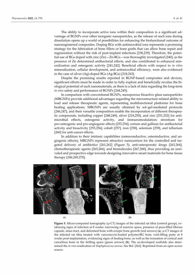
In addition to their intrinsic capabilities (osteoconductive, osteoinductive, and an‐
giogenic effects), MBGNPs represent attractive nanocarriers for the controlled and tar‐
geted delivery of antibiotics [261,262] (Figure 5), anti‐osteoporotic drugs [263,264],
chemotherapeutic agents [265,266], and biomolecules [267,268], thus providing an unri‐
valed and prospective edge towards designing innovative smart materials for bone tissue
therapy [246,269,270].
Figure 5. Micro‐computed tomography (μ‐CT) images of the infected rat tibia (control group), ev‐
idencing signs of infection at 8 weeks: narrowing of marrow space, presence of puss‐filled fibrous
capsule, sinus tract, and deformed bone with ectopic bone growth (red arrows) (a). μ‐CT images of
the infected rat tibia treated with vancomycin‐loaded polymer/BG bone void‐filling putty at 8
weeks post‐implantation, evidencing signs of healing bone, as well as the formation of cortical and
cancellous bone in the drilling space (green arrows) (b). The as‐developed scaffolds also deter‐
mined the in vivo eradication of Staphylococcus aureus. See Ref. [262]. Reprinted from an open access
source.
Pharmaceutics 2022, 14, 770 9 of 41
3. Oxide Nanoparticles
3.1. Mesoporous Silica
Silicon (Si) is naturally found in the human body, and it has a regulatory role during
the normal development of the skeleton and connective tissues, and also has beneficial
effects during collagen synthesis and matrix mineralization [271,272]. Besides repre‐
senting a major source of Si ions, silica (SiO2)‐based nanomaterials—especially mesopo‐
rous silica nanoparticles (MSNs)—provide attractive and tunable characteristics for bi‐
omedical applications, including drug/biomolecule delivery systems [273–275], tissue
engineering [276–278], regenerative medicine [279–281], and cancer therapy [282–284].
A large surface area and pore volume ratio, adjustable particle size, well‐structured
internal and external porosity, uniform and controllable pore size, impressive surface
functionalization, and intrinsic biocompatibility, represent the key features of MSNs used
for the fabrication of therapeutic biomaterials and devices [285–287]. The porosity char‐
acteristics of MSNs can be explored for loading various therapeutics, including biomol‐
ecules, soluble and insoluble drugs, targeting molecular drugs, and imaging agents, as
well as their different combinations, which may be simultaneously released within the
impaired tissues to achieve improved local concentration and synergistic drug therapy
and diagnostics (theranostics) [288–290]. Moreover, the pore‐opening gating mechanisms
distinguished in MSNs provide indisputable advantages over the controlled release of
the therapeutic cargo in response to internal (e.g., weakly acidic local microenvironment,
cancer‐overexpressed enzymes, or other biomolecules) and external (e.g., light, ultra‐
sound, and magnetic field exposure) stimuli [291,292].
Although MSNs represent one of the most appealing nanomaterials for the fabrica‐
tion of performance‐enhanced constructs for bone healing applications, some critical
parameters must be considered in order to achieve the desired therapeutic effects. By
optimizing the synthesis parameters (such as the type of silica precursor, the pH and
temperature during the reaction, and the type and concentration of surfactant), the size,
morphology, and porosity of MSNs can be modified [293,294]. Conventional and modi‐
fied sol–gel, evaporation‐induced self‐assembly, and core‐templating synthesis (in the
case of hollow MSNs) represent the most explored strategies for fabricating MSNs with
controllable particle and pore sizes [295,296].
Vital events involved in bone repair and regeneration, including cellular prolifera‐
tion and differentiation, bone matrix mineralization, osteoinduction, and osteogenesis,
can all be triggered or boosted by means of Si‐enriched nanosized and nanostructured
materials [297,298]. Through their modulatory effects on the specific molecular com‐
plexes involved in bone homeostasis, MSNs stimulate pro‐osteoblastic action and min‐
eralization, induce osteogenic differentiation and angiogenesis, and inhibit osteoclasts,
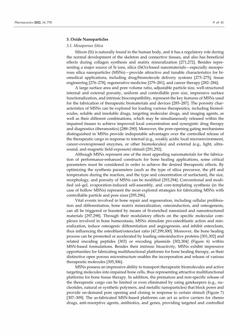
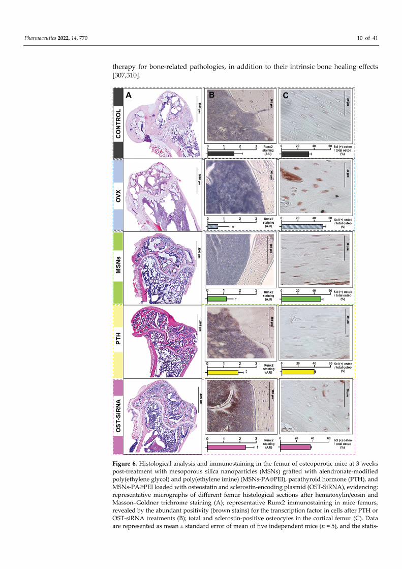
thus influencing the osteoblast/osteoclast ratio [47,299,300]. Moreover, the bone healing
process can be promoted or accelerated by loading osteoinductive proteins [301,302] and
related encoding peptides [303] or encoding plasmids [302,304] (Figure 6) within
MSN‐based formulations. Besides their intrinsic bioactivity, MSNs exhibit impressive
opportunities for fabricating multifunctional platforms for bone healing therapy, as their
distinctive open porous microstructure enables the incorporation and release of various
therapeutic molecules [305,306].
MSNs possess an impressive ability to transport therapeutic biomolecules and active
targeting molecules into impaired bone cells, thus representing attractive multifunctional
platforms for bone tissue therapy. In addition, the premature and non‐specific release of
the therapeutic cargo can be limited or even eliminated by using gatekeepers (e.g., nu‐
cleotides, natural or synthetic polymers, and metallic nanoparticles) that block pores and
provide on‐demand pore opening and closing in response to certain stimuli (Figure 7)
[307–309]. The as‐fabricated MSN‐based platforms can act as active carriers for chemo
drugs, anti‐resorptive agents, antibiotics, and genes, providing targeted and controlled
Pharmaceutics 2022, 14, 770 10 of 41
therapy for bone‐related pathologies, in addition to their intrinsic bone healing effects
[307,310].
Figure 6. Histological analysis and immunostaining in the femur of osteoporotic mice at 3 weeks
post‐treatment with mesoporous silica nanoparticles (MSNs) grafted with alendronate‐modified
poly(ethylene glycol) and poly(ethylene imine) (MSNs‐PA@PEI), parathyroid hormone (PTH), and
MSNs‐PA@PEI loaded with osteostatin and sclerostin‐encoding plasmid (OST‐SiRNA), evidencing:
representative micrographs of different femur histological sections after hematoxylin/eosin and
Masson–Goldner trichrome staining (A); representative Runx2 immunostaining in mice femurs,
revealed by the abundant positivity (brown stains) for the transcription factor in cells after PTH or
OST‐siRNA treatments (B); total and sclerostin‐positive osteocytes in the cortical femur (C). Data
are represented as mean ± standard error of mean of five independent mice (n = 5), and the statis‐
Pharmaceutics 2022, 14, 770 11 of 41
tical significance is indicated as # p < 0.001 vs. control, * p < 0.05 vs. ovariectomized mice (OVX), and
** p < 0.001 vs. OVX. See Ref. [302]. Reprinted from an open access source.
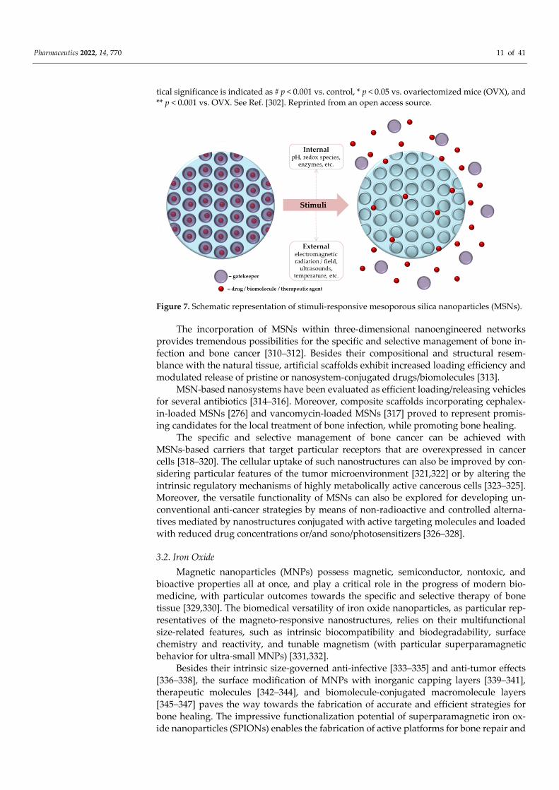
Figure 7. Schematic representation of stimuli‐responsive mesoporous silica nanoparticles (MSNs).
The incorporation of MSNs within three‐dimensional nanoengineered networks
provides tremendous possibilities for the specific and selective management of bone in‐
fection and bone cancer [310–312]. Besides their compositional and structural resem‐
blance with the natural tissue, artificial scaffolds exhibit increased loading efficiency and
modulated release of pristine or nanosystem‐conjugated drugs/biomolecules [313].
MSN‐based nanosystems have been evaluated as efficient loading/releasing vehicles
for several antibiotics [314–316]. Moreover, composite scaffolds incorporating cephalex‐
in‐loaded MSNs [276] and vancomycin‐loaded MSNs [317] proved to represent promis‐
ing candidates for the local treatment of bone infection, while promoting bone healing.
The specific and selective management of bone cancer can be achieved with
MSNs‐based carriers that target particular receptors that are overexpressed in cancer
cells [318–320]. The cellular uptake of such nanostructures can also be improved by con‐
sidering particular features of the tumor microenvironment [321,322] or by altering the
intrinsic regulatory mechanisms of highly metabolically active cancerous cells [323–325].
Moreover, the versatile functionality of MSNs can also be explored for developing un‐
conventional anti‐cancer strategies by means of non‐radioactive and controlled alterna‐
tives mediated by nanostructures conjugated with active targeting molecules and loaded
with reduced drug concentrations or/and sono/photosensitizers [326–328].
3.2. Iron Oxide
Magnetic nanoparticles (MNPs) possess magnetic, semiconductor, nontoxic, and
bioactive properties all at once, and play a critical role in the progress of modern bio‐
medicine, with particular outcomes towards the specific and selective therapy of bone
tissue [329,330]. The biomedical versatility of iron oxide nanoparticles, as particular rep‐
resentatives of the magneto‐responsive nanostructures, relies on their multifunctional
size‐related features, such as intrinsic biocompatibility and biodegradability, surface
chemistry and reactivity, and tunable magnetism (with particular superparamagnetic
behavior for ultra‐small MNPs) [331,332].
Besides their intrinsic size‐governed anti‐infective [333–335] and anti‐tumor effects
[336–338], the surface modification of MNPs with inorganic capping layers [339–341],
therapeutic molecules [342–344], and biomolecule‐conjugated macromolecule layers
[345–347] paves the way towards the fabrication of accurate and efficient strategies for
bone healing. The impressive functionalization potential of superparamagnetic iron ox‐
ide nanoparticles (SPIONs) enables the fabrication of active platforms for bone repair and
Pharmaceutics 2022, 14, 770 12 of 41
regeneration, as well as for bone infection and cancer. Such magnetic nanostructures can
act as active vehicles and therapeutic enhancers for their cargo, but their functionality can
be extended by means of external triggers (electromagnetic radiation and fields), which
represent the leading advantage of MNP‐based biomedicine [348–350].
Following their exposure to an alternating magnetic field, MNPs undergo important
magnetic relaxation, as their magnetic moment (given by unpaired spin electrons in the
outermost electron shell) rapidly flips its orientation between two stable states, but they
also can undergo physical rotation and circumstantial collisions, finally resulting in
converting the external energy into heat [351,352]. This peculiar behavior of MNPs gives
them an impressive potential for the local thermally‐induced alteration of pathological
cells by means of magnetic hyperthermia, which is being extensively investigated for
cancer management [353,354]. Moreover, if therapeutic agents are conjugated to MNPs,
their local release can be externally triggered and controlled. Even if the clinical applica‐
tion of magnetically targeted therapy by means of magnetized medications still requires
regulatory protocols [355,356], the preclinical evaluation of SPION‐mediated bone cancer
therapy is of great interest. Besides acting as mechanical reinforcements for polymeric
scaffolds, SPIONs contribute to the normal development of bone cells and promote the
mineralization process and osteogenic activity [357–359], and also promote the in vivo
bone repair and regeneration [359–361]. In addition to their ability to generate localized
hyperthermia while avoiding the impairment of surrounding normal tissues when com‐
bined with SPIONs, it has been reported that magnetic fields are beneficial for promoting
the osteogenic activity of progenitor cells. Magnetic fields regulate the cellular uptake of
SPIONs via stem cells and preosteoblasts and promote their osteogenic differentiation
and bone matrix mineralization, and also contribute to their proliferation, migration, and
organization inside scaffolds [362,363], finally resulting in magnetically guided osteo‐
genesis and angiogenesis [364–366]. SPION‐loaded constructs (e.g., porous inorganic
scaffolds, polymer sponges, and hydrogels) and external magnetic fields synergistically
act to provide successful therapeutic alternatives for bone healing [329,367].
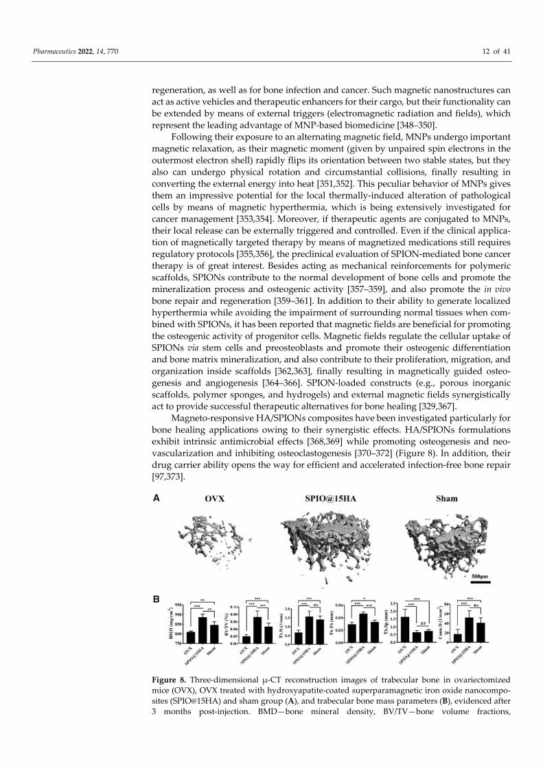
Magneto‐responsive HA/SPIONs composites have been investigated particularly for
bone healing applications owing to their synergistic effects. HA/SPIONs formulations
exhibit intrinsic antimicrobial effects [368,369] while promoting osteogenesis and neo‐
vascularization and inhibiting osteoclastogenesis [370–372] (Figure 8). In addition, their
drug carrier ability opens the way for efficient and accelerated infection‐free bone repair
[97,373].
Figure 8. Three‐dimensional μ‐CT reconstruction images of trabecular bone in ovariectomized
mice (OVX), OVX treated with hydroxyapatite‐coated superparamagnetic iron oxide nanocompo‐
sites (SPIO@15HA) and sham group (A), and trabecular bone mass parameters (B), evidenced after
3 months post‐injection. BMD—bone mineral density, BV/TV—bone volume fractions,
Pharmaceutics 2022, 14, 770 13 of 41
Tb.N—trabecular number, Tb.Th—trabecular thickness, Tb.Sp—trabecular spacing,
Conn.D—connectivity density. Data are expressed as mean ± standard deviation of seven inde‐
pendent mice (n = 7), ns means no significance, and the statistical significance is indicated as * p <
0.05, ** p < 0.01, and *** p < 0.001. See Ref. [370]. Reprinted from an open access source.
Given the extensive use of metallic implants in the clinical restoration and replace‐
ment of bone tissue, an attractive nanotechnology‐derived approach consists of enhanc‐
ing their bioactivity and osteogenic activity using surface coatings [374–376]. It has been
reported that the incorporation of SPIONs within HA [377,378] or polymer [379,380]
coatings leads to significant improvements in the wettability and corrosion resistance of
titanium‐based biomaterials, and also enhanced apatite‐forming ability and cellular
events. As the direct interactions between SPIONs and therapeutic agents determine the
formation of highly stable nanosystems with potentiated therapeutic effects, such
nanostructures have been extensively investigated with respect to the development of
new pharmaceuticals [35,381,382]. The therapeutic outcome of metallic implants can be
achieved by means of synthetic polyester coatings embedded with MNPs conjugated
with natural antimicrobial extracts [383], electroactive polymer coatings embedded with
antibiotic‐functionalized MNPs [379,380], and chemo drug‐loaded SPIONs/cyclodextrin
coatings [384].
3.3. Other Oxides
The therapeutic implications of other oxide nanoparticles in bone healing applica‐
tions have been also explored [385,386]. For instance, magnesium oxide (MgO) and zinc
oxide (ZnO) nanoparticles have been investigated for the fabrication of functional bone
substitutes [387,388]. MgO and ZnO NPs exert strong antimicrobial and anti‐biofilm ac‐
tivity [389,390], and also antioxidant effects [391,392], making them suitable candidates
for boosting the performance of HA‐based substitutes [393–396].
Following their dissolution, MgO NPs provide mineral nutrients that are essential
for most biological processes, including new bone formation, by promoting osteogenic
proliferation and differentiation and bone‐like mineral deposition [397–399]. By exerting
positive immunomodulatory effects, MgO NPs indirectly suppress the activity of osteo‐
clasts [400]. Besides acting as mechanical reinforcements for polymeric scaffolds, MgO
NPs also modulate their hydrophilicity and degradation, whilst the polymeric matrix
enables the gradual release of therapeutic ions, finally resulting in enhancing the bone
healing ability of such composites [401,402].
Given the fact that an imbalance in the normal zinc deposits and cellular zinc ho‐
meostasis may occur after bone tissue injuries (as the human skeleton is a major source of
zinc), producing zinc‐enriched substitutes is of great importance for bone healing and
normal skeletal development [403,404]. ZnO NPs synergistically act on the bone cells
involved in bone formation and remodeling by inducing osteogenic effects [405,406] and
modulating the osteoclastogenic events [407,408]. The oxidative events induced by ZnO
NPs (mediated by free zinc ions and reactive oxygen species) can be further explored for
bone tissue regeneration and bone cancer therapy through their pro‐angiogenic [409,410]
and anti‐angiogenic [411,412] properties, respectively.
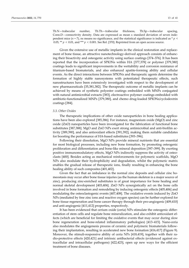
It has been evidenced that cerium oxide (ceria) NPs stimulate the osteogenic differ‐
entiation of stem cells and regulate bone mineralization, and also exhibit antioxidant ef‐
fects (which are beneficial for limiting the oxidative events that may occur during slow
bone regeneration and bone‐related inflammatory pathologies) [413–415]. Nano‐ceria
also modulates the angiogenesis process of ceramic and polymeric biomaterials follow‐
ing their implantation, resulting in accelerated new bone formation [416,417] (Figure 9).
Moreover, the stimuli‐responsive ability of ceria NPs [418,419], together with their ra‐
dio‐protective effects [420,421] and intrinsic antibacterial effects (evidenced against ex‐
tracellular and intracellular pathogens) [422,423], open up new ways for the efficient
treatment of bone diseases.
Pharmaceutics 2022, 14, 770 14 of 41
Figure 9. Histological analysis of rat cranial defects treated with bare and nano‐ceria‐loaded poly‐
caprolactone/gelatin membranes (PG M and PG‐CeO2 M, respectively) for 4 and 8 weeks (w), evi‐
denced by Masson’s trichrome staining. Control group (a,d), PG M group (b,e), and PG‐CeO2 M
group (c,f). M—membrane, B—bone, scale bar—100 μm. See Ref. [417]. Reprinted from an open
access source.
Recently, hollow manganese oxide NPs were proposed as efficient platforms for the
immunotherapy of osteosarcoma, with their additional tumor‐targeting ability and im‐
aging‐guided drug delivery [424]. These oxide nanoparticles exhibit important osteo‐
genic activity and bone‐forming ability [425,426], while their excellent antioxidant effects
proved to be beneficial for the management of osteoarthritis [427,428].
A significant improvement in the mechanical behavior and thermal stability of
polymeric biomaterials has been evidenced after the incorporation of titanium oxide (ti‐
tania) NPs, with such nanostructured platforms being proposed for the long‐term use in
bone regeneration [429,430]. The efficiency of nano‐titania on osteoblast/osteoclast ho‐
meostasis [431,432] and collagen deposition (by inducing the secretion of biomolecules
that actively regulate bone repair) [433], without affecting the differentiation and miner‐
alization of osteoblasts [433,434], has been reported.
4. Metallic Nanoparticles
This review also covers the implications of metal‐based nanoparticles in bone tissue
therapy. Owing to their peculiar nanosize‐related characteristics, which include biome‐
chanics and thermochemistry, stability and optical behavior, reduced toxicity and good
biocompatibility, proliferative and intrinsic osteogenic potential, cellular development
modulation, and intrinsic antimicrobial and anti‐cancer effects, metallic NPs are versatile
candidates for bone healing applications [385,435].
4.1. Gold
Gold nanoparticles (AuNPs) are biocompatible and inert nanosized structures with
high monodispersity, electroconductivity, and excellent optical properties [436,437]. The
impressive use of AuNPs in modern biomedicine relies on their highly remarkable sur‐
face functionalization potential, and includes targeted therapeutic formulations (drug,
macromolecule, peptide, protein, and gene delivery), biomedical imaging and diagnosis
(biodetection and biosensing), and complex therapy (photothermal, photodynamic, and
radiation therapy) [438–440].
In relation to bone healing therapy, it has been evidenced that AuNPs exhibit in‐
trinsic osteogenic effects (by promoting the differentiation of pluripotent cells and bio‐
mimetic apatite formation) [441,442], inhibit osteoclastogenesis [443,444], and accelerate
de novo bone formation [445,446]. Several molecular mechanisms were proposed for
AuNP‐mediated osteogenic differentiation [439,447]. Stem cells may undergo osteogenic
differentiation in response to extracellular AuNPs (physical and/or chemical modifica‐
tion of the microenvironment) and intracellular AuNPs (mechanical stress) by means of
the integrin‐mediated signaling pathway [448,449], transcellular pathway [441,450], and
Pharmaceutics 2022, 14, 770 15 of 41
autophagy [442,451]. It has also been evidenced that the osteogenic ability of AuNPs is
strongly related to their concentration [452], size [445], and shape [453].
What is more, AuNPs also exhibit important antimicrobial [454,455] and anti‐cancer
[456,457] activity. By considering the multifunctional therapeutic effects of AuNPs, and
also their impressive functionalization versatility, substantial efforts have been oriented
towards the fabrication of AuNP‐embedded composites and complex formulations for
bone repair and regeneration [439,458]. Moreover, given their peculiar electrical and op‐
tical behavior, AuNPs have been explored for the targeted and controlled management of
bone infections and bone cancers [385,459].
4.2. Silver
Silver nanoparticles (AgNPs) are one of the most explored nanosized noble metals in
modern industrial and biomedical applications, owing to their intrinsic catalytic effect,
chemical stability, good electrical conductivity, optical behavior, and versatile biological
activity [460,461]. In its ionic, metallic, and nanoparticulate forms, silver has been exten‐
sively used as an antibacterial agent [462,463]. The particular anti‐pathogenic effects of
nano‐silver have been assigned to their ability to adhere to bacterial cell walls and pro‐
duce oxidative stress, resulting in the bacterial cell wall and membrane impairment and
subsequent cytoplasmic leakage, and the denaturation of bacterial macromolecules and
alteration of vital cellular processes, respectively [464–466]. Silver ions released by
AgNPs mediate bacterial death by impairing the peptidoglycan component of cell walls,
hindering bacterial protein synthesis and obstructing replication signals and ener‐
gy‐dependent survival processes by binding to nucleic acids [467,468].
In the realm of orthopedics and dentistry, where the infection susceptibility of im‐
planted devices is a continuous danger, the clinical potential of nano‐silver is of special
interest [469–471]. Since AgNPs stimulate osteogenesis and inhibit osteoclastogenesis
[472,473], their use in bone healing applications gives rise to multifunctional platforms,
and such nanostructures can be used to induce or potentiate the antimicrobial effects of
nanoengineered constructs and clinically used devices, while stimulating the osteogenic
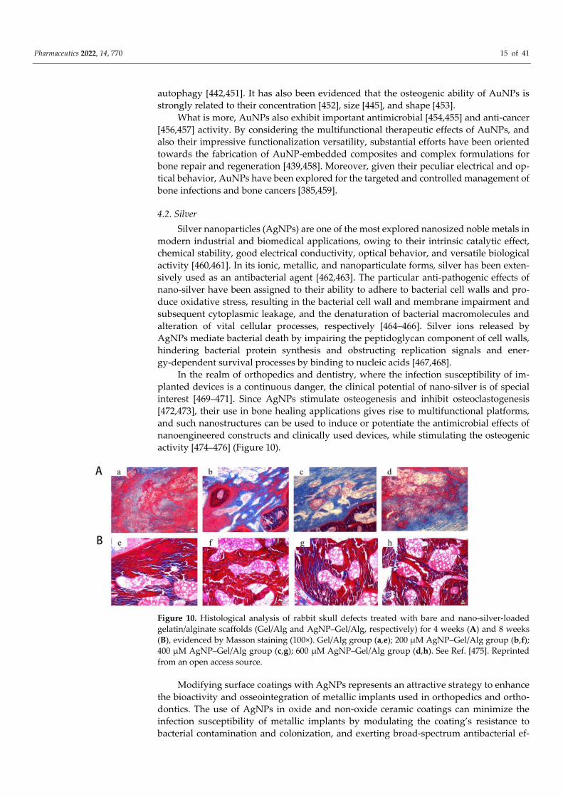
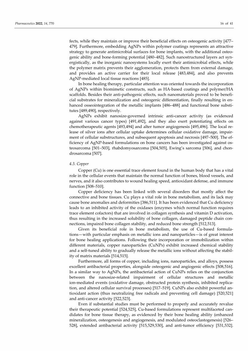
activity [474–476] (Figure 10).
Figure 10. Histological analysis of rabbit skull defects treated with bare and nano‐silver‐loaded
gelatin/alginate scaffolds (Gel/Alg and AgNP–Gel/Alg, respectively) for 4 weeks (A) and 8 weeks
(B), evidenced by Masson staining (100×). Gel/Alg group (a,e); 200 μM AgNP–Gel/Alg group (b,f);
400 μM AgNP–Gel/Alg group (c,g); 600 μM AgNP–Gel/Alg group (d,h). See Ref. [475]. Reprinted
from an open access source.
Modifying surface coatings with AgNPs represents an attractive strategy to enhance
the bioactivity and osseointegration of metallic implants used in orthopedics and ortho‐
dontics. The use of AgNPs in oxide and non‐oxide ceramic coatings can minimize the
infection susceptibility of metallic implants by modulating the coating’s resistance to
bacterial contamination and colonization, and exerting broad‐spectrum antibacterial ef‐
Pharmaceutics 2022, 14, 770 16 of 41
fects, while they maintain or improve their beneficial effects on osteogenic activity [477–
479]. Furthermore, embedding AgNPs within polymer coatings represents an attractive
strategy to generate antimicrobial surfaces for bone implants, with the additional osteo‐
genic ability and bone‐forming potential [480–482]. Such nanostructured layers act syn‐
ergistically, as the inorganic nanosystems locally exert their antimicrobial effects, while
the polymer matrix prevents their agglomeration, protects them from external damage,
and provides an active carrier for their local release [483,484], and also prevents
AgNP‐mediated local tissue reactions [485].
In bone healing therapy, particular attention was oriented towards the incorporation
of AgNPs within biomimetic constructs, such as HA‐based coatings and polymer/HA
scaffolds. Besides their anti‐pathogenic effects, such nanomaterials proved to be benefi‐
cial substrates for mineralization and osteogenic differentiation, finally resulting in en‐
hanced osseointegration of the metallic implants [486–488] and functional bone substi‐
tutes [489,490], respectively.
AgNPs exhibit nanosize‐governed intrinsic anti‐cancer activity (as evidenced
against various cancer types) [491,492], and they also exert potentiating effects on
chemotherapeutic agents [493,494] and alter tumor angiogenesis [495,496]. The local re‐
lease of silver ions after cellular uptake determines cellular oxidative damage, impair‐
ment of cellular substructures, and subsequent apoptosis and necrosis [497–500]. The ef‐
ficiency of AgNP‐based formulations on bone cancers has been investigated against os‐
teosarcoma [501–503], rhabdomyosarcoma [504,505], Ewing’s sarcoma [506], and chon‐
drosarcoma [507].
4.3. Copper
Copper (Cu) is one essential trace element found in the human body that has a vital
role in the cellular events that maintain the normal function of bones, blood vessels, and
nerves, and it also contributes to wound healing speed, antioxidant defense, and immune
function [508–510].
Copper deficiency has been linked with several disorders that mostly affect the
connective and bone tissues. Cu plays a vital role in bone metabolism, and its lack may
cause bone anomalies and deformities [386,511]. It has been evidenced that Cu deficiency
leads to an inhibited activity of the oxidases (enzymes which normal function requires
trace element cofactors) that are involved in collagen synthesis and vitamin D activation,
thus resulting in the increased solubility of bone collagen, damaged peptide chain con‐
nections, impaired bone collagen stability, and reduced bone strength [512,513].
Given its beneficial role in bone metabolism, the use of Cu‐based formula‐
tions—with particular emphasis on metallic ions and nanoparticles—is of great interest
for bone healing applications. Following their incorporation or immobilization within
different materials, copper nanoparticles (CuNPs) exhibit increased chemical stability
and a self‐tuned ability to gradually release the metallic ions without affecting the stabil‐
ity of matrix materials [514,515].
Furthermore, all forms of copper, including ions, nanoparticles, and alloys, possess
excellent antibacterial properties, alongside osteogenic and angiogenic effects [508,516].
In a similar way to AgNPs, the antibacterial action of CuNPs relies on the conjunction
between the nanosize‐related impairment of cellular structures and metallic
ion‐mediated events (oxidative damage, obstructed protein synthesis, inhibited replica‐
tion, and altered cellular survival processes) [517–519]. CuNPs also exhibit powerful an‐
tioxidant action (thus neutralizing free radicals and preventing cell damage) [520,521]
and anti‐cancer activity [522,523].
Even if substantial studies must be performed to properly and accurately revalue
their therapeutic potential [524,525], Cu‐based formulations represent multifaceted can‐
didates for bone tissue therapy, as evidenced by their bone healing ability (enhanced
mineralization, osteogenesis and angiogenesis, and modulated osteoclastogenesis) [526–
528], extended antibacterial activity [515,529,530], and anti‐tumor efficiency [531,532].
Pharmaceutics 2022, 14, 770 17 of 41
CuNPs have also been investigated with respect to dental applications, as efficient anti‐
microbials for denture base resins [533], endodontic treatment [534], and periodontitis
management [535].
5. Conclusions and Perspectives
Designing successful devices and substitutes for bone therapy still represents a
challenge for modern biomedicine, as it implies the accurate understanding of bone
pathophysiology, the proper selection of biomaterials and fabrication protocols, and
maximal therapeutic efficiency.
Nanoparticle‐based biomaterials and biotechnologies have been lately validated as
viable alternatives to traditional scaffolding protocols. In particular, bioceramic, oxide,
and metallic nanoparticles demonstrated impressive therapeutic outcomes for bone re‐
pair and regeneration, and also for bone pathologies management.
Owing to their bioactivity, biomimetic composition, and good incorporation within
the natural bone structure, bioceramic nanoparticles represent the best choice for repara‐
tive and regenerative bone therapy. Their acknowledged cytocompatibility and benefi‐
cial interactions with living tissues can be explored in conjunction with polymeric con‐
structs and other inorganic (ions, nanoparticles, alloys, and composites) or organic sub‐
stances (drugs and biomolecules) in order to fabricate bone‐mimicking platforms for the
specific and selective management of bone pathologies.
Even if substantial efforts should be made to completely understand and finely tune
the implications of oxide and metallic nanoparticles in bone healing, their functional
versatility (as nanocarriers, imaging agents, and sensitizers) and intrinsic therapeutic ac‐
tivity are impressive. Such peculiar characteristics pave the way towards the develop‐
ment of multifunctional bone substitutes, including platforms for targeted and localized
drug delivery (antimicrobial, anti‐inflammatory, anti‐resorptive, and anti‐cancer thera‐
py), specific and selective detection and diagnosis, and effective combined therapy.
Besides being active components for bone processes (contributing with their oste‐
oconductive, osteoinductive, and osteogenic effects), the previously discussed inorganic
nanomaterials exhibit additional biological activities (antimicrobial, antioxidant, im‐
munomodulatory, anti‐resorptive, and anti‐cancer). The nanosize‐governed surface
chemistry of these nanoparticles provides active sites for the conjugation of various
therapeutic agents (e.g., ions, nanostructures, drugs, biomolecules, and nucleic acids),
and also enables their immobilization or incorporation into more complex constructs,
finally resulting in the development of versatile and performance‐enhanced candidates
for bone healing applications.
Author Contributions: A.‐C.B., O.G., E.A., A.M.G. and A.F. designed and wrote the paper. All
authors have read and agreed to the published version of the manuscript.
Funding: All authors would like to acknowledge and thank for the financial support provided by
the University Politehnica of Bucharest. This paper acknowledges the support of the Ministry of
Education and Research, CNCS UEFISCDI, project no. 524PED/2020 (PN‐III‐P2‐2.1‐PED‐2019).
Conflicts of Interest: The authors declare no conflict of interest.
References
1. Ralston, S.H. Bone structure and metabolism. Medicine 2021, 49, 567–571. https://doi.org/10.1016/j.mpmed.2021.06.009.
2. Abe, K.; Shimozaki, S.; Domoto, T.; Yamamoto, N.; Tsuchiya, H.; Minamoto, T. Glycogen synthase kinase 3β biology in bone
and soft tissue sarcomas. J. Cancer Metastasis Treat. 2020, 6, 51. https://doi.org/10.20517/2394‐4722.2020.117.
3. Chen, J.; Ashames, A.; Buabeid, M.A.; Fahelelbom, K.M.; Ijaz, M.; Murtaza, G. Nanocomposites drug delivery systems for the
healing of bone fractures. Int. J. Pharm. 2020, 585, 119477. https://doi.org/10.1016/j.ijpharm.2020.119477.
4. Kupikowska‐Stobba, B.; Kasprzak, M. Fabrication of nanoparticles for bone regeneration: New insight into applications of
nanoemulsion technology. J. Mater. Chem. B 2021, 9, 5221–5244. https://doi.org/10.1039/D1TB00559F.
5. Sohn, H.‐S.; Oh, J.‐K. Review of bone graft and bone substitutes with an emphasis on fracture surgeries. Biomater. Res. 2019, 23,
9. https://doi.org/10.1186/s40824‐019‐0157‐y.
Pharmaceutics 2022, 14, 770 18 of 41
6. Chandra, G.; Pandey, A. Biodegradable bone implants in orthopedic applications: A review. Biocybern. Biomed. Eng. 2020, 40,
596–610. https://doi.org/10.1016/j.bbe.2020.02.003.
7. Tahmasebi, E.; Alam, M.; Yazdanian, M.; Tebyanian, H.; Yazdanian, A.; Seifalian, A.; Mosaddad, S.A. Current biocompatible
materials in oral regeneration: A comprehensive overview of composite materials. J. Mater. Res. Technol. 2020, 9, 11731–11755.
https://doi.org/10.1016/j.jmrt.2020.08.042.
8. Collon, K.; Gallo, M.C.; Lieberman, J.R. Musculoskeletal tissue engineering: Regional gene therapy for bone repair. Biomaterials
2021, 275, 120901. https://doi.org/10.1016/j.biomaterials.2021.120901.
9. Kumar, P.; Saini, M.; Dehiya, B.S.; Sindhu, A.; Kumar, V.; Kumar, R.; Lamberti, L.; Pruncu, C.I.; Thakur, R. Comprehensive
Survey on Nanobiomaterials for Bone Tissue Engineering Applications. Nanomaterials 2020, 10, 2019.
https://doi.org/10.3390/nano10102019.
10. Huang, H.; Feng, W.; Chen, Y.; Shi, J. Inorganic nanoparticles in clinical trials and translations. Nano Today 2020, 35, 100972.
https://doi.org/10.1016/j.nantod.2020.100972.
11. Lyons, J.G.; Plantz, M.A.; Hsu, W.K.; Hsu, E.L.; Minardi, S. Nanostructured Biomaterials for Bone Regeneration. Front. Bioeng.
Biotechnol. 2020, 8, 922–922. https://doi.org/10.3389/fbioe.2020.00922.
12. Wang, W.; Yeung, K.W.K. Bone grafts and biomaterials substitutes for bone defect repair: A review. Bioact. Mater. 2017, 2, 224–
247. https://doi.org/10.1016/j.bioactmat.2017.05.007.
13. Gao, X.; Li, L.; Cai, X.; Huang, Q.; Xiao, J.; Cheng, Y. Targeting nanoparticles for diagnosis and therapy of bone tumors: Op‐
portunities and challenges. Biomaterials 2021, 265, 120404. https://doi.org/10.1016/j.biomaterials.2020.120404.
14. Ojo, O.A.; Olayide, I.I.; Akalabu, M.C.; Ajiboye, B.O.; Ojo, A.B.; Oyinloye, B.E.; Ramalingam, M. Nanoparticles and their bio‐
medical applications. Biointerface Res. Appl. Chem. 2020, 11, 8431–8445.
15. Khan, I.; Saeed, K.; Khan, I. Nanoparticles: Properties, applications and toxicities. Arab. J. Chem. 2019, 12, 908–931.
https://doi.org/10.1016/j.arabjc.2017.05.011.
16. Zheng, K.; Xie, J. Engineering Ultrasmall Metal Nanoclusters as Promising Theranostic Agents. Trends Chem. 2020, 2, 665–679.
https://doi.org/10.1016/j.trechm.2020.04.011.
17. van der Meel, R.; Sulheim, E.; Shi, Y.; Kiessling, F.; Mulder, W.J.M.; Lammers, T. Smart cancer nanomedicine. Nat. Nanotechnol.
2019, 14, 1007–1017. https://doi.org/10.1038/s41565‐019‐0567‐y.
18. Mitchell, M.J.; Billingsley, M.M.; Haley, R.M.; Wechsler, M.E.; Peppas, N.A.; Langer, R. Engineering precision nanoparticles for
drug delivery. Nat. Rev. Drug Discov. 2021, 20, 101–124. https://doi.org/10.1038/s41573‐020‐0090‐8.
19. Baldwin, P.; Li, D.J.; Auston, D.A.; Mir, H.S.; Yoon, R.S.; Koval, K.J. Autograft, Allograft, and Bone Graft Substitutes: Clinical
Evidence and Indications for Use in the Setting of Orthopaedic Trauma Surgery. J. Orthop. Trauma 2019, 33, 203–213.
https://doi.org/10.1097/BOT.0000000000001420.
20. Hu, C.; Ashok, D.; Nisbet, D.R.; Gautam, V. Bioinspired surface modification of orthopedic implants for bone tissue engi‐
neering. Biomaterials 2019, 219, 119366. https://doi.org/10.1016/j.biomaterials.2019.119366.
21. Hench, L.L.; Thompson, I. Twenty‐first century challenges for biomaterials. J. R. Soc. Interface 2010, 7 (Suppl. S4), S37–S391.
https://doi.org/10.1098/rsif.2010.0151.focus.
22. Fattahian, H.; Mansouri, K.; Mansouri, N. Biomaterials, substitutes, and tissue engineering in bone repair: Current and future
concepts. Comp. Clin. Pathol. 2019, 28, 879–891. https://doi.org/10.1007/s00580‐017‐2507‐2.
23. Jin, S.; Xia, X.; Huang, J.; Yuan, C.; Zuo, Y.; Li, Y.; Li, J. Recent advances in PLGA‐based biomaterials for bone tissue regenera‐
tion. Acta Biomater. 2021, 127, 56–79. https://doi.org/10.1016/j.actbio.2021.03.067.
24. Wu, F.; Harper, B.J.; Harper, S.L. Differential dissolution and toxicity of surface functionalized silver nanoparticles in
small‐scale microcosms: Impacts of community complexity. Environ. Sci. Nano 2017, 4, 359–372.
https://doi.org/10.1039/c6en00324a.
25. Wang, N.; Dheen, S.T.; Fuh, J.Y.H.; Kumar, A.S. A review of multi‐functional ceramic nanoparticles in 3D printed bone tissue
engineering. Bioprinting 2021, 23, e00146.
26. Wang, N.; Maskomani, S.; Meenashisundaram, G.K.; Fuh, J.Y.H.; Dheen, S.T.; Anantharajan, S.K. A study of Titanium and
Magnesium particle‐induced oxidative stress and toxicity to human osteoblasts. Mater. Sci. Eng. C Mater. Biol. Appl. 2020, 117,
111285. https://doi.org/10.1016/j.msec.2020.111285.
27. Tortella, G.R.; Rubilar, O.; Durán, N.; Diez, M.C.; Martínez, M.; Parada, J.; Seabra, A.B. Silver nanoparticles: Toxicity in model
organisms as an overview of its hazard for human health and the environment. J. Hazard. Mater. 2020, 390, 121974.
https://doi.org/10.1016/j.jhazmat.2019.121974.
28. Khan, M.A.; Singh, D.; Ahmad, A.; Siddique, H.R. Revisiting inorganic nanoparticles as promising therapeutic agents: A par‐
adigm shift in oncological theranostics. Eur. J. Pharm. Sci. 2021, 164, 105892. https://doi.org/10.1016/j.ejps.2021.105892.
29. Gherasim, O.; Popescu, R.C.; Gherasim, T.G.; Grumezescu, V.; Andronescu, E. Pharmacotherapy and nanotechnology. In Na‐
noparticles in Pharmacotherapy; William Andrew (Elsevier): Oxford, United Kingdom, 2019; pp. 1–21.
30. Chenthamara, D.; Subramaniam, S.; Ramakrishnan, S.G.; Krishnaswamy, S.; Essa, M.M.; Lin, F.‐H.; Qoronfleh, M.W. Thera‐
peutic efficacy of nanoparticles and routes of administration. Biomater. Res. 2019, 23, 20.
https://doi.org/10.1186/s40824‐019‐0166‐x.
31. Yao, Y.; Zhou, Y.; Liu, L.; Xu, Y.; Chen, Q.; Wang, Y.; Wu, S.; Deng, Y.; Zhang, J.; Shao, A. Nanoparticle‐Based Drug Delivery in
Cancer Therapy and Its Role in Overcoming Drug Resistance. Front. Mol. Biosci. 2020, 7, 193.
https://doi.org/10.3389/fmolb.2020.00193.
Pharmaceutics 2022, 14, 770 19 of 41
32. Heuer‐Jungemann, A.; Feliu, N.; Bakaimi, I.; Hamaly, M.; Alkilany, A.; Chakraborty, I.; Masood, A.; Casula, M.F.; Kostopou‐
lou, A.; Oh, E.; et al. The Role of Ligands in the Chemical Synthesis and Applications of Inorganic Nanoparticles. Chem. Rev.
2019, 119, 4819–4880. https://doi.org/10.1021/acs.chemrev.8b00733.
33. Chandrakala, V.; Aruna, V.; Angajala, G. Review on metal nanoparticles as nanocarriers: Current challenges and perspectives
in drug delivery systems. Emergent Mater. 2022. https://doi.org/10.1007/s42247‐021‐00335‐x.
34. Chakraborty, I.; Parak, W.J. Protein‐Induced Shape Control of Noble Metal Nanoparticles. Adv. Mater. Interfaces 2019, 6,
1801407. https://doi.org/10.1002/admi.201801407.
35. Mihai, A.D.; Chircov, C.; Grumezescu, A.M.; Holban, A.M. Magnetite nanoparticles and essential oils systems for advanced
antibacterial therapies. Int. J. Mol. Sci. 2020, 21, 7355. https://doi.org/10.3390/ijms21197355.
36. Chiozzi, V.; Rossi, F. Inorganic–organic core/shell nanoparticles: Progress and applications. Nanoscale Adv. 2020, 2, 5090–5105.
https://doi.org/10.1039/D0NA00411A.
37. Zarrintaj, P.; Paran, S.M.R.; Jafari, S.; Mozafari, M. Application of Compatibilized Polymer Blends in Biomedical Fields. In
Compatibilization of Polymer Blends; Elsevier: Amsterdam, The Netherlands, 2020; pp. 511–537.
38. Marques, C.F.; Olhero, S.; Abrantes, J.C.C.; Marote, A.; Ferreira, S.; Vieira, S.I.; Ferreira, J.M.F. Biocompatibility and antimicro‐
bial activity of biphasic calcium phosphate powders doped with metal ions for regenerative medicine. Ceram. Int. 2017, 43,
15719–15728. https://doi.org/10.1016/j.ceramint.2017.08.133.
39. Samanta, S.K.; Devi, K.B.; Das, P.; Mukherjee, P.; Chanda, A.; Roy, M.; Nandi, S.K. Metallic ion doped tri‐calcium phosphate
ceramics: Effect of dynamic loading on in vivo bone regeneration. J. Mech. Behav. Biomed. Mater. 2019, 96, 227–235.
https://doi.org/10.1016/j.jmbbm.2019.04.051.
40. Strutynska, N.; Livitska, O.; Prylutska, S.; Yumyna, Y.; Zelena, P.; Skivka, L.; Malyshenko, A.; Vovchenko, L.; Strelchuk, V.;
Prylutskyy, Y.; et al. New nanostructured apatite‐type (Na+,Zn2+,CO32−)‐doped calcium phosphates: Preparation, mechanical
properties and antibacterial activity. J. Mol. Struct. 2020, 1222, 128932. https://doi.org/10.1016/j.molstruc.2020.128932.
41. Kaur, P.; Singh, K.J.; Kaur, S.; Kaur, S.; Singh, A.P. Sol‐gel derived strontium‐doped SiO2–CaO–MgO–P2O5 bioceramics for
faster growth of bone like hydroxyapatite and their in vitro study for orthopedic applications. Mater. Chem. Phys. 2020, 245,
122763. https://doi.org/10.1016/j.matchemphys.2020.122763.
42. Sarin, N.; Singh, K.; Singh, D.; Arora, S.; Singh, A.P.; Mahajan, H.J.M.C. Preliminary studies of strontium and selenium binary
doped CaO–SiO2–P2O5–MgO bioceramics for faster growth of hydroxyapatite and bone regeneration applications. Mater. Chem.
Phys. 2020, 253, 123329.
43. Thompson, F.C.; Matsumoto, M.A.; Biguetti, C.C.; Rennó, A.C.M.; de Andrade Holgado, L.; Santiago Junior, J.F.; Munerato,
M.S.; Saraiva, P.P. Distinct healing pattern of maxillary sinus augmentation using the vitroceramic Biosilicate®: Study in rab‐
bits. Mater. Sci. Eng. C 2019, 99, 726–734. https://doi.org/10.1016/j.msec.2019.02.011.
44. Munerato, M.S.; Biguetti, C.C.; Parra da Silva, R.B.; Rodrigues da Silva, A.C.; Zucon Bacelar, A.C.; Lima da Silva, J.; Rondina
Couto, M.C.; Húngaro Duarte, M.A.; Santiago‐Junior, J.F.; Bossini, P.S.; et al. Inflammatory response and macrophage polari‐
zation using different physicochemical biomaterials for oral and maxillofacial reconstruction. Mater. Sci. Eng. C 2020, 107,
110229. https://doi.org/10.1016/j.msec.2019.110229.
45. Zafar, B.; Mottaghitalab, F.; Shahosseini, Z.; Negahdari, B.; Farokhi, M. Silk fibroin/alumina nanoparticle scaffold using for
osteogenic differentiation of rabbit adipose‐derived stem cells. Materialia 2020, 9, 100518.
https://doi.org/10.1016/j.mtla.2019.100518.
46. Li, X.; Qi, M.; Sun, X.; Weir, M.D.; Tay, F.R.; Oates, T.W.; Dong, B.; Zhou, Y.; Wang, L.; Xu, H.H.K. Surface treatments on tita‐
nium implants via nanostructured ceria for antibacterial and anti‐inflammatory capabilities. Acta Biomater. 2019, 94, 627–643.
https://doi.org/10.1016/j.actbio.2019.06.023.
47. Kanniyappan, H.; Venkatesan, M.; Panji, J.; Ramasamy, M.; Muthuvijayan, V. Evaluating the inherent osteogenic and angio‐
genic potential of mesoporous silica nanoparticles to augment vascularized bone tissue formation. Microporous Mesoporous
Mater. 2021, 311, 110687. https://doi.org/10.1016/j.micromeso.2020.110687.
48. Yang, R.; Yan, Y.; Wu, Z.; Wei, Y.; Song, H.; Zhu, L.; Zhao, C.; Xu, N.; Fu, J.; Huo, K. Resveratrol‐loaded titania nanotube
coatings promote osteogenesis and inhibit inflammation through reducing the reactive oxygen species production via regula‐
tion of NF‐κB signaling pathway. Mater. Sci. Eng. C 2021, 131, 112513. https://doi.org/10.1016/j.msec.2021.112513.
49. Goldschmidt, G.M.; Krok‐Borkowicz, M.; Zybała, R.; Pamuła, E.; Telle, R.; Conrads, G.; Schickle, K. Biomimetic in situ precip‐
itation of calcium phosphate containing silver nanoparticles on zirconia ceramic materials for surface functionalization in
terms of antimicrobial and osteoconductive properties. Dent. Mater. 2021, 37, 10–18. https://doi.org/10.1016/j.dental.2020.09.018.
50. Cao, H.; Zhang, W.; Meng, F.; Guo, J.; Wang, D.; Qian, S.; Jiang, X.; Liu, X.; Chu, P.K. Osteogenesis Catalyzed by Titani‐
um‐Supported Silver Nanoparticles. ACS Appl. Mater. Interfaces 2017, 9, 5149–5157. https://doi.org/10.1021/acsami.6b15448.
51. Radwan‐Pragłowska, J.; Janus, L.; Piatkowski, M.; Bogdał, D.; Matysek, D. 3D hierarchical, nanostructured chitosan/PLA/HA
scaffolds doped with TiO2/Au/Pt NPs with tunable properties for guided bone tissue engineering. Polymers 2020, 12, 792.
https://doi.org/10.3390/POLYM12040792.
52. Heidari, F.; Tabatabaei, F.S.; Razavi, M.; Bazargan‐Lari, R.; Tavangar, M.; Romanos, G.E.; Vashaee, D.; Tayebi, L. 3D construct
of hydroxyapatite/zinc oxide/palladium nanocomposite scaffold for bone tissue engineering. J. Mater. Sci. Mater. Med. 2020, 31,
85. https://doi.org/10.1007/s10856‐020‐06409‐2.
Pharmaceutics 2022, 14, 770 20 of 41
53. Tiomnova, O.T.; Coelho, F.; Pellizaro, T.A.G.; Enrique, J.; Chanfrau, R.; de Oliveira Capote, T.S.; Basmaji, P.; Pantoja, Y.V.;
Guastaldi, A.C. Preparation of Scaffolds of Amorphous Calcium Phosphate and Bacterial Cellulose for Use in Tissue Regeneration by
Freeze‐Drying Process. Biointerface Res. Appl. Chem. 2021, 11, 7357–7367, https://doi.org/10.33263/BRIAC111.73577367.
54. Gillman, C.E.; Jayasuriya, A.C. FDA‐approved bone grafts and bone graft substitute devices in bone regeneration. Mater. Sci.
Eng. C 2021, 130, 112466. https://doi.org/10.1016/j.msec.2021.112466.
55. Zhao, R.; Yang, R.; Cooper, P.R.; Khurshid, Z.; Shavandi, A.; Ratnayake, J. Bone Grafts and Substitutes in Dentistry: A Review
of Current Trends and Developments. Molecules 2021, 26, 3007. https://doi.org/10.3390/molecules26103007.
56. Ridi, F.; Meazzini, I.; Castroflorio, B.; Bonini, M.; Berti, D.; Baglioni, P. Functional calcium phosphate composites in nanomed‐
icine. Adv. Colloid Interface Sci. 2017, 244, 281–295. https://doi.org/10.1016/j.cis.2016.03.006.
57. Parent, M.; Baradari, H.; Champion, E.; Damia, C.; Viana‐Trecant, M. Design of calcium phosphate ceramics for drug delivery
applications in bone diseases: A review of the parameters affecting the loading and release of the therapeutic substance. J.
Control. Release 2017, 252, 1‐17.
58. Andronescu, E.; Grumezescu, A.M.; Guşă, M.I.; Holban, A.M.; Ilie, F.C.; Irimia, A.; Nicoară, I.F.; Ţone, M.
Nano‐hydroxyapatite: Novel approaches in biomedical applications. In Nanobiomaterials in Hard Tissue Engineering: Applications
of Nanobiomaterials; William Andrew (Elsevier): Oxford, United Kingdom, 2016; pp. 189–213.
59. Florea, D.A.; Chircov, C.; Grumezescu, A.M. Hydroxyapatite particles‐directing the cellular activity in bone regeneration
processes: An up‐to‐date review. Appl. Sci. 2020, 10, 3483. https://doi.org/10.3390/app10103483.
60. Liang, W.; Ding, P.; Li, G.; Lu, E.; Zhao, Z. Hydroxyapatite Nanoparticles Facilitate Osteoblast Differentiation and Bone For‐
mation Within Sagittal Suture During Expansion in Rats [Corrigendum]. Drug Des. Dev. Ther. 2021, 15, 3617–3618.
61. Dasgupta, S.; Mondal, S.; Ray, S.; Singh, Y.; Maji, K. Hydroxyapatite‐Collagen Nanoparticles Reinforced Polyanhydride Based
Injectable Paste for Bone Substitution: Effect of Dopant Addition in Vitro. J. Biomater. Sci. Polym. Ed. 2021, 32, 1312–1336.
https://doi.org/10.1080/09205063.2021.1916867.
62. Mohd Pu’ad, N.A.S.; Koshy, P.; Abdullah, H.Z.; Idris, M.I.; Lee, T.C. Syntheses of hydroxyapatite from natural sources. Heliyon
2019, 5, e01588. https://doi.org/10.1016/j.heliyon.2019.e01588.
63. Fiume, E.; Magnaterra, G.; Rahdar, A.; Verné, E.; Baino, F. Hydroxyapatite for biomedical applications: A short overview.
Ceramics 2021, 4, 542–563. https://doi.org/10.3390/ceramics4040039.
64. Duta, L.; Dorcioman, G.; Grumezescu, V. A review on biphasic calcium phosphate materials derived from fish discards. Na‐
nomaterials 2021, 11, 2856. https://doi.org/10.3390/nano11112856.
65. Tite, T.; Popa, A.C.; Balescu, L.M.; Bogdan, I.M.; Pasuk, I.; Ferreira, J.M.F.; Stan, G.E. Cationic substitutions in hydroxyapatite:
Current status of the derived biofunctional effects and their in vitro interrogation methods. Materials 2018, 11, 2081.
https://doi.org/10.3390/ma11112081.
66. Rincón‐López, J.A.; Hermann‐Muñoz, J.A.; Giraldo‐Betancur, A.L.; De Vizcaya‐Ruiz, A.; Alvarado‐Orozco, J.M.;
Muñoz‐Saldaña, J. Synthesis, characterization and in vitro study of synthetic and bovine‐derived hydroxyapatite ceramics: A
comparison. Materials 2018, 9, 333. https://doi.org/10.3390/ma11030333.
67. Duta, L.; Neamtu, J.; Melinte, R.P.; Zureigat, O.A.; Popescu‐Pelin, G.; Chioibasu, D.; Oktar, F.N.; Popescu, A.C. In vivo as‐
sessment of bone enhancement in the case of 3d‐printed implants functionalized with lithium‐doped biological‐derived hy‐
droxyapatite coatings: A preliminary study on rabbits. Coatings 2020, 10, 992. https://doi.org/10.3390/coatings10100992.
68. Ramesh, N.; Ratnayake, J.T.B.; Moratti, S.C.; Dias, G.J. Effect of chitosan infiltration on hydroxyapatite scaffolds derived from
New Zealand bovine cancellous bones for bone regeneration. Int. J. Biol. Macromol. 2020, 160, 1009–1020.
https://doi.org/10.1016/j.ijbiomac.2020.05.269.
69. Duta, L.; Mihailescu, N.; Popescu, A.C.; Luculescu, C.R.; Mihailescu, I.N.; Çetin, G.; Gunduz, O.; Oktar, F.N.; Popa, A.C.;
Kuncser, A.; et al. Comparative physical, chemical and biological assessment of simple and titanium‐doped ovine den‐
tine‐derived hydroxyapatite coatings fabricated by pulsed laser deposition. Appl. Surf. Sci. 2017, 413, 129–139.
https://doi.org/10.1016/j.apsusc.2017.04.025.
70. Ekren, N. Reinforcement of sheep‐bone derived hydroxyapatite with bioactive glass. J. Ceram. Proces. Res. 2017, 18, 64–68.
71. Sobczak‐Kupiec, A.; Pluta, K.; Drabczyk, A.; Włoś, M.; Tyliszczak, B. Synthesis and characterization of ceramic—Polymer
composites containing bioactive synthetic hydroxyapatite for biomedical applications. Ceram. Int. 2018, 44, 13630–13638.
https://doi.org/10.1016/j.ceramint.2018.04.199.
72. Ramirez‐Gutierrez, C.F.; Londoño‐Restrepo, S.M.; del Real, A.; Mondragón, M.A.; Rodriguez‐García, M.E. Effect of the tem‐
perature and sintering time on the thermal, structural, morphological, and vibrational properties of hydroxyapatite derived
from pig bone. Ceram. Int. 2017, 43, 7552–7559. https://doi.org/10.1016/j.ceramint.2017.03.046.
73. Mahmoud, E.M.; Sayed, M.; El‐Kady, A.M.; Elsayed, H.; Naga, S.M. In vitro and in vivo study of naturally derived algi‐
nate/hydroxyapatite bio composite scaffolds. Int. J. Biol. Macromol. 2020, 165, 1346–1360.
https://doi.org/10.1016/j.ijbiomac.2020.10.014.
74. Surya, P.; Nithin, A.; Sundaramanickam, A.; Sathish, M. Synthesis and characterization of nano‐hydroxyapatite from Sar‐
dinella longiceps fish bone and its effects on human osteoblast bone cells. J. Mech. Behav. Biomed. Mater. 2021, 119, 104501.
https://doi.org/10.1016/j.jmbbm.2021.104501.
75. Balu, S.; Sundaradoss, M.V.; Andra, S.; Jeevanandam, J. Facile biogenic fabrication of hydroxyapatite nanorods using cuttlefish
bone and their bactericidal and biocompatibility study. Beilstein J. Nanotechnol. 2020, 11, 285–295.
https://doi.org/10.3762/bjnano.11.21.
Pharmaceutics 2022, 14, 770 21 of 41
76. Arjama, M.; Mehnath, S.; Rajan, M.; Jeyaraj, M. Injectable cuttlefish HAP and macromolecular fibroin protein hydrogel for
natural bone mimicking matrix for enhancement of osteoinduction progression. React. Funct. Polym. 2021, 160, 104841.
https://doi.org/10.1016/j.reactfunctpolym.2021.104841.
77. Karacan, I.; Ben‐Nissan, B.; Wang, H.A.; Juritza, A.; Swain, M.V.; Müller, W.H.; Chou, J.; Stamboulis, A.; Macha, I.J.; Taraschi,
V. Mechanical testing of antimicrobial biocomposite coating on metallic medical implants as drug delivery system. Mater. Sci.
Eng. C 2019, 104, 109757. https://doi.org/10.1016/j.msec.2019.109757.
78. Lin, X.; Hunziker, E.B.; Liu, T.; Hu, Q.; Liu, Y. Enhanced biocompatibility and improved osteogenesis of coralline hydroxyap‐
atite modified by bone morphogenetic protein 2 incorporated into a biomimetic coating. Mater. Sci. Eng. C 2019, 96, 329–336.
https://doi.org/10.1016/j.msec.2018.11.017.
79. Hussein, A.I.; Ab‐Ghani, Z.; Mat, A.N.C.; Ghani, N.A.A.; Husein, A.; Rahman, I.A. Synthesis and characterization of spherical
calcium carbonate nanoparticles derived from cockle shells. Appl. Sci. 2020, 10, 7170. https://doi.org/10.3390/app10207170.
80. Citradewi, P.W.; Hidayat, H.; Purwiandono, G.; Fatimah, I.; Sagadevan, S. Clitorea ternatea‐mediated silver nanoparti‐
cle‐doped hydroxyapatite derived from cockle shell as antibacterial material. Chem. Phys. Lett. 2021, 769, 138412.
https://doi.org/10.1016/j.cplett.2021.138412.
81. Hembrick‐Holloman, V.; Samuel, T.; Mohammed, Z.; Jeelani, S.; Rangari, V.K. Ecofriendly production of bioactive tissue en‐
gineering scaffolds derived from egg‐ and sea‐shells. J. Mater. Res. Technol. 2020, 9, 13729–13739.
https://doi.org/10.1016/j.jmrt.2020.09.093.
82. Wan Jusoh, W.N.; Matori, K.A.; Zaid, M.H.M.; Zainuddin, N.; Khiri, M.Z.A.; Rahman, N.A.A.; Jalil, R.A.; Kul, E. Incorporation
of hydroxyapatite into glass ionomer cement (Gic) formulated based on alumino‐silicate‐fluoride glass ceramics from waste
materials. Materials 2021, 14, 954. https://doi.org/10.3390/ma14040954.
83. Scialla, S.; Carella, F.; Dapporto, M.; Sprio, S.; Piancastelli, A.; Palazzo, B.; Adamiano, A.; Esposti, L.D.; Iafisco, M.; Piccirillo, C.
Mussel shell‐derived macroporous 3D scaffold: Characterization and optimization study of a bioceramic from the circular
economy. Mar. Drugs 2020, 18, 309. https://doi.org/10.3390/md18060309.
84. Karunakaran, G.; Cho, E.B.; Kumar, G.S.; Kolesnikov, E.; Janarthanan, G.; Pillai, M.M.; Rajendran, S.; Boobalan, S.; Sudha, K.G.;
Rajeshkumar, M.P. Mesoporous Mg‐doped hydroxyapatite nanorods prepared from bio‐waste blue mussel shells for implant
applications. Ceram. Int. 2020, 46, 28514–28527. https://doi.org/10.1016/j.ceramint.2020.08.009.
85. Yinka, K.M.; Olayiwola, A.J.; Sulaiman, A.; Ali, A.; Iqbal, F. Preparation and characterization of hydroxyapatite powder for
biomedical applications from giant african land snail shell using a hydrothermal technique. Eng. Appl. Sci. Res. 2020, 47, 275–
286. https://doi.org/10.14456/easr.2020.30.
86. Januariyasa, I.K.; Ana, I.D.; Yusuf, Y. Nanofibrous poly(vinyl alcohol)/chitosan contained carbonated hydroxyapatite nano‐
particles scaffold for bone tissue engineering. Mater. Sci. Eng. C 2020, 107, 110347. https://doi.org/10.1016/j.msec.2019.110347.
87. Chuysinuan, P.; Nooeaid, P.; Thanyacharoen, T.; Techasakul, S.; Pavasant, P.; Kanjanamekanant, K. Injectable eggshell‐derived
hydroxyapatite‐incorporated fibroin‐alginate composite hydrogel for bone tissue engineering. Int. J. Biol. Macromol. 2021, 193,
799–808. https://doi.org/10.1016/j.ijbiomac.2021.10.132.
88. Dumitrescu, C.R.; Neacsu, I.A.; Surdu, V.A.; Nicoara, A.I.; Iordache, F.; Trusca, R.; Ciocan, L.T.; Ficai, A.; Andronescu, E.
Nano‐hydroxyapatite vs. Xenografts: Synthesis, characterization, and in vitro behavior. Nanomaterials 2021, 11, 2289.
https://doi.org/10.3390/nano11092289.
89. Miculescu, F.; Mocanu, A.C.; Stan, G.E.; Miculescu, M.; Maidaniuc, A.; Cîmpean, A.; Mitran, V.; Voicu, S.I.; Machedon‐Pisu, T.;
Ciocan, L.T. Influence of the modulated two‐step synthesis of biogenic hydroxyapatite on biomimetic products’ surface. Appl.
Surf. Sci. 2018, 438, 147–157. https://doi.org/10.1016/j.apsusc.2017.07.144.
90. Hartatiek; Yudyanto; Wuriantika, M.I.; Utomo, J.; Nurhuda, M.; Masruroh; Santjojo, D.J.D.H. Nanostructure, porosity and
tensile strength of PVA/Hydroxyapatite composite nanofiber for bone tissue engineering. Mater. Today Proc. 2021, 44, 3203–
3206.
91. Lara‐Ochoa, S.; Ortega‐Lara, W.; Guerrero‐Beltrán, C.E. Hydroxyapatite Nanoparticles in Drug Delivery: Physicochemistry
and Applications. Pharmaceutics 2021, 13, 1642. https://doi.org/10.3390/pharmaceutics13101642.
92. Khalifehzadeh, R.; Arami, H. Biodegradable calcium phosphate nanoparticles for cancer therapy. Adv. Colloid Interface Sci. 2020,
279, 102157. https://doi.org/10.1016/j.cis.2020.102157.
93. Oryan, A.; Hassanajili, S.; Sahvieh, S.; Azarpira, N. Effectiveness of mesenchymal stem cell‐seeded onto the 3D polylactic ac‐
id/polycaprolactone/hydroxyapatite scaffold on the radius bone defect in rat. Life Sci. 2020, 257, 118038.
https://doi.org/10.1016/j.lfs.2020.118038.
94. Wang, K.; Cheng, W.; Ding, Z.; Xu, G.; Zheng, X.; Li, M.; Lu, G.; Lu, Q. Injectable silk/hydroxyapatite nanocomposite hydrogels
with vascularization capacity for bone regeneration. J. Mater. Sci. Technol. 2021, 63, 172–181.
https://doi.org/10.1016/j.jmst.2020.02.030.
95. Kazimierczak, P.; Przekora, A. Osteoconductive and Osteoinductive Surface Modifications of Biomaterials for Bone Regenera‐
tion: A Concise Review. Coatings 2020, 10, 971. https://doi.org/10.3390/coatings10100971.
96. Zhao, S.; Cui, W.; Rajendran, N.K.; Su, F.; Rajan, M. Investigations of Gold nanoparticles‐mediated Carbon Nanotube Rein‐
forced Hydroxyapatite Composite for Bone Regenerations. J. Saudi Chem. Soc. 2021, 25, 101261.
https://doi.org/10.1016/j.jscs.2021.101261.
97. Zhao, X.; Zhu, L.; Fan, C. Sequential alendronate delivery by hydroxyapatite‐coated maghemite for enhanced bone fracture
healing. J. Drug Deliv. Sci. Technol. 2021, 66, 102761. https://doi.org/10.1016/j.jddst.2021.102761.
Pharmaceutics 2022, 14, 770 22 of 41
98. Guo, X.; Xue, M.; Chen, F.; Guo, Q.; Zhou, X.; Lin, H.; Chen, Y. Local delivery and controlled release of miR‐34a loaded in
hydroxyapatite/mesoporous organosilica nanoparticles composite‐coated implant wire to accelerate bone fracture healing.
Biomaterials 2022, 280, 121300. https://doi.org/10.1016/j.biomaterials.2021.121300.
99. Hadji, H.; Bouchemal, K. Effect of micro‐ and nanoparticle shape on biological processes. J. Control. Release 2022, 342, 93–110.
https://doi.org/10.1016/j.jconrel.2021.12.032.
100. Murahashi, Y.; Yano, F.; Nakamoto, H.; Maenohara, Y.; Iba, K.; Yamashita, T.; Tanaka, S.; Ishihara, K.; Okamura, Y.; Moro, T.; et
al. Multi‐layered PLLA‐nanosheets loaded with FGF‐2 induce robust bone regeneration with controlled release in critical‐sized
mouse femoral defects. Acta Biomater. 2019, 85, 172–179. https://doi.org/10.1016/j.actbio.2018.12.031.
101. Yanagisawa, Y.; Ito, A.; Hara, Y.; Mutsuzaki, H.; Murai, S.; Fujii, K.; Sogo, Y.; Hirose, M.; Oyane, A.; Kobayashi, F.; et al. Initial
clinical trial of pins coated with fibroblast growth factor‐2–apatite composite layer in external fixation of distal radius fractures.
J. Orthop. 2019, 16, 69–73. https://doi.org/10.1016/j.jor.2018.12.012.
102. Raftery, R.M.; Mencía‐Castaño, I.; Sperger, S.; Chen, G.; Cavanagh, B.; Feichtinger, G.A.; Redl, H.; Hacobian, A.; O’Brien, F.J.
Delivery of the improved BMP‐2‐Advanced plasmid DNA within a gene‐activated scaffold accelerates mesenchymal stem cell
osteogenesis and critical size defect repair. J. Control. Release 2018, 283, 20–31. https://doi.org/10.1016/j.jconrel.2018.05.022.
103. Chen, S.; Shi, Y.; Zhang, X.; Ma, J. Evaluation of BMP‐2 and VEGF loaded 3D printed hydroxyapatite composite scaffolds with
enhanced osteogenic capacity in vitro and in vivo. Mater. Sci. Eng. C 2020, 112, 110893.
https://doi.org/10.1016/j.msec.2020.110893.
104. Casarrubios, L.; Gómez‐Cerezo, N.; Sánchez‐Salcedo, S.; Feito, M.J.; Serrano, M.C.; Saiz‐Pardo, M.; Ortega, L.; de Pablo, D.;
Díaz‐Güemes, I.; Fernández‐Tomé, B.; et al. Silicon substituted hydroxyapatite/VEGF scaffolds stimulate bone regeneration in
osteoporotic sheep. Acta Biomater. 2020, 101, 544–553. https://doi.org/10.1016/j.actbio.2019.10.033.
105. Yazdanian, M.; Arefi, A.H.; Alam, M.; Abbasi, K.; Tebyaniyan, H.; Tahmasebi, E.; Ranjbar, R.; Seifalian, A.; Rahbar, M. Decel‐
lularized and biological scaffolds in dental and craniofacial tissue engineering: A comprehensive overview. J. Mater. Res.
Technol. 2021, 15, 1217–1251. https://doi.org/10.1016/j.jmrt.2021.08.083.
106. Zhang, Y.; Jiang, Y.; Zou, D.; Yuan, B.; Ke, H.Z.; Li, W. Therapeutics for enhancement of spinal fusion: A mini review. J. Orthop.
Transl. 2021, 31, 73–79. https://doi.org/10.1016/j.jot.2021.11.001.
107. Safari, B.; Davaran, S.; Aghanejad, A. Osteogenic potential of the growth factors and bioactive molecules in bone regeneration.
Int. J. Biol. Macromol. 2021, 175, 544–557. https://doi.org/10.1016/j.ijbiomac.2021.02.052.
108. Fitzpatrick, V.; Martín‐Moldes, Z.; Deck, A.; Torres‐Sanchez, R.; Valat, A.; Cairns, D.; Li, C.; Kaplan, D.L. Functionalized
3D‐printed silk‐hydroxyapatite scaffolds for enhanced bone regeneration with innervation and vascularization. Biomaterials
2021, 276, 120995. https://doi.org/10.1016/j.biomaterials.2021.120995.
109. Lee, D.; Wufuer, M.; Kim, I.; Choi, T.H.; Kim, B.J.; Jung, H.G.; Jeon, B.; Lee, G.; Jeon, O.H.; Chang, H.; et al. Sequential du‐
al‐drug delivery of BMP‐2 and alendronate from hydroxyapatite‐collagen scaffolds for enhanced bone regeneration. Sci. Rep.
2021, 11, 746. https://doi.org/10.1038/s41598‐020‐80608‐3.
110. Rittipakorn, P.; Thuaksuban, N.; Mai‐Ngam, K.; Charoenla, S.; Noppakunmongkolchai, W. Bioactivity of a novel polycapro‐
lactone‐hydroxyapatite scaffold used as a carrier of low dose bmp‐2: An in vitro study. Polymers 2021, 13, 466.
https://doi.org/10.3390/polym13030466.
111. Bal, Z.; Korkusuz, F.; Ishiguro, H.; Okada, R.; Kushioka, J.; Chijimatsu, R.; Kodama, J.; Tateiwa, D.; Ukon, Y.; Nakagawa, S.; et
al. A novel nano‐hydroxyapatite/synthetic polymer/bone morphogenetic protein‐2 composite for efficient bone regeneration.
Spine J. 2021, 21, 865–873. https://doi.org/10.1016/j.spinee.2021.01.019.
112. Sonatkar, J.; Kandasubramanian, B. Bioactive glass with biocompatible polymers for bone applications. Eur. Polym. J. 2021, 160,
110801. https://doi.org/10.1016/j.eurpolymj.2021.110801.
113. Li, X.; Zhang, R.; Tan, X.; Li, B.; Liu, Y.; Wang, X. Synthesis and Evaluation of BMMSC‐seeded BMP‐6/nHAG/GMS Scaffolds
for Bone Regeneration. Int. J. Med. Sci. 2019, 16, 1007–1017. https://doi.org/10.7150/ijms.31966.
114. Gherasim, O.; Grumezescu, A.M.; Grumezescu, V.; Andronescu, E.; Negut, I.; Bîrcă, A.C.; Gălățeanu, B.; Hudiță, A. Bioactive
coatings loaded with osteogenic protein for metallic implants. Polymers 2021, 13, 4303. https://doi.org/10.3390/polym13244303.
115. Hu, Y.; Zheng, L.; Zhang, J.; Lin, L.; Shen, Y.; Zhang, X.; Wu, B. Dual delivery of bone morphogenetic protein‐2 and basic fi‐
broblast growth factor from nanohydroxyapatite/collagen for bone tissue engineering. Appl. Biol. Chem. 2019, 62, 49.
https://doi.org/10.1186/s13765‐019‐0453‐1.
116. Godoy‐Gallardo, M.; Portolés‐Gil, N.; López‐Periago, A.M.; Domingo, C.; Hosta‐Rigau, L. Immobilization of bmp‐2 and vegf
within multilayered polydopamine‐coated scaffolds and the resulting osteogenic and angiogenic synergy of co‐cultured hu‐
man mesenchymal stem cells and human endothelial progenitor cells. Int. J. Mol. Sci. 2020, 21, 6418.
https://doi.org/10.3390/ijms21176418.
117. Kang, F.; Yi, Q.; Gu, P.; Dong, Y.; Zhang, Z.; Zhang, L.; Bai, Y. Controlled growth factor delivery system with osteogen‐
ic‐angiogenic coupling effect for bone regeneration. J. Orthop. Transl. 2021, 31, 110–125. https://doi.org/10.1016/j.jot.2021.11.004.
118. Santora, A.C.; Sharma, A. Bisphosphonates: Mechanisms of Action and Role in Osteoporosis Therapy. In Osteoporosis: Patho‐
physiology and Clinical Management; Humana: Cham, Switzerland, 2020; pp. 277–307.
https://doi.org/10.1007/978‐3‐319‐69287‐6_14.
119. Oryan, A.; Sahvieh, S. Effects of bisphosphonates on osteoporosis: Focus on zoledronate. Life Sci. 2021, 264, 118681. https://doi.org/10.1016/j.lfs.2020.118681.
Pharmaceutics 2022, 14, 770 23 of 41
120. Chen, S.; Guo, R.; Xie, C.; Liang, Q.; Xiao, X. Biomimetic mineralization of nanocrystalline hydroxyapatites on aminated mod‐
ified polylactic acid microspheres to develop a novel drug delivery system for alendronate. Mater. Sci. Eng. C 2020, 110, 110655.
https://doi.org/10.1016/j.msec.2020.110655.
121. Sahana, H.; Khajuria, D.K.; Razdan, R.; Mahapatra, D.R.; Bhat, M.R.; Suresh, S.; Rao, R.R.; Mariappan, L. Improvement in bone
properties by using risedronate adsorbed hydroxyapatite novel nanoparticle based formulation in a rat model of osteoporosis.
J. Biomed. Nanotechnol. 2013, 9, 193–201. https://doi.org/10.1166/jbn.2013.1482.
122. Gyanewali, S.; Kesharwani, P.; Sheikh, A.; Ahmad, F.J.; Trivedi, R.; Talegaonkar, S. Formulation development and in vitro–in
vivo assessment of protransfersomal gel of anti‐resorptive drug in osteoporosis treatment. Int. J. Pharm. 2021, 608, 121060.
https://doi.org/10.1016/j.ijpharm.2021.121060.
123. Xu, Y.; Zhang, Z.; Wang, H.; Zhong, W.; Sun, C.; Sun, W.; Wu, H. Zoledronic Acid‐Loaded Hybrid Hyaluronic Ac‐
id/Polyethylene Glycol/Nano‐Hydroxyapatite Nanoparticle: Novel Fabrication and Safety Verification. Front. Bioeng. Biotech‐
nol. 2021, 9, 28.
124. Khajuria, D.K.; Razdan, R.; Mahapatra, D.R. Development, in vitro and in vivo characterization of zoledronic acid functional‐
ized hydroxyapatite nanoparticle based formulation for treatment of osteoporosis in animal model. Eur. J. Pharm. Sci. 2015, 66,
173–183. https://doi.org/10.1016/j.ejps.2014.10.015.
125. Pyo, S.W.; Kim, Y.M.; Kim, C.S.; Lee, I.S.; Park, J.U. Bone formation on biomimetic calcium phosphate‐coated and
zoledronate‐immobilized titanium implants in osteoporotic rat tibiae. Int. J. Oral Maxillofac. Implant. 2014, 29, 478–484.
https://doi.org/10.11607/jomi.3423.
126. Shen, X.; Ma, P.; Hu, Y.; Xu, G.; Xu, K.; Chen, W.; Ran, Q.; Dai, L.; Yu, Y.; Mu, C.; et al. Alendronate‐loaded hydroxyap‐
atite‐TiO2 nanotubes for improved bone formation in osteoporotic rabbits. J. Mater. Chem. B 2016, 4, 1423–1436.
https://doi.org/10.1039/c5tb01956g.
127. Cometa, S.; Bonifacio, M.A.; Tranquillo, E.; Gloria, A.; Domingos, M.; De Giglio, E. A 3d printed composite scaffold loaded
with clodronate to regenerate osteoporotic bone: In vitro characterization. Polymers 2021, 13, 150.
https://doi.org/10.3390/polym13010150.
128. Kettenberger, U.; Luginbuehl, V.; Procter, P.; Pioletti, D.P. In vitro and in vivo investigation of bisphosphonate‐loaded hy‐droxyapatite particles for peri‐implant bone augmentation. J. Tissue Eng. Regen. Med. 2017, 11, 1974–1985.
https://doi.org/10.1002/term.2094.
129. Demir‐Oğuz, Ö.; Ege, D. Effect of zoledronic acid and graphene oxide on the physical and in vitro properties of injectable bone
substitutes. Mater. Sci. Eng. C 2021, 120, 111758. https://doi.org/10.1016/j.msec.2020.111758.
130. Kotak, D.J.; Devarajan, P.V. Bone targeted delivery of salmon calcitonin hydroxyapatite nanoparticles for sublingual osteopo‐
rosis therapy (SLOT). Nanomed. Nanotechnol. Biol. Med. 2020, 24, 102153. https://doi.org/10.1016/j.nano.2020.102153.
131. Watanabe, H.; Ikoma, T.; Sotome, S.; Okawa, A. Local administration and enhanced release of bone metabolic antibodies from
hydroxyapatite/chondroitin sulfate nanocomposite microparticles using zinc cations. J. Mater. Chem. B 2021, 9, 757–766.
https://doi.org/10.1039/d0tb02050h.
132. Dave, J.R.; Dewle, A.M.; Mhaske, S.T.; Phulpagar, P.T.; Mathe, V.L.; More, S.E.; Khan, A.A.; Murthy, A.V.R.; Datar, S.S.; Jog,
A.J.; et al. Hydroxyapatite nanorods loaded with parathyroid hormone (PTH) synergistically enhance the net formative effect
of PTH anabolic therapy. Nanomed. Nanotechnol. Biol. Med. 2019, 15, 218–230. https://doi.org/10.1016/j.nano.2018.10.003.
133. Martín‐del‐Campo, M.; Sampedro, J.G.; Flores‐Cedillo, M.L.; Rosales‐Ibañez, R.; Rojo, L. Bone Regeneration Induced by
Strontium Folate Loaded Biohybrid Scaffolds. Molecules 2019, 24, 1660. https://doi.org/10.3390/molecules24091660.
134. Surmenev, R.A.; Shkarina, S.; Syromotina, D.S.; Melnik, E.V.; Shkarin, R.; Selezneva, I.I.; Ermakov, A.M.; Ivlev, S.I.; Cecilia, A.;
Weinhardt, V.; et al. Characterization of biomimetic silicate‐ and strontium‐containing hydroxyapatite microparticles embed‐
ded in biodegradable electrospun polycaprolactone scaffolds for bone regeneration. Eur. Polym. J. 2019, 113, 67–77.
https://doi.org/10.1016/j.eurpolymj.2019.01.042.
135. Luz, E.P.C.G.; das Chagas, B.S.; de Almeida, N.T.; de Fátima Borges, M.; Andrade, F.K.; Muniz, C.R.; Castro‐Silva, I.I.; Teixeira,
E.H.; Popat, K.; de Freitas Rosa, M.; et al. Resorbable bacterial cellulose membranes with strontium release for guided bone
regeneration. Mater. Sci. Eng. C 2020, 116, 111175. https://doi.org/10.1016/j.msec.2020.111175.
136. Stipniece, L.; Wilson, S.; Curran, J.M.; Chen, R.; Salma‐Ancane, K.; Sharma, P.K.; Meenan, B.J.; Boyd, A.R. Strontium substi‐
tuted hydroxyapatite promotes direct primary human osteoblast maturation. Ceram. Int. 2021, 47, 3368–3379.
https://doi.org/10.1016/j.ceramint.2020.09.182.
137. Cirillo, M.; Martelli, G.; Boanini, E.; Rubini, K.; Di Filippo, M.; Torricelli, P.; Pagani, S.; Fini, M.; Bigi, A.; Giacomini, D. Stron‐
tium substituted hydroxyapatite with β‐lactam integrin agonists to enhance mesenchymal cells adhesion and to promote bone
regeneration. Colloids Surf. B Biointerfaces 2021, 200, 111580. https://doi.org/10.1016/j.colsurfb.2021.111580.
138. Hidalgo‐Robatto, B.M.; López‐Álvarez, M.; Azevedo, A.S.; Dorado, J.; Serra, J.; Azevedo, N.F.; González, P. Pulsed laser dep‐
osition of copper and zinc doped hydroxyapatite coatings for biomedical applications. Surf. Coat. Technol. 2018, 333, 168–177.
https://doi.org/10.1016/j.surfcoat.2017.11.006.
139. Fernandes, M.H.; Alves, M.M.; Cebotarenco, M.; Ribeiro, I.A.C.; Grenho, L.; Gomes, P.S.; Carmezim, M.J.; Santos, C.F. Citrate
zinc hydroxyapatite nanorods with enhanced cytocompatibility and osteogenesis for bone regeneration. Mater. Sci. Eng. C 2020,
115, 111147. https://doi.org/10.1016/j.msec.2020.111147.
140. Maleki‐Ghaleh, H.; Hossein Siadati, M.; Fallah, A.; Zarrabi, A.; Afghah, F.; Koc, B.; Dalir Abdolahinia, E.; Omidi, Y.; Barar, J.;
Akbari‐Fakhrabadi, A.; et al. Effect of zinc‐doped hydroxyapatite/graphene nanocomposite on the physicochemical properties
Pharmaceutics 2022, 14, 770 24 of 41
and osteogenesis differentiation of 3D‐printed polycaprolactone scaffolds for bone tissue engineering. Chem. Eng. J. 2021, 426,
131321. https://doi.org/10.1016/j.cej.2021.131321.
141. He, L.; Li, H.; Chen, X.; Xu, T.; Sun, T.; Huang, H.; Lu, M.; Yin, Y.; Ge, J.; Weng, J.; et al. Selenium‐substituted hydroxyapatite
particles with regulated microstructures for osteogenic differentiation and anti‐tumor effects. Ceram. Int. 2019, 45, 13787–13798.
https://doi.org/10.1016/j.ceramint.2019.04.075.
142. Muthusamy, S.; Mahendiran, B.; Sampath, S.; Jaisankar, S.N.; Anandasadagopan, S.K.; Krishnakumar, G.S. Hydroxyapatite
nanophases augmented with selenium and manganese ions for bone regeneration: Physiochemical, microstructural and bio‐
logical characterization. Mater. Sci. Eng. C 2021, 126, 112149. https://doi.org/10.1016/j.msec.2021.112149.
143. Barbanente, A.; Palazzo, B.; Degli Esposti, L.; Adamiano, A.; Iafisco, M.; Ditaranto, N.; Migoni, D.; Gervaso, F.; Nadar, R.;
Ivanchenko, P.; et al. Selenium‐doped hydroxyapatite nanoparticles for potential application in bone tumor therapy. J. Inorg.
Biochem. 2021, 215, 111334. https://doi.org/10.1016/j.jinorgbio.2020.111334.
144. Li, X.; Wang, Y.; Chen, Y.; Zhou, P.; Wei, K.; Wang, H.; Wang, J.; Fang, H.; Zhang, S. Hierarchically constructed seleni‐
um‐doped bone‐mimetic nanoparticles promote ROS‐mediated autophagy and apoptosis for bone tumor inhibition. Bio‐
materials 2020, 257, 120253. https://doi.org/10.1016/j.biomaterials.2020.120253.
145. Peñaflor, T.; Chai, Y.; Tagaya, M. Hydroxyapatite Nanoparticle Coating on Polymer for Constructing Effective Biointeractive
Interfaces. J. Nanomater. 2019, 2019, 6495239. https://doi.org/10.1155/2019/6495239.
146. Anita Lett, J.; Sagadevan, S.; Fatimah, I.; Hoque, M.E.; Lokanathan, Y.; Léonard, E.; Alshahateet, S.F.; Schirhagl, R.; Oh, W.C.
Recent advances in natural polymer‐based hydroxyapatite scaffolds: Properties and applications. Eur. Polym. J. 2021, 148,
110360. https://doi.org/10.1016/j.eurpolymj.2021.110360.
147. Siniscalco, D.; Dutreilh‐Colas, M.; Hjezi, Z.; Cornette, J.; El Felss, N.; Champion, E.; Damia, C. Functionalization of hydroxy‐
apatite ceramics: Raman mapping investigation of silanization. Ceramics 2019, 2, 29. https://doi.org/10.3390/ceramics2020029.
148. Sandomierski, M.; Buchwald, Z.; Voelkel, A. Calcium montmorillonite and montmorillonite with hydroxyapatite layer as fill‐
ers in dental composites with remineralizing potential. Appl. Clay Sci. 2020, 198, 105822.
https://doi.org/10.1016/j.clay.2020.105822.
149. Salim, S.A.; Loutfy, S.A.; El‐Fakharany, E.M.; Taha, T.H.; Hussien, Y.; Kamoun, E.A. Influence of chitosan and hydroxyapatite
incorporation on properties of electrospun PVA/HA nanofibrous mats for bone tissue regeneration: Nanofibers optimization
and in‐vitro assessment. J. Drug Deliv. Sci. Technol. 2021, 62, 102417. https://doi.org/10.1016/j.jddst.2021.102417.
150. Tian, J.; Zhou, H.; Jiang, R.; Chen, J.; Mao, L.; Liu, M.; Deng, F.; Liu, L.; Zhang, X.; Wei, Y. Preparation and biological imaging of
fluorescent hydroxyapatite nanoparticles with poly(2‐ethyl‐2‐oxazoline) through surface‐initiated cationic ring‐opening
polymerization. Mater. Sci. Eng. C 2020, 108, 110424. https://doi.org/10.1016/j.msec.2019.110424.
151. Tham, D.Q.; Huynh, M.D.; Linh, N.T.D.; Van, D.T.C.; Van Cong, D.; Dung, N.T.K.; Trang, N.T.T.; Van Lam, P.; Hoang, T.; Lam,
T.D. Pmma bone cements modified with silane‐treated and pmma‐grafted hydroxyapatite nanocrystals: Preparation and
characterization. Polymers 2021, 13, 3860. https://doi.org/10.3390/polym13223860.
152. Heragh, B.K.; Javanshir, S.; Mahdavinia, G.R.; Jamal, M.R.N. Hydroxyapatite grafted chitosan/laponite RD hydrogel: Evalua‐
tion of the encapsulation capacity, pH‐responsivity, and controlled release behavior. Int. J. Biol. Macromol. 2021, 190, 351–359.
https://doi.org/10.1016/j.ijbiomac.2021.08.220.
153. Zhou, Q.; Wang, T.; Wang, C.; Wang, Z.; Yang, Y.; Li, P.; Cai, R.; Sun, M.; Yuan, H.; Nie, L. Synthesis and characterization of
silver nanoparticles‐doped hydroxyapatite/alginate microparticles with promising cytocompatibility and antibacterial prop‐
erties. Colloids Surf. A Physicochem. Eng. Asp. 2020, 585, 124081. https://doi.org/10.1016/j.colsurfa.2019.124081.
154. Ma, L.; Su, W.; Ran, Y.; Ma, X.; Yi, Z.; Chen, G.; Chen, X.; Deng, Z.; Tong, Q.; Wang, X.; et al. Synthesis and characterization of
injectable self‐healing hydrogels based on oxidized alginate‐hybrid‐hydroxyapatite nanoparticles and carboxymethyl chitosan.
Int. J. Biol. Macromol. 2020, 165, 1164–1174. https://doi.org/10.1016/j.ijbiomac.2020.10.004.
155. Chen, R.; Shi, J.; Zhu, B.; Zhang, L.; Cao, S. Mesoporous hollow hydroxyapatite capped with smart polymer for multi‐stimuli
remotely controlled drug delivery. Microporous Mesoporous Mater. 2020, 306, 110447.
https://doi.org/10.1016/j.micromeso.2020.110447.
156. Li, K.; Chen, J.; Xue, Y.; Ding, T.; Zhu, S.; Mao, M.; Zhang, L.; Han, Y. Polymer brush grafted antimicrobial peptide on hy‐
droxyapatite nanorods for highly effective antibacterial performance. Chem. Eng. J. 2021, 423, 130133.
https://doi.org/10.1016/j.cej.2021.130133.
157. Ram Prasad, S.; Jayakrishnan, A.; Sampath Kumar, T.S. Hydroxyapatite‐poly(vinyl alcohol) core‐shell nanoparticles for dual
delivery of methotrexate and gemcitabine for bone cancer treatment. J. Drug Deliv. Sci. Technol. 2019, 51, 629–638.
https://doi.org/10.1016/j.jddst.2019.03.041.
158. Verma, G.; Shetake, N.G.; Pandrekar, S.; Pandey, B.N.; Hassan, P.A.; Priyadarsini, K.I. Development of surface functionalized
hydroxyapatite nanoparticles for enhanced specificity towards tumor cells. Eur. J. Pharm. Sci. 2020, 144, 105206.
https://doi.org/10.1016/j.ejps.2019.105206.
159. Rial, R.; Hassan, N.; Liu, Z.; Ruso, J.M. The design and green nanofabrication of noble hydrogel systems with encapsulation of
doped bioactive hydroxyapatite toward sustained drug delivery. J. Mol. Liq. 2021, 343, 117598.
https://doi.org/10.1016/j.molliq.2021.117598.
160. Vázquez‐Hernández, F.; Mendoza‐Acevedo, S.; Mendoza‐Barrera, C.O.; Mendoza‐Álvarez, J.; Luna‐Arias, J.P. Anti‐
body‐coupled hydroxyapatite nanoparticles as efficient tools for labeling intracellular proteins. Mater. Sci. Eng. C 2017, 71, 909–
918. https://doi.org/10.1016/j.msec.2016.10.082.
Pharmaceutics 2022, 14, 770 25 of 41
161. Cipreste, M.F.; Mussel, W.D.N.; Batista da Silva, J.; Freitas Marques, M.B.D.; Batista, R.J.C.; Gastelois, P.L.; Augusto de Almeida
Macedo, W.A.D.A.; Sousa, E.M.B.D. A new theranostic system for bone disorders: Functionalized folate‐MDP hydroxyapatite
nanoparticles with radiolabeled copper‐64. Mater. Chem. Phys. 2020, 254, 123265.
https://doi.org/10.1016/j.matchemphys.2020.123265.
162. Kim, H.S.; Lee, J.H.; Mandakhbayar, N.; Jin, G.Z.; Kim, S.J.; Yoon, J.Y.; Jo, S.B.; Park, J.H.; Singh, R.K.; Jang, J.H.; et al. Thera‐
peutic tissue regenerative nanohybrids self‐assembled from bioactive inorganic core/chitosan shell nanounits. Biomaterials
2021, 274, 120857. https://doi.org/10.1016/j.biomaterials.2021.120857.
163. Xue, Z.; Yang, M.; Xu, D. Nucleation of Biomimetic Hydroxyapatite Nanoparticles on the Surface of Type I Collagen: Molecular
Dynamics Investigations. J. Phys. Chem. C 2019, 123, 2533–2543. https://doi.org/10.1021/acs.jpcc.8b10342.
164. Koons, G.L.; Diba, M.; Mikos, A.G. Materials design for bone‐tissue engineering. Nat. Rev. Mater. 2020, 5, 584–603.
https://doi.org/10.1038/s41578‐020‐0204‐2.
165. Jain, S.; Gujjala, R.; Abdul Azeem, P.; Ojha, S.; Samudrala, R.K. A review on mechanical and In‐vitro studies of polymer rein‐
forced bioactive glass‐scaffolds and their fabrication techniques. Ceram. Int. 2021, 48, 5908–5921,
https://doi.org/10.1016/j.ceramint.2021.11.206.
166. Bahremandi‐Toloue, E.; Mohammadalizadeh, Z.; Mukherjee, S.; Karbasi, S. Incorporation of inorganic bioceramics into elec‐
trospun scaffolds for tissue engineering applications: A review. Ceram. Int. 2021, 48, 8803–8837,
https://doi.org/10.1016/j.ceramint.2021.12.125.
167. Lackington, W.A.; Gehweiler, D.; Zderic, I.; Nehrbass, D.; Zeiter, S.; González‐Vázquez, A.; O’Brien, F.J.; Stoddart, M.J.;
Thompson, K. Incorporation of hydroxyapatite into collagen scaffolds enhances the therapeutic efficacy of rhBMP‐2 in a
weight‐bearing femoral defect model. Mater. Today Commun. 2021, 29, 102933. https://doi.org/10.1016/j.mtcomm.2021.102933.
168. El‐Habashy, S.E.; El‐Kamel, A.H.; Essawy, M.M.; Abdelfattah, E.Z.A.; Eltaher, H.M. 3D printed bioinspired scaffolds inte‐
grating doxycycline nanoparticles: Customizable implants for in vivo osteoregeneration. Int. J. Pharm. 2021, 607, 121002.
https://doi.org/10.1016/j.ijpharm.2021.121002.
169. Echave, M.C.; Erezuma, I.; Golafshan, N.; Castilho, M.; Kadumudi, F.B.; Pimenta‐Lopes, C.; Ventura, F.; Pujol, A.; Jimenez, J.J.;
Camara, J.A.; et al. Bioinspired gelatin/bioceramic composites loaded with bone morphogenetic protein‐2 (BMP‐2) promote
osteoporotic bone repair. Mater. Sci. Eng. C 2021, In press, 112539. https://doi.org/10.1016/j.msec.2021.112539.
170. Yu, X.; Shen, G.; Shang, Q.; Zhang, Z.; Zhao, W.; Zhang, P.; Liang, D.; Ren, H.; Jiang, X. A Naringin‐loaded gela‐
tin‐microsphere/nano‐hydroxyapatite/silk fibroin composite scaffold promoted healing of critical‐size vertebral defects in
ovariectomised rat. Int. J. Biol. Macromol. 2021, 193, 510–518. https://doi.org/10.1016/j.ijbiomac.2021.10.036.
171. Deininger, C.; Wagner, A.; Heimel, P.; Salzer, E.; Vila, X.M.; Weißenbacher, N.; Grillari, J.; Redl, H.; Wichlas, F.; Freude, T.; et al.
Enhanced bmp‐2‐mediated bone repair using an anisotropic silk fibroin scaffold coated with bone‐like apatite. Int. J. Mol. Sci.
2022, 23, 283. https://doi.org/10.3390/ijms23010283.
172. Jahan, K.; Manickam, G.; Tabrizian, M.; Murshed, M. In vitro and in vivo investigation of osteogenic properties of
self‐contained phosphate‐releasing injectable purine‐crosslinked chitosan‐hydroxyapatite constructs. Sci. Rep. 2020, 10, 11603.
https://doi.org/10.1038/s41598‐020‐67886‐7.
173. Li, T.T.; Zhang, Y.; Ren, H.T.; Peng, H.K.; Lou, C.W.; Lin, J.H. Two‐step strategy for constructing hierarchical pore structured
chitosan–hydroxyapatite composite scaffolds for bone tissue engineering. Carbohydr. Polym. 2021, 260, 117765.
https://doi.org/10.1016/j.carbpol.2021.117765.
174. Sofi, H.S.; Akram, T.; Shabir, N.; Vasita, R.; Jadhav, A.H.; Sheikh, F.A. Regenerated cellulose nanofibers from cellulose acetate:
Incorporating hydroxyapatite (HAp) and silver (Ag) nanoparticles (NPs), as a scaffold for tissue engineering applications.
Mater. Sci. Eng. C 2021, 118, 111547. https://doi.org/10.1016/j.msec.2020.111547.
175. Cao, S.; Li, Q.; Zhang, S.; Liu, K.; Yang, Y.; Chen, J. Oxidized bacterial cellulose reinforced nanocomposite scaffolds for bone
repair. Colloids Surf. B Biointerfaces 2022, 211, 112316. https://doi.org/10.1016/j.colsurfb.2021.112316.
176. Iglesias‐Mejuto, A.; García‐González, C.A. 3D‐printed alginate‐hydroxyapatite aerogel scaffolds for bone tissue engineering.
Mater. Sci. Eng. C 2021, 131, 112525. https://doi.org/10.1016/j.msec.2021.112525.
177. Luo, Y.; Chen, B.; Zhang, X.; Huang, S.; Wa, Q. 3D printed concentrated alginate/GelMA hollow‐fibers‐packed scaffolds with
nano apatite coatings for bone tissue engineering. Int. J. Biol. Macromol. 2022, 202, 366–374.
https://doi.org/10.1016/j.ijbiomac.2022.01.096.
178. Zimina, A.; Senatov, F.; Choudhary, R.; Kolesnikov, E.; Anisimova, N.; Kiselevskiy, M.; Orlova, P.; Strukova, N.; Generalova,
M.; Manskikh, V.; et al. Biocompatibility and physico‐chemical properties of highly porous PLA/HA scaffolds for bone recon‐
struction. Polymers 2020, 12, 2938. https://doi.org/10.3390/polym12122938.
179. Wang, W.; Zhang, B.; Li, M.; Li, J.; Zhang, C.; Han, Y.; Wang, L.; Wang, K.; Zhou, C.; Liu, L.; et al. 3D printing of PLA/n‐HA
composite scaffolds with customized mechanical properties and biological functions for bone tissue engineering. Compos. Part
B Eng. 2021, 224, 109192. https://doi.org/10.1016/j.compositesb.2021.109192.
180. Chang, P.C.; Luo, H.T.; Lin, Z.J.; Tai, W.C.; Chang, C.H.; Chang, Y.C.; Cochran, D.L.; Chen, M.H. Preclinical evaluation of a
3D‐printed hydroxyapatite/poly(lactic‐co‐glycolic acid) scaffold for ridge augmentation. J. Formos. Med. Assoc. 2021, 120, 1100–
1107. https://doi.org/10.1016/j.jfma.2020.10.022.
181. Wei, J.; Yan, Y.; Gao, J.; Li, Y.; Wang, R.; Wang, J.; Zou, Q.; Zuo, Y.; Zhu, M.; Li, J. 3D‐printed hydroxyapatite microspheres
reinforced PLGA scaffolds for bone regeneration. Mater. Sci. Eng. C 2022, In press, 112618.
https://doi.org/10.1016/j.msec.2021.112618.
Pharmaceutics 2022, 14, 770 26 of 41
182. Ciocca, L.; Giorgio, L.I.; Sara, R.; Sabrina, G.; Andrea, V.; Annapaola, P.; Alessandro, S.; Barbara, D.; Paolo, M.; Adriano, P.; et
al. Nanostructured surface bioactive composite scaffold for filling of bone defects. Biointerface Res. Appl. Chem. 2020, 10, 5038–
5047. https://doi.org/10.33263/BRIAC102.038047.
183. Yedekçi, B.; Tezcaner, A.; Yılmaz, B.; Demir, T.; Evis, Z. 3D porous PCL‐PEG‐PCL/strontium, magnesium and boron mul‐
ti‐doped hydroxyapatite composite scaffolds for bone tissue engineering. J. Mech. Behav. Biomed. Mater. 2022, 125, 104941.
https://doi.org/10.1016/j.jmbbm.2021.104941.
184. Volkov, A.V.; Muraev, A.A.; Zharkova, I.I.; Voinova, V.V.; Akoulina, E.A.; Zhuikov, V.A.; Khaydapova, D.D.; Chesnokova,
D.V.; Menshikh, K.A.; Dudun, A.A.; et al. Poly(3‐hydroxybutyrate)/hydroxyapatite/alginate scaffolds seeded with mesen‐
chymal stem cells enhance the regeneration of critical‐sized bone defect. Mater. Sci. Eng. C 2020, 114, 110991.
https://doi.org/10.1016/j.msec.2020.110991.
185. Senatov, F.; Zimina, A.; Chubrik, A.; Kolesnikov, E.; Permyakova, E.; Voronin, A.; Poponova, M.; Orlova, P.; Grunina, T.; Ni‐
kitin, K. Effect of recombinant BMP‐2 and erythropoietin on osteogenic properties of biomimetic PLA/PCL/HA and PHB/HA
scaffolds in critical‐size cranial defects model. Mater. Sci. Eng. C 2022, In press, 112680.
186. Molino, G.; Palmieri, M.C.; Montalbano, G.; Fiorilli, S.; Vitale‐Brovarone, C. Biomimetic and mesoporous nano‐hydroxyapatite
for bone tissue application: A short review. Biomed. Mater. 2020, 15, 022001. https://doi.org/10.1088/1748‐605X/ab5f1a.
187. Pan, P.; Yue, Q.; Li, J.; Gao, M.; Yang, X.; Ren, Y.; Cheng, X.; Cui, P.; Deng, Y. Smart Cargo Delivery System based on Meso‐
porous Nanoparticles for Bone Disease Diagnosis and Treatment. Adv. Sci. 2021, 8, 2004586.
https://doi.org/10.1002/advs.202004586.
188. Qiu, Y.; Xu, X.; Guo, W.; Zhao, Y.; Su, J.; Chen, J. Mesoporous Hydroxyapatite Nanoparticles Mediate the Release and Bioac‐
tivity of BMP‐2 for Enhanced Bone Regeneration. ACS Biomater. Sci. Eng. 2020, 6, 2323–2335.
https://doi.org/10.1021/acsbiomaterials.9b01954.
189. Liao, Y.; Li, H.; Shu, R.; Chen, H.; Zhao, L.; Song, Z.; Zhou, W. Mesoporous Hydroxyapatite/Chitosan Loaded with Recombi‐
nant‐Human Amelogenin Could Enhance Antibacterial Effect and Promote Periodontal Regeneration. Front. Cell. Infect. Mi‐
crobiol. 2020, 10, 180. https://doi.org/10.3389/fcimb.2020.00180.
190. Piard, C.; Luthcke, R.; Kamalitdinov, T.; Fisher, J. Sustained delivery of vascular endothelial growth factor from mesoporous
calcium‐deficient hydroxyapatite microparticles promotes in vitro angiogenesis and osteogenesis. J. Biomed. Mater. Res.‐Part A
2021, 109, 1080–1087. https://doi.org/10.1002/jbm.a.37100.
191. Aggarwal, A.; Singh, R.P.; Danewalia, S.S.; Saggu, H.S. Novel mesoporous anionic substituted hydroxyapatite particles for
multipurpose applications. Ceram. Int. 2021, 48, 6313–6321. https://doi.org/10.1016/j.ceramint.2021.11.174.
192. Karunakaran, G.; Cho, E.B.; Kumar, G.S.; Kolesnikov, E.; Sudha, K.G.; Mariyappan, K.; Han, A.; Choi, S.S. Citric Acid‐Mediated
Microwave‐Hydrothermal Synthesis of Mesoporous F‐Doped HAp Nanorods from Bio‐Waste for Biocidal Implant Applica‐
tions. Nanomaterials 2022, 12, 315. https://doi.org/10.3390/nano12030315.
193. Lett, J.A.; Sagadevan, S.; Prabhakar, J.J.; Hamizi, N.A.; Badruddin, I.A.; Johan, M.R.; Marlinda, A.R.; Wahab, Y.A.; Khan,
T.M.Y.; Kamangar, S. Drug leaching properties of Vancomycin loaded mesoporous hydroxyapatite as bone substitutes. Pro‐
cesses 2019, 7, 826. https://doi.org/10.3390/pr7110826.
194. Singh, G.; Jolly, S.S.; Singh, R.P. Cerium substituted hydroxyapatite mesoporous nanorods: Synthesis and characterization for
drug delivery applications. Mater. Today Proc. 2020, 28, 1460–1466.
195. Meshkini, A.; Oveisi, H. Methotrexate‐F127 conjugated mesoporous zinc hydroxyapatite as an efficient drug delivery system
for overcoming chemotherapy resistance in osteosarcoma cells. Colloids Surf. B Biointerfaces 2017, 158, 319–330.
https://doi.org/10.1016/j.colsurfb.2017.07.006.
196. Izadi, A.; Meshkini, A.; Entezari, M.H. Mesoporous superparamagnetic hydroxyapatite nanocomposite: A multifunctional
platform for synergistic targeted chemo‐magnetotherapy. Mater. Sci. Eng. C 2019, 101, 27–41.
https://doi.org/10.1016/j.msec.2019.03.066.
197. Huang, H.; Du, M.; Chen, J.; Zhong, S.; Wang, J. Preparation and characterization of abalone shells derived biological meso‐
porous hydroxyapatite microspheres for drug delivery. Mater. Sci. Eng. C 2020, 113, 110969.
https://doi.org/10.1016/j.msec.2020.110969.
198. Lowe, B.; Hardy, J.G.; Walsh, L.J. Optimizing Nanohydroxyapatite Nanocomposites for Bone Tissue Engineering. ACS Omega
2020, 5, 1–9. https://doi.org/10.1021/acsomega.9b02917.
199. Fong, M.K.; Rahman, M.N.A.; Arifin, A.M.T.; Haq, R.H.A.; Hassan, M.F.; Taib, I. Characterization, thermal and biological
properties of PCL/PLA/PEG/N‐HA composites. Biointerface Res. Appl. Chem. 2021, 11, 9017–9026.
200. Francisco, E.M.; Zoccolotti, J.d.O.; Tiomno Tiomnova, O.; Gustavo Tolaba, A.; Rodriguez Chanfrau, J.E.; Jorge, J.H.; Basmaji, P.;
Guastaldi, A.C. Sterilization of Scaffolds of Calcium Phosphates and Bacterial Cellulose for their Use in Tissue Regeneration.
Biointerface Res. Appl. Chem. 2021, 11, 10089.
201. Redondo, F.L.; Giaroli, M.C.; Ciolino, A.E.; Ninago, M.D. Preparation of Porous Poly (Lactic Acid)/Tricalcium Phosphate
Composite Scaffolds for Tissue Engineering. Biointerface Res. Appl. Chem. 2022, 12, 5610‐5624,
https://doi.org/10.33263/BRIAC124.56105624
202. Krishna, E.S.; Suresh, G. Bioactive Titanium‐Hydroxyapatite Composites by Powder Metallurgy Route. Biointerface Res. Appl.
Chem.2022, 12, 5375–5383, https://doi.org/10.33263/BRIAC124.53755383
203. Hench, L.L.; Jones, J.R. Bioactive glasses: Frontiers and Challenges. Front. Bioeng. Biotechnol. 2015, 3, 194.
https://doi.org/10.3389/fbioe.2015.00194.
Pharmaceutics 2022, 14, 770 27 of 41
204. van Vugt, T.A.; Geurts, J.A.P.; Arts, J.J.; Lindfors, N.C. Biomaterials in treatment of orthopedic infections. In Management of
Periprosthetic Joint Infections (PJIs). Woodhead Publishing (Elsevier): Duxford, United Kingdom; 2017; pp. 41–68.
205. Kaur, G.; Kumar, V.; Baino, F.; Mauro, J.; Pickrell, G.; Evans, I.; Bretcanu, O. Mechanical properties of bioactive glasses, ce‐
ramics, glass‐ceramics and composites: State‐of‐the‐art review and future challenges. Mater. Sci. Eng. C 2019, 104, 109895.
https://doi.org/10.1016/j.msec.2019.109895.
206. Brunello, G.; Elsayed, H.; Biasetto, L. Bioactive glass and silicate‐based ceramic coatings on metallic implants: Open challenge
or outdated topic? Materials 2019, 12, 2929. https://doi.org/10.3390/ma12182929.
207. Srinath, P.; Abdul Azeem, P.; Venugopal Reddy, K. Review on calcium silicate‐based bioceramics in bone tissue engineering.
Int. J. Appl. Ceram. Technol. 2020, 17, 2450–2464. https://doi.org/10.1111/ijac.13577.
208. Hench, L.L. Opening paper 2015‐ some comments on bioglass: Four eras of discovery and development. Biomed. Glasses 2015, 1,
1–11. https://doi.org/10.1515/bglass‐2015‐0001.
209. Kargozar, S.; Baino, F.; Hamzehlou, S.; Hill, R.G.; Mozafari, M. Bioactive Glasses: Sprouting Angiogenesis in Tissue Engineer‐
ing. Trends Biotechnol. 2018, 36, 430–444. https://doi.org/10.1016/j.tibtech.2017.12.003.
210. Sergi, R.; Bellucci, D.; Cannillo, V. A Review of Bioactive Glass/Natural Polymer Composites: State of the Art. Materials 2020,
13, 5560. https://doi.org/10.3390/ma13235560.
211. Karakuzu‐Ikizler, B.; Terzioğlu, P.; Basaran‐Elalmis, Y.; Tekerek, B.S.; Yücel, S. Role of magnesium and aluminum substitution
on the structural properties and bioactivity of bioglasses synthesized from biogenic silica. Bioact. Mater. 2020, 5, 66–73.
https://doi.org/10.1016/j.bioactmat.2019.12.007.
212. de Araujo Bastos Santana, L.; Oliveira Junior, P.H.; Damia, C.; dos Santos Tavares, D.; dos Santos, E.A. Bioactivity in SBF ver‐
sus trace element effects: The isolated role of Mg2+ and Zn2+ in osteoblast behavior. Mater. Sci. Eng. C 2021, 118, 111320.
https://doi.org/10.1016/j.msec.2020.111320.
213. Chen, Y.H.; Tseng, S.P.; Wu, S.M.; Shih, C.J. Structure‐dependence of anti‐methicillin‐resistant staphylococcus aureus (MRSA)
activity on ZnO‐containing bioglass. J. Alloy Compd. 2020, 848, 156487. https://doi.org/10.1016/j.jallcom.2020.156487.
214. Marin, C.P.; Crovace, M.C.; Zanotto, E.D. Competent F18 bioglass‐Biosilicate® bone graft scaffold substitutes. J. Eur. Ceram. Soc.
2021, 41, 7910–7920. https://doi.org/10.1016/j.jeurceramsoc.2021.08.056.
215. Chitra, S.; Bargavi, P.; Balasubramaniam, M.; Chandran, R.R.; Balakumar, S. Impact of copper on in‐vitro biomineralization,
drug release efficacy and antimicrobial properties of bioactive glasses. Mater. Sci. Eng. C 2020, 109, 110598.
https://doi.org/10.1016/j.msec.2019.110598.
216. Akhtach, S.; Tabia, Z.; El Mabrouk, K.; Bricha, M.; Belkhou, R. A comprehensive study on copper incorporated bio‐glass matrix
for its potential antimicrobial applications. Ceram. Int. 2021, 47, 424–433. https://doi.org/10.1016/j.ceramint.2020.08.149.
217. Vale, A.C.; Pereira, P.R.; Barbosa, A.M.; Torrado, E.; Alves, N.M. Optimization of silver‐containing bioglass nanoparticles en‐
visaging biomedical applications. Mater. Sci. Eng. C 2019, 94, 161–168. https://doi.org/10.1016/j.msec.2018.09.027.
218. Mortazavi, S.; Rahsepar, M.; Hosseinzadeh, S. Modification of mesoporous structure of silver‐doped bioactive glass with an‐
tibacterial properties for bone tissue applications. Ceram. Int. 2021, 48, 8276–8285. https://doi.org/10.1016/j.ceramint.2021.12.032.
219. Araujo, M.; Silva, A.; Cabal, B.; Bartolomé, J.; Mello‐Castanho, S. In vitro bioactivity and antibacterial capacity of 45S5 Bio‐
glass®‐based compositions containing alumina and strontium. J. Mater. Res. Technol. 2021, 13, 154–161.
220. Katunar, M.R.; Pastore, J.I.; Cisilino, A.; Merlo, J.; Alonso, L.S.; Baca, M.; Haddad, K.; Cere, S.; Ballarre, J. Early osseointegration
of strontium‐doped coatings on titanium implants in an osteoporotic rat model. Surf. Coat. Technol. 2022, 433, 128159.
221. Babu, M.M.; Prasad, P.S.; Venkateswara Rao, P.; Govindan, N.P.; Singh, R.K.; Kim, H.W.; Veeraiah, N. Titanium incorporated
Zinc‐Phosphate bioactive glasses for bone tissue repair and regeneration: Impact of Ti4+ on physico‐mechanical and in vitro
bioactivity. Ceram. Int. 2019, 45, 23715–23727. https://doi.org/10.1016/j.ceramint.2019.08.087.
222. Matamoros‐Veloza, A.; Hossain, K.M.Z.; Scammell, B.E.; Ahmed, I.; Hall, R.; Kapur, N. Formulating injectable pastes of porous
calcium phosphate glass microspheres for bone regeneration applications. J. Mech. Behav. Biomed. Mater. 2020, 102, 103489.
https://doi.org/10.1016/j.jmbbm.2019.103489.
223. Babu, M.M.; Rao, P.V.; Singh, R.K.; Kim, H.W.; Veeraiah, N.; Özcan, M.; Prasad, P.S. ZnO incorporated high phosphate bioac‐
tive glasses for guided bone regeneration implants: Enhancement of in vitro bioactivity and antibacterial activity. J. Mater. Res.
Technol. 2021, 15, 633–646. https://doi.org/10.1016/j.jmrt.2021.08.020.
224. Cui, X.; Huang, W.; Zhou, J.; Wang, H.; Zhou, N.; Wang, D.; Cui, X.; Huang, C.; Yu, Z.; Wang, L.; et al. Evaluation of an in‐
jectable bioactive borate glass cement to heal bone defects in a rabbit femoral condyle model. Mater. Sci. Eng. C 2017, 73, 585–
595. https://doi.org/10.1016/j.msec.2016.12.101.
225. Yin, H.; Yang, C.; Gao, Y.; Wang, C.; Li, M.; Guo, H.; Tong, Q. Fabrication and characterization of strontium‐doped bo‐
rate‐based bioactive glass scaffolds for bone tissue engineering. J. Alloy Compd. 2018, 743, 564–569.
https://doi.org/10.1016/j.jallcom.2018.01.099.
226. Ali, A.; Singh, B.N.; Yadav, S.; Ershad, M.; Singh, S.K.; Mallick, S.P.; Pyare, R. CuO assisted borate 1393B3 glass scaffold with
enhanced mechanical performance and cytocompatibility: An In vitro study. J. Mech. Behav. Biomed. Mater. 2021, 114, 104231.
https://doi.org/10.1016/j.jmbbm.2020.104231.
227. Danewalia, S.S.; Singh, K. Bioactive glasses and glass–ceramics for hyperthermia treatment of cancer: State‐of‐art, challenges,
and future perspectives. Mater. Today Bio 2021, 10, 100100. https://doi.org/10.1016/j.mtbio.2021.100100.
Pharmaceutics 2022, 14, 770 28 of 41
228. Zeimaran, E.; Pourshahrestani, S.; Fathi, A.; Razak, N.A.B.A.; Kadri, N.A.; Sheikhi, A.; Baino, F. Advances in bioactive
glass‐containing injectable hydrogel biomaterials for tissue regeneration. Acta Biomater. 2021, 136, 1–36.
https://doi.org/10.1016/j.actbio.2021.09.034.
229. Erol‐Taygun, M.; Unalan, I.; Idris, M.I.B.; Mano, J.F.; Boccaccini, A.R. Bioactıve Glass‐Polymer Nanocomposites for Bone Tıssue
Regeneration Applicatıons: A Revıew. Adv. Eng. Mater. 2019, 21, 1900287. https://doi.org/10.1002/adem.201900287.
230. Skallevold, H.E.; Rokaya, D.; Khurshid, Z.; Zafar, M.S. Bioactive glass applications in dentistry. Int. J. Mol. Sci. 2019, 20, 5960.
https://doi.org/10.3390/ijms20235960.
231. Thomas, A.; Johnson, E.; Agrawal, A.K.; Bera, J. Preparation and characterization of glass–ceramic reinforced alginate scaffolds
for bone tissue engineering. J. Mater. Res. 2019, 34, 3798–3809. https://doi.org/10.1557/jmr.2019.343.
232. Tian, T.; Xie, W.; Gao, W.; Wang, G.; Zeng, L.; Miao, G.; Lei, B.; Lin, Z.; Chen, X. Micro‐nano bioactive glass particles incorpo‐
rated porous scaffold for promoting osteogenesis and angiogenesis in vitro. Front. Chem. 2019, 7, 186.
https://doi.org/10.3389/fchem.2019.00186.
233. Tabia, Z.; Akhtach, S.; Bricha, M.; El Mabrouk, K. Tailoring the biodegradability and bioactivity of green‐electrospun poly‐
caprolactone fibers by incorporation of bioactive glass nanoparticles for guided bone regeneration. Eur. Polym. J. 2021, 161,
110841. https://doi.org/10.1016/j.eurpolymj.2021.110841.
234. Daskalakis, E.; Huang, B.; Vyas, C.; Acar, A.A.; Fallah, A.; Cooper, G.; Weightman, A.; Koc, B.; Blunn, G.; Bartolo, P. Novel 3D
Bioglass Scaffolds for Bone Tissue Regeneration. Polymers 2022, 14, 445. https://doi.org/10.3390/polym14030445.
235. Ji, L.; Xu, T.; Gu, J.; Liu, Q.; Zhou, S.; Shi, G.; Zhu, Z. Preparation of bioactive glass nanoparticles with highly and evenly doped
calcium ions by reactive flash nanoprecipitation. J. Mater. Sci. Mater. Med. 2021, 32, 48.
https://doi.org/10.1007/s10856‐021‐06521‐x.
236. Lins, C.E.C.; Oliveira, A.A.R.; Gonzalez, I.; Macedo, W.A.A.; Pereira, M.M. Structural analysis of fluorine‐containing bioactive
glass nanoparticles synthesized by sol–gel route assisted by ultrasound energy. J. Biomed. Mater. Res.‐Part B Appl. Biomater. 2018,
106, 360–366. https://doi.org/10.1002/jbm.b.33846.
237. Pinto, E.; Souz, I.E.; De Carvalho, S.M.; Martins, T.; De Magalhães Pereira, M. Fluorine‐containing bioactive glass spherical
particles synthesized by sol‐gel route assisted by ultrasound energy or mechanical mixing. Mater. Res. 2020, 23, e20200070,
https://doi.org/10.1590/1980‐5373‐MR‐2020‐0070.
238. Jones, J.R. Reprint of: Review of bioactive glass: From Hench to hybrids. Acta Biomater. 2015, 23, S53–S82.
https://doi.org/10.1016/j.actbio.2015.07.019.
239. Pajares‐Chamorro, N.; Chatzistavrou, X. Bioactive Glass Nanoparticles for Tissue Regeneration. ACS Omega 2020, 5, 12716–
12726. https://doi.org/10.1021/acsomega.0c00180.
240. Kim, D.; Shim, Y.S.; An, S.Y.; Lee, M.J. Role of Zinc‐Doped Bioactive Glass Encapsulated with Microspherical Gelatin in Lo‐
calized Supplementation for Tissue Regeneration: A Contemporary Review. Molecules 2021, 26, 1823.
https://doi.org/10.3390/molecules26071823.
241. Neščáková, Z.; Zheng, K.; Liverani, L.; Nawaz, Q.; Galusková, D.; Kaňková, H.; Michálek, M.; Galusek, D.; Boccaccini, A.R.
Multifunctional zinc ion doped sol—Gel derived mesoporous bioactive glass nanoparticles for biomedical applications. Bioact.
Mater. 2019, 4, 312–321. https://doi.org/10.1016/j.bioactmat.2019.10.002.
242. Paramita, P.; Ramachandran, M.; Narashiman, S.; Nagarajan, S.; Sukumar, D.K.; Chung, T.‐W.; Ambigapathi, M. Sol–gel based
synthesis and biological properties of zinc integrated nano bioglass ceramics for bone tissue regeneration. J. Mater. Sci. Mater.
Med. 2021, 32, 5. https://doi.org/10.1007/s10856‐020‐06478‐3.
243. Akhtach, S.; Tabia, Z.; Bricha, M.; El Mabrouk, K. Structural characterization, in vitro bioactivity, and antibacterial evaluation
of low silver‐doped bioactive glasses. Ceram. Int. 2021, 47, 29036–29046. https://doi.org/10.1016/j.ceramint.2021.07.066.
244. Schatkoski, V.M.; Larissa do Amaral Montanheiro, T.; Canuto de Menezes, B.R.; Pereira, R.M.; Rodrigues, K.F.; Ribas, R.G.;
Morais da Silva, D.; Thim, G.P. Current advances concerning the most cited metal ions doped bioceramics and silicate‐based
bioactive glasses for bone tissue engineering. Ceram. Int. 2021, 47, 2999–3012.
245. Liang, W.; Wu, X.; Dong, Y.; Shao, R.; Chen, X.; Zhou, P.; Xu, F. In vivo behavior of bioactive glass‐based composites in animal
models for bone regeneration. Biomater. Sci. 2021, 9, 1924–1944. https://doi.org/10.1039/D0BM01663B.
246. Wang, Y.; Pan, H.; Chen, X. The preparation of hollow mesoporous bioglass nanoparticles with excellent drug delivery capac‐
ity for bone tissue regeneration. Front. Chem. 2019, 7, 283.
247. Mao, Y.; Liao, J.; Wu, M.; Wen, J.; Xu, J.; Li, Y.; Xie, Y.; Ying, Q. Preparation of nano spherical bioglass by alkali‐catalyzed mixed
template. Mater. Res. Express 2020, 7, 105202. https://doi.org/10.1088/2053‐1591/abc373.
248. Zheng, K.; Kang, J.; Rutkowski, B.; Gawȩda, M.; Zhang, J.; Wang, Y.; Founier, N.; Sitarz, M.; Taccardi, N.; Boccaccini, A.R.
Toward highly dispersed mesoporous bioactive glass nanoparticles with high cu concentration using cu/ascorbic acid complex
as precursor. Front. Chem. 2019, 7, 497. https://doi.org/10.3389/fchem.2019.00497.
249. Baino, F. Copper‐doped ordered mesoporous bioactive glass: A promising multifunctional platform for bone tissue engineer‐
ing. Bioengineering 2020, 7, 45. https://doi.org/10.3390/bioengineering7020045.
250. Kumar, A.; Mittal, A.; Das, A.; Sen, D.; Mariappan, C.R. Mesoporous electroactive silver doped calcium borosilicates: Struc‐
tural, antibacterial and myogenic potential relationship of improved bio‐ceramics. Ceram. Int. 2021, 47, 3586–3596.
https://doi.org/10.1016/j.ceramint.2020.09.207.
Pharmaceutics 2022, 14, 770 29 of 41
251. Westhauser, F.; Decker, S.; Nawaz, Q.; Rehder, F.; Wilkesmann, S.; Moghaddam, A.; Kunisch, E.; Boccaccini, A.R. Impact of
zinc‐or copper‐doped mesoporous bioactive glass nanoparticles on the osteogenic differentiation and matrix formation of
mesenchymal stromal cells. Materials 2021, 14, 1864. https://doi.org/10.3390/ma14081864.
252. Sun, H.; Zheng, K.; Zhou, T.; Boccaccini, A.R. Incorporation of zinc into binary SiO2‐CaO mesoporous bioactive glass nano‐
particles enhances anti‐inflammatory and osteogenic activities. Pharmaceutics 2021, 13, 2124.
https://doi.org/10.3390/pharmaceutics13122124.
253. Fiorilli, S.; Molino, G.; Pontremoli, C.; Iviglia, G.; Torre, E.; Cassinelli, C.; Morra, M.; Vitale‐Brovarone, C. The incorporation of
strontium to improve bone‐regeneration ability of mesoporous bioactive glasses. Materials 2018, 11, 678.
https://doi.org/10.3390/ma11050678.
254. Fiorilli, S.; Pagani, M.; Boggio, E.; Gigliotti, C.L.; Dianzani, C.; Gauthier, R.; Pontremoli, C.; Montalbano, G.; Dianzani, U.; Vi‐
tale‐Brovarone, C. Sr‐containing mesoporous bioactive glasses bio‐functionalized with recombinant ICOS‐Fc: An in vitro
study. Nanomaterials 2021, 11, 321. https://doi.org/10.3390/nano11020321.
255. Shruti, S.; Andreatta, F.; Furlani, E.; Marin, E.; Maschio, S.; Fedrizzi, L. Cerium, gallium and zinc containing mesoporous bio‐
active glass coating deposited on titanium alloy. Appl. Surf. Sci. 2016, 378, 216–223. https://doi.org/10.1016/j.apsusc.2016.03.209.
256. Kurtuldu, F.; Mutlu, N.; Michálek, M.; Zheng, K.; Masar, M.; Liverani, L.; Chen, S.; Galusek, D.; Boccaccini, A.R. Cerium and
gallium containing mesoporous bioactive glass nanoparticles for bone regeneration: Bioactivity, biocompatibility and anti‐
bacterial activity. Mater. Sci. Eng. C 2021, 124, 112050. https://doi.org/10.1016/j.msec.2021.112050.
257. El‐Fiqi, A.; Kim, H.W. Sol‐gel synthesis and characterization of novel cobalt ions‐containing mesoporous bioactive glass nan‐
ospheres as hypoxia and ferroptosis‐inducing nanotherapeutics. J. Non‐Cryst. Solids 2021, 569, 120999.
https://doi.org/10.1016/j.jnoncrysol.2021.120999.
258. El‐Fiqi, A.; Kim, H.W. Iron ions‐releasing mesoporous bioactive glass ultrasmall nanoparticles designed as ferroptosis‐based
bone cancer nanotherapeutics: Ultrasonic‐coupled sol–gel synthesis, properties and iron ions release. Mater. Lett. 2021, 294,
129759. https://doi.org/10.1016/j.matlet.2021.129759.
259. Zhang, Y.; Hu, M.; Zhang, W.; Zhang, X. Homology of selenium (Se) and tellurium (Te) endow the functional similarity of
Se‐doped and Te‐doped mesoporous bioactive glass nanoparticles in bone tissue engineering. Ceram. Int. 2022, 48, 3729–3739.
https://doi.org/10.1016/j.ceramint.2021.10.155.
260. Zhang, Y.; Hu, M.; Zhang, W.; Zhang, X. Construction of tellurium‐doped mesoporous bioactive glass nanoparticles for bone
cancer therapy by promoting ROS‐mediated apoptosis and antibacterial activity. J. Colloid Interface Sci. 2022, 610, 719–730.
https://doi.org/10.1016/j.jcis.2021.11.122.
261. Heras, C.; Jiménez‐Holguín, J.; Doadrio, A.L.; Vallet‐Regí, M.; Sánchez‐Salcedo, S.; Salinas, A.J. Multifunctional antibiotic‐ and
zinc‐containing mesoporous bioactive glass scaffolds to fight bone infection. Acta Biomater. 2020, 114, 395–406.
https://doi.org/10.1016/j.actbio.2020.07.044.
262. Hasan, R.; Schaner, K.; Mulinti, P.; Brooks, A. A bioglass‐based antibiotic (Vancomycin) releasing bone void filling putty to
treat osteomyelitis and aid bone healing. Int. J. Mol. Sci. 2021, 22, 7736. https://doi.org/10.3390/ijms22147736.
263. Wang, X.; Zeng, D.; Weng, W.; Huang, Q.; Zhang, X.; Wen, J.; Wu, J.; Jiang, X. Alendronate delivery on amino modified mes‐
oporous bioactive glass scaffolds to enhance bone regeneration in osteoporosis rats. Artif. Cells Nanomed. Biotechnol. 2018, 46,
171–181. https://doi.org/10.1080/21691401.2018.1453825.
264. Ravanbakhsh, M.; Labbaf, S.; Karimzadeh, F.; Pinna, A.; Houreh, A.B.; Nasr‐Esfahani, M.H. Mesoporous bioactive glasses for
the combined application of osteosarcoma treatment and bone regeneration. Mater. Sci. Eng. C 2019, 104, 109994.
https://doi.org/10.1016/j.msec.2019.109994.
265. Hu, M.; Fang, J.; Zhang, Y.; Wang, X.; Zhong, W.; Zhou, Z. Design and evaluation a kind of functional biomaterial for bone
tissue engineering: Selenium/mesoporous bioactive glass nanospheres. J. Colloid Interface Sci. 2020, 579, 654–666.
https://doi.org/10.1016/j.jcis.2020.06.122.
266. ur Rahman, M.S.; Tahir, M.A.; Noreen, S.; Yasir, M.; Khan, M.B.; Mahmood, T.; Bahadur, A.; Shoaib, M. Osteogenic silver oxide
doped mesoporous bioactive glass for controlled release of doxorubicin against bone cancer cell line (MG‐63): In vitro and in
vivo cytotoxicity evaluation. Ceram. Int. 2020, 46, 10765–10770. https://doi.org/10.1016/j.ceramint.2020.01.086.
267. Berkmann, J.C.; Herrera Martin, A.X.; Pontremoli, C.; Zheng, K.; Bucher, C.H.; Ellinghaus, A.; Boccaccini, A.R.; Fiorilli, S.;
Brovarone, C.V.; Duda, G.N.; et al. In vivo validation of spray‐dried mesoporous bioactive glass microspheres acting as pro‐
longed local release systems for bmp‐2 to support bone regeneration. Pharmaceutics 2020, 12, 823.
https://doi.org/10.3390/pharmaceutics12090823.
268. Xin, T.; Mao, J.; Liu, L.; Tang, J.; Wu, L.; Yu, X.; Gu, Y.; Cui, W.; Chen, L. Programmed Sustained Release of Recombinant
Human Bone Morphogenetic Protein‐2 and Inorganic Ion Composite Hydrogel as Artificial Periosteum. ACS Appl. Mater. In‐
terfaces 2020, 12, 6840–6851. https://doi.org/10.1021/acsami.9b18496.
269. Jiménez‐Holguín, J.; Sánchez‐Salcedo, S.; Vallet‐Regí, M.; Salinas, A.J. Development and evaluation of copper‐containing
mesoporous bioactive glasses for bone defects therapy. Microporous Mesoporous Mater. Off. J. Int. Zeolite Assoc. 2020, 308, 110454.
https://doi.org/10.1016/j.micromeso.2020.110454.
270. Lalzawmliana, V.; Anand, A.; Roy, M.; Kundu, B.; Nandi, S.K. Mesoporous bioactive glasses for bone healing and biomolecules
delivery. Mater. Sci. Eng. C 2020, 106, 110180. https://doi.org/10.1016/j.msec.2019.110180.
Pharmaceutics 2022, 14, 770 30 of 41
271. Uribe, P.; Johansson, A.; Jugdaohsingh, R.; Powell, J.J.; Magnusson, C.; Davila, M.; Westerlund, A.; Ransjö, M. Soluble silica
stimulates osteogenic differentiation and gap junction communication in human dental follicle cells. Sci. Rep. 2020, 10, 9923.
https://doi.org/10.1038/s41598‐020‐66939‐1.
272. Rondanelli, M.; Faliva, M.A.; Peroni, G.; Gasparri, C.; Perna, S.; Riva, A.; Petrangolini, G.; Tartara, A. Silicon: A neglected mi‐
cronutrient essential for bone health. Exp. Biol. Med. 2021, 246, 1500–1511. https://doi.org/10.1177/1535370221997072.
273. Almáši, M.; Matiašová, A.A.; Šuleková, M.; Beňová, E.; Ševc, J.; Váhovská, L.; Lisnichuk, M.; Girman, V.; Zeleňáková, A.;
Hudák, A.; et al. In vivo study of light‐driven naproxen release from gated mesoporous silica drug delivery system. Sci. Rep.
2021, 11, 20191. https://doi.org/10.1038/s41598‐021‐99678‐y.
274. Galhano, J.; Marcelo, G.A.; Duarte, M.P.; Oliveira, E. Ofloxacin@Doxorubicin‐Epirubicin functionalized MCM‐41 mesoporous
silica–based nanocarriers as synergistic drug delivery tools for cancer related bacterial infections. Bioorg. Chem. 2022, 118,
105470. https://doi.org/10.1016/j.bioorg.2021.105470.
275. Wang, Y.; Shahi, P.K.; Wang, X.; Xie, R.; Zhao, Y.; Wu, M.; Roge, S.; Pattnaik, B.R.; Gong, S. In vivo targeted delivery of nucleic
acids and CRISPR genome editors enabled by GSH‐responsive silica nanoparticles. J. Control. Release 2021, 336, 296–309.
https://doi.org/10.1016/j.jconrel.2021.06.030.
276. Paris, J.L.; Lafuente‐Gómez, N.; Cabañas, M.V.; Román, J.; Peña, J.; Vallet‐Regí, M. Fabrication of a nanoparticle‐containing 3D
porous bone scaffold with proangiogenic and antibacterial properties. Acta Biomater. 2019, 86, 441–449.
https://doi.org/10.1016/j.actbio.2019.01.013.
277. Li, Y.; Xu, T.; Tu, Z.; Dai, W.; Xue, Y.; Tang, C.; Gao, W.; Mao, C.; Lei, B.; Lin, C. Bioactive antibacterial silica‐based nanocom‐
posites hydrogel scaffolds with high angiogenesis for promoting diabetic wound healing and skin repair. Theranostics 2020, 10,
4929–4943. https://doi.org/10.7150/thno.41839.
278. Yu, Y.; Yu, X.; Tian, D.; Yu, A.; Wan, Y. Thermo‐responsive chitosan/silk fibroin/amino‐functionalized mesoporous silica hy‐
drogels with strong and elastic characteristics for bone tissue engineering. Int. J. Biol. Macromol. 2021, 182, 1746–1758.
https://doi.org/10.1016/j.ijbiomac.2021.05.166.
279. Hou, Y.T.; Wu, K.C.W.; Lee, C.Y. Development of glycyrrhizin‐conjugated, chitosan‐coated, lysine‐embedded mesoporous
silica nanoparticles for hepatocyte‐targeted liver tissue regeneration. Materialia 2020, 9, 100568.
https://doi.org/10.1016/j.mtla.2019.100568.
280. Zhang, B.; Ding, Z.; Dong, J.; Lin, F.; Xue, Z.; Xu, J. Macrophage‐mediated degradable gelatin‐coated mesoporous silica nano‐
particles carrying pirfenidone for the treatment of rat spinal cord injury. Nanomed. Nanotechnol. Biol. Med. 2021, 37, 102420.
https://doi.org/10.1016/j.nano.2021.102420.
281. Serati‐Nouri, H.; Rasoulpoor, S.; Pourpirali, R.; Sadeghi‐Soureh, S.; Esmaeilizadeh, N.; Dadashpour, M.; Roshangar, L.; Zar‐
ghami, N. In vitro expansion of human adipose‐derived stem cells with delayed senescence through dual stage release of
curcumin from mesoporous silica nanoparticles/electrospun nanofibers. Life Sci. 2021, 285, 119947.
https://doi.org/10.1016/j.lfs.2021.119947.
282. Chen, M.; Hu, J.; Wang, L.; Li, Y.; Zhu, C.; Chen, C.; Shi, M.; Ju, Z.; Cao, X.; Zhang, Z. Targeted and redox‐responsive drug
delivery systems based on carbonic anhydrase IX‐decorated mesoporous silica nanoparticles for cancer therapy. Sci. Rep. 2020,
10, 14447. https://doi.org/10.1038/s41598‐020‐71071‐1.
283. Zhang, B.B.; Chen, X.J.; Fan, X.D.; Zhu, J.J.; Wei, Y.H.; Zheng, H.S.; Zheng, H.Y.; Wang, B.H.; Piao, J.G.; Li, F.Z. Li‐
pid/PAA‐coated mesoporous silica nanoparticles for dual‐pH‐responsive codelivery of arsenic trioxide/paclitaxel against
breast cancer cells. Acta Pharmacol. Sin. 2021, 42, 832–842. https://doi.org/10.1038/s41401‐021‐00648‐x.
284. Kundu, M.; Sadhukhan, P.; Ghosh, N.; Ghosh, S.; Chatterjee, S.; Das, J.; Brahmachari, G.; Sil, P.C. In vivo therapeutic evaluation
of a novel bis‐lawsone derivative against tumor following delivery using mesoporous silica nanoparticle based re‐
dox‐responsive drug delivery system. Mater. Sci. Eng. C 2021, 126, 112142. https://doi.org/10.1016/j.msec.2021.112142.
285. Chircov, C.; Spoială, A.; Păun, C.; Crăciun, L.; Ficai, D.; Ficai, A.; Andronescu, E.; Turculeƫ, Ș.C. Mesoporous Silica Platforms
with Potential Applications in Release and Adsorption of Active Agents. Molecules 2020, 25, 3814.
https://doi.org/10.3390/molecules25173814.
286. Kankala, R.K.; Han, Y.‐H.; Xia, H.‐Y.; Wang, S.‐B.; Chen, A.‐Z. Nanoarchitectured prototypes of mesoporous silica nanoparti‐
cles for innovative biomedical applications. J. Nanobiotechnology2022, 20, 126.
287. Živojević, K.; Mladenović, M.; Djisalov, M.; Mundzic, M.; Ruiz‐Hernandez, E.; Gadjanski, I.; Knežević, N. Advanced mesopo‐
rous silica nanocarriers in cancer theranostics and gene editing applications. J. Control. Release Off. J. Control. Release Soc. 2021,
337, 193–211. https://doi.org/10.1016/j.jconrel.2021.07.029.
288. Jafari, S.; Derakhshankhah, H.; Alaei, L.; Fattahi, A.; Varnamkhasti, B.S.; Saboury, A.A. Mesoporous silica nanoparticles for
therapeutic/diagnostic applications. Biomed. Pharmacother. 2019, 109, 1100–1111. https://doi.org/10.1016/j.biopha.2018.10.167.
289. Rastegari, E.; Hsiao, Y.‐J.; Lai, W.‐Y.; Lai, Y.‐H.; Yang, T.‐C.; Chen, S.‐J.; Huang, P.‐I.; Chiou, S.‐H.; Mou, C.‐Y.; Chien, Y. An
Update on Mesoporous Silica Nanoparticle Applications in Nanomedicine. Pharmaceutics 2021, 13, 1067.
https://doi.org/10.3390/pharmaceutics13071067.
290. Sábio, R.M.; Meneguin, A.B.; Martins dos Santos, A.; Monteiro, A.S.; Chorilli, M. Exploiting mesoporous silica nanoparticles as
versatile drug carriers for several routes of administration. Microporous Mesoporous Mater. 2021, 312, 110774.
https://doi.org/10.1016/j.micromeso.2020.110774.
291. He, Q.; Chen, J.; Yan, J.; Cai, S.; Xiong, H.; Liu, Y.; Peng, D.; Mo, M.; Liu, Z. Tumor microenvironment responsive drug delivery
systems. Asian J. Pharm. Sci. 2020, 15, 416–448. https://doi.org/10.1016/j.ajps.2019.08.003.
Pharmaceutics 2022, 14, 770 31 of 41
292. Wang, K.; Lu, J.; Li, J.; Gao, Y.; Mao, Y.; Zhao, Q.; Wang, S. Current trends in smart mesoporous silica‐based nanovehicles for
photoactivated cancer therapy. J. Control. Release 2021, 339, 445–472. https://doi.org/10.1016/j.jconrel.2021.10.005.
293. Eivazzadeh‐Keihan, R.; Chenab, K.K.; Taheri‐Ledari, R.; Mosafer, J.; Hashemi, S.M.; Mokhtarzadeh, A.; Maleki, A.; Hamblin,
M.R. Recent advances in the application of mesoporous silica‐based nanomaterials for bone tissue engineering. Mater. Sci. Eng.
C Mater. Biol. Appl. 2020, 107, 110267. https://doi.org/10.1016/j.msec.2019.110267.
294. Ghosh, S.; Webster, T.J. Mesoporous Silica Based Nanostructures for Bone Tissue Regeneration. Front. Mater. 2021, 8, 213.
295. Narayan, R.; Nayak, U.Y.; Raichur, A.M.; Garg, S. Mesoporous silica nanoparticles: A comprehensive review on synthesis and
recent advances. Pharmaceutics 2018, 10, 118. https://doi.org/10.3390/pharmaceutics10030118.
296. Pal, N.; Lee, J.‐H.; Cho, E.‐B. Recent Trends in Morphology‐Controlled Synthesis and Application of Mesoporous Silica Na‐
noparticles. Nanomaterials 2020, 10, 2122. https://doi.org/10.3390/nano10112122.
297. Gandhimathi, C.; Quek, Y.J.; Ezhilarasu, H.; Ramakrishna, S.; Bay, B.‐H.; Srinivasan, D.K. Osteogenic Differentiation of Mes‐
enchymal Stem Cells with Silica‐Coated Gold Nanoparticles for Bone Tissue Engineering. Int. J. Mol. Sci. 2019, 20, 5135.
https://doi.org/10.3390/ijms20205135.
298. Hosseinpour, S.; Walsh, L.J.; Xu, C. Modulating Osteoimmune Responses by Mesoporous Silica Nanoparticles. ACS Biomater.
Sci. Eng. 2021, https://doi.org/10.1021/acsbiomaterials.1c00899.
299. Beck, G.R.; Ha, S.W.; Camalier, C.E.; Yamaguchi, M.; Li, Y.; Lee, J.K.; Weitzmann, M.N. Bioactive silica‐based nanoparticles
stimulate bone‐forming osteoblasts, suppress bone‐resorbing osteoclasts, and enhance bone mineral density in vivo. Nanomed.
Nanotechnol. Biol. Med. 2012, 8, 793–803. https://doi.org/10.1016/j.nano.2011.11.003.
300. Shi, M.; Zhou, Y.; Shao, J.; Chen, Z.; Song, B.; Chang, J.; Wu, C.; Xiao, Y. Stimulation of osteogenesis and angiogenesis of
hBMSCs by delivering Si ions and functional drug from mesoporous silica nanospheres. Acta Biomater. 2015, 21, 178–189.
https://doi.org/10.1016/j.actbio.2015.04.019.
301. Xu, C.; Xiao, L.; Cao, Y.; He, Y.; Lei, C.; Xiao, Y.; Sun, W.; Ahadian, S.; Zhou, X.; Khademhosseini, A.; et al. Mesoporous silica
rods with cone shaped pores modulate inflammation and deliver BMP‐2 for bone regeneration. Nano Res. 2020, 13, 2323–2331.
https://doi.org/10.1007/s12274‐020‐2783‐z.
302. Mora‐Raimundo, P.; Lozano, D.; Benito, M.; Mulero, F.; Manzano, M.; Vallet‐Regí, M. Osteoporosis Remission and New Bone
Formation with Mesoporous Silica Nanoparticles. Adv. Sci. 2021, 8, 2101107. https://doi.org/10.1002/advs.202101107.
303. Cui, W.; Liu, Q.; Yang, L.; Wang, K.; Sun, T.; Ji, Y.; Liu, L.; Yu, W.; Qu, Y.; Wang, J.; et al. Sustained Delivery of BMP‐2‐Related
Peptide from the True Bone Ceramics/Hollow Mesoporous Silica Nanoparticles Scaffold for Bone Tissue Regeneration. ACS
Biomater. Sci. Eng. 2018, 4, 211–221. https://doi.org/10.1021/acsbiomaterials.7b00506.
304. Zhou, X.; Zhang, Q.; Chen, L.; Nie, W.; Wang, W.; Wang, H.; Mo, X.; He, C. Versatile Nanocarrier Based on Functionalized
Mesoporous Silica Nanoparticles to Codeliver Osteogenic Gene and Drug for Enhanced Osteodifferentiation. ACS Biomater.
Sci. Eng. 2019, 5, 710–723. https://doi.org/10.1021/acsbiomaterials.8b01110.
305. Castillo, R.R.; Lozano, D.; Vallet‐Regí, M. Mesoporous Silica Nanoparticles as Carriers for Therapeutic Biomolecules. Pharma‐
ceutics 2020, 12, 432. https://doi.org/10.3390/pharmaceutics12050432.
306. Abbasi, M.; Hafez Ghoran, S.; Niakan, M.H.; Jamali, K.; Moeini, Z.; Jangjou, A.; Izadpanah, P.; Amani, A. Mesoporous silica
nanoparticle: Heralding a brighter future in cancer nanomedicine. Microporous Mesoporous Mater. 2021, 319, 110967.
https://doi.org/10.1016/j.micromeso.2021.110967.
307. Gisbert‐Garzarán, M.; Manzano, M.; Vallet‐Regí, M. Mesoporous Silica Nanoparticles for the Treatment of Complex Bone
Diseases: Bone Cancer, Bone Infection and Osteoporosis. Pharmaceutics 2020, 12, 83.
https://doi.org/10.3390/pharmaceutics12010083.
308. Zhu, H.; Zheng, K.; Boccaccini, A.R. Multi‐functional silica‐based mesoporous materials for simultaneous delivery of biologi‐
cally active ions and therapeutic biomolecules. Acta Biomater. 2021, 129, 1–17. https://doi.org/10.1016/j.actbio.2021.05.007.
309. Kuang, Y.; Zhai, J.; Xiao, Q.; Zhao, S.; Li, C. Polysaccharide/mesoporous silica nanoparticle‐based drug delivery systems: A
review. Int. J. Biol. Macromol. 2021, 193, 457–473. https://doi.org/10.1016/j.ijbiomac.2021.10.142.
310. Zhou, S.; Zhong, Q.; Wang, Y.; Hu, P.; Zhong, W.; Huang, C.B.; Yu, Z.Q.; Ding, C.D.; Liu, H.; Fu, J. Chemically engineered
mesoporous silica nanoparticles‐based intelligent delivery systems for theranostic applications in multiple cancer‐
ous/non‐cancerous diseases. Coord. Chem. Rev. 2022, 452, 214309. https://doi.org/10.1016/j.ccr.2021.214309.
311. Bernardos, A.; Piacenza, E.; Sancenón, F.; Hamidi, M.; Maleki, A.; Turner, R.J.; Martínez‐Máñez, R. Mesoporous Silica‐Based
Materials with Bactericidal Properties. Small 2019, 15, 1900669. https://doi.org/10.1002/smll.201900669.
312. Carvalho, G.C.; Sábio, R.M.; de Cássia Ribeiro, T.; Monteiro, A.S.; Pereira, D.V.; Ribeiro, S.J.L.; Chorilli, M. Highlights in Mes‐
oporous Silica Nanoparticles as a Multifunctional Controlled Drug Delivery Nanoplatform for Infectious Diseases Treatment.
Pharm. Res. 2020, 37, 191. https://doi.org/10.1007/s11095‐020‐02917‐6.
313. Seljak, K.B.; Kocbek, P.; Gašperlin, M. Mesoporous silica nanoparticles as delivery carriers: An overview of drug loading
techniques. J. Drug Deliv. Sci. Technol. 2020, 59, 101906. https://doi.org/10.1016/j.jddst.2020.101906.
314. Andrade, G.F.; Faria, J.A.Q.A.; Gomes, D.A.; de Barros, A.L.B.; Fernandes, R.S.; Coelho, A.C.S.; Takahashi, J.A.; da Silva Cunha,
A.; de Sousa, E.M.B. Mesoporous silica SBA‐16/hydroxyapatite‐based composite for ciprofloxacin delivery to bacterial bone
infection. J. Sol‐Gel Sci. Technol. 2018, 85, 369–381. https://doi.org/10.1007/s10971‐017‐4557‐y.
315. Szewczyk, A.; Skwira, A.; Konopacka, A.; Sadej, R.; Prokopowicz, M. Mesoporous silica‐bioglass composite pellets as bone
drug delivery system with mineralization potential. Int. J. Mol. Sci. 2021, 22, 4708. https://doi.org/10.3390/ijms22094708.
Pharmaceutics 2022, 14, 770 32 of 41
316. Yao, C.; Zhu, M.; Han, X.; Xu, Q.; Dai, M.; Nie, T.; Liu, X. A Bone‐Targeting Enoxacin Delivery System to Eradicate Staphylo‐
coccus Aureus‐Related Implantation Infections and Bone Loss. Front. Bioeng. Biotechnol. 2021, 9, 749910.
https://doi.org/10.3389/fbioe.2021.749910.
317. Zhou, X.; Weng, W.; Chen, B.; Feng, W.; Wang, W.; Nie, W.; Chen, L.; Mo, X.; Su, J.; He, C. Mesoporous silica nanoparti‐
cles/gelatin porous composite scaffolds with localized and sustained release of vancomycin for treatment of infected bone de‐
fects. J. Mater. Chem. B 2018, 6, 740–752. https://doi.org/10.1039/c7tb01246b.
318. Lu, Y.; Li, L.; Lin, Z.; Li, M.; Hu, X.; Zhang, Y.; Peng, M.; Xia, H.; Han, G. Enhancing Osteosarcoma Killing and CT Imaging
Using Ultrahigh Drug Loading and NIR‐Responsive Bismuth Sulfide@Mesoporous Silica Nanoparticles. Adv. Healthc. Mater.
2018, 7, 1800602. https://doi.org/10.1002/adhm.201800602.
319. Martínez‐Carmona, M.; Lozano, D.; Colilla, M.; Vallet‐Regí, M. Lectin‐conjugated pH‐responsive mesoporous silica nanopar‐
ticles for targeted bone cancer treatment. Acta Biomater. 2018, 65, 393–404. https://doi.org/10.1016/j.actbio.2017.11.007.
320. Hu, H.; Yang, W.; Liang, Z.; Zhou, Z.; Song, Q.; Liu, W.; Deng, X.; Zhu, J.; Xing, X.; Zhong, B.; et al. Amplification of oxidative
stress with lycorine and gold‐based nanocomposites for synergistic cascade cancer therapy. J. Nanobiotechnol. 2021, 19, 221.
https://doi.org/10.1186/s12951‐021‐00933‐1.
321. Moodley, T.; Singh, M. Polymeric mesoporous silica nanoparticles for enhanced delivery of 5‐fluorouracil in vitro. Pharmaceu‐
tics 2019, 11, 288. https://doi.org/10.3390/pharmaceutics11060288.
322. Moodley, T.; Singh, M. Polymeric mesoporous silica nanoparticles for combination drug delivery in vitro. Biointerface Res. Appl.
Chem. 2020, 11, 11905–11919.
323. Shin, T.H.; Seo, C.; Lee, D.Y.; Ji, M.; Manavalan, B.; Basith, S.; Chakkarapani, S.K.; Kang, S.H.; Lee, G.; Paik, M.J.; et al. Sili‐
ca‐coated magnetic nanoparticles induce glucose metabolic dysfunction in vitro via the generation of reactive oxygen species.
Arch. Toxicol. 2019, 93, 1201–1212. https://doi.org/10.1007/s00204‐019‐02402‐z.
324. Tong, F.; Ye, Y.; Chen, B.; Gao, J.; Liu, L.; Ou, J.; Van Hest, J.C.M.; Liu, S.; Peng, F.; Tu, Y. Bone‐Targeting Prodrug Mesoporous
Silica‐Based Nanoreactor with Reactive Oxygen Species Burst for Enhanced Chemotherapy. ACS Appl. Mater. Interfaces 2020,
12, 34630–34642. https://doi.org/10.1021/acsami.0c08992.
325. Shao, L.; Li, Y.; Huang, F.; Wang, X.; Lu, J.; Jia, F.; Pan, Z.; Cui, X.; Ge, G.; Deng, X.; et al. Complementary autophagy inhibition
and glucose metabolism with rattle‐structured polydopamine@mesoporous silica nanoparticles for augmented
low‐temperature photothermal therapy and in vivo photoacoustic imaging. Theranostics 2020, 10, 7273–7286.
https://doi.org/10.7150/thno.44668.
326. Moodley, T.; Singh, M. Current Stimuli‐Responsive Mesoporous Silica Nanoparticles for Cancer Therapy. Pharmaceutics 2021,
13, 71. https://doi.org/10.3390/pharmaceutics13010071.
327. Abdo, G.G.; Zagho, M.M.; Khalil, A. Recent advances in stimuli‐responsive drug release and targeting concepts using meso‐
porous silica nanoparticles. Emergent Mater. 2020, 3, 407–425. https://doi.org/10.1007/s42247‐020‐00109‐x.
328. Castillo, R.R.; Vallet‐Regí, M. Functional Mesoporous Silica Nanocomposites: Biomedical applications and Biosafety. Int. J. Mol.
Sci. 2019, 20, 929. https://doi.org/10.3390/ijms20040929.
329. Friedrich, R.P.; Cicha, I.; Alexiou, C. Iron Oxide Nanoparticles in Regenerative Medicine and Tissue Engineering. Nanomaterials
2021, 11, 2337. https://doi.org/10.3390/nano11092337.
330. Fan, D.; Wang, Q.; Zhu, T.; Wang, H.; Liu, B.; Wang, Y.; Liu, Z.; Liu, X.; Fan, D.; Wang, X. Recent Advances of Magnetic Na‐
nomaterials in Bone Tissue Repair. Front. Chem. 2020, 8, 745. https://doi.org/10.3389/fchem.2020.00745.
331. Popescu, R.C.; Andronescu, E.; Vasile, B.S. Recent advances in magnetite nanoparticle functionalization for nanomedicine.
Nanomaterials 2019, 9, 1791. https://doi.org/10.3390/nano9121791.
332. Ramazanov, M.; Karimova, A.; Shirinova, H. Magnetism for drug delivery, mri and hyperthermia applications: A review.
Biointerface Res. Appl. Chem. 2021, 11, 8654–8668. https://doi.org/10.33263/BRIAC112.86548668.
333. Sathishkumar, G.; Logeshwaran, V.; Sarathbabu, S.; Jha, P.K.; Jeyaraj, M.; Rajkuberan, C.; Senthilkumar, N.; Sivaramakrishnan,
S. Green synthesis of magnetic Fe3O4 nanoparticles using Couroupita guianensis Aubl. fruit extract for their antibacterial and
cytotoxicity activities. Artif. Cells Nanomed. Biotechnol. 2018, 46, 589–598. https://doi.org/10.1080/21691401.2017.1332635.
334. Vasantharaj, S.; Sathiyavimal, S.; Senthilkumar, P.; LewisOscar, F.; Pugazhendhi, A. Biosynthesis of iron oxide nanoparticles
using leaf extract of Ruellia tuberosa: Antimicrobial properties and their applications in photocatalytic degradation. J. Photo‐
chem. Photobiol. B Biol. 2019, 192, 74–82. https://doi.org/10.1016/j.jphotobiol.2018.12.025.
335. Armijo, L.M.; Wawrzyniec, S.J.; Kopciuch, M.; Brandt, Y.I.; Rivera, A.C.; Withers, N.J.; Cook, N.C.; Huber, D.L.; Monson, T.C.;
Smyth, H.D.C.; et al. Antibacterial activity of iron oxide, iron nitride, and tobramycin conjugated nanoparticles against Pseu‐
domonas aeruginosa biofilms. J. Nanobiotechnol. 2020, 18, 35. https://doi.org/10.1186/s12951‐020‐0588‐6.
336. Sakthi Sri, S.P.; Taj, J.; George, M. Facile synthesis of magnetite nanocubes using deep eutectic solvent: An insight to anticancer
and photo‐Fenton efficacy. Surf. Interfaces 2020, 20, 100609. https://doi.org/10.1016/j.surfin.2020.100609.
337. Yusefi, M.; Shameli, K.; Yee, O.S.; Teow, S.Y.; Hedayatnasab, Z.; Jahangirian, H.; Webster, T.J.; Kuča, K. Green synthesis of
Fe3O4 nanoparticles stabilized by a garcinia mangostana fruit peel extract for hyperthermia and anticancer activities. Int. J.
Nanomed. 2021, 16, 2515–2532. https://doi.org/10.2147/IJN.S284134.
338. Badry, M.D.; Wahba, M.A.; Khaled, R.K.; Ali, M.M. Hydrothermally assisted synthesis of magnetic iron oxide‐chitosan nano‐
composites: Electrical and biological evaluation. Biointerface Res. Appl. Chem. 2022, 12, 2229–2241.
https://doi.org/10.33263/BRIAC122.22292241.
Pharmaceutics 2022, 14, 770 33 of 41
339. Divband, B.; Gharehaghaji, N.; Atashi, Z. High Transverse Relaxivity and Anticancer Agent Loading/Release Characteristics of
Porous Calcium Phosphate Coated Iron Oxide Nanoparticles. Biointerface Res. Appl. Chem. 2020, 11, 10402–10411.
340. Miola, M.; Bellare, A.; Gerbaldo, R.; Laviano, F.; Vernè, E. Synthesis and characterization of magnetic and antibacterial nano‐
particles as filler in acrylic cements for bone cancer and comorbidities therapy. Ceram. Int. 2021, 47, 17633–17643.
https://doi.org/10.1016/j.ceramint.2021.03.082.
341. Wu, V.M.; Huynh, E.; Tang, S.; Uskoković, V. Brain and bone cancer targeting by a ferrofluid composed of superparamagnetic
iron‐oxide/silica/carbon nanoparticles (earthicles). Acta Biomater. 2019, 88, 422–447. https://doi.org/10.1016/j.actbio.2019.01.064.
342. Popescu, R.C.; Andronescu, E.; Vasile, B.Ș.; Truşcă, R.; Boldeiu, A.; Mogoantă, L.; Mogoșanu, G.D.; Temelie, M.; Radu, M.;
Grumezescu, A.M.; et al. Fabrication and cytotoxicity of gemcitabine‐functionalized magnetite nanoparticles. Molecules 2017,
22, 1080. https://doi.org/10.3390/molecules22071080.
343. Raghubir, M.; Rahman, C.N.; Fang, J.; Matsui, H.; Mahajan, S.S. Osteosarcoma growth suppression by riluzole delivery via iron
oxide nanocage in nude mice. Oncol. Rep. 2020, 43, 169–176. https://doi.org/10.3892/or.2019.7420.
344. Pang, Y.; Su, L.; Fu, Y.; Jia, F.; Zhang, C.; Cao, X.; He, W.; Kong, X.; Xu, J.; Zhao, J.; et al. Inhibition of furin by bone targeting
superparamagnetic iron oxide nanoparticles alleviated breast cancer bone metastasis. Bioact. Mater. 2021, 6, 712–720.
https://doi.org/10.1016/j.bioactmat.2020.09.006.
345. Zarei, S.; Sadighian, S.; Rostamizadeh, K.; Khalkhali, M. Theragnostic magnetic core‐shell nanoparticle as versatile nanoplat‐
form for magnetic resonance imaging and drug delivery. Biointerface Res. Appl. Chem. 2021, 11, 13276–13289.
346. Gong, M.; Liu, H.; Sun, N.; Xie, Y.; Yan, F.; Cai, L. Polyethylenimine‐dextran‐coated magnetic nanoparticles loaded with
miR‐302b suppress osteosarcoma in vitro and in vivo. Nanomed. Nanotechnol. Biol. Med. 2020, 15, 711–723.
https://doi.org/10.2217/nnm‐2019‐0218.
347. Khodaei, A.; Jahanmard, F.; Madaah Hosseini, H.R.; Bagheri, R.; Dabbagh, A.; Weinans, H.; Amin Yavari, S. Controlled tem‐
perature‐mediated curcumin release from magneto‐thermal nanocarriers to kill bone tumors. Bioact. Mater. 2022, 11, 107–117.
https://doi.org/10.1016/j.bioactmat.2021.09.028.
348. Popescu, R.C.; Straticiuc, M.; Mustăciosu, C.; Temelie, M.; Trușcă, R.; Vasile, B.Ș.; Boldeiu, A.; Mirea, D.; Andrei, R.F.; Cenușă,
C.; et al. Enhanced internalization of nanoparticles following ionizing radiation leads to mitotic catastrophe in MG‐63 human
osteosarcoma cells. Int. J. Mol. Sci. 2020, 21, 7220. https://doi.org/10.3390/ijms21197220.
349. Dong, S.; Chen, Y.; Yu, L.; Lin, K.; Wang, X. Magnetic Hyperthermia–Synergistic H2O2 Self‐Sufficient Catalytic Suppression of
Osteosarcoma with Enhanced Bone‐Regeneration Bioactivity by 3D‐Printing Composite Scaffolds. Adv. Funct. Mater. 2020, 30,
1907071. https://doi.org/10.1002/adfm.201907071.
350. Lu, J.W.; Yang, F.; Ke, Q.F.; Xie, X.T.; Guo, Y.P. Magnetic nanoparticles modified‐porous scaffolds for bone regeneration and
photothermal therapy against tumors. Nanomed. Nanotechnol. Biol. Med. 2018, 14, 811–822.
https://doi.org/10.1016/j.nano.2017.12.025.
351. Musielak, M.; Piotrowski, I.; Suchorska, W.M. Superparamagnetic iron oxide nanoparticles (SPIONs) as a multifunctional tool
in various cancer therapies. Rep. Pract. Oncol. Radiother. 2019, 24, 307–314. https://doi.org/10.1016/j.rpor.2019.04.002.
352. Samrot, A.V.; Sahithya, C.S.; Selvarani, A.J.; Purayil, S.K.; Ponnaiah, P. A review on synthesis, characterization and potential
biological applications of superparamagnetic iron oxide nanoparticles. Curr. Res. Green Sustain. Chem. 2021, 4, 100042.
https://doi.org/10.1016/j.crgsc.2020.100042.
353. Gavilán, H.; Avugadda, S.K.; Fernández‐Cabada, T.; Soni, N.; Cassani, M.; Mai, B.T.; Chantrell, R.; Pellegrino, T. Magnetic
nanoparticles and clusters for magnetic hyperthermia: Optimizing their heat performance and developing combinatorial
therapies to tackle cancer. Chem. Soc. Rev. 2021, 50, 11614–11667. https://doi.org/10.1039/d1cs00427a.
354. Schneider, M.G.M.; Martín, M.J.; Otarola, J.; Vakarelska, E.; Simeonov, V.; Lassalle, V.; Nedyalkova, M. Biomedical Applica‐
tions of Iron Oxide Nanoparticles: Current Insights Progress and Perspectives. Pharmaceutics 2022, 14, 204.
https://doi.org/10.3390/pharmaceutics14010204.
355. Ignatovich, Z.; Novik, K.; Abakshonok, A.; Koroleva, E.; Beklemisheva, A.; Panina, L.; Kaniukov, E.; Anisovich, M.;
Shumskaya, A. One‐Step Synthesis of Magnetic Nanocomposite with Embedded Biologically Active Substance. Molecules 2021,
26, 937. https://doi.org/10.3390/molecules26040937.
356. Senthilkumar, N.; Kumar Sharma, P.; Sood, N.; Bhalla, N. Designing magnetic nanoparticles for in vivo applications and un‐
derstanding their fate inside human body. Coord. Chem. Rev. 2021, 445, 214082. https://doi.org/10.1016/j.ccr.2021.214082.
357. Bin, S.; Wang, A.; Guo, W.; Yu, L.; Feng, P. Micro Magnetic Field Produced by Fe3O4 Nanoparticles in Bone Scaffold for En‐
hancing Cellular Activity. Polymers 2020, 12, 2045. https://doi.org/10.3390/polym12092045.
358. Saraiva, A.S.; Ribeiro, I.A.C.; Fernandes, M.H.; Cerdeira, A.C.; Vieira, B.J.C.; Waerenborgh, J.C.; Pereira, L.C.J.; Cláudio, R.;
Carmezim, M.J.; Gomes, P.; et al. 3D‐printed platform multi‐loaded with bioactive, magnetic nanoparticles and an antibiotic
for re‐growing bone tissue. Int. J. Pharm. 2021, 593, 120097. https://doi.org/10.1016/j.ijpharm.2020.120097.
359. Shuai, C.; Yang, W.; He, C.; Peng, S.; Gao, C.; Yang, Y.; Qi, F.; Feng, P. A magnetic micro‐environment in scaffolds for stimu‐
lating bone regeneration. Mater. Des. 2020, 185, 108275. https://doi.org/10.1016/j.matdes.2019.108275.
360. Li, M.; Liu, J.; Cui, X.; Sun, G.; Hu, J.; Xu, S.; Yang, F.; Zhang, L.; Wang, X.; Tang, P. Osteogenesis effects of magnetic nanopar‐
ticles modified‐porous scaffolds for the reconstruction of bone defect after bone tumor resection. Regen. Biomater. 2019, 6, 373–
381. https://doi.org/10.1093/rb/rbz019.
Pharmaceutics 2022, 14, 770 34 of 41
361. Jia, L.; Yang, Z.; Sun, L.; Zhang, Q.; Guo, Y.; Chen, Y.; Dai, Y.; Xia, Y. A three‐dimensional‐printed SPION/PLGA scaffold for
enhanced palate‐bone regeneration and concurrent alteration of the oral microbiota in rats. Mater. Sci. Eng. C Mater. Biol. Appl.
2021, 126, 112173. https://doi.org/10.1016/j.msec.2021.112173.
362. Paun, I.A.; Calin, B.S.; Mustaciosu, C.C.; Mihailescu, M.; Moldovan, A.; Crisan, O.; Leca, A.; Luculescu, C.R. 3D superpara‐
magnetic scaffolds for bone mineralization under static magnetic field stimulation. Materials 2019, 12, 2834.
https://doi.org/10.3390/ma12172834.
363. Tanasa, E.; Zaharia, C.; Hudita, A.; Radu, I.C.; Costache, M.; Galateanu, B. Impact of the magnetic field on 3T3‐E1 preosteo‐
blasts inside SMART silk fibroin‐based scaffolds decorated with magnetic nanoparticles. Mater. Sci. Eng. C 2020, 110, 110714.
https://doi.org/10.1016/j.msec.2020.110714.
364. Filippi, M.; Dasen, B.; Guerrero, J.; Garello, F.; Isu, G.; Born, G.; Ehrbar, M.; Martin, I.; Scherberich, A. Magnetic nanocomposite
hydrogels and static magnetic field stimulate the osteoblastic and vasculogenic profile of adipose‐derived cells. Biomaterials
2019, 223, 119468. https://doi.org/10.1016/j.biomaterials.2019.119468.
365. Wu, D.; Chang, X.; Tian, J.; Kang, L.; Wu, Y.; Liu, J.; Wu, X.; Huang, Y.; Gao, B.; Wang, H.; et al. Bone mesenchymal stem cells
stimulation by magnetic nanoparticles and a static magnetic field: Release of exosomal miR‐1260a improves osteogenesis and
angiogenesis. J. Nanobiotechnol. 2021, 19, 209. https://doi.org/10.1186/s12951‐021‐00958‐6.
366. Zhao, Y.Z.; Chen, R.; Xue, P.P.; Luo, L.Z.; Zhong, B.; Tong, M.Q.; Chen, B.; Yao, Q.; Yuan, J.D.; Xu, H.L. Magnetic PLGA mi‐
crospheres loaded with SPIONs promoted the reconstruction of bone defects through regulating the bone mesenchymal stem
cells under an external magnetic field. Mater. Sci. Eng. C 2021, 122, 111877. https://doi.org/10.1016/j.msec.2021.111877.
367. Piñeiro, Y.; González Gómez, M.; de Castro Alves, L.; Arnosa Prieto, A.; García Acevedo, P.; Seco Gudiña, R.; Puig, J.; Teijeiro,
C.; Yáñez Vilar, S.; Rivas, J. Hybrid Nanostructured Magnetite Nanoparticles: From Bio‐Detection and Theragnostics to Re‐
generative Medicine. Magnetochemistry 2020, 6, 4. https://doi.org/10.3390/magnetochemistry6010004.
368. Singh, S.; Singh, G.; Bala, N. Synthesis and characterization of iron oxide‐hydroxyapatite‐chitosan composite coating and its
biological assessment for biomedical applications. Prog. Org. Coat. 2021, 150, 106011.
https://doi.org/10.1016/j.porgcoat.2020.106011.
369. Wei, X.; Zhang, X.; Yang, Z.; Li, L.; Sui, H. Osteoinductive potential and antibacterial characteristics of collagen coated iron
oxide nanosphere containing strontium and hydroxyapatite in long term bone fractures. Arab. J. Chem. 2021, 14, 102984.
https://doi.org/10.1016/j.arabjc.2020.102984.
370. Li, M.; Fu, S.; Cai, Z.; Li, D.; Liu, L.; Deng, D.; Jin, R.; Ai, H. Dual regulation of osteoclastogenesis and osteogenesis for osteo‐
porosis therapy by iron oxide hydroxyapatite core/shell nanocomposites. Regen. Biomater. 2021, 8, rbab027.
https://doi.org/10.1093/rb/rbab027.
371. Ferreira‐Ermita, D.A.; Valente, F.L.; Carlo‐Reis, E.C.; Araújo, F.R.; Ribeiro, I.M.; Cintra, C.C.; Borges, A.P. Characterization and
in vivo biocompatibility analysis of synthetic hydroxyapatite compounds associated with magnetite nanoparticles for a drug
delivery system in osteomyelitis treatment. Results Mater. 2020, 5, 100063.
372. Liu, Q.; Feng, L.; Chen, Z.; Lan, Y.; Liu, Y.; Li, D.; Yan, C.; Xu, Y. Ultrasmall Superparamagnetic Iron Oxide Labeled Silk Fi‐
broin/Hydroxyapatite Multifunctional Scaffold Loaded With Bone Marrow‐Derived Mesenchymal Stem Cells for Bone Re‐
generation. Front. Bioeng. Biotechnol. 2020, 8, 697. https://doi.org/10.3389/fbioe.2020.00697.
373. Wang, G.; Xu, W.; Zhang, J.; Tang, T.; Chen, J.; Fan, C. Induction of bone remodeling by raloxifene‐doped iron oxide function‐
alized with hydroxyapatite to accelerate fracture healing. J. Biomed. Nanotechnol. 2021, 17, 932–941.
374. Gherasim, O.; Grumezescu, V.; Socol, G.; Ficai, A. Nanoarchitectonics prepared by laser processing and their biomedicinal
applications. In Nanoarchitectonics in Biomedicine. William Andrew (Elsevier): Oxford, United Kingdom; 2019; pp. 23–53.
375. Hussain, M.; Askari Rizvi, S.H.; Abbas, N.; Sajjad, U.; Shad, M.R.; Badshah, M.A.; Malik, A.I. Recent developments in coatings
for orthopedic metallic implants. Coatings 2021, 11, 791. https://doi.org/10.3390/coatings11070791.
376. Montazerian, M.; Hosseinzadeh, F.; Migneco, C.; Fook, M.V.; Baino, F. Bioceramic coatings on metallic implants: An overview.
Ceram. Int. 2022, 48, 8987–9005, https://doi.org/10.1016/j.ceramint.2022.02.055
377. Montiel, A.; Bustamante, E.; Escudero, M. Synthesis and Electrochemical Characterisation of Magnetite Coatings on
Ti6Al4V‐ELI. Metals 2020, 10, 1640. https://doi.org/10.3390/met10121640.
378. Predoi, D.; Iconaru, S.L.; Ciobanu, S.C.; Predoi, S.‐A.; Buton, N.; Megier, C.; Beuran, M. Development of Iron‐Doped Hydrox‐
yapatite Coatings. Coatings 2021, 11, 186. https://doi.org/10.3390/coatings11020186.
379. Popescu‐Pelin, G.; Fufă, O.; Popescu, R.C.; Savu, D.; Socol, M.; Zgură, I.; Holban, A.M.; Vasile, B.Ş.; Grumezescu, V.; Socol, G.
Lincomycin–embedded PANI–based coatings for biomedical applications. Appl. Surf. Sci. 2018, 455, 653–666.
https://doi.org/10.1016/j.apsusc.2018.06.016.
380. Visan, A.I.; Popescu‐Pelin, G.; Gherasim, O.; Grumezescu, V.; Socol, M.; Zgura, I.; Florica, C.; Popescu, R.C.; Savu, D.; Holban,
A.M.; et al. Laser processed antimicrobial nanocomposite based on polyaniline grafted lignin loaded with Gentami‐
cin‐functionalized magnetite. Polymers 2019, 11, 283. https://doi.org/10.3390/polym11020283.
381. Rodrigues, G.R.; López‐Abarrategui, C.; de la Serna Gómez, I.; Dias, S.C.; Otero‐González, A.J.; Franco, O.L. Antimicrobial
magnetic nanoparticles based‐therapies for controlling infectious diseases. Int. J. Pharm. 2019, 555, 356–367.
https://doi.org/10.1016/j.ijpharm.2018.11.043.
382. Iranpour, S.; Bahrami, A.R.; Saljooghi, A.S.; Matin, M.M. Application of smart nanoparticles as a potential platform for effective
colorectal cancer therapy. Coord. Chem. Rev. 2021, 442, 213949. https://doi.org/10.1016/j.ccr.2021.213949.
Pharmaceutics 2022, 14, 770 35 of 41
383. Negut, I.; Grumezescu, V.; Ficai, A.; Grumezescu, A.M.; Holban, A.M.; Popescu, R.C.; Savu, D.; Vasile, B.S.; Socol, G. MAPLE
deposition of Nigella sativa functionalized Fe3O4 nanoparticles for antimicrobial coatings. Appl. Surf. Sci. 2018, 455, 513–521.
https://doi.org/10.1016/j.apsusc.2018.05.202.
384. Puiu, R.A.; Balaure, P.C.; Constantinescu, E.; Grumezescu, A.M.; Andronescu, E.; Oprea, O.C.; Vasile, B.S.; Grumezescu, V.;
Negut, I.; Nica, I.C.; et al. Anti‐cancer nanopowders and maple‐fabricated thin coatings based on spions surface modified with
paclitaxel loaded β‐cyclodextrin. Pharmaceutics 2021, 13, 1356. https://doi.org/10.3390/pharmaceutics13091356.
385. Wang, N.; Fuh, J.Y.H.; Dheen, S.T.; Senthil Kumar, A. Functions and applications of metallic and metallic oxide nanoparticles
in orthopedic implants and scaffolds. J. Biomed. Mater. Res.‐Part B Appl. Biomater. 2021, 109, 160–179.
https://doi.org/10.1002/jbm.b.34688.
386. Ghosh, S.; Webster, T.J. Metallic nanoscaffolds as osteogenic promoters: Advances, challenges and scope. Metals 2021, 11, 1356.
https://doi.org/10.3390/met11091356.
387. Bedair, T.M.; Heo, Y.; Ryu, J.; Bedair, H.M.; Park, W.; Han, D.K. Biocompatible and functional inorganic magnesium ceramic
particles for biomedical applications. Biomater. Sci. 2021, 9, 1903–1923. https://doi.org/10.1039/d0bm01934h.
388. Li, Y.; Yang, Y.; Qing, Y.; Li, R.; Tang, X.; Guo, D.; Qin, Y. Enhancing zno‐np antibacterial and osteogenesis properties in or‐thopedic applications: A review. Int. J. Nanomed. 2020, 15, 6247–6262. https://doi.org/10.2147/IJN.S262876.
389. Noori, A.J.; Kareem, F.A. The effect of magnesium oxide nanoparticles on the antibacterial and antibiofilm properties of
glass‐ionomer cement. Heliyon 2019, 5, e02568. https://doi.org/10.1016/j.heliyon.2019.e02568.
390. Spirescu, V.A.; Șuhan, R.; Niculescu, A.G.; Grumezescu, V.; Negut, I.; Holban, A.M.; Oprea, O.C.; Bîrcă, A.C.; Vasile, B.Ș.;
Grumezescu, A.M.; et al. Biofilm‐resistant nanocoatings based on ZnO nanoparticles and linalool. Nanomaterials 2021, 11, 2564.
https://doi.org/10.3390/nano11102564.
391. Younis, I.Y.; El‐Hawary, S.S.; Eldahshan, O.A.; Abdel‐Aziz, M.M.; Ali, Z.Y. Green synthesis of magnesium nanoparticles me‐
diated from Rosa floribunda charisma extract and its antioxidant, antiaging and antibiofilm activities. Sci. Rep. 2021, 11, 16868.
https://doi.org/10.1038/s41598‐021‐96377‐6.
392. Jan, H.; Shah, M.; Andleeb, A.; Faisal, S.; Khattak, A.; Rizwan, M.; Drouet, S.; Hano, C.; Abbasi, B.H. Plant‐Based Synthesis of
Zinc Oxide Nanoparticles (ZnO‐NPs) Using Aqueous Leaf Extract of Aquilegia pubiflora: Their Antiproliferative Activity
against HepG2 Cells Inducing Reactive Oxygen Species and Other in Vitro Properties. Oxidative Med. Cell. Longev. 2021, 2021,
4786227. https://doi.org/10.1155/2021/4786227.
393. Coelho, C.C.; Araújo, R.; Quadros, P.A.; Sousa, S.R.; Monteiro, F.J. Antibacterial bone substitute of hydroxyapatite and mag‐
nesium oxide to prevent dental and orthopaedic infections. Mater. Sci. Eng. C 2019, 97, 529–538.
https://doi.org/10.1016/j.msec.2018.12.059.
394. Kumar, S.; Gautam, C.; Chauhan, B.S.; Srikrishna, S.; Yadav, R.S.; Rai, S.B. Enhanced mechanical properties and hydrophilic
behavior of magnesium oxide added hydroxyapatite nanocomposite: A bone substitute material for load bearing applications.
Ceram. Int. 2020, 46, 16235–16248. https://doi.org/10.1016/j.ceramint.2020.03.180.
395. Safari Gezaz, M.; Mohammadi Aref, S.; Khatamian, M. Investigation of structural properties of hydroxyapatite/zinc oxide
nanocomposites; an alternative candidate for replacement in recovery of bones in load‐tolerating areas. Mater. Chem. Phys.
2019, 226, 169–176. https://doi.org/10.1016/j.matchemphys.2019.01.005.
396. Yan, T.; Jiang, Z.; Li, P.; Chen, Q.; Zhou, J.; Cui, X.; Wang, Q. Novel hydroxyapatite whiskers modified by silver ion and nano
zinc oxide used for bone defect repairment. Coatings 2021, 11, 957. https://doi.org/10.3390/coatings11080957.
397. Shen, J.; Wang, W.; Zhai, X.; Chen, B.; Qiao, W.; Li, W.; Li, P.; Zhao, Y.; Meng, Y.; Qian, S.; et al. 3D‐printed nanocomposite
scaffolds with tunable magnesium ionic microenvironment induce in situ bone tissue regeneration. Appl. Mater. Today 2019, 16,
493–507. https://doi.org/10.1016/j.apmt.2019.07.012.
398. Wu, Z.; Meng, Z.; Wu, Q.; Zeng, D.; Guo, Z.; Yao, J.; Bian, Y.; Gu, Y.; Cheng, S.; Peng, L.; et al. Biomimetic and osteogenic 3D
silk fibroin composite scaffolds with nano MgO and mineralized hydroxyapatite for bone regeneration. J. Tissue Eng. 2020, 11,
2041731420967791. https://doi.org/10.1177/2041731420967791.
399. Niknam, Z.; Golchin, A.; Rezaei–Tavirani, M.; Ranjbarvan, P.; Zali, H.; Omidi, M.; Mansouri, V. Osteogenic differentiation
potential of adipose‐derived mesenchymal stem cells cultured on magnesium oxide/polycaprolactone nanofibrous scaffolds
for improving bone tissue reconstruction. Adv. Pharm. Bull. 2020, 12, 142–154.
400. Yin, Y.; Huang, Q.; Yang, M.; Xiao, J.; Wu, H.; Liu, Y.; Li, Q.; Huang, W.; Lei, G.; Zhou, K. MgO Nanoparticles Protect against
Titanium Particle‐Induced Osteolysis in a Mouse Model because of Their Positive Immunomodulatory Effect. ACS Biomater.
Sci. Eng. 2020, 6, 3005–3014. https://doi.org/10.1021/acsbiomaterials.9b01852.
401. Xing, X.; Cheng, G.; Yin, C.; Cheng, X.; Cheng, Y.; Ni, Y.; Zhou, X.; Deng, H.; Li, Z. Magnesium‐containing silk fibro‐
in/polycaprolactone electrospun nanofibrous scaffolds for accelerating bone regeneration. Arab. J. Chem. 2020, 13, 5526–5538.
https://doi.org/10.1016/j.arabjc.2020.03.031.
402. Zhao, Y.; Liang, H.; Zhang, S.; Qu, S.; Jiang, Y.; Chen, M. Effects of magnesium oxide (MgO) shapes on in vitro and in vivo
degradation behaviors of PLA/MgO composites in long term. Polymers 2020, 12, 1074. https://doi.org/10.3390/POLYM12051074.
403. Huang, T.; Yan, G.; Guan, M. Zinc homeostasis in bone: Zinc transporters and bone diseases. Int. J. Mol. Sci. 2020, 21, 1236.
https://doi.org/10.3390/ijms21041236.
404. O’Connor, J.P.; Kanjilal, D.; Teitelbaum, M.; Lin, S.S.; Cottrell, J.A. Zinc as a therapeutic agent in bone regeneration. Materials
2020, 13, 2211. https://doi.org/10.3390/ma13102211.
Pharmaceutics 2022, 14, 770 36 of 41
405. Zhao, Z.; Wan, Y.; Yu, M.; Wang, H.; Cai, Y.; Liu, C.; Zhang, D. Biocompability evaluation of micro textures coated with zinc
oxide on Ti‐6Al‐4V treated by nanosecond laser. Surf. Coat. Technol. 2021, 422, 127453.
https://doi.org/10.1016/j.surfcoat.2021.127453.
406. Tang, Y.; Rajendran, P.; Veeraraghavan, V.P.; Hussain, S.; Balakrishna, J.P.; Chinnathambi, A.; Alharbi, S.A.; Alahmadi, T.A.;
Rengarajan, T.; Mohan, S.K. Osteogenic differentiation and mineralization potential of zinc oxide nanoparticles from Scutel‐
laria baicalensis on human osteoblast‐like MG‐63 cells. Mater. Sci. Eng. C 2021, 119, 111656.
https://doi.org/10.1016/j.msec.2020.111656.
407. Zhang, R.; Liu, X.; Xiong, Z.; Huang, Q.; Yang, X.; Yan, H.; Ma, J.; Feng, Q.; Shen, Z. The immunomodulatory effects of
Zn‐incorporated micro/nanostructured coating in inducing osteogenesis. Artif. Cells Nanomed. Biotechnol. 2018, 46, 1123–1130.
https://doi.org/10.1080/21691401.2018.1446442.
408. Negrescu, A.M.; Necula, M.G.; Gebaur, A.; Golgovici, F.; Nica, C.; Curti, F.; Iovu, H.; Costache, M.; Cimpean, A. In vitro mac‐
rophage immunomodulation by poly(ε‐caprolactone) based‐coated AZ31 Mg Alloy. Int. J. Mol. Sci. 2021, 22, 909.
https://doi.org/10.3390/ijms22020909.
409. Ahtzaz, S.; Nasir, M.; Shahzadi, L.; Iqbal, F.; Chaudhry, A.A.; Yar, M.; Rehman, I.U.; Amir, W.; Anjum, A.; Arshad, R. A study
on the effect of zinc oxide and zinc peroxide nanoparticles to enhance angiogenesis‐pro‐angiogenic grafts for tissue regenera‐
tion applications. Mater. Des. 2017, 132, 409–418. https://doi.org/10.1016/j.matdes.2017.07.023.
410. Maimaiti, B.; Zhang, N.; Yan, L.; Luo, J.; Xie, C.; Wang, Y.; Ma, C.; Ye, T. Stable ZnO‐doped hydroxyapatite nanocoating for
anti‐infection and osteogenic on titanium. Colloids Surf. B Biointerfaces 2020, 186, 110731.
https://doi.org/10.1016/j.colsurfb.2019.110731.
411. Rahimi Kalateh Shah Mohammad, G.; Homayouni Tabrizi, M.; Ardalan, T.; Yadamani, S.; Safavi, E. Green synthesis of zinc
oxide nanoparticles and evaluation of anti‐angiogenesis, anti‐inflammatory and cytotoxicity properties. J. Biosci. 2019, 44, 40.
https://doi.org/10.1007/s12038‐019‐9845‐y.
412. Poier, N.; Hochstöger, J.; Hackenberg, S.; Scherzad, A.; Bregenzer, M.; Schopper, D.; Kleinsasser, N. Effects of zinc oxide na‐
noparticles in huvec: Cyto‐and genotoxicity and functional impairment after long‐term and repetitive exposure in vitro. Int. J.
Nanomed. 2020, 15, 4441–4452. https://doi.org/10.2147/IJN.S246797.
413. Purohit, S.D.; Singh, H.; Bhaskar, R.; Yadav, I.; Chou, C.F.; Gupta, M.K.; Mishra, N.C. Gelatin—Alginate—Cerium oxide
nanocomposite scaffold for bone regeneration. Mater. Sci. Eng. C 2020, 116, 111111. https://doi.org/10.1016/j.msec.2020.111111.
414. Mani, M.P.; Jaganathan, S.K.; Khudzari, A.Z.M. Evaluation of electrospun polyurethane scaffolds loaded with cerium oxide for
bone tissue engineering. J. Ind. Text. 2021. https://doi.org/10.1177/15280837211006668.
415. Wei, F.; Neal, C.J.; Sakthivel, T.S.; Kean, T.; Seal, S.; Coathup, M.J. Multi‐functional cerium oxide nanoparticles regulate in‐
flammation and enhance osteogenesis. Mater. Sci. Eng. C 2021, 124, 112041. https://doi.org/10.1016/j.msec.2021.112041.
416. Shan, J.; Wang, S.; Xu, H.; Zhan, H.; Geng, Z.; Liang, H.; Dai, M. Incorporation of cerium oxide into zirconia toughened alumina
ceramic promotes osteogenic differentiation and osseointegration. J. Biomater. Appl. 2022, 36, 976–984.
https://doi.org/10.1177/08853282211036535.
417. Ren, S.; Zhou, Y.; Zheng, K.; Xu, X.; Yang, J.; Wang, X.; Miao, L.; Wei, H.; Xu, Y. Cerium oxide nanoparticles loaded nanofibrous
membranes promote bone regeneration for periodontal tissue engineering. Bioact. Mater. 2022, 7, 242–253.
https://doi.org/10.1016/j.bioactmat.2021.05.037.
418. Yu, Y.; Zhao, S.; Gu, D.; Zhu, B.; Liu, H.; Wu, W.; Wu, J.; Wei, H.; Miao, L. Cerium oxide nanozyme attenuates periodontal bone
destruction by inhibiting ROS‐NFκB pathway. Nanoscale 2022, 14, 2628–2637.
419. Dou, C.; Li, J.; He, J.; Luo, F.; Yu, T.; Dai, Q.; Chen, Y.; Xu, J.; Yang, X.; Dong, S. Bone‐targeted pH‐responsive cerium nanopar‐
ticles for anabolic therapy in osteoporosis. Bioact. Mater. 2021, 6, 4697–4706. https://doi.org/10.1016/j.bioactmat.2021.04.038.
420. Caputo, F.; Giovanetti, A.; Corsi, F.; Maresca, V.; Briganti, S.; Licoccia, S.; Traversa, E.; Ghibelli, L. Cerium oxide nanoparticles
reestablish cell integrity checkpoints and apoptosis competence in irradiated HaCaT cells via novel redox‐independent activ‐
ity. Front. Pharmacol. 2018, 9, 1183. https://doi.org/10.3389/fphar.2018.01183.
421. Wei, F.; Neal, C.J.; Sakthivel, T.S.; Seal, S.; Kean, T.; Razavi, M.; Coathup, M. Cerium oxide nanoparticles protect against irra‐
diation‐induced cellular damage while augmenting osteogenesis. Mater. Sci. Eng. C 2021, 126, 112145.
https://doi.org/10.1016/j.msec.2021.112145.
422. Iqbal, N.; Anastasiou, A.; Aslam, Z.; Raif, E.M.; Do, T.; Giannoudis, P.V.; Jha, A. Interrelationships between the structural,
spectroscopic, and antibacterial properties of nanoscale (<50 nm) cerium oxides. Sci. Rep. 2021, 11, 20875.
https://doi.org/10.1038/s41598‐021‐00222‐9.
423. Matter, M.T.; Doppegieter, M.; Gogos, A.; Keevend, K.; Ren, Q.; Herrmann, I.K. Inorganic nanohybrids combat antibi‐
otic‐resistant bacteria hiding within human macrophages. Nanoscale 2021, 13, 8224–8234. https://doi.org/10.1039/d0nr08285f.
424. Fu, L.; Zhang, W.; Zhou, X.; Fu, J.; He, C. Tumor cell membrane‐camouflaged responsive nanoparticles enable MRI‐guided
immuno‐chemodynamic therapy of orthotopic osteosarcoma. Bioact. Mater. 2022, 17, 221–233.
425. Esmaeilnejad, A.; Mahmoudi, P.; Zamanian, A.; Mozafari, M. Synthesis of titanium oxide nanotubes and their decoration by
MnO nanoparticles for biomedical applications. Ceram. Int. 2019, 45, 19275–19282.
https://doi.org/10.1016/j.ceramint.2019.06.177.
426. Westhauser, F.; Wilkesmann, S.; Nawaz, Q.; Schmitz, S.I.; Moghaddam, A.; Boccaccini, A.R. Osteogenic properties of manga‐
nese‐doped mesoporous bioactive glass nanoparticles. J. Biomed. Mater. Res.‐Part A 2020, 108, 1806–1815.
https://doi.org/10.1002/jbm.a.36945.
Pharmaceutics 2022, 14, 770 37 of 41
427. Kumar, S.; Adjei, I.M.; Brown, S.B.; Liseth, O.; Sharma, B. Manganese dioxide nanoparticles protect cartilage from inflamma‐
tion‐induced oxidative stress. Biomaterials 2019, 224, 119467. https://doi.org/10.1016/j.biomaterials.2019.119467.
428. Chen, L.; Tiwari, S.R.; Zhang, Y.; Zhang, J.; Sun, Y. Facile Synthesis of Hollow MnO2 Nanoparticles for Reactive Oxygen Species
Scavenging in Osteoarthritis. ACS Biomater. Sci. Eng. 2021, 7, 1686–1692. https://doi.org/10.1021/acsbiomaterials.1c00005.
429. Rezk, A.I.; Bhattarai, D.P.; Park, J.; Park, C.H.; Kim, C.S. Polyaniline‐coated titanium oxide nanoparticles and simvas‐
tatin‐loaded poly(ε‐caprolactone) composite nanofibers scaffold for bone tissue regeneration application. Colloids Surf. B Bio‐
interfaces 2020, 192, 111007. https://doi.org/10.1016/j.colsurfb.2020.111007.
430. Tovar, C.D.G.; Castro, J.I.; Valencia, C.H.; Zapata, P.A.; Solano, M.A.; López, E.F.; Chaur, M.N.; Zapata, M.E.V.; Hernandez,
J.H.M. Synthesis of chitosan beads incorporating graphene oxide/titanium dioxide nanoparticles for in vivo studies. Molecules
2020, 25, 2308. https://doi.org/10.3390/molecules25102308.
431. Cheng, W.; Xu, X.; Lang, Y.; Cheng, Z.; Rizwan, M.; Tang, X.; Xie, L.; Liu, Y.; Xu, H.; Liu, Y. Anatase and rutile TiO2 nanopar‐
ticles lead effective bone damage in young rat model via the igf‐1 signaling pathway. Int. J. Nanomed. 2021, 16, 7233–7247.
https://doi.org/10.2147/IJN.S333632.
432. Valencia‐Llano, C.H.; Solano, M.A.; Grande‐Tovar, C.D. Nanocomposites of chitosan/graphene oxide/titanium dioxide nano‐
particles/blackberry waste extract as potential bone substitutes. Polymers 2021, 13, 2877. https://doi.org/10.3390/polym13223877.
433. Souza, W.; Piperni, S.G.; Laviola, P.; Rossi, A.L.; Rossi, M.I.D.; Archanjo, B.S.; Leite, P.E.; Fernandes, M.H.; Rocha, L.A.; Gran‐
jeiro, J.M.; et al. The two faces of titanium dioxide nanoparticles bio‐camouflage in 3D bone spheroids. Sci. Rep. 2019, 9, 9309.
https://doi.org/10.1038/s41598‐019‐45797‐6.
434. Ren, Y.; Feng, X.; Lang, X.; Wang, J.; Du, Z.; Niu, X. Evaluation of Osteogenic Potentials of Titanium Dioxide Nanoparticles
with Different Sizes and Shapes. J. Nanomater. 2020, 2020, 8887323. https://doi.org/10.1155/2020/8887323.
435. Eivazzadeh‐Keihan, R.; Noruzi, E.; Jafari, A.; Radinekiyan, F.; Hashemi, S.; Chenab, K.; Ahmadpour, F.; Behboudi, A.; Mokh‐
tarzadeh, A.; Maleki, A.; et al. Metal‐based nanoparticles for bone tissue engineering. J. Tissue Eng. Regen. Med. 2020, 14, 1687–
1714.. https://doi.org/10.1002/term.3131.
436. Dykman, L.A.; Khlebtsov, N.G. Gold nanoparticles in chemo‐, immuno‐, and combined therapy: Review. Biomed. Opt. Express
2019, 10, 3152–3182.
437. Nejati, K.; Dadashpour, M.; Gharibi, T.; Mellatyar, H.; Akbarzadeh, A. Biomedical Applications of Functionalized Gold Na‐
noparticles: A Review. J. Clust. Sci. 2022, 33, 1–16. https://doi.org/10.1007/s10876‐020‐01955‐9.
438. Vodyashkin, A.A.; Rizk, M.G.; Kezimana, P.; Kirichuk, A.A.; Stanishevskiy, Y.M. Application of Gold Nanoparticle‐Based
Materials in Cancer Therapy and Diagnostics. ChemEngineering 2021, 5, 69. https://doi.org/10.3390/chemengineering5040069.
439. Li, H.; Pan, S.; Xia, P.; Chang, Y.; Fu, C.; Kong, W.; Yu, Z.; Wang, K.; Yang, X.; Qi, Z. Advances in the application of gold na‐
noparticles in bone tissue engineering. J. Biol. Eng. 2020, 14, 14. https://doi.org/10.1186/s13036‐020‐00236‐3.
440. Hu, X.; Zhang, Y.; Ding, T.; Liu, J.; Zhao, H. Multifunctional Gold Nanoparticles: A Novel Nanomaterial for Various Medical
Applications and Biological Activities. Front. Bioeng. Biotechnol. 2020, 8, 990. https://doi.org/10.3389/fbioe.2020.00990.
441. Liang, H.; Xu, X.; Feng, X.; Ma, L.; Deng, X.; Wu, S.; Liu, X.; Yang, C. Gold nanoparticles‐loaded hydroxyapatite composites
guide osteogenic differentiation of human mesenchymal stem cells through Wnt/β‐catenin signaling pathway. Int. J. Nanomed.
2019, 14, 6151–6163. https://doi.org/10.2147/IJN.S213889.
442. Zhang, Y.; Wang, P.; Wang, Y.; Li, J.; Qiao, D.; Chen, R.; Yang, W.; Yan, F. Gold nanoparticles promote the bone regeneration of
periodontal ligament stem cell sheets through activation of autophagy. Int. J. Nanomed. 2021, 16, 61–73.
https://doi.org/10.2147/IJN.S282246.
443. Bai, X.; Gao, Y.; Zhang, M.; Chang, Y.N.; Chen, K.; Li, J.; Zhang, J.; Liang, Y.; Kong, J.; Wang, Y.; et al. Carboxylated gold na‐
noparticles inhibit bone erosion by disturbing the acidification of an osteoclast absorption microenvironment. Nanoscale 2020,
12, 3871–3878. https://doi.org/10.1039/c9nr09698a.
444. Nah, H.; Lee, D.; Lee, J.S.; Lee, S.J.; Heo, D.N.; Lee, Y.H.; Bang, J.B.; Hwang, Y.S.; Moon, H.J.; Kwon, I.K. Strategy to inhibit
effective differentiation of RANKL‐induced osteoclasts using vitamin D‐conjugated gold nanoparticles. Appl. Surf. Sci. 2020,
527, 146765. https://doi.org/10.1016/j.apsusc.2020.146765.
445. Zhang, Y.; Wang, P.; Mao, H.; Zhang, Y.; Zheng, L.; Yu, P.; Guo, Z.; Li, L.; Jiang, Q. PEGylated gold nanoparticles promote
osteogenic differentiation in in vitro and in vivo systems. Mater. Des. 2021, 197, 109231.
https://doi.org/10.1016/j.matdes.2020.109231.
446. Samadian, H.; Khastar, H.; Ehterami, A.; Salehi, M. Bioengineered 3D nanocomposite based on gold nanoparticles and gelatin
nanofibers for bone regeneration: In vitro and in vivo study. Sci. Rep. 2021, 11, 13877.
https://doi.org/10.1038/s41598‐021‐93367‐6.
447. Shi, Y.; Han, X.; Pan, S.; Wu, Y.; Jiang, Y.; Lin, J.; Chen, Y.; Jin, H. Gold Nanomaterials and Bone/Cartilage Tissue Engineering:
Biomedical Applications and Molecular Mechanisms. Front. Chem. 2021, 9, 546.
448. Niu, C.; Yuan, K.; Ma, R.; Gao, L.; Jiang, W.; Hu, X.; Lin, W.; Zhang, X.; Huang, Z. Gold nanoparticles promote osteogenic
differentiation of human periodontal ligament stem cells via the p38 MAPK signaling pathway. Mol. Med. Rep. 2017, 16, 4879–
4886. https://doi.org/10.3892/mmr.2017.7170.
449. Li, L.; Zhang, Y.; Wang, M.; Zhou, J.; Zhang, Q.; Yang, W.; Li, Y.; Yan, F. Gold Nanoparticles Combined Human β‐Defensin 3
Gene‐Modified Human Periodontal Ligament Cells Alleviate Periodontal Destruction via the p38 MAPK Pathway. Front. Bio‐
eng. Biotechnol. 2021, 9, 35. https://doi.org/10.3389/fbioe.2021.631191.
Pharmaceutics 2022, 14, 770 38 of 41
450. Zhou, J.; Zhang, Y.; Li, L.; Fu, H.; Yang, W.; Yan, F. Human β‐defensin 3‐combined gold nanoparticles for enhancement of
osteogenic differentiation of human periodontal ligament cells in inflammatory microenvironments. Int. J. Nanomed. 2018, 13,
555–567. https://doi.org/10.2147/IJN.S150897.
451. Zhang, S.; Zhou, H.; Kong, N.; Wang, Z.; Fu, H.; Zhang, Y.; Xiao, Y.; Yang, W.; Yan, F. L‐cysteine‐modified chiral gold nano‐
particles promote periodontal tissue regeneration. Bioact. Mater. 2021, 6, 3288–3299.
https://doi.org/10.1016/j.bioactmat.2021.02.035.
452. Huang, C.; Dong, J.; Zhang, Y.; Chai, S.; Wang, X.; Kang, S.; Yu, D.; Wang, P.; Jiang, Q. Gold Nanoparticles‐Loaded Polyvi‐
nylpyrrolidone/Ethylcellulose Coaxial Electrospun Nanofibers with Enhanced Osteogenic Capability for Bone Tissue Regen‐
eration. Mater. Des. 2021, 212, 110240. https://doi.org/10.1016/j.matdes.2021.110240.
453. Yuan, L.; Qi, X.; Qin, G.; Liu, Q.; Zhang, F.; Song, Y.; Deng, J. Effects of gold nanostructures on differentiation of mesenchymal
stem cells. Colloids Surf. B Biointerfaces 2019, 184, 110494. https://doi.org/10.1016/j.colsurfb.2019.110494.
454. Emilin Renitta, R.; Smitha, I.; Sahithya, C.S.; Samrot, A.V.; Abirami, S.; Dhiva, S.; Anand, D.A. Synthesis, characterization, and
antibacterial activity of biosynthesized gold nanoparticles. Biointerface Res. Appl. Chem. 2021, 11, 9619–9628.
https://doi.org/10.33263/BRIAC112.96199628.
455. Sathiyaraj, S.; Suriyakala, G.; Dhanesh Gandhi, A.; Babujanarthanam, R.; Almaary, K.S.; Chen, T.W.; Kaviyarasu, K. Biosyn‐
thesis, characterization, and antibacterial activity of gold nanoparticles. J. Infect. Public Health 2021, 14, 1842–1847.
https://doi.org/10.1016/j.jiph.2021.10.007.
456. Singh, N.; Das, M.K.; Ansari, A.; Mohanta, D.; Rajamani, P. Biogenic nanosized gold particles: Physico‐chemical characteriza‐
tion and its anticancer response against breast cancer. Biotechnol. Rep. 2021, 30, e00612.
https://doi.org/10.1016/j.btre.2021.e00612.
457. Saqr, A.A.; Khafagy, E.S.; Alalaiwe, A.; Aldawsari, M.F.; Alshahrani, S.M.; Anwer, M.K.; Khan, S.; Abu Lila, A.S.; Arab, H.H.;
Hegazy, W.A.H. Synthesis of gold nanoparticles by using green machinery: Characterization and in vitro toxicity. Nanomateri‐
als 2021, 11, 808. https://doi.org/10.3390/nano11030808.
458. Tan, H.‐L.; Teow, S.‐Y.; Pushpamalar, J. Application of Metal Nanoparticle⁻Hydrogel Composites in Tissue Regeneration.
Bioengineering 2019, 6, 17. https://doi.org/10.3390/bioengineering6010017.
459. Sun, J.; Xing, F.; Braun, J.; Traub, F.; Rommens, P.M.; Xiang, Z.; Ritz, U. Progress of phototherapy applications in the treatment
of bone cancer. Int. J. Mol. Sci. 2021, 22, 1354. https://doi.org/10.3390/ijms222111354.
460. Burdușel, A.‐C.; Gherasim, O.; Grumezescu, A.M.; Mogoantă, L.; Ficai, A.; Andronescu, E. Biomedical Applications of Silver
Nanoparticles: An Up‐to‐Date Overview. Nanomaterials 2018, 8, 681. https://doi.org/10.3390/nano8090681.
461. Gherasim, O.; Puiu, R.A.; Bîrcă, A.C.; Burdușel, A.‐C.; Grumezescu, A.M. An Updated Review on Silver Nanoparticles in Bi‐
omedicine. Nanomaterials 2020, 10, 2318. https://doi.org/10.3390/nano10112318.
462. Li, W.R.; Sun, T.L.; Zhou, S.L.; Ma, Y.K.; Shi, Q.S.; Xie, X.B.; Huang, X.M. A comparative analysis of antibacterial activity, dy‐
namics, and effects of silver ions and silver nanoparticles against four bacterial strains. Int. Biodeterior. Biodegrad. 2017, 123, 304–
310. https://doi.org/10.1016/j.ibiod.2017.07.015.
463. Lagashetty, A.; Ganiger, S.K.P.; Reddy, S. Green Synthesis, Characterization and Antibacterial Study of Ag‐Au Bimetallic
Nanocomposite using Tea Powder Extract. Biointerface Res. Appl. Chem. 2020, 11, 8087–8095.
https://doi.org/10.33263/BRIAC111.80878095.
464. Singh, M.; Renu, V.K.; Upadhyay, S.K.; Singh, R. Biomimetic Synthesis of Silver Nanoparticles from Aqueous Extract of Saraca
indica and its Profound Antibacterial Activity. Biointerface Res. Appl. Chem. 2021, 11, 8110–8120.
465. Thiruvengadam, V.; Bansod, A.V. Green Synthesis of Silver Nanoparticles Using Melia Azedarach and its Characterization,
Corrosion and Antibacterial Properties. Biointerface Res. Appl. Chem. 2021, 11, 8577–8586.
466. Quinteros, M.A.; Viviana, C.A.; Onnainty, R.; Mary, V.S.; Theumer, M.G.; Granero, G.E.; Paraje, M.G.; Páez, P.L. Biosynthe‐
sized silver nanoparticles: Decoding their mechanism of action in Staphylococcus aureus and Escherichia coli. Int. J. Biochem.
Cell Biol. 2018, 104, 87–93. https://doi.org/10.1016/j.biocel.2018.09.006.
467. Qing, Y.; Cheng, L.; Li, R.; Liu, G.; Zhang, Y.; Tang, X.; Wang, J.; Liu, H.; Qin, Y. Potential antibacterial mechanism of silver
nanoparticles and the optimization of orthopedic implants by advanced modification technologies. Int. J. Nanomed. 2018, 13,
3311–3327. https://doi.org/10.2147/IJN.S165125.
468. Poon, T.K.C.; Iyengar, K.P.; Jain, V.K. Silver Nanoparticle (AgNP) Technology applications in trauma and orthopaedics. J. Clin.
Orthop. Trauma 2021, 21, 101536. https://doi.org/10.1016/j.jcot.2021.101536.
469. Chen, Y.; Guan, M.; Ren, R.; Gao, C.; Cheng, H.; Li, Y.; Gao, B.; Wei, Y.; Fu, J.; Sun, J.; et al. Improved Immunoregulation of
Ultra‐Low‐Dose Silver Nanoparticle‐Loaded TiO(2) Nanotubes via M2 Macrophage Polarization by Regulating GLUT1 and
Autophagy. Int. J. Nanomed. 2020, 15, 2011–2026. https://doi.org/10.2147/ijn.S242919.
470. Marques, L.; Martinez, G.; Guidelli, É.; Tamashiro, J.; Segato, R.; Payão, S.L.M.; Baffa, O.; Kinoshita, A. Performance on Bone
Regeneration of a Silver Nanoparticle Delivery System Based on Natural Rubber Membrane NRL‐AgNP. Coatings 2020, 10, 323.
https://doi.org/10.3390/coatings10040323.
471. Coman, A.N.; Mare, A.; Tanase, C.; Bud, E.; Rusu, A. Silver‐Deposited Nanoparticles on the Titanium Nanotubes Surface as a
Promising Antibacterial Material into Implants. Metals 2021, 11, 92. https://doi.org/10.3390/met11010092.
472. He, W.; Zheng, Y.; Feng, Q.; Elkhooly, T.A.; Liu, X.; Yang, X.; Wang, Y.; Xie, Y. Silver nanoparticles stimulate osteogenesis of
human mesenchymal stem cells through activation of autophagy. Nanomed. Nanotechnol. Biol. Med. 2020, 15, 337–353.
https://doi.org/10.2217/nnm‐2019‐0026.
Pharmaceutics 2022, 14, 770 39 of 41
473. Lee, D.; Ko, W.K.; Kim, S.J.; Han, I.B.; Hong, J.B.; Sheen, S.H.; Sohn, S. Inhibitory effects of gold and silver nanoparticles on the
differentiation into osteoclasts in vitro. Pharmaceutics 2021, 13, 462. https://doi.org/10.3390/pharmaceutics13040462.
474. Nguyen, A.K.; Patel, R.; Noble, J.M.; Zheng, J.; Narayan, R.J.; Kumar, G.; Goering, P.L. Effects of Subcytotoxic Exposure of
Silver Nanoparticles on Osteogenic Differentiation of Human Bone Marrow Stem Cells. Appl. Vitr. Toxicol. 2019, 5, 123–133.
https://doi.org/10.1089/aivt.2019.0001.
475. Zhao, Y.; Liu, J.; Zhang, M.; He, J.; Zheng, B.; Liu, F.; Zhao, Z.; Liu, Y. Use of silver nanoparticle–gelatin/alginate scaffold to
repair skull defects. Coatings 2020, 10, 948. https://doi.org/10.3390/coatings10100948.
476. Ramyaa Shri, K.; Subitha, P.; Narasimhan, S.; Murugesan, R.; Narayan, S. Fabrication of dexamethasone‐silver nanoparticles
entrapped dendrimer collagen matrix nanoparticles for dental applications. Biointerface Res. Appl. Chem. 2021, 11, 14935–14955.
https://doi.org/10.33263/BRIAC116.1493514955.
477. Hu, C.C.; Chang, C.H.; Chang, Y.; Hsieh, J.H.; Ueng, S.W. Beneficial Effect of TaON‐Ag Nanocomposite Titanium on Anti‐
bacterial Capacity in Orthopedic Application. Int. J. Nanomed. 2020, 15, 7889–7900. https://doi.org/10.2147/ijn.S264303.
478. Zhang, C.; Lan, J.; Wang, S.; Han, S.; Yang, H.; Niu, Q.; Wang, J.; Wang, Q.; Xiang, Y.; Wu, Y.; et al. Silver nanowires on ac‐
id‐alkali‐treated titanium surface: Bacterial attachment and osteogenic activity. Ceram. Int. 2019, 45, 24528–24537.
https://doi.org/10.1016/j.ceramint.2019.08.180.
479. Saubade, F.J.; Hughes, S.; Wickens, D.J.; Wilson‐Nieuwenhuis, J.; Dempsey‐Hibbert, N.; Crowther, G.S.; West, G.; Kelly, P.;
Banks, C.E.; Whitehead, K.A. Effectiveness of titanium nitride silver coatings against Staphylococcus spp. in the presence of BSA
and whole blood conditioning agents. Int. Biodeterior. Biodegrad. 2019, 141, 44–51. https://doi.org/10.1016/j.ibiod.2018.06.016.
480. Bakhsheshi‐Rad, H.R.; Ismail, A.F.; Aziz, M.; Akbari, M.; Hadisi, Z.; Khoshnava, S.M.; Pagan, E.; Chen, X. Co‐incorporation of
graphene oxide/silver nanoparticle into poly‐L‐lactic acid fibrous: A route toward the development of cytocompatible and an‐
tibacterial coating layer on magnesium implants. Mater. Sci. Eng. C 2020, 111, 110812.
https://doi.org/10.1016/j.msec.2020.110812.
481. Oleshko, O.; Liubchak, I.; Husak, Y.; Korniienko, V.; Yusupova, A.; Oleshko, T.; Banasiuk, R.; Szkodo, M.; Matros‐Taranets, I.;
Kazek‐Kęsik, A.; et al. In vitro biological characterization of silver‐doped anodic oxide coating on titanium. Materials 2020, 13,
4359. https://doi.org/10.3390/ma13194359.
482. Wenhao, Z.; Zhang, T.; Yan, J.; Li, Q.; Xiong, P.; Li, Y.; Cheng, Y.; Zheng, Y. In vitro and in vivo evaluation of structural‐
ly‐controlled silk fibroin coatings for orthopedic infection and in‐situ osteogenesis. Acta Biomater. 2020, 116, 223–245.
https://doi.org/10.1016/j.actbio.2020.08.040.
483. Gaviria, J.; Alcudia, A.; Begines, B.; Beltrán, A.M.; Villarraga, J.; Moriche, R.; Rodríguez‐Ortiz, J.A.; Torres, Y. Synthesis and
deposition of silver nanoparticles on porous titanium substrates for biomedical applications. Surf. Coat. Technol. 2021, 406,
126667. https://doi.org/10.1016/j.surfcoat.2020.126667.
484. Mallakpour, S.; Abbasi, M. Hydroxyapatite mineralization on chitosan‐tragacanth gum/silica@silver nanocomposites and their
antibacterial activity evaluation. Int. J. Biol. Macromol. 2020, 151, 909–923. https://doi.org/10.1016/j.ijbiomac.2020.02.167.
485. Ferdous, Z.; Nemmar, A. Health Impact of Silver Nanoparticles: A Review of the Biodistribution and Toxicity Following Var‐
ious Routes of Exposure. Int. J. Mol. Sci. 2020, 21, 2375. https://doi.org/10.3390/ijms21072375.
486. Trincă, L.C.; Mareci, D.; Souto, R.M.; Lozano‐Gorrín, A.D.; Izquierdo, J.; Burtan, L.; Motrescu, I.; Vulpe, V.; Pavel, G.;
Strungaru, S.; et al. Osseointegration evaluation of ZrTi alloys with hydroxyapatite‐zirconia‐silver layer in pig’s tibiae. Appl.
Surf. Sci. 2019, 487, 127–137. https://doi.org/10.1016/j.apsusc.2019.05.003.
487. Lapaj, L.; Wozniak, W.; Markuszewski, J. Osseointegration of hydroxyapatite coatings doped with silver nanoparticles: Scan‐
ning electron microscopy studies on a rabbit model. Folia Morphol. 2019, 78, 107–113. https://doi.org/10.5603/FM.a2018.0055.
488. Sulej‐Chojnacka, J.; Woźniak, W.; Andrzejewski, D. The effect of hydroxyapatite coating with silver nanoparticles on osseoin‐
tegration of titanium implants. Eng. Biomater. 2020, 23, 9–15.
489. Yang, Y.; Cheng, Y.; Deng, F.; Shen, L.; Zhao, Z.; Peng, S.; Shuai, C. A bifunctional bone scaffold combines osteogenesis and
antibacterial activity via in situ grown hydroxyapatite and silver nanoparticles. Bio‐Des. Manuf. 2021, 4, 452–468.
https://doi.org/10.1007/s42242‐021‐00130‐x.
490. Abdelaziz, D.; Hefnawy, A.; Al‐Wakeel, E.; El‐Fallal, A.; El‐Sherbiny, I.M. New biodegradable nanoparticles‐in‐nanofibers
based membranes for guided periodontal tissue and bone regeneration with enhanced antibacterial activity. J. Adv. Res. 2021,
28, 51–62. https://doi.org/10.1016/j.jare.2020.06.014.
491. Miranda, R.R.; Sampaio, I.; Zucolotto, V. Exploring silver nanoparticles for cancer therapy and diagnosis. Colloids Surf. B Bio‐
interfaces 2022, 210, 112254. https://doi.org/10.1016/j.colsurfb.2021.112254.
492. Kovács, D.; Igaz, N.; Gopisetty, M.K.; Kiricsi, M. Cancer Therapy by Silver Nanoparticles: Fiction or Reality? Int. J. Mol. Sci.
2022, 23, 839. https://doi.org/10.3390/ijms23020839.
493. Gounden, S.; Daniels, A.; Singh, M. Chitosan‐modified silver nanoparticles enhance cisplatin activity in breast cancer cells.
Biointerface Res. Appl. Chem. 2021, 11, 10572–10584. https://doi.org/10.33263/BRIAC113.1057210584.
494. Karuppaiah, A.; Siram, K.; Selvaraj, D.; Ramasamy, M.; Babu, D.; Sankar, V. Synergistic and enhanced anticancer effect of a
facile surface modified non‐cytotoxic silver nanoparticle conjugated with gemcitabine in metastatic breast cancer cells. Mater.
Today Commun. 2020, 23, 100884. https://doi.org/10.1016/j.mtcomm.2019.100884.
495. Ghandehari, S.; HomayouniTabrizi, M.; Ardalan, P. Evaluation of Anti‐angiogenic Activity of Silver Nanoparticle Synthesis by
Rubina tinctorum L (Ru‐AgNPs) Using Chicken Chorioallantoic Membrane (CAM) Assay. J. Arak Univ. Med. Sci. 2018, 21, 82–90.
Pharmaceutics 2022, 14, 770 40 of 41
496. Baghani, M.; Es‐Haghi, A. Characterization of silver nanoparticles biosynthesized using Amaranthus cruentus. Bioinspired
Biomim. Nanobiomater. 2020, 9, 129–136. https://doi.org/10.1680/jbibn.18.00051.
497. Kumari, R.; Saini, A.K.; Chhillar, A.K.; Saini, V.; Saini, R.V. Antitumor effect of bio‐fabricated silver nanoparticles towards
ehrlich ascites carcinoma. Biointerface Res. Appl. Chem. 2021, 11, 12958–12972. https://doi.org/10.33263/BRIAC115.1295812972.
498. Li, J.; Zhang, B.; Chang, X.; Gan, J.; Li, W.; Niu, S.; Kong, L.; Wu, T.; Zhang, T.; Tang, M.; et al. Silver nanoparticles modulate
mitochondrial dynamics and biogenesis in HepG2 cells. Environ. Pollut. 2020, 256, 113430.
https://doi.org/10.1016/j.envpol.2019.113430.
499. Ferreira, L.A.B.; Garcia‐Fossa, F.; Radaic, A.; Durán, N.; Fávaro, W.J.; de Jesus, M.B. Biogenic silver nanoparticles: In vitro and
in vivo antitumor activity in bladder cancer. Eur. J. Pharm. Biopharm. 2020, 151, 162–170.
https://doi.org/10.1016/j.ejpb.2020.04.012.
500. Baker, A.; Iram, S.; Syed, A.; Elgorban, A.M.; Al‐Falih, A.M.; Bahkali, A.H.; Khan, M.S.; Kim, J. Potentially bioactive fungus
mediated silver nanoparticles. Nanomaterials 2021, 11, 3227. https://doi.org/10.3390/nano11123227.
501. Wen, X.; Wang, Q.; Dai, T.; Shao, J.; Wu, X.; Jiang, Z.; Jacob, J.A.; Jiang, C. Identification of possible reductants in the aqueous
leaf extract of mangrove plant Rhizophora apiculata for the fabrication and cytotoxicity of silver nanoparticles against human
osteosarcoma MG‐63 cells. Mater. Sci. Eng. C 2020, 116, 111252. https://doi.org/10.1016/j.msec.2020.111252.
502. Danışman‐Kalındemirtaş, F.; Kari ̇per, İ.A.; Hepokur, C.; Erdem‐Kuruca, S. Selective cytotoxicity of paclitaxel bonded silver
nanoparticle on different cancer cells. J. Drug Deliv. Sci. Technol. 2021, 61, 102265. https://doi.org/10.1016/j.jddst.2020.102265.
503. Michalakis, K.; Bakopoulou, A.; Papachristou, E.; Vasilaki, D.; Tsouknidas, A.; Michailidis, N.; Johnstone, E. Evaluation of the
Response of HOS and Saos‐2 Osteosarcoma Cell Lines When Exposed to Different Sizes and Concentrations of Silver Nano‐
particles. BioMed Res. Int. 2021, 2021, 5013065. https://doi.org/10.1155/2021/5013065.
504. Khan, T.; Yasmin, A.; Townley, H.E. An evaluation of the activity of biologically synthesized silver nanoparticles against bac‐
teria, fungi and mammalian cell lines. Colloids Surf. B Biointerfaces 2020, 194, 111156.
https://doi.org/10.1016/j.colsurfb.2020.111156.
505. Tariq, H.; Rafi, M.; Amirzada, M.I.; Muhammad, S.A.; Yameen, M.A.; Mannan, A.; Ismail, T.; Shahzadi, I.; Murtaza, G.; Fatima,
N. Photodynamic cytotoxic and antibacterial evaluation of Tecoma stans and Narcissus tazetta mediated silver nanoparticles.
Arab. J. Chem. 2022, 15, 103652. https://doi.org/10.1016/j.arabjc.2021.103652.
506. Da Silva Ferreira, V.; Eugenio, M.F.C.; Del Nery Dos Santos, E.; De Souza, W.; Santanna, C. Cellular toxicology and mechanism
of the response to silver‐based nanoparticle exposure in Ewing’s sarcoma cells. Nanotechnology 2020, 32, 115101.
https://doi.org/10.1088/1361‐6528/abcef3.
507. Rolim, W.R.; Lamilla, C.; Pieretti, J.C.; Nascimento, M.H.M.; Ferreira, F.F.; Tortella, G.R.; Diez, M.C.; Barrientos, L.; Rubilar, O.;
Seabra, A.B. Antibacterial Activity and Cytotoxicity of Silver Chloride/Silver Nanocomposite Synthesized by a Bacterium Iso‐
lated from Antarctic Soil. BioNanoScience 2020, 10, 136–148. https://doi.org/10.1007/s12668‐019‐00693‐1.
508. Wang, Y.; Zhang, W.; Yao, Q. Copper‐based biomaterials for bone and cartilage tissue engineering. J. Orthop. Transl. 2021, 29,
60–71. https://doi.org/10.1016/j.jot.2021.03.003.
509. Szabo, R.; Bodolea, C.; Mocan, T. Iron, copper, and zinc homeostasis: Physiology, physiopathology, and nanomediated appli‐
cations. Nanomaterials 2021, 11, 2985. https://doi.org/10.3390/nano11112958.
510. Zoroddu, M.A.; Aaseth, J.; Crisponi, G.; Medici, S.; Peana, M.; Nurchi, V.M. The essential metals for humans: A brief overview.
J. Inorg. Biochem. 2019, 195, 120–129. https://doi.org/10.1016/j.jinorgbio.2019.03.013.
511. Rondanelli, M.; Faliva, M.A.; Infantino, V.; Gasparri, C.; Iannello, G.; Perna, S.; Riva, A.; Petrangolini, G.; Tartara, A.; Peroni, G.
Copper as dietary supplement for bone metabolism: A review. Nutrients 2021, 13, 2246. https://doi.org/10.3390/nu13072246.
512. Gaffney‐Stomberg, E. The Impact of Trace Minerals on Bone Metabolism. Biol. Trace Elem. Res. 2019, 188, 26–34.
https://doi.org/10.1007/s12011‐018‐1583‐8.
513. Lin, W.; Xu, L.; Li, G. Molecular Insights Into Lysyl Oxidases in Cartilage Regeneration and Rejuvenation. Front. Bioeng. Bio‐
technol. 2020, 8, 359. https://doi.org/10.3389/fbioe.2020.00359.
514. Mitra, D.; Li, M.; Kang, E.T.; Neoh, K.G. Transparent Copper‐Based Antibacterial Coatings with Enhanced Efficacy against
Pseudomonas aeruginosa. ACS Appl. Mater. Interfaces 2019, 11, 73–83. https://doi.org/10.1021/acsami.8b09640.
515. van Hengel, I.A.J.; Tierolf, M.W.A.M.; Valerio, V.P.M.; Minneboo, M.; Fluit, A.C.; Fratila‐Apachitei, L.E.; Apachitei, I.; Zadpoor,
A.A. Self‐defending additively manufactured bone implants bearing silver and copper nanoparticles. J. Mater. Chem. B 2020, 8,
1589–1602. https://doi.org/10.1039/C9TB02434D.
516. Shen, Q.; Qi, Y.; Kong, Y.; Bao, H.; Wang, Y.; Dong, A.; Wu, H.; Xu, Y. Advances in Copper‐Based Biomaterials with Antibac‐
terial and Osteogenic Properties for Bone Tissue Engineering. Front. Bioeng. Biotechnol. 2022, 9, 795425.
https://doi.org/10.3389/fbioe.2021.795425.
517. Asghar, M.A.; Asghar, M.A. Green synthesized and characterized copper nanoparticles using various new plants extracts
aggravate microbial cell membrane damage after interaction with lipopolysaccharide. Int. J. Biol. Macromol. 2020, 160, 1168–
1176. https://doi.org/10.1016/j.ijbiomac.2020.05.198.
518. Vijayakumar, G.; Kesavan, H.; Kannan, A.; Arulanandam, D.; Kim, J.H.; Kim, K.J.; Song, H.J.; Kim, H.J.; Rangarajulu, S.K.
Phytosynthesis of copper nanoparticles using extracts of spices and their antibacterial properties. Processes 2021, 9, 1341.
https://doi.org/10.3390/pr9081341.
Pharmaceutics 2022, 14, 770 41 of 41
519. Nieto‐Maldonado, A.; Bustos‐Guadarrama, S.; Espinoza‐Gomez, H.Z.; Flores‐López, L.; Ramirez‐Acosta, K.; Alonso‐Nuñez,
G.; Cadena‐Nava, R.D. Green synthesis of copper nanoparticles using different plant extracts and their antibacterial activity. J.
Environ. Chem. Eng. 2022, 10, 107130. https://doi.org/10.1016/j.jece.2022.107130.
520. Hemmati, S.; Ahmeda, A.; Salehabadi, Y.; Zangeneh, A.; Zangeneh, M.M. Synthesis, characterization, and evaluation of cyto‐
toxicity, antioxidant, antifungal, antibacterial, and cutaneous wound healing effects of copper nanoparticles using the aqueous
extract of Strawberry fruit and L‐Ascorbic acid. Polyhedron 2020, 180, 114425. https://doi.org/10.1016/j.poly.2020.114425.
521. Ginting, B.; Maulana, I.; Karnila, I. Biosynthesis Copper Nanoparticles using Blumea balsamifera Leaf Extracts: Characteriza‐
tion of its Antioxidant and Cytotoxicity Activities. Surf. Interfaces 2020, 21, 100799. https://doi.org/10.1016/j.surfin.2020.100799.
522. Xu, D.; Li, E.; Karmakar, B.; Awwad, N.S.; Ibrahium, H.A.; Osman, H.E.H.; El‐kott, A.F.; Abdel‐Daim, M.M. Green preparation
of copper nanoparticle‐loaded chitosan/alginate bio‐composite: Investigation of its cytotoxicity, antioxidant and anti‐human
breast cancer properties. Arab. J. Chem. 2022, 15, 103638. https://doi.org/10.1016/j.arabjc.2021.103638.
523. Gholami‐Shabani, M.; Sotoodehnejadnematalahi, F.; Shams‐Ghahfarokhi, M.; Eslamifar, A.; Razzaghi‐Abyaneh, M. Physico‐
chemical properties, anticancer and antimicrobial activities of metallic nanoparticles green synthesized by Aspergillus kam‐
barensis. IET Nanobiotechnology 2022, 16, 1–13. https://doi.org/10.1049/nbt2.12070.
524. Karekar, N.; Karan, A.; Khezerlou, E.; Prajapati, N.; Pernici, C.D.; Murray, T.A.; DeCoster, M.A. Self‐assembled metal‐organic
biohybrids (MOBs) using copper and silver for cell studies. Nanomaterials 2019, 9, 1282. https://doi.org/10.3390/nano9091282.
525. Prajapati, N.; Karan, A.; Khezerlou, E.; DeCoster, M.A. The Immunomodulatory Potential of Copper and Silver Based
Self‐Assembled Metal Organic Biohybrids Nanomaterials in Cancer Theranostics. Front. Chem. 2021, 8, 1296.
https://doi.org/10.3389/fchem.2020.629835.
526. Wang, X.; Molino, B.Z.; Pitkänen, S.; Ojansivu, M.; Xu, C.; Hannula, M.; Hyttinen, J.; Miettinen, S.; Hupa, L.; Wallace, G. 3D
Scaffolds of Polycaprolactone/Copper‐Doped Bioactive Glass: Architecture Engineering with Additive Manufacturing and
Cellular Assessments in a Coculture of Bone Marrow Stem Cells and Endothelial Cells. ACS Biomater. Sci. Eng. 2019, 5, 4496–
4510. https://doi.org/10.1021/acsbiomaterials.9b00105.
527. Bozorgi, A.; Mozafari, M.; Khazaei, M.; Soleimani, M.; Jamalpoor, Z. Fabrication, characterization, and optimization of a novel
copper‐incorporated chitosan/gelatin‐based scaffold for bone tissue engineering applications. BioImpacts 2021,
11.https://doi.org/10.34172/bi.2021.23451.
528. Wu, H.; Yang, S.; Xiao, J.; Ouyang, Z.; Yang, M.; Zhang, M.; Zhao, D.; Huang, Q. Facile synthesis of multi‐functional
nano‐composites by precise loading of Cu2+ onto MgO nano‐particles for enhanced osteoblast differentiation, inhibited osteo‐
clast formation and effective bacterial killing. Mater. Sci. Eng. C 2021, 130, 112442. https://doi.org/10.1016/j.msec.2021.112442.
529. Ryan, E.J.; Ryan, A.J.; González‐Vázquez, A.; Philippart, A.; Ciraldo, F.E.; Hobbs, C.; Nicolosi, V.; Boccaccini, A.R.; Kearney,
C.J.; O’Brien, F.J. Collagen scaffolds functionalised with copper‐eluting bioactive glass reduce infection and enhance osteo‐
genesis and angiogenesis both in vitro and in vivo. Biomaterials 2019, 197, 405–416.
https://doi.org/10.1016/j.biomaterials.2019.01.031.
530. Zou, F.; Jiang, J.; Lv, F.; Xia, X.; Ma, X. Preparation of antibacterial and osteoconductive 3D‐printed PLGA/Cu(I)@ZIF‐8 nano‐
composite scaffolds for infected bone repair. J. Nanobiotechnol. 2020, 18, 39. https://doi.org/10.1186/s12951‐020‐00594‐6.
531. Ma, H.; Ma, Z.; Chen, Q.; Li, W.; Liu, X.; Ma, X.; Mao, Y.; Yang, H.; Ma, H.; Wang, J. Bifunctional, Copper‐Doped, Mesoporous
Silica Nanosphere‐Modified, Bioceramic Scaffolds for Bone Tumor Therapy. Front. Chem. 2020, 8, 1099.
https://doi.org/10.3389/fchem.2020.610232.
532. Pang, L.; Zhao, R.; Chen, J.; Ding, J.; Chen, X.; Chai, W.; Cui, X.; Li, X.; Wang, D.; Pan, H. Osteogenic and anti‐tumor Cu and
Mn‐doped borosilicate nanoparticles for syncretic bone repair and chemodynamic therapy in bone tumor treatment. Bioact.
Mater. 2022, 12, 1–15. https://doi.org/10.1016/j.bioactmat.2021.10.030.
533. Patnaik, A.; Aiyer, P.; Gali, S.; Deveswaran, R. Flexural strength and anti‐fungal activity of copper nano‐particles on
poly‐methyl methacrylate denture base resins. Mater. Today Proc. 2021, 46, 8761–8766.
534. Rojas, B.; Soto, N.; Villalba, M.; Bello‐Toledo, H.; Meléndrez‐Castro, M.; Sánchez‐Sanhueza, G. Antibacterial activity of copper
nanoparticles (Cunps) against a resistant calcium hydroxide multispecies endodontic biofilm. Nanomaterials 2021, 11, 2254.
https://doi.org/10.3390/nano11092254.
535. Gad El‐Rab, S.M.F.; Basha, S.; Ashour, A.A.; Enan, E.T.; Alyamani, A.A.; Felemban, N.H. Green Synthesis of Copper
Nano‐Drug and Its Dental Application upon Periodontal Disease‐Causing Microorganisms. J. Microbiol. Biotechnol. 2021, 31,
1656–1666. https://doi.org/10.4014/jmb.2106.06008.
Related Documents