Inner Ear 2

Inner Ear 2. Organ of Corti IHC & OHC Inner Ear Physiology When the oval window is moved in by the stapes, the perilymph of the scala vestibuli at the.
Jan 01, 2016
Welcome message from author
This document is posted to help you gain knowledge. Please leave a comment to let me know what you think about it! Share it to your friends and learn new things together.
Transcript
Inner Ear 2
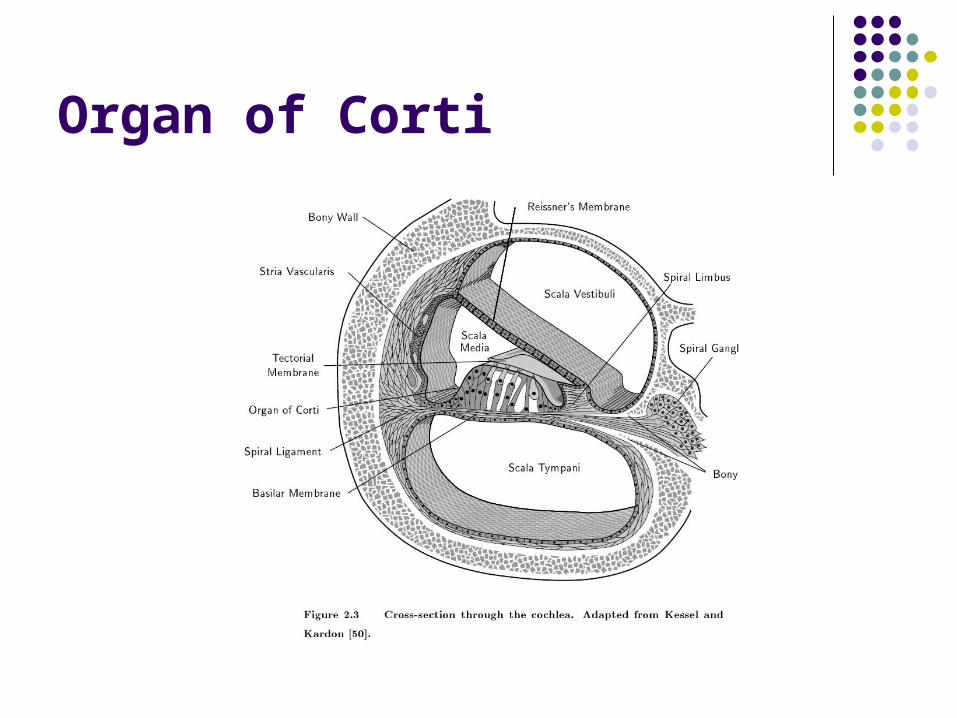
Organ of Corti
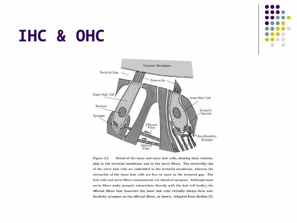
IHC & OHC
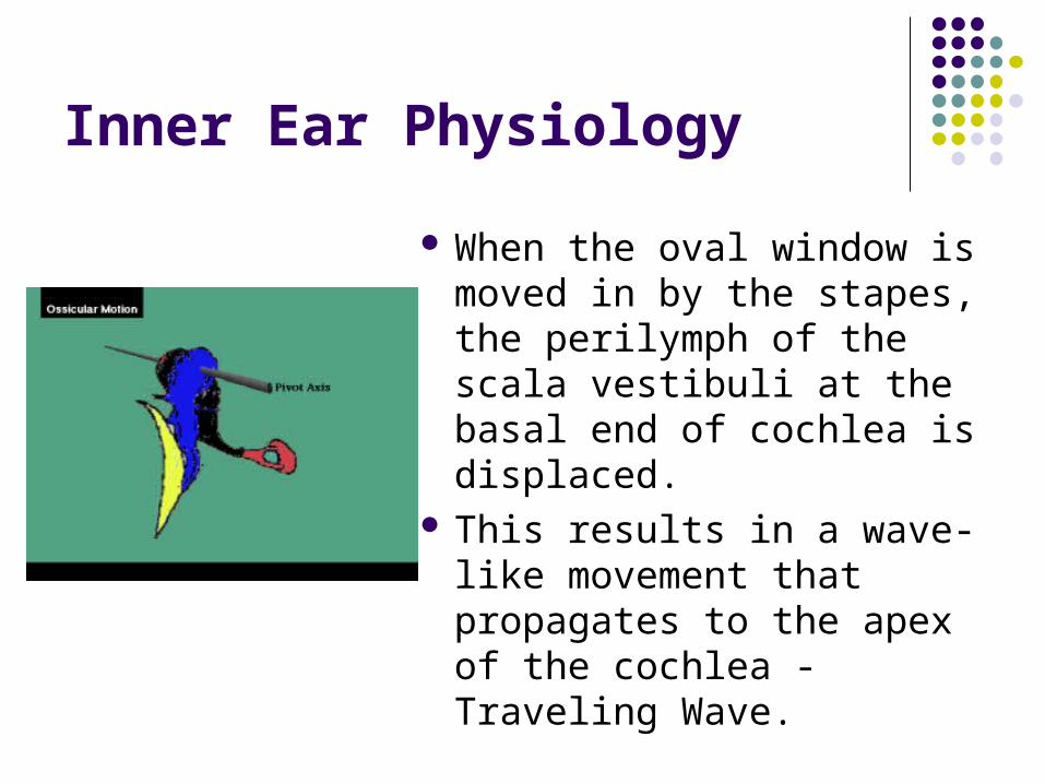
Inner Ear Physiology
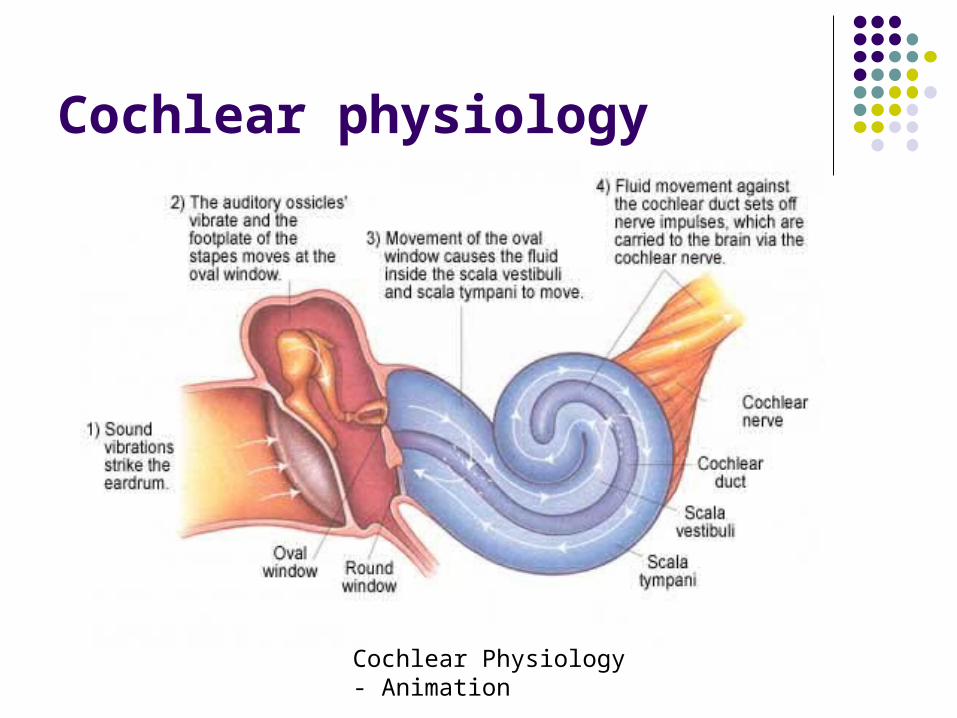
When the oval window is moved in by the stapes, the perilymph of the scala vestibuli at the basal end of cochlea is displaced.
This results in a wave-like movement that propagates to the apex of the cochlea - Traveling Wave.
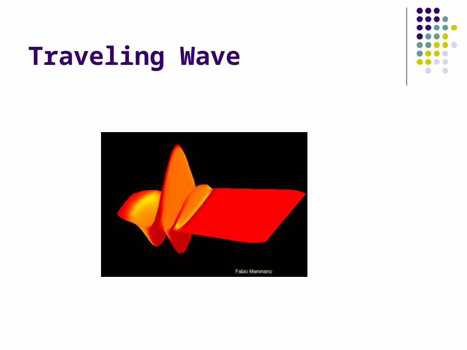
Traveling Wave
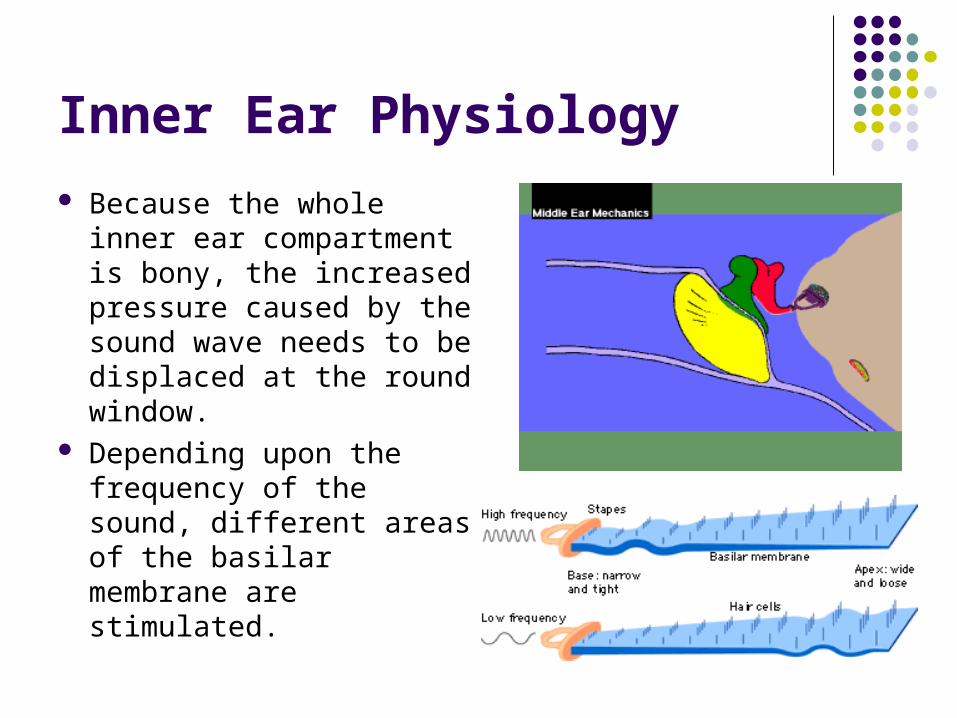
Inner Ear Physiology
Because the whole inner ear compartment is bony, the increased pressure caused by the sound wave needs to be displaced at the round window.
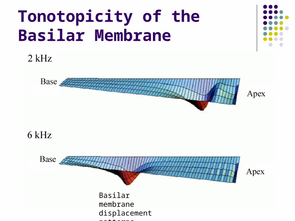
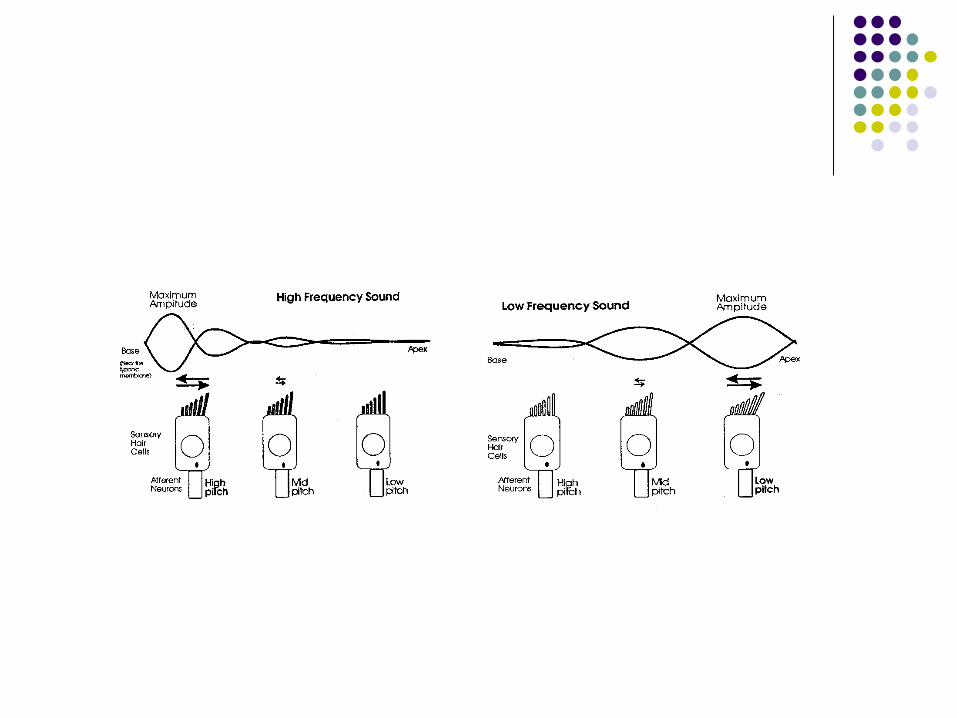
Depending upon the frequency of the sound, different areas of the basilar membrane are stimulated.
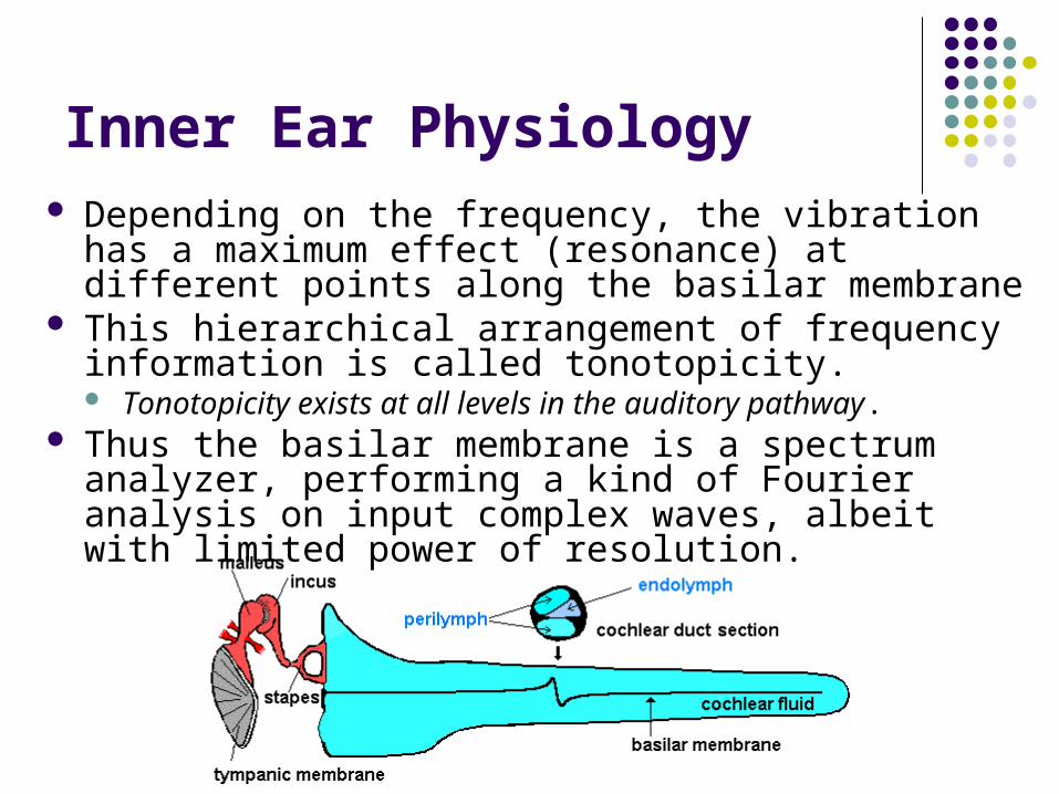
Inner Ear Physiology Depending on the frequency, the vibration has a
maximum effect (resonance) at different points along the basilar membrane
This hierarchical arrangement of frequency information is called tonotopicity. Tonotopicity exists at all levels in the auditory pathway.
Thus the basilar membrane is a spectrum analyzer, performing a kind of Fourier analysis on input complex waves, albeit with limited power of resolution.
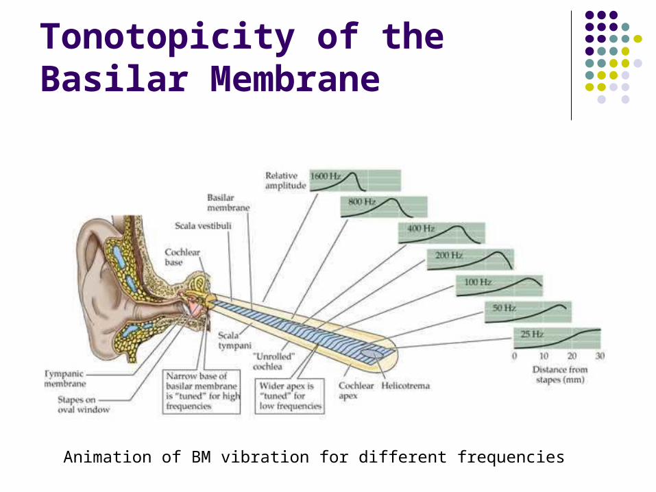
Tonotopicity of the Basilar Membrane
Animation of BM vibration for different frequencies
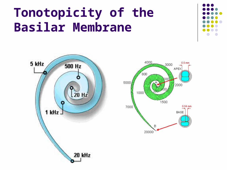
Tonotopicity of the Basilar Membrane
Tonotopicity of the Basilar Membrane
Basilar membrane displacement patterns
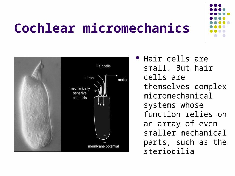
Cochlear micromechanics
Hair cells are small. But hair cells are themselves complex micromechanical systems whose function relies on an array of even smaller mechanical parts, such as the steriocilia
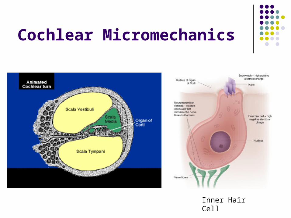
Cochlear Micromechanics
Inner Hair Cell
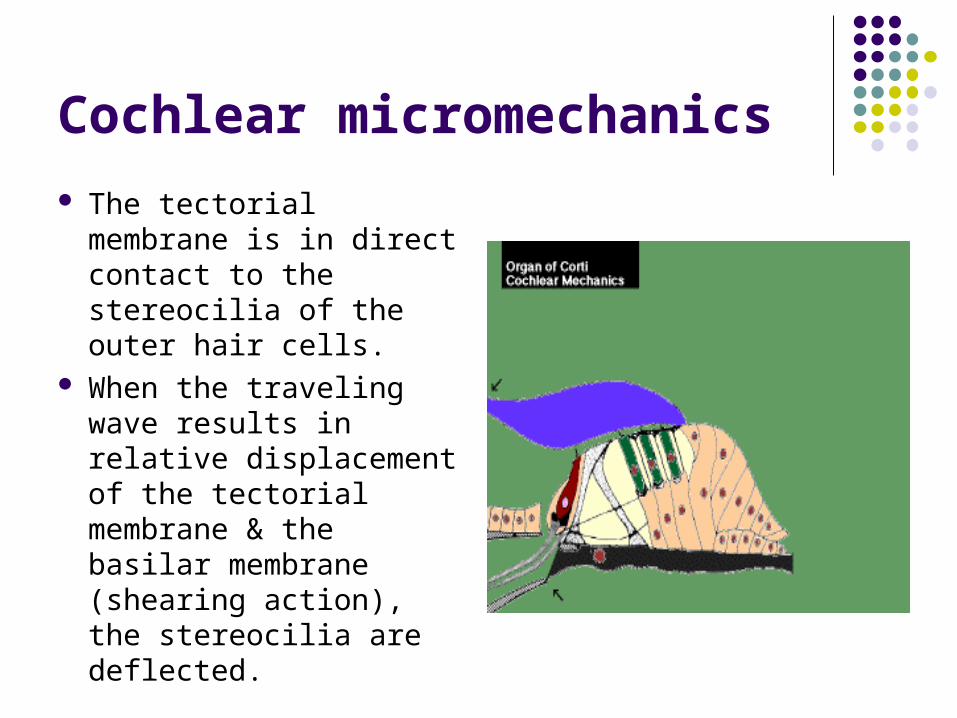
Cochlear micromechanics
The tectorial membrane is in direct contact to the stereocilia of the outer hair cells.
When the traveling wave results in relative displacement of the tectorial membrane & the basilar membrane (shearing action), the stereocilia are deflected.
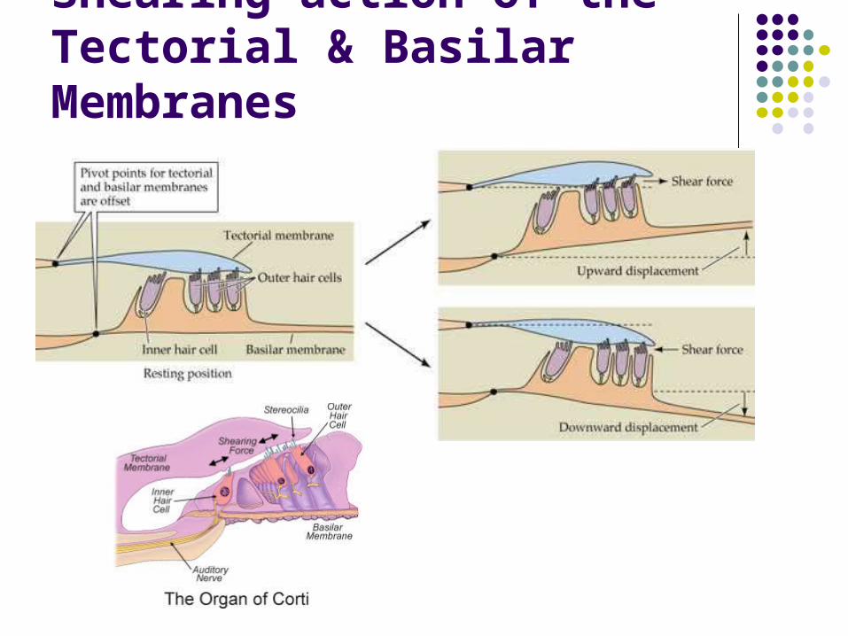
Shearing action of the Tectorial & Basilar Membranes
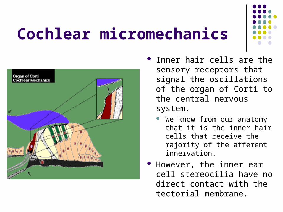
Cochlear micromechanics
Inner hair cells are the sensory receptors that signal the oscillations of the organ of Corti to the central nervous system. We know from our anatomy
that it is the inner hair cells that receive the majority of the afferent innervation.
However, the inner ear cell stereocilia have no direct contact with the tectorial membrane.
Cochlear micromechanics
Recent research has suggested that Inner hair cells sterocilia are moved by the force of the fluid streaming in the narrow channel between the tectorial membrane and the organ of corti.
Cochlear Transduction
The hair cells are responsible for converting the mechanical movement of the sterocilia into electrical energy that can be conducted by the auditory nerve. This process is called Cochlear transduction.
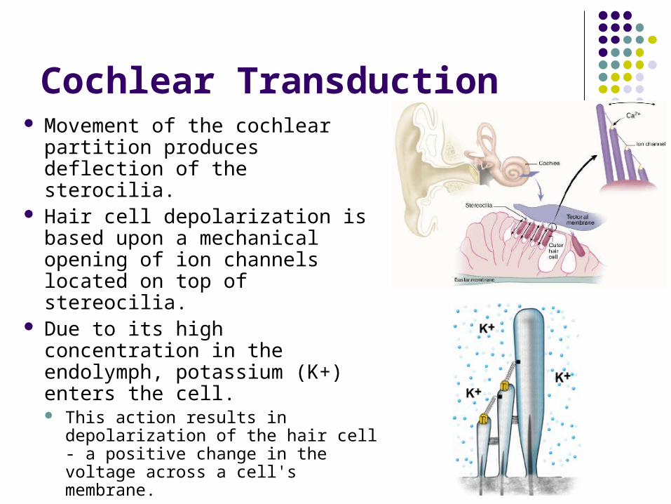
Cochlear Transduction Movement of the cochlear
partition produces deflection of the sterocilia.
Hair cell depolarization is based upon a mechanical opening of ion channels located on top of stereocilia.
Due to its high concentration in the endolymph, potassium (K+) enters the cell. This action results in
depolarization of the hair cell - a positive change in the voltage across a cell's membrane.
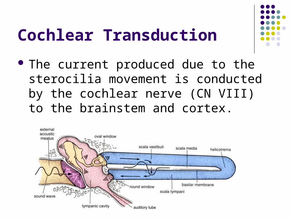
Cochlear Transduction
The current produced due to the sterocilia movement is conducted by the cochlear nerve (CN VIII) to the brainstem and cortex.
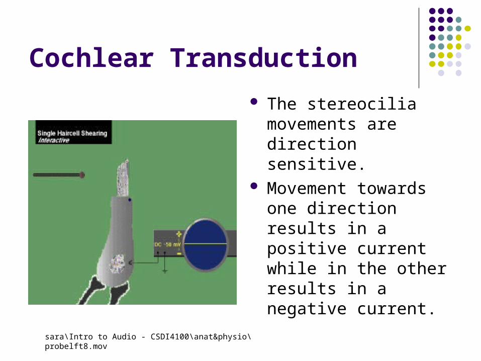
Cochlear Transduction
The stereocilia movements are direction sensitive.
Movement towards one direction results in a positive current while in the other results in a negative current.
sara\Intro to Audio - CSDI4100\anat&physio\probelft8.mov
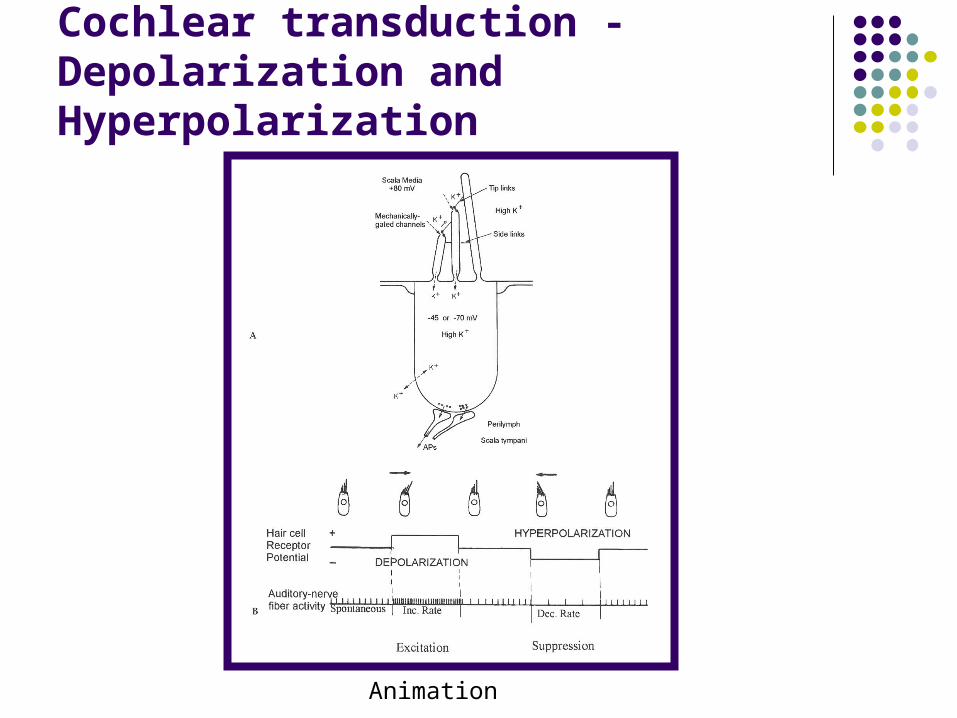
Cochlear transduction - Depolarization and Hyperpolarization
Animation
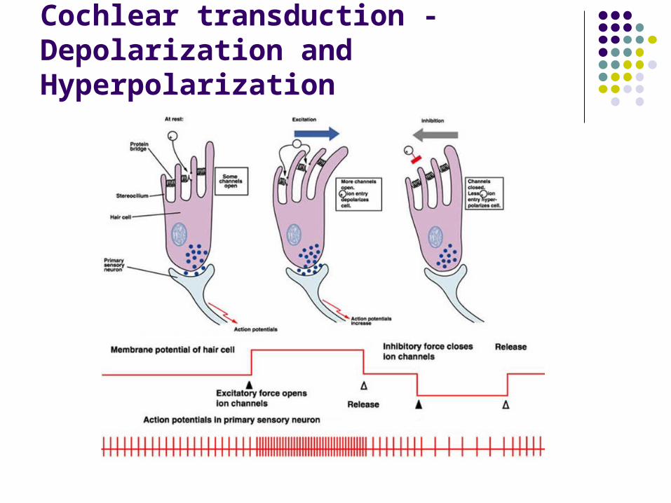
Cochlear transduction - Depolarization and Hyperpolarization
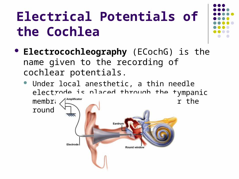
Electrical Potentials of the Cochlea Electrocochleography (ECochG) is the name
given to the recording of cochlear potentials. Under local anesthetic, a thin needle electrode is placed
through the tympanic membrane onto the promontory, near the round window niche.
Electrical Potentials of the Cochlea
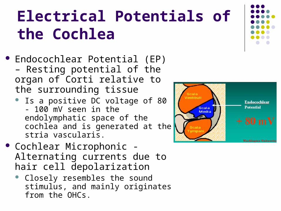
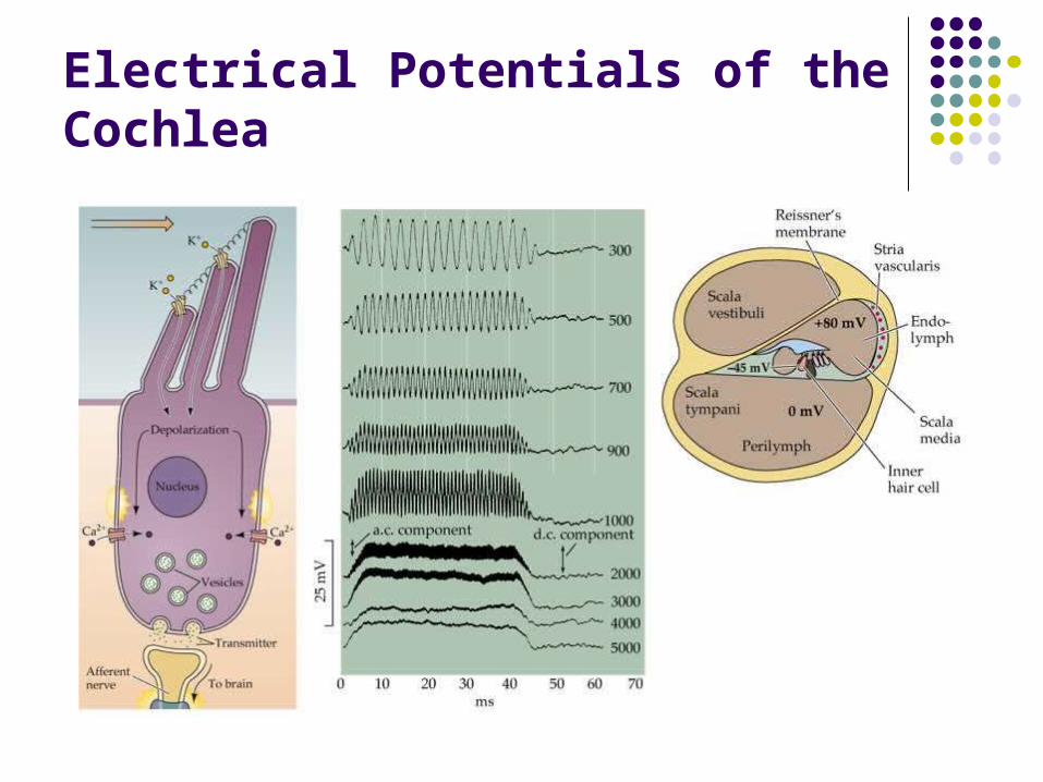
Endocochlear Potential (EP) – Resting potential of the organ of Corti relative to the surrounding tissue Is a positive DC voltage of 80 - 100
mV seen in the endolymphatic space of the cochlea and is generated at the stria vascularis.
Cochlear Microphonic - Alternating currents due to hair cell depolarization Closely resembles the sound
stimulus, and mainly originates from the OHCs.

Electrical Potentials of the Cochlea
Summating Potential – Change of EP in response to sound stimulation (DC current) Is mainly a reflection of IHC activity
Action Potential – Is the result of synchronous activity of the auditory nerve fibers All or none response of auditory nerve fibers
Electrical Potentials of the Cochlea
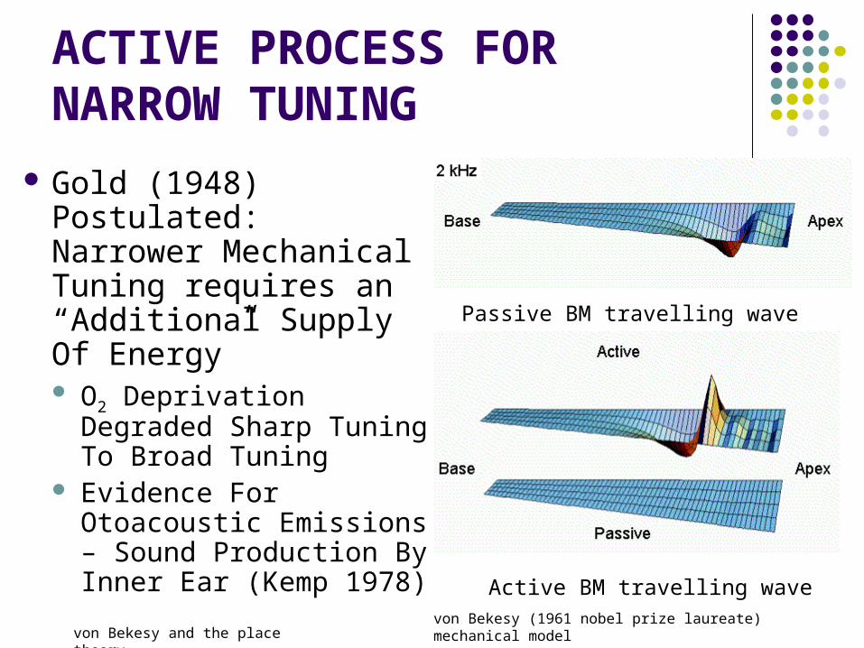
ACTIVE PROCESS FOR NARROW TUNING
Gold (1948) Postulated: Narrower Mechanical Tuning requires an “Additional Supply Of Energy” O2 Deprivation
Degraded Sharp Tuning To Broad Tuning
Evidence For Otoacoustic Emissions – Sound Production By Inner Ear (Kemp 1978)
Passive BM travelling wave
Active BM travelling wavevon Bekesy (1961 nobel prize laureate) mechanical model
von Bekesy and the place theory
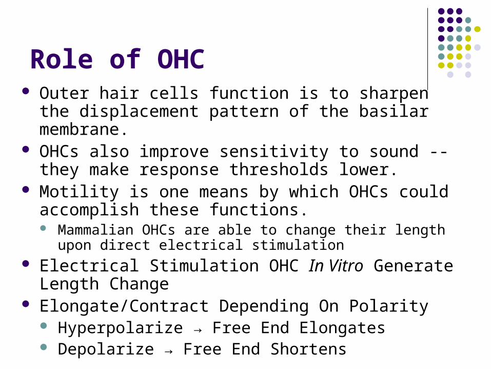
Role of OHC Outer hair cells function is to sharpen the displacement
pattern of the basilar membrane. OHCs also improve sensitivity to sound -- they make
response thresholds lower. Motility is one means by which OHCs could accomplish
these functions. Mammalian OHCs are able to change their length upon direct
electrical stimulation Electrical Stimulation OHC In Vitro Generate Length
Change Elongate/Contract Depending On Polarity
Hyperpolarize → Free End Elongates Depolarize → Free End Shortens
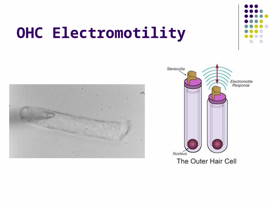
OHC Electromotility
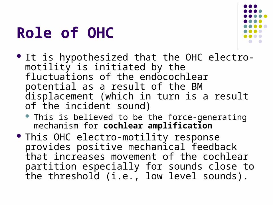
Role of OHC
It is hypothesized that the OHC electro-motility is initiated by the fluctuations of the endocochlear potential as a result of the BM displacement (which in turn is a result of the incident sound) This is believed to be the force-generating
mechanism for cochlear amplification This OHC electro-motility response provides
positive mechanical feedback that increases movement of the cochlear partition especially for sounds close to the threshold (i.e., low level sounds).
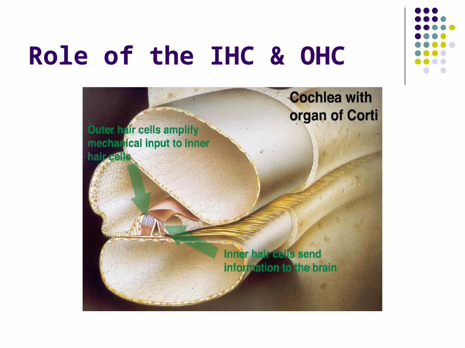
Role of the IHC & OHC
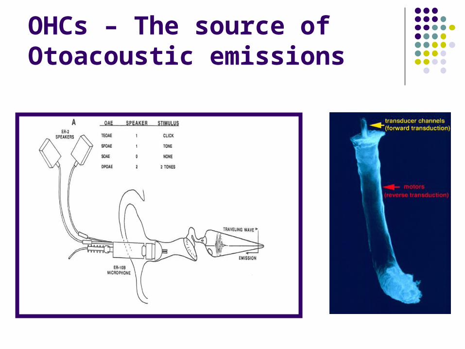
OHCs – The source of Otoacoustic emissions

Why the inner-ear is snail-shaped? Too boost sensitivity to low
frequencies! Although the spiral shape of the
cochlea had little impact on the average vibrational energy traveling along the tube, as the wave progresses, this energy increasingly accumulates near the outside edge of the spiral, rather than remaining evenly spread across it.
Low frequencies travel the furthest into the spiral, so the effect is strongest for them.
D. Manoussaki, E.K. Dimitriadis & R.S. Chadwick (2006)

Why the inner-ear is snail-shaped?
Serpentine Water Slide
Straight Water Slide
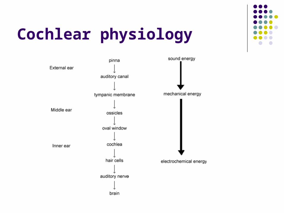
Cochlear physiology – Synopsis
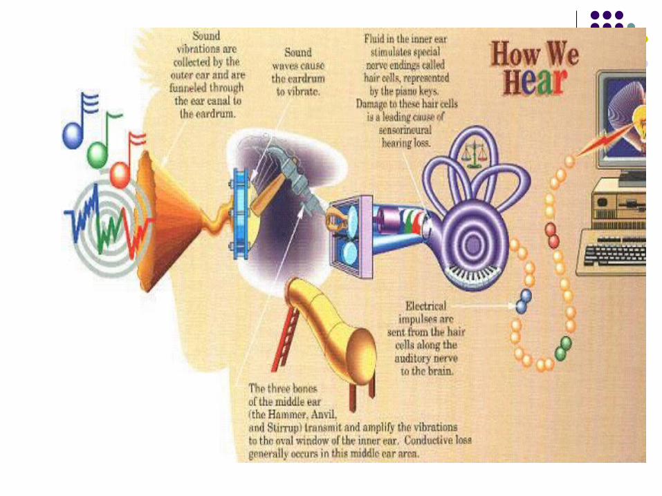
The acoustic energy of the sound waves in the outer ear is converted to mechanical energy by the tympanic membrane.
This mechanical energy is conducted by the middle ear ossicles to the oval window of the inner ear where it results in fluid movement (hydraulic energy).
Cochlear physiology This fluid movement results in deflection of the stereocilia of the
inner hair cells. The deflection of the stereocilia is converted into electrical
energy at the base of the hair cells & conducted by the auditory nerve fibers to higher auditory centers of the brainstem and cortex. The brain interprets it as sound!
In short, the auditory Hair cells are specialized so that motion of their stereocilia changes their electrical potential, resulting in neurotransmitter release and action potentials in the nerve fibers that contact the hair cells.
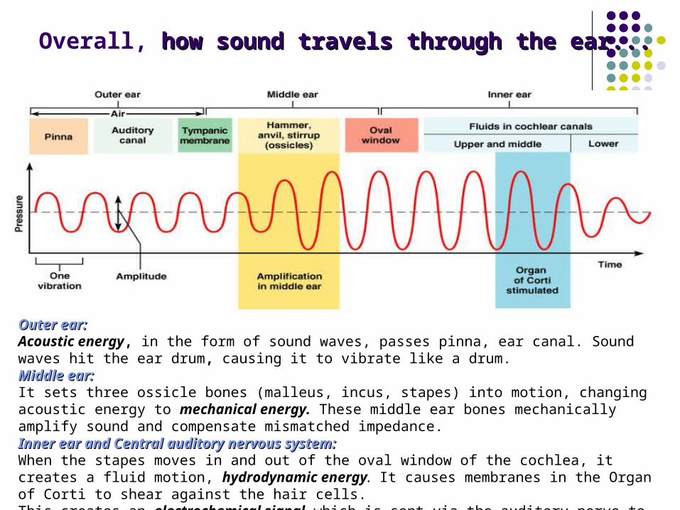
Outer ear:Outer ear:Acoustic energy, in the form of sound waves, passes pinna, ear canal. Sound waves hit the ear drum, causing it to vibrate like a drum. Middle ear:Middle ear: It sets three ossicle bones (malleus, incus, stapes) into motion, changing acoustic energy to mechanical energy. These middle ear bones mechanically amplify sound and compensate mismatched impedance. Inner ear and Central auditory nervous system:Inner ear and Central auditory nervous system:When the stapes moves in and out of the oval window of the cochlea, it creates a fluid motion, hydrodynamic energy. It causes membranes in the Organ of Corti to shear against the hair cells.This creates an electrochemical signal which is sent via the auditory nerve to the brain.
Overall, how sound travels through the ear...how sound travels through the ear...
Cochlear physiology
Cochlear physiology
Cochlear Physiology - Animation
Related Documents


![Inner Ear Anatomy[1]](https://static.cupdf.com/doc/110x72/5528566b4979591c048b47a6/inner-ear-anatomy1.jpg)