Injuries on a skull from the Ancient Bronze Age (Ballabio, Lecco, Italy): a natural or an anthropic origin? Vanessa Samantha Manzon, Ursula Thun Hohenstein, Emanuela Gualdi-Russo * Department of Biology and Evolution, University of Ferrara, Corso Ercole I d’Este 32, 44121 Ferrara, Italy article info Article history: Received 23 December 2011 Received in revised form 5 June 2012 Accepted 8 June 2012 Keywords: Cranial injury Bronze Age SEM Taphonomy 3D digital models Facial reconstruction abstract This paper discusses the results of the analysis of a female skull from a collective burial dated to the Ancient Bronze Age in Italy (Ballabio, LC). A virtual restoration and 3D reconstruction was also produced from the digitalized skull to complete the damaged parts and to recreate the facial appearance of this young adult female from the Bronze Age. The skull shows clear evidence of post-mortem modifications, as some series of scraping marks on the external cranial vault cross the parietal bones longitudinally. The contemporaneous presence of taphonomic linear marks on the skull and periostitis on the frontal bone, as well as the provenance of the specimen from a secondary burial (a typical funerary habit documented in Italy during the Copper Age and Ancient Bronze Age), makes it difficult to interpret the case (scalping, surgery, or ritual practice linked to secondary burial). The advanced methods used to analyse the skull surface allowed us to discriminate intentional marks from modifications due to other taphonomic processes and to determine the timing of their formation (peri- or post-mortem). The possibility that the scraping marks are related to a ritual practice, conducted during the individual’s life (with specific symbolic or social value) or after death or at the moment of secondary burial, is discussed. Ó 2012 Elsevier Ltd. All rights reserved. 1. Introduction The Early Bronze Age of northern Italy (2300e1600 BC) has been well documented in terms of its settlements (e.g. the pile-dwelling villages of the “Polada culture”), whereas its funerary aspects were almost unknown until a few years ago (De Marinis, 2003; Leonini and Sarti, 2006). Recent discoveries in the northern Italian Alps and Pre-Alps, especially in the regions of Veneto, Lombardy and Trentino, have allowed the investigation of some aspects of the burial rituals of the communities that lived in these areas during the Chalcolithic and Bronze Age (De Marinis, 2003; Leonini and Sarti, 2006). The use of natural environments for burial areas, well known in western Europe and southern France during the 4th and 3rd millennium BC (Duday, 2005), also occurred in northern Italy during the Early Bronze Age (2300e1600 BP). Different burial rites occurred in the same area (and even in the same burial site) with the following possible modalities: single individual burials, collective burials, cases of partial or total cremation, primary burials, secondary burials with both displacement of skeletal remains (perhaps as a relic) and disarticulation and commingling of several individuals buried together (Leonini and Sarti, 2006). However, the ideologies and ceremonies that accompanied these burials and caused their differentiation are poorly known. Some evidence of ceremonies at the time of secondary burial, such as ritual fires, disarticulation and new placement of the skeletal remains in structured charnel houses, has been reported for Lombardy (Barfield et al., 1995; Baioni, 2004), Trentino (Nicolis, 1996, 2001), Piedmont and Valle d’Aosta (Mezzena,1997; Gambari and Venturino Gambari, 2003). In the last two regions, the construction of “houses of the dead”, such as dolmens and collective burial chambers, is also known, according to a practice similar to those widely used in southern France during the Chalcolithic (Duday, 2005) and in the Near East during the Bronze Age (Chesson, 1999; Mednikova, 2002). A skull showing extraordinary evidence of scraping marks on the parietals, together with a bony reaction (periostitis) of the pericranium in the middle of the frontal bone, was recently discovered in a collective burial from the Pre-Alps of Lombardy dated to the transition between the Early and Middle Bronze Age. This find raises many questions about the rituals linked to primary and secondary burial, funeral ideology and belief, religion, medical and therapeutic knowledge, as well as intra- and inter-tribal aggression in the Bronze Age communities of northern Italy. As far as we know, no case of scalping had been identified in Italy. According to Mednikova (2002), the term “scalping” means the removal of skin with the hair from the head of a dead or living * Corresponding author. Tel.: þ39 0532293793; fax: þ39 0532208561. E-mail addresses: [email protected] (V.S. Manzon), [email protected] (U. Thun Hohenstein), [email protected] (E. Gualdi-Russo). Contents lists available at SciVerse ScienceDirect Journal of Archaeological Science journal homepage: http://www.elsevier.com/locate/jas 0305-4403/$ e see front matter Ó 2012 Elsevier Ltd. All rights reserved. http://dx.doi.org/10.1016/j.jas.2012.06.003 Journal of Archaeological Science 39 (2012) 3428e3435

Welcome message from author
This document is posted to help you gain knowledge. Please leave a comment to let me know what you think about it! Share it to your friends and learn new things together.
Transcript
at SciVerse ScienceDirect
Journal of Archaeological Science 39 (2012) 3428e3435
Contents lists available
Journal of Archaeological Science
journal homepage: http: / /www.elsevier .com/locate/ jas
Injuries on a skull from the Ancient Bronze Age (Ballabio, Lecco, Italy):a natural or an anthropic origin?
Vanessa Samantha Manzon, Ursula Thun Hohenstein, Emanuela Gualdi-Russo*
Department of Biology and Evolution, University of Ferrara, Corso Ercole I d’Este 32, 44121 Ferrara, Italy
a r t i c l e i n f o
Article history:Received 23 December 2011Received in revised form5 June 2012Accepted 8 June 2012
Keywords:Cranial injuryBronze AgeSEMTaphonomy3D digital modelsFacial reconstruction
* Corresponding author. Tel.: þ39 0532293793; faxE-mail addresses: [email protected] (V.S. Manzon),
Hohenstein), [email protected] (E. Gualdi-Rus
0305-4403/$ e see front matter � 2012 Elsevier Ltd.http://dx.doi.org/10.1016/j.jas.2012.06.003
a b s t r a c t
This paper discusses the results of the analysis of a female skull from a collective burial dated to theAncient Bronze Age in Italy (Ballabio, LC). A virtual restoration and 3D reconstruction was also producedfrom the digitalized skull to complete the damaged parts and to recreate the facial appearance of thisyoung adult female from the Bronze Age. The skull shows clear evidence of post-mortemmodifications, assome series of scraping marks on the external cranial vault cross the parietal bones longitudinally. Thecontemporaneous presence of taphonomic linear marks on the skull and periostitis on the frontal bone,as well as the provenance of the specimen from a secondary burial (a typical funerary habit documentedin Italy during the Copper Age and Ancient Bronze Age), makes it difficult to interpret the case (scalping,surgery, or ritual practice linked to secondary burial). The advanced methods used to analyse the skullsurface allowed us to discriminate intentional marks from modifications due to other taphonomicprocesses and to determine the timing of their formation (peri- or post-mortem). The possibility that thescraping marks are related to a ritual practice, conducted during the individual’s life (with specificsymbolic or social value) or after death or at the moment of secondary burial, is discussed.
� 2012 Elsevier Ltd. All rights reserved.
1. Introduction
The Early Bronze Age of northern Italy (2300e1600 BC) has beenwell documented in terms of its settlements (e.g. the pile-dwellingvillages of the “Polada culture”), whereas its funerary aspects werealmost unknown until a few years ago (De Marinis, 2003; Leoniniand Sarti, 2006). Recent discoveries in the northern Italian Alpsand Pre-Alps, especially in the regions of Veneto, Lombardy andTrentino, have allowed the investigation of some aspects of theburial rituals of the communities that lived in these areas during theChalcolithic and Bronze Age (De Marinis, 2003; Leonini and Sarti,2006). The use of natural environments for burial areas, wellknown in western Europe and southern France during the 4th and3rd millennium BC (Duday, 2005), also occurred in northern Italyduring the Early Bronze Age (2300e1600 BP). Different burial ritesoccurred in the same area (and even in the same burial site)with thefollowing possible modalities: single individual burials, collectiveburials, cases of partial or total cremation, primary burials,secondary burials with both displacement of skeletal remains(perhaps as a relic) and disarticulation and commingling of several
: þ39 [email protected] (U. Thunso).
All rights reserved.
individuals buried together (Leonini and Sarti, 2006). However, theideologies and ceremonies that accompanied these burials andcaused their differentiation are poorly known. Some evidence ofceremonies at the time of secondary burial, such as ritual fires,disarticulation and new placement of the skeletal remains instructured charnel houses, has been reported for Lombardy (Barfieldet al., 1995; Baioni, 2004), Trentino (Nicolis, 1996, 2001), Piedmontand Valle d’Aosta (Mezzena,1997; Gambari and Venturino Gambari,2003). In the last two regions, the construction of “houses of thedead”, such as dolmens and collective burial chambers, is alsoknown, according to a practice similar to those widely used insouthern France during the Chalcolithic (Duday, 2005) and in theNear East during the Bronze Age (Chesson,1999;Mednikova, 2002).
A skull showing extraordinary evidence of scraping marks onthe parietals, together with a bony reaction (periostitis) of thepericranium in the middle of the frontal bone, was recentlydiscovered in a collective burial from the Pre-Alps of Lombardydated to the transition between the Early and Middle Bronze Age.This find raises many questions about the rituals linked to primaryand secondary burial, funeral ideology and belief, religion, medicaland therapeutic knowledge, as well as intra- and inter-tribalaggression in the Bronze Age communities of northern Italy.
As far as we know, no case of scalping had been identified inItaly. According to Mednikova (2002), the term “scalping” meansthe removal of skin with the hair from the head of a dead or living
V.S. Manzon et al. / Journal of Archaeological Science 39 (2012) 3428e3435 3429
person. Despite the enormous amount of data available for theAmericas (Axtell and Sturtevant, 1980; Allen et al., 1985; Willey,1990; Owsley, 1994; Jacobi et al., 1996; Milner, 1999; Toyne, 2011,etc.), there is relatively little evidence of scalping in Eurasia (Reese,1940; Anger and Diek, 1978; Jordana et al., 2009; Mednikova, 2002;Murphy et al., 2002; Cáceres et al., 2007). Archaeological findingsprovide evidence of scalping in Eurasia from the Neolithic to theIron Age, especially in the steppes of Eurasia and Scandinavia(Mednikova, 2002). Scalping was surely practised in Eurasia inantiquity: Herodotus was the first to describe scalping in his His-toriae (V century BC), considering it a typical activity of theScythians (a group of archaeological cultures living in the steppes ofUkraine, Russia, Kazakhstan, Mongolia and northern Chinabetween the VII and II centuries BC) for the purpose of trophytaking during warfare (Murphy et al., 2002; Jordana et al., 2009).Other written accounts report scalping practised as a militarycustom, a penalty for thieves (e.g. Anglo-Saxon laws) and a medicalintervention, up until the last written documentation of scalping inEurasia dating to 1933 during the Soviet colonization of Siberia(Mednikova, 2002).
Although the clearmarks on the examined skull initially led us tobelieve that this could be a case of scalping practised in the BronzeAge, some characteristics suggested that it was not a case of scalpinglinked to warfare. This led the discussion into even less well knownareas, such as rituals accompanying secondary burial or aspects ofprimitive surgery. Therefore, the aim of our study was to documenttheparticular injuries observedon the skull and to interpret thembymeans of advanced techniques. Furthermore, to acquire a better ideaof the physical appearance of the individual, we carried out a facialreconstruction by the tissue depth method after virtual restorationand 3D reconstruction of the digitalized skull.
2. The site
The site of Ballabio-Prato della Chiesa (Lecco, Italy) is a rockshelter at 700 m above sea level, at the foot of a rocky wall in thePre-Alps of Lombardy (northern Italy). It was discovered in 2004during a geological survey, when a remarkable quantity of humanbones was found and the police were alerted. After determinationof the ancient funerary connotation of the shelter, it was partiallyexcavated between 2006 and 2007 (Lorenzi et al., 2010). Twoadjacent funerary structures, interpreted as primary burials, wereidentified behind the rocky wall (Grave 1 and Grave 2). Two nichesinside the rocky wall were found near each structure, containinghuman remains belonging to many different individuals. Theseareas, called “Grave 1 Area” and “Grave 2 Area”, were interpreted ascollective secondary burials (Lorenzi et al., 2010), where the skel-etal remains were definitively placed in a chaotic manner afterhaving decomposed elsewhere. The burial context was14C dated tothe final stages of the Ancient Bronze Age/Early Middle Bronze Age,precisely 3230 � 90 years BP.
The ritual practice found at Ballabio refers structurally toarchaeological evidences documented from the Chalcolithic to theAncient BronzeAge in thenorthern ItalianAlps andPre-Alps: the useof single and collective burials in rock shelters and natural caves,frequently in secondary deposition, is well known in the regions ofVeneto, Piedmont, Liguria and Lombardy (Nicolis, 2001; De Marinis,2003; Leonini and Sarti, 2006). In particular, Ballabio shows conver-gences with rituals recorded in Lombardy and in the provinces ofVerona and Trento from the initial phase of the Bronze Age (BA A1).
3. Materials and methods
The skull arrived at the Laboratory of Archaeo-anthropology andForensic Anthropology of the University of Ferrara (Dept. of Biology
and Evolution) together with the other osteological remains fromthe Ballabio site (Lecco, Italy). However, as it was recovered duringthe first intervention of the police, it was preserved out of itsarchaeological context in a metal box with the single topographicindication “Shelter 1”, together with some other osteological finds.The osteological material was restored and subjected to prelimi-nary anthropological studies (Gualdi-Russo et al., 2010).
3.1. Anthropological analysis
The skull was analysed anthropologically by means of tradi-tional and innovative techniques. Following standard osteologicalprocedures, the age-at-death was estimated based on closure ofecto- and endo-cranial sutures (Acsádi and Nemeskéri, 1970; WEA,1980) and dental wear (Brothwell, 1981). The sex was determinedby standard anthropological methods (Acsádi and Nemeskéri, 1970;WEA, 1980). The skull was also analysed macroscopically torecognize eventual pathological conditions, after which a radio-graphic examinationwas carried out at S. Anna Hospital, Laboratoryof Radiology (Ferrara, Italy).
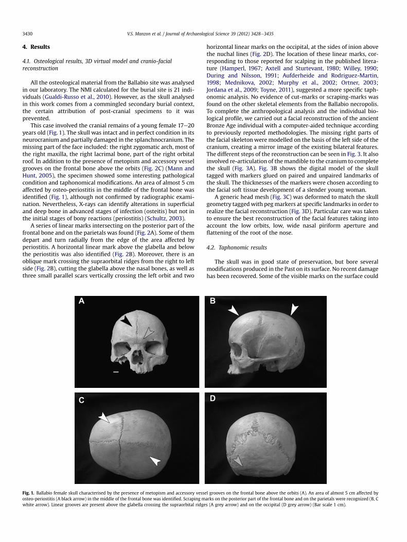
Following innovative procedures of virtual anthropology, weperformed three-dimensional scanning with a NextEngine DesktopScanner, which works on the principle of optical triangulation(Guidi et al., 2010). A 3-D model of the skull was generated from 10scans using the scanning and modelling Scan Studio Core software(NextEngine, Inc.). Amandible from the Ballabio site, also kept in themetal box, has characteristics consistent with the skull. Hence, thismandible was scanned to complete the previous 3D model of theskull and to perform the reconstruction. The virtual skull, completedwith the mirror-image right maxillary and the digitalized mandible,was positioned in the Frankfurt plane and then equipped with pegmarkers reflecting the tissue depth in Caucasians at different points(landmarks) (De Greef et al., 2006) according to the sex and age ofthe individual and assuming a “lean build”. All the surface manip-ulations in the 3D environment (virtual restoration and computer-aided facial reconstruction) were carried out using Autodesk Maya3D software. A generic head model (FaceGen, Singular InversionsInc.) was imported and moulded on the skull tagged with pegmarkers oriented orthogonally to the skull.
The main rules for facial reconstruction were applied accordingto indications by Iscan and Helmer (1993), Yoshino and Seta (2000),Kähler et al. (2003) and Wilkinson (2004).
3.2. Taphonomic analysis
The surface of the skull was analysed from a taphonomicperspective using techniques developed in archaeozoology (Pottsand Shipman, 1981; Shipman and Rose, 1983, 1988; D’Errico andGiacobini, 1985), anthropology (D’Errico and Giacobini, 1985) andforensic science (Bartelink et al., 2001). Linear marks, identifiedmacroscopically, were analysed under a Leica MZ3 binocularstereomicroscope in the Laboratory of Archaeozoology in order todiscriminate between traces left by intentional human action andmarks left by other taphonomic events. Replicas of the marks(Orschiedt et al., 2003; Rose, 1983) were made in order to performSEM analyses on small areas of the surfaces bearing the scrapingand other linear marks, thus avoiding metallization of the originalspecimen and assuring a high degree of definition/reproduction ofthe traces below 1 m (Shipman, 1981; D’Errico, 1988; Giacobini,1995). We used silicone elastomer (Provil L Bayer, Leverkusen,Germany) for the casts and epoxy resin (Araldite LY 554 andHardener HY 956, Ciba Geigy, Basel, Switzerland) for the positivecopies. Scanning electron microscope (SEM) analyses were carriedout on the replicas at the ElectronMicroscopy Centre of the Dept. ofChemistry, University of Ferrara.
V.S. Manzon et al. / Journal of Archaeological Science 39 (2012) 3428e34353430
4. Results
4.1. Osteological results, 3D virtual model and cranio-facialreconstruction
All the osteological material from the Ballabio site was analysedin our laboratory. The NMI calculated for the burial site is 21 indi-viduals (Gualdi-Russo et al., 2010). However, as the skull analysedin this work comes from a commingled secondary burial context,the certain attribution of post-cranial specimens to it wasprevented.
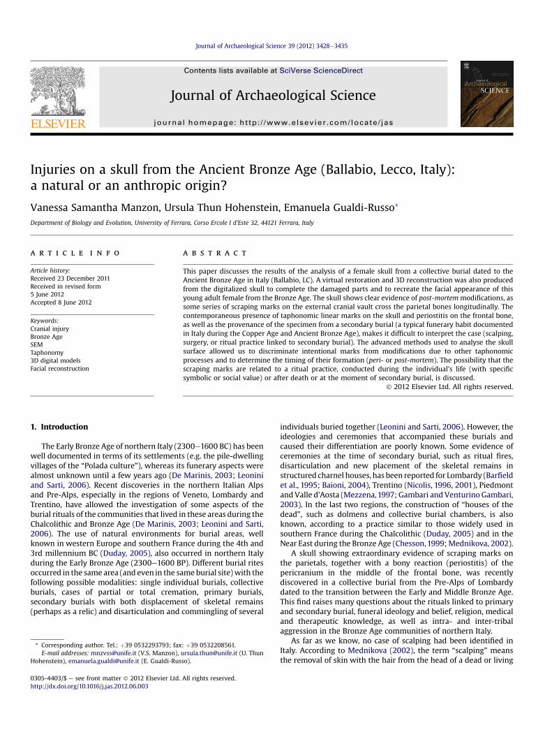
This case involved the cranial remains of a young female 17e20years old (Fig. 1). The skull was intact and in perfect condition in itsneurocranium and partially damaged in the splanchnocranium. Themissing part of the face included: the right zygomatic arch, most ofthe right maxilla, the right lacrimal bone, part of the right orbitalroof. In addition to the presence of metopism and accessory vesselgrooves on the frontal bone above the orbits (Fig. 2C) (Mann andHunt, 2005), the specimen showed some interesting pathologicalcondition and taphonomical modifications. An area of almost 5 cmaffected by osteo-periostitis in the middle of the frontal bone wasidentified (Fig. 1), although not confirmed by radiographic exami-nation. Nevertheless, X-rays can identify alterations in superficialand deep bone in advanced stages of infection (osteitis) but not inthe initial stages of bony reactions (periostitis) (Schultz, 2003).
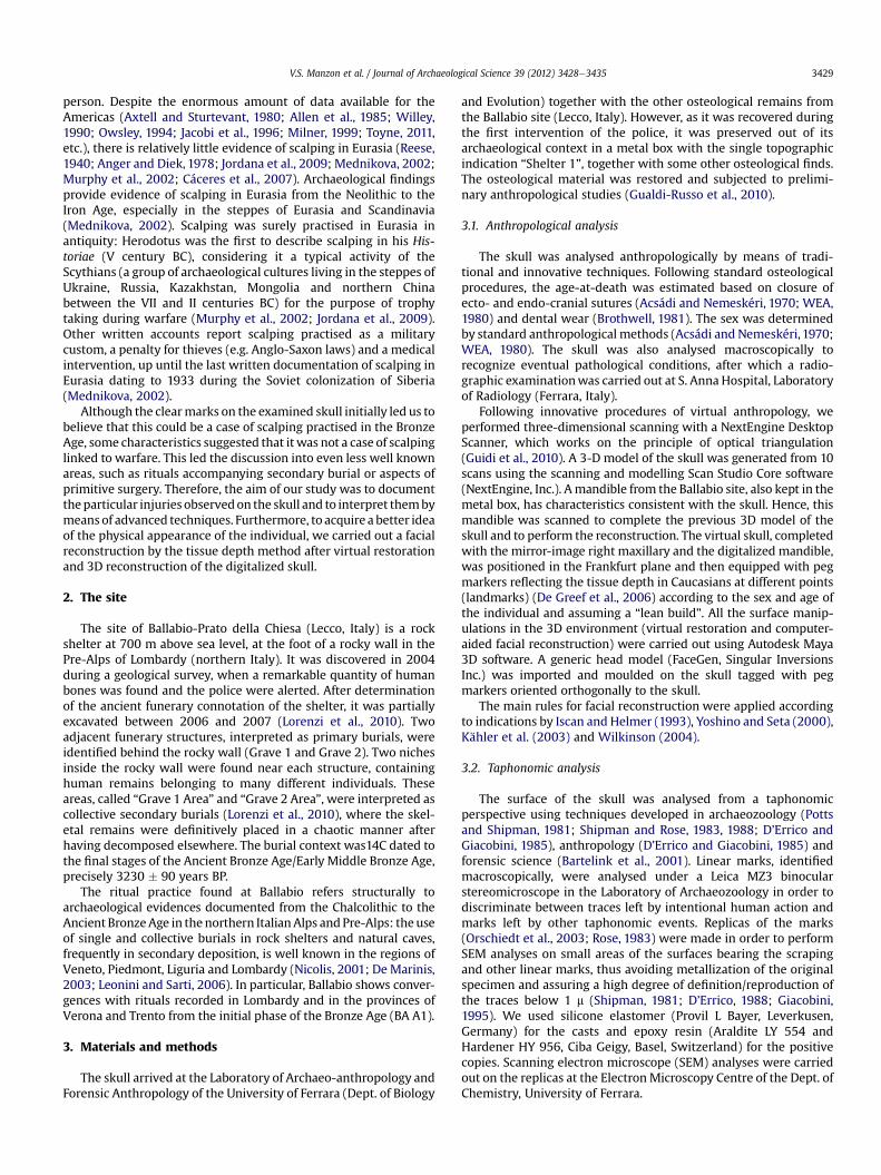
A series of linear marks intersecting on the posterior part of thefrontal bone and on the parietals was found (Fig. 2A). Some of themdepart and turn radially from the edge of the area affected byperiostitis. A horizontal linear mark above the glabella and belowthe periostitis was also identified (Fig. 2B). Moreover, there is anoblique mark crossing the supraorbital ridges from the right to leftside (Fig. 2B), cutting the glabella above the nasal bones, as well asthree small parallel scars vertically crossing the left orbit and two
Fig. 1. Ballabio female skull characterised by the presence of metopism and accessory vesseosteo-periostitis (A black arrow) in the middle of the frontal bone was identified. Scraping mwhite arrow). Linear grooves are present above the glabella crossing the supraorbital ridge
horizontal linear marks on the occipital, at the sides of inion abovethe nuchal lines (Fig. 2D). The location of these linear marks, cor-responding to those reported for scalping in the published litera-ture (Hamperl, 1967; Axtell and Sturtevant, 1980; Willey, 1990;During and Nilsson, 1991; Aufderheide and Rodriguez-Martin,1998; Mednikova, 2002; Murphy et al., 2002; Ortner, 2003;Jordana et al., 2009; Toyne, 2011), suggested a more specific taph-onomic analysis. No evidence of cut-marks or scraping-marks wasfound on the other skeletal elements from the Ballabio necropolis.To complete the anthropological analysis and the individual bio-logical profile, we carried out a facial reconstruction of the ancientBronze Age individual with a computer-aided technique accordingto previously reported methodologies. The missing right parts ofthe facial skeletonwere modelled on the basis of the left side of thecranium, creating a mirror image of the existing bilateral features.The different steps of the reconstruction can be seen in Fig. 3. It alsoinvolved re-articulation of themandible to the cranium to completethe skull (Fig. 3A). Fig. 3B shows the digital model of the skulltagged with markers glued on paired and unpaired landmarks ofthe skull. The thicknesses of the markers were chosen according tothe facial soft tissue development of a slender young woman.
A generic head mesh (Fig. 3C) was deformed to match the skullgeometry taggedwith pegmarkers at specific landmarks in order torealize the facial reconstruction (Fig. 3D). Particular care was takento ensure the best reconstruction of the facial features taking intoaccount the low orbits, low, wide nasal piriform aperture andflattening of the root of the nose.
4.2. Taphonomic results
The skull was in good state of preservation, but bore severalmodifications produced in the Past on its surface. No recent damagehas been recovered. Some of the visible marks on the surface could
l grooves on the frontal bone above the orbits (A). An area of almost 5 cm affected byarks on the posterior part of the frontal bone and on the parietals were recognized (B, Cs (A grey arrow) and on the occipital (D grey arrow) (Bar scale 1 cm).
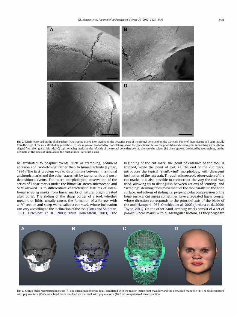
Fig. 2. Marks observed on the skull surface. (A) Scraping marks intersecting on the posterior part of the frontal bone and on the parietals. Some of them depart and spin radiallyfrom the edge of the area affected by periostitis. (B) Linear groove, produced by root-etching, above the glabella and below the periostitis and crossing the superciliary arches (browridges) from the right to left side. (C) Light scraping marks on the left side of the frontal bone that overlap the vascular sulcus. (D) Linear groove, produced by root-etching, on theoccipital, at the sides of inion above the nuchal lines (Bar scale 1 cm).
V.S. Manzon et al. / Journal of Archaeological Science 39 (2012) 3428e3435 3431
be attributed to edaphic events, such as trampling, sedimentabrasion and root-etching, rather than to human activity (Lyman,1994). The first problem was to discriminate between intentionalanthropic marks and the other traces left by taphonomic and post-depositional events. The micro-morphological observation of theseries of linear marks under the binocular stereo-microscope andSEM allowed us to differentiate characteristic features of inten-tional scraping marks from linear marks of natural origin createdafter burial. The sliding of the sharp border of a tool, whethermetallic or lithic, usually causes the formation of a furrow witha “V” section and steep walls, called a cut mark, whose inclinationcan vary according to the inclination of the tool (Potts and Shipman,1981; Orschiedt et al., 2003; Thun Hohenstein, 2003). The
Fig. 3. Cranio-facial reconstruction steps: (A) The virtual model of the skull, completed withwith peg markers. (C) Generic head mesh moulded on the skull with peg markers. (D) Fina
beginning of the cut mark, the point of entrance of the tool, isthinned, while the point of exit, i.e. the end of the cut mark,introduces the typical “swallowtail” morphology, with divergentinclination of the last trait. Through microscopic observation of thecut marks, it is also possible to reconstruct the way the tool wasused, allowing us to distinguish between actions of “cutting” and“scraping”, deriving frommovement of the tool parallel to the bonesurface, and actions of sliding, i.e. perpendicular compression of thebone surface. Cut marks sometimes have a repeated linear course,whose direction corresponds to the principal axis of the blade ofthe tool (Hamperl, 1967; Orschiedt et al., 2003; Jordana et al., 2009;Toyne, 2011). On the other hand, scraping marks consist of a set ofparallel linear marks with quadrangular bottom, as they originate
the mirror-image right maxillary and the digitalized mandible. (B) The skull equippedl computerized reconstruction.
V.S. Manzon et al. / Journal of Archaeological Science 39 (2012) 3428e34353432
from a movement perpendicular to the principal axis of the blade;in practice, the tool is used in a transverse direction with respect tothe surface of the bone to scrape away the overlying tissues.
In contrast, post-depositional linear marks left by sediments donot have a prevalent direction and often cross each other, theirbottom is usually flat with a quadrangular section and the startingpoint is abrupted; root-etching is characterized by rounded (“U-shaped”) bottom, showing branched lines wandering from themain one.
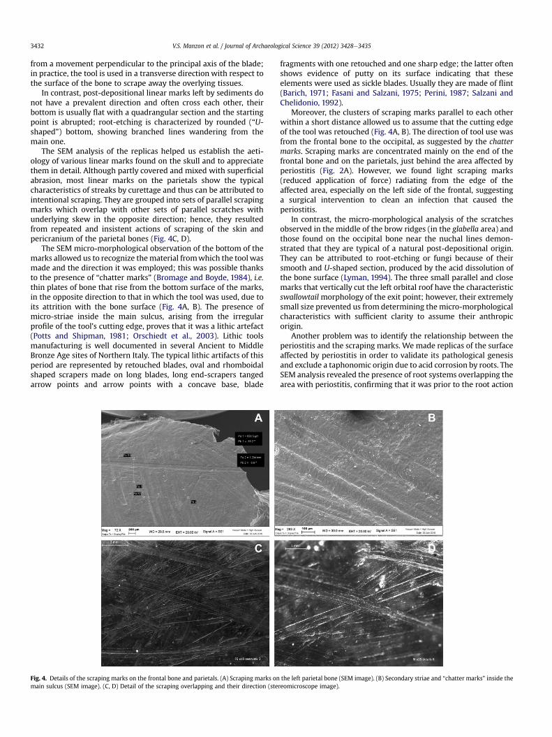
The SEM analysis of the replicas helped us establish the aeti-ology of various linear marks found on the skull and to appreciatethem in detail. Although partly covered and mixed with superficialabrasion, most linear marks on the parietals show the typicalcharacteristics of streaks by curettage and thus can be attributed tointentional scraping. They are grouped into sets of parallel scrapingmarks which overlap with other sets of parallel scratches withunderlying skew in the opposite direction; hence, they resultedfrom repeated and insistent actions of scraping of the skin andpericranium of the parietal bones (Fig. 4C, D).
The SEM micro-morphological observation of the bottom of themarks allowed us to recognize thematerial fromwhich the tool wasmade and the direction it was employed; this was possible thanksto the presence of “chatter marks” (Bromage and Boyde, 1984), i.e.thin plates of bone that rise from the bottom surface of the marks,in the opposite direction to that in which the tool was used, due toits attrition with the bone surface (Fig. 4A, B). The presence ofmicro-striae inside the main sulcus, arising from the irregularprofile of the tool’s cutting edge, proves that it was a lithic artefact(Potts and Shipman, 1981; Orschiedt et al., 2003). Lithic toolsmanufacturing is well documented in several Ancient to MiddleBronze Age sites of Northern Italy. The typical lithic artifacts of thisperiod are represented by retouched blades, oval and rhomboidalshaped scrapers made on long blades, long end-scrapers tangedarrow points and arrow points with a concave base, blade
Fig. 4. Details of the scraping marks on the frontal bone and parietals. (A) Scraping marks onmain sulcus (SEM image). (C, D) Detail of the scraping overlapping and their direction (ste
fragments with one retouched and one sharp edge; the latter oftenshows evidence of putty on its surface indicating that theseelements were used as sickle blades. Usually they are made of flint(Barich, 1971; Fasani and Salzani, 1975; Perini, 1987; Salzani andChelidonio, 1992).
Moreover, the clusters of scraping marks parallel to each otherwithin a short distance allowed us to assume that the cutting edgeof the tool was retouched (Fig. 4A, B). The direction of tool use wasfrom the frontal bone to the occipital, as suggested by the chattermarks. Scraping marks are concentrated mainly on the end of thefrontal bone and on the parietals, just behind the area affected byperiostitis (Fig. 2A). However, we found light scraping marks(reduced application of force) radiating from the edge of theaffected area, especially on the left side of the frontal, suggestinga surgical intervention to clean an infection that caused theperiostitis.
In contrast, the micro-morphological analysis of the scratchesobserved in the middle of the brow ridges (in the glabella area) andthose found on the occipital bone near the nuchal lines demon-strated that they are typical of a natural post-depositional origin.They can be attributed to root-etching or fungi because of theirsmooth and U-shaped section, produced by the acid dissolution ofthe bone surface (Lyman, 1994). The three small parallel and closemarks that vertically cut the left orbital roof have the characteristicswallowtailmorphology of the exit point; however, their extremelysmall size prevented us from determining themicro-morphologicalcharacteristics with sufficient clarity to assume their anthropicorigin.
Another problem was to identify the relationship between theperiostitis and the scraping marks. We made replicas of the surfaceaffected by periostitis in order to validate its pathological genesisand exclude a taphonomic origin due to acid corrosion by roots. TheSEM analysis revealed the presence of root systems overlapping thearea with periostitis, confirming that it was prior to the root action
the left parietal bone (SEM image). (B) Secondary striae and “chatter marks” inside thereomicroscope image).
V.S. Manzon et al. / Journal of Archaeological Science 39 (2012) 3428e3435 3433
and had occurred before burial. Unfortunately, the absence ofa clear overlap between the scraping marks and periostitis pre-vented us from determining if it took place before or after thescraping action. In the former case, scraping would be the result ofa medical treatment of an infection that probably spread fromadjacent soft tissues (Holliday, 1993; Toyne, 2011) or of an autopsyprocedure (Le Mort and Duday, 1987; Duday, 2005); in the lattercase, the periostitis would be the consequence of scalping, asevidence of healing (Hamperl, 1967; Ortner, 2003; Toyne, 2011).
After discriminating the anthropic marks from those of post-depositional origin, we tried to establish the timing of theirformation: ante-mortem, peri-mortem or post-mortem. The absenceof healing processes on the scraping marks suggested that they didnot originate a long time before death. In contrast, periostitis needsseveral weeks to develop (Hamperl, 1967), and thus its cause musthave occurred well before death.
5. Discussion
We made a biological profile of the specimen using traditionaland innovative methods. The latter involved virtual anthropologyto restore the incomplete skull and to recreate an in vivo image ofthe young woman. The missing areas of the skull were relativelysmall and the mirror image of the existing features of the left sidewas used to virtually remodel the right side. The accuracy of thefacial reconstruction procedure should not be greatly affected bythe remodelling of missing areas. According to experimentsreported by Wilkinson (2004), even if an asymmetry of the facemight have been present, the remodelled parts of the skull were notsignificantly different from the originals except for the mandibleremodelling. In this case, however, the mandible probably comesfrom the same individual as the remaining part of the skull, assuggested by the morphometric similarities and the possibility ofarticulation. Nevertheless, because of the absence of stratigraphicindications or genetic data for these specimens, we cannot claim tohave made the reconstruction of a skull with its mandible. More-over, in the reconstruction, there are obvious limits linked tochoices based on average anatomical features and artistic principlesand also to choices for which no information is obtainable from theskeleton. It should also be noted that the protrusion of the lips inthis case has reduced reliability due to the absence of the upperincisors (lost post-mortem).
The unusual discovery of a female skull in a funerary shelterdated to the Ancient Bronze Age, showing evidence of several linearmarks located in anatomical position consistent with scalping, ledus to analyse their micro-morphological aspects in order todiscriminate their origin.
The SEM analyses allowed us to identify some natural markssuch as the vascular sulci and root-etching and several anthropicscraping marks on the cranial vault.
Moreover, we have dealt with the difficult task of establishingthe relationship between the presence of scraping marks and theperiostitis identified on the frontal bone, the time they were made(ante-mortem, peri-mortem, post-mortem), and their function.Regarding the scraping marks, the assumptions discussed here arethe following:
1) ante-mortem production due to scalping with survival of theindividual for a certain period of time;
2) ante-mortem or peri-mortem production in relation to a thera-peutic or surgical intervention to treat an infection;
3) peri-mortem or post-mortem production for medical, thera-peutic or magical reasons, such as an autopsy regardinga particular disease of which the individual was suffering;
4) post-mortem production at the moment of secondary burial.
The presence of a bony reaction on the frontal bone in associ-ation with scratches agrees with the hypothesis of scalping withsurvival of the individual for a certain period, allowing theinflammatory reaction of the pericranium: the skull surface,deprived of its pericranium, dries and becomes necrotic, after whichnew inflammatory granulation tissue replaces the necrotic tissue(Hamperl, 1967; Ortner, 2003; Smith, 2008).However, a few dayswould be necessary to produce such a bony reaction (Jacobi et al.,1996; Buchan, 2006; Toyne, 2011),while the scraping marks showno evidence of healing (e.g. bone proliferation, etc.), suggesting thatthe individual did not survive after the removal of her scalp.Moreover, given the taphonomic origin of linear marks found onthe frontal above the glabella and on the occipital, this skull doesnot present any cut-marks in a position diagnostic of scalping.
In the archaeological record of both the Americas and Eurasia,scalping has frequently been found in association with otherevidence of inter-tribal warfare (Allen et al., 1985; Buzhilova et al.,2006; Jordana et al., 2009; Mednikova, 2002; Murphy et al., 2002;Owsley, 1994; Toyne, 2011; Willey, 1990). It is important to notethat our case study is a female, whereas males are usually thecommon targets in the case of interpersonal violence and trophytaking (Tung, 2008; Toyne, 2011). Yet, previous studies suggest thatthere may be other reasons for scalping, such as intra-group ritualsacrifice, medical surgery, removal of a relic before or after burial(e.g. skull taken as part of the “ancestor cult”) (Murphy et al., 2002;Buchan, 2006; Toyne, 2011). In the Near East, for example, skullworship is documented also for female skulls (Cauvin, 1994).
A second possible interpretation is that, if the periostitis affectedthe individual in life, the scraping marks could be the result ofincision for surgical removal of the infection (Owsley, 1994; Le Mortand Duday, 1987; Murphy et al., 2002; Duday, 2005; Buchan, 2006).Some of them, in fact, depart and turn radially from the edge of thearea affected by periostitis. Osteomyelitis and periostitis can ariseby three potential pathogenic mechanisms: haematogenousspread, direct inoculation from trauma or surgery, or contiguousspread from an adjacent infection of soft tissues. Osteo-periostitisof the frontal bone is rare and is usually secondary to contiguousspread of infection, especially in the localized type (Lane et al.,2006). As noted in the case of cradle-boarding for intentionalskull deformation, the constant friction of the hard surface againstthe scalp may lead to mechanical irritation, producing a conditionfavourable to bacterial infection, or it can obstruct proper circula-tion in the area, leading to cellular necrosis and ischaemic ulcera-tion (Holliday, 1993). In these cases, cut-marks would be the resultof secondary surgical intervention to drain the infection or removethe damaged scalp tissue. In the examined case, the individual mayhave worn tumplines over the forehead to carry heavy loads on theback or may have worn some jewel or diadem on her foreheadduring life, which may have caused mechanical irritation of thescalp and secondary infection of the cranial vault; scraping marksmight then be the result of medical treatment to clean and heal theinfection. Bloodletting is an ancient practice, used even today inmany traditional societies, based on the idea that “bad blood” cancause the disease and that its removal promotes healing. For thisreason, cuts, nicks and scraping to cause bleeding have been per-formed to treat infections, as well as some other diseases (Stewart,1956; Mays, 2005; Buchan, 2006; Smith, 2008).
On the other hand, scraping could also have been carried outsoon after death as part of an autopsy procedure, with removal ofthe skin to inspect the cranial pathology. Surgery is an extremelyancient practise known almost from Neolithic and often linked tomagic (Buchan, 2006). It is likely that, as nowadays, the firstdevelopment of medicine was accompanied by autoptical inspec-tion of pathologies. Indeed, the cut marks on a pathological bone(humerus) from a Neolithic collective burial in southern France
V.S. Manzon et al. / Journal of Archaeological Science 39 (2012) 3428e34353434
were interpreted as the result of anatomical inspection conductedafter death (Le Mort and Duday, 1987). A skull from Kolinov Mound(northern Caucasus, Bronze Age Catacomb culture) with cut-marksin the central-lateral part of the parietal bone made with a sharpinstrument and presenting crossing lines could be proof of surgeryor the beginning of trepanation during the Bronze Age (Mednikova,2002). In Native American tribes, besides being used as trophy, thescalp had supernatural or religious significance, and scalping waseven used for therapeutic reasons (Murphy et al., 2002; Owsley,1994).
However, as suggested by archaeological data from collectivesecondary burials from the European Bronze Age (Barfield et al.,1995; Mezzena, 1997; Chesson, 1999; De Marinis, 2003; Baioni,2004; Duday, 2005; Leonini and Sarti, 2006), a fourth hypothesiswould place the moment of scraping post-mortem in relation toceremonies linked to secondary burial. As reported above, theBallabio burial site is characterized by mixed rituals: primary burialin specially structured tombs and collective secondary burialswhere the skeletons were placed after decomposition elsewhere.The typical aspect of secondary burials is the presence of dis-articulated skeletal remains. The ritual consists in the plannedrepositioning of earlier burials after the bodies have decomposedelsewhere in order to make space for new arrivals. But it is not onlya question of space: the secondary burial is planned at the time ofthe temporary first burial and is accompanied by a ceremony inwhich the living commemorate the dead in order to repair the tearin the social fabric of the community (Chesson, 1999; Duday, 2005;Leonini and Sarti, 2006). Depending on the time between the firstand second burial (weeks, months or years), the skeletal remains inthe secondary burial can be completely disarticulated or, if theperiod was too short for complete defleshing of the body, stillpartially articulated. In our case study, the partially decomposedremains at the time of the second burial could have been subjectedto remove any remaining soft tissue. However, signs of sucha procedure are not present on other bones from this necropolis.
The examined specimenmay also be evidence of “skull worship”or “ancestor worship”, often mentioned in the Neolithic to BronzeAge literature: the skull may have been taken from its first burialafter a certain period or it may have been exposed; its incompletedecomposition might have led the community to complete thestripping in order to make it an object of worship on severaloccasions before its secondary burial. In this case, a particular socialposition of the individual (Chesson, 1999) or a particular diseaseconsidered interesting from a magical or medical point of view(Buchan, 2006) was probably the reason for the worship. Therealization of a funerary receptacle, in which the remains werestored definitively but were always accessible for the community(in this case, a shelter chosen as a funerary chamber and speciallystructured), ensured access to the memory, authority and power ofthe ancestral remains, tangibly representing the living person’sclaim to authority through the ancestors (Chesson, 1999). Theabsence of data on the topographic location of the skull within thesite does not allow us to exclude or confirm a particular location(e.g. a receptacle) and treatment of it prior to its secondary burial.
6. Conclusions
The presence of a skull which had been scraped in an AncientBronze Age site in northern Italy sheds light on some poorly knownaspects of the communities of the European Bronze Age, especiallyregarding their funerary habits and medical knowledge and abili-ties. The use of advanced techniques typical of archaeozoology andforensic science (in particular stereo-microscopy and SEM) for theanalysis of scraping marks on the skull surface allowed us todiscriminate between natural linearmarks and intentional scraping
marks, and to determine the development of periostitis prior tohuman actions of scraping and edaphic modification of the spec-imen. The SEM analysis also allowed us to investigate the scrapingaction, its direction and the tool used to perform it: a flint knifewith a sharp retouched margin.
The absence of cut marks encircling the skull (diagnostic ofscalping) and the lack of healing processes on the identifiedscraping marks suggests that this was not a case of scalping withsurvival of the individual, despite the presence of periostitis on thefrontal bone. Indeed, the presence of periostitis, together with theconcentration of scraping marks in a rear and radial position withrespect to it, suggests that this could be a case of primitive surgery,performed to clean and treat an infection of the skin of the fore-head. This would attest to care and support of the suffering indi-vidual by the social group, indicative of an evolved and highlycohesive tribal society.
Nevertheless, the repeated and persistent action of scraping onthe parietals seems to be too aggressive to have been carried out fortherapeutic reasons; instead, it seems to be an action of scrapingsoon after death as part of an autopsy to inspect the results ofa particular disease which affected the individual during life as inthe case of pathological humerus found by Le Mort and Duday(1987) in a Neolithic collective burial in southern France (abovementioned).Moreover, as the specimen was found in a context ofa collective secondary burial, we cannot exclude the action ofscraping at the time of the second burial, especially if it took placenot long after the first burial in order to complete the cleaning ofthe skull.
On the other hand, based on various claims of a “cult of skulls”and ancestor worship in the Old World, it is possible that we aredealing with an extraordinary example of this type of worship. The“cult of skulls” seems to have been present in the Old World sincethe Palaeolithic, with the burial of skulls (De Marinis, 2003). It iswidely documented during the Pre-Pottery Neolithic B, when theskulls were remodelledwith clay and placed in household shrines inclear relation to the worship of ancestors (Cauvin, 1994); it is alsoknown for the Bronze Age Catacomb culture in the regions of Dnepr,Zaporozh, Kherson, Nikolaev, southern Donets, Crimean steppes(Mednikova, 2002), and it is often mentioned for the Bronze Age ofnorthern Italy (De Marinis, 2003; Leonini and Sarti, 2006).
Finally, since most studies on facial reconstruction (Wilkinson,2004) support the possibility of recreating the face of anunknown individual from a skull with an acceptable accuracy, weattempted to represent the face of this young woman who livedduring the Bronze Age and was probably subjected to special rites.The multi-disciplinary integration of computer-aided techniques,anthropological knowledge and art allowed us to arrive at a plau-sible reconstruction of the face of this ancient inhabitant ofnorthern Italy, whose cranial injuries were analysed in detail andinterpreted in this study.
Acknowledgements
We would like to thank Prof. Paolo Campioni (Dep. of Surgery,Anaesthesiology and Radiology, University of Ferrara) for radiolog-ical analysis and diagnosis, Maria Rita Bovolenta (Centre of Elec-tronicMicroscopy, University of Ferrara) and Simonetta Zonari (Dep.of Biology and Evolution, University of Ferrara) for technical support.Special thanks are also due to the “Soprintendenza per i BeniArcheologici” of Lombardy, for permission to carry out this study.
References
Acsádi, G., Nemeskéri, I., 1970. History of Human Life Span and Mortality. AkademiaiKiado, Budapest.
V.S. Manzon et al. / Journal of Archaeological Science 39 (2012) 3428e3435 3435
Allen, W.H., Merbs, C.F., Birkby, W.H., 1985. Evidence for prehistoric scalping atNuvakwewtaqa (Chavez Pass) and Grasshopper Ruin, Arizona. In: Merbs, C.F.,Miller, R.J. (Eds.), Health and Disease in the Prehistoric Southwest, Arizona StateUniversity Anthropological Research Papers 34. Arizona State University Press,Arizona, pp. 23e42.
Anger, S., Diek, A., 1978. Skalpieren in Europa seit dem Neolithikum bis um 1767Nach Chr. e Eine Materialsammlung. Bonner Hefte zur Vorgherschichte 17,153e239.
Aufderheide, A.C., Rodriguez-Martin, C., 1998. The Cambridge Encyclopedia ofHuman Paleopathology. Cambridge University Press, Cambridge.
Axtell, J., Sturtevant, W.C., 1980. The unkindest cut, or who invented scalping. WMQ37, 451e472.
Baioni, M., 2004. Relazione Preliminare sulle ricerche archeologiche della CornaNibbia di Bione (BS), Civico Museo Archeologico della Valle Sabbia. Annali delMuseo 19, 59e78.
Barfield, L.H., Buteux, S., Bocchio, G., 1995. Monte Covolo: una montagna e il suopassato. Ricerche archeologiche 1972e1994. Birmingham University FieldArchaeology Unit.
Barich, B.E., 1971. Il complesso industriale della stazione di Polada alla luce dei piùrecenti dati. B.P.I. XXII, 80, pp. 77e182.
Bartelink, E.J., Wiersema, J.M., Demaree, R.S., 2001. Quantitative analysis of sharp-force trauma: an application of scanning electron microscopy in forensicanthropology. J. Forensic Sci. 46, 1288e1293.
Bromage, T.G., Boyde, A., 1984. Microscopic criteria for the determination ofdirectionality of cutmarks on bone. Am. J. Phys. Anthropol. 65, 359e366.
Brothwell, D.R., 1981. Digging Up Bones. British Museum (Natural History), OxfordUniversity Press, Oxford.
Buchan, A.D., 2006. Primitive Surgery e an Overview. In: BAR Int. Ser. 1512.Buzhilova, A.P., Dobrovolskaya, M.V., Mednikova, M.B., 2006. Injuries on human
skeletal remains from Sopka-2 and their relevance for social relationshipamong the Baraba Steppe population. Archaeology, Ethnology & Anthropologyof Eurasia 3, 148e156.
Cáceres, I., Lozano, M., Saladié, P., 2007. Evidence for Bronze Age cannibalim in ElMirador Cave (Sierra de Atapuerca, Burgos, Spain). Am. J. Phys. Anthropol. 133,899e917.
Cauvin, J., 1994. Naissance des divinités. Naissance de l’agriculture. La révolutiondes symbols au Néolithique. CNRS éd., Paris.
Chesson, M.E., 1999. Libraries of the dead: Early Bronze Age charnel houses andsocial identity at Urban Bab edh-Dhra’. Jordan. J. Anth. Arch. 18, 137e164.
D’Errico, F., 1988. Lecture technologique de l’art mobilier grave nouvelles méthodeset premiers résultats sur les galets graves de Rochedane. L’Anthropologie (Paris)92, 101e122.
D’Errico, F., Giacobini, G., 1985. Approche méthodologique de l’ànalyse de l’otillageosseux. Un exemple d’étude. Anthropologie 89, 457e472.
De Greef, S., Claes, P., Vandermeulen, D., Mollemans, W., Suetens, P., Willems, G.,2006. Large-scale in-vivo Caucasian facial soft tissue thickness database forcraniofacial reconstruction. Forensic Sci. Int. 159S, S126eS146.
De Marinis, R., 2003. Riti funerari e problemi di paleo-demografia dell’antica età delBronzo nell’Italia settentrionale. NAB 11, 5e78.
Duday, H., 2005. Lezioni di Archeotanatologia. Archeologia funeraria e antropologiada campo. Soprintendenza per i Beni Archeologici di Roma. École Française deRome, École Pratique des Hautes Études, Roma.
During, E.M., Nilsson, L., 1991. Mechanical surface analysis of bone: a case study ofcut marks and enamel hypoplasia on a neolithic cranium from Sweden. Am. J.Phys. Anthropol. 84, 113e125.
Fasani, L., Salzani, L., 1975. Nuovo insediamento dell’Età del bronzo in località“Fondo Paviani” presso Legnago (VR). Boll. Mus. Civ. St. Nat. Verona 2, 259e281.
Gambari, F., Venturino Gambari, M., 2003. Monumenti e riti funerari nell’Eneoliticopiemontese. In: Atti della XXXV Riunione Scientifica I. I. P. P., Le comunità dellapreistoria italiana. Studi e ricerche sul Neolitico e l’età dei metalli, Lipari 2e7giugno 2000, pp. 367e378.
Giacobini, G., 1995. Identificazione delle trace di macellazione con strumenti litici.Analisi di microscopia elettronica a scansione. In: Atti del I Convegno Nazionaledi Archeozoologia, Padusa Quaderni, pp. 29e37.
Gualdi-Russo, E., Onisto, N., Vascon, S., 2010. Gli inumati di Ballabio. Prime osser-vazioni Antropologiche. In: Ruffa, M. (Ed.), Carta Archeologica della Provincia diLecco. Aggiornamento. Musei Civici di Lecco, pp. 48e52.
Guidi, G., Russo, M., Beraldin, J.-A., 2010. Acquisizione 3D e modellazione poligo-nale. McGraw-Hill, Milano.
Hamperl, H., 1967. The osteological consequences of scalping. In: Brothwell, D.,Sandison, A. (Eds.), Disease in Antiquity. Charles C. Thomas, Springfield,pp. 630e634.
Holliday, D.Y., 1993. Occipital lesions: a possible cost of cradleboards. Am. J. Phys.Anthropol. 90, 283e290.
Iscan, M.Y., Helmer, R.P., 1993. Forensic Analysis of the Skull. Craniofacial Analysis,Reconstruction, and Identification. Wiley-Liss Inc., New York.
Jacobi, K.P., Bridges, P.S., Powell, M.L., 1996. Healing stages of scalping in prehistoricremains. Am. J. Phys. Anthropol. S22, 130e131.
Jordana, X., Galtés, I., Turbat, T., Batsukh, D., Garcìa, C., Isidro, A., Giscard, P.H.,Malgosa, A., 2009. The warriors of the steppes: osteological evidence of warfareand violence from Pazyryk tumuli in the Mongolian Altai. J. Archaeol. Sci. 36,1319e1327.
Kähler, K., Haber, J., Seidel, H.P., 2003. Reanimating the dead: reconstruction ofexpressive faces from skull data. ACM T. Graphic. 22, 554e561.
Lane, J.E., Feltes, C.H., Johnston, K.W., Stephens, J.L., Kent, D.E., 2006. Osteomyelitisof the outer calvarial plate after dermatologic surgery: a case report and reviewof literature. Dermatol. Surg. 32, 1182e1188.
Le Mort, F., Duday, H., 1987. Traces de décharnement sur un humérus dysmorphiquenéolitique. Bull. et Mém. de la Soc. d’Anthrop. de Paris 4 (XIV), 17e24.
Leonini, V., Sarti, L., 2006. Sepolture e rituali funerari nell’Eneolitico e al passaggioall’Età del Bronzo in Italia. In: Martini, F. (Ed.), La cultura del morire nelleSocietà preistoriche e protostoriche italiane. Studio interdisciplinare dei dati eloro trattamento informatico. Origines, Istituto Italiano di preistoria e Proto-storia, Firenze.
Lorenzi, J., Corti, P., Gaetani, M., 2010. Un sito sepolcrale dell’età del Bronzo a Bal-labio. In: Ruffa, M. (Ed.), Carta Archeologica della Provincia di Lecco. Aggior-namento. Musei Civici di Lecco, pp. 29e52.
Lyman, R.L., 1994. Vertebrate Taphonomy. Cambridge Manuals in Archaeology,Cambridge.
Mann, R.W., Hunt, D.R., 2005. Photographic Regional Atlas of Bone Disease. In:A Guide to Pathologic and Normal Variation in the Human Skeleton. Charles CThomas Publisher, LTD, Springfield, Illinois, USA.
Mays, S.A., 2005. A possible case of surgical treatment of cranial blunt force injuryfrom Medieval England. Int. J. Osteoarchaeol 16, 95e103.
Mednikova, M.B., 2002. Scalping in Eurasia. A. A. Eur. 41, 57e67.Mezzena, F., 1997. La Valle d’Aosta nel Neolitico e nell’Eneolitico. In: Atti della XXXI
Riun. Sc. I. I. P. P., La Valle d’Aosta nel quadro della Preistoria e Protostoriadell’arco alpino centro-occidentale, Courmayeur 2e5 giugno 1994, pp. 17e138.
Milner, G.R., 1999. Warfare in prehistoric and early historic eastern North America.J. Archaeol. Res. 7, 105e151.
Murphy, E., Gokhman, I., Chistov, Y., Barkova, L., 2002. Prehistoric old worldscalping: new cases from the cemetery of Aymyrlyg, South Siberia. AJA 106,1e10.
Nicolis, F., 1996. Strutture e riti funebri. L’Italia settentrionale. In: Cocchi Genick, D.(Ed.), L’antica età del bronzo in Italia. Octavo editore, Firenze, pp. 337e344.
Nicolis, F., 2001. Il culto dei morti nell’antica e media età del Bronzo. In:Lanzigher, F., Marzatico, A., Pedrotti (Eds.), Storia del Trentino-I. La preistoria eProtostoria. Il Mulino ed., Bologna, pp. 337e365.
Orschiedt, J., Häuber, A., Haidle, M.N., Alt, K.W., Buitrago-Téllez, C.H., 2003. Survivalof a multiple skull trauma: the case of an early Neolithic individual from LBKenclosure at Herxheim (Southwest Germany). Int. J. Osteoarchaeol. 13,375e383.
Ortner, D.J., 2003. Identification of Pathological Conditions in Human SkeletalRemains. Academic Press, London.
Owsley, D., 1994. Warfare in coalescent traditional populations of the northernplains. In: Owsley, D.W., Jantz, R.L. (Eds.), Skeletal Biology of the Great Plains:Migration, Warfare, Health and Subsistence. Smithsonian Institution Press,Washington, pp. 333e343.
Perini, R., 1987. Scavi archeologici nella zona palafitticola di Fiavé-Carera, II. P.S.A.T., 9.Potts, R., Shipman, P., 1981. Cutmarks made by stone tools on bones from Olduvai
Gorge, Tanzania. Nature 291, 577e580.Reese, H.H., 1940. The History of Scalping and its Clinical Aspects. Yearbook of
Neurology, Psychiatry and Endocrinology, pp. 3e19.Rose, J.J., 1983. A replication technique for scanning electron microscopy: applica-
tion for anthropologists. Am. J. Phys. Anthropol. 62, 255e261.Salzani, L., Chelidonio, G., 1992. Abitato dell’età del Bronzo in località “I Camponi” di
Nogarole Rocca. Padusa 23, 53e86.Schultz, M., 2003. Light microscopic analysis in skeletal paleopathology. In:
Ortner, D.J. (Ed.), Identification of Pathological Conditions in Human SkeletalRemains. Academic Press, USA.
Shipman, P., Rose, J., 1983. Evidence of butchery activities at Torralba and Ambrona:an evaluation using microscopic techniques. J. Archaeol. Sci. 10, 465e474.
Shipman, P., Rose, J., 1988. Bone tools: an experimental approach. In: Olsen, S.L.(Ed.), Scanning Electron Microscopy in Archaeology. BAR Int. Ser. 452,pp. 303e335.
Shipman, P., 1981. Lifehistory of a Fossil: an Introduction to Taphonomy andPalaeoecology. Harvard University Press, Cambridge.
Smith, M.O., 2008. Adding insult to injury: opportunistic treponemal disease ina scalping survivor. Int. J. Osteoarchaeol. 18, 589e599.
Stewart, T.D., 1956. Significance of osteitis in ancient Peruvian trephining. Bull. Hist.Med. 30, 293e320.
Thun Hohenstein, U., 2003. I reperti paleontologici. In: Minelli, A., Peretto, C. (Eds.),Metodologie per lo scavo archeologico e il caso di Isernia La Pineta (Molise).CERP.
Toyne, J.M., 2011. Possible case of scalping from pre-hispanic highland Perù. Int. J.Osteoarchaeol. 21, 229e242.
Tung, T.A., 2008. Dismembering bodies for display: a bioarchaeological study oftrophy heads from the Wari Site of Conchopata. Peru. Am. J. Phys. Anthropol.136, 294e308.
Wilkinson, C., 2004. Forensic Facial Reconstruction. Cambridge University Press,Cambridge.
Willey, P.S., 1990. Prehistoric Warfare on the Great Plains: Skeletal Analysis of theCrow Creek Massacre Victims. Gaylord Publishing Inc., New York.
Workshop of European Anthropologists (WEA), 1980. Recommendation for age andsex diagnoses of skeletons. J. Hum. Evol. 9, 517e549.
Yoshino, M., Seta, S., 2000. Skull photo superimposition. In: Siegel, J.A., Saukil, P.J.,Knupfer, G.C. (Eds.), Encyclopedia of Forensic Sciences. Academic Press, SanDiego, pp. 807e815.
Related Documents