Abstract—The bird chili powder (Capsicum frutescens Linn.) was a source of aflatoxigenic fungus which was identified as Aspergillus flavus. The antagonist Bacillus subtilis BCC 6327 was shown to inhibit the growth and spore germination of the isolated aflatoxigenic fungus from bird chili powder. All the cell free supernatant from 12, 24 and 36 h of incubation could inhibit the growth and mycelium production with inhibition percentages of 92.1, 89.6 and 90.1%, respectively. Growth of aflatoxigenic fungi was inversely correlated with enzyme productions from B. subtilis. Productions of protease, chitinase and β-1,3-glucanase and the released sugars (total reducing sugar, glucose and N-acetylglucosamine) were enhanced by the dried fungal mycelia. B. subtilis culture filtrates, possessing protease, chitinase and β-1, 3-glucanase, were capable of hydrolyzing dried mycelia of the isolated aflatoxigenic fungi from bird chili powder. Index Terms—Bird chili powder, Capsicum frutescens., aflatoxigenic fungi, Bacillus subtilis, Aspergillus, protease, chitinase and β-1,3-glucanase I. INTRODUCTION Fungi are ubiquitous plant pathogens and major spoilage agents of foods and feedstuffs. The infection of plants by various fungi results in reducing crop yield and quality, leading to significant economic loss. Moreover, the contamination of grains with fungal poisonous secondary metabolites called mycotoxins, causes acute liver damage, liver cirrhosis, induction of tumors and teratogenic effects because mycotoxins are both acutely and chronically toxic to man and animals [1]. One family of mycotoxins, the aflatoxins, is a group of structurally related toxic metabolites produced by Aspergillus flavus and A. parasiticus. Among the major aflatoxins of concern, aflatoxin B 1 (AFB 1 ) is the most frequently found metabolite in contaminated samples and classified as a human carcinogen [2]. The toxins have been reported in many countries, especially in tropical and Manuscript received Octorber 15, 2012; revised December 5, 2012. This work was financially supported in part by Postgraduate Education and Research Program in Chemistry, PERCH-CIC and Graduate School, Chiang Mai University R. Thakaew is with the Graduate School in Biotechnology program, Center of Excellence for Innovation in Chemistry and Department of Chemistry, Faculty of Science, Chiang Mai University, Chiang Mai 50200, Thailand (e-mail: [email protected]). H. Niamsup is with Center of Excellence for Innovation in Chemistry and Department of Chemistry, Faculty of Science, Chiang Mai University, Chiang Mai 50200, Thailand (e-mail: [email protected]) subtropical regions where conditions of temperature and humidity are favorable for the growth of the molds and the production of the toxin. Unfortunately, aflatoxins are not eliminated completely in food chain. Furthermore, aflatoxins are heat-stable, therefore they are rarely degraded during cooking and processing, making it more difficult to control or eliminate aflatoxins in foods [3]. Chili is grown worldwide as a vegetable and a spice. In Thailand, pungent chili is an economically important crop grown for local consumption, for domestic and international food industry market [4]. Bird chili (Capsicum frutescens Linn.) is one of two chili types widely available in Thailand [5]. Chilies are subject to various pest and disease constraints for optimal production [6] because 1) there is a lack of a proper cleaning process for freshly harvested chili pods, 2) the use of traditional sun drying in the open air, and 3) dried chilies are stored for a long time with moisture contents of approximately 10-12%, leading to microbial contamination and development of mycotoxins [7]. Bacillus subtilis is an aerobic Gram-positive endospore forming microorganism, commonly found in soil and associated water sources. Along with other members of the genus, B. subtilis is used extensively in the industrial production of enzymes, biochemicals, antibiotics and insecticides [8]. B. subtilis shows antagonistic activities against several plant pathogens because they have a well-developed secretory system producing diverse secondary metabolites with a wide spectrum of antibiotic activities. Therefore, they are widely used in biocontrol of plant diseases and become very valuable for medical and agricultural applications [9]. The productions of several hydrolytic enzymes that degrade cell walls of pathogenic fungi involved in parasitism of phytopathogenic fungi. Especially chitinases, glucanases and proteases are considered key players in the lysis of cell walls of higher fungi and may be important factors in biological control [10]. The objectives of our study were to isolate aflatoxigenic fungi from chili powder because chilies are susceptible to aflatoxin contamination [11], and to use bacteria for direct biological control. II. PROCEDURE A. Chili powder samples The 3 samples of bird chili powder (Capsicum frutescens Linn.) were collected randomLy from local markets in Chiang Mai, Thailand. The samples were stored at room temperature (25-30°C) in sterile glass containers after purchase. Inhibitory Activity of Bacillus subtilis BCC 6327 Metabolites against Growth of Aflatoxigenic Fungi Isolated from Bird Chili Powder Rattanaporn Thakaew and Hataichanoke Niamsup International Journal of Bioscience, Biochemistry and Bioinformatics, Vol. 3, No. 1, January 2013 27 DOI: 10.7763/IJBBB.2013.V3.157

Welcome message from author
This document is posted to help you gain knowledge. Please leave a comment to let me know what you think about it! Share it to your friends and learn new things together.
Transcript

Abstract—The bird chili powder (Capsicum frutescens Linn.)
was a source of aflatoxigenic fungus which was identified as
Aspergillus flavus. The antagonist Bacillus subtilis BCC 6327
was shown to inhibit the growth and spore germination of the
isolated aflatoxigenic fungus from bird chili powder. All the cell
free supernatant from 12, 24 and 36 h of incubation could inhibit
the growth and mycelium production with inhibition
percentages of 92.1, 89.6 and 90.1%, respectively. Growth of
aflatoxigenic fungi was inversely correlated with enzyme
productions from B. subtilis. Productions of protease, chitinase
and β-1,3-glucanase and the released sugars (total reducing
sugar, glucose and N-acetylglucosamine) were enhanced by the
dried fungal mycelia. B. subtilis culture filtrates, possessing
protease, chitinase and β-1, 3-glucanase, were capable of
hydrolyzing dried mycelia of the isolated aflatoxigenic fungi
from bird chili powder.
Index Terms—Bird chili powder, Capsicum frutescens.,
aflatoxigenic fungi, Bacillus subtilis, Aspergillus, protease,
chitinase and β-1,3-glucanase
I. INTRODUCTION
Fungi are ubiquitous plant pathogens and major spoilage
agents of foods and feedstuffs. The infection of plants by
various fungi results in reducing crop yield and quality,
leading to significant economic loss. Moreover, the
contamination of grains with fungal poisonous secondary
metabolites called mycotoxins, causes acute liver damage,
liver cirrhosis, induction of tumors and teratogenic effects
because mycotoxins are both acutely and chronically toxic to
man and animals [1]. One family of mycotoxins, the
aflatoxins, is a group of structurally related toxic metabolites
produced by Aspergillus flavus and A. parasiticus. Among the
major aflatoxins of concern, aflatoxin B1 (AFB1) is the most
frequently found metabolite in contaminated samples and
classified as a human carcinogen [2]. The toxins have been
reported in many countries, especially in tropical and
Manuscript received Octorber 15, 2012; revised December 5, 2012. This
work was financially supported in part by Postgraduate Education and
Research Program in Chemistry, PERCH-CIC and Graduate School, Chiang
Mai University
R. Thakaew is with the Graduate School in Biotechnology program,
Center of Excellence for Innovation in Chemistry and Department of
Chemistry, Faculty of Science, Chiang Mai University, Chiang Mai 50200,
Thailand (e-mail: [email protected]).
H. Niamsup is with Center of Excellence for Innovation in Chemistry and
Department of Chemistry, Faculty of Science, Chiang Mai University,
Chiang Mai 50200, Thailand (e-mail: [email protected])
subtropical regions where conditions of temperature and
humidity are favorable for the growth of the molds and the
production of the toxin. Unfortunately, aflatoxins are not
eliminated completely in food chain. Furthermore, aflatoxins
are heat-stable, therefore they are rarely degraded during
cooking and processing, making it more difficult to control or
eliminate aflatoxins in foods [3].
Chili is grown worldwide as a vegetable and a spice. In
Thailand, pungent chili is an economically important crop
grown for local consumption, for domestic and international
food industry market [4]. Bird chili (Capsicum frutescens
Linn.) is one of two chili types widely available in Thailand
[5]. Chilies are subject to various pest and disease constraints
for optimal production [6] because 1) there is a lack of a
proper cleaning process for freshly harvested chili pods, 2)
the use of traditional sun drying in the open air, and 3) dried
chilies are stored for a long time with moisture contents of
approximately 10-12%, leading to microbial contamination
and development of mycotoxins [7].
Bacillus subtilis is an aerobic Gram-positive endospore
forming microorganism, commonly found in soil and
associated water sources. Along with other members of the
genus, B. subtilis is used extensively in the industrial
production of enzymes, biochemicals, antibiotics and
insecticides [8]. B. subtilis shows antagonistic activities
against several plant pathogens because they have a
well-developed secretory system producing diverse
secondary metabolites with a wide spectrum of antibiotic
activities. Therefore, they are widely used in biocontrol of
plant diseases and become very valuable for medical and
agricultural applications [9]. The productions of several
hydrolytic enzymes that degrade cell walls of pathogenic
fungi involved in parasitism of phytopathogenic fungi.
Especially chitinases, glucanases and proteases are
considered key players in the lysis of cell walls of higher fungi
and may be important factors in biological control [10].
The objectives of our study were to isolate aflatoxigenic
fungi from chili powder because chilies are susceptible to
aflatoxin contamination [11], and to use bacteria for direct
biological control.
II. PROCEDURE
A. Chili powder samples
The 3 samples of bird chili powder (Capsicum frutescens
Linn.) were collected randomLy from local markets in Chiang
Mai, Thailand. The samples were stored at room temperature
(25-30°C) in sterile glass containers after purchase.
Inhibitory Activity of Bacillus subtilis BCC 6327
Metabolites against Growth of Aflatoxigenic Fungi
Isolated from Bird Chili Powder
Rattanaporn Thakaew and Hataichanoke Niamsup
International Journal of Bioscience, Biochemistry and Bioinformatics, Vol. 3, No. 1, January 2013
27DOI: 10.7763/IJBBB.2013.V3.157
B. Isolation of Fungi from the Dried Chili Powder
One gram of each chili sample was added into sterile
peptone (1%w/v) solution and prepared dilution series up to
10-6
. One mL of each serial dilution was introduced into five
replicate sterile petri dishes and molten potato dextrose agar
(PDA) was poured over inoculum. Plates were manually
rotated and incubated for one week at 30±2°C [12]. Isolated
fungal colonies were transferred to fresh PDA plates under
sterile condition and PDA slant for storage. The isolated
fungal colonies in fresh PDA plates were incubated for 7 days
at 30±2°C and their morphological features were studied and
recorded. Slide cultures, freshly prepared slides under sterile
condition, culture on PDA with vegetative and reproductive
characters were observed under the microscope (40X). The
identification of the different forms of fungi was confirmed by
comparing with published data or descriptive key [13]. The
isolated aflatoxigenic fungi were used in further experiments.
C. Microorganism
Bacterial antagonist, Bacillus subtilis BCC 6327 strain was
obtained from National Center for Genetic Engineering and
Biotechnology (BIOTEC), Thailand. The strain was stored on
nutrient agar (NA). The stock culture was grown and
maintained at 30 °C for 3-4 days.
D. Inhibition of Mycelial Fungal Growth in Broth by Cell
Free Supernatant of B. subtilis.
The preculture of B. subtilis strain was inoculated in fresh
300 mL NB medium and incubated on a constant
temperature shaker (30°C, 160 rpm). 30 mL culture broth
from 3 replicates was collected during 12 h, 24 h and 36 h of
incubation. Cells were removed by centrifugation at 5,520
xg for 20 min at 4 °C. The inhibition of mycelia fungal
growth by bacterial cell free supernatant was estimated by
using the dried mycelial weight [14]. Cell free supernatants
were added to autoclaved and pre-cooled potato dextrose
broth (PDB) in 100 mL flasks at concentrations of 25% v/v
to a final volume of 30 mL. The control flask was used
without cell free culture filtrate. Each treatment flask in 3
replicates was inoculated with 100 µl of aflatoxigenic fungi
(A. flavus) spore suspension containing 8.62 × 10⁶
spores/mL and incubated at 30 °C in a shaker at 160 rpm.
Mycelia were harvested after 5 days, filtered, dried, and the
mycelial weights were recorded. The percentage of
inhibition of mycelial material was calculated from the
following equation.
% Inhibition of mycelial material =
(Dried weight of control – Dried weight of treatment) × 100
Dried weight of control
E. The Antagonistic Activity of B. Subtilis against Isolated
Aflatoxigenic Fungi on Plate.
The antifungal activity of B. subtilis was determined by
dual culture in nutrient agar plate against aflatoxigenic fungi.
B. subtilis culture was incubated in nutrient broth at 30 °C,
160 rpm for 54 h. The test plates for dual culture antagonism
were prepared by adding 1 mL spore suspension of B. subtilis
(108 spores/mL) in 10 mL nutrient agar and shaking by vortex.
The spore suspension in nutrient agar was poured into
autoclaved petri dish. After solidifying, a mycelial plug of 6
mm diameter from 3 days-old aflatoxigenic fungi was cut and
transferred to a nutrient agar plate inoculated with B. subtilis.
The fungal plug was additionally placed on an uninoculated
nutrient agar plate and used as a control. The radii of fungal
growth in both the control and dual culture plates were
measured at 3 days after incubation. The level of inhibition
was defined as the subtraction of the distance of the growth in
the dual culture plate ( r in centimeters) from the fungal
growth radius ( 0r in centimeters) of the control plate, where
∆ r =0r r . And the percentage of inhibition was calculated
using the following equation [15],
% Inhibition of fungal growth = 0
0
r r
r
× 100
F. Plate Screening of Hydrolytic Enzymes Produced from
B. subtilis
B. subtilis was screened for its capacity to produce
hydrolytic enzymes by agar plate screening. The B. subtilis
was grown on nutrient agar supplemented with different
substrates for each enzyme production. The different
substrates, i.e., 2%w/v soluble starch, 2%w/v colloidal chitin,
1%w/v casein, 0.2%w/v Na-Carboxymethyl cellulose and 1%
Tween 20 were used as substrates for assessment of amylase
[16], [17], chitinase [18], protease [19], cellulase [20] and
lipase, respectively. The 6 mm plug of B. subtilis was placed
at the center of each enzyme screening agar plate and
incubated at 30 °C for 2 or 3 days. After incubation, the
colony of B. subtilis which exhibited surrounding clear zone
was considered as positive for enzyme production in chitinase
and lipase plates. In case of amylase, protease and cellulase,
the plates were tested positive for enzyme with reagents 1%
iodine in 2% potassium iodine, 25% trichloroacetic acid
(TCA) and 25% congo red, respectively. Each experiment
was performed in three replicates.
G. Effect of Dried Mycelia on Production of Lytic
Enzymes
To prepare dried mycelia, 100 mL of potato dextrose broth
was incubated with 6 mm diameter plug of PDA of actively
growing mycelium of isolated A. flavus. The inoculated flasks
were incubated at 30°C for 7 days. The mycelium was
collected by filtration through Whatman No.1 filter paper,
washed with distilled water and homogenized in distilled
water using a laboratory homogenizer. The suspension was
centrifuged three times (5,520 xg for 20 min) after washing
with distilled water. The mycelium was stored at 4°C until
used as C-source [21].
The lytic enzyme production of B. subtilis in the in vitro
antagonism was tested by culturing the spore suspension of B.
subtilis (1x10⁸ spore/mL) in nutrient broth supplemented with
0.5% w/v dry fungal cell wall from A. flavus. 100 mL of
nutrient broth was incubated with a single colony of B. subtilis.
The inoculated flasks were incubated at 37°C for 20 h and
used as a pre-culture. Spore suspension inocula of B. subtilis
(1.0 × 10⁸ spore/mL of culture medium) were used and
inoculated into duplicated 100 mL Erlenmeyer flasks
containing 20 mL of nutrient broth supplemented with dried
mycelium as the sole carbon source (5 gL-1
). And the control
International Journal of Bioscience, Biochemistry and Bioinformatics, Vol. 3, No. 1, January 2013
28

flasks were nutrient broth without dried mycelium. The
culture was grown at 37°C with rotary shaking at 120 rpm for
5 days. The culture was centrifuged at 4°C for 10 min at 3,840
xg and clear supernatant was stored at -20°C [21] until used
for assaying enzyme activities and determining the amount of
released sugar. The cell free supernatant was measured for
chitinase, β-1, 3-glucanase and protease activities by
Somogyi’s and Nelson’s method [18], dinitrosalicylate (DNS)
method [22] and Folin reagent, respectively. In addition the
supernatant was determined for the release of glucose by
using DNS method [22] and measured for
N-acetylglucosamine (GlcNAc) reducing sugar using
Somogyi’s and Nelson’s reagent [18]. The amount of total
reducing sugars was calculated from summation of the
amount of glucose and GlcNAc.
To assess chitinase activity, the assay mixture was prepared
composing of 1,000 µl of 2% w/v colloidal chitin in 0.1 M
potassium phosphate buffer, pH 7.0 as a substrate and 600 µl
of crude extract. The reaction mixture was incubated for 3 h at
40°C and was stopped by adding 1 mL of Somogyi’s reagent.
The mixture reaction was boiled for 10 min and immediately
cooled. 1 mL of Nelson’s reagent was added and incubated at
room temperature for 20 minute and 1 mL of distilled water
was added. The mixture was centrifuged at 1,360 xg, 4°C for
20 minute. The supernatant were measured for absorbance at
520 nm. The amount of enzyme required to produce 1 µmol of
GlcNAc in 1 minute under the experimental condition is
defined as 1 unit (U) [18].
β-1,3-glucanase was assayed by incubating 2.0% (w/v)
laminarin in 50 mM acetate buffer (pH 4.8) with crude extract
at 45°C for 30 minute. The reaction was stopped by adding 2
mL of DNS reagent, boiled for 15 min and immediately
cooled. 4 mL distilled water were added and the absorbance at
540 nm was measured. One unit of β-1,3-glucanase is defined
as the amount of enzyme capable of producing 1 µmol of
glucose in 1 minute at 45°C [22].
Protease was assayed by incubating 1000 µl of 1.5% (w/v)
casein in 0.05 M Na-phosphate buffer (pH 7.0) with 500 µl
crude extract at 40°C for 10 minute. The reaction was stopped
by adding 2 mL of 0.4 M trichloracetic acid (TCA) and
centrifuge at 1,360 xg, 4°C for 20 minute. 250 µl of clear
supernatant was added to 1.25 mL of 0.4 M Na2CO3 and
shaken to mix well. 0.25 mL of Folin reagent was added and
incubated at room temperature for 10 minute. The absorbance
was measured at 660 nm. The amount of enzyme required to
produce 1 µmol of L-tyrosine in one minute, at 40 ͦC was
defined as 1 unit of proteolytic activity. And the activity of
each enzyme was expressed in specific activity (U/mg) per
milligram of protein. Protein content was determined by dye
binding method of Bradford [23], using bovine serum
albumin (BSA) as standard.
The data was statistically analyzed for significance using
the Statistix 8.1 program.
III. RESULTS AND DISCUSSIONS
Fungi isolated from serial dilutions mostly appeared as
white fungal colonies and, to a lesser extent, black colonies
(Fig. 1). Therefore 2 fungal genera identified were A. flavus
and A. niger, respectively. The occurrence frequency of A.
flavus in chili powder showing white colonies (94.62%) was
higher than black colonies of A. niger (5.38%). After pure
cultures were isolated on fresh PDA plates, white colonies
turned into green colonies by 4 days of incubation and
morphological and reproductive characteristics after slide
culture by microscope (40X) were similar to A. flavus (Fig.
2a., 2b). Whereas, black fungal colony was identified as
A.niger (Fig. 3a, 3b). The identification was confirmed using
a literature [13]. Each Aspergillus species from different chili
powders sample showed similar morphological and
reproductive characteristics.
Fig. 1. Aflatoxigenic fungal on serial dilution PDA plate
Fig. 2. Aspergillus flavus (a.) 1X, (b.) 40X magnification
Fig. 3. Aspergillus niger (a.) 1X, (b.) 40X magnification
The experiment of the mycelial weight inhibition by B.
subtilis cell free supernatant was determined after 5 days of
incubation. The treatment flasks contained 25% (v/v) of cell
free supernatant at 12, 24, 36 h after inoculation in potato
dextrose broth. Mycelia were filtrated, dried and weighed.
Compared with the control flask without cell free supernatant,
all treatment flasks with 25% v/v cell free supernatant showed
a significant reduction in mycelial weight of fungi. The result
in table I shows that the highest dried mycelia of fungi was
control flask (0.3171 g) which is significantly higher than
treatment flasks with 25% (v/v) cell free supernatant at 12, 24,
and 36 h (0.0250, 0.0330, and 0.0315 g respectively).
Mycelial production was reduced with inhibition percentages
of 92.1, 89.6 and 90.1% from cell free supernatants at 12, 24
and 36 h, respectively. All the cell free supernatants inhibited
growth of aflatoxigenic fungi. Hai [24] reported that B.
subtilis metabolites inhibited both spore germination and
hypha elongation, causing the decrease of fungal
development and consequent reduction of the aflatoxin
production.
a . b.
a. b.
International Journal of Bioscience, Biochemistry and Bioinformatics, Vol. 3, No. 1, January 2013
29
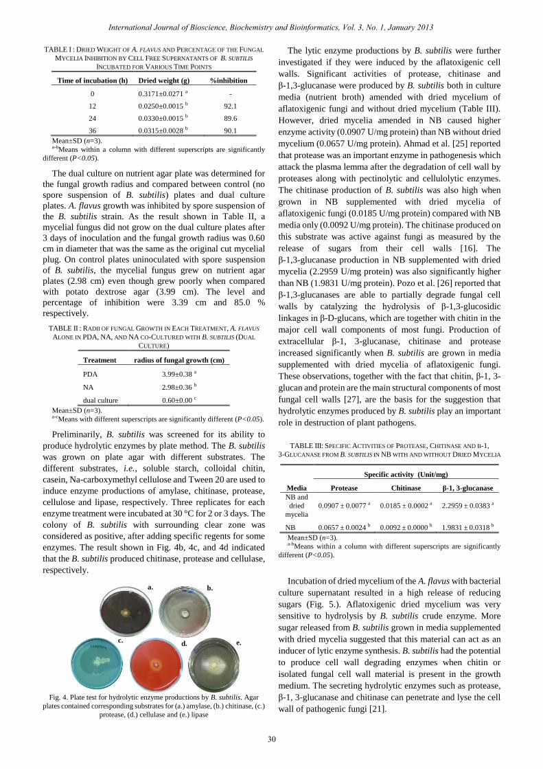
TABLE I : DRIED WEIGHT OF A. FLAVUS AND PERCENTAGE OF THE FUNGAL
MYCELIA INHIBITION BY CELL FREE SUPERNATANTS OF B. SUBTILIS
INCUBATED FOR VARIOUS TIME POINTS
Time of incubation (h) Dried weight (g) %inhibition
0 0.3171±0.0271 a -
12 0.0250±0.0015 b 92.1
24 0.0330±0.0015 b 89.6
36 0.0315±0.0028 b 90.1
Mean±SD (n=3). a-bMeans within a column with different superscripts are significantly
different (P<0.05).
The dual culture on nutrient agar plate was determined for
the fungal growth radius and compared between control (no
spore suspension of B. subtilis) plates and dual culture
plates. A. flavus growth was inhibited by spore suspension of
the B. subtilis strain. As the result shown in Table II, a
mycelial fungus did not grow on the dual culture plates after
3 days of inoculation and the fungal growth radius was 0.60
cm in diameter that was the same as the original cut mycelial
plug. On control plates uninoculated with spore suspension
of B. subtilis, the mycelial fungus grew on nutrient agar
plates (2.98 cm) even though grew poorly when compared
with potato dextrose agar (3.99 cm). The level and
percentage of inhibition were 3.39 cm and 85.0 %
respectively.
TABLE II : RADII OF FUNGAL GROWTH IN EACH TREATMENT, A. FLAVUS
ALONE IN PDA, NA, AND NA CO-CULTURED WITH B. SUBTILIS (DUAL
CULTURE)
Treatment radius of fungal growth (cm)
PDA 3.99±0.38 a
NA 2.98±0.36 b
dual culture 0.60±0.00 c
Mean±SD (n=3). a-cMeans with different superscripts are significantly different (P<0.05).
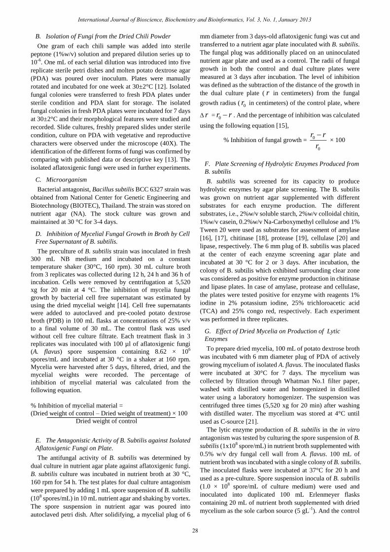
Preliminarily, B. subtilis was screened for its ability to
produce hydrolytic enzymes by plate method. The B. subtilis
was grown on plate agar with different substrates. The
different substrates, i.e., soluble starch, colloidal chitin,
casein, Na-carboxymethyl cellulose and Tween 20 are used to
induce enzyme productions of amylase, chitinase, protease,
cellulose and lipase, respectively. Three replicates for each
enzyme treatment were incubated at 30 °C for 2 or 3 days. The
colony of B. subtilis with surrounding clear zone was
considered as positive, after adding specific regents for some
enzymes. The result shown in Fig. 4b, 4c, and 4d indicated
that the B. subtilis produced chitinase, protease and cellulase,
respectively.
Fig. 4. Plate test for hydrolytic enzyme productions by B. subtilis. Agar
plates contained corresponding substrates for (a.) amylase, (b.) chitinase, (c.)
protease, (d.) cellulase and (e.) lipase
The lytic enzyme productions by B. subtilis were further
investigated if they were induced by the aflatoxigenic cell
walls. Significant activities of protease, chitinase and
β-1,3-glucanase were produced by B. subtilis both in culture
media (nutrient broth) amended with dried mycelium of
aflatoxigenic fungi and without dried mycelium (Table III).
However, dried mycelia amended in NB caused higher
enzyme activity (0.0907 U/mg protein) than NB without dried
mycelium (0.0657 U/mg protein). Ahmad et al. [25] reported
that protease was an important enzyme in pathogenesis which
attack the plasma lemma after the degradation of cell wall by
proteases along with pectinolytic and cellulolytic enzymes.
The chitinase production of B. subtilis was also high when
grown in NB supplemented with dried mycelia of
aflatoxigenic fungi (0.0185 U/mg protein) compared with NB
media only (0.0092 U/mg protein). The chitinase produced on
this substrate was active against fungi as measured by the
release of sugars from their cell walls [16]. The
β-1,3-glucanase production in NB supplemented with dried
mycelia (2.2959 U/mg protein) was also significantly higher
than NB (1.9831 U/mg protein). Pozo et al. [26] reported that
β-1,3-glucanases are able to partially degrade fungal cell
walls by catalyzing the hydrolysis of β-1,3-glucosidic
linkages in β-D-glucans, which are together with chitin in the
major cell wall components of most fungi. Production of
extracellular β-1, 3-glucanase, chitinase and protease
increased significantly when B. subtilis are grown in media
supplemented with dried mycelia of aflatoxigenic fungi.
These observations, together with the fact that chitin, β-1, 3-
glucan and protein are the main structural components of most
fungal cell walls [27], are the basis for the suggestion that
hydrolytic enzymes produced by B. subtilis play an important
role in destruction of plant pathogens.
a. b.
c. d. e.
International Journal of Bioscience, Biochemistry and Bioinformatics, Vol. 3, No. 1, January 2013
30
TABLE III: SPECIFIC ACTIVITIES OF PROTEASE, CHITINASE AND Β-1,
3-GLUCANASE FROM B. SUBTILIS IN NB WITH AND WITHOUT DRIED MYCELIA
Media
Specific activity (Unit/mg)
Protease Chitinase β-1, 3-glucanase
NB and
dried
mycelia
0.0907 ± 0.0077 a 0.0185 ± 0.0002 a 2.2959 ±0.0383 a
NB 0.0657 ± 0.0024 b 0.0092 ± 0.0000 b 1.9831 ± 0.0318 b
Mean±SD (n=3). a-bMeans within a column with different superscripts are significantly
different (P<0.05).
Incubation of dried mycelium of the A. flavus with bacterial
culture supernatant resulted in a high release of reducing
sugars (Fig. 5.). Aflatoxigenic dried mycelium was very
sensitive to hydrolysis by B. subtilis crude enzyme. More
sugar released from B. subtilis grown in media supplemented
with dried mycelia suggested that this material can act as an
inducer of lytic enzyme synthesis. B. subtilis had the potential
to produce cell wall degrading enzymes when chitin or
isolated fungal cell wall material is present in the growth
medium. The secreting hydrolytic enzymes such as protease,
β-1, 3-glucanase and chitinase can penetrate and lyse the cell
wall of pathogenic fungi [21].

Fig. 5. The amounts of total reducing sugars (R.S.), glucose and
N-acetylglucosamine (GlcNAc) released into B. subtilis culture supernatant of
NB with and without dried mycelia. Different alphabets above the bars of
the same sugar type designate significantly different values (P<0.05).
IV. CONCLUSION
A. flavus was isolated from bird chili powder samples
conferring high frequency of occurrence. The mycelial
growth of isolated aflatoxigenic fungi (A. flavus) was
potentially inhibited by hydrolytic enzymes from cell free
culture supernatant of B. subtilis. Production of extracellular
protease, chitinase and β-1, 3-glucanase from B. subtilis
affected to lyse the cell walls of A. flavus, leading to the
decrease of fungal development and, consequently reduction
of the aflatoxin production. Because of the B. subtilis
inhibitory activity against the aflatoxigenic fungi, the
metabolites may be useful as potential biocontrol agents
against aflatoxigenic fungi during food storage.
ACKNOWLEDGMENT
The authors would like to thank Assoc. Prof. Dr. Nuansri
Rakariyatham and Miss Pimpilai Fusawat for their kind
provision of the bacterial strain and also thank Miss Dujdao
Chunoi for the bird chili powder samples in our study.
REFERENCES
[1] A. A. Onilude, O. E. Fagade, M. M. Bello, and I. F. Fadahunsi,
“Inhibition of aflatoxin-producing aspergilli by lactic acid bacteria
isolates from indigenously fermented cereal gruels,” African Journal
of Biotechnology, vol. 4, no. 12, pp. 1404-1408, Dec 2005.
[2] H. S. Chun, H. J. Kim, H. E. Ok, J. B. Hwang, and D. H. Chung,
“Determination of aflatoxin levels in nuts and their products consumed
in South Korea,” Food Chemistry, vol. 102, pp. 385-391, May 2006.
[3] J. G. Kim, “Anti-aflatoxigenic activity of some bacteria related with
Fermentation,” in Communicating Current Research and Educational
Topics and Trends in Applied Microbiology, A. Méndez-Vilas, Ed.
2007, pp. 322-328.
[4] W. Kraikruan, S. Sukprakarn, O. Mongkolporn, and S. Wasee,
“Capsaicin and dihygrocapsaicin contents of Thai chili cultivars,”
Kasertsart Journal (Natural Science), vol. 42, pp. 611-616, 2008.
[5] W. Wangcharoen, and W. Morasuk, “Antioxidant capacity changes of
bird chili (Capsicum frutescens Linn.) during hot air drying,”
Kasetsart Journal (Natural science), vol. 43, pp. 12-20, 2009.
[6] R. R. M. Paterson, “Aflatoxins contamination in chili samples from
Pakistan,” Food Control, vol. 18, pp. 817-820, Apr 2007.
[7] A. D. Duman, “Storage of red chili pepper under hermetically sealed or
vacuum conditions for preservation of its quality and prevention of
mycotoxin occurrence,” Journal of Stored Products Research, vol. 46,
no. 3, pp.155-160, Jul 2010.
[8] C. R. Harwood, “Bacillus subtilis and its relatives: molecular
biological and industrial workhorses,” Trends in Biotechnology, vol.
10, pp. 247-256, Jan 1992.
[9] Y. Liu, Z. Chen, T. B. Ng, J. Zhang, M. Zhou, F. Song, F. Lu, and Y.
Liu, “Bacisubin, an antifungal protein with ribonuclease and
hemagglutinating activities from Bacillus subtilis strain B-916,”
Peptides, vol. 28, pp. 553-559, Nov 2006.
[10] C. Leifert, H. Li, S. Chidburee, S. Hampson, S. Workman, D. Sigee,
H.A.S. Epton, and A. Harbour, “Antibiotic production and biocontrol
activity by Bacillus subtilis CL27 and Bacillus pumilus CL45,”
Journal of Applied Bacteriology, vol. 78, no. 2, pp. 97-180, Feb 1995.
[11] S. Z. Iqbal, I. A. Bhatti, M. R. Asi, H. N. Bhatti, and M. A. Sheikh,
“Aflatoxin contamination in chilies from Punjab Pakistan with
reference to climate change,” International Journal of Agriculture
and Biology, vol. 13, no. 2, pp. 261-265, 2011.
[12] M. Rajasinghe, K. Abeywickrama, and R. Jayasekera, “Aflatoxigenic
Aspergillus flavus and aflatoxin formation in selected spices during
storage,” Tropical Agricultural Research and Extension, vol. 12, no. 1,
pp. 1-6, May 2009.
[13] H. L. Barnett, and B. B. Hunter, Illustrated Genera of Imperfect Fungi,
4th ed. New York, USA: Macmillan, 1987, pp. 92-107.
[14] T. Zhang, Z. Q. Shi, L. B. Hu, L. G. Cheng, and F. Wang, “Antifungal
compounds from Bacillus subtilis B-FS06 inhibiting the growth of
Aspergillus flavus,” World Journal Microbiology Biotechnology, vol.
24, pp. 783-788, Sep 2007.
[15] W. M. Yuan, and D. L. Crawford, “Characterization of Streptomyces
lydicus WYEC108 as a potential biocontrol agent against fungal root
and seed rots,” Applied and Environmental Microbiology, vol. 61, pp.
3119-3128, 1995.
[16] R. Gupta, R. K. Saxena, P. Chaturvedi, and J. S. Virdi, “Chitinase
production by Streptomyces viridificans: its potential on fungal cell
wall lysis,” Journal of Applied Bacteriology, vol. 78, no. 4, pp.
378-383, 1995.
[17] G. L. Maria, K. R. Sridhar, and N. S. Raviraja, “Antimicrobial and
enzyme activity of mangrove endophytic fungi of southwest coast of
India,” Journal of Agricultural Technology, vol. 1, pp. 67-80, 2005.
[18] T. Taechowisan, J. F. Peberdy, and S. Lumyong, “Chitinase
production by endophytic Streptomyces aureofaciens CMUAc130 and
its antagonism against phytopathogenic fungi,” Annals of
Microbiology, vol. 53, pp. 447-461, 2003.
[19] K. G. Siddalingeshwara, J. Uday, C. H. Huchesh, H. P. Puttaraju, J.
Karthic, K. Sudipta, T. Pramod, and T. Vishwanatha, “ Screening and
characterization of protease from Bacillus sp,” International Journal
of Applied Biology and Pharmaceutical Technology, vol. 1, no. 2, pp.
575-581, Aug-Oct 2010.
[20] R. C. Kasana, R. Salwan, H. Dhar, S. Dutt, and A. Gulati, “A rapid and
easy method for the detection of microbial cellulases on agar plates
using gram's iodine,” Current Microbiology, vol. 57, no. 5, pp.
503-507, November 2008.
[21] M. H. El-Katatny, W. Somitsch, K. H. Robra, M. S. El-Katatny, and G.
M. Gübitz1, “Production of chitinase and β-1,3-glucanase by
Trichoderma harzianum for control of the phytopathogenic fungus
Sclerotium rolfsii,” Food Technology and Biotechnology, vol. 38, no.
3, pp. 173-180, May 2000.
[22] G. L. Miller, “Use of dinitrosalicylic acid reagent for determination of
reducing sugar,” Analytical Chemistry, vol. 31, pp. 426-428, 1959.
[23] M. Bradford, “A rapid and sensitive method for the quantitation of
microgram quantities of protein utilizing the principle of protein-dye
binding,” Analytical Chemistry, vol. 72, pp. 248-254, 1976.
[24] N. N. Hai, “Bacillus subtilis possibly used for aflatoxin control,” in
Proc. of International Workshop on Biotechnology in Agriculture,
Nong Lam Univ., Ho Chi Minh city, Vietnam, Oct. 20-21, 2006.
[25] Y. Ahmad, A. Hameed, and A. Ghaffar, “Enzymatic activity of fungal
pathogens in corn,” Pakistan Journal of Botany, vol. 38, no. 4, pp.
1305-16, 2006.
[26] M. J. Pozo, C. A. Aguilar, E. D. Gaudot, and J. M. Barea, “β-1,
3-Glucanase activities in tomato roots inoculated with arbuscular
mycorrhizal fungi and/or Phytophthora parasitica and their possible
involvement in bioprotection,” Plant Science, vol. 141, pp. 149-157,
1999.
[27] J. F. Peberdy, “Fungal Cell Walls,” A Review in Biochemistry of Cell
Walls and Membranes in Fungi, P. J. Kuhn, A. P. J. Trinci, M. J. Jung,
M. W. Goosey, and L. G. Copping, Eds. Berlin: Springer-Verlag, 1990,
pp. 5-30.
International Journal of Bioscience, Biochemistry and Bioinformatics, Vol. 3, No. 1, January 2013
31

Rattanaporn Thakaew was born on June 12, 1985 in
Chiang Mai, Thailand. She received Bachelor degree
(1st Class Honor) in product development, Faculty of
Agro-industry (2008) from Chiang Mai University.
And now, she studies in biotechnology program,
Graduate School (2010-present) at Chiang Mai
University.
Hataichanoke Niamsup was born in Chiang Mai,
Thailand. She received her 1st class honor Bachelor
degree in chemistry (1991) from Chiang Mai
University and her PhD in biochemistry (1995) from
University of Illinois at Urbana-Champaign, USA.
She has published 24 papers in national and
international journals. She is currently appointed as
Assistant Professor at Chiang Mai University. Her
research interest is application of molecular biology
and biochemistry in agriculture.
Dr. Niamsup is a member of The Science Society of Thailand under the
Patronage of His Majesty the King, The Chemical Society of Thailand (CST)
under the Patronage of Her Royal Highness Princess Chulabhorn Mahidol,
The Thai Society for Biotechnology, The Association of Students supported
by the Development and Promotion for Science and Technology talents
project (ASDPST). She is also in the editorial board of Chiang Mai Journal
of Science.
International Journal of Bioscience, Biochemistry and Bioinformatics, Vol. 3, No. 1, January 2013
32
Related Documents