Paris et al. Breast Cancer Research 2010, 12:R27 http://breast-cancer-research.com/content/12/3/R27 Open Access RESEARCH ARTICLE BioMed Central © 2010 Paris et al.; licensee BioMed Central Ltd. This is an open access article distributed under the terms of the Creative Commons At- tribution License (http://creativecommons.org/licenses/by/2.0), which permits unrestricted use, distribution, and reproduction in any medium, provided the original work is properly cited. Research article Inhibition of phosphatidylcholine-specific phospholipase C downregulates HER2 overexpression on plasma membrane of breast cancer cells Luisa Paris †1 , Serena Cecchetti †1 , Francesca Spadaro 1 , Laura Abalsamo 1 , Luana Lugini 1 , Maria Elena Pisanu 1 , Egidio Iorio 1 , Pier Giorgio Natali 2 , Carlo Ramoni* 1 and Franca Podo* †1 Abstract Introduction: Overexpression on plasma membrane of human epidermal growth factor receptor 2 (HER2) is reported in 25% to 30% of breast cancers. Heterodimer formation with cognate members of the epidermal growth factor receptor (EGFR) family, such as HER3 and EGFR, activates abnormal cell-signalling cascades responsible for tumorigenesis and further transcriptional HER2 gene upregulation. Targeting the molecular mechanisms controlling HER2 overexpression and recycling may effectively deactivate this feedback-amplification loop. We recently showed that inactivation of phosphatidylcholine-specific phospholipase C (PC-PLC) may exert a pivotal role in selectively modulating the expression on the membrane of specific receptors or proteins relevant to cell function. In the present study, we investigated the capability of PC-PLC inhibition to target the molecular mechanisms controlling HER2 overexpression on the membrane of breast cancer cells by altering the rates of its endocytosis and lysosomal degradation. Methods: Localization on the membrane and interaction of PC-PLC with HER2, EGFR, and HER3 were investigated on HER2-overexpressing and HER2-low breast cancer cell lines, by using confocal laser scanning microscopy, flow cytometry, cell-surface biotinylation, isolation of lipid rafts, and immunoprecipitation experiments. The effects of the PC-PLC inhibitor tricyclodecan-9-yl-potassium xanthate (D609) on HER2 expression on the membrane and on the levels of overall HER2, HER2-HER3, and HER2-EGFR contents were monitored in the HER2-overexpressing SKBr3 cells, after either transient or continuous receptor engagement with anti-HER2 monoclonal antibodies, including trastuzumab. Changes of HER2 expression and cell proliferation were examined in SKBr3, BT-474, and MDA-MB-453 cells continuously exposed to D609 alone or combined with trastuzumab. Results: PC-PLC selectively accumulates on the plasma membrane of HER2-overexpressing cells, where it colocalizes and associates with HER2 in raft domains. PC-PLC inhibition resulted in enhanced HER2 internalization and lysosomal degradation, inducing downmodulation of HER2 expression on the membrane. Moreover, PC-PLC inhibition resulted in strong retardation of HER2 reexpression on the membrane and a decrease in the overall cellular contents of HER2, HER2-HER3, and HER2-EGFR heterodimers. The PC-PLC inhibitor also induced antiproliferative effects, especially in trastuzumab-resistant cells. Conclusions: The results pointed to PC-PLC inhibition as a potential means to counteract the tumorigenic effects of HER2 amplification and complement the effectiveness of current HER2-targeting therapies. * Correspondence: [email protected], [email protected] 1 Department of Cell Biology and Neurosciences, Section of Molecular and Cellular Imaging, Istituto Superiore di Sanità, Viale Regina Elena 299, 00161 Rome, Italy † Contributed equally Full list of author information is available at the end of the article

Welcome message from author
This document is posted to help you gain knowledge. Please leave a comment to let me know what you think about it! Share it to your friends and learn new things together.
Transcript
Paris et al. Breast Cancer Research 2010, 12:R27http://breast-cancer-research.com/content/12/3/R27
Open AccessR E S E A R C H A R T I C L E
Research articleInhibition of phosphatidylcholine-specific phospholipase C downregulates HER2 overexpression on plasma membrane of breast cancer cellsLuisa Paris†1, Serena Cecchetti†1, Francesca Spadaro1, Laura Abalsamo1, Luana Lugini1, Maria Elena Pisanu1, Egidio Iorio1, Pier Giorgio Natali2, Carlo Ramoni*1 and Franca Podo*†1
AbstractIntroduction: Overexpression on plasma membrane of human epidermal growth factor receptor 2 (HER2) is reported in 25% to 30% of breast cancers. Heterodimer formation with cognate members of the epidermal growth factor receptor (EGFR) family, such as HER3 and EGFR, activates abnormal cell-signalling cascades responsible for tumorigenesis and further transcriptional HER2 gene upregulation. Targeting the molecular mechanisms controlling HER2 overexpression and recycling may effectively deactivate this feedback-amplification loop. We recently showed that inactivation of phosphatidylcholine-specific phospholipase C (PC-PLC) may exert a pivotal role in selectively modulating the expression on the membrane of specific receptors or proteins relevant to cell function. In the present study, we investigated the capability of PC-PLC inhibition to target the molecular mechanisms controlling HER2 overexpression on the membrane of breast cancer cells by altering the rates of its endocytosis and lysosomal degradation.
Methods: Localization on the membrane and interaction of PC-PLC with HER2, EGFR, and HER3 were investigated on HER2-overexpressing and HER2-low breast cancer cell lines, by using confocal laser scanning microscopy, flow cytometry, cell-surface biotinylation, isolation of lipid rafts, and immunoprecipitation experiments. The effects of the PC-PLC inhibitor tricyclodecan-9-yl-potassium xanthate (D609) on HER2 expression on the membrane and on the levels of overall HER2, HER2-HER3, and HER2-EGFR contents were monitored in the HER2-overexpressing SKBr3 cells, after either transient or continuous receptor engagement with anti-HER2 monoclonal antibodies, including trastuzumab. Changes of HER2 expression and cell proliferation were examined in SKBr3, BT-474, and MDA-MB-453 cells continuously exposed to D609 alone or combined with trastuzumab.
Results: PC-PLC selectively accumulates on the plasma membrane of HER2-overexpressing cells, where it colocalizes and associates with HER2 in raft domains. PC-PLC inhibition resulted in enhanced HER2 internalization and lysosomal degradation, inducing downmodulation of HER2 expression on the membrane. Moreover, PC-PLC inhibition resulted in strong retardation of HER2 reexpression on the membrane and a decrease in the overall cellular contents of HER2, HER2-HER3, and HER2-EGFR heterodimers. The PC-PLC inhibitor also induced antiproliferative effects, especially in trastuzumab-resistant cells.
Conclusions: The results pointed to PC-PLC inhibition as a potential means to counteract the tumorigenic effects of HER2 amplification and complement the effectiveness of current HER2-targeting therapies.
* Correspondence: [email protected], [email protected] Department of Cell Biology and Neurosciences, Section of Molecular and Cellular Imaging, Istituto Superiore di Sanità, Viale Regina Elena 299, 00161 Rome, Italy† Contributed equallyFull list of author information is available at the end of the article
BioMed Central© 2010 Paris et al.; licensee BioMed Central Ltd. This is an open access article distributed under the terms of the Creative Commons At-tribution License (http://creativecommons.org/licenses/by/2.0), which permits unrestricted use, distribution, and reproduction in anymedium, provided the original work is properly cited.
Paris et al. Breast Cancer Research 2010, 12:R27http://breast-cancer-research.com/content/12/3/R27
Page 2 of 16
IntroductionMutation and dysregulation of epidermal growth factorreceptor (EGFR) family members are related to canceronset and progression [1,2]. In particular, overexpressionof the protooncogene encoding for human epidermalgrowth factor receptor 2 (HER2 or ErbB2 or C-neu) isimplicated in a variety of tumors [3,4], with an estimatedprevalence of 25% to 30% in patients with primary ormetastatic breast cancer [5] and reported poor prognosis[6-8].
Although lacking intrinsic ligand-binding capability,HER2 acts as the preferred partner for the formation ofmitogenically active heterodimers with the cognate EGFRfamily members epidermal growth factor 1 (HER1 orEGFR), EGFR receptor 3 (HER3), and receptor 4 (HER4)[4,9,10], HER2-HER3 being the prevalent and mostpotent of these complexes [1,8,11]. HER2-containing het-erodimers undergo slow endocytosis and more-rapidrecycling back to the cell surface [12-14]. These featurestranslate to potent mitogenic signal cascades involvingmultiple signalling pathways [15].
HER2 is therefore a relevant target for HER2-overex-pressing breast cancer therapy. Current targeted treat-ments are based on the use of trastuzumab, a humanizedanti-HER2 monoclonal antibody [16-22] or antibodiesagainst other EGFR family members [23,24] or inhibitorsof selective tyrosine kinase receptor phosphorylationsites [25-28].
An additional, still scarcely explored anti-HER2 treat-ment may selectively target molecular mechanisms con-trolling HER2 overexpression on the plasma membrane,its lysosomal pathway-dependent degradation [29], andrecycling back to membrane domains [30]. By inhibitingsignal-transduction cascades triggered by HER2 het-erodimer formation and affecting the downstream eventsresponsible for altered cell proliferation, survival, andgene overexpression [31], this approach might comple-ment or alternate with the present therapy protocols,especially in cases of severe side effects (for example, car-diotoxicity) or onset of specific resistance to currentlyused agents [28,32,33].
In previous studies on aberrant phosphatidylcholine(PC) metabolism in cancer cells [34-38], we reported thatinactivation of a 66-kDa PC-specific phospholipase C(PC-PLC) enzyme, recruited to the plasma membrane ofmitogen-stimulated [39], cytokine-activated [40] andtumor cells [41], downmodulates the expression on mem-brane of specific receptors or proteins relevant to cellfunction.
The present work reports the first evidence on PC-PLCaccumulation and association with HER2 on the plasmamembrane of HER2-overexpressing breast cancer cellsand on the effects of PC-PLC inhibition on HER2 inter-
nalization, degradation, and recycling and on cell prolif-eration after transient or continuous cell exposure toanti-HER2 monoclonal Abs, including trastuzumab.
Materials and methodsAntibodies and reagentsRabbit polyclonal antibodies (pAbs) raised against bacte-rial (Bacillus cereus) PC-PLC and selectively cross-react-ing with mammalian PC-PLC [42] was obtained andcharacterized as reported [39,43,44].
Anti-HER2 monoclonal antibodies (mAbs) 300G9 andW6/100 were developed by Dr. P.G. Natali at the IstitutoTumori Regina Elena (Rome, Italy). Rabbit anti-EGFR,anti-HER2, anti-HER3, and anti-Rab5B pAbs were pur-chased from Santa Cruz Biotechnology (Santa Cruz, CA,USA). Anti-β-actin mAb was from Sigma-Aldrich (St.Louis, MO, USA). Trastuzumab (Herceptin) and pro-tease-inhibitor cocktail were from Hoffman-La Roche(Basel, Switzerland). Anti-Lamp-2 mAb was supplied byBD Biosciences (San Jose, CA).
Alexa Fluor-488 and -594 F(ab)2 fragments of goat anti-rabbit IgG (H+L), Alexa Fluor-488 and -594 F(ab)2 frag-ments of goat anti-mouse IgG (H+L) were purchasedfrom Molecular Probes Inc. (Eugene, OR, USA), and goatanti-human FITC-conjugated from Cappel Co. (USA)were used as secondary Abs.
Goat anti-mouse and goat anti-rabbit IgG horseradishperoxidase (HRP)-conjugated antibodies and streptavi-din-HRP were supplied by BioRad Laboratories, Inc.(Hercules, CA, USA).
Triton X-100, NHS-biotin, propidium iodide, tricyclo-decan-9-yl-potassium xanthate (D609), 5-bromo-2'-deoxyuridine (BrdU), 3-(4,5-dimethyl-thiazol-2-yl)-2,5-diphenyltetrazolium bromide (MTT), and all other chem-icals and biochemicals were from Sigma-Aldrich, unlessotherwise specified.
CellsNontumorigenic, immortal human mammary epithelialcell lines (MCF-10A and MCF-12A) and HER2-overex-pressing (SKBr3, MDA-MB-453, BT-4T4) or HER2-low(MCF7, MDA-MB-231, MDA-MB-435) human mam-mary carcinoma cell lines were from American Type Cul-ture Collection (ATCC, Rockville, MD).
MCF-10A and MCF-12A cells were cultured in DMEMF12 medium, supplemented with 5% horse serum, hydro-cortisone (0.5 μg/ml), insulin (10 μg/mL), hEGF (20 ng/mL), and gentamicin/amphotericin-B (Gibco Laborato-ries, Grand Island, NY, USA). MCF7, MDA-MB-231 andMDA-MB-435 cells were grown in DMEM (Gibco Labo-ratories) supplemented with 1,000 g/L glucose and 10%foetal bovine serum (FBS). MDA-MB-453 and SKBr3cells were cultured in DMEM supplemented with 1,000 g/
Paris et al. Breast Cancer Research 2010, 12:R27http://breast-cancer-research.com/content/12/3/R27
Page 3 of 16
L glucose/10% FBS and 4500 g/L glucose/5% FBS (Gibco),respectively. BT-474 cells were cultured in RPMI supple-mented with 10% FCS. Cells were typically analyzed atsubconfluence, 72 hours after seeding.
Proliferation assayCell-proliferation/viability assays were performed on cellsexposed for 2 hours to MTT (5 μg/mL in PBS) at 37°C, in5% CO2 [45,46]. Optical absorbance was measured (in sixreplicates) at 595 nm with a Model 680 MicroplateReader (BioRad Laboratories).
Western blot analysesPreparation of total cell lysates, determination of proteinconcentration and Western blot analyses were performedas previously described [41]. Blots were incubated withanti-HER2 (Santa Cruz Biotechnology) or anti-PC-PLCpAbs. Densitometric analysis of protein bands was per-formed as reported [41].
In vitro PC-PLC activity assayPC-PLC activity was determined in whole-cell lysates byusing the Amplex Red PC-PLC-specific assay kit (Molec-ular Probes Inc.), modified as described [47].
Separation of lipid rafts by sucrose gradientCell lysis, gradient fractionation (5% to 30% sucrose), andprotein separation were carried out as previouslydescribed [40,41,48]. The distribution of HER2 and PC-PLC in the gradient fractions was assessed by separationin 7% SDS-PAGE followed by Western blotting.
Cell surface biotinylationSKBr3 cells were biotinylated on plasma membrane byadding 1.5 mg of NHS-biotin that links to hystidine andlysine proteins residues, and then lysed in ice-cold lysisbuffer and immunoprecipitated with anti-PC-PLC poly-clonal Abs. Detection of biotinylated proteins was per-formed by using streptavidin-HRP.
ImmunoprecipitationSamples for immunoprecipitation were prepared as pre-viously described [40]. In brief, total cell lysates (1 mg in 1mL) were incubated with 10% protein G Sepharose(Amersham Biosciences, Uppsala, Sweden) and with anti-HER2 (Santa Cruz Biotechnology) or anti-PC-PLC spe-cific Abs overnight at 4°C. After extensive washing, beadswere removed by centrifugation at 14,000 rpm and thenSDS sample buffer 4×, containing 2-mercaptoethanol(ICN Biomedicals Inc., Irvine, CA, USA) was added toelute proteins by heating the sample at 100°C for 5 min-utes. Immunoprecipitates were resolved by 7% SDS-PAGE under reducing conditions and blotted with relatedAbs.
Confocal Laser Scanning Microscopy (CLSM) and flow cytometry analysesCLSM observations were performed on either unfixed orfixed and permeabilized cells, as already reported [41].Flow-cytometry analyses were performed as previouslydescribed [39].
Statistical analysisData were analyzed by using GraphPad software, version3.03. Statistical significance of differences was deter-mined with one-way ANOVA or with Student's t test, asspecified. Differences were considered significant at P <0.05.
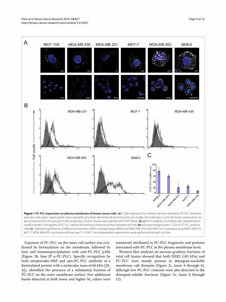
ResultsAccumulation of PC-PLC on the outer-membrane surface of breast HER2-overexpressing epithelial tumor cellsCLSM and flow-cytometry analyses on unfixed cellsshowed barely detectable PC-PLC levels on the plasmamembrane of the nontumoral immortalized breast epi-thelial cell lines MCF-10A (Figure 1a and 1b) and MCF-12A (data not shown). A massive, spotlike accumulationof PC-PLC was instead detected on the outer surface ofthe HER2-overexpressing breast carcinoma cell linesSKBr3 and MDA-MB-453. Similar fluorescence patternswere detected in BT-474 cells (Supplemental figure S1 inAdditional file 1). In HER2-low cancer cells, PC-PLC-positive granules were smaller and less abundant (Figure1a and 1b).
The percentage of PC-PLC-positive HER2-overex-pressing cells (Figure 1c) was significantly higher (78% ±13% in SKBr3; 61% ± 6% in MDA-MB-453; P < 0.001)than that of HER2-low tumor cells (MCF-7, 26% ± 7%;MDA-MB-231, 21% ± 15%; MDA-MB-435, 8% ± 3%) andnontumoral MCF-10A cells (<5% ± 2%).
These results show that PC-PLC selectively accumu-lates on the plasma membrane of HER2-overexpressingbreast cancer cells.
Colocalization and association of PC-PLC with HER2 on the plasma membrane of SKBr3 cellsCLSM analyses of unfixed SKBr3 cells showed extensivecolocalization of PC-PLC and HER2 on the outer plasmamembrane surface (Figure 2a, top and middle panels).Similar colocalization of the two proteins was observedon the plasma membrane of BT-474 cells (Supplementalfigure S1 in Additional file 1).
Analyses of fixed SKBr3 cells showed that PC-PLC wasalso present in inner cell compartments including thenucleus (Figure 2a, bottom panel), whereas the HER2receptor was, as expected, essentially confined to theplasma membrane.
Paris et al. Breast Cancer Research 2010, 12:R27http://breast-cancer-research.com/content/12/3/R27
Page 4 of 16
Exposure of PC-PLC on the outer cell surface was con-firmed by biotinylation on the membrane, followed bylysis and immunoprecipitation with anti-PC-PLC pAbs(Figure 2b, lane IP α-PC-PLC). Specific recognition byboth streptavidin-HRP and anti-PC-PLC antibody of abiotinylated protein with a molecular mass of 66 kDa [39-41], identified the presence of a substantial fraction ofPC-PLC on the outer membrane surface. Few additionalbands detected at both lower and higher Mr values were
tentatively attributed to PC-PLC fragments and proteinsassociated with PC-PLC at the plasma membrane level.
Western blot analyses on sucrose-gradient fractions oftotal cell lysates showed that both HER2 (185 kDa) andPC-PLC were mostly present in detergent-insolublemembrane raft domains (Figure 2c, lanes 4 through 6),although low PC-PLC contents were also detected in thedetergent-soluble fractions (Figure 2c, lanes 8 through12).
Figure 1 PC-PLC expression on plasma membrane of breast cancer cells. (a) CLSM analyses of six unfixed cell lines stained for PC-PLC detection (pseudo-color grey). Upper panels show examples of a three-dimensional reconstruction of a single cell (scale bars, 5 μm); the lower panels show sin-gle central sections of a group of cells (scale bars, 20 μm). Nuclei were stained with DAPI (blue). (b, c) FACS analyses of unfixed cells: representative cytofluorimetric histograms of PC-PLC relative fluorescence intensity of the indicated cell lines (b) and percentage (mean ± SD) of PC-PLC positive cells (c). Statistical significance of differences between HER2-overexpressing (SKBr3 and MDA-MB-453) and HER2-non-overexpressing (MDA-MB-231, MCF-7, MDA-MB-435) carcinoma cell lines was P < 0.001. Five independent experiments were performed on each cell line.

Paris et al. Breast Cancer Research 2010, 12:R27http://breast-cancer-research.com/content/12/3/R27
Page 5 of 16
Figure 2 Colocalization of PC-PLC and HER2 on plasma membrane of SKBr3 cells. (a) CLSM detection of PC-PLC and HER2 in either unfixed (top and middle panels) or fixed and permeabilized SKBr3 cells (bottom panel) by using rabbit polyclonal α-PC-PLC (green) and α-HER2 W6/100 mAb (red). Colocalization areas are represented in yellow. The middle panel shows the tridimensional reconstruction of PC-PLC and HER2 expression on the plas-ma membrane. Scale bars, 8 μm. At least five independent series of experiments were performed for each condition. (b) PC-PLC immunoblotting anal-yses of immunoprecipitated biotinylated proteins isolated from SKBr3 cells, detected by streptavidin-HRP (left, IB:STREP HRP) or by α-PC-PLC (right, IB:α-PC-PLC). TL, total lysate; bTL, biotinylated total lysate; CTR IgG, control for α-PC-PLC IgG; IP α-PC-PLC, α-PC-PLC immunoprecipitates. *IgG heavy chains. (c) Sucrose gradient fractions isolated from SKBr3 cell lysates and analyzed by Western blotting for HER2 (185 kDa) and PC-PLC (66 kDa) detec-tion. T, total cell lysate. (d, e) Western blot analyses of α-HER2 and α-PC-PLC (d) and α-EGFR and α-PC-PLC (e) immunoprecipitates (IP), blotted with the mutual Abs (α-PC-PLC, α-HER2, and α-EGFR, respectively), compared with the respective controls (CTR IgG α-PC-PLC, CTR IgG α-HER2, CTR IgG α-EGFR). Panels b, c, d, show representative results of three independent experiments. The immunoprecipitation in panel e was repeated twice.
Paris et al. Breast Cancer Research 2010, 12:R27http://breast-cancer-research.com/content/12/3/R27
Page 6 of 16
Finally, combined co-immunoprecipation and Westernblotting experiments showed that HER2 immunoprecipi-tates were associated with PC-PLC molecules (Figure 2d,lane IP α-HER2) and, conversely, a substantial amount ofHER2 was present in PC-PLC immunoprecipitates (Fig-ure 2d, lane IP α-PC-PLC).
Regarding other receptor-family members, EGFR (170kDa, Figure 2e) but not HER3 (180 kDa, not shown) wasalso found to co-immunoprecipitate with PC-PLC.
These results provide evidence of the existence of aphysical association between PC-PLC and HER2 (andEGFR) in HER2-overexpressing breast cancer cells.
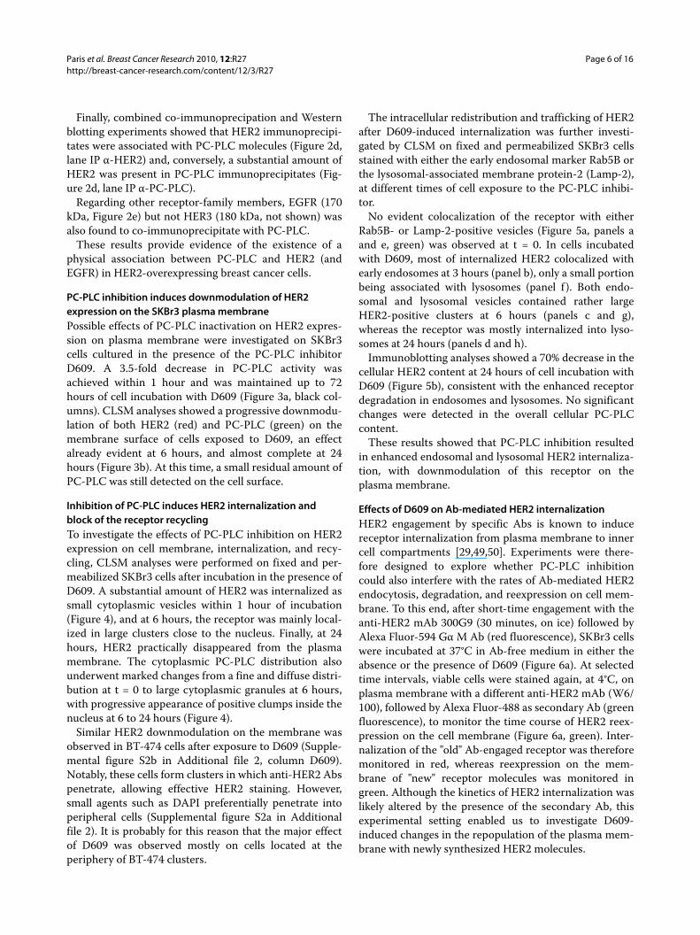
PC-PLC inhibition induces downmodulation of HER2 expression on the SKBr3 plasma membranePossible effects of PC-PLC inactivation on HER2 expres-sion on plasma membrane were investigated on SKBr3cells cultured in the presence of the PC-PLC inhibitorD609. A 3.5-fold decrease in PC-PLC activity wasachieved within 1 hour and was maintained up to 72hours of cell incubation with D609 (Figure 3a, black col-umns). CLSM analyses showed a progressive downmodu-lation of both HER2 (red) and PC-PLC (green) on themembrane surface of cells exposed to D609, an effectalready evident at 6 hours, and almost complete at 24hours (Figure 3b). At this time, a small residual amount ofPC-PLC was still detected on the cell surface.
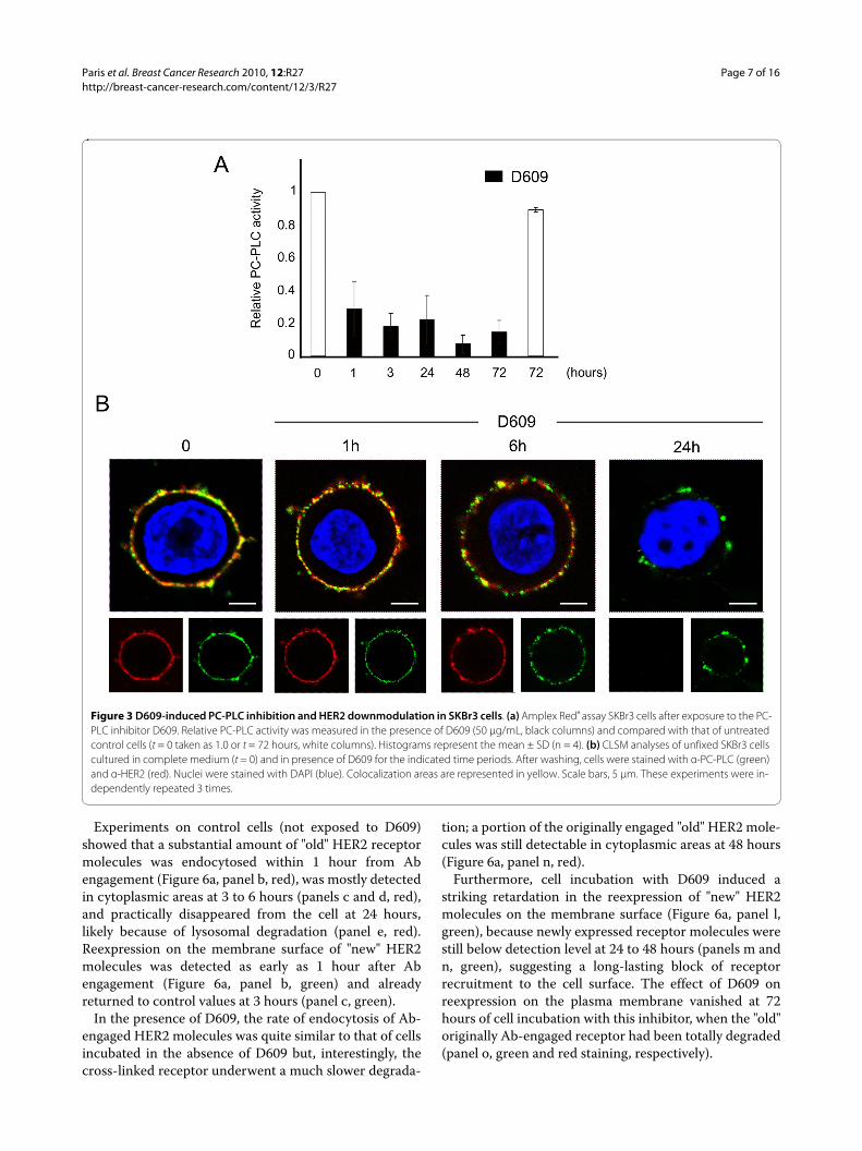
Inhibition of PC-PLC induces HER2 internalization and block of the receptor recyclingTo investigate the effects of PC-PLC inhibition on HER2expression on cell membrane, internalization, and recy-cling, CLSM analyses were performed on fixed and per-meabilized SKBr3 cells after incubation in the presence ofD609. A substantial amount of HER2 was internalized assmall cytoplasmic vesicles within 1 hour of incubation(Figure 4), and at 6 hours, the receptor was mainly local-ized in large clusters close to the nucleus. Finally, at 24hours, HER2 practically disappeared from the plasmamembrane. The cytoplasmic PC-PLC distribution alsounderwent marked changes from a fine and diffuse distri-bution at t = 0 to large cytoplasmic granules at 6 hours,with progressive appearance of positive clumps inside thenucleus at 6 to 24 hours (Figure 4).
Similar HER2 downmodulation on the membrane wasobserved in BT-474 cells after exposure to D609 (Supple-mental figure S2b in Additional file 2, column D609).Notably, these cells form clusters in which anti-HER2 Abspenetrate, allowing effective HER2 staining. However,small agents such as DAPI preferentially penetrate intoperipheral cells (Supplemental figure S2a in Additionalfile 2). It is probably for this reason that the major effectof D609 was observed mostly on cells located at theperiphery of BT-474 clusters.
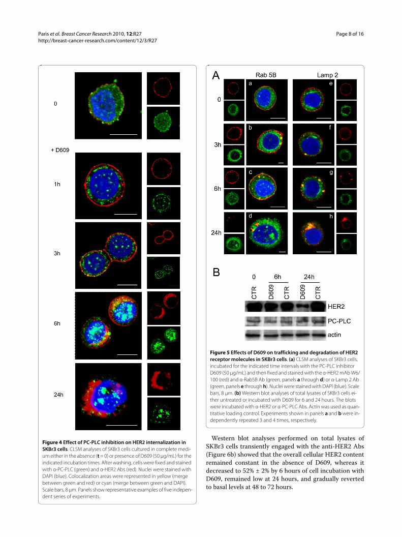
The intracellular redistribution and trafficking of HER2after D609-induced internalization was further investi-gated by CLSM on fixed and permeabilized SKBr3 cellsstained with either the early endosomal marker Rab5B orthe lysosomal-associated membrane protein-2 (Lamp-2),at different times of cell exposure to the PC-PLC inhibi-tor.
No evident colocalization of the receptor with eitherRab5B- or Lamp-2-positive vesicles (Figure 5a, panels aand e, green) was observed at t = 0. In cells incubatedwith D609, most of internalized HER2 colocalized withearly endosomes at 3 hours (panel b), only a small portionbeing associated with lysosomes (panel f ). Both endo-somal and lysosomal vesicles contained rather largeHER2-positive clusters at 6 hours (panels c and g),whereas the receptor was mostly internalized into lyso-somes at 24 hours (panels d and h).
Immunoblotting analyses showed a 70% decrease in thecellular HER2 content at 24 hours of cell incubation withD609 (Figure 5b), consistent with the enhanced receptordegradation in endosomes and lysosomes. No significantchanges were detected in the overall cellular PC-PLCcontent.
These results showed that PC-PLC inhibition resultedin enhanced endosomal and lysosomal HER2 internaliza-tion, with downmodulation of this receptor on theplasma membrane.
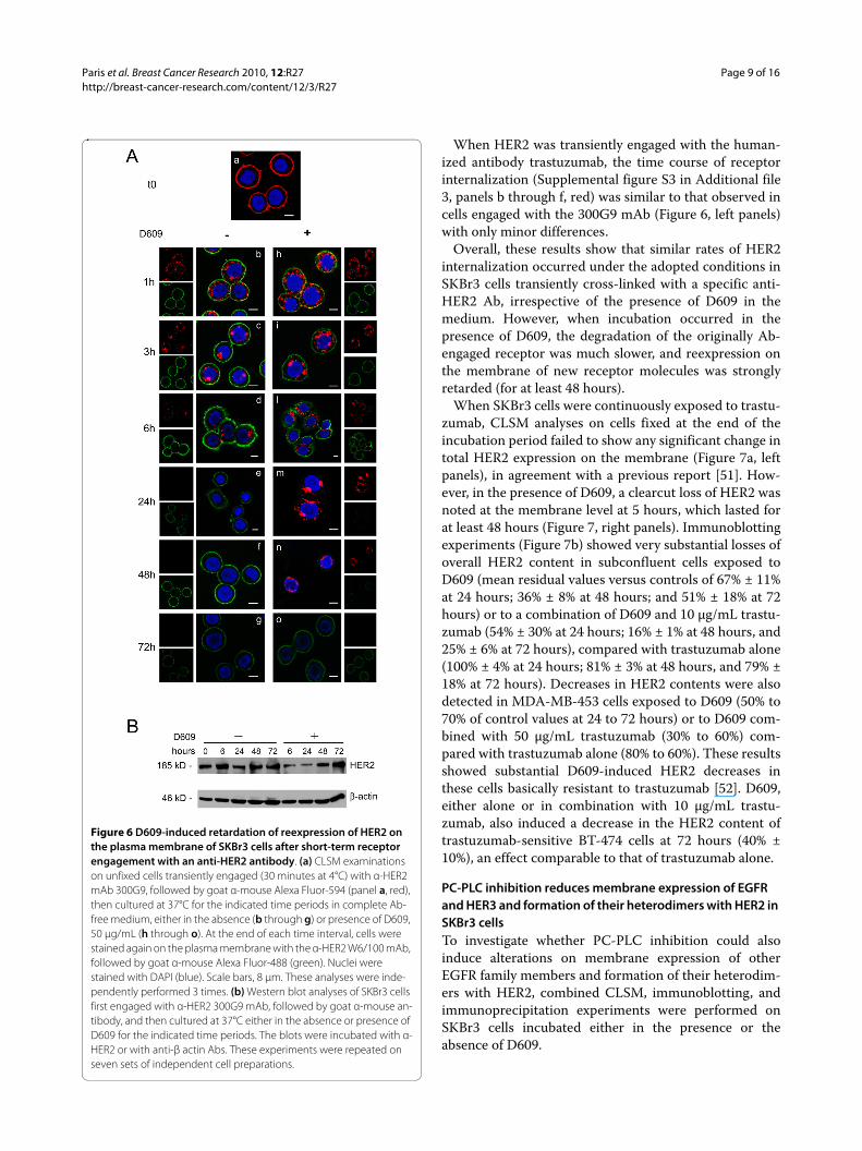
Effects of D609 on Ab-mediated HER2 internalizationHER2 engagement by specific Abs is known to inducereceptor internalization from plasma membrane to innercell compartments [29,49,50]. Experiments were there-fore designed to explore whether PC-PLC inhibitioncould also interfere with the rates of Ab-mediated HER2endocytosis, degradation, and reexpression on cell mem-brane. To this end, after short-time engagement with theanti-HER2 mAb 300G9 (30 minutes, on ice) followed byAlexa Fluor-594 Gα M Ab (red fluorescence), SKBr3 cellswere incubated at 37°C in Ab-free medium in either theabsence or the presence of D609 (Figure 6a). At selectedtime intervals, viable cells were stained again, at 4°C, onplasma membrane with a different anti-HER2 mAb (W6/100), followed by Alexa Fluor-488 as secondary Ab (greenfluorescence), to monitor the time course of HER2 reex-pression on the cell membrane (Figure 6a, green). Inter-nalization of the "old" Ab-engaged receptor was thereforemonitored in red, whereas reexpression on the mem-brane of "new" receptor molecules was monitored ingreen. Although the kinetics of HER2 internalization waslikely altered by the presence of the secondary Ab, thisexperimental setting enabled us to investigate D609-induced changes in the repopulation of the plasma mem-brane with newly synthesized HER2 molecules.
Paris et al. Breast Cancer Research 2010, 12:R27http://breast-cancer-research.com/content/12/3/R27
Page 7 of 16
Experiments on control cells (not exposed to D609)showed that a substantial amount of "old" HER2 receptormolecules was endocytosed within 1 hour from Abengagement (Figure 6a, panel b, red), was mostly detectedin cytoplasmic areas at 3 to 6 hours (panels c and d, red),and practically disappeared from the cell at 24 hours,likely because of lysosomal degradation (panel e, red).Reexpression on the membrane surface of "new" HER2molecules was detected as early as 1 hour after Abengagement (Figure 6a, panel b, green) and alreadyreturned to control values at 3 hours (panel c, green).
In the presence of D609, the rate of endocytosis of Ab-engaged HER2 molecules was quite similar to that of cellsincubated in the absence of D609 but, interestingly, thecross-linked receptor underwent a much slower degrada-
tion; a portion of the originally engaged "old" HER2 mole-cules was still detectable in cytoplasmic areas at 48 hours(Figure 6a, panel n, red).
Furthermore, cell incubation with D609 induced astriking retardation in the reexpression of "new" HER2molecules on the membrane surface (Figure 6a, panel l,green), because newly expressed receptor molecules werestill below detection level at 24 to 48 hours (panels m andn, green), suggesting a long-lasting block of receptorrecruitment to the cell surface. The effect of D609 onreexpression on the plasma membrane vanished at 72hours of cell incubation with this inhibitor, when the "old"originally Ab-engaged receptor had been totally degraded(panel o, green and red staining, respectively).
Figure 3 D609-induced PC-PLC inhibition and HER2 downmodulation in SKBr3 cells. (a) Amplex Red® assay SKBr3 cells after exposure to the PC-PLC inhibitor D609. Relative PC-PLC activity was measured in the presence of D609 (50 μg/mL, black columns) and compared with that of untreated control cells (t = 0 taken as 1.0 or t = 72 hours, white columns). Histograms represent the mean ± SD (n = 4). (b) CLSM analyses of unfixed SKBr3 cells cultured in complete medium (t = 0) and in presence of D609 for the indicated time periods. After washing, cells were stained with α-PC-PLC (green) and α-HER2 (red). Nuclei were stained with DAPI (blue). Colocalization areas are represented in yellow. Scale bars, 5 μm. These experiments were in-dependently repeated 3 times.
Paris et al. Breast Cancer Research 2010, 12:R27http://breast-cancer-research.com/content/12/3/R27
Page 8 of 16
Western blot analyses performed on total lysates ofSKBr3 cells transiently engaged with the anti-HER2 Abs(Figure 6b) showed that the overall cellular HER2 contentremained constant in the absence of D609, whereas itdecreased to 52% ± 2% by 6 hours of cell incubation withD609, remained low at 24 hours, and gradually revertedto basal levels at 48 to 72 hours.
Figure 4 Effect of PC-PLC inhibition on HER2 internalization in SKBr3 cells. CLSM analyses of SKBr3 cells cultured in complete medi-um either in the absence (t = 0) or presence of D609 (50 μg/mL) for the indicated incubation times. After washing, cells were fixed and stained with α-PC-PLC (green) and α-HER2 Abs (red). Nuclei were stained with DAPI (blue). Colocalization areas were represented in yellow (merge between green and red) or cyan (merge between green and DAPI). Scale bars, 8 μm. Panels show representative examples of five indepen-dent series of experiments.
Figure 5 Effects of D609 on trafficking and degradation of HER2 receptor molecules in SKBr3 cells. (a) CLSM analyses of SKBr3 cells, incubated for the indicated time intervals with the PC-PLC inhibitor D609 (50 μg/mL) and then fixed and stained with the α-HER2 mAb W6/100 (red) and α-Rab5B Ab (green, panels a through d) or α-Lamp 2 Ab (green, panels e through h). Nuclei were stained with DAPI (blue). Scale bars, 8 μm. (b) Western blot analyses of total lysates of SKBr3 cells ei-ther untreated or incubated with D609 for 6 and 24 hours. The blots were incubated with α-HER2 or α-PC-PLC Abs. Actin was used as quan-titative loading control. Experiments shown in panels a and b were in-dependently repeated 3 and 4 times, respectively.
Paris et al. Breast Cancer Research 2010, 12:R27http://breast-cancer-research.com/content/12/3/R27
Page 9 of 16
When HER2 was transiently engaged with the human-ized antibody trastuzumab, the time course of receptorinternalization (Supplemental figure S3 in Additional file3, panels b through f, red) was similar to that observed incells engaged with the 300G9 mAb (Figure 6, left panels)with only minor differences.
Overall, these results show that similar rates of HER2internalization occurred under the adopted conditions inSKBr3 cells transiently cross-linked with a specific anti-HER2 Ab, irrespective of the presence of D609 in themedium. However, when incubation occurred in thepresence of D609, the degradation of the originally Ab-engaged receptor was much slower, and reexpression onthe membrane of new receptor molecules was stronglyretarded (for at least 48 hours).
When SKBr3 cells were continuously exposed to trastu-zumab, CLSM analyses on cells fixed at the end of theincubation period failed to show any significant change intotal HER2 expression on the membrane (Figure 7a, leftpanels), in agreement with a previous report [51]. How-ever, in the presence of D609, a clearcut loss of HER2 wasnoted at the membrane level at 5 hours, which lasted forat least 48 hours (Figure 7, right panels). Immunoblottingexperiments (Figure 7b) showed very substantial losses ofoverall HER2 content in subconfluent cells exposed toD609 (mean residual values versus controls of 67% ± 11%at 24 hours; 36% ± 8% at 48 hours; and 51% ± 18% at 72hours) or to a combination of D609 and 10 μg/mL trastu-zumab (54% ± 30% at 24 hours; 16% ± 1% at 48 hours, and25% ± 6% at 72 hours), compared with trastuzumab alone(100% ± 4% at 24 hours; 81% ± 3% at 48 hours, and 79% ±18% at 72 hours). Decreases in HER2 contents were alsodetected in MDA-MB-453 cells exposed to D609 (50% to70% of control values at 24 to 72 hours) or to D609 com-bined with 50 μg/mL trastuzumab (30% to 60%) com-pared with trastuzumab alone (80% to 60%). These resultsshowed substantial D609-induced HER2 decreases inthese cells basically resistant to trastuzumab [52]. D609,either alone or in combination with 10 μg/mL trastu-zumab, also induced a decrease in the HER2 content oftrastuzumab-sensitive BT-474 cells at 72 hours (40% ±10%), an effect comparable to that of trastuzumab alone.
PC-PLC inhibition reduces membrane expression of EGFR and HER3 and formation of their heterodimers with HER2 in SKBr3 cellsTo investigate whether PC-PLC inhibition could alsoinduce alterations on membrane expression of otherEGFR family members and formation of their heterodim-ers with HER2, combined CLSM, immunoblotting, andimmunoprecipitation experiments were performed onSKBr3 cells incubated either in the presence or theabsence of D609.
Figure 6 D609-induced retardation of reexpression of HER2 on the plasma membrane of SKBr3 cells after short-term receptor engagement with an anti-HER2 antibody. (a) CLSM examinations on unfixed cells transiently engaged (30 minutes at 4°C) with α-HER2 mAb 300G9, followed by goat α-mouse Alexa Fluor-594 (panel a, red), then cultured at 37°C for the indicated time periods in complete Ab-free medium, either in the absence (b through g) or presence of D609, 50 μg/mL (h through o). At the end of each time interval, cells were stained again on the plasma membrane with the α-HER2 W6/100 mAb, followed by goat α-mouse Alexa Fluor-488 (green). Nuclei were stained with DAPI (blue). Scale bars, 8 μm. These analyses were inde-pendently performed 3 times. (b) Western blot analyses of SKBr3 cells first engaged with α-HER2 300G9 mAb, followed by goat α-mouse an-tibody, and then cultured at 37°C either in the absence or presence of D609 for the indicated time periods. The blots were incubated with α-HER2 or with anti-β actin Abs. These experiments were repeated on seven sets of independent cell preparations.

Paris et al. Breast Cancer Research 2010, 12:R27http://breast-cancer-research.com/content/12/3/R27
Page 10 of 16
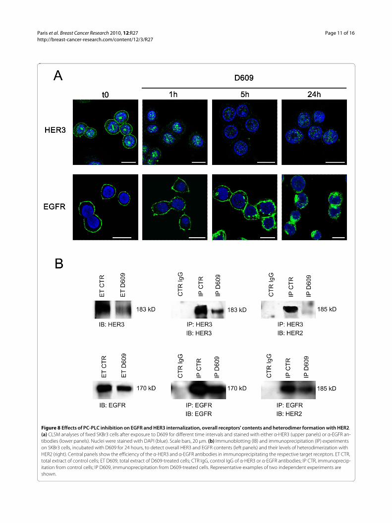
CLSM analyses of untreated and fixed SKBr3 cellsshowed that, as expected, both HER3 and EGFR wereexpressed mainly on the plasma membrane, although thetwo receptors were differently distributed among innercell compartments (Figure 8a). A substantial amount ofHER3 (but not EGFR) was also detected in nuclear areas.
Cell exposure to D609 for 24 hours induced downmod-ulation from the plasma membrane of both HER3 andEGFR, although the two receptors followed different rates
of internalization (Figure 8a). In particular, HER3 almostdisappeared from the plasma membrane within 1 hour ofcell exposure to D609, and only a small residual amount,confined mainly to the nucleus, was detected at 24 hours.At the latter time point, immunoblotting experiments ontotal cell lysates confirmed a strong decrease in the over-all HER3 content (Figure 8b). Under these conditions,immunoprecipitation with anti-HER3 followed by detec-tion with anti-HER2 Abs showed that HER2-HER3 het-erodimers, clearly detected in control cells, practicallydisappeared in D609-treated cells (Figure 8b). This resultwas further confirmed by immunoprecipitation withanti-HER2 followed by detection with anti-HER3 Abs(data not shown).
CLSM analyses on D609-treated cells showed that therate of EGFR endocytosis was very similar to that ofHER2 internalization (compare lower panels of Figure 8awith Figure 4). Under these conditions, a clear decrease inthe overall EGFR content was detected with Westernblotting of total lysates of cells collected after 24 hours ofexposure to D609. Co-immunoprecipitation experimentsshowed a simultaneous decrease in HER2-EGFR het-erodimers (Figure 8b).
In conclusion, these results showed that 24 hours of cellexposure to the PC-PLC inhibitor induced a substantialdecrease in both HER2-HER3 and HER2-EGFR heterodi-mers, a result that could derive not only from reducedHER2 expression (Figure 5), but also from the decreasedcontents of the respective partners.
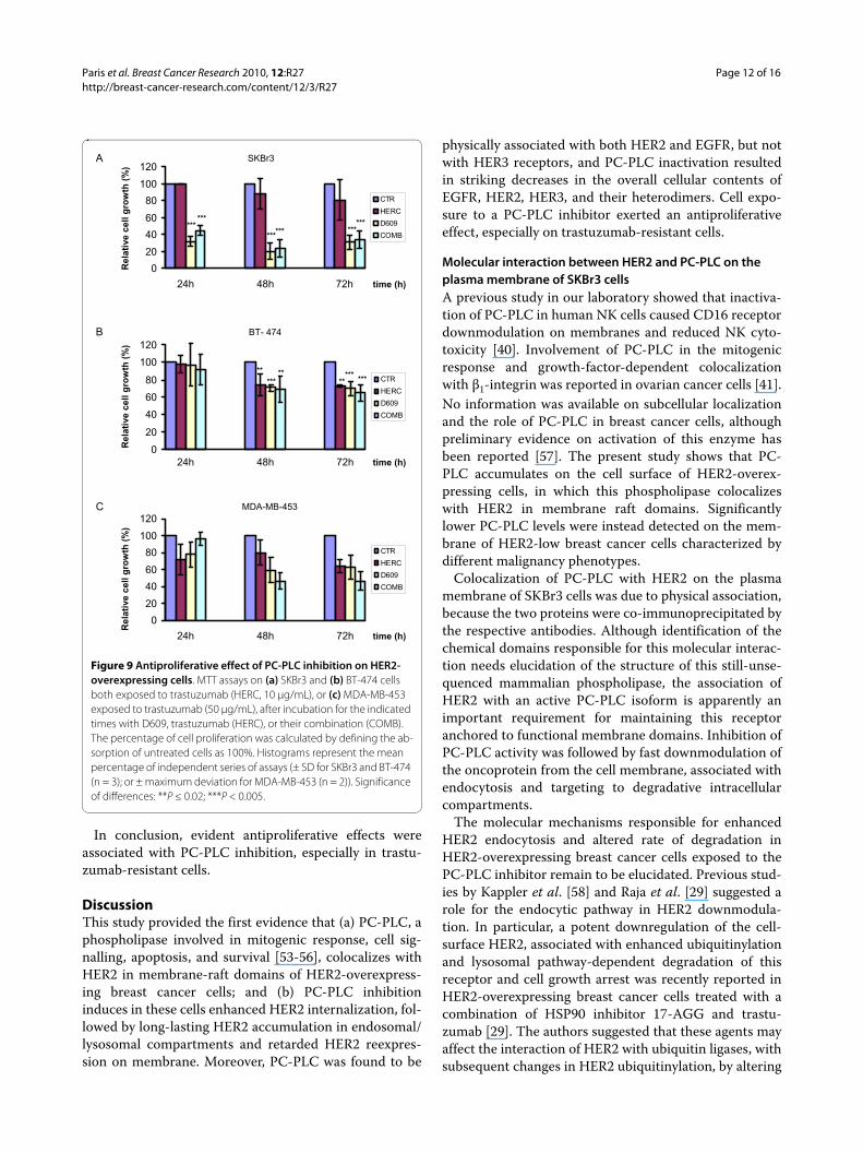
Effects of D609 on cell proliferation of HER2-overexpressing breast cancer cellsMTT assays showed that trastuzumab failed to exert sig-nificant antiproliferative effects (P > 0.05) on SKBr3 cellscontinuously exposed for 24 to 72 hours to this agent atdoses of either 10 μg/mL (Figure 9a) or 50 μg/mL (datanot shown).
A strong reduction in the percentage of viable/prolifer-ative cells was instead detected in SKBr3 cells exposed toD609 for 24 to 72 hours (down to 20% to 30% of the origi-nal levels in untreated control cells; P < 0.005). Combina-tion of D609 with trastuzumab (10 μg/mL) had similarantiproliferative effects to those induced by D609 alone(Figure 9a). Different antiproliferative patterns weredetected in the trastuzumab-sensitive BT-474 cells, inwhich this mAb reduced to 70% to 75% the percentage ofviable/proliferative cells at 48 to 72 hours (P < 0.02). Verysimilar antiproliferative effects were induced by D609 ora combination of the two agents (Figure 9b), although itcannot be excluded that differential penetration of D609in BT-474 clusters led to less-efficient PC-PLC inhibition.
Intermediate antiproliferative effects were exerted at 48to 72 hours by D609 or D609 plus trastuzumab (50 μg/mL) in MDA-MB-453 cells (Figure 9c).
Figure 7 Effect of D609 on HER2 expression in SKBr3 cells contin-uously exposed to trastuzumab. (a) CLSM analyses of cells fixed after exposure for the indicated time intervals to trastuzumab, in the pres-ence or absence of D609. Nuclei were stained with DAPI (blue). Scale bars, 20 μm. (b) Decrease in the overall HER2 content, detected by Western blotting, in total lysates of cells after incubation for different time intervals with D609 (50 μg/mL), trastuzumab (HERC, 10 μg/mL), or their combination (COMB). CTR, untreated control cells. Both CLSM and immunoblotting analyses were repeated 3 times.
Paris et al. Breast Cancer Research 2010, 12:R27http://breast-cancer-research.com/content/12/3/R27
Page 11 of 16
Figure 8 Effects of PC-PLC inhibition on EGFR and HER3 internalization, overall receptors' contents and heterodimer formation with HER2. (a) CLSM analyses of fixed SKBr3 cells after exposure to D609 for different time intervals and stained with either α-HER3 (upper panels) or α-EGFR an-tibodies (lower panels). Nuclei were stained with DAPI (blue). Scale bars, 20 μm. (b) Immunoblotting (IB) and immunoprecipitation (IP) experiments on SKBr3 cells, incubated with D609 for 24 hours, to detect overall HER3 and EGFR contents (left panels) and their levels of heterodimerization with HER2 (right). Central panels show the efficiency of the α-HER3 and α-EGFR antibodies in immunoprecipitating the respective target receptors. ET CTR, total extract of control cells; ET D609, total extract of D609-treated cells; CTR IgG, control IgG of α-HER3 or α-EGFR antibodies; IP CTR, immunoprecip-itation from control cells; IP D609, immunoprecipitation from D609-treated cells. Representative examples of two independent experiments are shown.
Paris et al. Breast Cancer Research 2010, 12:R27http://breast-cancer-research.com/content/12/3/R27
Page 12 of 16
In conclusion, evident antiproliferative effects wereassociated with PC-PLC inhibition, especially in trastu-zumab-resistant cells.
DiscussionThis study provided the first evidence that (a) PC-PLC, aphospholipase involved in mitogenic response, cell sig-nalling, apoptosis, and survival [53-56], colocalizes withHER2 in membrane-raft domains of HER2-overexpress-ing breast cancer cells; and (b) PC-PLC inhibitioninduces in these cells enhanced HER2 internalization, fol-lowed by long-lasting HER2 accumulation in endosomal/lysosomal compartments and retarded HER2 reexpres-sion on membrane. Moreover, PC-PLC was found to be
physically associated with both HER2 and EGFR, but notwith HER3 receptors, and PC-PLC inactivation resultedin striking decreases in the overall cellular contents ofEGFR, HER2, HER3, and their heterodimers. Cell expo-sure to a PC-PLC inhibitor exerted an antiproliferativeeffect, especially on trastuzumab-resistant cells.
Molecular interaction between HER2 and PC-PLC on the plasma membrane of SKBr3 cellsA previous study in our laboratory showed that inactiva-tion of PC-PLC in human NK cells caused CD16 receptordownmodulation on membranes and reduced NK cyto-toxicity [40]. Involvement of PC-PLC in the mitogenicresponse and growth-factor-dependent colocalizationwith β1-integrin was reported in ovarian cancer cells [41].No information was available on subcellular localizationand the role of PC-PLC in breast cancer cells, althoughpreliminary evidence on activation of this enzyme hasbeen reported [57]. The present study shows that PC-PLC accumulates on the cell surface of HER2-overex-pressing cells, in which this phospholipase colocalizeswith HER2 in membrane raft domains. Significantlylower PC-PLC levels were instead detected on the mem-brane of HER2-low breast cancer cells characterized bydifferent malignancy phenotypes.
Colocalization of PC-PLC with HER2 on the plasmamembrane of SKBr3 cells was due to physical association,because the two proteins were co-immunoprecipitated bythe respective antibodies. Although identification of thechemical domains responsible for this molecular interac-tion needs elucidation of the structure of this still-unse-quenced mammalian phospholipase, the association ofHER2 with an active PC-PLC isoform is apparently animportant requirement for maintaining this receptoranchored to functional membrane domains. Inhibition ofPC-PLC activity was followed by fast downmodulation ofthe oncoprotein from the cell membrane, associated withendocytosis and targeting to degradative intracellularcompartments.
The molecular mechanisms responsible for enhancedHER2 endocytosis and altered rate of degradation inHER2-overexpressing breast cancer cells exposed to thePC-PLC inhibitor remain to be elucidated. Previous stud-ies by Kappler et al. [58] and Raja et al. [29] suggested arole for the endocytic pathway in HER2 downmodula-tion. In particular, a potent downregulation of the cell-surface HER2, associated with enhanced ubiquitinylationand lysosomal pathway-dependent degradation of thisreceptor and cell growth arrest was recently reported inHER2-overexpressing breast cancer cells treated with acombination of HSP90 inhibitor 17-AGG and trastu-zumab [29]. The authors suggested that these agents mayaffect the interaction of HER2 with ubiquitin ligases, withsubsequent changes in HER2 ubiquitinylation, by altering
Figure 9 Antiproliferative effect of PC-PLC inhibition on HER2-overexpressing cells. MTT assays on (a) SKBr3 and (b) BT-474 cells both exposed to trastuzumab (HERC, 10 μg/mL), or (c) MDA-MB-453 exposed to trastuzumab (50 μg/mL), after incubation for the indicated times with D609, trastuzumab (HERC), or their combination (COMB). The percentage of cell proliferation was calculated by defining the ab-sorption of untreated cells as 100%. Histograms represent the mean percentage of independent series of assays (± SD for SKBr3 and BT-474 (n = 3); or ± maximum deviation for MDA-MB-453 (n = 2)). Significance of differences: **P ≤ 0.02; ***P < 0.005.
A
B
C
0
20
40
60
80
100
120
24h 48h 72h
CTR
HERC
D609
COMB
Rel
ativ
e ce
llg
row
th(%
)
time (h)
*****
****
*** ***
BT- 474
0
20
40
60
80
100
120
24h 48h 72h
CTR
HERC
D609
COMB
Rel
ativ
e ce
llg
row
th(%
)
time (h)
MDA-MB-453
Rel
ativ
e ce
llg
row
th(%
)
0
20
40
60
80
100
120
24h 48h 72h
CTR
HERC
D609
COMB***
***
*********
***
SKBr3
time (h)
Paris et al. Breast Cancer Research 2010, 12:R27http://breast-cancer-research.com/content/12/3/R27
Page 13 of 16
the arrangement of the HER2-associated HSP90 chaper-one complex. These findings support the interest ofexploring in our cell systems how deactivation of HER2-associated PC-PLC affects the differential recruitment ofspecific ubiquitin ligases to HER2 and cognate membersof the EGFR family. We might hypothesize that deactiva-tion of PC-PLC isoform(s) associated with HER2 likelymodifies the efficiency of ubiquitinylation and lysosomaldegradation through changes in steric hindrance effectsor structural changes of HER2 complexes or both.
Our results point to the attractive possibility of control-ling, by a simple switch of the activity status of PC-PLC,the levels of HER2 expression on the membrane andtherefore reducing the formation of HER2 heterodimerswith cognate members of the EGFR family, with expectedeffects on the control of tumorigenicity of HER2-overex-pressing cells. By enhancing HER2 endocytosis and lyso-somal degradation and retarding HER2 recycling to themembrane, PC-PLC inhibition resulted in HER2-EGFRand HER2-HER3 depletion. Similar effects on HER2 het-erodimers have so far been achieved by more-complexlaboratory procedures such as induction of intracellularexpression of single-chain HER2-specific antibodies incancer cells [59,60]. The D609-induced reduction in thelevels of HER2-HER3 and HER2-EGFR heterodimersmay originate from differential mechanisms, because co-immunoprecipitation experiments showed the physicalassociation of PC-PLC with either HER2 or EGFR but notwith HER3. Possibly linked to this differential behavior isthe much faster downmodulation of HER3 from the cellmembrane after PC-PLC inactivation. This result sug-gests the hypothesis that inhibition of PC-PLC activitymay facilitate dissociation of HER2-HER3 heterodimers,with possible effects on diversification of HER2-drivenintracellular cascades of downhill phosphorylation reac-tions [11]. Analyses should also be extended to HER4,temporarily excluded from this phase of the study.
Role of PC-PLC activity in controlling HER2 recyclingAlterations of cell recycling of HER2 in breast cancer cellsmay also have profound effects on cell-signalling deregu-lation in HER2-overexpressing cells [12-14]. Thereported downmodulation of HER2 from the plasmamembrane of HER2-overexpressing cells exposed to thePC-PLC inhibitor suggests that PC-PLC has a role in con-trolling the effects of the overall HER2-amplificationmachinery. Our experiments on cells engaged by anti-HER2 Abs showed that fast receptor internalization anddegradation was followed by rapid reexpression of newHER2 molecules on the membrane surface, consistentwith the capability of these cancer cells to restore andmaintain high expression levels of the amplified HER2receptor. In contrast, addition of D609 to cells incubatedin Ab-free medium after transient receptor-mAb cross-
linking, not only induced further receptor endocytosis,and prolonged receptor degradation within lysosomalstructures, but also slowed reexpression of newly synthe-sized HER2 molecules on the membrane. When cellswere continuously exposed in vitro to trastuzumab (acondition similar to that of tumor cells after intravenousinfusion of this agent to the patient), no substantialchange in the expression of HER2 on the plasma mem-brane was observed. This effect was in good agreementwith a previous report by Longva et al. [51] showing asubstantial lack of endocytotic downregulation of HER2in SKBr3 cells exposed to trastuzumab. When, however,cells were exposed to both trastuzumab and D609, a cleardecrease in HER2 expression on the membrane lasted forat least 48 hours. Furthermore, a most striking effectinduced, under these conditions, by the PC-PLC inhibitorwas a strong and prolonged decrease in the overall cellu-lar HER2 content, which lasted up to 72 hours.
Reduced cell proliferation of SKBr3 cells exposed to the PC-PLC inhibitorAmong the multiple antitumor activities exerted by tras-tuzumab on HER2-overexpressing breast cancer cells,inhibition of cell proliferation is reported to occur pri-marily by modulation of the cyclin-dependent kinaseinhibitor p27Kp1, via a complex network of signallingpathways, resulting in G1 arrest and growth inhibition[61].
This work shows a substantial lack of synergismbetween the antiproliferative effects exerted by trastu-zumab and D609, these processes likely being regulatedby different, independent molecular mechanisms. More-over, the antiproliferative effect of D609 can hardly beregarded as specific for HER2-overexpressing cells,because previous studies in our laboratory showed thatcell exposure to this xanthate is able to induce cell-cyclearrest in G0/G1 in different types of cells, such as HER2-non-overexpressing ovary cancer cells and mitogen-stim-ulated fibroblasts [39,40]. Further studies are needed toinvestigate whether and to what extent the antiprolifera-tive effect of D609, similar to that of another PC-PLCinhibitor, SC-ααδ9 (4-(benzyl-(2- [(2,5-diphenyoxazole-4-carbonyl)amino]ethyl)carbamoyl)-2-decanolaminobu-tyric acid), is linked to the phosphorylation or inhibitionor both of selective cyclin-dependent kinase activities[62].
Although a number of PC-PLC inhibitors have beenidentified or designed [63-65], D609 is widely accepted asselective competitive inhibitor of PC-specific phospholi-pase both in reaction mixtures and in cells [66]. Thiscompound, first introduced as antiviral and antitumoralagent in vitro and in vivo [66-70], has been shown toenhance the action of other drugs in the regression of
Paris et al. Breast Cancer Research 2010, 12:R27http://breast-cancer-research.com/content/12/3/R27
Page 14 of 16
various human tumor transplants in athymic mice [68],without side effects.
Our reported evidence on multiple effects induced byD609 on HER2 downmodulation from the plasma mem-brane suggests the interest of further evaluating in appro-priate model systems the added value of including a PC-PLC inhibitor as an adjunct to the current therapies tar-geted against HER2-overexpressing breast cancer cells.
ConclusionsA C-type phospholipase specific for the major phospho-lipid of eukaryotic cells associates with HER2 and EGFRreceptors in HER2-overexpressing breast cancer cells.Inhibition of this enzyme induces downregulation ofHER2 expression on the plasma membrane of these cells,promotes HER2 endocytosis and lysosomal degradation,induces retardation in HER2 recycling, and is associatedwith reduction of overall HER2, HER2-HER3, and HER2-EGFR contents.
The use of a PC-PLC inhibitor as a modulator of HER2overexpression and internalization in breast cancer cellsmay allow a better elucidation of the complex, stillincompletely understood molecular mechanisms under-lying cell tumorigenicity associated with HER2/neu geneamplification.
The results of this study point to PC-PLC inhibition asa possible means to counteract the tumorigenic effects ofHER2 amplification and complement the effectiveness ofcurrent HER2-targeting therapies aimed at increasingdisease-free and overall survival of patients affected byHER2-overexpressing breast cancer.
Additional material
AbbreviationsCLSM: confocal laser-scanning microscopy; Lamp-2: lysosomal-associatedmembrane protein-2; MTT: 3-(4,5-dimethylthiazol-2-yl)-2,5-diphenyltetrazo-lium bromide; PC: phosphatidylcholine; PC-PLC: phosphatidylcholine-specificphospholipase C.
Competing interestsThe authors declare that they have no competing interests.
Authors' contributionsLP and SC carried out the confocal laser scanning microscopy and the molecu-lar studies and drafted the manuscript. FS carried out the PC-PLC activityassays. LA performed the acquisition and analysis of the flow-cytometry data.LL participated in the molecular studies. MEP and EI carried out the MTT prolif-eration assays. PGN participated in the overall study design and criticallyrevised the manuscript. CR participated in the design of the study and in theinterpretation of data. FP conceived the study, participated in its design andcoordination, and helped to draft the manuscript. All authors read andapproved the final manuscript.
Authors' informationLP presented this work as her thesis for the degree in Biological Sciences at theUniversity of Rome "La Sapienza"; she is now a PhD student and research fellowat the Istituto Superiore di Sanità (ISS), Rome. LA and MEP are PhD students andresearch fellows at the ISS. SC, FS, and LL are PhD researchers at the ISS. EI isresearcher at the ISS. PGN acted as Director of the Immunology Section at theIstituto Tumori Regina Elena, Rome; CR was Senior Investigator and FP acted asDirector of the Molecular and Cellular Imaging Unit at the Department of CellBiology and Neurosciences, Istituto Superiore di Sanità, Rome.
AcknowledgementsWe are particularly grateful to Dr. Oreste Segatto, Istituto Tumori Regina Elena, Rome, for enlightening suggestions and critical reading of the manuscript. We gratefully acknowledge partial support by Associazione Italiana per la Ricerca sul Cancro (AIRC) grant 2007-2010 to FP and grant to PGN; the Integrated Oncology Program R0 06.5/N.ISS/Q09 (FP) and the Special Research Program in Oncology, Italian Ministry of Health, Onc_Ord 37/07 ISS N° 70CF/4 (FP). We thank Dr. C. Pini and Dr. B. Barletta, ISS, Rome for preparation and purification of the anti-PC-PLC antibodies and Dr. C. Raggi, ISS, for analyses of Western blot protein bands.
Author Details1Department of Cell Biology and Neurosciences, Section of Molecular and Cellular Imaging, Istituto Superiore di Sanità, Viale Regina Elena 299, 00161 Rome, Italy and 2Section of Immunology, Istituto Tumori Regina Elena, Via delle Messi D'Oro 156, 00158 Rome, Italy
References1. Yarden Y, Sliwkowski MX: Untangling the ErbB signalling network. Nat
Rev Mol Cell Biol 2001, 2:127-137.2. Mendelsohn J, Baselga J: Status of epidermal growth factor receptor
antagonists in the biology and treatment of cancer. J Clin Oncol 2003, 21:2787-2799.
3. Lemmon MA: The EGF receptor family as therapeutic targets in breast cancer. Breast Dis 2003, 18:33-43.
4. Hynes NE, Lane HA: ERBB receptors and cancer: the complexity of targeted inhibitors. Nat Rev Cancer 2005, 5:341-354.
5. Carlsson J, Nordgren H, Sjöström J, Wester K, Villman K, Bengtsson NO, Ostenstad B, Lundqvist H, Blomqvist C: HER2 expression in breast cancer primary tumours and corresponding metastases: original data and literature review. Br J Cancer 2004, 90:2344-2348.
6. Slamon DJ, Clark GM, Wong SG, Levin WJ, Ullrich A, McGuire WL: Human breast cancer: correlation of relapse and survival with amplification of the HER-2/neu oncogene. Science 1987, 235:177-182.
7. Sjögren S, Inganãs M, Lindgren A, Holmberg L, Bergh J: Prognostic and predictive value of c-erbB-2 overexpression in primary breast cancer, alone and in combination with other prognostic markers. J Clin Oncol 1998, 16:462-469.
Additional file 1 Supplemental figure S1. Colocalization of PC-PLC and HER2 on plasma membrane of BT-474 cells. (a, b) CLSM detection of PC-PLC and HER2 in unfixed BT-474 by using rabbit polyclonal α-PC-PLC (green) and α-HER2 W6/100 mAb (red). Colocalization areas are represented in yellow. (a) The three-dimensional reconstruction of PC-PLC and HER2 expression on the plasma membrane, and (b) the central section. Scale bars, 10 μm.Additional file 2 Supplemental figure S2. Effect of D609 on HER2 expression in BT-474 cells continuously exposed to trastuzumab. CLSM analyses on untreated BT-474 fixed cells (a) or fixed (b) after exposure for the indicated time intervals to trastuzumab (HERC, 10 μg/mL), D609 (50 μg/mL), or their combination (COMB). Nuclei were stained with DAPI (blue). Scale bars, 20 μm.Additional file 3 Supplemental figure S3. D609-induced retardation of HER2 re-expression on the plasma membrane of SKBr3 cells after short-term receptor engagement with trastuzumab. CLSM observa-tions on unfixed cells after transient cross-linking with trastuzumab (10 μg/mL, 30 minutes at 4°C), followed by goat α-human FITC-conjugated Ab (a, pseudo-color red), then cultured at 37°C for the indicated time periods in complete Ab-free medium, either in the absence (b through f) or presence of D609, 50 μg/mL (g through m). At the end of each time interval, cells were stained again on the plasma membrane with the α-HER2 W6/100 mAb, followed by goat α-mouse Alexa Fluor-594 (pseudo-color green). Nuclei were stained with DAPI (blue). Scale bars, 8 μm. Micrographs repre-sent results of three independent series of experiments performed.
Received: 13 October 2009 Revised: 18 March 2010 Accepted: 12 May 2010 Published: 12 May 2010This article is available from: http://breast-cancer-research.com/content/12/3/R27© 2010 Paris et al.; licensee BioMed Central Ltd. This is an open access article distributed under the terms of the Creative Commons Attribution License (http://creativecommons.org/licenses/by/2.0), which permits unrestricted use, distribution, and reproduction in any medium, provided the original work is properly cited.Breast Cancer Research 2010, 12:R27
http://www.ncbi.nlm.nih.gov/entrez/query.fcgi?cmd=Retrieve&db=PubMed&dopt=Abstract&list_uids=3798106
Paris et al. Breast Cancer Research 2010, 12:R27http://breast-cancer-research.com/content/12/3/R27
Page 15 of 16
8. Citri A, Skaria KB, Yarden Y: The deaf and the dumb: the biology of ErbB-2 and ErbB-3. Exp Cell Res 2003, 284:54-65.
9. Tzahar E, Waterman H, Chen X, Levkowitz G, Karunagaran D, Lavi S, Ratzkin BJ, Yarden Y: A hierarchical network of interreceptor interactions determines signal transduction by Neu differentiation factor/neuregulin and epidermal growth factor. Mol Cell Biol 1996, 16:5276-5287.
10. Hendriks BS, Opresko LK, Wiley HS, Lauffenburger D: Quantitative analysis of HER2-mediated effects on HER2 and epidermal growth factor receptor endocytosis. J Biol Chem 2003, 278:23343-23351.
11. Graus-Porta D, Beerli RR, Daly JM, Hynes NE: ErbB-2, the preferred heterodimerization partner of all ErbB receptors, is a mediator of lateral signalling. EMBO J 1997, 16:1647-1655.
12. Baulida J, Kraus MH, Alimandi M, Di Fiore PP, Carpenter G: All ErbB receptors other than the epidermal growth factor receptor are endocytosis impaired. J Biol Chem 1996, 271:5251-5257.
13. Lenferink AE, Pinkas-Kramarski R, Poll ML van de, van Vugt MJ, Klapper LN, Tzahar E, Waterman H, Sela M, van Zoelen EJ, Yarden Y: Differential endocytic routing of homo- and hetero-dimeric ErbB tyrosine kinases confers signaling superiority to receptor heterodimers. EMBO J 1998, 17:3385-3397.
14. Worthylake R, Opresko LK, Wiley HS: ErbB-2 amplification inhibits down-regulation and induces constitutive activation of both ErbB-2 and epidermal growth factor receptors. J Biol Chem 1999, 274:8865-8874.
15. Pinkas-Kramarski R, Shelly M, Glathe S, Ratzkin BJ, Yarden Y: Neu differentiation factor/neuregulin isoforms activate distinct receptor combinations. J Biol Chem 1996, 271:19029-19032.
16. Slamon D, Pegram M: Rationale for trastuzumab (Herceptin) in adjuvant breast cancer trials. Semin Oncol 2001, 28:13-19.
17. Lin A, Rugo HS: The role of trastuzumab in early stage breast cancer: current data and treatment recommendations. Curr Treat Options Oncol 2007, 8:47-60.
18. Dean-Colomb W, Esteva FJ: Her2-positive breast cancer: herceptin and beyond. Eur J Cancer 2008, 44:2806-2812.
19. Fabi A, Metro G, Ferretti G, Giannarelli D, Di Cosimo S, Papaldo P, Mottolese M, Carlini P, Felici A, Russillo M, Cognetti F: Do HER-2 positive metastatic breast cancer patients benefit from the use of trastuzumab beyond disease progression? A mono-institutional experience and systematic review of observational studies. Breast 2008, 17:499-505.
20. Bayo-Calero JL, Mayordomo JI, Sánchez-Rovira P, Pérez-Carrión R, Illaramendi JJ, García-Bueno JM, González-Flores E, Crespo C, Ramos-Vázquez M, García-Palomo A, Ruiz-Borrego M, de la Haba J, Gómez-Bernal A, Yubero-Esteban A: A phase II study of weekly vinorelbine and trastuzumab in patients with HER2-positive metastatic breast cancer. Clin Breast Cancer 2008, 8:264-268.
21. Perez EA, Baweja M: HER-2-positive breast cancer: current treatment strategies. Cancer Invest 2008, 26:545-552.
22. Storniolo AM, Pegram MD, Overmoyer B, Silverman P, Peacock NW, Jones SF, Loftiss J, Arya N, Koch KM, Paul E, Pandite L, Fleming RA, Lebowitz PF, Ho PT, Burris HA: Phase I dose escalation and pharmacokinetic study of lapatinib in combination with trastuzumab in patients with advanced ErbB2-positive breast cancer. J Clin Oncol 2008, 26:3317-3323.
23. Schechter NR, Yang DJ, Azhdarinia A, Kohanim S, Wendt R, Oh CS, Hu M, Yu DF, Bryant J, Ang KK, Forster KM, Kim EE, Podoloff DA: Assessment of epidermal growth factor receptor with 99 mTc-ethylenedicysteine-C225 monoclonal antibody. Anticancer Drugs 2003, 14:49-56.
24. Kamat V, Donaldson JM, Kari C, Quadros MR, Lelkes PI, Chaiken I, Cocklin S, Williams JC, Papazoglou E, Rodeck U: Enhanced EGFR inhibition and distinct epitope recognition by EGFR antagonistic mAbs C225 and 425. Cancer Biol Ther 2008, 7:726-733.
25. Wood ER, Truesdale AT, McDonald OB, Yuan D, Hassell A, Dickerson SH, Ellis B, Pennisi C, Horne E, Lackey K, Alligood KJ, Rusnak DW, Gilmer TM, Shewchuk L: A unique structure for epidermal growth factor receptor bound to GW572016 (Lapatinib): relationships among protein conformation, inhibitor off-rate, and receptor activity in tumor cells. Cancer Res 2004, 64:6652-6659.
26. Widakowich C, De Castro G Jr, De Azambuja E, Dinh P, Awada A: Review: side effects of approved molecular targeted therapies in solid cancers. The Oncologist 2007, 12:1443-1455.
27. Li X, Lewis MT, Huang J, Gutierrez C, Osborne CK, Wu MF, Hilsenbeck SG, Pavlick A, Zhang X, Chamness GC, Wong H, Rosen J, Chang JC: Intrinsic
resistance of tumorigenic breast cancer cells to chemotherapy. J Natl Cancer Inst 2008, 100:672-679.
28. Huang Z, Brdlik C, Jin P, Shepard HM: A pan-HER approach for cancer therapy: background, current status and future development. Expert Opin Biol Ther 2009, 9:97-110.
29. Raja SM, Clubb RJ, Bhattacharyya M, Dimri M, Cheng H, Pan W, Ortega-Cava C, Lakku-Reddi A, Naramura M, Band V, Band H: A combination of trastuzumab and 17-AAG induces enhanced ubiquitinylation and lysosomal pathway-dependent ErbB2 degradation and cytotoxicity in ErbB2-overexpressing breast cancer cells. Cancer Biol Ther 2008, 7:1630-1640.
30. Bache KG, Salgsvold T, Stenmark H: Defective downregulation of receptor tyrosine kinases in cancer. EMBO J 2004, 23:2707-2712.
31. Park JW, Neve RM, Szollosi J, Benz CC: Unraveling the biologic and clinical complexities of HER2. Clin Breast Cancer 2008, 8:392-401.
32. Telli ML, Hunt SA, Carlson RW, Guardino AE: Trastuzumab-related cardiotoxicity: calling into question the concept of reversibility. J Clin Oncol 2007, 25:3525-3533.
33. Gonzalez-Angulo AM, Morales-Vasquez F, Hortobagyi GN: Overview of resistance to systemic therapy in patients with breast cancer. Adv Exp Med Biol 2007, 608:1-22.
34. Negendank WG: Studies of human tumors by MRS: a review. NMR Biomed 1992, 5:303-324.
35. Podo F: Tumour phospholipid metabolism. NMR Biomed 1999, 12:413-439.
36. Iorio E, Mezzanzanica D, Alberti P, Spadaro F, Ramoni C, D'Ascenzo S, Millimaggi D, Pavan A, Dolo V, Canevari S, Podo F: Alterations of choline phospholipid metabolism in ovarian tumor progression. Cancer Res 2005, 65:9369-9376.
37. Glunde K, Ackerstaff E, Mori N, Jacobs MA, Bhujwalla ZM: Choline phospholipid metabolism in cancer: consequences for molecular pharmaceutical interventions. Mol Pharm 2006, 3:496-506.
38. Podo F, Sardanelli F, Iorio E, Canese R, Carpinelli G, Fausto A, Canevari S: Abnormal choline phospholipid metabolism in breast and ovary cancer: molecular bases for noninvasive imaging approaches. Curr Mol Imaging Rev 2007, 3:123-137.
39. Ramoni C, Spadaro F, Barletta B, Dupuis ML, Podo F: Phosphatidylcholine-specific phospholipase C in mitogen-stimulated fibroblasts. Exp Cell Res 2004, 299:370-372.
40. Cecchetti S, Spadaro F, Lugini L, Podo F, Ramoni C: Functional role of phosphatidylcholine-specific phospholipase C in regulating CD16 membrane expression in natural killer cells. Eur J Immunol 2007, 37:2912-2922.
41. Spadaro F, Ramoni C, Mezzanzanica D, Miotti S, Alberti P, Cecchetti S, Iorio E, Dolo V, Canevari S, Podo F: Phosphatidylcholine-specific phospholipase C activation in epithelial ovarian cancer cells. Cancer Res 2008, 68:6541-6549.
42. Clark MA, Shorr RG, Bomalaski JS: Antibodies prepared to Bacillus cereus phospholipases C cross react with a phosphatidylcholine preferring phospholipases C in mammalian cells. Biochem Biophys Res Commun 1986, 140:114-119.
43. Podo F, Ferretti A, Knijn A, Zhang P, Ramoni C, Barletta B, Pini C, Baccarini S, Pulciani S: Detection of phosphatidylcholine-specific phospholipase C in NIH-3T3 fibroblasts and their H-ras transformants: NMR and immunochemical studies. Anticancer Res 1996, 16:1399-1412.
44. Ramoni C, Spadaro F, Menegon M, Podo F: Cellular localization and functional role of phosphatidylcholine-specific phospholipase C in NK cells. J Immunol 2001, 167:2642-2650.
45. Mosmann T: Rapid colorimetric assay for cellular growth and survival: application to proliferation and cytotoxicity assays. J Immunol Methods 1983, 65:55-63.
46. Rubinstein LV, Shoemaker RH, Paull KD, Simm RM, Tosini S, Skehan P, Scadiero DA, Momks A, Boyd MR: Comparison of in vitro anticancer-drug-screening data generated with a tetrazolium assay versus a protein assay against a diverse panel of human tumor cell lines. J Natl Cancer Inst 1990, 82:1113-1120.
47. Spadaro F, Cecchetti S, Sanchez M, Ausiello CM, Podo F, Ramoni C: Expression and role of phosphatidylcholine-specific phospholipase C in human NK and T-lymphocyte subsets. Eur J Immunol 2006, 36:3277-3287.
48. Sargiacomo M, Sudol M, Tang Z, Lisanti MP: Signal transducing molecules and glycosyl-phosphatidylinositol-linked proteins form a
http://www.ncbi.nlm.nih.gov/entrez/query.fcgi?cmd=Retrieve&db=PubMed&dopt=Abstract&list_uids=8816440
http://www.ncbi.nlm.nih.gov/entrez/query.fcgi?cmd=Retrieve&db=PubMed&dopt=Abstract&list_uids=9130710
http://www.ncbi.nlm.nih.gov/entrez/query.fcgi?cmd=Retrieve&db=PubMed&dopt=Abstract&list_uids=8617810
http://www.ncbi.nlm.nih.gov/entrez/query.fcgi?cmd=Retrieve&db=PubMed&dopt=Abstract&list_uids=9628875
http://www.ncbi.nlm.nih.gov/entrez/query.fcgi?cmd=Retrieve&db=PubMed&dopt=Abstract&list_uids=8702572
http://www.ncbi.nlm.nih.gov/entrez/query.fcgi?cmd=Retrieve&db=PubMed&dopt=Abstract&list_uids=1333263
http://www.ncbi.nlm.nih.gov/entrez/query.fcgi?cmd=Retrieve&db=PubMed&dopt=Abstract&list_uids=3096314
http://www.ncbi.nlm.nih.gov/entrez/query.fcgi?cmd=Retrieve&db=PubMed&dopt=Abstract&list_uids=8694508
http://www.ncbi.nlm.nih.gov/entrez/query.fcgi?cmd=Retrieve&db=PubMed&dopt=Abstract&list_uids=6606682
Paris et al. Breast Cancer Research 2010, 12:R27http://breast-cancer-research.com/content/12/3/R27
Page 16 of 16
caveolin-rich insoluble complex in MDCK cells. J Cell Biol 1993, 122:789-807.
49. Zhu W, Okollie B, Artemov D: Controlled internalization of Her-2/neu receptors by cross-linking for targeted delivery. Cancer Biol Ther 2007, 6:1960-1966.
50. Hurwitz E, Stancovski I, Sela M, Yarden Y: Suppression and promotion of tumor growth by monoclonal antibodies to ErbB-2 differentially correlate with cellular uptake. Proc Natl Acad Sci USA 1995, 92:3353-3357.
51. Longva KE, Pedersen NM, Haslekas C, Stang E, Madshus IH: Herceptin-induced inhibition of ErbB2 signaling involves reduced phosphorylation of Akt but not endocytic down-regulation of ErbB2. Int J Cancer 2005, 116:359-367.
52. Ginester C, Adélaïde J, Gonçalves A, Repellini L, Sircoulomb F, Letessier A, Finetti P, Geneix J, Charafe-Jauffret E, Bertuci F, Jacquemier J, Viens P, Birnbaum D: ERBB2 phosphorylation and trastuzumab sensitivity of breast cancer cell lines. Oncogene 2007, 26:7163-7169.
53. Cifone MG, Roncaioli P, De Maria R, Camarda G, Santoni A, Ruberti G, Testi R: Multiple pathways originate at the Fas/APO-1 (CD95) receptor: sequential involvement of phosphatidylcholine-specific phospholipase C and acidic sphingomyelinase in the propagation of the apoptotic signal. EMBO J 1995, 14:5859-5868.
54. Liu X, Zhao Q, Araki S, Zhang S, Miao J: Contrasting effects of phosphatidylcholine-specific phospholipase C on apoptosis in cultured endothelial cells. Endothelium 2006, 13:205-211.
55. Zhao J, Zhao B, Wang W, Huang B, Zhang S, Miao J: Phosphatidylcholine-specific phospholipase C and ROS were involved in chicken blastodisc differentiation to vascular endothelial cell. J Cell Biochem 2007, 102:421-428.
56. Wang N, Xie K, Huo S, Zhao J, Zhang S, Miao J: Suppressing phosphatidylcholine-specific phospholipase C and elevating ROS level, NADPH oxidase activity and Rb level induced neuronal differentiation in mesenchymal stem cells. J Cell Biochem 2007, 100:1548-1557.
57. Glunde K, Jie C, Bhujwalla ZM: Molecular causes of the aberrant choline phospholipid metabolism in breast cancer. Cancer Res 2004, 64:4270-4276.
58. Kappler LN, Waterman H, Sela M, Yarden Y: Tumor-inhibitory antibodies to HER-2/ErbB-2 may act by recruiting c-cbl and enhancing ubiquitinylation of HER2. Cancer Res 2000, 60:3384-3388.
59. Beerli RR, Wels W, Hynes NE: Intracellular expression of single-chain antibodies reverts ErbB-2 transformation. J Biol Chem 1994, 269:23931-23936.
60. Graus-Porta D, Beerli RR, Hynes NE: Single-chain antibody mediated intracellular retention of ErbB-2 impairs neu differentiation factor and epidermal growth factor signalling. Mol Cell Biol 1995, 15:1182-1191.
61. Le XF, Pruefer F, Bast RC Jr: HER2-targeting antibodies modulate the cyclin-dependent kinase inhibitor p27Kip1 via multiple signaling pathways. Cell Cycle 2005, 4:87-95.
62. Tamura K, Rice RL, Wipf P, Lazo JS: Dual G1 and G2/M phase inhibition by SC-ααδ9, a combinatorially derived Cdc25 phosphatase inhibitor. Oncogene 1999, 18:6989-6996.
63. Vogt A, Pestell KE, Day BW, Lazo JS, Wipf P: The antisignaling agent SC-ααδ9,4-(benzy-(2-[(2,5-diphenyloxazole-4-carbonyl)amino]ethyl)carbamoyl-2-decanoylaminobutyric acid, is a structurally unique phospholipid analogue with phospholipase C inhibitory activity. Mol Cancer Ther 2002, 1:885-892.
64. Franklin CL, Li H, Martin SF: Design, synthesis, and evaluation of water-soluble phospholipid analogues as inhibitors of phospholipase C from Bacillus cereus. J Org Chem 2003, 68:7298-7307.
65. Gonzalez-Roura A, Casas J, Llebaria A: Synthesis and phospholipase C inhibitory activity of D609 diastereomers. Lipids 2002, 37:401-406.
66. Müller-Decker K, Doppler C, Amtmann E, Sauer G: Interruption of growth signal transduction by an antiviral and antitumoral xanthate compound. Exp Cell Res 1988, 177:295-302.
67. Müller-Decker K: Interruption of TPA-induced signals by an antiviral and antitumoral xanthate compound: inhibition of a phospholipase C type reaction. Biochem Biophys Res Commun 1989, 162:198-205.
68. Amtmann E, Sauer G: Tumor necrosis factor induces necrosis of human carcinoma xenografts in the presence of tricyclodecan-9-yl-xanthogenate and lauric acid. Int J Cancer 1990, 45:1113-1118.
69. Sauer G, Amtmann E, Hofmann W: Systemic treatment of a human epidermoid non-small cell lung carcinoma xenograft with a xanthate compound causes extensive intratumoral necrosis. Cancer Letters 1990, 53:97-102.
70. Amtmann E: The antiviral, antitumoral xanthate D609 is a competitive inhibitor of phosphatidylcholine-specific phospholipase C. Drugs Exp Clin Res 1996, 22:287-294.
doi: 10.1186/bcr2575Cite this article as: Paris et al., Inhibition of phosphatidylcholine-specific phospholipase C downregulates HER2 overexpression on plasma membrane of breast cancer cells Breast Cancer Research 2010, 12:R27
http://www.ncbi.nlm.nih.gov/entrez/query.fcgi?cmd=Retrieve&db=PubMed&dopt=Abstract&list_uids=8349730
http://www.ncbi.nlm.nih.gov/entrez/query.fcgi?cmd=Retrieve&db=PubMed&dopt=Abstract&list_uids=7724565
http://www.ncbi.nlm.nih.gov/entrez/query.fcgi?cmd=Retrieve&db=PubMed&dopt=Abstract&list_uids=8846779
http://www.ncbi.nlm.nih.gov/entrez/query.fcgi?cmd=Retrieve&db=PubMed&dopt=Abstract&list_uids=7929040
http://www.ncbi.nlm.nih.gov/entrez/query.fcgi?cmd=Retrieve&db=PubMed&dopt=Abstract&list_uids=7532277
http://www.ncbi.nlm.nih.gov/entrez/query.fcgi?cmd=Retrieve&db=PubMed&dopt=Abstract&list_uids=2839351
http://www.ncbi.nlm.nih.gov/entrez/query.fcgi?cmd=Retrieve&db=PubMed&dopt=Abstract&list_uids=2751648
http://www.ncbi.nlm.nih.gov/entrez/query.fcgi?cmd=Retrieve&db=PubMed&dopt=Abstract&list_uids=2141005
http://www.ncbi.nlm.nih.gov/entrez/query.fcgi?cmd=Retrieve&db=PubMed&dopt=Abstract&list_uids=2170004
Related Documents