The Plant Cell, Vol. 10, 1801–1816, November 1998, www.plantcell.org © 1998 American Society of Plant Physiologists Inhibition of Phenolic Acid Metabolism Results in Precocious Cell Death and Altered Cell Morphology in Leaves of Transgenic Tobacco Plants Lodovico Tamagnone, Angel Merida, 1 Nicola Stacey, Kitty Plaskitt, Adrian Parr, 2 Chi-Feng Chang, 3 David Lynn, 3 J. Maxwell Dow, Keith Roberts, and Cathie Martin 4 John Innes Centre, Norwich Research Park, Colney Lane, Norwich NR4 7UH, United Kingdom Several complex phenotypic changes are induced when the transcription factor AmMYB308 is overexpressed in trans- genic tobacco plants. We have previously shown that the primary effect of this transcription factor is to inhibit phenolic acid metabolism. In the plants that we produced, two morphological features were prominent: abnormal leaf palisade development and induction of premature cell death in mature leaves. Evidence from the analysis of these transgenic plants suggests that both changes resulted from the lack of phenolic intermediates. These results emphasize the im- portance of phenolic secondary metabolites in the normal growth and development of tobacco. We suggest that phe- nolic acid derivatives are important signaling molecules in the final stages of leaf palisade formation and that phenolic acid derivatives also play a prominent role in tissue senescence. INTRODUCTION Secondary metabolites, by definition, serve no obvious function in general metabolism (Bu’Lock, 1961). A more contentious issue is whether they serve any function in the normal growth and development of the organisms that pro- duce them. One view is that many secondary metabolic path- ways have evolved as deterrents to potential predators or pathogens (Osbourn, 1996), although it has been suggested that secondary metabolism has a primarily biochemical function, namely, relieving the accumulation of metabolic in- termediates, maintaining nitrogen cycling, and maintaining the reducing potential of cells through oxidative reactions (Haslam, 1993). The pathway of phenolic acid metabolism in plants re- quires the initial steps of general phenylpropanoid metabo- lism and provides the precursors for lignin biosynthesis. However, intermediates in the pathway and derivatives of these intermediates are ubiquitous in plants and accumulate to significant levels in tissues that do not synthesize lignin. The function of these intermediates, if any, is not yet clear, although several different roles have been proposed. Phenolic compounds, particularly hydroxycinnamates, are present at significant levels in plant cell walls, where they may act as molecular bridges. For example, in grasses, 4-coumaric acid and ferulic acid link lignin to polysaccharide polymers, such as glucuronoarabinoxylan, through labile es- ter and/or ether bonds (Jung et al., 1993; Wallace and Fry, 1994). In the Chenopodiaceae, hydroxycinnamic acids are found in all cell walls attached to pectic polysaccharides (Wallace and Fry, 1994). Pectins may also be cross-linked to other pectins or to other noncellulosic polysaccharides through ester linkages with dimerized hydroxycinnamic acids, such as diferulic acid (Fry, 1986; Parr et al., 1996; Waldron et al., 1997). Although the importance of this cross- linking role has been difficult to evaluate (Fry, 1983), there is evidence that cross-linking of matrix polysaccharides through diferulic acid bridges in oats plays a protective role, because it increases in an incompatible interaction with the crown rust pathogen to provide a barrier to pathogen ingress (Ikegawa et al., 1996). Because some phenolic acid derivatives can mimic the ef- fects of cytokinins, they have also been implicated in the regulation of cell division and expansion. Two enantiomeric isomers (A and B) of dehydrodiconiferyl alcohol glucoside (DCG) have significant growth-promoting activities when used in tobacco pith or tobacco leaf bioassay systems (Binns et al., 1987). A third enantiomeric isomer (E) has somewhat lower activity (Lynn et al., 1987). In tobacco, DCGs are present both in quiescent tissue and in rapidly growing auxin- or cytokinin-stimulated tissues. In rapidly growing tis- sues, however, the levels of DCGs are approximately two 1 Current address: Instituto Bioquimica Vegetal y Fotosintesis, Cen- tro de Investigaciones Cientificas, Isla de la Cartuja, 41092 Seville, Spain. 2 Current address: Institute of Food Research, Norwich Research Park, Colney Lane, Norwich NR4 7UH, United Kingdom. 3 Current address: Searle Chemistry Laboratory, University of Chi- cago, 5735 Ellis Avenue, Chicago, IL 60637. 4 To whom correspondence should be addressed. E-mail martin@ bbsrc.ac.uk; fax 44-1603-456844.

Welcome message from author
This document is posted to help you gain knowledge. Please leave a comment to let me know what you think about it! Share it to your friends and learn new things together.
Transcript
The Plant Cell, Vol. 10, 1801–1816, November 1998, www.plantcell.org © 1998 American Society of Plant Physiologists
Inhibition of Phenolic Acid Metabolism Results in Precocious Cell Death and Altered Cell Morphology in Leaves of Transgenic Tobacco Plants
Lodovico Tamagnone, Angel Merida,
1
Nicola Stacey, Kitty Plaskitt, Adrian Parr,
2
Chi-Feng Chang,
3
David Lynn,
3
J. Maxwell Dow, Keith Roberts, and Cathie Martin
4
John Innes Centre, Norwich Research Park, Colney Lane, Norwich NR4 7UH, United Kingdom
Several complex phenotypic changes are induced when the transcription factor AmMYB308 is overexpressed in trans-genic tobacco plants. We have previously shown that the primary effect of this transcription factor is to inhibit phenolicacid metabolism. In the plants that we produced, two morphological features were prominent: abnormal leaf palisadedevelopment and induction of premature cell death in mature leaves. Evidence from the analysis of these transgenicplants suggests that both changes resulted from the lack of phenolic intermediates. These results emphasize the im-portance of phenolic secondary metabolites in the normal growth and development of tobacco. We suggest that phe-nolic acid derivatives are important signaling molecules in the final stages of leaf palisade formation and that phenolicacid derivatives also play a prominent role in tissue senescence.
INTRODUCTION
Secondary metabolites, by definition, serve no obviousfunction in general metabolism (Bu’Lock, 1961). A morecontentious issue is whether they serve any function in thenormal growth and development of the organisms that pro-duce them. One view is that many secondary metabolic path-ways have evolved as deterrents to potential predators orpathogens (Osbourn, 1996), although it has been suggestedthat secondary metabolism has a primarily biochemicalfunction, namely, relieving the accumulation of metabolic in-termediates, maintaining nitrogen cycling, and maintainingthe reducing potential of cells through oxidative reactions(Haslam, 1993).
The pathway of phenolic acid metabolism in plants re-quires the initial steps of general phenylpropanoid metabo-lism and provides the precursors for lignin biosynthesis.However, intermediates in the pathway and derivatives ofthese intermediates are ubiquitous in plants and accumulateto significant levels in tissues that do not synthesize lignin.The function of these intermediates, if any, is not yet clear,although several different roles have been proposed.
Phenolic compounds, particularly hydroxycinnamates, arepresent at significant levels in plant cell walls, where theymay act as molecular bridges. For example, in grasses,4-coumaric acid and ferulic acid link lignin to polysaccharidepolymers, such as glucuronoarabinoxylan, through labile es-ter and/or ether bonds (Jung et al., 1993; Wallace and Fry,1994). In the Chenopodiaceae, hydroxycinnamic acids arefound in all cell walls attached to pectic polysaccharides(Wallace and Fry, 1994). Pectins may also be cross-linked toother pectins or to other noncellulosic polysaccharidesthrough ester linkages with dimerized hydroxycinnamicacids, such as diferulic acid (Fry, 1986; Parr et al., 1996;Waldron et al., 1997). Although the importance of this cross-linking role has been difficult to evaluate (Fry, 1983), there isevidence that cross-linking of matrix polysaccharides throughdiferulic acid bridges in oats plays a protective role, becauseit increases in an incompatible interaction with the crown rustpathogen to provide a barrier to pathogen ingress (Ikegawaet al., 1996).
Because some phenolic acid derivatives can mimic the ef-fects of cytokinins, they have also been implicated in theregulation of cell division and expansion. Two enantiomericisomers (A and B) of dehydrodiconiferyl alcohol glucoside(DCG) have significant growth-promoting activities whenused in tobacco pith or tobacco leaf bioassay systems (Binnset al., 1987). A third enantiomeric isomer (E) has somewhatlower activity (Lynn et al., 1987). In tobacco, DCGs arepresent both in quiescent tissue and in rapidly growingauxin- or cytokinin-stimulated tissues. In rapidly growing tis-sues, however, the levels of DCGs are approximately two
1
Current address: Instituto Bioquimica Vegetal y Fotosintesis, Cen-tro de Investigaciones Cientificas, Isla de la Cartuja, 41092 Seville,Spain.
2
Current address: Institute of Food Research, Norwich ResearchPark, Colney Lane, Norwich NR4 7UH, United Kingdom.
3
Current address: Searle Chemistry Laboratory, University of Chi-cago, 5735 Ellis Avenue, Chicago, IL 60637.
4
To whom correspondence should be addressed. E-mail [email protected]; fax 44-1603-456844.
1802 The Plant Cell
orders of magnitude higher than in quiescent tissues, sug-gesting that growth regulators, such as cytokinins, maystimulate the accumulation or availability of DCGs, which inturn promote some of the physiological responses of plantcells to the growth regulators (Binns et al., 1987). Therefore,DCGs may act as components in a cytokinin-mediated reg-ulatory cascade controlling cell division and possibly cell ex-pansion (Teutonico et al., 1991).
Hydroxycinnamic acid esters also serve as antioxidants invitro, and it has been suggested that in vivo they may serveas natural antioxidants for lipids (Daniels and Martin, 1961,1964, 1967; Daniels et al., 1963; Rice-Evans et al., 1997). Al-though more attention has been paid to flavonoids as poten-tial natural antioxidants (Rice-Evans et al., 1997; Yamasaki,1997), phenolic acid esters, such as chlorogenic acid, alsoperform well as antioxidants, particularly in protecting lipidsfrom peroxidation (Rice-Evans et al., 1997).
In summary, intermediates in phenolic acid metabolismand monolignol synthesis may have a variety of functionsthroughout the plant, namely, protecting it from pathogenand/or predator attack, providing stabilizing cell wall cross-links, providing signal molecules to regulate cell division andexpansion, and possibly playing a role as natural antioxi-dants.
A classic way to address the function of a pathway orcompound is to examine the effects of its loss on the formand function of the affected organism. We have analyzedtransgenic tobacco plants inhibited in phenolic acid metab-olism and monolignol biosynthesis (Tamagnone et al., 1998)to investigate whether intermediates in this branch of sec-ondary metabolism play a significant part in the growth anddevelopment of tobacco leaves. One aspect of the pheno-type that is invoked by overexpression of
AmMYB308
, agene encoding a MYB transcription factor from Antirrhinum,is reduced lignin biosynthesis, as predicted from the down-regulation of the biosynthetic pathway (Tamagnone et al.,1998). However, two other important aspects of the pheno-type created by overexpression of
AmMYB308
cannot beexplained by the inhibition of lignin production. These are amodification of leaf palisade cell development and an abnor-mal and premature cell death invoked when the leaf ma-tures. Analysis of these phenotypic traits provides evidencefor the important roles played by the derivatives of phenolicacid metabolism in regulating these aspects of normalgrowth and development in tobacco.
RESULTS
Effect of
AmMYB308
Overexpression on LeafCell Development
Overexpression of the
AmMYB308
gene from Antirrhinumin tobacco affected the growth of most organs of these plants
(termed Myb308 plants), but an effect observed very early indevelopment was that the leaves had pale pigmentation(Tamagnone et al., 1998). Cotyledons of Myb308 seedlingsappeared pale green, especially in contrast to residualdarker green tissue around the veins. This pale green phe-notype was maintained as the leaves matured. Leaf sectionswere prepared from young (2 cm long), intermediate (
z
10cm long; almost fully expanded), and older leaves (fully ex-panded with small lesions starting to appear) and were com-pared with leaf sections from untransformed Samsun plants(controls) of the same age (Figure 1).
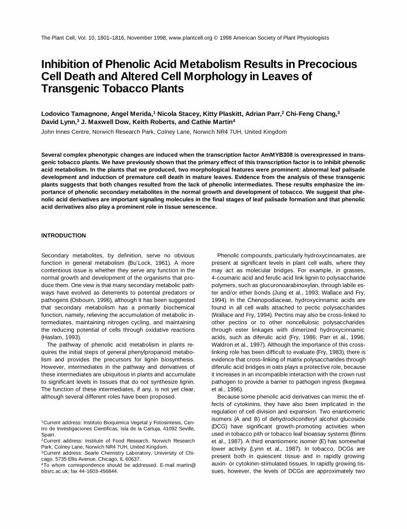
Young leaves from both Myb308 and control plants werebasically similar, with tightly packed cells in the mesophyll.In intermediate-aged leaves, differences in the palisade cellswere apparent (Figure 1A). In controls, the palisade meso-phyll could be distinguished as a layer of closely packed,columnar cells beneath the upper epidermis. In Myb308plants, the palisade cells were more isodiametric, with largeair spaces between them, suggesting reduced cell-to-celladhesion.
Striking differences were also observed in sections ofolder leaves (Figure 1B). In control leaves in which senes-cence was beginning, the cells were highly vacuolate, show-ing loss of cell contents, and the cell walls appeared fluted,arching inward at positions adjacent to chloroplasts. How-ever, nuclei and identifiable organelles could still be detected.In Myb308 leaves, the cells did not appear to senesce nor-mally. Rather, among cells that appeared morphologicallynormal, there were isolated dead cells that were unusual inappearance: collapsed, avacuolate, and very electron dense.Such isolated dying and dead cells were located largely inthe spongy and palisade layers of the mesophyll. Dead, col-lapsed cells of this type were never observed in control sec-tions. No viral particles or bacterial cells were ever seenassociated with the dead cells in Myb308 plants.
Form of the Dead Cells in Myb308 Leaves
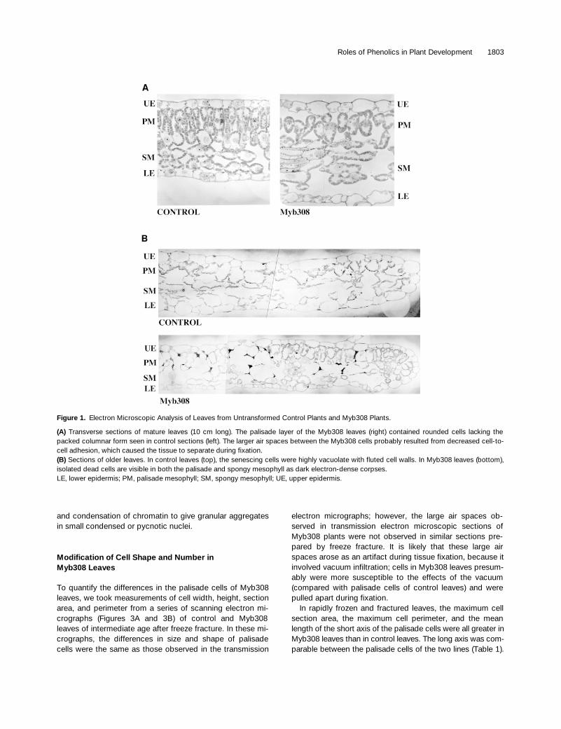
The cellular contents of the dead cells in Myb308 leaveswere very electron dense, although the remains of thylakoidmembranes could be distinguished (Figure 2A). The nucleiwere also smaller than in healthy cells, and they containedhighly condensed chromatin. Sections from older Myb308leaves showed some cells in a more intermediate stage ofcollapse, with highly condensed nuclei and expanded chlo-roplasts (Figure 2A; lower cell). The cytoplasmic contentshad precipitated or condensed to form darkly staining amor-phous material, and the cells were avacuolate. In some cellssurrounding dying or dead cells, the chloroplasts appearedswollen (Figure 2B, cell type II); this could represent an earlystage in cell death because otherwise their cytoplasmic in-tegrity appeared normal. The general appearance (corpsemorphology) was similar to that described in animal cells asapoptotic (Wyllie et al., 1980), because death occurred ini-tially in isolated cells and involved inward cellular collapse
Roles of Phenolics in Plant Development 1803
and condensation of chromatin to give granular aggregatesin small condensed or pycnotic nuclei.
Modification of Cell Shape and Number inMyb308 Leaves
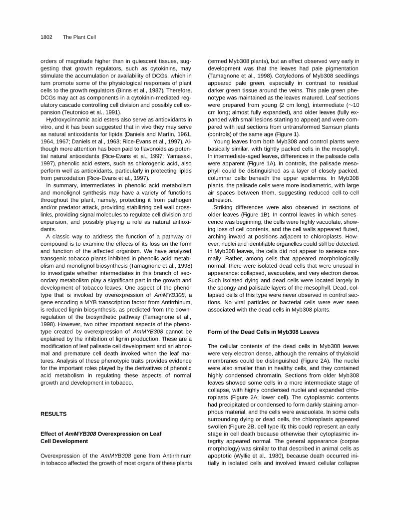
To quantify the differences in the palisade cells of Myb308leaves, we took measurements of cell width, height, sectionarea, and perimeter from a series of scanning electron mi-crographs (Figures 3A and 3B) of control and Myb308leaves of intermediate age after freeze fracture. In these mi-crographs, the differences in size and shape of palisadecells were the same as those observed in the transmission
electron micrographs; however, the large air spaces ob-served in transmission electron microscopic sections ofMyb308 plants were not observed in similar sections pre-pared by freeze fracture. It is likely that these large airspaces arose as an artifact during tissue fixation, because itinvolved vacuum infiltration; cells in Myb308 leaves presum-ably were more susceptible to the effects of the vacuum(compared with palisade cells of control leaves) and werepulled apart during fixation.
In rapidly frozen and fractured leaves, the maximum cellsection area, the maximum cell perimeter, and the meanlength of the short axis of the palisade cells were all greater inMyb308 leaves than in control leaves. The long axis was com-parable between the palisade cells of the two lines (Table 1).
Figure 1. Electron Microscopic Analysis of Leaves from Untransformed Control Plants and Myb308 Plants.
(A) Transverse sections of mature leaves (10 cm long). The palisade layer of the Myb308 leaves (right) contained rounded cells lacking thepacked columnar form seen in control sections (left). The larger air spaces between the Myb308 cells probably resulted from decreased cell-to-cell adhesion, which caused the tissue to separate during fixation.(B) Sections of older leaves. In control leaves (top), the senescing cells were highly vacuolate with fluted cell walls. In Myb308 leaves (bottom),isolated dead cells are visible in both the palisade and spongy mesophyll as dark electron-dense corpses.LE, lower epidermis; PM, palisade mesophyll; SM, spongy mesophyll; UE, upper epidermis.
1804 The Plant Cell
This confirmed our observation that Myb308 palisade cellswere wider and rounder than wild-type columnar palisadecells. The mean values for the long and short axes ofMyb308 and wild-type cells were used to calculate the aver-age cell surface area and volume, assuming that the controlcells were cylindrical and that the Myb308 cells were prolatespheroids. These values are listed in Table 2. Although thedifferences in surface area between the two cell types werenot striking, the volume of Myb308 cells was considerablygreater than that of control cells.
In sections of a given length, there were also thereforefewer palisade cells in Myb308 leaves than in control leaves(an average of 30% fewer cells), as shown in Table 3. Myb308leaves were smaller than control leaves at maturity, empha-sizing that
AmMYB308
has a significant effect on palisadecell number in leaves as well as on cell shape.
Gravimetric Analysis of Myb308 and Control Leaves
Thin sections of leaves suggested that cell wall productionin Myb308 palisade cells may be limited. We estimated theeffect of
AmMYB308
on cell wall production by gravimetricanalysis of fully expanded leaves because, developmentally,
these probably represent the end of net wall deposition dur-ing leaf development.
Given an equal amount of fresh leaf material, Myb308samples contained, on average, 39% less total dry weightthan did the controls (Table 4). Most of the loss of dry weightwas due to a reduction (48%) in insoluble material repre-senting cell wall material. There was a much smaller reduc-tion in soluble material (25%). Clearly, such a significantdecrease in cell wall material cannot be explained solely asa result of reduced cell wall phenolics, especially in the pre-dominantly nonlignified tissue of the leaf blade.
Suspension Cultures of Myb308 and ControlTobacco Cells
To determine the cause of the altered growth of Myb308palisade cells, we tried to feed phenolic acid intermediatesto Myb308 plants, but it was not possible to feed them to in-tact plants reliably, so cell suspension cultures were estab-lished from Myb308 and control plants (untransformed ortransformed with binary vector T-DNA alone). These cul-
Figure 2. Details of Cell Death in Myb308 Plants.
(A) Dead cells showing condensed nuclear material (n). The cyto-plasmic contents appear to be aggregated, and the position of thechloroplasts (c) can be distinguished by their thylakoid membranesand starch granules. The vacuole is absent, and the condensed cy-toplasmic contents have separated from the cell walls (w). The up-per cell is in a more advanced stage of collapse than is the lower cell.(B) Intermediate stages in cell death in Myb308 leaves. Three typesof cells are apparent: type I cells are normal with crescent-shapedplastids (c) and normal nuclei (n); type II cells have inflated chloro-plasts and condensed chromatin in their nuclei, suggesting thatthese cells may be at an intermediate stage between cell type I andcell type III, which are dead. All three cell types can be found to-gether, illustrating a degree of cell autonomy for the processes ofcell death. A stoma (s) is also shown.
Figure 3. Scanning Electron Microscopy of Freeze Fracture Sec-tions of Mature Control and Myb308 Leaves.
(A) Control section.(B) Myb308 leaf section.To measure the maximum perimeter, maximum section area, longaxis, and short axis, cells of the palisade that stood out on the pho-tograph and whose edges were clearly visible were circled and mea-sured with a computerized graphics tablet. The sections were alsoused to count the number of cells in a given length of leaf section, asshown in (A) and (B). Bars 5 100 mm.
Roles of Phenolics in Plant Development 1805
tured cells have been developed to model the growth of pal-isade cells (Stickens et al., 1996; Tao and Verbelen, 1996),especially developing palisade cells that are not fully au-totrophic but are dependent on both light and imported su-crose, as are the cultured cells.
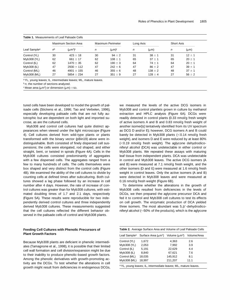
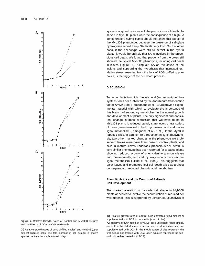
Myb308 and control cell cultures had quite distinct ap-pearances when viewed under the light microscope (Figure4). Cell cultures derived from wild-type plants or plantstransformed with the binary vector (pBin19) alone were in-distinguishable. Both consisted of finely dispersed cell sus-pensions; the cells were elongated, rod shaped, and eitherstraight, bent, or twisted in spirals (Figure 4A). Cells in theMyb308 cultures consisted predominantly of aggregateswith a few dispersed cells. The aggregates ranged from afew to many hundreds of cells. The cells themselves werebox shaped and very distinct from the control cells (Figure4B). We examined the ability of the cell cultures to divide bycounting cells at defined times after subculturing. Both cul-tures showed a lag phase followed by an increase in cellnumber after 4 days. However, the rate of increase of con-trol cultures was greater than for Myb308 cultures, with esti-mated doubling times of 1.7 and 2.1 days, respectively(Figure 5A). These results were reproducible for two inde-pendently derived control cultures and three independentlyderived Myb308 cultures. These measurements suggestedthat the cell cultures reflected the different behavior ob-served in the palisade cells of control and Myb308 plants.
Feeding Cell Cultures with Phenolic Precursors ofPlant Growth Factors
Because Myb308 plants are deficient in phenolic intermedi-ates (Tamagnone et al., 1998), it is possible that their limitedcell wall formation and cell division/expansion might be dueto their inability to produce phenolic-based growth factors.Among the phenolic derivatives with growth-promoting ac-tivity are the DCGs. To test whether the alterations in cellgrowth might result from deficiencies in endogenous DCGs,
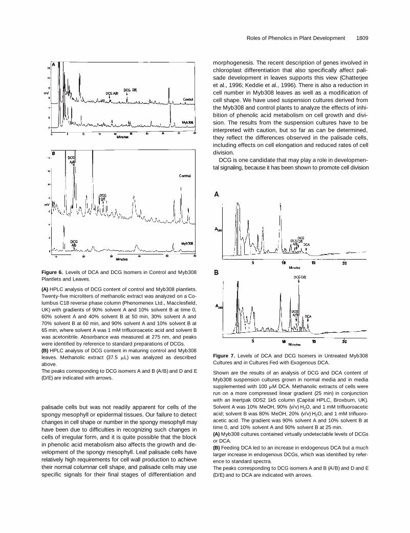
we measured the levels of the active DCG isomers inMyb308 and control plantlets grown in culture by methanolextraction and HPLC analysis (Figure 6A). DCGs werereadily detected in control plants (0.33 nmol/g fresh weightof active isomers A and B and 0.83 nmol/g fresh weight ofanother isomer[s] tentatively identified from its UV spectrumas DCG D and/or E); however, DCG isomers A and B couldbarely be detected in Myb308 plants (
,
0.14 nmol/g freshweight), and isomers D and E were reduced by at least 80%(
,
0.19 nmol/g fresh weight). The aglycone dehydrodico-niferyl alcohol (DCA) was undetectable in either control orMyb308 plants. We repeated these assays with maturingleaf tissue from independent plants. DCA was undetectablein control and Myb308 leaves. The active DCG isomers (Aand B) were measured at 7.1 nmol/g fresh weight, and theother isomers (D and E) were measured at 1.6 nmol/g freshweight in control leaves. Only the active isomers (A and B)were detected in Myb308 leaves and were measured at0.16 nmol/g fresh weight (Figure 6B).
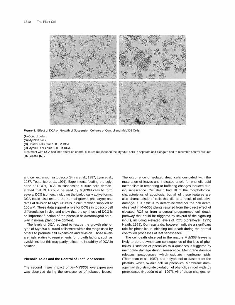
To determine whether the alterations in the growth ofMyb308 cells resulted from deficiencies in the levels ofDCGs, we then prepared the aglycone precursor DCA andfed it to control and Myb308 cell cultures to test its effectson cell growth. The enzymatic production of DCA yieldedthree isomers. The most abundant was 5,
b9
-dehydrodico-niferyl alcohol (
z
50% of the products), which is the aglycone
Table 1.
Measurements of Leaf Palisade Cells
Maximum Section Area Maximum Perimeter Long Axis Short Axis
Leaf Sample
a
n
b
(
m
m
2
)
c
n
(
m
m)
c
n
(
m
m)
n
(
m
m)
Control (YL) 30 423
6
18 30 94
6
2 31 38
6
1 31 12
6
1Myb308 (YL) 62 661
6
17 62 108
6
1 65 37
6
1 65 20
6
1Control (IL) 62 1470
6
35 62 190
6
3 64 74
6
1 64 20
6
1Myb308 (IL) 47 2930
6
112 47 242
6
6 47 86
6
2 47 39
6
1Control (ML) 46 4901
6
155 46 355
6
6 48 138
6
2 48 37
6
1Myb308 (ML) 27 5854
6
234 27 351
6
9 27 128
6
4 27 56
6
2
a
YL, young leaves; IL, intermediate leaves; ML, mature leaves.
b
n
, the number of sections analyzed.
c
Mean area (
m
m
2
) or dimension (
m
m)
6
SD
.
Table 2.
Average Surface Area and Volume of Leaf Palisade Cells
Leaf Sample
a
Surface Area (
m
m
2
) Volume (
m
m
3
) Volume/Area
Control (YL) 1,672 4,363 2.6Myb308 (YL) 2,053 7,992 3.9Control (IL) 5,191 22,629 4.4Myb308 (IL) 8,840 67,621 7.6Control (ML) 18,035 145,912 8.1Myb308 (ML) 18,997 211,207 11.1
a
YL, young leaves; IL, intermediate leaves; ML, mature leaves.
1806 The Plant Cell
of the active DCGs. HPLC analysis of methanolic extracts ofMyb308 cells that were fed DCA showed that intracellularDCA levels increased, as did the levels of various DCG iso-mers, including the most active forms of DCG, isomers Aand B (Figures 7A and 7B). The Myb308 cells therefore ap-peared to be able to convert DCA to DCGs. HPLC analysisshowed not only the formation of different DCG isomerswithin the cells but also the limited metabolism of DCA orDCG to other phenolics in the cultures, which is consistentwith previous results showing slow turnover of DCGs (Orrand Lynn, 1992). In contrast, when a mixture of DCG A plusB was fed to cells, the DCGs were not readily taken up andremained instead in the culture medium (data not shown).
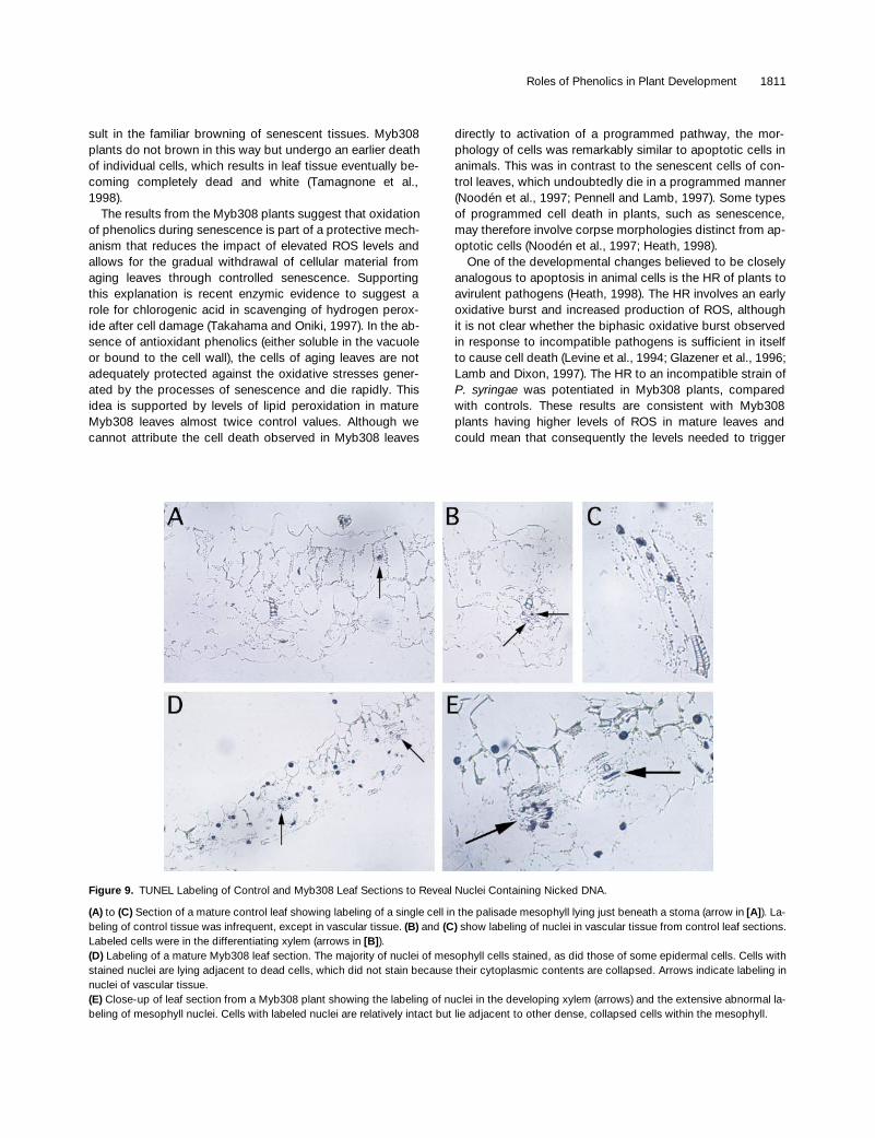
Treatment of control cells with DCA produced no obviousvisible changes in their appearance. However, DCA had adramatic effect on the appearance of Myb308 cells (Figures8A to 8D). After 4 days in the medium with DCA, some of thecells started to elongate, and the culture as a whole becamemore dispersed, starting to resemble a control culture.These changes peaked at day 7, when a large proportion ofthe cells were elongated and cell aggregates were greatlyreduced in number and in size. After 1 week, the effect be-gan to decline, although the cultures retained their alteredappearance up to day 16.
This experiment was repeated four times with DCA fromdifferent sources with consistent results. By counting cells,we also measured the effects of DCA on the rates of divisionof control and Myb308 cells. Treatment of control cultureswith DCA had no effect on cell proliferation (Figure 5B). Un-treated Myb308 cells again showed reduced rates of prolif-eration compared with controls. The treatment of twoindependent Myb308 cultures with DCA increased prolifera-tion rates to levels comparable with those of the control cul-tures (Figure 5C).
These results support the view that the abnormal growthand division of leaf palisade cells in Myb308 plants mighthave the same cause as the abnormal growth in the suspen-sion cultures and result from lower levels of phenolic acidderivatives. The particular derivatives involved were proba-bly the isomers of DCG. Presumably, the consequences of areduction in DCG levels are most marked in leaf palisadecells, because these have specific requirements for such
growth signals in developing their normal, tightly packed co-lumnar form in the final stages of leaf development. Interest-ingly, the levels of DCGs measured in maturing leaves ofcontrol plants were
.
10-fold greater than those measuredfor whole plantlets, indicating particularly high DCG accu-mulation in maturing leaves.
The altered shape of the palisade cells and the reducedcell number in Myb308 leaves, which we believe, from theseresults, to be due to a lack of phenolic signaling compounds,mean that the chloroplasts are not as densely packed oneabove the other in the cells as they are in the normal colum-nar cells of control leaves. This may explain the paler ap-pearance of Myb308 leaves.
Premature Cell Death in Myb308 Leaves
The other prominent effect of
AmMYB308
overexpressionwas the premature induction of cell death in mature leaves,similar to apoptotic cells described for animals by Wyllie etal. (1980). A characteristic event in many apoptotic animalcell deaths is the breakdown of nuclear DNA by endonucle-olytic cleavage. A sensitive method for detecting nicking ofDNA is in situ tagging of the 3
9
hydroxyl ends of DNA gener-ated during endonucleolytic DNA fragmentation (TUNEL la-beling; Gavrieli et al., 1992).
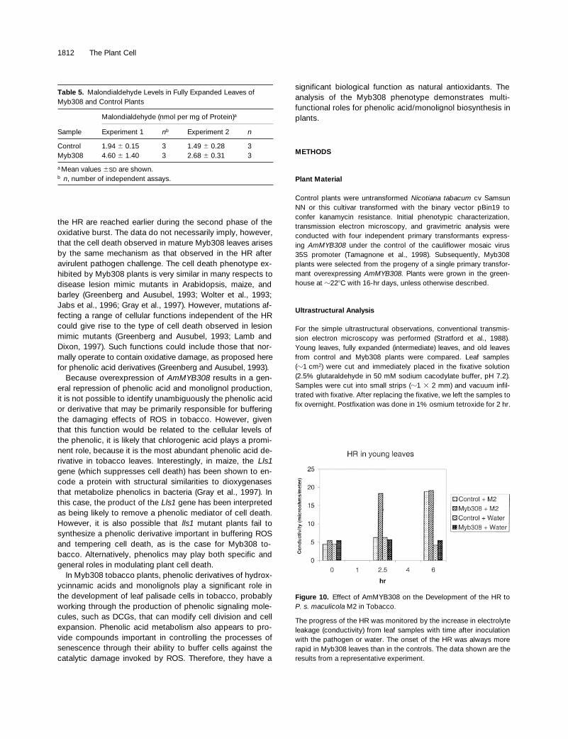
To investigate this cellular collapse, we compared sec-tions of mature leaves from Myb308 and control plants. Incontrol plants, very few nuclei were stained (Figure 9A), andthese were almost exclusively restricted to differentiatingxylem elements (Figure 9B), similar to results reported byMittler and Lam (1995). In Myb308 leaves, in contrast, manynuclei were stained. These were dispersed in a scatteredfashion and present in all tissues of the leaf, including vas-cular tissue (Figure 9C) and mesophyll and epidermal cells(Figure 9D). The nuclei stained in cells before their collapse(Figures 9D and 9E).
Table 3.
Number of Cells in Palisade Mesophyll
Leaf Sample
a
Average Number of Cells per mm
2
Control (YL) 8280Myb308 (YL) 4360Control (IL) 3364Myb308 (IL) 1225Control (ML) 784Myb308 (ML) 576
a
YL, young leaves; IL, intermediate leaves; ML, mature leaves.
Table 4.
Gravimetric Analysis of Fully Expanded Leaves from Myb308 and Control Tobacco Plants
Dry Weight
a
SampleTotal DryWeight
Cell WallMaterial
SolubleMaterial
Estimated Recovery(%)
Control leaves 14.0
6
1.2 7.8
6
1.3 4.4
6
0.5 87.0
6
5.2Myb308
leaves 8.6
6
0.8 4.0
6
0.7 3.3
6
0.7 85.2
6
7.0
a
Means and standard deviations of three control lines and sixMyb308 lines derived from four independent primary transformantsare shown. Measurements are given as the percentage of freshweight of tissue.
Roles of Phenolics in Plant Development 1807
Effects of AmMYB308 on Malondialdehyde Levels
The premature and abnormal cell death in the Myb308plants could result from increased oxidative stress, becausephenolic acid esters are excellent antioxidants (Daniels andMartin, 1961, 1964, 1967; Daniels et al., 1963). The defi-ciency of such natural antioxidants in Myb308 plants couldlead to an increase in the concentration of reactive oxygenspecies (ROS) above the threshold necessary to trigger celldeath. This might be particularly significant in mature andsenescing leaves, in which increases in the production ofO
2
2
and H
2
O
2
have been demonstrated (Thompson et al.,1987; Pastori and del Rio, 1997). Lipid peroxidation, whicharises from increased ROS, has been shown to increase intobacco leaves from the time that they reach maturity(Dhindsa et al., 1981). A by-product of lipid peroxidation ismalondialdehyde (MDA), and measurement of MDA levels incells can be used to assess the extent of damage to mem-brane lipids (Dhindsa et al., 1981).
We assayed the levels of MDA in two independent experi-ments. In each experiment, fully expanded leaves of threecontrol and three Myb308 plants were compared for theamount of MDA per milligram of protein. The results, sum-marized in Table 5, show that Myb308 leaves have doublethe MDA levels of control leaves of the same age.
The morphology of the dead and dying cells in Myb308leaves is very similar to that of cells that have died as part ofa hypersensitive response (HR) to an incompatible plantpathogen. An early component of the HR may be the oxida-tive burst generated in response to an avirulent pathogen(Doke and Ohashi, 1988; Chen et al., 1993; Legendre et al.,1993; Draper, 1997). The association of cell death with theproduction of ROS in both cases may account for the mor-phological similarities between dead cells resulting from theHR and the dead cells of Myb308 leaves.
To test the effect of lowered phenolic production on thedynamics of the HR, we compared the kinetics of the HR inMyb308 and control plants by using the M2 strain of
Pseudomonas syringae
pv
maculicola.
The development ofthe HR was monitored visually to determine the respond-ing leaf area and quantified by measuring ion leakage from7-mm discs cored from infected, water-flooded, and un-treated areas of leaves at successive time points.
Visual observation indicated that the kinetics of the HRwere different in Myb308 and control plants. The onset ofthe HR was always earlier in Myb308 plants; visible symp-toms (loss of turgor and a silvery sheen to the lower leafepidermis) appeared between 1 and 5 hr after infectioncompared with symptoms appearing between 2 and 5 hr af-ter infection in control plants. The final extent of HR in termsof the area of the leaf affected, however, was the same inMyb308 and control leaves. Ion leakage, measured as con-ductivity, also showed that the HR proceeds more rapidly inMyb308 than in control plants, although it was possible tosample only a few time points to avoid extensive mechanicaldamage to the leaves (Figure 10).
These results support the view that the absence of pheno-lics decreases the ability of Myb308 plants to withstandROS. Although an avirulent pathogen (presumably inducingan oxidative burst) was still required to induce the HR inMyb308 plants, the response was induced more rapidly. Wewere unable to induce the HR with nonpathogens, such as
Escherichia coli
, however.
Effect of Salicylic Acid on the Myb308 Phenotype
A block in phenylpropanoid metabolism after the step cata-lyzed by phenylalanine ammonia–lyase (Tamagnone et al.,1998) could lead to a buildup of cinnamate, resulting in in-creased synthesis of salicylic acid (SA) that might contributeto the formation of the HR-like lesions on the leaves ofMyb308 plants, perhaps by inducing the synthesis of ROS(Draper, 1997). SA is also involved in the systemic acquiredresistance of plants to pathogens (Dempsey and Klessig,1994). To ascertain the contribution of SA to the Myb308phenotype, we crossed Myb308 plants to a homozygous to-bacco line constitutively expressing the
nahG
gene from
Pseudomonas putida
(a gift of J. Draper; Bi et al., 1995). The
nahG
gene encodes the enzyme salicylate hydroxylase thatconverts SA to catechol, a compound that does not induce
Figure 4. Comparison of the Appearance of Suspension CultureCells from Control and Myb308 Tobacco Plants.
(A) Cells from a control culture transformed with pBin19 alone.(B) Cells from a Myb308 culture.Control cells typically had an elongated rod shape and were rela-tively dispersed, whereas Myb308 cells were box shaped and tendedto form large agregates.
1808 The Plant Cell
Figure 5.
Relative Growth Rates of Control and Myb308 Culturesand the Effects of DCA on Culture Growth.
(A)
Relative growth rates of control (filled circles) and Myb308 (opencircles) cultured cells. The fold increase in cell number is shownagainst the time from subculture in days.
(B)
Relative growth rates of control cells untreated (filled circles) orsupplemented with DCA in the media (open circles).
(C)
Relative growth rates of Myb308 cells untreated (filled circles,one culture line; filled squares, second independent culture line) andsupplemented with DCA in the media (open circles represent thefirst culture line treated with DCA; open squares represent the sec-ond culture line treated with DCA).
systemic acquired resistance. If the precocious cell death ob-served in Myb308 plants were the consequence of a high SAconcentration, hybrid plants should not show this aspect ofthe Myb308 phenotype, because the presence of salicylatehydroxylase would keep SA levels very low. On the otherhand, if the phenotype were still to persist in the hybridplants, it would be unlikely that SA is involved in the preco-cious cell death. We found that progeny from the cross stillshowed the typical Myb308 phenotype, including cell deathin leaves (Figure 11), ruling out SA as the cause of thelesions and supporting the hypothesis that increased ox-idative stress, resulting from the lack of ROS-buffering phe-nolics, is the trigger of the cell death process.
DISCUSSION
Tobacco plants in which phenolic acid (and monolignol) bio-synthesis has been inhibited by the Antirrhinum transcriptionfactor AmMYB308 (Tamagnone et al., 1998) provide experi-mental material with which to evaluate the importance ofthis branch of secondary metabolism in the normal growthand development of plants. The only significant and consis-tent change in gene expression that we have found inMyb308 plants is reduced steady state levels of transcriptsof those genes involved in hydroxycinnamic acid and mono-lignol metabolism (Tamagnone et al., 1998). In the Myb308tobacco lines, in addition to a reduction in lignin biosynthe-sis, two other marked changes in the phenotype were ob-served: leaves were paler than those of control plants, andcells in mature leaves undertook precocious cell death. Avery similar phenotype has been reported for tobacco plantsshowing reduced activity of phenylalanine ammonia–lyaseand, consequently, reduced hydroxycinnamic acid/mono-lignol metabolism (Elkind et al., 1990). This suggests thatpaler leaves and premature leaf cell death arise as a directconsequence of reduced phenolic acid metabolism.
Phenolic Acids and the Control of PalisadeCell Development
The marked alteration in palisade cell shape in Myb308plants appeared to involve the accumulation of reduced cellwall material. This is supported by ultrastructural analysis of
Roles of Phenolics in Plant Development 1809
palisade cells but was not readily apparent for cells of thespongy mesophyll or epidermal tissues. Our failure to detectchanges in cell shape or number in the spongy mesophyll mayhave been due to difficulties in recognizing such changes incells of irregular form, and it is quite possible that the blockin phenolic acid metabolism also affects the growth and de-velopment of the spongy mesophyll. Leaf palisade cells haverelatively high requirements for cell wall production to achievetheir normal columnar cell shape, and palisade cells may usespecific signals for their final stages of differentiation and
morphogenesis. The recent description of genes involved inchloroplast differentiation that also specifically affect pali-sade development in leaves supports this view (Chatterjeeet al., 1996; Keddie et al., 1996). There is also a reduction incell number in Myb308 leaves as well as a modification ofcell shape. We have used suspension cultures derived fromthe Myb308 and control plants to analyze the effects of inhi-bition of phenolic acid metabolism on cell growth and divi-sion. The results from the suspension cultures have to beinterpreted with caution, but so far as can be determined,they reflect the differences observed in the palisade cells,including effects on cell elongation and reduced rates of celldivision.
DCG is one candidate that may play a role in developmen-tal signaling, because it has been shown to promote cell division
Figure 7. Levels of DCA and DCG Isomers in Untreated Myb308Cultures and in Cultures Fed with Exogenous DCA.
Shown are the results of an analysis of DCG and DCA content ofMyb308 suspension cultures grown in normal media and in mediasupplemented with 100 mM DCA. Methanolic extracts of cells wererun on a more compressed linear gradient (25 min) in conjunctionwith an Inertpak 0DS2 1k5 column (Capital HPLC, Broxburn, UK).Solvent A was 10% MeOH, 90% (v/v) H2O, and 1 mM trifluoroaceticacid; solvent B was 80% MeOH, 20% (v/v) H2O, and 1 mM trifluoro-acetic acid. The gradient was 90% solvent A and 10% solvent B attime 0, and 10% solvent A and 90% solvent B at 25 min.(A) Myb308 cultures contained virtually undetectable levels of DCGsor DCA.(B) Feeding DCA led to an increase in endogenous DCA but a muchlarger increase in endogenous DCGs, which was identified by refer-ence to standard spectra.The peaks corresponding to DCG isomers A and B (A/B) and D and E(D/E) and to DCA are indicated with arrows.
Figure 6. Levels of DCA and DCG Isomers in Control and Myb308Plantlets and Leaves.
(A) HPLC analysis of DCG content of control and Myb308 plantlets.Twenty-five microliters of methanolic extract was analyzed on a Co-lumbus C18 reverse phase column (Phenomenex Ltd., Macclesfield,UK) with gradients of 90% solvent A and 10% solvent B at time 0,60% solvent A and 40% solvent B at 50 min, 30% solvent A and70% solvent B at 60 min, and 90% solvent A and 10% solvent B at65 min, where solvent A was 1 mM trifluoroacetic acid and solvent Bwas acetonitrile. Absorbance was measured at 275 nm, and peakswere identified by reference to standard preparations of DCGs.(B) HPLC analysis of DCG content in maturing control and Myb308leaves. Methanolic extract (37.5 mL) was analyzed as describedabove.The peaks corresponding to DCG isomers A and B (A/B) and D and E(D/E) are indicated with arrows.
1810 The Plant Cell
and cell expansion in tobacco (Binns et al., 1987; Lynn et al.,1987; Teutonico et al., 1991). Experiments feeding the agly-cone of DCGs, DCA, to suspension culture cells demon-strated that DCA could be used by Myb308 cells to formseveral DCG isomers, including the biologically active forms.DCA could also restore the normal growth phenotype andrates of division to Myb308 cells in culture when supplied at100 mM. These data support a role for DCGs in tobacco celldifferentiation in vivo and show that the synthesis of DCG isan important function of the phenolic acid/monolignol path-way in normal plant development.
The levels of DCA required to rescue the growth pheno-type of Myb308 cultured cells were within the range used byothers to promote cell expansion and division. Those levelsare high relative to requirements for growth factors, such ascytokinins, but this may partly reflect the instability of DCA insolution.
Phenolic Acids and the Control of Leaf Senescence
The second major impact of AmMYB308 overexpressionwas observed during the senescence of tobacco leaves.
The occurrence of isolated dead cells coincided with thematuration of leaves and indicated a role for phenolic acidmetabolism in tempering or buffering changes induced dur-ing senescence. Cell death had all of the morphologicalcharacteristics of apoptosis, but all of these features arealso characteristic of cells that die as a result of oxidativedamage. It is difficult to determine whether the cell deathobserved in Myb308 plants resulted from the direct effect ofelevated ROS or from a central programmed cell deathpathway that could be triggered by several of the signalinginputs, including elevated levels of ROS (Korsmeyer, 1995;Heath, 1998). Our results do, however, indicate a significantrole for phenolics in inhibiting cell death during the normalcontrolled processes of leaf senescence.
The cell death observed in the mature Myb308 leaves islikely to be a downstream consequence of the loss of phe-nolics. Oxidation of phenolics to o-quinones is triggered bymembrane damage during senescence. Membrane damagereleases lipoxygenase, which oxidizes membrane lipids(Thompson et al., 1987), and polyphenol oxidases from theplastids, which oxidize cellular phenolics. Membrane dam-age may also stimulate oxidation of phenolics in cell walls byperoxidases (Noodén et al., 1997). All of these changes re-
Figure 8. Effect of DCA on Growth of Suspension Cultures of Control and Myb308 Cells.
(A) Control cells.(B) Myb308 cells.(C) Control cells plus 100 mM DCA.(D) Myb308 cells plus 100 mM DCA.Treatment with DCA had little effect on control cultures but induced the Myb308 cells to separate and elongate and to resemble control cultures(cf. [B] and [D]).
Roles of Phenolics in Plant Development 1811
sult in the familiar browning of senescent tissues. Myb308plants do not brown in this way but undergo an earlier deathof individual cells, which results in leaf tissue eventually be-coming completely dead and white (Tamagnone et al.,1998).
The results from the Myb308 plants suggest that oxidationof phenolics during senescence is part of a protective mech-anism that reduces the impact of elevated ROS levels andallows for the gradual withdrawal of cellular material fromaging leaves through controlled senescence. Supportingthis explanation is recent enzymic evidence to suggest arole for chlorogenic acid in scavenging of hydrogen perox-ide after cell damage (Takahama and Oniki, 1997). In the ab-sence of antioxidant phenolics (either soluble in the vacuoleor bound to the cell wall), the cells of aging leaves are notadequately protected against the oxidative stresses gener-ated by the processes of senescence and die rapidly. Thisidea is supported by levels of lipid peroxidation in matureMyb308 leaves almost twice control values. Although wecannot attribute the cell death observed in Myb308 leaves
directly to activation of a programmed pathway, the mor-phology of cells was remarkably similar to apoptotic cells inanimals. This was in contrast to the senescent cells of con-trol leaves, which undoubtedly die in a programmed manner(Noodén et al., 1997; Pennell and Lamb, 1997). Some typesof programmed cell death in plants, such as senescence,may therefore involve corpse morphologies distinct from ap-optotic cells (Noodén et al., 1997; Heath, 1998).
One of the developmental changes believed to be closelyanalogous to apoptosis in animal cells is the HR of plants toavirulent pathogens (Heath, 1998). The HR involves an earlyoxidative burst and increased production of ROS, althoughit is not clear whether the biphasic oxidative burst observedin response to incompatible pathogens is sufficient in itselfto cause cell death (Levine et al., 1994; Glazener et al., 1996;Lamb and Dixon, 1997). The HR to an incompatible strain ofP. syringae was potentiated in Myb308 plants, comparedwith controls. These results are consistent with Myb308plants having higher levels of ROS in mature leaves andcould mean that consequently the levels needed to trigger
Figure 9. TUNEL Labeling of Control and Myb308 Leaf Sections to Reveal Nuclei Containing Nicked DNA.
(A) to (C) Section of a mature control leaf showing labeling of a single cell in the palisade mesophyll lying just beneath a stoma (arrow in [A]). La-beling of control tissue was infrequent, except in vascular tissue. (B) and (C) show labeling of nuclei in vascular tissue from control leaf sections.Labeled cells were in the differentiating xylem (arrows in [B]).(D) Labeling of a mature Myb308 leaf section. The majority of nuclei of mesophyll cells stained, as did those of some epidermal cells. Cells withstained nuclei are lying adjacent to dead cells, which did not stain because their cytoplasmic contents are collapsed. Arrows indicate labeling innuclei of vascular tissue.(E) Close-up of leaf section from a Myb308 plant showing the labeling of nuclei in the developing xylem (arrows) and the extensive abnormal la-beling of mesophyll nuclei. Cells with labeled nuclei are relatively intact but lie adjacent to other dense, collapsed cells within the mesophyll.
1812 The Plant Cell
the HR are reached earlier during the second phase of theoxidative burst. The data do not necessarily imply, however,that the cell death observed in mature Myb308 leaves arisesby the same mechanism as that observed in the HR afteravirulent pathogen challenge. The cell death phenotype ex-hibited by Myb308 plants is very similar in many respects todisease lesion mimic mutants in Arabidopsis, maize, andbarley (Greenberg and Ausubel, 1993; Wolter et al., 1993;Jabs et al., 1996; Gray et al., 1997). However, mutations af-fecting a range of cellular functions independent of the HRcould give rise to the type of cell death observed in lesionmimic mutants (Greenberg and Ausubel, 1993; Lamb andDixon, 1997). Such functions could include those that nor-mally operate to contain oxidative damage, as proposed herefor phenolic acid derivatives (Greenberg and Ausubel, 1993).
Because overexpression of AmMYB308 results in a gen-eral repression of phenolic acid and monolignol production,it is not possible to identify unambiguously the phenolic acidor derivative that may be primarily responsible for bufferingthe damaging effects of ROS in tobacco. However, giventhat this function would be related to the cellular levels ofthe phenolic, it is likely that chlorogenic acid plays a promi-nent role, because it is the most abundant phenolic acid de-rivative in tobacco leaves. Interestingly, in maize, the Lls1gene (which suppresses cell death) has been shown to en-code a protein with structural similarities to dioxygenasesthat metabolize phenolics in bacteria (Gray et al., 1997). Inthis case, the product of the Lls1 gene has been interpretedas being likely to remove a phenolic mediator of cell death.However, it is also possible that lls1 mutant plants fail tosynthesize a phenolic derivative important in buffering ROSand tempering cell death, as is the case for Myb308 to-bacco. Alternatively, phenolics may play both specific andgeneral roles in modulating plant cell death.
In Myb308 tobacco plants, phenolic derivatives of hydrox-ycinnamic acids and monolignols play a significant role inthe development of leaf palisade cells in tobacco, probablyworking through the production of phenolic signaling mole-cules, such as DCGs, that can modify cell division and cellexpansion. Phenolic acid metabolism also appears to pro-vide compounds important in controlling the processes ofsenescence through their ability to buffer cells against thecatalytic damage invoked by ROS. Therefore, they have a
significant biological function as natural antioxidants. Theanalysis of the Myb308 phenotype demonstrates multi-functional roles for phenolic acid/monolignol biosynthesis inplants.
METHODS
Plant Material
Control plants were untransformed Nicotiana tabacum cv SamsunNN or this cultivar transformed with the binary vector pBin19 toconfer kanamycin resistance. Initial phenotypic characterization,transmission electron microscopy, and gravimetric analysis wereconducted with four independent primary transformants express-ing AmMYB308 under the control of the cauliflower mosaic virus35S promoter (Tamagnone et al., 1998). Subsequently, Myb308plants were selected from the progeny of a single primary transfor-mant overexpressing AmMYB308. Plants were grown in the green-house at z228C with 16-hr days, unless otherwise described.
Ultrastructural Analysis
For the simple ultrastructural observations, conventional transmis-sion electron microscopy was performed (Stratford et al., 1988).Young leaves, fully expanded (intermediate) leaves, and old leavesfrom control and Myb308 plants were compared. Leaf samples(z1 cm2) were cut and immediately placed in the fixative solution(2.5% glutaraldehyde in 50 mM sodium cacodylate buffer, pH 7.2).Samples were cut into small strips (z1 3 2 mm) and vacuum infil-trated with fixative. After replacing the fixative, we left the samples tofix overnight. Postfixation was done in 1% osmium tetroxide for 2 hr.
Table 5. Malondialdehyde Levels in Fully Expanded Leaves of Myb308 and Control Plants
Malondialdehyde (nmol per mg of Protein)a
Sample Experiment 1 nb Experiment 2 n
Control 1.94 6 0.15 3 1.49 6 0.28 3Myb308 4.60 6 1.40 3 2.68 6 0.31 3
a Mean values 6SD are shown.b n, number of independent assays.
Figure 10. Effect of AmMYB308 on the Development of the HR toP. s. maculicola M2 in Tobacco.
The progress of the HR was monitored by the increase in electrolyteleakage (conductivity) from leaf samples with time after inoculationwith the pathogen or water. The onset of the HR was always morerapid in Myb308 leaves than in the controls. The data shown are theresults from a representative experiment.
Roles of Phenolics in Plant Development 1813
The strips were then infiltrated in medium grade London Resin white(London Resin Company, Basingstoke, UK) and transferred to ge-latine capsules, and the resin was polymerized for 12 hr in a 608Coven. The embedded samples were sectioned with a Reichert-Jungultramicrotome (Leica, Milton Keynes, UK) at a thickness of 70 to90 nm, and the sections were poststained with uranyl acetate andlead citrate (Millonig, 1961). Finally, the specimens were observedusing an electron microscope (model 1200 EX; JEOL, Welwyn Gar-den City, UK).
In Situ Detection of Apoptotic Cells
Leaf samples were cut from control and fully expanded Myb308leaves. Samples were taken from green areas in the middle part ofthe leaf. Sample fixation and wax embedding were performed in thesame way as for in situ hybridization (Jackson, 1992). Nuclei of cellsundergoing programmed cell death were stained by the TUNELmethod (Gavrieli et al., 1992) by using the ApopTag in situ apoptosisdetection kit (peroxidase version) (Appligene; Oncor, Birtley, UK). Themethod was adapted from that recommended by the manufacturer.Wax-embedded samples were sectioned to a thickness of 7 mm andthen dewaxed in xylene (two washes of 5 min each), absolute ethanol(two washes of 5 min each), 95% ethanol and then 70% ethanol for3 min each wash, and finally PBS (10 mM phosphate buffer and 145mM NaCl, pH 7.2) for a 5-min wash. Proteins were digested with pro-teinase K (20 mg/mL) and then washed four times in distilled water for2 min for each wash. Endogenous peroxidase activity was quenchedby treating the slides in 1% hydrogen peroxide in PBS for 5 min atroom temperature. Slides were then rinsed twice with PBS for 5 mineach, and then terminal transferase equilibration buffer (Appligene;
Oncor) was added. Terminal transferase and dUTP–digoxigenin werethen added to the reaction buffer, as recommended by the manufac-turer (Oncor). The reaction ran at 378C for 30 min. After washing threetimes in PBS, the anti–digoxigenin-peroxidase antibody conjugatewas applied to the slides and incubated for 30 min at room tempera-ture. The specimens were then washed three times in PBS and oncein distilled water. The peroxidase substrate, consisting of 4.8 mL ofdistilled water, 1.2 mL of chloronaphthol (30 mg/mL in methanol),and 7.5 mL of H2O2, was then added. The substrate was allowed toreact for a few minutes, and the slides were then washed copiouslywith distilled water. Sections were mounted in water and photo-graphed immediately by using bright-field microscopy. Controls inwhich water was used in place of terminal transferase were includedand showed no labeling of nuclei.
Malondialdehyde Assay
Fresh leaves (0.5 g) were ground in 3 mL of 10 mM sodium phos-phate buffer, pH 7.2, and then centrifuged at 2000g for 10 min. Asample of the supernatant (0.2 mL) was added to a glass tube con-taining 0.8 mL of distilled water, 0.5 mL of 20% (w/w) trichloroaceticacid, and 1 mL of 10 mM thiobarbituric acid. A control was run foreach sample in which thiobarbituric acid was replaced by an equalvolume of distilled water. The samples were heated in a boiling waterbath for 30 min and then centrifuged for 10 min at 2000g to removehaziness. The cleared samples were allowed to equilibrate to roomtemperature before the absorption at 532 nm was measured. Theconcentration of malondialdehyde (MDA) was calculated using itsextinction coefficient of 156 mmol cm21.
NahG Plants
A homozygous line of tobacco expressing the nahG gene fromPseudomonas putida under the control of the cauliflower mosaic vi-rus 35S promoter was provided by J. Draper (Bi et al., 1995). This linewas crossed reciprocally to a line homozygous for AmMYB308.Progeny from both crosses showed 100% resistance to kanamycinand developed lesions on their leaves.
Gravimetric Analysis
Before harvesting leaves, we kept plants in complete darkness for 2days to allow starch to mobilize; otherwise, the starch would sedi-ment with the cell wall pellets. This period of darkness was sufficientto obtain cell wall pellets free of starch, as determined by micro-scopic observation and iodine staining.
Fully expanded leaves of three control lines and Myb308 lines de-rived from four independent primary transformants were halved bycutting along their midveins. One-half of each leaf was weighed,freeze-dried, and weighed again to give the total dry weight. Theother half of each leaf was weighed and used for preparing crude cellwalls. Leaf material was frozen in liquid nitrogen and ground to a finepowder. The powder was resuspended in 40 mL of distilled waterand then centrifuged at 50g for 5 min at 48C to obtain a pellet com-prised mainly of cell walls. The supernatant was removed, and thepellet was washed twice in 80% ethanol and once in distilled water.Samples were then freeze-dried and weighed. The dry weight of the
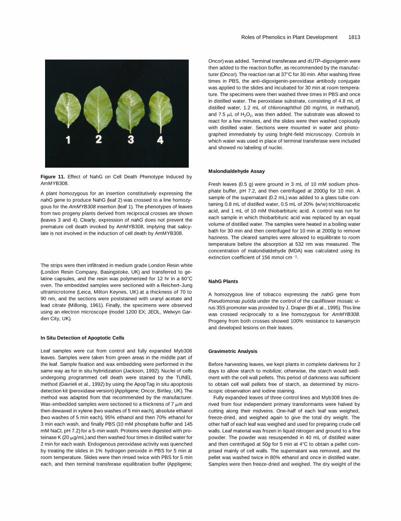
Figure 11. Effect of NahG on Cell Death Phenotype Induced byAmMYB308.
A plant homozygous for an insertion constitutively expressing thenahG gene to produce NahG (leaf 2) was crossed to a line homozy-gous for the AmMYB308 insertion (leaf 1). The phenotypes of leavesfrom two progeny plants derived from reciprocal crosses are shown(leaves 3 and 4). Clearly, expression of nahG does not prevent thepremature cell death invoked by AmMYB308, implying that salicy-late is not involved in the induction of cell death by AmMYB308.
1814 The Plant Cell
freeze-dried cell wall material was determined. Supernatants from allthe washes during the cell wall preparation were pooled and freeze-dried. This dry weight was considered the soluble fraction.
Measurement of Cell Sizes
Measurements of the long axis, short axis, maximum perimeter, andmaximum section area of cells were taken using a computerizedgraphic tablet from scanning electron micrographs of 10 control and10 Myb308 leaf blades. The figures obtained were used to calculatethe average surface area and volume of palisade cells in leaves ofdifferent ages. The mathematical formulas used were from Korn andKorn (1968).
Establishment of Cell Suspension Cultures
Seeds of control and Myb308 plants were surface-sterilized andsown individually in 100-mL Richardson’s (Leicester, UK) jars with20 mL of Murashige and Skoog (MS) plant salt and vitamin mixture(Murashige and Skoog, 1962) supplemented with 3% sucrose and0.8% Difco bacto agar. Seeds were germinated and grown asepti-cally for 4 weeks at 258C with 16-hr days. Cut pieces of small leaveswere placed in Petri dishes with 15 mL of MS agar supplementedwith 2 mg/L 2,4-D and 100 mg/mL kanamycin and placed in the darkfor z3 weeks until calli were formed. The calli were then transferredinto liquid culture by placing them in 250-mL conical flasks contain-ing 50 mL of MS medium, pH 5.8 (Murashige and Skoog, 1962), sup-plemented with 3% sucrose, 100 mg/L inositol, 1 mg/L thiamine, 0.2mg/L 2,4-D, and 200 mg/L KH2PO4. Flasks were placed on an orbitalshaker rotating at 100 rpm and grown at 258C under constant dimlight. The cultured cells were subcultured once a week by placing 10mL of the culture in a flask containing fresh MS medium.
To measure cell culture doubling time, we subcultured 5 mL ofeach culture into 50 mL of fresh medium. A 5-mL sample of the cul-ture was taken at the start and at various time points during the en-suing 8 days. Cells were counted using a hemocytometer, and thetotal number of cells in a 5-mL sample was calculated.
Production and Feeding of Dehydrodiconiferyl Alcohol
Dehydrodiconiferyl alcohol (DCA) was produced according toTeutonico et al. (1991) by using coniferyl alcohol and horseradishperoxidase (Sigma). DCA was analyzed by HPLC as described byTamagnone et al. (1998). The feeding experiment was done by sub-culturing control and Myb308 cells into different flasks containing 50mL of the medium plus DCA (100 mM), dehydrodiconiferyl alcoholglucoside (DCG; 10 mM), or zeatin riboside (0.1 mM). Samples (0.5mL) were taken daily for microscopic observation.
Extraction and Measurement of Phenolics
Plantlets grown in culture, maturing leaves, or suspension culturedcells were frozen in liquid nitrogen and ground with a pestle and mor-tar. Phenolics were extracted in dry, ice-cold methanol and analyzedby HPLC, as described by Tamagnone et al. (1998). For identification
of DCA and isomers of DCG, samples were compared with stan-dards prepared as described by Lynn et al. (1987) and verified bynuclear magnetic resonance and mass spectrometry. Positiveidentification of DCA and DCG isomers involved coelution with thestandards and demonstration of the appropriate, characteristic UVabsorption spectra profiles.
Inoculation of Plants with Pseudomonas syringae
P. syringae pv maculicola M2 was prepared as a bacterial suspen-sion at 108 colony-forming units per milliliter of water and used toflood large sectors of leaf mesophyll of control and Myb308 plantsgrown in growth cabinets at 228C under constant illumination. Devel-opment of the hypersensitive response (HR) was monitored visually.Two leaf discs from each inoculated area were placed in 10 mL ofhigh-purity deionized water of known conductivity. The discs werevacuum infiltrated for 2 min and then left to stand for 30 min at 228C.The conductivity of the water was then measured.
ACKNOWLEDGMENTS
We thank Maureen McCann and Brian Wells for advice and encour-agement during the realization of this work, John Draper for providingnahG-expressing tobacco plants, David Hopwood for constructivecomments on the manuscript, and Merry Limberg and Theresa Warrfor preparation of the manuscript. This research was funded by aBiotechnology and Biological Sciences Research Council (BBSRC)Link grant (CEMENT), by the European Union (Human Capital andMobility Project) through a category 30 training fellowship to L.T.(contract No. ERBCHBGCT920095), and by a Federation of EuropeanBiochemical Studies Fellowship to A.M.
Received July 9, 1998; accepted September 12, 1998.
REFERENCES
Bi, Y.-M., Kenton, P., Mur, L., Darby, R., and Draper, J. (1995).Hydrogen peroxide does not function downstream of salicylicacid in the induction of PR protein expression. Plant J. 8, 235–245.
Binns, A.N., Chen, R.H., Wood, H.N., and Lynn, D.G. (1987). Celldivision promoting activity of naturally occurring dehydrodicon-iferyl glucosides: Do cell wall components control cell division?Proc. Natl. Acad. Sci. USA 84, 980–984.
Bu’Lock, J.D. (1961). Intermediary metabolism and antibiotic syn-thesis. Adv. Appl. Microbiol. 3, 293–342.
Chatterjee, M., Sparvoli, S., Edmunds, C., Garosi, P., Findlay, K.,and Martin, C. (1996). DAG, a gene required for chloroplast dif-ferentiation and palisade development in Antirrhinum majus.EMBO J. 15, 4194–4207.
Chen, Z., Silva, H., and Klessig, D.F. (1993). Active oxygen speciesin the induction of plant systemic aquired resistance by salicylicacid. Science 262, 1883–1886.
Daniels, D.G.H., and Martin, H.F. (1961). Isolation of a new antioxi-dant from oats. Nature 191, 1302.
Roles of Phenolics in Plant Development 1815
Daniels, D.G.H., and Martin, H.F. (1964). Antioxidants in oats:Light-induced isomerization. Nature 203, 299.
Daniels, D.G.H., and Martin, H.F. (1967). Antioxidants in oats:Mono-esters of caffeic and ferulic acids. J. Sci. Food Agric. 18,589–595.
Daniels, D.G.H., King, H.G.C., and Martin, H.F. (1963). Antioxi-dants in oats: Esters of phenolic acids. J. Sci. Food Agric. 14,385–390.
Dempsey, D.M.A., and Klessig, D.F. (1994). Salicylic acid, activeoxygen species and systemic acquired resistance in plants.Trends Cell Biol. 4, 334–338.
Dhindsa, R.S., Plumb-Dhindsa, P., and Thorpe, T.A. (1981). Leafsenescence: Correlated with increased levels of membrane per-meability and lipid peroxidation, and decreased levels of superox-ide dismutase and catalase. J. Exp. Bot. 32, 93–101.
Doke, N., and Ohashi, Y. (1988). Involvement of an O2-generatingsystem in the induction of necrotic lesions on tobacco infectedwith tobacco mosaic virus. Physiol. Mol. Plant Pathol. 32, 163–175.
Draper, J. (1997). Salicylate, superoxide synthesis and cell suicidein plant defence. Trends Plant Sci. 2, 162–165.
Elkind, Y., Edwards, R., Mavandad, M., Hedrick, S.A., Ribak, O.,Dixon, R.A., and Lamb, C.J. (1990). Abnormal plant developmentand down regulation of phenyl propanoid biosynthesis in trans-genic tobacco containing a heterologous phenylalanine ammonialyase gene. Proc. Natl. Acad. Sci. USA 87, 9057–9061.
Fry, S.C. (1983). Feruloylated pectins from the primary cell wall:Their structure and possible functions. Planta 157, 111–123.
Fry, S.C. (1986). Cross-linking of matrix polymers in the growing cellwalls of angiosperms. Annu. Rev. Plant Physiol. 37, 165–186.
Gavrieli, Y., Sherman, Y., and Ben-Sasson, S.A. (1992). Identifica-tion of programmed cell death in situ via specific labelling ofnuclear DNA fragmentation. J. Cell Biol. 119, 493–501.
Glazener, J.A., Orlandi, E.W., and Baker, J.C. (1996). The activeoxygen response of cell suspensions to incompatible bacteria isnot sufficient to cause hypersensitive cell death. Plant Physiol.110, 759–763.
Gray, J., Close, P.S., Briggs, S.P., and Johal, G.S. (1997). A novelsuppressor of cell death in plants encoded by the Lls1 gene ofmaize. Cell 89, 25–31.
Greenberg, J.T., and Ausubel, F.M. (1993). Arabidopsis mutantscompromised for the control of cellular damage during pathogen-esis and ageing. Plant J. 4, 327–341.
Haslam, E. (1993). Shikimic Acid: Metabolism and Metabolites.(Chichester, UK: John Wiley and Sons).
Heath, M. (1998). Apoptosis, programmed cell death and the hyper-sensitive response. Eur. J. Plant Pathol. 104, 117–124.
Ikegawa, T., Mayama, S., Nakayashiki, H., and Kato, H. (1996).Accumulation of diferulic acid during the hypersensitive responseof oat leaves to Puccinia coronata f. sp. avenae and its role inresistance of oat leaves to cell wall degrading enzymes. Physiol.Mol. Plant Pathol. 48, 245–255.
Jabs, T., Dietrich, R.A., and Dangl, J.L. (1996). Initiation of run-away cell death in an Arabidopsis mutant by extracellular super-oxide. Science 273, 1853–1856.
Jackson, D. (1992). In situ hybridization in plants. In Molecular PlantPathology: A Practical Approach, S.J. Gurr, M.J. McPherson, andD.J. Bowles, eds (Oxford, UK: Oxford University Press), pp. 163–174.
Jung, H.G., Buxton, D.R., Hatfield, R.D., and Ralph, J. (1993). For-age Cell Wall Structure and Digestibility. (Madison, WI: AmericanSociety of Agronomy Inc.).
Keddie, J., Carol, B., Jones, J.D.G., and Gruissem, W. (1996). TheDCL gene of tomato is required for chloroplast development andpalisade cell morphogenesis in leaves. EMBO J. 15, 4208–4217.
Korn, G.A., and Korn, T.M., eds (1968). Mathematical Handbook forScientists and Engineers, 2nd ed. (New York: McGraw-Hill).
Korsmeyer, S.J. (1995). Regulators of cell death. Trends Genet. 11,101–105.
Lamb, C., and Dixon, R.A. (1997). The oxidative burst in plant dis-ease resistance. Annu. Rev. Plant Physiol. Plant Mol. Biol. 48,251–275.
Legendre, L., Reuters, S., Heinstein, P.F., and Low, P.S. (1993).Characterization of the oligogalacturonide-induced oxidative burstin cultured soybean (Glycine max) cells. Plant Physiol. 102, 233–240.
Levine, A., Tenhaken, R., Dixon, R., and Lamb, C.J. (1994). H2O2
from the oxidative burst orchestrates the plant hypersensitive dis-ease resistance response. Cell 79, 583–593.
Lynn, D.G., Chen, R.H., Manning, K.S., and Wood, H.N. (1987).The structural characterization of endogenous factors from Vincarosea crown gall tumors that promote cell division of tobaccocells. Proc. Natl. Acad. Sci. USA 84, 615–619.
Millonig, G. (1961). A modified procedure for lead staining of thinsections. J. Biophys. Biochem. Cytol. 11, 736–739.
Mittler, R., and Lam, E. (1995). In situ detection of nDNA fragmen-tation during the differentiation of tracheary elements in higherplants. Plant Physiol. 108, 489–493.
Murashige, T., and Skoog, F. (1962). A revised medium for rapidgrowth and bioassays with tobacco tissue cultures. Physiol. Plant.15, 473–497.
Noodén, L.D., Guiament, J.J., and John, I. (1997). Senescencemechanisms. Physiol. Plant. 101, 746–753.
Orr, J.D., and Lynn, D.G. (1992). Biosynthesis of dehydrodiconiferylalcohol glucosides: Implications for the control of tobacco cellgrowth. Plant Physiol. 98, 343–352.
Osbourn, A.E. (1996). Preformed antimicrobial compounds andplant defense against fungal attack. Plant Cell 8, 1821–1831.
Parr, A.J., Waldron, K.W., Ng, A., and Parker, M.L. (1996). Thewall-bound phenolics of chinese waterchestnut (Eleochans dulais).J. Sci. Food Agric. 71, 501–507.
Pastori, G.M., and del Rio, L.A. (1997). Natural senescence of pealeaves: An activated oxygen-mediated function for peroxisomes.Plant Physiol. 113, 411–418.
Pennell, R.I., and Lamb, C. (1997). Programmed cell death inplants. Plant Cell 9, 1157–1168.
Rice-Evans, C.A., Miller, N.J., and Paganga, G. (1997). Antioxidantproperties of phenolic compounds. Trends Plant Sci. 2, 152–159.
Stickens, D., Tao, W., and Verbelen, J.-P. (1996). A single cellmodel system to study hormone signal transduction. Plant GrowthRegul. 18, 149–154.
Roles of Phenolics in Plant Development 1816
Stratford, R., Plaskitt, K.A., Turner, D.S., Markham, P.G., andCovey, S.N. (1988). Molecular properties of Bari 1, a mild strain ofcauliflower mosaic virus. J. Gen. Virol. 69, 2375–2386.
Takahama, U., and Oniki, T. (1997). A peroxidase/phenolics/ascor-bate system can scavenge hydrogen peroxide in plant cells.Physiol. Plant. 101, 845–852.
Tamagnone, L., Merida, A., Parr, A., Mackay, S., Culianez-Macia,F.A., Roberts, K., and Martin, C. (1998). The AmMYB308 andAmMYB330 transcription factors from Antirrhinum regulate phe-nylpropanoid and lignin biosynthesis in transgenic tobacco. PlantCell 10, 135–154.
Tao, W., and Verbelen, J.-P. (1996). Switching on and off cell divi-sion and cell expansion in cultured mesophyll protoplasts oftobacco. Plant Sci. 116, 107–115.
Teutonico, R.A., Dudley, M.W., Orr, J.D., Lynn, D.G., and Binns,A.N. (1991). Activity and accumulation of cell division–promotingphenolics in tobacco tissue cultures. Plant Physiol. 97, 288–297.
Thompson, J.E., Legge, R.L., and Barber, R.F. (1987). The roleof free radicals in senescence and wounding. New Phytol. 105,317–344.
Waldron, K.W., Ng, A., Parker, M.L., and Parr, A.J. (1997). Ferulicacid dehydrodimers in the cell walls of Beta vulgaris and their pos-sible role in texture. J. Sci. Food Agric. 74, 221–228.
Wallace, G., and Fry, S.C. (1994). Phenolic compounds of the plantcell wall. Int. Rev. Cytol. 151, 229–267.
Wolter, M., Hollricher, K., Salamini, F., and Schulze-Lefert, P.(1993). The mlo resistance alleles to powdery mildew infection inbarley trigger a developmentally controlled defense mimic pheno-type. Mol. Gen. Genet. 239, 122–128.
Wyllie, A.H., Kerr, J.F.R., and Currie, A.R. (1980). Cell death: Thesignificance of apoptosis. Int. Rev. Cytol. 68, 251–306.
Yamasaki, H. (1997). A function of colour. Trends Plant Sci. 2, 7–8.
DOI 10.1105/tpc.10.11.1801 1998;10;1801-1816Plant Cell
Lynn, J. Maxwell Dow, Keith Roberts and Cathie MartinLodovico Tamagnone, Angel Merida, Nicola Stacey, Kitty Plaskitt, Adrian Parr, Chi-Feng Chang, David
Morphology in Leaves of Transgenic Tobacco PlantsInhibition of Phenolic Acid Metabolism Results in Precocious Cell Death and Altered Cell
This information is current as of June 6, 2014
References http://www.plantcell.org/content/10/11/1801.full.html#ref-list-1
This article cites 49 articles, 17 of which can be accessed free at:
Permissions https://www.copyright.com/ccc/openurl.do?sid=pd_hw1532298X&issn=1532298X&WT.mc_id=pd_hw1532298X
eTOCs http://www.plantcell.org/cgi/alerts/ctmain
Sign up for eTOCs at:
CiteTrack Alerts http://www.plantcell.org/cgi/alerts/ctmain
Sign up for CiteTrack Alerts at:
Subscription Information http://www.aspb.org/publications/subscriptions.cfm
is available at:Plant Physiology and The Plant CellSubscription Information for
ADVANCING THE SCIENCE OF PLANT BIOLOGY © American Society of Plant Biologists
Related Documents