Inhibition of hypoxia-inducible factor-1 function enhances the sensitivity of multiple myeloma cells to melphalan Yongzhen Hu, Keita Kirito, Kozue Yoshida, Toru Mitsumori, Kei Nakajima, Yumi Nozaki, Satoshi Hamanaka, Takahiro Nagashima, Masae Kunitama, Kumi Sakoe, and Norio Komatsu Department of Hematology and Oncology, University of Yamanashi, Yamanashi, Japan Abstract Abnormal activation of hypoxia-inducible factor-1 (HIF-1), one of the most important transcription factors for the adaptation of cells to hypoxia, is frequently observed in numerous types of solid tumors. Dysregulation of HIF-1 induces tumor angiogenesis and enhances the expression of anti-apoptotic proteins and glycolysis-associated enzymes in cancer cells, which in turn leads to the promo- tion of tumor growth. In the present study, we examined the pathophysiologic role of HIF-1 in multiple myeloma. Furthermore, we explored the possibility that HIF-1 may be a molecular target for myeloma therapy. We identified constitutive expression of the hypoxia-inducible factor-1 α (HIF-1α)-subunit in established myeloma cell lines and in primary myeloma cells. Treatment with insulin-like growth factor-1 (IGF-1) significantly increased HIF-1α expression through activation of the AKT and mitogen-activated pro- tein kinase signaling pathways. Inhibition of HIF-1 function either by echinomycin, a specific HIF-1 inhibitor, or a siRNA against HIF-1α resulted in enhanced sensitivity to melpha- lan in myeloma cells. This inhibition of HIF-1 also reversed the protective effect of IGF-1 on melphalan-induced apo- ptosis. Inhibition of HIF-1 drastically reduced both basal and IGF-1–induced expression of survivin, one of the most important anti-apoptotic proteins in myeloma cells. We conclude that HIF-1 inhibition may be an attractive therapeutic strategy for multiple myeloma. [Mol Cancer Ther 2009;8(8):2329–38] Introduction Despite new therapeutic approaches, multiple myeloma re- mains incurable. Myeloma cell intrinsic factors and micro- environmental stimuli contribute to the unfavorable biology of the disease (1). Cytogenetic abnormalities, in- cluding immunoglobulin H translocations, confer a growth advantage to myeloma cells through deregulated expres- sion of cyclin D family proteins. Several cytokines and che- mokines also support the growth and survival of myeloma cells, which ultimately leads to chemotherapy resistance. The biological functions of these intrinsic factors and micro- environmental stimuli are mediated by a series of transcrip- tion factors, including those of the nuclear factor κB, signal transducers and activators of transcription 3, and Forkhead transcription factor families (1). These transcription factors regulate the expression of anti-apoptotic proteins and en- hance cytokine production, which in turn induces patholog- ic angiogenesis, osteoblast activation, and the growth of myeloma cells in an autocrine fashion. Hypoxia-inducible factor-1 (HIF-1) is a transcription factor that serves as a master regulator of cellular responses to hyp- oxia (2) and regulates dozens of genes required for adaptation to hypoxia. HIF-1 is composed of two subunits, α-subunit and β-subunit. Although the expression of hypoxia-inducible fac- tor-1 β (HIF-1β)-subunit is constitutive, HIF-1α protein levels are regulated in response to oxygen tension. Under nor- moxic conditions, HIF-1α is degraded by the proteasome, whereas HIF-1 remains inactive. In hypoxia, HIF-1α is stabilized and forms a complex with HIF-1β that allows HIF-1 to function as a transcription factor. Thus, HIF-1 is activated only during hypoxia under normal physiologic conditions. In contrast, HIF-1 is frequently activated in cancer cells, including under normoxic conditions (3), by oncogene products, impaired activity of tumor suppressor genes (4), or the accumulation of metabolic glucose inter- mediates (5, 6). Constitutively activated HIF-1 enhances neovascularization, increases glucose metabolism, and in- duces the expression of anti-apoptotic proteins in cancer tissues. HIF-1 is thought to be one of the most important molecular targets in the treatment of cancer (7), and a va- riety of chemical inhibitors for HIF-1 have been introduced to date (8). In contrast to solid tumors, only a limited number of studies on the role of HIF-1 in hematologic malignancies have been published to date. HIF-1 is activated by cytokine stimuli in leukemic cells under normoxic conditions (9). HIF- 1 regulates vascular endothelial growth factor production in leukemic cells, which supports cell survival in an intracellu- lar autocrine fashion (9). HIF-1 has important functions in multiple myeloma. First, numerous important genes for the progression of multiple myeloma [i.e., vascular endothelial growth factor, stromal cell-derived factor-1, and myeloid cell Received 2/23/09; revised 5/18/09; accepted 5/19/09; published OnlineFirst 8/11/09. Grant support: Ministry of Health, Welfare, and Labor of Japan and Grants- in-Aid for Scientific Research from the Ministry of Education, Science, Sports, and Technology of Japan. The costs of publication of this article were defrayed in part by the payment of page charges. This article must therefore be hereby marked advertisement in accordance with 18 U.S.C. Section 1734 solely to indicate this fact. Note: Supplementary material for this article is available at Molecular Cancer Therapeutics Online (http://mct.aacrjournals.org/). Requests for reprints: Keita Kirito, University of Yamanashi, 1110 Shimokato, Chuo-shi, Yamanashi 409-3898, Japan. Phone: 81-55-273-9432; Fax: 81-55-273-1274. E-mail: [email protected] Copyright © 2009 American Association for Cancer Research. doi:10.1158/1535-7163.MCT-09-0150 Mol Cancer Ther 2009;8(8). August 2009 2329

Welcome message from author
This document is posted to help you gain knowledge. Please leave a comment to let me know what you think about it! Share it to your friends and learn new things together.
Transcript
Inhibition of hypoxia-inducible factor-1 function enhancesthe sensitivity of multiple myeloma cells to melphalan
Yongzhen Hu, Keita Kirito, Kozue Yoshida,Toru Mitsumori, Kei Nakajima, Yumi Nozaki,Satoshi Hamanaka, Takahiro Nagashima,Masae Kunitama, Kumi Sakoe, and Norio Komatsu
Department of Hematology and Oncology,University of Yamanashi, Yamanashi, Japan
AbstractAbnormal activation of hypoxia-inducible factor-1 (HIF-1),one of the most important transcription factors for theadaptation of cells to hypoxia, is frequently observed innumerous types of solid tumors. Dysregulation of HIF-1induces tumor angiogenesis and enhances the expressionof anti-apoptotic proteins and glycolysis-associatedenzymes in cancer cells, which in turn leads to the promo-tion of tumor growth. In the present study, we examinedthe pathophysiologic role of HIF-1 in multiple myeloma.Furthermore, we explored the possibility that HIF-1 maybe a molecular target for myeloma therapy. We identifiedconstitutive expression of the hypoxia-inducible factor-1 α(HIF-1α)-subunit in established myeloma cell lines and inprimary myeloma cells. Treatment with insulin-like growthfactor-1 (IGF-1) significantly increased HIF-1α expressionthrough activation of the AKT and mitogen-activated pro-tein kinase signaling pathways. Inhibition of HIF-1 functioneither by echinomycin, a specific HIF-1 inhibitor, or a siRNAagainst HIF-1α resulted in enhanced sensitivity to melpha-lan in myeloma cells. This inhibition of HIF-1 also reversedthe protective effect of IGF-1 on melphalan-induced apo-ptosis. Inhibition of HIF-1 drastically reduced both basaland IGF-1–induced expression of survivin, one of themost important anti-apoptotic proteins in myeloma cells.We conclude that HIF-1 inhibition may be an attractivetherapeutic strategy for multiple myeloma. [Mol CancerTher 2009;8(8):2329–38]
IntroductionDespite new therapeutic approaches, multiple myeloma re-mains incurable. Myeloma cell intrinsic factors and micro-environmental stimuli contribute to the unfavorablebiology of the disease (1). Cytogenetic abnormalities, in-cluding immunoglobulin H translocations, confer a growthadvantage to myeloma cells through deregulated expres-sion of cyclin D family proteins. Several cytokines and che-mokines also support the growth and survival of myelomacells, which ultimately leads to chemotherapy resistance.The biological functions of these intrinsic factors and micro-environmental stimuli are mediated by a series of transcrip-tion factors, including those of the nuclear factor κB, signaltransducers and activators of transcription 3, and Forkheadtranscription factor families (1). These transcription factorsregulate the expression of anti-apoptotic proteins and en-hance cytokine production, which in turn induces patholog-ic angiogenesis, osteoblast activation, and the growth ofmyeloma cells in an autocrine fashion.Hypoxia-inducible factor-1 (HIF-1) is a transcription factor
that serves as a master regulator of cellular responses to hyp-oxia (2) and regulates dozens of genes required for adaptationto hypoxia.HIF-1 is composedof two subunits,α-subunit andβ-subunit. Although the expression of hypoxia-inducible fac-tor-1 β (HIF-1β)-subunit is constitutive, HIF-1α protein levelsare regulated in response to oxygen tension. Under nor-moxic conditions, HIF-1α is degraded by the proteasome,whereas HIF-1 remains inactive. In hypoxia, HIF-1α isstabilized and forms a complex with HIF-1β that allowsHIF-1 to function as a transcription factor. Thus, HIF-1 isactivated only during hypoxia under normal physiologicconditions. In contrast, HIF-1 is frequently activated incancer cells, including under normoxic conditions (3), byoncogene products, impaired activity of tumor suppressorgenes (4), or the accumulation of metabolic glucose inter-mediates (5, 6). Constitutively activated HIF-1 enhancesneovascularization, increases glucose metabolism, and in-duces the expression of anti-apoptotic proteins in cancertissues. HIF-1 is thought to be one of the most importantmolecular targets in the treatment of cancer (7), and a va-riety of chemical inhibitors for HIF-1 have been introducedto date (8).In contrast to solid tumors, only a limited number of
studies on the role of HIF-1 in hematologic malignancieshave been published to date. HIF-1 is activated by cytokinestimuli in leukemic cells under normoxic conditions (9). HIF-1 regulates vascular endothelial growth factor production inleukemic cells, which supports cell survival in an intracellu-lar autocrine fashion (9). HIF-1 has important functions inmultiple myeloma. First, numerous important genes for theprogression of multiple myeloma [i.e., vascular endothelialgrowth factor, stromal cell-derived factor-1, and myeloid cell
Received 2/23/09; revised 5/18/09; accepted 5/19/09; publishedOnlineFirst 8/11/09.
Grant support:Ministry of Health, Welfare, and Labor of Japan and Grants-in-Aid for Scientific Research from the Ministry of Education, Science,Sports, and Technology of Japan.
The costs of publication of this article were defrayed in part by thepayment of page charges. This article must therefore be hereby markedadvertisement in accordance with 18 U.S.C. Section 1734 solely toindicate this fact.
Note: Supplementary material for this article is available at MolecularCancer Therapeutics Online (http://mct.aacrjournals.org/).
Requests for reprints: Keita Kirito, University of Yamanashi, 1110Shimokato, Chuo-shi, Yamanashi 409-3898, Japan. Phone: 81-55-273-9432;Fax: 81-55-273-1274. E-mail: [email protected]
Copyright © 2009 American Association for Cancer Research.
doi:10.1158/1535-7163.MCT-09-0150
Mol Cancer Ther 2009;8(8). August 2009
2329
leukemia sequence-1 (MCL-1)] are putative targets of HIF-1(3). Second, several transcription factors that play importantroles in the pathophysiology of multiple myeloma regulateHIF-1 expression and function. signal transducers and acti-vators of transcription 3 protects against HIF-1 degradation(10, 11). Several studies have shown that nuclear factor κBregulates HIF-1α transcription (12–14). Proteins from theForkhead transcription factor family, which are often inacti-vated in myeloma cells, negatively regulate HIF-1 function(15). In addition to these basic findings, recent studies haveproposed thatHIF-1may be amolecular target of certain newgeneration antimyeloma agents, including bortezomib (16)and heat shock protein 90 inhibitors (17). However, the bio-logical significance of HIF-1 inmultiplemyeloma is under in-vestigation, and to date, no study has investigated a newHIF-1–based strategy for multiple myeloma treatment.We investigated the molecular function of HIF-1 in multiple
myeloma cells using established myeloma cell lines andprimary CD138+ myeloma cells derived from patients. HIF-1is constitutively activated in these cells, even under normoxicconditions, and insulin-like growth factor-1 (IGF-1), one ofthe most crucial growth factors for myeloma cells, was clearlyshown to enhance HIF-1 levels. In addition, blocking HIF-1functionwas found to heighten the sensitivity ofmyeloma cellsto melphalan. Our observations suggest that inhibiting HIF-1function may be a new and effective therapeutic approach formultiple myeloma.
Materials and MethodsCell Culture
Human MM cell lines KMM-1, U266, and RPMI8226 wereobtained from Japanese Collection of Research Bioresourcesand cultured in RPMI-1640 media (Sigma Chemical)containing 10% FCS, 25 U/mL penicillin, and 25 ng/mLstreptomycin (GIBICO). For culture under hypoxic condi-tions, we used a hypoxic chamber (Juji Field).Reagents
IGF-1 and interleukin 6 were purchased from PeproTechEC. LY294002 was from EMD Biosciences, Inc. U0126 wasfrom Cell Signaling Technology. Echinomycin, melphalan,and dexamethasone were sourced from Sigma.Preparation of Cell Lysates and Immunoblotting
Whole cell lysates or nuclear and cytosolic fractions oflysates were prepared as described elsewhere (9). Nuclearextracts were prepared as described previously (18). Lysateproteins were size fractionated by SDS-PAGE and thenelectroblotted onto polyvinylidene difluoride membranes.The blots were probed with the following antibodies:anti–HIF-1α, anti–BCL-X, and anti–BCL-2 (BD Transduc-tion Laboratories); anti–phospho-AKT (Ser 473), anti-AKT,and anti–β-actin (Cell Signaling Technology); anti–MCL-1,antisurvivin, and anti-general transcription factor IIH (TFIIH;Santa Cruz); anti–HIF-1α (Novus Biologicals). The blotswere visualized using a chemiluminescence detection kit(LumiGLO, Cell Signaling).RNA Preparation and Real-Time PCR
Total cellular RNA was isolated using an RNeasy MiniKit (Qiagen). Reverse transcription was done using an
M-MLV reverse transcriptase kit (Invitrogen), according tothe manufacturer's instructions. Quantitative analysis ofsurvivin mRNA was done using predeveloped TaqManassay reagents (Applied Biosystems). 18S RNA was usedas an internal control for real-time RT-PCR.Establishment of Stable HIF-1α Knockdown CellsThe pSuper HIF-1α/siRNAvector and empty pSuper vec-
tor were generous gifts from Dr. Daniel Chung (Massachu-setts General Hospital, Harvard Medical School; ref. 19). Togenerate stable transfectants, KMM-1 cells were transfectedand selected with 0.5 μg/mL puromycin (Sigma) for 14 d,and clones were selected and screened for HIF-1α silencingby immunoblot analysis.Apoptosis Assay
Cellular apoptosis was determined using an AnnexinV–Azami-Green Apoptosis detection kit (Medical andBiological Laboratories), according to the manufacturer'sinstructions. For primary myeloma cells, apoptotic cellswere detected with the Hoechst staining method becauseof the limited number of cells.Isolation of Primary Multiple Myeloma Cells and
CD19-Positive Normal B Lymphocytes
This study was approved by the institutional reviewboard of the University of Yamanashi. After informedconsent, primary multiple myeloma cells were purifiedfrom bone marrow aspirates with anti-CD138 magneticbeads (Miltenyi Biotech) in accordance with the manufac-turer's protocol. These cells were further analyzed usingWright-Giemsa staining, and a purity of better than90% was confirmed. Clinical features are summarized inTable 1. CD19-positive B lymphocytes were collected fromperipheral blood of healthy volunteers using anti-CD19magnetic beads (Miltenyi Biotech) and used as normal Blymphocytes.
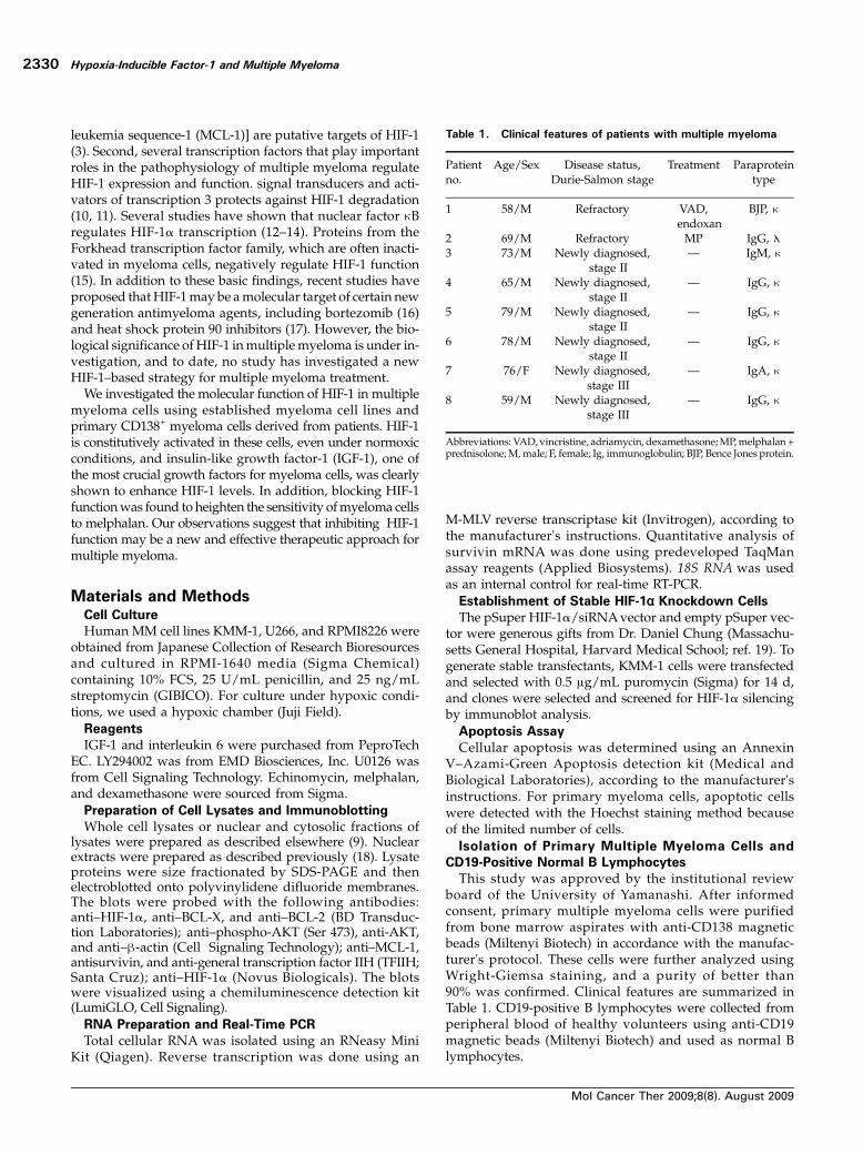
Table 1. Clinical features of patients with multiple myeloma
Patientno.
Age/Sex Disease status,Durie-Salmon stage
Treatment Paraproteintype
1 58/M Refractory VAD,endoxan
BJP, κ
2 69/M Refractory MP IgG, λ3 73/M Newly diagnosed,
stage II— IgM, κ
4 65/M Newly diagnosed,stage II
— IgG, κ
5 79/M Newly diagnosed,stage II
— IgG, κ
6 78/M Newly diagnosed,stage II
— IgG, κ
7 76/F Newly diagnosed,stage III
— IgA, κ
8 59/M Newly diagnosed,stage III
— IgG, κ
Abbreviations: VAD, vincristine, adriamycin, dexamethasone;MP,melphalan +prednisolone;M,male; F, female; Ig, immunoglobulin; BJP, Bence Jones protein.
Hypoxia-Inducible Factor-1 and Multiple Myeloma
Mol Cancer Ther 2009;8(8). August 2009
2330
Immunofluorescence Microscopic Analysis
Immunofluorescence microscopic analysis for HIF-1αwasdone in accordance with previous reports (9). Images wereobtained using an Olympus BX50 microscope (Olympus)and analyzed using the VB-7010 system (KEYENCE).Luciferase Assay
A luciferase reporter plasmid vector containing hypoxia-responsive elements (20) was a generous gift fromDr. ShinaeKondo (University of Kyoto). The reporter plasmid wasintroduced into the myeloma cell lines by lipofection withthe pRL-TKLuc internal control plasmid (Promega). Forty-eight hours after transfection, the cells were harvested and
subjected to dual luciferase assay, according to the manufac-turer's instructions (Promega).Statistical Analysis
Statistical analysis was done using the Student's t test;P < 0.05 was considered statistically significant.
ResultsHIF-1α Is Constitutively Expressed and IGF-1 Elevates
Protein Levels in Multiple Myeloma Cells
Initially, we investigated the expression ofHIF-1α byWest-ern blotting in the three kinds of multiple myeloma-derived
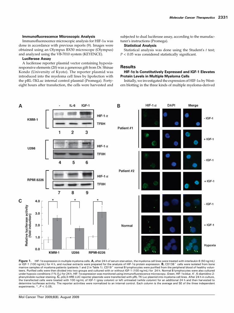
Figure 1. HIF-1α expression in multiple myeloma cells.A, after 24 h of serum starvation, the myeloma cell lines were treated with interleukin 6 (50 ng/mL)or IGF-1 (100 ng/mL) for 4 h, and nuclear extracts were prepared for the analysis of HIF-1α protein expression. B, CD138+ cells were isolated from bonemarrow samples of myeloma patients (patients 1 and 2 in Table 1). CD19+ normal B lymphocytes were purified from the peripheral blood of healthy volun-teers. Purified cells were then divided into two groups and cultured with or without IGF-1 (100 ng/mL) for 24 h. Normal B lymphocytes were also culturedunder hypoxic conditions (1% O2) for 24 h. HIF-1α expression was monitored using immunofluorescence microscopy.Green, HIF-1α blue, 4', 6-diamidino-2-phenylindole nuclear staining. C, pGL3-HRE-LUC reporter plasmids were transfected with pRL-TK Luc plasmid into myeloma cell lines. After 24 h in culture,the transfected cells were treated with 100 ng/mL of IGF-1 (gray column) or left untreated (white column) for an additional 24 h and then harvested todetermine luciferase activity. The reporter activities were normalized to an internal control. Each column is the average and SE of the three independentexperiments. *, P < 0.05.
Molecular Cancer Therapeutics
Mol Cancer Ther 2009;8(8). August 2009
2331
cell lines: KMM-1, U266, andRPMI8226. As shown in Fig. 1A(lanes 1, 4, and 7), HIF-1α protein was expressed in all threemyeloma cell lines under basal culture conditions. By con-trast, normoxic HIF-1α expression was not observed in nor-mal bone marrow mononuclear cells (data not shown).Because serum starvation may enhance HIF-1α expression(21), we compared expression of the protein in the presenceor absence of serum and found that serum starvation didnot enhance HIF-1α expression (Supplementary Fig. S1).We also analyzed HIF-1α expression in primary multiplemyeloma cells isolated from bone marrow aspirates fromtwo myeloma patients (patients 1 and 2 in Table 1) usingimmunofluorescence microscopy. CD19+ B lymphocyteswere isolated from healthy volunteers and used as thenormal control. Primary myeloma cells expressed HIF-1αprotein under basal conditions (Fig. 1B). In contrast, expres-sion of the protein was not recorded in normal B lympho-cytes under normoxic conditions, whereas culture of thesecells under hypoxic conditions drastically induced expres-sion of the protein. These results suggest that constitutiveexpression of HIF-1α is specific for multiple myeloma cells.In addition to constitutive activation, it is well known that
growth factors for cancer cells may enhance HIF-1 functionin several cancer cell types. To confirm this in our system, weevaluated whether interleukin 6 and IGF-1, both known tobe potent growth factors for myeloma cells, enhanced HIF-1α expression in these cells. As shown in Fig. 1A (lanes 3, 6,and 9), IGF-1 significantly induced HIF-1α expression in allmyeloma cell lines; by contrast, interleukin 6 exhibited onlymodest effects on HIF-1α expression levels (Fig. 1A, lanes 2,5, and 8). In accordance with the results from the establishedmyeloma cell lines, HIF-1α protein was drastically up-
regulated after culturing the primary myeloma cells withIGF-1 (Fig. 1B). We also found that IGF-1 failed to induceHIF-1α in normal B lymphocytes (Fig. 1B).To confirm that IGF-1 not only induces the expression of
HIF-1α but also activates HIF-1, we did a reporter assay us-ing a reporter plasmid that contained hypoxia-responsiveelements. As shown in Fig. 1C, treatment with IGF-1 signifi-cantly increased the transcriptional activity of HIF-1.Phosphatidylinositol 3 Kinase (PI3-K) and Mitogen-
Activated Protein Kinase (MAPK) Pathways Are Involved
in Constitutive and IGF-1–Induced HIF-1α Expression inMultiple Myeloma Cells
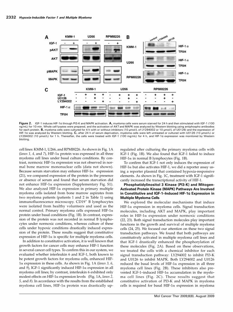
We explored the molecular mechanisms that induceHIF-1α expression in myeloma cells. Signal transductionmolecules, including AKT and MAPK, play importantroles in HIF-1α expression under normoxic conditions(22, 23). Both signal transduction molecules play importantfunctions in the growth and survival of multiple myelomacells (24, 25). We focused our attention on these two signaltransduction pathways. We found that both pathways areconstitutively activated in multiple myeloma cell lines andthat IGF-1 drastically enhanced the phosphorylation ofthese molecules (Fig. 2A). Based on these observations,we treated the cells with a chemical inhibitor for eachsignal transduction pathway: LY294002 to inhibit PI3-Kand U0126 to inhibit MAPK. Both LY294002 and U0126reduced the basal levels of HIF-1α expression in all threemyeloma cell lines (Fig. 2B). These inhibitors also pre-vented IGF-1–induced HIF-1α accumulation in the myelo-ma cell lines (Fig. 2C). These results suggest thatconstitutive activation of PI3-K and MAPK in myelomacells is required for basal HIF-1α expression in myeloma
Figure 2. IGF-1 induces HIF-1α through PI3-K and MAPK activation. A,myeloma cells were serum starved for 24 h and then stimulated with IGF-1 (100ng/mL) for 10 min. Whole cell lysates were prepared, and the activation of AKTand MAPK was analyzed by Western blotting using antiphospho antibodiesfor each protein. B, myeloma cells were cultured for 4 h with or without inhibitors (10 μmol/L of LY294002 or 10 μmol/L of U0126) and the expression ofHIF-1α was analyzed by Western blotting. C, after 24 h of serum deprivation, myeloma cells were left untreated or cultured with U0126 (10 μmol/L) orLY294002 (10 μmol/L) for 1 h. Thereafter, the cells were treated with IGF-1 (100 mg/mL) for 4 h, and HIF-1α expression was monitored by Westernblotting.
Hypoxia-Inducible Factor-1 and Multiple Myeloma
Mol Cancer Ther 2009;8(8). August 2009
2332
cells and that significant activation of these pathwaysby IGF-1 can lead to enhanced HIF-1α expression in mye-loma cell lines.Inhibition of HIF-1 Enhances the Sensitivity of
Myeloma Cells to Melphalan
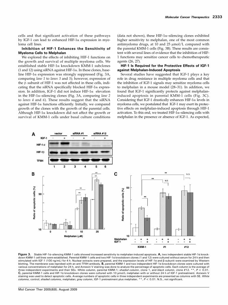
We explored the effects of inhibiting HIF-1 functions onthe growth and survival of multiple myeloma cells. Weestablished stable HIF-1α knockdown KMM-1 subclones(1 and 12) using siRNA against HIF-1α. In these clones, base-line HIF-1α expression was strongly suppressed (Fig. 3A,comparing lane 1 to lanes 3 and 5); however, expression ofthe β subunit of HIF-1 was not affected in these cells, indi-cating that the siRNA specifically blocked HIF-1α expres-sion. In addition, IGF-1 did not induce HIF-1α elevationin the HIF-1α–silencing clones (Fig. 3A, comparing lane 2to lanes 4 and 6). These results suggest that the siRNAagainst HIF-1α functions efficiently. Initially, we comparedgrowth of the clones with the growth of the parental cells.Although HIF-1α knockdown did not affect the growth orsurvival of KMM-1 cells under basal culture conditions
(data not shown), these HIF-1α–silencing clones exhibitedhigher sensitivity to melphalan, one of the most commonantimyeloma drugs, at 10 and 25 μmol/L compared withthe parental KMM-1 cells (Fig. 3B). These results are consis-tent with several lines of evidence that the inhibition of HIF-1 functions may sensitize cancer cells to chemotherapeuticagents (26, 27).HIF-1 Is Required for the Protective Effects of IGF-1
against Melphalan-Induced Apoptosis
Several studies have suggested that IGF-1 plays a keyrole in drug resistance in multiple myeloma cells and thatthe inhibition of IGF-1 signals may sensitize myeloma cellsto melphalan in a mouse model (28–31). In addition, wefound that IGF-1 significantly protects against melphalan-induced apoptosis in parental KMM-1 cells (Fig. 3C).Considering that IGF-1 drastically enhances HIF-1α levels inmyeloma cells, we postulated that IGF-1 may exert its protec-tive effects on melphalan-induced apoptosis through HIF-1activation. To this end, we treated HIF-1α–silencing cells withmelphalan in the presence or absence of IGF-1. As expected,
Figure 3. Stable HIF-1α–silencing KMM-1 cells showed increased sensitivity to melphalan-induced apoptosis. A, two independent stable HIF-1α knock-down KMM-1 cell lines were established. Parental KMM-1 cells and two HIF-1α knockdown clones (1 and 12) were cultured without serum for 24 h and thenstimulated with IGF-1 (100 ng/mL) for 4 h. Nuclear extracts were prepared, and the expression levels of HIF-1α and β-subunit were examined by Westernblotting. The membrane was reprobed with an anti-TFIIH antibody. B, parental KMM-1 and two independent HIF-1α knockdown clones were cultured withvarious concentrations of melphalan for 24 h, and Annexin V staining was done to analyze the percentage of apoptotic cells. Each column is the average ofthree independent experiments and their SEs. White column, parental KMM-1; shaded column, clone 1, and black column, clone #12. **, P < 0.01.C, parental KMM-1 cells and HIF-1α knockdown clones were cultured with 10 μmol/L melphalan with or without 24 h of IGF-1 pretreatment. Annexin Vstaining was used to detect apoptotic cells. Average numbers of apoptotic cells in three independent experiments are presented as columns with SE. Whitecolumns, control; shaded columns, melphalan; gray column, IGF-1 pretreatment plus melphalan. **, P < 0.01. N.S., not significant.
Molecular Cancer Therapeutics
Mol Cancer Ther 2009;8(8). August 2009
2333
IGF-1 failed to protect the HIF-1α knockdown cells frommelphalan-induced apoptosis (Fig. 3C), suggesting thatHIF-1α up-regulation is a prerequisite for the anti-apoptoticeffect of IGF-1 in myeloma cells.Echinomycin, A Small-Molecule Inhibitor of HIF-1,
Increases Melphalan-Induced Apoptosis in Myeloma
Cells
Considering that HIF-1 inhibitionmay be a new strategy toincrease the efficacy of melphalan and to overcome IGF-1–induced drug resistance in myeloma cells, we decided toexplore the effects of echinomycin, which blocks the DNA
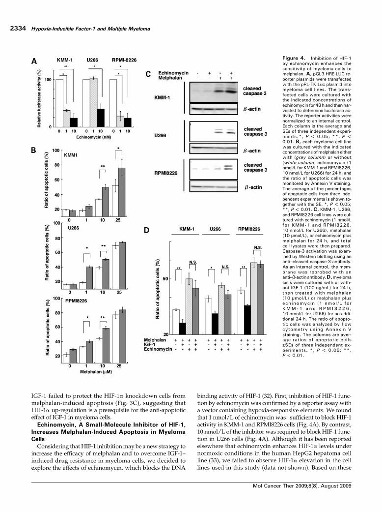
binding activity of HIF-1 (32). First, inhibition of HIF-1 func-tion by echinomycin was confirmed by a reporter assay witha vector containing hypoxia-responsive elements. We foundthat 1 nmol/L of echinomycin was sufficient to block HIF-1activity in KMM-1 and RPMI8226 cells (Fig. 4A). By contrast,10 nmol/L of the inhibitor was required to block HIF-1 func-tion in U266 cells (Fig. 4A). Although it has been reportedelsewhere that echinomycin enhances HIF-1α levels undernormoxic conditions in the human HepG2 hepatoma cellline (33), we failed to observe HIF-1α elevation in the celllines used in this study (data not shown). Based on these
Figure 4. Inhibition of HIF-1by echinomycin enhances thesensitivity of myeloma cells tomelphalan. A, pGL3-HRE-LUC re-porter plasmids were transfectedwith the pRL-TK Luc plasmid intomyeloma cell lines. The trans-fected cells were cultured withthe indicated concentrations ofechinomycin for48hand thenhar-vested to determine luciferase ac-tivity. The reporter activities werenormalized to an internal control.Each column is the average andSEs of three independent experi-ments.*, P < 0.05; **, P <0.01. B, each myeloma cell linewas cultured with the indicatedconcentrationsofmelphalan eitherwith (gray column) or without(white column) echinomycin (1nmol/L forKMM-1andRPMI8226,10 nmol/L for U266) for 24 h, andthe ratio of apoptotic cells wasmonitored by Annexin V staining.The average of the percentagesof apoptotic cells from three inde-pendent experiments is shown to-gether with the SE. *, P < 0.05;**, P < 0.01. C, KMM-1, U266,and RPMI8226 cell lines were cul-tured with echinomycin (1 nmol/Lfor KMM-1 and RPMI8226,10 nmol/L for U266), melphalan(10 μmol/L), or echinomycin plusmelphalan for 24 h, and totalcell lysates were then prepared.Caspase-3 activation was exam-ined by Western blotting using ananti–cleaved caspase-3 antibody.As an internal control, the mem-brane was reprobed with ananti–β-actinantibody.D,myelomacells were cultured with or with-out IGF-1 (100 ng/mL) for 24 h,then treated with melphalan(10 μmol/L) or melphalan plusech inomyc in (1 nmol /L forK M M - 1 a n d R P M I 8 2 2 6 ,10 nmol/L for U266) for an addi-tional 24 h. The ratio of apopto-tic cells was analyzed by flowcytometry using Annexin Vstaining. The columns are aver-age ratios of apoptotic cells±SEs of three independent ex-periments. *, P < 0.05; **,P < 0.01.
Hypoxia-Inducible Factor-1 and Multiple Myeloma
Mol Cancer Ther 2009;8(8). August 2009
2334
observations, we used the inhibitor at 1 nmol/L for KMM-1and RPMI8226 cells and at 10 nmol/L for the U266 cellsthroughout the study.We treated eachmyeloma cell linewithechinomycin and with various concentrations of melphalanfor 24 hours, andwemonitored the ratio of apoptotic cells byusing Annexin V staining. Consistent with siRNA silencingof the KMM-1 cells, treatment with echinomycin alone didnot induce apoptosis in myeloma cell lines (Fig. 4B). Howev-er, echinomycin significantly enhanced melphalan-inducedapoptosis in these cells (Fig. 4B). Similar results were ob-tained using a caspase-3 activation assay. Echinomycin alonedid not significantly increase the level of caspase-3 cleavagein myeloma cells during the observation period. However, itdrastically enhanced melphalan-induced caspase-3 cleavage(Fig. 4C). Importantly, the apoptosis-enhancing effects ofechinomycin were not observed in HIF-1α–silencingclones (Supplementary Fig. S2). This result indicates thatthe inhibitor exerts its apoptosis-enhancing effects specifi-cally through inhibition of HIF-1 activity. We also exam-ined whether the inhibitor inhibited the anti-apoptoticeffects of IGF-1 on melphalan-treated cells. As illustratedin Fig. 4D, IGF-1 pretreatment significantly reduced mel-phalan-induced apoptosis. However, adding echinomycincompletely canceled the IGF-1 anti-apoptotic effect againstmelphalan-induced apoptosis in these cell lines. These re-sults suggest that HIF-1 is an attractive agent to increasethe antimyeloma efficacy of melphalan. Because we knowthat IGF-1 also decreases the response of myeloma cells toother cytotoxic chemotherapeutic reagents, that is, doxo-rubicin (31), we investigated whether the inhibitor alsoenhanced the response of myeloma cells to doxorubicin.We found that echinomycin also enhanced apoptosis after
doxorubicin treatment and abrogated the protective effectsof IGF-1 (Supplementary Fig. S3). In contrast, the inhibitordid not show these effects when treated with dexametha-sone (data not shown).Echinomycin Sensitizes Primary Myeloma Cells to
Melphalan
To test the relevance of our findings, we used CD138+
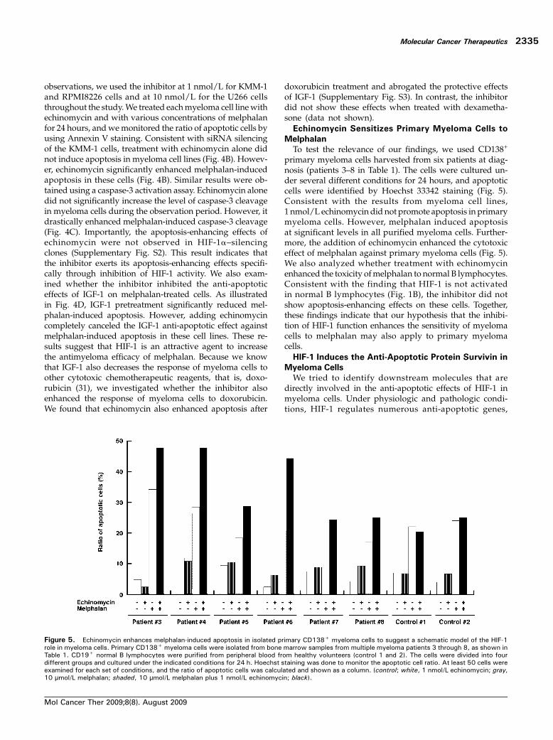
primary myeloma cells harvested from six patients at diag-nosis (patients 3–8 in Table 1). The cells were cultured un-der several different conditions for 24 hours, and apoptoticcells were identified by Hoechst 33342 staining (Fig. 5).Consistent with the results from myeloma cell lines,1 nmol/L echinomycin did not promote apoptosis in primarymyeloma cells. However, melphalan induced apoptosisat significant levels in all purified myeloma cells. Further-more, the addition of echinomycin enhanced the cytotoxiceffect of melphalan against primary myeloma cells (Fig. 5).We also analyzed whether treatment with echinomycinenhanced the toxicity ofmelphalan to normal B lymphocytes.Consistent with the finding that HIF-1 is not activatedin normal B lymphocytes (Fig. 1B), the inhibitor did notshow apoptosis-enhancing effects on these cells. Together,these findings indicate that our hypothesis that the inhibi-tion of HIF-1 function enhances the sensitivity of myelomacells to melphalan may also apply to primary myelomacells.HIF-1 Induces the Anti-Apoptotic Protein Survivin in
Myeloma Cells
We tried to identify downstream molecules that aredirectly involved in the anti-apoptotic effects of HIF-1 inmyeloma cells. Under physiologic and pathologic condi-tions, HIF-1 regulates numerous anti-apoptotic genes,
Figure 5. Echinomycin enhances melphalan-induced apoptosis in isolated primary CD138+ myeloma cells to suggest a schematic model of the HIF-1role in myeloma cells. Primary CD138+ myeloma cells were isolated from bone marrow samples from multiple myeloma patients 3 through 8, as shown inTable 1. CD19+ normal B lymphocytes were purified from peripheral blood from healthy volunteers (control 1 and 2). The cells were divided into fourdifferent groups and cultured under the indicated conditions for 24 h. Hoechst staining was done to monitor the apoptotic cell ratio. At least 50 cells wereexamined for each set of conditions, and the ratio of apoptotic cells was calculated and shown as a column. (control; white, 1 nmol/L echinomycin; gray,10 μmol/L melphalan; shaded, 10 μmol/L melphalan plus 1 nmol/L echinomycin; black).
Molecular Cancer Therapeutics
Mol Cancer Ther 2009;8(8). August 2009
2335
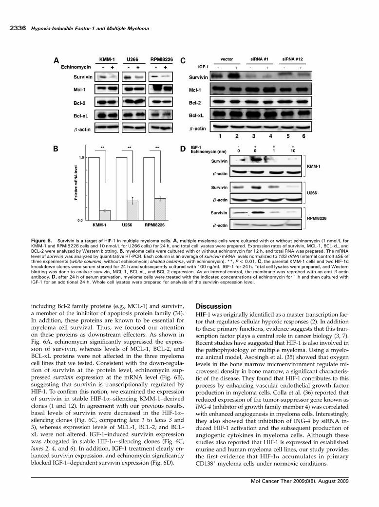
including Bcl-2 family proteins (e.g., MCL-1) and survivin,a member of the inhibitor of apoptosis protein family (34).In addition, these proteins are known to be essential formyeloma cell survival. Thus, we focused our attentionon these proteins as downstream effectors. As shown inFig. 6A, echinomycin significantly suppressed the expres-sion of survivin, whereas levels of MCL-1, BCL-2, andBCL-xL proteins were not affected in the three myelomacell lines that we tested. Consistent with the down-regula-tion of survivin at the protein level, echinomycin sup-pressed survivin expression at the mRNA level (Fig. 6B),suggesting that survivin is transcriptionally regulated byHIF-1. To confirm this notion, we examined the expressionof survivin in stable HIF-1α–silencing KMM-1–derivedclones (1 and 12). In agreement with our previous results,basal levels of survivin were decreased in the HIF-1α–silencing clones (Fig. 6C, comparing lane 1 to lanes 3 and5), whereas expression levels of MCL-1, BCL-2, and BCL-xL were not altered. IGF-1–induced survivin expressionwas abrogated in stable HIF-1α–silencing clones (Fig. 6C,lanes 2, 4, and 6). In addition, IGF-1 treatment clearly en-hanced survivin expression, and echinomycin significantlyblocked IGF-1–dependent survivin expression (Fig. 6D).
DiscussionHIF-1 was originally identified as a master transcription fac-tor that regulates cellular hypoxic responses (2). In additionto these primary functions, evidence suggests that this tran-scription factor plays a central role in cancer biology (3, 7).Recent studies have suggested that HIF-1 is also involved inthe pathophysiology of multiple myeloma. Using a myelo-ma animal model, Asosingh et al. (35) showed that oxygenlevels in the bone marrow microenvironment regulate mi-crovessel density in bone marrow, a significant characteris-tic of the disease. They found that HIF-1 contributes to thisprocess by enhancing vascular endothelial growth factorproduction in myeloma cells. Colla et al. (36) reported thatreduced expression of the tumor-suppressor gene known asING-4 (inhibitor of growth family member 4) was correlatedwith enhanced angiogenesis in myeloma cells. Interestingly,they also showed that inhibition of ING-4 by siRNA in-duced HIF-1 activation and the subsequent production ofangiogenic cytokines in myeloma cells. Although thesestudies also reported that HIF-1 is expressed in establishedmurine and human myeloma cell lines, our study providesthe first evidence that HIF-1α accumulates in primaryCD138+ myeloma cells under normoxic conditions.
Figure 6. Survivin is a target of HIF-1 in multiple myeloma cells. A, multiple myeloma cells were cultured with or without echinomycin (1 nmol/L forKMM-1 and RPMI8226 cells and 10 nmol/L for U266 cells) for 24 h, and total cell lysates were prepared. Expression rates of survivin, MCL-1, BCL-xL, andBCL-2 were analyzed by Western blotting. B, myeloma cells were cultured with or without echinomycin for 12 h, and total RNA was prepared. The mRNAlevel of survivin was analyzed by quantitative RT-PCR. Each column is an average of survivin mRNA levels normalized to 18S rRNA (internal control) ±SE ofthree experiments (white columns, without echinomycin; shaded columns, with echinomycin). **, P < 0.01. C, the parental KMM-1 cells and two HIF-1αknockdown clones were serum starved for 24 h and subsequently cultured with 100 ng/mL IGF-1 for 24 h. Total cell lysates were prepared, and Westernblotting was done to analyze survivin, MCL-1, BCL-xL, and BCL-2 expression. As an internal control, the membrane was reprobed with an anti–β-actinantibody. D, after 24 h of serum starvation, myeloma cells were treated with the indicated concentrations of echinomycin for 1 h and then cultured withIGF-1 for an additional 24 h. Whole cell lysates were prepared for analysis of the survivin expression level.
Hypoxia-Inducible Factor-1 and Multiple Myeloma
Mol Cancer Ther 2009;8(8). August 2009
2336
In addition to the baseline activation of HIF-1, IGF-1clearly enhances HIF-1α protein levels in established myelo-ma cell lines and in primary CD138+ myeloma cells. Theseresults suggest that HIF-1α lies downstream of the IGF-1signaling pathway in myeloma cells. This is supported bythe previous report that inhibition of the IGF-1 receptor sup-presses HIF-1 activity in the human myeloma cell line MM-1S (31). IGF-1 plays important roles in the pathophysiologyof multiple myeloma; it induces growth not only of estab-lished multiple myeloma cells but also of primary myelomacells from patients in vitro (24, 25). This cytokine confersmultiple drug resistance on myeloma cells (25, 30). Impor-tantly, clinical studies have revealed that increases in serumIGF-1 levels may be correlated with poor outcomes of mul-tiple myeloma (37). IGF-1 is also known to activate HIF-1 inseveral cancer cell lines. Fukuda et al. reported that expos-ing colon cancer cells to IGF-1 leads to activation of HIF-1and results in vascular endothelial growth factor secretion(22). They proposed amodel whereby IGF-1 regulates proteinsynthesis of the α-subunit of HIF-1 through the activationof the PI3-K/AKT and MAPK pathways (22). Elevation ofHIF-1α by IGF-1 was also reported in retinal epithelial cells,and PI3-K andMAPKwere also shown to be involved in thisprocess (23). In accordancewith these observations, our studyfound that inhibitors for PI3-K andMAPK can suppress IGF-1–induced HIF-1α elevation. In solid tumors, HIF-1 is be-lieved to be one of the most important molecular targets,and numerous reagents that target HIF-1 have been intro-duced in this field (8). In addition to solid tumors, our resultssuggest that HIF-1 may be an attractive molecular target inmultiple myeloma. Stable silencing of HIF-1α by siRNAcaused increased sensitivity to melphalan and loss ofthe IGF-1 protective effects against apoptosis. Moreover,we used a pharmacologic inhibitor, echinomycin, to blockHIF-1 functions. The inhibitory function of this reagent isconfirmed by in vitro and in vivo studies (38). In agree-ment with studies using siRNA against HIF-1α, echino-mycin clearly sensitized myeloma cells to conventionalchemotherapeutic agents and abrogated the protective ef-fect of IGF-1 on melphalan-induced apoptosis. Inhibitionof HIF-1 also enhanced the efficiency of doxorubicin, an-other key chemotherapeutic reagent for multiple myeloma(Supplementary Fig. S3). In addition to strategies that di-rectly block HIF-1 function (i.e., siRNA, specific pharmaco-logic inhibitors), recent studies highlight the way in whichseveral new-generation antimyeloma reagents already usedin clinical settings exhibit anti–HIF-1 activity. Bortezomib isone of the most important new-generation agents in thetreatment of myeloma (39). Although the precise molecularmechanism remains unclear, bortezomib inhibits the tran-scriptional activity of HIF-1 in cervical cancer cells (40), breastcancer cells (41), and myeloma cells (16). Inhibitors of heatshock protein 90 are also effective in controllingmultiple my-eloma (17). Heat shock protein 90 is a molecular chaperonethat stabilizes numerous proteins. Interestingly, Issacs et al.(42) reported that heat shock protein 90 regulates HIF-1αstability. Our preliminary observations also suggest that17-(dimethylaminoethylamino)-17-demethoxygeldanamy-
cin, a heat shock protein 90 inhibitor that exhibits anti-myeloma effects (43), clearly reduces basal and IGF-induced HIF-1α expression levels in myeloma cell lines.Although the pathway remains speculative at this stage,HIF-1 may be an important molecular target of heat shockprotein 90 inhibitors in multiple myeloma. Bortezomib orheat shock protein 90 inhibitors may enhance the cytotoxi-city of conventional chemotherapeutic agents, includingmelphalan through the inhibition of HIF-1 transcriptionalactivity.Finally, we also explored the mechanism by which HIF-1
inhibition sensitizes myeloma cells to melphalan. AlthoughHIF-1 regulates dozens of genes, we chose to focus on theexpression of anti-apoptotic proteins that are known to beimportant for myeloma cell survival. We found that echino-mycin or siRNA against HIF-1α specifically blocked bothbasal and IGF-1–induced survivin levels. Survivin is widelyexpressed in many types of established myeloma cell linesand contributes to cell survival (28, 44). Romagnoli et al.(44) showed that siRNA-mediated inhibition of survivinexpression sensitizes myeloma cells to conventional antimye-loma agents, including melphalan. Furthermore, Peng et al.found that HIF-1 regulates survivin expression in breastcancer cells (34). Although we did not show direct evidencethat survivin mediates a HIF-1 survival effect in myelomacells, survivin may serve as a downstream molecular targetof this HIF-1 inhibition strategy. If so, the combination of anti-myeloma agents and a survivin inhibitor may be a promisingtherapeutic strategy for multiple myeloma.In summary, this is the first study to show the relevance
of HIF-1 in multiple myeloma cells. Inhibition of HIF-1function enhanced the antimyeloma cytotoxicity of melpha-lan and also abrogated the survival effects of IGF-1 againstmelphalan-induced apoptosis in myeloma cells. Our resultssuggest that the inhibition of HIF-1 function can be used asan attractive therapeutic approach to overcome the unfavor-able biology of myeloma cells and improve patient out-comes in multiple myeloma.
Disclosure of Potential Conflicts of Interest
No potential conflicts of interest were disclosed.
Acknowledgments
We thank Mirai Iwaya, Mariko Ozawa, Hidemi Yamazoe, and Naoko Enyafor their technical assistance.
References
1. Hideshima T, Bergsagel PL, Kuehl WM, Anderson KC. Advances inbiology of multiple myeloma: clinical applications. Blood 2004;104:607–18.
2. Semenza GL. Life with oxygen. Science 2007;318:62–4.
3. Rankin EB, Giaccia AJ. The role of hypoxia-inducible factors in tumor-igenesis. Cell Death Differ 2008;15:678–85.
4. Zundel W, Schindler C, Haas-Kogan D, et al. Loss of PTEN facilitatesHIF-1-mediated gene expression. Genes Dev 2000;14:391–6.
5. Pollard PJ, Spencer-Dene B, Shukla D, et al. Targeted inactivation offh1 causes proliferative renal cyst development and activation of the hyp-oxia pathway. Cancer Cell 2007;11:311–9.
Molecular Cancer Therapeutics
Mol Cancer Ther 2009;8(8). August 2009
2337
6. Selak MA, Armour SM, MacKenzie ED, et al. Succinate links TCA cycledysfunction to oncogenesis by inhibiting HIF-α prolyl hydroxylase. CancerCell 2005;7:77–85.
7. Semenza GL. Targeting HIF-1 for cancer therapy. Nat Rev Cancer 2003;3:721–32.
8. Semenza GL. Evaluation of HIF-1 inhibitors as anticancer agents. DrugDiscov Today 2007;12:853–9.
9. Kirito K, Fox N, Komatsu N, Kaushansky K. Thrombopoietin en-hances expression of vascular endothelial growth factor (VEGF) in prim-itive hematopoietic cells through induction of HIF-1α. Blood 2005;105:4258–63.
10. Jung JE, Lee HG, Cho IH, et al. STAT3 is a potential modulator of HIF-1-mediated VEGF expression in human renal carcinoma cells. FASEB J2005;19:1296–8.
11. Xu Q, Briggs J, Park S, et al. Targeting Stat3 blocks both HIF-1and VEGF expression induced by multiple oncogenic growth signalingpathways. Oncogene 2005;24:5552–60.
12. Belaiba RS, Bonello S, Zahringer C, et al. Hypoxia up-regulates HIF-1α transcription by involving phosphatidylinositol 3-kinase and nuclearfactor κB in pulmonary artery smooth muscle cells. Mol Biol Cell 2007;18:4691–7.
13. Rius J, Guma M, Schachtrup C, et al. NF-κB links innate immunity tothe hypoxic response through transcriptional regulation of HIF-1α. Nature2008;453:807–11.
14. van Uden P, Kenneth NS, Rocha S. Regulation of hypoxia-induciblefactor-1α by NF-κB. Biochem J 2008;412:477–84.
15. Emerling BM, Weinberg F, Liu JL, Mak TW, Chandel NS. PTEN regu-lates p300-dependent hypoxia-inducible factor 1 transcriptional activitythrough Forkhead transcription factor 3a (FOXO3a). Proc Natl Acad SciU S A 2008;105:2622–7.
16. Shin DH, Chun YS, Lee DS, Huang LE, Park JW. Bortezomib inhibitstumor adaptation to hypoxia by stimulating the FIH-mediated repression ofhypoxia-inducible factor-1. Blood 2008;111:3131–6.
17. Mitsiades CS, Mitsiades NS, McMullan CJ, et al. Antimyeloma activ-ity of heat shock protein-90 inhibition. Blood 2006;107:1092–100.
18. Kirito K, Uchida M, Yamada M, Miura Y, Komatsu N. A distinct func-tion of STAT proteins in erythropoietin signal transduction. J Biol Chem1997;272:16507–13.
19. Mizukami Y, Li J, Zhang X, Zimmer MA, Iliopoulos O, Chung DC.Hypoxia-inducible factor-1-independent regulation of vascular endothe-lial growth factor by hypoxia in colon cancer. Cancer Res 2004;64:1765–72.
20. Ogura M, Shibata T, Yi J, et al. A tumor-specific gene therapy strategytargeting dysregulation of the VHL/HIF pathway in renal cell carcinomas.Cancer Sci 2005;96:288–94.
21. Fang Y, Sullivan R, Graham CH. Confluence-dependent resistance todoxorubicin in human MDA-MB-231 breast carcinoma cells requireshypoxia-inducible factor-1 activity. Exp Cell Res 2007;313:867–77.
22. Fukuda R, Hirota K, Fan F, Jung YD, Ellis LM, Semenza GL. Insulin-likegrowth factor 1 induces hypoxia-inducible factor 1-mediated vascular en-dothelial growth factor expression, which is dependent on MAP kinaseand phosphatidylinositol 3-kinase signaling in colon cancer cells. J BiolChem 2002;277:38205–11.
23. Treins C, Giorgetti-Peraldi S, Murdaca J, Semenza GL, Van ObberghenE. Insulin stimulates hypoxia-inducible factor 1 through a phosphatidylino-sitol 3-kinase/target of rapamycin-dependent signaling pathway. J BiolChem 2002;277:27975–81.
24. Ge NL, Rudikoff S. Insulin-like growth factor I is a dual effector ofmultiple myeloma cell growth. Blood 2000;96:2856–61.
25. Qiang YW, Kopantzev E, Rudikoff S. Insulinlike growth factor-I signal-ing in multiple myeloma: downstream elements, functional correlates, andpathway cross-talk. Blood 2002;99:4138–46.
26. Li L, Lin X, Shoemaker AR, Albert DH, Fesik SW, Shen Y. Hypoxia-inducible factor-1 inhibition in combination with temozolomide treatment
exhibits robust antitumor efficacy in vivo. Clin Cancer Res 2006;12:4747–54.
27. Liu F, Wang P, Jiang X, et al. Antisense hypoxia-inducible factor 1αgene therapy enhances the therapeutic efficacy of doxorubicin to combathepatocellular carcinoma. Cancer Sci 2008;99:2055–61.
28. Mitsiades CS, Mitsiades N, Poulaki V, et al. Activation of NF-κB andupregulation of intracellular anti-apoptotic proteins via the IGF-1/Akt sig-naling in human multiple myeloma cells: therapeutic implications. Onco-gene 2002;21:5673–83.
29. Oh SH, Jin Q, Kim ES, Khuri FR, Lee HY. Insulin-like growth factor-Ireceptor signaling pathway induces resistance to the apoptotic activitiesof SCH66336 (lonafarnib) through Akt/mammalian target of rapamycin-mediated increases in survivin expression. Clin Cancer Res 2008;14:1581–9.
30. Xu F, Gardner A, Tu Y, Michl P, Prager D, Lichtenstein A. Multiple my-eloma cells are protected against dexamethasone-induced apoptosis by in-sulin-like growth factors. Br J Haematol 1997;97:429–40.
31. Mitsiades CS, Mitsiades NS, McMullan CJ, et al. Inhibition of theinsulin-like growth factor receptor-1 tyrosine kinase activity as a therapeu-tic strategy for multiple myeloma, other hematologic malignancies, andsolid tumors. Cancer Cell 2004;5:221–30.
32. Kong D, Park EJ, Stephen AG, et al. Echinomycin, a small-moleculeinhibitor of hypoxia-inducible factor-1 DNA-binding activity. Cancer Res2005;65:9047–55.
33. Vlaminck B, Toffoli S, Ghislain B, Demazy C, Raes M, Michiels C.Dual effect of echinomycin on hypoxia-inducible factor-1 activity undernormoxic and hypoxic conditions. Febs J 2007;274:5533–42.
34. Peng X-H, Karna P, Cao Z, Jiang B-H, Zhou M, Yang L. Cross-talk be-tween epidermal growth factor receptor and hypoxia-inducible factor-1{α}signal pathways increases resistance to apoptosis by up-regulating survi-vin gene expression. J Biol Chem 2006;281:25903–14.
35. Asosingh K, De Raeve H, de Ridder M, et al. Role of the hypoxic bonemarrow microenvironment in 5T2MM murine myeloma tumor progression.Haematologica 2005;90:810–7.
36. Colla S, Tagliaferri S, Morandi F, et al. The new tumor-suppres-sor gene inhibitor of growth family member 4 (ING4) regulates theproduction of proangiogenic molecules by myeloma cells and sup-presses hypoxia-inducible factor-1 α (HIF-1α) activity: involvementin myeloma-induced angiogenesis. Blood 2007;110:4464–75.
37. Standal T, Borset M, Lenhoff S, et al. Serum insulinlike growth factoris not elevated in patients with multiple myeloma but is still a prognosticfactor. Blood 2002;100:3925–9.
38. Cairns RA, Papandreou I, Sutphin PD, Denko NC. Metabolic targetingof hypoxia and HIF1 in solid tumors can enhance cytotoxic chemotherapy.Proc Natl Acad Sci U S A 2007;104:9445–50.
39. Richardson PG, Barlogie B, Berenson J, et al. A phase 2 study ofbortezomib in relapsed, refractory myeloma. N Engl J Med 2003;348:2609–17.
40. Birle DC, Hedley DW. Suppression of the hypoxia-inducible factor-1response in cervical carcinoma xenografts by proteasome inhibitors.Cancer Res 2007;67:1735–43.
41. Kaluz S, Kaluzova M, Stanbridge EJ. Proteasomal inhibition attenu-ates transcriptional activity of hypoxia-inducible factor 1 (HIF-1) via spe-cific effect on the HIF-1α C-terminal activation domain. Mol Cell Biol 2006;26:5895–907.
42. Isaacs JS, Jung YJ, Mimnaugh EG, Martinez A, Cuttitta F, NeckersLM. Hsp90 regulates a von Hippel Lindau-independent hypoxia-inducible factor-1 α-degradative pathway. J Biol Chem 2002;277:29936–44.
43. Chatterjee M, Jain S, Stuhmer T, et al. STAT3 and MAPK signalingmaintain overexpression of heat shock proteins 90{α} and {β} in multiplemyeloma cells, which critically contribute to tumor-cell survival. Blood2007;109:720–8.
44. Romagnoli M, Trichet V, David C, et al. Significant impact of survivinon myeloma cell growth. Leukemia 2007;21:1070–8.
Hypoxia-Inducible Factor-1 and Multiple Myeloma
Mol Cancer Ther 2009;8(8). August 2009
2338
Related Documents