Original Contribution Inhibition of γ-secretase activity reduces Aβ production, reduces oxidative stress, increases mitochondrial activity and leads to reduced vulnerability to apoptosis: Implications for the treatment of Alzheimer's disease Baiyang Sheng a , Kai Gong a , Ying Niu a , Lingling Liu a , Yufang Yan a , Guangyuan Lu a , Lihai Zhang b , Min Hu c , Nanming Zhao a , Xiufang Zhang a , Peifu Tang b , Yandao Gong a,b, ⁎ a State Key Laboratory of Biomembrane and Membrane Biotechnology, Department of Biological Sciences and Biotechnology, Tsinghua University, Beijing 100084, China b Department of Orthopaedics, Chinese PLA General Hospital, Beijing 100853, China c Department of Stomatology, Chinese PLA General Hospital, Beijing 100853, China abstract article info Article history: Received 21 August 2008 Revised 24 January 2009 Accepted 18 February 2009 Available online 3 March 2009 Keywords: Alzheimer's Disease presenilins γ-secretase Oxidative stress Mitochondria Apoptosis It has been argued that γ-secretase should be considered as a pharmacological target, as there are few mechanism-based experimental and clinical studies on γ-secretase treatment. In this study, we found that N2a cells bearing APP695 or its Swedish mutant exhibited increased basal levels of ROS, nitric oxide (NO), protein carbonyls, MDA and intracellular calcium, as well as reduced level of the mitochondrial membrane potential and ATP. When the activity of γ-secretase was inhibited by expression of the D385A PS1 variant, cells (N2a/Swe.D385A) showed reduced basal levels of ROS, nitric oxide (NO), protein carbonyls, MDA and intracellular calcium, as well as increased mitochondrial membrane potential and ATP level. In addition, N2a/Swe.D385A cells showed reduced vulnerability to H 2 O 2 -induced apoptosis. The Bcl-2 and JNK/ERK pathways were proven to be involved in the change of vulnerability to H 2 O 2 -induced apoptosis. Moreover, we discovered that inhibition of γ-secretase by DAPT would lead to a reduction of ROS levels and stabilization of mitochondrial function in APP (N2a/APP695) and APP Swedish mutant (N2a/APPswe) transfected cells. At last, it was shown that Aβ antibody and antiserum prevented increase of ROS and reduction of mitochondrial membrane potential in N2a/Swe.ΔE9 cells but not in N2a/Swe.D385A cells, which indicated that reduced formation of Aβ was the reason for reduction of ROS formation and increase of mitochondrial membrane potential when PS-1 activity was impaired in N2a/Swe.D385A cells. We concluded that neurotoxicity was positively correlated with the activity of γ-secretase, which suggested inhibition of γ-secretase is a rational pharmacological target for Alzheimer's disease treatment. © 2009 Elsevier Inc. All rights reserved. Introduction Alzheimer's disease (AD), the most common progressive neuro- degenerative disease, is pathologically characterized by senile plaques (SP), intracellular neurofibrillary tangles (NFT) and neuronal death [1-4]. The primary protein component of SP is β-amyloid peptide (Aβ), which is derived from the amyloid precursor protein (APP) through an initial cleavage by β-secretase followed by an intramem- branous cut by γ-secretase. γ-secretase is a large multimeric membrane-bound protein composed of presenilins (PS1 and PS2), nicastrin, PEN-2 and Aph-1. Mutations in three different genes, APP and presenilin-1 and -2 (PS1 and PS2), are known to cause early onset familial AD (FAD) [5,6]. These mutations alter proteolytic processing of APP resulting in an overproduction and aggregation of neurotoxic forms of Aβ.Aβ is now widely considered to play a central role in the pathogenesis of AD, although it is not known whether this is direct [7,8] or indirect [9]. In any case, the mechanisms through which Aβ impairs neuronal function are still unknown. Oxidative stress and mitochondrial dysfunction have been observed in the AD brain [10-12]. The reduced energy metabolism in AD may be due to oxidative dysfunction of some key metabolic or mitochondrial enzymes [13-15]. It was reported that APP/PS-1 double mutant neurons displayed a significant basal increase in reactive oxygen species (ROS) when compared with the wild-type neurons [16]. ROS is highly reactive with biomolecules, including proteins, lipids, carbohydrate, DNA and RNA, which have been well documented to increase in AD [17]. Furthermore, studies indicated that Aβ oligomers could impair mitochondrial function via ROS production and further increase ROS levels [18]. In addition to the direct effects, oxidative stress could also stimulate additional damage in the brain via the overexpression of inducible nitric oxide synthase (iNOS) and the action of constitutive neuronal NOS (nNOS) that increase the Free Radical Biology & Medicine 46 (2009) 1362–1375 ⁎ Corresponding author. Department of Biological Sciences and Biotechnology, Tsinghua University, Beijing 100084, China. Fax: +8610 62794214. E-mail addresses: [email protected] (P. Tang), [email protected] (Y. Gong). 0891-5849/$ – see front matter © 2009 Elsevier Inc. All rights reserved. doi:10.1016/j.freeradbiomed.2009.02.018 Contents lists available at ScienceDirect Free Radical Biology & Medicine journal homepage: www.elsevier.com/locate/freeradbiomed

Welcome message from author
This document is posted to help you gain knowledge. Please leave a comment to let me know what you think about it! Share it to your friends and learn new things together.
Transcript
Free Radical Biology & Medicine 46 (2009) 1362–1375
Contents lists available at ScienceDirect
Free Radical Biology & Medicine
j ourna l homepage: www.e lsev ie r.com/ locate / f reeradb iomed
Original Contribution
Inhibition of γ-secretase activity reduces Aβ production, reduces oxidative stress,increases mitochondrial activity and leads to reduced vulnerability to apoptosis:Implications for the treatment of Alzheimer's disease
Baiyang Sheng a, Kai Gong a, Ying Niu a, Lingling Liu a, Yufang Yan a, Guangyuan Lu a, Lihai Zhang b, Min Hu c,Nanming Zhao a, Xiufang Zhang a, Peifu Tang b, Yandao Gong a,b,⁎a State Key Laboratory of Biomembrane and Membrane Biotechnology, Department of Biological Sciences and Biotechnology, Tsinghua University, Beijing 100084, Chinab Department of Orthopaedics, Chinese PLA General Hospital, Beijing 100853, Chinac Department of Stomatology, Chinese PLA General Hospital, Beijing 100853, China
⁎ Corresponding author. Department of BiologicalTsinghua University, Beijing 100084, China. Fax: +86 10
E-mail addresses: [email protected] (P. Tang), gon
0891-5849/$ – see front matter © 2009 Elsevier Inc. Adoi:10.1016/j.freeradbiomed.2009.02.018
a b s t r a c t
a r t i c l e i n f oArticle history:Received 21 August 2008Revised 24 January 2009Accepted 18 February 2009Available online 3 March 2009
Keywords:Alzheimer's Diseasepresenilinsγ-secretaseOxidative stressMitochondriaApoptosis
It has been argued that γ-secretase should be considered as a pharmacological target, as there are fewmechanism-based experimental and clinical studies on γ-secretase treatment. In this study, we found thatN2a cells bearing APP695 or its Swedish mutant exhibited increased basal levels of ROS, nitric oxide (NO),protein carbonyls, MDA and intracellular calcium, as well as reduced level of the mitochondrial membranepotential and ATP. When the activity of γ-secretase was inhibited by expression of the D385A PS1 variant,cells (N2a/Swe.D385A) showed reduced basal levels of ROS, nitric oxide (NO), protein carbonyls, MDA andintracellular calcium, as well as increased mitochondrial membrane potential and ATP level. In addition,N2a/Swe.D385A cells showed reduced vulnerability to H2O2-induced apoptosis. The Bcl-2 and JNK/ERKpathways were proven to be involved in the change of vulnerability to H2O2-induced apoptosis. Moreover, wediscovered that inhibition of γ-secretase by DAPT would lead to a reduction of ROS levels and stabilization ofmitochondrial function in APP (N2a/APP695) and APP Swedish mutant (N2a/APPswe) transfected cells. Atlast, it was shown that Aβ antibody and antiserum prevented increase of ROS and reduction of mitochondrialmembrane potential in N2a/Swe.ΔE9 cells but not in N2a/Swe.D385A cells, which indicated that reducedformation of Aβ was the reason for reduction of ROS formation and increase of mitochondrial membranepotential when PS-1 activity was impaired in N2a/Swe.D385A cells. We concluded that neurotoxicity waspositively correlated with the activity of γ-secretase, which suggested inhibition of γ-secretase is a rationalpharmacological target for Alzheimer's disease treatment.
© 2009 Elsevier Inc. All rights reserved.
Introduction
Alzheimer's disease (AD), the most common progressive neuro-degenerative disease, is pathologically characterized by senile plaques(SP), intracellular neurofibrillary tangles (NFT) and neuronal death[1-4]. The primary protein component of SP is β-amyloid peptide(Aβ), which is derived from the amyloid precursor protein (APP)through an initial cleavage by β-secretase followed by an intramem-branous cut by γ-secretase. γ-secretase is a large multimericmembrane-bound protein composed of presenilins (PS1 and PS2),nicastrin, PEN-2 and Aph-1. Mutations in three different genes, APPand presenilin-1 and -2 (PS1 and PS2), are known to cause early onsetfamilial AD (FAD) [5,6]. These mutations alter proteolytic processingof APP resulting in an overproduction and aggregation of neurotoxic
Sciences and Biotechnology,62794214.
[email protected] (Y. Gong).
ll rights reserved.
forms of Aβ. Aβ is now widely considered to play a central role in thepathogenesis of AD, although it is not known whether this is direct[7,8] or indirect [9]. In any case, the mechanisms through which Aβimpairs neuronal function are still unknown.
Oxidative stress and mitochondrial dysfunction have beenobserved in the AD brain [10-12]. The reduced energy metabolismin AD may be due to oxidative dysfunction of some key metabolic ormitochondrial enzymes [13-15]. It was reported that APP/PS-1 doublemutant neurons displayed a significant basal increase in reactiveoxygen species (ROS) when compared with the wild-type neurons[16]. ROS is highly reactive with biomolecules, including proteins,lipids, carbohydrate, DNA and RNA, which have beenwell documentedto increase in AD [17]. Furthermore, studies indicated that Aβoligomers could impair mitochondrial function via ROS productionand further increase ROS levels [18]. In addition to the direct effects,oxidative stress could also stimulate additional damage in the brainvia the overexpression of inducible nitric oxide synthase (iNOS) andthe action of constitutive neuronal NOS (nNOS) that increase the
1363B. Sheng et al. / Free Radical Biology & Medicine 46 (2009) 1362–1375
production of nitric oxide (NOU) and its derivative (reactive nitrogenspecies) [19,20]. Mitochondrion is an important reserve pool ofintracellular Ca2+. This organelle works as a regulator of intracellularCa2+ homeostasis. Conversely, impairment of mitochondrial functioncan result in increased cytoplasmic calcium levels. Increased mito-chondrial Ca2+ overload has been associated with the release ofproapoptotic mitochondrial proteins, leading to cell death. Studies ofexperimental models of AD suggested that mitochondrial impairmentmay promote dysregulation of neuronal calcium homeostasis [21].
Many oxidative stress factors have been shown to trigger apoptosisby stimulating stress-activated protein kinases (SAPKs) such as JNKand p38MAPK [22-24]. In AD model cells, it was shown that the APPSwedish mutation enhanced the vulnerability to oxidative stress,finally leading to apoptotic cell death through the activation of thec-Jun N-terminal kinase, caspases 3 and 9 [25] and a shift in the Bcl-xL/Bax ratio toward Bax [26]. A study indicated that Aβ-inducedapoptosis not only required oxidative stress-mediated activation ofSAPKs, but also operated through recruitment of classic apoptoticmitochondrial regulatory proteins, such as p53 and Bcl-2 [27].
As outlined above, it seems that a myriad of factors and signalingpathways are involved in the relationship of Aβ, oxidative stress andmitochondrial dysfunction. However, it is still not clear whether theoxidative stress is the cause or result of the process of Aβ plaqueformation. Recent evidence has indicated that mitochondria serve asintracellular aggregation sites for Aβ. Although both monomers andoligomers of Aβ are present within mitochondria [28-30], thepotential effects of Aβ on mitochondrial function are still an enigma.
More than 150 FAD-linked PS1 mutations have been identified todate and are suggested to affect γ-secretase activity to differentextents. Several studies showed that expression of mutant PS1 or PS2in cells led to enhanced generation of both intracellular [31] andextracellular [32] Aβ42, while there was an article [33] showing thatpresenilin clinical mutations could affect γ-secretase activity bydifferent mechanisms and led to a relative increase in the ratioof the Aβ42 to Aβ40 peptides but did not increase Aβ42. Hence,γ-secretase has been considered as a plausible molecular target as ameans to interferewith the production of Aβ. However, there has beenlittle research on the correlative mechanism. A recent study indicatedthat moderate reduction of γ-secretase showed promise as a clinicaltherapy [34]. Expression of the PS1 harboring aspartate to alaninesubstitution at codon 385 (D385A) reduced the levels of secreted Aβpeptides in mouse neuroblastoma, N2a cells [35]. In this study, weinvestigated the negative effect on oxidative stress, mitochondrialfunction and Ca2+ homeostasis of endogenous Aβ overproduction byusing N2a cell lines stably transfected with wild-type APP (N2a/APP695) or Swedish mutant APP (N2a/APPswe). We also tested thehypothesis that reduction in the level of secreted Aβ via inhibiting theactivity of γ- secretase would decrease the negative effects onoxidative stress, mitochondrial function and Ca2+ homeostasis byusing N2a cell lines stably co-transfected with Swedish mutant formsof APP genes and PS1 D385A mutant genes (N2a/Swe.D385A). Andthen we observed that N2a/Swe.D385A cells showed reducedvulnerability to a secondary insult, compared to cells expressingwild type PS1 (N2a/Swe.wt) or an FAD PS1ΔE9 mutation (N2a/Swe.ΔE9). At last, we proved that reduced formation of Aβ was thereason for reduction of ROS formation and increase of mitochondrialmembrane potential when PS-1 activity was impaired in N2a/Swe.D385A cells.
Materials and methods
Materials
N-[N-(3,5-Difluorophenacetyl)-L-alanyl]-Sphenylglycinet-butylester (DAPT), antibody 4G8 against Aβ, butyric acid, 2,7-dichloro-fluorescin-diacetole (DCFH-DA), Rhodamine 123, 3-(4,5-
dimethylthiazol- 2-yl)-2,5-diphenyltetrazolium bromide (MTT),Hoechest33258 and propidium iodide (PI) were obtained fromSigma (St. Louis, MO, USA). Anti-dinitrophnol antibodywas purchasedfrom Zymed Antibody Inc., USA. LDH kit was obtained from promega,USA. NO indicator 3-Amino, 4-aminomethyl-2′, 7′-difluorescein,diacetate (DAF-FM DA) and ATP assay kit were purchased fromBeyotime (Jiangsu, China). The assay kit for malondialdehyde (MDA)was purchased from Nanjing Jiancheng Bioengineering Institute(Nanjing, China). Calcium indicator Fluo3/AM was obtained fromCalbiochem, USA. Antibodies against JNK, Erk, Bax, Bcl-2and Actinwere obtained from Santa Cruz, USA.
Cell culture
Mouse neuroblastoma N2a cells and their derivative clones stablyexpressing human APP695 (N2a/APP695) or human Swedish muta-tion (K670M/N671L) APP695 (N2a/APPswe) gene, co-transfectedwith human APP695 harboring the “Swedish” double mutant andhuman wt PS1 or various PS1 mutants [36] were obtained from Dr.Huaxi Xu (The Burnham Institute, SD, USA). The cells weremaintainedin 50% Dulbecco's modified Eagle's medium (DMEM), 50% OPTI-MEMplus 5% fetal bovine serum in the presence of 200 μg/ml G418.
Measurement of intracellular ROS
To determine the intracellular ROS, N2a cells were plated at adensity of 1×10⁵ cells/ml the day before measurement. N2a cellswere incubated with butyric acid (5 mM), DAPT (250 nM), H2O2
(50 μM), Aβ monoclonal antibody 4G8 (0.05%), or anti Aβ1-10 serum(0.05%) for different time. Cells were rinsed with Krebs’ ringersolution (100 mM NaCl, 2.6 mM KCl, 25 mM NaHCO3, 1.2 mM MgSO4,1.2 mM KH2PO4 and 11 mM glucose), and 10 μMDCFH-DAwas loaded.After incubation at 37°C in a 5% CO2 incubator for 1 h, cells werewashed five times with the same buffer and examined under aconfocal fluorescence microscope (FV500, Olympus) equipped withan argon laser. The digital images were analyzed with Image-Pro Plussoftware (ver 5.0). The average fluorescent density of intracellularareas was measured to index the ROS level.
Determination of the mitochondrial membrane potential (Ψm)
To determine the mitochondrial membrane potential (Ψm), N2acells were plated the day before at a density of 1×10⁵ cells/ml. N2acells were incubated with butyric acid (5 mM), DAPT (250 nM), H2O2
(50 μM), Aβ monoclonal antibody 4G8 (0.05%), or anti Aβ1-10 serum(0.05%) for different time. Fluorescence dye Rhodamine 123 wasadded to the cell culture medium at a concentration of 1 μM for15 min. And then the cells were washed five times with Krebs’ ringersolution (100 mM NaCl, 2.6 mM KCl, 25 mM NaHCO3, 1.2 mM MgSO4,1.2 mM KH2PO4 and 11 mM glucose) and examined under a confocalfluorescence microscope (FV500, Olympus) equipped with an argonlaser. The digital images were analyzed with Image-Pro Plus software(ver 5.0). The average fluorescent density of intracellular areas wasmeasured to index the Ψm level.
Protein carbonyl assay
Oxidative protein modification was estimated via measurement ofprotein carbonyl content by Elisa [37]. Basically, protein carbonylswere reacted with 2, 4-dinitrophenylhydrazine (DNPH) and thehydrazone adducts were detected with anti-DNP antibody. OxidizedBSA containing additional carbonyls was prepared for use as areference by reacting (at 50 mg/ml in PBS) with hypochlorous acid(final concentration 5 mM) for 1 h at 37°C, followed by overnightdialysis against PBS at 4°C. For fully reduced BSA, a 0.01 g/ml naturalBSA solution in PBS was used to react with 0.02 g/ml sodium

1364 B. Sheng et al. / Free Radical Biology & Medicine 46 (2009) 1362–1375
borohydride for 30 min, followed by neutralization with 2 M HCl andovernight dialysis against PBS. DNPH was combined with the BSAstandards and the carbonyl content was determined colorimetricallyby measuring the absorbance at 375 nm (ɛ=22,000 M-1 cm-1) [38].BSA standards were prepared by mixing various proportions ofoxidized or reduced BSA, giving a final concentration of 4 mgprotein/ml with a range of protein carbonyl contents. The concentra-tion of protein samples of unknown carbonyl content was alsoadjusted to 4 mg protein/ml. The samples and standards wereincubated with 3 volumes of 10 mM DNPH in 6 M guanidine-HCl and0.5 M potassium phosphate (pH 2.5) for 45 min at room temperaturewith mixing every 10-15 min. 5 μl aliquots of each reaction mixturewere mixed with 1 ml PBS and 200 μl replicates were added per wellto 96-well immunoplates. The samples were incubated overnight at4°C. After washing with PBS, nonspecific binding sites were blockedwith 300 μl 0.1% reduced BSA in PBS for 2 h at 37°C. Wells wereincubated with anti-DNP antibody (1:1,000 dilution in 0.1% Tween20/PBS) for 1 h at 37°C, followed by incubation with a secondary ratanti-mouse monoclonal antibody, conjugated to horseradish perox-idase (1:3,000 dilution in 0.1% Tween 20/PBS). An o-phenylenedia-mine/peroxide solution (200 μl) was added to the reaction mixturefor 5 min (terminated with 100 μl of 2.5 M sulfuric acid) and read at490 nm. The specific absorbance for each sample was calculated bysubtracting the basal absorbance of the DNP reagent from the totalabsorbance.
Intracellular NO detection
The intracellular NO level ([NO]i) was measured using a NO-sensitive fluorescence probe DAF-FM DA by confocal microscopy asdescribed previously [39]. Briefly, cells were loaded with DAF-FM DA(10 μM) at 37°C for 30 min in Krebs’ ringer solution (100 mM NaCl,2.6 mM KCl, 25 mM NaHCO3, 1.2 mM MgSO4, 1.2 mM KH2PO4 and11 mM glucose). Then cells were gently washed three times andincubated for another 30 min to ensure complete cleavage of DAF-FMDA by the intracellular ester enzyme to release the NO-sensitive probe(DAF-FM). Fluorescence was detected with a laser scanning confocalmicroscope (FV 500, Olympus, Japan). The digital imageswere analyzedwith Image-Pro Plus software (ver 5.0). The average fluorescent densityof intracellular areas was measured to index the NO level.
MDA Determination
MDA, a compound that is produced during lipid peroxidation, wasdetermined by thiobarbituric acid (TBA) test as described in [40]. MDAwas reacted with thiobarbituric acid (TBA,) forming MDA-(TBA)2, ared-colored adduct withmaximum absorbance at 532 nm.MDA in cellhomogenate was measured and calculated according to manufac-turer's protocol of MDA assay kit.
Determination of ATP Levels with a Bioluminescence Assay
The level of ATP in N2a cell lines was determined using the ATPBioluminescence Assay Kit. Briefly, harvested cultured cells were lysedwith a lysis buffer, followed by centrifugation at 10,000 ×g for 2 minat 4°C. Finally, the level of ATP was determined by mixing 50 μl of thesupernatant with 50 μl of luciferase reagent, which catalyzed the lightproduction from ATP and luciferin. The emitted light was linearlyrelated to the ATP concentration and measured using a microplateluminometer.
Detection of intracellular calcium
The [Ca2+]i in cells was measured using the indicator Fluo3/AM.Fluo3/AM was solubilized in DMSO. Briefly, cells were loaded with5 μM Fluo3/AM in HBSS (145 mM NaCl, 2.5 mM KCl, 1 mM MgCl2,
20 mM HEPES, 10 mM glucose and 1.8 mM CaCl2) at 37°C for 30 min.After washed three times, cells were incubated for another 30 min toensure complete cleavage of Fluo3/AM by intracellular ester enzymeto release calcium-sensitive probe (Fluo-3). The fluorescent dye wasexcited at 488 nm, and the changes in fluorescence were monitoredusing a confocal microscope (FV 500, Olympus, Japan).
LDH Determination
The release of lactate dehydrogenase (LDH) is a marker of cellmembrane integrity. LDH release into the cultured medium wasdetected using a colorimetric reaction reading of absorbance at490 nm according to manufacturer's protocol of LDH assay kit.
Cell viability
Cell viability was determined by the MTT reduction assay method.The N2a cells were cultured with different concentrations of H2O2 for24 h in 96-well plates. MTT was added to each well with a finalconcentration of 1.0 mg/ml and the samples were incubated for 1 h ina CO2 incubator. After 4 h incubation at 37°C, the MTT solution wasremoved and the insoluble formazane crystal was dissolved in DMSO.The absorbance at 570 nm was measured using a microplate reader.
Morphological analysis of apoptosis
To visualize the nuclear morphology and chromatin condensationwith a confocal microscope, cells were incubated with H2O2 (50 μM)in an atmosphere with 5% CO2 at 37°C for 24 h. Cells were stained with50 μg/ml Hoechst 33258 and 50 μg/ml propidium iodide. The nuclearmorphology and chromatin condensation were observed by confocalmicroscopy. Apoptotic cells showed blue nuclear condensation andfragmentation, while necrotic cells showed red fluorescence asstained by propidium iodide. The fractions of apoptotic and necroticcells were determined relative to total cells. At least 200 cells werecounted in each experiment.
Western blot
About 1×10⁷ N2a cells were collected each time. Cell lysates wereprepared in 500 μl RIPA buffer containing 50 mM Tris-HCl, pH 7.4,150 mM NaCl, 0.5% Nonidet P-40 and 1 mM each of EDTA, EGTA,phenylmethyslysulfonyl fluoride and Na3VO4, and 10 μg/ml each ofthe protease inhibitors leupeptin, aprotinin and pepstatin followed bysonication for 10 s on ice. The lysates were then centrifuged at10,000 rpm for 10 min at 4°C. The protein content of the supernatantswas determined using the Bio-Rad protein assay reagent (bicincho-ninic acid). Then the samples were mixed with 4 ×sodium dodecylsulfate-polyacrylamide gel electrophoresis (SDS-PAGE) sample buffer(60 mM Tris, pH 6.8, 10% glycerol, 2% SDS, 10% β-mercaptoethanol and0.005% bromophenol blue) and boiled for 5 min at 100°C. Thesupernatants used for immunoblotting proteins were separated by10% SDS-PAGE and were electrotransferred onto nitrocellulosemembrane (pore size 0.45 μm). Immunoblots were analyzed usingspecific primary antibodies against specific proteins. After incubationwith alkaline phosphatase-conjugated secondary antibodies, proteinswere visualized by development using Sigma fast tablets (BCIP/NBTsubstrate). The bands on the membrane were scanned and analyzedusing the Pro-Plus imaging software (Ver 5.0).
Aβ measurement
The Aβ production of the supernatants was measured bytrichloroacetic acid (TCA) precipitation [41] and Tris-Tricine SDS-PAGE. Briefly, culture medium without serum was collected, treatedwith protease inhibitors (1 μg/ml pepstatin, 10 μg/ml leupeptin and

1365B. Sheng et al. / Free Radical Biology & Medicine 46 (2009) 1362–1375
1 mM phenylmethylsulfonyl fluoride) and BSA (0.1%). Supernatantswere mixed with equal volumes of 20% TCA. After 30 min at 4°C,the samples were centrifuged at 18000 ×g for 15 min at 4°C. Thesupernatants were removed and the pellets were washed with ice-cold acetone. After centrifugation at 10000 ×g for 5 min at 4°C, thepellets were dried and dissolved with RIPA buffer containing 50 mMTris-HCl, pH 7.4, 150 mM NaCl, 0.5% Nonidet P-40 and 1 mM each ofEDTA, EGTA, phenylmethyslysulfonyl fluoride and Na3VO4. Then theAβ content was assessed by Tris-Tricine SDS-PAGE as describedpreviously [35,42].
Results
Basal levels of ROS, protein carbonyls, [NO]i and MDA were increasedin N2a/APP695 and N2a/APPswe cells
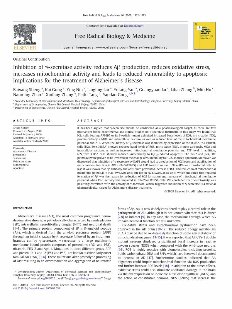
The levels of NO, ROS, protein carbonyls andMDA are four essentialparameters of oxidative stress [17]. Thus, we measured the intracel-lular NO, ROS, protein carbonyl and MDA levels in our cell models.N2a/APP695 and N2a/APPswe cells showed increased production of
Fig. 1. Overexpression of wild type and Swedish mutant APP-induced oxidative stress, mmeasured byWestern blot analysis, and extracellular Aβwasmeasured by TCA precipitation iN2a/APPswe cells showed increased intracellular ROS accumulation (ANOVA: ⁎⁎, pb0.005; ⁎APPswe cells compared with control (con) cells (ANOVA: ⁎⁎, pb0.005; ⁎pb0.05 versus contrNO levels compared to control cells (ANOVA: ⁎⁎, pb0.005; ⁎⁎⁎, pb0.001 versus control cellsmethod. Increase of MDA in N2a/APPwt and N2a/APPswe cells compared with control (con)potential (Ψm) was significantly decreased in N2a/APP695 and N2a/APPswe cells comparedlevels in N2a/APP695 andN2a/APPswe cells were significantly decreased compared to thosecalcium concentration in N2a/APP695 and N2a/APPswe cells compared with control cells w
Aβ (Fig. 1a). To study the possible role of endogenous Aβ in regulatingoxidative stress, we first compared the profile of intracellular ROS,protein carbonyls and [NO]i in N2a/APP695 cells and N2a/APPswecells with wild-type N2a cells. The results indicated that the levels ofROS (Fig. 1b), protein carbonyls (Fig. 1c) and [NO]i (Fig. 1d) weresignificantly enhanced in the following order: control cellsb N2a/APP695b N2a/APPswe. Oxidative stress arised in our transgenic cellswas accompanied by increased lipid peroxides. As shown in Fig. 1e,intracellular malondialdehyde (MDA), a product of lipid peroxidation,was increased by 81% in N2a/APP695 and by 112% in N2a/APPswecells (Pb0.005 vs. control, respectively). Consistent with our results,previous studies have shown that intracellular ROS is elevated in APP/PS1 double mutant neurons [16]. Moreover, protein carbonyls werefound to increase in vulnerable regions of the AD brain [43], ADmodelmice [44] and model cells [16].
Endogenous Aβ led to mitochondrial damage and increased [Ca2+]i
Ψm is a very importantmarker for the function of mitochondria. Aβtreatment of PC12 cells has been shown to lead to a significant
itochondrial damage and calcium dysfunction. (a) The expression profile of APP wasn N2a/wt (con), N2a/APP695 and N2a/APPswe cells, respectively; (b) N2a/APP695 andpb0.05 versus control cells); (c) Increase in protein carbonyls in N2a/APPwt and N2a/ol cells); (d) N2a/APP695 and N2a/APPswe cells exhibited a significant increase of basal; (e) Level of malondialdehyde (MDA) was assayed according to the thiobarbituric acidcells (ANOVA: ⁎⁎, pb0.005 versus control N2a cells); (f) The mitochondrial membranewith control cells (ANOVA: ⁎⁎, pb0.005, ⁎⁎⁎, pb0.001 versus control N2a cells); (g) ATPin control cells (ANOVA: ⁎⁎, pb0.005 versus control N2a cells; (h) Increased intracellularas observed by confocal microscopy.
1366 B. Sheng et al. / Free Radical Biology & Medicine 46 (2009) 1362–1375
decrease in Ψm [26]. Indeed, there is evidence that over-expression ofAPP and its Swedish mutation result in impaired Ψm in HCN-1Aneuronal cells [28] and PC12 cells [26].
To investigate whether endogenous Aβ was deleterious to thefunction of mitochondria in our model cells, we compared the Aβmodel cells with wild type ones. As expected, N2a/APP695 and N2a/APPswe cells showed an obvious reduction in Ψm (Fig. 1f).Mitochondria are the major source of ATP. The level of ATP wasdetermined in N2a/wt, N2a/APP695, and N2a/APPswe cells. Theresults indicated that the level of ATP in N2a/APP695 and N2a/APPswe cells was significantly decreased compared with those inN2a/wt cells (Pb0.005 vs. control) (Fig. 1g). Excessive elevation of[Ca2+]i is a major factor leading to apoptosis in many cell types.Degenerating neurons in the brains of AD patients showed increasedlevels of calcium [45,46]. Calcium dysregulation in AD also affects APPprocessing, endoplasmic reticulum (ER) dysfunction, and mitochon-drial changes [47]. Here, N2a/wt, N2a/APP695, and N2a/APPswe cellswere used to study the calcium dysregulation by measuring the level
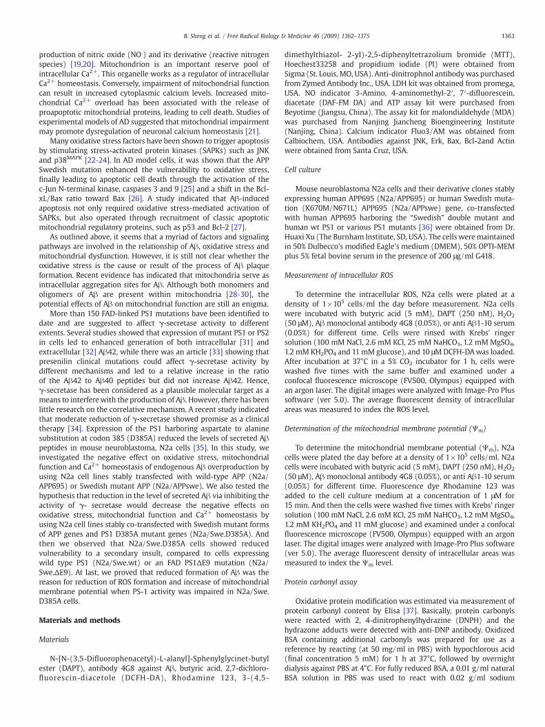
Fig. 2. Inhibition of γ-secretase by expressing PS1 D385A mutant protected cells from oxidatand APP C-terminal fragments were measured byWestern blot analysis, and extracellular Aβcells. Compared to N2a/Swe.wt and N2a/Swe.ΔE9 cells, N2a/Swe.D385A cells showed hundetectable; (b) N2a/Swe.D385A cells showed reduced intracellular ROS accumulationSwe.ΔE9; ⁎pb0.05 versus N2a/Swe.wt cells; (c) Reduced protein carbonyls in N2a/Swe.Dpb0.005 versus N2a/Swe.ΔE9; ⁎pb0.05 versus N2a/Swe.wt cells); (d) N2a/Swe.D385A ceSwe.ΔE9 cells (ANOVA: ⁎⁎, pb0.005 versus N2a/Swe.wt cells; ⁎⁎⁎, pb0.001 versus N2a/SwSwe.wt and N2a/Swe.ΔE9 cells (⁎⁎, pb0.005 versus N2a/Swe.wt cells; ⁎⁎⁎, pb0.001 versincreased in N2a/Swe.D385A cells compared with N2a/Swe.wt and N2a/Swe.ΔE9 cells (ANOdisplayed increased ATP level compared with N2a/Swe.wt and N2a/Swe.ΔE9 cells (ANOVintracellular calcium concentration in N2a/Swe.D385A cells compared with that in N2a/Sw
of [Ca2+]i. The results indicated that N2a/APP695, especially N2a/APPswe cells, showed higher intracellular fluorescence intensity,which demonstrated that [Ca2+]i was significantly higher than incontrol cells (Fig. 1h).
N2a/Swe.D385A cells showed decreased basal levels of ROS, proteincarbonyls, [NO]i, and MDA
Asp-385 is required for presenilin 1 and γ-secretase activity.The loss-of-function PS1 mutant (D385A) leads to a deficiency inγ-secretase activity, while the FAD PS1 mutant (ΔE9) increases theactivity of γ-secretase. Compared to N2a/Swe.ΔE9 and N2a/Swe.wtcells, N2a/Swe.D385A cells reduced Aβ secretion, and also increasedthe amount of holo-APP and the production of CTFs (Fig. 2A). In orderto further demonstrate that oxidative stress is due to the increasedendogenous Aβ but not due to the overexpression of APP and PS1,we compared the basal levels of [NO]i, ROS and protein carbonyls inN2a/Swe.ΔE9, N2a/Swe.wt and N2a/Swe.D385A cells. We found that
ive stress, mitochondrial damage and calcium dysfunction. (a) Expression profile of APPwas measured by TCA precipitation in N2a/Swe.D385A, N2a/Swe.wt and N2a/Swe.ΔE9igher levels of full length APP and C-terminal fragments, but the secreted Aβ wascompared to N2a/Swe.wt and N2a/Swe.ΔE9 cells (ANOVA: ⁎⁎ pb0.005 versus N2a/385A cells compared with those in N2a/Swe.wt and N2a/Swe.ΔE9 cells (ANOVA: ⁎⁎lls exhibited significantly reduced basal NO levels compared to N2a/Swe.wt and N2a/e.ΔE9); (e) N2a/Swe.D385A cells displayed decreased MDA level compared with N2a/us N2a/Swe.ΔE9); (f) The mitochondrial membrane potential (Ψm) was significantlyVA: ⁎⁎, pb0.005, versus N2a/Swe.wt and N2a/Swe.ΔE9 cells); (g) N2a/Swe.D385A cellsA: ⁎⁎ pb0.005 versus N2a/Swe.ΔE9; ⁎pb0.05 versus N2a/Swe.wt cells); (h) Reducede.wt and N2a/Swe.ΔE9 cells was observed by confocal microscopy.
1367B. Sheng et al. / Free Radical Biology & Medicine 46 (2009) 1362–1375
in all cases, the levels of ROS (Fig. 2b), protein carbonyls (Fig. 2c) and[NO]i (Fig. 2d) were in the following order: N2a/Swe.D385AbN2a/Swe.wt bN2a/Swe.ΔE9. Besides, N2a/Swe.D385Acells showed obviousreduction of MDA level compared to N2a/Swe.wt, and N2a/Swe.ΔE9cells (Fig. 2e). Together, these results suggested that the increased
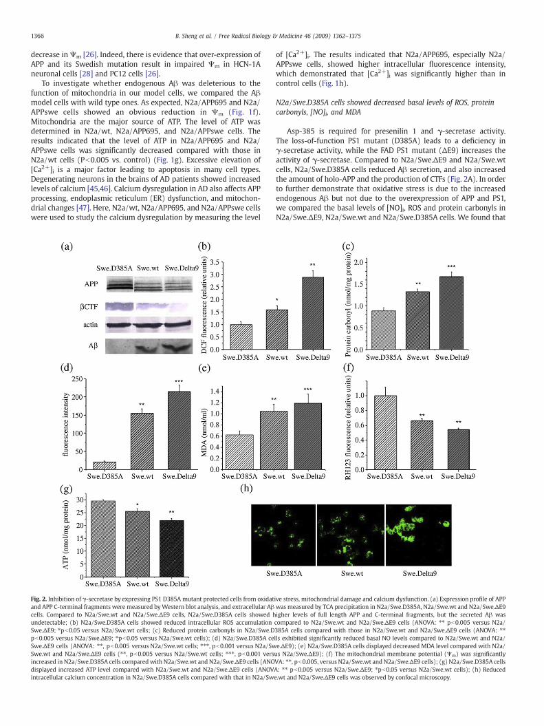
Fig. 3. LDH leakage, MTT reduction assay, ROS andmitochondrial membrane potential (Ψm)wgenotypes. (a) LDH release in N2a cells pretreated 24 h with H2O2 at concentrations of 50preparations (ANOVA: ⁎, pb0.05 and ⁎⁎, pb0.005 versus N2a/wt cells, left panel; ⁎, pb0.05viability was measured by MTT reduction assay after treatment with H2O2 for 24 h at conindependent preparations (ANOVA: ⁎, pb0.05 and ⁎⁎, pb0.005 versus N2a/wt cells, left paincreased in N2a/APP695 and N2a/APPswe (ANOVA: ⁎⁎, pb0.005 versus control cells, left pSwe.ΔE9 cells (ANOVA: ⁎⁎, pb0.005 versus N2a/Swe.wt cells; ⁎⁎⁎, pb0.001 versus N2a/Swe(ANOVA: ⁎⁎, pb0.005 versus control cells, left panel) but significantly reduced in N2a/Swe.DSwe.wt cells; ⁎⁎⁎, pb0.005 versus N2a/Swe.ΔE9 cells, right panel).
production of NO, ROS, protein carbonyls, and MDA in N2a/Swe.wtand N2a/Swe.ΔE9 cells were triggered by the production andaccumulation of Aβ, and, impaired production of endogenous Aβ bythe loss of PS1 function in the γ-cleavage of APP in N2a/Swe.D385Acells weakened the oxidative stress.
ere used to evaluate the effect of secondary insult of oxidative stress on cells of differentμm and 100 μm. Results were shown as mean±SEM obtained from five independent, ⁎⁎, pb0.005⁎⁎, and ⁎⁎⁎, pb0.001 versus N2a/Swe.D385A cells, right panel); (b) Cellcentrations of 50 μm and 100 μm. Results shown are mean±SEM obtained from fivenel; ⁎⁎, pb0.005 versus N2a/Swe.D385A cells, right panel); (c) ROS was significantlyanel) but significantly reduced in N2a/Swe.D385A compared to N2a/Swe.wt and N2a/.ΔE9, right panel); (d)Ψmwas significantly increased in N2a/APP695 and N2a/APPswe385A compared to N2a/Swe.wt and N2a/Swe.ΔE9 cells (ANOVA: ⁎, pb0.05 versus N2a/
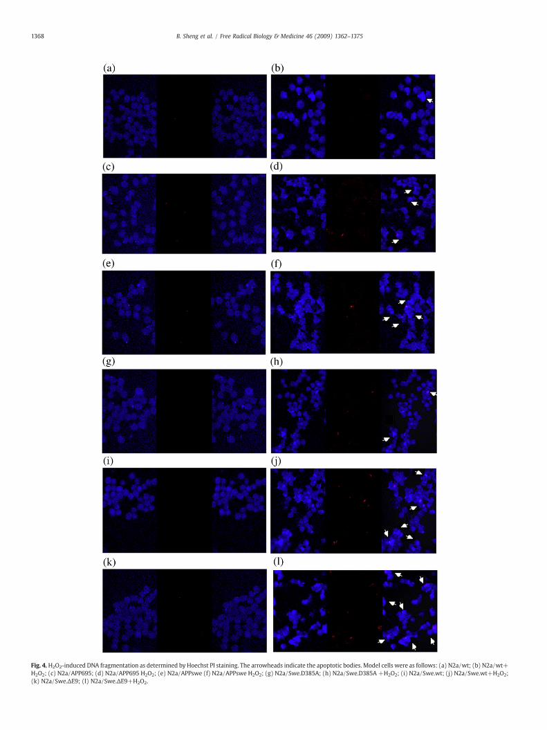
Fig. 4.H2O2-induced DNA fragmentation as determined by Hoechst PI staining. The arrowheads indicate the apoptotic bodies. Model cells were as follows: (a) N2a/wt; (b) N2a/wt+H2O2; (c) N2a/APP695; (d) N2a/APP695 H2O2; (e) N2a/APPswe (f) N2a/APPswe H2O2; (g) N2a/Swe.D385A; (h) N2a/Swe.D385A +H2O2; (i) N2a/Swe.wt; (j) N2a/Swe.wt+H2O2;(k) N2a/Swe.ΔE9; (l) N2a/Swe.ΔE9+H2O2.
1368 B. Sheng et al. / Free Radical Biology & Medicine 46 (2009) 1362–1375

1369B. Sheng et al. / Free Radical Biology & Medicine 46 (2009) 1362–1375
N2a/Swe.D385A cells showed reversed mitochondrial damage andcalcium dysfunction
A number of recent studies have indicated that mitochondriamight be an important target of Aβ. Based on the observation above ofoxidative stress, we supposed that when the production of Aβ wasreduced in N2a/Swe.D385A cells, mitochondrial damage and calciumdysfunction would be lessened. To validate our hypothesis, wedetected Ψm using fluorescence dye rhodamine123, ATP usingbioluminescence assay, and [Ca2+]i using fluo3/AM. As shown inFig. 2f, N2a/Swe.D385A cells showed significantly higher Ψm thanN2a/Swe.wt and N2a/Swe.ΔE9 cells. The mitochondrial function wasresumed in N2a/Swe.D385A cells shown by increased level of ATP. Theresults indicated that the levels of ATP in N2a/Swe.wt, and, especiallyin N2a/Swe.ΔE9 cells, were significantly decreased compared withthose in N2a/Swe.D385A cells (Fig. 2g). At the same time, it wasshown that N2a/Swe.D385A cells displayed significantly lower [Ca2+]ithan N2a/Swe.wt and N2a/Swe.ΔE9 cells.
N2a/Swe.D385A cells showed reduced sensitivity to oxidative stress
Oxidative stress was observed in the AD brain. Oxidative stressoccurs due to an imbalance in the prooxidant and antioxidant levels[48]. Increased production of ROS and free radicals might be involvedin the pathology of AD. Thus, we investigated the effect of a secondaryinsult in the presence of an oxidative damage agent, hydrogenperoxide. Firstly, the integrity of cell membrane was determined bythe release of LDH after 24 h exposure to 50 μM or 100 μM H2O2. Asshown in Fig. 3a, after 24 h exposure to 50 μM or 100 μM H2O2, theN2a/APP695 and N2a/APPswe cells displayed markedly increased
Fig. 5. (a) Changes in levels of P-JNK, P-ERK1/2, Bax and Bcl-2 after secondary insult of oxidAPPswe cells exhibited significantly enhanced protein expression levels of JNK activation⁎pb0.05 versus control cells); (c) The phosphorylation of ERK1/2 was reduced in N2a/APP6significant change in phosphorylation of ERK1/2 in N2a/APP695, especially in N2a/APPswereduced shift in the Bcl-2 /Bax ratio in N2a/APP695 and N2a/APPswe cells after exposure to hpb 0.001 versus control cells in 6 h).
LDH leakage into the medium compared to control ones, while N2a/Swe.D385A cells showed reduced LDH efflux compared to N2a/Swe.wt and N2a/Swe.ΔE9 ones. Secondly, cell viability was evaluated byMTT after 24 h exposure to 50 μM or 100 μM H2O2. The N2a/APP695and N2a/APPswe cells showed increased vulnerability compared tothe control ones, while N2a/Swe.D385A cells showed reducedvulnerability compared to N2a/Swe.wt and N2a/Swe.ΔE9 ones(Fig. 3b). Then, after exposure to hydrogenperoxide, the ROS elevationwas more pronounced in N2a/APP695 and N2a/APPswe cellscompared to control ones, while N2a/Swe.D385A cells showed greaterendurance to ROS elevation induced by hydrogen peroxide comparedwith N2a/Swe.wt and N2a/Swe.ΔE9 cells (Fig. 3c). Moreover, N2a/APP695 and N2a/APPswe cells showed a significantly decreased Ψm
after exposure to hydrogen peroxide in comparison with control N2acells. And N2a/Swe.D385A cells showed less reduction ofΨm than didthe N2a/Swe.wt and N2a/Swe.ΔE9 cells (Fig. 3d).
Reduced vulnerability of N2a/Swe.D385A cells to H2O2 inducedapoptosis
Using equal concentrations of H2O2, we investigated theapoptosis of different cell lines in response to oxidative stress. Weexamined apoptotic bodies by Hochest33258 and PI staining. After24 h incubation with 50 μM H2O2, a significant proportion of theN2a/APP695 and N2a/APPswe cells had condensed and their nucleihad fragmented; these cells were stained more brightly than thecontrol ones. For doubly transfected cells, N2a/Swe.ΔE9 cellsshowed the highest apoptotic and necrotic portion, and N2a/Swe.D385A cells displayed the lowest apoptotic and necrotic portion(Fig. 4).
ative stress in N2a/wt, N2a/APP695 and N2a/APPswe cells; (b) N2a/APP695 and N2a/compared with control cells with or without H2O2 treatment (ANOVA: ⁎⁎, pb0.005;95 and N2a/APPswe cells (⁎⁎, pb0.005; ⁎pb0.05 versus control cells), but there was acells compared to wild type cells after H2O2 treatment; (d) We observed a significantlyydrogen peroxide (ANOVA: ##, pb0.005 versus control cells in 3 h; ΔΔ, pb0.005; ΔΔΔ,

1370 B. Sheng et al. / Free Radical Biology & Medicine 46 (2009) 1362–1375
Reduced activation of JNK and ERK pathways in N2a/Swe.D385A cells
Reactive oxygen species have been indicated to induce theactivation of mitogen-activated protein kinases, including JNK, P38and ERK. The JNK and P38 activation was observed to localize toamyloid deposits in AD model mice [49]. ERK activation was reportedin hippocampal slices after treatment with soluble Aβ1-42 [50],though it was not found in AD model mice. Hence, we addressed thequestion of whether inhibiting abnormal γ-cleavage of APP couldmodulate MAPK family-involved apoptotic cell death in N2a cells.Interestingly, the endogenous JNK activity both at the basal state andin the H2O2-stimulated state was higher in N2a/APP695 and N2a/APPswe cells than that in control ones (Fig. 5a), while N2a/Swe.D385A cells reversed this elevation of JNK activity compared to N2a/Swe.wt and N2a/Swe.ΔE9 cells (Fig. 6a). Then we examined theactivity of ERK. Surprisingly, we found the basal activity of ERK wassignificantly decreased in N2a/APP695 and N2a/APPswe cellscompared to N2a/wt ones (Fig. 5a), and in N2a/Swe.D385A cellscompared to N2a/Swe.wt and N2a/Swe.ΔE9 cells (Fig. 6a). After 3 hand 6 h treatment with 50 μM H2O2, a pathologic model of N2a cellsshowed an obviously increased phosphorylated level of ERK (Fig. 5a)(Fig. 6a), while the control (Fig. 5a) and N2a/Swe.D385A cells wereonly slightly influenced (Fig. 6a).
The impact of inhibiting γ-secretase abnormal cleavage of APP on Bcl-2and Bax
Having established the mitochondrial damage during the processof H2O2-induced apoptosis, we set out to find whether there was anydistinction between the effect of hydrogen peroxide on the pro-apoptotic and anti-apoptotic pathway. By western blot analysis, wemonitored the protein expression of Bax (a pro-apoptotic member)
Fig. 6. (a) Changes in levels of P-JNK, P-ERK1/2, Bax and Bcl-2 after secondary insult of oxidand N2a/Swe.ΔE9 cells exhibited significantly enhanced protein expression levels of JN(ANOVA: ⁎⁎, pb0.005; ⁎pb0.05 versus control cells); (c) The phosphorylation of ERK1/2 wSwe.D385A cells), but there was a significant change in phosphorylation of ERK1/2 in N2a/treatment; (d) We observed a significantly reduced shift in the Bcl-2 /Bax ratio in N2a/Sweversus N2a/Swe.D385A cells in 3 h; ΔΔ, pb0.005; ΔΔΔ, pb 0.001 versus N2a/Swe.D385A ce
and Bcl-2 (an anti-apoptotic member) following treatment of N2a cellsby hydrogen peroxide. After 6 h of treatment with hydrogen peroxide,we found a reduced amount of the cytosolic anti-apoptotic proteinBcl-2 in N2a/APP695 cells and especially in N2a/APPswe cells (Fig. 5a).As far as doubly transfected cells were concerned, N2a/Swe.wt cells,and especially N2a/Swe.ΔE9 cells, showed significantly reduced levelsof Bcl-2, whereas N2a/Swe.D385A cells showed only a minor change(Fig. 6a). Negligible changes in Bax protein content were observed inall of the cell lines (Figs. 5a and 6a). Thus, the Bcl-2/Bax ratio wasobviously decreased in pathological cell models (Figs. 5d and 6d), butnot in N2a/wt and N2a/Swe.D385A cells (Fig. 6d). All the aboveapoptosis studies suggest that, in the presence of an oxidative damage-inducing agent, inhibition of abnormal cleavage of APP by γ-secretasehelped to resist apoptosis.
Aβ level positively correlated with ROS levels and mitochondrial damage
Having demonstrated a significant increase in ROS andmitochondrial damage with endogenous Aβ, we postulated thatthe production of ROS and mitochondrial damage is positivelycorrelated with the Aβ level. To test this idea, we treated N2a/APP695 and N2a/APPswe cells for 24 h with a low concentrationof butyric acid [51] to induce overexpression of the transfectedgenes. As shown in Fig. 7b, the ROS level increased after thetreatment, concomitantly with increased APP and Aβ levels(Fig.7a). At the same time, the levels of Ψm and ATP weredetected. After treatment with butyric acid, both N2a/APP695 andN2a/APPswe cells showed a significant reduction of Ψm (fig.7c)and ATP (fig.7d) which showed the dose-dependent effects of Aβon cellular damage. To further demonstrate the deleterious effectof endogenous Aβ, N2a/APP695 and N2a/APPswe cells weretreated with DAPT, a functional γ-secretase inhibitor that reduces
ative stress in N2a/Swe.D385A, N2a/Swe.wt and N2a/Swe.ΔE9 cells; (b) N2a/Swe.wtK activation compared with N2a/Swe.D385A cells with or without H2O2 treatmentas reduced in N2a/Swe.wt and N2a/Swe.ΔE9 cells (⁎⁎, pb0.005; ⁎pb0.05 versus N2a/Swe.wt, especially in N2a/Swe.ΔE9 cells compared to N2a/Swe.D385A cells after H2O2
.wt and N2a/Swe.ΔE9 cells after exposure to hydrogen peroxide (ANOVA: ##, pb0.005lls in 6 h).

Fig. 7. Neurotoxicity was positively correlated with Aβ level, and the γ-secretase inhibitor DAPT reduced ROS levels and restored mitochondrial activity. (a) N2a/APP695 and N2a/APPswe cells were incubated in the presence or absence of 5mM butyric acid (BA) for 24 h to induce APP expression and Aβ secretion. Levels of Aβwere quantified and normalized tothose of N2a/APP 695 cells without BA treatment ( AVOVA: ⁎⁎ pb0.005 versus untreated N2a/APP695 cells; ##, pb0.005 versus untreated N2a/APPswe cells ); (b) The ROS level wassignificantly increased in both N2a/APP695 (AVOVA: ⁎⁎ pb0.005 versus untreated N2a/APP695 cells) and N2a/APPswe cells (AVOVA: ##, pb0.005 versus untreated N2a/APPswecells) after treatment with BA; (c) Ψm was significantly reduced in both N2a/APP695 (AVOVA: ⁎, pb0.05 versus untreated N2a/APP695 cells) and N2a/APPswe cells (AVOVA: #,pb0.005 versus untreated N2a/APPswe cells) after treatment with BA; (d) ATP level was significantly reduced in both N2a/APP695 (AVOVA: ⁎⁎, pb0.005 versus untreated N2a/APP695 cells) and N2a/APPswe cells (AVOVA: ##, pb0.005 versus untreated N2a/APPswe cells) after treatment with BA; (e) N2a/APP695 and N2a/APPswe cells were incubated inthe presence of 250 nm DAPT. β-CTF and Aβwere detected. Aβ level was densitometrically analyzed (AVOVA: ⁎⁎⁎ pb0.001 versus untreated N2a/APP695 cells; ###, pb0.001 versusuntreatedN2a/APPswe cells); (f) The ROS levelwas significantly reduced in bothN2a/APP695 (AVOVA: ⁎ pb0.05 versus untreatedN2a/APP695 cells ) andN2a/APPswe cells (AVOVA:#, pb0.05 versus untreated N2a/APPswe cells) after treatment with DAPT; (g)Ψmwas significantly increased in both N2a/APP695 (AVOVA: ⁎ pb0.05 versus untreated N2a/APP695cells) and N2a/APPswe cells (AVOVA: #, pb0.05 versus untreated N2a/APPswe cells) after treatment with DAPT; (h) ATP level was significantly increased in both N2a/APP695(AVOVA: ⁎⁎, pb0.005 versus untreated N2a/APP695 cells) and N2a/APPswe cells (AVOVA: ##, pb0.005 versus untreated N2a/APPswe cells) after treatment with DAPT.
1371B. Sheng et al. / Free Radical Biology & Medicine 46 (2009) 1362–1375
intracellular Aβ. An obvious increase of βCTF and reduction ofsecreted Aβ was detected by TCA precipitation after 24 htreatment of DAPT (Fig. 7e). DAPT led to a significantly reducedROS level (Fig. 7f) as well as a strongly increased Ψm in both N2a/APP695 and N2a/APPswe cells (Fig. 7 g). Moreover, N2a/APP695and N2a/APPswe cells showed increased level of ATP aftertreatment with DAPT (Fig. 7h). Thus, we proved that inhibitionof Aβ by exogenous γ-secretase inhibitor led to a decrease in ROSlevel and a stabilization of mitochondrial function, whichcoincided with the endogenous gene mutant results.
Effects of anti Aβ antibodies on ROS and Ψm in N2a/Swe.ΔE9 cells
N2a/Swe.D385A cells secreted little Aβ, while N2a/Swe.ΔE9 cellssecreted abundant Aβ. To verify if Aβ was the cause of cell damage inN2a pathologically model cells, 0.05% Aβ monoclonal antibody 4G8and anti Aβ serum were added into the culture medium of N2a/Swe.D385A cells and N2a/Swe.ΔE9 cells independently. 24 hours after thetreatment, the levels of ROS and Ψm were determined. The resultsshowed that both Aβ antibody 4G8 and anti Aβ serum decreased thelevel of ROS in N2a/Swe.ΔE9 cells (Fig. 8a), while not in N2a/Swe.

Fig. 8. Aβ antibody and antiserum prevented increase of ROS and reduction of mitochondrial membrane potential in N2a/Swe. ΔE9 cells but not in N2a/Swe.D385A cells. (a) ROSwassignificantly reduced in N2a/Swe.ΔE9 cells after treatment with Aβ monoclonal antibody 4G8 and Aβ antiserum (ANOVA: ⁎⁎, pb0.005); (b) ROS nearly kept constant in N2a/Swe.D385A after treatment with Aβ antibody 4G8 and Aβ antiserum; (c)Ψmwas significantly increased in N2a/Swe.ΔE9 cells after treatment with Aβmonoclonal antibody 4G8 and Aβantiserum (ANOVA: ⁎⁎, pb0.005); (d) Ψm was nearly kept constant in N2a/Swe.D385A after treatment with Aβ antibody 4G8 and Aβ antiserum.
1372 B. Sheng et al. / Free Radical Biology & Medicine 46 (2009) 1362–1375
D385A cells (Fig. 8b). Correspondingly, N2a/Swe.ΔE9 cells showed amarkedly increasedΨm after treatment with the Aβ antibody 4G8 andanti Aβ serum (Fig. 8c), while the level of Ψm kept almost constantafter 4G8 and anti Aβ serum treatment in N2a/Swe.D385A cells(Fig. 8d). It demonstrated that cell damage in N2a/Swe.ΔE9 wasmainly caused by Aβ overproduction. And reduced production of Aβwas the reason for the reduction of ROS formation and increase ofΨm
in N2a/Swe. D385A cells.
Discussion
The overproduction and accumulation of Aβ-peptide in the brainhas been considered as the central pathological event in AD. Geneticmutations in genes for APP, presenilin-1 or presenilin-2 have beenreported to increase the production of Aβ42 and lead to early onset ofAD [52]. Many therapeutic strategies have been proposed based on Aβbiology [53]. Several studies showed that β- and γ-secretase is apromising pharmacological target as a means to interfere with theproduction of Aβ. Many γ-secretase inhibitors have been tested inlaboratories or clinical experiments [54,55]. However, strong inhibitionof γ-secretase leads to severe adverse effects, as it interferes withsignaling by notch proteins and other cell surface receptors [56]. Thus, ithas been debated whether using γ-secretase inhibitor to treat AD issecure enough. A recent study concluded that a γ-secretase reduction of30% was sufficient to attenuate Aβ deposition with little or no adverseside effects, suggesting the existence of an optimal window oftherapeutic γ-secretase inhibition [34]. It was reported that expressionof PS1 harboring alanine substitutions of highly conserved aspartateresidue at position 385 in Chinese hamster ovary cells [57] or N2a cells[36] led to reduced Aβ secretion and accumulation of APP CTFs.Intrigued with this observation, our research objective was to explorethe feasibility of γ-secretase inhibition. In this report, we offered severalimportant insights into the changes induced by the PS1 D385A varianton oxidative stress, mitochondrial damage and apoptosis.
Oxidative stress and mitochondrial damage are known to occur inthe AD brain. Our data showed that N2a/APP695 and N2a/APPswecells exhibited increased basal levels of NO, ROS, protein carbonyls andMDA, which play important roles in Aβ-induced neurotoxicity and celldeath. Previous study showed that increased oxidative damagewas anearly event in AD, and increases in Aβ depositionwere associated withdecreased oxidative damage. They suggested that AD was associatedwith compensatory changes that reduced damage from reactiveoxygen [58]. This result didn’t deny the positive correlation betweenAβ production and oxidative stress. It is widely believed that solubleAβ oligomers (Aβ-derived diffusible ligands, ADDLs) are more toxicthan insoluble Aβ polymers [59]. Aβ deposition transforms soluble Aβoligomers into insoluble precipitate of Aβ polymers. Therefore, Aβdeposition could be a compensatory response to toxic effects,including oxidative stress, induced by overproduced soluble Aβoligomers. Increased oxidative stress was consistent with theobservation that the sensitivities to oxidative stress induced by H2O2
in N2a/APP695, especially in N2a/APPswe cells, were significantlypromoted compared with control cells. Oxidative stress also plays akey role in the Aβ-mediated neurotoxicity. It was reported that H2O2
participated in mediating Aβ toxicity [60] and Aβ could directlyproduce H2O2 through metal ion reduction [61]. These resultsenhanced our understanding of Аβ central pathology. We alsoinvestigated the effects on oxidative stress and mitochondrial damageof reduced production of Aβ by inhibiting γ-secretase. When cellswere co-transfected with Swedish mutant APP and PS1 dominantnegative mutant (PS1D385A), no secreted Aβ was detected by TCAprecipitation. Compared to N2a/Swe.wt and N2a/Swe.ΔE9 cells, N2a/Swe.D385A cells showed reduced neurotoxicity, including levels ofNO, ROS, protein carbonyls and mitochondrial dysfunction. Moreover,when treated with a secondary insult (H2O2), N2a/Swe.D385A cellsdisplayed stronger resistance and could better maintain cellularfunction. The results of this study supported the hypothesis that therewas a reduced basal neurotoxicity and sensibility to oxidative stress
1373B. Sheng et al. / Free Radical Biology & Medicine 46 (2009) 1362–1375
when PS1 activity was inhibited. Oxidative stress and Aβ productionare positively correlated to each other. There is evidence suggestingthat Aβ induces oxidative stress both in vivo and in vitro [62,63],and oxidative stress promotes the production of Aβ [64]. A recentstudy [65] showed that oxidative stress induced by H2O2 and HNEtreatments activated a positive feedback between the γ- and β-secretase cleavages of the β-amyloid precursor protein, which led toan increase in Aβ40 and Aβ42 production as well as Aβ42/40 ratios.This kind of feedback required the activation of the JNK/c-junpathway. Combined with our results, it seemed that when theγ-secretase activity was inhibited in N2a/Swe.D385A cells, thepositive feedback could not be observed. Moreover, the effects oncellular ROS, Ψm and ATP of DAPT were a supplementary result.
Apoptosis is attributed to neurodegenerative diseases and neuro-logical disorders, such as AD and Parkinson's disease. Increased levelsof ROS and mitochondrial dysfunction are involved in Aβ-inducedapoptosis [66]. Studies have established that Aβ peptide inducesapoptosis in mouse neuronal cultures [67]. Familial AD (FAD)mutations in presenilins render neurons vulnerable to apoptosisinduced by Aβ [68]. Consistent with previous studies, we found thatN2a/APP695 and N2a/APPswe cells, which have high Aβ levels,showed significantly increased proportions of apoptotic cells aftertreatment with 50 μM H2O2. As the MAPK pathway has been found toplay a role in AD pathology [25,69], we found that after treatmentwithH2O2 for 3 h and 6 h, N2a/APP695 and N2a/APPswe cells exhibitedsignificantly enhanced protein expression levels of ERK and JNKactivation compared to control cells. This result suggested that theendogenous Aβmight promote the activation of JNK and ERK inducedby oxidative stress. Furthermore, this effect is Aβ dose-dependent.Given the important relationship between Aβ and apoptosis, weinvestigated the influence on apoptosis when the activity ofγ-secretase was inhibited by transfectionwith PS1 dominant negativemutant. Interestingly, we observed that N2a/Swe.D385A cells, whichsecreted little Aβ, displayed significantly reduced proportions ofapoptotic cells after treatment with 50 μM H2O2 compared to N2a/Swe.wt and N2a/Swe.ΔE9 cells. At the same time, we found thatexpression of the D385A PS1variant retarded activation of the JNK andERK1/2 pathways. Both ERK1/2 and JNK pathways are generallyactivated in susceptible neuronal populations in individuals with AD.And there is evidence suggesting that the activation of ERK and JNKcan independently serve to initiate, but both are necessary topropagate disease pathogenesis [70]. They called this “two hitshypothesis” [71]. And they indicated that only with “two hits” couldthe disease process be started.” Combined with our results, wespeculated that oxidative stress, as an early event in AD, played animportant role in activating ERK1/2 and JNK pathways. It was shownthat when γ-secretase activity was inhibited in N2a/Swe.D385A cells,the “two hits” was delayed.
Though the expression of Bax stayed consistent, a shift in the Bcl-2/Bax ratio toward Baxmight be involved in the different sensitivity ofthe three cell lines after secondary stress insult. We speculate thatboth reduced Aβ production and depressed oxidative stress lead toreduced vulnerability to apoptotic cell death.
External applied Aβ monoclonal antibody could bind the extra-cellular Aβ. The level of ROS could be decreased and Ψm could beincreased in the presence of Aβ antibody in N2a/Swe.ΔE9 cells whilenot in N2a/Swe.D385A cells, which demonstrated that the extra-cellularly secreted Aβ played an important role in cell damagesmentioned above. A recent study showed that extracellular fluores-cent Aβ could be taken up by human SH-SY5Y neuroblastoma cellsand later partly localized to mitochondria [72]. Besides, Saavedra andcolleagues [73] have proved that Aβ42 is internalized by primaryneurons in the absence of ApoE. These results implied that secreted Aβcould be reinternalized into cells and came in contact withmitochondria. Combined with our data, we speculated that Aβ,including the intracellular and the reinternalized extracellular, led to
intracellular toxicity, including oxidative stress, mitochondrial dys-function, and apoptosis. In accordance with our results, primaryneuronal cultures from knock-in mice expressing mutant human PS-1and APP neurons exhibited increased vulnerability to oxidative stress,mitochondrial dysfunction and apoptosis in the presence of Aβ42compared with the wild-type neurons [16]. SH-SY5Y cells pretreatedwith Aβ antibody significantly prevented the cell neurotoxicityinduced by Aβ [74].
In summary, compared to AD pathological cell models, N2a/Swe.D385A cells showed reduced oxidative stress, increasedmitochondrialactivity and decreased vulnerability to apoptosis. Thus, our datasupport the view that γ-secretase is a therapeutic target for treatmentof AD. Optimum γ-secretase inhibitors, especially APP-specific γ-secretase inhibitors, are a good choice. However, additional work isrequired to understand the long-term effect associated with γ-secretase inhibitors.
Acknowledgments
We are grateful to Dr. H.X. Xu from the Burnham Institute forproviding the N2a cell lines and antibodies. This work was supportedby grants from the Tsinghua-Yue-Yuen Medical Sciences Fund (No.20240000514), the National Natural Science Foundation of China (No.30872898) and the "86.3" program of China (No. 2007AA02Z402 and2008AA022403).
References
[1] Goedert, M.; Spillantini, M. G. A Century of Alzheimer's Disease. Science 314:777–781; 2006.
[2] Kidd, M. Paired helical filaments in electron microscopy of Alzheimer's disease.Nature 197:192–193; 1963.
[3] Terry, R. D.; Gonatas, N. K.; Weiss, M. Ultrastructural Studies in Alzheimer'sPresenile Dementia. Am J Pathol. 44:269–297; 1964.
[4] Roher, A.; Wolfe, D.; Palutke, M.; KuKuruga, D. Purification, ultrastructure, andchemical analysis of Alzheimer's disease amyloid plaque core protein. Proc NatlAcad Sci USA. 83:2662–2666; 1986.
[5] LaFerla, F. M.; Oddo, S. Alzheimer's disease: Aβ, tau and synaptic dysfunction.Trends Mol Med. 11:170–176; 2005.
[6] Sherrington, R.; Rogaev, E. I.; Liang, Y.; Rogaeva, E. A.; Levesque, G.; Ikeda, M.;Chi, H.; Lin, C.; Li, G.; Holman, K. Cloning of a gene bearing missense mutations inearly-onset familial Alzheimer's disease. Nature 375:754–760; 1995.
[7] Hardy, J. A.; Higgins, G. A. Alzheimer's disease: the amyloid cascade hypothesis.Science 256:184–185; 1992.
[8] Mattson, M. P. Pathways towards and away from Alzheimer's disease. Nature 430:631–639; 2004.
[9] Lee, H. G.; Casadesus, G.; Zhu, X.; Takeda, A.; Perry, G.; Smith, M. A. Challenging theamyloid cascade hypothesis: senile plaques and amyloid-β as protectiveadaptations to Alzheimer disease. Ann. N.Y. Acad. Sci. 1019:1–4; 2004.
[10] Hirai, K.; Aliev, G.; Nunomura, A.; Fujioka, H.; Russell, R. L.; Atwood, C. S.; Johnson,A. B.; Kress, Y.; Vinters, H.; Tabaton, M.; Shimohama, S.; Cash, A. D.; Siedlak, S. L.;Harris, P. L.; Jones, P. K.; Petersen, R. B.; Perry, G.; Smith, M. A. Mitochondrialabnormalities in Alzheimer's disease. J. Neurosci. 21:3017–3023; 2001.
[11] Butterfield, D. A.; Drake, J.; Pocernich, C.; Castegna, A. Evidence of oxidativedamage in Alzheimer's disease brain: central role for amyloid β-peptide. TrendsMol. Med. 7:548–554; 2001.
[12] Smith, M. A.; Harris, P. L.; Sayre, L. M.; Perry, G. Iron accumulation in Alzheimer'sdisease is a source of redox-generated free radicals. Proc. Natl Acad. Sci. U. S. A. 94:9866–9868; 1997.
[13] Castegna, A.; Aksenov, M.; Aksenova, M.; Thongboonkerd, V.; Klein, J. B.; Pierce,W. M.; Booze, R.; Markesbery, W. R.; Butterfield, D. A. Proteomic identification ofoxidatively modified proteins in Alzheimer's disease brain. Part I. Creatine kinaseBB, glutamine synthase, and ubiquitin carboxyterminal hydrolase L-1. Free Radic.Biol. Med. 33:562–571; 2002.
[14] Castegna, A.; Thongboonkerd, V.; Klein, J. B.; Lynn, B.; Markesbery, W. R.;Butterfield, D. A. Proteomic identification of nitrated proteins in Alzheimer'sdisease brain. J. Neurochem. 85:1394–1401; 2003.
[15] Castegna, A.; Thongboonkerd, V.; Klein, J.; Lynn, B. C.; Wang, Y. L.; Osaka, H.;Wada,K.; Butterfield, D. A. Proteomic analysis of brain proteins in the gracile axonaldystrophy (gad) mouse, a syndrome that emanates from dysfunctional ubiquitincarboxyl-terminal hydrolase L-1 reveals oxidation of key proteins. J. Neurochem.88:1540–1546; 2004.
[16] Mohmmad Abdul, H.; Sultana, R.; Keller, J. N.; St Clair, D. K.; Markesbery, W. R.;Butterfield, D. A. Mutations in amyloid precursor protein and presenilin-1 genesincrease the basal oxidative stress in murine neuronal cells and lead to increasedsensitivity to oxidative stress mediated by amyloid β-peptide (1-42), H2O2 andkainic acid: implications for Alzheimer's disease. J. Neurochem. 96:1322–1335;2006.
1374 B. Sheng et al. / Free Radical Biology & Medicine 46 (2009) 1362–1375
[17] Butterfield, D. A.; Reed, T.; Newman, S. F.; Sultana, R. Roles of amyloid β-peptide-associated oxidative stress and brain protein modifications in the pathogenesis ofAlzheimer's disease and mild cognitive impairment. Free Radic. Biol. Med. 43:658–677; 2007.
[18] Casley, C. S.; Canevari, L.; Land, J. M.; Clark, J. B.; Sharpe, M. A. β-amyloid inhibitsintegrated mitochondrial respiration and key enzyme activities. J. Neurochem. 80:91–100; 2002.
[19] Sarti, P.; Arese, M.; Giuffrè, A. The molecular mechanisms by which nitric oxidecontrols mitochondrial complex IV. Ital. J. Biochem. (Engl. Ed.) 52:37–42; 2003.
[20] Radi, R.; Cassina, A.; Hodara, R.; Quijano, C.; Castro, L. Peroxynitrite reactions andformation in mitochondria. Free Radic. Biol. Med. 33:1451–1464; 2002.
[21] Keller, J. N.; Guo, Q.; Holtsberg, F. W.; Bruce-Keller, A. J.; Mattson, M. P. Increasedsensitivity to mitochondrial toxin-induced apoptosis in neural cells expressingmutant presenilin-1 is linked to perturbed calcium homeostasis and enhancedoxy-radical production. J. Neurosci. 18:4439–4450; 1998.
[22] Parola, M.; Robino, G.; Marra, F.; Pinzani, M.; Bellomo, G.; Leonarduzzi, G.;Chiarugi, P.; Camandola, S.; Poli, G.; Waeg, G.; Gentilini, P.; Dianzani, M. U. HNEinteracts directly with JNK isoforms in human hepatic stellate cells. J. Clin. Invest.102:1942–1950; 1998.
[23] Soh, Y.; Jeong, K. S.; Lee, I. J.; Bae, M. A.; Kim, Y. C.; Song, B. J. Selective activation ofthe c-Jun N-terminal protein kinase pathway during 4-hydroxynonenal-inducedapoptosis of PC12 cells. Mol. Pharmacol. 58:535–541; 2000.
[24] Kurata, S. Selective activation of p38 MAPK cascade and mitotic arrest caused bylow level oxidative stress. J. Biol. Chem. 275:23413–23416; 2000.
[25] Marques, C. A.; Keil, U.; Bonert, A.; Steiner, B.; Haass, C.; Muller, W. E.; Eckert, A.Neurotoxic mechanisms caused by the Alzheimer's disease-linked Swedishamyloid precursor protein mutation: oxidative stress, caspases, and the JNKpathway. J. Biol. Chem. 278:28294–28302; 2003.
[26] Keil, U.; Bonert, A.; Marques, C. A.; Scherping, I.; Weyermann, J.; Strosznajder, J. B.;Müller-Spahn, F.; Haass, C.; Czech, C.; Pradier, L.; Müller, W. E.; Eckert, A. Amyloidβ-induced Changes in Nitric Oxide Production and Mitochondrial Activity Lead toApoptosis. J. Biol. Chem. 279:50310–50320; 2004.
[27] Tamagno, E.; Parola, M.; Guglielmotto, M.; Santoro, G.; Bardini, P.; Marra, L.;Tabaton, M.; Danni, O. Multiple signaling events in amyloid β-induced, oxidativestress-dependent neuronal apoptosis. Free Radic. Biol. Med. 35:45–58; 2003.
[28] Anandatheerthavarada, H. K.; Biswas, G.; Robin, M. A.; Avadhani, N. G.Mitochondrial targeting and a novel transmembrane arrest of Alzheimer'samyloid precursor protein impairs mitochondrial function in neuronal cells.J. Cell Biol. 161:41–54; 2003.
[29] Caspersen, C.; Wang, N.; Yao, J.; Sosunov, A.; Chen, X.; Lustbader, J. W.; Xu, H. W.;Stern, D.; McKhann, G.; Yan, S. D. Mitochondrial Aβ: a potential focal point forneuronal metabolic dysfunction in Alzheimer's disease. FASEB J. 19:2040–2041;2005.
[30] Manczak, M.; Anekonda, T. S.; Henson, E.; Park, B. S.; Quinn, J.; Reddy, P. H.Mitochondria are a direct site of Aβ accumulation in Alzheimer's disease neurons:implications for free radical generation and oxidative damage in diseaseprogression. Hum. Mol. Genet. 15:1437–1449; 2006.
[31] Takeda, K.; Araki, W.; Tabira, T. Enhanced generation of intracellular Aβ42 amyloidpeptide bymutation of presenilins PS1 and PS2. Eur. J. Neurosci.19:258–364; 2006.
[32] Tienari, P. J.; Ida, N.; Ikonen, E.; Simons, M.; Weidemann, A.; Multhaup, G.;Masters, C. L.; Dotti, C. G.; Beyreuther, K. Intracellular and secreted Alzheimer'samyloid β species are generated by distinct mechanism in cultured hippocampalneurons. Proc. Natl. Acad. Sci. U. S. A. 94:4125–4130; 1997.
[33] Bentahir, M.; Nyabi, O.; Verhamme, J.; Tolia, A.; Horré, K.; Wiltfang, J.; Esselmann,H.; De Strooper, B. Presenilin clinical mutations can affect gamma-secretaseactivity by different mechanisms. J. Neurochem. 96:732–742; 2006.
[34] Li, T.; Wen, H.; Brayton, C.; Laird, F. M.; Ma, G.; Peng, S.; Placanica, L.; Wu, T. C.;Crain, B. J.; Price, D. L.; Eberhart, C. G.; Wong, P. C. Moderate reduction ofγ-secretase attenuates amyloid burden and limits mechanism-based liabilities.J. Neurosci. 27:10849–10859; 2007.
[35] Kim, S. H.; Leem, J. Y.; Lah, J. J.; Slunt, H. H.; Levey, A. I.; Thinakaran, G.; Sisodia, S. S.Multiple effects of aspartate mutant presenilin 1 on the processing and traffickingof amyloid precursor protein. J. Biol. Chem. 276:43343–43350; 2001.
[36] Cai, D.; Leem, J. Y.; Greenfield, J. P.; Wang, P.; Kim, B. S.; Wang, R.; Lopes, K. O.; Kim,S. H.; Zheng, H.; Greengard, P.; Sisodia, S. S.; Thinakaran, G.; Xu, H. Presenilin-1Regulates Intracellular Trafficking and Cell Surface Delivery of β-AmyloidPrecursor Protein. J. Biol. Chem. 278:3446–3454; 2003.
[37] Buss, H.; Chan, T. P.; Sluis, K. B.; Domigan, N. M.; Winterbourn, C. C. Proteincarbonyl measurement by a sensitive ELISA method. Free Radic. Biol. Med. 23:361–366; 1997.
[38] Winterbourn, C. C.; Buss, I. H. Protein carbonyl measurement by enzyme linkedimmunosorbent assay. Methods Enzymol. 300:106–111; 1999.
[39] Liao, X.; Liu, J. M.; Du, L.; Tang, A.; Shang, Y.; Wang, S. Q.; Chen, L. Y.; Chen, Q. Nitricoxide signaling in stretch-induced apoptosis of neonatal rat cardiomyocytes.FASEB J. 20:1883–1885; 2006.
[40] Gutteridge, J. M.; Halliwell, B. The measurement and mechanism of lipidperoxidation in biological systems. Trends. Biochem. Sci. 15:129–134; 1990.
[41] Pérez, A.; Morelli, L.; Cresto, J. C.; Castaño, E. M. Degradation of soluble amyloidβ-peptides 1-40, 1-42, and the Dutch variant 1-40Q by insulin degrading enzymefrom Alzheimer's disease and control brains. Neurochem. Res. 25:247–255; 2000.
[42] Takeda, K.; Araki, W.; Tabira, T. Enhanced generation of intracellular Aβ42 amyloidpeptide bymutation of presenilins PS1 and PS2. Eur. J. Neurosci.19:258–364; 2006.
[43] Smith, M. A.; Perry, G.; Richey, P. L.; Sayre, L. M.; Anderson, V. E.; Beal, M. F.; Kowall,N. Oxidative damage in Alzheimer's. Nature 382 (6587):120–121; 1996.
[44] MohmmadAbdul, H.;Wenk, G. L.; Gramling,M.; Hauss-Wegrzyniak, B.; Butterfield,D. A. APP and PS-1 mutations induce brain oxidative stress independent of dietary
cholesterol: implications for Alzheimer's disease. Neurosci. Lett. 368:148–150;2004.
[45] Green, K. N.; Smith, I. F.; Laferla, F. M. Role of calcium in the pathogenesis ofAlzheimer's disease and transgenic models. Subcell. Biochem. 45:507–521; 2004.
[46] Thibault, O.; Gant, J. C.; Landfield, P. W. Expansion of the calcium hypothesis ofbrain aging and Alzheimer's disease: minding the store. Aging Cell. 6:307–317;2007.
[47] Brzyska, M.; Elbaum, D. Dysregulation of calcium in Alzheimer's disease. ActaNeurobiol. Exp. 63:171–183; 2003.
[48] Butterfield, D. A.; Reed, T.; Newman, S. F.; Sultana, R. Roles of amyloid β-peptide-associated oxidative stress and brain protein modifications in the pathogenesis ofAlzheimer's disease and mild cognitive impairment. Free Radic. Biol. Med. 43:658–677; 2007.
[49] Savage, M. J.; Lin, Y. G.; Ciallella, J. R.; Flood, D. G.; Scott, R. W. Activation of c-JunN-terminal kinase and p38 in an Alzheimer's disease model is associated withamyloid deposition. J. Neurosci. 22:3376–3385; 2002.
[50] Dineley, K. T.; Westerman, M.; Bui, D.; Bell, K.; Ashe, K. H.; Sweatt, J. D. β-Amyloidactivates the mitogen-activated protein kinase cascade via hippocampal 7nicotinic acetylcholine receptors: in vitro and in vivo mechanisms related toAlzheimer's disease. J. Neurosci. 21:4125–4133; 2001.
[51] Han, P.; Dou, F.; Li, F.; Zhang, X.; Zhang, Y. W.; Zheng, H.; Lipton, S. A.; Xu, H.; Liao,F. F. Suppression of cyclin-dependent kinase 5 activation by amyloid precursorprotein: a novel excitoprotective mechanism involving modulation of tauphosphorylation. J. Neurosci. 25:11542–11552; 2005.
[52] Selkoe, D. J. Alzheimer's disease: genes, proteins, and therapy. Physiol. Rev. 81:741–766; 2001.
[53] Hardy, J.; Selkoe, D. J. The Amyloid Hypothesis of Alzheimer's Disease: Progressand Problems on the Road to Therapeutics. Science 297:353–356; 2002.
[54] Tomita, T.; Iwatsubo, T. γ-Secretase as a Therapeutic Target for Treatment ofAlzheimer's Disease. Curr. Pharm. Des. 12:661–670; 2006.
[55] Lundkvist, J.; Näslund, J. γ-Secretase: a complex target for Alzheimer's disease.Curr. Opin. Pharmacol. 7:112–118; 2007.
[56] Milano, J.; McKay, J.; Dagenais, C.; Foster-Brown, L.; Pognan, F.; Gadient, R.;Jacobs, R. T.; Zacco, A.; Greenberg, B.; Ciaccio, P. J. Modulation of notch processingby γ-secretase inhibitors causes intestinal goblet cell metaplasia and inductionof genes known to specify gut secretory lineage differentiation. Toxicol. Sci. 82:341–358; 2004.
[57] Wolfe, M. S.; Xia, W.; Ostaszewski, B. L.; Diehl, T. S.; Kimberly, W. T.; Selkoe, D. J.Two transmembrane aspartates in presenilin-1 required for presenilin endopro-teolysis and gamma-secretase activity. Nature 398:513–517; 1999.
[58] Nunomura, A.; Perry, G.; Aliev, G.; Hirai, K.; Takeda, A.; Balraj, E. K.; Jones, P. K.;Ghanbari, H.; Wataya, T.; Shimohama, S.; Chiba, S.; Atwood, C. S.; Petersen, R. B.;Smith, M. A. Oxidative damage is the earliest event in Alzheimer disease.J. Neuropathol. Exp. Neurol. 60 (8):759–767; 2001.
[59] Catalano, S. M.; Dodson, E. C.; Henze, D. A.; Joyce, J. G.; Krafft, G. A.; Kinney, G. G.The role of amyloid-beta derived diffusible ligands (ADDLs) in Alzheimer'sdisease. Curr. Top Med. Chem. 6 (6):597–608; 2006.
[60] Behl, C.; Davis, J. B.; Lesley, R.; Schubert, D. Hydrogen peroxide mediates amyloidβ-protein toxicity. Cell 77:817–827; 1994.
[61] Huang, X.; Atwood, C. S.; Hartshorn, M. A.; Multhaup, G.; Goldstein, L. E.; Scarpa,R. C.; Cuajungco, M. P.; Gray, D. N.; Lim, J.; Moir, R. D.; Tanzi, R. E.; Bush, A. I. TheAβ-peptide of Alzheimer's disease directly produces hydrogen peroxide throughmetal ion reduction. Biochemistry 38:7609–7616; 1999.
[62] Hensley, K.; Carney, J. M.; Mattson, M. P.; Aksenova, M.; Harris, M.; Wu, J. F.; Floyd,R. A.; Butterfield, D. A. A model for beta-amyloid aggregation and neurotoxicitybased on free radical generation by the peptide: relevance to Alzheimer disease.Proc. Natl. Acad. Sci. U. S. A. 91:3270–3274; 1994.
[63] Murakami, K.; Irie, K.; Ohigashi, H.; Hara, H.; Nagao, M.; Shimizu, T.; Shirasawa, T.Formation and stabilization model of the 42-mer Abeta radical: implications forthe long-lasting oxidative stress in Alzheimer's disease. J. Am. Chem. Soc. 127:15168–15174; 2005.
[64] Tong, Y.; Zhou, W.; Fung, V.; Christensen, M. A.; Qing, H.; Sun, H.; Sun, X.; Song, W.Oxidative stress potentiates BACE1 gene expression and Abeta generation. Neural.Transm. 112:455–469; 2005.
[65] Tamagno, E.; Guglielmotto, M.; Aragno, M.; Borghi, R.; Autelli, R.; Giliberto, L.;Muraca, G.; Danni, O.; Zhu, X.; Smith, M. A.; Perry, G.; Jo, D. G.; Mattson, M. P.;Tabaton, M. Oxidative stress activates a positive feedback between the gamma-and beta-secretase cleavages of the beta-amyloid precursor protein. J. Neurochem.104 (3):683–695; 2008.
[66] Steiner, B.; Marques, C. A.; Leutz, S.; Romig, H.; Haass, C.; Muller, W. E. Elevatedvulnerability to oxidative stress-induced cell death and activation of caspase-3by the Swedish amyloid precursor protein mutation. J. Neurosci. R. 64:183–192;2001.
[67] Loo, D. T.; Copani, A.; Pike, C. J.;Whittemore, E. R.;Walencewicz, A. J.; Cotman, C.W.Apoptosis is induced by β-amyloid in cultured central nervous system neurons.Proc. Natl. Acad. Sci. U. S. A. 90:7951–7955; 1993.
[68] Mattson, M. P. Apoptosis in neurodegenerative disorders. Nat. Rev. Mol. Cell Biol. 1:120–129; 2000.
[69] Zhu, X.; Raina, A. K.; Rottkamp, C. A.; Aliev, G.; Perry, G.; Boux, H.; Smith, M. A.Activation and redistribution of c-jun N-terminal kinase/stress activated proteinkinase in degenerating neurons in Alzheimer's disease. J. Neurochem. 76:435–441;2001.
[70] Zhu, X.; Castellani, R. J.; Takeda, A.; Nunomura, A.; Atwood, C. S.; Perry, G.;Smith, M. A. Differential activation of neuronal ERK, JNK/SAPK and p38 inAlzheimer disease: the 'two hit' hypothesis. Mech. Ageing Dev. 123 (1):39–46;2001.

1375B. Sheng et al. / Free Radical Biology & Medicine 46 (2009) 1362–1375
[71] Zhu, X.; Raina, A. K.; Perry, G.; Smith, M. A. Alzheimer's disease: the two-hithypothesis. Lancet Neurol. 3 (4):219–226; 2004.
[72] Hansson Petersen, C. A.; Alikhani, N.; Behbahani, H.; Wiehager, B.; Pavlov, P. F.;Alafuzoff, I.; Leinonen, V.; Ito, A.; Winblad, B.; Glaser, E.; Ankarcrona, M. Theamyloid beta-peptide is imported into mitochondria via the TOM importmachinery and localized to mitochondrial cristae. Proc. Natl. Acad. Sci. U. S. A.105:13145–13150; 2008.
[73] Saavedra, L.; Mohamed, A.; Ma, V.; Kar, S.; de Chaves, E. P. Internalization of betaamyloid peptide by primary neurons in the absence of apolipoprotein E. J. Biol.Chem. 282:35722–35732; 2007.
[74] Ma, Q. L.; Lim, G. P.; Harris-White, M. E.; Yang, F.; Ambegaokar, S. S.; Ubeda, O. J.;Glabe, C. G.; Teter, B.; Frautschy, S. A.; Cole, G. M. Antibodies against beta-amyloidreduce Abeta oligomers, glycogen synthase kinase-3beta activation and tauphosphorylation in vivo and in vitro. J. Neurosci. Res. 83 (3):374–384; 2006.
Related Documents

![anaemia and disease activity in patients · proves anaemia and reduces disease activity in patients with rheumatoid ... activity, and erythrocyte sedimentation rate (ESR)] improve.](https://static.cupdf.com/doc/110x72/5e831e92b884ce6106762d7e/anaemia-and-disease-activity-in-patients-proves-anaemia-and-reduces-disease-activity.jpg)