Information Storage and Retrieval using Macromolecules as Storage Media M. Mansuripurt, P.K. Khulbet, S.M. Kueb1er, J.W. Perryt, M.S. Giridhart, J. Kevin Erwint, Kibyung Seongt, Seth Mardert, and N. Peyghambariant tOptical Sciences Center and Department ofChemistry, University of Arizona, Tucson, Arizona, 85721 <masudu.arizonaedu> Introduction. To store information at extremely high-density and data-rate, we propose to adapt, integrate, and extend the techniques developed by chemists and molecular biologists for the purpose of manipulating bioloical and other macromolecules. In principle, volumetric densities in excess of 1021 bits/cm can be achieved when individual molecules having dimensions below a nanometer or so are used to encode the 0's and 1 's of a binary string of data. In practice, however, given the limitations of electron-beam lithography, thin film deposition and patterning technologies, molecular manipulation in submicron dimensions, etc., we believe that volumetric storage densities on the order of 1016 bits/cm3 (i.e., petabytes per cubic centimeter) should be readily attainable, leaving plenty of room for ftiture growth. The unique feature of the proposed new approach is its focus on the feasibility of storing bits of information in individual molecules, each only a few angstroms in size. Nature provides proof of principle for this type of data storage through the ubiquitous existence of DNA and RNA molecules, which encode the blueprint of life in four nucleic acids: Adenine, Guanine, Cytosine, and ymin/U' These macromolecules are created on an individual basis by enzymes and protein-based machinery of biological cells, are stable over a fairly wide range of temperatures, are read and decoded by ribosomes (for the purpose of manufacturing proteins), and can be readily copied and stored under normal conditions. Advances in molecular biology over the past decades have made it possible to create (i.e., write) artificial molecules of arbitrary basesequence, and also to decode (i.e., read) such sequences. These techniques can now be adapted and extended in the service of a new generation of ultra-high-density data storage devices. Macromolecular strings (representing blocks of data several megabytes long) can be created on-demand, then stored in secure locations (parking spots) on a chip. These data blocks can then be retrieved by physically moving them to decoding stations (also located on the same chip), subjecting them to a "read" process, then returning them to their secure parking spots until the next request for readout is issued, or until there is a call for their removal and destruction (i.e., erasure). System architecture. The required parking lots as well as the read, write, and erase stations for data encoding/decoding can be fabricated in integrated fashion on the surface of a glass substrate using lithography or other surface patterning techniques. Transfer of the molecular data blocks between the parking lots and the various read/write/erase stations may be achieved via controlled electric-field gradients, optical tweezers, micro-fluidic pumps, opto.. electronic micromotors, etc. Schemes for high-resolution reading and writing of data blocks utilizing electro-photo-chemical processes will be described in the following sections. As for achievable data rates, although the individual read/write stations envisioned in this paper may not be able to handle data rates in excess of a few megabits per second, the possibility of using multiple parallel read/write stations should enable the proposed scheme to compete with the projected data-transfer-rates in conventional optical and magnetic storage technologies. Optical Data Storage 2003, Michael O'Neill, Naoyasu Miyagawa, Editors, SPIE Vol. 5069 (2003) © 2003 SPIE · 0277-786X/03/$15.00 231

Welcome message from author
This document is posted to help you gain knowledge. Please leave a comment to let me know what you think about it! Share it to your friends and learn new things together.
Transcript
Information Storage and Retrieval using Macromolecules as Storage Media
M. Mansuripurt, P.K. Khulbet, S.M. Kueb1er, J.W. Perryt, M.S. Giridhart,J. Kevin Erwint, Kibyung Seongt, Seth Mardert, and N. Peyghambariant
tOptical Sciences Center and Department ofChemistry, University of Arizona,Tucson, Arizona, 85721 <masudu.arizonaedu>
Introduction. To store information at extremely high-density and data-rate, we propose toadapt, integrate, and extend the techniques developed by chemists and molecular biologistsfor the purpose of manipulating bioloical and other macromolecules. In principle, volumetricdensities in excess of 1021 bits/cm can be achieved when individual molecules havingdimensions below a nanometer or so are used to encode the 0's and 1 's of a binary string ofdata. In practice, however, given the limitations of electron-beam lithography, thin filmdeposition and patterning technologies, molecular manipulation in submicron dimensions,etc., we believe that volumetric storage densities on the order of 1016 bits/cm3 (i.e., petabytesper cubic centimeter) should be readily attainable, leaving plenty of room for ftiture growth.The unique feature of the proposed new approach is its focus on the feasibility of storing bitsof information in individual molecules, each only a few angstroms in size. Nature providesproof of principle for this type of data storage through the ubiquitous existence of DNA andRNA molecules, which encode the blueprint of life in four nucleic acids: Adenine, Guanine,Cytosine, and ymin/U' These macromolecules are created on an individual basis byenzymes and protein-based machinery of biological cells, are stable over a fairly wide rangeof temperatures, are read and decoded by ribosomes (for the purpose of manufacturingproteins), and can be readily copied and stored under normal conditions.
Advances in molecular biology over the past decades have made it possible to create (i.e.,write) artificial molecules of arbitrary basesequence, and also to decode (i.e., read) suchsequences. These techniques can now be adapted and extended in the service of a newgeneration of ultra-high-density data storage devices. Macromolecular strings (representingblocks of data several megabytes long) can be created on-demand, then stored in securelocations (parking spots) on a chip. These data blocks can then be retrieved by physicallymoving them to decoding stations (also located on the same chip), subjecting them to a "read"process, then returning them to their secure parking spots until the next request for readout isissued, or until there is a call for their removal and destruction (i.e., erasure).
System architecture. The required parking lots as well as the read, write, and erase stationsfor data encoding/decoding can be fabricated in integrated fashion on the surface of a glasssubstrate using lithography or other surface patterning techniques. Transfer of the moleculardata blocks between the parking lots and the various read/write/erase stations may beachieved via controlled electric-field gradients, optical tweezers, micro-fluidic pumps, opto..electronic micromotors, etc. Schemes for high-resolution reading and writing of data blocksutilizing electro-photo-chemical processes will be described in the following sections. As forachievable data rates, although the individual read/write stations envisioned in this paper maynot be able to handle data rates in excess of a few megabits per second, the possibility ofusing multiple parallel read/write stations should enable the proposed scheme to compete withthe projected data-transfer-rates in conventional optical and magnetic storage technologies.
Optical Data Storage 2003, Michael O'Neill, Naoyasu Miyagawa, Editors,SPIE Vol. 5069 (2003) © 2003 SPIE · 0277-786X/03/$15.00
231
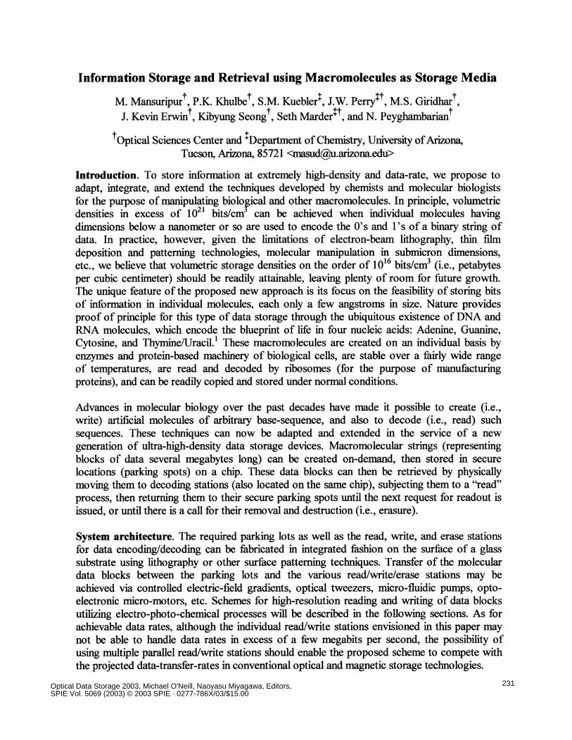
A possible implementation of the proposed storage device, depicted schematically in Fig. 1,shows a specific arrangement of 16 parking spots in conjunction with a single read/writestation on a chip surface. Various canals are etched on this surface to connect the parkingspots to the read/write chamber. Binary valves placed at the cross-sections of these canalscontrol the flow direction (a possible design of a binary j.i-valve appears in Fig. 2). Controllingthe valves is accomplished by means of electrical signals on four separate lines.
——— ...".....""...".. .(. .——Fig. 2. Binary valve channels the flow ofincoming liquid toward either of outlets 1or 2. An electronic command signal isrequired to compress the spring and pullthe switch block to the lower position.When the command signal is removed,the spring relaxes to its initial state, thusredirecting the flow toward outlet 1.
The aforementioned binary scheme ofgrouping the parking spots results in anumber of address lines that is thebase-2 logarithm of the number ofspots. One can thus address over amillion parking spots on the surface ofa chip with only 20 lines. If eachparking spot, for example, occupies a
10 x 1Otm2 area, one million such spots will cover a square centimeter of the chip surface.With the macromolecule stored in each parking spot corresponding to a megabyte block ofuser data, the areal density of storage on the chip will approach 1 TeraBytes/cm2. Moreover, if
Fig. 1. Diagramshowing thepatterned surface ofthe proposed datastorage chip. Once aparking spot isselected, itsmacromolecularcontent will betransferred to theread/write stationunder the influenceof an appliedelectric voltage (i.e.,electro-phoretictransfer). Followingthe completion ofthe read/writeoperation, themacromolecule isreturned to itsdesignated spot.
l3inary Vaive .
p11 p1, p1. p1 p1, p11 p1
.P, Pr, P, P,1
Na.no—pore in lipid membrane
ReadfWnteMacrom ...olerule Station
(a) Outlet. 2
(b)
::::: Outkt I
232 Proc. of SPIE Vol. 5069
the parking spots (and associated read/write/erase stations) are stacked in 1O.tm-thick layers,the volumetric capacity ofthe envisioned device will easily reach 1 PetaBytes/cm3.
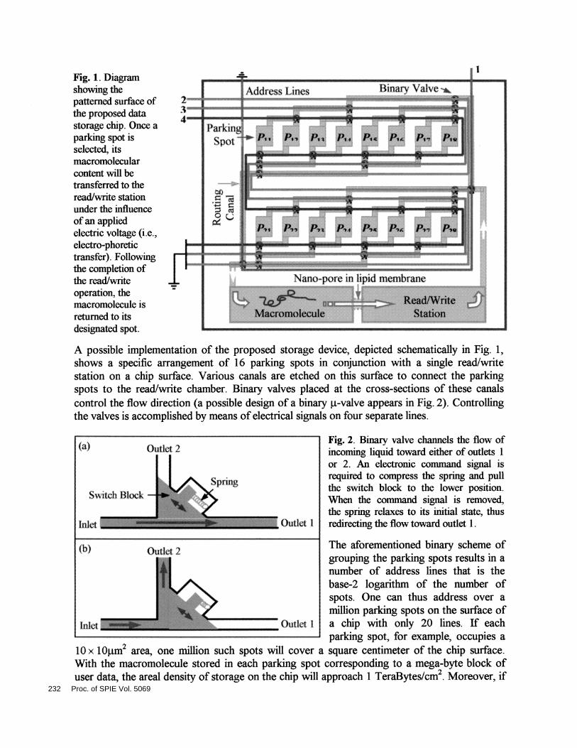
Macromolecular readout by translocation through a nano-pore. The diagrams in Fig. 3depict a method of readout by translocation through a nano-pore.2 As different nucleotidespass through the pore, they hamper the flow of the ionic current differently. Fluctuations incurrent blockage are due to differences in the size and/or charge of the various nucleotides.Given a sufficient SNR, one should be able to uniquely identify the base sequence of thetranslocated DNA molecule by analyzing the electrolytic current waveform.
Fig. 3. (a) Nano-porecreated in a lipid
membrane by a-hemolysinproteins. The bilayerseparates two sections of abuffer solution. With100 mV applied across thebilayer, —12O pA of iomccurrent flows through thenano-pore. (b) DNA strandpassing through the nano-pore partially blocks theionic current. ________________________________________________________
Figure 4 is a schematic diagram of a setup for conducting DNA translocation experiments inour laboratory. The system consists of two 15OiL wells, machined in a teflon block andconnected by a U-shaped tube. One end of the U-tube has a sleeve of teflon tubing, which israpidly tapered to provide a 4Oim diameter aperture. Ag-AgC1 electrodes are inserted intoboth wells. To form a lipid bilayer at the 40p.m aperture, one must first prime the aperture byapplying 5tL of a 2OOpg/mL solution of lipid (diphytanoyl phosphatidylcholine) dissolved inspectroscopic-grade hexane. The primer is then allowed to evaporate in a mild stream ofnitrogen gas. The wells on both sides of the aperture are subsequently filled with a buffersolution (1.OM KC1 + 10 mM HEPES/KOH, pH = 8.0). At this point, a relatively large ioniccurrent passes through the aperture (several hundred nA at l2OmV applied voltage). To forma lipid bilayer, a single bristle of a fine brush is dipped in a 3mg/mL lipid solution (dissolvedin spectroscopic-grade hexadecane), then brushed across the aperture. Formation of thebilayer is indicated by the blockage of the ionic current (several hundred GO resistancebetween the Ag-AgC1 electrodes).
Once a stable bilayer has been created, 80 ng of a-hemolysin in 2p.L buffer solution is addedto the well that contains the aperture and, subsequently, a l2OmV potential is applied to theelectrodes. Typically, a single ion-channel consisting of seven a-hemolysin molecules self-assembles in a time span of 5-20 minutes; successful self-assembly is indicated by an abruptincrease of the electrolytic current to l2OpA. To avoid formation of multiple ion-channels, theexcess a-hemolysin is removed by perftision with '-2OmL of fresh buffer. (In our experiments,individual ion-channels were stable for up to six hours.) With an ion-channel formed stablywithin the lipid bilayer, we add single-stranded DNA molecules (ssDNA) to the cis (i.e.,
I(pA)
+
(a)
(1,
Il2OpA
Open nano-pore
DNA
DNA
Time
Proc. of SPIE Vol. 5069 233
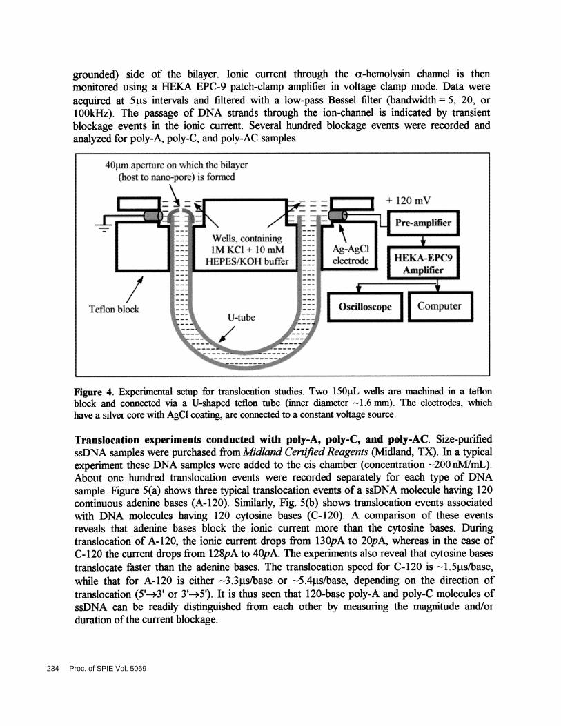
grounded) side of the bilayer. Ionic current through the a-hemolysin channel is thenmonitored using a HEKA EPC-9 patch-clamp amplifier in voltage clamp mode. Data wereacquired at 5ts intervals and filtered with a low-pass Bessel filter (bandwidth = 5, 20, or100kHz). The passage of DNA strands through the ion-channel is indicated by transientblockage events in the ionic current. Several hundred blockage events were recorded andanalyzed for poly-A, poly-C, and poly-AC samples.
Figure 4. Experimental setup for translocation studies. Two 15OiL wells are machined in a teflonblock and connected via a U-shaped teflon tube (inner diameter 1.6 mm). The electrodes, whichhave a silver core with AgCl coating, are connected to a constant voltage source.
Translocation experiments conducted with poly-A, poly-C, and poly-AC. Size-purifiedssDNA samples were purchased from Midland Cert/1ed Reagents (Midland, TX). In a typicalexperiment these DNA samples were added to the cis chamber (concentration 2OO nMJmL).About one hundred translocation events were recorded separately for each type of DNAsample. Figure 5(a) shows three typical translocation events of a ssDNA molecule having 120continuous adenine bases (A-120). Similarly, Fig. 5(b) shows translocation events associatedwith DNA molecules having 120 cytosine bases (C-120). A comparison of these eventsreveals that adenine bases block the ionic current more than the cytosine bases. Duringtranslocation of A-120, the ionic current drops from l3OpA to 2OpA, whereas in the case ofC-120 the current drops from 128pA to 4OpA. The experiments also reveal that cytosme basestranslocate faster than the adenine bases. The translocation speed for C-120 is 1 .5.ts/base,while that for A-120 is either 3.3jts/base or -'5.4j.ts/base, depending on the direction oftranslocation (5'—÷3' or 3'—*5'). It is thus seen that 120-base poly-A and poly-C molecules ofssDNA can be readily distinguished from each other by measuring the magnitude and/orduration ofthe current blockage.
4()iim aperture on shtch the bilaver(host to nano—pore) is tbrtned
234 Proc. of SPIE Vol. 5069
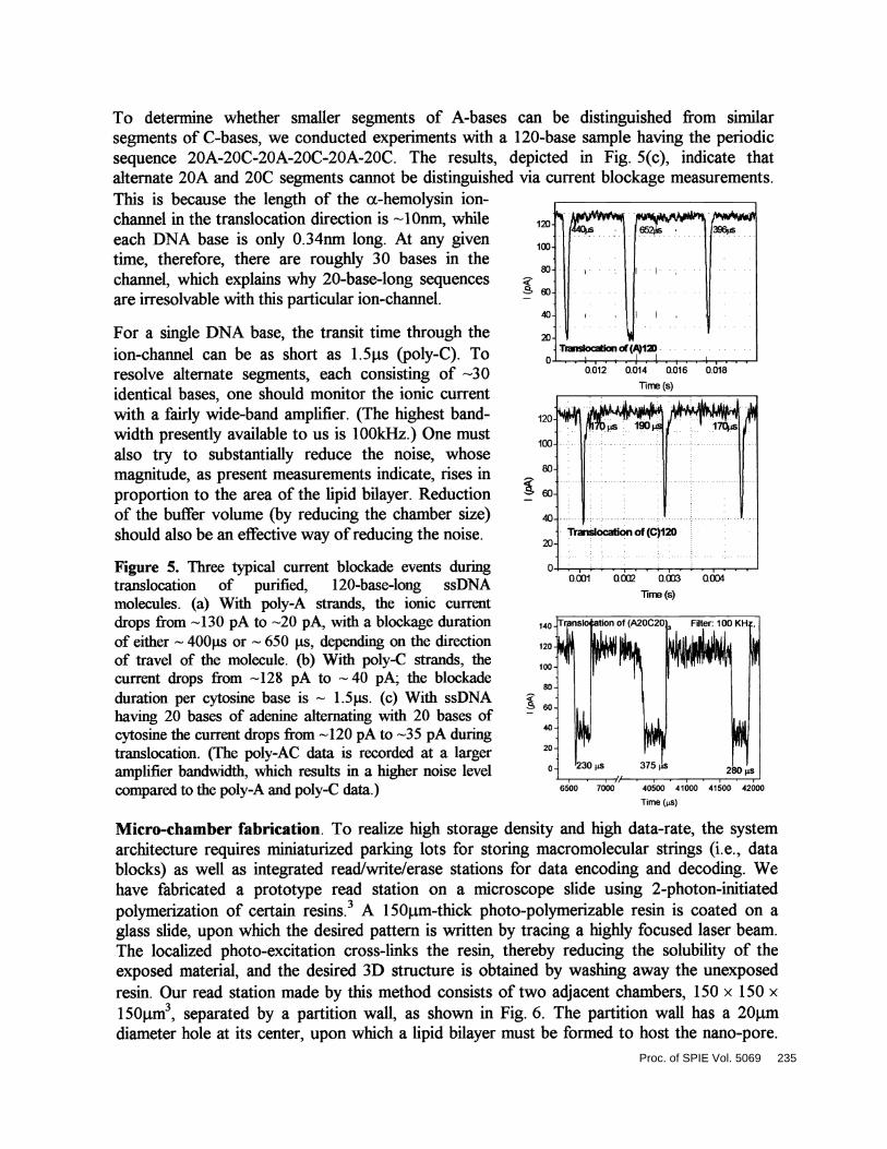
To determine whether smaller segments of A-bases can be distinguished from similarsegments of C-bases, we conducted experiments with a 120-base sample having the periodicsequence 2OA-2OC-2OA-2OC2OA-2OC. The results, depicted in Fig. 5(c), indicate thatalternate 20A and 20C segments cannot be distinguished via current blockage measurements.This is because the length of the a-hemolysin ion-channel in the translocation direction is 1Onm, while
•
. . 44c6 652 396seach DNA base is only O.34nm long. At any given . . . .
time, therefore, there are roughly 30 bases in the .
channel, which explains why 20-base-long sequences.
are irresolvable with this particular ion-channel. ' ° . ..
.
. ..
.
For a single DNA base, the transit time through the 2. .
ion-channel can be as short as 1.5j.ts (poly-C). To ..
I•
resolve alternate segments, each consisting of 3Oidentical bases, one should monitor the ionic currentwith a fairly wide-band amplifier. (The highest band-width presently available to us is 100kHz.) One mustalso try to substantially reduce the noise, whosemagnitude, as present measurements indicate, rises inproportion to the area of the lipid bilayer. Reductionof the buffer volume (by reducing the chamber size)should also be an effective way ofreducing the noise.
Figure 5. Three typical current blockade events duringtranslocation of purified, 120-base-long ssDNAmolecules. (a) With poly-A strands, the ionic currentdrops from 'l3O pA to 2O pA, with a blockage durationof either 4OOs or 650 is, depending on the directionof travel of the molecule. (b) With poly-C strands, thecurrent drops from '428 pA to '40 pA; the blockadeduration per cytosine base is ' 1 .5j.is. (c) With ssDNAhaving 20 bases of adenine alternating with 20 bases ofcytosine the current drops from 42O pA to 35pA duringtranslocation. (The poly-AC data is recorded at a largeramplifier bandwidth, which results in a higher noise levelcompared to the poly-A and poly-C data)
Micro-chamber fabrication. To realize high storage density and high data-rate, the systemarchitecture requires miniaturized parking lots for storing macromolecular strings (i.e., datablocks) as well as integrated read/write/erase stations for data encoding and decoding. Wehave fabricated a prototype read station on a microscope slide using 2-photon-initiatedpolymerization of certain resins.3 A 1 50pm-thick photo-polymerizable resin is coated on aglass slide, upon which the desired pattern is written by tracing a highly focused laser beam.The localized photo-excitation cross-links the resin, thereby reducing the solubility of theexposed material, and the desired 3D structure is obtained by washing away the unexposedresin. Our read station made by this method consists of two adjacent chambers, 1 50x 1 50 x150pm3, separated by a partition wall, as shown in Fig. 6. The partition wall has a 2O.tmdiameter hole at its center, upon which a lipid bilayer must be formed to host the nano-pore.
0.012 0.014 0.016 0.018
ThT(s)
...
:T.i19o
...
. Traislocation of(C)12020
0.0.001 0.002 0.003
line(s)u604
Time (tS)
Proc. of SPIE Vol. 5069 235
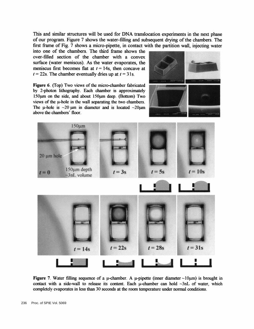
Figure 7. Water filling sequence of a p.-chamber. A p-pipette (inner diameter '1Oprn) is brought incontact with a side-wall to release its content. Each g-chamber can hold 3nL of water, whichcompletely evaporates in less than 30 seconds at the room temperature under normal conditions.
This and similar structures will be used for DNA translocation experiments in the next phaseof our program. Figure 7 shows the waterfilling and subsequent drying of the chambers. Thefirst frame of Fig. 7 shows a micro-pipette, in contact with the partition wall, injecting waterinto one of the chambers. The third frame shows theover-filled section of the chamber with a convexsurface (water meniscus). As the water evaporates, themeniscus first becomes flat at t = 14s, then concave att = 22s. The chamber eventually dries up at t =31 s.
Figure 6. (Top) Two views of the micro-chamber fabricatedby 2-photon lithography. Each chamber is approximately150pm on the side, and about 150pm deep. (Bottom) Twoviews of the si-hole in the wall separating the two chambers.The p-hole is 20 pm in diameter and is located '—'2Opmabove the chambers' floor.
I I I I I I I II ....I I I I I
236 Proc. of SPIE Vol. 5069
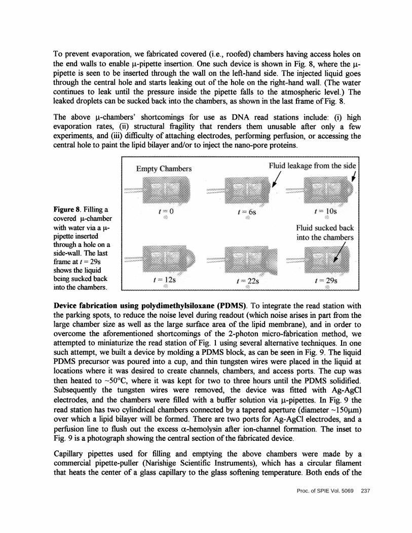
To prevent evaporation, we fabricated covered (i.e., roofed) chambers having access holes onthe end walls to enable jt-pipette insertion. One such device is shown in Fig. 8, where the .t-pipette is seen to be inserted through the wall on the lefthand side. The injected liquid goesthrough the central hole and starts leaking out of the hole on the right-hand wall. (The watercontinues to leak until the pressure inside the pipette falls to the atmospheric level.) Theleaked droplets can be sucked back into the chambers, as shown in the last frame ofFig. 8.
The above .t-chambers' shortcomings for use as DNA read stations include: (i) highevaporation rates, (ii) structural fragility that renders them unusable after only a fewexperiments, and (iii) difficulty of attaching electrodes, performing perfusion, or accessing thecentral hole to paint the lipid bilayer and/or to inject the nano-pore proteins.
Figure 8. Filling acovered p-chamberwith water via apipette insertedthrough a hole on aside-wall. The lastframe at t= 29sshows the liquidbeing sucked backinto the chambers. ... . . .
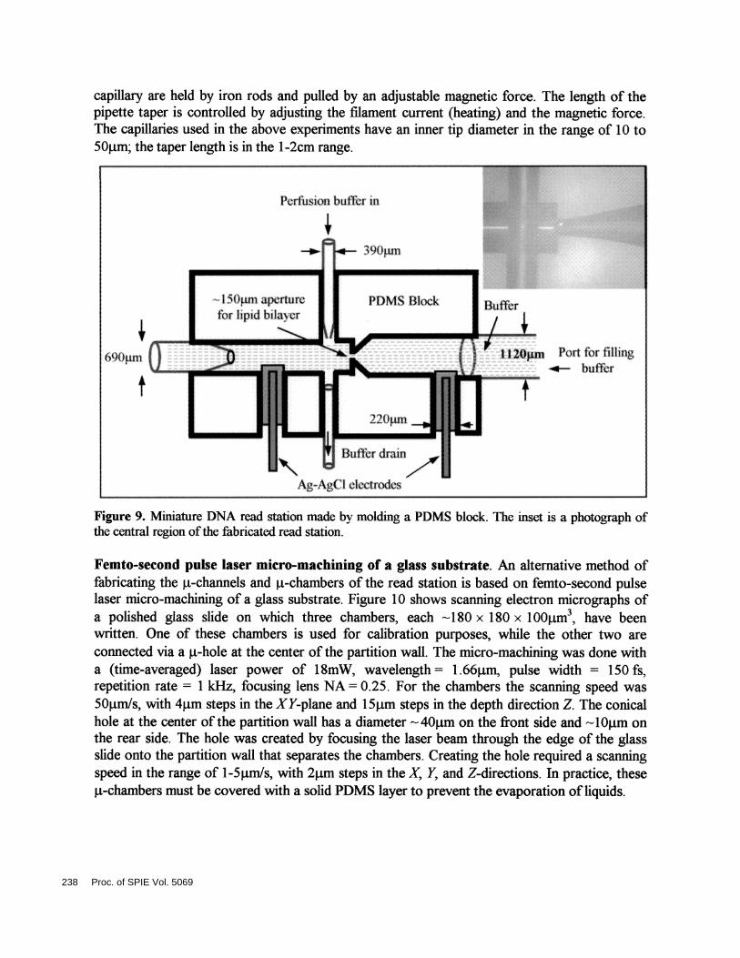
Device fabrication using polydimethylsioxane (PDMS). To integrate the read station withthe parking spots, to reduce the noise level during readout (which noise arises in part from thelarge chamber size as well as the large surface area of the lipid membrane), and in order toovercome the aforementioned shortcomings of the 2-photon micro-fabrication method, weattempted to miniaturize the read station of Fig. 1 using several alternative techniques. In onesuch attempt, we built a device by molding a PDMS block, as can be seen in Fig. 9. The liquidPDMS precursor was poured into a cup, and thin tungsten wires were placed in the liquid atlocations where it was desired to create channels, chambers, and access ports. The cup wasthen heated to 5O°C, where it was kept for two to three hours until the PDMS solidified.Subsequently the tungsten wires were removed, the device was fitted with Ag-AgClelectrodes, and the chambers were filled with a buffer solution via jpipettes. In Fig. 9 theread station has two cylindrical chambers connected by a tapered aperture (diameter -1 50j.tm)over which a lipid bilayer will be formed. There are two ports for Ag-AgCl electrodes, and aperfusion line to flush out the excess a-hemolysin after ion-channel formation. The inset toFig. 9 is a photograph showing the central section ofthe fabricated device.
Capillary pipettes used for filling and emptying the above chambers were made by acommercial pipette-puller (Narishige Scientific Instruments), which has a circular filamentthat heats the center of a glass capillary to the glass softening temperature. Both ends of the
I mpty Chambcis
I 0
tiutd leakage from th side
/I b' I los
Fluid sucked backinto the chambers/
I 22s I 29s/ 12s
Proc. of SPIE Vol. 5069 237
capillary are held by iron rods and pulled by an adjustable magnetic force. The length of thepipette taper is controlled by adjusting the filament current (heating) and the magnetic force.The capillaries used in the above experiments have an inner tip diameter in the range of 10 to50tm; the taper length is in the 1-2cm range.
Figure 9. Miniature DNA read station made by molding a PDMS block. The inset is a photograph ofthe central region ofthe fabricated read station.
Femto-second pulse laser micro-machining of a glass substrate. An alternative method offabricating the p-channels and j.t-chambers of the read station is based on femto-second pulselaser micro-machining of a glass substrate. Figure 1 0 shows scanning electron micrographs ofa polished glass slide on which three chambers, each 180 x 180 x lOOp.m3, have beenwritten. One of these chambers is used for calibration purposes, while the other two areconnected via a p-hole at the center of the partition wall. The micro-machining was done witha (time-averaged) laser power of 18mW, wavelength = 1.66tm, pulse width = 150 fs,repetition rate 1 kHz, focusing lens NA = 0.25. For the chambers the scanning speed was50jnn/s, with 4pm steps in the XY-plane and 15p.m steps in the depth direction Z. The conicalhole at the center of the partition wall has a diameter '4Opim on the front side and 10tm onthe rear side. The hole was created by focusing the laser beam through the edge of the glassslide onto the partition wall that separates the chambers. Creating the hole required a scanningspeed in the range of 1-5p.m/s, with 2tm steps in the X, Y, and Z-directions. In practice, thesept-chambers must be covered with a solid PDMS layer to prevent the evaporation of liquids.
Pcrfü ;ion tLLfl. cr in
PDMS Block
()Otrn
tPort for fiflüig
4— hufThr
' 7Ag Ag(.l electrodes
238 Proc. of SPIE Vol. 5069
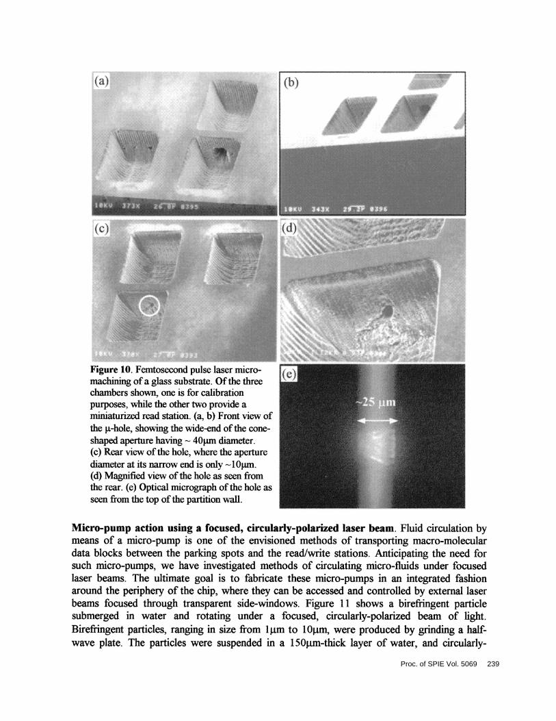
the p.t-hole, showing the wide-end ofthe cone-
shaped aperture having 4Oim diameter.(c) Rear view ofthe hole, where the aperturediameter at its narrow end is only 'lOjim.(d) Magnified view ofthe hole as seen fromthe rear. (e) Optical micrograph ofthe hole asseen from the top ofthe partition wall.
Micro-pump action using a focused, circularly-polarized laser beam. Fluid circulation bymeans of a micro-pump is one of the envisioned methods of transporting macro-moleculardata blocks between the parking spots and the read/write stations. Anticipating the need forsuch micropumps, we have investigated methods of circulating microfluids under focusedlaser beams. The ultimate goal is to fabricate these micro-pumps in an integrated fashionaround the periphery of the chip, where they can be accessed and controlled by external laserbeams focused through transparent side-windows. Figure 11 shows a birefringent particlesubmerged in water and rotating under a focused, circularly-polarized beam of light.Birefringent particles, ranging in size from lj.tm to lOj.tm, were produced by grinding a half-wave plate. The particles were suspended in a 15Otm-thick layer of water, and circularly-
Figure 10. Femtosecond pulse laser micro-machining of a glass substrate. Of the threechambers shown, one is for calibrationpurposes, while the other two provide aminiaturized read station. (a, b) Front view of
Proc. of SPIE Vol. 5069 239
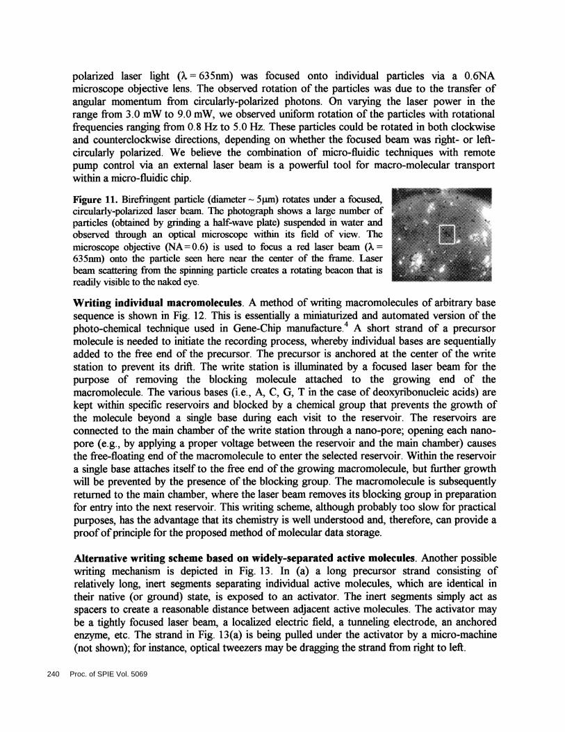
polarized laser light (.= 635mn) was focused onto individual particles via a O.6NAmicroscope objective lens. The observed rotation of the particles was due to the transfer ofangular momentum from circularly-polarized photons. On varying the laser power in therange from 3.0 mW to 9.0 mW, we observed uniform rotation of the particles with rotationalfrequencies ranging from 0.8 Hz to 5.0 Hz. These particles could be rotated in both clockwiseand counterclockwise directions, depending on whether the focused beam was right- or left-circularly polarized. We believe the combination of micro-fluidic techniques with remotepump control via an external laser beam is a powerful tool for macro-molecular transportwithin a micro-fluidic chip.
Figure 11. Birefringent particle (diameter 5pm) rotates under a focused,circularly-polarized laser beam. The photograph shows a large number ofparticles (obtained by grinding a half-wave plate) suspended in water andobserved through an optical microscope within its field of view. Themicroscope objective (NA 0.6) is used to focus a red laser beam ( =635nm) onto the particle seen here near the center of the frame. Laserbeam scattering from the spinning particle creates a rotating beacon that isreadily visible to the naked eye.
Writing individual macromolecules. A method of writing macromolecules of arbitrary basesequence is shown in Fig. 12. This is essentially a miniaturized and automated version of thephoto-chemical technique used in Gene-Chip manufacture.4 A short strand of a precursormolecule is needed to initiate the recording process, whereby individual bases are sequentiallyadded to the free end of the precursor. The precursor is anchored at the center of the writestation to prevent its drift. The write station is illuminated by a focused laser beam for thepurpose of removing the blocking molecule attached to the growing end of themacromolecule. The various bases (i.e., A, C, G, T in the case of deoxyribonucleic acids) arekept within specific reservoirs and blocked by a chemical group that prevents the growth ofthe molecule beyond a single base during each visit to the reservoir. The reservoirs areconnected to the main chamber of the write station through a nano-pore; opening each nano-pore (e.g., by applying a proper voltage between the reservoir and the main chamber) causesthe free-floating end of the macromolecule to enter the selected reservoir. Within the reservoira single base attaches itself to the free end of the growing macromolecule, but further growthwill be prevented by the presence of the blocking group. The macromolecule is subsequentlyreturned to the main chamber, where the laser beam removes its blocking group in preparationfor entry into the next reservoir. This writing scheme, although probably too slow for practicalpurposes, has the advantage that its chemistry is well understood and, therefore, can provide aproofofprinciple for the proposed method ofmolecular data storage.
Alternative writing scheme based on widely-separated active molecules. Another possiblewriting mechanism is depicted in Fig. 1 3 . In (a) a long precursor strand consisting ofrelatively long, inert segments separating individual active molecules, which are identical intheir native (or ground) state, is exposed to an activator. The inert segments simply act asspacers to create a reasonable distance between adjacent active molecules. The activator maybe a tightly focused laser beam, a localized electric field, a tunneling electrode, an anchoredenzyme, etc. The strand in Fig. 13(a) is being pulled under the activator by a micro-machine(not shown); for instance, optical tweezers may be dragging the strand from right to left.
240 Proc. of SPIE Vol. 5069
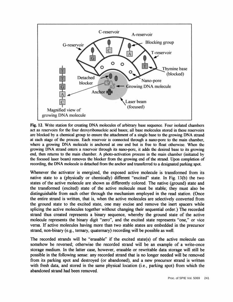
Fig. 12. Write station for creating DNA molecules of arbitrary base sequence. Four isolated chambersact as reservoirs for the four deoxyribonucleic acid bases; all base molecules stored in these reservoirsare blocked by a chemical group to ensure the attachment of a single base to the growing DNA strandat each stage of the process. Each reservoir is connected through a nano-pore to the main chamber,where a growing DNA molecule is anchored at one end but is free to float otherwise. When thegrowing DNA strand enters a reservoir through its nanopore, it adds the desired base to its growingend, then returns to the main chamber. A photo-activation process in the main chamber (initiated bythe focused laser beam) removes the blocker from the growing end of the strand. Upon completion ofrecording, the DNA molecule is detached from the anchor and transferred to a designated parking spot.
Whenever the activator is energized, the exposed active molecule is transformed from itsnative state to a (jthysically or chemically) different "excited" state. In Fig. 13(b) the twostates of the active molecule are shown as differently colored. The native (ground) state andthe transformed (excited) state of the active molecule must be stable; they must also bedistinguishable from each other through the mechanism employed in the read station. (Oncethe entire strand is written, that is, when the active molecules are selectively converted fromthe ground state to the excited state, one may excise and remove the inert spacers whilesplicing the active molecules together without changing their sequential order.) The recordedstrand thus created represents a binary sequence, whereby the ground state of the activemolecule represents the binary digit "zero", and the excited state represents "one," or viceversa. If active molecules having more than two stable states are embedded in the precursorstrand, non-binary (e.g., ternary, quatemary) recording will be possible as well.
The recorded strands will be "erasable" if the excited state(s) of the active molecule cansomehow be reversed; otherwise the recorded strand will be an example of a write-oncestorage medium. In the latter case, however, erasable or rewritable data storage will still bepossible in the following sense: any recorded strand that is no longer needed will be removedfrom its parking spot and destroyed (or abandoned), and a new precursor strand is writtenwith fresh data, and stored in the same physical location (i.e., parking spot) from which theabandoned strand had been removed.
(.rcscrvoir
(irescrvoir
.A:.4eSe.t\O1T
Blockmg group
F re'eivoit
i..L : blocker
Li.:.M
LtJManiiied view of
groviing i.)NA niolecule
\iymine base(blocked)
... .Nan-pore
rowing L)NA molecule
s.Laser beam( fbcused)
Proc. of SPIE Vol. 5069 241
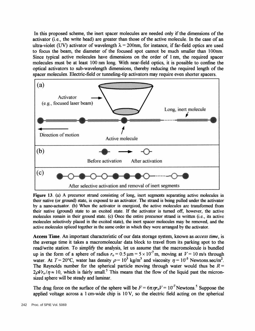
In this proposed scheme, the inert spacer molecules are needed only if the dimensions of theactivator (i.e., the write head) are greater than those of the active molecule. In the case of anultra-violet (UV) activator of wavelength A = 200nm, for instance, if far-field optics are usedto focus the beam, the diameter of the focused spot cannot be much smaller than 1 OOnm.Since typical active molecules have dimensions on the order of 1 nm, the required spacermolecules must be at least 100 nm long. With near-field optics, it is possible to confine theoptical activators to sub-wavelength dimensions, thereby reducing the required length of thespacer molecules. Electric-field or tunneling-tip activators may require even shorter spacers
(I) 4 ..l3clbrc ar1vation Aflei activation
(c)
After selective activation and rcnmval of inert segments
Figure 13. (a) A precursor strand consisting of long, inert segments separating active molecules intheir native (or ground) state, is exposed to an activator. The strand is being pulled under the activatorby a nano-actuator. (b) When the activator is energized, the active molecules are transformed fromtheir native (ground) state to an excited state. If the activator is turned off, however, the activemolecules remain in their ground state. (c) Once the entire precursor strand is written (i.e., its activemolecules selectively placed in the excited state), the inert spacer molecules may be removed, and theactive molecules spliced together in the same order in which they were arranged by the activator.
Access Time. An important characteristic of our data storage system, known as access time, isthe average time it takes a macromolecular data block to travel from its parking spot to theread/write station. To simplify the analysis, let us assume that the macromolecule is bundledup in the form of a sphere of radius r0 =0.5 p.m = 5 x 1 O m, moving at V = 10 rn/s throughwater. At T =20°C, water has density p = 1O kg/rn3 and viscosity ii = 1oa Newtons.sec/m2.The Reynolds number for the spherical particle moving through water would thus be R =2pVr0/q 10, which is fairly small.5 This means that the flow of the liquid past the micron-sized sphere will be steady and laminar.
The drag force on the surface of the sphere will be F =6icqr0V iO Newtons.5 Suppose theapplied voltage across a 1 cm-wide chip is I 0 V, so the electric field acting on the spherical
(a)
Activator(et tbcused laser bean)
Direction of fl)()tlOfl
L1ong, irrt flotecule
IActive molecule
242 Proc. of SPIE Vol. 5069
particle will be E iO V/rn. Since F qE, the particle must have a charge of q = 10'°C inorder to move at the desired velocity under the applied field. Since a singIestranded DNAmolecule has (on average) 2.5 electrons per base (i.e., 4 x 10'9C per base), the macromoleculemust have 2.5 x 10⁸ nucleic acid bases. (This corresponds to a 60 Mbyte block of dataassociated with each molecule.) Since each base occupies roughly 1 nm, a 2.5 x 10⁸-basemolecule should fit in a micron-sized sphere, in agreement with our earlier assumptions.
The conclusion is that, if a polymer consisting of 1 8 1O building blocks (monomers) isused to encode each data block, and if each monomer happens to have a few unpairedelectrons, then the molecule can be transferred between its parking spot and the read/writestation in a matter of milliseconds under the influence of a reasonable electric field (e.g.,10 Volts across a 1 x 1 cm2 chip). We are aware, ofcourse, that a charged polymer as assumedcannot be bundled up into a compact sphere, and a more realistic analysis needs to beundertaken in order to estimate the actual access time of the proposed system. Experimenta[data from gel electrophoresis indicate that the travel time of macromolecules under the aboveconditions should be on the order of a few seconds (rather than milliseconds). We plan toinvestigate this question further and, if necessary, consider alternative techniques fortransporting our molecular data blocks. For instance, it is possible to neutralize the charge ona single-stranded DNA molecule by pairing it with its complement (i.e., creating double-stranded DNA), then place the uncharged molecule in a small lipid vesicle (similar to thoseused in biological systems) and move the vesicle across the system using either anelectromagnetic field or a flow field created by the action of a micro-pump.
Concluding remarks. The objective of our research program is to build a data storage deviceusing macromolecules, in general, and DNA molecules, in particular, as the storage medium.We use micro-fluidic techniques for transporting the molecules through various chambers(i.e., read, write, erase, and storage chambers) within a small, integrated chip. To date wehave succeeded in building several micro-chambers, conducted DNA translocationexperiments in different-sized chambers, and also demonstrated the principle of opticalpumping in these devices. Future work includes fabrication of nano-pores in jt-chambers,conducting translocation experiments within these jt-chambers, and investigating variousschemes ofmacromolecular writing and transportation.
Acknowledgement. The authors are grateful to Professors Raphael Gruener and Michael Hogan of theUniversity of Arizona for illuminating discussions. The paper is based upon work supported in part bythe National Science Foundation STC Program, under Agreement No. DMR-O 120967.
1. M. Mansuripur, "DNA, Human Memory, and the Storage Technology ofthe 2l Century," keynote addressat the 2001 Optical Data Storage Conference (Santa Fe, New Mexico, April 2001). Published in SPIEProceedings 4342, 1-29 (2001).
2. A. Meller, L. Nivon, and D. Branton, "Voltage-driven DNA translocations through a nano-pore," Phys. Rev.Lett. 86, 3435-3438 (2001); A. Meller, L. Nivon, E. Brandin, J. Golovchenko, and D. Branton, "Rapidnanopore discrimination between single polynucleotide molecules," PNAS, Vol. 97, 1079-1084 (2000).
3. B. K. Cumpston et. al. "Two-photonpolymerization initiators for three-dimensional optical data storage andmicrofàbrication," Nature 398, 51 (1999).
4. S. Fodor et al, "Light-directed, spatially addressable parallel chemical synthesis," Science 251, 767-773(1991); A. Pease et al, "Light-generated oligonucleotide arrays for rapid DNA sequence analysis," Proc.Nati. Acad. Sci. 91, 5022-5026 (1994); E. Southern, K. Mir, and M. Shchepinov, "Molecular interactions onmicroarrays," Nature Genet. 21, Supplement, pp 1-5.
5. RP. Feynman, RB. Leighton, M. Sands, The Feynman Lectures on Physics, Vol. II, Chap. 41.
Proc. of SPIE Vol. 5069 243
Related Documents