Influence of Physical and Chemical Treatments on Cell Survival and Acquisition of Pluripotency Ricardo Jorge Carvalho Correia Thesis to obtain the Master of Science Degree in Biological Engineering Supervisors: MD, PhD Petra de Sutter PhD Cláudia Alexandra Martins Lobato da Silva Examination Committee Chairperson: Professor Arsénio do Carmo Sales Mendes Fialho Supervisor: Professor Cláudia Alexandra Martins Lobato da Silva Member of the Committee: Professor Maria Margarida Fonseca Rodrigues Diogo November 2014

Welcome message from author
This document is posted to help you gain knowledge. Please leave a comment to let me know what you think about it! Share it to your friends and learn new things together.
Transcript

Influence of Physical and Chemical Treatments on Cell Survival and
Acquisition of Pluripotency
Ricardo Jorge Carvalho Correia
Thesis to obtain the Master of Science Degree in
Biological Engineering
Supervisors: MD, PhD Petra de Sutter
PhD Cláudia Alexandra Martins Lobato da Silva
Examination Committee
Chairperson: Professor Arsénio do Carmo Sales Mendes Fialho
Supervisor: Professor Cláudia Alexandra Martins Lobato da Silva
Member of the Committee: Professor Maria Margarida Fonseca Rodrigues Diogo
November 2014

I
Acknowledgments
First of all, I would like to thank my external promoters, Professor Petra de Sutter and Björn Heindryckx, for
believing in me and accept and treat me as one of their own research group members, allowing me to develop
my knowledge and interest on this research field, and providing me the resources to conduct an organized and
scientifically useful project.
I would also like to thank Sharat Warrier for guiding me through all the past months, helping me during both the
practical experiences and writing process, and for teaching me almost everything. Also, his wife, Galbha Duggal,
for all her help. To both, I want to show my special gratitude for receiving me as more than a colleague, as a
friend. Without you, this work could never be possible.
To my internal promoter, Professor Cláudia Lobato Silva, for the constant availability to answer my questions and
for all the advices, helping me to finish my work.
To all the research team at the Ghent Hospital, for all the support, resources and time dedicated to my project.
To all my friends in Ghent, thank you for being my family during the last months and for making me feel home in
a city and country of which I didn’t know anything until then. Thank you for all the good moments we spent
together. I’m truly thankful for all the friends for life I made in the last months.
To all my family and friends in Portugal, thank you for all the daily messages that made me feel closer to my
country, and for all the support to overcome the distance between us.
I would also like to express my thankfulness to my parents for allowing me to have resources to live in another
country, for supporting me during the whole time, for believing in me, and for considering my graduation and
success as some of their biggest dreams. This thesis and all my commitment to my academic and work careers
are dedicated to you.
And last but not the least, I want to express my gratefulness to my girlfriend, Maria, who was the first to convince
me to face this adventure. For all the love, understanding and dedication during the whole time we were far
from each other. For making me feel close to her no matter the distance, and for giving me the strength when I
thought I didn’t have it. She was and will always be my biggest support.
To all, a heartfelt thank you.

II
Abstract
Pluripotent human embryonic stem cells represent a promising source to develop medical therapies and unveil
the secrets behind degenerative diseases. The necessity of large quantities of viable pluripotent cells is urgent.
Several ethical problems regarding the use of embryos for research result in the necessity of high throughput
reprogramming techniques. Recently, new data reported the possibility of reprogramming murine somatic cells
into a state of pluripotency through the induction of external strong stimuli, such as the exposure to an acidic
solution and/or constant physical stress. Intrigued by this, it was decided not only to try to reproduce the
accomplished results, but also to adapt the protocol to accomplish a reprogramming event using human cells.
Several different types of murine somatic cells, as well as embryoid bodies generated in vitro from both human
and mouse embryonic stem cells, were exposed to three consecutive physical trituration steps with increasingly
smaller lumen pipettes, followed by 30 minutes exposure to an acidic solution. Cell survival and resistance was
monitored during the experimental steps. Together, the results obtained showed incapacity of the described
technique to reprogram differentiated cells into a pluripotent state. The challenging protocol resulted in constant
cell death during the experiments. The possible reprogramming of somatic cells into pluripotency through this
simple and fast method would represent a remarkable improvement in the use of stem cells-based therapies.
Key words: Pluripotency, Differentiation, Somatic Cells, Embryoid Bodies, Reprogramming, External Stimuli
Resumo
As células estaminais pluripotentes humanas representam uma fonte promissora de terapias médicas e
conhecimento de doenças degenerativas. A necessidade de grandes quantidades de células pluripotentes viáveis
é urgente. Diversos problemas éticos, relacionados com o uso de embriões para investigação desencadeiam a
grande necessidade de técnicas de reprogramação. Recentemente, foi reportado ser possível reprogramar
células somáticas murinas em células pluripotentes, através de fortes estímulos externos, como a exposição a
uma solução acídica e/ou a perturbação física constante. Foi decidido não só tentar reproduzir os resultados
previamente reportados, como também alcançar o feito de reprogramar células humanas através do mesmo
método. Diversas células somáticas murinas diretamente isoladas, assim como corpos embrióides produzidos in
vitro a partir de células estaminais pluripotentes humanas e de rato, foram expostas a três perturbações físicas
consecutivas, seguidas de exposição a uma solução acídica durante 30 minutos. Os resultados obtidos
demonstram a incapacidade da técnica descrita de induzir um estado de pluripotência em células diferenciadas.
Ainda, a dificuldade técnica associada ao protocolo resultou em morte celular constante durante as experiências.
A possível reprogramação de células diferenciadas em células pluripotentes através deste simples e rápido
método representaria um notável desenvolvimento para o uso de terapias baseadas em células estaminais.
Palavras-chave: Pluripotência, Diferenciação, Células Somáticas, Corpos Embrióides, Reprogramação, Estímulo Externo

III
General Index
I. Figures Index ................................................................................................................................... V
II. Tables Index .................................................................................................................................... XI
III. Symbols and Abbreviations Index ............................................................................................. XII
1. State of Art ...................................................................................................................................... 1
1.1. Introduction to Embryonic Stem Cells..................................................................................... 1
1.1.1. Establishment of the first stable Mouse Embryonic Stem Cell line in culture ................ 2
1.1.2. Establishment of Human Embryonic Stem Cell lines ....................................................... 3
1.1.3. Identifying pluripotent stem cells ................................................................................... 3
1.1.4. Differentiation of Human Embryonic Stem Cells ............................................................ 4
1.2. Human Embryonic Stem Cells: The promise for Therapeutics ................................................ 5
1.2.1. Controversy of stem cells and ethical problems ............................................................. 5
1.3. Reprogramming ....................................................................................................................... 6
1.3.1. Somatic Cell Nuclear Transfer ......................................................................................... 7
1.3.2. Induced Pluripotent Stem Cells ....................................................................................... 9
1.4. Mouse and Human Embryonic Stem cells: Different States of pluripotency ........................ 13
1.4.1. Embryogenesis .............................................................................................................. 13
1.4.2. Naïve and primed pluripotency ..................................................................................... 14
1.5. Signaling Pathways: The roadmap to Embryonic Stem cell maintenance ............................ 19
1.5.1. LIF/Stat3 Signaling – Establishment of 2i and 3i conditions .......................................... 19
1.5.2. BMP4/Smad1,5,8 Signaling – Establishment of serum-free conditions ........................ 21
1.5.3. TGFβ, Activin and Nodal/Smad2,3 Signaling ................................................................. 21
1.5.4. FGF and MAPK/ERK Signaling – Use of bFGF in Human Embryonic Stem cells culture 22
1.5.5. PI3K/Akt Signaling ......................................................................................................... 23
1.5.6. Wnt/GSK3β/β-Catenin Signaling ................................................................................... 23
1.6. Stimulus Triggered Acquisition of Pluripotency .................................................................... 24
2. Aim of Studies and Motivation ...................................................................................................... 27
3. Materials and Methods ................................................................................................................. 29
4. Results and Discussion .................................................................................................................. 38
4.1. Embryoid Bodies .................................................................................................................... 40
4.1.1. Chemical and Physical stresses: A First Approach (EB1) ............................................... 40
4.1.2. Second Approach: Handling and Dissociation Improvement (EB2) .............................. 40
4.1.3. Further Experiments with Embryoid Bodies (EB3, EB4 and EB5) .................................. 42
4.2. Somatic Cells.......................................................................................................................... 53

IV
4.2.1. Granulosa and Cumulus Cells ........................................................................................ 53
4.2.2. Mouse Tail Tips .............................................................................................................. 56
4.2.3. Mouse Embryonic Fibroblasts ....................................................................................... 60
4.3. Reverse Transcription quantitative Polymerase Chain Reaction analysis ............................. 67
4.4. Complementary work and Controls ...................................................................................... 69
4.4.1. Mouse Embryonic Stem cells cultured in sphere media ............................................... 69
4.4.2. Protocol performed directly on Mouse Embryonic Stem cells ..................................... 70
4.4.3. Negative Control – Embryoid Bodies are fully differentiated ....................................... 72
4.4.4. Positive Control – Mouse Embryonic Stem cells express Oct4 and Nanog ................... 73
4.4.5. Negative Control – Mouse Embryonic Fibroblast cells do not express pluripotency ... 74
4.5. Further Discussion ................................................................................................................. 75
5. Conclusion and Perspectives ......................................................................................................... 78
6. References ........................................................................................................................................ i
V
I. Figures Index
Figure 1: Three main reprogramming techniques converting differentiated somatic cells into a state of
pluripotency. Cell fusion of embryonic stem cells with a differentiated somatic cell results in the epigenetic
dominance of the undifferentiated cell, resulting in an undifferentiated pluripotent cell population. Through
somatic cell nuclear transfer, previously enucleated oocytes reprogram the nucleus of the differentiated cells
into a pluripotent state, thus resulting an embryo from which embryonic stem cells can be derived. Alternatively,
embryo can develop into a fully functional organism. Induced pluripotent stem cells are based on the
reprogramming event of differentiated somatic cells into a pluripotent state after being exogenously exposed to
pluripotency-related transcription factors.............................................................................................................. 6
Figure 2: Early embryogenesis in mouse and human follow different developmental stages. In the mouse, the
ICM segregates into a layer of cells representing the primitive endoderm surrounding the naïve epiblast, after
which an epithelium-like structure called egg cylinder arises. Contrastingly, in humans, a primed epiblast arises,
and an embryonic bilaminar disc is formed rather than an egg cylinder. The different pluripotent states of the
derived mouse and human embryonic stem cells are directly related with their in vivo counterparts, namely the
naïve and primed epiblast, respectively. .............................................................................................................. 13
Figure 3: Signaling pathways leading to naïve pluripotency or differentiation. The maintenance of the
pluripotency state is facilitated by the use of LIF and BMP4. This two compounds lead to the activation of STAT3
and Smad1,5,8 respectively, which will posteriorly activate several genes encoding to transcription factors that
characterize the maintenance of pluripotency. The suppression of both GSK3β and Erk is fundamental, and
therefore two inhibitors (PD0325901 and CHIR99021) are essential to maintain a naïve pluripotent state. The
combination of these two inhibitors in the culture cocktail is sufficient to maintain naïve pluripotency, being
often used along with LIF to increase the efficiency. Also, Wnt leads to the suppression of GSK3β, thus
contributing to pluripotency. ................................................................................................................................ 20
Figure 4: Signaling pathways leading to primed pluripotency or differentiation. Contrastingly to naïve
pluripotency, BMP4 and Wnt signaling pathways’ activation leads to differentiation. Also, the activation of GSK3β
leads to pluripotency rather than differentiation. The use of FGF compounds (usually basic FGF) and/or Activin
in the media cocktail is recurrent to maintain the primed state of pluripotency. ................................................ 22
Figure 5: Stimulus- triggered acquisition of pluripotency can be obtained by exposing somatic cells to a strong
external environment such as an acid solution. Also, extra cellular stress defined by physical trituration of the
cells may be helpful to successfully accomplish reprogramming event. .............................................................. 25
Figure 6: Main steps describing the adaptation of the protocol to use with embryoid bodies cultured in the lab.
Embryonic stem cells were expanded and differentiated into embryoid bodies during 14 days, after which they
were dissociated into single cells. Those isolated cells faced then three pipetting steps with increasingly smaller
lumen tips. Physical stress was followed by chemical stress described as 30 minutes of acid exposure. Cells were
then cultured during 7 days in sphere media, after which pluripotency was analyzed. ...................................... 38
Figure 7: Experimental procedure performed in murine somatic cells directly isolated from mice or cultured in
the lab. The procedure started directly with the exposure to the three physical stress steps, after which the

VI
exposure to an acidic solution during 30 minutes was imposed. After being cultured for 7 days in sphere media,
pluripotency of the cells was analyzed. ................................................................................................................ 39
Figure 8: Cell density after each perturbation step of the EB2 experiment. A. The cell density obtained after the
dissociation of the EBs' structure was particularly low. B, C, D. Also, the low cell density tended to decrease after
the first, second and third pipetting steps, respectivelly. E. Following acid treatment, cell loss was almost total.
As a result of these observation, a low quantity of cells was cultured in sphere media, predicting inefficient
culture. .................................................................................................................................................................. 41
Figure 9: Cell culture progression of cells resulting from the EB2 experiment. A. The few cells cultured showed
some proliferation capacity resulting in a higher amount of cells after 3 days of culture. B, C. However, the cell
density observed did not increase until days 5 and 7 respectively, suggesting the loss of proliferation capacity.
This absence of normal cell functionality may be related with the harsh condition to which cells were exposed
during the experiment. ......................................................................................................................................... 42
Figure 10: Cell density after each perturbation step of the EB3 experiment. A. An improvement in the dissociation
step was verified by using trypsin-EDTA 0.25% rather than 0.05%, resulting in a higher amount of cells prior to
the first pipetting step. B, C, D. The two first pipetting steps maintained the amount of cells already observed
after the dissociation step. However, the third trituration, characterized by the lowest lumen pipette tip used,
resulted in a slight cell loss, possibly due to the really small space through which the cell are obligated to pass
continuously. E. Acid exposure was shown to induce massive cell loss. .............................................................. 43
Figure 11: Cell culture progression of cells resulting from the EB3 experiment. A. After 3 days of culture, cell
density demonstrated considerable high values, suggesting the existence of proliferation capacity. B. Following
5 days of culture, the proliferation and developmental capacity of the cell was confirmed by the existence of
differentiated-like structures with considerable size resulting from the aggregation of the cells present in culture.
C. The differentiation tendency leaded the cell aggregates to form structures with completely differentiated-like
morphology resembling early days of EBs differentiation culture. This observation suggests inexistence of
pluripotency after the 7 days of culture. .............................................................................................................. 44
Figure 12: EB3 experiment resulted in the absence of pluripotency as demonstrated by the absence of both Oct4
and Nanog expression. Cells stained with DAPI do not show pluripotency genes' expression. A slight expression
of Oct4 and Nanog is present, although not coincident with DAPI, being probably a result of unspecific binding of
the antibodies. ...................................................................................................................................................... 45
Figure 13: Cell density after each perturbation step of the EB4 experiment. A. Cell density following dissociation
of the embryoid bodies' structures presented reasonable values. B, C, D. Cell density was maintained after the
first pipetting step (B) but reduced following the second one (C), being maintained again after the third trituration
(D). E. Cell density faced an intriguing increase following acid treatment. .......................................................... 46
Figure 14: Cell culture progression of cells resulting from the EB4 experiment. A. Even only after 3 days of culture,
cells showed differentiation tendencies characterized by several dark colored agglomerates. B, C. The
differentiation tendencies were still present after 5 (B) and 7 (C) days of culture. The colonies acquired an EB-like
morphology. .......................................................................................................................................................... 47

VII
Figure 15: Immunostaining results of the EB4 experiment from samples taken after 5 days of culture showed
absence of pluripotency factors Oct4 and Nanog expression, demonstrating that these cells still resided in a
differentiated state at this point. .......................................................................................................................... 48
Figure 16: Immunostaining results of the EB4 experiment from samples taken in the last day of culture (day 7)
showed apparent presence of pluripotency, due to almost perfect overlapping of both Oct4, Nanog and DAPI
expressions. Despite Oct4 showed faint expression, this represented an interesting and intriguing result that
required further analysis through qPCR................................................................................................................ 49
Figure 17: Cell density after each perturbation step of the EB5 experiment. A. Cell density prior to the first
trituration step was high comparing to the previous experiments performed. This observations resulted of a very
efficient disruption of the EBs’ structure, resulting in the release of a huge number of single cells to the medium.
B, C, D. Cell density after the first (B), second (C) and third (D) pipetting steps was still at very high levels. E. Facing
acid exposure, cell density remained at high levels. These observations may suggest higher resistance to the
protocol in general, in comparison with the human cells previously tested. ....................................................... 50
Figure 18: Cell culture progression of cells resulting from the EB5 experiment. A. Uncontrolled cell density was
observed in the first day of culture. B. Following that observation, excess cells were removed during medium
refreshing, resulting in a lower cell density. C. Cell density was verified to reach uncontrolled values again in the
next day and in the following days. ...................................................................................................................... 51
Figure 19: Immunostaining results obtained from a sample taken after the 7 days of culture showed apparent
positive results (bottom right especially) that clearly differ from the clearly negative results observed in the
sample of cells from the EB5 experiment. However, cells negatively expressing Oct4 and Nanog highly expressed
DAPI, whereas the apparent pluripotency-expressing cells showed very faint expression of DAPI. .................... 52
Figure 20: Cell density after each perturbation step of the GCs1 experiment. A. Granulosa cells isolated from
B6D2/F1 mice showed sufficient quantities prior to the first titration step. B. C, D, E. Cell density tended to
decrease after each one of the following trituration (B, C and D) and acid exposure (E) steps. .......................... 53
Figure 21: Cell culture progression of cells resulting from the GCs1 experiment. A. After three days of culture,
almost all the cells were dead. B, C. Following 5 (B) and 7 (C) days of culture, the same cells previously observed
were identified. However, proliferation capacity was not verified, and these cells were assumed to be dead. . 54
Figure 22: Cell density after each perturbation step of the GCs2 experiment. A. Despite using 6 mice rather than
2, the amount of cells initially isolated was lower. B, C, D, E. This lower amount of cells resulted in an equally
lower cell density after the first (B), second (C) and third trituration steps, and even after acid exposure (E). .. 54
Figure 23: Cell culture progression of cells resulting from the GCs2 experiment. A. A reasonable cell amount was
obtained after 3 days of culture. B. After 5 days in culture, cell density was increased, suggesting the existence
of proliferative capacity in these cells. C. However, after 7 days in culture, cell density was again reduced,
demonstrating the loss of the proliferative capacities of these cells. .................................................................. 55
Figure 24: Immunostaining results of 7 days culture cells show clear absence of pluripotency in cells resulting
from the GCs2 experiment, due to impossible observation of overlapping expression of the pluripotency genes
Oct4 and Nanog. ................................................................................................................................................... 56

VIII
Figure 25: Cell density after each perturbation step of the TTs1 experiment. A. Following the trituration steps, a
high amount of cells was obtained. B. Acid exposure led the most part of the cells to die, thus resulting in
insufficient cell density for culture........................................................................................................................ 57
Figure 26: Cell culture progression of cells resulting from the TTs1 experiment. A. Following 3 days of culture,
low cell density was observed. B. Cell density increased after 5 days in culture in comparison with the third day.
C. The cell density stabilized. It is difficult to conclude about the viability of the present cells, since a lot of
contamination was present in the culture. ........................................................................................................... 57
Figure 27: Cell density after each perturbation step of the TTs2 experiment. A. Increased amount of isolated cells
was obtained from 6 rather than 2 mice. B, C, D. Following the first pipetting step (B), cell density remained at
the same values as the previous step. However, the second trituration (C) resulted in notable cell loss,
maintaining the cell density after the last pipetting step (D). E. Acid treatment resulted in huge cell loss as
predicted from the first experiment. .................................................................................................................... 58
Figure 28: Cell culture progression of cells resulting from the TTs2 experiment. A. Higher cell density at the time
of culture was obtained, resulting in higher cell amounts after 3 days of culture. B, C. However, proliferation
capacity was not observed, leading to almost complete cell loss after 5 (B) and 7 (C) days in culture. ............... 59
Figure 29: Immunostaining analysis after 7 days of culture shows clear absence of pluripotency in cells from TTs2
experiment, defined by a completely blurred Oct4 and Nanog expressions, not identifying any specific cell stained
with DAPI............................................................................................................................................................... 60
Figure 30: Cell density after each perturbation step of the MEFs1 experiment. A. Sufficient cell amount was
obtained after isolation and prior to the first trituration. B, C, D. The first pipetting step (B) resulted in particularly
high cell loss, being the cell density maintained after the second (C) and third (D) pipetting steps. E. Acid
treatment drastically decreased cell density. ....................................................................................................... 61
Figure 31: Cell culture progression of cells resulting from the MEFs1 experiment. A. The amount of cells cultured
was sufficient to proliferate resulting in a particularly high amount of cell after 3 days of culture. B, C. Proliferation
capacity was lost in the following days, and cell density decreased. Cell clusters with differentiated-like
morphology were observed both after 5 (B) and 7 (C) days of culture. ............................................................... 62
Figure 32: Immunostaining analyses showed some results that could apparently mean acquisition of pluripotency
in cells resulting from the MEFs1 experiment, defined by an almost perfect overlapping expression of both
pluripotency genes Oct4 and Nanog. However, DAPI expression, used to stain the nuclear content of the cells,
did not match perfectly with the pluripotency genes expression, thus suggesting a fake positive result. .......... 62
Figure 33: Cell density after each perturbation step of the MEFs2 experiment. A. The amount of isolated cells
was considerably higher. B, C, D. Cell density decreased following the first physical stress imposed (B), similarly
to the result observed in the first experiment. Cell density was maintained through the next pipetting steps (C
and D). E. Cell density slightly decreased again following acid exposure. Globally, the amount of cells obtained in
this second experiment was considerably higher. ................................................................................................ 63
Figure 34: Cell culture progression of cells resulting from the MEFs2 experiment. A. Cells showed high
proliferation capacity in the first days in culture, thus resulting in the formation of morphologically differentiated-
like structures at day 3. B, C. Cell proliferation was not observed in the next culture days. Instead, all the cells

IX
surrounding the apparently differentiated clumps started to die after 5 days, thus resulting in apparently
differentiated aggregates at day 7. These clumps had a morphology resembling MEFs defined by the formation
of elongations structures rather than round borders. .......................................................................................... 64
Figure 35: Immunostaining analysis confirms the absence of pluripotency in cells from the MEFs2 experiment.
DAPI perfectly identified cells, whereas Nanog and Oct4 expressions observed were dispersed and blurry. ..... 64
Figure 36: Cell density after each perturbation step on MEFs with physical stress only. A. Cell density after
isolation had considerable values, as verified in the previous experiment using MEFs. B. Similarly to the previous
results, the whole physical stress procedure, including the three steps, led to high cell loss. However, a sufficient
number of cells for posterior cell culture was obtained. ...................................................................................... 65
Figure 37: Cell culture progression of cells resulting from MEFs with physical stress only. A, B, C. The experiment
resulted in the same observations of the MEFs2 experiment, characterized by differentiation tendencies after 3
days in culture (A), cell loss and continuous differentiation after 5 days (B) and acquisition of MEF-resembling
morphology after 7 days (C).................................................................................................................................. 66
Figure 38: Immunostaining results confirmed the absence of pluripotency on MEFs exposed only to physical
stress. .................................................................................................................................................................... 66
Figure 39: Results obtained from qPCR analysis of the cells from the EB4 experiment show absence of
pluripotency, confirmed by a significant fold-decrease in the expression of the pluripotency genes OCT4 and
NANOG as compared with H1 reporter cell line used as positive control. ........................................................... 67
Figure 40: Results obtained by qPCR showed absence of pluripotency in the MEF-related experiments. EB5
experiment presented some interesting expression of Rex1 and Gbx2 comparing to the pluripotent control.
However, low expression of both Nanog and SSEA1 suggests differentiation. .................................................... 69
Figure 41: Naïve mouse ES cells directly cultured in sphere media could not survive the culture conditions during
7 days. A. After 3 days in culture, cell density was particularly high. B. By 5 days in culture, cell density decreased
comparing to the previous days. C. Cell density continued to decrease until day 7. Sphere media was
demonstrated to be incapable of maintaining naïve mES cells in culture. ........................................................... 70
Figure 42: Naive mouse embryonic stem cells faced the whole protocol and showed high resistance to every step.
A. High cell density was used as starting material. B, C, D. High resistance to the trituration steps resulted in high
cell density after the first (B), second (C) and third (D) pipetting steps. E. Cell quantity was maintained even after
acid exposure. ....................................................................................................................................................... 71
Figure 43: Mouse ES cells were directly exposed to the protocol and cultured in sphere media. A. After 3 days in
culture, high cell density was observed, along with differentiation tendencies. B, C. After 5 (B) and 7 (C) days in
culture, cells tended to aggregate into completely differentiated clumps. These observations suggested that
undifferentiated pluripotent cell can survive the whole experiment, maintaining their capabilities to proliferate
and differentiate in vitro. ...................................................................................................................................... 72
Figure 44: EBs' immunostaining results showed absence of pluripotency-expressing cells in the structure of the
EB after 14 days of differentiation culture. ........................................................................................................... 72
Figure 45: Immunostaining analysis performed of mouse ES cells showed correct expression of pluripotency
genes Oct4 and Nanog. This analysis confirmed the efficiency of the staining method being used. ................... 73

X
Figure 46: Immunostaining results show absence of pluripotency on MEF cells prior to the protocol. Oct4 and
Nanog expression was completely absence.......................................................................................................... 74
XI
II. Tables Index
Table 1: Naïve and Primed pluripotency states show different potentials and morphology. Naive stem cells are
the in vitro counterpart of the cells present in the mouse early epiblast, whereas primed stem cells, human
embryonic stem cells and mouse epiblast stem cells, correspond to the in vivo human embryonic bilaminar disc
and mouse egg cylinder cells, respectively. Both are capable of teratoma formation, although this test, when
performed with human cells, relies in an inter-species teratoma formation, following injection into mice.
However, only naïve pluripotent cells are capable of chimera formation and single cell passaging. Epigenetic
changes are visible, specially characterized by the activation of the two X chromosomes in naïve female cells,
whereas one of the X chromosomes is inactivated in the primed ones. Moreover, morphology analysis identifies
naïve colonies with a domed shape, while primed colonies maintain a flat appearance. ................................... 17
Table 2: Naïve pluripotency maintenance is based on the LIF/Stat3 signaling pathway activation, along with the
use of 2i conditions (which suppress GSK3β and ERK activation). These culture conditions lead to low or very low
differentiation. Primed pluripotency is mainly maintained through the activation of the FGF/ERK signaling
pathway, promoting self-renewal in these conditions. ........................................................................................ 24
Table 3: Experiments’ overview show that EBs’ dissociation changes successfully resulted in more efficient post-
stresses culture, although all of them adapted uncontrolled differentiated-like status. First experiment using
MEFs yielded apparently positive results, while the second refuted that possibility. Experiments conducted with
TTs and GCs resulted in contamination and insufficient cell number, respectively, thus resulting in completely
negative results. .................................................................................................................................................... 68

XII
III. Symbols and Abbreviations Index
2i: 2 inhibitors (culture conditions)
3i: 3 inhibitors (culture conditions)
ACTH: Adrenocorticotropic Hormone
Akt: also known as Protein kinase B (PKB)
bFGF: basic Fibroblast Growth Factor
BMP4: Bone Morphogenetic Protein 4
BSA: Bovine Serum Albumin
Cy3: Cyanine Dye 3
DAPI: 4’,6-diamidino-2-phenylindole (nucleic acid staining)
DMEM: Dulbecco’s Modified Eagle’s Medium
DPBS: Dulbecco’s Phosphate-Buffered Saline
DUSP9: Dual Specificity Phosphatase 9
E: Embryonic Day
EB1: First experiment using embryoid bodies (human UGENT11-2 cell line)
EB2: Second experiment using embryoid bodies (human UGENT11-2 cell line)
EB3: Third experiment using embryoid bodies (human UGENT11-2 cell line)
EB4: Fourth experiment using embryoid bodies (human H1 reporter cell line)
EB5: Fifth experiment using embryoid bodies (mouse Embryonic Stem cell line)
EBs: Embryoid Bodies
EC: Embryonal Carcinoma
EGF: Epidermal Growth Factor
EpiSCs: Epiblast Stem Cells
ERK: Extracellular signal-Regulated Kinase
ES: Embryonic Stem
FBS: Fetal Bovine Serum
FCS: Fetal Calf Serum
FGF: Fibroblast Growth Factor
FITC: Fluorescein Isothiocyanate
GCs: Granulosa Cells
GFP: Green Fluorescent Protein
GSK3β: Glycogen Synthase Kinase 3 β
HBSS: Hank’s Balanced Salt Solution
HCl: Hydrochloric Acid
hES: human Embryonic Stem
hiPS: human induced Pluripotent Stem
ICM: Inner Cell Mass

XIII
iPS: induced Pluripotent Stem
IVF: in vitro Fertilization
Jak: Janus Kinase
LIF: Leukemia Inhibitory Factor
MAPK: Mitogen-Activated Protein Kinase
MEFs: Mouse Embryonic Fibroblasts
Mek: MAPK Kinase
mES: mouse Embryonic Stem
miPS: mouse induced Pluripotent Stem
mTOR: mammalian Target Of Rapamycin
NHSM: Naïve Human Stem Sell Medium
NOD: Non-Obese Diabetic
NT-ES: Nuclear Transfer-Embryonic Stem
PBS: Phosphate-Buffered Saline
PE: Primitive Endoderm
PGCs: Primordial Germ Cells
PGD: Pre-implantation Genetic Diagnosis
PI3K: Phosphoinositide 3-Kinase
qPCR: quantitative Polymerase Chain Reaction
ROCK: Rho-associated protein kinase
SCID: Severe Combined Immunodeficiency
SCNT: Somatic Cell Nuclear Transfer
STAP: Stimulus Triggered Acquisition of Pluripotency
Stat3: Signal transducer and activator of transcription 3
TGFβ: Transforming Growth Factor β
TTs: Tail Tips
XaXa: both copies of the X chromosome activated (female cells)
XaXi: one copy of the X chromosome inactivated (female cells)

XIV

1
1. State of Art
1.1. Introduction to Embryonic Stem Cells
Embryonic Stem (ES) cells are pluripotent cells isolated from the early embryo (see chapter 1.4) and grown as a
stable and expandable cell line in tissue culture (Evans, 2011). These cells are characterized by two main features:
pluripotency, being capable of giving rise to every cell type of a fully developed adult body, and their proliferative
capacity, being able to be maintained and multiplied for undefined time. Since ES cells can be differentiated into
every cell type, these cells can be a really valuable tool to understand the complex mechanisms involved in the
development of specialized cells and establishment of an entire organ. The proliferative and self-renewal
capacity of ES cells allows in vitro generation of an unlimited number of pluripotent cells that can be
differentiated into any distinct cell type, presenting more possibilities for use in regenerative medicine (Vazin
and Freed, 2010).
A tumor can arise from a population of ES cells, since these cells have the ability to differentiate into derivatives
of all the three germ layers. A teratocarcinoma, or teratoma, which usually arises from germ cells, contain a
mixture of differentiated cell types, as a consequence of random and spontaneous differentiation of stem cells.
In general, the differentiated derivatives of the stem cells are normal, non-malignant cells. However, in some
tumors (teratocarcinomas), a portion of cells continue to proliferate in vivo maintaining the undifferentiated
state, forming a population of Embryonal Carcinoma (EC) cells thus giving rise to malignant properties in the
tumor. Alternatively, a tumor can be considered benign (teratomas) if it is composed only of differentiated cells.
Nevertheless, this last definition is frequently used for both cases (Martin, 1980). Following injection into
immunodeficient (SCID, severe combined immunodeficiency) mice (lack both B and T cells and therefore can be
used to study the effect of the injection of ES cells without using immunosuppressive drugs), ES cells give rise to
tumors, containing cells from the three primary germ layers: endoderm, mesoderm and ectoderm. Therefore,
the capacity of a cell line to form a teratoma in vivo following injection into SCID mice is used as a pluripotency-
capacity test. Pluripotent cells must have the capacity to spontaneously and randomly differentiate into
derivatives of the three germ layers in vitro when cultured in conditions that do not support their pluripotent
state. This methodology of spontaneous in vitro differentiation is usually associated with Embryoid Bodies (EBs)
formation assay (see chapter 1.1.4). Moreover, following blastocyst injection, pluripotent ES cells must be
capable of contributing to chimeric animals and germ-line transmission. Being a promise to regenerative
medicine, the safety and purity of pluripotent stem cells are of upmost importance. It is crucial to efficiently
identify and isolate pluripotent cells from the surrounding differentiated cells. Every cell type is characterized by
their own nuclear factors and cell surface markers. To evaluate the pluripotency of a cell line, the expression
levels of these factors must be in accordance with the expression patterns and levels that characterize
pluripotent ES cells (see chapter 1.1.3).
The discovery of ES cells arises from the conjunction of studies in human pathology, mouse genetics, early mouse
embryo development, cell surface immunology and tissue culture (Evans, 2011). These ES cells were isolated

2
from the inner cell mass (ICM) of the pre-implantation blastocyst stage-embryo (Evans and Kaufman, 1981;
Martin, 1981; Thomson et al., 1998). The isolation of ES cells from several different organisms (Carpenter et al.,
2003) has been accomplished, being the derivation of ES cells from the murine and primate systems especially
important to conduct studies covering general science and medicine, drug test, disease modelling and
development.
Optimal culture condition can be established both for mouse and human pluripotent ES cells, leading to the
maintenance of pluripotency and proliferative capacity. The optimal conditions stand on a base media that
successfully provides the necessary nutrients for cell maintenance, and several components that lead to the
maintenance of pluripotency and proliferation. Mouse embryonic stem (mES) cells and human embryonic stem
(hES) cells have different characteristics and behaviors in the presence of several extracellular compounds. The
optimal culture conditions are the ones that activate as much signaling pathways contributing to pluripotency as
possible, blocking the ones that lead to differentiation, and still keeping the cell’s proliferation at high rates.
Having different responses to different extracellular components, it is predictable that human and mouse ES cells
follow different pathways to pluripotency and, therefore, require different factors in the respective culture
media. Growth factors and inhibitors are commonly used, and the choice of these components is directly related
with the pathways in which they are involved (growth factors usually responsible for promoting a pathway that
leads to pluripotency and inhibitors usually responsible for blocking a pathway that leads to differentiation). A
detailed description of the signaling pathways that rule both mouse and human ES cells is therefore necessary
to properly define a culture condition for these cells. These pathways, as well as the reason of using certain
components in the culture media, are later explained in more detail (see chapters 1.4 and 1.5).
1.1.1. Establishment of the first stable Mouse Embryonic Stem Cell line in culture
The malign undifferentiated cells present in the tumors, termed EC cells, or teratocarcinoma stem cells, can be
isolated and established in culture, and are remarkably similar to the cells of the early embryo. These cells have
morphological, biochemical, and immunological properties in common with pluripotent embryonic cells and
therefore can be used as in vitro models for the study of the developing embryo (Evans, 1972; Martin, 1980;
Martin and Evans, 1974). Moreover, the transplantation of a single cell in vivo was shown to result in a
teratocarcinoma, containing a variety of differentiated tissues, confirming the pluripotency of these tumor stem
cells, similarly to the ones present in the embryo (Kleinsmith and Pierce, 1964). The possibility to isolate and
expand in culture pluripotent embryonic-like cells, derived from tumors, gave rise to the idea that pluripotent
stem cells can be isolated directly from the embryo, following the homology between EC cells and early
embryonic cells, without an intermediary tumor step. Established cultures of EC stem cells represented the
derivation of the first pluripotent cells, and led to the development of appropriate culture conditions and the
determination of the optimal stage of isolation of pluripotent ES cells. A later study in 1975 of mouse blastocyst’s
development in culture resulted in the establishment of a number of blastocyst-derived murine cell lines.
However, the origin and nature of these cell lines was not, at that time, clear, since the cell lines obtained
contained cell types other than undifferentiated ES cells, did not reveal tumor-forming capacity in vivo in any of

3
the cell lines, and developed chromosomal abnormalities. Although the proportion of blastocysts showing
substantial ICM proliferation in vitro was higher than previously reported, a truly stable ES cell line was not
obtained (Sherman, 1975). Later, the conjunction of these studies resulted in the successful derivation of the
first stable mES cell lines in 1981, making possible new approaches to the study of early mammalian
development. These cell lines were generated by removing the ICM of the mouse pre-implantation blastocyst
and by successive passaging of these cells on feeder layers. Explantation of a sufficient number of cells is crucial
for the derivation of a cell line. Since the number of cells isolated from in vivo embryos was routinely small,
mouse blastocysts were diapaused (blocking their development but enabling some proliferation) before
implantation, during which the cell number increased, and were collected and cultured intact after 4-6 days,
from which a population of pluripotent cells was isolated. It was then demonstrated to be possible to isolate
pluripotent cells directly from early embryos, having a completely normal karyotype, and behaving in a manner
equivalent to established feeder-dependent EC cell lines isolated from teratocarcinomas. Following this first
accomplishment, another study showed it possible to obtain teratoma-forming pluripotent cell cultures by
growing the ES cells in a conditioned medium, showing that these cells have all the properties previously reported
for EC cells, creating the term ES cells as it is known nowadays (Evans and Kaufman, 1981; Martin, 1980, 1981).
The establishment of mouse ES cell lines provided a crucial tool for manipulating mouse embryos allowing the
study of mouse genetics, development and physiology.
1.1.2. Establishment of Human Embryonic Stem Cell lines
The work on mouse ES was followed by later advances in other species. The development of culture systems for
nonhuman primate ES cell lines, such as the establishment of ES cell lines from rhesus monkey (Thomson et al.,
1995), led to the first successful generation of hES cell lines, derived from human embryos that were produced
by in vitro fertilization (IVF) for clinical purposes, achieved by Thompson and co-workers. These cells exhibited
all the characteristics of ES cells, including a normal karyotype when grown on mouse embryonic fibroblast (MEF)
feeders, and were able to generate teratomas in vivo when engrafted into SCID mice (Thomson et al., 1998). The
establishment of hES cell lines opened great avenues to understand the developmental process of the human
embryo and enabled to perform several drug tests, disease modeling studies and direct differentiation assays,
thus demonstrating the great medical potential of these cells.
1.1.3. Identifying pluripotent stem cells
As stated before, pluripotent capacity of a cell line can be evaluated through the formation of teratomas in vivo,
differentiation into the three germ layers in vitro and even germ-line transmission. However, the capacity to
identify pluripotent cells amongst the differentiated competitors is crucial. Different cell types can be identified
by their specific surface markers and/or transcription factors. There are important markers of pluripotency that
must be taken into consideration when a pluripotency analysis is performed, by comparing the expression level
of these pluripotency markers with truly pluripotent control cell lines. Oct4 (also known as Oct3/4 or POU5F1)

4
can be considered as the main pluripotency nuclear transcription factor, and its expression is essential to the
development of the ICM in vivo and the derivation of ES cells, and plays key roles in the regulation of stem cell
pluripotency or differentiation (Schöler et al., 1989). Other transcription factor that plays a crucial role in the
maintenance of pluripotency and self-renewal in mouse and human ES cells is Nanog. It operates with other
factors, such as Oct4 and Sox2, to establish the identity of ES cells (Chambers et al., 2003; Mitsui et al., 2003).
Sox2 represents another pluripotency-maintenance associated transcription factor, similarly to Oct4,
contributing to the self-renewal of ES cells. Sox2 is also expressed in multipotent cell lineages rather than
exclusively pluripotent cells (Avilion et al., 2003; Yuan et al., 1995). The expression of ES cell pluripotency related
transcription factors can then be regulated by other factors. Interestingly, the Klf family (especially Klf2, Klf4 and
Klf5) transcription factors can regulate Nanog and other pluripotency-related factors expression, playing an
indirect role in the self-renewal and pluripotency maintenance in ES cells (Li et al., 2005; Parisi et al., 2008). Cell
identity can also be evaluated by its surface cell markers expression that control cell surface interactions during
development, such as stage specific embryonic antigens, usually termed SSEA. SSEA-1 expression increases upon
differentiation in human cells and decreases upon differentiation of mouse cells. SSEA-3/4 are expressed in
undifferentiated primate ES cells, being absent in pluripotent murine ES cells but appearing upon differentiation
(Fox et al., 1981; Henderson et al., 2002; Shevinsky et al., 1982). Many other factors are expressed in pluripotent
ES cells and its analysis can elucidate about the proper pluripotency expression levels on those cells. It is,
however, very difficult to identify a pluripotent ES cell only by its factor expression levels, since most part of
these factors are expressed in several cell types, including functional fully differentiated cells. The purification of
ES cells from other cell types still represents a major obstacle to the clinical applications of these cells.
1.1.4. Differentiation of Human Embryonic Stem Cells
Human ES cells, similarly to mouse ES cells, can be directly differentiated in vitro towards a specific cell lineage
by activating endogenous transcription factors, transfection of ES cells with ubiquitously expressing transcription
factors, exposure of ES cells to selected growth factors or co-culture of ES cells with cell types capable of lineage
induction (Trounson, 2006). This possibility opens tremendous avenues for their therapeutic use. Human ES cells
have been shown to be directly differentiated into several cell types including mesoderm origin chondrocytes
(Oldershaw et al., 2010) and cardiomyocytes (Burridge and Zambidis, 2013), ectoderm origin neural progenitors
(Reubinoff et al., 2001) and endoderm origin insulin producing cells (Takeuchi et al., 2014). However, this
differentiation capacity of human ES cells still represents a drawback to their use in regenerative medicine, since
even a small portion of remaining undifferentiated cells is enough to form a tumor upon transplantation. The
capacity of ES cells to differentiate into cells representing the three embryonic lineages represents the basic idea
behind the teratoma formation-based pluripotency tests.
The differentiation capacity of ES cells can be addressed in vitro not only through directed differentiation, but
also through differentiation into EBs, three dimensional multicellular aggregates with cells randomly
representing the three germ layers. EBs recapitulate many aspects of cell differentiation during early
embryogenesis, and play an important role in the differentiation of ES cells into a variety of cell types in vitro.

5
For this purpose, ES cells must be cultured in suspension without anti-differentiation factors (that characterize
commonly used ES cell media), which leads to spontaneous differentiation (Kurosawa, 2007).
1.2. Human Embryonic Stem Cells: The promise for Therapeutics
Human ES cells, as well as human induced pluripotent stem (hiPS) cells (see chapter 1.3.2), represent crucial cell
resources holding the most valuable promise for cell-based therapies, drug discovery, disease modeling and
pharmaceutical applications. Therefore, they are a great therapeutic promise to generate specialized cells
following differentiation of the ES cells, being able to replace damaged tissues in patients that suffer from various
degenerative diseases, opening avenues to successful clinical trials. However, problems related with the survival
and the functional efficacy of the transplanted cells represent challenges that must be addressed. The signaling
mechanisms involved in lineage restriction, that lead ES cells to adopt a certain phenotype and function upon
differentiation, must be understood, in order to generate a population of differentiated cells completely stable
and fully comprised of a certain type of cell. It is imperative to develop efficient methods to detect and eliminate
residual hES cells to assure the safety of hES cell-based therapies, since these cells may form tumors upon
transplantation. The extracellular environment plays an important role in differentiation. This influence must be
assessed to completely understand the signaling cues received from the medium that dictate the differentiation
into specific cell types. Therefore, in order to generate a genetically stable and homogenous stem cell population,
avoiding consequences that compromise the health of the patient post transplantation, appropriate culture
conditions must be developed (Chen et al., 2014; Vazin and Freed, 2010).
1.2.1. Controversy of stem cells and ethical problems
Although being a great promise for future cell replacement therapies and the treatment of many diseases, the
use of human ES cells faces complex legal, political and ethical questions, associated with the necessity of
intensive research in these field (Robertson, 2001). In addition to the destruction of embryos for research
(Thomson et al., 1998), the creation of embryos specifically for research is also associated with ethical concerns.
ES cells are taken from an embryo that could have developed into a person if given the change. Several religious
faiths and moral convictions defend the embryos’ rights, since it can be considered a living human being at a
conception level, having the same moral status as an adult or a live-born child. Associated with this, accusations
of murder are commonly raised (Lo and Parham, 2009). Developments allowed the derivation of hES cell lines
from a single blastomere through pre-implantation genetic diagnosis (PGD) technique, not interfering with the
embryo’s developmental potential (Chung et al., 2008; Chung et al., 2006; Klimanskaya et al., 2006). This ability
to create stem cell lines and therapies without destroying embryos, along with methods to derive ES cell lines
through reprogramming or cloning of pre-existing somatic cells (see chapter 1.3), addresses the mentioned
ethical issues. However, a lot of concern is still maintained about the safety of stem cell-based treatments.
6
1.3. Reprogramming
The drawbacks imposed to the use of embryos for research and medicine limit the quantity of ES cells available,
comparing to the amounts necessary for a chirurgical treatment, impairing the affirmation of stem cells-based
therapeutics in regenerative medicine. Transplantations of cells that result from the differentiation of previously
derived ES cells can face immune rejection by the receiving patient. Therefore, the discovery of techniques that
provide new sources of viable pluripotent cells is indispensable. Through cellular differentiation, cells become
specialized and increasingly restricted in their developmental potential. However, nuclear-transfer experiments
over the past years established that, despite the low developmental potential of fully grown cells, the nucleus
retains a plasticity that allows the reprogramming of that cell into an embryonic state, thus refuting the old
dogma that development is an irreversible process (Hochedlinger and Jaenisch, 2006). Expedite techniques that
enable the generation of pluripotent stem cells from embryo-unrelated sources have been developed through
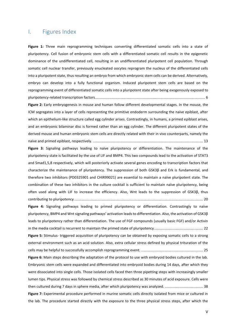
time. Cells can be reprogrammed into a pluripotent state by two main mechanisms (Figure 1): exposure of a
somatic cell’s nucleus to factors present in the oocyte by nuclear transfer (Somatic Cell Nuclear Transfer, SCNT)
and overexpression of defined pluripotency-related transcription factors (induced Pluripotent Stem Cells, iPS
cells) (Gurdon, 1962; Takahashi and Yamanaka, 2006).
Figure 1: Three main reprogramming techniques converting differentiated somatic cells into a state of pluripotency. Cell fusion
of embryonic stem cells with a differentiated somatic cell results in the epigenetic dominance of the undifferentiated cell,
resulting in an undifferentiated pluripotent cell population. Through somatic cell nuclear transfer, previously enucleated
oocytes reprogram the nucleus of the differentiated cells into a pluripotent state, thus resulting an embryo from which
embryonic stem cells can be derived. Alternatively, embryo can develop into a fully functional organism. Induced pluripotent
stem cells are based on the reprogramming event of differentiated somatic cells into a pluripotent state after being
exogenously exposed to pluripotency-related transcription factors.

7
Through the insertion of a somatic cell nucleus into an enucleated and unfertilized oocyte, SCNT experiments
have shown that the oocyte can reprogram an adult nucleus into an embryonic state that can follow the
development into a new organism. This technique opened new windows for the derivation of patient-specific ES
cells, thus demonstrating the potential of using this methodology in regenerative medicine.
Induced pluripotent stem cells can be derived directly from patient’s somatic cells. These cells can further be
differentiated into fully functional cells that overcome the possibility of immune rejection after being
transplanted. Considering patient’s somatic cells as the new source of ES cells, the ethical issues regarding the
use of embryos for therapies are surpassed.
These reprogramming techniques are described below in more detail. A third well known reprogramming
technique is based on the fusion of somatic cells with ES cells rather than empty oocytes, named cell fusion
(Cowan et al., 2005). This method demonstrated that human somatic cells (such as human fibroblast) can be
fused with human ES cells, resulting in hybrid cells that maintain a tetraploid DNA content and show morphology,
growth rate and antigen expression patterns characteristic of hES cells. This methodology is however not as
studied as SCNT and iPS cells.
1.3.1. Somatic Cell Nuclear Transfer
SCNT technique is based on the transplantation of a nucleus into an enucleated unfertilized oocyte, and
represents an experimental approach to reprogram somatic cells. This reprogramming method represents a
powerful tool to study developmental biology and may have great potential in regenerative medicine. The main
objective of SCNT is to generate uncommitted stem and progenitor cells, which are useful for medical research,
prospecting the derivation of patient-specific ES cells (Meissner and Jaenisch, 2006). Somatic cloning
demonstrates that even differentiated cells contain all the genetic information required for the development of
an entire organism and that oocytes contain factors that can reprogram these somatic cell’s nucleus (Yamanaka,
2012). The term “nuclear reprogramming” was then introduced to define the reversal of the differentiated state
of a mature cell to an undifferentiated embryonic stage (Hochedlinger and Jaenisch, 2006).
Cells of a multicellular organism are functionally different due to the different gene expression that characterizes
each lineage. This differential gene expression arises from the elimination of certain genes that are known to be
silenced and the retention of other genes that are expressed, in any particular kind of cell. However, the
accomplishment of cloning experiments, first in amphibians and later in mammals, refuted this idea
(Hochedlinger and Jaenisch, 2006). The successful reprogramming of fully differentiated cells using SCNT
demonstrated that during development, the genetic content of each cell remains, with few exceptions (antigen
or antigen receptor genes in lymphocytes), identical to that of the zygote (Meissner and Jaenisch, 2006). In
practical terms, this means that differentiated cells contain all the genetic information necessary to generate an
entire organism (nuclear totipotency). The genetic information of the donor nucleus is not lost during
development, and therefore the reprogramming event does not need to acquire genetic information. This
evidence suggests that cells’ differentiation and development are mediated by an epigenetic mechanism rather
genetic, and the epigenetic changes that direct terminal differentiation and permanent exit from the cell cycle

8
are reversible. These epigenetic modifications include mitotically stable modifications of DNA or chromatin that
do not alter the primary nucleotide sequence. Therefore, cellular reprogramming is a result of epigenetic
reprogramming following the resetting of the epigenetic modifications from a fully differentiated to a less
differentiated state (ideally, to a pluripotent embryonic state) (Hochedlinger and Jaenisch, 2006; Meissner and
Jaenisch, 2006).
The first nuclear-transfer experiments were performed using amphibian oocytes (Briggs and King, 1952; Gurdon,
1962), generating tadpoles from unfertilized eggs that received a nucleus from the intestinal cells of adult frogs.
Progress was made leading to the birth of “Dolly” the sheep, the first reported mammal to be cloned from adult
cells (Campbell et al., 1996). The combination of these accomplishments with the derivation of hES cell lines
(Thomson et al., 1998) raised the hopes to develop a technique capable of deriving ES cells from the patients and
apply them to therapeutics (Amabile and Meissner, 2009). Further studies led to the derivation of nuclear
transfer embryonic stem cells (NT-ES cells) from somatic cells in mouse models (Munsie et al., 2000), and to the
first therapeutic accomplishment, in which derived immunodeficient mouse NT-ES cells were differentiated in
vitro into hematopoietic precursors and transplanted to the donor, restoring normal lymphocyte population in
the mouse (Rideout et al., 2002). Despite numerous applications of SCNT in animal cloning, the nature of the
factors present in the oocyte, which are capable of reprogramming the somatic nuclei, remains unclear
(Tachibana et al., 2013).
The main issue leading to the failure in human NT-ES cells derivations was related with early embryonic arrest of
the embryos after cloning, prior to NT-ES cell derivation. Cells were usually unable to progress further than 8-cell
stage due to the inability to activate critical embryonic genes from the somatic donor cell nucleus (Noggle et al.,
2011). Tachibana et al demonstrated the successful reprogramming of human somatic cells into ES cells following
SCNT (Tachibana et al., 2013). Using a SCNT protocol previously optimized for nonhuman primates, and using
high quality oocytes from healthy volunteers, the formation of the blastocyst from SCNT-cloned embryo was
significantly improved. ES cell lines were derived, containing nuclear DNA exclusively from the somatic cell and
verifying pluripotency in extensive assays. In addition to demonstrate the ability to differentiate into a variety of
cell types, these human NT-ES cells were directly differentiated in vitro into contracting cardiomyocytes, thus
demonstrating their potential for regenerative medicine.
SCNT technique is technically challenging, inefficient and dependent on voluntary donation of a large number of
unfertilized oocytes (Amabile and Meissner, 2009). The extreme inefficiency in the generation of animals by
nuclear transplantation leads to the death of most clones after implantation, and the few clones that survive
beyond birth are often afflicted with severe abnormalities (Hochedlinger and Jaenisch, 2006). Some technical
issues such as enucleation, the handling, isolation and type of donor cells, as well as posterior activation and
culture conditions may be some of the possible explanations for the incapacity of the reprogrammed nucleus to
support development into a new functional organism. Despite showing potential in several applications from
disease studies to regenerative therapies, some ethical issues are still present. If the intent is to derive a cell line
from a cloned embryo, the blastocyst generated has to be necessarily destroyed, thus destroying a possible
source of human life and raising really severe ethical issues (Meissner and Jaenisch, 2006).

9
1.3.2. Induced Pluripotent Stem Cells
Considering the hypothesis that some factors, that play important roles in the maintenance of pluripotency, can
confer totipotency or pluripotency to somatic cells, the addition of only a few defined factors has demonstrated
it possible to induce pluripotent stem cells from both mouse embryonic or adult fibroblasts (Takahashi and
Yamanaka, 2006) and adult human fibroblasts (Takahashi et al., 2007), under ES cell culture conditions. This
reprogramming approach presents a way to prevent both ethical issues, regarding the use of human embryos
for research, and the rejection in transplantation procedures, due to the possibility to grow patient-specific
pluripotent stem cells directly from the patient’s somatic cells. Furthermore, the successful reprogramming of
differentiated human somatic cells into a pluripotent state allows for the creation of disease-specific stem cells.
The most valuable advantage of iPS cells technology is its simplicity, since they can be easily generated in any
laboratory (Yamanaka, 2009).
In mouse studies, 24 candidate factors were selected in a first instance, from which four were elected (Oct3/4,
Sox2, c-Myc and Klf4). Somatic cells were transduced with retrovirus carrying these four pluripotency
transcription factors’ transgenes, which were integrated into the genome, leading to iPS cells generation. These
cells showed endogenous expression of several ES cell marker genes, although at different levels as compared to
mouse ES cells, showing that iPS cells were similar, but not identical, to ES cells. Both in vitro and in vivo assays
proved the ability of these factors-induced stem cells to give rise to cells from the three germ layers,
demonstrating that these cells exhibit pluripotency, although in initial works they could not remain
undifferentiated when cultured in the absence of feeder cells, even if leukemia inhibitory factor (LIF) was present
(Takahashi and Yamanaka, 2006). Afterwards, blastocyst injection confirmed the contribution of these cells to all
the three germ layers of the embryos obtained, the ability to give rise to adult chimeras and competence for
germline transmission (Okita et al., 2007; Takahashi and Yamanaka, 2006; Wernig et al., 2007). Mouse induced
pluripotent stem (miPS) cells were, therefore, indistinguishable from mouse ES cells both in morphology,
proliferation, gene expression (at different levels) and differentiation capacity.
Despite the differences in terms of maintenance of pluripotency in the human and mouse systems (see chapter
1.5), the same four factors were able to generate iPS cells from adult human dermal fibroblasts, by retroviral
transfection of these transgene factors. These hiPS cells were similar to human ES cells, both in morphology and
gene expression. Proliferation capacity and feeder dependence were also similar to the in vivo counterparts, as
well as telomerase activity and histone modification. In vitro assays confirmed the ability of these cells to
differentiate into cells representative of the three germ layers. Additionally, inter-species in vivo assays,
transplanting human iPS cells into mice, showed that these cells successfully formed teratomas. The generation
of iPS cells from other human somatic cells attested the ability of this four factors combination to induce
pluripotency in human cells. The retrovirus was proven to be strongly silenced in hiPS cells, thus demonstrating
that these cells were efficiently reprogrammed from somatic cells, and did not depend on the expression of the
transgenes for self-renewal and proliferation. However, similarly to the mouse experiments that resulted in miPS
cells, only a small number of human fibroblasts, transduced with the factors, acquired pluripotency. The low

10
reprogramming efficiency is an inherent factor in the most part of the reported reprogramming events
(Takahashi et al., 2007).
Induced pluripotent stem cells have been obtained from other species including monkey (Liu et al., 2008) and rat
(Liao et al., 2009). In addition to fibroblasts, miPS cells have been generated from other tissue sources, such as
neural stem cells (Kim et al., 2008), bone marrow cells (Takahashi and Yamanaka, 2006) and B lymphocytes
(Hanna et al., 2008). On the other hand, hiPS cells have been generated from skin fibroblasts, keratinocytes
(Aasen et al., 2008), and blood cells (Loh et al., 2009; Tan et al., 2014), among others. It is however important
not to forget that iPS cells generated from different origins may have different propensities to differentiate,
compromising the clinical application of these cells (Yamanaka, 2009). This reprogramming technique frequently
results in low reprogramming efficiencies from 0.1% to 10%, although a recent study reported efficiencies of
almost 100% within a week (Rais et al., 2013). It is however important to note that differences in the quality of
iPS cell clones seem to be largely due to technical variables, such as factor combinations, gene delivery methods,
and culture conditions. Moreover, some variation can be attributed to stochastic events during reprogramming,
which cannot be controlled (Yamanaka, 2012). Importantly, iPS cells have been efficiently differentiated into
various functional cell types, including neurons, cardiomyocytes and hematopoietic cells (Dimos et al., 2008;
Hanna et al., 2007; Wernig et al., 2008), showing important contribution to drug screening and disease modelling
through the generation of disease-specific iPS cells. Contrastingly to SCNT, direct reprogramming through iPS
cells provides a more realistic way to generate sufficient amounts of patient-specific pluripotent stem cells
(Amabile and Meissner, 2009).
A common obstacle regarding both ES cells and iPS cell technology, which still impairs its affirmation in the
regenerative medicine field, is the possible formation of teratomas after a transplantation procedure. Following
an iPS cells differentiation assay to create functional cells to be engrafted into a living body, it is important to
understand that even a small amount of cells that remain undifferentiated within the grafted cells is sufficient to
form a teratoma in vivo after transplantation. There is always the possibility of having cells residing in different
reprogramming stages within an iPS cells’ colony. Therefore, cells that undergo incomplete nuclear
reprogramming are not truly pluripotent, which can interfere with the following differentiation procedure,
impairing differentiation ability and increasing the probability of post-transplantation teratoma formation in vivo
(Yamanaka, 2009).
The main obstacle found in the first studies with iPS cells was the presence of transgenes. The first iPS cells were
generated following transduction of somatic cells with retroviruses or lentiviruses carrying four pluripotency
transcription factors’ transgenes, which were integrated in the cell genome. Despite being commonly silenced in
iPS cells, the reactivation of these transgenes can lead to tumor formation, especially the transgene that encodes
c-Myc, resulting in tumor formation in approximately 50% of the chimeric mice generated from iPS cells (Okita
et al., 2007). However, iPS cells were shown to be generated without viral integration through the use of plasmids
(Okita et al., 2008), although the efficiency of iPS cell generation was even lower than using retrovirus. As an
alternative, iPS cells can be generated using chemicals or small molecules that can replace the reprogramming
factors commonly used (Shi et al., 2008). It is however important to bear in mind that iPS cells can integrate small
plasmid fragments or have chemically induced mutations due to the use of small molecules, leading to genetic

11
alterations that can give rise to diseases like cancer (Yamanaka, 2009). To compensate the drawbacks of
transducing cells with viruses, iPS cells can be generated with less factors comparing to the traditional method,
being especially useful if c-Myc can be avoided. Mouse iPS cells were even shown to be generated from adult
mouse neural stem cells using Oct3/4 alone (Kim et al., 2009). Studies aimed at the generation of iPS cells using
fewer transcription factors are of upmost importance, since iPS cells could be safer. However, the use of less
factors may suggest incomplete or inefficient reprogramming (Yamanaka, 2009).
1.3.2.1. Granulosa Cells
It has been shown that stemness (the possession of stem cell characteristics) in somatic cells facilitates
reprogramming, as demonstrated by a more efficient reprogramming of progenitor stem cells to iPS cells (Eminli
et al., 2009). The requirement for fewer factors for iPS cells generation is evident, since the integration of some
factors (such as c-Myc) into the genome may negatively affect the potential of these cells for cell replacement
therapies. The endogenous high expression levels of some pluripotent factors can reduce the number of factors
needed to reprogram cells (Eminli et al., 2008). The nuclear lamin LaminA is highly expressed in differentiated
cells. Granulosa cells and cumulus cells (hereon called GCs) are present in the ovarian follicles. Human GCs are
commonly byproducts from IVF clinic and also from IVF and embryo biotechnology. Mouse GCs were the first to
effectively generate cloned mice by SCNT. These cells are characterized by the absence of LaminA expression
and high endogenous expression of Klf4 and c-Myc (when compared to other somatic cell types such as
fibroblasts), which means these cells exhibit a certain degree of stemness.
Therefore, the question of whether these cells could be more easily reprogrammed to a pluripotent state using
fewer factors, in comparison with a fully differentiated and functional somatic cells, was addressed (Mao et al.,
2014). GCs were shown to be efficiently reprogrammed into iPS cells using only two of the Yamanaka factors
(Takahashi and Yamanaka, 2006), Oct4 and Sox2, thus facilitating the reprogramming process. Since Klf4 and c-
Myc are both carcinogenic, their removal would improve the safety of iPS cells when used for clinic. Both in vitro
and in vivo assays confirmed the acquisition of pluripotency, demonstrated by high expression of pluripotency
genes Oct4, Nanog and SSEA-1, differentiation into cells representing the three germ layers by EBs’ formation (in
vitro test) and teratoma formation (in vivo) and efficient generation of chimeras following blastocyst injection of
the generated iPS cells. Following these results, it was stated that the reduced expression of LaminA can improve
the reprogramming efficiency into a pluripotent state (Mao et al., 2014).
1.3.2.1.1. Lamin A
The nuclear lamina is the major structural component of the nucleus, with a scaffold-like structure that lines the
inner cell membrane, and is composed by type V intermediate filament proteins, named nuclear lamins. These
components of the nuclear envelope interact with the chromatin, nuclear complexes and lamin-associated
proteins, having fundamental functions in nuclear structural support and organization, DNA replication,
transcription and DNA processing (Aebi et al., 1986; Constantinescu et al., 2006; Eckersley-Maslin et al., 2013).

12
Seven lamin proteins have been identified in mammals, and can be divided into two subtypes, A- and B-type
lamins. B-type lamins include lamin B1, encoded by the gene LMNB1, and lamins B2 and B3, encoded by the gene
LMNB2. These lamins are present in every embryonic and adult cell types, and B1 and B2 are essential for cell
growth and survival. A-type lamins include lamins A, AΔ10, C and C2, all encoded by the LMNA gene. Unlike B-
type lamins, A-type lamins are primarily found in differentiated cells, demonstrated by the lack of expression for
most mouse embryonic development, in various adult cells that are not fully differentiated, and in ES cell lines
(Constantinescu et al., 2006; Röber et al., 1989). Lamin A is one of the major nuclear architectural lamins
responsible for the maintenance of the inner structure integrity and function of the nuclear membrane, and its
dysfunction triggers DNA damage response, cellular senescence or apoptosis (Eckersley-Maslin et al., 2013). The
observation that all the cell types contain B-type but no A-type lamins, the latter ones primarily present in
differentiated cells and absent in undifferentiated cells, suggests that A-type lamins could either maintain the
differentiated state or be a marker of pluripotency. Since ES cells are isolated from blastocyst-stage embryos and
are pluripotent, it was hypothesized that these undifferentiated ES cells should express B-type but not A-type
lamins and that, upon differentiation, lamin A/C expression should be observed. Consistent with this idea,
previous reports have demonstrated the absence of lamin A/C in both human and mouse ES cells, and its
activation upon differentiation, being lamin A/C expressed only in differentiated cell types. Pluripotent ES cells
maintain their self-renewal and undifferentiated capacity, without expressing Lamin A, since this protein is
expressed at a minimal or undetectable level, whereas somatic cells show strong expression of Lamin A in the
nuclear envelope, turning Lamin A into a novel marker for undifferentiated ES cells (Constantinescu et al., 2006;
Röber et al., 1989). While stemness facilitates reprogramming, cell senescence resulting from the absence of
Lamin A impairs reprogramming to pluripotency in iPS induction experiments. Aging somatic cells express higher
levels of Lamin A than younger cells, making the misregulation of Lamin A of adult stem cells associated with
accelerated ageing. Experiments conducting iPS induction showed that cells with high expression levels of Lamin
A have slower and less efficient induction into the pluripotent state, speculating that it might negatively affect
the expression of pluripotency genes. To support this statement, it was proven that reduced levels of Lamin A by
short hairpin RNA accelerate the iPS colony formation, increasing both the reprogramming efficiency and the
endogenous expression of pluripotency genes. Thus, the overexpression of Lamin A reduces reprogramming
efficiency, forming iPS clones at lower pace and quality. Besides leading to telomere shortening in differentiated
somatic cells, low Lamin A level in pluripotent stem cells facilitates telomere elongation, being essential for the
maintenance of pluripotency in iPS or ES cells (Zuo et al., 2012). Although reported to be completely absent in
ES cells, a more recent study has shown that ES cells express lamin A/C and that its absence is not a marker of
the undifferentiated state. ES cells have lower levels of lamin A/C when compared to somatic cells, such as MEFs,
which may be an explanation to why previous reports have failed to detect lamin A/C in ESCs. Despite recognized
by a very faint staining, lamin A/C is expressed at both the RNA and protein levels in multiple primary and well-
established ES cell lines. This lamin was also detected in both Nanog positive and Nanog negative cells in the
developing blastocyst. Thus, the lamin A/C expression is not acquired upon ES cell derivation, or during cell
culture, since ES cells are derived from the ICM of the blastocyst, composed by Nanog positive cells. The dynamic
nuclear structure of ES cells represents a link between nuclear structure and pluripotency, which is therefore not

13
due to the absence of lamin A. Thus, it remains to be determined how the highly irregular nuclear structure of
ES cells is maintained in the presence of low levels of lamin A/C, and how this relates to the pluripotent nature
of these cells (Eckersley-Maslin et al., 2013).
1.4. Mouse and Human Embryonic Stem cells: Different States of pluripotency
1.4.1. Embryogenesis
In order to fully understand the origin of ES cells and their characteristics, a basic understanding of early
embryogenesis is fundamental. Although the information about human early embryogenesis remains largely
limited as compared to mice, findings suggest that there are notable differences between the early
developments of these two species (Figure 2). Following fertilization, the embryo progresses through several
developmental stages until reaching the blastocyst stage. Upon blastocyst formation, at embryonic day (E) 3.5 in
the mouse and E 4-5 in human, two distinct lineages are formed (Niakan and Eggan, 2013): the ICM (can give rise
to the whole embryo) and the trophectoderm (extra-embryonic lineage that gives rise to the placenta). Extra-
embryonic lineages have a primary role of mediating uterine implantation, as well as posterior maternal
sustenance of the growing embryo and subsequent fetus (Gardner, 1983; Selwood and Johnson, 2006). Extra-
embryonic tissues also supply inductive signals to the early epiblast, specifying early embryo development. These
signals will influence the development of the early epiblast (Nichols and Smith, 2009).
Figure 2: Early embryogenesis in mouse and human follow different developmental stages. In the mouse, the ICM segregates
into a layer of cells representing the primitive endoderm surrounding the naïve epiblast, after which an epithelium-like
structure called egg cylinder arises. Contrastingly, in humans, a primed epiblast arises, and an embryonic bilaminar disc is
formed rather than an egg cylinder. The different pluripotent states of the derived mouse and human embryonic stem cells
are directly related with their in vivo counterparts, namely the naïve and primed epiblast, respectively.

14
The ICM is later divided, forming an outer layer of cells named primitive endoderm (PE) surrounding the
remaining interior cells that form the pluripotent epiblast (from which ES cells can be derived). At the time of
implantation, around E 4.5 in the mouse, the murine epiblast cells proliferate and form a ball of cells. Epiblast
internal cells undergo apoptosis, leading the epiblast to form a cup-shaped epithelium surrounded by PE. The
resulting structure is named egg cylinder. Contrastingly, the segregation of the human epiblast and PE from the
ICM leads to the formation of the bilaminar embryonic disc (Hassani et al., 2014a).
Commonly, ES cells can be derived from the ICM, more specifically from the pluripotent epiblast. Important to
note, the epiblast undergoes a developmental change upon implantation, from a pre- to a post-implantation
epiblast. As discussed next, mouse and human ES cells show different states of pluripotency (see chapter 1.4.2),
in which mES cells resemble the mouse pre-implantation stage epiblast cells and hES cells resemble the mouse
post-implantation stage epiblast cells (although derived from a pre-implantation stage human epiblast). The
differences between the early embryogenesis of the two species are directly related with the differences
observed in ES cells derived from them. The generation of the epiblast several days after fertilization is a process
unique to the mammalian life cycle (Nichols and Smith, 2009).
1.4.2. Naïve and primed pluripotency
In 2009, Jennifer Nichols and Austin Smith proposed that two different states of pluripotency could be defined:
naïve and primed. These two pluripotency states show several differences both in gene expression and
morphology, resembling different embryonic stages of the in vivo counterparts, thus resulting in different
pluripotent potentials. Mouse epiblast stem cells (EpiSCs) were derived, which share several characteristics with
hES cells.
1.4.2.1. Mouse embryonic stem cells reside in a naïve state of pluripotency: the Ground State of
Pluripotency
Before defining the naïve and primed states of pluripotency, it is important to understand the definition of the
ground state of pluripotency. The mammalian egg theoretically gives rise to the entire embryo, thus being
described as totipotent, the highest level of potency possible for a cell. However, in practical terms, the egg only
gives rise directly to two cell lineages: the trophoblast and the ICM (see chapter 1.4.1). In order to progress with
the development, the ICM produces another extra-embryonic lineage, the PE, leaving the remaining cells
together in a cluster that develops into a pluripotent epiblast, which must be able to give rise to the whole
organism. Thus, it was suggested that the development from the primitive post-fertilization embryo stage,
considered totipotent, to the pre-implantation blastocyst stage, that includes a pluripotent epiblast, represents
an increase rather than decrease of potency. In other words, the development from egg to epiblast is a transition
from a determined to an uncommitted state. The pre-implantation pluripotent epiblast was defined as the
developmental ground state, since it represents a fully unrestricted population of cells with the developmental
potency and flexibility to produce all the embryonic lineages (Nichols and Smith, 2009).

15
1.4.2.1.1. Derivation and maintenance of naïve mES cells
Mouse ES cells were derived from the cells that constitute the pluripotent epiblast (Evans and Kaufman, 1981),
thus representing immortalization of this naïve epiblast. These cells exhibited unlimited self-renewal capacity
when maintained in appropriate culture conditions, maintaining the epigenetic identity and the potency of the
epiblast from which they were derived. Additionally, these cells derived from the mouse blastocyst efficiently
contributed to chimeric embryos, were relatively refractory in their potential to differentiate into primordial
germ cells (PGCs) in vitro, and shared an epigenetic characteristic with the pre-implantation epiblast, the
presence of two active X chromosomes (XaXa) in female cells. The X chromosome inactivation, along with all the
characteristics described, presents a shared characteristic with the naïve pre-implantation epiblast that was
defined as the ground state of pluripotency, thus defining mES cells as naïve pluripotent stem cells. Naïve mES
cells can be cloned with high efficiency and grow as packed colonies (Hanna et al., 2010; Nichols and Smith,
2009). These cells can be maintained in vitro in the naïve ICM-like configuration by providing exogenous
stimulation with LIF and small molecule inhibition of the extracellular signal-regulated kinase (ERK1/2) and
glycogen synthase kinase 3β (GSK3β) signaling pathways, termed 2i/LIF conditions (see chapter 1.5.1). The
inhibition of those two pathways is accomplished by the use of the inhibitors PD0325901 and CHIR99021
(CHIRON) (Gafni et al., 2013; Hanna et al., 2010; Ying et al., 2008).
1.4.2.2. Epiblast stem cells as a result of derivation from the post-implantation epiblast
After implantation, the murine epiblast adopts a cup-shaped structure known as the egg cylinder. These post-
implantation epiblast cells can no longer contribute to chimeras, nor can give rise to ES cells with the same
characteristics as the previously defined naïve mES cells (Nichols and Smith, 2009). Advances led to the derivation
of cell lines, defined as EpiSCs, from this post-implantation mouse epiblast, using culture conditions without LIF
but including fibroblast growth factor (FGF) and Activin (Brons et al., 2007). Despite expressing some pluripotency
factors, such as Oct4, Sox2, and Nanog, these cells differ from the previously defined naïve mES cells (in vitro
counterpart of the pre-implantation epiblast). Although capable of forming teratomas, further studies have
shown that these cells are highly inefficient in repopulating the ICM upon aggregation or injection into host
blastocyst, leading to the incapacity to form chimeras, placing these cells in a distinct developmental and
functional state from the naïve mES cells (see Table 1). EpiSCs, now described as primed pluripotent cells,
undergo X-chromosome inactivation (XaXi) in female cells and are able to differentiate into PGC precursors in
vitro (Brons et al., 2007; Hayashi and Surani, 2009; Tesar et al., 2007).
Embryonic cells continue their developmental progress when they are put in culture, not preserving the exact
developmental stage at which they are isolated. Thus, even when cells are derived from pre-epiblast early stage
embryos, they may progress to the epiblast stage in culture. This idea is consistent with the fact that all the mES
cell lines are molecularly and phenotypically similar, however they were derived, and may explain why ES cells
have even been obtained from mouse embryos that were put in culture prior to epiblast formation (Nichols and
Smith, 2009). Therefore, representing a more advanced developmental stage of embryo development, EpiSCs

16
can be produced in culture from previously cultured mES cells, in response to growth factor cues. Naïve mES cells
grown and maintained in 2i/LIF conditions (see chapter 1.5.1) can drift towards a primed state of pluripotency
resembling EpiSCs upon withdrawal of the inhibitors. One copy of the X chromosome in female cells is
epigenetically silenced when naïve mES cells are converted to EpiSCs, thus confirming their deviation from the
naïve state of pluripotency (Brons et al., 2007; Tesar et al., 2007). These EpiSCs can however be converted back
to the naïve state upon induction of the LIF/Stat3 (Signal transducer and activator of transcription 3) signaling
pathway. This conversion can be further enhanced with the expression of factors including Klf4, Klf2, Nanog or
c-Myc. The resulting cells exhibit all the characteristics present in naïve mES cells (Bao et al., 2009; Guo et al.,
2009; Hanna et al., 2010), thus demonstrating the capacity to achieve inter-conversion between these two states
of pluripotency. As a result of all these observations, EpiSCs were described as stem cells residing in a primed
state of pluripotency.
1.4.2.3. Human embryonic stem cells reside in a primed state that resembles EpiSCs
Pluripotent cell lines can be derived from human blastocysts (Thomson et al., 1998). Culture studies have
demonstrated that these lines differ significantly from mES cells both in culture requirements, morphology,
clonogenicity, differentiation, behavior, and molecular profile (see Table 1). Chimera generation tests cannot be
applied with human cells, which implies that functional tests of hES cells are based on teratoma formation in
murine specimen after injection. The capacity of teratoma formation is a characteristic of both naïve mES cells
and primed EpiSCs. Being capable of teratoma formation, and yet being different from mES cells in so many
characteristics, hES cells were defined as analogous to the EpiSCs isolated from rodents. Looking back, mES cells
are derived from the pre-implantation epiblast, whereas EpiSCs are derived from post-implantation epiblast.
However, ES cells can continue to progress after being put in culture. This phenomenon may explain why hES
cells, although derived from cultured blastocysts, can progress into a post-implantation epiblast-like state,
making them the human counterpart of the primed state EpiSCs. Primed EpiSCs and hES cells are characterized
by a flattened morphology, incapacity to passaging as single cells, X chromosome inactivation in female cell lines
and dependence on basic FGF (bFGF) and transforming growth factor β (TGFβ)/Activin signaling pathways rather
than LIF/Stat3. (Hanna et al., 2010; Nichols and Smith, 2009).
These observations made scientists wonder why primate embryos continue to progress leading ES cells to adopt
a primed state while rodent ES cells prevail in a naïve state. In non-rodent mammalian species, after formation
of the blastocyst, the embryo does not form an egg cylinder. Instead, the epiblast develops into a simple flattened
structure called embryonic bilaminar disc. In rodent species the formation of the egg cylinder is characterized by
reorganization of the epiblast from a ball of cells into a cup-shaped epiblast surrounded by the PE. Associated to
this process is the apoptosis of the internal cells of the epiblast. It was suggested that these events may present
a hurdle to progression out of the naïve pluripotency in rodent embryos, facilitating the derivation of naïve mES
cells. In contrast, in non-rodent embryos there may be no barriers inhibiting epiblast cells to progress from a
naïve to a primed state, hampering the capture of the transient naïve state of the epiblast cells. Also, diapause
may represent another drawback to the derivation of true primate ES cells. This phenomenon is defined as a

17
state of arrested embryonic development that occurs naturally in rodents, in which embryos arrest
synchronously at the late blastocyst stage right after the segregation of the epiblast and PE. During diapause,
despite cell division being highly reduced, some cell turnover does occur, replacing old cells with new ones. Thus,
the naïve epiblast seems to have an intrinsic facility for self-renewal in these species during diapause, providing
the biological foundation for the ex vivo propagation of the ground state epiblast. The absence of diapause may
present a reason for the difficult derivation of naïve ES cells in humans (Nichols and Smith, 2009).
Table 1: Naïve and Primed pluripotency states show different potentials and morphology. Naive stem cells are the in vitro
counterpart of the cells present in the mouse early epiblast, whereas primed stem cells, human embryonic stem cells and
mouse epiblast stem cells, correspond to the in vivo human embryonic bilaminar disc and mouse egg cylinder cells,
respectively. Both are capable of teratoma formation, although this test, when performed with human cells, relies in an inter-
species teratoma formation, following injection into mice. However, only naïve pluripotent cells are capable of chimera
formation and single cell passaging. Epigenetic changes are visible, specially characterized by the activation of the two X
chromosomes in naïve female cells, whereas one of the X chromosomes is inactivated in the primed ones. Moreover,
morphology analysis identifies naïve colonies with a domed shape, while primed colonies maintain a flat appearance.
Naive Primed
Embryonic tissue associated Early epiblast Egg cylinder or embryonic disc
Stem cells cultured Rodent ES cells Rodent EpiSCs, primate ES cells
Chimaeras formation Yes No (not applicable to primates)
Teratoma formation Yes Yes
Colony morphology Domed Flat
Single-cell clonogenicity Yes No
X chromosome status (female) XaXa XaXi
A question that still prevails is whether ES cells, with the epigenetic characteristics that they present, are a result
of the adaptation to the ex vivo medium, or are captured directly in this state from the epiblast. Previous studies
suggested that only a subset of cells present in the epiblast is truly pluripotent and is in the origin of ES cells,
since the plating of dissociated epiblast cells resulted in a few number of established ES cell colonies (Nichols and
Smith, 2009).
1.4.2.4. Naïve and primed pluripotent cells cultures and the break of species barrier
Some culture media, when supplemented with small molecules or growth factors, inhibit differentiation and/or
reinforce some key pathways that stabilize the core transcriptional circuity of naïve pluripotency. This
manipulation includes the inhibition of GSK3β and ERK1/2 in addition to LIF/Stat3 stimulation or exogenous
expression of Klf4 or c-Myc. Using this culture medium, it was possible to derive naïve pluripotent stem cells both
from non-obese diabetic (NOD) mouse strains and from rats, species previously considered “non-permissive” for
ES cell derivation. Withdrawal of the exogenous supporting factors and addition of bFGF/Activin, known to
maintain primed state of pluripotency, leads NOD naïve pluripotent cells to a primed state of pluripotency,

18
whereas reintroduction of these exogenous factors reprograms NOD cells to the naïve state again (Buehr et al.,
2008; Hanna et al., 2010; Hanna et al., 2009). The possible conversion between the naïve and primed states in
NOD cells, allied with the similarities between hES cells and EpiSCs, led to the supposition that the verified primed
state of hES cells was a matter of instability of the naïve state in the conventional culture conditions commonly
used (Hanna et al., 2010). Appropriate culture conditions and exposure to the right factors would be the key to
the successful establishment of naïve hES cells.
A first study indicated that human cells may require additional exogenous factors, in comparison to NOD cells,
to stabilize naïve pluripotent cells in vitro. Previously well-defined conditions, such as 2i/LIF, were not sufficient
to prolong self-renewal in hES cells/hiPS cells and led to their differentiation. The adaptation of 2i/LIF conditions
with additional exogenous factors could allow in vitro stabilization of transgene-independent and continuously
stable human naïve pluripotent stem cells (Hanna et al., 2010). Although it was difficult to obtain a stabilized
naïve pluripotent human cell line, human ES cells with biological and epigenetic characteristics similar to those
of naïve mES cells were established. Naïve hES cells/hiPS cells were distinct from conventional hES cells/hiPS
cells, closely resembling naïve mES cells in terms of molecular and biological criteria including growth properties,
signaling pathways dependency, state of X-chromosome inactivation and transcriptional characteristics. These
results supported the idea that distinct states of pluripotency can be established according to different culture
conditions, and the requirement for exogenous factors, as well as their appropriate concentrations, differ within
species. Human cells are the least permissive to maintain a naïve state, being obtained in low frequencies and
requiring the manipulation of several signaling pathways to obtain epigenetic reversion of the common hES cells
(Hanna et al., 2010). Further studies were necessary to establish truly stabilized cultures of naïve cells from hES
cells for undefined number of passages without genetic manipulation.
1.4.2.5. Derivation of genetically unmodified human naïve pluripotent stem cells
In 2013, Gafni et al established defined condition that facilitated the derivation of genetically unmodified human
naïve pluripotent stem cells from already established primed hES cells, from hiPS cells and directly from
blastocysts. These novel naïve hES cells were distinct from previously defined primed hES cells and retained
molecular characteristics and functional properties highly similar to those of mES cells, including the possibility
to generate cross-species chimeric mouse following injection of naïve hES cells into mouse morulas. This study
resulted in the establishment of optimized chemically defined conditions, defined as NHSM (naïve human stem
cell medium), that enabled the expansion of karyotypically normal OCT4-GFP+ human iPS cells for more than 50
passages, independent of exogenous gene activation. This accomplishment demonstrated that the maintenance
of human naïve pluripotent stem cells required a unique combination of cytokines and small molecule inhibitors.
Cells grown in these conditions developed into dome-shaped colonies resembling naïve ES cells, thus introducing
the term naïve human pluripotent stem cells (include both hES cells and hiPS cells). NHSM confers to human
pluripotent cells defining features of gene expression patterns observed in the ground state naïve mES cells
(Gafni et al., 2013).

19
1.5. Signaling Pathways: The roadmap to Embryonic Stem cell maintenance
To fully understand the effect of the main components used in the culture media defined to maintain
pluripotency, both in mouse and human ES cells, the analysis of the pathways contributing to naïve and primed
pluripotency is necessary (Figure 3 and Figure 4). A typical culture medium known to maintain self-renewal and
pluripotency, in addition to contain a base medium with the proper nutrients that support cell survival, contains
inhibitors that block the pathways leading to differentiation, and factors that promote the pathways leading to
pluripotency. It is crucial to understand which pathways lead to pluripotency and/or to differentiation. The
pathways involved in the maintenance of the two different states of pluripotency are different, thus leading to
the use of different culture conditions. As a result of this analysis, several pathways mainly important to the
maintenance of the naïve and primed states can be identified (see Table 2).
1.5.1. LIF/Stat3 Signaling – Establishment of 2i and 3i conditions
Following the establishment of the first mES cell lines, using MEFs as feeder layer, it was discovered that the
secretome of these feeder cells contained the cytokine LIF, active in the maintenance of the pluripotency of these
mES cells. Thus, it was suggested that LIF could be used to substitute the necessity of a feeder layer in naïve mES
cell culture (Smith et al., 1988; Williams et al., 1988). The binding of LIF to its receptor can trigger three
intracellular signaling pathways: the Janus kinase (Jak)/Stat3 pathway, the phosphoinositide 3-kinase
(PI3K)/protein kinase Akt pathway, and the mitogen-activated protein kinase (MAPK)/ERK pathway (Niwa et al.,
2009).
1.5.1.1. Jak/Stat3 Signaling Pathway
The Jak/Stat3 signaling pathway was the first pluripotency-related signaling pathway to be defined in naïve mES
cells, and its activation is responsible for the maintenance of mES cells self-renewal and undifferentiated state
(Hirai et al., 2011; Matsuda et al., 1999). The main role of the Jak/Stat3 pathway is the activation of transcription
factors c-Myc and Klf4 in mES cells (Cartwright et al., 2005; Hassani et al., 2014a), two factors involved in the
induction of pluripotency in somatic cells (see chapter 1.3.2). Moreover, further studies have shown that some
target genes of this signaling pathway have distinct roles in the inhibition of mesoderm and endoderm
differentiation (Bourillot et al., 2009), thus contributing for the maintenance of pluripotency.
1.5.1.2. PI3K/Akt and MAPK/ERK Signaling Pathways
The cytokine LIF also activates PI3K/Akt and MAPK/ERK signaling pathways in naïve mES cells. The activation of
the PI3K/Akt signaling is also responsible for the maintenance of pluripotency, in this case both in hES cells and
mES cells (Hassani et al., 2014a), thus contributing both for naïve and primed pluripotency. In the naïve mES cells,

20
the activation of this pathway leads to the inactivation of GSK3β, leading to the increase of Nanog and c-Myc
expression levels, genes of two transcription factors of pluripotency (Bechard and Dalton, 2009). Contrastingly,
the activation of the MAPK/ERK signaling pathway is responsible for mES cells differentiation (Nichols et al.,
2009), and therefore its suppression promotes naïve pluripotency. According to these observations, LIF can
either sustain pluripotency or induce lineage differentiation in naïve mES cells.
Figure 3: Signaling pathways leading to naïve pluripotency or differentiation. The maintenance of the pluripotency state is
facilitated by the use of LIF and BMP4. This two compounds lead to the activation of STAT3 and Smad1,5,8 respectively, which
will posteriorly activate several genes encoding to transcription factors that characterize the maintenance of pluripotency.
The suppression of both GSK3β and Erk is fundamental, and therefore two inhibitors (PD0325901 and CHIR99021) are essential
to maintain a naïve pluripotent state. The combination of these two inhibitors in the culture cocktail is sufficient to maintain
naïve pluripotency, being often used along with LIF to increase the efficiency. Also, Wnt leads to the suppression of GSK3β,
thus contributing to pluripotency.
1.5.1.3. The Methodology of 2i and 3i conditions
The combination of LIF with the bone morphogenetic protein 4 (BMP4, the main component of serum used to
sustain pluripotency in mES cells) was shown to be dispensable and easily substituted by chemical compounds.
As described (Ying et al., 2008), the ground state of pluripotency can be maintained using small-molecule
inhibitors that directly suppress signaling pathways that lead to differentiation. Naïve mES cells can maintain
continuous self-renewal and a state of pluripotency completely indistinguishable from the wild-type mES cells
using 3i conditions. In this culture conditions, 3i stands for the use of three inhibitors: SU5402, which suppresses
the FGF receptor, FGFR; PD184352, which suppresses MAPK kinase, Mek; and CHIR99021, which suppresses
GSK3β. Also, a combination of just two inhibitors (2i), including the most potent Mek kinase PD0325901 along
with CHIR99021 can completely substitute the use of conventional LIF and serum/BMP4-containing medium. The
use of this combination of factors turned possible the derivation and maintenance for long-term of mES cells
from different mouse strains, including rats and NOD mice, previously defined non-permissive to pluripotency
21
(Hanna et al., 2009). Although being sufficient alone, 2i conditions are often used in combination with LIF in order
to increase derivation and maintenance efficiency (Hassani et al., 2014a) .
1.5.2. BMP4/Smad1,5,8 Signaling – Establishment of serum-free conditions
BMP4 leads to the activation, by phosphorylation, of Smad1,5,8 proteins, and the activation of the
BMP4/Smad1,5,8 signaling pathway proscribes mES cell differentiation, sustaining pluripotency (Ying et al.,
2003). It was suggested that BMP4 sustains pluripotency in mES cells by preventing entry into the late epiblast
stage by suppressing ERK signaling (Zhang et al., 2010). Moreover, activated Smad1,5 up-regulates ERK-specific
dual specificity phosphatase 9 (DUSP9) that inhibits ERK (Li et al., 2012), promoting naïve pluripotency. DUSP9
establishes the link between BMP4 and the ERK signaling, thus proving that even BMP4 can be dispensable upon
introduction of this molecule.
Contrastingly to naïve mES cells, in primed EpiSCs and hES cells, BMP4 induces differentiation and represses self-
renewal. BMP4 plays a role in the differentiation of primed ES cells into several different cell types, including
extra-embryonic lineages (Hassani et al., 2014a). The treatment of hES cells with BMP4 can cause differentiation
into extra-embryonic endoderm and trophectoderm, and the same treatment, conjugated with FGF2 or FGF2
and Activin, can cause differentiation into mesoderm or mesendoderm, respectively (Bernardo et al., 2011;
Vallier et al., 2009; Zhang et al., 2008).
1.5.2.1. The establishment of serum-free conditions
The derivation of the first mES cell line was accomplished using fetal calf serum (FCS, currently termed fetal
bovine serum, FBS) (Evans and Kaufman, 1981; Martin, 1981). However, both serum-free and feeder-free culture
conditions contribute to maintain the homogeneity and safety of cultured ES cells, especial if they are intended
to be used for medical applications. The main factor that sustains pluripotency in serum, BMP4, was shown to
be a substitute to the previously used FCS (Ying et al., 2003). Combined with LIF, the use of BMP4 allows the
establishment of feeder-free and serum-free culture conditions sustaining naïve mES cells, although it can be
completely substituted by 2i conditions, as described.
1.5.3. TGFβ, Activin and Nodal/Smad2,3 Signaling
Several studies have identified TGFβ as an active signaling pathway in both naïve and primed ES cells with
different roles, known to be involved in the specification of some differentiated lineages in the early embryo
(Akhurst et al., 1990; Hassani et al., 2014a; Zhou et al., 1993).
The suppression of TGFβ by SB431542, along with Mek inhibitor PD0325901, allowed efficient reproducible
generation of mES cells from different strains (Hassani et al., 2012), supporting the aspects of ground state
pluripotency (Hassani et al., 2014b). Thus, suppression of TGFβ appears to play a role in the maintenance of naïve

22
pluripotency. TGFβ inhibition was suggested to indirectly augment BMP4 signaling, promoting self-renewal of
mES cells (Galvin et al., 2010).
Figure 4: Signaling pathways leading to primed pluripotency or differentiation. Contrastingly to naïve pluripotency, BMP4 and
Wnt signaling pathways’ activation leads to differentiation. Also, the activation of GSK3β leads to pluripotency rather than
differentiation. The use of FGF compounds (usually basic FGF) and/or Activin in the media cocktail is recurrent to maintain the
primed state of pluripotency.
TGFβ signaling was identified in the secretome of MEFs, used as feeder cells in hES cells cultures, and was
considered the responsible for the maintenance of the primed state of pluripotency of these cells. To confirm
this statement, feeder-free cultures supplemented with Activin (has the same effect as TGFβ, enhances the
Activin/Smad2,3 signaling) was sufficient to maintain hES cells in long-term culture. The presence of SB431542,
the inhibitor for the TGFβ/Smad2,3 pathway, leads hES cells to exit from the pluripotent state and differentiate
(Beattie et al., 2005; Vallier et al., 2005; Xiao et al., 2006). Activated Smad2,3 induces the expression of the
pluripotency marker gene NANOG (Xu et al., 2008).
1.5.4. FGF and MAPK/ERK Signaling – Use of bFGF in Human Embryonic Stem cells culture
FGF/MAPK/ERK is another well-defined pathway in pluripotent stem cells. In addition to FGF, also LIF can activate
MAPK/ERK signaling (Hassani et al., 2014a). FGF/ERK signaling activation by FGF leads to differentiation of mES
cells (Kunath et al., 2007), similarly to the differentiation caused by the MAPK/ERK signaling activation in
response to the use of LIF.
Nevertheless, the FGF signaling pathway is essential for the maintenance of primed hES cells and EpiSCs. Despite
the fact that the mechanisms leading to the maintenance of pluripotency by this pathway are still unknown, its
inhibition was demonstrated to lead to hES cells differentiation (Hassani et al., 2014a).

23
1.5.4.1. The use of bFGF in Human Embryonic Stem cells culture
Basic FGF is commonly used in conventional hES cell culture media. This growth factor acts on MEFs (commonly
used as feeder layer for hES cells) leading them to express and release some key members of the TGFβ pathway,
such as Activin. As mentioned, Activin promotes primed pluripotency in combination with Smad2,3, and thus the
addition of bFGF to the cocktail supports hES cells growth and maintenance. However, bFGF influences primed
pluripotency in two distinct ways, depending on its concentrations. Low concentration (10ng) activates
MAPK/ERK, leading to hES cells self-renewal. On the other hand, high concentration (100ng) of bFGF activates
PI3K/Akt signaling, suppressing ERK signaling. PI3K would inhibit the effect of high level of MAPK/ERK signaling
activation, inducing hES cell differentiation into mesendoderm (Singh et al., 2012).
1.5.5. PI3K/Akt Signaling
This signaling pathway plays essential roles in the maintenance of pluripotency in both human and mouse stem
cells (Hassani et al., 2014a). Important downstream effectors of this pathway include the mammalian target of
rapamycin (mTOR) and GSK3β, being this signaling pathway cross-related with different signaling pathways such
as TGFβ/Smad2,3 and MAPK/ERK (Singh et al., 2012; Watanabe et al., 2006; Zhou et al., 2009). In primed hES
cells, this pathway is mediated mainly by bFGF (Hassani et al., 2014a; Singh et al., 2012).
In mES cells, mTOR is defined as a key regulator of self-renewal. Therefore, the inhibition of PI3K, by LY294002,
that inhibits mTOR, leads mES cells to exit from the pluripotent state and differentiate (Hassani et al., 2014a).
The PI3K/Akt/mTOR signaling has been found to stabilize the core pluripotency factors in hES cells (OCT4, NANOG
and SOX2) and support self-renewal, suppressing differentiation (Zhou et al., 2009). Relatively to GSK3β, its
suppression is indispensable in mES cell cultures. The inactivation of GSK3β leads to the expression of genes of
the transcription factors of pluripotency, Nanog and c-Myc. PI3K/Akt leads to the desired inactivation of GSK3β
(Hassani et al., 2014a). The same inactivation is possible using the inhibitors CHIR99021, as described for the 2i
and 3i culture conditions, following the absence of LIF.
In hES cells, PI3K/Akt/GSK3β signaling inhibits GSK3β but also preserves a separate supply of it in its active form,
which will suppress Wnt/β-catenin at the same time (see chapter 1.5.6). ERK signaling leads to the inhibition of
GSK3β. However, the PI3K/Akt also inhibits ERK, preventing the inhibition of GSK3β and thus promoting
pluripotency. Although TGFβ/Smad2,3 signaling transduction mediates the induction of NANOG expression,
excess of phosphorylated (active) Smad2,3 leads to hES cells differentiation. PI3K/Akt signaling also controls the
level of activated Smad2,3, thus controlling pluripotency (Hassani et al., 2014a; Singh et al., 2012).
1.5.6. Wnt/GSK3β/β-Catenin Signaling
Wnt/β-catenin signaling is suggested to be involved in the self-renewal of naïve pluripotent cells. Wnt signaling
activation, by adding exogenous Wnt3a or GSK3β inhibitor, as well as overexpression of β-catenin, enhances mES

24
cell self-renewal (Hassani et al., 2014a; Sokol, 2011). The activation of this pathway has the same final effect as
the previously described use of CHIR99021, suppressing GSK3β, leading to pluripotency.
In primed hES cells and EpiSCs, Wnt signaling activation appears to promote differentiation rather than the
maintenance of pluripotency (Davidson et al., 2012). β-Catenin was found to induce differentiation of hES cells
into mesoderm (Singh et al., 2012). However, activation of the Wnt signaling results in the necessary effect on β-
catenin in order to prevent differentiation (Hassani et al., 2014a).
Table 2: Naïve pluripotency maintenance is based on the LIF/Stat3 signaling pathway activation, along with the use of 2i
conditions (which suppress GSK3β and ERK activation). These culture conditions lead to low or very low differentiation. Primed
pluripotency is mainly maintained through the activation of the FGF/ERK signaling pathway, promoting self-renewal in these
conditions.
Naïve mES cells Primed EpiSCs and hES cells
Response to LIF/Stat3 Self-renewal None
Response to 2i conditions Self-renewal Differentiation/death
Response to FGF/ERK Differentiation Self-renewal
Expression of early
differentiation markers
Low (in LIF + serum/BMP4);
very low (in 2i) High
Self-renewal culture conditions LIF + Serum, LIF + BMP4, 2i bFGF + Activin A + feeder co-
culture
1.6. Stimulus Triggered Acquisition of Pluripotency
Conventional reprogramming methodology to reverse the differentiated status of cells has been mainly based
on nuclear artificial physical or genetic manipulation, such as SCNT, or on the induction of pluripotency by the
introduction of multiple transcription factors (iPS cells), as described before (see chapter 1.3). The development
of reprogramming techniques that easily and quickly reprogram any kind of differentiated cell into a state of
pluripotency is of upmost importance, and intensive studies on the improvement of the existing techniques
and/or discovery on new techniques are essential.
Recently, Obokata et al. reported a unique cellular reprogramming phenomenon, denominated stimulus-
triggered acquisition of pluripotency (STAP). The authors claimed it possible to reprogram murine somatic cells
into a state of pluripotency by simply exposing them to strong external stimuli, such as a transient low-pH
exposure, thus not requiring any nuclear transfer or introduction of transcription factors. These reprogrammed
cells were defined as STAP cells. Besides focusing on the reprogramming of somatic hematopoietic cells, the
authors claimed it possible to reprogram cells from other tissue sources, including brain, skin, muscle, fat, bone
marrow, lung and liver (Obokata et al., 2014d). Ideally, the present method could be affirmed as the third main
reprogramming technique, since it was described to be possible to achieve, as compared to the conventional
methods, equally reprogrammed cells, with a much simpler practical technique.

25
Figure 5: Stimulus- triggered acquisition of pluripotency can be obtained by exposing somatic cells to a strong external
environment such as an acid solution. Also, extra cellular stress defined by physical trituration of the cells may be helpful to
successfully accomplish reprogramming event.
The authors mainly addressed the question of whether hematopoietic cells positive for CD45 (leukocyte common
antigen), commonly lineage-committed somatic cells without expression of pluripotency, could acquire
pluripotency after being subjected to extreme environmental conditions. These cells were sorted from the spleen
of 1-week old C57BL/6 mice (Obokata et al., 2014d).
The acquisition of pluripotency following exposure to acid treatment was monitored by the activation of the Oct4
promoter, following 7 days of culture in a medium composed mainly by Dulbecco’s Modified Eagle Medium
(DMEM)/F12 and supplemented with LIF and B27, denominated LIF+B27 medium. As expected, CD45+ cells
cultured directly in the defined medium, without exposure to the chemical stress, did not show any expression
of Oct4, thus demonstrating that pluripotency was not acquired. Contrastingly, 30 minutes (25 incubation plus 5
centrifugation) exposure to low pH, in the defined optimal range of 5.4 to 5.8, led to the growth of spherical
clusters following 7 days of culture, expressing Oct4 positively, and characterized by small cell size and reduced
cytoplasm. As compared to the maintenance media commonly used (see chapter 1.4.2.1.1) for growing naïve
pluripotent stem cells (Ying et al., 2008), LIF+B27 medium was shown to yield higher efficiency of cells showing
the expression of Oct4. Expression of Oct4 was not observed when the cells were cultured in media used in EpiSCs
culture (Brons et al., 2007; Tesar et al., 2007). The growing cells were observed to have a dependency on LIF from
days 2 to 4, out of 7 days of culture, being dispensable in the first 2 days, during which it had no effect on the
reprogramming efficiency (Obokata et al., 2014d).
Overall, STAP cells were shown to have all the characteristics of the well-defined naïve mES cells. After 7 days in
culture, these cells expressed pluripotency-related marker proteins (Oct4, SSEA1, Nanog and E-cadherin) and
marker genes (Oct4, Nanog, Sox2, Ecat1, Esg1, Dax1 and Rex1), in a frequency comparable to the one observed
in naïve mES cells (Niwa et al., 2009). Differentiation assays performed in vitro demonstrated that STAP cells
could give rise to derivatives of the three germ layers. Also, when grafted into mice, low-pH-induced cell clusters
formed teratomas. Moreover, these cells showed a substantial decrease in DNA methylation in the regulatory
regions of pluripotency marker genes. Blastocyst injection showed that these cells efficiently contributed both
to chimeric animals and to offspring via germline transmission, contributing to all tissues examined. These
observations are not only a criteria for pluripotency but also for genetic and epigenetic normality. Moreover,
STAP cells alone were shown to be sufficient to construct an entire embryonic structure, in a tetraploid (4N)
complementation assay, the ultimate test to verify pluripotency (Obokata et al., 2014d).

26
However, STAP cells were barely proliferative and could not be efficiently maintained for additional passages in
conventional LIF+FBS-containing medium or 2i medium. They were not able to form colonies in dissociation
culture even in the presence of the rho-associated protein kinase (ROCK) inhibitor Y-27632. The presence of
H3K27me3-dense foci is indicative of inactivated X chromosomes, and therefore this absence of H3K27me3-
dense foci is typical in female mES cells. However, a considerable percentage of H3K27me3-dense foci was shown
in STAP cells, thus suggesting X chromosome inactivation and drifting them away from the ground state of
pluripotency. It was however stated to be possible to derive robust and expandable pluripotent cells lines from
STAP cells by culturing them in adrenocorticotropic hormone (ACTH)+LIF-containing medium and after 7 days in
culture these new STAP cells, unlike the previous ones, could be passaged as single cells, grow in 2i medium, and
expand continuously. As a result, the proliferative cells derived from STAP cells were defined as STAP stem cells.
Once again, these cells expressed all the characteristics of naïve mES cells, including the expression of protein
and RNA markers of pluripotency, low DNA methylation levels at the Oct4 and Nanog loci, nuclear fine structure
similar to mES cells, absence of H3K27me3-dense foci, capacity do differentiate into cells of the three germ layers
in vitro and into teratomas in vivo, efficient contribution for chimeras and germline transmission, and positive
tetraploid complementary assays (Obokata et al., 2014a).
Notably, it was also suggested that other stresses may play a role in the reprogramming phenomenon, especially
physical stress by rigorous trituration or membrane perforation to obtain cells expressing pluripotency (Obokata
et al., 2014d).
This reprogramming method would be a revolutionary way to efficiently produce patient-specific stem cells with
high therapeutic potential. However, not a single laboratory around the world was able to reproduce the exact
same protocol. Since January, when the STAP papers were published, the authors have gone through a severe
investigation, and Obokata’s work was deeply investigated under the accusation of fraudulent data. In March,
RIKEN Center for Development Biology established a committee to investigate the concerns about the STAP
papers, and alleged that Obokata committed misconduct for data manipulation, being later considered guilty.
The uncertainty about the functioning of the described method remained for months until recently, in the end
of past June, genetics data analysis suggested mix or switch as responsible for the observed results. Both papers
were finally retracted by the journal Nature in July (Obokata et al., 2014b; Obokata et al., 2014c).
During all the indecision period, and questioned by the feasibility of this technique, several supposedly helpful
documentation was shared online by the authors of the papers, including a refined technical protocol focused
on the detailed explanation of every step of the firstly described protocol (Obokata et al.) and a later explanation
of essential technical tips, introducing the addition of physical stress in addition to the acid exposure (Figure 5)
as essential for the generation of the previously described STAP cells (Vacanti). Although all those provided tips,
the successful generation of STAP cells by another laboratory was not reported.

27
2. Aim of Studies and Motivation
Intrigued by the recent statement of the possibility to reprogram totally differentiated and functional somatic
cells into a state of pluripotency by such a fast and easy method (Obokata et al., 2014d), it was decided to focus
efforts to successfully accomplish this technique by adapting the protocol to reprogram several murine cells and
expand it to new horizons by using the method with human cells. Driven by the therapeutic promises for human
health that this reprogramming technique represents, its improvement would surely represent a new era in the
use of ES cells in regenerative medicine, enabling the rapid growth of patient-specific stem cells.
The discovery of iPS cells surely revolutionized the world of regenerative medicine. The acquisition of patient-
specific stem cells eliminated the problems related with immune-rejection of the grafted cells, differentiated in
vitro from ES cells. The limited source of ES cells from embryo origin is a well-known drawback to the use of stem
cells in therapeutics. The discovery of the iPS cells technology made it possible to create infinite number of
pluripotent ES cells in vitro from differentiated cells, eliminating the problem related with the limited sources.
Moreover, the ethical concerns regarding the use of human embryos were surpassed.
With this in mind, it is possible to understand that it is of upmost interest to invest efforts in the improvement
of techniques based on reprogrammed cells. The possibility of reprogramming differentiated cells into a state of
pluripotency just by exposing them to an external stimulus for a short time represents an idea that must earn
the good graces of scientists around the world. Both the time and money consumed in the induction of
pluripotent cells using transcription factors are well-known issues. Moreover, the efficiency of iPS cells technique
is still very low (from 0.1 to 10%, with recent claims of almost 100% efficiencies (Rais et al., 2013)). The possibility
to induce pluripotent cells just by exposing them 30 minutes to an acidic solution could be, therefore, the answer
to most of these problems, since it is both cheap and fast, enabling to obtain undifferentiated pluripotent ES cells
in just a few weeks. The improvement of this technique would lead to the achievement of a higher efficiency fast
and cheap reprogramming method, which is surely a motivational reason to study this event.
The establishment of this technique as standard reprogramming method in a near future would allow for a fast
generation of a considerable amount of functional differentiated cells. In the cases that an urgent surgery
intervention would be needed, this possibility to rapidly acquire an amount of pluripotent cells able to be
differentiated into several functional cells would increase the probability of saving the patient.
The use of stem cells surely changed the thoughts about regenerative medicine in the last decades, and new
expedite methods that improve this field are constantly being studied. Efforts must be made in order to improve
the available techniques. It was then found essential to give special attention to this new upcoming
reprogramming method using external environment as stimulus to acquire pluripotency.
The aim of the present study was essentially the reproduction of the stated STAP technique, with several personal
adaptations to the protocol to reach the same goal, the achievement of functional pluripotent cells. Also, the
innovative and main objective of the experiments would be to successfully achieve positive results on the
reprogramming event of differentiated human cells. Therefore, special efforts were made to adapt the protocol
to test this reprogramming capacity with human cells. The results obtained with the present project would

28
elucidate about the actual possibility or incapacity to achieve this reprogramming event, following the treatment
of several differentiated cells with acid, facilitated by extra physical perturbation stimuli, and conclude about the
reliability of the present technique. Moreover, special attention was paid to the several steps of the defined
protocol to elucidate about the cells’ response after being exposed to each perturbation, trying to identify the
main steps contributing to higher cell death or damage leading to the possible failure of the present technique.
Although several evidences of reprogramming incapacity were constantly published online by scientist from all
around the world, the uncertainty about the functionality of this technique remained during the whole time the
experiments were performed.

29
3. Materials and Methods
Mouse embryonic fibroblasts seeding and growth. CD1 mouse embryonic fibroblast cells were used as feeder
cells for human embryonic stem cells cultures. The cells were thawed at passage 3 and cultured in MEF media
containing DMEM 1X (Invitrogen cat. 41965-039), Fetal Bovine Serum (10% (v/v), Invitrogen cat. 10270-106), L-
Glutamine (1% (v/v), Invitrogen cat. 25030-024) and penicillin/streptomycin (1% (v/v), Invitrogen cat. 15140-
122), in T75 flasks (VWR cat. 734-0046). To be used as feeder layer, MEFs were inactivated (using conventional
Mitomycin C (SIGMA-ALDRICH cat. M4287) inactivation) and seeded in T25 flasks (VWR cat. 734-0044) (pre-
coated with 0.1% gelatin (SIGMA-ALDRICH cat. G1890) solution for at least 20 minutes). In addition to being used
as feeder cells for human embryonic stem cells culture, MEF cells were directly collected from the T75 flasks to
be subjected to the whole protocol, as discussed in the next sub-sections.
Stem cells culture and expansion. Human embryonic stem cell line UGENT11-2 was on passage 50 when culture
and expansion started. Cells were expanded until passage 53 (when first embryoid bodies’ culture (EB1) was
initiated), passage 56 (when second embryoid bodies’ culture (EB2) was initiated) and finally passage 59 (when
third embryoid bodies’ culture (EB3) was initiated). Cells were cultured in human embryonic stem cell media
containing KnockOut DMEM (Invitrogen cat. 10829-018), KnockOut Serum Replacement (20% (v/v), Invitrogen
cat. 10828-028), L-Glutamine (2.5% (v/v)), MEM non-essential amino acids (1% (v/v), Invitrogen cat. 11140-035),
penicillin/streptomycin (1% (v/v)), β-mercaptoethanol (0.2% (v/v), Invitrogen cat. 31350-010) and basic FGF
(0.008% (v/v)), Peprotech cat. 100-18B). For cell passaging, cells were detached from MEF feeder cells using
3ml/T25 flask of collagenase type IV solution (100mg collagenase type IV (Invitrogen cat. 17140-019) in 100ml
KnockOut DMEM) and colonies were cut using sterile glass beads (approximately 5-6 beads/flask). Human H1
reporter cell line was cultured and passaged using the same methods described above, and cells were expanded
until passage 107, when embryoid bodies’ differentiation (EB4) started. Both cell lines were expanded in T25
flasks, using MEF cells as feeder cells. Cells were refreshed every two days and split according to cell density
observed (usually split every one week). Mouse embryonic stem cells were on passage 38 when the expansion
started, and were expanded and passaged until passage 40, when embryoid bodies’ culture (EB5) was initiated.
These cells were cultured in 2i/LIF media containing base N2B27 medium (containing DMEM/F12 (Invitrogen cat.
31331-028) and Neurobasal media (Invitrogen cat. 21103-049) in equal quantities, 1% (v/v) N2 supplement
(Invitrogen cat. 17504-044), 2% (v/v) B27 (Invitrogen cat. 17504-044), 1% (v/v) MEM non-essential amino acids,
1% (v/v) L-glutamine, 1% (v/v) penicillin/streptomycin, 0.2% (v/v) β-mercaptoethanol and 0.5% (m/v) BSA
(Calbiochem cat. 12657-5)) supplemented with mouse LIF (1000U - 1μl/ml, SIGMA-ALDRICH cat. L5158-5UG) and
the two inhibitors PD0325901 1μM (1μl/ml, Cayman cat. 13034) and CHIR99021 0.3μM (0.6μl/ml, Axon
Medchem cat. 1386) on gelatin coated T25 flasks. Cells were passaged using conventional trypsin-EDTA method
(0.05% trypsin-EDTA, Invitrogen cat. 25300-054).

30
Embryoid bodies differentiation. Previously described human stem cell lines were re-suspended in
differentiation media containing KnockOut DMEM, Fetal Bovine Serum (20% (v/v)), L-Glutamine (1% (v/v)), MEM
non-essential amino acids (1% (v/v)), Penicillin/streptomycin (1% (v/v)) and β-mercaptoethanol (0.2% (v/v)).
Differentiation culture lasted 14 days after which fully differentiated embryoid bodies were obtained, defined by
big round agglomerates dark in color. Cells were cultured in ultra-low attachment 24 wells plate (SIGMA-ALDRICH
cat. CLS 3473-24EA) and were refreshed with differentiation media every 2 days from day 0. For the first two
experiments using UGENT11-2 cell line (EB1 and EB2), embryoid bodies were grown in 6 well of the 24 well plate.
For the following UGENT11-2 and H1 cell lines experiments (EB3 and EB4 respectively), 9 wells of a 24 well plate
were used. Mouse embryonic stem cells were differentiated into embryoid bodies (EB5) by culturing them in the
previously described N2B27 media, without the addition of LIF and the two inhibitors. Differentiation lasted 14
days and cells were cultured in a 24 wells plate. The first refreshing should be performed by collecting all the
cells in a 15ml Falcon tube (VWR cat. 734-0450), letting the cells settle at room temperature for 10-15 minutes
and finally aspirate the old media and re-suspend in fresh differentiation media. In the following refreshing days,
old medium can be aspirated directly from the wells in which cells are cultured, leaving the volume enough to
maintain the cells in suspension, and adding the same volume of fresh differentiation media. Each well of
embryoid bodies should have a total volume of 1ml of medium.
Physical and chemical stresses: first experiment with embryoid bodies from human UGENT11-2 cell line (EB1).
The protocol was technically based on the information published by Obokata et al (2014). Embryoid bodies from
human UGENT11-2 cell line were recovered in a 15ml Falcon tube and were centrifuged at 750 rpm for 5 minutes
(Eppendorf cat. 5804R). Supernatant was aspirated and cells were re-suspended in 3ml 0.05% trypsin-EDTA.
Following incubation at 37ºC for 5 minutes, trypsin-EDTA was neutralized by adding 6ml of differentiation media
and the cell suspension was centrifuged at 1200 rpm for 5 minutes, after which cells were re-suspended in 1ml
of neutral HBSS (Invitrogen cat. 14170-112). The authors suggested that the cells should be suspended at a
concentration of 1x10⁶ cells/ml HBSS, and a total volume of 2-3ml should be used. The low cell density (around
4x10⁵ total cell number) observed after dissociation hindered the work with such concentrations. Cells were
suspended in 2ml of HBSS, the minimum suggested for the pipetting steps, at a concentration of 2x10⁵ cells/ml.
Physical stress started with a first pipetting step, using an aspirator tip (1.5mm, Hilgenberg pasteur pipettes)
attached to the tip of a 5ml sterile Falcon graduated pipette (VWR cat. 734-0350). Cell suspension was triturated
in and out for 5 minutes, with special attention for the cell suspension not to pass the aspirator tip and touch the
tip of the attached 5ml pipette. To note that the aspirator tip must be pre-coated with neutral HBSS. A second
pipetting step was performed using a Yellow Stripper Tip (135μm, Origio cat. MXL3-135), in accordance to the
100-150μm range suggested by the authors. Cell suspension was pipetted for 10 minutes, followed by a third
pipetting step using a tip with approximately 60μm diameter pre-prepared in the lab. Cell suspension was
pipetted for 15 minutes. After being centrifuged at 1200 rpm for 5 minutes, cells were re-suspended in 1ml of
acidic HBSS (neutral HBSS was titrated with 1M HCl (SIGMA-ALDRICH H9892) until pH 5.4 was reached). A
concentration of 2x10⁶cells/ml acidic HBSS was suggested by the authors, and cell density was too low (around
3x10⁵ total cell number), suggesting the use of 150μl of acidic solution, although cells were re-suspended in 1ml,

31
at a concentration of 3x10⁵ cells/ml. The acid exposure includes a centrifugation step and therefore it was
decided to use a minimum volume of 1ml of acid to avoid cell loss during centrifugation and the following
aspiration and re-suspension procedure. Cells were incubated at 37ºC for 25 minutes and centrifuged at 1200
rpm for 5 minutes. Supernatant was removed and cells were re-suspended in 1ml of sphere media (containing
DMEM/F-12, 1% penicillin/streptomycin and 2% B27), supplemented with heparin (0.2%, Stem Cell Technologies
cat. 07980), EGF (20ng/ml, Peprotech cat. 100-15) and bFGF (20ng/ml). A culture concentration of 1x10⁵-1x10⁶
cells/cm2 was suggested by the authors. Due to the low cell density observed, cells suspended in 1ml of sphere
media were cultured in 1 well of an ultra-low attachment 24 wells plate at a concentration of 1x10⁵ cells/cm2.
This media combination was used in all the experiments involving human cells (EB1, EB2, EB3 and EB4).
Physical and chemical stresses: second experiment with embryoid bodies from human UGENT11-2 cell line
(EB2). A second experiment using embryoid bodies from the same human UGENT11-2 cell line was performed,
following the exact same protocol as described above for the first experiment, with two slight improvements.
The exposure to 0.05% trypsin-EDTA was increased to 8 minutes incubation. During the incubation time, tube
was agitated every 1 minute to avoid settling of the embryoid bodies in the bottom of the tube. A total cell
number of 1x10⁶ was obtained after dissociation, and thus cells were re-suspended in the minimum volume
suggested of 2ml of neutral HBSS, at a concentration of 0.5x10⁶ cells/ml. After re-suspension of the cell in neutral
HBSS, right before starting the first pipetting step, cell suspension was transferred to a sterile 5ml tube (VWR
cat. 734-0436) in order to facilitate the second and third pipetting steps, in which tiny tips are used (with a shorter
tube, the tips will not attach to the wall and break). The remaining steps were exactly the same as described for
the first experiment. After physical treatment a total amount of 7x10⁵ cells was obtained, and the minimum
previously established volume of 1ml of acid was used to re-suspend the cells, although not reaching the
suggested concentration. Cells were re-suspended in 1ml of sphere media (with supplements) and cultured in
only 1 well of the same plate previously described, at a concentration of 2x10⁵ cells/cm2.
Physical and chemical stresses: last experiment with embryoid bodies from human UGENT11-2 cell line (EB3).
A last experiment using embryoid bodies from human UGENT11-2 cell line was performed. The protocol followed
the same main steps already described, including the modifications adapted in EB2, with slight modifications in
relation to that previous trial. Trypsin-EDTA exposure time was reduced back to 5 minutes again, and a new
trypsin-EDTA was used, 0.25% trypsin-EDTA (Invitrogen cat. 25200-056), which more efficiently broke the
embryoid bodies’ physical structure. Cells were re-suspended in 2ml of neutral HBSS at the suggested
concentration of 1x10⁶ cells/ml. Following physical treatment, cells were re-suspended in 1ml of acidic HBSS
(established before as minimum value) at a concentration of 1.5x10⁶ cells/ml, achieving a concentration closer
to the suggested (2x10⁶ cells/ml) by the authors as compared to the previous trials. Following acid treatment, a
total amount of 6x10⁵ cells was obtained, which were re-suspended in 2ml of sphere media (with supplements)
and cultured in 2 wells of an ultra-low attachment 24 wells plate at a concentration of 1.6x10⁵ cells/cm2.

32
Physical and chemical stresses: embryoid bodies from human H1 reporter cell line (EB4). The experiment using
embryoid bodies from human H1 OCT4-GFP+ reporter cell line followed the steps previously optimized during
the experiments with the human UGENT11-2 cell line. Therefore, the protocol used for this cells was exactly the
same used for the third experiment with UGENT11-2 cell line, starting with the collection of the embryoid bodies
in a 15ml tube, centrifugation at 750 rpm for 5 minutes, re-suspension in 3ml of 0.25% trypsin-EDTA and
incubation for 5 minutes at 37ºC, agitating the tube every minute. Trypsin-EDTA was neutralized by 6ml of human
stem cell media and cells were centrifuged at 1200 rpm for 5 minutes. A post-dissociation total cell amount of
2x10⁶ cells was obtained. These cells were re-suspended in 2 ml of neutral HBSS at a concentration of 1x10⁶
cells/ml, and faced the trituration steps (pipetting for 5 minutes with an aspirator tip/5ml pipette system,
pipetting for 10 minutes with a 135μm diameter pipette and pipetting for 15 minutes with a 60μm lumen
pipette). Following centrifugation at 1200 rpm for 5 minutes, the amount of 1.5x10⁶ cells was obtained, which
were re-suspended in 1ml of acidic HBSS (pH around 5.4) at a concentration of 1.5x10⁶ cells/ml, incubated for 25
minutes at 37 ºC and centrifuged at 1200 rpm for 5 minutes. Following chemical treatment, 1.3x10⁶ cells were
obtained, which were re-suspended in 2ml of sphere media (including supplements already described for human
cells) and cultured in 2 wells of an ultra-low attachment 24 wells plate at a concentration of 3.5x10⁵ cells/cm2.
Physical and chemical stresses: embryoid bodies from mouse embryonic stem cells (EB5). The main steps of
the protocol used before with human H1 reporter cell line, described as the more efficient according to the
objective of the experiment, were applied for embryoid bodies that resulted from the differentiation of mouse
embryonic stem cells. Since a whole 24 wells plate of embryoid bodies was obtained, they were collected in a
50ml Falcon tube (VWR cat. 734-0453), centrifuged at 750 rpm for 5 minutes, re-suspended in 3ml of 0.25%
trypsin-EDTA and incubated for 5 minutes, agitating the tube every minute. After dissociation into single cells
with trypsin-EDTA and neutralization with 6ml of N2B27 medium, cell suspension was centrifuged at 1200 rpm
for 5 minutes, and a total amount of 1x10⁷ cells was obtained. A total volume of 2-3ml of HBSS should be used
for the pipetting steps as suggested by the authors and thus cells were re-suspended in 3 ml of neutral HBSS at
the concentration of 3.3x10⁶ cells/ml and transferred to the 5ml tube. The physical treatment followed the exact
same steps already described before (5 minutes pipetting with an aspirator tip attached to a 5ml pipette, 10
minutes pipetting with a 135μm tip pipette, and 15 minutes pipetting with a 60μm lumen pipette). After
centrifugation at 1200 rpm for 5 minutes, cells were exposed to acidic HBSS at pH 5.4 (4ml, 25 minutes incubation
at 37 ºC and 5 minutes centrifugation at 1200 rpm) at the suggested concentration of 2x10⁶ cells/ml. Following
acid treatment, cells were centrifuged at 1200 rpm for 5 minutes, re-suspended in 12ml of sphere media
supplemented with heparin (0.2%), EGF (20 ng/ml) and mouse LIF (1000U, 1μl/ml), and cultured in 12 wells of
an ultra-low attachment 24 wells plate at a concentration of 3x10⁵ cells/cm2. The medium here defined by the
base sphere media with the mentioned supplements was used to all the experiments involving mouse cells,
described next.
Mouse tail tips experiments (TTs1 and TTs2). B6D2/F1 hybrid strain of mice was purchased from Charles River
Laboratories (Brussels, Belgium). All animal experiments were approved by the Animal Ethics Committee of the
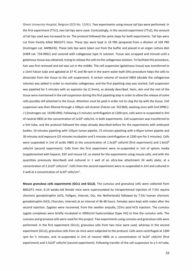
33
Ghent University Hospital, Belgium (ECD No. 12/61). Two experiments using mouse tail tips were performed. In
the first experiment (TTs1), two tail tips were used. Contrastingly, in the second experiment (TTs2), the amount
of tail tips used was increased to six. The protocol followed the same steps for both experiments. Tail tips were
cut from freshly killed B6D2/F1 mice. These tips were kept in 1X PBS (prepared from a dilution of 10X PBS
(Invitrogen cat. AM9624)). These tails tips were taken out from the buffer and placed in an organ culture dish
(VWR cat. 734-0961) and covered with collagenase type IV solution. Tissue was scrapped and minced until a
gelatinous tissue was obtained, trying to release the cells to the collagenase solution. To facilitate this procedure,
hair was first removed and tail was cut in the middle. The cell suspension (gelatinous tissue) was transferred to
a 15ml Falcon tube and agitated at 37 ºC and 90 rpm in the warm water bath (this procedure helps the cells to
dissociate from the tissue to the cell suspension). A certain volume of neutral HBSS (double the collagenase
volume) was added in order to neutralize collagenase, and the first pipetting step was started. Cell suspension
was pipetted for 5 minutes with an aspirator tip (1.5mm), as already described. Hairs, skin and the rest of the
tissue were maintained in the cell suspension during this first pipetting step in order to allow the release of some
cells possibly still attached to the tissue. Attention must be paid in order not to clog the tip with the tissue. Cell
suspension was then filtered through a 100µm cell strainer (Falcon cat. 352360), washing once with 5ml DPBS (-
/-) (Invitrogen cat. 14190-094). Following a 5 minutes centrifugation at 1200 rpm, cells were re-suspended in 3ml
of neutral HBSS at the concentration of 1x10⁶ cells/ml, in both experiments. Cell suspension was transferred to
a 5ml tube, and the protocol followed the steps already described before for the experiments with embryoid
bodies: 10 minutes pipetting with 135µm lumen pipette, 15 minutes pipetting with a 60μm lumen pipette and
30 minutes acid exposure (25 minutes incubation and 5 minutes centrifugation at 1200 rpm for 5 minutes). Cells
were suspended in 1ml of acidic HBSS at the concentration of 1.5x10⁶ cells/ml (first experiment) and 1.6x10⁶
cells/ml (second experiment). Cells from the first experiment were re-suspended in 1ml of sphere media
(supplemented with heparin, EGF and mouse LIF, as stated for the experiments using mouse cells, and with the
quantities previously described) and cultured in 1 well of an ultra-low attachment 24 wells plate, at a
concentration of 2.2x10⁵ cells/cm2. Cells from the second experiment were re-suspended in 2ml and cultured in
2 well at a concentration of 2x10⁵ cells/cm2.
Mouse granulosa cells experiments (GCs1 and GCs2). The cumulus and granulosa cells were collected from
B6D2/F1 mice. 6-14 weeks-old female mice were superovulated by intraperitoneal injection of 7.5IU equine
chorionic gonadotrophin (eCG, Folligon, Intervet, Oss, the Netherlands) followed by 7.5IU human chorionic
gonadotrophin (hCG, Chorulon, Intervet) at an interval of 46-48 hours. Females were kept with males after the
second injection. Zygotes were recovered, from the swollen ampulla, 21hrs post-hCG injection. The cumulus
zygote complexes were briefly incubated in 200IU/ml hyaluronidase (type VIII) to free the cumulus cells. The
cumulus and granulosa cells were used for this project. Two experiments using cumulus and granulosa cells were
performed. In the first experiment (GCs1), granulosa cells from two mice were used, whereas in the second
experiment (GCs2), granulosa cells from six mice were subjected to the protocol. Cells were centrifuged at 1200
rpm for 5 minutes, and re-suspended in 2ml of neutral HBSS at a concentration of 5x10⁵ cells/ml (first
experiment) and 5.5x10⁵ cells/ml (second experiment). Following transfer of the cell suspension to a 5 ml tube,

34
the first physical treatment was applied as previously described. This was followed by a second pipetting step for
10 minutes using a 135µm pipette and a third pipetting step for 15 minutes using a 60μm lumen pipette, similarly
to previous experiments. Cells were then centrifuged at 1200 rpm for 5 minutes and re-suspended in only 1ml of
acidic HBSS (volume previously decided as minimum, and with pH 5.4, previously) and incubated for 25 minutes
at 37 ºC. Cell concentration suggested by the authors (2x10⁶ cells/ml) was not reached in neither the two
experiments. After centrifuging at 1200 rpm for 5 minutes, cells were re-suspended in 1ml of sphere media
(supplemented with heparin, EGF and mouse LIF as usual for mouse cells, with the quantities already defined)
and cultured in 1 well of an ultra-low attachment 24 wells plate at a concentration of 1.6x10⁵ cells/cm2 (first
experiment) and 1x10⁵ cells/cm2 (second experiment).
Mouse Embryonic Fibroblasts experiments (MEFs1 and MEFs2). Two experiments were performed using mouse
embryonic fibroblasts commonly grown to be used as feeder cells for human embryonic stem cells culture. These
MEFs were grown in T75 flasks as described, being one flask used for the first experiment (MEFs1) and three
flasks used for the second trial (MEFs2). Excepting the amount of cells used, the protocol for either the
experiments was exactly the same. MEFs, grown in MEF medium, were washed once with DPBS (-/-) and
incubated for 5 minutes (37ºC) in the presence of 3ml/flask of 0.25% trypsin-EDTA. Following incubation, trypsin-
EDTA was neutralized by 6ml of MEF medium (double the volume of trypsin-EDTA) and cell suspension was
transferred to a 15ml Falcon tube (in the second experiment in which three flasks of cells were used, cell
suspensions from the three flaks were divided into two 15 ml Falcon tubes). Cells were centrifuged at 1200 rpm
for 5 minutes. In the first experiment, cells were re-suspended in 3ml of neutral HBSS at a concentration of
0.8x10⁶ cells/ml. In the case of the second experiment, since cells were divided in two tubes, they were re-
suspended in 2ml of neutral HBSS in each tube, and both suspensions were mixed in one of the tubes in a total
of 4ml, achieving a concentration of 1.3x10⁶ cells/ml. Part of these cell suspension obtained in the second
experiment (1ml) was used as negative control for the acquisition of pluripotency in these MEF cells, and no
stress was applied to these cells. For this negative control, 1ml of the cell suspension was transferred to a 15ml
tube, centrifuged at 1200 rpm for 5 minutes, re-suspended in 1ml of sphere media (with heparin, EGF and mouse
LIF, in the quantities already mentioned) and cultured in 1 well of an ultra-low attachment 24 wells plate at a
concentration of 7x10⁵ cells/cm2. Following 7 days in culture, the cells from this negative control were frozen for
qPCR analysis. Thus, both experiments started the physical stress with 3ml of cell suspension. Cell suspension
was transferred to a 5ml tube to facilitate the trituration, and physical stresses followed the procedures already
described (first pipetting step for 5 minutes with an aspirator tip/5ml combination, second pipetting step for 10
minutes with a 135μm lumen pipette, and third pipetting step for 15 minutes with a 60μm lumen pipette).
Following a centrifugation at 1200 rpm for 5 minutes, cells from the first experiment were re-suspended in 1ml
of acidic HBSS (previously decided as minimum volume, although the suggested concentration was not achieved),
incubated for 25 minutes and centrifuged at 1200 rpm for 5 minutes. Cells from the second experiment were re-
suspended in 2ml of acidic HBSS at a concentration of 1.4x10⁶ cells/ml. Cells from the first experiment were re-
suspended in 1ml of sphere media (supplemented with heparin, EGF and mouse LIF in the quantities already
mentioned) and cultured in 1 well of an ultra-low attachment 24 wells plate at a concentration of 1x10⁵ cells/cm2.

35
Cells from the second experiment were re-suspended in 2ml of sphere media and cultured in 2 wells at a
concentration of 5x10⁵ cells/cm2. A last parallel experiment was performed in which cells was subjected to the
protocol only until the end of the physical stresses, to check if only the titrations would reprogram the cells into
a pluripotent state. Following the physical treatment, cell suspension was centrifuged at 1200 rpm for 5 minutes
and cells were re-suspended in 2ml of sphere media (with heparin, EGF and mouse LIF) and cultured in 2 wells
of the same ultra-low attachment 24 wells plate at a concentration of 5x10⁵ cells/cm2.
Cell’s Culture. Cell cultures following the experiments described above were monitored daily under the
microscope. As already described, human cells were cultured in sphere media supplemented with bFGF, heparin
and EGF, and mouse cells were cultured in sphere media supplemented with mouse LIF, heparin and EGF. The
culture systems, as well as media refreshing methods, were the same for all the cultures, except for the
experiment with embryoid bodies from mouse embryonic stem cells (EB5). Cells were cultured for 7 days and at
day 1 extra media (1ml) was added to each well. From day 2 until day 7, cells were refreshed every day by
removing 1ml of old media from the wells with a 1ml graduated pipette (VWR cat. 612-3707) (being careful not
to remove any cells) and adding 1ml of fresh media. These methods were efficient for all the cultures using
human cells (EB1-4), as well as all the cells resulting from the protocol applied to isolated murine somatic cells
(MEFs1,2, TTs1,2 and GCs1,2)). Contrastingly, cells obtained from the protocol performed in embryoid bodies
from mouse embryonic stem cells (EB5), which were cultured in 12 wells, showed high cell density at day 1 of
culture. Therefore, instead of normal addition of 1ml of extra media already described, all the cells were
collected, centrifuged at 750 rpm for 5 minutes, re-suspended in 24ml of media and cultured in the whole 24
wells plate. However, cell density was high in the following days. Therefore, cell’s excess was removed every day
from day 2 to day 7, by mixing cell suspension in the wells (pipetting gently) and removing 1ml of media, including
cells, followed by addition of 1ml of fresh media to each well. For the experiments with mouse cells, 1000U
mouse LIF was added every day, from culture day 2 to 7. For all the cultures, both human and mouse, every well
was pipetted constantly and gently for 5 minutes using a 1ml sterile Falcon pipette after being refreshed, in order
to prevent cells from settling in the bottom of the wells and promoting the formation of round floating clusters.
Immunostaining analysis. Immunostaining samples were taken from several experiments after 7 days of culture,
and fixed in coverslips (VWR Microscope cover glass cat. ECN 631-1578). For this purpose, coverslips were pre-
coated with a 17.5% collagen solution, composed by 1/4 of a 70% collagen solution (collagen type I (Corning cat.
354249) diluted on water for deionized water (SIGMA-ALDRICH cat. 38796-1L)) and 3/4 of a 60% ethanol solution
(absolute ethanol (VWR cat. 20816-298) diluted on deionized water). Coverslips were inserted in the wells of a
4-wells plate (VWR cat. 734-2176), covered with 100-150μl of the 17.5% pre-defined collagen solution and left
to dry overnight at room temperature. Next day, a sample containing cells to analyze (around 200μl) was placed
on top of the pre-coated coverslip and left to dry in the incubator (37ºC) for 3-4 hours, in order to attach cells to
the coverslip. Coverslips were washed once with 1X PBS and placed in 4% paraformaldehyde (SIGMA cat. F1635)
solution, pre-warmed to room temperature, for 20 minutes. Samples were washed twice with 1X PBS for 5
minutes each and covered by a permeable solution (1X PBS with 0.1% Triton X100 (Sigma cat. T8787)) for 5

36
minutes. Following two more washes in 1X PBS each (5 minutes), coverslips were covered by a blocking solution
(1X PBS with 0.05% Tween (Sigma P2287) and 1% BSA) for 1 hour. Coverslips were then placed in drops of 50 μl
of primary antibody solution containing primary antibodies for Oct4 and Nanog genes (see next sub-section), and
left in the 4ºC fridge overnight, properly covered. Next day, coverslips were washed twice with 1X PBS for 5
minutes each time and placed in secondary antibody solution (see next sub-section), and left in the dark for 1
hour at room temperature. This was followed by a double wash in 1X PBS, after which coverslips were placed in
glass slides (Marienfeld cat. 1000000 76x26x1mm) on a drop of DAPI (Vector laboratories - Vectashield mounting
medium with DAPI-H1200) and stored in the dark at 4ºC until posterior analysis. An embryoid bodies’
immunostaining analysis, in order to confirm absence of pluripotency, was performed. Embryoid bodies from
mouse embryonic stem cells were cultured for 14 days, washed once with 1X PBS, fixed with 4%
paraformaldehyde (at room temperature) for 20 minutes and dehydrated with methanol (10 minutes in 25%
methanol, 10 minutes in 50% methanol, 10 minutes in 75% methanol, 10 minutes in 90% methanol and 10
minutes in 100% methanol). All the solutions were prepared by diluting 100% methanol (SIGMA-ALDRICH cat.
34860-1L) with PBT solution (1X PBS with 0.1% Tween). Embryoid bodies were then stored at -20ºC in 100%
methanol until further use. After rehydration (same set of methanol solutions in opposite order), embryoid
bodies were washed twice for 10 minutes with PBT and washed once for 15 minutes in TBST (1% PBS with 0.1%
Triton X-100). Embryoid bodies were then transferred to a blocking solution (TBST with 0.5% BSA) and kept
overnight at 4ºC. Next day, embryoid bodies were placed in the primary antibodies’ solution (primary antibodies
for Oct4 and Nanog, see next sub-section) and kept for 2 days at 4ºC in a rotating plate. After five washes with
TBST for 6 minutes each, and rotating, they were transferred to secondary antibodies solution and kept
overnight, at 4ºC and rotating. Meanwhile, glass slides were prepared with a small droplet of DAPI on top of
them. Finally, embryoid bodies were washed again for five times (6 minutes each) with TBST and transferred to
the drops of DAPI previously placed in the slides (around 4 to 5 embryoid bodies per drop of DAPI) and a coverslip
was place on top of the drop to press and fix the sample in the slide. Samples were kept in the dark at 4ºC until
further use.
Antibodies solutions. Two pluripotency genes were analysed: Oct4 and Nanog. The primary antibodies’
solutions, used both for cell suspension and antibodies analysis, contain the primary antibodies anti-goat Oct4
(Santa Cruz Biotechnology) diluted 1:200 and anti-rabbit Nanog (R&D Systems) diluted 1:200. Dilutions were
made with blocking solution, which varies if cell’s suspensions or embryoid bodies were analyzed, as shown
before. Secondary antibodies’ solutions contained the secondary antibodies Oct4 donkey anti-goat (stained with
FiTC: green, Bioconnect) diluted 1:100 and Nanog donkey anti-rabbit (stained with cy3: red, Bioconnect) diluted
1:500. Again, dilutions were made in blocking solution, which varies if cell’s suspensions or embryoid bodies were
stained.
qPCR analysis. Samples to be analyzed by quantitative polymerase chain reaction were collected and re-
suspended in 1ml TRIzol (Invitrogen cat. 15596-026) and frozen at -80ºC until further use. RNA extraction was
performed using the RNeasy Mini Kit (Qiagen cat. 74106), followed by cDNA generation using the iSCRIPT

37
Advanced cDNA Synthesis Kit (Biorad cat. 1708843). Quantification was performed using the Qubit ssDNA Assay
Kit (Invitrogen cat. Q10212) and gene expression analysis was done using the ABI Prism 7000 Sequence Detection
System (Applied Biosystems). The examination included the pluripotency associated markers for Nanog, Gapdh,
SSEA1, Rex1 and Gbx2 (for the mouse samples) and markers for OCT4, NANOG, SSEA1, GAPDH and B2M (for the
human samples). A mastermix, comprising gene-specific primers and either iTAQ Universal SYBR Green Supermix
(Biorad cat. 172-5122) or iTAQ Universal Probes Supermix (Biorad cat. 172-5133) was prepared. All the samples
were analyzed in triplicates with a total final reaction volume of 25µl, including 10ng cDNA template (previously
generated) and 20µl of mastermix. Real time quantitative polymerase chain reaction was performed using the
following thermic cycles: 2 minutes at 95ºC, 45 cycles at 95ºC for 15 seconds and 1 minute at 60ºC. The
normalization of the CT values was done against housekeeping genes (GAPDH and B2M) and the ΔΔCT method
was used to calculate the fold change (comparison to control samples for each sample: mouse embryonic stem
cells for the mouse samples and H1 human embryonic stem cells for the human samples).

38
4. Results and Discussion
Pluripotent stem cells follow a differentiation event in vivo through a natural process of development of the
animal, or can be directly influenced to adopt a certain phenotype and function through influence of external
factors in vitro (see chapter 1.1.4). It was considered ES cells that spontaneously differentiated in vitro into EBs
could reside in a more primitive and less differentiated state as compared to a fully differentiated and functional
somatic cells that can be isolated from an animal. The main objective of the study was to reprogram
differentiated cells back to a pluripotent stat. The technique was adapted to both mouse and human cells, to
replicate the results previously claimed (Obokata et al., 2014d) and to adapt the same idea to human cells to
achieve results never described before. The reprogramming of human cells was the main accomplishment
expected to be obtained. Pluripotent ES cells (mouse and human) were differentiated in vitro through the
generation of EBs, which were then exposed to the protocol.
Figure 6: Main steps describing the adaptation of the protocol to use with embryoid bodies cultured in the lab. Embryonic
stem cells were expanded and differentiated into embryoid bodies during 14 days, after which they were dissociated into
single cells. Those isolated cells faced then three pipetting steps with increasingly smaller lumen tips. Physical stress was
followed by chemical stress described as 30 minutes of acid exposure. Cells were then cultured during 7 days in sphere media,
after which pluripotency was analyzed.
According to the technical procedure suggested by Vacanti et al, the cells were subjected to both physical and
chemical stresses in order to reprogram them to a pluripotent state. The main idea of this protocol (Figure 6)
was to start with ES cells growth and expansion, followed by differentiation into EBs. The differentiation step
took 14 days, after which EBs were recovered and dissociated into single cells/small clusters, which were

39
subjected to the physical and chemical perturbations. Physical stress was characterized by three consecutive
pipetting steps of 5, 10 and 15 minutes with increasingly smaller lumen pipettes. Physical stress was followed by
chemical stress, characterized by 30 minutes exposure to an acidic solution (neutral HBSS titrated to pH 5.4,
which should increase to around 5.6 when in contact with the cells). Cells were then re-suspended in sphere
media supplemented with several factors, and cultured in an ultra-low attachment 24 well plate (to achieve small
round floating clusters as described in Obokata’s protocol). The resistance of the cells to the protocol was
considered in several main steps: following single cell dissociation, following each physical trituration and
following acid exposure. Different cells had different behaviors facing each step, and preliminary studies were
performed to adapt the technical procedure, as described in the next sections. Different factor combinations to
support sphere media were used accordingly to the cell type. For a more detailed protocol, see Materials and
Methods section (see chapter 3).
Other somatic cells, namely MEFs, mouse tail tips (TTs) and GCs were also tested. Following the same technical
procedure suggested by Vacanti, the cells were subjected to both physical and chemical stresses, similarly to the
approach involving EBs. The protocol for directly isolated somatic cells (Figure 7) follows the main steps of the
previous one, without the differentiation into EBs and following dissociation steps.
Figure 7: Experimental procedure performed in murine somatic cells directly isolated from mice or cultured in the lab. The
procedure started directly with the exposure to the three physical stress steps, after which the exposure to an acidic solution
during 30 minutes was imposed. After being cultured for 7 days in sphere media, pluripotency of the cells was analyzed.
Following the technical suggestions and feedback obtained, the detailed protocol was adapted for every type of
cell and improved over time according to the results obtained and observations to the practical procedure.
Different survival rates were obtained in different stages of the protocol, as well as different culture observations
(see Table 3). All the results of the two main approaches to the protocol (EBs or directly isolated somatic cells)
are described in the next sub-sections.

40
4.1. Embryoid Bodies
4.1.1. Chemical and Physical stresses: A First Approach (EB1)
As described, the main objective of the study was to apply the technique on the human model by differentiating
hES cells into EBs and then attempt to reprogram the cells into a pluripotent state. A first experimental trial,
hereon called EB1, was performed using human ES cell line UGENT11-2, which was differentiated into EBs, and
then treated following the steps of the pre-defined protocol (Figure 6). Human UGENT11-2 ES cells on passage
50 were grown and expanded in conventional human ES cell media for two weeks. EBs were grown for 14 days
in conventional differentiation media in 6 wells of a 24 well-plate, after which they were dissociated into single
cells. Following dissociation, physical stress was applied. This included a first 5 minutes pipetting using an
aspirating pipette (1.5mm) attached to a 5ml pipette, followed by a 10 minutes pipetting using a yellow stripper
pipette (135µm) and finally a 15 minutes pipetting using a pre-prepared 60µm diameter pipette. Posterior to the
physical stress, cells were submitted to a chemical stress by 30 minutes exposure to acidic HBSS at pH of 5.4.
Cells were re-suspended in sphere media supplemented with bFGF, heparin and EGF in an ultra-low attachment
24 wells plate. The main steps of the protocol here described served as a base for the following experiments,
which carried some alterations.
To track cell progression, trypan blue was used after every stress step, in order to understand which steps led to
higher cell loss and to check if the amount of cells followed certain standards and minimum values to enable cell
culture and expansion. Together, physical and chemical stresses resulted in a high cell loss (total cell amount
reduced from 4x10⁵ after dissociation to 2x10⁵ after acid treatment) and damage, from which resulted an
inefficient post stress cell culture (data not shown), although a proper cell culture density was used (1x10⁵
cells/cm2). Cells could barely be identified under the microscope. Accordingly to observations made from the
first trial, the dissociation of the EBs into single cells by 5 minutes incubation in 0.05% trypsin-EDTA was defined
as the constriction step, leading to ineffective dissociation of the cells and to low density obtained in suspension.
EBs’ structure was not efficiently disrupted and maintained a big physical matrix, which retained most part of
the cells. A more efficient cell dissociation process was therefore indispensable. Moreover, the handling of the
60µm small lumen pipettes was also deficient due to the attachment of the pipettes’ long tips to the wall of the
15ml Falcon tube used, thus leading to the break of several pipette tips. This resulted in cell loss and damage
during the pipetting and the following centrifugations (cells were eventually lost inside the glass tips and
disrupted during the centrifugations due to the presence of glass in the tube). Moreover, acid exposure at a low
concentration of 3x10⁵ cells/ml could have been severe for the cells.
4.1.2. Second Approach: Handling and Dissociation Improvement (EB2)
The same cell line was tested a second time (EB2), following the same basic steps of the protocol initially used,
with some adaptations. Human UGENT11-2 ES cells on passage 53 were grown for two weeks, followed by

41
differentiation into EBs for 14 days, grown in 6 well of a 24 well-plate, after which the stresses were imposed.
Following the inefficient dissociation of the EBs into single cells in the first trial (EB1), in this new approach, EBs
were incubated for a longer time (8 minutes) in 0.05% trypsin-EDTA and the tube was agitated every minute, in
order to avoid possible deposition of the EBs in the bottom of the tube and to spread the cells along the volume
of trypsin-EDTA, facilitating the dissociation. Moreover, in order to facilitate the small lumen pipettes handling,
the cell suspension was transferred to a 5ml tube, thus preventing attachment of the glass tips to the walls of
the 15ml falcon tube previously used. The physical and chemical stresses followed the same procedure of the
previous approach, as well as the following culture.
Figure 8: Cell density after each perturbation step of the EB2 experiment. A. The cell density obtained after the dissociation of
the EBs' structure was particularly low. B, C, D. Also, the low cell density tended to decrease after the first, second and third
pipetting steps, respectivelly. E. Following acid treatment, cell loss was almost total. As a result of these observation, a low
quantity of cells was cultured in sphere media, predicting inefficient culture.
The dissociation into single cells was, however, not efficient enough since the physical structure of the EBs was
maintained and not sufficiently broken. This resulted in the presence of a big mass structure floating in the tube,
which not only interfered with the pipetting in the next steps but also retained a large number of cells that
remained attached to it, not being released to the medium. Therefore, it was possible to conclude that these
cells were not totally and efficiently exposed to the physical stress, since cells attached to the big mass bulk did
not pass through the pipette tip, and thus technically did not suffer from any stress. The dissociation step needed
to be revised.
Low cell density after the dissociation step (Figure 8-A) was obtained, as predicted by the inefficient break of the
EBs’ structures, although at higher values as compared to the first experiment (1 million of total cell number was

42
obtained). Also, this low cell count further tended to be reduced during the physical stress steps (Figure 8-B, C,
D) as a result of the harsh conditions that the cells faced, resulting in even lower cell density after the acid
treatment (Figure 8-E). However, higher cell density was obtained as compared the first experiment, and cells
were cultured at a concentration of 2x10⁵ cells/cm2. At this point, better culture results were expected.
The dissociation surely contributed to the low cell density, particularly due to the described inefficient EBs’
physical disruption. Also, the quantity of starting EBs (only 6 wells cultured) may not be enough. This resulted in
particular low cell concentration at the acid exposure step (7x10⁵ cells/ml acid) as compared to the optimum
value suggested by Obokata et al (2x10⁶ cells/ml acid). The low cell density used resulted in higher effect of the
acid exposure, thus resulting in more severe perturbation. The amount of cells observed in the well at the time
of culture (day 0, data not shown) was particularly low, but some initial proliferation capacity was observed
resulting in a slight but still low increase of cell density observed on day 3 (Figure 9-A). However, these cells lost
their proliferative capacity (characteristic of pluripotent cells), and maintained almost the same cell density until
day 5 (Figure 9-B) and day 7 (Figure 9-C). It was therefore possible to conclude that most part of the cells
eventually died during the stresses or immediately after being cultured. The ones that survived were
characterized as unstable, showing only initial capacity to be maintained and proliferate. Immunostaining
analysis performed on samples taken after 7 days in culture showed absence of pluripotency (data not shown),
with complete absence of both OCT4 and NANOG expression.
Figure 9: Cell culture progression of cells resulting from the EB2 experiment. A. The few cells cultured showed some
proliferation capacity resulting in a higher amount of cells after 3 days of culture. B, C. However, the cell density observed did
not increase until days 5 and 7 respectively, suggesting the loss of proliferation capacity. This absence of normal cell
functionality may be related with the harsh condition to which cells were exposed during the experiment.
4.1.3. Further Experiments with Embryoid Bodies (EB3, EB4 and EB5)
Three different cell lines were tested: human UGENT11-2 cell line (EB3), to refine the practical protocol; human
H1 reporter line (EB4), an OCT4-GFP+ cell line, which easily allowed to check for pluripotency gene OCT4; mES
cell line (EB5), to check reprogramming possibility with mouse cells. The practical procedure was the same for
both experiments.

43
4.1.3.1. Human UGENT11-2 cell line (EB3)
Following the previous observations, an important adaptation was made. According to the EBs’ dissociation into
single cells, trypsin-EDTA 0.05% was shown to be inefficient, even for a longer time exposure (8 minutes).
Therefore, it was decided that 0.25% trypsin-EDTA would be used from then on. The incubation time was reduced
back to 5 minutes, since 8 minutes in stronger trypsin-EDTA could be too harsh for the cells. The EBs’ physical
structures were shown to be efficiently broken using this stronger trypsin-EDTA, resulting in a more turbid cell
suspension, as compared to the previous results. Also, higher quantity of starting material was used (9 wells of
EBs as compared to 6 previously used), facing the requirement for a higher amount of cells.
Figure 10: Cell density after each perturbation step of the EB3 experiment. A. An improvement in the dissociation step was
verified by using trypsin-EDTA 0.25% rather than 0.05%, resulting in a higher amount of cells prior to the first pipetting step.
B, C, D. The two first pipetting steps maintained the amount of cells already observed after the dissociation step. However,
the third trituration, characterized by the lowest lumen pipette tip used, resulted in a slight cell loss, possibly due to the really
small space through which the cell are obligated to pass continuously. E. Acid exposure was shown to induce massive cell loss.
Cells were subjected to physical stress with the three consecutive pipetting steps already described, followed by
chemical stress with acid exposure. Cells were then re-suspended in 2ml of sphere media supplemented with
heparin, bFGF and EGF, and cultured in 2 wells of a 24 wells ultra-low attachment plate and grown for 7 days, as
described before. Samples for immunostaining were taken at day 7.
By tracking cell progression and viability, it was possible to verify an improvement in the dissociation step (Figure
10-A) defined by a higher amount of viable and dissociated single cells (2x10⁶ cells, double the amount obtained
after the dissociation in the EB2 experiment), as predicted by the efficient rupture of the physical structure of

44
the EBs. A higher amount of viable cells after physical stress was verified (1.5x10⁶ cells in total), as compared to
the previous approach to the technique (Figure 8-D). Besides the amount of cells did not seem to change after
the first and second trituration (Figure 10-B, C), cell density slightly decreased after the last pipetting step (Figure
10-D). This observation may be explained by a harsh trituration associated to the small size (60µm) of the third
pipette. The pipetting steps required the cells to be out of the incubator for a long period, suspended in an
unconventional medium (neutral HBSS) for cell maintenance and growth. Therefore, long maintenance of the
cells outside a proper cocktail and the incubator may also explain the increase in cell loss along time. Although
cell density decreased during physical treatment, acid treatment was performed at a concentration (1.5x10⁶
cells/ml) closer to the optimal concentration suggested by Obokata et al. However, acid treatment provoked a
significant cell loss (Figure 10-E). Cells showed greater resistance to physical stress in comparison to acid
exposure, which was significantly severe. Following the higher amount of cells obtained and cultured, it was
possible to observe cell growth almost daily (Figure 11), with significant changes along days 3, 5 and 7 of culture.
Figure 11: Cell culture progression of cells resulting from the EB3 experiment. A. After 3 days of culture, cell density
demonstrated considerable high values, suggesting the existence of proliferation capacity. B. Following 5 days of culture, the
proliferation and developmental capacity of the cell was confirmed by the existence of differentiated-like structures with
considerable size resulting from the aggregation of the cells present in culture. C. The differentiation tendency leaded the cell
aggregates to form structures with completely differentiated-like morphology resembling early days of EBs differentiation
culture. This observation suggests inexistence of pluripotency after the 7 days of culture.
With these observations it was possible to conclude that a rigorous and efficient pre-stress dissociation step into
single cell was indispensable, as predicted. The efficiency of this step not only allowed a higher number of cells
to be pipetted through the pipette’s tips, but also destroyed the EBs’ big physical matrices that could clog the tip
and block the passage of cells through it. In addition to efficiently cause stress to the cells, the passage of small
cell agglomerates into the tip possibly disrupted these agglomerates and increased the amount of single cells in
solution (as desired for an efficient physical stress) that passed through the tip, offsetting the cell loss along time
and increasing the possible reprogramming efficiency. Cell concentration in the acid treatment step was closer
to the optimal, suggesting more efficient perturbation. The higher concentration of cells exposed to the acid
surely resulted in less severe perturbation as compared to the previous experiments. This evidence possibly
contributed to maintain cell integrity and enabled a following more efficient cell culture. Although highly reduced
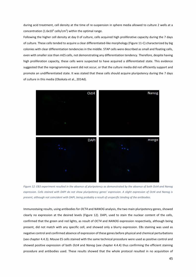
45
during acid treatment, cell density at the time of re-suspension in sphere media allowed to culture 2 wells at a
concentration (1.6x10⁵ cells/cm2) within the optimal range.
Following the higher cell density at day 0 of culture, cells acquired high proliferative capacity during the 7 days
of culture. These cells tended to acquire a clear differentiated-like morphology (Figure 11-C) characterized by big
colonies with clear differentiation tendencies in the middle. STAP cells were described as small and floating cells,
even with smaller size than mES cells, not demonstrating any differentiation tendency. Therefore, despite having
high proliferation capacity, these cells were suspected to have acquired a differentiated state. This evidence
suggested that the reprogramming event did not occur, or that the culture media did not efficiently support and
promote an undifferentiated state. It was stated that these cells should acquire pluripotency during the 7 days
of culture in this media (Obokata et al., 2014d).
Figure 12: EB3 experiment resulted in the absence of pluripotency as demonstrated by the absence of both Oct4 and Nanog
expression. Cells stained with DAPI do not show pluripotency genes' expression. A slight expression of Oct4 and Nanog is
present, although not coincident with DAPI, being probably a result of unspecific binding of the antibodies.
Immunostaing results, using antibodies for OCT4 and NANOG analysis, the two main pluripotency genes, showed
clearly no expression at the desired levels (Figure 12). DAPI, used to stain the nuclear content of the cells,
confirmed that the green and red lights, as result of OCT4 and NANOG expression respectively, although being
present, did not match with any specific cell, and showed only a blurry expression. EBs staining was used as
negative control and confirmed absence of expression of these genes before physical and chemical perturbations
(see chapter 4.4.3). Mouse ES cells stained with the same technical procedure were used as positive control and
showed positive expression of both Oct4 and Nanog (see chapter 4.4.4) thus confirming the efficient staining
procedure and antibodies used. These results showed that the whole protocol resulted in no acquisition of

46
pluripotency genes expression by these cells, thus demonstrating the inexistence of a reprogramming event.
Human UGENT11-2 stem cells, differentiated into EBs, did not acquire any pluripotency and maintained a
differentiated state after being exposed to the stresses and cultured in media that was claimed to efficiently
induce the pluripotent state.
4.1.3.2. Human H1 Reporter ES cell line (EB4)
The first experiments had the main purpose to optimize the protocol to be used with the human H1 reporter
line. This cells line is OCT4-GPF+, and thus the antibody to analyze Oct4 expression in an immunostaining analysis
is not necessary (although it was used to enforce OCT4 expression). Therefore, immunostaining analysis may be
significantly more efficient, preventing possible problems associated with the antibody’s specificity or with the
staining process itself.
The same dissociation method used with the previous experiment (EB3), defined as the more efficient, was used
with the reporter cell line. The dissociation using 0.25% trypsin-EDTA resulted in a more turbid cell suspension,
characteristic of a high cell density suspension, which was maintained during both physical and chemical stresses.
Cell progression was tracked with trypan blue during the whole experiment. Cells were cultured in 2 wells of a
24 wells ultra-low attachment plate for 7 days. Immunostaining samples were taken at day 5 and day 7, and all
the cells were frozen for posterior quantitative polymerase chain reaction (qPCR) analysis.
Figure 13: Cell density after each perturbation step of the EB4 experiment. A. Cell density following dissociation of the
embryoid bodies' structures presented reasonable values. B, C, D. Cell density was maintained after the first pipetting step (B)
but reduced following the second one (C), being maintained again after the third trituration (D). E. Cell density faced an
intriguing increase following acid treatment.

47
Following dissociation into single cells, a reasonable cell density (a total of 2 million cells) was obtained (Figure
13-A), as predicted by the observations of the efficient dissociation. The cell density slightly decrease following
the second pipetting step (Figure 13-C), which was maintained during the next physical stress step. The higher
amount of cells obtained allowed to expose cells to the acidic solution at a concentration (1.5x10⁶ cells/ml) close
the optimal (2x10⁶ cells/ml). Interestingly, the current experiment yielded, until this point, the exact same cell
amount as the previous experiment (and both started with approximately the same amount of EBs), showing the
acquired consistency of the dissociation and physical trituration methods, and the coherence acquired when cells
with similar origin were used (both were hES cells). Cell density after acid treatment (Figure 13-E) was
considerably higher (1.3x10⁶ cells in total) as compared to the previous experiment (6x10⁵ cells), suggesting
higher resistance of this cell line to the acid exposure and allowing higher cell density in culture. Contrastingly to
the first experiments (namely EB1 and EB2), cells were shown to resist the whole protocol despite the relative
cell loss in the first steps. More than half of the starting material remained intact after all the perturbations.
Figure 14: Cell culture progression of cells resulting from the EB4 experiment. A. Even only after 3 days of culture, cells showed
differentiation tendencies characterized by several dark colored agglomerates. B, C. The differentiation tendencies were still
present after 5 (B) and 7 (C) days of culture. The colonies acquired an EB-like morphology.
The cultured cells showed huge proliferative capacity that led to the formation of big colonies after 3 days (Figure
14-A) in culture. Contrasting to the previous experiments, the present cell culture resulted in a higher cell density
in culture, characterized by the formation of big cluster colonies with differentiation evidence observed after the
7 days of culture (Figure 14-C). It was possible to conclude that the resistance to the protocol and the higher
amount of starting material, resulting in greater (viable) cell density in culture, contributed to the higher
expansion capacity verified. Despite being stated that these cells should acquire an ES cell-like morphology
(Obokata et al., 2014d), characterized by small size cells without differentiation tendencies, morphology
observations suggested that there was no acquisition of pluripotency by these cells.
The significant proliferation capacity observed is characteristic of pluripotent ES cells, since some specialized
cells, upon differentiation, do not replicate themselves. Following 14 days of EBs differentiation, all the cells were
completely differentiated (see chapter 4.4.3). Nerve cells, as well as muscle cells, for example, reside in G0 phase
and thus do not replicate (Wang et al., 2011), similarly to other cells (like bone cells) which enter temporarily in
the G0 phase. Therefore, due to the presence of such cell types, the proliferation capacity should have been
highly reduced as compared to this same ability in undifferentiated cells (which do not stop replicating). It must

48
be kept in mind that during EBs’ differentiation, incomplete differentiation may result in some cells residing in a
pluripotent state, contributing to the high proliferation observed. However, the negative control (see chapter
4.4.3) proved that EBs did not show pluripotency after 14 days in culture, thus eliminating this possibility.
Similarly to the previous experiment, the efficient pluripotency expression identified in a positive control (see
chapter 4.4.4) argued against possible problems with the staining procedure. The high proliferation capacity was,
therefore, an intriguing result.
It was demonstrated that mouse ES cells, upon direct culture in sphere media without any stress, showed
significant cell death and complete absence of proliferative capacity, resulting in reduced cell density following
7 days of culture (see chapter 4.4.1). The maintenance of the ground-state of pluripotency (Ying et al., 2008) is
dependent on the use of two inhibitors along with LIF, supporting the idea that the defined sphere media could
not maintain or help to induce a pluripotent state. Nevertheless, this media should at least support cell survival
and possibly induce mouse ES cells, directly cultured in it, to adopt a differentiated status as result of
uncontrolled proliferation and loss of their pluripotency. It is intriguing why this media was said to efficiently
promote puripotency. Associated with the low efficiency that characterizes reprogramming techniques (Obokata
et al., 2014d; Tachibana et al., 2013; Takahashi and Yamanaka, 2006), the low amount of starting material used
in this experiment (only 9 wells of a 24 wells plate with EBs) contributed to reduce the chances of finding possibly
reprogramed cell aggregates.
Figure 15: Immunostaining results of the EB4 experiment from samples taken after 5 days of culture showed absence of
pluripotency factors Oct4 and Nanog expression, demonstrating that these cells still resided in a differentiated state at this
point.

49
Intrigued by the big proliferation capacity and the differentiated-like morphology obtained in early days of
culture, samples for immunostaining were taken at day 5. These samples showed that after 5 days of culture the
cells had no relevant expression of both OCT4 and NANOG (Figure 15), thus showing that pluripotency was not
acquired.
Figure 16: Immunostaining results of the EB4 experiment from samples taken in the last day of culture (day 7) showed
apparent presence of pluripotency, due to almost perfect overlapping of both Oct4, Nanog and DAPI expressions. Despite Oct4
showed faint expression, this represented an interesting and intriguing result that required further analysis through qPCR.
At day 7, immunostaining sample showed an apparent positive result characterized by almost perfect, despite
faint, overlapping of OCT4 and NANOG expression patterns, which also matched with DAPI staining (Figure 16).
These cells were, however, the only ones matching from the whole sample, and the expression of the
pluripotency genes looked very faded. Unspecific binding of the antibodies, resulting in the staining of non-viable
pluripotent cells, could be the reason for the observed result. However, in those cases, the probability of
obtaining the overlapping expression of OCT4, NANOG and DAPI in a shape characteristic of a viable cell is very
low, and thus this result was not discarded nor considered unspecific binding. This represented the best result
obtained until this point. A very low efficiency would be expected in case of efficient reprogramming, thus
explaining that these were the only cells positively matching. However, further qPCR analysis (see chapter 4.3)
showed a negative result for the expression of pluripotency genes in these cells, thus demonstrating that
pluripotency was acquired.

50
4.1.3.3. Mouse ES cell line (EB5)
As a final experiment using EBs’ methodology, mES cells were grown and passaged in 2i/LIF media. Due to the
faster proliferation of mES cells comparing to hES cells, a larger cell amount was obtained in shorter time and a
whole 24 well plate was fulfilled with cells, cultured in medium that efficiently induces differentiation (N2B27
medium used for 2i/LIF but without any inhibitor nor LIF). At day 14 of EBs’ culture, cells faced the already defined
physical and chemical perturbations protocol.
Figure 17: Cell density after each perturbation step of the EB5 experiment. A. Cell density prior to the first trituration step was
high comparing to the previous experiments performed. This observations resulted of a very efficient disruption of the EBs’
structure, resulting in the release of a huge number of single cells to the medium. B, C, D. Cell density after the first (B), second
(C) and third (D) pipetting steps was still at very high levels. E. Facing acid exposure, cell density remained at high levels. These
observations may suggest higher resistance to the protocol in general, in comparison with the human cells previously tested.
Dissociation of EBs into single cells followed the previous protocol described by 5 minutes incubation with
exposure to 0.25% trypsin-EDTA. The physical structure of the EBs was almost completely disrupted, and the cell
suspension was completely opaque. This result was very satisfactory at this time point and allowed an efficient
physical stress since most part of the cells were efficiently forced to pass through the pipette’s tips. Mouse EBs
were shown to be more easily disrupted and dissociated into single cells as compared to the human counterparts.
After acid exposure, due to the significantly higher amount of cells still alive, they were re-suspended in 12ml of
sphere media supplemented with heparin, mouse LIF and EGF, and cultured in 12 wells of an ultra-low
attachment 24 well plate. To note that due to the high proliferation capacity observed, the initially cultured 12
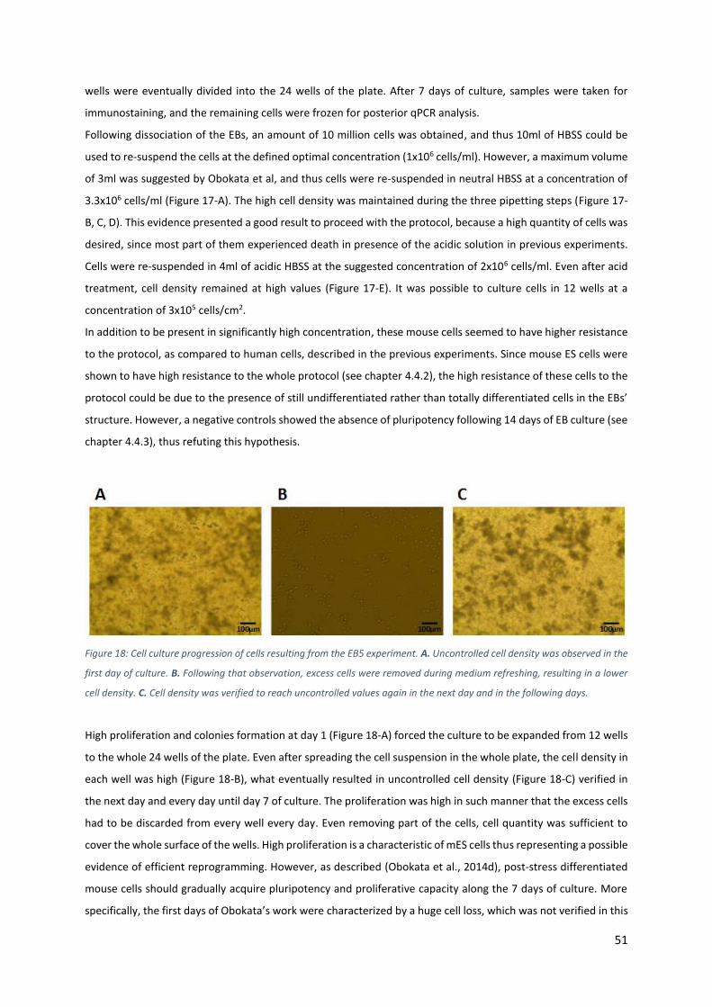
51
wells were eventually divided into the 24 wells of the plate. After 7 days of culture, samples were taken for
immunostaining, and the remaining cells were frozen for posterior qPCR analysis.
Following dissociation of the EBs, an amount of 10 million cells was obtained, and thus 10ml of HBSS could be
used to re-suspend the cells at the defined optimal concentration (1x10⁶ cells/ml). However, a maximum volume
of 3ml was suggested by Obokata et al, and thus cells were re-suspended in neutral HBSS at a concentration of
3.3x10⁶ cells/ml (Figure 17-A). The high cell density was maintained during the three pipetting steps (Figure 17-
B, C, D). This evidence presented a good result to proceed with the protocol, because a high quantity of cells was
desired, since most part of them experienced death in presence of the acidic solution in previous experiments.
Cells were re-suspended in 4ml of acidic HBSS at the suggested concentration of 2x10⁶ cells/ml. Even after acid
treatment, cell density remained at high values (Figure 17-E). It was possible to culture cells in 12 wells at a
concentration of 3x10⁵ cells/cm2.
In addition to be present in significantly high concentration, these mouse cells seemed to have higher resistance
to the protocol, as compared to human cells, described in the previous experiments. Since mouse ES cells were
shown to have high resistance to the whole protocol (see chapter 4.4.2), the high resistance of these cells to the
protocol could be due to the presence of still undifferentiated rather than totally differentiated cells in the EBs’
structure. However, a negative controls showed the absence of pluripotency following 14 days of EB culture (see
chapter 4.4.3), thus refuting this hypothesis.
Figure 18: Cell culture progression of cells resulting from the EB5 experiment. A. Uncontrolled cell density was observed in the
first day of culture. B. Following that observation, excess cells were removed during medium refreshing, resulting in a lower
cell density. C. Cell density was verified to reach uncontrolled values again in the next day and in the following days.
High proliferation and colonies formation at day 1 (Figure 18-A) forced the culture to be expanded from 12 wells
to the whole 24 wells of the plate. Even after spreading the cell suspension in the whole plate, the cell density in
each well was high (Figure 18-B), what eventually resulted in uncontrolled cell density (Figure 18-C) verified in
the next day and every day until day 7 of culture. The proliferation was high in such manner that the excess cells
had to be discarded from every well every day. Even removing part of the cells, cell quantity was sufficient to
cover the whole surface of the wells. High proliferation is a characteristic of mES cells thus representing a possible
evidence of efficient reprogramming. However, as described (Obokata et al., 2014d), post-stress differentiated
mouse cells should gradually acquire pluripotency and proliferative capacity along the 7 days of culture. More
specifically, the first days of Obokata’s work were characterized by a huge cell loss, which was not verified in this

52
cell culture, in which a huge proliferation rate was observed on the first day. The negative control showing the
absence of pluripotency in the EBs (see chapter 4.4.3) demonstrated that the 14 days culture was sufficient for
fully differentiation, and the presence of residual pluripotent stem cells in the suspension as responsible for the
high proliferation was discarded. Interestingly, despite having an apparent differentiated-like morphology due
to the high proliferation rate, all those colonies characterized by darker color were easily broken (during the
refreshing), resulting in isolated single cells in culture (Figure 18-B) with mES cells-like morphology. Mouse ES
cells survived the whole protocol (see chapter 4.4.2), and acquired differentiated-like morphology during the 7
days of culture. Therefore, the fact that all the cells in this experiment showed ES-like morphology also suggests
that the incomplete EBs’ differentiation is improbable. Therefore, the differentiated-like morphology can be a
result of uncontrolled cell growth rather than differentiation tendency. Therefore, the possible acquisition of
pluripotency was not discarded at this point.
Figure 19: Immunostaining results obtained from a sample taken after the 7 days of culture showed apparent positive results
(bottom right especially) that clearly differ from the clearly negative results observed in the sample of cells from the EB5
experiment. However, cells negatively expressing Oct4 and Nanog highly expressed DAPI, whereas the apparent pluripotency-
expressing cells showed very faint expression of DAPI.
Immunostaining analysis showed apparent positive results for the expression of Oct4 and Nanog, defined by the
simultaneous expression of both the pluripotency genes in some cells, as well as DAPI (Figure 19). Interestingly,
the cells that perfectly overlapped the expression of Oct4 and Nanog had low DAPI expression intensity. It was
not clear if these were effectively intact cells that for some reason were poorly marked with DAPI, or if the blurry
DAPI expression meant unspecific binding of both Oct4 and Nanog antibodies. This would mean that the matches
between the green and red lights would not represent viable pluripotent cells, but rather represented a strong

53
point of unspecific binding to which even DAPI got slightly bound. The matching between Oct4 and Nanog was
very strong. Although both genes could bind to unspecific spots, the perfect matching of the two markers
showing perfect cell-like shape would be very improbable, thus suggesting a possible existence of some
pluripotent cells in the mixture. Posterior qPCR analysis (see chapter 4.3) showed the inexistence of pluripotency.
4.2. Somatic Cells
A second approach to the reprogramming method was followed through several studies using somatic cells
directly isolated from mice or cultured in vitro. Only mouse cells were used and these include GCs (see chapter
1.3.2.1), TTs and MEFs (grown in vitro and commonly used as feeder cells for hES cells culture). The main protocol
follows the same basic steps previously described for the EBs experiments, excepting the step characterized by
dissociation into single cells (Figure 7). In these particular cases, somatic cells were directly isolated or expanded
and the protocol started with the physical stress.
4.2.1. Granulosa and Cumulus Cells
GCs were described to be easier to reprogram (see chapter 1.3.2.1) into iPS cells. Intrigued by this fact, two
experiments using these cells were performed. GCs were isolated from B6D2/F1 mice (see chapter 3). After being
extracted, cells were re-suspended in neutral HBSS and exposed to the protocol, already defined.
Figure 20: Cell density after each perturbation step of the GCs1 experiment. A. Granulosa cells isolated from B6D2/F1 mice
showed sufficient quantities prior to the first titration step. B. C, D, E. Cell density tended to decrease after each one of the
following trituration (B, C and D) and acid exposure (E) steps.

54
Figure 21: Cell culture progression of cells resulting from the GCs1 experiment. A. After three days of culture, almost all the
cells were dead. B, C. Following 5 (B) and 7 (C) days of culture, the same cells previously observed were identified. However,
proliferation capacity was not verified, and these cells were assumed to be dead.
In a first trial (GCs1), GCs from two mice were used, and the amount of cells after re-suspension in HBSS (around
1x10⁶ cells) was verified to be sufficient to proceed with the protocol (Figure 20-A). From the post-first titration
to the post-acid treatment (Figure 20-B, C, D, E) step, a decreasing amount of cells was obtained, predicting
possible insufficient cell density for the following culture.
Figure 22: Cell density after each perturbation step of the GCs2 experiment. A. Despite using 6 mice rather than 2, the amount
of cells initially isolated was lower. B, C, D, E. This lower amount of cells resulted in an equally lower cell density after the first
(B), second (C) and third trituration steps, and even after acid exposure (E).
These cells were not able to be maintained in sphere media. After three days of culture, cells were already almost
impossible to identify (Figure 21-A), and this result was maintained in the following days. Cells were cultured at

55
a concentration of 1.6x10⁵ cells/cm2, which was within the optimal range, although close to the minimum value
suggested (1x10⁵ cells/cm2). Following these observations, one of two options was identified as the cause for the
cell culture failure: cells were completely dead during the whole procedure (improbable, since cells were alive
and at a sufficient number at the time of culture) or the media did not efficiently support cell survival.
Immunostaining samples were not taken due to the inexistence of viable cells in culture.
A second experiment (GCs2) followed the same steps of the previous one, using cells from 6 mice. Interestingly,
the amount of cells obtained after recovering the cells and re-suspend them in neutral HBSS was equivalent
(Figure 22-A) to the first trial (Figure 20-A) in which only 2 mice were used. Following the whole protocol (Figure
22-E), a lower amount of cells (2x10⁵ cells) was obtained as compared to the first trial (3x10⁵ cells), thus resulting
in a cell culture at the minimum concentration (1x10⁵ cells/cm2) suggested.
Interestingly, cells survived the culture and the amount of cells obtained after 3 days of culture (Figure 23-A) was
relevant. Contrastingly to the previous experiment, cells were shown to successfully grow during the first 5 days
of culture (Figure 23-B), although with low proliferation. However, after 7 days of culture (Figure 23-C), the
amount of cells was shown to be lower. Nevertheless, this second experiment resulted in overall better results
as compared to the first one, and a sufficient cell amount for immunostaining analysis was obtained.
Figure 23: Cell culture progression of cells resulting from the GCs2 experiment. A. A reasonable cell amount was obtained after
3 days of culture. B. After 5 days in culture, cell density was increased, suggesting the existence of proliferative capacity in
these cells. C. However, after 7 days in culture, cell density was again reduced, demonstrating the loss of the proliferative
capacities of these cells.
Cells were shown to look alive as single cells, being this a characteristic of mouse pluripotent cells. However, they
lacked the proliferative capacity, lost in the last days of culture. Cell density was meant to be reduced drastically
in the first days and maintained in the last ones (Obokata et al., 2014d). This fact was not observed, since cells
started to die after the fifth day of culture after an initial period high some proliferation. It was unclear if a longer
culture period would lead to the maintenance of cell concentration and possibly acquisition of proliferation, thus
suggesting that the phase at which cells stabilize their amount and start acquiring pluripotency would be delayed
in this kind of cells. Further studies with longer culture could clarify this.
Immunostaining analysis showed evident absence of pluripotency in these cells, since no overlapping expression
of both pluripotency genes, along with DAPI, in a properly defined cell, was obtained (Figure 24). Due to the
negative results verified, no positive or negative controls were prepared.

56
Figure 24: Immunostaining results of 7 days culture cells show clear absence of pluripotency in cells resulting from the GCs2
experiment, due to impossible observation of overlapping expression of the pluripotency genes Oct4 and Nanog.
Conclusions about the reprogramming capacity using this cell type were difficult to obtain. A first constricting
point was related with the isolation of these cells. Apparently, the amount of cells obtained from a certain mouse
sample can be random and thus hard to predict, since a higher amount of mice extractions resulted in equal cell
number. A proper protocol defining the precise amount of mice to be used as starting material would be difficult
to establish. In general, the amount of cells was not great after the perturbations, and a greater amount of
starting cells would be needed for a proper experiment. It was unclear the reason why the cells obtained in the
first experiment did not survive the culture conditions, contrastingly to the cells from the second trial. The
defined culture conditions, characterized by sphere media supplemented with mouse LIF, heparin and EGF, were
shown to efficiently maintain the cells after the described protocol, as stated (Obokata et al., 2014d). Following
this observation, cells from the first experiment did not experience the desired effect of the perturbations,
otherwise they would acquire pluripotency or at least survive the culture conditions. It was unclear why the cells
from the second experiment effectively survived the culture step, contrastingly to the first experiment.
4.2.2. Mouse Tail Tips
Mouse tail tips were cut from B6D2/F1 mice. For this purpose, around 1cm of tail was cut from each mice. Two
experiments with this cells were performed. Cells were placed in a tissue culture dish, covered with collagenase
type IV solution. The tails tips were then minced by scissors and forceps to extract all the cells from the tissue,
until a gelatinous tissue was obtained. Cell suspension was transferred to a tube, including hairs and skin, and

57
agitated for 30 minutes in the warm-water bath, at 37ºC, to promote detachment of the remaining cells from
the tissue. The mixture was immediately exposed to the first pipetting step. Following filtration, cells were re-
suspended in neutral HBSS at the optimal concentration of 1x10⁶ cells/ml in both experiments. Cells were
exposed to the two following trituration steps, followed by acid exposure and cell culture.
Figure 25: Cell density after each perturbation step of the TTs1 experiment. A. Following the trituration steps, a high amount
of cells was obtained. B. Acid exposure led the most part of the cells to die, thus resulting in insufficient cell density for culture.
In the first trial (TTs1), two mouse tail tips were used. For this first approach, cell viability was monitored and
registered in two time points: after the complete physical stress and after the acid exposure.
Cell viability analysis showed that a significant amount of cells was obtained following the pipetting steps (Figure
25-A). Although being exposed to acid at a great concentration (1.5-1.6x10⁶ cells/ml) according to the suggestion
(2x10⁶ cells/ml), these cells suffered a massive cell loss during the chemical treatment (Figure 25-B). Mouse tail
tip cells showed extreme fragility to acid exposure in this first experiment. Cells were cultured at a concentration
of 2.2x10⁵ cells/cm2.
Figure 26: Cell culture progression of cells resulting from the TTs1 experiment. A. Following 3 days of culture, low cell density
was observed. B. Cell density increased after 5 days in culture in comparison with the third day. C. The cell density stabilized.
It is difficult to conclude about the viability of the present cells, since a lot of contamination was present in the culture.
Tracking cell progression during culture showed the presence of large amounts of impurities, being difficult to
identify viable cells. From the third day of culture (Figure 26-A) until day 5 (Figure 26-B), some proliferation was

58
observed. Cell number increased but cells remained as small clumps in most part of the cases. From day 5 until
day 7 (Figure 26-C), no significant cell growth was observed, thus suggesting that cells acquired a senescent state
without capacity to be propagated in vitro and died. Immunostaining analysis was discarded due to the unviability
of most the cells. The high amount of contamination within the cell culture was surely due to the nature of these
cells and to the way they were isolated. Despite filtered through a cell strainer, a certain amount of undesirable
material was eventually able to pass through the 100µm pores and remained in the cell suspension. This material
not only interfered with the pipetting steps, hindering the passage of the cells through the pipette tips, but also
represented extra physical stress within the suspension, leading to possible cell disruption. A more efficient cell
filtering system, specifically using smaller pores, could be the answer to eliminate the existing contamination.
Considering dermal fibroblasts within the skin of the tail tips as a type of cell isolated in these experiment, which
rarely exceed 50µm in diameter (Wang et al., 2008), a 50µm pores cell strainer could be used, and would surely
reduce the chances of obtaining contamination within the cell suspension. However, this eventual cell isolation
method would possibly yield even lower amounts of cells. The dissociation of the tail tip’s tissue to release the
cells should be revised. Further studies with shorter exposure to the collagenase solution, or alternatively using
trypsin-EDTA, could be performed.
In a second approach (TTs2) with these cells, a higher amount of tail tips was used so that the number of starting
cells could augment. Six tail tips were used with the same technique. Immunostaining samples were taken after
the 7 days of culture.
Figure 27: Cell density after each perturbation step of the TTs2 experiment. A. Increased amount of isolated cells was obtained
from 6 rather than 2 mice. B, C, D. Following the first pipetting step (B), cell density remained at the same values as the
previous step. However, the second trituration (C) resulted in notable cell loss, maintaining the cell density after the last
pipetting step (D). E. Acid treatment resulted in huge cell loss as predicted from the first experiment.

59
Following cell’s isolation and the first trituration (Figure 27-A, B), cell density was very acceptable. However,
following the second and third physical perturbations, cell quantity was reduced (Figure 27-C, D) to values
comparable to the ones verified after all the physical trituration in the first experiment. This result demonstrated
that the second pipetting step was the responsible for the cell quantities observed at the end of the physical
stresses of both the first and second trials, suggesting that a significantly less severe step would prevent this
result. The difference between the diameters of the tips used in the first trituration (1.5mm), as compared to the
next ones (135 and 60µm), was significant. A third experiment increasing the diameter of the second and third
tips, or eventually introducing an intermediate pipetting step between the first and second pipetting steps,
serving as crossover to “prepare” the cells for the low lumen pipettes, would allow to elucidate if these cell loss
was effectively a result of the severe change from a 1.5mm pipette to the low lumen pipettes. Following the acid
exposure, cell loss was even higher (Figure 27-E), thus confirming that these cells were effectively susceptible
when exposed to acidic media, as previously predicted in the first experiment. Cells were cultured at a
concentration of 2x10⁵ cells/cm2.
Tracking the cell progression in culture allowed to understand that the amount of impurities, already present
during the first experiment, was verified, thus confirming that a more efficient cell suspension filtration step was
needed. Despite resulting in a higher amount of cells after 3 (Figure 28-A) days of culture, proliferative capacity
was never observed, thus resulting in almost complete cell death, as demonstrated after 5 (Figure 28-B) and 7
(Figure 28-C) days of culture.
Figure 28: Cell culture progression of cells resulting from the TTs2 experiment. A. Higher cell density at the time of culture was
obtained, resulting in higher cell amounts after 3 days of culture. B, C. However, proliferation capacity was not observed,
leading to almost complete cell loss after 5 (B) and 7 (C) days in culture.
Following 7 days of culture, immunostaining samples were taken. The results (Figure 29) showed that
pluripotency was not acquired, fact that was already predicted, not only by the fact that cells did not have
proliferative capacity, but also by the fact that most part of the cells were apparently dead among all the
impurities in culture. Cell death during the culture time did not allow the reprogramming event to occur. A
possible reprogramming capacity would be hindered by the incapacity to maintain the cells in this culture media.
Due to the absence of positive results, positive and negative control samples were not performed.
These results showed that the protocol used for these cells needed to be revisited. The contamination
represented a crucial drawback that must be challenged with an improved cell isolation method. The prolonged

60
exposure to collagenase, during the cell extraction, could also be a reason for the observed cell loss, or may have
caused weakness of the cells that, this way, were not able to survive the stresses to which they were subjected.
However, reducing the step in which the cells were maintained in collagenase would possibly result in insufficient
cell amount in the suspension to proceed with the work. Nevertheless, posterior experiments with shorter
exposure to collagenase solution and using most rigorous filtration should be addressed.
Figure 29: Immunostaining analysis after 7 days of culture shows clear absence of pluripotency in cells from TTs2 experiment,
defined by a completely blurred Oct4 and Nanog expressions, not identifying any specific cell stained with DAPI.
4.2.3. Mouse Embryonic Fibroblasts
MEFs, generally used as feeder cells to culture hES cells, were expanded in vitro. Cultured MEFs were re-
suspended in neutral HBSS at a concentration of 0.8x10⁶ cells/ml (first experiment) and 1.3x10⁶ cells/ml (second
experiment) and subjected to the whole protocol. This protocol followed the same basic steps as the previously
described experiments, characterized by three continuous pipetting steps with increasingly smaller lumen
pipettes, followed by exposure to acidic HBSS for half an hour, and culture during 7 days in sphere media. Cells
were fixed for immunostaining analysis, following the 7 days of culture. Two experiments were performed using
these cells. The first trial resulted in an interestingly result that was explored in the second approach.
In the first experiment (MEFs1), MEFs expansion resulted in a considerably high cell density as starting point for
the first pipetting step (Figure 30-A). However, the first trituration resulted in a substantial cell loss (Figure 30-B)
which was maintained during the second and third ones (Figure 30-C, D). Low cell density resulted in low

61
concentration in the acid exposure step, and cell density decreased again (Figure 30-E). These cells were very
sensible to the whole protocol, suffering a huge cell loss even after just 5 minutes pipetting.
It was unclear if this cell loss was mainly due to the trituration, or due to the fact that cells were suspended for
a long time in the HBSS buffer solution, permanently out of the incubator. A considerable cell loss after the first
pipetting, which uses a large lumen pipette (1.5mm), was unusual during all the experiments but verified in this
one. Further studies, possibly using larger pipettes could elucidate about the sensibility of these cells to the
physical treatment.
Despite the cell density predicted after acid treatment, cells were cultured at the concentration of 1x10⁵
cells/cm2, which resulted in a sufficiently high amount of cells in culture that eventually proliferated.
Following 3 days of culture (Figure 31-A), these cells started forming clusters and looked perfectly alive and in
sufficient number. However, instead acquiring ES cells-like morphology, these cells tended to aggregate and form
differentiated-like colonies. Mouse ES cells reside in a naïve state, and can survive and be maintained as single
cells. Therefore, if pluripotency was acquired, these cells should survive in culture isolated from each other.
However, this tendency was not observed since all the single cells disappeared after 5 (Figure 31-B) and 7 (Figure
31-C) days of culture, presumably dead or aggregated to the differentiated clumps, thus suggesting that these
cells did not acquire pluripotency or did not survive.
Figure 30: Cell density after each perturbation step of the MEFs1 experiment. A. Sufficient cell amount was obtained after
isolation and prior to the first trituration. B, C, D. The first pipetting step (B) resulted in particularly high cell loss, being the
cell density maintained after the second (C) and third (D) pipetting steps. E. Acid treatment drastically decreased cell density.

62
Figure 31: Cell culture progression of cells resulting from the MEFs1 experiment. A. The amount of cells cultured was sufficient
to proliferate resulting in a particularly high amount of cell after 3 days of culture. B, C. Proliferation capacity was lost in the
following days, and cell density decreased. Cell clusters with differentiated-like morphology were observed both after 5 (B)
and 7 (C) days of culture.
Figure 32: Immunostaining analyses showed some results that could apparently mean acquisition of pluripotency in cells
resulting from the MEFs1 experiment, defined by an almost perfect overlapping expression of both pluripotency genes Oct4
and Nanog. However, DAPI expression, used to stain the nuclear content of the cells, did not match perfectly with the
pluripotency genes expression, thus suggesting a fake positive result.
Nevertheless, immunostaining analysis showed apparently positive results (Figure 32) following 7 days in culture,
representing an intriguing result due to the previous observations. It was unclear if these results were effectively
positive. Pluripotency genes Oct4 and Nanog had a perfect simultaneous expression, which usually only happens
in pluripotent cells. Differentiated cells may have expression of one of these genes, or even both, but such a
strong simultaneous expression suggested the presence of pluripotent cells. However, this expression did not

63
perfectly match with DAPI staining, which was supposed to perfectly identify the cells. Therefore, the presence
of several non-specific binding spots for both pluripotency genes in this sample, possibly resulting from cell
disruption or contamination within the culture, was not discarded. Mouse ES cells staining (see chapter 4.4.4),
used as positive control, demonstrated the efficiency of the staining method, thus suggesting that the apparent
pluripotency observed in this sample represented results that should not be discarded. A negative control (see
chapter 4.4.5) demonstrated the absence of pluripotency in these cells prior to the protocol, thus demonstrating
that the eventual pluripotency observed would be acquired rather than inherited. Moreover, MEF cells cultured
directly in sphere media showed complete absence of pluripotency (data not shown), thus confirming that these
cells, upon direct culture without any perturbation, did not acquire a pluripotent state.
A second experiment (MEFs2) using the same cell type was performed. The practical protocol followed the exact
same steps. Cells were fixed for immunostaining and frozen for qPCR analysis following 7 days of culure.
In this second trial, the amount of cells was increased, resulting in higher cell density prior to the first pipetting
step (Figure 33-A). Once again, the exposure of the cells to the physical stress resulted in considerable cell loss.
However, a much greater amount of cells was obtained comparatively to the first experiment, both after the first
(Figure 33-B), second (Figure 33-C) and third (Figure 33-D) pipetting steps. Cells were then re-suspended in acidic
HBSS at a relatively good concentration of 1.4x10⁶ cells/ml, thus resulting in high cell density, even after the
exposure to the acidic HBSS (Figure 33-E).
Figure 33: Cell density after each perturbation step of the MEFs2 experiment. A. The amount of isolated cells was considerably
higher. B, C, D. Cell density decreased following the first physical stress imposed (B), similarly to the result observed in the first
experiment. Cell density was maintained through the next pipetting steps (C and D). E. Cell density slightly decreased again
following acid exposure. Globally, the amount of cells obtained in this second experiment was considerably higher.
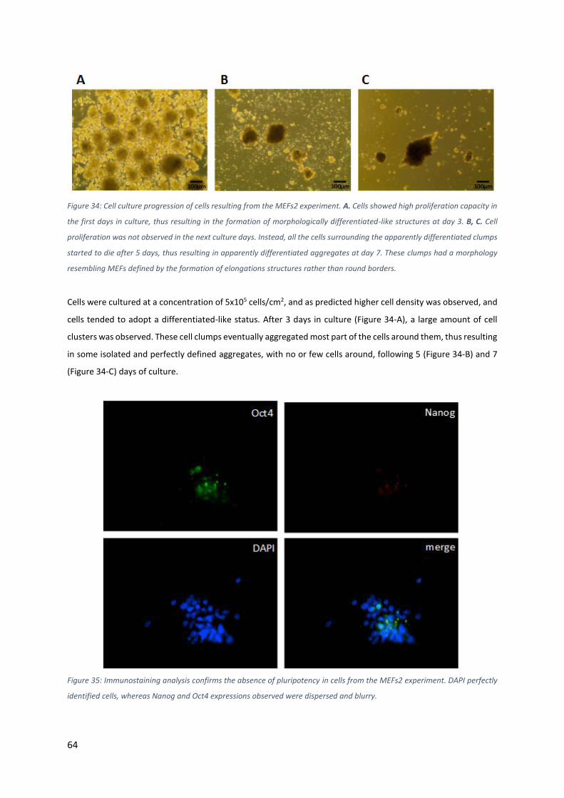
64
Figure 34: Cell culture progression of cells resulting from the MEFs2 experiment. A. Cells showed high proliferation capacity in
the first days in culture, thus resulting in the formation of morphologically differentiated-like structures at day 3. B, C. Cell
proliferation was not observed in the next culture days. Instead, all the cells surrounding the apparently differentiated clumps
started to die after 5 days, thus resulting in apparently differentiated aggregates at day 7. These clumps had a morphology
resembling MEFs defined by the formation of elongations structures rather than round borders.
Cells were cultured at a concentration of 5x10⁵ cells/cm2, and as predicted higher cell density was observed, and
cells tended to adopt a differentiated-like status. After 3 days in culture (Figure 34-A), a large amount of cell
clusters was observed. These cell clumps eventually aggregated most part of the cells around them, thus resulting
in some isolated and perfectly defined aggregates, with no or few cells around, following 5 (Figure 34-B) and 7
(Figure 34-C) days of culture.
Figure 35: Immunostaining analysis confirms the absence of pluripotency in cells from the MEFs2 experiment. DAPI perfectly
identified cells, whereas Nanog and Oct4 expressions observed were dispersed and blurry.

65
It was possible to conclude that single cells did not survive this culture, contrastingly to what was desired (mouse
pluripotent cells should survive as single cells, suggesting that pluripotency was not acquired). Interestingly, after
the seven days in culture, cells showed an elongated morphology tendency, characteristic of normally
differentiated and functional MEFs, thus suggesting some of these cells did not acquire any pluripotency and
survived the protocol maintaining their epigenetic identity during their exposure to the stress.
Intrigued by the results obtained with the first experiment using MEF cells, immunostaining analysis of this
second experiment was performed to clarify about the possible acquisition of pluripotency.
Through this analysis (Figure 35), it was not possible to identify any positive results. The expression of
pluripotency genes was very blurry and dispersed, thus not showing any perfectly defined cell. The absence of
pluripotency was confirmed by qPCR analysis (see chapter 4.3). The same controls were used to evaluate the
gene expression of this sample (for positive control see chapter 4.4.4 and for negative control see chapter 4.4.5).
4.2.3.1. Physical stress only
Since MEF cells were demonstrated to be very sensible to the stresses applied, a parallel study using only physical
stress was performed. Cells followed the same protocol already applied, and similarly to the previous
experiment, a sufficient amount of cells was obtained following all the trituration steps (Figure 36-B), despite
being considerably lower in comparison with the number of isolated cells (Figure 36-A), and thus cells were
cultured at the same concentration of the previous experiment (5x10⁵ cells/cm2).
Figure 36: Cell density after each perturbation step on MEFs with physical stress only. A. Cell density after isolation had
considerable values, as verified in the previous experiment using MEFs. B. Similarly to the previous results, the whole physical
stress procedure, including the three steps, led to high cell loss. However, a sufficient number of cells for posterior cell culture
was obtained.
Cell culture yielded the same results (Figure 37) observed in the experiment with both physical and chemical
stresses. In general, the elimination of the last perturbation step did not change the culture results, and the cells
acquired a similar morphology after the 7 days of culture. As expected, the typical morphology observed in
functional differentiated MEFs, characterized by elongated structures, was observed again.

66
Figure 37: Cell culture progression of cells resulting from MEFs with physical stress only. A, B, C. The experiment resulted in
the same observations of the MEFs2 experiment, characterized by differentiation tendencies after 3 days in culture (A), cell
loss and continuous differentiation after 5 days (B) and acquisition of MEF-resembling morphology after 7 days (C).
This observation would be expected due to the absence of the acid treatment. The main hope for the
reprogramming event was defined as the acid exposure (Obokata et al., 2014d), and the physical trituration was
intended to be only an extra step to facilitate the acquisition of pluripotency. Therefore, it would be expected
that these cells would maintain a differentiated state. Absence of pluripotency was expected.
Immunostaining results (Figure 38) obtained from samples taken at day 7 of culture showed the predictable
inexistence of pluripotency in these cells.
Figure 38: Immunostaining results confirmed the absence of pluripotency on MEFs exposed only to physical stress.

67
4.3. Reverse Transcription quantitative Polymerase Chain Reaction analysis
Some samples (as mentioned) were collected to be analyzed by qPCR. This analysis included a comparison of
those samples with two control pluripotent lines (mES cell line for the mouse samples and human H1 ES cell line
for the human sample), observing expression fold change of pluripotency genes, to elucidate about the possible
acquisition of a pluripotent state following the experiments.
Regarding the human sample analyzed (experiment EB4, which followed the differentiation of the human H1
OCT4-GFP+ reporter cell line into EBs to which the protocol was applied), qPCR results showed clear absence of
pluripotency, demonstrated by almost imperceptible OCT4 and NANOG genes expression levels, and showing
only SSEA1 expression (Figure 39).
Regarding mouse experiments, three cell samples were collected: cells resulting from the second experiment
(MEFs2) with MEF cells, MEFs cultured in sphere media, and cells resulting from the EB5 experiment (followed
the differentiation of mouse ES cells into EBs to which the protocol was applied). The results obtained from the
qPCR analysis (Figure 40) suggest the absence of pluripotency in the MEFs cells undergoing the protocol and
MEFs cultured directly in sphere media (as expected), described by almost complete absence of Nanog, SSEA1
and Rex1 expression, as compared to the control. Cells exposed to the protocol and MEFs directly cultured in
sphere media showed similar expression patterns, thus suggesting the protocol did not result in any gene
expression level alteration in these cells.
Figure 39: Results obtained from qPCR analysis of the cells from the EB4 experiment show absence of pluripotency, confirmed
by a significant fold-decrease in the expression of the pluripotency genes OCT4 and NANOG as compared with H1 reporter cell
line used as positive control.
0
0,2
0,4
0,6
0,8
1
1,2
1,4
OCT4 NANOG SSEA1Fold
Ch
ange
in g
en
e e
xpre
ssio
n r
ela
tive
to
u
nd
iffe
ren
tiat
ed
co
ntr
ol h
ES c
ell
line
Genes of Interest
H1 OCT4-GFP+control
EB4

68
Table 3: Experiments’ overview show that EBs’ dissociation changes successfully resulted in more efficient post-stresses culture, although all of them adapted uncontrolled differentiated-like status. First experiment using MEFs yielded apparently positive results, while the second refuted that possibility. Experiments conducted with TTs and GCs resulted in contamination and insufficient cell number, respectively, thus resulting in completely negative results.
Starting cell density Cell density after Proliferation capacity
Culture observation
Experiment Starting Material
Dissociation Method
Before Pipetting
1st pipetting 2nd pipetting 3rd pipetting Acid treatment
EB1 (human
UGENT11-2)
6 wells of EBs
0,05% trypsin-EDTA (5 min)
Low (4x10⁵ cells)
Unknown Unknown Low (3x10⁵ cells)
Very low (2x10⁵ cells)
No Not possible
EB2 (human
UGENT11-2)
6 wells of EBs
0,05% trypsin-EDTA (8 min)
Reasonable (1x10⁶ cells)
Low Low Low (7x10⁵ cells)
Low (4x10⁵ cells)
Some at the beginning
Single cells or small colonies (presumably dead after 7
days)
EB3 (human
UGENT11-2)
9 wells of EBs
0,25% trypsin-EDTA (5 min)
Medium (2x10⁶ cells)
Medium Medium Medium (1.5x10⁶ cells)
Low (6x10⁵ cells)
Yes (high) Increasingly big differentiated colonies
EB4 (human reporter H1)
9 wells of EBs
0,25% trypsin-EDTA (5 min)
Medium (2x10⁶ cells)
Medium Medium (slightly low)
Medium (1.5x10⁶ cells)
Medium (1.3x10⁶
cells)
Yes (very high)
Completely differentiated EB-like colonies
EB5 (mouse ES cells)
24 wells of EBs
0,25% trypsin-EDTA (5 min)
Very high (1x10⁷ cells)
Very high Very high Very high (8x10⁶ cells)
Very high (6x10⁶ cells)
Yes (very high)
Uncontrolled growth with possible differentiation
TTs1 Medium (3x10⁶ cells)
Not observed
Not observed
Medium (1.5x10⁶ cells)
Low (4.2x10⁵
cells)
Some Some contamination present leading to incapacity to grow
TTs2 Medium (3x10⁶ cells)
Medium Medium (slightly low)
Medium (1.6x10⁶ cells)
Low (7.8x10⁵
cells)
Almost none A lot of contamination present, cell death
GCs1 Reasonable (1x10⁶ cells)
Low Low Low (4.2x10⁵ cells)
Low (3x10⁵ cells)
No All cells dead
GCs2 Reasonable (1x10⁶ cells)
Medium Low Low (4.2x10⁵ cells)
Low (2x10⁵ cells)
Some Single cells with some initial proliferation
MEFs1 Medium (2.4x10⁶
cells)
Low Low Low (4.8x10⁵ cells)
Very low (2x10⁵ cells)
Yes (at the beginning)
Cell loss and differentiation
MEFs2
High (3.9x10⁶
cells)
Medium (slightly
high)
Medium (slightly
high)
Medium (2.8x10⁶ cells)
Medium (1x10⁶ cells)
Very high (at the
beginning)
Complete differentiation into cells resembling MEFs'
morphology

69
Figure 40: Results obtained by qPCR showed absence of pluripotency in the MEF-related experiments. EB5 experiment
presented some interesting expression of Rex1 and Gbx2 comparing to the pluripotent control. However, low expression of
both Nanog and SSEA1 suggests differentiation.
Contrastingly, cells from EB5 experiment showed some interesting results. Gbx2, the mouse homeobox gene,
known for being expressed in mES cells, having its expression decreased in differentiated cells (Chapman et al.,
1997), showed similar expression levels in the EB5 sample as compared to the control. Also, Rex1, a zinc finger
protein expressed mainly in undifferentiated cells (Scotland et al., 2009) showed an expression level that even
comparable to the undifferentiated ES control cells. Despite showing evidences of pluripotency, even evaluating
the slight Nanog expression observed, these cells did not acquire pluripotency. The surface marker gene SSEA1
expression level was particularly low. Since the expression level of this gene is known to decrease upon
differentiation (Zhao et al., 2012), this consisted in an evidence of differentiation of these cells.
4.4. Complementary work and Controls
4.4.1. Mouse Embryonic Stem cells cultured in sphere media
Somatic cells, following acid treatment, were stated to be reprogrammed into a pluripotent state during a 7 days
culture (Obokata et al., 2014d), in the termed sphere media, being supposed not to attach and to form small
round shaped cells. Only mouse cell data is available, and therefore it is unclear at this point if human cells,
0
0,2
0,4
0,6
0,8
1
1,2
1,4
1,6
mES Cell control MEF (cultured insphere media)
MEFs2 EB5
Fold
Ch
ange
in G
en
e E
xpre
ssio
n r
ela
tive
to
u
nd
iffe
ren
tiat
ed
mES
C c
on
tro
l
Different media for STAP induction
NANOG
SSEA1
REX1
GBX2

70
following these experiment, should adopt a morphology characteristic of hES cells or instead a status
characteristic of naïve hES cells, since they were cultured in ultra-low attachment plates (and hES cells are usually
grown in feeder-dependent conditions, usually with MEFs, to which they attach). However, since cells following
the protocol should acquire pluripotency through the 7 days of culture, it was admitted that sphere media was
(despite not having any compound to promote naïve pluripotency except LIF) capable of sustaining/induce both
proliferative capacity and pluripotent state of the cells cultured in it. Otherwise, the supposition that pluripotent
cells can arise in culture in this medium would not make sense. Intrigued by this supposition, it was decided to
culture mouse ES cells directly in sphere media (supplemented with heparin, EGF and mouse LIF) without being
exposed to any stress, and check cell progression through several days of culture.
Figure 41: Naïve mouse ES cells directly cultured in sphere media could not survive the culture conditions during 7 days. A.
After 3 days in culture, cell density was particularly high. B. By 5 days in culture, cell density decreased comparing to the
previous days. C. Cell density continued to decrease until day 7. Sphere media was demonstrated to be incapable of
maintaining naïve mES cells in culture.
Interestingly, proliferative capacity was not demonstrated on mouse ES cells directly cultured in sphere media,
fact demonstrated by a cell loss from day 3 to days 5 and 7 (Figure 41). Since both inhibitors that characterize 2i
conditions were not present, it was expected that these cells could not maintain a naïve state of pluripotency.
However, the medium should at least be capable of supporting cell survival, and it would be expected that these
cells could acquire a random proliferative capacity and differentiation tendency (due to the absence of 2i
conditions). Both of them were not verified. It is intriguing why a culture medium that is in fact incapable of
maintaining both naïve state of pluripotency and cell survival was suggested as capable of maintaining the
cultured cells for 7 days and induce them to acquire pluripotency. Therefore, this result represented an evidence
arguing against the possible acquisition of pluripotency by the cells cultured in this medium.
4.4.2. Protocol performed directly on Mouse Embryonic Stem cells
Intrigued by the response of pluripotent cells after being exposed to the stresses included in the protocol,
pluripotent mouse ES cells (the same used to grow embryoid bodies, EB5) were collected and directly exposed
both to the physical trituration steps and to acidic HBSS.

71
Figure 42: Naive mouse embryonic stem cells faced the whole protocol and showed high resistance to every step. A. High cell
density was used as starting material. B, C, D. High resistance to the trituration steps resulted in high cell density after the
first (B), second (C) and third (D) pipetting steps. E. Cell quantity was maintained even after acid exposure.
The protocol followed the exact same steps used for the EB5 experience, after the dissociation of those EBs into
single cells. High cell density was verified as starting material (Figure 42-A), after both the trituration steps (Figure
42-B, C, D), and even after acid exposure (Figure 42-E). Mouse ES cells showed, therefore, high resistance to the
described protocol, maintaining their integrity along the experiment and showing a particularly good cell density
before being cultured.
Following culture in sphere media (supplemented with the proper mouse compounds, EGF, heparin and mouse
LIF), cell growth was monitored during the 7 days of culture. As demonstrated, these cells showed high
proliferation capacity during the culture period, increasing the number of cells, and forming aggregates with
differentiation tendencies (Figure 43).
It was therefore possible to conclude that pluripotent mouse ES cells successfully survived and maintained their
capacities after being exposed to the defined stresses. Since pluripotent cells (possibly present in the initial
material) survived the whole protocol, they could be present and contribute to the differentiated-like
morphology observed in most of the experiments performed. Although at low probability, this would also mean
that a single pluripotent cell, which could still remain in that state in the 14 days differentiated EBs and maintain
its epigenetic characteristics during the experiment, would be sufficient to result in a false positive result. It
would be uncertain if the pluripotency observed would be a result of a previously pluripotent cell rather than
reprogramming. A negative control showed absence of pluripotency in the 14 days EBs (see chapter 4.4.3).

72
Figure 43: Mouse ES cells were directly exposed to the protocol and cultured in sphere media. A. After 3 days in culture, high
cell density was observed, along with differentiation tendencies. B, C. After 5 (B) and 7 (C) days in culture, cells tended to
aggregate into completely differentiated clumps. These observations suggested that undifferentiated pluripotent cell can
survive the whole experiment, maintaining their capabilities to proliferate and differentiate in vitro.
4.4.3. Negative Control – Embryoid Bodies are fully differentiated
A negative control for Oct4 and Nanog expression was performed by staining mouse EBs grown for 14 days. To
avoid discrepancy between the experiments and this biological replicate, the exact same culture conditions were
used, including the exact same culture medium (N2B27 without adding mouse LIF nor any inhibitor).
Figure 44: EBs' immunostaining results showed absence of pluripotency-expressing cells in the structure of the EB after 14
days of differentiation culture.

73
This control mainly aimed to prove the absence of the pluripotency genes expression, Oct4 and Nanog, after 14
days in culture. This analysis was useful to exclude the possibility of having pluripotent cells, after the 7 days of
culture in sphere media, which could be already present before the protocol, rather than being reprogrammed.
It was assumed that experiments using both mouse and human cells, despite using cells with different
characteristics and different differentiation culture media, would result in a similar differentiation level after 14
days culture. Therefore, this negative control performed with mES cells should be sufficient to elucidate about
the differentiation level after 14 days in all the experiments and the absence of pluripotency at this stage.
Immunostaining analysis showed clear absence of consistent expression of pluripotency genes Oct4 and Nanog
(Figure 44). Although there was some overlap between Oct4 expression and DAPI staining, thus suggesting that
some intact cells expressed Oct4 at the time of fixation, the absence of a Nanog expression overlapping with
Oct4 clarified about the absence of pluripotency. The observed expression of these genes was therefore due to
unspecific binding or eventually due to a possible existence of non-pluripotent Oct4 or Nanog expressing cells
(Ambady et al., 2010; Zangrossi et al., 2007), as a result of the spontaneous differentiation characteristic of EBs’
culture. It was therefore possible to conclude about the absence of pluripotency in EBs’ cells following 14 days
culture in the defined conditions.
4.4.4. Positive Control – Mouse Embryonic Stem cells express Oct4 and Nanog
Figure 45: Immunostaining analysis performed of mouse ES cells showed correct expression of pluripotency genes Oct4 and
Nanog. This analysis confirmed the efficiency of the staining method being used.

74
A positive control was performed, to test both the efficiency of the staining method applied and to prove the
successful expression of Oct4 and Nanog in pluripotent cells (naïve mES cells in this case).
For that, a sample of mouse ES cells, expanded to be differentiated into EBs (EB5 experiment), was collected,
fixed and stained using antibodies for Oct4 and Nanog genes (see chapter 3). These mouse ES cells reside in a
naïve state of pluripotency and, similarly to primed pluripotent cells, must perfectly express pluripotency-
associated genes Oct4 and Nanog (Loh et al., 2006; Nichols et al., 1998).
As demonstrated (Figure 45), Oct4 and Nanog expression patterns almost perfectly overlapped. Both genes also
overlapped with most part of the cells identified by DAPI, thus confirming those were intact pluripotent cells
(positive control for pluripotency expression) and that both the staining methods and the antibodies used for
the analysis were efficient.
4.4.5. Negative Control – Mouse Embryonic Fibroblast cells do not express pluripotency
Facing the apparently positive expression of pluripotency genes Oct4 and Nanog on the first experiment
conducted with MEF cells (MEFs1), a negative control proving the absence of pluripotency in those MEF cells
prior to be perturbed by any stress was necessary. Therefore, MEFs at passage 3 (the same used as starting
material for the experiment) were stained.
Figure 46: Immunostaining results show absence of pluripotency on MEF cells prior to the protocol. Oct4 and Nanog expression was completely absence.

75
Immunostaining results confirmed that completely functional and normal MEF cells, not being exposed to any
stress, showed clear absence of pluripotency, without any Oct4 or Nanog expression. This result demonstrated
that prior to the experiment, these cells did not show any pluripotency and thus the possible pluripotency
expression after the protocol would be due to reprogramming rather than being inherited.
4.5. Further Discussion
The reprogramming of somatic cells into pluripotency is generally associated with low efficiency, and the
requirement of a large amount of cells as starting material to obtain a proper quantity of pluripotent cells, no
matter the method used (Obokata et al., 2014d; Tachibana et al., 2013; Takahashi and Yamanaka, 2006), is a
well-known drawback of those techniques. In addition to the low efficiency predicted, several factors could have
contributed to the failure of the present work.
Experiments conducted with human cells during this work, through EBs’ growth, showed sufficient but not
particularly high amount of starting cells. Associated with the particularly low efficiency, in case of possible
reprogramming event, high amounts of reprogramed pluripotent cells could never be expected. Also, facing the
challenging protocol, which resulted in a lot of difficulties to maintain the cells alive during the experiments, it
was necessary to analyze cell survival and response to the perturbations as deeply as the pluripotency analysis
itself. Further studies using a higher amount of starting material (ideally two complete 24 well plates of EBs)
should be performed. A higher amount of starting cells would increase the probability of obtaining a pluripotent
cell population. Even so, a considerably high amount of material, following differentiation of mouse ES cells into
EBs (EB5), was exposed to the perturbations defined in the protocol. However, truly pluripotency was not
acquired. Further optimizations are necessary for this approach. The idea of starting with ES cells, posteriorly
differentiated into EBs, which were subjected to the reprogramming technique, should be related with a higher
facility to acquire reprogramming event, since EBs could reside in a less differentiated state than a functional
somatic cells, as suggested before. However, this methodology faced some drawbacks. In addition to the
necessity of large EBs’ cultures to have a larger amount of starting cells, the massive cell loss and inefficiency of
the dissociation step, which was indispensable before the cells were exposed to the physical perturbations,
represented issues that need to be revisited. Theoretically, the experiment EB5, following differentiation of mES
cells into EBs, should be the one from which the best results would be expected. Since the protocol was stated
to work with murine cells (Obokata et al., 2014d), and EBs should represent the possibly less differentiated
mouse cells used during this study, the reprogramming efficiency should be the highest possible to obtain. The
combination of the limited data available about this technique with the incapacity to reprogram mouse EBs into
a state of pluripotency suggested low expectations to the experiments using other kind of cells.
The necessity for a proper starting material was not only applied for the first methodology of the present work.
Isolation of somatic cells also requires efficient and high throughput methods. The method through which murine
somatic cells were isolated can be definitely related with the inefficiency of the procedure. Importantly, the
contamination observed during experiences with MEF cells was surely critical to the failure of the experiment.
More than perturbing cell culture, posterior to the whole treatment protocol, these impurities may have

76
interfered with the trituration procedures by blocking the passage of the cells through the pipette’s tip.
Moreover, the presence of these contaminations may have led to cells’ disruption following physical contact with
them, largely increasing cell death along the procedures. Again, the amount of cells needed for these
experiments was shown to be high, since high cell loss was verified even after the first pipetting step.
In all the experiments performed, the long procedure may have influenced negative results. A long maintenance
of the cells out of the incubator and in a buffer medium directly increased the amount of cells that were lost
during the experiment. The requirement of successive trituration steps followed by acid exposure, along with
several centrifugation rounds, left the cells in external extreme survival conditions. More than interfering with
the cell’s capacity to be maintained alive, this harsh conditions at which cells had to survive may have interfered
with the protocol itself, modifying the response of the cells to the external stimuli and possibly leaving them too
susceptible. During the present protocol these facts were unavoidable, since the whole protocol assumed the
necessity of a time-consuming physical treatment for the cells, which came out as a response for the failure and
incapacity to replicate the pre-defined protocol with acid treatment only. These experiments using chemical
treatment only, alleged not to work, were discarded and experiments including both physical and chemical
treatments were a priority. This evidence turned it impossible to decrease the experiment duration to the original
30 minutes only protocol. However, further experiments using the originally claimed protocol, with a single acid
exposure to induce pluripotency, should be performed, to analyze the results and compare expression levels of
pluripotency genes and morphology with the ones obtained with the present study.
Acid treatment on cells can be definitely considered as a severe method that leaves few chances of acquiring a
proper functional cell. By disrupting membrane proteins structure due to pH alterations, breaking their
supportive ionic bonds, the presence of an acidic environment leads the cells both to open potentially dangerous
channels in their membrane and lose their physical integrity (Campos et al., 2009; Lowes and Simmons, 2001;
Niero and Machado-Santelli, 2013). It is therefore more than expected that an acidic solution potentially kills the
cells that are exposed. Such evidence can explain both the cell loss verified after some of the acidic treatments
realized and the incompetence of the surviving cells to be maintained and proliferate after being cultured again,
associated with the intrinsic damage caused by that exposure to the acid. Different kinds of cells may have
different resistance to the exposure to equally different acidic solutions and/or compounds (Lampe et al., 2009).
Interestingly, acid can kill cancer cells (Mei et al., 2014). However, not all the cells have this response. Gastric
mucosa, the cells that line the stomach, are daily exposed to pH around 2 and still maintain their integrity. It is
therefore difficult at this point to predict the response of different cell types to the acidic solution, as performed
in the experiments. Further studies to evaluate the resistance of the different studied cell types to different acidic
solutions and pH levels could result in an optimal acid exposure, unveiling a relatively more optimized protocol
to be applied to each cell type.
The technical tips suggesting physical stress as essential to generate STAP cells came along with the idea that the
reprogramming event could be facilitated by any kind of stress induced on the cells. Therefore, following this
idea and as it was stated (Obokata et al., 2014d), other experimental procedures that equally expose the cells to
stressful conditions would or could have influence on the acquisition of pluripotency. The answer to the question
of whether strong external stimuli, of any kind, have any influence on a differentiated cell leading to a pluripotent

77
reprogrammed state, remains unclear. Cells’ trituration surely provoked several damage compromising their
integrity. It is hard to understand which effect this physical treatment could have on cells to influence them to
acquire pluripotency. Successive trituration may leave cells more susceptible and weak, and thus further
perturbations (such as the acid exposure) can be more efficient. This could eventually be the reason why this
physical treatment would facilitate reprogramming, since a direct relation between physical perturbation and
epigenetic change cannot be found. This way, the actual effect of the physical trituration is supposed to have on
the cells, which could scientifically explain an epigenetic change leading to a pluripotent state and justify the use
of such physical pipetting steps, remains undefined.

78
5. Conclusion and Perspectives
Together, the results obtained showed the incapacity of differentiated cells to acquire pluripotency following
acid exposure, contradicting what was previously claimed (Obokata et al., 2014d), even inducing various physical
stresses that were said to help the reprogramming event.
The ultimate goal to be achieved in the present work would be the efficient reprogramming using human cells,
assuming that the same reprogramming with mouse cells would be effectively possible and reproducible.
However, the results obtained showed clear divergence from a possible pluripotency acquisition. Moreover, it
was shown to be very difficult to conduct the whole protocol and reach the culture phase during which cells
would acquire pluripotency. All these difficulties faced during the study obligated the analysis to be equally
focused on the monitoring of the cells’ survival during the experiments and then on the possible acquisition of
pluripotency (if the cells efficiently reached the culture phase), rather than being only focused on the efficient
reprogramming event.
A completely adapted protocol, using embryoid bodies as starting material to be subjected to the perturbations,
was studied. In general, even starting from embryoid bodies, presumably in a less differentiated state than a fully
differentiated and functional somatic cells integrated in a leaving being, physical and chemical stresses together
were not shown to have the capacity to induce a reprogramming event. However, additional studies would be
necessary to evaluate these observations.
Experiments performed in functional and directly isolated, or cultured in vitro, murine somatic cells confirmed
the incapacity of this technique to induce pluripotency, contrastingly to what was stated. Mouse embryonic
fibroblasts represented an easily obtainable source of somatic cells for this study, since these cells are regularly
used as feeder layer for human embryonic stem cells culture studies. Although apparent positive results were
obtained in a first trial, a second experiment using a higher amount of cells from the same source, which could
possibly lead to more expressive positive results, refuted the first results, resulting in a fully differentiated cell
population. Cells from mouse tail tips showed no expression of pluripotency posteriorly to be exposed to these
perturbations. Moreover, experiments with these cells led to the presence of a large amount of impurities within
the cell culture. These contamination was directly related with the method by which mouse tail tip cells were
isolated, and thus this method should be improved. The amount of cells from both two and six tail tips was shown
to be insufficient due to the high cell loss verified during the protocol. Therefore, experiments involving these
cells can be hindered by ethical issues regarding the amount of animals that need to be sacrificed or damaged to
make ultimate experiments and achieve trustable results, with this and other different types of cells from the
organism. Also, following the facility in reprogramming granulosa cells into induced pluripotent stem cells, two
experiments were performed with this cell type, hoping that some pluripotency expression would be observed.
However, no pluripotency was acquired in neither the experiments. Moreover, the amount of cells obtained as
starting material for the experiments was shown to be particularly random, since cells extracted from six mice
yielded the same density as compared to only two mice. Therefore, the same issue already described for cells
from the tail tips (and for cells from any other tissue from the organism), related with the amount of animals

79
necessary to have a proper amount of cells to ultimately test this technique, is present with granulosa cells.
Accordingly to the amount of cells obtained as starting material from six sacrificed mice, it is expected to be quite
hard to obtain a proper amount of cells regarding both ethical and economical costs involved.
Although several optimizations were made during the experiences, regarding practical technique and handling,
as well as certain compounds used in certain steps, such as the use of trypsin-EDTA 0.25% that more effitiently
dissociated embryoid bodies into single cells, several improvements can be made in the whole protocol.
Generally, the volume of neutral HBSS in which cells were re-suspended before starting the first pipetting, the
volume of acid used to treat the cells, and the volume of sphere media in which cells were re-suspended and
posteriorly cultured, should be optimized. Although optimal values were suggested in Obokata’s papers, the
amount of cells obtained in every experiment varied drastically, and thus the optimal concentrations were not
obtained in all the experiments. The volume of neutral HBSS in which the starting material is suspended has a
crucial influence in the following physical stress steps, since a variation in the concentration of cells influences
the amount of cells that pass through the tips of the pipettes and suffer trituration by them. A too small cell
density means that a few cells are perturbed in each pipetting, whereas a too high cell density could mean
constriction of the tip, leading to an excessively severe trituration. An optimal volume of 2-3ml of HBSS was
suggested, at a concentration of 1x10⁶ cells/ml, and thus ideally a maximum of 3 million cells would be subjected
to the initial physical stress. Associated to the high cell loss observed during the protocol, this can be considered
a relatively slow amount of cells to be used as starting material, and surely reduces the chances to obtain a
proper amount of viable cells in culture. Relatively to the acidic HBSS treatment, the cell density is crucial. The
perfect cell concentration in this acidic suspension should be achieved in order to have a treatment that both
does not kill the cells and helps to reprogram them into a pluripotent state. A too small quantity of acid, relatively
to the amount of cells, may not have sufficient effect on the cells. On the other hand, a higher amount of acid
per cell can be too harsh for them, leading to death. Although an optimal concentration of 2x10⁶ cells/ml of acid
was suggested by Obokata et al, the issues faced during the present experiments regarding the limited quantity
of starting cells and the massive cell loss during the experiments rarely allowed to reach that concentration value.
Therefore, the lower concentration of cells in the acidic solution used in most part of the experiments surely
contributed for a particularly severe chemical perturbation, and may explain the extensive cell loss verified.
Despite all these optimizations that are necessary, the results obtained with this technique do not seem to create
any expectation regarding the possible acquisition of pluripotency following the exposure of cells to an external
strong environment, as previously stated, thus suggesting the incapacity of this technique to actually work.
The merely interesting results obtained would surely face several implications for the use of these cells for
therapeutics. Showing reprogramming capacity, this technique would be the third main reprogramming
possibility after somatic cell nuclear transfer and induced pluripotent stem cells. Interestingly, this
reprogramming method would have several advantages. Generally, as a reprogramming event, this technique
would allow the production of pluripotent stem cells from pre-existing differentiated somatic cells, thus
retrieving the usually necessary use of embryos as stem cells source. This would not only amplify the availability
of stem cells, but also overcome possible ethical issues regarding the use of embryos for research and
therapeutics. As a matter of fact, this technique would surpass somatic cell nuclear transfer, since it would not

80
be dependent of viable and good quality unfertilized donated oocytes. Also, somatic cell nuclear transfer
technically generates an embryo, thus generating a possible source of life. The reprogramming of somatic cells
rather than use of oocytes overcomes this ethical issue. Due to its simplicity, through the simple exposure of
somatic cells to an acidic solution for half an hour (and possibly to some extra physical treatment), it would be a
promising method to acquire the so much desired high amounts of pluripotent stem cells, necessary for
therapeutic purposes. Induced pluripotent stem cells’ technique can be considered the standard reprogramming
method, since this technique potentially overcomes all the issues enumerated above. However, problems related
with possible viral activation, since transgenes are transfected into the somatic cells through retroviruses to
acquire pluripotency, are a reality. Also, other direct reprogramming methods using plasmids or small molecules
can modify the genome of the cells, leading to mutations, despite overcoming the problems associated with the
use of virus. Interestingly, if this external stimuli-based reprogramming method would work, this issue related
with the virus-dependency would also be surpassed. However, the completely negative results obtained during
this work turn unclear if the process itself would result in several genome modifications, deviating the possibly
reprogrammed cells from an ES-like state, and thus not being suitable for proper research or therapeutics. Since
no viable pluripotent cells were obtained, is it not possibly to evaluate the capacity of the eventually generated
cells to be differentiated into different cells types with an efficiency comparable to ES cells. The unpredictability
of this stimulus-based reprogramming technique immediately argues against the possible use of the generated
pluripotent cells for research or therapeutics, since it would face a lot of safety-related issues.
The existence of STAP stem cells would be remarkable. The exceptionally easy method through which pluripotent
cells are supposed to be created is just unbeatable in the stem cells field. In the case of efficient reprogramming,
it would surely be the easiest and most accepted method to reprogram differentiated cells into pluripotency,
completely changing the way scientists would approach stem cells. However, if cells could be reprogrammed and
lose their functionality through external stresses, this would mean that a common cell from a common living
being could spontaneously lose its functionality anytime. The conditions involved in the reprogramming
technique are far away from the daily environment at which the organisms are exposed. However, the possibility
of a living being to acquire a tumor after reprogramming of its somatic cells due to being exposed to possibly
strong environmental condition (and all the living being are daily exposed to several severe conditions) cannot
be discarded. Following common sense, this fact surely argues against the capacity of this reprogramming
technique to reprogram somatic cells.
It is intriguing why a physical and/or chemical stress would lead to the acquisition of pluripotency the way it was
stated. Following the proof of mix or switch of cells and inclusion of fraudulent data by Obokata in her
publication, the possible acquisition of a pluripotent state triggered by an external stimulus remains a mystery.
However, at this points, the evidences of the obtained results suggest that this reprogramming method, in the
way it was suggested (Obokata et al., 2014d) and in the way it was adapted to the present work, is incapable of
inducing a differentiated cell into a state of truly pluripotency.

i
6. References
Aasen, T., Raya, A., Barrero, M.J., Garreta, E., Consiglio, A., Gonzalez, F., Vassena, R., Bilić, J., Pekarik, V., Tiscornia, G., et al. (2008). Efficient and rapid generation of induced pluripotent stem cells from human keratinocytes. Nat Biotechnol 26, 1276-1284. Aebi, U., Cohn, J., Buhle, L., and Gerace, L. (1986). The nuclear lamina is a meshwork of intermediate-type filaments. Nature 323, 560-564. Akhurst, R.J., Lehnert, S.A., Faissner, A., and Duffie, E. (1990). TGF beta in murine morphogenetic processes: the early embryo and cardiogenesis. Development 108, 645-656. Amabile, G., and Meissner, A. (2009). Induced pluripotent stem cells: current progress and potential for regenerative medicine. Trends Mol Med 15, 59-68. Ambady, S., Malcuit, C., Kashpur, O., Kole, D., Holmes, W.F., Hedblom, E., Page, R.L., and Dominko, T. (2010). Expression of NANOG and NANOGP8 in a variety of undifferentiated and differentiated human cells. Int J Dev Biol 54, 1743-1754. Avilion, A.A., Nicolis, S.K., Pevny, L.H., Perez, L., Vivian, N., and Lovell-Badge, R. (2003). Multipotent cell lineages in early mouse development depend on SOX2 function. Genes Dev 17, 126-140. Bao, S., Tang, F., Li, X., Hayashi, K., Gillich, A., Lao, K., and Surani, M.A. (2009). Epigenetic reversion of post-implantation epiblast to pluripotent embryonic stem cells. Nature 461, 1292-1295. Beattie, G.M., Lopez, A.D., Bucay, N., Hinton, A., Firpo, M.T., King, C.C., and Hayek, A. (2005). Activin A maintains pluripotency of human embryonic stem cells in the absence of feeder layers. Stem Cells 23, 489-495. Bechard, M., and Dalton, S. (2009). Subcellular localization of glycogen synthase kinase 3beta controls embryonic stem cell self-renewal. Mol Cell Biol 29, 2092-2104. Bernardo, A.S., Faial, T., Gardner, L., Niakan, K.K., Ortmann, D., Senner, C.E., Callery, E.M., Trotter, M.W., Hemberger, M., Smith, J.C., et al. (2011). BRACHYURY and CDX2 mediate BMP-induced differentiation of human and mouse pluripotent stem cells into embryonic and extraembryonic lineages. Cell Stem Cell 9, 144-155. Bourillot, P.Y., Aksoy, I., Schreiber, V., Wianny, F., Schulz, H., Hummel, O., Hubner, N., and Savatier, P. (2009). Novel STAT3 target genes exert distinct roles in the inhibition of mesoderm and endoderm differentiation in cooperation with Nanog. Stem Cells 27, 1760-1771. Briggs, R., and King, T.J. (1952). Transplantation of Living Nuclei From Blastula Cells into Enucleated Frogs' Eggs. Proc Natl Acad Sci U S A 38, 455-463. Brons, I.G., Smithers, L.E., Trotter, M.W., Rugg-Gunn, P., Sun, B., Chuva de Sousa Lopes, S.M., Howlett, S.K., Clarkson, A., Ahrlund-Richter, L., Pedersen, R.A., et al. (2007). Derivation of pluripotent epiblast stem cells from mammalian embryos. Nature 448, 191-195. Buehr, M., Meek, S., Blair, K., Yang, J., Ure, J., Silva, J., McLay, R., Hall, J., Ying, Q.L., and Smith, A. (2008). Capture of authentic embryonic stem cells from rat blastocysts. Cell 135, 1287-1298. Burridge, P.W., and Zambidis, E.T. (2013). Highly efficient directed differentiation of human induced pluripotent stem cells into cardiomyocytes. Methods Mol Biol 997, 149-161. Campbell, K.H., McWhir, J., Ritchie, W.A., and Wilmut, I. (1996). Sheep cloned by nuclear transfer from a cultured cell line. Nature 380, 64-66. Campos, F.M., Couto, J.A., Figueiredo, A.R., Tóth, I.V., Rangel, A.O., and Hogg, T.A. (2009). Cell membrane damage induced by phenolic acids on wine lactic acid bacteria. Int J Food Microbiol 135, 144-151. Carpenter, M.K., Rosler, E., and Rao, M.S. (2003). Characterization and differentiation of human embryonic stem cells. Cloning Stem Cells 5, 79-88. Cartwright, P., McLean, C., Sheppard, A., Rivett, D., Jones, K., and Dalton, S. (2005). LIF/STAT3 controls ES cell self-renewal and pluripotency by a Myc-dependent mechanism. Development 132, 885-896. Chambers, I., Colby, D., Robertson, M., Nichols, J., Lee, S., Tweedie, S., and Smith, A. (2003). Functional expression cloning of Nanog, a pluripotency sustaining factor in embryonic stem cells. Cell 113, 643-655. Chapman, G., Remiszewski, J.L., Webb, G.C., Schulz, T.C., Bottema, C.D., and Rathjen, P.D. (1997). The mouse homeobox gene, Gbx2: genomic organization and expression in pluripotent cells in vitro and in vivo. Genomics 46, 223-233. Chen, K.G., Mallon, B.S., McKay, R.D., and Robey, P.G. (2014). Human pluripotent stem cell culture: considerations for maintenance, expansion, and therapeutics. Cell Stem Cell 14, 13-26. Chung, Y., Klimanskaya, I., Becker, S., Li, T., Maserati, M., Lu, S.J., Zdravkovic, T., Ilic, D., Genbacev, O., Fisher, S., et al. (2008). Human embryonic stem cell lines generated without embryo destruction. Cell Stem Cell 2, 113-117.

ii
Chung, Y., Klimanskaya, I., Becker, S., Marh, J., Lu, S.J., Johnson, J., Meisner, L., and Lanza, R. (2006). Embryonic and extraembryonic stem cell lines derived from single mouse blastomeres. Nature 439, 216-219. Constantinescu, D., Gray, H.L., Sammak, P.J., Schatten, G.P., and Csoka, A.B. (2006). Lamin A/C expression is a marker of mouse and human embryonic stem cell differentiation. Stem Cells 24, 177-185. Cowan, C.A., Atienza, J., Melton, D.A., and Eggan, K. (2005). Nuclear reprogramming of somatic cells after fusion with human embryonic stem cells. Science 309, 1369-1373. Davidson, K.C., Adams, A.M., Goodson, J.M., McDonald, C.E., Potter, J.C., Berndt, J.D., Biechele, T.L., Taylor, R.J., and Moon, R.T. (2012). Wnt/β-catenin signaling promotes differentiation, not self-renewal, of human embryonic stem cells and is repressed by Oct4. Proc Natl Acad Sci U S A 109, 4485-4490. Dimos, J.T., Rodolfa, K.T., Niakan, K.K., Weisenthal, L.M., Mitsumoto, H., Chung, W., Croft, G.F., Saphier, G., Leibel, R., Goland, R., et al. (2008). Induced pluripotent stem cells generated from patients with ALS can be differentiated into motor neurons. Science 321, 1218-1221. Eckersley-Maslin, M.A., Bergmann, J.H., Lazar, Z., and Spector, D.L. (2013). Lamin A/C is expressed in pluripotent mouse embryonic stem cells. Nucleus 4, 53-60. Eminli, S., Foudi, A., Stadtfeld, M., Maherali, N., Ahfeldt, T., Mostoslavsky, G., Hock, H., and Hochedlinger, K. (2009). Differentiation stage determines potential of hematopoietic cells for reprogramming into induced pluripotent stem cells. Nat Genet 41, 968-976. Eminli, S., Utikal, J., Arnold, K., Jaenisch, R., and Hochedlinger, K. (2008). Reprogramming of neural progenitor cells into induced pluripotent stem cells in the absence of exogenous Sox2 expression. Stem Cells 26, 2467-2474. Evans, M. (2011). Discovering pluripotency: 30 years of mouse embryonic stem cells. Nat Rev Mol Cell Biol 12, 680-686. Evans, M.J. (1972). The isolation and properties of a clonal tissue culture strain of pluripotent mouse teratoma cells. J Embryol Exp Morphol 28, 163-176. Evans, M.J., and Kaufman, M.H. (1981). Establishment in culture of pluripotential cells from mouse embryos. Nature 292, 154-156. Fox, N., Damjanov, I., Martinez-Hernandez, A., Knowles, B.B., and Solter, D. (1981). Immunohistochemical localization of the early embryonic antigen (SSEA-1) in postimplantation mouse embryos and fetal and adult tissues. Dev Biol 83, 391-398. Gafni, O., Weinberger, L., Mansour, A.A., Manor, Y.S., Chomsky, E., Ben-Yosef, D., Kalma, Y., Viukov, S., Maza, I., Zviran, A., et al. (2013). Derivation of novel human ground state naive pluripotent stem cells. Nature 504, 282-286. Galvin, K.E., Travis, E.D., Yee, D., Magnuson, T., and Vivian, J.L. (2010). Nodal signaling regulates the bone morphogenic protein pluripotency pathway in mouse embryonic stem cells. J Biol Chem 285, 19747-19756. Gardner, R.L. (1983). Origin and differentiation of extraembryonic tissues in the mouse. Int Rev Exp Pathol 24, 63-133. Guo, G., Yang, J., Nichols, J., Hall, J.S., Eyres, I., Mansfield, W., and Smith, A. (2009). Klf4 reverts developmentally programmed restriction of ground state pluripotency. Development 136, 1063-1069. Gurdon, J.B. (1962). The developmental capacity of nuclei taken from intestinal epithelium cells of feeding tadpoles. J Embryol Exp Morphol 10, 622-640. Hanna, J., Cheng, A.W., Saha, K., Kim, J., Lengner, C.J., Soldner, F., Cassady, J.P., Muffat, J., Carey, B.W., and Jaenisch, R. (2010). Human embryonic stem cells with biological and epigenetic characteristics similar to those of mouse ESCs. Proc Natl Acad Sci U S A 107, 9222-9227. Hanna, J., Markoulaki, S., Mitalipova, M., Cheng, A.W., Cassady, J.P., Staerk, J., Carey, B.W., Lengner, C.J., Foreman, R., Love, J., et al. (2009). Metastable pluripotent states in NOD-mouse-derived ESCs. Cell Stem Cell 4, 513-524. Hanna, J., Markoulaki, S., Schorderet, P., Carey, B.W., Beard, C., Wernig, M., Creyghton, M.P., Steine, E.J., Cassady, J.P., Foreman, R., et al. (2008). Direct reprogramming of terminally differentiated mature B lymphocytes to pluripotency. Cell 133, 250-264. Hanna, J., Wernig, M., Markoulaki, S., Sun, C.W., Meissner, A., Cassady, J.P., Beard, C., Brambrink, T., Wu, L.C., Townes, T.M., et al. (2007). Treatment of sickle cell anemia mouse model with iPS cells generated from autologous skin. Science 318, 1920-1923. Hassani, S.N., Totonchi, M., Farrokhi, A., Taei, A., Larijani, M.R., Gourabi, H., and Baharvand, H. (2012). Simultaneous suppression of TGF-β and ERK signaling contributes to the highly efficient and reproducible generation of mouse embryonic stem cells from previously considered refractory and non-permissive strains. Stem Cell Rev 8, 472-481. Hassani, S.N., Totonchi, M., Gourabi, H., Schöler, H.R., and Baharvand, H. (2014a). Signaling roadmap modulating naive and primed pluripotency. Stem Cells Dev 23, 193-208.

iii
Hassani, S.N., Totonchi, M., Sharifi-Zarchi, A., Mollamohammadi, S., Pakzad, M., Moradi, S., Samadian, A., Masoudi, N., Mirshahvaladi, S., Farrokhi, A., et al. (2014b). Inhibition of TGFβ signaling promotes ground state pluripotency. Stem Cell Rev 10, 16-30. Hayashi, K., and Surani, M.A. (2009). Self-renewing epiblast stem cells exhibit continual delineation of germ cells with epigenetic reprogramming in vitro. Development 136, 3549-3556. Henderson, J.K., Draper, J.S., Baillie, H.S., Fishel, S., Thomson, J.A., Moore, H., and Andrews, P.W. (2002). Preimplantation human embryos and embryonic stem cells show comparable expression of stage-specific embryonic antigens. Stem Cells 20, 329-337. Hirai, H., Karian, P., and Kikyo, N. (2011). Regulation of embryonic stem cell self-renewal and pluripotency by leukaemia inhibitory factor. Biochem J 438, 11-23. Hochedlinger, K., and Jaenisch, R. (2006). Nuclear reprogramming and pluripotency. Nature 441, 1061-1067. Kim, J.B., Sebastiano, V., Wu, G., Araúzo-Bravo, M.J., Sasse, P., Gentile, L., Ko, K., Ruau, D., Ehrich, M., van den Boom, D., et al. (2009). Oct4-induced pluripotency in adult neural stem cells. Cell 136, 411-419. Kim, J.B., Zaehres, H., Wu, G., Gentile, L., Ko, K., Sebastiano, V., Araúzo-Bravo, M.J., Ruau, D., Han, D.W., Zenke, M., et al. (2008). Pluripotent stem cells induced from adult neural stem cells by reprogramming with two factors. Nature 454, 646-650. Kleinsmith, L.J., and Pierce, G.B. (1964). Multipotentiality of single embryonal carcinoma cells. Cancer Res 24, 1544-1551. Klimanskaya, I., Chung, Y., Becker, S., Lu, S.J., and Lanza, R. (2006). Human embryonic stem cell lines derived from single blastomeres. Nature 444, 481-485. Kunath, T., Saba-El-Leil, M.K., Almousailleakh, M., Wray, J., Meloche, S., and Smith, A. (2007). FGF stimulation of the Erk1/2 signalling cascade triggers transition of pluripotent embryonic stem cells from self-renewal to lineage commitment. Development 134, 2895-2902. Kurosawa, H. (2007). Methods for inducing embryoid body formation: in vitro differentiation system of embryonic stem cells. J Biosci Bioeng 103, 389-398. Lampe, K.J., Namba, R.M., Silverman, T.R., Bjugstad, K.B., and Mahoney, M.J. (2009). Impact of lactic acid on cell proliferation and free radical-induced cell death in monolayer cultures of neural precursor cells. Biotechnol Bioeng 103, 1214-1223. Li, Y., McClintick, J., Zhong, L., Edenberg, H.J., Yoder, M.C., and Chan, R.J. (2005). Murine embryonic stem cell differentiation is promoted by SOCS-3 and inhibited by the zinc finger transcription factor Klf4. Blood 105, 635-637. Li, Z., Fei, T., Zhang, J., Zhu, G., Wang, L., Lu, D., Chi, X., Teng, Y., Hou, N., Yang, X., et al. (2012). BMP4 Signaling Acts via dual-specificity phosphatase 9 to control ERK activity in mouse embryonic stem cells. Cell Stem Cell 10, 171-182. Liao, J., Cui, C., Chen, S., Ren, J., Chen, J., Gao, Y., Li, H., Jia, N., Cheng, L., Xiao, H., et al. (2009). Generation of induced pluripotent stem cell lines from adult rat cells. Cell Stem Cell 4, 11-15. Liu, H., Zhu, F., Yong, J., Zhang, P., Hou, P., Li, H., Jiang, W., Cai, J., Liu, M., Cui, K., et al. (2008). Generation of induced pluripotent stem cells from adult rhesus monkey fibroblasts. Cell Stem Cell 3, 587-590. Lo, B., and Parham, L. (2009). Ethical issues in stem cell research. Endocr Rev 30, 204-213. Loh, Y.H., Agarwal, S., Park, I.H., Urbach, A., Huo, H., Heffner, G.C., Kim, K., Miller, J.D., Ng, K., and Daley, G.Q. (2009). Generation of induced pluripotent stem cells from human blood. Blood 113, 5476-5479. Loh, Y.H., Wu, Q., Chew, J.L., Vega, V.B., Zhang, W., Chen, X., Bourque, G., George, J., Leong, B., Liu, J., et al. (2006). The Oct4 and Nanog transcription network regulates pluripotency in mouse embryonic stem cells. Nat Genet 38, 431-440. Lowes, S., and Simmons, N.L. (2001). Human intestinal cell monolayers are preferentially sensitive to disruption of barrier function from basolateral exposure to cholic acid: correlation with membrane transport and transepithelial secretion. Pflugers Arch 443, 265-273. Mao, J., Zhang, Q., Ye, X., Liu, K., and Liu, L. (2014). Efficient induction of pluripotent stem cells from granulosa cells by Oct4 and Sox2. Stem Cells Dev 23, 779-789. Martin, G.R. (1980). Teratocarcinomas and mammalian embryogenesis. Science 209, 768-776. Martin, G.R. (1981). Isolation of a pluripotent cell line from early mouse embryos cultured in medium conditioned by teratocarcinoma stem cells. Proc Natl Acad Sci U S A 78, 7634-7638. Martin, G.R., and Evans, M.J. (1974). The morphology and growth of a pluripotent teratocarcinoma cell line and its derivatives in tissue culture. Cell 2, 163-172. Matsuda, T., Nakamura, T., Nakao, K., Arai, T., Katsuki, M., Heike, T., and Yokota, T. (1999). STAT3 activation is sufficient to maintain an undifferentiated state of mouse embryonic stem cells. EMBO J 18, 4261-4269.

iv
Mei, W., Dong, C., Hui, C., Bin, L., Fenggen, Y., Jingjing, S., Cheng, P., Meiling, S., Yawen, H., Xiaoshan, W., et al. (2014). Gambogenic acid kills lung cancer cells through aberrant autophagy. PLoS One 9, e83604. Meissner, A., and Jaenisch, R. (2006). Mammalian nuclear transfer. Dev Dyn 235, 2460-2469. Mitsui, K., Tokuzawa, Y., Itoh, H., Segawa, K., Murakami, M., Takahashi, K., Maruyama, M., Maeda, M., and Yamanaka, S. (2003). The homeoprotein Nanog is required for maintenance of pluripotency in mouse epiblast and ES cells. Cell 113, 631-642. Munsie, M.J., Michalska, A.E., O'Brien, C.M., Trounson, A.O., Pera, M.F., and Mountford, P.S. (2000). Isolation of pluripotent embryonic stem cells from reprogrammed adult mouse somatic cell nuclei. Curr Biol 10, 989-992. Niakan, K.K., and Eggan, K. (2013). Analysis of human embryos from zygote to blastocyst reveals distinct gene expression patterns relative to the mouse. Dev Biol 375, 54-64. Nichols, J., Silva, J., Roode, M., and Smith, A. (2009). Suppression of Erk signalling promotes ground state pluripotency in the mouse embryo. Development 136, 3215-3222. Nichols, J., and Smith, A. (2009). Naive and primed pluripotent states. Cell Stem Cell 4, 487-492. Nichols, J., Zevnik, B., Anastassiadis, K., Niwa, H., Klewe-Nebenius, D., Chambers, I., Schöler, H., and Smith, A. (1998). Formation of pluripotent stem cells in the mammalian embryo depends on the POU transcription factor Oct4. Cell 95, 379-391. Niero, E.L., and Machado-Santelli, G.M. (2013). Cinnamic acid induces apoptotic cell death and cytoskeleton disruption in human melanoma cells. J Exp Clin Cancer Res 32, 31. Niwa, H., Ogawa, K., Shimosato, D., and Adachi, K. (2009). A parallel circuit of LIF signalling pathways maintains pluripotency of mouse ES cells. Nature 460, 118-122. Noggle, S., Fung, H.L., Gore, A., Martinez, H., Satriani, K.C., Prosser, R., Oum, K., Paull, D., Druckenmiller, S., Freeby, M., et al. (2011). Human oocytes reprogram somatic cells to a pluripotent state. Nature 478, 70-75. Obokata, H., Sasai, Y., and Niwa, H. Essential technical tips for STAP convertion culture from somatic cells. URL: E http://www.nature.com/protocolexchange/protocols/3013 (accessed 03.06.14) Obokata, H., Sasai, Y., Niwa, H., Kadota, M., Andrabi, M., Takata, N., Tokoro, M., Terashita, Y., Yonemura, S., Vacanti, C.A., et al. (2014a). Bidirectional developmental potential in reprogrammed cells with acquired pluripotency. Nature 505, 676-680. Obokata, H., Sasai, Y., Niwa, H., Kadota, M., Andrabi, M., Takata, N., Tokoro, M., Terashita, Y., Yonemura, S., Vacanti, C.A., et al. (2014b). Retraction: Bidirectional developmental potential in reprogrammed cells with acquired pluripotency. Nature 511, 112. Obokata, H., Wakayama, T., Sasai, Y., Kojima, K., Vacanti, M.P., Niwa, H., Yamato, M., and Vacanti, C.A. (2014c). Retraction: Stimulus-triggered fate conversion of somatic cells into pluripotency. Nature 511, 112. Obokata, H., Wakayama, T., Sasai, Y., Kojima, K., Vacanti, M.P., Niwa, H., Yamato, M., and Vacanti, C.A. (2014d). Stimulus-triggered fate conversion of somatic cells into pluripotency. Nature 505, 641-647. Okita, K., Ichisaka, T., and Yamanaka, S. (2007). Generation of germline-competent induced pluripotent stem cells. Nature 448, 313-317. Okita, K., Nakagawa, M., Hyenjong, H., Ichisaka, T., and Yamanaka, S. (2008). Generation of mouse induced pluripotent stem cells without viral vectors. Science 322, 949-953. Oldershaw, R.A., Baxter, M.A., Lowe, E.T., Bates, N., Grady, L.M., Soncin, F., Brison, D.R., Hardingham, T.E., and Kimber, S.J. (2010). Directed differentiation of human embryonic stem cells toward chondrocytes. Nat Biotechnol 28, 1187-1194. Parisi, S., Passaro, F., Aloia, L., Manabe, I., Nagai, R., Pastore, L., and Russo, T. (2008). Klf5 is involved in self-renewal of mouse embryonic stem cells. J Cell Sci 121, 2629-2634. Rais, Y., Zviran, A., Geula, S., Gafni, O., Chomsky, E., Viukov, S., Mansour, A.A., Caspi, I., Krupalnik, V., Zerbib, M., et al. (2013). Deterministic direct reprogramming of somatic cells to pluripotency. Nature 502, 65-70. Reubinoff, B.E., Itsykson, P., Turetsky, T., Pera, M.F., Reinhartz, E., Itzik, A., and Ben-Hur, T. (2001). Neural progenitors from human embryonic stem cells. Nat Biotechnol 19, 1134-1140. Rideout, W.M., Hochedlinger, K., Kyba, M., Daley, G.Q., and Jaenisch, R. (2002). Correction of a genetic defect by nuclear transplantation and combined cell and gene therapy. Cell 109, 17-27. Robertson, J.A. (2001). Human embryonic stem cell research: ethical and legal issues. Nat Rev Genet 2, 74-78. Röber, R.A., Weber, K., and Osborn, M. (1989). Differential timing of nuclear lamin A/C expression in the various organs of the mouse embryo and the young animal: a developmental study. Development 105, 365-378. Schöler, H.R., Hatzopoulos, A.K., Balling, R., Suzuki, N., and Gruss, P. (1989). A family of octamer-specific proteins present during mouse embryogenesis: evidence for germline-specific expression of an Oct factor. EMBO J 8, 2543-2550. Scotland, K.B., Chen, S., Sylvester, R., and Gudas, L.J. (2009). Analysis of Rex1 (zfp42) function in embryonic stem cell differentiation. Dev Dyn 238, 1863-1877.

v
Selwood, L., and Johnson, M.H. (2006). Trophoblast and hypoblast in the monotreme, marsupial and eutherian mammal: evolution and origins. Bioessays 28, 128-145. Sherman, M.I. (1975). Long term culture of cells derived from mouse blastocysts. Differentiation 3, 51-67. Shevinsky, L.H., Knowles, B.B., Damjanov, I., and Solter, D. (1982). Monoclonal antibody to murine embryos defines a stage-specific embryonic antigen expressed on mouse embryos and human teratocarcinoma cells. Cell 30, 697-705. Shi, Y., Desponts, C., Do, J.T., Hahm, H.S., Schöler, H.R., and Ding, S. (2008). Induction of pluripotent stem cells from mouse embryonic fibroblasts by Oct4 and Klf4 with small-molecule compounds. Cell Stem Cell 3, 568-574. Singh, A.M., Reynolds, D., Cliff, T., Ohtsuka, S., Mattheyses, A.L., Sun, Y., Menendez, L., Kulik, M., and Dalton, S. (2012). Signaling network crosstalk in human pluripotent cells: a Smad2/3-regulated switch that controls the balance between self-renewal and differentiation. Cell Stem Cell 10, 312-326. Smith, A.G., Heath, J.K., Donaldson, D.D., Wong, G.G., Moreau, J., Stahl, M., and Rogers, D. (1988). Inhibition of pluripotential embryonic stem cell differentiation by purified polypeptides. Nature 336, 688-690. Sokol, S.Y. (2011). Maintaining embryonic stem cell pluripotency with Wnt signaling. Development 138, 4341-4350. Tachibana, M., Amato, P., Sparman, M., Gutierrez, N.M., Tippner-Hedges, R., Ma, H., Kang, E., Fulati, A., Lee, H.S., Sritanaudomchai, H., et al. (2013). Human embryonic stem cells derived by somatic cell nuclear transfer. Cell 153, 1228-1238. Takahashi, K., Tanabe, K., Ohnuki, M., Narita, M., Ichisaka, T., Tomoda, K., and Yamanaka, S. (2007). Induction of pluripotent stem cells from adult human fibroblasts by defined factors. Cell 131, 861-872. Takahashi, K., and Yamanaka, S. (2006). Induction of pluripotent stem cells from mouse embryonic and adult fibroblast cultures by defined factors. Cell 126, 663-676. Takeuchi, H., Nakatsuji, N., and Suemori, H. (2014). Endodermal differentiation of human pluripotent stem cells to insulin-producing cells in 3D culture. Sci Rep 4, 4488. Tan, H.K., Toh, C.X., Ma, D., Yang, B., Liu, T.M., Lu, J., Wong, C.W., Tan, T.K., Li, H., Syn, C., et al. (2014). Human finger-prick induced pluripotent stem cells facilitate the development of stem cell banking. Stem Cells Transl Med 3, 586-598. Tesar, P.J., Chenoweth, J.G., Brook, F.A., Davies, T.J., Evans, E.P., Mack, D.L., Gardner, R.L., and McKay, R.D. (2007). New cell lines from mouse epiblast share defining features with human embryonic stem cells. Nature 448, 196-199. Thomson, J.A., Itskovitz-Eldor, J., Shapiro, S.S., Waknitz, M.A., Swiergiel, J.J., Marshall, V.S., and Jones, J.M. (1998). Embryonic stem cell lines derived from human blastocysts. Science 282, 1145-1147. Thomson, J.A., Kalishman, J., Golos, T.G., Durning, M., Harris, C.P., Becker, R.A., and Hearn, J.P. (1995). Isolation of a primate embryonic stem cell line. Proc Natl Acad Sci U S A 92, 7844-7848. Trounson, A. (2006). The production and directed differentiation of human embryonic stem cells. Endocr Rev 27, 208-219. Vacanti, C. Refined protocol for generating STAP cells from mature somatic cells. URL: https://research.bwhanesthesia.org/site_assets/51520d191eea6679ce000001/cterm/Refined_STAP_protocol-9a685fc86fec5ca857ad58ae75462d07.pdf (accessed 03.25.14) Vallier, L., Alexander, M., and Pedersen, R.A. (2005). Activin/Nodal and FGF pathways cooperate to maintain pluripotency of human embryonic stem cells. J Cell Sci 118, 4495-4509. Vallier, L., Touboul, T., Chng, Z., Brimpari, M., Hannan, N., Millan, E., Smithers, L.E., Trotter, M., Rugg-Gunn, P., Weber, A., et al. (2009). Early cell fate decisions of human embryonic stem cells and mouse epiblast stem cells are controlled by the same signalling pathways. PLoS One 4, e6082. Vazin, T., and Freed, W.J. (2010). Human embryonic stem cells: derivation, culture, and differentiation: a review. Restor Neurol Neurosci 28, 589-603. Wang, J., Dodd, C., Shankowsky, H.A., Scott, P.G., Tredget, E.E., and Group, W.H.R. (2008). Deep dermal fibroblasts contribute to hypertrophic scarring. Lab Invest 88, 1278-1290. Wang, Y.Z., Plane, J.M., Jiang, P., Zhou, C.J., and Deng, W. (2011). Concise review: Quiescent and active states of endogenous adult neural stem cells: identification and characterization. Stem Cells 29, 907-912. Watanabe, S., Umehara, H., Murayama, K., Okabe, M., Kimura, T., and Nakano, T. (2006). Activation of Akt signaling is sufficient to maintain pluripotency in mouse and primate embryonic stem cells. Oncogene 25, 2697-2707. Wernig, M., Meissner, A., Foreman, R., Brambrink, T., Ku, M., Hochedlinger, K., Bernstein, B.E., and Jaenisch, R. (2007). In vitro reprogramming of fibroblasts into a pluripotent ES-cell-like state. Nature 448, 318-324.

vi
Wernig, M., Zhao, J.P., Pruszak, J., Hedlund, E., Fu, D., Soldner, F., Broccoli, V., Constantine-Paton, M., Isacson, O., and Jaenisch, R. (2008). Neurons derived from reprogrammed fibroblasts functionally integrate into the fetal brain and improve symptoms of rats with Parkinson's disease. Proc Natl Acad Sci U S A 105, 5856-5861. Williams, R.L., Hilton, D.J., Pease, S., Willson, T.A., Stewart, C.L., Gearing, D.P., Wagner, E.F., Metcalf, D., Nicola, N.A., and Gough, N.M. (1988). Myeloid leukaemia inhibitory factor maintains the developmental potential of embryonic stem cells. Nature 336, 684-687. Xiao, L., Yuan, X., and Sharkis, S.J. (2006). Activin A maintains self-renewal and regulates fibroblast growth factor, Wnt, and bone morphogenic protein pathways in human embryonic stem cells. Stem Cells 24, 1476-1486. Xu, R.H., Sampsell-Barron, T.L., Gu, F., Root, S., Peck, R.M., Pan, G., Yu, J., Antosiewicz-Bourget, J., Tian, S., Stewart, R., et al. (2008). NANOG is a direct target of TGFbeta/activin-mediated SMAD signaling in human ESCs. Cell Stem Cell 3, 196-206. Yamanaka, S. (2009). A fresh look at iPS cells. Cell 137, 13-17. Yamanaka, S. (2012). Induced pluripotent stem cells: past, present, and future. Cell Stem Cell 10, 678-684. Ying, Q.L., Nichols, J., Chambers, I., and Smith, A. (2003). BMP induction of Id proteins suppresses differentiation and sustains embryonic stem cell self-renewal in collaboration with STAT3. Cell 115, 281-292. Ying, Q.L., Wray, J., Nichols, J., Batlle-Morera, L., Doble, B., Woodgett, J., Cohen, P., and Smith, A. (2008). The ground state of embryonic stem cell self-renewal. Nature 453, 519-523. Yuan, H., Corbi, N., Basilico, C., and Dailey, L. (1995). Developmental-specific activity of the FGF-4 enhancer requires the synergistic action of Sox2 and Oct-3. Genes Dev 9, 2635-2645. Zangrossi, S., Marabese, M., Broggini, M., Giordano, R., D'Erasmo, M., Montelatici, E., Intini, D., Neri, A., Pesce, M., Rebulla, P., et al. (2007). Oct-4 expression in adult human differentiated cells challenges its role as a pure stem cell marker. Stem Cells 25, 1675-1680. Zhang, K., Li, L., Huang, C., Shen, C., Tan, F., Xia, C., Liu, P., Rossant, J., and Jing, N. (2010). Distinct functions of BMP4 during different stages of mouse ES cell neural commitment. Development 137, 2095-2105. Zhang, P., Li, J., Tan, Z., Wang, C., Liu, T., Chen, L., Yong, J., Jiang, W., Sun, X., Du, L., et al. (2008). Short-term BMP-4 treatment initiates mesoderm induction in human embryonic stem cells. Blood 111, 1933-1941. Zhao, W., Ji, X., Zhang, F., Li, L., and Ma, L. (2012). Embryonic stem cell markers. Molecules 17, 6196-6236. Zhou, J., Su, P., Wang, L., Chen, J., Zimmermann, M., Genbacev, O., Afonja, O., Horne, M.C., Tanaka, T., Duan, E., et al. (2009). mTOR supports long-term self-renewal and suppresses mesoderm and endoderm activities of human embryonic stem cells. Proc Natl Acad Sci U S A 106, 7840-7845. Zhou, X., Sasaki, H., Lowe, L., Hogan, B.L., and Kuehn, M.R. (1993). Nodal is a novel TGF-beta-like gene expressed in the mouse node during gastrulation. Nature 361, 543-547. Zuo, B., Yang, J., Wang, F., Wang, L., Yin, Y., Dan, J., Liu, N., and Liu, L. (2012). Influences of lamin A levels on induction of pluripotent stem cells. Biol Open 1, 1118-1127.
Related Documents