pISSN 2287-2728 eISSN 2287-285X http://dx.doi.org/10.3350/cmh.2015.21.3.257 Clinical and Molecular Hepatology 2015;21:257-267 Original Article Corresponding author : Maria F. Botelho Biophysics Unit, Faculty of Medicine, University of Coimbra, Azinhaga de Santa Comba – Celas, 3000-541 Coimbra, Portugal Tel: +351 239480200, Fax: +351 239480217 E-mail: [email protected] Abbreviations: HCC, Hepatocellular Carcinoma; p-P53, phosphorylated P53 protein; ROS, reactive oxygen species Received : Apr. 1, 2015 / Revised : Jun. 13, 2015 / Accepted : Jul. 27, 2015 INTRODUCTION Hepatocellular carcinoma (HCC) is the most common primary liver malignancy with a rising incidence worldwide, being the sec- ond cause of cancer death. 1 The prognosis of HCC patients is poor, and therapies are in most cases only the palliative approach. Surgical treatments have more satisfactory results, however, few patients can benefit from it. The therapeutic options for HCC of- ten have more limitations and disappointing results. 2,3 In this con- text, it is urgent to study and develop new therapeutic strategies. Radiotherapy is one of the most effective anticancer therapies and it has been used to treat a wide variety of tumors. The suc- Influence of P53 on the radiotherapy response of hepatocellular carcinoma Ana R. Gomes 1 , Ana M. Abrantes 1,2,3 , Ana F. Brito 1,2,3 , Mafalda Laranjo 1,2,3 , João E. Casalta-Lopes 1,2 , Ana C. Gonçalves 2,3,4 , Ana B. Sarmento-Ribeiro 2,3,4 , Maria F. Botelho 1,2,3 , and José G. Tralhão 1,2,5 1 Biophysics Unit, Faculty of Medicine of University of Coimbra, Coimbra, Portugal; 2 Center of Investigation on Environmental, Genetics and Oncobiology (CIMAGO), Faculty of Medicine of University of Coimbra, Coimbra, Portugal; 3 CNC.IBILI, University of Coimbra, Coimbra, Portugal; 4 Applied Molecular Biology and Hematology Group, Faculty of Medicine of University of Coimbra, Coimbra, Portugal; 5 Surgical Department A, CHUC, Coimbra, Portugal Background/Aims: Hepatocellular carcinoma (HCC) is one of the most common cancers worldwide, and it has a poor prognosis and few therapeutic options. Radiotherapy is one of the most effective forms of cancer treatment, and P53 protein is one of the key molecules determining how a cell responds to radiotherapy. The aim of this study was to determine the therapeutic efficacy of iodine-131 in three human HCC cell lines. Methods: Western blotting was used to measure P53 expression. The effects of radiotherapy with iodine-131 were assessed by using the clonogenic assay to evaluate cell survival. Flow cytometry was carried out to examine the effects of iodine-131 on cell death, oxidative stress, reduced intracellular glutathione expression, the mitochondrial membrane potential, and the cell cycle. Results: The P53 protein was not expressed in Hep3B2.1-7 cells, was expressed at normal levels in HepG2 cells, and was overexpressed in HuH7 cells. P53 expression in the HuH7 and HepG2 cell lines increased after internal and external irradiation with iodine-131. Irradiation induced a decrease in cell survival and led to a decrease in cell viability in all of the cell lines studied, accompanied by cell death via late apoptosis/necrosis and necrosis. Irradiation with 131-iodine induced mostly cell-cycle arrest in the G0/G1 phase. Conclusions: These results suggest that P53 plays a key role in the radiotherapy response of HCC. (Clin Mol Hepatol 2015;21:257-267) Keywords: Hepatocellular carcinoma; Iodine-131; Radiotherapy; P53 Copyright © 2015 by The Korean Association for the Study of the Liver This is an Open Access article distributed under the terms of the Creative Commons Attribution Non-Commercial License (http://creativecommons.org/licenses/by-nc/3.0/) which permits unrestricted non-commercial use, distribution, and reproduction in any medium, provided the original work is properly cited.

Welcome message from author
This document is posted to help you gain knowledge. Please leave a comment to let me know what you think about it! Share it to your friends and learn new things together.
Transcript

pISSN 2287-2728 eISSN 2287-285X
http://dx.doi.org/10.3350/cmh.2015.21.3.257Clinical and Molecular Hepatology 2015;21:257-267Original Article
Corresponding author : Maria F. BotelhoBiophysics Unit, Faculty of Medicine, University of Coimbra, Azinhaga de Santa Comba – Celas, 3000-541 Coimbra, PortugalTel: +351 239480200, Fax: +351 239480217E-mail: [email protected]
Abbreviations: HCC, Hepatocellular Carcinoma; p-P53, phosphorylated P53 protein; ROS, reactive oxygen species
Received : Apr. 1, 2015 / Revised : Jun. 13, 2015 / Accepted : Jul. 27, 2015
INTRODUCTION
Hepatocellular carcinoma (HCC) is the most common primary
liver malignancy with a rising incidence worldwide, being the sec-
ond cause of cancer death.1 The prognosis of HCC patients is
poor, and therapies are in most cases only the palliative approach.
Surgical treatments have more satisfactory results, however, few
patients can benefit from it. The therapeutic options for HCC of-
ten have more limitations and disappointing results.2,3 In this con-
text, it is urgent to study and develop new therapeutic strategies.
Radiotherapy is one of the most effective anticancer therapies
and it has been used to treat a wide variety of tumors. The suc-
Influence of P53 on the radiotherapy response of hepatocellular carcinomaAna R. Gomes1, Ana M. Abrantes1,2,3, Ana F. Brito1,2,3, Mafalda Laranjo1,2,3, João E. Casalta-Lopes1,2, Ana C. Gonçalves2,3,4, Ana B. Sarmento-Ribeiro2,3,4, Maria F. Botelho1,2,3, and José G. Tralhão1,2,5
1Biophysics Unit, Faculty of Medicine of University of Coimbra, Coimbra, Portugal; 2Center of Investigation on Environmental, Genetics and Oncobiology (CIMAGO), Faculty of Medicine of University of Coimbra, Coimbra, Portugal; 3CNC.IBILI, University of Coimbra, Coimbra, Portugal; 4Applied Molecular Biology and Hematology Group, Faculty of Medicine of University of Coimbra, Coimbra, Portugal; 5Surgical Department A, CHUC, Coimbra, Portugal
Background/Aims: Hepatocellular carcinoma (HCC) is one of the most common cancers worldwide, and it has a poor prognosis and few therapeutic options. Radiotherapy is one of the most effective forms of cancer treatment, and P53 protein is one of the key molecules determining how a cell responds to radiotherapy. The aim of this study was to determine the therapeutic efficacy of iodine-131 in three human HCC cell lines. Methods: Western blotting was used to measure P53 expression. The effects of radiotherapy with iodine-131 were assessed by using the clonogenic assay to evaluate cell survival. Flow cytometry was carried out to examine the effects of iodine-131 on cell death, oxidative stress, reduced intracellular glutathione expression, the mitochondrial membrane potential, and the cell cycle.Results: The P53 protein was not expressed in Hep3B2.1-7 cells, was expressed at normal levels in HepG2 cells, and was overexpressed in HuH7 cells. P53 expression in the HuH7 and HepG2 cell lines increased after internal and external irradiation with iodine-131. Irradiation induced a decrease in cell survival and led to a decrease in cell viability in all of the cell lines studied, accompanied by cell death via late apoptosis/necrosis and necrosis. Irradiation with 131-iodine induced mostly cell-cycle arrest in the G0/G1 phase. Conclusions: These results suggest that P53 plays a key role in the radiotherapy response of HCC. (Clin Mol Hepatol 2015;21:257-267)Keywords: Hepatocellular carcinoma; Iodine-131; Radiotherapy; P53
Copyright © 2015 by The Korean Association for the Study of the LiverThis is an Open Access article distributed under the terms of the Creative Commons Attribution Non-Commercial License (http://creativecommons.org/licenses/by-nc/3.0/) which permits unrestricted non-commercial use, distribution, and reproduction in any medium, provided the original work is properly cited.

258 http://www.e-cmh.orghttp://dx.doi.org/10.3350/cmh.2015.21.3.257
Clin Mol HepatolVolume_21 Number_3 September 2015
cess of radiotherapy against cancer depends on its ability to kill
tumor cells, while preserving the adjacent normal tissue.4,5 Radio-
therapy can be external and internal. In external radiotherapy, the
device directs rays of high energy to the tumor tissue affecting
also a small volume of surrounding normal tissue. In internal ra-
diotherapy, the radiation source is placed in/or near the tumor site
emitting radiation continuously for weeks, months or even one
year, allowing an higher total dose of radiation to be applied in a
shorter time than in the external radiotherapy.4,6,7 Several types of
radiation sources can be used. One of them is beta radiation from
different radionuclides.6,7
When, in 1943, radio-iodine was first used in the treatment of
metastasized thyroid carcinoma, its success in tumor response
was considered a “miracle”. Since then, many efforts have been
made to apply radioisotope therapy in other tumors.8 However,
only a small number of radionuclides have been used to the HCC
treatment, such as iodine-131, yttrium-90, rhenium-188 and hol-
mium-166 with promising results.9-11
Iodine-131 is a radionuclide, with interesting properties for anti-
cancer therapy. It is available with high radiochemical purity and
specific activity, has a long half-life of 8.06 days and decays with
production of beta particles (maximum energy of 606 keV), and
gamma emissions (main peak at 364 keV).12
In radiotherapy there are a variety of cellular processes involving
essential proteins, and which determine the success of treatment.
The P53 protein is a transcription factor associated with more
than 50% of human tumors and is a key molecule involved in the
response to radiotherapy.13,14 It is encoded by the tumor suppres-
sor gene TP53 located on the short arm of chromosome 17
(17p13.1 locus) and has 393 amino acids and four domains.14,15
The role of the tumor suppressor genes is to help in the preven-
tion and repair of genetic damage. When cell damage occurs in
the DNA, and P53 is functional, this protein may induce growth
arrest, often in G1 or G2 phases of cell cycle, or lead to cell sui-
cide by apoptosis, preventing uncontrolled proliferation and tumor
development.4,5,13,16 In contrast, if the P53 protein is inactive,
which can happen by mutation or degradation, DNA damage is
not detected which leads to a genetic instability. This facilitates
the initiation and progression of tumors and besides the tumors
become resistant to chemo and radiotherapy.4,13,14 The resistance
to radiation appears to be related to germ line P53 mutation,
however, the association between P53 expression and radiothera-
py response in HCC has not been well studied. Thus, the signifi-
cance of P53 aberration in HCC remains inconclusive.4,5,13
This work aims to investigate the efficiency of iodine-131 radio-
therapy in three HCC cell lines, and the influence of the P53 pro-
tein in the biological mechanisms of HCC. Three human HCC cell
lines were exposed to different doses of internal or external radia-
tion sources of iodine-131, which differ in P53 expression 17 and
their viability, cell death and reactive oxygen species (ROS) pro-
duction were as well evaluated.
MATERIAL AND METHODS
Cell culture We used three human HCC cell lines: HepG2, HuH7 and
Hep3B2.1-7. HepG2 and Hep3B2.1-7 cell lines were obtained from
American Tissue Cell Collection (ATCC), USA. HuH7 cell line was
obtained from Japanese Collection of Research Bioresources
(JCRB), Japan. Cells were propagated on adherent cultures in Dul-
becco’s Modified Eagle Medium (DMEM) with 10% Fetal Bovine
Serum (FBS) (Gibco 2010-09), 100 units/mL of penicillin and 100
µg/mL of streptomycin (Gibco 15140-122), and incubated at 37°C
in 5% CO2 atmosphere.
IrradiationDuring the cells exponential growth phase, 5x10⁵ cells were
exposed to internal and external radiation with iodine-131 (IBA Molecular). The conditions used for each irradiation type are out-
lined in Tables 1 and 2. Concerning the internal radiation, the ra-
dionuclide was placed directly in contact with the cells that were
Table 1. Internal irradiation conditions
Activity (mCi)
Time (min)
Internal radiation (Gy)
SourceArea (cm2)
2,9×10⁻⁴ 5 1 131I 25
5,5×10⁻⁴ 5 2 131I 25
1,4×10⁻³ 5 5 131I 25
2,9×10⁻³ 5 10 131I 25
5,5×10⁻³ 5 20 131I 25
Table 2. External irradiation conditions
Activity (mCi)
Time (min)
External radiation (Gy)
SourceArea (cm2)
4 7 1 131I 25
4 13,6 2 131I 25
4 34 5 131I 25
4 68 10 131I 25
8 68 20 131I 25

259
Ana R. Gomes, et al. P53 expression in radiotherapy response: in vitro studies
http://www.e-cmh.org http://dx.doi.org/10.3350/cmh.2015.21.3.257
washed afterwards with phosphate buffered saline (PBS). Related
to the external radiation, a radionuclide source was positioned
below the flask containing the cells. Doses were calculated as-
suming the worst case scenario, i.e., all the emitted energy in the
decay process was absorbed by the cells. To calculate the internal
radiation exposure dose, was used the equation 1,
� � ���������������Ē
���� (equation 1)
������
���� �
� � � ����� (equation 2)
(equation 1)
where D is the absorbed dose (Gy), A0 the initial activity of the radioactive source (mCi), T1/2 the half-life time (s), t the irradia-tion time (s), Ē the average energy per disintegration (eV) and M the mass of the sample subjected to irradiation (kg).
To calculate the external radiation exposure dose, was used the equation 2, where D is the exposure dose (mGy), Г radiation
constant specifies
� � ���������������Ē
���� (equation 1)
������
���� �
� � � ����� (equation 2)
, A the radioactive source activity
(GBq), t the exposure time (h) and d the distance from the source (m).
� � ���������������Ē
���� (equation 1)
������
���� �
� � � ����� (equation 2)
(equation 2)
P53 and phosphorylated P53 expression P53 and phosphorylated P53 protein (p-P53) protein expression
levels were determined by western blot. Cell extracts were pre-
pared in ice using a solution of radioimmunoprecipitation assay
(RIPA) buffer and complete Mini Ethylenediamine tetraacetic acid
(EDTA)-free (Roche). Protein concentrations were determined by
bicinchoninic (BCA) method. Sodium dodecyl sulfate polyacryl-
amide gel electrophoresis (SDS-PAGE) was held using a 10%
acrylmide gel. Proteins were electrotransfered to nitrocellulose
membrane (PVDF) at 4°C, during 1 hour. Membrane blocking was
performed with 4% bovine serum albumin (BSA) in tris-buffered
saline tween-20 (TBS-T) for 1 hour at room temperature. Mem-
branes were incubated with primary antibody (P53 or p-P53 or
β-actin, from Santa Cruz Biotechnology) and secondary antibody
(mouse antibody, from Sigma) according to the manufacturer’s in-
structions. The blots were stained with elemental chlorine free
(ECF, Sigma) and read in 9000 Typhoon FLA equipment.
Cell survivalCell survival was evaluated using clonogenic assay. Six hundred
plated cells were irradiated with internal or external source of io-
dine-131 and twelve days afterwards were fixed with methanol
and stained with crystal violet. Colonies with more than 50 cells
were counted and the efficiency plate (EP) (relation between the
number of colonies counted and the number of cells plated) and
survival factor (SF) (relationship between EP of the treated sample
and the EP of the control) were determined.
Flow cytometryFlow cytometry was used to analyze cell viability, cell death and
cell cycle as well as to determine ROS and reduced glutathione
production and mitochondrial membrane potential. All flow cy-
tometry studies used 106 cells per probe and exposures to internal
or external radiation of 1 or 20 Gy occurred 24 h before the assay.
For all procedures were performed controls, i.e. cells not subject
to irradiation.
Cell viability and cell deathThe influence of irradiation with iodine-131 on cell viability, and
the types of induced cell death were determined by flow cytome-
try using annexin-V/propidium iodide (AV/PI) (KIT Immunotech).
One of the main features of cell death by apoptosis is the redistri-
bution of plasma membrane phosphatidylserine, a phospholipid
that, in apoptotic cells, is translocated from the inner to the outer
leaflet of the plasmatic membrane and binds to AV. Complemen-
tarily, PI, which does not permeate viable cells, binds to deoxyri-
bonucleic acid (DNA) intercalating between the bases on late
apoptotic and necrotic cells.18 In this assay, 106 cells were incubat-
ed during 15 min in binding buffer with 1 µL of AV (Kit Immuno-
tech) and 5 µL of PI (Kit Immunotech). Subsequently, cells were
excited at a wavelength of 525 nm for AV and 640 nm for PI, col-
lecting 104 events to assess the percentage of viable, early apop-
totic, late apoptotic/necrotic, and necrotic cells.18
Evaluation of ROS and reduced glutathione production, mitochondrial membrane potential and cell cycle
ROS production: intracellular peroxide were measured using
2’,7’-dichlorodihydrofluorescein diacetate (DCFH2-DA, Invitrogen);
intracellular superoxide was determined using the dihydroethidium
probe (DHE, Sigma). Expression of intracellular reduced glutathione
was evaluated with mercury orange (GSH, Sigma). Mitochondrial
membrane potential was analyzed with 5,5’,6,6’-tetrachloro-
1,1’,3,3’-tetraethylbenzimidazol carbocyanine (JC-1, Invitrogen). Cell
cycle was evaluated with PI/RNase solution (Immunostep). Assays
were performed according to a method described by Mamede et
al.18 Results are presented as mean fluorescence intensity (MFI).
260 http://www.e-cmh.orghttp://dx.doi.org/10.3350/cmh.2015.21.3.257
Clin Mol HepatolVolume_21 Number_3 September 2015
Statistical analysisCell survival curves were obtained using Origin Pro v8.0, fitting
the experimental data to two models: for lower doses, the data
was fitted to a linear-quadratic model, according to the equation
SF=e-αD-βD2; for higher radiation doses, we fitted the data to a
linear model, using SF=e-C-αD. These two models were chosen as
they fitted the data when used together. P53, p-P53 expression,
and all flow cytometry results were analyzed using IBM SPSS® v.19
(IBM Corporation, Armonk, New York, USA). In the descriptive
analysis measures of central tendency (mean and median) and
dispersion (standard deviation and interquartile range) were ob-
tained for quantitative variables. To assess normality of the distri-
bution of quantitative variables, Shapiro-Wilk’s test was used.
Parametric tests were used for comparisons in the case normality
was observed; in the opposite case we used nonparametric tests.
For comparison of quantitative variables between two groups we
used Student’s t test (parametric) or Mann-Whitney’s test (non-
parametric). For more than two groups we used one factor Analysis
of Variance (ANOVA) test, with post-hoc Tukey test (parametric) or
Kruskal-Wallis test with multiple comparisons performed using the
Mann-Whitney’s test with Bonferroni correction (nonparametric).
For all comparisons a significance level of 5% was considered.
RESULTS
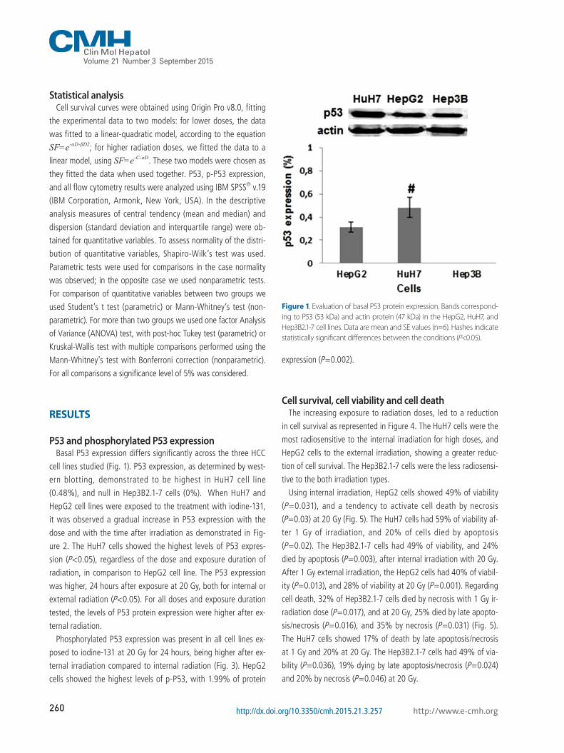
P53 and phosphorylated P53 expression Basal P53 expression differs significantly across the three HCC
cell lines studied (Fig. 1). P53 expression, as determined by west-
ern blotting, demonstrated to be highest in HuH7 cell line
(0.48%), and null in Hep3B2.1-7 cells (0%). When HuH7 and
HepG2 cell lines were exposed to the treatment with iodine-131,
it was observed a gradual increase in P53 expression with the
dose and with the time after irradiation as demonstrated in Fig-
ure 2. The HuH7 cells showed the highest levels of P53 expres-
sion (P<0.05), regardless of the dose and exposure duration of
radiation, in comparison to HepG2 cell line. The P53 expression
was higher, 24 hours after exposure at 20 Gy, both for internal or
external radiation (P<0.05). For all doses and exposure duration
tested, the levels of P53 protein expression were higher after ex-
ternal radiation.
Phosphorylated P53 expression was present in all cell lines ex-
posed to iodine-131 at 20 Gy for 24 hours, being higher after ex-
ternal irradiation compared to internal radiation (Fig. 3). HepG2
cells showed the highest levels of p-P53, with 1.99% of protein
expression (P=0.002).
Cell survival, cell viability and cell death The increasing exposure to radiation doses, led to a reduction
in cell survival as represented in Figure 4. The HuH7 cells were the
most radiosensitive to the internal irradiation for high doses, and
HepG2 cells to the external irradiation, showing a greater reduc-
tion of cell survival. The Hep3B2.1-7 cells were the less radiosensi-
tive to the both irradiation types.
Using internal irradiation, HepG2 cells showed 49% of viability
(P =0.031), and a tendency to activate cell death by necrosis
(P=0.03) at 20 Gy (Fig. 5). The HuH7 cells had 59% of viability af-
ter 1 Gy of irradiation, and 20% of cells died by apoptosis
(P=0.02). The Hep3B2.1-7 cells had 49% of viability, and 24%
died by apoptosis (P=0.003), after internal irradiation with 20 Gy.
After 1 Gy external irradiation, the HepG2 cells had 40% of viabil-
ity (P=0.013), and 28% of viability at 20 Gy (P=0.001). Regarding
cell death, 32% of Hep3B2.1-7 cells died by necrosis with 1 Gy ir-
radiation dose (P=0.017), and at 20 Gy, 25% died by late apopto-
sis/necrosis (P=0.016), and 35% by necrosis (P=0.031) (Fig. 5).
The HuH7 cells showed 17% of death by late apoptosis/necrosis
at 1 Gy and 20% at 20 Gy. The Hep3B2.1-7 cells had 49% of via-
bility (P=0.036), 19% dying by late apoptosis/necrosis (P=0.024)
and 20% by necrosis (P=0.046) at 20 Gy.
Figure 1. Evaluation of basal P53 protein expression. Bands correspond-ing to P53 (53 kDa) and actin protein (47 kDa) in the HepG2, HuH7, and Hep3B2.1-7 cell lines. Data are mean and SE values (n=6). Hashes indicate statistically significant differences between the conditions (P<0.05).

261
Ana R. Gomes, et al. P53 expression in radiotherapy response: in vitro studies
http://www.e-cmh.org http://dx.doi.org/10.3350/cmh.2015.21.3.257
ROS and reduced glutathione production, mitochondrial membrane potential and cell cycle
Regarding to the intracellular peroxide concentrations in cells
exposed to internal 1 Gy irradiation was observed an increase in
intracellular peroxide concentration in Hep3B2.1-7 cell line
(P=0.015), relative to control cell population (Fig. 6A). Similarly, in
the external irradiation, it was observed, for the same irradiation
dose, an increase in intracellular peroxide concentration in HuH7
Figure 2. Evaluation of P53 protein expressions in HepG2 and HuH7 cell lines in response to internal irradiation (IR) and external irradiation (ER) with different doses of iodine-131 and for different exposure durations. Data are mean and SE values (n=5). Asterisks indicate statistically significant differ-ences compared to controls (P<0.05).

262 http://www.e-cmh.orghttp://dx.doi.org/10.3350/cmh.2015.21.3.257
Clin Mol HepatolVolume_21 Number_3 September 2015
cells (P=0.013) as well as for 20 Gy (P=0.016), in comparison to
control. The intracellular reduced glutathione concentration ex-
posed to 1Gy external irradiation, increased in Hep3B2.1-7 cells
(P=0.016), in comparison to control.
Comparing both irradiation types, there was a general tendency
for a higher increase of ROS production especially in intracellular
peroxide concentrations in HuH7 for 1 Gy (P=0.018) and 20 Gy
(P=0.021) irradiation doses, and in superoxide radical concentra-
tion in HepG2 only for 1 Gy irradiation dose (P=0.046). Concern-
ing glutathione production it was observed a decrease especially
in Hep3B2.1-7 cell line after 1 Gy irradiation dose (P=0,027) as
well as for mitochondrial membrane potential in which it was ob-
served a decreased comparing the external with the internal irra-
diation results (Fig. 6A)
The results obtained in the cell cycle study showed that, com-
pared with the control and regardless of cell line, dose and irradi-
ation type used, the cell cycle was mostly arrested in G0/G1 phase
(Fig. 6B). Moreover, arrest in G0/G1 phase was more evident in
cells externally irradiated than in cells internally irradiated, for
HuH7 and Hep3B2.1-7 cells. The HuH7 cells were the most sensi-
tive.
DISCUSSION
Worldwide, HCC is the second leading cause of cancer-related
death.1 Overall survival at 5 years for patients with HCC is approx-
imately 2-10%, so it is urgent to develop new therapeutic strate-
gies for this highly aggressive cancer.3 Radiotherapy is one of the
most effective forms of cancer treatment and P53 is a key mole-
cule involved in cellular response to ionizing radiation.5,13,14,19
Given the importance of P53 expression in the development
and response to treatment of many types of tumors, and in order
to determine the effectiveness of radiotherapy in different HCC
cell lines, we evaluated the P53 expression protein in three HCC
Figure 3. Evaluation of phosphorylated P53 protein expressions in HepG2 and HuH7 cell lines in response to IR and ER with different doses of iodine-131 and for different exposure durations. Data are mean and SE values (n=3). Asterisks indicate statistically significant differences com-pared to controls (P<0.05).
Figure 4. Cell survival curves for HepG2, HuH7, and Hep3B2.1-7 cell lines in response to IR and ER with iodine-131. Data are mean and SD values (n=3).
Internal irradiation
Dose (Gy)
HepG2HUH-7Hep3B
HepG2HUH-7Hep3B
1
0.1
0.01
1E-3
1
0.1
0.01
1E-3
Dose (Gy)0 5 10 15 20
0 5 10 15 20
SFSF
Externalirradiation

263
Ana R. Gomes, et al. P53 expression in radiotherapy response: in vitro studies
http://www.e-cmh.org http://dx.doi.org/10.3350/cmh.2015.21.3.257
cell lines. One of the biochemical features which differ between
these cell lines is the P53 expression levels. Thus, in response to
radiotherapy the cell lines used showed differences in P53 protein
expression. HuH7 and HepG2 had a basal P53 protein expression.
The Hep3B2.1-7 cell line, which has a homozygous deletion in
exon 11 of the TP53 gene, does not have the ability to express
P53 protein.20-31 HepG2 cells, express P53 at intermediate level
comparatively to HuH7 and Hep3B2.1-7 cell lines, as reported in
literature.21,23,26,31 As expected and also described in literature by
Reiser et al.26 and Bressac et al.,23 mutated P53 at codon 220 cys-
tyr of exon 6 in HuH7 cells, induces an overexpression of this pro-
tein.22,25 Hep3B2.1-7 cell line did not express P53, and thus the
expression of this protein in response to irradiation was not evalu-
ated. But after internal and external radiation exposure, differenc-
es in P53 expression in HepG2 and HuH7 cells were observed.
When comparing the P53 protein expression levels in HepG2 and
HuH7 cell lines, using internal or external irradiation, the cell line
that showed higher P53 expression was HuH7. Thus, we can con-
firm the P53 protein overexpression in HuH7 cells, as described in
the literature.23,26 It was also found that, independently of cell
line, for all doses and exposure duration, P53 protein expression
was higher in external comparing with the radiation internal ex-
posure.
In order to confirm if P53 protein was active after irradiation in
HepG2 and HuH7 cell lines, phosphorylated P53 expression was
evaluated for 24 hours exposure duration and 20 Gy dose. It was
verified that the P53 protein was active in both cell lines, but
more active when exposed to external radiation than internal ra-
diation, which is coincident with P53 expression. Thus, the cell
lines exposed to external radiation have a greater ability to repair
cellular damage.
After verifying that the P53 expression was largely affected by
Figure 5. Cell viability analysis using flow cytometry with AnV/IP double staining. The results are percentages of cells that are viable, in apoptosis, in late apoptosis/necrosis, and in necrosis at 24 hours after IR or ER with iodine-131. Data are mean and SE values (n=8). Asterisks indicate statistically sig-nificant differences compared to controls (P<0.05).

264 http://www.e-cmh.orghttp://dx.doi.org/10.3350/cmh.2015.21.3.257
Clin Mol HepatolVolume_21 Number_3 September 2015
Figure 6. Figure 6. (a) Flow cytometry analysis of the production of intracellular peroxides and superoxide radicals, and of reduced intracellular gluta-thione expression and the mitochondrial membrane potential. Data are mean and SE values (n=8). Asterisks and hashes respectively indicate statisti-cally significant differences compared to controls and between IR and ER (P<0.05). (b) Cell-cycle analysis in the HepG2, HuH7, and Hep3B2.1-7 cell lines by flow cytometry at 24 h after IR and ER exposure to iodine-131 at 1 to 20 Gy. Data are mean and SE values (n=8).
A
B
HepG2 Hep3B2.1-7HuH7HepG2Hep3B2.1-7HuH7

265
Ana R. Gomes, et al. P53 expression in radiotherapy response: in vitro studies
http://www.e-cmh.org http://dx.doi.org/10.3350/cmh.2015.21.3.257
the exposure to iodine-131, cell survival and cell viability were
evaluated. As described in the literature by Gudkov and Koma-
rova,4 “…P53 can be a determinant of radiosensitivity”. The dif-
ferences in radiosensitivity between cell lines are due the different
levels of P53 expression: the most radiosensitive cell lines were
those that exhibited a higher expression of P53, which is support-
ed by a greater decrease on cell survival (HepG2 e HuH7). Allow-
ing them, a greater ability to recognize cellular damage, than
Hep3b2.1-7 cells.4,5,14 So, cell survival after treatment with io-
dine-131 might be influenced by P53 expression. The internal ra-
diation is a little more aggressive than the external radiation, like-
ly due to the direct exposure of cells to beta particles and gamma
rays, while in the external radiation, the cells were exposed only
to gamma rays.
It was expected that P53 protein triggered mostly apoptotic cell
death.4,5,16,32,33 However, HepG2, HuH7 and Hep3B2.1-7 cells died
by late apoptosis/necrosis and necrosis.34 Necrosis is triggered as
a response to high dose radiation. Therefore, we must take into
account the aggressiveness of radiation to which cells were sub-
jected in the treatment with iodine-131, and the studies were per-
formed 24 hours after treatment. Moreover, we cannot exclude
the possibility that we observed a late stage in cell death path-
way, which could initially or earlier have been apoptosis or au-
tophagy or mitotic death.35
Through the observation of Figure 5, it appears that the HepG2
cell line is more sensitive to both the treatments used (internal
and external irradiation) than HuH7 cell line. This result may be
related to the fact that the HepG2 cell line express the normal and
functional form of P53,17 thereby recognizing the damage caused
and triggering a cell death process. In fact it is widely recognized
the key role that P53 plays in the process of apoptosis, however,
some studies have shown that P53 also has a leading role in ne-
crosis,35,36 which reinforces the obtained results .
ROS are involved in a variety of cellular processes, including cell
growth arrest or cell death. ROS are mainly produced in the body by
exposure to ionizing radiation, and high concentrations of ROS cause
cell death preferably by necrosis.38-40 In this study it was found some
necrotic cell death and high levels of cell growth arrest.38-40
As expected and described in the literature by Gudkov and Ko-
marova,4 Begg et al.5 and Levine and Oren,13 the phase of cell cy-
cle in which there was higher cell growth arrest was the G0/G1
phase, being more evident for the highest dose (20 Gy), since very
high doses induce higher activation of the P53 protein, responsi-
ble for cell growth arrest.4,5,13,14,16 It should also be observed that
the levels of cell growth arrest were higher with external radia-
tion, which was likely due to the aggressiveness of direct expo-
sure of cells to beta particles and gamma rays, which causes ad-
ditional cell stress and prohibits activation of the P53 protein.14,41
Reported by several researchers as a key molecule involved in cell response to ionizing radiation, the study of P53 protein in this experimental work was of high importance.4,5,13,14 Coin-cident with various studies one of main findings is that cells expressing P53 in larger quantities were the most radiosensi-tive (decreased viability), which is consistent with previously published reports on P53 protein expression in response to ra-diotherapy. This may implicate a more favorable prognosis for tumors which express this gene.4,5,14 Thus, we conclude that the TP53 tumor suppressor might be a key factor in response to radiotherapy.5,13 Another important message from this work is that the molecular profile of tumors should be explored in great detail, to try to predict the response of tumor cells to various therapeutic approaches. The signaling pathways in-volved in each mechanism of tumor development must be in-vestigated, because the assessment of one protein does not provide a thorough understanding of the mechanisms of cellu-lar response and, therefore, further investigation should be performed. The P53 mutations are one of the most significant alterations in cell regulation, which can lead to the develop-ment of HCC. Thus, we believe that this research is important for a better evaluation of clinical prognostic and development of therapeutic strategies that ensure a better patient survival.
Acknowledgements Ana F Brito would like to thank the Portuguese Foundation for
Science and Technology for the award of a PhD scholarship
(SFRH/BD/61378/2009).
Mafalda Laranjo would like to thank the Portuguese Founda-
tion for Science and Technology for the award of a PhD scholar-
ship (SFRH/BD/44957/2008).
The authors thank to the Fundação Calouste Gulbenkian by the
financing of the project 96442.
The authors thank to the FTC, Portugal (Strategic Project PEst-C/
SAU/UI3282/2013 and UID/NEU/04539/2013), COMPETE-FEDER.
The authors thank Professor Francisco Caramelo from Biophys-
ics Unit - Faculty of Medicine of Coimbra, University of Coimbra,
for his support in the calculation of irradiation doses.
Conflicts of InterestThe authors have no conflicts to disclose.

266 http://www.e-cmh.orghttp://dx.doi.org/10.3350/cmh.2015.21.3.257
Clin Mol HepatolVolume_21 Number_3 September 2015
REFERENCES
1. World Health Organization. GLOBOCAN 2012: Estimated Cancer
Incidence, Mortality and Prevalende Worldwide in 2012. WHO web
site, <http://globocan.iarc.fr/Default.aspx> Accessed 2011.
2. Thomas MB, Zhu AX. Hepatocellular carcinoma: the need for prog-
ress. J Clin Oncol 2005;23:2892-2899.
3. Schwartz JM, Ham JM. Treatment of hepatocellular carcinoma. Curr
Treat Options Gastroenterol 2003;6:465-472.
4. Gudkov AV, Komarova EA. The role of P53 in determining sensitivity
to radiotherapy. Nat Rev Cancer 2003;3:117-129.
5. Begg AC, Stewart FA, Vens C. Strategies to improve radiotherapy
with targeted drugs. Nat Rev Cancer 2011;11:239-253.
6. Katz D, Ito E, Liu FF. On the path to seeking novel radiosensitizers.
Int J Radiat Oncol Biol Phys 2009;73:988-996.
7. Banfi A. Principles of radiotherapy. In: Bonnadona G, Robustelli
della Cuna G, ed. Handbook of medical oncology. Milano: Masson;
Chicago: Year Book Medical, 1988;191-196.
8. Brans B, Linden O, Giammarile F, Tennvall J, Punt C. Clinical applica-
tions of newer radionuclide therapies. Eur J Cancer 2006;42:994-
1003.
9. Keng GH, Sundram FX. Radionuclide therapy of hepatocellular carci-
noma. Ann Acad Med Singapore 2003;32:518-524.
10. Keng GH, Sundram FX, Yu SW, Somanesan S, Premaraj J, Oon CJ, et
al. Preliminary experience in radionuclide therapy of hepatocellular
carcinoma using hepatic intra-arterial radio-conjugates. Ann Acad
Med Singapore 2002;31:382-386.
11. Raoul JL, Boucher E, Rolland Y, Garin E. Treatment of hepatocellular
carcinoma with intra-arterial injection of radionuclides. Nature re-
views. Nat Rev Gastroenterol Hepatol 2010;7:41-49.
12. Wyszomirska A. Iodine-131 for therapy of thyroid diseases. Physical
and biological basis. Nucl Med Rev Cent East Eur 2012;15:120-123.
13. Levine AJ, Oren M. The first 30 years of p53: growing ever more
complex. Nat Rev Cancer 2009;9:749-758.
14. Cuddihy AR, Bristow RG. The p53 protein family and radiation sensi-
tivity: Yes or no? Cancer Metastasis Rev 2004;23:237-257.
15. Baker SJ, Fearon ER, Nigro JM, Hamilton SR, Preisinger AC, Jessup
JM, et al. Chromosome 17 deletions and p53 gene mutations in
colorectal carcinomas. Science. 1989;244:217-221.
16. Okada H, Mak TW. Pathways of apoptotic and non-apoptotic death
in tumour cells. Nat Rev Cancer 2004;4:592-603.
17. Kaino M. Alterations in the tumor supressor genes p53, RB, p16/
MTS1, and p15/MTS2 in human pancreatic cancer and hepatoma cell
lines. J Gastroenterol 1997;32:40-46.
18. Mamede AC, Pires AS, Abrantes AM, Tavares SD, Gonçalves AC,
Casalta-Lopes JE, et al. Cytotoxicity of ascorbic acid in a human
colorectal adenocarcinoma cell line (WiDr): in vitro and in vivo stud-
ies. Nutr Cancer 2012;64:1049-1057.
19 Brito AF, Mendes M, Abrantes AM, Tralhão JG, Botelho MF. Positron
emission tomography diagnostic imaging in multidrug-resistant he-
patocellular carcinoma: focus on 2-deoxy-2-(18F)Fluoro-D-Glucose.
Mol Diagn Ther 2014;18:495-504.
20. Li J, He M, Shen B, Yuan D, Shao C. Alpha particle-induced bystand-
er effect is mediated by ROS via a p53-dependent SCO2 pathway in
hepatoma cells. Int J Radiat Biol 2013;89:1028-1034.
21. He M, Zhao M, Shen B, Prise KM, Shao C. Radiation-induced inter-
cellular signaling mediated by cytochrome-c via a p53-dependent
pathway in hepatoma cells. Oncogene 2011;30:1947-1955.
22. Jiang Y, Zhou X, Chen X, Yang G, Wang Q, Rao K, et al. Benzo(a)
pyrene-induced mitochondrial dysfunction and cell death in p53-null
Hep3B cells. Mutat Res 2011;726:75-83.
23. Bressac B, Galvin KM, Liang TJ, Isselbacher KJ, Wands JR, Ozturk M.
Abnormal structure and expression of p53 gene in human hepato-
cellular carcinoma. Proc Natl Acad Sci U S A 1990;87:1973-1977.
24. Friedman SL, Shaulian E, Littlewood T, Resnitzky D, Oren M. Re-
sistance to p53-mediated growth arrest and apoptosis in Hep 3B
hepatoma cells. Oncogene 1997;15:63-70.
25. Kuo PL. Lin TC, Lin CC. The antiproliferative activity of aloe-emodin
is through p53-dependent and p21-dependent apoptotic pathway in
human hepatoma cell lines. Life Sci 2002;71:1879-1892.
26. Reiser M, Neumann I, Schmiegel W, Wu PC, Lau JY. Induction of
cell proliferation arrest and apoptosis in hepatoma cells through
adenoviral-mediated transfer of p53 gene. J Hepatol 2000;32:771-
782.
27. Chan KT, Lung ML. Mutant p53 expression enhances drug resistance
in a hepatocellular carcinoma cell line. Cancer Chemother Pharma-
col 2004;53:519-526.
28. Zhu GN, Zuo L, Zhou Q, Zhang SM, Zhu HQ, Gui SY, et al. Loss of
heterozygosity on chromosome 10q22-10q23 and 22q11.2-22q12.1
and p53 gene in primary hepatocellular carcinoma. World J Gastro-
enterol 2004;10:1975-1978.
29. Slebos RJ, Resnick MA, Taylor JA. Inactivation of the p53 tumor sup-
pressor gene via a novel Alu rearrangement. Cancer Res 1998;58:5333-
5336.
30. Knowles BB, Aden DP. Human hepatoma derived cell line, pro-
cess for preparation thereof, and uses therefor. U.S. Patent No. 4,
393,133.Washington: U.S. Patent and Trademark Office, 1983.
31 Brito AF, Abrantes AM, Ribeiro M, Oliveira R, Casalta-Lopes J, Gon-
çalves AC, et al. Fluorine-18 fluorodeoxyglucose uptake in hepatocel-
lular carcinoma: correlation with Glucose Transporters and p53 expres-
sion. J Clin Exp Hepatol 2015;June.
32. Verheij M. Clinical biomarkers and imaging for radiotherapy-induced
cell death. Cancer Metastasis Rev 2008;27:471-480.
33. Ross GM. Induction of cell death by radiotherapy. Endocr Relat Can-
cer 1999;6:41-44.
34. Zong WX, Thompson CB. Necrotic death as a cell fate. Genes Dev

267
Ana R. Gomes, et al. P53 expression in radiotherapy response: in vitro studies
http://www.e-cmh.org http://dx.doi.org/10.3350/cmh.2015.21.3.257
2006;20:1-15.
35. Paillas S, Boudousq V, Piron B, Kersual N, Bardiès M, Chouin N, et
al. Apoptosis and p53 are not involved in the anti-tumor efficacy
of 125I-labeled monoclonal antibodies targeting the cell membrane.
Nucl Med Biol 2013;40:471-480.
36. Baumann K. Cell death: multitasking p53 promotes necrosis. Nat
Rev Mol Cell Biol 2012;13:480-481.
37. Andrianantoandro E. p53 Opens the Gates to Necrosis. Sci Signal
2012;5:ec175.
38. Riley PA. Free radicals in biology: oxidative stress and the effects of
ionizing radiation. Int J Radiat Biol 1994;65:27-33.
39. Valko M, Rhodes CJ, Moncol J, Izakovic M, Mazur M. Free radicals,
metals and antioxidants in oxidative stress-induced cancer. Chem
Biol Interact 2006;160:1-40.
40. Wang J, Yi J. Cancer cell killing via ROS: to increase or decrease, that
is the question. Cancer Biol Ther 2008;7:1875-1884.
41. Van Pelt WR, Drzyzga M. Beta radiation shielding with lead and
plastic: effect on bremsstrahlung radiation when switching the
shielding order. Health Phys 2007;92(2 Suppl):S13-S17.
Related Documents