Influence of intensive and extensive breeding on lactic acid bacteria isolated from Gallus gallus domesticus ceca Marcelo R. Souza a , Joa ˜o L. Moreira b , Fla ´vio H.F. Barbosa c , Mo ˆnica M.O.P. Cerqueira a ,A ´ lvaro C. Nunes b , Jacques R. Nicoli c, * a Departamento de Tecnologia e Inspec ¸a ˜o de Produtos de Origem Animal, Escola Veterina ´ria, Universidade Federal de Minas Gerais, Belo Horizonte, MG, Brazil b Departamento de Biologia Geral, Instituto de Cie ˆncias Biolo ´gicas, Universidade Federal de Minas Gerais, Belo Horizonte, MG, Brazil c Departamento de Microbiologia, Instituto de Cie ˆncias Biolo ´gicas, Universidade Federal de Minas Gerais, C.P. 486, 30161-970 Belo Horizonte, MG, Brazil Received 31 January 2006; received in revised form 10 October 2006; accepted 17 October 2006 Abstract In the present study, lactic acid bacteria (LAB) from the cecum of chickens bred either under intensive (commercial broilers) or extensive (free-range) conditions were isolated, identified and some of their probiotic characteristics determined. The LAB identified by 16S–23S rRNA PCR-ARDRAwere mainly of Lactobacillus species and to a lesser extent of Enterococcus spp. for all animals. Free-range chickens showed a higher presence of Lactobacillus acidophilus while Lactobacillus reuteri and Lactobacillus johnsonii were more frequently recovered from commercial broilers. Lactobacillus crispatus was found only in commercial broilers, Lactobacillus vaginalis and Lactobacillus agilis only in free-range chickens and Lactobacillus salivarius in both types. Enterococcus isolates from ceca of commercial broilers showed a higher resistance to antimicrobial drugs. Lactobacillus isolates from free-range chickens presented a higher frequency of in vitro antagonistic activity against selected pathogens than from commercial broilers. All LAB isolates had predominantly non-hydrophobic surfaces, but with variations depending on age of the chickens and breeding conditions. Animal breeding caused variation on composition, antimicrobial susceptibility, antagonistic activity and surface hydrophobicity of LAB from chicken cecum. LAB isolates from ceca of free-range chickens have potential as probiotic agents, which may be used in the future as replacing the use of antimicrobials as growth promoters. # 2006 Elsevier B.V. All rights reserved. Keywords: Lactic acid bacteria; Microbiota; Ceca; Free-range chickens; Broiler chickens; Probiotics 1. Introduction The gastrointestinal microbiota plays an important role in nutrition, detoxification of certain compounds, growth performance, and protection against infection www.elsevier.com/locate/vetmic Veterinary Microbiology 120 (2007) 142–150 * Corresponding author. Tel.: +55 31 3499 27 57; fax: +55 31 3499 27 30. E-mail address: [email protected] (J.R. Nicoli). 0378-1135/$ – see front matter # 2006 Elsevier B.V. All rights reserved. doi:10.1016/j.vetmic.2006.10.019

Welcome message from author
This document is posted to help you gain knowledge. Please leave a comment to let me know what you think about it! Share it to your friends and learn new things together.
Transcript
www.elsevier.com/locate/vetmic
Veterinary Microbiology 120 (2007) 142–150
Influence of intensive and extensive breeding on lactic acid
bacteria isolated from Gallus gallus domesticus ceca
Marcelo R. Souza a, Joao L. Moreira b, Flavio H.F. Barbosa c,Monica M.O.P. Cerqueira a, Alvaro C. Nunes b, Jacques R. Nicoli c,*
a Departamento de Tecnologia e Inspecao de Produtos de Origem Animal, Escola Veterinaria,
Universidade Federal de Minas Gerais, Belo Horizonte, MG, Brazilb Departamento de Biologia Geral, Instituto de Ciencias Biologicas, Universidade Federal de Minas Gerais,
Belo Horizonte, MG, Brazilc Departamento de Microbiologia, Instituto de Ciencias Biologicas, Universidade Federal de Minas Gerais,
C.P. 486, 30161-970 Belo Horizonte, MG, Brazil
Received 31 January 2006; received in revised form 10 October 2006; accepted 17 October 2006
Abstract
In the present study, lactic acid bacteria (LAB) from the cecum of chickens bred either under intensive (commercial broilers) or
extensive (free-range) conditions were isolated, identified and some of their probiotic characteristics determined. The LAB
identified by 16S–23S rRNA PCR-ARDRAwere mainly of Lactobacillus species and to a lesser extent of Enterococcus spp. for all
animals. Free-range chickens showed a higher presence of Lactobacillus acidophilus while Lactobacillus reuteri and Lactobacillus
johnsonii were more frequently recovered from commercial broilers. Lactobacillus crispatus was found only in commercial
broilers, Lactobacillus vaginalis and Lactobacillus agilis only in free-range chickens and Lactobacillus salivarius in both types.
Enterococcus isolates from ceca of commercial broilers showed a higher resistance to antimicrobial drugs. Lactobacillus isolates
from free-range chickens presented a higher frequency of in vitro antagonistic activity against selected pathogens than from
commercial broilers. All LAB isolates had predominantly non-hydrophobic surfaces, but with variations depending on age of the
chickens and breeding conditions. Animal breeding caused variation on composition, antimicrobial susceptibility, antagonistic
activity and surface hydrophobicity of LAB from chicken cecum. LAB isolates from ceca of free-range chickens have potential as
probiotic agents, which may be used in the future as replacing the use of antimicrobials as growth promoters.
# 2006 Elsevier B.V. All rights reserved.
Keywords: Lactic acid bacteria; Microbiota; Ceca; Free-range chickens; Broiler chickens; Probiotics
* Corresponding author. Tel.: +55 31 3499 27 57;
fax: +55 31 3499 27 30.
E-mail address: [email protected] (J.R. Nicoli).
0378-1135/$ – see front matter # 2006 Elsevier B.V. All rights reserved
doi:10.1016/j.vetmic.2006.10.019
1. Introduction
The gastrointestinal microbiota plays an important
role in nutrition, detoxification of certain compounds,
growth performance, and protection against infection
.
M.R. Souza et al. / Veterinary Microbiology 120 (2007) 142–150 143
(McCracken and Lorenz, 2001). Lactic acid bacteria
(LAB) colonize in high population levels (higher than
107 viablecellspergramofcontents) thegastrointestinal
tract of broiler chickens, but LAB species differ
depending on the anatomical site (Zhu and Joerger,
2003). The lactobacilli are predominant populations
in association with other bacterial genera in the
cecum (Zhu et al., 2002), but alone in the crop (Guan
et al., 2003) and the ileum (Knarreborg et al., 2002).
In Brazil, commercial broiler chickens are reared in
large-scale farms and fed with growth promoters.
Free-range chickens (‘‘caipira’’) are not raised
according to modern breeding technologies and eat
only what they find on the ground and corn grains.
The breeding environment and feeding are impor-
tant factors in determining the intestinal microbial
communities (Knarreborg et al., 2002). Growth
promoters in the feed alter the intestinal microbiota
and induce a dissemination of antimicrobial resistance
(WHO/FAO/OIE, 2003). Hence, there is an increasing
interest in developing alternative methods of control-
ling the gastrointestinal microbial ecosystem, enhan-
cing the growth of indıgenous beneficial bacteria (i.e.,
prebiotics) or introducing viable bacteria (i.e.,
probiotics) that benefit the host (Nashashon et al.,
1996). Probiotics are preferentially isolated from the
gastrointestinal microbiota of the animal species of
interest and frequently selected among LAB. Sensi-
tivity to antimicrobials (to avoid resistance transfer),
production of inhibitory substances (to antagonize
pathogenic microorganisms) and hydrophobic cell
wall (to facilitate adhesion to intestinal epithelium) are
other desirable properties for probiotic use.
Because the gastrointestinal microbiota in Brazi-
lian ‘‘caipira’’ chickens is not known, the objectives of
the present work were to determine the influence of
intensive or extensive breeding conditions on LAB
composition in ceca of chickens (Gallus gallus
domesticus) and to evaluate probiotic properties of
these bacteria.
2. Materials and methods
2.1. Animals
Ten Cobb commercial broilers (Gallus gallus
domesticus) intensively raised in a large-scale chicken
farm were used. Five of them were 4 days old and
other five 45 days old. The feed varied according to
age (pre-initial 1–7 days; initial 8–21 days; growth 22–
40 days and finishing 41–45 days old) and contained
corn, soy meal, degommed soy meal, bone and meat
meal, salt and vitamin premixes. The first three age
stages also received coccidiostatics. The manufacturer
did not identify the antimicrobials present in the feed.
Ten free-range chickens (Gallus gallus domesti-
cus), five 14-day-old and other five 180-day-old, bred
under extensive conditions were also used. They did
not have a specific breed, being the result of several
crossings among indigenous farm chickens. Feeding
was based on corn grain, grass, vegetable wastes,
insects, ticks and earthworms. No antimicrobial drug
was used.
2.2. Isolation and physiological characterization
of LAB
The animals were transported to the laboratory and
immediately sacrificed by cervical dislocation. Cecum
was removed under aseptic conditions inside a laminar
flow hood (VECO, Campinas, Brazil). Luminal content
and mucous scraping of each fowl were recovered,
weighed and submitted to serial decimal dilution in
buffered saline (5.61 g NaCl; 1 g KH2PO4; 2 g
Na2HPO4 and 0.11 g KCl in 1000 ml distillated water)
up to 10�5. Materials were introduced immediately
into an anaerobic chamber (Forma Scientific Company,
Marietta, USA, containing an atmosphere of N2 85%,
H2 10% and CO2 5%) and 0.1 ml of each dilution was
spread onto plates containing Man, Rogosa and Sharp
(MRS) agar (Merck, Darmstadt, Germany). The plates
were incubated in the anaerobic chamber for 48 h at
37 8C. Each colony presenting distinct morphology
was isolated, stained by Gram and tested for
catalase. Respiratory tests under aerobic, microaer-
ophilic and anaerobic conditions were also performed
using MRS agar (Difco, Sparks, USA) incubated
during 48 h at 37 8C. Finally, the isolated microorgan-
isms were inoculated into 5 ml MRS broth (Difco) and
anaerobically incubated for 48 h at 37 8C. After
growth, 500 ml of the broth were transferred to
Eppendorf tubes containing 50 ml of sterile glycerol
before freezing at �18 8C. The remaining broth was
used for molecular identification of the isolated
bacteria.
M.R. Souza et al. / Veterinary Microbiology 120 (2007) 142–150144
2.3. DNA extraction
Chromosomal DNA was isolated from overnight
cultures of all bacterial isolates in 10 ml MRS broth
(Difco). After washing the cells with deionised water,
the pellet was suspended in 1 ml of 5 M LiCl and
incubated for 1 h with constant shaking. After a second
washing with 1 ml of deionised water, the pellet was
suspended in 1 ml protoplasting buffer (50 mM Tris–
HCl pH 8.0, 10 mM EDTA, 10 mg/ml of lysozyme,
100 mg/ml of RNAse). After incubation for 1 h at 37 8Cand centrifugation for 5 min, the pellet was suspended
in 500 ml of protoplasting buffer without lysozyme and
100 ml of 10% sodium dodecyl sulfate were added to
allow cells to lyse. After lysis, the mixturewas extracted
once with phenol, phenol–chloroform–isoamyl alcohol
(25:24:1) and with chloroform–isoamyl alcohol (24:1).
After isopropanol precipitation, the DNAwas dissolved
in 100 ml of TE buffer.
2.4. PCR amplification of 16S–23S rDNA
intergenic spacer
The 16S–23S intergenic spacer region amplification
was carried out according to Tilsala and Alatossava
(1997) by using the primer 16-1A (GAATCGCTAG-
TAATCG) that anneals to a conserved region of the 16S
rRNA gene and primer 23-1B (GGGTTCCCCCA-
TTCGGA) that anneals to a conserved region of the 23S
rRNA gene using a PTC-1001 Thermal cycler (MJ
Research). The reaction mixture (50 ml) contained
10 pM of each primer, 0.2 mM of each deoxyribonu-
cleotide triphosphate, reaction buffer, 5 U of Taq DNA
polymerase (Phoneutria Biotecnologia & Servicos,
Belo Horizonte, Brazil) and 5 ml of template DNA
solution. The amplification program was 95 8C for
2 min, 35 cycles of 95 8C for 30 s, 55 8C for 1 min,
72 8C for 1 min and finally 72 8C for 10 min. PCR
products were electrophoresed in a 1.4% agarose gel
and visualized by UV transillumination after staining
with an ethidium bromide solution (5 mg/ml).
2.5. Amplified ribosomal DNA restriction analysis
(16S–23S rRNA)
PCR-ARDRA (amplified ribosomal DNA restriction
analysis) of the 16S–23S rRNA intergenic regions was
performed according to Moreira et al. (2005). Briefly,
the 16S–23S rRNA intergenic spacer regions of LAB
were amplified by PCR and submitted to restriction
analysis by a set of 12 enzymeschosen after compilation
of nucleotide sequences already deposited at the
GenBank and in silico restriction digestion using the
Webcutter 2.0 tool (Max Heiman 1997; http://rna.lund-
berg.gu.se/cutter2/). SphI, NcoI and NheI enzymes cut
inside 16S gene, SspI, SfuI, DraI, VspI, HincII and
EcoRI enzymes cut inside the intergenic region, and
AvrII and HindIII enzymes cut inside 23S gene.
EcoRV enzyme cut inside spacer region for Lactoba-
cillus casei group and in the 23S gene for Lactobacillus
acidophilus group. For several lactobacilli species no
spacer nucleotide sequences were deposited at the
present timeandonly fragmentsof16Sand/or23Sgenes
were found. All restriction enzymes were purchased
from Promega Corporation (Madison, USA).
2.6. Susceptibility of the microorganisms to
antimicrobial drugs
The susceptibility test to antimicrobials ceftriaxone
(30 mg), amoxicillin (10 mg), nalidixic acid (30 mg),
tetracycline (30 mg), ampicillin (45 mg), vancomycin
(30 mg), oxacillin (1 mg), gentamicin (10 mg), chlor-
amphenicol (30 mg), erythromycin (15 mg), amikacin
(30 mg) and penicillin (10 U) were carried out
according to specific assays for LAB as described by
Charteris et al. (1998). The isolated microorganisms
were grown on MRS agar (Difco), under anaerobiosis,
for 24–48 h at 37 8C. From their colonies, concentra-
tions of 108 viable cells (McFarland scale) were
prepared using 3.5 ml of 0.85% buffered saline. Swabs
from those dilutions were spread onto the surface of
14 cm diameter plates containing MRS agar (Difco).
The drug disks (Oxoid, Basingstoke, England) were
distributed on the surface of the plates, which were
incubated under anaerobiosis, for 24–48 h at 37 8C.
Then, the diameters of the inhibition zones were
determined using a digital pachymeter. Quality control
of discs containing the antimicrobials was performed
using E. coli ATCC 25922.
2.7. Determination of the antagonistic activity by
‘‘in vitro’’ assay
The isolated bacteria were cultured in MRS broth
(Difco) for 24 h at 37 8C in the anaerobic chamber.
M.R. Souza et al. / Veterinary Microbiology 120 (2007) 142–150 145
After growth, an aliquot (5 ml) of the culture was
spotted onto MRS agar (Difco). After incubation at
37 8C for 48 h under anaerobic conditions, the cells
were killed by exposure to chloroform during 20 min.
Residual chloroform was allowed to evaporate and
Petri dishes were overlaid with 3.5 ml of BHI or MRS
soft (0.7%) agar (Difco) which had been inoculated
with 0.2 ml of a 24 h culture of Salmonella enterica
serotype Typhimurium 864, Escherichia coli ATCC
25723, L. acidophilus ATCC 4356, Enterococcus
faecalis ATCC 19433, Listeria monocytogenes ATCC
15313 or Staphylococcus aureus ATCC 29213.
Lactobacillus salivarius 35, Lactobacillus johnsonii
51, L. acidophilus, and L. salivarius 67 isolated and
identified in the present work were also used as
indicator strains. After 24 h of incubation at 37 8C,
under aerobic or anaerobic conditions depending on
the indicator strain, the plates were evaluated for the
presence of a growth inhibition zone.
2.8. Hydrophobic cell surface test
The test was performed according to Perez et al.
(1998). The microorganisms were grown in MRS
broth (Difco), under anaerobic conditions, at 37 8C for
24 h. After three activations, they were centrifuged at
1100–1500 � g for 15 min and the cells twice washed
in buffer (KH2PO4–Na2HPO4 50 mM, pH 7.0),
suspended in KNO3 (0.1 M, pH 6.2) and the optical
density (ODA) determined at 600 nm. Then, 4 ml of
the bacterial suspension were added to 1 ml of xylene
(apolar solvent), chloroform (acid solvent) or ethyl
Table 1
Number and frequency (%) of Lactobacillus spp., Enterococcus spp. and ot
chickens
Microorganism Free-range chickens
14 days 180 days
Lactobacillus. acidophilus 3 (27.2) 5 (26.4)
Lactobacillus agilis 0 (0) 1 (5.3)
Lactobacillus crispatus 0 (0) 0 (0)
Lactobacillus johnsonii 1 (9.1) 0 (0)
Lactobacillus reuteri 2 (18.2) 2 (10.5)
Lactobacillus salivarius 2 (18.2) 3 (15.8)
Lactobacillus vaginalis 0 (0) 2 (10.5)
LAB 1 (9.1) 2 (10.5)
Enterococcus spp. 2 (18.2) 4 (21.0)
Total 11 (100) 19 (100)
acetate (basic solvent). After 5 min, the phases were
vortexed for 2 and 60 min later, after the separation of
the phases, the OD600 nm (ODB) was determined in the
aqueous phase. The percentage of bacterial adhesion
to the solvents was obtained according to the
following formula: [(ODA �ODB) � 100)]/ODA.
2.9. Statistical analysis
The data were statistically analyzed using the
Fisher exact and Kruskal–Wallis tests, at a probability
level of 0.05, using the Saeg 8.1 software.
3. Results and discussion
3.1. Cecum microbiota
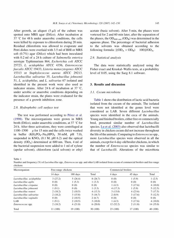
Table 1 shows the distribution of lactic acid bacteria
isolated from the cecum of the animals. The isolated
that were not identified at the genus level were
considered as LAB. Seven different Lactobacillus
species were identified in the ceca of the animals.
Young and finished broilers, either free or commercially
bred, presented similar number of Lactobacillus
species. Lu et al. (2003) also observed that lactobacilli
diversity in chicken cecum did not increase throughout
the life of the animals. Comparing to Enterococcus spp.,
more Lactobacillus species were observed in all the
animals, except for 4-day-old broiler chickens, in which
the number of Enterococcus species was similar to
that of Lactobacilli. Alterations of the microbiota
her LAB isolated from cecum of commercial broilers and free-range
Commercial broilers
Total 4 days 45 days Total
8 (26.7) 0 (0) 1 (5.9) 1 (2.5)
1 (3.3) 0 (0) 0 (0) 0 (0)
0 (0) 1 (4.3) 3 (17.6) 4 (10.0)
1 (3.3) 4 (17.3) 1 (5.9) 5 (12.5)
4 (13.3) 3 (13.0) 4 (23.6) 7 (17.5)
5 (16.7) 2 (8.9) 3 (17.6) 5 (12.5)
2 (6.7) 0 (0) 0 (0) 0 (0)
3 (10.0) 1 (4.3) 3 (17.6) 4 (10.0)
6 (20.0) 12 (52.2) 2 (11.8) 14 (35.0)
30 (100) 23 (100) 17 (100) 40 (100)
M.R. Souza et al. / Veterinary Microbiology 120 (2007) 142–150146
composition in the cecum of broiler chickens were also
reported by Sarra et al. (1992), while Jin et al. (1998)
also found that Lactobacillus is as a genus of LAB
frequently found in chicken gut microbiota. This is
highly desirable, since Lactobacillus may be consid-
ered beneficial bacteria (Fuller, 1989). Their positive
effects in gut ecosystem include immunomodulation,
bacteriocin production and lactic and acetic acid
production, which decrease the local pH and avoid
undesirable bacteria to develop. L. acidophilus was the
most frequently identified species in free-range chick-
ens, while Enterococcus spp., L. johnsonii and
Lactobacillus reuteri were isolated in larger numbers
in commercial broilers. Lactobacillus crispatus was
found only in commercial broilers, Lactobacillus
vaginalis and Lactobacillus agilis only in free-range
chickens and L. salivarius in both. Several authors
(Shome et al., 2001; Gong et al., 2002; Zhu and Joerger,
2003) also found these bacteria in the ceca of broiler
chickens. These differences may be caused by diet,
Table 2
Frequency of resistance to antimicrobial drugs (%) of Lactobacillus and En
chickens
Fowls Antimicrobials
CTX AMO ANL TET AMP VAN
E14
L 0 0 100 75 0 50
E 0 0 100 100 0 50
E180
L 0 0 100 53.8 0 38.5
E 0 0 75 0 0 0
TE
L 0 0 100 71.4 0 42.9
E 0 0 83.3 33.3 0 16.7
I4
L 0 0 100 90 0 50
E 8.3 16.7 100 58.3 0 0
I45
L 0 0 100 75 0 58.3
E 0 0 100 100 0 100
TI
L 0 0 100 81.8 0 54.5
E 7.14 14.3 100 64.3 0 14.3
TG
L 0 0 100 72.0 0 48.8
E 5 10 45 55 0 15
E14: 14 days old, free-range chickens; E180: 180 days old, commercial
broilers; I45: 45 days old, commercial broilers; TI: total commercial bro
(ceftriaxone), AMO (amoxicillin), ANL (nalidixic acid), TET (tetracyclin
(gentamicin), CLO (chloramphenicol), ERI (erythromycin), AMK (amika
level of stress and the use of growth promoter drugs as
suggested by Alzueta et al. (2003) and Wage (2003).
Enterococcus spp. were also found in cecum microbiota
of broiler chicken (Zhu and Joerger, 2003). Even though
they have been used as probiotics for fowls (Maiorka
et al., 2001), recent publications showed a link between
that genus and the presence and transmission of
antimicrobial resistance genes to other microorganisms
(Kolar et al., 2002; Edens, 2003).
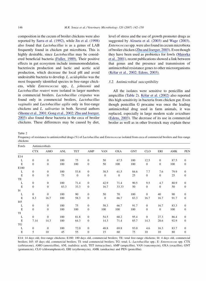
3.2. Antimicrobial susceptibility
All the isolates were sensitive to penicillin and
ampicillin (Table 2). Kolar et al. (2002) also reported
this high sensitivity in bacteria from chicken gut. Even
though penicillin G procaine was once the leading
antimicrobial drug used in farm animals, it was
replaced, especially in large modern scale aviculture
(Edens, 2003). The decrease of its use in commercial
broiler as well as in other livestock may explain these
terococcus isolated from ceca of commercial broilers and free-range
OXA GNT CLO ERI AMK PEN
67.5 100 12.5 0 87.5 0
100 100 0 0 100 0
61.5 84.6 7.7 7.6 79.9 0
0 25 0 0 25 0
71.4 90.5 9.5 4.7 80.9 0
33.33 50 0 0 50 0
70 100 0 40 90 0
66.7 83.3 16.7 16.7 91.7 0
66.7 91.7 0 16.7 83.3 0
100 100 0 0 100 0
68.2 95.4 0 27.3 86.4 0
71.4 85.7 14.3 28.6 92.9 0
69.8 93.0 4.6 16.3 83.7 0
60 75 10 10 80 0
broilers; TE: total free-range chickens; I4: 4 days old, commercial
ilers; TG: total; L: Lactobacillus spp.; E: Enterococcus spp. CTX
e), AMP (ampicillin), VAN (vancomycin), OXA (oxacillin), GNT
cina) and PEN (penicillin).
M.R. Souza et al. / Veterinary Microbiology 120 (2007) 142–150 147
results. Resistance to ceftriaxone and amoxicillin was
only observed in three Enterococci isolated from 4-day-
old commercial broiler chicks. Concerning suscept-
ibility to amoxicillin, there are reports showing the
resistance of Escherichia and Salmonella isolated from
chicken gut. As the literature does not mention chicken
gut LAB resistant to this drug, a possible resistance
gene transfer between Enterobacteriaceae and LAB
within cecum microbiota could explain the results. All
the microorganisms isolated from 14-day-old free-
range chickens and from all the commercial broilers
were resistant to nalidixic acid. Kolar et al. (2002) also
described this fact in the Czech Republic. The
resistance of Lactobacillus to erythromycin was
observed essentially for L. reuteri. According to
Axelsson et al. (1998), the L. reuteri resistance is
related to a plasmidial gene. The presence of some
strains of Lactobacillus spp. or Enterococcus spp.
resistant to chloramphenicol is highly undesirable,
since the use of that drug in livestock is prohibited in
Brazil (Brasil, 2003). High numbers of LAB isolates
were resistant to gentamicin and amikacin, irrespective
of breeding conditions or age. Kozlova et al. (1992) also
reported the resistance of 136 samples of Lactobacillus
isolated from fowls and 23 from elder human subjects to
several antimicrobials, being most of the bacteria
resistant to aminoglycosids. Only half of the Lactoba-
cillus isolates were resistant to vancomycin. This is
quite interesting, since vancomycin resistance is
considered as intrinsic to Lactobacillus (Kolar et al.,
2002; Danielsen and Wind, 2003; Wage, 2003). Even
though that resistance is genetically determined,
sensitive bacteria might have lost it probably due to
the lack of pressure selection.
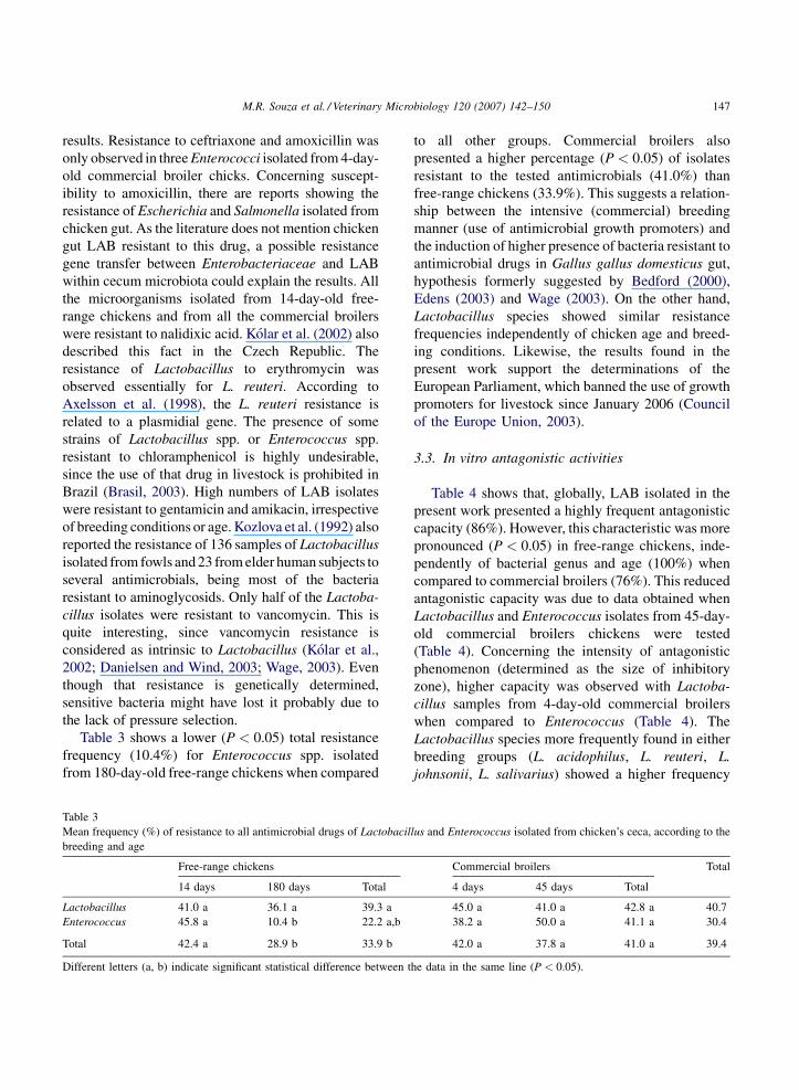
Table 3 shows a lower (P < 0.05) total resistance
frequency (10.4%) for Enterococcus spp. isolated
from 180-day-old free-range chickens when compared
Table 3
Mean frequency (%) of resistance to all antimicrobial drugs of Lactobacil
breeding and age
Free-range chickens
14 days 180 days Total
Lactobacillus 41.0 a 36.1 a 39.3 a
Enterococcus 45.8 a 10.4 b 22.2 a,b
Total 42.4 a 28.9 b 33.9 b
Different letters (a, b) indicate significant statistical difference between t
to all other groups. Commercial broilers also
presented a higher percentage (P < 0.05) of isolates
resistant to the tested antimicrobials (41.0%) than
free-range chickens (33.9%). This suggests a relation-
ship between the intensive (commercial) breeding
manner (use of antimicrobial growth promoters) and
the induction of higher presence of bacteria resistant to
antimicrobial drugs in Gallus gallus domesticus gut,
hypothesis formerly suggested by Bedford (2000),
Edens (2003) and Wage (2003). On the other hand,
Lactobacillus species showed similar resistance
frequencies independently of chicken age and breed-
ing conditions. Likewise, the results found in the
present work support the determinations of the
European Parliament, which banned the use of growth
promoters for livestock since January 2006 (Council
of the Europe Union, 2003).
3.3. In vitro antagonistic activities
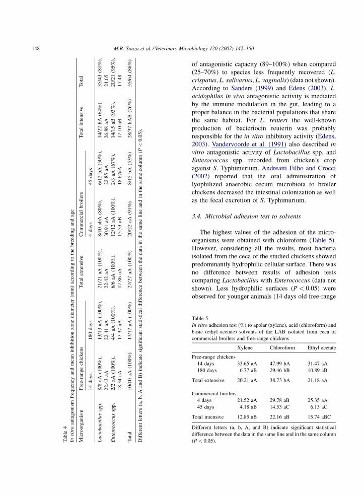
Table 4 shows that, globally, LAB isolated in the
present work presented a highly frequent antagonistic
capacity (86%). However, this characteristic was more
pronounced (P < 0.05) in free-range chickens, inde-
pendently of bacterial genus and age (100%) when
compared to commercial broilers (76%). This reduced
antagonistic capacity was due to data obtained when
Lactobacillus and Enterococcus isolates from 45-day-
old commercial broilers chickens were tested
(Table 4). Concerning the intensity of antagonistic
phenomenon (determined as the size of inhibitory
zone), higher capacity was observed with Lactoba-
cillus samples from 4-day-old commercial broilers
when compared to Enterococcus (Table 4). The
Lactobacillus species more frequently found in either
breeding groups (L. acidophilus, L. reuteri, L.
johnsonii, L. salivarius) showed a higher frequency
lus and Enterococcus isolated from chicken’s ceca, according to the
Commercial broilers Total
4 days 45 days Total
45.0 a 41.0 a 42.8 a 40.7
38.2 a 50.0 a 41.1 a 30.4
42.0 a 37.8 a 41.0 a 39.4
he data in the same line (P < 0.05).
M.R. Souza et al. / Veterinary Microbiology 120 (2007) 142–150148
Tab
le4
Invi
tro
anta
gonis
mfr
equen
cyan
dm
ean
inhib
itio
nzo
ne
dia
met
er(m
m)
acco
rdin
gto
the
bre
edin
gan
dag
e
Mic
roorg
anis
mF
ree-
range
chic
ken
sT
ota
lex
tensi
ve
Com
mer
cial
bro
iler
sT
ota
lin
tensi
ve
Tota
l
14
day
s1
80
day
s4
day
s4
5d
ays
La
cto
ba
cill
us
spp
.8
/8aA
(10
0%
),
22
.43
aA
13
/13
aA(1
00
%),
22
.41
aA
21
/21
aA(1
00
%),
22
.42
aA
8/1
0ab
A(8
0%
),
30
.91
aA
6/1
2b
A(5
0%
),
22
.85
aA
14
/22
bA
(64
%),
26
.88
aA
35
/43
(81
%),
24
.65
En
tero
cocc
us
spp
.2
/2aA
(10
0%
),
18
.34
aA
4/4
aA(1
00
%),
17
.37
aA
6/6
aA(1
00
%),
17
.86
aA
12
/12
aA(1
00
%),
15
.53
aB
2/3
aA(6
7%
),
18
.67
aA
14
/15
aB(9
3%
),
17
.10
aB
20
/21
(95
%),
17
.48
To
tal
10
/10
aA(1
00
%)
17
/17
aA(1
00
%)
27
/27
aA(1
00
%)
20
/22
aA(9
1%
)8
/15
bA
(53
%)
28
/37
bA
B(7
6%
)5
5/6
4(8
6%
)
Dif
fere
nt
lett
ers
(a,
b,
Aan
dB
)in
dic
ate
sig
nifi
can
tst
atis
tica
ld
iffe
ren
ceb
etw
een
the
dat
ain
the
sam
eli
ne
and
inth
esa
me
colu
mn
(P<
0.0
5).
of antagonistic capacity (89–100%) when compared
(25–70%) to species less frequently recovered (L.
crispatus, L. salivarius, L. vaginalis) (data not shown).
According to Sanders (1999) and Edens (2003), L.
acidophilus in vivo antagonistic activity is mediated
by the immune modulation in the gut, leading to a
proper balance in the bacterial populations that share
the same habitat. For L. reuteri the well-known
production of bacteriocin reuterin was probably
responsible for the in vitro inhibitory activity (Edens,
2003). Vandervoorde et al. (1991) also described in
vitro antagonistic activity of Lactobacillus spp. and
Enterococcus spp. recorded from chicken’s crop
against S. Typhimurium. Andreatti Filho and Crocci
(2002) reported that the oral administration of
lyophilized anaerobic cecum microbiota to broiler
chickens decreased the intestinal colonization as well
as the fecal excretion of S. Typhimurium.
3.4. Microbial adhesion test to solvents
The highest values of the adhesion of the micro-
organisms were obtained with chloroform (Table 5).
However, considering all the results, most bacteria
isolated from the ceca of the studied chickens showed
predominantly hydrophilic cellular surface. There was
no difference between results of adhesion tests
comparing Lactobacillus with Enterococcus (data not
shown). Less hydrophilic surfaces (P < 0.05) were
observed for younger animals (14 days old free-range
able 5
n vitro adhesion test (%) to apolar (xylene), acid (chloroform) and
asic (ethyl acetate) solvents of the LAB isolated from ceca of
ommercial broilers and free-range chickens
Xylene Chloroform Ethyl acetate
ree-range chickens
14 days 33.65 aA 47.99 bA 31.47 aA
180 days 6.77 aB 29.46 bB 10.89 aB
otal extensive 20.21 aA 38.73 bA 21.18 aA
ommercial broilers
4 days 21.52 aA 29.78 aB 25.35 aA
45 days 4.18 aB 14.53 aC 6.13 aC
otal intensive 12.85 aB 22.16 aB 15.74 aBC
ifferent letters (a, b, A, and B) indicate significant statistical
ifference between the data in the same line and in the same column
P < 0.05).
T
I
b
c
F
T
C
T
D
d
(
M.R. Souza et al. / Veterinary Microbiology 120 (2007) 142–150 149
chickens and 4 days old commercial broilers) when
compared to older ones as well as for all free-range
chickens when compared to commercial broilers. Jin
et al. (1996) described hydrophobic differences
between L. acidophilus and Lactobacillus fermentum
cells isolated from chicken intestines, and in a posterior
report found high levels of in vitro adhesion of L.
acidophilus cells (Jin et al., 1998). Gusils et al. (1999)
showed differences among adhesion values among
bacteria of distinct species. Garriga et al. (1998) related
differences among L. salivarius strains CTC2183 and
2197 in colonizing capacity when inoculated in chicks’
gizzard or ceca.
Concluding, animal breeding and age caused
variation on composition, antimicrobial susceptibility,
antagonistic activity and surface hydrophobicity of
LAB from chicken cecum.
Acknowledgments
This study was supported by grants and fellowships
from Conselho Nacional do Desenvolvimento Cientı-
fico e Tecnologico (CNPq) and Fundacao de Amparo a
Pesquisa do Estado de Minas Gerais (FAPEMIG). The
authors are grateful to Maria Gorete Barbosa Ribas for
valuable technical help.
References
Alzueta, C., Rodriguez, M.L., Cutuli, M.T., 2003. Effect of whole
and demucilaged linseed in broiler chicken diets on digesta
viscosity, nutrient utilization and intestinal microflora. Brit.
Poultry Sci. 44, 67–74.
Andreatti Filho, R.L., Crocci, A.J., 2002. Efeito protetor da micro-
biota cecal congelada e liofilizada sobre a infeccao experimental
de frangos de corte por Salmonella enterica sorovar Enteritidis.
Arq. Br. Med. Vet. Zootec. 54, 140–145.
Axelsson, L.T., Ahrne, S.E.I., Andersson, M.C., Stahl, S.R., 1998.
Identification and cloning of a plasmid-encoded erythromycin
resistance determinant for Lactobacillus reuteri. Plasmid 20,
171–174.
Bedford, M., 2000. Removal of antibiotic growth promoters from
poultry diets: implications and strategies to minimize subse-
quent problems. World Poultry Sci. J. 56, 347–365.
Brasil Ministerio da Agricultura, Pecuaria e Abastecimento, 2003.
Instrucao Normativa no. 9 de 27/06/2003. Proibicao de Clor-
anfenicol e Nitrofuranos para Uso Veterinario e Suscetıveis de
Emprego na Alimentacao de Todos os Animais e Insetos.
Ministerio da Agricultura, Brasılia, p. 12.
Charteris, W.P., Kelly, P.M., Morelli, L., Collins, J.K., 1998. Anti-
biotic susceptibility of potentially probiotic Lactobacillus spe-
cies. J. Food Protect. 61, 1636–1643.
Council of the Europe Union, 2003. Council Regulation on the
Authorization of the Additive Avilamycin in Feeding Stuffs.
Accessed October 15, 2003. Online: http://register.consiliu-
m.eu.int/pdf/em/03/st06/st06120en03.pdf.
Danielsen, N., Wind, A., 2003. Susceptibility of Lactobacillus spp.
to antimicrobial agents. Intern. J. Food Microbiol. 82, 1–11.
Edens, F.W., 2003. An alternative for antibiotic use in poultry:
probiotics. Rev. Br. Cienc. Avıcola 5, 1–40.
Fuller, R., 1989. Probiotics in man and animals. J. Appl. Bacteriol.
66, 365–378.
Garriga, M., Pascual, M., Monfort, J.M., Hugas, M., 1998. Selection
of lactobacilli for chicken probiotics adjuncts. J. Appl. Micro-
biol. 84, 125–132.
Gong, J., Forster, R.J., Yu, H., Chambers, J.R., Wheatcroft, R.,
Sabour, P.M., Chen, S., 2002. Molecular analysis of bacterial
populations in the ileum of broiler chickens and comparison
with bacterial in the caecum. FEMS Microbiol. 41, 171–
179.
Guan, L.L., Hagen, K.E., Tannock, G.W., Korver, D.R., Fasenko,
G.M., Alisson, G.E., 2003. Detection and identification of
Lactobacillus species in crops of broilers of different ages by
using PCR-denaturing gradient gel electrophoresis and ampli-
fied ribosomal DNA restriction analysis. Appl. Environ. Micro-
biol. 69, 6750–6757.
Gusils, C., Gonzalez, S.M., Oliver, G., 1999. Lactobacilli isolated
from chicken intestines: potential use as probiotics. J. Food
Protect. 62, 252–256.
Jin, L.Z., Ho, Y.W., Ali, M.A., Abdullah, N., Ong, K.B., Jaladulin,
S., 1996. Effect of adherent Lactobacillus sp. on in vitro
adherence of Salmonella to epithelial cells of chicken. J. Appl.
Bacteriol. 81, 201–206.
Jin, L.Z., Ho, Y.W., Abdullah, N., Ali, M.A., Jalaludin, S., 1998.
Effects of adherent Lactobacillus cultures on growth, weight of
organs and intestinal microflora and volatile fatty acids in
broilers. Anim. Feed Sci. Technol. 70, 197–209.
Knarreborg, A., Simon, M.A., Engberg, R.M., Jensen, B.B., Tan-
nock, G.W., 2002. Effect of dietary fat source and subtherapeutic
levels of antibiotic on the bacterial community in the ileum of
broiler chickens at various ages. Appl. Environ. Microbiol. 68,
5918–5924.
Kolar, M., Pantueek, R., Bardoo, J., Vagnerova, I., Typovska, H.,
Doskar, J., Valka, I., 2002. Occurrence of antibiotic-resistant
bacterial strains isolated in poultry. Vet. Med. Czech. 47,
52–59.
Kozlova, E.V., Malinovskaia, I.V., Aminov, R.I., Kovalenko, N.K.,
Voronin, A.M., 1992. Antibiotic resistance of Lactobacillus
strains. Antibiot. Khimioter. 37, 5–12.
Lu, J., Idris, U., Harmon, B., Hofacre, C., Maurer, J.J., Lee, M.D.,
2003. Diversity and succession of the intestinal bacteria com-
munity of the maturing broiler chicken. Appl. Environ. Micro-
biol. 69, 6816–6824.
Maiorka, A., Santin, E., Sugeta, S.M., Almeida, J.G., Mari, M.,
2001. Utilizacao de prebioticos, probioticos ou simbioticos em
dietas para frangos. Rev. Br. Cienc. Avıcola 3, 15–30.
M.R. Souza et al. / Veterinary Microbiology 120 (2007) 142–150150
McCracken, V.J., Lorenz, R.G., 2001. The gastrointestinal ecosys-
tem: a precarious alliance among epithelium, immunity and
microbiota. Cell Microbiol. 3, 1–11.
Moreira, J.L.S., Mota, R.M., Horta, M.F., Teixeira, S.M.R., Neu-
mann, E., Nicoli, J.R., Nunes, A.C., 2005. Identification to the
species level of Lactobacillus isolated in probiotic prospecting
studies of human, animal or food origin by 16S-23S rRNA
restriction profiling. BMC Microbiol. 5, 15–20.
Nashashon, S.N., Nakaue, H.S., Mirosh, L.W., 1996. Performance of
single comb white Leghorn layers fed a diet with a live microbial
during the growth and egg laying phases. Anim. Feed Sci.
Technol. 57, 25–38.
Perez, P., Minaard, Y., Disalvo, E., De Antoni, G., 1998. Surface
properties of bifidobacterial strains of human origin. Appl.
Environ. Microbiol. 64, 21–26.
Sanders, M.E., 1999. Probiotics. Food Technol. 53, 67–77.
Sarra, P.G., Morelli, L., Bottazzi, F., 1992. The lactic microflora of
fowl. The Lactic Acid Bacteria in Health and Disease, vol. 1.
Elsevier, New York.
Shome, B.R., Senani, S., Padhi, M.K., Saha, S.K., Rai, R.B., Shome,
R., 2001. Isolation, identification and characterisation of auto-
chthonous Lactobacillus from chicken intestine. Indian Vet. J.
78, 278–281.
Tilsala, A.T., Alatossava, T., 1997. Development of oligonucleotide
primers from the 16S–23S rDNA intergenic sequences for
identifying different dairy and probiotic lactic acid bacteria
by PCR. Intern. J. Food Microbiol. 35, 49–56.
Vandervoorde, L., Christiaens, H., Verstraete, W., 1991. In vitro
appraisal of the probiotic value of intestinal lactobacilli. World J.
Microbiol. Technol. 7, 587–592.
Wage, S.W., 2003. The Role of Enteric Antibiotics in Livestock
Production. Avcare Limited, Camberra, p. 338.
WHO/FAO/OIE Food Agricultural Organization/World Health
Organization Background document for the Joint WHO/FAO/
OIE expert, 2003.In: Workshop on Non-Human Antimicrobials
Usage and Antimicrobials Resistance Scientific Assessment,
Geneva, Switzerland, December 1–5.
Zhu, X.Y., Joerger, R.D., 2003. Composition of microbiota in
content and mucus from caeca of broiler chickens as measured
by fluorescent in situ hybridization with group-specific, 16S
rRNA-targeted oligonucleotides probes. Poultry Sci. 82, 1242–
1249.
Zhu, X.Y., Zhong, T., Pandya, Y., Joerger, R.D., 2002. 16S rRNA-
based analysis of microbiota from the cecum of broiler chickens.
Appl. Environ. Microbiol. 68, 124–137.
Related Documents