Pferdeheilkunde 24 193 C. Werren et al. Pferdeheilkunde 24 (2008) 2 (März/April) 193-202 Influence of culture conditions on expansion and re-differentiation of chondrocytes from horses of different ages Cecile Werren 1 , Jose Diaz-Romero 2 , Walter Brehm 1 and Dobrila Nesic 2 Equine Clinic, VetSuisse Faculty 1 , University Bern, Bern, Switzerland and Osteoarticular Research Group, Institute of Pathology 2 , University of Bern, Bern, Switzerland Summary To evaluate the influence of types of sera and the donors age on proliferation and re-differentiation capacities of equine chondrocytes, car- tilage tissue from 10 horses was obtained in the age range 2.5 to 19 years. Chondrocytes were isolated from the fetlock- joints and expan- ded in media containing different types of sera: fetal calf serum (FCS), fetal equine serum (FES), adult equine serum (AES) and autologous serum. Cells were expanded in monolayers at low and high seeding densities. Chondrocytes re-differentiation capacities were assessed in micromass pellet cultures after 4 weeks of incubation in chondrogenic medium supplemented with or without 5ng/ml TGFβ1. The neo- cartilagineous tissue formation was evaluated through proteoglycan and collagen type II deposition, and Berne score visual evaluation system. Results showed that the low seeding density allows for higher proliferation rates in FCS, FES and AES. In addition chondrocytes from younger horses proliferate faster independently of the serum type. Re-differentiation capacities are influenced by the animals age, type of serum during expansion, and the presence of TGFβ1 in pellet cultures. Cells from young horses appear to produce better neocartilage upon expansion in autologous serum, while cells from older horses re-differentiated better upon expansion in FCS. The expansion condi- tions of potential clinical relevance include the correct choice of sera according to the age of horse during monolayer culture for better subsequent re-differentiation and lower seeding densities allowing for smaller biopsy sampling and therefore less injury to the donor site. Keywords: cartilage repair, chondrocyte cell culture and biology, differentiation, tissue engineering, cartilage, bone Einfluss von Kultivierungsbedingungen auf die Vermehrung und die Redifferenzierung von Chondrozyten von Pferden verschiedenen Alters Um den Einfluss von verschiedenen Seren, des Alters des Knorpelspenders und der Redifferenzierungs-Kapazität von Pferdechondrozyten zu beobachten, wurde von 10 Pferden im Alter zwischen 2.5 und 19 Jahren Knorpelgewebe entnommen. Knorpelzellen wurden aus Fes- selgelenken isoliert und in Medien mit verschiednen Seren vermehrt: fötales Kälberserum (FCS), fötales Pferdeserum (FES), adultes Pferde- serum (AES) und autologes Serum. Zellen werden in hoher- und in tiefer Aussaatdichte in Monolayerkulturen vermehrt. Die Redifferenzie- rungskapazität von Pferdechondrozyten wurde in Micromasspelletkulturen, nach einer Inkubation von 4 Wochen in einem chondrogenen Medium mit oder ohne 5ng/ml TGFβ1 beurteilt. Die Bildung von Knorpelpellets wurden mit Hilfe der Deposition von Proteoglykanen und Kollagen Typ II und dem Bern Score einem visuellen Auswertungssystems bewertet. Die Resultate zeigten, dass tiefere Aussaatdichten höhe- re Proliferationsraten in FCS, FES und AES erlauben. Chondrozyten von jungen Pferden vermehren sich besser unabhängig vom Serum. Die Redifferenzierungs-Kapazität ist beeinflusst durch das Alter des Tieres, dem Serum während der Zellvermehrung und der Präsenz von TGFβ1 während der Kultivierung zu Pellets. Die Zellen von jungen Pferden scheinen bessere Knorpelpellets nach Vermehrung in autolo- gem Serum zu produzieren, während Zellen von älteren Pferden sich besser redifferenzieren, nach der Vermehrung in FCS. Klinisch rele- vante Bedingungen sind die richtige Wahl des Serums, in Abhängigkeit des Alters des Pferdes, um anschließend eine bessere Redifferen- zierung zu erreichen, sowie tiefere Aussaatdichten die die Entnahme kleinerer Biopsien erlauben und dadurch weniger Schädigung an der Entnahmestelle zur Folge haben Schlüsselwörter: Knorpelheilung, Chondrozytenkultur, Chondrozytenbiologie, Differenzierung, Tissue Engineering, Knorpel Introduction Articular cartilage is a nonvascular, noninnervated tissue consisting of round chondrocytes characterized by their ability to produce high levels of collagen types II, IX, and XI and proteoglycans, the main constituents of the hyaline extracellular matrix (Mendler et al. 1989, Cremer et al. 1998). Cartilage serves as a load-bearing elastic material that is responsible for the frictionless movement of the sur- faces in the articulating joints. Articular cartilage lesions are frequent and arise from trauma and/or different joint disea- ses as a consequence of genetic and metabolic disorders. Due to its particular structure, hyaline cartilage has limited intrinsic reparative capacity and heals poorly (Ulrich-Vinther et al. 2003). The size, depth and site of lesion highly influence the healing process. Ultimately, defects can lead to osteoarthritis. Cartilage defects represent a challenge in equine orthopaedics, in particular for the treatment of racing and sport horses (McIlwraith et al. 1987). Further- more, the horse is considered as an important and relevant large animal model for cartilage repair studies due to the cartilage thickness and naturally occurring osteoarthritis, which are similar to human (Reinholz et al. 2004, Frisbie et al. 2006).

Welcome message from author
This document is posted to help you gain knowledge. Please leave a comment to let me know what you think about it! Share it to your friends and learn new things together.
Transcript
Pferdeheilkunde 24 193
C. Werren et al.
Pferdeheilkunde 24 (2008) 2 (März/April) 193-202
Influence of culture conditions on expansion and re-differentiation of chondrocytes from horses of different agesCecile Werren1, Jose Diaz-Romero2, Walter Brehm1 and Dobrila Nesic2
Equine Clinic, VetSuisse Faculty1, University Bern, Bern, Switzerland and Osteoarticular Research Group, Institute of Pathology2, University of Bern, Bern, Switzerland
Summary
To evaluate the influence of types of sera and the donors age on proliferation and re-differentiation capacities of equine chondrocytes, car-tilage tissue from 10 horses was obtained in the age range 2.5 to 19 years. Chondrocytes were isolated from the fetlock- joints and expan-ded in media containing different types of sera: fetal calf serum (FCS), fetal equine serum (FES), adult equine serum (AES) and autologousserum. Cells were expanded in monolayers at low and high seeding densities. Chondrocytes re-differentiation capacities were assessed inmicromass pellet cultures after 4 weeks of incubation in chondrogenic medium supplemented with or without 5ng/ml TGFβ1. The neo-cartilagineous tissue formation was evaluated through proteoglycan and collagen type II deposition, and Berne score visual evaluationsystem. Results showed that the low seeding density allows for higher proliferation rates in FCS, FES and AES. In addition chondrocytes fromyounger horses proliferate faster independently of the serum type. Re-differentiation capacities are influenced by the animals age, type ofserum during expansion, and the presence of TGFβ1 in pellet cultures. Cells from young horses appear to produce better neocartilageupon expansion in autologous serum, while cells from older horses re-differentiated better upon expansion in FCS. The expansion condi-tions of potential clinical relevance include the correct choice of sera according to the age of horse during monolayer culture for bettersubsequent re-differentiation and lower seeding densities allowing for smaller biopsy sampling and therefore less injury to the donor site.
Keywords: cartilage repair, chondrocyte cell culture and biology, differentiation, tissue engineering, cartilage, bone
Einfluss von Kultivierungsbedingungen auf die Vermehrung und die Redifferenzierung von Chondrozyten von Pferdenverschiedenen Alters
Um den Einfluss von verschiedenen Seren, des Alters des Knorpelspenders und der Redifferenzierungs-Kapazität von Pferdechondrozytenzu beobachten, wurde von 10 Pferden im Alter zwischen 2.5 und 19 Jahren Knorpelgewebe entnommen. Knorpelzellen wurden aus Fes-selgelenken isoliert und in Medien mit verschiednen Seren vermehrt: fötales Kälberserum (FCS), fötales Pferdeserum (FES), adultes Pferde-serum (AES) und autologes Serum. Zellen werden in hoher- und in tiefer Aussaatdichte in Monolayerkulturen vermehrt. Die Redifferenzie-rungskapazität von Pferdechondrozyten wurde in Micromasspelletkulturen, nach einer Inkubation von 4 Wochen in einem chondrogenenMedium mit oder ohne 5ng/ml TGFβ1 beurteilt. Die Bildung von Knorpelpellets wurden mit Hilfe der Deposition von Proteoglykanen undKollagen Typ II und dem Bern Score einem visuellen Auswertungssystems bewertet. Die Resultate zeigten, dass tiefere Aussaatdichten höhe-re Proliferationsraten in FCS, FES und AES erlauben. Chondrozyten von jungen Pferden vermehren sich besser unabhängig vom Serum.Die Redifferenzierungs-Kapazität ist beeinflusst durch das Alter des Tieres, dem Serum während der Zellvermehrung und der Präsenz vonTGFβ1 während der Kultivierung zu Pellets. Die Zellen von jungen Pferden scheinen bessere Knorpelpellets nach Vermehrung in autolo-gem Serum zu produzieren, während Zellen von älteren Pferden sich besser redifferenzieren, nach der Vermehrung in FCS. Klinisch rele-vante Bedingungen sind die richtige Wahl des Serums, in Abhängigkeit des Alters des Pferdes, um anschließend eine bessere Redifferen-zierung zu erreichen, sowie tiefere Aussaatdichten die die Entnahme kleinerer Biopsien erlauben und dadurch weniger Schädigung an derEntnahmestelle zur Folge haben
Schlüsselwörter: Knorpelheilung, Chondrozytenkultur, Chondrozytenbiologie, Differenzierung, Tissue Engineering, Knorpel
Introduction
Articular cartilage is a nonvascular, noninnervated tissueconsisting of round chondrocytes characterized by theirability to produce high levels of collagen types II, IX, and XIand proteoglycans, the main constituents of the hyalineextracellular matrix (Mendler et al. 1989, Cremer et al.1998). Cartilage serves as a load-bearing elastic materialthat is responsible for the frictionless movement of the sur-faces in the articulating joints. Articular cartilage lesions arefrequent and arise from trauma and/or different joint disea-ses as a consequence of genetic and metabolic disorders.
Due to its particular structure, hyaline cartilage has limitedintrinsic reparative capacity and heals poorly (Ulrich-Vintheret al. 2003). The size, depth and site of lesion highlyinfluence the healing process. Ultimately, defects can leadto osteoarthritis. Cartilage defects represent a challenge inequine orthopaedics, in particular for the treatment ofracing and sport horses (McIlwraith et al. 1987). Further-more, the horse is considered as an important and relevantlarge animal model for cartilage repair studies due to thecartilage thickness and naturally occurring osteoarthritis,which are similar to human (Reinholz et al. 2004, Frisbie etal. 2006).
In the past decade tissue engineering has emerged as a newtherapy with the goal to provide structural and functional sub-stitutes for damaged organs and tissues by combining cells,biomaterials and signalling molecules (Vacanti et al. 1998).Autologous chondrocyte implantation (ACI) has gained interestas a clinically applied cell based therapy for the repair of loca-lized traumatic cartilage lesions in humans (Marlovits et al.2006), and this procedure, following the original method [Britt-berg et al. 1994), has also been tested in full thickness cartila-ge defects in the horse (Litzke et al. 2004). The procedure des-cribed here comprises chondrocyte isolation from biopsiestaken from a low weight-bearing area of the hock, followed bycell expansion in monolayer. When a sufficient number of cellswas obtained, chondrocytes were implanted into the lesionunder a periosteal flap or collagen membrane. During theexpansion phase in monolayer, chondrocytes alter their mor-phology and metabolism in a process termed de-differentiation(Schnabel et al. 2002), whereby cells gradually lose their chon-drogenic „commitment“ and phenotype. Recently, the produc-tion of large scaffold free cartilage constructs, after expandingequine chondrocytes in monolayer culture, has been described(Brehm et al. 2007). Therefore, a prerequisite for cell basedtherapies is an efficient method for the expansion of the chon-drocytes. This method must support the chondrocytes' re-diffe-rentiation capacity and allow the production of extracellularmartrix that is of sufficient quality to provide a functional arti-cular joint. During the expansion phase chondrocytes must beprovided with nutrients and growth factors to proliferate. Fetalcalf serum (FCS) is commonly used as a supplement to theexpansion medium providing growth factors, adhesion mole-cules, and vitamins. The exposure of cells to FCS however, hasseveral disadvantages including possible viral or bacterialinfections (Dedrick 1997) and transmission of prions (Klein andDumble 1993). If the cells have been exposed to FCS duringcultivation, the following transplantation may induce immuneor local inflammatory responses (Selvaggi et al. 1997, Heiska-nen et al. 2007). Therefore, the development of FCS-free cul-turing protocols certainly represents an advantage for futuredevelopment of cellular therapies, and the use of serum-freemedia, allogenic, and autologous serum have being alreadyinvestigated as potential alternatives for growing cells in humanregenerative medicine (Stute et al. 2004, Shahdadfar et al.2005, Tallheden et al. 2005). In addition to serum-supple-mentation, chondrocyte proliferation rates in monolayer cultu-re have been shown to depend on factors such as the cell-see-ding density (Mandl et al. 2004) or the age of the donor (Bar-bero et al. 2004). Therefore, optimizing culturing conditions forequine chondrocytes can contribute towards more efficient tre-atment of horse injuries in equine clinics and provide furtherknowledge towards development of cell based treatments inhuman patients. The goal of this study was to examine equinechondrocyte proliferation in xenogenic (FCS), allogenic (FESand AES) and autologous serum regimes, under different see-ding densities and with respect to horse age, and to comparetheir subsequent re-differentiation capacities.
Materials and Methods
Cartilage sampling
Full thickness equine articular cartilage samples were obtai-ned from the trochlea metacarpale and from the fossa arti-
Influence of culture conditions on expansion and re-differentiation of chondrocytes from horses of different ages
Pferdeheilkunde 24194
cularis of the first phalanx of fetlock-joints from 10 sport hor-ses at autopsy within 24 hours post-mortem. Cartilage sam-ples derived from slaughtered horses, aged 4.4±2.2 year(mean±SEM) for experiments 1 and 2 (n=5), 8.6±2.5 years(mean±SEM) (n=3) and 19±0 years (mean±SEM) (n=2) forexperiment 3. None of the horses was under drug treatment,had clinical history of arthritis or other pathologies affectingcartilage. The specimens appeared normal by morphologicalexamination (no change in colour and no fibrillation). Theterm “young” was employed for horses below 12 years, andthe term “old” for horses older than 12 years. Cartilage sam-ples were kept in transport medium (D-MEM/F12, 100Upenicillin/streptomycin, Life Technologies, Basel, Switzerland)for a maximum of 24 hours post sample collection and priorto further processing.
Chondrocyte isolation
Cartilage was sequentially digested with pronase and colla-genase P as previously described (Diaz-Romero et al. 2005).Briefly, cartilage was diced in PBS and incubated for one hourwith 2.5 mg/ml pronase (Roche, Rotkreuz, Switzerland) indigestion medium (10ml per gram of wet tissue): D-MEM/F12 containing 1g/ml bovine insulin (Life Technologies,Basel, Switzerland); 50g/ml ascorbic acid (Fluka, Buchs, Swit-zerland) in a shaking water bath at 37°C. After removal ofpronase and washing with PBS, digestion was continued over-night by addition of 250 g/ml of collagenase P (Roche, Rot-kreuz, Switzerland) in digestion medium (10 ml/g of wet tis-sue) in a shaking water bath at 37°C. The resulting cellsuspension was filtered through a 100 m nylon cell strainer toremove debris. Cells were centrifuged (Sorvall, RT6000D;rotor 1000B) at 700g for 6 minutes at 10∞C, washed twicewith PBS, counted, and either cryopreserved in 90% of corre-sponding serum and 10% DMSO, or seeded in tissue cultureflasks.
Monolayer culture
After isolation, cells were seeded for 48 hours at 65’000cells/cm2 to allow recovery in D-MEM/F12 containing L-glu-tamine and 100u penicillin/streptomycin. Medium was sup-plemented with 10% of different sources of sera: fetal calfserum (HyClone, Lausanne, Switzerland), fetal equine serum(Biowest, Nuaillé, France), adult equine serum (Biowest,Nuaillé, France) or autologous serum. Chondrocytes werecultured at 37°C in a humidified atmosphere containing 5%CO2. After 48 hours, cells were harvested by trypsin/EDTAincubation. Trypsin activity was neutralized with the corre-sponding serum, and after washing and counting, cells wereeither cryopreserved or re-seeded in monolayer culture at twodifferent densities: high-10’000 cells/cm2 and low-3.333cells/cm2. The medium was changed the following day andthen every 2-3 days. Monolayer cultures were maintaineduntil 40 days with passaging at 10 days intervals.
Preparation of autologous sera
Autologous serum was prepared from 3 young and 2 old hor-ses after stunning. Between 0.5 to 2 litres of blood were asep-
tically collected from the Vena jugularis, allowed to clot for 4hours at 4-8°C and centrifuged at 830g for 10 minutes. Theobtained supernatant was subsequently centrifuged at 1300gand then at 1800g each time for 10 minutes. Debris wasremoved by filtration through a 100 m nylon cell strainer.Serum was sterilized by filtering through 0.2 mm filter, heat-inactivated by incubation at 56°C for 30 minutes, and ali-quots were prepared and stored at -20°C until further use.
Pellet cultures
Chondrogenic re-differentiation was induced in micromasspellet cultures using a defined serum-free medium as pre-viously described (Körner et al. 2006). Cells expanded at lowseeding density were harvested after 20 days in monolayerculture and resuspended in high glucose DMEM mediumsupplemented with ITS +1 (10g/ml insulin, 5.5g/ml transfer-ring, 5ng/ml selenium, 0.5 mg/ml bovine serum albumin4.7g/ml linoleic acid, Sigma-Aldrich, Buchs, Switzerland), 0.1mM ascorbic acid, 10-7M dexamethasone and rh-TGF1(Acrys, Hiddenhausen, Germany). 5 x 10⁵ chondrocytes wereplaced in 15ml falcon tube, resuspended in 3ml of differenti-ation media and centrifuged at 300g for 5 minutes at 4°C.Pellets were incubated at 37°C in a humidified atmospherecontaining 5% CO2 for 4 weeks, with medium change twiceweekly.
Histology
Pellets were fixed for 24 hours in 4% paraformaldehyde,embedded in paraffin and cut in 4mm thick sections. Hae-matoxylin-eosin, Alcian blue and Safranin O staining wereperformed to assess cell morphology and proteoglycan syn-thesis. Bright field pictures were taken with Axioplan 2 micro-scope and Axiocam camera (Zeiss). Berne score, a visual gra-ding system for assessment of in vitro engineered cartilage-neous tissue was applied for each slide stained with Safranin-O (Grogan et al. 2006). The uniformity and intensity of Safra-nin-O staining, distance between cells versus the amount ofmatrix produced and cell morphology were determined. Foreach criterion a maximum of three points was allocated.
Immunohistochemistry
Immunohistochemistry was performed as previously described(Wang et al. 2004). Pellets were fixed for maximum of 24hours with 4% paraformaldehyde, embedded in paraffin andcut in 4 mm thick sections. After pre-treatment with 1 mg/mlchondroitinase, histology slides were incubated with primarymonoclonal antibodies for collagen type II (II-II6B3, Hybrido-ma Bank, University of Iowa, Iowa, USA) for 1 hour. The sec-tions were then incubated with a biotinylated goat anti-mou-se secondary antibody for 35 minutes, followed by Strep-tABCcomplex/AP (StreptABCcomplex/AP, DAKO, Glostrup,Denmark) for 45 minutes. Bound antibodies were revealedwith Fuchsin/Naphtol AS-BI substrate (Fuchsin/Naphtol AS-BIsubstrate, Sigma-Aldrich, Buchs, Switzerland). Positive controlcomprised human growth plate section, and negative controlwere slides where the primary antibody was omitted. Pictureswere taken as described for histology slides.
C. Werren et al.
Pferdeheilkunde 24 195
Statistical analysis
Samples were analyzed with the GraphPad software, employ-ing the un-paired Student t-test. Statistical significance wasconsidered for P values <0.05. Mixed factors Anova andBonferroni multiple comparison tests were applied to assessthe effect of donor age, source of sera during the expansionphase and the presence of TGFβ1 in pellet cultures. Statisti-cal significance was considered for P values <0.05.
Results
Effect of different sera and seeding densities on chondrocyteproliferation
To assess the process of attachment and subsequent chon-drocyte de-differentiation associated phenotype, cell recove-ry after seeding and changes in cell morphology were moni-tored. Upon isolation, chondrocytes from five horses(mean=4.4 years) were allowed to attach in media contai-ning 10% FCS, 10 % FES or 10% AES for 48 hours. The per-centage of recovered cells was 77.5±12.4 (mean±SEM) forFCS, 67.6±13.8 for FES and 76.6±16.8 for AES, indicatingsimilar adhesion in all tested sources of sera (FCS, FES andAES). Chondrocyte morphology also changed independentlyof the source of sera in the medium: within the first four dayscells appeared small and round-shaped, in accordance withtheir still differentiated state (Figure 1 A, C, E). Over the nextfew days, regardless of the source of sera, cells started toelongate and acquired more fibroblast-like shape, a hallmarkof the de-differentiation process (Figure 1 B, D, F). These dataindicated that the commercially produced types of sera in themedium had no influence on the initial cell attachment andresulted in identical changes in cell morphology in monolay-er culture.
The potential effect of low and high seeding density on chon-drocyte proliferation rates was analyzed next. Cells were cul-tured at low – 3’333/cm2, and high – 10’000 cells/cm2 den-sity for 40 days in FCS, FES and AES and cumulative popula-tion doublings (PD) were calculated for each donor. Obtaineddata demonstrated that chondrocytes proliferated at the samerates independently of the serum type used (FCS, FES or AES)(Figure 2). The seeding density however, greatly influencedcell proliferation rates. The difference was obvious alreadyafter 20 days in culture where cells under low seeding densi-ty regime proliferated with cumulative PD of 4.5 compared to3.22 at high densities (p<0.01). The difference became evenmore pronounced after 40 days where cumulative PD for lowdensity reached 11 compared to 7.5 for high density(p<0.001). To assess whether cells at higher density decrea-sed their proliferation rates due to saturated cell densities,confluence expressed as cells/cm2 were calculated at eachharvesting step. While the average cell density was signifi-cantly higher (P<0.05) for high seeding density cultures(mean=43’250±2’900) compared to low seeding densitycultures (mean=34’740±1’520), the maximum cells harve-sted was 105’000 cells/cm2, suggesting that cells on avera-ge were not contact inhibited under either seeding condition.In summary, equine chondrocytes efficiently attached, under-went typical morphological changes associated with the pro-cess of de-differentiation under monolayer culturing condi-
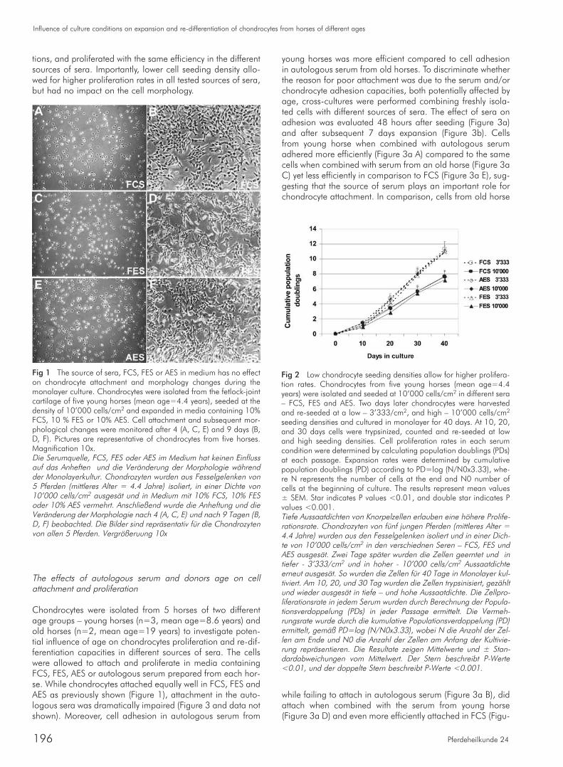
tions, and proliferated with the same efficiency in the differentsources of sera. Importantly, lower cell seeding density allo-wed for higher proliferation rates in all tested sources of sera,but had no impact on the cell morphology.
The effects of autologous serum and donors age on cellattachment and proliferation
Chondrocytes were isolated from 5 horses of two differentage groups – young horses (n=3, mean age=8.6 years) andold horses (n=2, mean age=19 years) to investigate poten-tial influence of age on chondrocytes proliferation and re-dif-ferentiation capacities in different sources of sera. The cellswere allowed to attach and proliferate in media containingFCS, FES, AES or autologous serum prepared from each hor-se. While chondrocytes attached equally well in FCS, FES andAES as previously shown (Figure 1), attachment in the auto-logous sera was dramatically impaired (Figure 3 and data notshown). Moreover, cell adhesion in autologous serum from
Influence of culture conditions on expansion and re-differentiation of chondrocytes from horses of different ages
Pferdeheilkunde 24196
young horses was more efficient compared to cell adhesionin autologous serum from old horses. To discriminate whetherthe reason for poor attachment was due to the serum and/orchondrocyte adhesion capacities, both potentially affected byage, cross-cultures were performed combining freshly isola-ted cells with different sources of sera. The effect of sera onadhesion was evaluated 48 hours after seeding (Figure 3a)and after subsequent 7 days expansion (Figure 3b). Cellsfrom young horse when combined with autologous serumadhered more efficiently (Figure 3a A) compared to the samecells when combined with serum from an old horse (Figure 3aC) yet less efficiently in comparison to FCS (Figure 3a E), sug-gesting that the source of serum plays an important role forchondrocyte attachment. In comparison, cells from old horse
while failing to attach in autologous serum (Figure 3a B), didattach when combined with the serum from young horse(Figure 3a D) and even more efficiently attached in FCS (Figu-
Fig 1 The source of sera, FCS, FES or AES in medium has no effecton chondrocyte attachment and morphology changes during themonolayer culture. Chondrocytes were isolated from the fetlock-jointcartilage of five young horses (mean age=4.4 years), seeded at thedensity of 10’000 cells/cm2 and expanded in media containing 10%FCS, 10 % FES or 10% AES. Cell attachment and subsequent mor-phological changes were monitored after 4 (A, C, E) and 9 days (B,D, F). Pictures are representative of chondrocytes from five horses.Magnification 10x.Die Serumquelle, FCS, FES oder AES im Medium hat keinen Einflussauf das Anheften und die Veränderung der Morphologie währendder Monolayerkultur. Chondrozyten wurden aus Fesselgelenken von5 Pferden (mittleres Alter = 4.4 Jahre) isoliert, in einer Dichte von10’000 cells/cm2 ausgesät und in Medium mit 10% FCS, 10% FESoder 10% AES vermehrt. Anschließend wurde die Anheftung und dieVeränderung der Morphologie nach 4 (A, C, E) und nach 9 Tagen (B,D, F) beobachted. Die Bilder sind repräsentativ für die Chondrozytenvon allen 5 Pferden. Vergrößeruung 10x
Fig 2 Low chondrocyte seeding densities allow for higher prolifera-tion rates. Chondrocytes from five young horses (mean age=4.4years) were isolated and seeded at 10’000 cells/cm2 in different sera– FCS, FES and AES. Two days later chondrocytes were harvestedand re-seeded at a low – 3’333/cm2, and high – 10’000 cells/cm2
seeding densities and cultured in monolayer for 40 days. At 10, 20,and 30 days cells were trypsinized, counted and re-seeded at lowand high seeding densities. Cell proliferation rates in each serumcondition were determined by calculating population doublings (PDs)at each passage. Expansion rates were determined by cumulativepopulation doublings (PD) according to PD=log (N/N0x3.33), whe-re N represents the number of cells at the end and N0 number ofcells at the beginning of culture. The results represent mean values± SEM. Star indicates P values <0.01, and double star indicates Pvalues <0.001.Tiefe Aussaatdichten von Knorpelzellen erlauben eine höhere Prolife-rationsrate. Chondrozyten von fünf jungen Pferden (mittleres Alter =4.4 Jahre) wurden aus den Fesselgelenken isoliert und in einer Dich-te von 10’000 cells/cm2 in den verschiednen Seren – FCS, FES undAES ausgesät. Zwei Tage später wurden die Zellen geerntet und intiefer - 3’333/cm2 und in hoher - 10’000 cells/cm2 Aussaatdichteerneut ausgesät. So wurden die Zellen für 40 Tage in Monolayer kul-tiviert. Am 10, 20, und 30 Tag wurden die Zellen trypsinisiert, gezähltund wieder ausgesät in tiefe – und hohe Aussaatdichte. Die Zellpro-liferationsrate in jedem Serum wurden durch Berechnung der Popula-tionsverdoppelung (PDs) in jeder Passage ermittelt. Die Vermeh-rungsrate wurde durch die kumulative Populationsverdoppelung (PD)ermittelt, gemäß PD=log (N/N0x3.33), wobei N die Anzahl der Zel-len am Ende und N0 die Anzahl der Zellen am Anfang der Kultivie-rung repräsentieren. Die Resultate zeigen Mittelwerte und ± Stan-dardabweichungen vom Mittelwert. Der Stern beschreibt P-Werte<0.01, und der doppelte Stern beschreibt P-Werte <0.001.
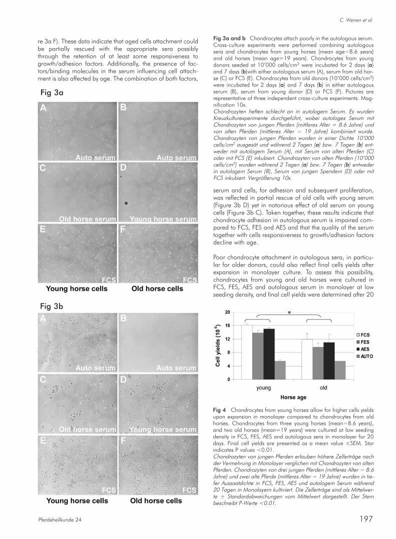
re 3a F). These data indicate that aged cells attachment couldbe partially rescued with the appropriate sera possiblythrough the retention of at least some responsiveness togrowth/adhesion factors. Additionally, the presence of fac-tors/binding molecules in the serum influencing cell attach-ment is also affected by age. The combination of both factors,
C. Werren et al.
Pferdeheilkunde 24 197
serum and cells, for adhesion and subsequent proliferation,was reflected in partial rescue of old cells with young serum(Figure 3b D) yet in notorious effect of old serum on youngcells (Figure 3b C). Taken together, these results indicate thatchondrocyte adhesion in autologous serum is impaired com-pared to FCS, FES and AES and that the quality of the serumtogether with cells responsiveness to growth/adhesion factorsdecline with age.
Poor chondrocyte attachment in autologous sera, in particu-lar for older donors, could also reflect final cells yields afterexpansion in monolayer culture. To assess this possibility,chondrocytes from young and old horses were cultured inFCS, FES, AES and autologous serum in monolayer at lowseeding density, and final cell yields were determined after 20
Fig 3a and b Chondrocytes attach poorly in the autologous serum.Cross-culture experiments were performed combining autologoussera and chondrocytes from young horses (mean age=8.6 years)and old horses (mean age=19 years). Chondrocytes from youngdonors seeded at 10’000 cells/cm2 were incubated for 2 days (a)and 7 days (b)with either autologous serum (A), serum from old hor-se (C) or FCS (E). Chondrocytes from old donors (10’000 cells/cm2)were incubated for 2 days (a) and 7 days (b) in either autologousserum (B), serum from young donor (D) or FCS (F). Pictures arerepresentative of three independent cross-culture experiments. Mag-nification 10x.Chondrozyten heften schlecht an in autologem Serum. Es wurdenKreuzkulturexperimente durchgeführt, wobei autologes Serum mitChondrozyten von jungen Pferden (mittleres Alter = 8.6 Jahre) undvon alten Pferden (mittleres Alter = 19 Jahre) kombiniert wurde.Chondrozyten von jungen Pferden wurden in einer Dichte 10’000cells/cm2 ausgesät und während 2 Tagen (a) bzw. 7 Tagen (b) ent-weder mit autologem Serum (A), mit Serum von alten Pferden (C)oder mit FCS (E) inkubiert. Chondrozyten von alten Pferden (10’000cells/cm2) wurden während 2 Tagen (a) bzw. 7 Tagen (b) entwederin autologem Serum (B), Serum von jungen Spendern (D) oder mitFCS inkubiert. Vergrößerung 10x.
Fig 4 Chondrocytes from young horses allow for higher cells yieldsupon expansion in monolayer compared to chondrocytes from oldhorses. Chondrocytes from three young horses (mean=8.6 years),and two old horses (mean=19 years) were cultured at low seedingdensity in FCS, FES, AES and autologous sera in monolayer for 20days. Final cell yields are presented as a mean value ±SEM. Starindicates P values <0.01.Chondrozyten von jungen Pferden erlauben höhere Zellerträge nachder Vermehrung in Monolayer verglichen mit Chondrozyten von altenPferden. Chondrozyten von drei jungen Pferden (mittleres Alter = 8.6Jahre) und zwei alte Pferde (mittleres Alter = 19 Jahre) wurden in tie-fer Aussaatdichte in FCS, FES, AES und autologem Serum während20 Tagen in Monolayern kultiviert. Die Zellerträge sind als Mittelwer-te ± Standardabweichungen vom Mittelwert dargestellt. Der Sternbeschreibt P-Werte <0.01.
Fig 3a
Fig 3b
days (Figure 4). As expected, due to their poor attachment ateach seeding step, significantly lower final yields were obtai-ned from cells cultured in autologous sera in both agegroups. On the other hand, cell yields in FCS, FES and AESwere comparable within each age group. Importantly, cellsfrom young donors had higher cells yields in FCS, FES andAES compared to cells from old donors (p<0.01) indicatingthat age has an effect on equine chondrocyte proliferationcapacities. Due to the small number of tested animals indivi-dual variations could have influenced the results.
The effects of age and source of serum during cell expansionon chondrocyte re-differentiation capacities
The prerequisite for cell-based therapies in cartilage repairis the capacity of chondrocytes to re-differentiate and pro-duce extracellular matrix. Different growth factors havedemonstrated the potential to facilitate this process, inclu-ding the members of TGFβ family (Mackay et al. 1998,Hanada et al. 2001). To establish optimal re-differentiationconditions chondrocytes from two horses (mean age 5.5)were expanded at low seeding density for 20 days in mono-layer in medium containing 10% FCS and subsequentlyincubated in pellets for 4 weeks in the defined serum freemedium supplemented with different concentrations ofTGFβ1 (0, 1, 5, 10 ng/ml). The evaluation of neochondro-genesis performed on Safranin O stained histology slidesusing Berne score (Grogan et al. 2006) indicated that neo-cartilagenous tissue of similar quality can be obtained with0 or 5ng/ml of TGFβ1 (data not shown). These conditionsfor re-differentiation in pellet cultures were used in subse-quent experiments.
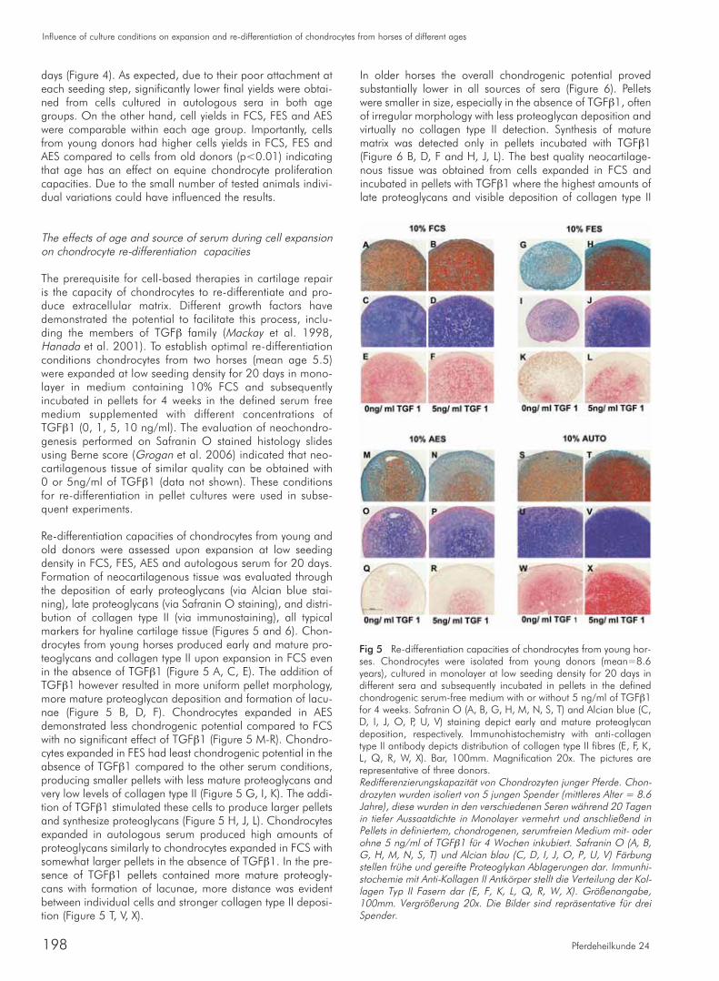
Re-differentiation capacities of chondrocytes from young andold donors were assessed upon expansion at low seedingdensity in FCS, FES, AES and autologous serum for 20 days.Formation of neocartilagenous tissue was evaluated throughthe deposition of early proteoglycans (via Alcian blue stai-ning), late proteoglycans (via Safranin O staining), and distri-bution of collagen type II (via immunostaining), all typicalmarkers for hyaline cartilage tissue (Figures 5 and 6). Chon-drocytes from young horses produced early and mature pro-teoglycans and collagen type II upon expansion in FCS evenin the absence of TGFβ1 (Figure 5 A, C, E). The addition ofTGFβ1 however resulted in more uniform pellet morphology,more mature proteoglycan deposition and formation of lacu-nae (Figure 5 B, D, F). Chondrocytes expanded in AESdemonstrated less chondrogenic potential compared to FCSwith no significant effect of TGFβ1 (Figure 5 M-R). Chondro-cytes expanded in FES had least chondrogenic potential in theabsence of TGFβ1 compared to the other serum conditions,producing smaller pellets with less mature proteoglycans andvery low levels of collagen type II (Figure 5 G, I, K). The addi-tion of TGFβ1 stimulated these cells to produce larger pelletsand synthesize proteoglycans (Figure 5 H, J, L). Chondrocytesexpanded in autologous serum produced high amounts ofproteoglycans similarly to chondrocytes expanded in FCS withsomewhat larger pellets in the absence of TGFβ1. In the pre-sence of TGFβ1 pellets contained more mature proteogly-cans with formation of lacunae, more distance was evidentbetween individual cells and stronger collagen type II deposi-tion (Figure 5 T, V, X).
Influence of culture conditions on expansion and re-differentiation of chondrocytes from horses of different ages
Pferdeheilkunde 24198
In older horses the overall chondrogenic potential provedsubstantially lower in all sources of sera (Figure 6). Pelletswere smaller in size, especially in the absence of TGFβ1, oftenof irregular morphology with less proteoglycan deposition andvirtually no collagen type II detection. Synthesis of maturematrix was detected only in pellets incubated with TGFβ1(Figure 6 B, D, F and H, J, L). The best quality neocartilage-nous tissue was obtained from cells expanded in FCS andincubated in pellets with TGFβ1 where the highest amounts oflate proteoglycans and visible deposition of collagen type II
Fig 5 Re-differentiation capacities of chondrocytes from young hor-ses. Chondrocytes were isolated from young donors (mean=8.6years), cultured in monolayer at low seeding density for 20 days indifferent sera and subsequently incubated in pellets in the definedchondrogenic serum-free medium with or without 5 ng/ml of TGFβ1for 4 weeks. Safranin O (A, B, G, H, M, N, S, T) and Alcian blue (C,D, I, J, O, P, U, V) staining depict early and mature proteoglycandeposition, respectively. Immunohistochemistry with anti-collagentype II antibody depicts distribution of collagen type II fibres (E, F, K,L, Q, R, W, X). Bar, 100mm. Magnification 20x. The pictures arerepresentative of three donors.Redifferenzierungskapazität von Chondrozyten junger Pferde. Chon-drozyten wurden isoliert von 5 jungen Spender (mittleres Alter = 8.6Jahre), diese wurden in den verschiedenen Seren während 20 Tagenin tiefer Aussaatdichte in Monolayer vermehrt und anschließend inPellets in definiertem, chondrogenen, serumfreien Medium mit- oderohne 5 ng/ml of TGFβ1 für 4 Wochen inkubiert. Safranin O (A, B,G, H, M, N, S, T) und Alcian blau (C, D, I, J, O, P, U, V) Färbungstellen frühe und gereifte Proteoglykan Ablagerungen dar. Immunhi-stochemie mit Anti-Kollagen II Antkörper stellt die Verteilung der Kol-lagen Typ II Fasern dar (E, F, K, L, Q, R, W, X). Größenangabe,100mm. Vergrößerung 20x. Die Bilder sind repräsentative für dreiSpender.

was observed (Figure 6 B, D, F). The evaluation of neocartila-genous tissue formation was further assessed with Berne sco-re (Figure 7). Based on mixed factors Anova tests, chondrocy-tes from young horses produced pellets with a higher Bernescore compared to chondrocytes from old horses (P<0.05).The highest score from young donors was obtained from cellsexpanded in autologous serum and incubated in pellets withTGFβ1 (Score=6). However, it was not statistically differentcompared to cells expanded in FCS, suggesting that cells fromyounger donors retain their re-differentiation capacities uponexpansion in FCS and autologous serum. Cells from olddonors demonstrated a tendency for best re-differentiationupon expansion in FCS and incubation with TGFβ1 in pellets(Score=4.3). These results indicate that while the source ofsera and the presence of TGFβ1 appear to influence chon-
C. Werren et al.
Pferdeheilkunde 24 199
drocyte re-differentiation capacities, donors age clearly has astatistically significant effect.
Discussion
Understanding the biology and behavior of equine chondro-cytes is a prerequisite for the proper design of cell-based tre-atment for cartilage repair in horses. Chondrocyte expansionwith the maintenance of cell chondrogenic re-differentiationcapacities is fundamental for the application of cartilage tis-sue engineering treatments, including ACI. In the present stu-dy equine chondrocyte attachment, expansion and re-diffe-rentiation capacities were examined. Chondrocytes efficientlyadhered and proliferated in medium containing allogenic
Fig 6 Re-differentiation capacities of chondrocytes from old horses.Chondrocytes were isolated from old donors (mean=19 years). Cul-tures and analysis were as described for Figure 5. Safranin O (A, B,G, H, M, N, S, T) and Alcian blue (C, D, I, J, O, P, U, V) stainingdepict early and mature proteoglycan deposition, respectively. Immu-nohistochemistry with anti-collagen type II antibody depicts distribu-tion of collagen type II fibres (E, F, K, L, Q, R, W, X). The pictures arerepresentative of two donors.Redifferenzierungskapazität von alten Pferden. Chondrozyten vonalten Spendern wurden isoliert (mittleres Alter = 19 Jahre). Kultivie-rung und Analysen wurden gleich ausgeführt wie für Bild 5. SafraninO (A, B, G, H, M, N, S, T) und Alcian blau (C, D, I, J, O, P, U, V)Färbung stellen frühe und gereifte Proteoglykan-Ablagerungen dar.Immunhistochemie mit Anti-Kollagen II Antkörper stellt die Verteilungder Kollagen Typ II Fasern dar (E, F, K, L, Q, R, W, X). Die Bilder sindrepräsentativ für zwei Spender.
Fig 7A and B Neocartilagenous tissue formation from chondrocy-tes expanded in different sources of sera is age dependent. Chon-drocytes from young donors (A) or old donors (B) were cultured inmonolayer (3’333 cells/cm2) for 20 days in FCS, FES, AES and auto-logous sera. Pellet cultures were incubated in the defined chondro-genic serum-free medium with or without 5 ng/ml of TGFβ1 for 4weeks. Histology slides were stained with Safranin O and evaluatedaccording to the Berne score for neocartilagenous tissue formation. Die Bildung von neuem Knorpelgewebe aus Chondrozyten, vermehrtin unterschiedlichen Seren, ist altersabhängig. Chondrozyten von jun-gen Spendern (A) oder alten Spendern (B) wurden während 20Tagen in Monlayern (3.333 Zellen/cm2) in FCS, FES, AES und auto-logem Serum kulitiviert. Pelletkulturen wurden in definiertem, chon-drogenen, serumfreien Medium, mit oder ohne 5 ng/ml of TGFβ1,während 4 Wochen inkubiert. Histologieschnitte wurden mit SafraninO gefärbt und mit Hilfe des Berne score wurde das neugebildeteKnorpelgewebe beurteilt.

FES, allogenic AES or xenogenic FCS. However, poor chon-drocyte attachment in autologous sera at each passage resul-ted in lower final cell yields compared to the other sera tested. Development of serum free or autologous serum culturingsystems for cell expansion in tissue engineering aims to cir-cumvent the problem of disease transmission and immuno-genic reaction (Heng et al. 2004, Heiskanen et al. 2007).Several studies have demonstrated beneficial effect of growthfactor addition to the serum-free expansion medium for theproliferation and maintenance of the chondrogenic phenoty-pe (Mandl et al. 2002, Malpeli et al. 2004), even overco-ming the age-related differences (Giannoni et al. 2005). Alt-hough promising, the need for exogenously added growthfactors remains the disadvantage of the serum-free approach.Autologous serum represents another alternative to FCS forcell expansion. Studies have demonstrated the possibility toreplace FCS for expansion of human mesenchymal stem cells(Stute et al. 2004, Shahdadfar et al. 2005). In human chon-drocytes recent data have demonstrated higher proliferationrates of articular and auricular chondrocytes in autologousand allogenic human serum in comparison to FCS (Tallhedenet al. 2005, Yanaga et al. 2006). Equine chondrocyte expan-sion in autologous serum has been successfully employed forthe ACI treatment (Litzke et al. 2004). However, no compari-son to FCS has been established. Our data indicate less effi-cient chondrocyte expansion in autologous serum comparedto allogenic AES and FES or xenogenic FCS due to the pro-nounced difficulty in cell attachment at each passage. Theimpaired adhesion in autologous serum could be attributedto the fact that the sera derived from stunned horses whichwere transported for slaughtering and thus experiencedstress: during the procedures for optimizing serum prepara-tion, sera were prepared from living horses within the horseclinic (unstressed), and tested on homologous chondrocytes.No significant differences in cell adhesion and comparableproliferation rates were observed between chondrocytes inFCS or in individual homologous sera from living horses(data not shown), suggesting that trauma-related transportand stunning may have caused the release of stress factors,including catecholamines, which could alter expression ofadhesion molecules in vitro, as suggested in other cell types(Shepard 2003). The problem of attachment however, had noeffect on young chondrocytes re-differentiation capacities,suggesting that factors important for adhesion differ from tho-se required for chondrogenenic capacities.
The age of horses also affected chondrocyte attachment inautologous serum. Our data suggest that not only chondro-cyte responsiveness to serum factors declines with age, butthat the presence of growth factors and extracelular matrixmolecules facilitating adhesion decreases with age. Age-rela-ted changes in chondrocyte biology are well documented.Chondrocytes number in cartilage significantly decrease withadvancing age (Bobacz et al. 2004), their proliferative respon-se to serum changes and declines (Guerne et al. 1995, Bar-bero et al. 2004), cells senesce and shorten their telomerelength (Martin and Buckwalter 2003). In horses, proteoglycansynthesis and chondrocyte responsiveness to TGFβ1 steadilydecreases with increasing age (Iqbal et al. 2000].
Seeding density had an important effect on chondrocyte proli-feration in FCS, FES and AES. Cells seeded at low seedingdensities proliferated faster compared to chondrocytes seeded
Influence of culture conditions on expansion and re-differentiation of chondrocytes from horses of different ages
Pferdeheilkunde 24200
at high seeding density. The long accepted postulation hasbeen that low seeding densities allow for higher proliferationrates but lead to more pronounced de-differentiation (Holtzeret al. 1960, Watt 1988). Several mechanisms have been sug-gested to explain this phenomenon, including more populationdoublings before confluence is reached and changes in cyto-skeletal microfilament organization (Lim et al. 2000, Mandl etal. 2004). However, recent studies using human auricular andnasal septal chondrocytes have demonstrated that low seedingdensity in monolayer cultures resulted in fewer rounds of pas-saging (exposure to trypsin) to obtain similar cell yields, andpreserved chondrocyte chondrogenic potential (Mandl et al.2004, Hicks et al. 2005). Our data showed that chondrocytesseeded at high density reach the average cell density of43’250 cells/cm2 at the time of harvest. This value is far belowthe maximum density of 105’000 cells/cm2 observed in thehigh density cultures, indicating that cells at 43’250 cells/cm2
have not reached saturation density, and suggesting that con-tact inhibition is an unlikely cause for lower cell yields obser-ved. Faster proliferation at lower densities has also been repor-ted for human MSC (Sekiya et al. 2002, Both et al. 2006). Pos-sible explanation includes involvement of Wnt signaling (Gre-gory et al. 2003, De Boer et al. 2004). The firm conclusionhas however, not been reached, most probably due to severaldifferent factors contributing to this phenomenon. One couldhypothesize that similar mechanisms may be implicated inequine chondrocytes. Importantly, the capacity of equine arti-cular chondrocytes to re-differentiate upon expansion at lowseeding densities in FCS, FES, AES and autologous sera indi-cates preservation of their chondrogenic potential. Suchexpansion regime would allow initiating cell expansion withlower cell numbers and hence smaller biopsies.
High cell density three-dimensional culture is a necessaryrequirement for in vitro cartilage formation. Furthermore,chondrogenesis can be facilitated by the addition of exoge-nous growth factors including TGFβ, BMP-2, and IGF-1 (Kuz-netsov et al. 2001, Heng et al. 2004). TGFβ and IGF-1 havealso demonstrated positive chondrogenic effect on equinechondrocytes (Fortier et al. 1997, Fortier et al. 2002). In thisstudy chondrocyte re-differentiation capacities were influen-ced by the source of sera during the expansion phase and theage of donors. The presence of TGFβ1 during the re-diffe-rentiation phase proved less critical for chondrocytes fromyoung donors which had retained their re-differentiationcapacities even in the absence of TGFβ1, while cells from oldhorses required the presence of TGFβ1. Importantly, whilechondrocytes from young donors re-differentiated efficientlyupon expansion in FCS or autologous serum, expansion ofchondrocytes from old donors required FCS.
In summary, our data demonstrate that equine chondrocytescan be isolated from smaller biopsies and expanded at lowseeding densities to allow higher yields. Importantly, the ageof the horse has a profound effect on chondrocyte behaviorand emphasizes the necessity to understand individual equi-ne chondrocyte requirements to successfully design cell basedtreatment of cartilage defects. Our data indicate that for cellsfrom young donors autologous serum could replace FCS the-reby avoiding possible exposure to infections and inflamma-tory reactions. In contrast, cells from older horses althoughwith obvious impaired proliferation and re-differentiationcapacities, could still be employed providing that they are
expanded in FCS and re-differentiated in the presence ofTGFβ1. More detailed studies on larger cohorts of animalsare needed to determine the age threshold.
Abbreviations
FCS fetal calf serumFES fetal equine serumAES adult equine serumACI autologous chondrocyte implantation
Acknowledgements
This work was supported by an educational grant from Alle-via AG, Switzerland.
The authors would like to thank Verena Winkelmann, Chan-tal Pauli, Kathrin Boschung and Simon Herren for excellenttechnical assistance.
References
Barbero A., S. Grogan, D. Schafer, M. Heberer, P. Mainil-Varlet andI. Martin (2004) Age related changes in human articular chondro-cyte yield, proliferation and post-expansion chondrogenic capaci-ty. Osteoarthritis Cartilage 12, 476-484
Bobacz K., L. Erlacher, J. Smolen, A. Soleiman and W. B. Graninger(2004) Chondrocyte number and proteoglycan synthesis in theaging and osteoarthritic human articular cartilage. Ann. Rheum.Dis. 63, 1618-1622
Both S. K., A. J. Muijsenberg, C. A. Blitterswijk, J. D. Boer and J. D.Bruijn (2007) A Rapid and Efficient Method for Expansion ofHuman Mesenchymal Stem Cells. Tissue Eng. 13, 3-9
Brehm W., C. Werren and P. Mainil-Varlet (2007) Production of verylarge sized scaffold-free engineered cartilage constructs fromadult equine donor - a basis for therapeutic and experimentalapplication in horses. Pferdeheilkunde 23, 111-117
Brittberg M., A. Lindahl, A. Nilsson, C. Ohlsson, O. Isaksson and L.Peterson (1994) Treatment of deep cartilage defects in the kneewith autologous chondrocyte transplantation. N. Engl. J. Med.331, 889-895
Cremer M. A., E. F. Rosloniec and A. H. Kang (1998) The cartilagecollagens: a review of their structure, organization, and role in thepathogenesis of experimental arthritis in animals and in humanrheumatic disease. J. Mol. Med. 76, 275-288
De Boer J., H. J. Wang and C. Van Blitterswijk (2004) Effects of Wntsignaling on proliferation and differentiation of human mesenchy-mal stem cells. Tissue Eng. 10, 393-401
Dedrick V. (1997) Determining the safety of medical devices contai-ning animal tissues: the new European standards. J. Regul. AffairsProf. Soc. 2, 20
Diaz-Romero J., J. P. Gaillard, S. P. Grogan, D. Nesic, T. Trub and P.Mainil-Varlet (2005) Immunophenotypic analysis of human articu-lar chondrocytes: changes in surface markers associated with cellexpansion in monolayer culture. J. Cell. Physiol. 202, 731-742
Fortier L. A., H. O. Mohammed, G. Lust and A. J. Nixon (2002) Insu-lin-like growth factor-I enhances cell-based repair of articular car-tilage. J. Bone Joint Surg. Br. 84, 276-288
Fortier L. A., A. J. Nixon, H. O. Mohammed and G. Lust (1997) Alte-red biological activity of equine chondrocytes cultured in a three-dimensional fibrin matrix and supplemented with transforminggrowth factor beta-1. Am. J. Vet. Res. 58, 66-70
C. Werren et al.
Pferdeheilkunde 24 201
Frisbie D. D., M. W. Cross and C. W. McIlwraith (2006) A compa-rative study of articular cartilage thickness in the stifle of animalspecies used in human pre-clinical studies compared to articularcartilage thickness in the human knee. Vet. Comp. Orthop. Trau-matol. 19,142-146
Giannoni P., A. Pagano, E. Maggi, R. Arbico, N. Randazzo, M. Gran-dizio, R. Cancedda and B. Dozin (2005) Autologous chondrocyteimplantation (ACI) for aged patients: development of the propercell expansion conditions for possible therapeutic applications.Osteoarthritis Cartilage 13, 589-600
Gregory C. A., H. Singh, A. S. Perry and D. J. Prockop (2003) TheWnt signaling inhibitor dickkopf-1 is required for reentry into thecell cycle of human adult stem cells from bone marrow. J. Biol.Chem. 278(, 28067-28078
Grogan S. P., A. Barbero, V. Winkelmann, F. Rieser, J. S. Fitzsim-mons, S. O'Driscoll, I. Martin and P. Mainil-Varlet (2006) Visualhistological grading system for the evaluation of in vitro-generatedneocartilage. Tissue Eng. 12, 2141-2149
Guerne P. A., F. Blanco, A. Kaelin, A. Desgeorges and M. Lotz(1995) Growth factor responsiveness of human articular chondro-cytes in aging and development. Arthritis Rheum. 38, 960-968
Hanada K., L. A. Solchaga, A. I. Caplan, T. M. Hering, V. M. Gold-berg, J. U. Yoo and B. Johnstone (2001) BMP-2 induction andTGF-beta 1 modulation of rat periosteal cell chondrogenesis. J.Cell Biochem. 81, 284-294
Heiskanen A., T. Satomaa, S. Tiitinen, A. Laitinen, S. Mannelin, U.Impola, M. Mikkola, C. Olsson, H. Miller-Podraza, M. Blomqvist,A. Olonen, H. Salo, P. Lehenkari, T. Tuuri, T. Otonkoski, J. Natu-nen, J. Saarinen and J. Laine (2007) N-glycolylneuraminic acidxenoantigen contamination of human embryonic and mesenchy-mal stem cells is substantially reversible. Stem Cells 25, 197-202
Heng B. C., T. Cao and E. H. Lee (2004) Directing stem cell diffe-rentiation into the chondrogenic lineage in vitro. Stem Cells 22,1152-1167
Hicks D. L., A. B. Sage, B. L. Schumacher, R. L. Sah and D. Watson(2005) Growth and phenotype of low-density nasal septal chon-drocyte monolayers. Otolaryngol Head Neck Surg 133, 417-422
Holtzer H., J. Abbott, J. Lash and S. Holtzer (1960) The Loss of Phe-notypic Traits by Differentiated Cells in Vitro, I. Dedifferentiation ofCartilage Cells. Proc. Natl. Acad. Sci. USA 46, 1533-1542
Iqbal J., J. Dudhia, J. L. Bird and M. T. Bayliss (2000) Age-relatedeffects of TGF-beta on proteoglycan synthesis in equine articularcartilage. Biochem. Biophys. Res. Commun. 274, 467-471
Klein R. and L. J. Dumble (1993) Transmission of Creutzfeldt-Jakobdisease by blood transfusion. Lancet 341, 768
Koerner J., D. Nesic, J. D. Romero, W. Brehm, P. Mainil-Varlet andS. P. Grogan (2006) Equine peripheral blood-derived progenitorsin comparison to bone marrow-derived mesenchymal stem cells.Stem Cells 24, 1613-1619
Kuznetsov S. A., M. H. Mankani, S. Gronthos, K. Satomura, P. Bian-co and P. G. Robey (2001) Circulating skeletal stem cells. J. CellBiol. 153, 1133-1140
Lim Y. B., S. S. Kang, T. K. Park, Y. S. Lee, J. S. Chun and J. K. Sonn(2000) Disruption of actin cytoskeleton induces chondrogenesis ofmesenchymal cells by activating protein kinase C-alpha signaling.Biochem. Biophys. Res. Commun. 273, 609-613
Litzke L. E., E. Wagner, W. Baumgaertner, U. Hetzel, O. Josimovic-Alasevic and J. Libera (2004): Repair of extensive articular cartila-ge defects in horses by autologous chondrocyte transplantation.Ann. Biomed. Eng. 32, 57-69
Mackay A. M., S. C. Beck, J. M. Murphy, F. P. Barry, C. O. Chichesterand M. F. Pittenger (1998) Chondrogenic differentiation of culturedhuman mesenchymal stem cells from marrow. Tissue Eng. 4, 415-428
Malpeli M., N. Randazzo, R. Cancedda and B. Dozin (2004) Serum-free growth medium sustains commitment of human articularchondrocyte through maintenance of Sox9 expression. Tissue Eng.10, 145-155
Mandl E. W., S. W. van der Veen, J. A. Verhaar and G. J. van Osch(2002) Serum-free medium supplemented with high-concentrationFGF2 for cell expansion culture of human ear chondrocytes pro-motes redifferentiation capacity. Tissue Eng. 8, 573-580
Mandl E. W., S. W. van der Veen, J. A. Verhaar and G. J. van Osch(2004) Multiplication of human chondrocytes with low seedingdensities accelerates cell yield without losing redifferentiationcapacity. Tissue Eng. 10, 109-118
Marlovits S., P. Zeller, P. Singer, C. Resinger and V. Vecsei (2006)Cartilage repair: generations of autologous chondrocyte trans-plantation. Eur. J. Radiol. 57, 24-31
Martin J. A. and J. A. Buckwalter (2003) The role of chondrocytesenescence in the pathogenesis of osteoarthritis and in limitingcartilage repair. J. Bone Joint Surg. Am. 85-A Suppl 2, 106-110
McIlwraith C. W., J. V. Yovich and G. S. Martin (1987) Arthroscopicsurgery for the treatment of osteochondral chip fractures in theequine carpus. J. Am. Vet. Med. Assoc. 191, 531-540
Mendler M., S. G. Eich-Bender, L. Vaughan, K. H. Winterhalter andP. Bruckner (1989) Cartilage contains mixed fibrils of collagentypes II, IX, and XI. J. Cell Biol. 108, 191-197.
Reinholz G. G., L. Lu, D. B. Saris, M. J. Yaszemski and S. W. O'Dri-scoll (2004) Animal models for cartilage reconstruction. Biomate-rials 25, 1511-1521
Schnabel M., S. Marlovits, G. Eckhoff, I. Fichtel, L. Gotzen, V. Vecseiand J. Schlegel (2002) Dedifferentiation-associated changes inmorphology and gene expression in primary human articularchondrocytes in cell culture. Osteoarthritis Cartilage 10, 62-70.
Sekiya I., B. L. Larson, J. R. Smith, R. Pochampally, J. G. Cui and D.J. Prockop (2002) Expansion of human adult stem cells from bonemarrow stroma: conditions that maximize the yields of early pro-genitors and evaluate their quality. Stem Cells 20, 530-541
Selvaggi T. A., R. E. Walker and T. A. Fleisher (1997) Developmentof antibodies to fetal calf serum with arthus-like reactions inhuman immunodeficiency virus-infected patients given syngeneiclymphocyte infusions. Blood 89, 776-779
Shahdadfar A., K. Fronsdal, T. Haug, F. P. Reinholt and J. E. Brinchmann(2005) In vitro expansion of human mesenchymal stem cells: choiceof serum is a determinant of cell proliferation, differentiation, geneexpression, and transcriptome stability. Stem Cells 23, 1357-1366
Shephard R. J. (2003) Adhesion molecules, catecholamines andleucocyte redistribution during and following exercise. SportsMed. 33, 261-284
Stute N., K. Holtz, M. Bubenheim, C. Lange, F. Blake and A. R. Zan-der (2004) Autologous serum for isolation and expansion ofhuman mesenchymal stem cells for clinical use. Exp. Hematol.32, 1212-1225
Tallheden T., J. van der Lee, C. Brantsing, J. E. Mansson, E. Sjogren-Jansson and A. Lindahl (2005) Human serum for culture of arti-cular chondrocytes. Cell Transplant 14, 469-479
Ulrich-Vinther M., M. D. Maloney, E. M. Schwarz, R. Rosier and R. J.O'Keefe (2003) Articular cartilage biology. J. Am. Acad. Orthop.Surg. 11, 421-430
Vacanti J. P., R. Langer, J. Upton and J. J. Marler (1998) Transplan-tation of cells in matrices for tissue regeneration. Adv. Drug. Deliv.Rev. 33, 165-182
Wang X., S. P. Grogan, F. Rieser, V. Winkelmann, V. Maquet, M. L.Berge and P. Mainil-Varlet (2004) Tissue engineering of biphasiccartilage constructs using various biodegradable scaffolds: an invitro study. Biomaterials 25, 3681-3688
Watt F. M. (1988) Effect of seeding density on stability of the diffe-rentiated phenotype of pig articular chondrocytes in culture. J.Cell Sci. 89 (Pt 3), 373-378
Yanaga H., K. Yanaga, K. Imai, M. Koga, C. Soejima and K. Ohmo-ri (2006) Clinical application of cultured autologous human auri-cular chondrocytes with autologous serum for craniofacial ornasal augmentation and repair. Plast. Reconstr. Surg. 117, 2019-2030; discussion 2031-2012
Dobrila Nesic, PhDOsteoarticular Research GroupInstitute of Pathology, University of BernMurtenstraße 31, 3010 [email protected]
Pferdeheilkunde 24
R R
Related Documents