This article was downloaded by: [Anna University] On: 11 February 2015, At: 21:19 Publisher: Taylor & Francis Informa Ltd Registered in England and Wales Registered Number: 1072954 Registered office: Mortimer House, 37-41 Mortimer Street, London W1T 3JH, UK Click for updates International Journal of Polymeric Materials and Polymeric Biomaterials Publication details, including instructions for authors and subscription information: http://www.tandfonline.com/loi/gpom20 Influence of Anionic Surface Charged Biocompatible Dendrimer With a Photosensitizer, Protoporphyrin IX, on Human Red Blood Cells: A Spectroscopic Investigation M. Suresh Kumar a , M. Yuvaraj a , P. Aruna a , D. Koteeswaran b & S. Ganesan a a Department of Medical Physics, Anna University, Chennai, India b Department of Oral Medicine and Radiology, Meenakshi Ammal Dental College and Hospital, Chennai, India Accepted author version posted online: 02 Feb 2015. To cite this article: M. Suresh Kumar, M. Yuvaraj, P. Aruna, D. Koteeswaran & S. Ganesan (2015) Influence of Anionic Surface Charged Biocompatible Dendrimer With a Photosensitizer, Protoporphyrin IX, on Human Red Blood Cells: A Spectroscopic Investigation, International Journal of Polymeric Materials and Polymeric Biomaterials, 64:10, 519-525, DOI: 10.1080/00914037.2014.977899 To link to this article: http://dx.doi.org/10.1080/00914037.2014.977899 PLEASE SCROLL DOWN FOR ARTICLE Taylor & Francis makes every effort to ensure the accuracy of all the information (the “Content”) contained in the publications on our platform. However, Taylor & Francis, our agents, and our licensors make no representations or warranties whatsoever as to the accuracy, completeness, or suitability for any purpose of the Content. Any opinions and views expressed in this publication are the opinions and views of the authors, and are not the views of or endorsed by Taylor & Francis. The accuracy of the Content should not be relied upon and should be independently verified with primary sources of information. Taylor and Francis shall not be liable for any losses, actions, claims, proceedings, demands, costs, expenses, damages, and other liabilities whatsoever or howsoever caused arising directly or indirectly in connection with, in relation to or arising out of the use of the Content. This article may be used for research, teaching, and private study purposes. Any substantial or systematic reproduction, redistribution, reselling, loan, sub-licensing, systematic supply, or distribution in any form to anyone is expressly forbidden. Terms & Conditions of access and use can be found at http:// www.tandfonline.com/page/terms-and-conditions

Welcome message from author
This document is posted to help you gain knowledge. Please leave a comment to let me know what you think about it! Share it to your friends and learn new things together.
Transcript
This article was downloaded by: [Anna University]On: 11 February 2015, At: 21:19Publisher: Taylor & FrancisInforma Ltd Registered in England and Wales Registered Number: 1072954 Registered office: Mortimer House,37-41 Mortimer Street, London W1T 3JH, UK
Click for updates
International Journal of Polymeric Materials andPolymeric BiomaterialsPublication details, including instructions for authors and subscription information:http://www.tandfonline.com/loi/gpom20
Influence of Anionic Surface Charged BiocompatibleDendrimer With a Photosensitizer, ProtoporphyrinIX, on Human Red Blood Cells: A SpectroscopicInvestigationM. Suresh Kumara, M. Yuvaraja, P. Arunaa, D. Koteeswaranb & S. Ganesana
a Department of Medical Physics, Anna University, Chennai, Indiab Department of Oral Medicine and Radiology, Meenakshi Ammal Dental College andHospital, Chennai, IndiaAccepted author version posted online: 02 Feb 2015.
To cite this article: M. Suresh Kumar, M. Yuvaraj, P. Aruna, D. Koteeswaran & S. Ganesan (2015) Influence of AnionicSurface Charged Biocompatible Dendrimer With a Photosensitizer, Protoporphyrin IX, on Human Red Blood Cells: ASpectroscopic Investigation, International Journal of Polymeric Materials and Polymeric Biomaterials, 64:10, 519-525, DOI:10.1080/00914037.2014.977899
To link to this article: http://dx.doi.org/10.1080/00914037.2014.977899
PLEASE SCROLL DOWN FOR ARTICLE
Taylor & Francis makes every effort to ensure the accuracy of all the information (the “Content”) containedin the publications on our platform. However, Taylor & Francis, our agents, and our licensors make norepresentations or warranties whatsoever as to the accuracy, completeness, or suitability for any purpose of theContent. Any opinions and views expressed in this publication are the opinions and views of the authors, andare not the views of or endorsed by Taylor & Francis. The accuracy of the Content should not be relied upon andshould be independently verified with primary sources of information. Taylor and Francis shall not be liable forany losses, actions, claims, proceedings, demands, costs, expenses, damages, and other liabilities whatsoeveror howsoever caused arising directly or indirectly in connection with, in relation to or arising out of the use ofthe Content.
This article may be used for research, teaching, and private study purposes. Any substantial or systematicreproduction, redistribution, reselling, loan, sub-licensing, systematic supply, or distribution in anyform to anyone is expressly forbidden. Terms & Conditions of access and use can be found at http://www.tandfonline.com/page/terms-and-conditions
Influence of Anionic Surface Charged BiocompatibleDendrimer With a Photosensitizer, Protoporphyrin IX, onHuman Red Blood Cells: A Spectroscopic Investigation
M. SURESH KUMAR1, M. YUVARAJ1, P. ARUNA1, D. KOTEESWARAN2, and S. GANESAN1
1Department of Medical Physics, Anna University, Chennai, India2Department of Oral Medicine and Radiology, Meenakshi Ammal Dental College and Hospital, Chennai, India
Received 9 July 2014, Accepted 19 September 2014
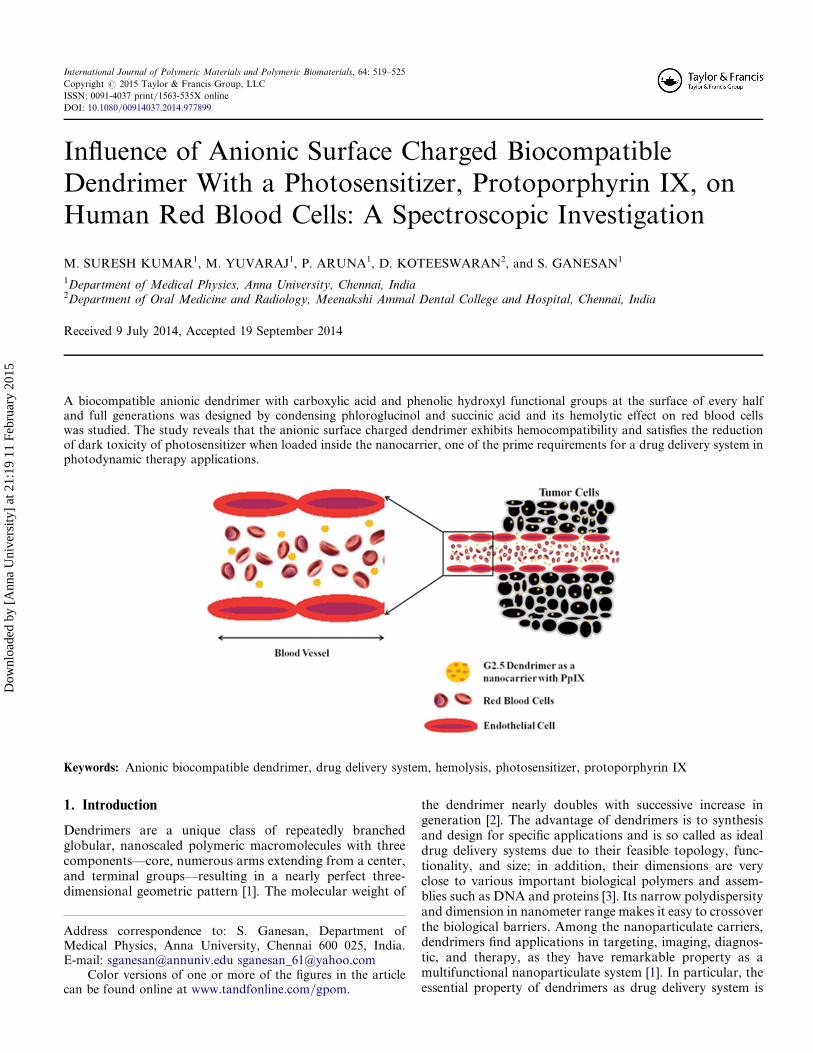
A biocompatible anionic dendrimer with carboxylic acid and phenolic hydroxyl functional groups at the surface of every halfand full generations was designed by condensing phloroglucinol and succinic acid and its hemolytic effect on red blood cellswas studied. The study reveals that the anionic surface charged dendrimer exhibits hemocompatibility and satisfies the reductionof dark toxicity of photosensitizer when loaded inside the nanocarrier, one of the prime requirements for a drug delivery system inphotodynamic therapy applications.
Keywords: Anionic biocompatible dendrimer, drug delivery system, hemolysis, photosensitizer, protoporphyrin IX
1. Introduction
Dendrimers are a unique class of repeatedly branchedglobular, nanoscaled polymeric macromolecules with threecomponents—core, numerous arms extending from a center,and terminal groups—resulting in a nearly perfect three-dimensional geometric pattern [1]. The molecular weight of
the dendrimer nearly doubles with successive increase ingeneration [2]. The advantage of dendrimers is to synthesisand design for specific applications and is so called as idealdrug delivery systems due to their feasible topology, func-tionality, and size; in addition, their dimensions are veryclose to various important biological polymers and assem-blies such as DNA and proteins [3]. Its narrow polydispersityand dimension in nanometer range makes it easy to crossoverthe biological barriers. Among the nanoparticulate carriers,dendrimers find applications in targeting, imaging, diagnos-tic, and therapy, as they have remarkable property as amultifunctional nanoparticulate system [1]. In particular, theessential property of dendrimers as drug delivery system is
Address correspondence to: S. Ganesan, Department ofMedical Physics, Anna University, Chennai 600 025, India.E-mail: [email protected] [email protected]
Color versions of one or more of the figures in the articlecan be found online at www.tandfonline.com/gpom.
International Journal of Polymeric Materials and Polymeric Biomaterials, 64: 519–525
Copyright # 2015 Taylor & Francis Group, LLC
ISSN: 0091-4037 print/1563-535X online
DOI: 10.1080/00914037.2014.977899
Dow
nloa
ded
by [
Ann
a U
nive
rsity
] at
21:
19 1
1 Fe
brua
ry 2
015
to provide an environment for incorporating photosensitizer(PS), which can be administered in monomeric form, withoutaltering its activity [4]. The three main properties of dendrimeras nanoscale containers (i.e., encapsulation of a drug),nano-scaffolding (i.e., surface adsorption or attachment of adrug), and biocompatibility [2,3,5–10] provides the dendrimerto customize for the specific therapeutic needs as an ultimatecarriers for small drugs and biomolecules. The routes ofadministration of dendrimer may be intravenous, oral, trans-dermal and ocular [10,11] and it should be water soluble tofacilitate systemic administration [12].
In general, it is essential to consider the safety administra-tion of dendrimer in a proposed application, so it is requiredfor any polymeric carrier to be non-toxic, non-immunogenic,and it should be preferably biodegradable [4,13,14].Encapsulation=attachment of wide variety of molecules hasbeen successfully achieved inside dendrimers for drug andgene delivery applications [14]. Polyaryl ether based dendri-mers derivatized with the PS protoporphyrin, have been eval-uated as candidates for the PDT of solid tumors and thisproperty of dendrimers as a nanocarrier for photosensitizerscan be improved by appropriate unfunctionalization of theirperiphery [11]. Glycerol and succinic acid based dendrimerswere recently developed to deliver 10-hydroxy-camptothecinand tested in various cell lines [15]. Although the use ofdendrimer as therapeutic agents is a rapidly growing field,but its toxicity and effects on biological systems makes itdifficult to use as therapeutic agents [16]. There are reportson dendrimers such as polylysines, anionic polyamidoamine(PAMAM)-COOH dendrimers, and polyester dendrimers,which can parade its distribution highly to the liver and quickexcretion into the urine [12] when administered. One of themost appropriate ways to deliver drugs based on dendrimersis by intravenous injection [17]. However, if that is done, redblood cells (RBCs) are expected to be the first and unwantedtargets of their action; in particular, hemolysis may occur.Hemolysis is the release of hemoglobin from the damagederythrocytes (also known as RBCs) [16]. Recently a minire-view has been focused on discussing the in vitro interactionsbetween dendrimers and RBCs, and pointed out the possiblestrategies to facilitate the dendrimer hemotoxicity [18].
A major problem reported with commercially availablePAMAM, starburst dendrimer [19], is cytotoxicity whenexposed with (amine terminated) cationic surface groupsand its interaction with bilayer membranes. In the same con-text in vitro preliminary studies with polyether dendrimer andPAMAM dendrimer of anionic surface with COOH groups,poly (ethylene glycol) surface showed no hemolytic effectsand cytotoxic effects [20,21]. PAMAM and PPI dendrimerswith PS such as PpIX and rosebengal (RB) in HeLa cellsand human cervix cancer SiH cells have shown good PDTactivity in both in vitro and in vivo studies and reported asnontoxic because of PEGylation on their surfaces [22,23].There are also reports on RBCs exposed to the anionicPAMAM dendrimer of generations 3.5–9.5, showing nomorphological changes up to a concentration of 2 mg=mL[10]. Small generation dendrimers with charged surface orhydrophilic surfaces (-OH or PEGylated) escapes from rapid
clearance. Mostly the cytotoxicity of the cationic surfacefunctionality of PAMAM, poly (lysine) dendrimers havebeen minimized by PEGylation and glutamate surface mod-ifications [24,25] to act as nontoxic biocompatible drug deliv-ery systems. Similarly, commercially available dendrimershas been reported to be minimal in cytotoxicity when functio-nalized with acetylation, glycosylation, and PEGylation [26].Dendrimers with cationic surface groups produce hemolyticeffects by electrostatic attraction with negatively chargedred blood cells [27], and the release of hemoglobin fromerythrocyte will be due to the disruption of cell membraneof RBCs by the formation of holes [28].
In this study, biocompatible dendrimer of generations(G0.5–G2.5) with phenolic OH and carboxylic acid(–COOH) terminal functional groups were synthesized bycondensing phloroglucinol and succinic acid (PGSA) andits impact on RBC morphology is inspected spectroscopicallyand microscopically after one hour incubation with RBCsuspension. In this context, preliminary steps have beentaken to use our novel dendrimer as nanocarrier for darktoxic photosensitizer, PpIX. In order to be nontoxic and bio-compatible, it has been tested for hemotoxicity with RBCs, asthey are the first to come in to contact with the dendrimerwhen administered intravenously. Dendrimers can also easilycarry along certain nanoparticles along with them to crossthe biological barriers and reach the targeted site.
2. Experimental
2.1 Materials
The precursors phloroglucinol (2,4,6 trihydroxy benzene)and succinic acid were procured from Sigma Aldrich (USA)to synthesize the novel biocompatible dendrimer of genera-tions from 0.5 to 2.5. Freshly drawn blood from healthyvolunteers was used for the experiments. The other chemicalsutilized in the experiments were used of AR grade.
2.2 Synthesis and Characterization of PpIX Loaded AnionicSurface Charged Biocompatible PGSA Dendrimer
The anionic surface charged PGSA dendrimer was synthe-sized using phloroglucinol (2,4,6 trihydroxy benzene) as acore and succinic acid as the branching units in the mole ratioof 1:3 initially to form the first half generation (G0.5) byreflux method [19]. The successive half generations (G1.5,G2.5) were developed by the addition of succinic acid toG1.0 and G2.0 products and full generations (G1.0, G2.0),were developed by adding phloroglucinol to the G0.5,G1.5, and G2.5 with respect to the molecular weight of eachgeneration, respectively. The carboxylic acid (–COOH)groups and phenolic –OH groups in the respective half andfull generation doubles, as generation increases.
The presence of carboxylate functional groups on everyhalf generation and phenolic –OH groups on every full gen-eration of PGSA dendrimer were characterized by Fouriertransform infrared spectroscopy (FTIR; Bruker OpticsGmbH-Alpha T spectrometer, Germany) and zeta potential(Malvern instrument, England) was used for surface charge
520 M. S. Kumar et al.
Dow
nloa
ded
by [
Ann
a U
nive
rsity
] at
21:
19 1
1 Fe
brua
ry 2
015
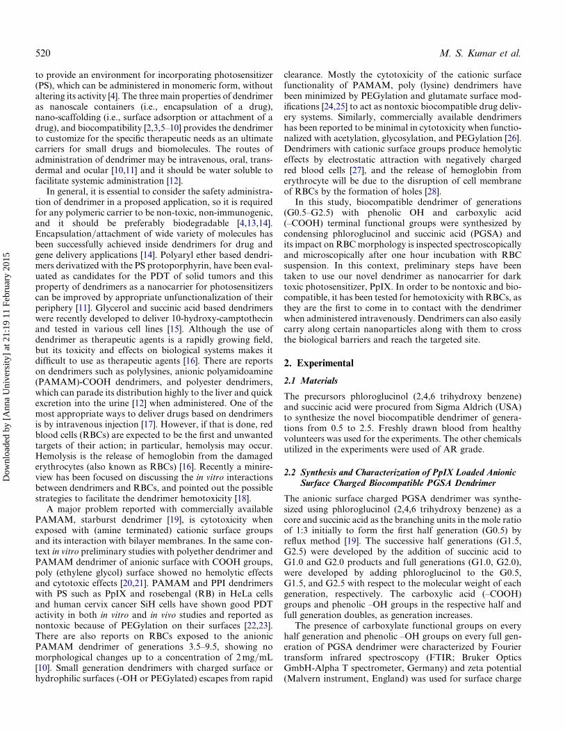
measurement [29]. It is stated that if the particles are havingnegative potentials above �20 mV, then the particles are saidto be monodispersity. The structure of G2.5 biocompatiblePGSA dendrimer with anionic surface is shown in Figure 1.
A dark toxic PS, PpIX was loaded inside the dendriticcavity of G2.5 dropwise with vigorous stirring for 2 h indark. It must be noted that the incorporation of PpIX insidethe dendritic cavity of G2.5 has already been confirmed byMurugesan et al. [29] using UV-Vis.; FTIR spectroscopy.It is further confirmed by transmission electron microscope(TEM) in the present work.
2.3 Preparation of Erythrocytes (RBCs)
Erythrocytes were separated from the plasma and leucocytesby centrifugation (1500 rpm, 10 min) and washed three timeswith phosphate-buffered saline (PBS; 150 mM NaCl, 1.9 mMNaH2PO4, 8.1 mM Na2HPO4, pH 7.4). To measure thehemolysis caused by our anionic surface charged dendrimers,stock dendrimer (1 mg=mL) solutions were prepared in PBS.Erythrocytes at a hematocrit of 0.5% (the proportion of theblood volume occupied by red blood cells) were suspendedin dendrimer solutions at concentrations ranging from10 mg to 100 mg=mL and incubated at 37�C for 1 h under gen-tle shaking. After incubation period, the whole suspensionswere centrifuged and the supernatant was investigated byoptical density (O.D) absorbance at 415 nm. The optical den-sity of the red blood cell suspensions which were treated withdistilled water is considered as complete hemolysis (100%)[16] and the percentage of hemolysis was calculated as
Hemolysis ð%Þ ¼ ðA=BÞ � 100
Where A is O.D of RBC suspension with test samples treatedwith PBS, and B is O.D of the RBC suspension treated withdistilled water (100% hemolysis).
2.4 Investigation with Inverted Fluorescence Microscopy
To observe the overall shape morphology of RBCs, invertedfluorescence microscope (Nikon Eclipse TE 300) has beenused to obtain the images at magnification of 800�. Samplevolume of 10ml from each incubated well was taken andplaced on microscopic glass slides to image the RBCs.
3. Results and Discussion
3.1 TEM Characterization
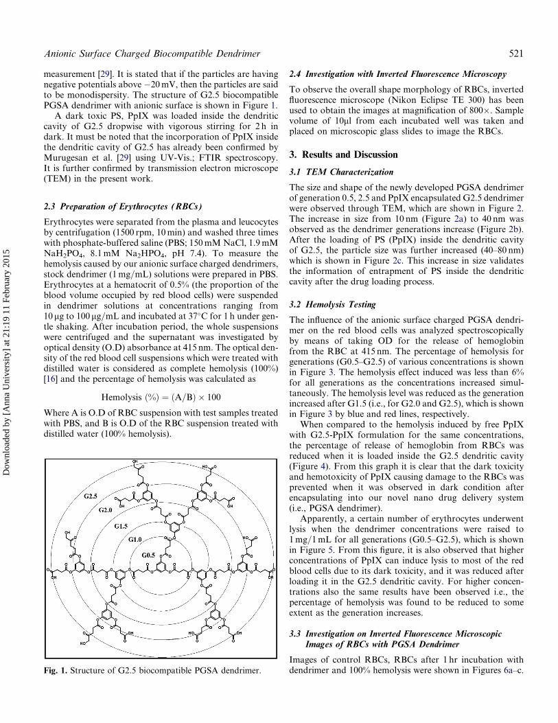
The size and shape of the newly developed PGSA dendrimerof generation 0.5, 2.5 and PpIX encapsulated G2.5 dendrimerwere observed through TEM, which are shown in Figure 2.The increase in size from 10 nm (Figure 2a) to 40 nm wasobserved as the dendrimer generations increase (Figure 2b).After the loading of PS (PpIX) inside the dendritic cavityof G2.5, the particle size was further increased (40–80 nm)which is shown in Figure 2c. This increase in size validatesthe information of entrapment of PS inside the dendriticcavity after the drug loading process.
3.2 Hemolysis Testing
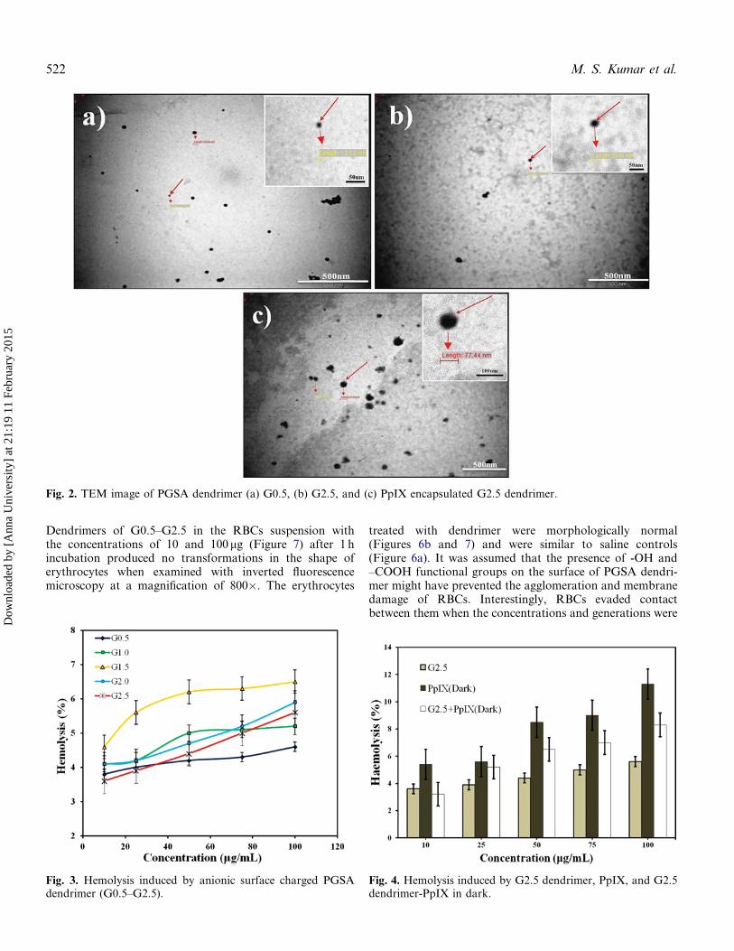
The influence of the anionic surface charged PGSA dendri-mer on the red blood cells was analyzed spectroscopicallyby means of taking OD for the release of hemoglobinfrom the RBC at 415 nm. The percentage of hemolysis forgenerations (G0.5–G2.5) of various concentrations is shownin Figure 3. The hemolysis effect induced was less than 6%for all generations as the concentrations increased simul-taneously. The hemolysis level was reduced as the generationincreased after G1.5 (i.e., for G2.0 and G2.5), which is shownin Figure 3 by blue and red lines, respectively.
When compared to the hemolysis induced by free PpIXwith G2.5-PpIX formulation for the same concentrations,the percentage of release of hemoglobin from RBCs wasreduced when it is loaded inside the G2.5 dendritic cavity(Figure 4). From this graph it is clear that the dark toxicityand hemotoxicity of PpIX causing damage to the RBCs wasprevented when it was observed in dark condition afterencapsulating into our novel nano drug delivery system(i.e., PGSA dendrimer).
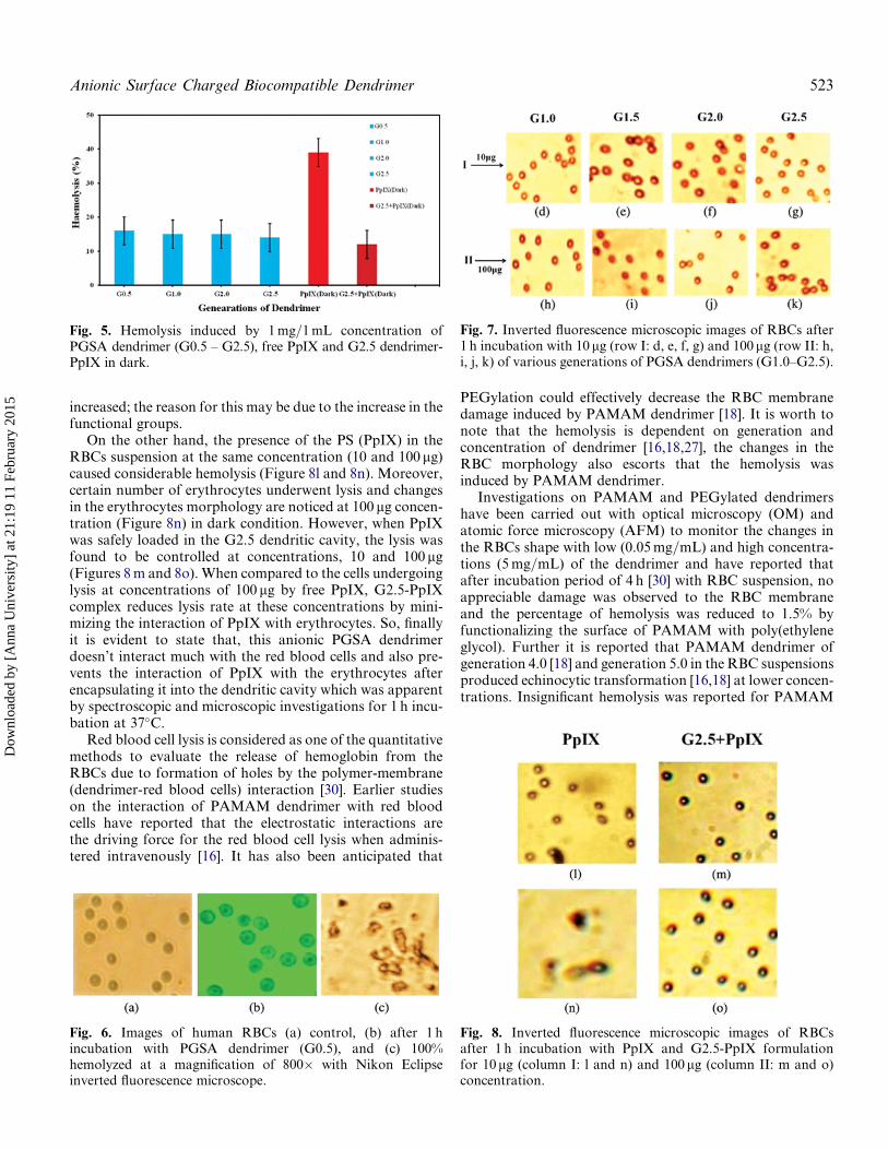
Apparently, a certain number of erythrocytes underwentlysis when the dendrimer concentrations were raised to1 mg=1 mL for all generations (G0.5–G2.5), which is shownin Figure 5. From this figure, it is also observed that higherconcentrations of PpIX can induce lysis to most of the redblood cells due to its dark toxicity, and it was reduced afterloading it in the G2.5 dendritic cavity. For higher concen-trations also the same results have been observed i.e., thepercentage of hemolysis was found to be reduced to someextent as the generation increases.
3.3 Investigation on Inverted Fluorescence MicroscopicImages of RBCs with PGSA Dendrimer
Images of control RBCs, RBCs after 1 hr incubation withdendrimer and 100% hemolysis were shown in Figures 6a–c.Fig. 1. Structure of G2.5 biocompatible PGSA dendrimer.
Anionic Surface Charged Biocompatible Dendrimer 521
Dow
nloa
ded
by [
Ann
a U
nive
rsity
] at
21:
19 1
1 Fe
brua
ry 2
015
Dendrimers of G0.5–G2.5 in the RBCs suspension withthe concentrations of 10 and 100mg (Figure 7) after 1 hincubation produced no transformations in the shape oferythrocytes when examined with inverted fluorescencemicroscopy at a magnification of 800�. The erythrocytes
treated with dendrimer were morphologically normal(Figures 6b and 7) and were similar to saline controls(Figure 6a). It was assumed that the presence of -OH and–COOH functional groups on the surface of PGSA dendri-mer might have prevented the agglomeration and membranedamage of RBCs. Interestingly, RBCs evaded contactbetween them when the concentrations and generations were
Fig. 2. TEM image of PGSA dendrimer (a) G0.5, (b) G2.5, and (c) PpIX encapsulated G2.5 dendrimer.
Fig. 3. Hemolysis induced by anionic surface charged PGSAdendrimer (G0.5–G2.5).
Fig. 4. Hemolysis induced by G2.5 dendrimer, PpIX, and G2.5dendrimer-PpIX in dark.
522 M. S. Kumar et al.
Dow
nloa
ded
by [
Ann
a U
nive
rsity
] at
21:
19 1
1 Fe
brua
ry 2
015
increased; the reason for this may be due to the increase in thefunctional groups.
On the other hand, the presence of the PS (PpIX) in theRBCs suspension at the same concentration (10 and 100 mg)caused considerable hemolysis (Figure 8l and 8n). Moreover,certain number of erythrocytes underwent lysis and changesin the erythrocytes morphology are noticed at 100 mg concen-tration (Figure 8n) in dark condition. However, when PpIXwas safely loaded in the G2.5 dendritic cavity, the lysis wasfound to be controlled at concentrations, 10 and 100 mg(Figures 8 m and 8o). When compared to the cells undergoinglysis at concentrations of 100 mg by free PpIX, G2.5-PpIXcomplex reduces lysis rate at these concentrations by mini-mizing the interaction of PpIX with erythrocytes. So, finallyit is evident to state that, this anionic PGSA dendrimerdoesn’t interact much with the red blood cells and also pre-vents the interaction of PpIX with the erythrocytes afterencapsulating it into the dendritic cavity which was apparentby spectroscopic and microscopic investigations for 1 h incu-bation at 37�C.
Red blood cell lysis is considered as one of the quantitativemethods to evaluate the release of hemoglobin from theRBCs due to formation of holes by the polymer-membrane(dendrimer-red blood cells) interaction [30]. Earlier studieson the interaction of PAMAM dendrimer with red bloodcells have reported that the electrostatic interactions arethe driving force for the red blood cell lysis when adminis-tered intravenously [16]. It has also been anticipated that
PEGylation could effectively decrease the RBC membranedamage induced by PAMAM dendrimer [18]. It is worth tonote that the hemolysis is dependent on generation andconcentration of dendrimer [16,18,27], the changes in theRBC morphology also escorts that the hemolysis wasinduced by PAMAM dendrimer.
Investigations on PAMAM and PEGylated dendrimershave been carried out with optical microscopy (OM) andatomic force microscopy (AFM) to monitor the changes inthe RBCs shape with low (0.05 mg=mL) and high concentra-tions (5 mg=mL) of the dendrimer and have reported thatafter incubation period of 4 h [30] with RBC suspension, noappreciable damage was observed to the RBC membraneand the percentage of hemolysis was reduced to 1.5% byfunctionalizing the surface of PAMAM with poly(ethyleneglycol). Further it is reported that PAMAM dendrimer ofgeneration 4.0 [18] and generation 5.0 in the RBC suspensionsproduced echinocytic transformation [16,18] at lower concen-trations. Insignificant hemolysis was reported for PAMAM
Fig. 5. Hemolysis induced by 1 mg=1 mL concentration ofPGSA dendrimer (G0.5 – G2.5), free PpIX and G2.5 dendrimer-PpIX in dark.
Fig. 6. Images of human RBCs (a) control, (b) after 1 hincubation with PGSA dendrimer (G0.5), and (c) 100%hemolyzed at a magnification of 800� with Nikon Eclipseinverted fluorescence microscope.
Fig. 7. Inverted fluorescence microscopic images of RBCs after1 h incubation with 10 mg (row I: d, e, f, g) and 100mg (row II: h,i, j, k) of various generations of PGSA dendrimers (G1.0–G2.5).
Fig. 8. Inverted fluorescence microscopic images of RBCsafter 1 h incubation with PpIX and G2.5-PpIX formulationfor 10mg (column I: l and n) and 100mg (column II: m and o)concentration.
Anionic Surface Charged Biocompatible Dendrimer 523
Dow
nloa
ded
by [
Ann
a U
nive
rsity
] at
21:
19 1
1 Fe
brua
ry 2
015
dendrimer concentration <0.01 mg=mL but for 0.1 mg=mLconcentration hemolysis was significant (i.e., the percentageof hemolysis exceeded 5%). Further, for 0.5 mg=mL concen-tration, the aggregated forms of erythrocytes are observedthrough optical microscope [30]. The percentage of hemolysiswas also measured spectrophotometrically by measuringthe O.D at 415 nm and observed that 50% of hemoglobinwas released at lower concentration of higher generation ofPAMAM dendrimer for 1 h incubation at 37�C [27]. Klajnertet al. [16] in his work, attempted to reduce the hemolysisinduced by the dendrimer (PAMAM) with the aid of interac-tion of proteins, human serum albumin (HSA) with dendri-mer. This interaction between the HSA and the cationicPAMAM dendrimer prevented much interaction with RBCsand reduced the hemotoxicity of PAMAM dendrimer [16].
In this present work we have made an attempt (an in vitrostudy) to establish whether our novel anionic surface chargedPGSA dendrimer with phenolic –OH groups and carboxylate–COOH groups, interacts less with RBC cell membranes ornot. In this regard, it was observed that the lysis rate less than6% was observed spectrophotometrically for all generationsof dendrimer (G0.5–G2.5) of various concentrations (0.01–0.1 mg=mL) after 1 h incubation with RBC suspension. Nosignificant percentage of hemolysis was observed for smallerconcentration, such as 10 and 100 ng. At higher concen-tration of 1 mg=1 mL, 10–15% of hemolysis was observed.But the images of RBCs on interaction with anionic surfacecharged PGSA dendrimer does not show any changes inthe morphology when observed through inverted fluores-cence microscopy (Figure 6a).
The observed spectroscopic results for the release ofhemoglobin from RBC suspension when incubated withvarious concentrations (10, 25, 50, 75, 100 mg) of PGSAdendrimer of G0.5–G2.5, clearly highlights that, the anionicsurface charged dendrimer does not increase the hemolysisrate more than 6% even at 100 mg concentrations. On theother hand, when compared to other generations, onlyG1.5 has produced 6% of hemolysis and other generationscauses lysis lesser than 6%. The percentage of hemolysisstarted to decrease with increase in the generation.
As our main aim is to use this novel anionic surfacecharged PGSA dendrimer as a nanocarrier for drug deliveryand to reduce the dark toxicity of any anticancer drugs orsensitizers, the dark toxic behavior of the photosensitizer,PpIX was encapsulated in the dendritic cavity of G2.5. Inaddition the dark hemotoxicity of free PpIX and dendrimer-PpIX (G2.5-PpIX) complex was further analyzed both spec-troscopically and microscopically.
When compared to the spectroscopic measurements ofhemolysis of free PpIX, the G2.5-PpIX complex has almostreduced the RBC lysis for all the concentrations (10–100mg).Free PpIX with RBC suspension at higher concentration of1 mg=1 mL produce 39% of hemolysis in dark. This wasreduced to 12% when it is inside the G2.5 dendritic cavity.From this it is understood that when a dark toxic photosensi-tizer is encapsulated into the G2.5 dendritic cavity, its toxicitycan be reduced even in dark. This was also confirmedby inverted fluorescence microscopy images (Figure 8). As
expected, the spectroscopic and microscopic results werepromising and highlight that, even in dark, for PpIX at higherconcentration (1 mg=mL) the hemolysis was reduced from 39%to 19% when entrapped inside the dendritic cavity of G2.5.
The vital part of this work is that the surface of our nega-tive charged PGSA dendrimer was not modified (or) functio-nalized with PEG in order to reduce the hemotoxicity (or) thepercentage of hemolysis and cytotoxicity as emphasized inmany reports [20,21,30]. Because of these phenolic –OHgroup and carboxylate –COOH groups on the surface of allhalf (G0.5, G1.5, G2.5) and full generation (G1.0, G2.0)dendrimer, without any functionalization, it does not showany interaction with the RBCs. So, from this study we liketo report that, anionic surface PGSA dendrimer, which wedeveloped as nanodrug delivery system is very safe andbiocompatible. Especially it can be considered as a worthycarrier for dark toxic photosensitizers in PDT applicationsfor cancer treatment.
4. Conclusion
Our novel anionic surface charged PGSA dendrimer did notinteract much with RBCs due to the presence of hydroxyland carboxylate (-OH and -COOH) functional groups onthe successive half and full generation dendrimer. This waspractically observed for concentrations ranging from 10 to100 mg. This concentration does not change the morphologyof the red blood cells and rupture the RBC membrane, whenobserved through inverted fluorescence microscopy. But itwas able to produce 10–15% hemolysis when the concen-tration was raised to 1 mg=1 mL. Second point to be noticedis that, it has the ability to reduce the dark toxicity and theinteraction of photosensitizer (PpIX) with erythrocytes whenencapsulated into the dendritic cavity of G2.5. To consider anovel nanodrug delivery system for therapeutic applications,it should be biocompatible and should not possess hemotoxi-city when intend to pass through the circulatory system. Tosum up, our preliminary in vitro studies in blood with thisnegative surface charged PGSA dendrimer provided goodand satisfactory results to state that we are the pioneers toreport this work and its effect on hemocompatibility with adark toxic photosensitizer loaded inside the G2.5 dendriticcavity without any surface modification are concerned. Thescope of this study will provide a good opportunity to furtherextend this to in vivo characterization and focus on the thera-peutic applications for photodynamic therapy of cancers.
Acknowledgement
The author is very thankful to Dr. S. Ramesh, associate pro-fessor and Head, Centralised Instrumentation Lab, MadrasVeterinary College, Vepery, Chennai-7, for providing TEMfacility.
Funding
The work was supported by DAE-BRNS Grant (Ref.No. 2009=38=BRNS=3206), Govt. of India. The author
524 M. S. Kumar et al.
Dow
nloa
ded
by [
Ann
a U
nive
rsity
] at
21:
19 1
1 Fe
brua
ry 2
015
M. Suresh Kumar would like to thank the BRNS forproviding the fellowship.
References1. Bharali, D. J.; Khalil, M.; Gurbuz, M.; Simone, T. M.; Mousa, S.
A. Int. J. Nanomedicine 2009, 4, 1–7.2. Tomalia, D. A. Prog Polym Sci. 2005, 30, 294–324.3. Svenson, S. Kirk-Othmer Encyc. Chem. Technol. 2007, 26, 786–812.4. Chatterjee, D. K.; Fong, L. S.; Zhang; Y. Adv. Drug Delivery Rev.
2008, 60, 1627–1637.5. Gillies, E. R.; Frechet, J. M. J. Drug Discovery Today 2005, 10,
35–43.6. Svenson, S.; Tomalia, D. A. Adv. Drug Deliv. Rev. 2005, 57,
2106–2129.7. Gupta, U.; Agashe, H. B.; Asthana, A.; Jain, N. K. Biomacromol.
2006, 7, 649–658.8. Cheng, Y.; Wang, J.; Rao, T.; He, X.; Xu, T. Front. Biosci. 2008,
13, 1447–1471.9. Li, Y.; Cheng, Y.; Xu, T. Curr. Drug Discov. Technol. 2007, 4,
246–254.10. Cheng, Y.; Xu, Z.; Ma, M.; Xu, T. J. Pharm. Sci. 2008, 97, 123–143.11. Ravindra, S.; Deepak, K. S.; Anil, K. M. Prasanjit, P.; Faizi, M.;
Ruchi, B. J. Pharm. Res. 2010, 3, 2238–2247.12. Medina, H. S.; EI-Sayed, M. E. Chem. Rev. 2009, 109, 3141–3157.13. Duncan, R.; Izzo, L. Adv. Drug Delivery Rev. 2005, 57, 2215–2237.14. Balaji, S.; Parimala Devi, B. Int. J. Pharm. Biol. Sci. 2010, 1,
186–201.15. Mintzer, M. A.; Grinstaff, M. W. Chem. Soc. Rev. 2011, 40,
173–190.16. Klajnert, B.; Pikala, S.; Bryszewska, M. Proc. R. Soc. A 2010, 466,
1527–1534.
17. Okuda, T.; Kawakami, S.; Maeie, T.; Niidome, T.; Yamashita, F.;Hashida, M. J. Control. Release 2006, 114, 69–77.
18. Ziemba, B. Matuszko, G. Bryszewska, M.; Klajnert, B. Cell. Mol.Biol. Lett. 2012, 17, 21–35.
19. Tomalia, D. A.; Baker, H.; Dewald, J.; Hall, M.; Kallos, G.; Martin,S.; Roeck, J. Ryder, J.; Smith, P. Polym. J. 1985, 17, 117–132.
20. Malik, N.; Wiwattanapatapee, R.; Klopsch, R.; Lorenz, K.; Frey,H.; Weener, J. W. Meijer, E. W.; Paulus, W.; Duncan, R. J. Con-trol. Release 2000, 65, 133–148.
21. Chen, H. T.; Neerman, M. F. Parrish, A. R.; Simanek, E. E. J. Am.Chem. Soc. 2004, 126, 10044–10048.
22. Conway, C. L.; Walker, I.; Bell, A.; Roberts, D. J. H.; Brown, S.B.; Vernon, D. I. Photochem. Photobiol. Sci. 2008, 7, 290–298.
23. Kojima, C.; Toi, Y.; Harada, A.; Kono, K. Bioconjug. Chem. 2007,18, 663–670.
24. Gupta, U.; Jain N. K. Adv. Drug Delivery Rev. 2010, 62, 478–490.25. Miyano, T.; Wijagkanalan, W.; Kawakami, S.; Yamashita, F.;
Hashida, M. Mol. Pharmaceut. 2010, 7, 1318–1327.26. Cheng, Y.; Zhao, L.; Li, Y.; Xu, T. Chem. Soc. Rev. 2011, 40,
2673–2703.27. Doman ski, D. M. Klajnert, B.; Bryszewska, M. Bioelectrochemis-
try 2004, 63, 189–191.28. Hong, S.; Bielinska, A. U.; Mecke, A.; Keszler, B.; Beals, J. L.; Shi,
X.; Balogh, L. Orr, B. G.; Baker, J. R. Jr.; Banaszak Holl, M. M.Bioconjug. Chem. 2004, 15, 774–782.
29. Suresh Kumar, M.; Anish, B.; Murugesan, R.; Jeyasubramanian,K. Nano Biomed. Eng. 2012, 4, 132–138.
30. Wang, W.; Xiong, W.; Zhu, Y.; Xu, H.; Yang, X. J. Biomed.Mater. Res., Part B 2010, 93, 59–64.
31. Par, G. W. The Assessment of Pheophorbide a Ethanolamide as aPotential Dual Action Anti Cancer Photosensitizer; Ph. D. Thesis,Trois-Rivieres, University of Quebec, 1995.
Anionic Surface Charged Biocompatible Dendrimer 525
Dow
nloa
ded
by [
Ann
a U
nive
rsity
] at
21:
19 1
1 Fe
brua
ry 2
015
Related Documents