Arch Dermatol Res (2009) 301:539–547 DOI 10.1007/s00403-009-0953-7 123 ORIGINAL PAPER InXuence of a commercial tattoo ink on protein production in human Wbroblasts Mirella Falconi · Gabriella Teti · Michela Zago · Angela Galanzi · Lorenzo Breschi · Susi Pelotti · Alessandra Ruggeri · Giovanni Mazzotti Received: 29 July 2008 / Revised: 25 March 2009 / Accepted: 30 March 2009 / Published online: 17 April 2009 © Springer-Verlag 2009 Abstract Tattooing is an ancient art and is still widely practiced all over the world. Since the biocompatibility of tattoo dyes has not been well researched, we studied the toxicity of a commercial tattoo ink, commonly used in tat- too lab and esthetic centers, on human Wbroblasts. To test cell viability, MTT assays were carried out and scanning electron microscopy to visualize changes in the cell surface after the dye exposure was performed. A possible inXuence of the pigment on the expression of procollagen 1 type I protein was visualized by western blotting analysis. The results showed a reduction in cell viability, and electron microscopy demonstrated an unmodiWed cell surface com- pletely covered by pigment particles. Western blotting anal- ysis demonstrated a clear interference of the pigment on the expression of procollagen 1 type I protein. These data demonstrated that the commercial tattoo dye has a time- dependent eVect on protein expression. A possible connec- tion of the inXuence of the tattoo ink with clinical eVects is discussed. Keywords Human Wbroblasts · In vitro testing · Tattoo pigment · Tattoo toxicity Introduction Tattoo and permanent make-up are widely used by all age groups, both sexes, and in many diVerent occupations and socioeconomic groups. The procedure used in making a tattoo or permanent make-up consists of puncturing the dermis and depositing an amount of ink depending on the characteristics of the needle, the ink, the skin and the depth of penetration. After the procedure, a wound healing pro- cess starts with an instantaneously acute inXammatory phase, followed by a proliferative and maturation phase to restore tissue integrity [34]. Tattoo dyes have been reported to induce skin disorders in humans [35]. Some risks are bacterial and viral infections [24], eczematous dermal reactions [4], lichenoid and granu- lomatous reactions [36] and cutaneous lymphoma [44]. Colored inks are based on metallic mixtures [39] or, more recently, on organic substances [1, 23]. Mercury, chromium, cobalt and cadmium are components of colored inks and can produce a number of many adverse eVects. Metallic salts were mainly responsible for immediate, delayed, persistent and photosensitive cutaneous reactions [13, 14]. Recently, the photochemical cleavage of tattoo pigments under the exposure of UV radiation and/or sun- light has been demonstrated [8, 10]. The decomposition products are hazardous showing a potential risk of being toxic or even carcinogenic [9]. M. Falconi (&) · G. Teti · M. Zago · A. Galanzi · A. Ruggeri · G. Mazzotti Department of Anatomical Sciences, University of Bologna, via Irnerio, 48, 40126 Bologna, Italy e-mail: [email protected] M. Falconi · M. Zago Polo ScientiWco-Didattico di Rimini, University of Bologna, 40100 Bologna, Italy L. Breschi Unit of Dental Sciences and Biomaterials, Department of Biomedicine, University of Trieste, 34138 Trieste, Italy L. Breschi Unit of Bologna c/o IOR, IGM-CNR, Bologna, Italy S. Pelotti Section of Legal Medicine, Department of Medicine and Public Health, University of Bologna, 40126 Bologna, Italy

Welcome message from author
This document is posted to help you gain knowledge. Please leave a comment to let me know what you think about it! Share it to your friends and learn new things together.
Transcript
Arch Dermatol Res (2009) 301:539–547
DOI 10.1007/s00403-009-0953-7ORIGINAL PAPER
InXuence of a commercial tattoo ink on protein production in human Wbroblasts
Mirella Falconi · Gabriella Teti · Michela Zago · Angela Galanzi · Lorenzo Breschi · Susi Pelotti · Alessandra Ruggeri · Giovanni Mazzotti
Received: 29 July 2008 / Revised: 25 March 2009 / Accepted: 30 March 2009 / Published online: 17 April 2009© Springer-Verlag 2009
Abstract Tattooing is an ancient art and is still widelypracticed all over the world. Since the biocompatibility oftattoo dyes has not been well researched, we studied thetoxicity of a commercial tattoo ink, commonly used in tat-too lab and esthetic centers, on human Wbroblasts. To testcell viability, MTT assays were carried out and scanningelectron microscopy to visualize changes in the cell surfaceafter the dye exposure was performed. A possible inXuenceof the pigment on the expression of procollagen �1 type Iprotein was visualized by western blotting analysis. Theresults showed a reduction in cell viability, and electronmicroscopy demonstrated an unmodiWed cell surface com-pletely covered by pigment particles. Western blotting anal-ysis demonstrated a clear interference of the pigment on theexpression of procollagen �1 type I protein. These datademonstrated that the commercial tattoo dye has a time-
dependent eVect on protein expression. A possible connec-tion of the inXuence of the tattoo ink with clinical eVects isdiscussed.
Keywords Human Wbroblasts · In vitro testing · Tattoo pigment · Tattoo toxicity
Introduction
Tattoo and permanent make-up are widely used by all agegroups, both sexes, and in many diVerent occupations andsocioeconomic groups. The procedure used in making atattoo or permanent make-up consists of puncturing thedermis and depositing an amount of ink depending on thecharacteristics of the needle, the ink, the skin and the depthof penetration. After the procedure, a wound healing pro-cess starts with an instantaneously acute inXammatoryphase, followed by a proliferative and maturation phase torestore tissue integrity [34].
Tattoo dyes have been reported to induce skin disordersin humans [35]. Some risks are bacterial and viral infections[24], eczematous dermal reactions [4], lichenoid and granu-lomatous reactions [36] and cutaneous lymphoma [44].
Colored inks are based on metallic mixtures [39] or,more recently, on organic substances [1, 23]. Mercury,chromium, cobalt and cadmium are components of coloredinks and can produce a number of many adverse eVects.Metallic salts were mainly responsible for immediate,delayed, persistent and photosensitive cutaneous reactions[13, 14]. Recently, the photochemical cleavage of tattoopigments under the exposure of UV radiation and/or sun-light has been demonstrated [8, 10]. The decompositionproducts are hazardous showing a potential risk of beingtoxic or even carcinogenic [9].
M. Falconi (&) · G. Teti · M. Zago · A. Galanzi · A. Ruggeri · G. MazzottiDepartment of Anatomical Sciences, University of Bologna, via Irnerio, 48, 40126 Bologna, Italye-mail: [email protected]
M. Falconi · M. ZagoPolo ScientiWco-Didattico di Rimini, University of Bologna, 40100 Bologna, Italy
L. BreschiUnit of Dental Sciences and Biomaterials, Department of Biomedicine, University of Trieste, 34138 Trieste, Italy
L. BreschiUnit of Bologna c/o IOR, IGM-CNR, Bologna, Italy
S. PelottiSection of Legal Medicine, Department of Medicine and Public Health, University of Bologna, 40126 Bologna, Italy
123
540 Arch Dermatol Res (2009) 301:539–547
Red tattoo pigments are known to include organic com-ponent and less frequently mercury [2, 21] and can producemany hypersensitive reactions. They are the most commoncause of delayed reactions which, in some situations, canoccur from weeks to many years after tattooing. The mostcommon reactions are lichenoid and granulomatous reac-tions [28], while nodular and pseudolymphomatous pat-terns are less frequently seen [6].
Tattoo dye reactions are well described in scientiWc liter-ature but all the data are based on the histological descrip-tion of the dermal reactions which occur after the tattooingprocess [6, 17, 28, 30]. Few data have been reporteddescribing the composition of tattoo dyes [36] but, up tonow, no studies have been done on in vitro testing on celllines or the biocompatibility of tattoo dyes and their com-ponents. In vitro testing of tattoo pigments could be the Wrststep in investigating the toxicity, and the mutagenic andcarcinogenic potentiality which could be connected to somecomponents of the dyes.
To understand the eVects of tattoo dyes, we tested twocommercial tattoo dyes, Strong black and Biolip 27, on aprimary culture of human Wbroblasts, testing cell viabilityby 3-(4,5-dimethylthiazol-2-yl)-2,5-diphenyltetrazoliumbromide assay (MTT assay). Cell viability data showed that“Biolip 27” was more cytotoxic compare to “strong black”dye. The subsequent experiments were concentrated onanalyzing in more detail the toxic eVect of Biolip 27 inves-tigating cell morphology by high-resolution scanning elec-tron microscopy and the possible interference of the redpigment in the production of procollagen �1 type I protein,precursor of the protein collagen type I, one of the mainproteins produced by Wbroblasts. Collagen type I is themost abundant protein of the extracellular matrix with astructural function [21]. In vitro cultured Wbroblasts pro-duce procollagen alpha 1 type I in a great amount [38, 43]and toxic eVects to Wbroblasts are related with modiWca-tions in the synthesis of this protein [31, 37].
Materials and methods
Culture of human Wbroblasts
Human Wbroblasts (HGFs) were obtained from fragmentsof healthy marginal gingival tissue during the surgicalextraction of impacted third molars. Signed informed con-sent was obtained from the donors according to a protocolapproved by the University of Bologna. The gingival tissuefragments were immediately placed in Dulbecco’sModiWed Eagle’s Medium (DMEM)/F12 (Euroclone LifeSciences Division, Milano, Italy) for at least 1 h, rinsedthree times in phosphate buVer saline solution (PBS),minced into small tissue pieces and cultured in DMEM/F12,
containing 10% fetal bovine serum (FBS), 1£ penicillin andstreptomycin, 1£ fungizone. The cells obtained were sub-sequently detached from the Petri dish and cultured in T25Xasks with the same cell medium, at 37°C in a humidiWedatmosphere of 5% (v/v) CO2. Cultured HGFs of passage4–8 were used for these studies.
Strong black and Biolip 27 exposure
Human Wbroblasts were exposed to diVerent dilutions (1/500,1/103, 1/104, 1/105, 1/106) of a commercial black tattoo dye,Strong black, and red tattoo dye, Biolip 27 (Biotek, Milano,Italy) for diVerent periods of time which are subsequentlydescribed. Each dilution was made by DMEM mediumsupplemented with 2% FBS, 1£ penicillin and streptomy-cin and 1£ fungizone. During the treatment, the cellmedium was changed every 2 days.
Controls consisted of HGFs exposed to the same DMEMmedium without the dye and supplemented as previouslydescribed. For all experiments, the control cells wereexposed for the same periods of time.
MTT assays
First an MTT assay was carried out to evaluate cell viabilityafter exposure to the diVerent dilutions of Strong black dyefor 72 h. The HGFs were seeded into 96-well plates at adensity of 1.5 £ 104 cells/well and, after 24 h, they wereexposed to the following dilutions: 1/500, 1/103, 1/104, 1/105,1/106 prepared in cell medium as previously described.After 72 h, the medium was substituted by a fresh mediumcontaining 0.5 mg/ml of MTT (Fluka, Sigma-AldrichLaborchemikalien GmbH, Seelze, Germany) and incubatedfor 5 h at 37°C. MTT salt was dissolved with a solution of0.1 N HCl in isopropanol and then the absorbance wasmeasured at 570 nm with a Model 680, Bio-Rad microplatereader (Bio-Rad Life Science Research Group, Hercules,CA, USA).
Second MTT assay was performed with the same diVer-ent dilutions of the strong black dye as described above, for2 weeks. After the treatment, the medium was substitutedby a fresh one and the MTT assay was carried out asdescribed above.
To evaluate cell viability after exposure to diVerentdilutions of the dye Biolip 27, an MTT assay was per-formed for 72 h following the protocol described above.The HGFs were seeded into 96-well plates at a density of1.5 £ 104 cells/well and, after 24 h, they were exposed tothe following dilutions of Biolip 27: 1/500, 1/103, 1/104,1/105, 1/106 prepared in cell medium as previouslydescribed. After the exposure, the medium was substitutedby a fresh one and the MTT assay was carried outdescribed above.
123
Arch Dermatol Res (2009) 301:539–547 541
A last MTT assay was carried out to test the 1/103 dilu-tion of Biolip 27 on HGF viability. The cells were seededinto 96-well plates at a density of 1.5 £ 104 cells/well and,after 24 h, they were exposed to Biolip 27 diluted 1/103 incell medium for 72, 96 h, 1 and 2 weeks. After the treat-ment, the medium was substituted by a fresh medium andthe MTT assay was carried out as previously described.
In all the MTT assays, the viability of the treated cellswas compared to the viability of the cells of controls whichwas found to be 100%.
Each experiment was carried out in triplicate and inde-pendently repeated at least three times. Statistical diVer-ences were assessed by one-way ANOVA (P < 0.05) andDunnett’s multiple comparison test (P < 0.05). Statisticalanalysis was performed with GRAPH PAD PRISM 5.0software (San Diego, CA, USA).
Cell morphology analysis with Weld emission in lens scanning electron microscope (FEISEM)
For FEISEM analysis, the HGFs were seeded on silicawafers placed in the bottom of each well of a 6-well cultureplate, at a density of 3 £ 103 cells/well. For each treatment,two silica wafers were prepared and the entire experimentwas performed three times. After 24 h of growth, the cellswere exposed to Biolip 27 diluted 1/103, using the samemodalities as previously described, for 72, 96 h, 1 and2 weeks. At the end of the treatment, the cells were post-Wxed in a solution of 2% glutaraldehyde (Fluka, Sigma-Aldrich Laborchemikalien GmbH, Seelze, Germany) in0.1 M phosphate buVer for 1 h at 4°C. After washing in thesame buVer, the samples were immersed in a solution of1% OsO4 in PBS for 30 min at room temperature; subse-quently, they were dehydrated in an ethanol series (70, 90,100%), critical point-dried (CPD 030, Balzers, Lichten-stein) and platinum metal-coated (MED 010 Balzers,Liechtenstein). A sample observation was carried out byFEISEM (JSM 890, Jeol LTD, Tokyo, Japan), at 7-kVaccelerating voltage and 1 £ 10¡11 A probe current.
Protein extraction and western blot analysis
Human Wbroblasts (cultured at a density of 1 £ 106 cells/T25 Xask) incubated with Biolip 27 diluted 1/103 for thesame periods of time as previously described were trypsini-zed after the treatment and centrifuged at 1,250 rpm for10 min at 4°C. The pellets were lysed with a RIPA modi-Wed lysis buVer (50 mmol/L; Tris–HCl pH 7,4; 1% NP-40;150 mmol/L NaCl; 2 mmol/L EDTA; 0.1% SDS; 1 mmol/LEGTA; 1 mmol/L PMSF; 0.15% �ME) containing a prote-ase inhibitor cocktail (Sigma-Aldrich, Saint Louis, Mis-souri, USA) and then centrifuged at 14,000 rpm for 10 min
at 4°C. A protein assay as described by Bradford [5] wascarried out to quantify the amount of proteins obtained ineach sample. For each sample, 20 �g of total protein wasseparated on 8% SDS polyacrylamide gel electrophoresis(SDS-PAGE) and then electrophoretically transferred into anitrocellulose membrane using a wet blotting apparatus(Mini Tank Electroblotting System, Owl, Portsmouth, UK).The membranes were saturated in 5% dry milk (Bebilac,Cesena, Italy) in Tris buVer solution/0.1% Tween 20(blocking reagent) for 2 h at room temperature (RT) andthen incubated with anti-procollagen �1 type I antibody(Santa Cruz Biotechnology, Inc., Santa Cruz, CA, USA) oranti-� tubulin antibody (Sigma-Aldrich, Saint Louis, MO,USA) diluted 1:10,000 in blocking reagent for 1 h at 37°Cfollowed by horseradish peroxidase-conjugated anti-goatIgG antibody (Santa Cruz Biotechnology, Inc., Santa Cruz,CA, USA) speciWc for anti-procollagen antibody and horse-radish peroxidase-conjugated anti-mouse IgG antibody(Sigma-Aldrich, Saint Louis, MO, USA) speciWc for anti-�tubulin antibody. Both antibodies were diluted 1:80,000 inblocking reagent for 1 h at 37°C. Bands were visualizedwith the chemiluminescence detection system (ECL plus,Amersham Biosciences, Little Chalfont Buckinghamshire,UK). Images were obtained by Image Station 2000R(Kodak, NY, USA).
ImmunoXuorescence for procollagen �1 type I
Human Wbroblasts were grown in a monolayer on coverglasses (at the density of 3 £ 103 cells/cover glass) andtreated with Biolip 27 diluted 1/103 for the same periods oftime as previously described. Two cover glasses were pre-pared for each treatment, and the entire experiment wasperformed three times. The samples were washed threetimes in PBS and Wxed with 4% formalin/0.1% Triton£100 in PBS for 20 min at 4°C. After a brief rinsing, thesamples were blocked in 1% dry milk in PBS (Bebilac,Cesena, Italy) for 30 min at room temperature and thenincubated with anti-procollagen �1 type I antibody (SantaCruz Biotechnology, Inc., Santa Cruz, CA, USA) dilutedto 1:400 in blocking reagent at 37°C for 1 h. After threewashes in PBS for 10 min each, the samples were incu-bated with CY3-conjugated anti-goat IgG antibody(Sigma, Saint Louis, MO, USA) diluted to 1:2,000 inblocking reagent at 37°C for 1 h. Finally, the slides werewashed three times in PBS and then mounted in VECTA-SHIELD® mounting medium with 4�,6-diamidino-2-phe-nylindole (DAPI) (Vector Laboratories, Burlingame, CA,USA). The slides were observed under a Xuorescencemicroscope (Nikon Eclipse E800, Tokyo, Japan) assem-bled with the Nikon dual band Xuorescence Wlter FITC-TRITC.
123

542 Arch Dermatol Res (2009) 301:539–547
Results
MTT assays
DiVerent MTT assays testing two tattoo color dyes, Strongblack and Biolip 27, were performed to check cell viabilityafter tattoo exposure. Figure 1a showed HGF’s viabilityafter diVerent dilutions of strong black dye, starting from1/500 to 1/106, for 72 h. HGF viability was expressed as apercentage relative to the HGFs not exposed to the dye. Inall the treated samples an high cell viability was detectablecompared to control. Results were similar after 2 weeks ofexposure (Fig. 1b), demonstrating that the tattoo dye Strongblack had no toxic eVects on human Wbroblasts.
Similar MTT assays were performed to test HGF viabil-ity after exposure with Biolip 27. DiVerent dilutions of thered pigment starting from 1/500 to 1/106 were tested for72 h. Results showed a reduction of cell viability to 47%corresponding to the lowest dilution (1/500) of the red dye(Fig. 2a), while the viability slowly increased until 78%with the highest dilution tested (1/106). These data clearly
demonstrated a dose-dependent toxic eVect of the red dyewhich is reduced with the highest dilution examined.
The cell viability corresponding to the dilution of Biolip27 1/103 was 58%, and this dose was chosen for the nextexperiments to test if prolonged exposures of the red tattooink with HGFs would have eVects on cell viability and pro-tein expression.
The second MTT assay showed a reduced cell viabilityin samples exposed for 72 h which was comparable to pre-vious data, while the viability slowly decreased with theincrease of time exposure (Fig. 2b), demonstrating a time-dependent toxic eVect of the red pigment on the Wbroblasts.
These data demonstrated that only Biolip 27 had toxiceVects on human Wbroblasts.
Cell morphology analysis by FEISEM
Control cells, analyzed by FEISEM, showed an elongatedand smooth surface (Fig. 3a) or a modiWed surface with Wneand short Wlaments (Fig. 3b). Cells appeared to be intercon-nected by numerous thin Wlaments and appeared to anchorthe cells to the silica wafers (Fig. 3a). Control cells after2 weeks of treatment showed a similar morphology(Fig. 3b).
Fig. 1 a MTT assay showing human Wbroblast viability after treat-ment with diVerent Strong black dilutions for 72 h. b MTT assay show-ing the cell viability after treatment with diVerent Strong blackdilutions for 2 weeks. In both assays, cell viability is expressed as theratio (%) of optical density values of treated cells to control cells. Inboth MTT assays, high values of cell viability were detectable after thetreatment. The MTT data were statistically analyzed by one-wayANOVA followed by the Dunnett’s test
Fig. 2 a MTT assay showing human Wbroblast viability after treat-ment with diVerent Biolip 27 dilutions for 72 h. b MTT assay showingthe cell viability after 1/103 Biolip 27 treatment for 72, 96 h, 1 and2 weeks. In both assays, cell viability is expressed as the ratio (%) ofoptical density values of treated cells to control cells. The MTT datawere statistically analyzed by one-way ANOVA followed by theDunnett’s test. Asterisk indicates statistically signiWcant diVerencesbetween the groups (P < 0.05 vs. the control)
123

Arch Dermatol Res (2009) 301:539–547 543
After 72 h of Biolip 27 treatment, the cells still showedan elongated surface, but they were mostly covered bysmall and bright particles corresponding to the red dye(Fig. 3c). The particles remained Wrmly attached to the cellsurface and remained there even after 96 h of treatment(Fig. 3d) and 1 (Fig. 3e) or 2 weeks (Fig. 3f). During theexposure, the cells never lost their classical Wbroblasticmorphology (Fig. 3f). The silica backgrounds were freefrom pigment particles, demonstrating speciWc adhesion ofthe dye to the cell structure (Fig. 3e, f).
Western blot analysis for procollagen �1 type I protein
In an eVort to verify the possibility of Biolip 27 interferingwith procollagen �1 type I protein, a precursor of collagen
type I protein, one of the main proteins produced byhuman Wbroblasts, a western blotting analysis was carriedout.
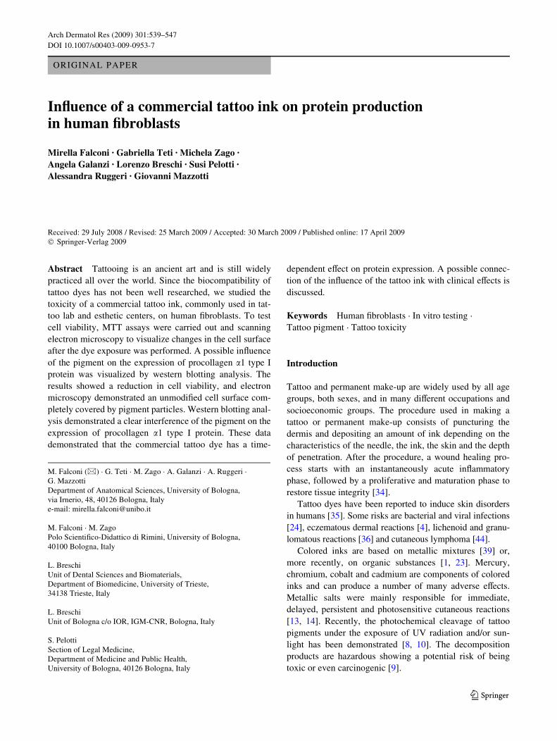
Figure 4 showed the presence of a faint band corre-sponding to the protein in the samples treated for 72 and96 h, while the signal was almost absent in the samplestreated for 1 and 2 weeks. All the controls showed a strongand well-detectable band as compared to treated samples.
ImmunoXuorescence for procollagen �1 type I protein
To visualize the procollagen �1 type I protein inside thecells, and to show a possible interference of the tattoo dyewith the presence and localization of the same protein,immunolabeling for procollagen �1 type I detectable by
Fig. 3 FEISEM analysis show-ing Wbroblast cell morphology after Biolip 27 treatment. a Con-trol cells after 72 h of exposure. The classical Wbroblast elon-gated shape with smooth surface (Wlled triangles) or covered by a thin Wlament (asterisks) is ob-served. (bar 10 �m; magniWca-tion £500). b Control cells after 2 weeks of treatment. The cell morphology is the same as described in Fig. 3a (bar 10 �m; magniWcation £500). c HGFs after 72 h of Biolip 27 exposure. The cell surface is predomi-nantly covered by particles of the dye (asterisks) (bar 10 �m; magniWcation £500). d HGFs after 96 h of Biolip 27 exposure. The particles of the tattoo dye are still observed on the cell sur-face. No modiWcation of cell morphology due to the tattoo ink treatment is detectable (bar 10 �m; magniWcation £500). HGFs after 1 week of treatment (e) and 2 weeks of treatment (f). The cells still showed their classical Wbroblastic shape with the surface covered with round and bright particles of the ink. e (bar 10 �m; magniWcation £500); f (bar 10 �m; magniWcation: £500)
123
544 Arch Dermatol Res (2009) 301:539–547
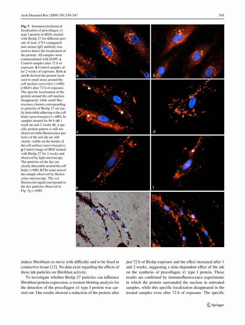
Xuorescence microscopy was carried out. Figure 5a, b, cor-responding to control samples exposed for 72 h and2 weeks of treatment, shows a peculiar staining pattern ofthe protein mainly localized around the nucleus and appear-ing as small clusters. After 72 h of Biolip 27 exposure, theXuorescence signal was strongly decreased as compared tothe control samples while big clusters of Xuorescence sig-nal were detectable in the cells but far from the nucleus,and they corresponded to the particles of Biolip 27 attachedto the cell surface (Fig. 5c). HGFs treated for 96 h, 1 and2 weeks showed no signal corresponding to the proteinaround the nucleus (Fig. 5d–f), while clusters of tattoo dyestill covered the cell surface (Fig 5f).
Discussion
In recent times, decorative tattoo and permanent make-uphave become enormously popular. Many skin disorders,such as eczematous dermal reaction [4], lichenoid and gran-ulomatous reactions [36], cutaneous lymphoma [6, 44],have been reported but the majority of them are based onhistological evaluation after the tattooing process [7, 16,29, 40]. Few data are available about the toxicity and bio-compatibility of tattoo inks, their pigments and other com-ponents on human cells. A Wrst report of the potentialtoxicity of tattoo inks came from the study of Baumler andco-workers [1] in which they reported that the degradationof red pigment PR22 resulted in the formation of 2-methyl-5-nitroaniline, a known carcinogen. These data haverecently been conWrmed by other studies on the degradationof tattoo inks followed by the formation of a number ofdiVerent toxic products with carcinogenic potential [7, 10,40]. However, these authors just described the generationof tattoo degradation products without testing any of themon human cell lines to demonstrate potential toxicity.
In an eVort to understand the toxicological proprieties oftattoo inks, in this study, we proposed an in vitro procedureto test tattoo dyes on human cells. This in vitro procedurewas designed to predict the toxicity of tattoo inks. For thisreason, we tested two commercial tattoo inks, Strong blackand Biolip 27, on human Wbroblasts for diVerent periods of
time ranging from 72 h to 2 weeks. The composition of thecommercial inks and the amounts of the single componentswere not provided by the manufacturer.
Using MTT assay, we initially tested diVerent dilutionsof the tattoo dyes starting from 1/500 to 1/106 to verify adose–response relationship between cell viability and thetattoo pigment. The data obtained from MTT assays on theblack strong ink showed high levels of cell viability, dem-onstrating a no toxic eVects on human cells.
In contrast, MTT results suggest a dose-dependent toxiceVect of Biolip 27 on HGF viability showing values rang-ing from 47% corresponding to the highest concentration(1/500), to 79% corresponding to the highest dilution (1/106). For this reason, we decided to study into more detailthe toxic eVect of Biolip 27 on human cells.
From Biolip 27 MTT data, the dilution of 1/103 showinga cell viability of 58%, was chosen to be tested in the fol-lowing experiments. Although Biolip 27 is a ready-to-usesolution, we decided to test the dilution of 1/103 to simulatea modiWed amount of dye which reaches the cells of thedermis. Due to inXammation and lymphatic transportation,the amount and location of pigments can change in the skinand these processes should be Wnished after a couple ofweeks [41]. The aim was to study the possibility that Biolip27 was able to induce toxic eVects on human cells whenthey are exposed to the pigment for prolonged periods oftime ranging from 72 h to 2 weeks. This range of time waschosen because the tattooing procedure induces trauma onthe skin, and the recovery and complete regeneration of theepidermis requires approximately 2 weeks [16].
To test cell viability after prolonged exposure to 1/103
Biolip 27, a second MTT assay was carried out at 72, 96 h,1 and 2 weeks and the results showed a time-dependenttoxic eVect of the red pigment on Wbroblasts.
To investigate the localization of the ink particles in con-tact with the cells and to also investigate the possible mor-phological changes in the cell surface after dye exposure, ahigh-resolution scanning electron microscopic analysis wasperformed. The particles of the ink appeared as small crys-tals adhered to the cell surface. They did not show a spe-ciWc localization on the cell surface but were scattered onthe cell body or just clustered in some areas. The silica sup-port was free of pigment particles demonstrating adhesionof the ink to the cell surface and not only a simple deposi-tion on it. Although the MTT results demonstrated aremarkable reduction in HGF viability, the cells did notshow any deep morphological changes after the dye treat-ment. The classic Wbroblastic and elongated shape of thecells was preserved even after 2 weeks of dye exposure.
Many histological reports have described the localiza-tion of dye particles in the dermis, between collagen bun-dles of connective tissue or adhering to Wbroblast cellbodies [12, 16, 26]. The uptake and storage of ink particles
Fig. 4 Western blotting analysis showing procollagen �1 type I pro-tein after Biolip 27 treatment. c72 control cells after 72 h, c96 controlcells after 96 h, c1w control cells after 1 week, c2w control cells after2 weeks, tr 72 HGFs exposed to Biolip for 72 h, tr 96 cells exposedfor 96 h, tr1w cells exposed for 1 week, tr2w cells exposed for2 weeks. � tubulin represents the loading control
123
Arch Dermatol Res (2009) 301:539–547 545
induce Wbroblasts to move with diYculty and to be Wxed inconnective tissue [12]. No data exist regarding the eVects ofthese ink particles on Wbroblast activity.
To investigate whether Biolip 27 particles can inXuenceWbroblast protein expression, a western blotting analysis forthe detection of the procollagen �1 type I protein was car-ried out. Our results showed a reduction of the protein after
just 72 h of Biolip exposure and the eVect increased after 1and 2 weeks, suggesting a time-dependent eVect of the inkon the synthesis of procollagen �1 type I protein. Theseresults are conWrmed by immunoXuorescence experimentsin which the protein surrounded the nucleus in untreatedsamples, while this speciWc localization disappeared in thetreated samples even after 72 h of exposure. The speciWc
Fig. 5 Immunocytochemical localization of procollagen �1 type I protein in HGFs treated with Biolip 27 for diVerent peri-ods of time. CY3-conjugated anti-mouse IgG antibody was used to detect the localization of the protein. All samples were counterstained with DAPI. a Control samples after 72 h of exposure. b Control samples af-ter 2 weeks of exposure. Both a and b showed the protein local-ized in small areas around the cell nucleus (asterisks) (£600). c HGFs after 72 h of exposure. The speciWc localization of the protein around the cell nucleus disappeared, while small Xuo-rescence clusters corresponding to particles of Biolip 27 are eas-ily detectable adhering to the cell body (open triangles) (£600). In samples treated for 96 h (d) 1 week (e) and 2 weeks (f), a spe-ciWc protein pattern is still not observed while Xuorescence par-ticles of the red ink are still clearly visible on the border of the cell surface (open triangles). g Control image of HGF treated with Biolip 27 for 2 weeks and observed by light microscopy. The particles of the dye are clearly detectable around the cell body (£600). h The same area of the sample observed by Xuores-cence microscopy. The red Xuorescent signal corresponds to the dye particles observed in Fig. 5g (£600)
123
546 Arch Dermatol Res (2009) 301:539–547
localization of the protein around the cell nucleus hasalready been described [11] and it is believed that it isstrictly connected to the position of the rough endoplasmicreticulum which is well developed in these cells [9, 12].
Collagen is the main component of the dermis. Morethan 70% is collagen type I protein [25]. Collagen Wbers areconsidered to be the structural scaVold of the skin; the inter-molecular cross-links inside the Wbers are essential for pro-viding the stability and tensile strength of the skin [42].According to our data, after the tattooing procedure andduring the wound healing, a reduced amount of collagenWbers may be produced due to the eVects of the ink particleson procollagen �1 type I protein (the precursor of type Icollagen protein).
Tattooing is a widespread practice worldwide. Despitemalignant lesions being described in relation to tattooingsuch as basal and squamous cell carcinoma [3, 15], mela-noma [32] keratoacanthoma [19], the association is rare andit is often considered as coincidental or as trauma inducedduring the tattooing procedure. However, the real patho-genesis of malignant lesions developing at tattoo sites isstill unknown. In many cases, the malignant lesions areoften developed in the red dye [19, 22, 32] suggesting thatthe red pigment is a factor in melanoma development.Although our results did not demonstrate any associationbetween the red tattoo tested and malignant lesions, thedata reported showed a cell suVering condition in Wbro-blasts exposed to the tattoo ink which could make cellsmore exposed to other factors associated with malignantlesions such as ultraviolet/sun exposure that contribute tothe development of melanoma [32]. It has been demon-strated that cutaneus melanoma has a tendency to give riseto metastases, and stromal cells such as Wbroblasts andendothelial cells are implicated in the metastatic process,including proliferation, matrix degradation, or migration ofmelanoma cells. These stromal reactions are mainly charac-terized by collagen and elastin proteolysis preferentiallylocalized around the tumor at the invasive front [20], andthe degradation and reduction of collagen type I proteinpromotes the tumor progression [18, 33, 45].
As the incidence of tattoo placement continues toincrease, so does the demand for tattoo removal. Amongthe several methods known in removing tattooing, the Q-switched laser method is the treatment of choice providinggreat eYciency with minimal side eVects. Tattoo particlesare shattered under the powerful energy of the Q-switchedlaser and they can kill the cells in which the pigment resides[27]. We suggest that the tattoo particles released by thelaser treatment may spread to the cells nearby and triggersome adverse eVects such as the downregulation of proteinsynthesis.
In conclusion, our results demonstrate that the exposureof a commercial tattoo ink to human Wbroblasts induces
toxic reactions in exposed cells with eVects on cell deathand protein synthesis in which the production of procolla-gen �1 type I protein is inhibited. We cannot underestimatethe potential eVects of tattoo inks and their componentsand, for this reason, the in vitro analysis we suggest repre-sents a useful method for investigating and anticipating thetoxicity of tattoo pigments.
Acknowledgments This study was supported by grants from theUniversity of Bologna RFO (ex-60%), Del Monte Foundation ofBologna and Ravenna, Italy, and Servizio Sanitario Regionale Emilia-Romagna, Italy.
ConXict of interest statement The authors declare that they have noconXict of interest.
References
1. Baumler W, Eibler ET, Hohenleutner U et al (2000) Q-switchlaser and tattoo pigments: Wrst results of the chemical and photo-physical analysis of 41 compounds. Lasers Surg Med 26:13–21.doi:10.1002/(SICI)1096-9101(2000)26:1<13::AID-LSM4>3.0.CO;2-S
2. Beute TC, Miller CH, Timko YAL, Ross EV (2008) In vitro spec-tral analysis of tattoo pigments. Dermatol Surg 34:508–516.doi:10.1111/j.1524-4725.2007.34096.x
3. Birnie AJ, Kulkarni K, Varma S (2006) Basal cell carcinoma aris-ing in a tattoo. Clin Exp Dermatol 31:820–821. doi:10.1111/j.1365-2230.2006.02201.x
4. Biro L, Klein WP (1967) Unusual complications of mercurial(cinnabar) tattoo. Generalized eczematous eruption followinglaceration of a tattoo. Arch Dermatol 96:165–167. doi:10.1001/archderm.96.2.165
5. Bradford MM (1976) A rapid and sensitive method for the quanti-tation of microgram quantities of protein utilizing the principle ofprotein-dye binding. Anal Biochem 72:248–254. doi:10.1016/0003-2697(76)90527-3
6. Chave TA, Mortimer NJ, Johnston GA (2004) Simultaneouspseudolymphomatous and lichenoid tattoo reactions triggered byretattooing. Clin Exp Dermatol 29:197–199. doi:10.1111/j.1365-2230.2004.01459.x
7. Cui W, McGregor DH, Stark SP et al (2007) Pseudoepithelioma-tous hyperplasia—an unusual reaction following tattoo: report ofa case and review of the literature. Int J Dermatol 46:743–745.doi:10.1111/j.1365-4632.2007.03150.x
8. Cui Y, Spann AP, Couch LH et al (2004) Photodecomposition ofpigment yellow 74, a pigment used in tattoo inks. PhotochemPhotobiol 80:175–184. doi:10.1562/2004-04-06-RA-136.1
9. Engel D, Schroeder HE, Gay R (1980) Fine structure of culturedhuman gingival Wbroblasts and demonstration of simultaneoussynthesis of types I and III collagen. Arch Oral Biol 25:283–296.doi:10.1016/0003-9969(80)90037-0
10. Engel E, Spannberger A, Vasold R, König B (2007) Photochemi-cal cleavage of a tattoo pigment by UVB radiation or naturalsunlight. J Dtsch Dermatol Ges 5:583–589. doi:10.1111/j.1610-0387.2007.06333.x
11. Falconi M, Teti G, Zago M et al (2007) EVects of HEMA on typeI collagen protein in human gingival Wbroblasts. Cell Biol Toxicol23:313–322. doi:10.1007/s10565-006-0148-3
12. Fujita H, Nishii Y, Yamashita K, Kawamata S et al (1988) Theuptake and long-term storage of India ink particles and latex beadsby Wbroblasts in the dermis and subcutis of mice, with special
123

Arch Dermatol Res (2009) 301:539–547 547
regards to the non-inXammatory defense reaction by Wbroblasts.Arch Histol Cytol 51:285–294. doi:10.1679/aohc.51.285
13. Gallo R, Parodi A, Cozzani E (1998) Allergic reaction to India inkin a black tattoo. Contact Dermatitis 38:346–347. doi:10.1111/j.1600-0536.1998.tb05779.x
14. Goldberg HM (1996) Tattoo allergy. Plast Reconstr Surg98:1315–1316. doi:10.1097/00006534-199612000-00040
15. Goldenberg G, Patel S, Patel MJ (2008) Eruptive squamous cellcarcinomas, keratoacanthoma type, arising in a multicolor tattoo.J Cutan Pathol 35:62–64. doi:10.1111/j.1600-0560.2007.00869.x
16. Gopee NV, Cui Y, Olson G et al (2005) Response of mouse skinto tattooing: use of SKH-1 mice as a surrogate model for humantattooing. Toxicol Appl Pharmacol 209:145–158. doi:10.1016/j.taap.2005.04.003
17. Gutermuth J, Hein R, Fend F et al (2007) Cutaneous pseudolym-phoma arising after tattoo placement. J Eur Acad Dermatol Vene-reol 21:566–567
18. Iida J, McCarthy JB (2007) Expression of collagenase-1 (MMP-1)promotes melanoma growth through the generation of active trans-forming growth factor-beta. Melanoma Res 17:205–213.doi:10.1097/CMR.0b013e3282a660ad
19. Kluger N, Minier-Thoumin C, Plantier F (2008) Keratoacanthomaoccurring within the red dye of a tattoo. J Cutan Pathol 35:504–507. doi:10.1111/j.1600-0560.2007.00833.x
20. Labrousse AL, Ntayi C, Hornebeck W (2004) Stromal reaction incutaneous melanoma. Crit Rev Oncol Hematol 49:269–275.doi:10.1016/j.critrevonc.2003.10.007
21. Leblond CP (1989) Synthesis and secretion of collagen by cells ofconnective tissue, bone, and dentin. Anat Rec 224:23–38.doi:10.1002/ar.1092240204
22. Lee YT, Craig JR (1984) Melanoma in a tattoo of the breast. J SurgOncol 25:100–101. doi:10.1002/jso.2930250210
23. Lehmann G, Pierchalla P (1988) Tattooing dyes. Derm Beruf Um-welt 36:152–156
24. Long GE, Rickman LS (1994) Infectious complications of tattoos.Clin Infect Dis 18:610–619
25. Lovell CR, Smolenski KA, Duance VC et al (1987) Type I and IIIcollagen content and Wbre distribution in normal human skinduring ageing. Br J Dermatol 117:419–428. doi:10.1111/j.1365-2133.1987.tb04921.x
26. Mann R, Klingmuller G (1981) Electron-microscopic investiga-tion of tattoos in rabbit skin. Arch Dermatol Res 271:367–372.doi:10.1007/BF00406680
27. Mariwalla K, Dover JS (2006) The use of lasers for decorativetattoo removal. Skin Ther Lett 11:8–11
28. Mortimer NJ, Chave TA, Johnston GA (2003) Red tattoo reac-tions. Clin Exp Dermatol 28:508–510. doi:10.1046/j.1365-2230.2003.01358.x
29. Müller KM, Schmitz I, Hupe-Nörenberg L (2002) Reaction pat-terns to cutaneous particulate and ornamental tattoos. Pathologe23:46–53. doi:10.1007/s00292-001-0507-z
30. Munoz C, Guilabert A, Mascarò JM Jr (2005) An embossed tattoo.Clin Exp Dermatol 31:309–310. doi:10.1111/j.1365-2230.2005.01956.x
31. Olszowski T (2003) Evaluation of toxic doses of Xuorine onexpression of collagen genes and synthesis of some collagenproteins in rat skin. Ann Acad Med Stetin 49:45–62
32. Paradisi A, Capizzi R, De Simone C et al (2006) Malignant mela-noma in a tattoo: case report and review of the literature. Mela-noma Res 16:375–376. doi:10.1097/01.cmr.0000222591.75858.fb
33. Rosenthal EL, Zhang W, Talbert M et al (2005) Extracellularmatrix metalloprotease inducer-expressing head and necksquamous cell carcinoma cells promote Wbroblast-mediated type Icollagen degradation in vitro. Mol Cancer Res 3:195–202
34. Sato Y, Ohshima T, Kondo T (1999) Regulatory role of endoge-nous interleukin-10 in cutaneous inXammatory response of murinewound healing. Biochem Biophys Res Commun 265:194–199.doi:10.1006/bbrc.1999.1455
35. Sperry K (1992) Tattoo and tattooing: part II. Gross pathology,histopathology, medical complications, and applications. Am JForensic Med Pathol 13:7–17. doi:10.1097/00000433-199203000-00003
36. TaaVe A, Knight AG, Marks R (1978) Lichenoid tattoo hypersen-sitivity. BMJ 1:616–618
37. Tani-Ishii N, Hamada N, Watanabe K, Tujimoto Y, Teranaka T,Umemoto T (2007) Expression of bone extracellular matrixproteins on osteoblast cells in the presence of mineral trioxide.J Endod 33:836–839. doi:10.1016/j.joen.2007.02.003
38. Teti G, Mazzotti G, Zago M, Ortolani M, Breschi L, Pelotti S,Ruggeri A, Falconi M (2008) HEMA down-regulates procollagenalpha1 type I in human gingival Wbroblasts. J Biomed Mater ResA. doi:10.1002/jbm.a.32082
39. Timko AL, Miller CH, Johnson FB et al (2001) In vitro quantita-tive chemical analysis of tattoo pigments. Arch Dermatol137:143–147
40. VageW MR, Dragan L, Hughes SM et al (2006) Adverse reactionsto permanent eyeliner tattoo. Ophthal Plast Reconstr Surg 22:48–51. doi:10.1097/01.iop.0000196713.94608.29
41. Vasold R, Engel E, König B, Landthaler M, Bäumler W (2008)Health risks of tattoo colors. Anal Bioanal Chem 391:9–13.doi:10.1007/s00216-008-1978-z
42. Wulf HC, Sandby-Møller J, Kobayasi T et al (2004) Skin agingand natural photoprotection. Micron 35:185–191. doi:10.1016/j.micron.2003.11.005
43. Zago M, Teti G, Mazzotti G, Ruggeri A, Breschi L, Pelotti S,Ortolani M, Falconi M (2008) Expression of procollagen alpha1type I and tenascin proteins induced by HEMA in human pulpWbroblasts. Toxicol In Vitro 22:1153–1159. doi:10.1016/j.tiv.2008.03.008
44. Zinberg M, Heilman E, Glickman F (1982) Cutaneous pseudolym-phoma resulting from tattoo. J Dermatol Surg Oncol 8:955–958
45. Ziober BL, Turner MA, Palefsky JM et al (2000) Type I collagendegradation by invasive oral squamous cell carcinoma. Oral Oncol36:365–372. doi:10.1016/S1368-8375(00)00019-1
123
Related Documents