Inflammatory mast cells up-regulate angiogenesis during squamous epithelial carcinogenesis Lisa M. Coussens, 1,6 Wilfred W. Raymond, 2 Gabriele Bergers, 1 Marion Laig-Webster, 2 Ole Behrendtsen, 3 Zena Werb, 3 George H. Caughey, 2,4 and Douglas Hanahan 1,5 1 Hormone Research Institute, 2 Cardiovascular Research Institute, and Departments of 3 Anatomy, 4 Medicine, and 5 Biochemistry and Biophysics, University of California, San Francisco. San Francisco, California 94143-0534 USA Expression of HPV16 early region genes in basal keratinocytes of transgenic mice elicits a multistage pathway to squamous carcinoma. We report that infiltration by mast cells and activation of the matrix metalloproteinase MMP-9/gelatinase B coincides with the angiogenic switch in premalignant lesions. Mast cells infiltrate hyperplasias, dysplasias, and invasive fronts of carcinomas, but not the core of solid tumors, where they degranulate in close apposition to capillaries and epithelial basement membranes, releasing mast-cell-specific serine proteases MCP-4 (chymase) and MCP-6 (tryptase). MCP-6 is shown to be a mitogen for dermal fibroblasts that proliferate in the reactive stroma, whereas MCP-4 can activate progelatinase B and induce hyperplastic skin to become angiogenic in an in vitro bioassay. Notably, premalignant angiogenesis is abated in a mast-cell-deficient (KIT W / KITW Wv ) HPV16 transgenic mouse. The data indicate that neoplastic progression in this model involves exploitation of an inflammatory response to tissue abnormality. Thus, regulation of angiogenesis during squamous carcinogenesis is biphasic: In hyperplasias, dysplasias, and invading cancer fronts, inflammatory mast cells are conscripted to reorganize stromal architecture and hyperactivate angiogenesis; within the cancer core, upregulation of angiogenesis factors in tumor cells apparently renders them self-sufficient at sustaining neovascularization. [Key Words: angiogenesis; cancer; gelatinase B; HPV16; inflammation; mast cells] Received March 24, 1999; revised version accepted April 20, 1999. Tumorigenesis is invariably a multistep process (Fearon and Vogelstein 1990; Folkman and Hanahan 1991; Chris- tofori and Hanahan 1994), that involves not only trans- formed cells but also an assemblage of normal support cells, including stromal fibroblasts, and endothelial cells (Dvorak 1986; Hanahan 1998; Kinzler and Vogelstein 1998). Tumor growth demonstrably depends on angio- genesis, whereby concomitant increases in the tumor vasculature supply nutrients and oxygen to the expand- ing neoplastic mass (Folkman 1990). The skin is composed of an avascular epidermal com- partment and a subjacent vascularized dermis. Whereas the skin vasculature is typically quiescent (Detmar 1996), it maintains a capability to initiate transitory neo- vascular responses to diverse epithelial stimuli, e.g., pathogen assault and wounding. The skin microenviron- ment likely facilitates angiogenic responses; among its salient characteristics is the propensity for rapid inflam- matory responses to abnormal tissue conditions, result- ing in increased blood supply, increased vascular perme- ability, and extravasation of diverse cytokine-producing leukocytes, e.g., macrophages, lymphocytes, and mast cells (for review, see Roitt et al. 1989), seeking to resolve the abnormality. Mast cells (MCs) play an important role in acute in- flammation owing to their release of stored and newly synthesized inflammatory mediators following activa- tion (Yong 1997). MC density within skin connective tissue correlates with blood vessel density (Eady et al. 1979), and activation and degranulation of MCs is asso- ciated with connective tissue degradation around sites of neovascularization (Dabbous et al. 1986). As neovascu- larization of otherwise quiescent tissues, such as skin, involves the action of angiogenic growth factors and the localized proteolytic modification of the extracellular matrix (ECM), MCs are implicated as accessory cells for angiogenesis (Meininger 1995; Yong 1997). We have investigated mechanisms regulating angio- genesis in a mouse model of epithelial carcinogenesis wherein the early region genes of human papillomavirus type 16 (HPV16) are expressed under the control of the keratin 14 (K14) promoter/enhancer (Arbeit et al. 1994). In this skin cancer model, mice are born phenotypically normal; by 1 month of age mice develop epidermal hy- perplasia with 100% penetrance, which advances fo- 6 Corresponding author. E-MAIL: [email protected]; FAX (415) 502-6779. 1382 GENES & DEVELOPMENT 13:1382–1397 © 1999 by Cold Spring Harbor Laboratory Press ISSN 0890-9369/99 $5.00; www.genesdev.org Cold Spring Harbor Laboratory Press on June 29, 2020 - Published by genesdev.cshlp.org Downloaded from

Welcome message from author
This document is posted to help you gain knowledge. Please leave a comment to let me know what you think about it! Share it to your friends and learn new things together.
Transcript
Inflammatory mast cells up-regulateangiogenesis during squamousepithelial carcinogenesisLisa M. Coussens,1,6 Wilfred W. Raymond,2 Gabriele Bergers,1 Marion Laig-Webster,2
Ole Behrendtsen,3 Zena Werb,3 George H. Caughey,2,4 and Douglas Hanahan1,5
1Hormone Research Institute, 2Cardiovascular Research Institute, and Departments of 3Anatomy, 4Medicine,and 5Biochemistry and Biophysics, University of California, San Francisco. San Francisco, California 94143-0534 USA
Expression of HPV16 early region genes in basal keratinocytes of transgenic mice elicits a multistage pathwayto squamous carcinoma. We report that infiltration by mast cells and activation of the matrixmetalloproteinase MMP-9/gelatinase B coincides with the angiogenic switch in premalignant lesions. Mastcells infiltrate hyperplasias, dysplasias, and invasive fronts of carcinomas, but not the core of solid tumors,where they degranulate in close apposition to capillaries and epithelial basement membranes, releasingmast-cell-specific serine proteases MCP-4 (chymase) and MCP-6 (tryptase). MCP-6 is shown to be a mitogenfor dermal fibroblasts that proliferate in the reactive stroma, whereas MCP-4 can activate progelatinase B andinduce hyperplastic skin to become angiogenic in an in vitro bioassay. Notably, premalignant angiogenesis isabated in a mast-cell-deficient (KITW/KITWWv) HPV16 transgenic mouse. The data indicate that neoplasticprogression in this model involves exploitation of an inflammatory response to tissue abnormality. Thus,regulation of angiogenesis during squamous carcinogenesis is biphasic: In hyperplasias, dysplasias, andinvading cancer fronts, inflammatory mast cells are conscripted to reorganize stromal architecture andhyperactivate angiogenesis; within the cancer core, upregulation of angiogenesis factors in tumor cellsapparently renders them self-sufficient at sustaining neovascularization.
[Key Words: angiogenesis; cancer; gelatinase B; HPV16; inflammation; mast cells]
Received March 24, 1999; revised version accepted April 20, 1999.
Tumorigenesis is invariably a multistep process (Fearonand Vogelstein 1990; Folkman and Hanahan 1991; Chris-tofori and Hanahan 1994), that involves not only trans-formed cells but also an assemblage of normal supportcells, including stromal fibroblasts, and endothelial cells(Dvorak 1986; Hanahan 1998; Kinzler and Vogelstein1998). Tumor growth demonstrably depends on angio-genesis, whereby concomitant increases in the tumorvasculature supply nutrients and oxygen to the expand-ing neoplastic mass (Folkman 1990).
The skin is composed of an avascular epidermal com-partment and a subjacent vascularized dermis. Whereasthe skin vasculature is typically quiescent (Detmar1996), it maintains a capability to initiate transitory neo-vascular responses to diverse epithelial stimuli, e.g.,pathogen assault and wounding. The skin microenviron-ment likely facilitates angiogenic responses; among itssalient characteristics is the propensity for rapid inflam-matory responses to abnormal tissue conditions, result-ing in increased blood supply, increased vascular perme-ability, and extravasation of diverse cytokine-producing
leukocytes, e.g., macrophages, lymphocytes, and mastcells (for review, see Roitt et al. 1989), seeking to resolvethe abnormality.
Mast cells (MCs) play an important role in acute in-flammation owing to their release of stored and newlysynthesized inflammatory mediators following activa-tion (Yong 1997). MC density within skin connectivetissue correlates with blood vessel density (Eady et al.1979), and activation and degranulation of MCs is asso-ciated with connective tissue degradation around sites ofneovascularization (Dabbous et al. 1986). As neovascu-larization of otherwise quiescent tissues, such as skin,involves the action of angiogenic growth factors and thelocalized proteolytic modification of the extracellularmatrix (ECM), MCs are implicated as accessory cells forangiogenesis (Meininger 1995; Yong 1997).
We have investigated mechanisms regulating angio-genesis in a mouse model of epithelial carcinogenesiswherein the early region genes of human papillomavirustype 16 (HPV16) are expressed under the control of thekeratin 14 (K14) promoter/enhancer (Arbeit et al. 1994).In this skin cancer model, mice are born phenotypicallynormal; by 1 month of age mice develop epidermal hy-perplasia with 100% penetrance, which advances fo-
6Corresponding author.E-MAIL: [email protected]; FAX (415) 502-6779.
1382 GENES & DEVELOPMENT 13:1382–1397 © 1999 by Cold Spring Harbor Laboratory Press ISSN 0890-9369/99 $5.00; www.genesdev.org
Cold Spring Harbor Laboratory Press on June 29, 2020 - Published by genesdev.cshlp.orgDownloaded from

cally into angiogenic dysplasias between 3 and 6 months.By one year, 50% of the mice develop invasive squamouscell carcinomas (SCC) (Coussens et al. 1996). Character-istically, cancers appear on the ears and upper trunk. Wehave determined previously that neoplastic progressionin this model is accompanied by up-regulation of genesencoding several pro-angiogenic growth factors (Arbeit etal. 1996), implicating these molecules in tumor angio-genesis in the skin. We now present evidence that in-flammatory mast cells are involved in activating prema-lignant neovascularization in the skin, and propose func-tional roles for MC-specific proteases in angiogenesisand stromal remodeling in this transgenic mouse modelof epithelial carcinogenesis.
Results
Premalignant dysplasias are characterizedby alterations in vascular architecture
In the skin of K14–HPV16 transgenic mice (Smith-Mc-Cune et al. 1997), as in human cervical carcinogenesis(Smith-McCune and Weidner 1994; Smith-McCune et al.1997), neoplastic progression involves an early increasein capillary density first evident subjacent to hyperplas-tic epithelium. We now report that dramatic changes indermal capillary location, density, and architecture ac-company both premalignant and malignant progression.Normal mouse skin contained infrequent capillaries lo-
cated deep within the dermis as visualized by immuno-localization of platelet endothelial cell (EC) adhesionmolecule (PECAM-1/CD31), an endothelial cell-specificmarker (DeLisser et al. 1994) (Fig. 1A). Hyperplastic le-sions showed a modest increase in the density and dila-tion of capillaries that remain distal to the neoplasticepidermis (Fig. 1B). Dysplastic lesions contained dilatedand enlarged capillaries that are increased in number,and localized proximal to the epithelial basement mem-brane (Fig. 1C). This vascular pattern is indicative of anangiogenic switch from vascular quiescence to modestneovascularization in early low-grade lesions (hyperpla-sias), followed by a striking upregulation of angiogenesisin high-grade lesions (dysplasias). The core of SCCs, bothwell differentiated (WDSC; Fig. 1D) and moderately topoorly differentiated (M-PDSC; Fig. 1E), as well as theinvasive fronts of the tumors (Fig. 1F), were likewise wellvascularized, containing a chaotic array of small and di-lated capillary webs.
Stromal infiltration of mast cells accompaniesactivation of angiogenesis and progression to dysplasia
Based on clues revealed by whole-mount visualization ofthe vasculature, we asked whether MCs were associatedwith the distinctive stages of carcinogenesis by utilizingchloroacetate esterase (CAE) histochemistry on paraffin-embedded sections of staged neoplastic skin (Fig. 2). CAEdetects the presence of chymotrypsin-like serine ester-
Figure 1. Altered morphology and architecture of capillaries during premalignant neovascularization. Immunohistochemical stainingof 5-µm paraffin-embedded tissue sections for CD31 expressed on capillary endothelial cells (red staining) counterstained with methylgreen in (A) normal nontransgenic (-lm) ear skin, (B) hyperplastic (hyp) ear skin, (C) dysplastic (dys) ear skin, (D) ear WDSC, (E) truncalM-PDSC center, and (F) the invasive front (Inv front) of a truncal M-PDSC with arrows indicating direction of tumor expansion.(Dashed line) Epidermal–dermal interface or location of skin basement membrane zone; (e) epidermis. Bar, 44.6 µm (A–F).
Mast cells potentiate angiogenesis
GENES & DEVELOPMENT 1383
Cold Spring Harbor Laboratory Press on June 29, 2020 - Published by genesdev.cshlp.orgDownloaded from

ases (chymases) present within connective tissue MCs(Leder 1979; Caughey et al. 1988). Normal skin con-tained few MCs distributed within the dermis distal tothe epidermal basement membrane (Fig. 2A). MC num-bers increased at the hyperplastic stage, and were asso-ciated predominately with small blood vessels withindermal stroma (Fig. 2B). With the switch to intense an-giogenesis in dysplasias, MC density increased markedlyin the dermal connective tissue in both early (3-month-old) and late (6-month-old) angiogenic lesions (Fig. 2C,D,respectively). In contrast to the angiogenic dysplasias,tumor stroma within M-PDSCs was essentially devoid ofMCs (Fig. 2E). However, the stroma around invadingfronts of the tumors contained abundant MCs, most fre-quently abutting capillaries (Fig. 2F). MCs were neverobserved within the epithelial compartment at any stageof neoplastic progression.
Exploiting the avidin and toluidine blue binding capa-bilities of tissue MCs to view their location at high reso-lution (Befus et al. 1982; Tharp et al. 1985), we saw anintimate association between infiltrating MCs in angio-genic dysplasias with both subendothelial and epithelialbasement membranes (Fig. 2, G and H, respectively).
We assessed the possibility that intense MC infiltra-tion of dysplastic lesions was an antibody-mediated re-
sponse to chronic bacterial infection of the skin, by treat-ing one cohort of transgenic mice systemically with an-tibiotics [sulfamethoxole (60 mg/tablet), trimethoprim(10 mg/tablet), 1 tablet/2 mice per week], and by breed-ing a second cohort into the immunodeficient Rag1/nullbackground (Mombaerts et al. 1992). These treatmentsdid not alter influx of MCs into the stroma beneath dys-plastic epidermis (data not shown). Thus, the presence ofMCs in angiogenic dysplasias does not appear to be aresponse to activation of the humoral immune system.
Angiogenic dysplasias are highly enriched in mastcell-specific serine proteases
We next quantified the activities of MC-specific trypticand chymotryptic serine proteases (tryptases and chy-mases, respectively) in extracts of tissue from each neo-plastic stage with selective solution assays (Fig. 3A). Al-though chymase-like activity was detectable in normalnegative littermate control tissue [n = 2; 62.5 ± 17.5nmoles of pNA/min per mg protein (S.E.M.)] and in hy-perplastic tissue (n = 4; 39.8 ± 13.5 nmoles of pNA/minper mg protein), there was a significant increase in ac-tivity in angiogenic dysplasias (n = 6; 204 ± 3.6 nmoles ofpNA/min/mg protein; P < 0.003). In addition, angio-
Figure 2. Mast cell infiltration during activation of angiogenesis. Chloroacetate esterase histochemistry (red staining) on 5-µmparaffin-embedded tissue sections counterstained with hematoxylin (blue) identifies location of infiltrating MCs during neoplasticprogression in (A) nontransgenic litter mate (-lm) ear skin, (B) hyperplastic (hyp) ear skin, (C and D) dysplastic (dys) ear skin, (E) thecenter of a chest M-PDSC, (F) and at the invading front of a truncal M-PDSC. Arrows indicate direction of tumor expansion. (G)Whole-mount microscopy of lectin-perfused transgenic mouse with dysplastic truncal lesions. Darkly stained mast cells (arrows)appear to adhere directly to basement membranes surrounding the dermal capillaries (c) in angiogenic dysplasias. (H) Metachromaticgranules in MCs stained with toluidine blue. Degranulating mast cells (arrows) present within a dysplastic lesion are juxtaposed tightlyto skin basement membrane (bm) and dermal capillaries (c, arrowhead). (d) Dermis, (e) epidermis. Bar, 44.6 µm (A–D, F); 89 µm (E); 19.7µm (G); 12.9 µm (H).
Coussens et al.
1384 GENES & DEVELOPMENT
Cold Spring Harbor Laboratory Press on June 29, 2020 - Published by genesdev.cshlp.orgDownloaded from
genic dysplasias contained higher levels of chymase-likeactivity than SCCs (n = 3; 15.3 ± 8.2 nmoles of pNA/min/mg protein). Chymase-like activity in extracts wascompletely inhibited by 10 µM soybean trypsin inhibitor(SBTI), but not by 100 µg/ml aprotinin, as is character-istic of MC chymases (Caughey 1995).
Similarly, tryptase-like activity was detectable in bothnormal (n = 2; 13 ± 13 nmoles of pNA/min per mg pro-tein) and hyperplastic (n = 4; 15.7 ± 10.6 nmoles of pNA/min/mg protein) skin; again, angiogenic dysplasias pos-sessed significantly elevated activity (n = 6; 131 ± 49.3nmoles of pNA/min per mg protein; P < 0.04), comparedto the former and to frank carcinomas (n = 3; 30 ± 12.6nmoles of pNA/min/mg protein). The tryptase-like ac-tivity in extracts was inhibited by 1.5 mM benzamidine,a general inhibitor of tryptic serine proteases, but not by10% mouse plasma (data not shown). Resistance to ser-ine protease inhibitors present in plasma is an idiosyn-cratic feature of MC tryptases.
MCs in different anatomic locations and in differentpathologic conditions selectively express distinct chy-
mase transcripts [mouse mast cell protease (mMCP) 1,-2, -4 and/or -5] and tryptase transcripts (mMCP-6 and/or -7) (Reynolds et al. 1990; Stevens et al. 1994). Wedetected mRNA representing only mMCP-4, a b-chy-mase, and mMCP-6, a tryptase, by RT–PCR analysis ofRNA isolated from dysplastic ear skin (Fig. 3B). We con-firmed the presence of their encoded gene products byimmunoblot analysis of neoplastic skin lysates; an ∼28kD mMCP-4 was detected with affinity-purified anti-ratMCP-1 IgG (rat MCP-1 is the ortholog of mMCP-4;Caughey 1995) and an ∼34 kD mMCP-6 was detectedwith affinity-purified anti-mMCP-6 IgG (Ghildyal et al.1996) (data not shown).
The chymase purified from angiogenic dysplasias rep-resented 99% of the chymotryptic activity loaded ontoan SBTI-agarose column. It had a specific activity of 5.5-µmole of succinyl-L-Ala-Ala-Pro-Phe-4-nitroanilide hy-drolyzed/min per mg, migrated as a predominant band at∼28 kD by SDS-PAGE (Fig. 3C), and reacted with anti-ratMCP-1 IgG in immunoblotting (Fig. 3D). The amino-terminal sequence of the purified chymase was IIG-
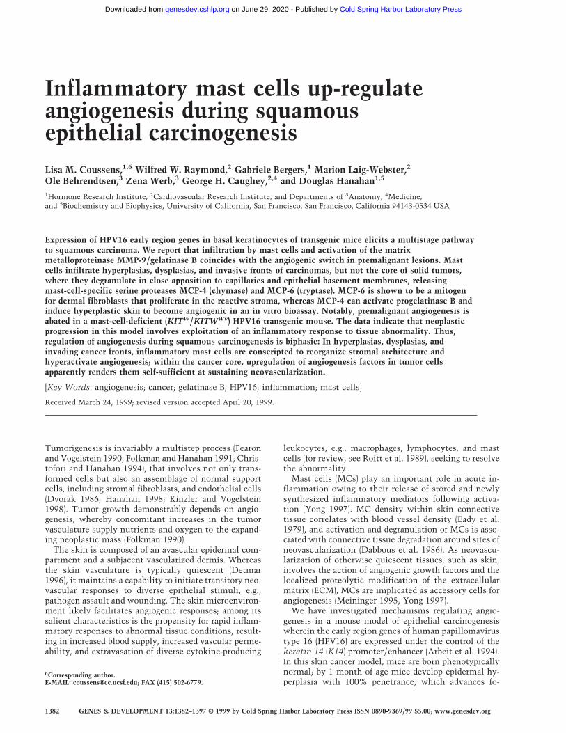
Figure 3. Chymase and tryptase in neoplastic skin. (A) Chy-motryptic and tryptic activity in neoplastic skin. Values rep-resent mean chymase and tryptase activity in ear skin biopsiesfrom negative littermate (N), hyperplastic (H), dysplastic (D),and tumor (T) solubilized by extraction at high ionic strength,normalized to the amount of protein in extracts ±S.E.M.); 5-µleach of tissue sample were incubated with 1 mM chymotrypticor tryptic substrate. The change in absorbance at 410 nm at37°C is shown where units indicate µmoles of substratecleaved/min per mg protein. (B) RT–PCR analysis for allmMCPs in dysplastic ear mRNA. Sizes shown indicate basepairs. All primer pairs were tested for specific priming of con-trol RNA. (C) SDS-PAGE of mMCP-4 purification from K14–HPV16 mouse ears. Samples were reduced with dithiothreitoland visualized with Coomassie brilliant blue. (Lane 1), 2 M
NaCl extract of K14-HPV mouse ears (5 µg of protein); (lane 2)2 M NaCl extract SBTI-agarose unbound fraction (1 µg of pro-tein); (lane 3), 5 µg of pure mMCP-4, SBTI-agarose acid eluate.(D) Anti-rat MCP-1 IgG reacts specifically with mMCP-4.(Lane 1) Crude high-salt extract of ears (2 µl); (lane 2), 2.5 ng ofpurified mMCP-4 from SBTI-agarose acid eluate. (E) SDS-PAGE of mMCP-6 purification from K14–HPV16 mouse ears.Samples were reduced with dithiothreitol and visualized withCoomassie brilliant blue. (Lane 1) Unbound fraction from p-aminobenzamidine–agarose chromatography, 2 µl; (lane 2), 2M NaCl extract, unbound fraction, 2 µl; (lane 3) MMCP-6 peak(240 ng) from heparin–HPLC NaCl gradient elution. Molecu-lar masses are shown in kD.
Mast cells potentiate angiogenesis
GENES & DEVELOPMENT 1385
Cold Spring Harbor Laboratory Press on June 29, 2020 - Published by genesdev.cshlp.orgDownloaded from

GVESRP, which is identical to the published amino acidsequence of mMCP-4 (Huang et al. 1991). PurifiedmMCP-4 was inhibited completely by SBTI in a 1:100enzyme-to-inhibitor molar ratio, but inhibited onlyweakly by aprotinin in a 500-fold molar excess (data notshown).
mMCP-6 was purified from dysplastic K14–HPV16transgenic mouse ears by high-salt buffer extraction fol-lowed by p-amino benzamidine and heparin affinitychromatography. The purified tryptase migrated as asingle protein band at ∼34 kD (Fig. 3E), and immunob-lotted with anti-mMCP-6 antibody (data not shown).The amino-terminal sequence was IVGGHEASES, iden-tical to mMCP-6 (Reynolds et al. 1991) and distinct fromthe other mouse tryptase, mMCP-7. The specific activityof purified mMCP-6 was 56 µmole of tosyl-Gly-Pro-Lys-4-nitroanilide hydrolyzed/min per mg. This activity rep-resented 99% of the tryptic activity present in the high-salt buffer loaded onto the benzamidine-agarose column(data not shown). MMCP-6 was inhibited by bis (5-ami-dino-2-benzimidazoyl) methane (BABIM) at a 1:500 mo-lar ratio, a potent inhibitor of mast cell tryptases(Caughey et al. 1993), but was resistant to inhibition byaprotinin, at a 1:75 enzyme-to-inhibitor molar ratio.
Mouse MCP-6 is a mitogen for dermal fibroblasts
We observed that the cellularity of dermal fibroblastsand the deposition of collagen into the ECM, as demon-strated by Masson’s trichrome staining (data not shown),increased incrementally during early neoplastic progres-sion. This is notable, as stromal fibroblasts in skin,much like ECs, are typically quiescent (Gregoire andLieubeau 1995). In wound healing and neoplastic situa-tions, proliferating fibroblasts are the source of newlysynthesized type I procollagen and are responsible fordesmoplastic phenotypes (Vuorio and de Crombrugghe1990; Gregoire and Lieubeau 1995). Tryptase is a mito-gen (Hartmann et al. 1992) and potent stimulator of typea1(I) procollagen mRNA synthesis (Cairns and Wells1997; Gruber et al. 1997) in human fibroblasts in vitro.These activities of tryptase may be mediated throughinteraction with protease activated receptor-2 (PAR-2)present on fibroblast plasma membranes (Molino et al.1997; Schechter et al. 1998).
Accordingly, we tested the ability of mMCP-6 tryptaseand mMCP-4 chymase to stimulate DNA synthesis ofquiescent serum-deprived subconfluent primary murinedermal fibroblasts (PMDFs) and quiescent human um-bilical vein endothelial cells (HUVECs), in the absence ofexogenous growth factors. MMCP-6 stimulated fibro-blast DNA synthesis selectively in a concentration-dependent manner yet did not affect proliferation ofHUVECs. (Fig. 4A,B). In contrast, mMCP-4 did notstimulate DNA synthesis in either cell population at anyconcentration tested (Fig. 4A,B). These results demon-strate that mMCP-6 tryptase has cell-type-specific mito-genic potency.
In vivo, we observed that normal skin exhibitedpatchy expression of type a1(I) procollagen mRNA in
stroma adjacent to capillaries (Fig. 4C), whereas mRNAexpression was up-regulated incrementally in hyperplas-tic lesions adjacent to capillaries and subjacent to epi-thelium (Fig. 4D). In angiogenic dysplasias heavily infil-trated with MCs, type a1(I) procollagen mRNA was ex-pressed widely in stroma (Fig. 4E), consistent with highlevels of tryptase, increased cellularity of dermal fibro-blasts, and increased deposition of collagen.
MMCP-4 induces ECM remodeling and angiogenesisby direct and indirect mechanisms
The cell proliferation assay pointed to a role for tryptasein activating quiescent fibroblasts to become a prolifera-tive component of the reactive neoplastic stroma; thatsame assay revealed no mitogenic activity of chymase onfibroblasts or endothelial cells. Therefore, we askedwhether chymase contributed by altering tissue micro-environment in some other way. Interestingly, canineand human MC a-chymases are known to activate thelatent extracellular protease progelatinase B (proMMP-9)(Fang et al. 1996 1997). Gelatinase B is a member of thematrix metalloproteinase (MMP) family and is involvedin both ECM remodeling (Coussens and Werb 1996) andregulation of angiogenesis (Vu et al. 1998).
We hypothesized, therefore, that mMCP-4 contributedto activation of angiogenesis by inducing ECM remodel-ing through the activation of progelatinase B. To addressthis, we first assessed the relative activity of gelatinase Bin tissue lysates representing distinct neoplastic stagesutilizing gelatin-substrate zymography (Fig 5A).Progelatinase B was not detectable in normal skin, butwas up-regulated incrementally in hyperplastic, dysplas-tic, and tumor tissue; however, active gelatinase B ap-peared only in the dysplastic and tumor lysates. In addi-tion, this assay revealed the activity profile of anotherMMP family member, namely pro- and active gelatinaseA (MMP-2), whose activity changed only modestly in thedifferent neoplastic stages. The appearance of activatedgelatinase B in dysplastic lesions correlated with dra-matic MC infiltration and elevated levels of mMCP-4chymase activity.
To test if mMCP-4 could activate progelatinase B,similar to canine or human a-chymases, lysates fromhyperplastic skin (containing predominantly the pro-form of gelatinase B) were incubated with purifiedmMCP-4 prior to substrate zymography. Incubation ofthe lysates with mMCP-4 led to a dramatic increase inthe amount of activated gelatinase B, whereas neitherthe pro nor active forms of gelatinase A were changed(Fig. 5B). Addition of ecotin (an inhibitor of mMCP-4 butnot urokinase) abolished mMCP-4-induced activation ofgelatinase B completely, whereas 1 mM amiloride (an in-hibitor of urokinase) had no effect (data not shown). Fur-thermore, immunolocalization of mMCP-4 (red staining;Fig. 5C,D) and gelatinase B (blue staining; Fig. 5E) indysplastic tissue sections revealed colocalization of bothenzymes proximal to epithelial and subendothelial base-ment membranes as demonstrated by immunolocaliza-tion of laminin-b1 (brown staining, Fig. 5C, D). Thus,
Coussens et al.
1386 GENES & DEVELOPMENT
Cold Spring Harbor Laboratory Press on June 29, 2020 - Published by genesdev.cshlp.orgDownloaded from

mMCP-4 colocalizes with and induces activation ofprogelatinase B, but not progelatinase A, in early neo-plastic tissue.
We next asked if mMCP-4 chymase, mMCP-6 trypt-ase, or gelatinase B could convert an otherwise nonan-giogenic tissue to an angiogenic one utilizing an in vitroangiogenesis bioassay (Fig. 6). Hyperplastic ear skin from1-month-old transgenic mice was incubated in mediumalone or medium containing mMCP-4, mMCP-6, orgelatinase B. Subsequently, the hyperplastic skin wasembedded into a collagen gel alone, or a collagen gelcontaining randomly dispersed bovine capillary endothe-lial cells (BCEs). Hyperplastic skin pieces, both controland protease-treated, were placed into collagen gels lack-ing BCEs. They showed negligible cell emigration frombiopsies (data not shown). BCEs remained randomly dis-tributed around untreated hyperplastic skin (Fig. 6A).Treatment of skin with mMCP-6 also yielded no re-sponse (Fig. 6B), even though human tryptase is a knownEC morphogen (Blair et al. 1997). In contrast, mMCP-4-treated skin induced a dramatic response of the BCEs,with radial alignment, proliferation, migration, and tubeformation toward the dermal surface of the skin pieceswith no evidence of migration or tube formation in thedirection of the epidermis (Fig. 6C,D). GelatinaseB-treated hyperplastic skin showed a similar response to
that observed with mMCP-4 (data not shown). NeithermMCP-4 nor gelatinase B added directly to BCEs in thecollagen gel induced a response (data not shown), indi-cating that these activities were caused directly by re-lease of sequestered activity from the skin pieces. Thus,both mMCP-4 and gelatinase B induce ECM remodelingof hyperplastic tissue directly leading to release of se-questered angiogenic activity.
MC deficiency abates premalignant progression
Our data suggest that MC-derived factors, including chy-mase and tryptase, are involved in the early events ofneoplastic progression, e.g., fibroblast activation, ECMremodeling, and activation of angiogenesis. To test thishypothesis, we sought to take advantage of mice thatwere genetically MC deficient. The receptor tyrosine ki-nase c-Kit, is encoded by the white spotting (W) locus onmouse chromosome 5 (Chabot et al. 1988). Completeloss of c-Kit function is lethal; however, mice carryingKITW mutations (W37, Wv, W41, and W) are viable tovarying degrees, but manifest pleiotropic developmentaldefects, including sterility, coat color abnormalities, se-vere macrocytic anemia, and mast-cell deficiency (Bern-stein et al. 1990; Nocka et al. 1990). The tissues of adultWBB6F1 KITW/KITWv mice contain <1% the number of
Figure 4. Effects of tryptase on skin fibroblasts. Effect of MCPs on HUVEC proliferation (A) and on PMDF proliferation (B). Agonistswere added to subconfluent, serum-deprived cultures (VEGF, 1 and 10 ng/ml; bFGF, 1 and 10 ng/ml; mMCP-6, 1 and 10 nM; mMCP-4,2 and 20 nM; heparin at 74 and 740 ng/ml) for 16 hr. [3H]Thymidine incorporation was then determined. Results shown indicate meanvalues (±S.E.M.; n = 3). (C–E) Dark-field photographs of ear-tissue sections from (C) negative litter mate (-lm) normal skin, (D) hyper-plastic (hyp) skin, and (E) dysplastic (dys) skin hybridized with a (a1)I procollagen antisense mRNA probe. Arrows point to procollagenexpressing cells within the stroma. (e) Epidermis; (d) dermis, (c) central ear cartilage. Bar, 44.6 µm (C–E).
Mast cells potentiate angiogenesis
GENES & DEVELOPMENT 1387
Cold Spring Harbor Laboratory Press on June 29, 2020 - Published by genesdev.cshlp.orgDownloaded from

MCs compared to their normal (+/+) littermates (Kita-mura et al. 1978). Thus, we attempted to generate K14–HPV16 KITW/KITWv mice to assess the importance ofMCs as extrinsic potentiators of early neoplastic progres-sion.
Because WBB6F1 KITW/KITWv mice are sterile (Nockaet al. 1990), and because neoplastic progression in K14–
HPV16 is only permissive in FVB/n (Coussens et al.1996), we first crossed both KITW/+ (WB/ReJ) andKITWv/+ (C57BL/6J) alleles into FVB/n for four genera-tions. At the fourth generation, we intercrossed each Wallele with K14–HPV16; genotypes of the expected Men-delian ratios were obtained. To obtain K14–HPV16KITW/KITWv genotypes, we crossed HPV16 KITW/+with KITWv/+, and HPV16 KITWv/+ with KITW/+, gen-erating 104 litters and 712 pups. The expected simulta-neous transmission frequency of all three desired loci is12.5%. Thus, of the 712 live pups, 179 should have beenKITW/KITWv and of those, 89 would be predicted to beK14–HPV16 KITW/KITWv. Instead, one K14–HPV16KITW/KITWv mouse and no KITW/KITWv mice wereborn. When we analyzed litter size of the KITW/+ withKITWv/+ crosses, we realized that on average, litters con-tained 25% fewer live births [6.85 ± 2.94 P < 0.0001.]
Figure 5. Effect of mMCP-4 on progelatinase B present in hy-perplastic tissue and localization of mMCP-4 and gelatinase B invivo. (A) Gelatinolytic activity in tissue lysates (2 µl) from nor-mal (N), hyperplastic (H), dysplastic (D) and carcinoma (T) bi-opsies. Incubation of gelatin zymograms with 1,10 phenanthro-line (an inhibitor of MMPs), but not PMSF (an inhibitor of serineproteases) following electrophoresis abolished the 68, 72, 80,and 90-kD bands completely (data not shown). (B) In vitro re-constitution of gelatinase B activity. Two microliters of hyper-plastic (H) lysate alone or 2 µl of hyperplastic lysate incubatedfor 30 min at 37°C with 40 ng of purified mMCP-4. Molecularmasses are shown in kD. (C–D) Immunolocalization ofmMCP-4 (red staining) and laminin (brown staining) at base-ment membranes (bm, arrows) adjacent to epithelium (e) andcapillaries (c) in dysplastic skin. (E) Immunolocalization ofgelatinase B (blue staining) in dysplastic skin localizes to base-ment membranes (bm) adjacent to epithelium (e, closed arrows)and around capillaries (c, open arrows) in dermis. (F) Control fornonspecific binding using a control rabbit IgG; backgroundstaining was negligible. Bar, 44.6 µm (C–F).
Figure 6. Effect of mMCP-4 on sequestered angiogenic activityin premalignant tissue. Hyperplastic ear skin (S) from a 1-monthold K14–HPV16 mouse was incubated for 48 hr in the presenceor absence of enzymes at 37°C followed by implantation into acollagen gel with EC coculture. Cultures were maintained for 3weeks, with photographs of tube ingrowths taken after 12 daysof coculture. (A) untreated ear skin implanted into EC-collagengel; (B) mMCP-6-treated (10 nM) ear skin implanted into EC-collagen gel; (C and D) mMCP-4-treated (20 nM) ear skin im-planted into EC-collagen gel showing an angiogenic responsewith extensive EC tube formation. Lower magnification (D)shows the polarized direction of tubular structures towards thedermal (d) surface, as opposed to the epidermal (e) surface of theskin. Small arrows (A and B) indicate the more-or-less randomgrowth of ECs, whereas thick arrows (C and D) indicate radiallyaligned tubules growing toward the skin piece. (h) Hair. Bar: 58µm (A–C); 116 µm (D).
Coussens et al.
1388 GENES & DEVELOPMENT
Cold Spring Harbor Laboratory Press on June 29, 2020 - Published by genesdev.cshlp.orgDownloaded from
as compared to K14–HPV16 with FVB/n crosses[8.79 ± 3.31 (±S.D.), n = 80 litters, 703 mice), leading us toconclude that in the FVB/n background, KITW/KITWv
produces a near-lethal phenotype. Hence, much likewith many targeted gene knockouts, genetic backgroundhas a profound effect on viability.
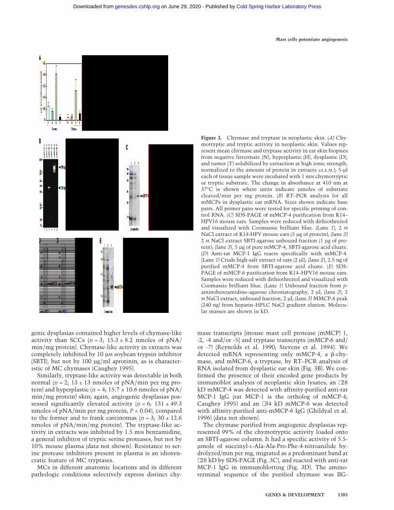
We analyzed the neoplastic phenotype in the K14–HPV16 KITW/KITWv mouse at 2 months of age (shortlybefore it died) in comparison to MC-proficient litter-mates (KIT+/KIT+ and K14–HPV16 KIT+/KIT+; Fig. 7). Atthis point in neoplastic progression, the hyperplasticphenotype in K14–HPV16 mice is 100% penetrant(n = 380), and is characterized by a thickened epidermiswith underlying dermal MC infiltration (CAE, red stain-ing; Fig. 7B), hyperproliferation of keratinocytes (Ki-67,brown staining; Fig. 7E), and altered vascular architec-ture including increased vascular density and dilation(CD-31, brown staining; Fig. 7H). In the K14–HPV16KITW/KITWv mouse, MCs were absent from the dermis(Fig. 7C). Keratinocyte proliferation, as measured by im-munoreactivity of Ki-67, although enhanced [Fig. 7F,brown staining; 35% ± 2.8 (S.E.M.), n = 10, 20× fields]compared to skin of negative littermates [Fig. 7D, brownstaining; 12% ± 1.2 (S.E.M.), n = 10, 20× fields/mouse, 3mice], was markedly decreased compared to K14–HPV16
KIT+/KIT+ littermates (Fig. 7E, brown staining;48% ± 1.6% (S.E.M.), n = 10, 20× fields/mouse, 8 mice].Likewise, dermal vasculature in the K14–HPV16 KITW/KITWv skin (Fig. 7I) was more similar to quiescent vas-culature typical of normal skin (Fig. 7G) than to thatobserved in angiogenic skin (Fig. 7H), with infrequent,nondilated vessels found distal to the epidermal base-ment membrane. Thus, by three different criteria (epi-dermal thickness, keratinocyte proliferation, and vascu-larization) the absence of MCs functionally compro-mises early neoplastic progression.
Discussion
Inflammation is a protective reaction elicited by the hostin response to infection, injury, or tissue damage. Undernormal physiologic circumstances, this response servesto destroy pathogens, and initiate repair. Inflammatoryreactions are also observed commonly in human neopla-sia (Wernert 1997; Ohtani 1998). Thus, several parallelsexist between the generation of tumor stroma and heal-ing of wounds, supporting the concept that tumorstroma is, in part, wound healing gone awry (Dvorak1986). In this report, we demonstrate that an inflamma-tory reaction, characterized by stromal infiltration of
Figure 7. Mast cell deficiency abates early neoplastic progression. Presence of mast cells (CAE, red staining; A–C), proliferation ofkeratinocytes (Ki-67, brown staining; D–F), and capillary architecture and densities (CD-31, brown staining; panels G–I) were comparedbetween wild-type littermates (-LM; n = 3; A, D, and G), K14–HPV16 KIT+/KIT+ (n = 8; B, E, and H), and K14–HPV16 KITW/KITWWv
(n = 1; C, F, and I) mice. Ear tissue biopsies (5 × 2-mm) were removed from 2-month old littermates, embedded in OCT freezingmedium, and sectioned (10 µm). Dashed line indicates epidermal–dermal interface. Arrows point to dilated capillaries subjacent tohyperplastic skin. (f) Hair follicle. Bar, 44.6 µm (A–I).
Mast cells potentiate angiogenesis
GENES & DEVELOPMENT 1389
Cold Spring Harbor Laboratory Press on June 29, 2020 - Published by genesdev.cshlp.orgDownloaded from
mast cells, occurs coincident with activation of angio-genesis in a mouse model of squamous epithelial carci-nogenesis and demonstrate a role for MCs in jump-start-ing the angiogenic switch. We show that MCs withinangiogenic dysplasias are juxtaposed tightly to epithelialand subendothelial basement membranes, both of whichare sites of active ECM remodeling (Coussens et al.1996). The data support a model in which MCs contrib-ute to premalignant progression in part by releasing twoMC specific serine proteases, mMCP-6 (a tryptase),mMCP-4 (a chymase), as well as progelatinase B. MCdeficiency in K14–HPV16 animals results in a severe at-tenuation of early neoplasia, strengthening the notionthat MCs are important functionally. Thus, during thehyperplastic and dysplastic stages of squamous carcino-genesis, an ostensibly antitumor inflammatory responseis hijacked, instead facilitating the angiogenic switchand the conversion of a normal dermis into an aberrantstromal support, setting the stage for subsequent pro-gression to malignancy.
Inflammation, mast cells, and angiogenesis:parameters of early neoplastic progression
Angiogenesis in the adult organism is limited normallyto conditions of tissue repair/remodeling such as existduring menstruation, mammary gland involution,wound healing, inflammation, and neoplasia (Salamon-sen and Wooley 1996; Hanahan and Folkman 1996). An-giogenesis involves the activation, proliferation, and mi-gration of endothelial cells in concert with localized pro-teolytic modification of the ECM (Ingber and Folkman1989). Proteolysis of the ECM facilitates endothelial cellmigration and proliferation, releasing stored angiogenicsignaling molecules from sequestered latent reserves(Meininger 1995; Brown et al. 1997; Friedl et al. 1997;Poltarek et al. 1997), and loosening the stromal milieu tofacilitate migration (Coussens and Werb 1996). In cuta-neous organs, activation of angiogenesis and infiltrationof stroma by inflammatory cells occurs within secondsfollowing tissue injury. The implication is that skin con-tains regulatory machinery that can both maintain vas-cular quiescence, as well as initiate rapid neovascularresponses. How might this be accomplished?
MCs release diverse factors known to enhance angio-genic phenotypes, including heparin, heparinase, hista-mine, metallo- and serine proteinases, and various poly-peptide growth factors, including bFGF [basic (fibroblastgrowth factor)] and VEGF/VPF (vascular endothelialgrowth factor/vascular permeability factor) (Bashkin etal. 1990; Vlodavsky et al. 1992; Meininger 1995; Reed etal. 1995; Grutzkau et al. 1998). Thus, MCs provide directmitogens for fibroblasts, endothelial, and epithelial cells,as well as diverse enzymatic activities. Activation of an-giogenesis in K14–HPV16 transgenic mice was coinci-dent with marked infiltration of dermal stroma by MCs.MCs accumulated around capillaries, as well as subja-cent to epithelial basement membranes, where degranu-lation and release of MC products was evident. Strik-ingly, MCs were essentially absent within solid tumor
stroma, but were prominent at tumor-host interfaces.MC exclusion from tumors reveals a distinction betweenthe core tumor microenvironment and the microenvi-ronments associated with premalignant progression andwith cancer cell invasion into normal tissue.
A clear goal for the future is to identify the signals thatrecruit the MC inflammation. As the HPV-expressingskin becomes hyperkeratotic quite early, signals result-ing from the loss of skin barrier function are candidates.One such effector could be activation of humoral immu-nity in response to consequential infections. However,when K14–HPV16 transgenic mice were treated with an-tibiotics or bred into Rag1/null background, they hadsimilar spatial/temporal infiltration of dysplasias byMCs, arguing against infection as the stimulus. Instead,the signal may emanate from the incipient epidermalneoplasia itself, or perhaps arise in response to other con-sequential abnormalities of the affected skin.
Two neutral MC-specific serine proteolytic activitieswere identified in angiogenic dysplasias, mMCP-4 andmMCP-6. Mouse MCP-4, a b-chymase, is the major chy-motryptic enzyme present in angiogenic dysplasias. Thelimited diffusability of the mMCP-4 enzyme, as demon-strated by immunolocalization, suggests that in vivosubstrates are likewise located proximal to basementmembranes. Indeed, our data show that mMCP-4 acti-vates progelatinase B, and in addition, can switch other-wise nonangiogenic tissue to the angiogenic phenotypeas revealed by endothelial coculture assays for angiogen-esis. We propose that mMCP-4 directly, and indirectlythrough its ability to activate progelatinase B, releasessequestered angiogenic factors such as VEGF/VPF anda/bFGF from neoplastic skin, thereby effecting induc-tion of angiogenesis. Support for the angiogenic factorsequestration/release mechanism comes from the workof Whitlock et al. (1996), who demonstrated that strome-lysin-1, collagenase-3, and heparinase each degrade theECM component perlecan resulting in release of boundbFGF. Direct evidence that gelatinase B is important forgeneration of angiogenic signals comes from studies ofbone growth plates in gelatinase B-knockout mice,which show delayed angiogenesis (Vu et al. 1998). Theidentity of the angiogenic growth factor(s) released bygelatinase B and/or mMCP-4 from skin ECM remains tobe determined.
Our data on MCP-6 suggest that this serine proteasedoes not affect EC growth directly, but instead activatesdermal fibroblasts, thus contributing to induction ofa1(I) procollagen synthesis, supporting a role for tryptasein stroma generation and matrix remodeling associatedwith neovascularization.
Analysis of the hyperplastic stage in MC-deficientmice documents the importance of an early MC-inflam-matory response. Notably, the vascular density and ar-chitecture was more characteristic of a quiescent vascu-lature than that observed typically underlying hyperplas-tic epithelium in age-matched MC-proficient HPV mice.We speculate that the impaired vascular response iscaused by the absence of angiogenic growth factors (e.g.,bFGF and VEGF/VPF) and MC-proteases delivered nor-
Coussens et al.
1390 GENES & DEVELOPMENT
Cold Spring Harbor Laboratory Press on June 29, 2020 - Published by genesdev.cshlp.orgDownloaded from

mally by MCs as they focally degranulate around vascu-lar beds and epidermal basement membranes.
Generality of mast cells and neoplasia
The presence of MCs at the borders of tumors was firstreported by Westphal in 1891 (Westphal 1891). Subse-quently, numerous investigators have confirmed thepresence of MCs at the periphery of both experimentallyinduced rodent tumors (Norrby and Wooley 1993; Mein-inger 1995), and diverse human neoplasms (Aaltomaa etal. 1993; Shea and Prieto 1994; Duncan et al. 1998; J.M.McKerrow, V. Bhargava, E. Hansell, T. Kuwahara, M.Matley, L. Coussens, and R. Warren, in prep.); thus, MCrecruitment to sites of neoplasia would appear to beHPV16-independent. Interestingly, three of the most ag-gressive human cancers, malignant melanoma, breastcarcinoma, and colorectal adenocarcinoma, are associ-ated commonly with a dramatic host response com-prised of various inflammatory cells, especially MCs atthe tumor periphery (Norrby and Wooley 1993).
Are MCs merely a smoking gun? Our analysis of neo-plastic progression in a MC-deficient background, albeitone mouse, suggests that MCs are much more than pas-sive bystanders. More likely, MCs and their products aresubverted in the neoplastic tissue and functionally assistin jump-starting angiogenesis, thereby acting as key ex-trinsic modulators of neoplasia. Two other studies like-wise support a causal role for MCs in tumorigenesis:MC-deficient KITW/KITWv mice were demonstrated tohave decreased tumor-associated angiogenesis (Starkeyet al. 1988); and, rats treated with a compound (FPL55618) that inhibits MC degranulation, resulted in a70% reduction in growth of mammary adenocarcinomas(Dabbous et al. 1991).
Conclusion
A primary distinction between angiogenesis associatedwith wound healing and that of neoplasia lies in theability to turn off pro-angiogenic signals in the former.Following wounding, sequestered angiogenic activatorscan be released by degranulation of inflammatory cellsand made available rapidly, thus abrogating the necessityfor transcriptional induction of angiogenic growth factorgenes by the wounded tissue. Turning this switch effec-tively ‘off’ could be as simple as shifting the balancebetween pro- and anti-inflammatory cytokines regulat-ing infiltration and/or stability of inflammatory cells.With such programs readily available, it is not surprisingthat neoplastic tissues may take advantage of this ma-chinery to initiate tissue remodeling and angiogenesis,thus providing nutrients and generating a tumor stromafor a rapidly expanding neoplastic mass. Once a tumorforms, transcription of angiogenic factors increases (Ar-beit et al. 1996; Hanahan and Folkman 1996; Hanahan etal. 1996; Bergers et al. 1998). Thus, overtly malignanttissues that constitutively express high levels of angio-genic factors and ECM-degrading proteinases may no
longer require the assistance of inflammatory cell-de-rived factors. Moreover, the presence within tumors ofinflammatory cells whose mission may have been to re-sist (and not assist) neoplasia might also have deleteriouseffects, such that their exclusion could be beneficial forexpansive tumor growth.
The data presented herein suggest that, in squamouscarcinogenesis, angiogenic regulation is biphasic (Fig. 8).In the premalignant early phase of hyperplasia and dys-plasia, infiltrating MCs degranulate and activate dermalfibroblasts. And, both directly and via activation ofprogelatinase B, the MCs turn on and progressively in-tensify angiogenesis by releasing sequestered angiogenicactivators. As neoplastic progression proceeds, angio-genic growth factor gene expression is up-regulated inthe cancer cells, marking progression to the second can-cer phase, wherein the tumor cells control their angio-genic phenotype directly instead of depending on the ma-nipulation of inflammatory cells to indirectly effect neo-vascularization. Interestingly, invasion from solidtumors apparently again benefits from MC assistance:MCs accumulate at the leading edges of cancers in whichtumor cells invade into normal tissue stroma, likelyagain to help convert it into a neoplastic stroma withconcomitant angiogenesis.
Our study demonstrates the significance of the MC asa key accessory during the premalignant stages of squa-mous carcinogenesis. We show a link between MC in-flammation and the angiogenic switch by a mechanismthat includes the release of pro-angiogenic proteases.Thus, MC chymase can activate progelatinase B, itself acomponent of MC granules, as well as release angiogenicactivity sequestered in the skin. MC tryptase inducesnormally quiescent fibroblasts to participate in forma-tion of neoplastic stroma. The involvement of MCs andtheir proteases has implications for the pathogenicmechanism and potential therapeutic intervention in cu-taneous malignancy, in particular for anti-angiogenicstrategies aimed at arresting neoplastic progression priorto the emergence of overtly malignant tumors.
Materials and methods
Transgenic mice and tissue preparation
The K14–HPV16 transgenic mice (Arbeit et al. 1994), the char-acterization of neoplastic stages based on keratin intermediatefilament expression, and preparation of tissue sections for his-tologic examination (hematoxylin and eosin) have been re-ported previously (Coussens et al. 1996). To identify metachro-matic granules of MCs, 1 µm thick sections of EPON 812-em-bedded skin biopsies were stained for 5 min in 1% toluidineblue (in water), rinsed in deionized water, dried, and mountedunder cover slips in Cytoseal 60 (Stephens Scientific).
Immunohistochemistry
Tissue pieces from transgenic animals were either quick-frozenin OCT Freezing Medium (Sakura) and stored at −80°C or im-mersion-fixed in 3.75% paraformaldehyde and phosphate-buff-ered saline (PBS) followed by dehydration through graded alco-
Mast cells potentiate angiogenesis
GENES & DEVELOPMENT 1391
Cold Spring Harbor Laboratory Press on June 29, 2020 - Published by genesdev.cshlp.orgDownloaded from

hols and xylene, and embedded in paraffin. OCT sections (10-µm thick) were cut using a Hacker cryostat set at −20°C. Priorto using, cut frozen sections were dried at room temperature,followed by fixation in ice-cold acetone for 10 min. Alterna-tively, 5-µm-thick paraffin sections were cut using a Leica 2135microtome. Sections were deparaffinized and subjected to im-munohistochemical staining as described previously (Coussenset al. 1996). Dilution of primary antibodies was 1:200 for rabbitanti-rat MCP-1 (recognizing mMCP-4; Moredun Research Insti-tute), 1:100 for rabbit anti-mouse gelatinase B (Behrendtsen etal. 1992), 1:100 for rat anti-mouse laminin-b1 (Upstate Biotech-nology, Inc.), 1:100 for rat anti-mouse CD31 (Pharmingen), and1:50 for rabbit anti-mouse Ki-67 (Dianova) in blocking solutioncontaining PBS (pH 7.4), 2.5% goat serum and 1% bovine serumalbumin (BSA). Incubation with primary antibody was 4 hr atroom temperature. Following incubation with a biotinylatedsecondary antibody (goat anti-rabbit IgG or goat anti-rat IgG,
1:200, Pierce Chemical) for 30 min at ambient temperature,antigens were revealed with either 3,38-diaminobenzidine(DAB; Sigma,) or alkaline phosphatase substrate (Vector) con-taining Levamisole (Vector). Sections were counterstained in1% methyl green (1 min), dehydrated in graded alcohols (70%,95%, 100% ethanol), mounted in Cytoseal 60 (Stephens Scien-tific) and visualized with Nomarski optics. All immunolocal-ization experiments were repeated three times on multiple tis-sue sections and included negative controls for determination ofbackground staining, which was negligible.
Perfusion fixation and lectin binding
For visualization of the vascular bed in situ, lectin perfusion andvisualization of the bound lectin was done essentially as de-scribed previously (Tharp et al. 1985; Thurston et al. 1996).Briefly, animals were anesthetized with 50 mg/kg pentobarbital
Figure 8. A model for biphasic control of angiogenesis during squamous carcinogenesis. Skin is compartmentalized into an avascularepidermis, composed of keratinocytes and dendritic cells, and a vascularized dermis composed of fibroblasts, various hemopoietic celltypes, and endothelial and smooth muscle cells forming blood vessels, embedded in a quiescent stromal milieu. During premalignantprogression, stroma adjacent to neoplastic epithelium resembles that observed in chronic wounds characterized by proliferatingfibroblasts, increased synthesis of a1(I) procollagen, increased vessel density, vascular permeability, increased expression and activityof ECM-degrading proteinases, increased presence of diverse leukocytes, and degranulation of MCs. MCs are exploited by neoplasticepithelia in early lesions and act to jump-start angiogenesis by their release of several bioactive molecules, e.g., bFGF, VEGF, heparin,histamine, chymase, and tryptase. Tryptase increases vascular permeability, and is a potent mitogen and activator of fibroblasts andinducer of a1(I) procollagen synthesis. Chymase, although not a direct mitogen, induces activation of angiogenesis by releasingsequestered angiogenic activity from stromal reservoirs, through gelatinase B-dependent and independent mechanisms. Gelatinase Bmade by reactive stromal cells, although likely involved in ECM-remodeling, releases ECM-sequestered angiogenic activity alsostimulating EC chemotaxis, proliferation, and tube formation. In contrast, maintenance of neovascularization within tumor stromais MC-independent. Sustaining angiogenesis within the cancer phase is likely accomplished directly via dramatic up-regulation ofmultiple heparin-binding growth factor genes in fully malignant epithelial cells.
Coussens et al.
1392 GENES & DEVELOPMENT
Cold Spring Harbor Laboratory Press on June 29, 2020 - Published by genesdev.cshlp.orgDownloaded from

sodium by intraperitoneal injection and then perfused via theascending aorta with fixative [1% paraformaldehyde (PFA),0.5% glutaraldehyde, in PBS at pH 7.4], for 5 min at a pressureof 130 mmHg. Fixation was followed by perfusion of (1) 50 mlPBS (1 min); (2) 25 ml PBS, 1% BSA (80 sec); (3) 50 ml 5-µg/mlbiotinylated Lycopersicon esculentum lectin (Vector) in PBS,1% BSA (80 sec); (4) 25 ml PBS, 1% BSA (80 sec). Tissues werethen removed and immersion-fixed overnight in PBS, 4% PFA.This was followed by permeabilization in PBS, 0.3% TritonX-100 overnight at room temperature on a rotating platform. Tolocalize lectin binding, tissues were incubated on a rotatingplatform for 48 hr with avidin-peroxidase complex (Vector) di-luted 1:200, washed for 2 hr with 50 mM Tris at pH 7.4, 1%Triton X-100, and exposed for 10 min to 0.5 mg/ml DAB(Sigma), 50 mM Tris at pH 7.4, 0.01% H2O2 at room tempera-ture. After staining, tissues were washed in H2O and dehydratedthrough graded alcohols (50%, 70%, 95%, and 100% ethanol).Tissues were then flattened between two glass slides in 100%ethanol overnight, cleared in toluene, and mounted in Cytoseal60 (Stephens Scientific). Whole mounts of tissues were visual-ized by Nomarski optics.
Enzyme histochemistry
MCs were visualized in either 10-µm OCT-embedded acetone-fixed sections, or, 5-µm paraffin-embedded tissue sections de-paraffinized in xylene, hydrated through graded alcohols (100%,95%, 70%, 50% ethanol), and equilibrated in H2O. Chloroac-etate esterase histochemistry was performed as described pre-viously to reveal presence of the chymotrypsin-like serine es-terase activity of MCs (Leder 1979; Caughey et al. 1988). Slideswere then washed extensively in water, counterstained in GillsHematoxylin #3 (3 sec), dehydrated in 100% alcohol, andmounted under coverslips in 100% glycerol. Data shown arerepresentative of results obtained following examination of tis-sues removed from a minimum of 10 different K14–HPV16transgenic animals each representing a distinctive stage of neo-plastic progression.
RNA isolation and RT–PCR analysis
Total cellular RNA was isolated from control tissues (auxiliarylymph node and intestine) or pieces representing distinct histo-logic stages removed from K14–HPV16 transgenic mice usingTrizol reagent (GIBCO–BRL) according to the manufacturer’sspecifications and was quantified by ultraviolet absorbance at260 nm. Reverse transcription of total RNA (1–5 µg) was per-formed with oligo(dT) primer and Superscript II RT (GIBCO–BRL) according to the manufacturer’s specifications. Equal vol-umes of cDNA were then amplified in a thermocycler (Perkin-Elmer/Cetus) in a final volume of 50 µl containing 50 mM KCl,10 mM Tris at pH 8.3, 4 mM MgCl2, 200 µM each of all fourdNTPs, 0.4 µM 58 and 38 oligonucleotide primers, and 0.6 unit ofTaq polymerase (Perkin-Elmer Corp.). Samples were first dena-tured at 95°C for 5 min and amplified for 30 cycles (1 cycle:denature at 95°C 1 min, annealing at 55°C for 30 sec, and ex-tension at 72°C for 1 min) with a final extension of 5 min at72°C. Samples were then removed and electrophoresed on 5%polyacrylamide gels.
Oligonucleotide primers
Primers used in PCR reactions were mMCP-1 (Wastling et al.1997) sense 58-CAGCTGGGGACAGAATGGGG-38, antisense58-GAGCTCTCTGGTACTCTTTG-38 (100 bp); mMCP-2 (Xiaet al. 1996) sense 58-ACTGGCAAAATGCAGGCC-38, anti-
sense 58-CATCATCACAGACATGTG-38 (910 bp); mMCP-4(Jippo et al. 1994) sense 58-GGAGACTCTGGAGGACCTCT-38,antisense 58-ACAGGGAACAGTCCATCATC-38 (336 bp);mMCP-5 (McNeil et al. 1991) sense 58-ACTCTGGAGCTTTT-GCCAG-38, antisense 58-CAGTCGACAATCTGGGTCT-38
(200 bp); mMCP-6 (Hunt et al. 1996) sense 58-GCACAT-CAAAAGCCCACAGC-38 antisense 58-TAGACAGGGGAGA-CAGAGGAC -38 (700 bp); mMCP-7 (Hunt et al. 1996) sense58-GCACTACTCCTCACTGTG-38, antisense 58-CGCATTT-TATTGAGGCATAGCAGA-38 (1003 bp).
Protein extraction
Ear and chest skin from K14–HPV transgenic and nontransgenicwild-type FVB/n mice of various ages and stages of neoplasticprogression were harvested and frozen at −80°C. While still fro-zen, samples were minced and ground under liquid N2 to a finepowder, followed by weighing and resuspension in 10 mM bis-Tris-HCl at pH 6.1 (low-salt buffer) at a concentration of 10µl/mg of tissue. After vortexing for 3 min the suspension wascentrifuged for 5 min at 16,000g. The supernatant was saved;the pellet was resuspended in the above buffer containing 2 M
NaCl (high-salt buffer) at 10 µl/mg of tissue, then vortexed andcentrifuged as above. Recovered supernatants were saved forassay.
Protease solution assays
Chymase-like activity was measured by addition of 5-µl ali-quots of samples to assay buffer containing 1 mM succinyl-L-Ala-Ala-Pro-Phe-4-nitroanilide (Sigma), 1.8 M NaCl, and 9% di-methylsulfoxide in 0.45 M Tris-HCl (pH 8.0). Tryptase-like ac-tivity was measured by the addition of 5-µl aliquots of samplesto assay buffer containing 0.1 mmole tosyl-Gly-L-Pro-Lys-4-ni-troanilide (Sigma) in 50 mM Tris-HCl at pH 7.6, 120 mM NaCl,30 µg/ml bovine lung heparin. Change in absorbance at 410 nmwas monitored spectrophotometrically at 37°C. Amounts ofsubstrate cleaved were estimated using an extinction coefficientfor free 4-nitroaniline of 8800/M per cm.
Immunoblotting
High-salt extracts of protein samples were electrophoresed inthe presence of dithiothreitol on 12.5% SDS–polyacrylamidegels, then electroblotted to polyvinylidene difluoride (PVDF)membranes, which were blocked for 30 min at room tempera-ture in a solution of 0.3% Tween-20 and 0.5 M NaCl in 50 mM
Tris-HCl (pH 7.2), followed by incubation in the same buffer for1 hr with 1:1000 dilutions of rabbit antisera or purified rabbitantibodies. Blots were washed three times for 5 min with theTris/NaCl/ Tween-20 buffer indicated above. Bound primaryantibody was detected with goat anti-rabbit IgG-alkaline phos-phatase antibody and Fast Red TR/Naphthol AS-MX Phosphate(Sigma). Richard Stevens (Harvard University, Cambridge, MA)provided the rabbit antiserum to mMCP-6.
Mouse MCP-4 purification
MC chymase in the angiogenic dysplasias was purified usingmodifications to the methods for human skin chymase purifi-cation described by Schechter et al. (1986). Ears (840 mg total) of6-month-old K14–HPV16 transgenic mice were extracted se-quentially in low- and high-salt buffer as above. High-salt ex-tracts were applied to a 1-ml column of SBTI-agarose (Sigma)equilibrated with high-salt buffer. The column was washed firstwith high-salt buffer, then with H2O, until absorbance of the
Mast cells potentiate angiogenesis
GENES & DEVELOPMENT 1393
Cold Spring Harbor Laboratory Press on June 29, 2020 - Published by genesdev.cshlp.orgDownloaded from

eluate at 280 nm reached baseline, after which 1 mM HCl wasapplied to the column. Eluted fractions were neutralized imme-diately with one-fifth volume of 0.5 M Tris-HCl (pH 8.0). Proteinin eluted fractions was separated by SDS-PAGE and visualizedby Coomassie blue staining. Protein concentration in fractionswas determined using protein assay reagent (Bio-Rad) with BSAas standard. The purified chymase protein concentration wasdetermined by using the theoretical molar extinction coeffi-cient 33,760/M per cm at 280 nm, computed from the deducedamino acid sequence by the method of Creighton (1984).
Mouse MCP-6 purification
Thirty-five milliliters of high-salt extracts from 5 to 6 month-old K14–HPV16 transgenic mouse ears were applied to a 4-mlp-aminobenzamidine-agarose column (Sigma) equilibrated in 10mM bis-Tris-HCl (pH 6.1) with 2 M NaCl (high-salt buffer). Notryptase-like activity was detected in the unbound fraction. Thecolumn was eluted with 150 mM benzamidine in high-saltbuffer. The eluate was then diluted threefold with 10 mM bis-Tris-HCl (pH 6.1) and applied to a 7.5 × 75-mm heparin 5PWHPLC column (Toso-Haas) equilibrated with 0.7 M NaCl, 10 mM
bis-Tris-HCl (pH 6.1). Active mMCP-6 eluted with the applica-tion of a linear gradient of 0.7 M NaCl to 2 M NaCl over 15 mlat 1.4 M NaCl. Amino acid sequencing, SDS-PAGE analysis, andimmunoblotting with anti-mMCP-6 polyclonal antisera con-firmed the identity and purity of the eluted activity. The puri-fied mMCP-6 protein concentration was determined by usingthe theoretical molar extinction coefficient 68,860/M per cm at280 nm, computed from the deduced amino acid sequence bythe method of Creighton (1984).
Protein sequencing
Amino-terminal sequence analysis was performed by the Bio-molecular Resource Center, University of California, San Fran-cisco. Purified mMCP-4 and mMCP-6 (2.5-µg of proteins) wereapplied individually to a PVDF membrane disk and subjected toEdman degradation in a 470A gas-phase sequencer with an on-line 120A PTH analyzer (Applied Biosystems).
Substrate zymography
Tissue samples representing distinct histological stages of neo-plastic progression, as verified by histological analysis of paraf-fin-embedded sections from adjacent skin, were weighed andthen homogenized (1:4 weight to volume) in lysis buffer con-taining 50 mM Tris-HCl (pH 8.0), 150 mM NaCl, 1% NP-40,0.5% deoxycholate, and 0.1% SDS. Soluble and insoluble ex-tracts were separated by centrifugation and subsequently storedat −20°C. Equivalent amounts of soluble extract were analyzedby gelatin zymography (Herron et al. 1986a,b) on 10% SDS–polyacrylamide gels copolymerized with substrate (1 mg/ml ofgelatin) in sample buffer (10% SDS, 0.25 M Tris-HCl, and 0.1%bromophenol blue at pH 6.8). After electrophoresis, gels werewashed twice for 15 min in 2.5% Triton X-100, incubated for 16hr at 37°C in 50 mM Tris-HCl, 10 mM CaCl2 (pH 7.6), and thenstained in 0.5% Coomassie blue and destained in 50% metha-nol. Negative staining indicates the location of active proteasebands. Exposure of proenzymes within tissue extracts to SDSduring gel separation procedures leads to activation withoutproteolytic cleavage (Talhouk et al. 1991). For inhibition of pro-teolytic activities, substrate gels were incubated in substratebuffer with either 4 mM 1,10-phenanthroline (Sigma), 5 mM
PMSF (Sigma), or 10 µg/ml recombinant ecotin (McGrath et al.1991; provided by Charles Craik, University of California, San
Francisco) as described (Adler et al. 1990). Data shown are rep-resentative of results obtained following examination of 91 tis-sue pieces representing various stages of neoplastic progressionin K14–HPV16 transgenic mice.
Cell culture
PMDFs were isolated from FVB/n mice by the method of Bossy-Wetzel et al. (1992) and maintained in culture at subconfluencein DMEM containing 4.5 grams/liter glucose, and supple-mented with 10% fetal bovine serum (GIBCO-BRL), 50 U/mlpenicillin, 50 µg/ml streptomycin, at 37°C in 5% CO2. HU-VECs (Clonetics) were maintained in EBM complete medium(Clonetics) supplemented with 5% fetal calf serum. BCEs werea gift from the laboratory of Judah Folkman (Harvard Univer-sity, Boston, MA), and were maintained in DMEM containing10% heat-inactivated calf serum.
DNA synthesis assay
Stimulation of DNA synthesis in subconfluent cultures ofPMDFs or HUVECs was determined by radiolabeled thymidineincorporation. Cells were plated at 2500 cells/cm2 in multiwellplates containing growth medium supplemented with fetal calfserum [PMDFs, 10%; HUVECs, 5% (GIBCO-BRL)]; prior to plat-ing HUVECs, culture dishes were precoated with 1.5% gelatinin PBS. After 24 hr, cells were washed three times with PBS andcovered with serum-free DMEM (GIBCO-BRL) containing 0.5%bovine serum albumin (GIBCO-BRL). After an additional 24 hr,cells were incubated for 16 hr with agonists [murine bFGF, 1and 10 ng/ml, (GIBCO-BRL); human VEGF at 1 and 10 ng/ml(Sigma); purified mMCP-4, 2 and 20 nM in 50 mM Tris-HCl atpH 8.0; purified mMCP-6, 1 and 10 nM in 10 mM bis-Tris at pH6.1, 0.7 M NaCl, 225 µg/ml heparin (Sigma); heparin at 74 and740 ng/ml ], followed by the addition of [methyl-3H]thymidine(2 mCi/ml, NEN) for an additional 6 hr. Trichloroacetic acid-precipitable material was solubilized in 0.3 N NaOH, and liquidscintillation counting assessed the incorporated radioactivity.Each assay condition was in triplicate; results shown are repre-sentative from two independent experiments.
In situ hybridization
In situ hybridization analysis on 5-µm paraffin-embedded nor-mal and transgenic skin was carried out as described previously(Coussens et al. 1996). The riboprobe used to detect a1(I) col-lagen (pmCol1–129) mRNA was provided by Paul Bornstein(University of Washington, Seattle, WA).
Endothelial tube formation assay
BCEs were trypsinized and resuspended in DMEM with 10%calf serum. Cells (4 × 10⁵/ml) were mixed 1:1 with a chilledcollagen solution (Vitrogen 100; Collagen Corp.) in MEM me-dium containing 0.1% NaHCO3, 2 mM glutamine, and 10 mM
HEPES. Skin biopsies were removed from 1-month-old trans-genic mice, washed in PBS, and cut into <1 mm2 pieces and thenincubated for 24 hr at 37°C in 5% CO2 balanced air incubator inDMEM containing 0.5% BSA and either 10 mM mMCP-6, 20mM mMCP-4, 1 ng/µl gelatinase B (Calbiochem), or medium/BSA alone. After 24 hr, medium was removed and fresh mediumcontaining appropriate agonist was replaced and allowed to in-cubate for an additional 24 hr. Aliquots (250 µl) of the collagen-cell mixture were pipetted into 48-well tissue culture plates,and skin pieces were added to each well before the solution wasallowed to gel at 37°C. Culture plates were incubated in 10%
Coussens et al.
1394 GENES & DEVELOPMENT
Cold Spring Harbor Laboratory Press on June 29, 2020 - Published by genesdev.cshlp.orgDownloaded from

CO2 balanced air atmosphere and observed for endothelial cellgrowth, migration, and tube formation over a period of 2 weeks.In addition, 10 mM mMCP-6, 20 mM mMCP-4, and 1 ng/µlgelatinase B (Calbiochem) were added individually to collagen–EC mixtures, and monitored for tube formation over a 1-weekperiod.
KITW/KITWv genotyping
WB/ReJ KITW/+ and C57BL/6J-KITWv/+ mice (Jackson Labora-tories) were each intercrossed into the FVB/n strain four gen-erations. Genotypes were determined from tail DNA by a PCR-based assay (Laig-Webster et al. 1998). Briefly, to trace the KITW
mutation, oligonucleotide primers specific for intron 9 (58-CCAAGAGAAAGCTTTGTTCCCTGAATGTGC-38) and in-tron 10 (58-AGAACAATTCAATGCTCAT-38) from the mousec-KIT gene (Hayashi et al. 1991; Gokkel et al. 1992) were gen-erated. To trace the KITWv mutation, oligonucleotide primersspecific for exon 12 (58-CATTGGGAGCTGGTGCCTTCGG-GAAGGT-38) and exon 13 (58-AGACTACCTCCCACCA-38)from the mouse c-KIT gene (Gokkel et al. 1992) were generated.Utilizing these primers, mutant alleles, but not wild-type alle-les, were amplified preferentially in 50-µl reactions containing 3µl of genomic tail DNA (100 ng), 2.5 units of AmpliTaq Gold(Perkin-Elmer), 10 pmoles of each oligonucleotide primer, 200µM dNTPs and 1.5 mM MgCl2. KITW and KITWv mutations weredetected by PCR amplification (KITW: 1 cycle at 94°C for 5 min,20 cycles at 94°C for 30 sec and 72°C for 1 min, 40 cycles at94°C for 30 sec, 58°C for 1 min and 72°C for 30 sec; KITWv: 1cycle at 94°C for 5 min, 10 cycles at 94°C for 30 sec and 72°C for1 min, 50 cycles at 94°C for 30 sec, 51°C for 1 min, and 72°C for30 sec. PCR products were visualized with ultraviolet light on2% agarose gels stained with ethidium bromide. KITW/KITWv
genotypes were confirmed by DNA sequence analysis.
Acknowledgments
We thank Donald McDonald, Gavin Thurston, and Tom Mur-phy for assistance with lectin perfusions and whole mount mi-croscopy, Richard Stevens for rabbit anti-mMCP-6 IgG, PaulBornstein for type I collagen cDNA, Charles Craik and SushmaSelvarajan for recombinant ecotin, John Blount for genotypeanalysis, and Emily Bergsland, Jeffrey Hager, and Dylan Danielfor valuable discussion. This study was supported by grantsfrom the National Institutes of Health (HL24136, G.H.C;CA72006, Z.W.), by grants from the National Cancer Institure(D.H.), the American Social Health Association/Pfizer Fellow-ship in Sexually Transmitted Diseases (L.M.C.), and a HowardHughes Medical Institute Transgenic Animal Research Grant(G.H.C.)
The publication costs of this article were defrayed in part bypayment of page charges. This article must therefore be herebymarked ‘advertisement’ in accordance with 18 USC section1734 solely to indicate this fact.
References
Aaltomaa, S., P. Lipponen, S. Papinaho, and V-M. Kosma. 1993.Mast cells in breast cancer. Anticancer Res. 13: 785–788.
Adler, R.R., C.A. Brenner, and Z. Werb. 1990. Expression ofextracellular matrix-degrading metalloproteinases and me-talloproteinase inhibitors is developmentally regulated dur-ing endoderm differentiation of embryonal carcinoma cells.Development 110: 211–220.
Arbeit, J., K. Munger, P. Howley, and D. Hanahan. 1994. Pro-gressive squamous epithelial neoplasia in K14-human papil-lomavirus type 16 transgenic mice. J. Virol. 68: 358–368.
Arbeit, J.M., D. Olson, and D. Hanahan. 1996. Upregulation offibroblast growth factors and their receptors during multi-stage epidermal carcinogenesis in K14–HPV16 transgenicmice. Oncogene 13: 1847–1857.
Bashkin, P., E. Razin, A. Eldor, and I. Vlodavsky. 1990. Degranu-lating mast cells secrete an endoglycosidase which degradesheparin sulfate in subendothelial extracellular matrix. Blood75: 2204–32212.
Befus, A.D., F.L. Pearce, J. Gauldie, P. Horsewood, and J. Bienen-stock. 1982. Mucosal mast cells: Isolation and functionalcharacteristics of rat intestinal mast cells. J. Immunol.128: 2475–2486.
Behrendtsen, O., C.M. Alexander, and Z. Werb. 1992. Metallo-proteinases mediate extracellular matrix degradation bycells from mouse blastocyst outgrowths. Development114: 447–456.
Bergers, G., D. Hanahan, and L.M. Coussens. 1998. Angiogene-sis and apoptosis are cellular parameters of neoplastic pro-gression in transgenic mouse models of tumorigenesis. Int. J.Dev. Biol. 42: 995–1002.
Bernstein, A., B. Chabot, P. Durbreuil, A. Reith, K. Mocka, S.Majumder, P. Rat, and P. Bessmer. 1990. The mouse W/c-KITlocus. Ciba Found. Symp. 148: 148–166.
Blair, R.J., H. Meng, M.J. Marchese, S. Ren, L.B. Schwartz, M.G.Tonnesen, and B.L. Gruber. 1997. Human mast cells stimu-late vascular tube formation. J. Clin. Invest. 99:2691–2700.
Bossy-Wetzel, E., R. Bravo, and D. Hanahan. 1992. Transcrip-tion factors JunB and c-Jun are selectively up-regulated andfunctionally implicated in fibrosarcoma development.Genes & Dev. 6: 2340–2351.
Brown, L., M. Detmar, K. Claffey, J. Nagy, D. Feng, A. Dvorak,and H. Dvorak. 1997 Vascular permeability factor/vascularendothelial growth factor. A multifunctional angiogenic cy-tokine. In Regulation of angiogenesis. (ed. I. Goldberg and E.Rosen). pp. 233–269. Birkhauser Verlag, Basel, Switzerland.
Cairns, J.A. and A.F. Wells. 1997. Mast cell tryptase stimulatesthe synthesis of type I collagen in human lung fibroblasts. J.Clin. Invest. 99: 1313–1321.
Caughey, G.H. 1995. Mast cell chymases and tryptases: Phylog-eny, family relations, and biogenesis. In Mast cell proteasesin immunology and biology (ed. G.H. Caughey), pp. 305–329. Marcel Dekker, New York, NY.
Caughey, G.H., N.F. Viro, L.D. Calonico, D.M. McDonald, S.C.Lazarus, and W.M. Gold. 1988. Chymase and tryptase in dogmastocytoma cells: Asynchronous expression as revealed byenzyme cytochemical staining. J. Histochem. Cytochem.36: 1053–1060.
Caughey, G.H., W.W. Raymond, E. Bacci, R.J. Lombardy, andR.R. Tidwell. 1993. Bis(5-amidino-2-benzimidazolyl)meth-ane and related amidines are potent, reversible inhibitors ofmast cell tryptases. J. Pharmacol. Exp. Ther. 264: 676–682.
Chabot, B., D.A. Stephenson, V.M. Chapman, P. Besmer, and A.Bernstein. 1988. The proto-oncogene c-kit encoding a trans-membrane tyrosine kinase receptor maps to the mouse Wlocus. Nature 335: 88–89.
Christofori, G. and D. Hanahan. 1994. Molecular dissection ofmulti-stage tumorigenesis in transgenic mice. In Seminarsin cancer biology (ed. Glenn Merlino). pp 3–12. AcademicPress, London, UK.
Coussens L.M. and Z. Werb. 1996. Matrix metalloproteinasesand the development of cancer. Chem. Biol. 3: 895–904.
Coussens, L.M., D. Hanahan, and J.M. Arbeit. 1996. Geneticpredisposition and parameters of malignant progression in
Mast cells potentiate angiogenesis
GENES & DEVELOPMENT 1395
Cold Spring Harbor Laboratory Press on June 29, 2020 - Published by genesdev.cshlp.orgDownloaded from
K14–HPV16 transgenic mice. Am. J. Pathol. 149: 1899–1917.Creighton, T.F. 1984. Chemical properties of polypeptides. In
Proteins, structures and molecular properties, p. 14. W.H.Freeman and Company, New York, NY.
Dabbous, M.K., R. Walker, L. Haney, L.M. Carter, G.L. Nicol-son, and D.E. Wooley. 1986. Mast cells and matrix degrada-tion at sites of tumor invasion in rat mammary adenocarci-nomas. Br. J. Cancer 54: 459–465.
Dabbous, M.K., L. Laney, G.L. Nicolson, D. Eckley, and D.E.Wooley. 1991. Mast cell modulation of tumor cell prolifera-tion in rat mammary adenocarcinoma 13762NF. Br. J. Can-cer 63: 873–878.
DeLisser, H.M., P.J. Newman, and S.M. Albelda. 1994. Molecu-lar and functional aspects of PECAM-1/CD-31. Immunol.Today 15: 490–495.
Detmar, M. 1996. Molecular regulation of angiogenesis in theskin. J. Invest. Derm. 149: 1899–1917.
Duncan L.M., L.A. Richards, and M.C. Mihm. 1998. Increasedmast cell density in invasive melanoma. J. Cutan. Pathol.25: 11–15.
Dvorak, H.F. 1986. Tumors: Wounds that do not heal. NewEngl. J. Med. 315: 1650–1659.
Eady, R.A.J., T. Cowen, T.F. Marshall, V. Plummer, and M.W.Greaves. 1979. Mast cell population density, blood vesseldensity and histamine content in normal skin. Br. J. Derma-tol. 100: 635–640.
Fang, K., W.W. Raymond, S.C. Lazarus, and G.H. Caughey.1996. Dog mastocytoma cells secrete a 92-kD gelatinase ac-tivated extracellularly by mast cell chymase. J. Clin. Invest.97: 1589–1596.
Fang, K., W.W. Raymond, J. Blount, and G.H. Caughey. 1997.Dog mast cell a-chymase activates progelatinase B by cleav-ing the Phe88-Gln89 and Phe91-Glu92 bonds of the catalyticdomain. J. Biol. Chem. 272: 25628–25635.
Fearon, E.R. and B. Vogelstein. 1990. A genetic model for colo-rectal tumorigenesis. Cell 61: 759–767.
Folkman, J. 1990. What is the evidence that tumors are angio-genesis dependent? J. Natl. Cancer Inst. 82: 4–6.
Folkman, J. and D. Hanahan. 1991. Expression of the angiogenicphenotype during development of murine and human can-cer. In Origins of human cancer: A comprehensive review(ed. J. Brugge, J. Curran, E. Harlow, and F. McCormick), pp.803–814. Cold Spring Harbor Laboratory Press, Cold SpringHarbor, NY.
Friedl, A., Z. Chang, A. Tierney, and A.C. Rapraeger. 1997. Dif-ferential binding of fibroblast growth factor-2 and -7 to base-ment membrane heparin sulfate. Am. J. Pathol. 150: 1443–1455.
Ghildyal, N., D.S. Friend, R.L. Stevens, K.F. Austen, C. Huang,J.F. Penrose, Sali, and M.F. Gurush. 1996. Fate of two mastcell tryptases in V3 mastocytosis and normal BALB/c miceundergoing passive systemic anaphylaxis: Prolonged reten-tion of exocytosed mMCP-6 in connective tissue, and rapidaccumulation of enzymatically active mMCP-7 in the blood.J. Exp. Med. 184: 1061–1073.
Gokkel, E., Z. Grossman, B. Ramot, Y. Yarden, G. Rechavi, andD. Givol. 1992. Structural organization of the murine c-kitproto-oncogene. Oncogene 7: 1423–1429.
Gregoire, M. and B. Lieubeau. 1995. The role of fibroblasts intumor behavior. Cancer Metast. Rev. 14: 339–350.
Gruber, B.L., R.R. Kew, A. Jelaska, M.J. Marchese, J. Garlick, S.Ren, L.B. Schwartz, and J.H. Korn. 1997. Human mast cellsactivate fibroblasts. J. Immunol. 158: 2310–2317.
Grutzkau, G.A., S. Kruger-Krasagakes, H. Baumeister, C.Schwartz, H. Kogel, P. Welker, U. Lippert, B.M. Henz, and A.Moller. 1998. Synthesis, storage, and release of vascular en-
dothelial growth factor/vascular permeability factor (VEGF/VPF) by human mast cells: Implications for the biologicalsignificance of VEGF206. Mol. Biol. Cell 9: 875–884.
Hanahan, D. 1998. A flanking attack on cancer. Nat. Med. 4:1–2.
Hanahan, D. and J. Folkman. 1996. Patterns and emergingmechanisms of the angiogenic switch during tumorigenesis.Cell 86: 353–364.
Hanahan, D., G. Christofori, P. Naik, and J. Arbeit. 1996. Trans-genic mouse models of tumor angiogenesis: the angiogenicswitch, its molecular controls, and prospects for preclinicaltherapeutic models. Eur. J. Cancer 32: 2386–2393.
Hartmann, T., S.J. Ruoss, W.W. Raymond, K. Seuwen, and G.H.Caughey. 1992. Human tryptase as a potent, cell-specificmitogen: role of signaling pathways in synergistic responses.Am. J. Physiol. 262: 528–534.
Hayashi, S., T. Kunisada, M. Ogawa, K. Yamaguchi, and S.Nishikawa. 1991. Exon skipping by mutation of an authenticsplice site of c-kit gene in W/W mouse. Nucleic Acids Res.19: 1267–1271.
Herron, G.S., Z. Werb, K. Dwyer, and M.J. Banda. 1986a. Secre-tion of metalloproteinases by stimulated capillary endothe-lial cells. I. Production of procollagenases and prostromely-sin exceeds expression of proteolytic activity. J. Biol. Chem.261: 2810–2813.
Herron, G.S., M.J. Banda, E.J. Clark, J. Gavrilovic, and Z. Werb.1986b. Secretion of metalloproteinases by stimulated capil-lary endothelial cells. II. Expression of collagenase andstromelysin activities is regulated by endogenous inhibitors.J. Biol. Chem. 261: 2814–2818.
Huang, R., T. Blom, and L. Hellman. 1991. Cloning and struc-tural analysis of MMCP-1, MMCP-4 and MMCP-5, threemouse mast cell-specific serine proteases. Eur. J. Immunol.21: 1611–1621.
Hunt, J.E., R. Stevens, K. Austen, J. Zhang, Z. Xia, and N.Ghildyal. 1996. Natural distribution of the mouse mast cellprotease 7 gene in the C57BL/6 mouse. J. Biol Chem.271: 2851–2855.
Ingber, D.E. and J. Folkman. 1989. How does extracellular ma-trix control capillary morphogenesis? Cell 58: 803–805.
Jippo, T., H. Ushio, S. Hirota, H. Mizuno, A. Yamatodani, S.Nomura, H. Matsuda, and Y. Kitamura. 1994. Poor responseof cultured mast cells derived from mi/mi mutant mice tonerve growth factor. Blood 84: 2977–2983.
Kinzler, K.W. and B. Vogelstein. 1998. Landscaping the cancerterrain. Science 280: 1036–1037.
Kitamura, Y., S. Go, and K. Hatanaka. 1978. Decrease of mastcells in W/Wv mice and their increase by bone marrow trans-plantation. Blood 52: 447–452.
Laig-Webster, M., L.M. Coussens, D. Hanahan, and G. Caughey.1998. A general method for genotyping mast cell-deficientKITW/KITW-v mice by allele-specific polymerase chain reac-tion. FASEB J. 12: A895.
Leder, L.D. 1979. The chloroacetate esterase reaction. A usefulmeans of histological diagnosis of hematological disordersfrom paraffin sections of skin. Am. J. Dermatopath. 1: 39–42.
McGrath, M.E., W.M. Hines, J.A. Sakanari, R.J. Fletterick, andC.S. Craik. 1991. The sequence and reactive site of ecotin. Ageneral inhibitor of pancreatic serine proteases from Esche-richia coli. J. Biol. Chem. 266: 6620–6625.
McNeil, H., K. Austen, L. Someville, M. Gurish, and R. Stevens.1991. Molecular cloning of the mouse mast cell protease-5gene. J. Biol. Chem. 266: 20316–20322.
Meininger, C. 1995. Mast cells and tumor-associated angiogen-esis. In Human basophils and mast cells: Clinical aspects.(ed. G. Marone), pp. 238–256. Karger, Basel, Switzerland.
Coussens et al.
1396 GENES & DEVELOPMENT
Cold Spring Harbor Laboratory Press on June 29, 2020 - Published by genesdev.cshlp.orgDownloaded from

Molino, M., E.S. Barnatham, R. Numerof, J. Clark, M. Dreyer, A.Cumashi, J.A.Hoxie, N. Schechter, M. Woolkalis, and L.F.Brass. 1997. Interactions of mast cell tryptase with thrombinreceptors and PAR-2. J. Biol. Chem. 272: 4043–4049.
Mombaerts, P., J. Iacomini, R.S. Johnson, K. Herrup, S. To-negawa, and V.E. Papaioannou. 1992. RAG-1 deficient micehave no mature B and T lymphocytes. Cell 68: 869–877.
Nocka, K., J.C. Tan, E. Chiu, T.Y. Chu, P. Ray, P. Traktman, andP. Bessmer. 1990. Molecular basis of dominant negative andloss of function mutations at the murine c-kit/white spot-ting locus: W37, Wv, W41, and W. EMBO J. 9: 1805–1813.
Norrby, K. and D.E. Wooley. 1993. Role of mast cells in mito-genesis and angiogenesis in normal tissue and tumor tissue.Adv. Biosci. 89: 71–115.
Ohtani, H. 1998. Stromal reaction in cancer tissue: Pathophysi-ologic significance of the expression of matrix-degrading en-zymes in relation to matrix turnover and immune/inflam-matory reactions. Pathol. Int. 48: 1–9.
Poltarek, Z., T. Cohen, R. Sivan, Y. Kandelis, G. Spira, I.Vlodovsky, E. Keshet, and G. Neufeld. 1997. VEGF145, a se-creted vascular endothelial growth factor isoform that bindsto extracellular matrix. J. Biol. Chem. 272: 7151–7158.
Reed, J.A., A.P. Albino, and N.S. McNutt. 1995. Human cuta-neous mast cells express basic fibroblast growth factor. Lab.Invest. 72: 215–222.
Reynolds, D.S., R.L. Stevens, W.S. Lane, M.H. Carr, K.F. Aus-ten, and W.E. Serafin. 1990. Different mouse mast cell popu-lations express various combinations of at least six distinctmast cell serine proteases. Proc. Natl. Acad. Sci. 87: 3230–3234.
Reynolds, D.S., D.S. Gurley, K.F. Austen, and W.E. Serafin.1991. Cloning of the cDNA and gene of mouse mast cellprotease-6. Transcription by progenitor mast cells and mastcells of the connective tissue subclass. J. Biol. Chem.266: 3847–3853.
Roitt, I.M., J. Brostoff, and D.K. Male, eds. 1989. Cells involvedin the immune response. In Immunology, pp. 11–28. Gower,London, UK.
Salamonsen, L. and D.E. Wooley. 1996. Matrix metalloprotein-ases in normal menstruation. Hum. Reprod. 11: 124–133.
Schechter, N.M., J.K. Choi, D.A. Slavin, D.T. Deresienski, S.Sayama, G. Dong, R.M. Lavker, D. Proud, and G.S. Lazarus.1986. Identification of a chymotrypsin-like proteinase in hu-man mast cells. J. Immunol. 137: 962–970.
Schechter, N.M., L.F. Brass, R.M. Lavker, and P.J. Jensen. 1998Reaction of mast cell protease tryptase and chymase withprotease activated receptors (PARs) on keratinocytes and fi-broblasts. J. Cell. Physiol. 176: 365–373.
Shea, C.R. and V.G. Prieto. 1994. Mast cells in angiolipomas andhemangiomas of human skin: Are they important for angio-genesis? J. Cutan. Pathol. 21: 247–251.
Smith-McCune, K. and N. Weidner. 1994. Demonstration andcharacterization of the angiogenic properties of cervical dys-plasia. Cancer Res. 54: 800–804.
Smith-McCune, K., Y-H. Zhu, D. Hanahan, and J. Arbeit. 1997.Cross-species comparison of angiogenesis during the prema-lignant stages of squamous carcinogenesis in the human cer-vix and K14–HPV16 transgenic mice. Cancer Res. 57: 1294–1300.
Starkey, JR., P.K. Crowle, and S. Taubenberger. 1988. Mast-celldeficient W/Wv mice exhibit a decreased rate of tumor an-giogenesis. Int. J. Cancer 42: 48–52.
Stevens, R.L., D.S. Friend, H.P. McNeil, V. Schiller, N.Ghildyal, and K.F. Austen. 1994. Strain-specific and tissue-specific expression of mouse mast cell secretory granule pro-teases. Proc. Natl. Acad. Sci. 91: 128–132.
Talhouk, R.S., J.R. Chin, E.N. Unemori, Z. Werb, and M.J. Bis-sell. 1991. Proteinases of the mammary gland: Developmen-tal regulation in vivo and vectorial secretion in culture. De-velopment 112: 439–449.
Tharp, M.D., L.L. Seelig, R.E. Tigelaar, and P.R. Bergstresser.1985. Conjugated avidin binds to mast cell granules. J. His-tochem. Cytochem. 33: 27–32.
Thurston, G., P. Baluk, A. Hirata, and D. McDonald. 1996. Per-meability-related changes revealed at endothelial cell bor-ders in inflamed venules by lectin binding. Am. J. Physiol.271: 2547–2562.
Vlodavsky, I., A. Eldor, A. Haimovitz-Friedman, Y. Matzner, R.Ishai-Michaeli, O. Lider, T. Naparstek, I.R. Cohen, and Z.Fuks. 1992. Expression of heparanase by platelets and circu-lating cells of the immune system: Possible involvement indiapedesis and extravasation. Inv. Metast. 12: 112–127.
Vu, T.V., J.M. Shipley, G. Bergers, J. Berger, J.A. Helms, D.Hanahan, S.D. Shapiro, R. Senior, and Z. Werb. 1998. Matrixmetalloproteinase gelatinase B is a key regulator of growthplate angiogenesis and apoptosis of hypertrophic chondro-cytes. Cell 93: 411–422.
Vuorio, E. and B. de Crombrugghe. 1990. The family of collagengenes. Annu Rev. Biochem. 59: 837–872.
Wastling, J., C. Scudamore, E. Thornton, G. Newlands, and H.Miller. 1997. Constitutive expression of mouse mast cellprotease-1 in normal BALB/c mice and its up-regulation dur-ing intestinal nematode infection. Immunology 90: 308–313.
Wernert, N. 1997. The multiple roles of tumor stroma. Vir-chows Arch. 430: 433–443.
Westphal, E. 1891. Uber mastzellen. In Farbenanalytische Ut-ersuchuugen zur Histologie und Klinik des Blutes: gesam-melte Mitt(h)eilungen (ed. P. Ehrlich), Vol. 1, p. 17. Hirsch-wald Press, Berlin, Germany.
Whitlock, J.M., A.D. Murdoch, R.V. Iozzo, and P.A. Underwood.1996. The degradation of human endothelial cell-derivedperlecan and release of bound basic fibroblast growth factorby stromelysin, collagenase, plasmin, and heparanases. J.Biol. Chem. 271: 10079–10086.
Xia, Z., N. Ghildyal, K.F. Austen, and R.L. Stevens. 1996. Post-transcriptional regulation of chymase expression in mastcells. J. Biol. Chem. 271: 8747–8753.
Yong, L.C. 1997. The mast cell: Origin, morphology, distribu-tion, and function. Exp. Toxicol. Pathol. 49: 409–424.
Mast cells potentiate angiogenesis
GENES & DEVELOPMENT 1397
Cold Spring Harbor Laboratory Press on June 29, 2020 - Published by genesdev.cshlp.orgDownloaded from

13:1999, Genes Dev. Lisa M. Coussens, Wilfred W. Raymond, Gabriele Bergers, et al. epithelial carcinogenesisInflammatory mast cells up-regulate angiogenesis during squamous
References
http://genesdev.cshlp.org/content/13/11/1382.full.html#ref-list-1
This article cites 77 articles, 30 of which can be accessed free at:
License
ServiceEmail Alerting
click here.right corner of the article or
Receive free email alerts when new articles cite this article - sign up in the box at the top
Cold Spring Harbor Laboratory Press
Cold Spring Harbor Laboratory Press on June 29, 2020 - Published by genesdev.cshlp.orgDownloaded from
Related Documents