42 CHAPTER OUTLINE Acute Inflammatory Process Phases of the Acute Inflammatory Process Initiation Phase Amplification Phase Destruction Phase Termination Phase Cellular Components of the Inflammatory Process Granulocytes Agranulocytes Mast Cells Chemical Mediators Exogenous Chemical Mediators Endogenous Chemical Mediators Cell-Derived Chemical Mediators Plasma-Derived Chemical Mediators The Complement System The Coagulation System The Kinin System Systemic Manifestations of Inflammation Outcomes of Acute Inflammation Chronic Inflammation Granulomatous Inflammation Abscess Formation Resolution of the Inflammatory Process Regeneration Fibrous Repair Types of Fibrous Repair Healing by Primary Intention Healing by Secondary Intention Factors That Affect Wound Healing Complications of Wound Healing Tooth Extraction: Bone and Soft Tissue Repair LEARNING OUTCOMES 1. Define the terms used to describe the inflammatory process. 2. Describe the normal sequence of events in the acute inflammatory process. 3. Describe the function of each type of cell that takes part in the acute inflammatory process. 4. Identify and describe the functions of the major chemical mediators involved in the inflammatory process. 5. Identify the two major forms of exudate. 6. List the positive aspects of edema. 7. Identify the expected outcomes of acute inflammation. 8. Describe the chronic inflammatory process. 9. Identify and describe the functions of the cells that take part in chronic inflammation. 10. List the systemic manifestations of inflammation. 11. Define and differentiate between the processes of regeneration and repair. 12. List the sequence of events in the repair process. 13. Identify the major chemical mediators involved in the repair process. 14. Describe healing by primary and secondary intention. 15. List factors that can affect wound healing. 16. List specific ways that tissue can be damaged during the chronic inflammatory process. 17. Identify the complications of wound healing. 18. Describe the clinical characteristics of alveolar osteitis. 19. Describe possible ways to prevent and treat alveolar osteitis. Inflammation and Repair CHAPTER 3

Welcome message from author
This document is posted to help you gain knowledge. Please leave a comment to let me know what you think about it! Share it to your friends and learn new things together.
Transcript
42
Chap te r O u t li n e
Acute Inflammatory Process Phases of the Acute Inflammatory Process
Initiation PhaseAmplification PhaseDestruction PhaseTermination Phase
Cellular Components of the Inflammatory Process GranulocytesAgranulocytesMast Cells
Chemical Mediators Exogenous Chemical MediatorsEndogenous Chemical Mediators
Cell-Derived Chemical MediatorsPlasma-Derived Chemical Mediators
The Complement SystemThe Coagulation SystemThe Kinin System
Systemic Manifestations of InflammationOutcomes of Acute Inflammation
Chronic Inflammation Granulomatous Inflammation
Abscess FormationResolution of the Inflammatory ProcessRegenerationFibrous Repair
Types of Fibrous Repair Healing by Primary IntentionHealing by Secondary Intention
Factors That Affect Wound HealingComplications of Wound Healing
Tooth Extraction: Bone and Soft Tissue Repair
le ar n i n g O u tC O m e s
1. Define the terms used to describe the inflammatory process.
2. Describe the normal sequence of events in the acute inflammatory process.
3. Describe the function of each type of cell that takes part in the acute inflammatory process.
4. Identify and describe the functions of the major chemical mediators involved in the inflammatory process.
5. Identify the two major forms of exudate.6. List the positive aspects of edema.7. Identify the expected outcomes of acute
inflammation.8. Describe the chronic inflammatory process.9. Identify and describe the functions of the cells
that take part in chronic inflammation.10. List the systemic manifestations of
inflammation.11. Define and differentiate between the processes
of regeneration and repair.12. List the sequence of events in the repair process.13. Identify the major chemical mediators involved
in the repair process.14. Describe healing by primary and secondary
intention.15. List factors that can affect wound healing.16. List specific ways that tissue can be damaged
during the chronic inflammatory process.17. Identify the complications of wound healing.18. Describe the clinical characteristics of alveolar
osteitis.19. Describe possible ways to prevent and treat
alveolar osteitis.
inflammation and repair
C h a p t e r
3
0003305250.INDD 42 11/1/2017 5:16:56 PM
Chapter 3 Inflammation and Repair 43
Acute Inflammatory ProcessAny time the body is injured by exogenous or endoge-nous elements, it must respond. The inflammatory pro-cess is the mechanism for dealing with injuries caused by these elements. Most of the time the inflammatory process is beneficial, but occasionally, the inflammatory process is ultimately the cause of severe damage and must be held under control. Many immune system dis-eases (discussed in Chapter 4) are the result of excessive or unnecessary inflammatory processes. There are two broad categories of inflammatory processes, acute and
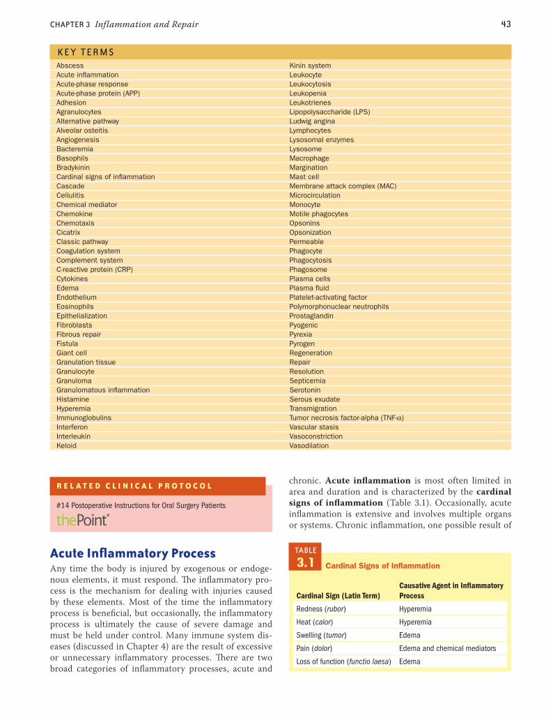
chronic. Acute inflammation is most often limited in area and duration and is characterized by the cardinal signs of inflammation (Table 3.1). Occasionally, acute inflammation is extensive and involves multiple organs or systems. Chronic inflammation, one possible result of
K e y te r m sAbscessAcute inflammationAcute-phase responseAcute-phase protein (APP)AdhesionAgranulocytesAlternative pathwayAlveolar osteitisAngiogenesisBacteremiaBasophilsBradykininCardinal signs of inflammationCascadeCellulitisChemical mediatorChemokineChemotaxisCicatrixClassic pathwayCoagulation systemComplement systemC-reactive protein (CRP)CytokinesEdemaEndotheliumEosinophilsEpithelializationFibroblastsFibrous repairFistulaGiant cellGranulation tissueGranulocyteGranulomaGranulomatous inflammationHistamineHyperemiaImmunoglobulinsInterferonInterleukinKeloid
Kinin systemLeukocyteLeukocytosisLeukopeniaLeukotrienesLipopolysaccharide (LPS)Ludwig anginaLymphocytesLysosomal enzymesLysosomeMacrophageMarginationMast cellMembrane attack complex (MAC)MicrocirculationMonocyteMotile phagocytesOpsoninsOpsonizationPermeablePhagocytePhagocytosisPhagosomePlasma cellsPlasma fluidPlatelet-activating factorPolymorphonuclear neutrophilsProstaglandinPyogenicPyrexiaPyrogenRegenerationRepairResolutionSepticemiaSerotoninSerous exudateTransmigrationTumor necrosis factor-alpha (TNF-α)Vascular stasisVasoconstrictionVasodilation
#14 Postoperative Instructions for Oral Surgery Patients
R e l A t e d C l I n I C A l P R o t o C o l
Cardinal Signs of Inflammation
taBle
3.1
Cardinal Sign (Latin Term)Causative Agent in Inflammatory Process
Redness (rubor) Hyperemia
Heat (calor) Hyperemia
Swelling (tumor) Edema
Pain (dolor) Edema and chemical mediators
Loss of function (functio laesa) Edema
0003305250.INDD 43 11/1/2017 5:16:56 PM
44 part i General Pathology
acute inflammation, is characterized by a long duration or a history of repeated insults or injuries. Other out-comes of acute inflammation are abscess formation, res-olution of inflammation (reversal of the inflammatory process with return to normal), and healing or repair of the area. Although the repair process is discussed at the end of this chapter, repair begins at almost the same time the inflammatory process is activated. That is, they occur simultaneously.
phases of the acute inflammatory processThere are four phases of the acute inflammatory process. The first phase, or initiation, is activated when the injury occurs. It comprises changes to the structure of small blood vessels (microcirculation) in the area of the injury, lead-ing to loss of fluid from the blood and movement of white blood cells from the blood vessels to the injured area. The second phase (amplification) involves the action of chemi-cal substances that direct more and different types of white blood cells into the injured area increasing the response. During the third phase (destruction), the white blood cells quickly neutralize whatever caused the injury, and clean up the debris resulting from the injury. The fourth phase (termination) requires other chemical substances to stop or inhibit the inflammatory process; if the inflammatory
process continues unhindered, more damage than the ini-tial injury will result.
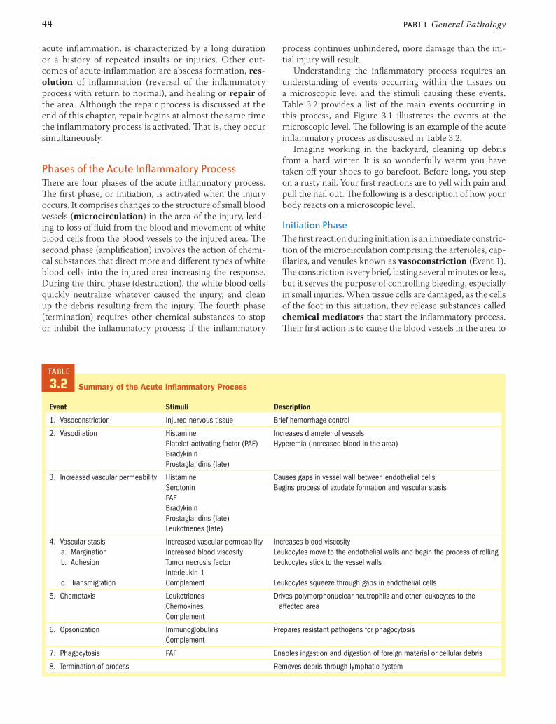
Understanding the inflammatory process requires an understanding of events occurring within the tissues on a microscopic level and the stimuli causing these events. Table 3.2 provides a list of the main events occurring in this process, and Figure 3.1 illustrates the events at the microscopic level. The following is an example of the acute inflammatory process as discussed in Table 3.2.
Imagine working in the backyard, cleaning up debris from a hard winter. It is so wonderfully warm you have taken off your shoes to go barefoot. Before long, you step on a rusty nail. Your first reactions are to yell with pain and pull the nail out. The following is a description of how your body reacts on a microscopic level.
initiation phaseThe first reaction during initiation is an immediate constric-tion of the microcirculation comprising the arterioles, cap-illaries, and venules known as vasoconstriction (Event 1). The constriction is very brief, lasting several minutes or less, but it serves the purpose of controlling bleeding, especially in small injuries. When tissue cells are damaged, as the cells of the foot in this situation, they release substances called chemical mediators that start the inflammatory process. Their first action is to cause the blood vessels in the area to
Summary of the Acute Inflammatory Process
taBle
3.2
Event Stimuli Description
1. Vasoconstriction Injured nervous tissue Brief hemorrhage control
2. Vasodilation HistaminePlatelet-activating factor (PAF)BradykininProstaglandins (late)
Increases diameter of vesselsHyperemia (increased blood in the area)
3. Increased vascular permeability HistamineSerotoninPAFBradykininProstaglandins (late)Leukotrienes (late)
Causes gaps in vessel wall between endothelial cellsBegins process of exudate formation and vascular stasis
4. Vascular stasisa. Marginationb. Adhesion
c. Transmigration
Increased vascular permeabilityIncreased blood viscosityTumor necrosis factorInterleukin-1Complement
Increases blood viscosityLeukocytes move to the endothelial walls and begin the process of rollingLeukocytes stick to the vessel walls
Leukocytes squeeze through gaps in endothelial cells
5. Chemotaxis LeukotrienesChemokinesComplement
Drives polymorphonuclear neutrophils and other leukocytes to the affected area
6. Opsonization ImmunoglobulinsComplement
Prepares resistant pathogens for phagocytosis
7. Phagocytosis PAF Enables ingestion and digestion of foreign material or cellular debris
8. Termination of process Removes debris through lymphatic system
0003305250.INDD 44 11/1/2017 5:16:56 PM
Chapter 3 Inflammation and Repair 45
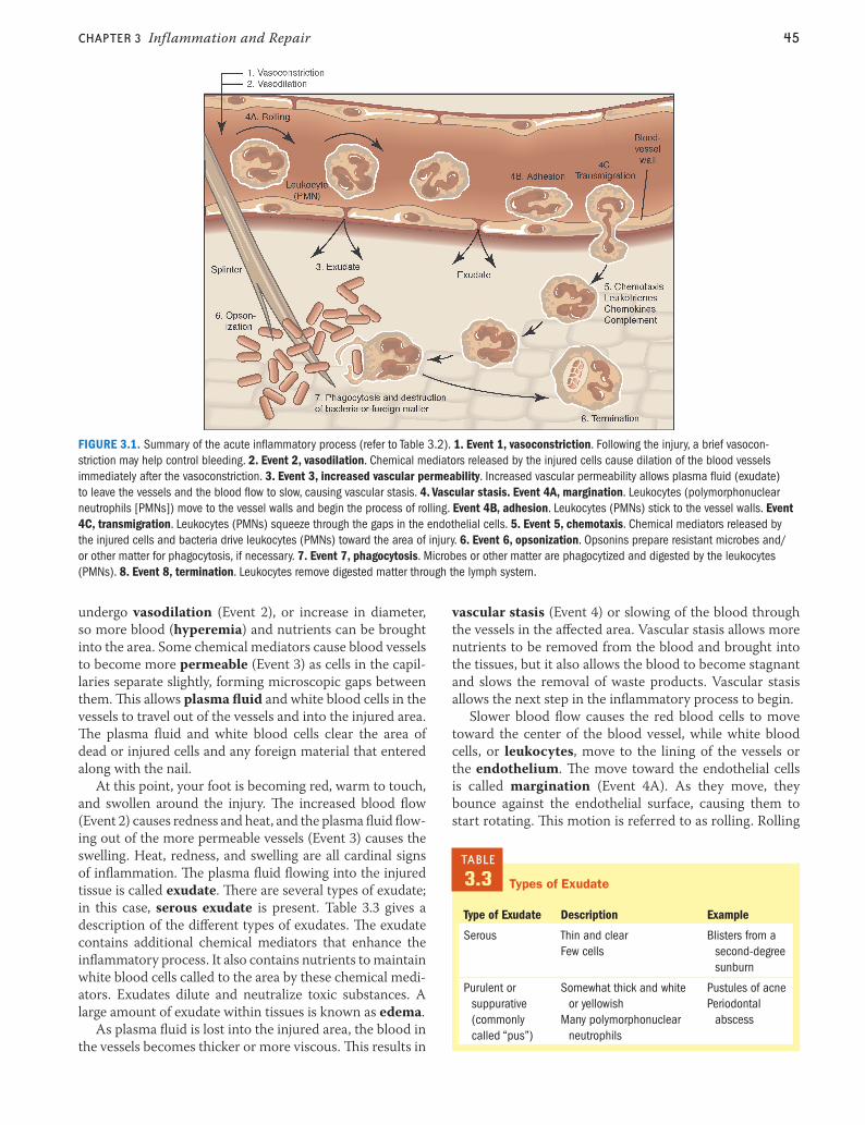
undergo vasodilation (Event 2), or increase in diameter, so more blood (hyperemia) and nutrients can be brought into the area. Some chemical mediators cause blood vessels to become more permeable (Event 3) as cells in the capil-laries separate slightly, forming microscopic gaps between them. This allows plasma fluid and white blood cells in the vessels to travel out of the vessels and into the injured area. The plasma fluid and white blood cells clear the area of dead or injured cells and any foreign material that entered along with the nail.
At this point, your foot is becoming red, warm to touch, and swollen around the injury. The increased blood flow (Event 2) causes redness and heat, and the plasma fluid flow-ing out of the more permeable vessels (Event 3) causes the swelling. Heat, redness, and swelling are all cardinal signs of inflammation. The plasma fluid flowing into the injured tissue is called exudate. There are several types of exudate; in this case, serous exudate is present. Table 3.3 gives a description of the different types of exudates. The exudate contains additional chemical mediators that enhance the inflammatory process. It also contains nutrients to maintain white blood cells called to the area by these chemical medi-ators. Exudates dilute and neutralize toxic substances. A large amount of exudate within tissues is known as edema.
As plasma fluid is lost into the injured area, the blood in the vessels becomes thicker or more viscous. This results in
vascular stasis (Event 4) or slowing of the blood through the vessels in the affected area. Vascular stasis allows more nutrients to be removed from the blood and brought into the tissues, but it also allows the blood to become stagnant and slows the removal of waste products. Vascular stasis allows the next step in the inflammatory process to begin.
Slower blood flow causes the red blood cells to move toward the center of the blood vessel, while white blood cells, or leukocytes, move to the lining of the vessels or the endothelium. The move toward the endothelial cells is called margination (Event 4A). As they move, they bounce against the endothelial surface, causing them to start rotating. This motion is referred to as rolling. Rolling
FIGURE 3.1. Summary of the acute inflammatory process (refer to Table 3.2). 1. Event 1, vasoconstriction. Following the injury, a brief vasocon-striction may help control bleeding. 2. Event 2, vasodilation. Chemical mediators released by the injured cells cause dilation of the blood vessels immediately after the vasoconstriction. 3. Event 3, increased vascular permeability. Increased vascular permeability allows plasma fluid (exudate) to leave the vessels and the blood flow to slow, causing vascular stasis. 4. Vascular stasis. Event 4A, margination. Leukocytes (polymorphonuclear neutrophils [PMNs]) move to the vessel walls and begin the process of rolling. Event 4B, adhesion. Leukocytes (PMNs) stick to the vessel walls. Event 4C, transmigration. Leukocytes (PMNs) squeeze through the gaps in the endothelial cells. 5. Event 5, chemotaxis. Chemical mediators released by the injured cells and bacteria drive leukocytes (PMNs) toward the area of injury. 6. Event 6, opsonization. Opsonins prepare resistant microbes and/or other matter for phagocytosis, if necessary. 7. Event 7, phagocytosis. Microbes or other matter are phagocytized and digested by the leukocytes (PMNs). 8. Event 8, termination. Leukocytes remove digested matter through the lymph system.
Types of Exudate
taBle
3.3
Type of Exudate Description Example
Serous Thin and clearFew cells
Blisters from a second-degree sunburn
Purulent or suppurative (commonly called “pus”)
Somewhat thick and white or yellowish
Many polymorphonuclear neutrophils
Pustules of acnePeriodontal
abscess
0003305250.INDD 45 11/1/2017 5:16:58 PM
46 part i General Pathology
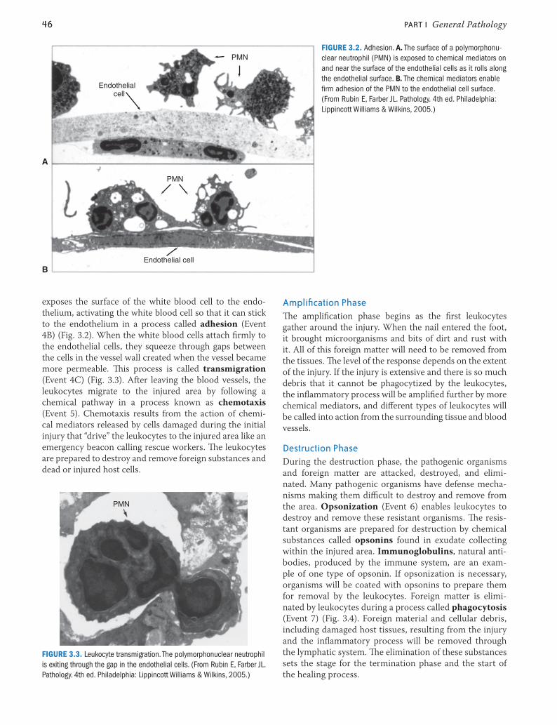
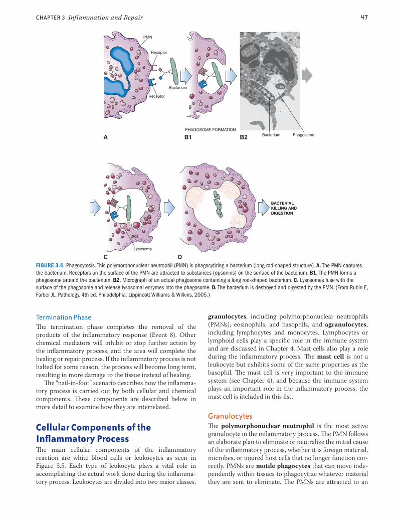
exposes the surface of the white blood cell to the endo-thelium, activating the white blood cell so that it can stick to the endothelium in a process called adhesion (Event 4B) (Fig. 3.2). When the white blood cells attach firmly to the endothelial cells, they squeeze through gaps between the cells in the vessel wall created when the vessel became more permeable. This process is called transmigration (Event 4C) (Fig. 3.3). After leaving the blood vessels, the leukocytes migrate to the injured area by following a chemical pathway in a process known as chemotaxis (Event 5). Chemotaxis results from the action of chemi-cal mediators released by cells damaged during the initial injury that “drive” the leukocytes to the injured area like an emergency beacon calling rescue workers. The leukocytes are prepared to destroy and remove foreign substances and dead or injured host cells.
amplification phaseThe amplification phase begins as the first leukocytes gather around the injury. When the nail entered the foot, it brought microorganisms and bits of dirt and rust with it. All of this foreign matter will need to be removed from the tissues. The level of the response depends on the extent of the injury. If the injury is extensive and there is so much debris that it cannot be phagocytized by the leukocytes, the inflammatory process will be amplified further by more chemical mediators, and different types of leukocytes will be called into action from the surrounding tissue and blood vessels.
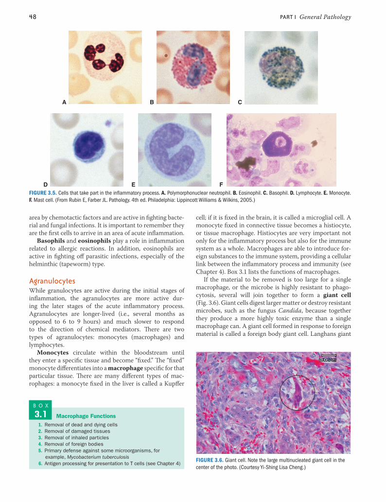
Destruction phaseDuring the destruction phase, the pathogenic organisms and foreign matter are attacked, destroyed, and elimi-nated. Many pathogenic organisms have defense mecha-nisms making them difficult to destroy and remove from the area. Opsonization (Event 6) enables leukocytes to destroy and remove these resistant organisms. The resis-tant organisms are prepared for destruction by chemical substances called opsonins found in exudate collecting within the injured area. Immunoglobulins, natural anti-bodies, produced by the immune system, are an exam-ple of one type of opsonin. If opsonization is necessary, organisms will be coated with opsonins to prepare them for removal by the leukocytes. Foreign matter is elimi-nated by leukocytes during a process called phagocytosis (Event 7) (Fig. 3.4). Foreign material and cellular debris, including damaged host tissues, resulting from the injury and the inflammatory process will be removed through the lymphatic system. The elimination of these substances sets the stage for the termination phase and the start of the healing process.
PMN
FIGURE 3.3. Leukocyte transmigration. The polymorphonuclear neutrophil is exiting through the gap in the endothelial cells. (From Rubin E, Farber JL. Pathology. 4th ed. Philadelphia: Lippincott Williams & Wilkins, 2005.)
PMN
Endothelial cell
A
B
Endothelial cell
PMNFIGURE 3.2. Adhesion. A. The surface of a polymorphonu-clear neutrophil (PMN) is exposed to chemical mediators on and near the surface of the endothelial cells as it rolls along the endothelial surface. B. The chemical mediators enable firm adhesion of the PMN to the endothelial cell surface. (From Rubin E, Farber JL. Pathology. 4th ed. Philadelphia: Lippincott Williams & Wilkins, 2005.)
0003305250.INDD 46 11/1/2017 5:16:59 PM
Chapter 3 Inflammation and Repair 47
termination phaseThe termination phase completes the removal of the products of the inflammatory response (Event 8). Other chemical mediators will inhibit or stop further action by the inflammatory process, and the area will complete the healing or repair process. If the inflammatory process is not halted for some reason, the process will become long term, resulting in more damage to the tissue instead of healing.
The “nail-in-foot” scenario describes how the inflamma-tory process is carried out by both cellular and chemical components. These components are described below in more detail to examine how they are interrelated.
Cellular Components of the Inflammatory ProcessThe main cellular components of the inflammatory reaction are white blood cells or leukocytes as seen in Figure 3.5. Each type of leukocyte plays a vital role in accomplishing the actual work done during the inflamma-tory process. Leukocytes are divided into two major classes,
granulocytes, including polymorphonuclear neutrophils (PMNs), eosinophils, and basophils, and agranulocytes, including lymphocytes and monocytes. Lymphocytes or lymphoid cells play a specific role in the immune system and are discussed in Chapter 4. Mast cells also play a role during the inflammatory process. The mast cell is not a leukocyte but exhibits some of the same properties as the basophil. The mast cell is very important to the immune system (see Chapter 4), and because the immune system plays an important role in the inflammatory process, the mast cell is included in this list.
granulocytesThe polymorphonuclear neutrophil is the most active granulocyte in the inflammatory process. The PMN follows an elaborate plan to eliminate or neutralize the initial cause of the inflammatory process, whether it is foreign material, microbes, or injured host cells that no longer function cor-rectly. PMNs are motile phagocytes that can move inde-pendently within tissues to phagocytize whatever material they are sent to eliminate. The PMNs are attracted to an
Receptor
Receptor
PMN
Bacterium
PHAGOSOME FORMATION
Lysosome
BACTERIALKILLING ANDDIGESTION
Bacterium PhagosomeA B1 B2
C DFIGURE 3.4. Phagocytosis. This polymorphonuclear neutrophil (PMN) is phagocytizing a bacterium (long rod-shaped structure). A. The PMN captures the bacterium. Receptors on the surface of the PMN are attracted to substances (opsonins) on the surface of the bacterium. B1. The PMN forms a phagosome around the bacterium. B2. Micrograph of an actual phagosome containing a long rod-shaped bacterium. C. Lysosomes fuse with the surface of the phagosome and release lysosomal enzymes into the phagosome. D. The bacterium is destroyed and digested by the PMN. (From Rubin E, Farber JL. Pathology. 4th ed. Philadelphia: Lippincott Williams & Wilkins, 2005.)
0003305250.INDD 47 11/1/2017 5:16:59 PM
48 part i General Pathology
area by chemotactic factors and are active in fighting bacte-rial and fungal infections. It is important to remember they are the first cells to arrive in an area of acute inflammation.
Basophils and eosinophils play a role in inflammation related to allergic reactions. In addition, eosinophils are active in fighting off parasitic infections, especially of the helminthic (tapeworm) type.
agranulocytesWhile granulocytes are active during the initial stages of inflammation, the agranulocytes are more active dur-ing the later stages of the acute inflammatory process. Agranulocytes are longer-lived (i.e., several months as opposed to 6 to 9 hours) and much slower to respond to the direction of chemical mediators. There are two types of agranulocytes: monocytes (macrophages) and lymphocytes.
Monocytes circulate within the bloodstream until they enter a specific tissue and become “fixed.” The “fixed” monocyte differentiates into a macrophage specific for that particular tissue. There are many different types of mac-rophages: a monocyte fixed in the liver is called a Kupffer
cell; if it is fixed in the brain, it is called a microglial cell. A monocyte fixed in connective tissue becomes a histiocyte, or tissue macrophage. Histiocytes are very important not only for the inflammatory process but also for the immune system as a whole. Macrophages are able to introduce for-eign substances to the immune system, providing a cellular link between the inflammatory process and immunity (see Chapter 4). Box 3.1 lists the functions of macrophages.
If the material to be removed is too large for a single macrophage, or the microbe is highly resistant to phago-cytosis, several will join together to form a giant cell (Fig. 3.6). Giant cells digest larger matter or destroy resistant microbes, such as the fungus Candida, because together they produce a more highly toxic enzyme than a single macrophage can. A giant cell formed in response to foreign material is called a foreign body giant cell. Langhans giant
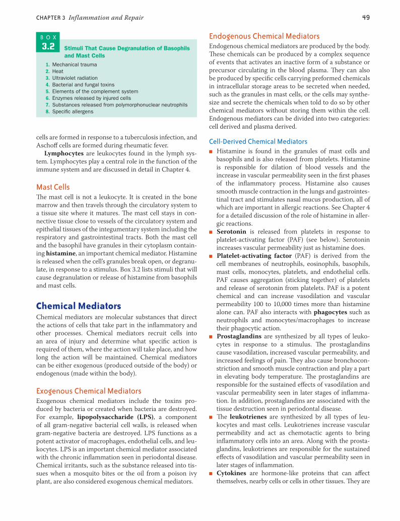
FIGURE 3.5. Cells that take part in the inflammatory process. A. Polymorphonuclear neutrophil. B. Eosinophil. C. Basophil. D. Lymphocyte. E. Monocyte. F. Mast cell. (From Rubin E, Farber JL. Pathology. 4th ed. Philadelphia: Lippincott Williams & Wilkins, 2005.)
C
D
A B
E F
Macrophage Functions1. Removal of dead and dying cells2. Removal of damaged tissues3. Removal of inhaled particles4. Removal of foreign bodies5. Primary defense against some microorganisms, for
example, Mycobacterium tuberculosis6. Antigen processing for presentation to T cells (see Chapter 4)
B O X
3.1
FIGURE 3.6. Giant cell. Note the large multinucleated giant cell in the center of the photo. (Courtesy Yi-Shing Lisa Cheng.)
0003305250.INDD 48 11/1/2017 5:17:04 PM
Chapter 3 Inflammation and Repair 49
cells are formed in response to a tuberculosis infection, and Aschoff cells are formed during rheumatic fever.
Lymphocytes are leukocytes found in the lymph sys-tem. Lymphocytes play a central role in the function of the immune system and are discussed in detail in Chapter 4.
mast CellsThe mast cell is not a leukocyte. It is created in the bone marrow and then travels through the circulatory system to a tissue site where it matures. The mast cell stays in con-nective tissue close to vessels of the circulatory system and epithelial tissues of the integumentary system including the respiratory and gastrointestinal tracts. Both the mast cell and the basophil have granules in their cytoplasm contain-ing histamine, an important chemical mediator. Histamine is released when the cell’s granules break open, or degranu-late, in response to a stimulus. Box 3.2 lists stimuli that will cause degranulation or release of histamine from basophils and mast cells.
Chemical MediatorsChemical mediators are molecular substances that direct the actions of cells that take part in the inflammatory and other processes. Chemical mediators recruit cells into an area of injury and determine what specific action is required of them, where the action will take place, and how long the action will be maintained. Chemical mediators can be either exogenous (produced outside of the body) or endogenous (made within the body).
exogenous Chemical mediatorsExogenous chemical mediators include the toxins pro-duced by bacteria or created when bacteria are destroyed. For example, lipopolysaccharide (LPS), a component of all gram-negative bacterial cell walls, is released when gram-negative bacteria are destroyed. LPS functions as a potent activator of macrophages, endothelial cells, and leu-kocytes. LPS is an important chemical mediator associated with the chronic inflammation seen in periodontal disease. Chemical irritants, such as the substance released into tis-sues when a mosquito bites or the oil from a poison ivy plant, are also considered exogenous chemical mediators.
endogenous Chemical mediatorsEndogenous chemical mediators are produced by the body. These chemicals can be produced by a complex sequence of events that activates an inactive form of a substance or precursor circulating in the blood plasma. They can also be produced by specific cells carrying preformed chemicals in intracellular storage areas to be secreted when needed, such as the granules in mast cells, or the cells may synthe-size and secrete the chemicals when told to do so by other chemical mediators without storing them within the cell. Endogenous mediators can be divided into two categories: cell derived and plasma derived.
Cell-Derived Chemical mediators■■ Histamine is found in the granules of mast cells and
basophils and is also released from platelets. Histamine is responsible for dilation of blood vessels and the increase in vascular permeability seen in the first phases of the inflammatory process. Histamine also causes smooth muscle contraction in the lungs and gastrointes-tinal tract and stimulates nasal mucus production, all of which are important in allergic reactions. See Chapter 4 for a detailed discussion of the role of histamine in aller-gic reactions.
■■ Serotonin is released from platelets in response to platelet-activating factor (PAF) (see below). Serotonin increases vascular permeability just as histamine does.
■■ Platelet-activating factor (PAF) is derived from the cell membranes of neutrophils, eosinophils, basophils, mast cells, monocytes, platelets, and endothelial cells. PAF causes aggregation (sticking together) of platelets and release of serotonin from platelets. PAF is a potent chemical and can increase vasodilation and vascular permeability 100 to 10,000 times more than histamine alone can. PAF also interacts with phagocytes such as neutrophils and monocytes/macrophages to increase their phagocytic action.
■■ Prostaglandins are synthesized by all types of leuko-cytes in response to a stimulus. The prostaglandins cause vasodilation, increased vascular permeability, and increased feelings of pain. They also cause bronchocon-striction and smooth muscle contraction and play a part in elevating body temperature. The prostaglandins are responsible for the sustained effects of vasodilation and vascular permeability seen in later stages of inflamma-tion. In addition, prostaglandins are associated with the tissue destruction seen in periodontal disease.
■■ The leukotrienes are synthesized by all types of leu-kocytes and mast cells. Leukotrienes increase vascular permeability and act as chemotactic agents to bring inflammatory cells into an area. Along with the prosta-glandins, leukotrienes are responsible for the sustained effects of vasodilation and vascular permeability seen in later stages of inflammation.
■■ Cytokines are hormone-like proteins that can affect themselves, nearby cells or cells in other tissues. They are
Stimuli That Cause Degranulation of Basophils and Mast Cells
1. Mechanical trauma2. Heat3. Ultraviolet radiation4. Bacterial and fungal toxins5. Elements of the complement system6. Enzymes released by injured cells7. Substances released from polymorphonuclear neutrophils8. Specific allergens
B O X
3.2
0003305250.INDD 49 11/1/2017 5:17:04 PM
50 part i General Pathology
produced by macrophages and some types of lympho-cytes. Examples of cytokines active in the inflammatory process are chemokines, interferons, tumor necrosis factor-alpha (TNF-α), and interleukins. The chemo-kines are very strong chemotactic agents for cells involved in the inflammatory process. Interferons activate macro-phages and stimulate the production of cytokines. TNF-α and the interleukins (IL-1β, IL-6, IL-8, and others) have numerous effects during all stages of the inflammatory process. TNF-α and the interleukins increase leukocyte adherence, prostaglandin synthesis, and fibroblast pro-duction. They also produce fever, increase need for sleep, and decrease appetite. These substances are involved with the tissue destruction that occurs in periodontal disease.
plasma-Derived Chemical mediatorsThree major plasma protein systems are involved in media-tion of the inflammatory process. These systems include the complement system, the coagulation system, and the kinin system. These systems consist of a series of inactive enzymes. Once the first enzyme in a series is activated, it initiates the next in a series of reactions in which the prod-uct of the last reaction is the initiator of the next reaction. This type of process is called a cascade.
The Complement SystemActivation of the complement system is important in both the inflammatory process and in immunity. The com-plement system comprises a complex series of reactions between plasma proteins. The end-product of this cas-cade, a substance called the membrane attack complex (MAC), punches a hole in the cell membrane of microbes targeted for destruction by the immune system. Other sub-stances produced in the cascade influence events in the inflammatory process, including vascular effects, leukocyte activation, adhesion and chemotaxis, and enhancement of microbial phagocytosis. In addition, products of the complement system cause mast cells to release their hista-mine (vascular effects). Other products cause leukocytes to become more active and increase adherence to endothelial cells. Substances that enhance the action of leukocytes are also very strong chemotactic agents stimulating leukocytes to travel to an injured or compromised area. Still another product of the complement cascade is a type of opsonin that attaches to microbial cell walls making them easier to phagocytize (opsonization).
The complement cascade can be triggered by two dif-ferent pathways, the classic and the alternative pathway. The classic pathway is triggered, or started, by antibodies created specifically for the agent causing the inflammatory process. The alternative pathway can be triggered by bac-terial LPSs or aggregates (clumps) of preformed immuno-globulins already circulating through the body.
The Coagulation SystemThe coagulation system cascade is activated when a plasma protein called the Hageman factor comes in contact
with cellular debris from an endothelial or vessel injury. Although best known for its blood clotting effects, which will be involved in the repair process, the coagulation sys-tem is involved in activation of both the kinin system and the complement system. Thus, the coagulation system is an important factor in the inflammatory process.
The Kinin SystemActivation of the kinin system cascade results in the for-mation of the chemical mediator bradykinin. Bradykinin is capable of causing vasodilation, increased vascular perme-ability, and pain. The kinin system is activated by the same substance that activates the coagulation system (Hageman factor).
Now that more details have been provided regarding the elements of the acute inflammatory process, refer back to the “nail-in-foot” scenario and replace some of the broader terms such as “chemical mediators” with the names of the specific chemical mediator involved in the action. The same can be done with the broader term “leukocytes.” Table 3.2 can be used to complete this exercise. In addition to a localized reaction, you may see systemic manifestations of inflammation in some cases.
Systemic Manifestations of InflammationIdeally, the effects of the inflammatory response remain in a localized area. However, in some instances, the localized response results in systemic involvement. The systemic manifestations of inflammation include the acute-phase response, alteration in the number of white blood cells, and lymphadenopathy.
The acute-phase response is regulated by the chemi-cal mediators produced by leukocytes, monocytes, mac-rophages, and other inflammatory cells that enter the circulation where they are transported throughout the body. The most obvious clinical sign of the acute-phase response is fever or pyrexia. In addition to the chemical mediators released by inflammatory cells, many substances released by bacteria will also cause fever. Fever-producing substances are called pyrogens. Pyrogens stimulate the production of prostaglandins, which activate the thermo-regulatory center in the hypothalamus, thereby causing an elevation in temperature. An elevated temperature can be important because many pathogens have a very narrow temperature range within which they operate, and even a slight rise in temperature may help destroy them. In addi-tion, small elevations in temperature have been shown to increase the activity and motility of white blood cells and enhance the immune system response. Pyrexia can also be caused by noninfectious agents. For example, pyrexia can be caused by excess thyroid hormones, severe dehy-dration, and cancer. Other clinical manifestations of the acute-phase response include anorexia (lack of appetite), sleepiness, pain, and malaise. Chemically, the acute-phase response is characterized by alterations in plasma levels of
0003305250.INDD 50 11/1/2017 5:17:04 PM
Chapter 3 Inflammation and Repair 51
acute-phase proteins. Acute-phase proteins (APPs) are low molecular weight proteins produced by the liver in response to circulating chemical mediators released dur-ing the inflammatory response. Many of the APPs are com-ponents of the coagulation and the complement systems. The functions of APPs include opsonization and trapping of microorganisms and their products, activating the com-plement system, modulating the coagulation system, neu-tralizing lysosomal enzymes, and regulating the immune response, among others (Khan & Khan, 2010). C-reactive protein (CRP) was the first APP to be described. CRP binds to the surface of a foreign microorganism and targets it for destruction by complement and phagocytosis (opso-nization). It also interacts with T cells and B cells helping to coordinate the inflammatory and immune responses.
Elevated levels of CRP in the blood are being used to help diagnose the presence of acute inflammation associated with infections, traumatic injuries, chronic inflammatory conditions such as rheumatoid arthritis, and some can-cers. The presence of elevated levels of CRP indicates the presence of inflammation, but it does not tell us the source of that inflammation; thus, testing for CRP supplies only part of the information necessary for diagnosis. The level of CRP may tell physicians whether a certain anti-inflam-matory drug is working effectively for a patient with, for example, rheumatoid arthritis. Chronically elevated levels of CRP have been shown to be a strong risk factor for car-diovascular disease (CVD). Interestingly, elevated levels of CRP decline with exercise and may be an indicator of well-ness (Aronson et al., 2004).
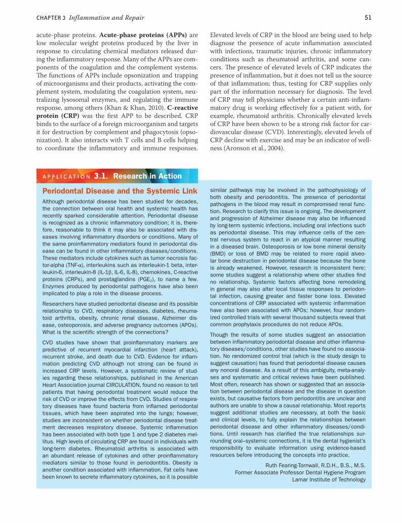
Periodontal Disease and the Systemic LinkAlthough periodontal disease has been studied for decades, the connection between oral health and systemic health has recently sparked considerable attention. Periodontal disease is recognized as a chronic inflammatory condition; it is, there-fore, reasonable to think it may also be associated with dis-eases involving inflammatory disorders or conditions. Many of the same proinflammatory mediators found in periodontal dis-ease can be found in other inflammatory diseases/conditions. These mediators include cytokines such as tumor necrosis fac-tor-alpha (TNF-α), interleukins such as interleukin-1 beta, inter-leukin-6, interleukin-8 (IL-1β, IL-6, IL-8), chemokines, C-reactive proteins (CRPs), and prostaglandins (PGE2), to name a few. Enzymes produced by periodontal pathogens have also been implicated to play a role in the disease process.
Researchers have studied periodontal disease and its possible relationship to CVD, respiratory diseases, diabetes, rheuma-toid arthritis, obesity, chronic renal disease, Alzheimer dis-ease, osteoporosis, and adverse pregnancy outcomes (APOs). What is the scientific strength of the connections?
CVD studies have shown that proinflammatory markers are predictive of recurrent myocardial infarction (heart attack), recurrent stroke, and death due to CVD. Evidence for inflam-mation predicting CVD although not strong can be found in increased CRP levels. However, a systematic review of stud-ies regarding these relationships, published in the American Heart Association journal CIRCULATION, found no reason to tell patients that having periodontal treatment would reduce the risk of CVD or improve the effects from CVD. Studies of respira-tory diseases have found bacteria from inflamed periodontal tissues, which have been aspirated into the lungs; however, studies are inconsistent on whether periodontal disease treat-ment decreases respiratory disease. Systemic inflammation has been associated with both type 1 and type 2 diabetes mel-litus. High levels of circulating CRP are found in individuals with long-term diabetes. Rheumatoid arthritis is associated with an abundant release of cytokines and other proinflammatory mediators similar to those found in periodontitis. Obesity is another condition associated with inflammation. Fat cells have been known to secrete inflammatory cytokines, so it is possible
similar pathways may be involved in the pathophysiology of both obesity and periodontitis. The presence of periodontal pathogens in the blood may result in compromised renal func-tion. Research to clarify this issue is ongoing. The development and progression of Alzheimer disease may also be influenced by long-term systemic infections, including oral infections such as periodontal disease. This may influence cells of the cen-tral nervous system to react in an atypical manner resulting in a diseased brain. Osteoporosis or low bone mineral density (BMD) or loss of BMD may be related to more rapid alveo-lar bone destruction in periodontal disease because the bone is already weakened. However, research is inconsistent here; some studies suggest a relationship where other studies find no relationship. Systemic factors affecting bone remodeling in general may also alter local tissue responses to periodon-tal infection, causing greater and faster bone loss. Elevated concentrations of CRP associated with systemic inflammation have also been associated with APOs; however, four random-ized controlled trials with several thousand subjects reveal that common prophylaxis procedures do not reduce APOs.
Though the results of some studies suggest an association between inflammatory periodontal disease and other inflamma-tory diseases/conditions, other studies have found no associa-tion. No randomized control trial (which is the study design to suggest causation) has found that periodontal disease causes any nonoral disease. As a result of this ambiguity, meta-analy-ses and systematic and critical reviews have been published. Most often, research has shown or suggested that an associa-tion between periodontal disease and the disease in question exists, but causative factors from periodontitis are unclear and authors are unable to show a causal relationship. Most reports suggest additional studies are necessary, at both the basic and clinical levels, to fully explain the relationships between periodontal disease and other inflammatory diseases/condi-tions. Until research has clarified the true relationships sur-rounding oral–systemic connections, it is the dental hygienist’s responsibility to evaluate information using evidence-based resources before introducing the concepts into practice.
Ruth Fearing-Tornwall, R.D.H., B.S., M.S.Former Associate Professor Dental Hygiene Program
Lamar Institute of Technology
a p p l i C a t i O n 3.1. Research in Action
0003305250.INDD 51 11/1/2017 5:17:04 PM
52 part i General Pathology
An alteration in the number of white blood cells is a systemic manifestation of the inflammatory response. An increase in the number of white cells in the blood is called leukocytosis. Normal white blood cell counts range from 4,000 to 10,000 per mm3; however, during leukocy-tosis, white blood cell counts increase up to 100,000 per mm3. Neutrophils or PMNs increase in number in bacte-rial infections, in inflammatory disorders, and in response to certain drugs. Lymphocytes are the primary responders in viral infections, and monocytes predominate in chronic infections. Leukopenia, a decrease in white blood cells, may occur in overwhelming infections or if the body has an impaired ability to produce white cells.
The lymphatic system is important in draining the edema fluid or exudate and clearing the cellular debris and foreign matter from the affected area. Lymphadenopathy, enlargement of the lymph nodes, is another common sys-temic manifestation of the inflammatory process. The lymph nodes become enlarged, firm, and tender. In local-ized lymphadenopathy, one or more nodes in the area of the infection or inflammation become swollen and tender, such as when a streptococcal throat infection causes the cervical lymph nodes to become involved. In generalized involvement, nodes all over the body become swollen and tender, for example, the persistent generalized lymphade-nopathy (PGL) seen in HIV infection (see Chapter 21). The lymphatic system is responsible for removing all waste products from the inflamed area before healing can begin. If this cannot be accomplished, or if for some other reason the inflammatory process cannot be halted, the reaction will progress to a chronic phase.
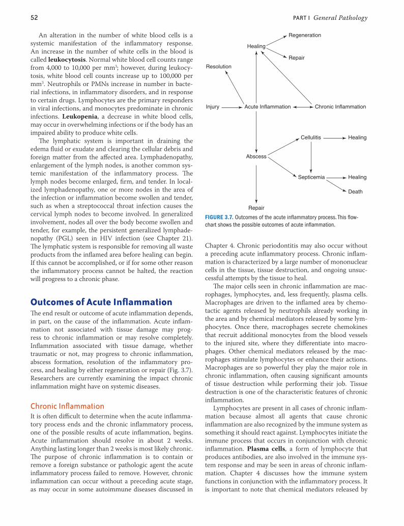
outcomes of Acute InflammationThe end result or outcome of acute inflammation depends, in part, on the cause of the inflammation. Acute inflam-mation not associated with tissue damage may prog-ress to chronic inflammation or may resolve completely. Inflammation associated with tissue damage, whether traumatic or not, may progress to chronic inflammation, abscess formation, resolution of the inflammatory pro-cess, and healing by either regeneration or repair (Fig. 3.7). Researchers are currently examining the impact chronic inflammation might have on systemic diseases.
Chronic inflammationIt is often difficult to determine when the acute inflamma-tory process ends and the chronic inflammatory process, one of the possible results of acute inflammation, begins. Acute inflammation should resolve in about 2 weeks. Anything lasting longer than 2 weeks is most likely chronic. The purpose of chronic inflammation is to contain or remove a foreign substance or pathologic agent the acute inflammatory process failed to remove. However, chronic inflammation can occur without a preceding acute stage, as may occur in some autoimmune diseases discussed in
Chapter 4. Chronic periodontitis may also occur without a preceding acute inflammatory process. Chronic inflam-mation is characterized by a large number of mononuclear cells in the tissue, tissue destruction, and ongoing unsuc-cessful attempts by the tissue to heal.
The major cells seen in chronic inflammation are mac-rophages, lymphocytes, and, less frequently, plasma cells. Macrophages are driven to the inflamed area by chemo-tactic agents released by neutrophils already working in the area and by chemical mediators released by some lym-phocytes. Once there, macrophages secrete chemokines that recruit additional monocytes from the blood vessels to the injured site, where they differentiate into macro-phages. Other chemical mediators released by the mac-rophages stimulate lymphocytes or enhance their actions. Macrophages are so powerful they play the major role in chronic inflammation, often causing significant amounts of tissue destruction while performing their job. Tissue destruction is one of the characteristic features of chronic inflammation.
Lymphocytes are present in all cases of chronic inflam-mation because almost all agents that cause chronic inflammation are also recognized by the immune system as something it should react against. Lymphocytes initiate the immune process that occurs in conjunction with chronic inflammation. Plasma cells, a form of lymphocyte that produces antibodies, are also involved in the immune sys-tem response and may be seen in areas of chronic inflam-mation. Chapter 4 discusses how the immune system functions in conjunction with the inflammatory process. It is important to note that chemical mediators released by
Healing
Healing
Regeneration
Repair
Repair
Healing
Injury Acute Inflammation
Abscess
Cellulitis
Septicemia
Death
Chronic Inflammation
Resolution
FIGURE 3.7. Outcomes of the acute inflammatory process. This flow-chart shows the possible outcomes of acute inflammation.
0003305250.INDD 52 11/1/2017 5:17:04 PM
Chapter 3 Inflammation and Repair 53
lymphocytes can stimulate or enhance the action of macro-phages. The simultaneous stimulation of both macrophages and lymphocytes by each other enables the persistence of chronic inflammation.
The tissue destruction seen in chronic inflammation is caused by chemicals released from cells that are attempt-ing to eliminate the offending agent or substance from the area. Many of these chemical substances are found within the lysosomes of these cells. Lysosomes are organelles containing strong digestive enzymes, called lysosomal enzymes, which are associated with the digestion or elimi-nation of phagocytized foreign matter. When a phagocytic cell traps a foreign substance, it creates an intracellular space or vacuole called a phagosome to hold it in (Fig. 3.4). Chemicals released when the phagosome is being cre-ated drive the cell’s lysosomes to the surface of the phago-some. The lysosomes fuse with the phagosome and release all of their lysosomal enzymes into it, enabling the diges-tion of the foreign matter. Problems occur when lysosomal enzymes find their way out of the cells and into the tissues. The enzymes may leak from the cell as it is digesting a for-eign substance, or all of the intracellular substances may be released when the cell dies. The lysosomal enzymes can destroy normal cells and collagen fibers in the area. They can also activate osteoclasts, causing bone destruction. Bone destruction that occurs during chronic adult peri-odontitis is a good example of this process. While these destructive processes are occurring, the tissue is trying to heal itself.
Chronic inflammation has been described as “frustrated healing” by many, because everything needed for repair, such as fibroblasts (immature connective tissue cell that can differentiate into cells that produce collagen and other tissues) and small blood vessels, is present in the affected tissues. Chronic inflammation will only resolve when all of the agents causing it are eliminated. Box 3.3 presents a list of factors that can contribute to the development and maintenance of chronic inflammation.
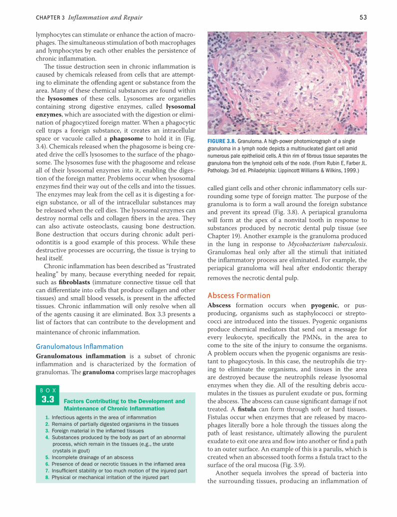
granulomatous inflammationGranulomatous inflammation is a subset of chronic inflammation and is characterized by the formation of granulomas. The granuloma comprises large macrophages
called giant cells and other chronic inflammatory cells sur-rounding some type of foreign matter. The purpose of the granuloma is to form a wall around the foreign substance and prevent its spread (Fig. 3.8). A periapical granuloma will form at the apex of a nonvital tooth in response to substances produced by necrotic dental pulp tissue (see Chapter 19). Another example is the granuloma produced in the lung in response to Mycobacterium tuberculosis. Granulomas heal only after all the stimuli that initiated the inflammatory process are eliminated. For example, the periapical granuloma will heal after endodontic therapy removes the necrotic dental pulp.
abscess FormationAbscess formation occurs when pyogenic, or pus-producing, organisms such as staphylococci or strepto-cocci are introduced into the tissues. Pyogenic organisms produce chemical mediators that send out a message for every leukocyte, specifically the PMNs, in the area to come to the site of the injury to consume the organisms. A problem occurs when the pyogenic organisms are resis-tant to phagocytosis. In this case, the neutrophils die try-ing to eliminate the organisms, and tissues in the area are destroyed because the neutrophils release lysosomal enzymes when they die. All of the resulting debris accu-mulates in the tissues as purulent exudate or pus, forming the abscess. The abscess can cause significant damage if not treated. A fistula can form through soft or hard tissues. Fistulas occur when enzymes that are released by macro-phages literally bore a hole through the tissues along the path of least resistance, ultimately allowing the purulent exudate to exit one area and flow into another or find a path to an outer surface. An example of this is a parulis, which is created when an abscessed tooth forms a fistula tract to the surface of the oral mucosa (Fig. 3.9).
Another sequela involves the spread of bacteria into the surrounding tissues, producing an inflammation of
FIGURE 3.8. Granuloma. A high-power photomicrograph of a single granuloma in a lymph node depicts a multinucleated giant cell amid numerous pale epithelioid cells. A thin rim of fibrous tissue separates the granuloma from the lymphoid cells of the node. (From Rubin E, Farber JL. Pathology. 3rd ed. Philadelphia: Lippincott Williams & Wilkins, 1999.)
Factors Contributing to the Development and Maintenance of Chronic Inflammation
1. Infectious agents in the area of inflammation2. Remains of partially digested organisms in the tissues3. Foreign material in the inflamed tissues4. Substances produced by the body as part of an abnormal
process, which remain in the tissues (e.g., the urate crystals in gout)
5. Incomplete drainage of an abscess6. Presence of dead or necrotic tissues in the inflamed area7. Insufficient stability or too much motion of the injured part8. Physical or mechanical irritation of the injured part
B O X
3.3
0003305250.INDD 53 11/1/2017 5:17:05 PM

54 part i General Pathology
the connective tissue called cellulitis (Fig. 3.10). Ludwig angina, bilateral cellulitis involving the submandibu-lar, submental, and sublingual spaces, is a serious condi-tion causing rapid swelling of the floor of the mouth with elevation and posterior displacement of the tongue. The swelling often results in partial or complete airway obstruc-tion, making Ludwig angina a life-threatening emergency (Fig. 3.11). In some cases, the bacteria enter the blood and produce a bacteremia, or bacteria in the blood. This can lead to septicemia, or blood poisoning, and several other conditions. One of the more serious and sometimes fatal conditions is the spread of infection to the cavernous sinus, resulting in the formation of a blood clot or cavernous sinus thrombosis. This is not a common occurrence; however, media reports of children dying from complications of a neglected dental abscess always cause dismay among dental
professionals who know how easily these can be prevented. In any case, abscesses need to be treated quickly before any of these conditions can occur.
resolution of the inflammatory processIn many cases, the inflammatory process is not triggered by a traumatic injury or microbial assault. In these cases, because there was no traumatic tissue damage, resolution can take place. When the stimulus that initiated the pro-cess, such as pollen in hay fever, is neutralized, the inflam-mation resolves, and the tissues return to normal. In the case of tissue damage, healing can only be completed when all of the stimuli that initiated the inflammatory process are neutralized or removed.
regenerationThe most desired outcome of the inflammatory process is regeneration. This is the body’s attempt to restore itself to its original state. Regeneration occurs when the stimu-lus that caused the inflammation is completely removed, the vascular system returns to normal, the injured tissue is replaced with the same type of tissues and cells that were damaged, and the area regains full function. Regeneration depends on the type of cell that was damaged and the extent of the injury. The epithelial cells that line the oral cavity will regenerate because they are already constantly
FIGURE 3.9. Abscess. A parulis that forms as a result of a periapical abscess is an example of an outcome of acute inflammation. (From Fleisher GR, Ludwig S, Baskin MN. Atlas of Pediatric Emergency Medicine. Philadelphia: Lippincott Williams & Wilkins, 2004.)
FIGURE 3.10. Cellulitis. This child had a painful swollen cheek and infra-orbital cellulitis caused by an abscessed tooth. (Courtesy of Dr. Debra Weiner; From Fleisher GR, Ludwig S, Baskin MN. Atlas of Pediatric Emer-gency Medicine. Philadelphia: Lippincott Williams & Wilkins, 2004.)
FIGURE 3.11. Ludwig angina. Pictured is a patient with Ludwig angina. Note the classic swelling and protrusion of the tongue. This patient was in respiratory distress necessitating a tracheotomy to maintain an open airway. Drains are sutured in place to allow purulent exudate to leave the tissues. (Courtesy of Bechara Y. Ghorayeb, MD.)
0003305250.INDD 54 11/1/2017 5:17:07 PM
Chapter 3 Inflammation and Repair 55
replicating themselves to keep the mucosal barrier intact. Liver cells will regenerate. In transplantations, the liver of the donor will completely regenerate, and the part of the liver that is transplanted into the recipient will regenerate to a more normal size. Brain cells, however, are permanent or nondividing cells; therefore, injured brain cells will not replicate. Some of the lost function may be regained by forming alternative pathways through uninjured brain cells, but the cells that were lost will not regenerate. No matter what type of tissue is involved, if the injured area is large enough, regeneration will not be an option.
Fibrous repairIf regeneration is not possible, then fibrous repair will be the final outcome. Fibrous repair results in the creation of a cicatrix (scar) that may recreate normal or near-normal tissue formation or architecture but not normal function. Chronic inflammation inevitably resolves with scar forma-tion. This outcome is related to the extended healing time and to the amount of tissue damage usually associated with chronic inflammation. Repair is one of the primary func-tions of the immune system. The process of repair, like the inflammatory process, requires many chemical mediators that control the timing of the wound healing. Some of these chemical mediators include cytokines and epidermal-, fibroblast-, and platelet-derived growth factors.
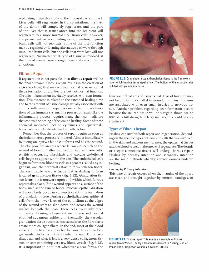
Remember that the process of repair begins as soon as the inflammatory process is initiated. Almost immediately following an injury, a blood clot forms and fills the wound. The clot provides an area where leukocytes can clean the wound of foreign matter and dead or injured tissue cells. As this is occurring, fibroblasts and vascular endothelial cells begin to appear within the clot. The endothelial cells begin to form new blood vessels in a process called angio-genesis, and the fibroblasts start to form collagen fibers. The very fragile vascular tissue that is starting to form is called granulation tissue (Fig. 3.12). Granulation tis-sue forms the framework upon and within which fibrous repair takes place. If the wound appears on a surface of the body, such as the skin or buccal mucosa, epithelialization will most likely occur in conjunction with the formation of granulation tissue. During epithelialization, epithelial cells from the lower layer of the epithelium at the edges of the wound start to slide down and across the wound surface beneath the scab. These cells eventually meet and unite, forming a basement membrane and normal stratified squamous epithelium. Eventually, the vascular granulation tissue becomes less vascular as the fibroblasts create more collagen fibers. In the end, most of the blood vessels in the tissue are resorbed because they are no lon-ger needed to bring nutrients into the area. Fibroblasts disappear, and what is left is a very dense collagenous tis-sue, or scar, containing very few blood vessels (Fig. 3.13). It is important to note that whenever a scar forms, the
function of that area of tissue is lost. Loss of function may not be crucial in a small skin wound, but many problems are associated with even small injuries to nervous tis-sue. Another problem regarding scar formation occurs because the injured tissue will only regain about 70% to 80% of its full strength; in large injuries, this could be very significant.
types of Fibrous repairHealing can involve both repair and regeneration, depend-ing on the specific types of tissue and cells that are involved. In the skin and mucous membranes, the epidermal tissues and the blood vessels in the area will regenerate. The dermis or deeper connective tissues will undergo fibrous repair. Healing by primary intention and secondary intention are the main methods whereby surface wounds undergo healing.
Healing by Primary IntentionThis type of repair occurs when the margins of the injury are clean and brought together by sutures, bandages, or
FIGURE 3.12. Granulation tissue. Granulation tissue is the framework upon which healing tissue repairs itself. The bottom of this extraction site is filled with granulation tissue.
FIGURE 3.13. Fibrous repair. This scar is an example of fibrous repair. (From Weber J, Kelley J. Health Assessment in Nursing. 2nd ed. Philadelphia: Lippincott Williams & Wilkins, 2003.)
0003305250.INDD 55 11/1/2017 5:17:08 PM
56 part i General Pathology
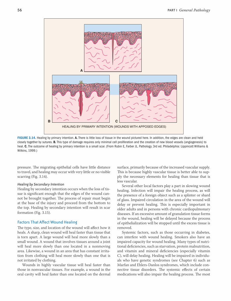
pressure. The migrating epithelial cells have little distance to travel, and healing may occur with very little or no visible scarring (Fig. 3.14).
Healing by Secondary IntentionHealing by secondary intention occurs when the loss of tis-sue is significant enough that the edges of the wound can-not be brought together. The process of repair must begin at the base of the injury and proceed from the bottom to the top. Healing by secondary intention will result in scar formation (Fig. 3.15).
Factors that affect Wound healingThe type, size, and location of the wound will affect how it heals. A sharp, clean wound will heal faster than tissue that is torn apart. A large wound will heal more slowly than a small wound. A wound that involves tissues around a joint will heal more slowly than one located in a nonmoving area. Likewise, a wound in an area that has constant irrita-tion from clothing will heal more slowly than one that is not irritated by clothing.
Wounds in highly vascular tissue will heal faster than those in nonvascular tissues. For example, a wound in the oral cavity will heal faster than one located on the dermal
surface, primarily because of the increased vascular supply. This is because highly vascular tissue is better able to sup-ply the necessary elements for healing than tissue that is less vascular.
Several other local factors play a part in slowing wound healing. Infection will impair the healing process, as will the presence of a foreign object such as a splinter or shard of glass. Impaired circulation in the area of the wound will delay or prevent healing. This is especially important in older adults and in persons with chronic cardiopulmonary diseases. If an excessive amount of granulation tissue forms in the wound, healing will be delayed because the process of epithelialization will be stopped until the excess tissue is removed.
Systemic factors, such as those occurring in diabetes, can interfere with wound healing. Smokers also have an impaired capacity for wound healing. Many types of nutri-tional deficiencies, such as starvation, protein malnutrition, and vitamin and mineral deficiencies (especially vitamin C), will delay healing. Healing will be impaired in individu-als who have genetic syndromes (see Chapter 6) such as Marfan and Ehlers-Danlos syndromes, which include con-nective tissue disorders. The systemic effects of certain medications will also impair the healing process. The most
A
B C
HEALING BY PRIMARY INTENTION (WOUNDS WITH APPOSED EDGES)
FIGURE 3.14. Healing by primary intention. A. There is little loss of tissue in the wound pictured here. In addition, the edges are clean and held closely together by sutures. B. This type of damage requires only minimal cell proliferation and the creation of new blood vessels (angiogenesis) to heal. C. The outcome of healing by primary intention is a small scar. (From Rubin E, Farber JL. Pathology. 3rd ed. Philadelphia: Lippincott Williams & Wilkins, 1999.)
0003305250.INDD 56 11/1/2017 5:17:09 PM
Chapter 3 Inflammation and Repair 57
HEALING BY SECONDARY INTENTION (WOUNDS WITH SEPARATED EDGES)
A B
C D
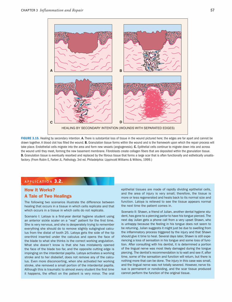
FIGURE 3.15. Healing by secondary intention. A. There is substantial loss of tissue in the wound pictured here; the edges are far apart and cannot be drawn together. A blood clot has filled the wound. B. Granulation tissue forms within the wound and is the framework upon which the repair process will take place. Endothelial cells migrate into the area and form new vessels (angiogenesis). C. Epithelial cells continue to migrate down into and across the wound until they meet, forming the new basement membrane. Fibroblasts create collagen fibers that are deposited within the granulation tissue. D. Granulation tissue is eventually resorbed and replaced by the fibrous tissue that forms a large scar that is often functionally and esthetically unsatis-factory. (From Rubin E, Farber JL. Pathology. 3rd ed. Philadelphia: Lippincott Williams & Wilkins, 1999.)
How it Works?A Tale of Two HealingsThe following two scenarios illustrate the difference between healing that occurs in a tissue in which cells replicate and that which occurs in a tissue in which cells do not replicate.
Scenario I: Latoya is a first-year dental hygiene student using an anterior sickle scaler on a “real” patient for the first time. She is very nervous, and she is desperately trying to remember everything she should do to remove slightly subgingival calcu-lus from the distal of tooth 25. Latoya gets the side of the tip one-third inserted under the calculus and opens the face of the blade to what she thinks is the correct working angulation. What she doesn’t know is that she has mistakenly opened the face of the blade too far, and the opposite cutting edge is impinging on the interdental papilla. Latoya activates a working stroke and to her disbelief, does not remove any of the calcu-lus. Even more disconcerting, when she activated her working stroke, she removed a small portion of the interdental papilla. Although this is traumatic to almost every student the first time it happens, the effect on the patient is very minor. The oral
epithelial tissues are made of rapidly dividing epithelial cells, and the area of injury is very small; therefore, the tissue is more or less regenerated and heals back to its normal size and function. Latoya is relieved to see the tissue appears normal the next time the patient comes in.
Scenario II: Shawn, a friend of Julian, another dental hygiene stu-dent, has gone to a piercing parlor to have his tongue pierced. The next day Julian gets a phone call from a very upset Shawn, who is unhappy because the feeling in his tongue does not seem to be returning. Julian suggests it might just be due to swelling from the inflammatory process triggered by the injury and that Shawn should give it time to heal. Several days later, Shawn is still expe-riencing a loss of sensation in his tongue and some loss of func-tion. After consulting with his dentist, it is determined a portion of the lingual nerve was most likely damaged during the tongue piercing. The dentist’s recommendation is to wait and see if, after time, some of the sensation and function will return, but there is nothing more that can be done. The injury in this case was small, and the lingual nerve was not totally severed. However, nerve tis-sue is permanent or nondividing, and the scar tissue produced cannot perform the function of the original tissue.
a p p l i C a t i O n 3.2.
0003305250.INDD 57 11/1/2017 5:17:10 PM
58 part i General Pathology
important of these are medications that interfere with the inflammatory process or with the immune system, such as corticosteroid, nonsteroidal anti-inflammatory, and other immunosuppressive drugs.
Complications of Wound healingThe most common complications of healing are inadequate scar formation, excessive scar formation, and excessive contracture of the scar tissue. Inadequate scar formation usually occurs as the result of the production of an inad-equate amount of granulation tissue. The most serious result of this is the bursting of a wound after surgery; if a wound bursts open after abdominal surgery, there is a 30% chance of mortality (Rubin & Reisner, 2014). Excessive scar formation results in a hypertrophic scar, or keloid (Fig. 3.16). These are usually unsightly and tend to recur after removal. The last complication is excessive contracture of the edges of the wound causing extensive deformity, espe-cially if the area is near a joint where limitations in mobility can cause a significant problem. Severe burns often result in wounds that exhibit excessive contracture (Fig. 3.17).
tooth extraction: Bone and soft tissue repairThe repair of an extraction site involves most of the same processes discussed above, with slight variation. The acute inflammatory process is triggered by the injury, and PMNs rush into the area. A blood clot fills the extraction site. As stated above, the inflammatory process and the process of repair are occurring at the same time. As the acute inflam-matory process continues, the epithelial cells at the edges of the wound begin the process of epithelialization and completely cover the extraction site in about 10 to 12 days. Figure 3.18 shows an extraction site that is almost fully healed. In the meantime, under the surface, osteoblasts from the bone marrow migrate into the clot and begin to produce new bone. The entire area of the extraction under-goes remodeling. The alveolar process is resorbed because it is no longer needed to maintain a tooth and is replaced with spongy bone. The bone is completely filled in by about 12 weeks postextraction. The same conditions that affect soft tissue wound healing, as stated above, will affect bone repair.
One of the more common problems associated with bone repair in the oral cavity is a dry socket also known as alveolar osteitis. This complication is specific to the healing of extraction sites and occurs most often with third molar extractions. Alveolar osteitis occurs when the initial blood clot within the socket is disrupted or lost and the bone surface becomes exposed to the intraoral environ-ment, increasing the risk of infection and causing severe pain. In addition, the exposed bone becomes necrotic and may produce a foul odor. The patient becomes aware of the condition within 2 to 4 days after the extraction. In cases of alveolar osteitis, the necrotic bone must be resorbed by the body, and granulation tissue must form along the walls of the socket instead of within a clot. This process is much slower, often taking several weeks longer than normal. The risk factors associated with alveolar osteitis include exces-sive trauma to the bone during extraction, smoking after extraction, oral contraceptive use, previous extraction site
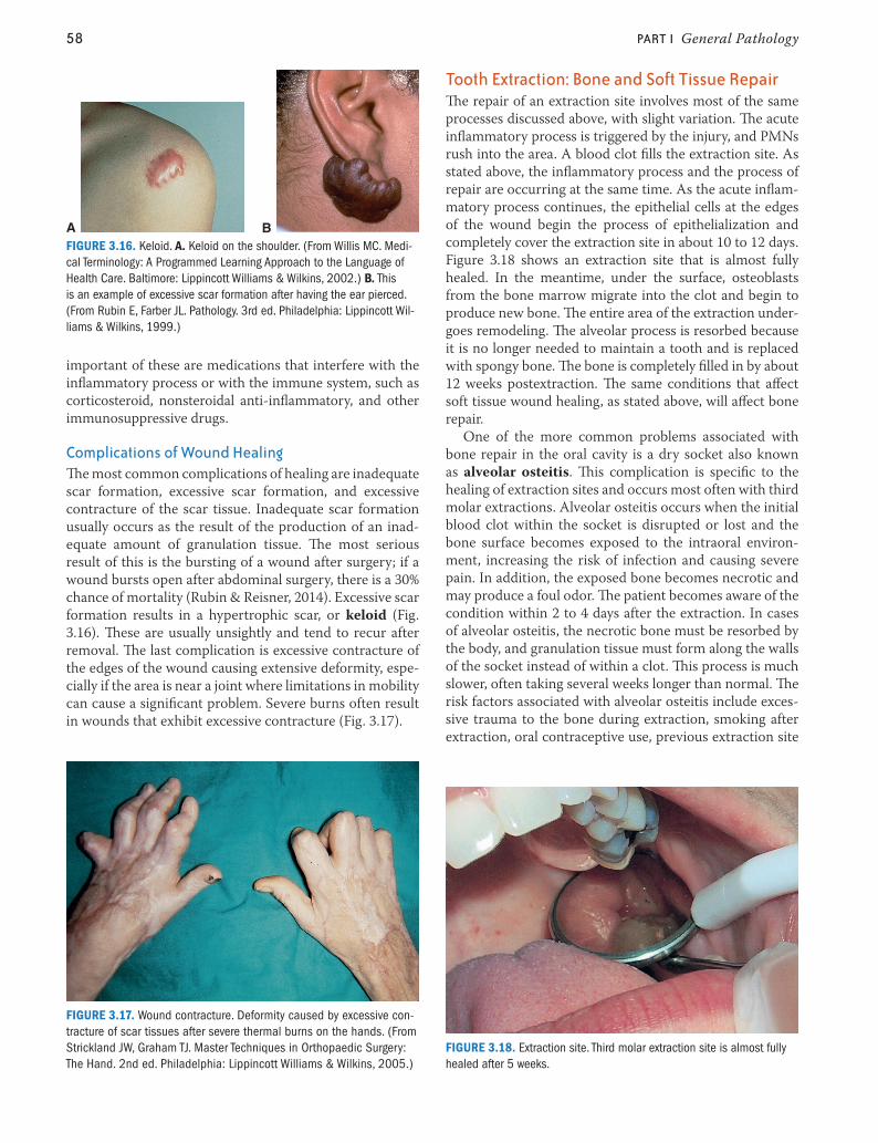
FIGURE 3.16. Keloid. A. Keloid on the shoulder. (From Willis MC. Medi-cal Terminology: A Programmed Learning Approach to the Language of Health Care. Baltimore: Lippincott Williams & Wilkins, 2002.) B. This is an example of excessive scar formation after having the ear pierced. (From Rubin E, Farber JL. Pathology. 3rd ed. Philadelphia: Lippincott Wil-liams & Wilkins, 1999.)
A B
FIGURE 3.17. Wound contracture. Deformity caused by excessive con-tracture of scar tissues after severe thermal burns on the hands. (From Strickland JW, Graham TJ. Master Techniques in Orthopaedic Surgery: The Hand. 2nd ed. Philadelphia: Lippincott Williams & Wilkins, 2005.)
FIGURE 3.18. Extraction site. Third molar extraction site is almost fully healed after 5 weeks.
0003305250.INDD 58 11/1/2017 5:17:12 PM
Chapter 3 Inflammation and Repair 59
infection, and noncompliance with postoperative instruc-tions (Akpata et al., 2013; Halabi et al., 2012; Kolokythas et al., 2010; Larsen, 1992). Most cases of alveolar osteitis can be prevented by limiting the amount of trauma during the extraction and by the patient complying with postop-erative instructions focused on maintaining the integrity of the blood clot. For example, the patient should avoid rinsing for 24 hours after the procedure (see Clinical Protocol #14).
Alveolar osteitis is treated by gently packing strips of a special type of gauze into the socket. The gauze is treated with an antiseptic to reduce the risk of infection and a medication that can soothe the exposed nerves, such as clove oil. In addition, the physical presence of the gauze reduces the chance that food will become lodged in the area. The patient returns every day or two to replace the packing and to check on the healing process until completed.
Conditions and diseases that manifest as inflammatory processes are caused by hundreds of different stimuli, both endogenous and exogenous. Almost every disorder that can be imagined has inflammatory components within its clinical manifestations. The most common diseases and conditions and those that are important in the practice of dental hygiene are discussed in later chapters, where they are addressed within the context of sharing similar clinical manifestations.
Summary■■ The acute inflammatory process is carried out by the
body using both cellular and chemical mechanisms.■■ There are four phases in the acute inflammatory process:
initiation, amplification, destruction, and termination.■■ Initiation involves changes in the microcirculation
in which chemical mediators instruct the blood ves-sels to briefly constrict and then dilate and become more permeable. The vascular changes enable PMNs to emigrate through the endothelial walls into the connective tissue, where they follow the direction of more chemical mediators to the areas where they are needed. In addition, exudate is allowed to flow from the blood vessels into the surrounding area, creating edema.
■■ Amplification involves gathering all of the necessary cells into the area to phagocytize all of the microorgan-isms, foreign matter, or other debris that needs to be removed before healing can take place.
■■ Destruction and elimination of foreign objects and pathogens is accomplished in the destruction phase.
■■ Termination requires shutting down the inflammatory process through the action of other chemical mediators so healing can be completed.
■■ The complement, kinin, and coagulation systems are all involved in mediating the inflammatory process.
■■ During the process of acute inflammation, systemic manifestations such as the acute-phase response, leu-kocytosis or leukopenia, and lymphadenopathy may occur.
■■ CRP, an APP, is an indicator of inflammation. Increased levels of CRP in the blood are used to help diagnose infections and certain inflammatory diseases. It is also a strong predictor of cardiovascular disease risk.
■■ Chronic inflammation may result from unresolved acute inflammation, or it may occur without this stimulus.
■■ The chronic inflammatory process is controlled by dif-ferent cells and chemical mediators that attempt to neutralize whatever stimulus is causing the reaction. Continuation of chronic inflammation leads to signifi-cant tissue destruction and delayed or abnormal healing.
■■ Other outcomes of the inflammatory process include abscesses, fistula formation, and cellulitis.
■■ Chronic inflammation always stimulates an immune system response.
■■ Tissue regeneration or repair begins at almost the same time as the inflammatory process and continues in con-junction with it.
■■ Regeneration occurs when the tissue is repaired with the same type of tissue that was lost and the area regains full function. Anything less than this is consid-ered repair.
■■ Repair of surface wounds occurs by either primary or secondary intention and involves the production of granulation tissue within the wound. Lost tissue is replaced by newly formed connective tissue, and the blood supply is reestablished through the process of angiogenesis.
■■ Epithelialization replaces the skin covering the wound. The end-product is fibrous repair or scar formation.
■■ Many factors can affect how a wound heals, including the type, size, and location of the wound; the type of tis-sue involved; and the health status of the individual.
■■ Keloids, excessive wound contracture, and wound rup-ture are complications of the repair process.
■■ Healing of extraction sites involves bone repair and remodeling in addition to soft tissue healing.
■■ Alveolar osteitis, a common complication following extraction of third molars, can be prevented by reduc-ing the amount of trauma during the surgery, avoiding tobacco use, and following postoperative instruction.
0003305250.INDD 59 11/1/2017 5:17:12 PM
60 part i General Pathology
ChAPteR RevIew
Chapter 3 review Questions1. Which one of the following lists the first four events
in the acute inflammatory process in the correct sequence?A. Vasodilation, vasoconstriction, vascular stasis, increased
vascular permeabilityB. Vasoconstriction, vasodilation, increased vascular perme-
ability, vascular stasisC. Vasoconstriction, increased vascular permeability, vasodi-
lation, vascular stasisD. Vasodilation, increased vascular permeability, vasocon-
striction, vascular stasis
2. Which one of the following vascular events in the acute inflammatory process enables PMNs to transmi-grate into the injured tissues?A. StasisB. DilationC. ConstrictionD. Increased permeability
3. Which one of the following events in the acute inflam-matory process enables leukocytes to destroy and remove resistant organisms?A. AdhesionB. ChemotaxisC. OpsonizationD. Phagocytosis
For answers and additional review activities, log in to thePoint.lww.com
For answers and additional review activities, log in to thePoint.lww.com
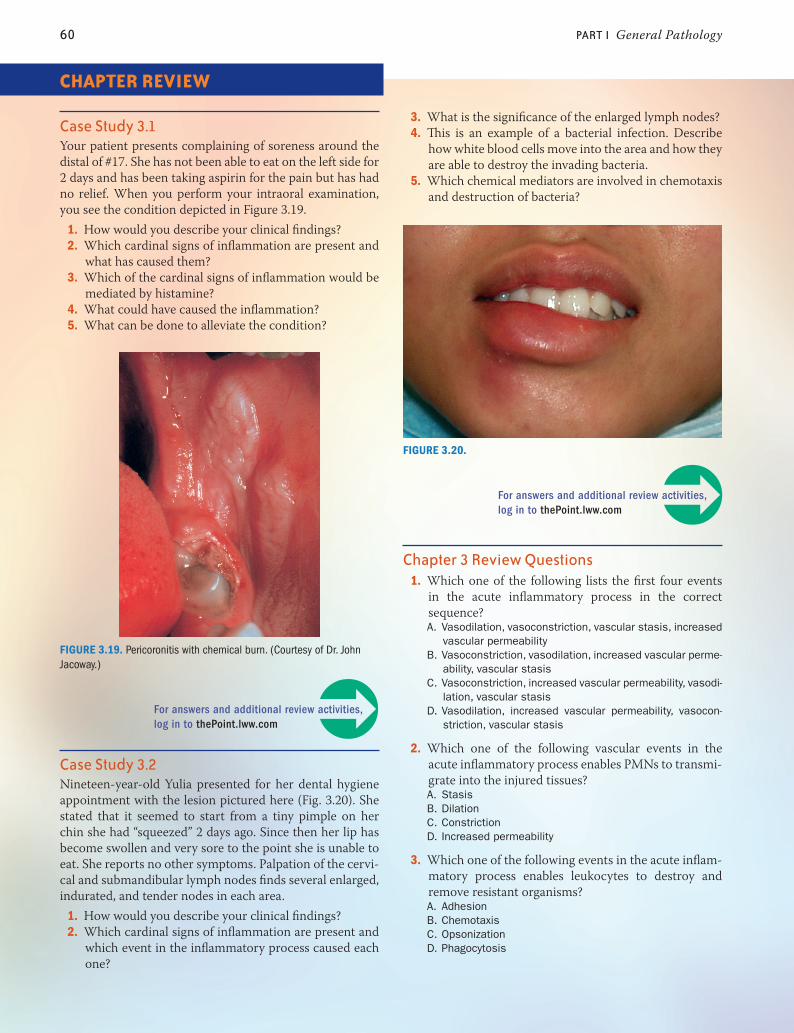
Case study 3.1Your patient presents complaining of soreness around the distal of #17. She has not been able to eat on the left side for 2 days and has been taking aspirin for the pain but has had no relief. When you perform your intraoral examination, you see the condition depicted in Figure 3.19.
1. How would you describe your clinical findings?2. Which cardinal signs of inflammation are present and
what has caused them?3. Which of the cardinal signs of inflammation would be
mediated by histamine?4. What could have caused the inflammation?5. What can be done to alleviate the condition?
Case study 3.2Nineteen-year-old Yulia presented for her dental hygiene appointment with the lesion pictured here (Fig. 3.20). She stated that it seemed to start from a tiny pimple on her chin she had “squeezed” 2 days ago. Since then her lip has become swollen and very sore to the point she is unable to eat. She reports no other symptoms. Palpation of the cervi-cal and submandibular lymph nodes finds several enlarged, indurated, and tender nodes in each area.
1. How would you describe your clinical findings?2. Which cardinal signs of inflammation are present and
which event in the inflammatory process caused each one?
FIGURE 3.19. Pericoronitis with chemical burn. (Courtesy of Dr. John Jacoway.)
FIGURE 3.20.
3. What is the significance of the enlarged lymph nodes?4. This is an example of a bacterial infection. Describe
how white blood cells move into the area and how they are able to destroy the invading bacteria.
5. Which chemical mediators are involved in chemotaxis and destruction of bacteria?
0003305250.INDD 60 11/1/2017 5:17:15 PM
Chapter 3 Inflammation and Repair 61
4. Which one of the following cells is NOT a leukocyte?A. Mast cellB. EosinophilC. MonocyteD. MacrophageE. Polymorphonuclear neutrophil
5. Histiocytes are monocytes that have become fixed in which one of the following tissues?A. LiverB. BrainC. LungD. Connective
6. Which one of the following chemical mediators is a more potent vasodilator than histamine?A. PAFB. TNFC. IL-1D. LPS
7. Which one of the following is a strong chemotactic agent?A. BradykininB. ProstaglandinC. Hageman factorD. Tumor necrosis factor
8. The membrane attack complex is formed by which one of the following systems?A. KininB. CoagulationC. Complement
9. All of the following are expected outcomes of acute inflammation EXCEPT one. Which one is the EXCEPTION?A. DeathB. RepairC. EdemaD. ResolutionE. Chronic inflammation
10. Identify the cells that play a major role in chronic inflammation?A. Macrophages, PMNs, and mast cellsB. Macrophages, lymphocytes, and plasma cellsC. Eosinophils, lymphocytes, leukocytes, and plasma cellsD. Leukocytes, mast cells, lymphocytes, monocytes, and
PMNs
11. Which one of the following is a systemic manifestation of inflammation?A. PyrexiaB. EdemaC. RednessD. Hyperemia
12. An increase in the number of white cells above normal levels in the blood is called:A. CellulitisB. SepticemiaC. HyperemiaD. LeukocytosisE. Lymphadenopathy
13. A granuloma forms as a result of which one of the following?A. RegenerationB. Acute inflammationC. Chronic inflammationD. Healing by primary intention
14. Ludwig angina is a form of which one of the following?A. FistulaB. CellulitisC. GranulomaD. AngiogenesisE. Fibrous repair
15. A cicatrix is the result of which one of the following?A. RegenerationB. Fibrous repair
16. Granulation tissue provides which of the following during the repair process?A. Blood clotB. FrameworkC. Epithelial cellsD. New blood vessels
17. Which one of the following terms denotes the process whereby a new basement membrane is formed under a scab?A. AngiogenesisB. Fibrous repairC. RegenerationD. EpithelializationE. Healing by secondary intention
18. Which one of the following will involve healing by sec-ondary intention?A. Paper cutB. Small surgical incisionC. Laceration closed with suturesD. Area of chronic inflammation
19. Which of the following will result in slower healing?A. Impaired circulationB. Highly vascularized tissueC. Nonmoving area of injuryD. Absence of irritating factors
20. Which of the following terms describes excessive scar tissue formation?A. KeloidB. GranulationC. HypotrophicD. ContractureE. Epithelialization
21. Alveolar osteitis is associated with all of the following EXCEPT one. Which one is the EXCEPTION?A. Exposed boneB. Increased painC. Increased risk of infectionD. Excessive granulation tissueE. Occurs 2 to 4 days after extraction
0003305250.INDD 61 11/1/2017 5:17:16 PM
62 part i General Pathology
22. All of the following are elements of the acute-phase response EXCEPT one. Which one is the EXCEPTION?A. PyrexiaB. MalaiseC. AnorexiaD. LeukocytosisE. Elevated CRP
23. An increased risk for cardiovascular disease is strongly associated with elevated levels of which one of the following?A. InterferonB. Interleukin-1C. C-reactive proteinD. LipopolysaccharideE. Lysosomal enzymes
24. Which one of the following terms describes the local response of the lymphatic system to acute inflammation?A. LeukocytosisB. LeukopeniaC. Ludwig anginaD. LymphadenopathyE. Generalized persistent lymphadenopathy
25. All of the following chemical mediators are essential for the healing process EXCEPT one. Which one is the EXCEPTION?A. CytokinesB. Platelet-activating factorC. Epidermal growth factorD. Platelet-derived growth factor
26. Which one of the following is the etiology of alveolar osteitis?A. Excessive bleedingB. Smoking after the extractionC. Loss or disruption of the blood clotD. Excessive trauma during the extractionE. Noncompliance with postoperative instructions
Critical thinking activities1. What would be the positive and negative aspects of not
having an inflammatory process?2. What do you think would happen if the inflammatory
process were to run out of control? How would you attempt to control it?
3. How can you use information you have learned about the inflammatory process to explain gingivitis and periodontitis to your patient?
media menuWeb sites
Journal of Periodontology: Volume 79, Number 8s, August 2008—Issue focuses on the inflammatory response; all articles in this issue can be accessed free of charge
http://www.joponline.org/toc/jop/79/8s
Centers for Disease Control and Prevention: Bensley L, Van Eenwyk J, Ossiander EM. Associations of self-reported periodontal disease with metabolic syndrome and number of self-reported chronic conditions. Prev Chronic Dis 2011;8(3):A50.
http://www.cdc.gov/pcd/ issues/2011/may/10_0087.htm
Journal of Inflammation: Available at https://journal-inflammation.biomedcentral.com/
The Pathology of Inflammation: Images available athttp://library.med.utah.edu/WebPath/INFLHTML/
INFLIDX.html
multimedia resourcesCrawling Neutrophil Chasing a Bacterium http://youtu.be/I_xh-bkiv_c
Histopathology Soft Tissue: Foreign body reaction (foreign body giant cells)
http://youtu.be/CRmwufRisfc
study toolsTake advantage of additional online resources to help you study and ace your exams!
■■ Answers to case studies in the book■■ Additional interactive case studies■■ Interactive quiz bank with answer feedback■■ Clinical protocol e-book■■ Condition summary tables■■ Expanded Media Menu with direct links to related Web
sites and multimedia resources■■ Supplemental references
Online resources and links are available at http://thepoint.lww.com/DeLong3e
ReferencesAkpata O, Omoregie OF, et al. Alveolar osteitis: patients’ compli-
ance to post-extraction instructions following extraction of molar teeth. Niger Med J 2013;54:335–338.
Aronson D, et al. C-reactive protein is inversely related to physical fit-ness in middle-aged subjects. Arthrosclerosis 2004;176(1):173–179.
Halabi D, Escobar J, et al. Logistic regression analysis of risk factors for the development of alveolar osteitis. J Oral Maxillofac Surg May 2012;(70):1040–1044.
Khan FA, Khan MF. Inflammation and acute phase response. Int J Appl Biol Pharm Technol 2010;1(2):312–321. Available at: www.ijabpt.com, accessed on September 2016.
Kolokythas A, Olech E, Miloro M. Alveolar osteitis: a comprehensive review of concepts and controversies. Int J Dent 2010;2010:249073. doi:10.1155/2010/249073.
Larsen PE. Alveolar osteitis after surgical removal of impacted man-dibular third molars. Identification of the patient at risk. Oral Surg Oral Med Oral Pathol 1992;73(4):393–397.
Rubin E, Reisner H. Essentials of Rubin’s pathology. 6th ed. Baltimore: Lippincott Williams & Wilkins, 2014:25–69.
62 part i General Pathology
0003305250.INDD 62 11/1/2017 5:17:16 PM
Related Documents