Hindawi Publishing Corporation Mediators of Inflammation Volume 2010, Article ID 535918, 17 pages doi:10.1155/2010/535918 Review Article Inflammation, a Link between Obesity and Cardiovascular Disease Zhaoxia Wang and Tomohiro Nakayama Division of Laboratory Medicine, Department of Pathology and Microbiology, Nihon University School of Medicine, 30-1 Ooyaguchi-kamimachi, Itabashi-ku, Tokyo 173-8610, Japan Correspondence should be addressed to Tomohiro Nakayama, [email protected] Received 30 November 2009; Revised 10 March 2010; Accepted 17 June 2010 Academic Editor: Gema Fr¨ uhbeck Copyright © 2010 Z. Wang and T. Nakayama. This is an open access article distributed under the Creative Commons Attribution License, which permits unrestricted use, distribution, and reproduction in any medium, provided the original work is properly cited. Obesity, the most common nutritional disorder in industrialized countries, is associated with an increased mortality and morbidity of cardiovascular disease (CVD). Obesity is primarily considered to be a disorder of energy balance, and it has recently been suggested that some forms of obesity are associated with chronic low-grade inflammation. The present paper focuses on the current status of our knowledge regarding chronic inflammation, a link between obesity and CVDs, including heart diseases, vascular disease and atherosclerosis. The paper discusses the methods of body fat evaluation in humans, the endocrinology and distribution of adipose tissue in the genders, the pathophysiology of obesity, the relationship among obesity, inflammation, and CVD, and the adipose tissue-derived cytokines known to affect inflammation. Due to space limitations, this paper focuses on C- reactive protein, serum amyloid A, leptin, adiponectin, resistin, visfatin, chemerin, omentin, vaspin, apelin, and retinol binding protein 4 as adipokines. 1. Introduction Obesity, the most common nutritional disorder in industri- alized countries, is associated with an increased mortality and morbidity of cardiovascular disease (CVD) [1]. The World Health Organization estimates that more than 1 billion adults worldwide are overweight, 300 million of whom are clinically obese—defined as having a body mass index (BMI) equal to or greater than 30 kg m −2 , or a waist circumference greater than 94 cm for men and 80 cm for women [2]. Obesity is a chronic, multifactorial, and complex disease resulting from a long-term positive energy balance, in which both genetic and environmental factors are involved [3, 4]. It was recently suggested that some forms of obesity are associated with chronic low-grade inflammation [5]. CVDs, including heart disease, vascular disease and atherosclerosis, are the most critical global health threat, contributing to more than one-third of the global mor- bidity. In most cases, these clinical conditions result from atherosclerosis, which was once identified as a lipid-storage disease. At the present time, CVD is recognized as a chronic inflammatory condition of the vessel wall that results from the transendothelial passage (transcytosis) of cholesterol- rich atherogenic Apo-B lipoproteins (VLDL, IDL and LDL) from the plasma into the intima. These lipoproteins are retained in the subendothelial space, which leads to infil- tration of macrophages and T cells that ultimately then interact with each other and with the cells of the arterial wall [6, 7]. It is likely that inflammation induced by obesity accelerates the atherosclerosis. Adipose tissue is recognized as an important player in obesity-mediated CVD. In 1994, adipose tissue was first identified as the source of the hormone leptin, opening the door for a new era of research that focused on adipocyte endocrinology [8]. It is now apparent that adipocytes have a more complex physiological role [9]. Adipocytes produce large numbers of hormones, peptides, and other molecules that affect cardiovascular function, not only in an endocrine manner, but also by autocrine and paracrine mechanisms [10]. This might lead to cytokine-mediated inflammatory, changes in the liver, systemic inflammation and atherosclerosis.

Welcome message from author
This document is posted to help you gain knowledge. Please leave a comment to let me know what you think about it! Share it to your friends and learn new things together.
Transcript
Hindawi Publishing CorporationMediators of InflammationVolume 2010, Article ID 535918, 17 pagesdoi:10.1155/2010/535918
Review Article
Inflammation, a Link between Obesity andCardiovascular Disease
Zhaoxia Wang and Tomohiro Nakayama
Division of Laboratory Medicine, Department of Pathology and Microbiology, Nihon University School of Medicine,30-1 Ooyaguchi-kamimachi, Itabashi-ku, Tokyo 173-8610, Japan
Correspondence should be addressed to Tomohiro Nakayama, [email protected]
Received 30 November 2009; Revised 10 March 2010; Accepted 17 June 2010
Academic Editor: Gema Fruhbeck
Copyright © 2010 Z. Wang and T. Nakayama. This is an open access article distributed under the Creative Commons AttributionLicense, which permits unrestricted use, distribution, and reproduction in any medium, provided the original work is properlycited.
Obesity, the most common nutritional disorder in industrialized countries, is associated with an increased mortality and morbidityof cardiovascular disease (CVD). Obesity is primarily considered to be a disorder of energy balance, and it has recently beensuggested that some forms of obesity are associated with chronic low-grade inflammation. The present paper focuses on thecurrent status of our knowledge regarding chronic inflammation, a link between obesity and CVDs, including heart diseases,vascular disease and atherosclerosis. The paper discusses the methods of body fat evaluation in humans, the endocrinology anddistribution of adipose tissue in the genders, the pathophysiology of obesity, the relationship among obesity, inflammation, andCVD, and the adipose tissue-derived cytokines known to affect inflammation. Due to space limitations, this paper focuses on C-reactive protein, serum amyloid A, leptin, adiponectin, resistin, visfatin, chemerin, omentin, vaspin, apelin, and retinol bindingprotein 4 as adipokines.
1. Introduction
Obesity, the most common nutritional disorder in industri-alized countries, is associated with an increased mortalityand morbidity of cardiovascular disease (CVD) [1]. TheWorld Health Organization estimates that more than 1billion adults worldwide are overweight, 300 million ofwhom are clinically obese—defined as having a body massindex (BMI) equal to or greater than 30 kg m−2, or awaist circumference greater than 94 cm for men and 80 cmfor women [2]. Obesity is a chronic, multifactorial, andcomplex disease resulting from a long-term positive energybalance, in which both genetic and environmental factors areinvolved [3, 4]. It was recently suggested that some forms ofobesity are associated with chronic low-grade inflammation[5].
CVDs, including heart disease, vascular disease andatherosclerosis, are the most critical global health threat,contributing to more than one-third of the global mor-bidity. In most cases, these clinical conditions result fromatherosclerosis, which was once identified as a lipid-storage
disease. At the present time, CVD is recognized as a chronicinflammatory condition of the vessel wall that results fromthe transendothelial passage (transcytosis) of cholesterol-rich atherogenic Apo-B lipoproteins (VLDL, IDL and LDL)from the plasma into the intima. These lipoproteins areretained in the subendothelial space, which leads to infil-tration of macrophages and T cells that ultimately theninteract with each other and with the cells of the arterialwall [6, 7]. It is likely that inflammation induced by obesityaccelerates the atherosclerosis. Adipose tissue is recognizedas an important player in obesity-mediated CVD. In 1994,adipose tissue was first identified as the source of thehormone leptin, opening the door for a new era of researchthat focused on adipocyte endocrinology [8]. It is nowapparent that adipocytes have a more complex physiologicalrole [9]. Adipocytes produce large numbers of hormones,peptides, and other molecules that affect cardiovascularfunction, not only in an endocrine manner, but also byautocrine and paracrine mechanisms [10]. This might leadto cytokine-mediated inflammatory, changes in the liver,systemic inflammation and atherosclerosis.
2 Mediators of Inflammation
This paper focuses on the inflammation related toobesity and CVD. It will discuss the methods of body fatevaluation in humans, the endocrinology and distributionof adipose tissue in the genders, the pathophysiology ofobesity, the relationship among obesity, inflammation andCVD, and the adipose tissue-derived cytokines known toaffect inflammation.
2. Methods of Evaluation of Body Fat in Humans
There are many methods that can be used to evaluate bodyfat in different populations [2, 11, 12]. While anthropometricmeasurements of weight-for-height have been traditionallyused to evaluate obesity, more recently, BMI has becomea standard parameter. BMI is defined as weight in kilo-grams divided by height in square meters. The normalrange is 19–24.9 kg/m2, with overweight defined as 25–29.9 kg/m2, and obesity as ≥30 kg/m2. BMI is not always areliable measurement of body composition in individuals,particularly in older and younger people. Unfortunately,BMI does not provide any insight into regional body fatdistribution. Thus, simple anthropometric measurements,such as waist circumference, can also be used to determinethe valid index of visceral fat accumulation, in addition tobeing able to serve as an indicator of health risks associatedwith visceral obesity. A waist circumference of greater than102 cm in men and 88 cm in women is a risk factor forCVD. A particularly important anthropometric parameterthat has been increasingly applied in recent years is sagittalabdominal diameter (SAD) [13]. Using a simple caliperthat was originally developed by Kahn, this anthropometricindicator can measure visceral fat tissue alone [14].
With regard to other techniques, one of the first thatshould be considered is the measuring of body density, as thisprovides information on the relationship between the bodymass and volume. With tetrapolar bioelectric impedanceanalysis, data is obtained by measuring the resistance ofthe body exposed to the impact of an alternating currentof 50 kHz at a strength of 800 μA. Radioisotopic techniquesuse deuterium or tritium as markers to measure the totalbody liquid and total body potassium. Infrared spectrometryis a simple but not particularly reliable method, based onthe application of two sources of monochromatic light.Ultrasonographic measuring of fat tissue is currently thefavored technique by which one can measure both thesubcutaneous and visceral fat tissues. Measurements arecarried out using a 7.5- and 3.5-mHz transducer for thesubcutaneous and visceral fat tissue, respectively. The mostaccurate method for measuring central obesity is throughthe use of magnetic resonance imaging or computer-assistedtomographic scanning. Unfortunately, these approaches aretoo expensive for routine use.
3. Endocrinology and Distribution ofAdipose Tissue between Genders
It is now apparent that adipose tissue is not simplya storage reservoir of fat, but is an active endocrine
Adipose tissue
Leptin
Adiponectin
FFA
IL-1IL-6TNF-αResistinProstaglandinsAngiotensinogenEndothelinPAI-1CRP
Adipocyte Macrophage
Figure 1: Cytokines secreted by adipocytes and/or macrophages inhuman adipose tissue.
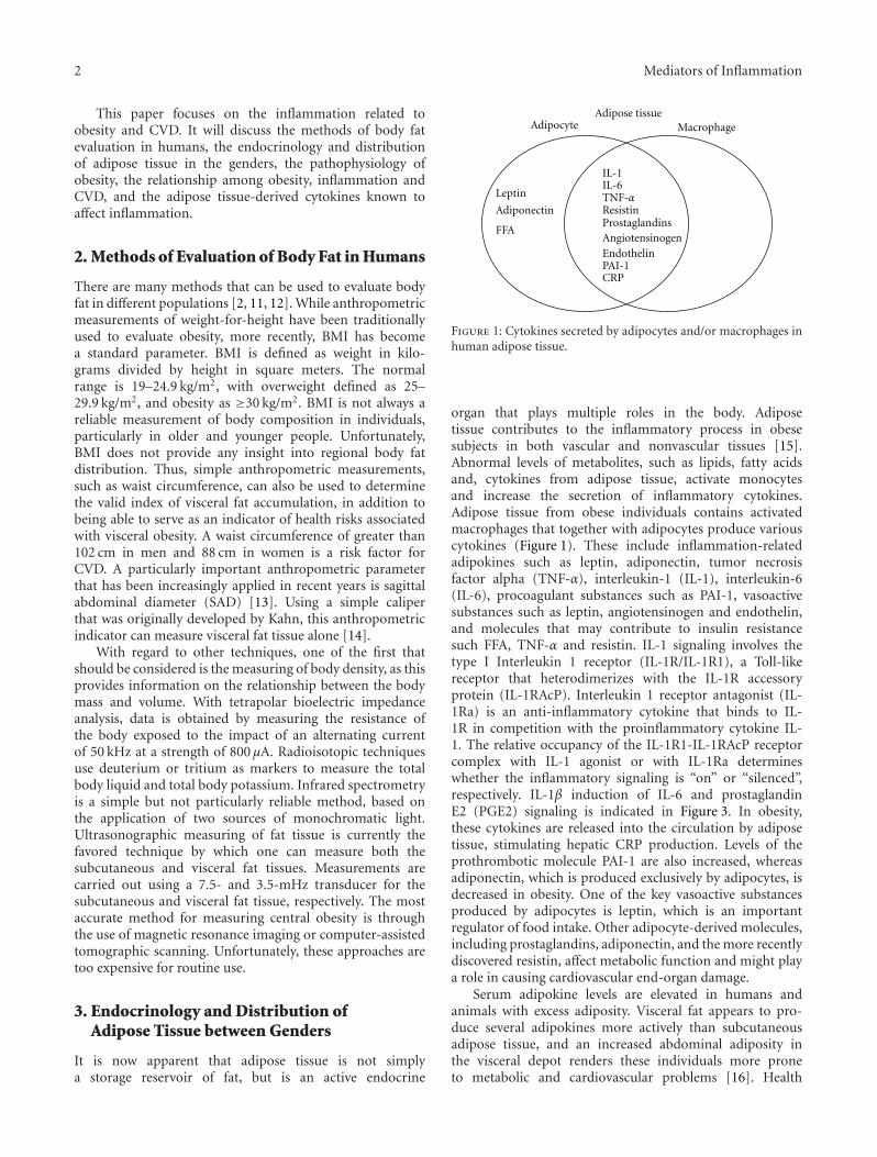
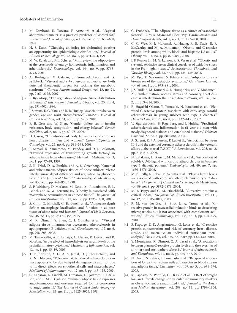
organ that plays multiple roles in the body. Adiposetissue contributes to the inflammatory process in obesesubjects in both vascular and nonvascular tissues [15].Abnormal levels of metabolites, such as lipids, fatty acidsand, cytokines from adipose tissue, activate monocytesand increase the secretion of inflammatory cytokines.Adipose tissue from obese individuals contains activatedmacrophages that together with adipocytes produce variouscytokines (Figure 1). These include inflammation-relatedadipokines such as leptin, adiponectin, tumor necrosisfactor alpha (TNF-α), interleukin-1 (IL-1), interleukin-6(IL-6), procoagulant substances such as PAI-1, vasoactivesubstances such as leptin, angiotensinogen and endothelin,and molecules that may contribute to insulin resistancesuch FFA, TNF-α and resistin. IL-1 signaling involves thetype I Interleukin 1 receptor (IL-1R/IL-1R1), a Toll-likereceptor that heterodimerizes with the IL-1R accessoryprotein (IL-1RAcP). Interleukin 1 receptor antagonist (IL-1Ra) is an anti-inflammatory cytokine that binds to IL-1R in competition with the proinflammatory cytokine IL-1. The relative occupancy of the IL-1R1-IL-1RAcP receptorcomplex with IL-1 agonist or with IL-1Ra determineswhether the inflammatory signaling is “on” or “silenced”,respectively. IL-1β induction of IL-6 and prostaglandinE2 (PGE2) signaling is indicated in Figure 3. In obesity,these cytokines are released into the circulation by adiposetissue, stimulating hepatic CRP production. Levels of theprothrombotic molecule PAI-1 are also increased, whereasadiponectin, which is produced exclusively by adipocytes, isdecreased in obesity. One of the key vasoactive substancesproduced by adipocytes is leptin, which is an importantregulator of food intake. Other adipocyte-derived molecules,including prostaglandins, adiponectin, and the more recentlydiscovered resistin, affect metabolic function and might playa role in causing cardiovascular end-organ damage.
Serum adipokine levels are elevated in humans andanimals with excess adiposity. Visceral fat appears to pro-duce several adipokines more actively than subcutaneousadipose tissue, and an increased abdominal adiposity inthe visceral depot renders these individuals more proneto metabolic and cardiovascular problems [16]. Health

Mediators of Inflammation 3
DiabetesHypertension
Smoking
Physical inactivity
Hypercholesterolemia
Artery wall:
Endothelial dysfunction
Obesity
Liver inflammation Systemic inflammationCRP
IL-6
IL-1TNF-α
IL-6IL-1TNF-α
(+)
Systemic oxidative stress
(+)
(+)
(+)(+)
(+)
Subclinical atherosclerosis
Atherosclerosis
Cardiovascular disease
(+) (+) (+)
(+)
(+)
(+)
(+)
Up-regulationof fibrinolysis
and coagulation
Productionof mediatorsand surface
protein
Changesof vasculartone
(+)(+)
(+)
Adipose tissue:Macrophage accumulation
? ?
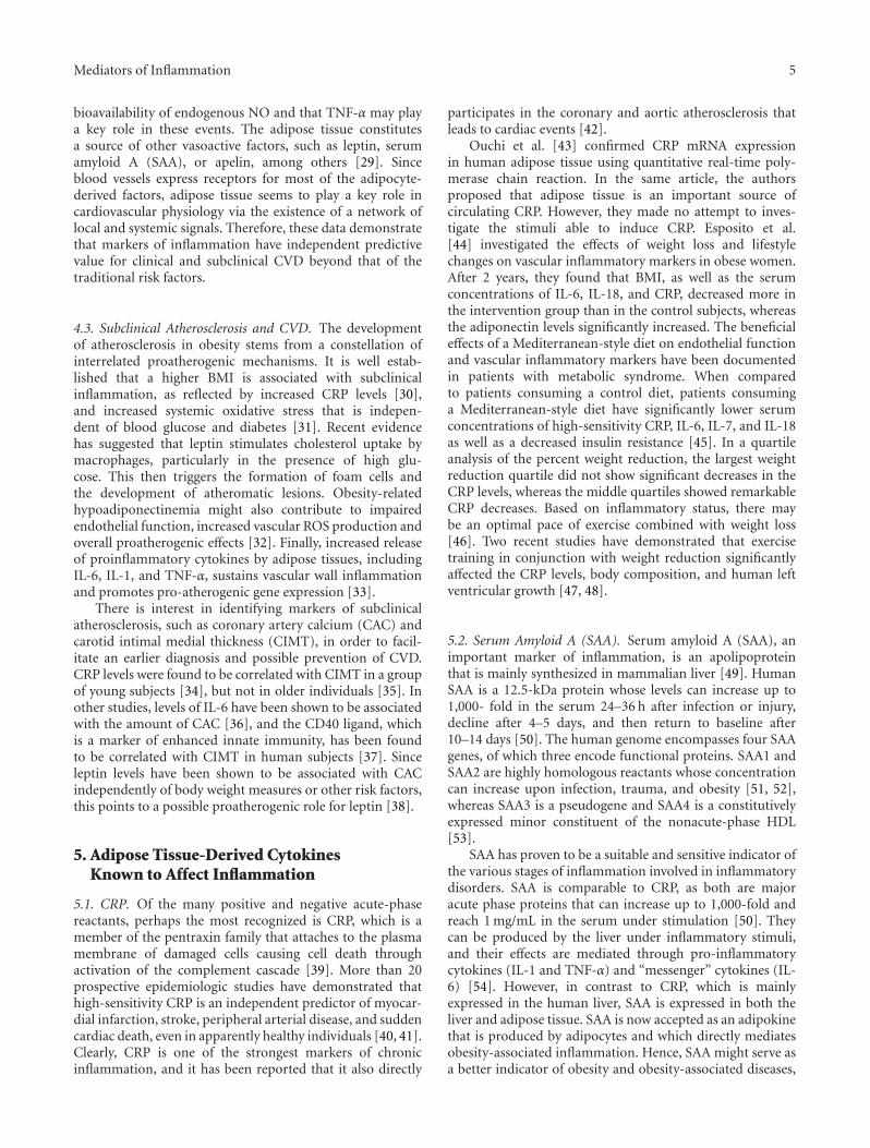
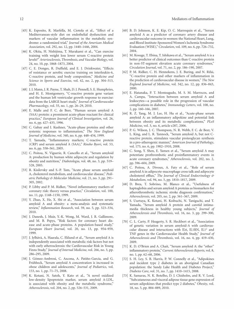
Figure 2: Mechanism of the relationship between inflammation induced by obesity and cardiovascular disease. Gray box shows thetraditional cardiovascular disease (CVD) risk factors. The arrow and plus symbols indicate the enhanced courses. Smoking, obesity,hypertension, diabetes, physical inactivity and hypercholesterolemia are established risk factors of CVD. In obese individuals, macrophagesfirst accumulate within the adipose tissue, leading to local inflammation. As the obesity increases, several proinflammatory factors, includingIL-1, IL-6 and TNF-α, are produced in the adipose tissue. Macrophage accumulation and the subsequent local inflammation are believedto result in numerous metabolic dysfunctions that typically accompany obesity, including systemic inflammation. Endothelial dysfunctionoccurs during the early stages of atherosclerosis and is responsible for the pathophysiological changes in subclinical atherosclerosis, whichinclude changes in a variety of mediators, surface proteins, and in autacoids that are involved in vasomotion, coagulation and inflammation.Obesity also can increase systemic oxidative stress independently of blood glucose and diabetes. One of the major events of atherosclerosis isCVD.
problems associated with obesity are generally related moreto the central (abdominal, visceral) distribution of fat ratherthan to the amount of fat, and the distribution of fatdiffers between males and females [17, 18]. Men exhibit amore central accumulation of fat, whereas women exhibit amore gluteal/femoral accumulation. The original definitionof obesity for males (android type) and females (gynoid type)dates to the first clinical observations made by Vague in 1947.The greatest health risk is associated with fat distribution inthe central or upper body (android) parts. Recent research[19, 20] has shown that sex hormones play an important rolein obesity and that there are differences in the occurrence ofinsulin resistance and heart diseases that are dependent upongender. Findings have indicated that both the total amount offat that an individual carries and the distribution of that fatare important. At present, it is difficult to accurately measurefat in the body, and there is currently no simple methodavailable for routine clinical use.
Epidemiological and clinical evidence strongly suggestsa major role for sex steroid hormones in the regulation ofadipose tissue distribution. Sex steroid hormones, such asestrogen, progesterone, and androgen, are involved in the
metabolism, accumulation, and distribution of adipose tis-sues. Normal distribution of body fat occurs when sex steroidhormones are present. If a decrease in sex steroid hormonesoccurs, such as that seen during aging or gonadectomy,there is a greater tendency for obesity states, in addition toincreases in major risk factors for CVD.
4. Mechanisms of the Relationship amongObesity, Inflammation, and CVD
4.1. Systemic Inflammation. As individuals become obeseand their adipocytes enlarge, the adipose tissue undergoesmolecular and cellular alterations that subsequently affectsystemic metabolism (Figure 2). First, macrophages accumu-late within adipose tissue, leading to local inflammation. Sev-eral proinflammatory factors are produced in adipose tissueas obesity increases. When compared to lean individuals,adipose tissue in obese individuals shows higher expressionof proinflammatory proteins, including TNF-α and IL-6 [21, 22]. Macrophage numbers in adipose tissue alsoincrease with obesity [23], apparently acting as scavengers

4 Mediators of Inflammation
IL-1β
IL-1R1 IL-1RAcP
IL-6 IL-1Ra
NF-κB
MyD88
iNOSCOX-2
AA PGH2
PGE2
PGES
EP4EP2EP1 EP3
Regulation of CRPIncrease of fibrinogenEndothelial dysfunctionCNS-food intake and energy balance
Regulation of leptinCNS-food intake and energy balanceModulation of sleeping
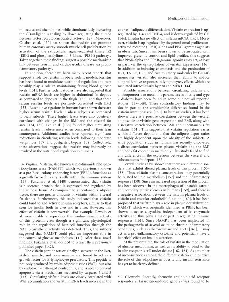
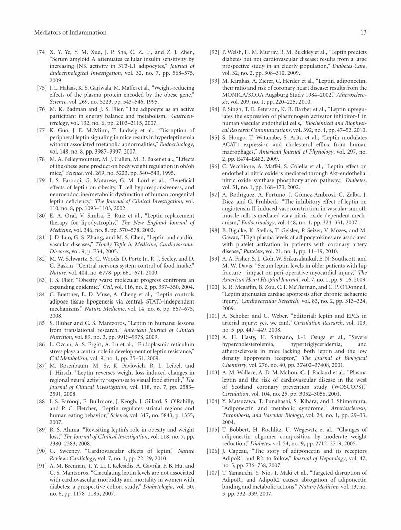
Figure 3: Interleukin-1β (IL-1β) induction of interleukin-6 (IL-6) and prostaglandin E2 (PGE2) signaling. IL-1β binds to the IL-1R1/IL-1R1AcP heterodimer, which then initiates the signaling cascade that causes the translocation of the transcription factor nuclear factor-κB(NF-κB) into the nucleus, where it induces the transcription of pro- and anti-inflammatory genes including inducible nitric oxide synthetase(iNOS), IL-6, IL-1Ra and cyclooxygenase-2 (COX-2). COX-2 catalyses the conversion of arachidonic acid (AA) to prostaglandin H2 (PGH2).PGH2 is converted into PGE2 by terminal PGE synthase (PGES). PGE2 signals occur via four different G-protein coupled receptors, EP1R-EP4R, each of which has multiple splice variants with different signaling properties.
of apoptotic adipocytes. It also has been reported that thereis a marked increase in these scavengers in obese subjects[24]. Macrophage accumulation and the subsequent localinflammation are believed to result in numerous metabolicdysfunctions that accompany obesity, including systemicinflammation and atherosclerosis.
Visceral fat secretes more cytokines than subcutaneousadipose tissue [16]. A recent study elegantly demonstratedthat transplantation of visceral adipose tissue from gene-tically obese mice into Apoe-deficient mice increased athero-sclerosis in the recipient animals, suggesting that inflamedadipose tissue exerts distinct vascular effects, presumablythrough inflammatory cells such as macrophages within thevisceral adipose tissue [25]. Macrophages within visceraladipose tissue are known to express and release cytokines.These cytokines reach the liver though the portal circulation,where they can stimulate hepatic inflammation [26], therebyinducing a chronic systemic inflammatory response.
4.2. Endothelial Dysfunction. Clinical and experimental datasupport a link between systemic inflammation and endothe-lial dysfunction. Mounting evidence shows that disturbedendothelial function may be an early marker of an ongoingatherosclerotic process. Thus, endothelial dysfunction hasincreasingly been recognized to play an important role in anumber of conditions associated with a high prevalence ofatherosclerotic CVD. Inflammatory cytokines are importantprotagonists in the formation of atherosclerotic plaques,
eliciting effects throughout the atherosclerotic vessel. Impor-tantly, the development of atherosclerotic lesions, regardlessof risk factors (e.g., diabetes, hypertension, obesity), ischaracterized by the disruption of the normal function ofendothelial cells.
The reasons for coronary endothelial dysfunction arecomplex and may involve ischemia/reperfusion injury.Smoking, obesity, hypertension, diabetes, physical inactivity,and hypercholesterolemia are established atherogenic riskfactors. Endothelial dysfunction is regarded as an earlystage of atherosclerosis, which is a chronic inflammatorydisease [27]. Chronic inflammation is a major contributingfactor to atherosclerosis and various markers of inflam-mation, fibrinolysis, and coagulation are upregulated inpatients with established atherosclerotic disease. For vascularhomeostasis, endothelial cells are of the utmost importanceand they produce a variety of mediators, surface proteins,and autacoids involved in vasomotion, coagulation, andinflammation. Adipose tissue expresses enzymes involved inthe angiotensin system (RAS) (renin, angiotensin-convertingenzyme (ACE)), as well as the nonrenin-angiotensin system(NRAS) (cathepsin D, cathepsin G, tonin, chymase) [28].The identification of elevated CRP as a transient independentrisk factor for endothelial dysfunction might provide animportant clue for linking a systemic marker of inflam-mation to the progression of atherosclerotic disease. Thus,CRP has been proposed for risk assessment of CVD inthe at-risk general population. Available evidence suggeststhat low-grade inflammation is accompanied by decreased
Mediators of Inflammation 5
bioavailability of endogenous NO and that TNF-α may playa key role in these events. The adipose tissue constitutesa source of other vasoactive factors, such as leptin, serumamyloid A (SAA), or apelin, among others [29]. Sinceblood vessels express receptors for most of the adipocyte-derived factors, adipose tissue seems to play a key role incardiovascular physiology via the existence of a network oflocal and systemic signals. Therefore, these data demonstratethat markers of inflammation have independent predictivevalue for clinical and subclinical CVD beyond that of thetraditional risk factors.
4.3. Subclinical Atherosclerosis and CVD. The developmentof atherosclerosis in obesity stems from a constellation ofinterrelated proatherogenic mechanisms. It is well estab-lished that a higher BMI is associated with subclinicalinflammation, as reflected by increased CRP levels [30],and increased systemic oxidative stress that is indepen-dent of blood glucose and diabetes [31]. Recent evidencehas suggested that leptin stimulates cholesterol uptake bymacrophages, particularly in the presence of high glu-cose. This then triggers the formation of foam cells andthe development of atheromatic lesions. Obesity-relatedhypoadiponectinemia might also contribute to impairedendothelial function, increased vascular ROS production andoverall proatherogenic effects [32]. Finally, increased releaseof proinflammatory cytokines by adipose tissues, includingIL-6, IL-1, and TNF-α, sustains vascular wall inflammationand promotes pro-atherogenic gene expression [33].
There is interest in identifying markers of subclinicalatherosclerosis, such as coronary artery calcium (CAC) andcarotid intimal medial thickness (CIMT), in order to facil-itate an earlier diagnosis and possible prevention of CVD.CRP levels were found to be correlated with CIMT in a groupof young subjects [34], but not in older individuals [35]. Inother studies, levels of IL-6 have been shown to be associatedwith the amount of CAC [36], and the CD40 ligand, whichis a marker of enhanced innate immunity, has been foundto be correlated with CIMT in human subjects [37]. Sinceleptin levels have been shown to be associated with CACindependently of body weight measures or other risk factors,this points to a possible proatherogenic role for leptin [38].
5. Adipose Tissue-Derived CytokinesKnown to Affect Inflammation
5.1. CRP. Of the many positive and negative acute-phasereactants, perhaps the most recognized is CRP, which is amember of the pentraxin family that attaches to the plasmamembrane of damaged cells causing cell death throughactivation of the complement cascade [39]. More than 20prospective epidemiologic studies have demonstrated thathigh-sensitivity CRP is an independent predictor of myocar-dial infarction, stroke, peripheral arterial disease, and suddencardiac death, even in apparently healthy individuals [40, 41].Clearly, CRP is one of the strongest markers of chronicinflammation, and it has been reported that it also directly
participates in the coronary and aortic atherosclerosis thatleads to cardiac events [42].
Ouchi et al. [43] confirmed CRP mRNA expressionin human adipose tissue using quantitative real-time poly-merase chain reaction. In the same article, the authorsproposed that adipose tissue is an important source ofcirculating CRP. However, they made no attempt to inves-tigate the stimuli able to induce CRP. Esposito et al.[44] investigated the effects of weight loss and lifestylechanges on vascular inflammatory markers in obese women.After 2 years, they found that BMI, as well as the serumconcentrations of IL-6, IL-18, and CRP, decreased more inthe intervention group than in the control subjects, whereasthe adiponectin levels significantly increased. The beneficialeffects of a Mediterranean-style diet on endothelial functionand vascular inflammatory markers have been documentedin patients with metabolic syndrome. When comparedto patients consuming a control diet, patients consuminga Mediterranean-style diet have significantly lower serumconcentrations of high-sensitivity CRP, IL-6, IL-7, and IL-18as well as a decreased insulin resistance [45]. In a quartileanalysis of the percent weight reduction, the largest weightreduction quartile did not show significant decreases in theCRP levels, whereas the middle quartiles showed remarkableCRP decreases. Based on inflammatory status, there maybe an optimal pace of exercise combined with weight loss[46]. Two recent studies have demonstrated that exercisetraining in conjunction with weight reduction significantlyaffected the CRP levels, body composition, and human leftventricular growth [47, 48].
5.2. Serum Amyloid A (SAA). Serum amyloid A (SAA), animportant marker of inflammation, is an apolipoproteinthat is mainly synthesized in mammalian liver [49]. HumanSAA is a 12.5-kDa protein whose levels can increase up to1,000- fold in the serum 24–36 h after infection or injury,decline after 4–5 days, and then return to baseline after10–14 days [50]. The human genome encompasses four SAAgenes, of which three encode functional proteins. SAA1 andSAA2 are highly homologous reactants whose concentrationcan increase upon infection, trauma, and obesity [51, 52],whereas SAA3 is a pseudogene and SAA4 is a constitutivelyexpressed minor constituent of the nonacute-phase HDL[53].
SAA has proven to be a suitable and sensitive indicator ofthe various stages of inflammation involved in inflammatorydisorders. SAA is comparable to CRP, as both are majoracute phase proteins that can increase up to 1,000-fold andreach 1 mg/mL in the serum under stimulation [50]. Theycan be produced by the liver under inflammatory stimuli,and their effects are mediated through pro-inflammatorycytokines (IL-1 and TNF-α) and “messenger” cytokines (IL-6) [54]. However, in contrast to CRP, which is mainlyexpressed in the human liver, SAA is expressed in both theliver and adipose tissue. SAA is now accepted as an adipokinethat is produced by adipocytes and which directly mediatesobesity-associated inflammation. Hence, SAA might serve asa better indicator of obesity and obesity-associated diseases,

6 Mediators of Inflammation
especially when vascular diseases and metabolic disorders arepresent.
SAA is known to be a marker for obesity, as its expressionis well correlated with obesity [55]. Some studies haveshown that SAA levels are positively associated with BMIlevels and that weight loss led to decreased SAA levels. In1999, Danesh et al. [56] first reported that concentrationsof SAA protein were strongly correlated with obesity. Sincethen, more than ten studies have shown that SAA isstrongly associated with obesity [57–59]. In addition, ithas been shown that SAA gene expression is increased inthe adipose tissue of obese subjects and is significantlycorrelated with adipocyte size and inflammatory biomarkers[52].
Recent studies have shown that SAA elevation canpredict cardiovascular events analogously with or even betterthan CRP by itself [60–62] and in this sense, it has beenspeculated that SAA might be one of the links or even aproatherogenic risk factor between inflammation and CVD[63, 64]. SAA is able to both alter vascular proteoglycans ina proatherogenic manner [65] and stimulate the productionof various inflammatory mediators in cultured vascularendothelial cells, neutrophils, and monocytes [66]. Endothe-lial cells, smooth muscle cells, monocytes, and macrophagesin atherosclerotic lesions have been reported to accountfor the extrahepatic production of SAA, as the presence ofboth SAA mRNA and protein products have been detectedin these cell types [67]. SAA has also been accepted asbeing a biomarker of cerebrovascular disease and carotidartery intima-media thickness, which is an early indexof atherosclerosis [68–71]. However, a very recent studyindicated that SAA does not mediate early atherosclerosis[57].
SAA has also been found to be associated with metabolicdisorders, such as diabetes, insulin resistance, and metabolicsyndrome [72, 73]. Additionally, genes critical for insulinsensitivity were also found to be downregulated in adipocytestreated with recombinant SAA [74].
5.3. Leptin. Leptin, which was the first adipocyte hormoneidentified, influences food intake through direct effects onthe hypothalamus [75]. The adipocyte-derived hormoneleptin has actions in the brain (e.g., hypothalamus, cortexand limbic areas) and in a number of peripheral tissues aswell (e.g., cells of the pancreas, liver and immune system).However, the central action of leptin in the brain, and inparticular the hypothalamus, has been best characterizedwith regards to energy homeostasis and its importancefor reproductive functions [76]. Moreover, disruption ofperipheral leptin signaling in mice has been shown to causeno significant changes in either the energy balance or glucosehomeostasis [77].
Mice lacking the gene coding for leptin (named ob/obmice) are obese and diabetic. When ob/ob mice are treatedwith regular injections of leptin, they show reduced foodintake, increased metabolic rate, and weight loss [78].These effects appear to be mediated mainly by the centralnervous system, as intracerebroventricular injection of leptin
produces significant effects at much lower doses than thoserequired by systemic injection. Systemic injections of leptinhave a beneficial effect in children with congenital leptindeficiencies [79]. In a pioneering study, administration ofexogenous leptin to individuals with lipoatrophic diabetesresulted in marked reductions in triacylglycerol concentra-tions, liver volume, and glycated hemoglobin. Ultimately,this treatment resulted in the discontinuation of or largereductions in the patient’s antidiabetic therapy [80]. Unfor-tunately, leptin concentrations are already high in mostobese individuals because of the increased amount of leptin-secreting adipose tissue [81]. In these individuals, increasingthe leptin concentrations only induces the target cells tobecome resistant to actions of the hormone. Therefore,further studies need to be undertaken to clarify potentialtherapeutic strategies using leptin in these types of patients.
Leptin is involved in the control of not only energy home-ostasis but also immunity. During fasting/starvation, whenplasma leptin levels decline, neural pathways in the hypotha-lamus cause the appetite to increase and energy expenditureto decrease as the body attempts to restore its fat stores[82]. In addition, the fall in plasma leptin diminishes thyroidhormone production and inhibits the reproductive axis, bothof which help to save energy during nutritionally lean times[83]. These metabolic effects of leptin are in part centrallymediated by activation of the hypothalamic-sympatheticnervous system axis [84]. In addition to the complete leptindeficiency disorder, relative leptin deficiency is an emergingclinical syndrome that is now being seen more often inseveral clinical conditions, including congential or acquiredlipodystrophy as well as exercise-induced energy deficiencyand hypothalamic amenorrhea or anorexia nervosa. Leptinreplacement therapy might prove to be a therapeutic optionfor patients with these disorders [85]. Very recently, admin-istration of chemical chaperones that decrease ER stress alsorestored leptin sensitivity in diet-induced obese mice [86].In obese subjects who have lost weight, modifications thatlead to decreased energy expenditure may predispose theindividual to regain the weight. However, when subjects areadministered “replacement” doses of leptin that restore theircirculating leptin concentrations to preweight-loss levels,the weight gain can be prevented [87]. This suggests thatthe weight-reduced state is a condition of relative leptindeficiency. Recent reports have shown that in additionto its action on the hypothalamus, leptin may also acton the cortex and limbic areas, which are involved incognitive and emotional regulation of feeding behavior [88].Teleologically, the adaptations mediated by reduced leptinmay have evolved as a protection against the threat of star-vation by limiting energy use and enhancing energy storage[89].
The potential effects of leptin in the pathophysiology ofcardiovascular complications of obesity remain diverse [90].While some studies [91, 92] have indicated that circulatingleptin levels are not significantly related to the risk of CVDor mortality in a diabetic population, these studies did findthat leptin was associated with obesity and inflammatorymarkers. Even so, other reports have suggested that leptindoes contribute to atherosclerosis and CVD in obese subjects

Mediators of Inflammation 7
[93]. Therefore, this protein may elevate the blood pressureby stimulating the autonomic nervous system. Leptin hasbeen found to have multiple effects on the cells of the arterywall. In human vascular endothelial cells, leptin upregulatesthe expression of the plasminogen activator inhibitor-1[94], and leptin also helps modulate ACAT1 expressionand cholesterol efflux from human macrophages [95]. Inaddition, leptin has been reported to increase nitric oxide(NO) bioavailability in blood vessels via the activation ofendothelial NO synthase (eNOS) [96] and inducible NOsynthase (iNOS) [97] in the endothelial and smooth musclecells, respectively. Recent studies that measured coronaryartery disease have demonstrated that hyperleptinemiawas associated with coronary atherosclerosis [98, 99], withthe association determined to be independent of insulinresistance. Other studies have shown that leptin may have arole in neointimal formation in response to arterial injury[100, 101]. In fact, very obese, leptin-deficient mice havebeen found to be protected from atherosclerosis despite allof the metabolic risk factors, suggesting that this hormonemay directly contribute to the risk of vascular disease [102].Moreover, in a prospective study in humans in whichanthropometric and metabolic risk factors were controlled,the circulating leptin concentrations were shown to be anindependent risk factor for predicting cardiovascular events[103]. Therefore, when chronically elevated concentrationsof leptin are seen in obese individuals, this may indicatea predisposition to progression of atherosclerosis in theseindividuals.
5.4. Adiponectin. Adiponectin is a product of adipocytes, andits levels in humans decrease in obese subjects [104]. As oneof the most extensively studied adipokines, adiponectin has 3different oligomers, each of which may have a different bio-logical function [105]. The major receptors for adiponectinare AdipoR1 and AdipoR2. These belong to a new family ofreceptors that are predicted to contain seven transmembranedomains but which will be structurally and functionallydistinct from the G-protein coupled receptors. A recent studyhas shown that AdipoR2 stimulates energy dissipation byincreasing fatty acid oxidation while inhibiting oxidativestress and inflammation [106, 107]. Adipocytes secrete highlevels of adiponectin that then exert anti-inflammatoryeffects, most notably in atherosclerotic plaques [108]. Theseeffects occur due to the suppression of TNF-α and proinflam-matory cytokines such as IL-6 and interferon-c, along withthe induction of other anti-inflammatory factors such asthe IL-1 receptor antagonist [109]. In contrast, adiponectinlevels have been shown to be low in several different formsof insulin resistance. In vivo, adiponectin enhances energyconsumption and fatty acid oxidation in the liver and muscle,which reduces the tissue triglyceride content, thereby furtherenhancing the insulin sensitivity [110]. In adiponectin trans-genic mice, there is improvement of the lipid profile [111,112], and when plasma triglycerides are reduced, this leads toan increased VLDL catabolism in the skeletal muscle [113].Taken together with its metabolic and anti-inflammatoryeffects, it has been proposed that adiponectin not only
contributes to the beneficial effects of body weight loss butalso has a role in modulating the cardiovascular system.
As might be expected based on the above observa-tions, adiponectin promotes an antiatherogenic and anti-inflammatory program of gene expression and function inthe vessel wall. Adiponectin downregulates the expressionof adhesion molecules on the endothelial cells and directlyimproves endothelial dysfunction [114, 115]. Adiponectinalso reduces proliferation in a receptor-independent fashionin the vascular smooth muscle cells [116]. In a very recentstudy, it has been shown that adiponectin reduces lipidaccumulation, down-regulates the expression of scavengerreceptors in macrophages, and promotes macrophage polar-ization, all of which play a role in anti-inflammatoryactivities [117]. Other studies have also indicated thatadiponectin has an important role in cardiovascular protec-tion. Hypoadiponectinemia is found in patients with angio-graphically demonstrated coronary artery disease [118]. Inobese children, it has been reported that reduced adiponectinconcentrations are of more importance than conventionalcardiovascular risk factors, and that this inflammation statusis related to early atherosclerosis [119]. However, in a largeprospective study that was combined with a meta-analysisof previously published prospective studies, the adiponectinlevels at baseline were found to be rather weak predictorsof CVD risk [120]. However, other studies have shown thatadiponectin exerts beneficial effects at nearly all stages of theatherogenesis process [121], and that the adiponectin levelsare inversely correlated to the progression of the coronaryartery calcium in both diabetic and nondiabetic subjects[122]. Serum total and high-molecular weight adiponectinare also associated with biomarkers of inflammation, insulinresistance, and endothelial function, all of which are inde-pendent risk factors of CVD [123].
5.5. Resistin. Resistin, which is one of the most recentlyidentified adipokines, has been proposed to be an inflam-matory marker for atherosclerosis. While it has been shownto induce increases in CRP production by the macrophages[124], resistin is an example of the new adipokines thatappear to have contrasting roles when examined in mice versushumans. For example, confirmation of the results foundin mice has proven to be difficult in human populations.This may be due to the fact that resistin appears to bederived from different sources in humans as compared torodents. This protein was initially shown to be releasedin large amounts from mouse adipocytes, with obese micehaving elevated levels that were accompanied by insulinresistance [125]. However, investigations in humans suggestthat resistin is expressed in adipocytes with monocytes andmacrophages [126, 127]. This lack of homology betweenthe human and mouse resistin genes suggests a potentialdivergence in function [128]. Since macrophages are knowninflammatory modulators, resistin may be an inflammatorymarker in humans. Supporting this possible inflamma-tory role in humans are results that show recombinantresistin activates human endothelial cells, as measured by anincreased expression of endothelin-1 and various adhesion
8 Mediators of Inflammation
molecules and chemokines, while simultaneously increasingthe CD40-ligand signaling by down-regulating the tumornecrosis factor receptor-associated factor-3 [129]. Moreover,Calabro et al. [130] has shown that resistin can promotehuman coronary artery smooth muscle cell proliferation byactivation of the extracellular signal-regulated kinase 1/2(ERK) and phosphatidylinositol 3-kinase (PI3 K) pathways.Taken together, these findings suggest a possible mechanisticlink between resistin and cardiovascular disease via proin-flammatory pathways.
In addition, there have been many recent reports thatsupport a role for resistin in obese rodent models. Resistinhas been found to modulate nutritional regulation and maypossibly play a role in maintaining fasting blood glucoselevels [131]. Further rodent studies have also suggested thatresistin mRNA levels are higher in abdominal fat depots,as compared to deposits in the thigh [132], and that theseserum resistin levels are positively correlated with BMI[133]. Recent investigations in humans have shown there arehigher serum resistin levels in obese subjects as comparedto lean subjects. These higher levels were also positivelycorrelated with changes in the BMI and the visceral fatarea [134, 135]. Lee et al. [136] found higher circulatingresistin levels in obese mice when compared to their leancounterparts. Additional studies have reported significantreductions in circulating resistin levels following moderateweight loss [137] and postgastric bypass [138]. Collectively,these observations suggest that resistin may indirectly beinvolved in the nutritional regulation in humans.
5.6. Visfatin. Visfatin, also known as nicotinamide phospho-ribosyltransferase (NAMPT), which was previously knownas a pre-B cell colony-enhancing factor (PBEF), functions asa growth factor for early B cells within the immune system[139]. Fukuhara et al. [140] demonstrated that visfatinis a secreted protein that is expressed and regulated bythe adipose tissue. As compared to subcutaneous adiposetissue, there are greater amounts of visfatin within visceralfat depots. Furthermore, this study indicated that visfatincould bind to and activate insulin receptors, similar to thatseen for insulin both in vivo and in vitro. However, thiseffect of visfatin is controversial. For example, Revollo etal. were unable to reproduce the insulin-mimetic activityof this protein, even though a significant physiologicalrole in the regulation of beta-cell function through theNAD biosynthetic activity was detected. Thus, the authorssuggested that NAMPT could play an important role inthe control of glucose metabolism [141]. After these novelfindings, Fukuhara et al. decided to retract their previouslypublished paper [142].
The visfatin peptide was originally discovered in the liver,skeletal muscle, and bone marrow and found to act as agrowth factor for B-lymphocyte precursors. This peptide isnot only produced by white adipose tissue (WAT), but alsoby endotoxin-challenged neutrophils, and is able to preventapoptosis via a mechanism mediated by caspases 3 and 8[143]. Circulating visfatin levels are closely correlated withWAT accumulation and visfatin mRNA levels increase in the
course of adipocyte differentiation. Visfatin expression is up-regulated by IL-6 and TNF-α, and is down-regulated by GH[144]. Insulin has no effect on visfatin mRNA [145]. More-over, visfatin is up-regulated by the peroxisomal proliferator-activated receptor (PPAR)-alpha and PPAR-gamma agonistsin obese rats. Since it has been shown to be associated withimproved glycemic control and lipid profiles, this suggeststhat PPAR-alpha and PPAR-gamma agonists may act, at leastin part, via the up-regulation of visfatin expression [146].In addition to inducing chemotaxis and the production ofIL-1, TNF-α, IL-6, and costimulatory molecules by CD14Cmonocytes, visfatin also increases their ability to inducealloproliferative responses in lymphocytes, effects which aremediated intracellularly by p38 and MEK1 [144].
Possible associations between circulating visfatin andanthropometric or metabolic parameters in obesity and type2 diabetes have been found in some but not all reportedstudies [147–149]. These contradictory findings may bedue in part to the considerable differences found in thevisfatin immunoassays [150]. In human studies, it has beenshown there is a positive correlation between the visceraladipose tissue visfatin gene expression and BMI, along witha negative correlation between BMI and subcutaneous fatvisfatin [151]. This suggests that visfatin regulation varieswithin different depots and that the adipose depot ratiosare highly dependent upon the obesity of the subjects. Awide population study in humans has recently discovereda direct correlation between plasma visfatin and the BMIand body fat content in males only. This study failed to findany differences in the expression between the visceral andsubcutaneous fat depots [152].
Several studies have shown that there are different disor-ders that exhibit altered plasma levels of this protein [153–156]. Thus, visfatin plasma concentrations may potentiallybe related to lipid metabolism [157] and the inflammatoryresponse [158]. Since an increased expression of this proteinhas been observed in the macrophages of unstable carotidand coronary atherosclerosis in humans [159], and there isa negative association between the visfatin plasma levels ofvisfatin and vascular endothelial function [160], it has beenproposed that visfatin plays a role in plaque destabilization.NAMPT, which was originally identified as PBEF, has beenshown to act as a cytokine independent of its enzymaticactivity, and thus plays a major part in regulating immuneresponses [161]. Since NAMPT has been implicated inthe pathogenesis of several acute or chronic inflammatoryconditions, such as atherosclerosis and CVD [161], it mayact as a pro-inflammatory cytokine and potentially have abeneficial effect on insulin secretion.
At the present time, the role of visfatin in the modulationof glucose metabolism, as well as its ability to bind to theinsulin receptor is still under debate [162–164]. As a numberof inconsistencies among the different visfatin studies exist,the role of this adipokine in obesity and insulin resistancehas yet to be clearly defined.
5.7. Chemerin. Recently, chemerin (retinoic acid receptorresponder 2, tazarotene-induced gene 2) was found to be

Mediators of Inflammation 9
highly expressed in adipose tissue and liver [165]. Chemerinis an agonist of the orphan G-protein coupled receptorchemokine-like receptor 1 (CMKLR1, ChemR23) [166] thatis expressed by cells of the innate immune system [167].Therefore, chemerin might be further evidence of a linkbetween obesity and inflammation. Chemerin is secreted asan inactive precursor, and then activated through proteolyticcleavage by serine proteases of the coagulation, fibrinolyticand inflammatory cascades. Chemerin appears to be a noveland promising adipokine, and in several recent studies,human chemerin plasma levels have been shown to havea significant association with the BMI, inflammation, andmetabolic syndrome [168–170].
Platelets have been found to be a rich cellular sourceof chemerin. In some pathological conditions, chemerin isactivated and then released, which leads to the elevationof blood chemerin levels [171]. Recent studies have shownthat both adipocytes [172] and fibroblast cells [173] canproduce chemerin. Chemerin has also been measured ina number of human inflammatory exudates, includingascitic fluids from human ovary cancer and liver cancer,as well as synovial fluids from arthritic patients [174].Angiotensin-converting enzyme (ACE) may be responsiblefor the activation of prochemerin. If so, as has been shownin vitro, this effect should be able to be blocked by an ACEinhibitor such as captopril [175]. However, further studieswill be necessary to clarify this potential mechanism invivo. There is also growing evidence that the bioactivity ofchemerin is closely regulated by proteolytic cleavage in the C-terminal region, which may control its maximal chemotacticor anti-inflammatory effects [176]. While the primary aminoacid sequences indicate that chemerin is structurally distinctfrom the CXC and CC chemokines, it functions exactlylike a chemokine and can induce leukocyte migration andintracellular calcium mobilization. Chemerin also exertspotent anti-inflammatory effects on activated macrophages,which express the chemerin receptor CMKLR1 (chemokine-like receptor-1) in a cysteine protease-dependent manner[177].
Chemerin is a newly described adipokine with effectson adipocyte differentiation and metabolism in vitro [165].In rodents, there is conflicting data with regard to theassociation of chemerin with obesity and diabetes. Whilethere is a decreased chemerin expression in the adipose tissueof db/db mice as compared with controls [178], chemerinexpression is significantly higher in the adipose tissue ofimpaired glucose tolerant and diabetic Psammomys obesusas compared with normal glucose-tolerant sand rats [179].It has also been demonstrated that chemerin or chemerinreceptor knockdown impairs the differentiation of 3T3-L1cells into adipocytes, reduces the expression of adipocytegenes involved in glucose and lipid homeostasis, includingadiponectin and leptin, and alters the metabolic functionsin mature adipocytes [170]. In humans, no significantdifferences were noted for the chemerin levels betweensubjects with type 2 diabetes and normal controls. However,in normal glucose-tolerant subjects, chemerin levels weresignificantly associated with BMI, triglycerides, and bloodpressure [179]. Plasma chemerin levels in normal subjects
are also significantly associated with BMI, circulating triglyc-erides, and blood pressure, suggesting a strong relationshipof this protein with obesity-associated complications [179].
It is possible that visceral fat may potentially contributeto the chronic inflammation that is observed in obeseindividuals. However, only a few studies have investigatedthe adipokine concentrations in the portal circulation [180].In order to be able to determine the physiological roleof chemerin in the glucose metabolism, and to identifychemerin’s target tissues as well as relevant signal transduc-tion pathways, further studies will need to be undertaken.
5.8. Omentin, Apelin, Vaspin, and Retinol-Binding Protein4 (RBP4). Omentin, which was originally referred to asintelectin and first found in the intestinal Paneth cells, has apredicted molecular weight of 33 kDa [181]. Omentin is a fatdepot-specific secretory protein synthesized by the visceralstromal vascular cells, but not the adipocytes. It has alsobeen found in the human lung, intestine, and heart [182]and is strongly expressed in the human ovaries and placenta[183]. This new adipokine is codified by two genes (1 and 2)and is highly and selectively expressed in the visceral adiposetissue. In obesity, omentin 1 plasma levels and the adiposetissue gene expression are decreased, and there is a positivecorrelation with the plasma adiponectin and high-densitylipoprotein. These levels were negatively correlated withwaist circumference, BMI, and insulin resistance [184, 185].Administration of glucose and insulin to human omentaladipose tissue explants resulted in a dose-dependent reduc-tion of the omentin-1 expression. Furthermore, prolongedinsulin-glucose infusion in healthy individuals resultedin significantly decreased plasma omentin-1 levels [186].Recombinant omentin enhances the uptake of glucose inisolated adipocytes and dramatically increases the insulininduction of Akt/PKB phosphorylation [182]. However,further studies need to be undertaken, as the physiologicalrole of omentin in glucose metabolism along with omentin’starget tissues, receptor, and the relevant signal transductionpathways have yet to be determined.
Apelin is a bioactive peptide that is produced byadipocytes, vascular stromal cells, the heart, and the car-diovascular system [187]. In humans, both obesity andinsulin significantly elevate the plasma levels of apelin andthis peptide appears to act as a circulating and paracrinehormone [187]. The gene that encodes the apelin, receptorshares the greatest sequence identity with the angiotensinAT1 receptor [187]. In experimental animal models of heartfailure, the cardiac apelin system is down-regulated byangiotensin II, while restoration is achieved after treatmentwith an angiotensin type 1 receptor blocker [188]. In thecardiovascular tissues of rats, apelin production is up-regulated by hypoxia [189] and ischemic cardiomyopathy[190], which perhaps may be a compensatory mechanism.In spontaneously hypertensive rats, exercise training has alsobeen shown to up-regulate the apelin production [191].Apelin has a positive hemodynamic effect, as it acts aninotrope in both normal and failing rat hearts and inisolated cardiomyocytes [192, 193]. Apelin may be able to

10 Mediators of Inflammation
regulate insulin resistance by facilitating the expression ofbrown adipose tissue uncoupling proteins and by alteringadiponectin levels [194]. Decreased plasma apelin levels havebeen observed in patients with lone atrial fibrillation [195]and chronic heart failure [196]. Cardiac resynchronizationtherapy has been used to treat these patients successfully,with increases in the apelin levels observed after initiation ofthe therapy [197].
Vaspin is a member of the serine protease inhibitorfamily. This adipocytokine has been isolated from thevisceral adipose tissue of Otsuka Long-Evans TokushimaFatty (OLETF) rats that are at an age when the bodyweight and hyperinsulinemia has peaked [198]. OLETFrats are commonly used as a model of human type 2diabetes. This model also shares common components of thehuman metabolic syndrome, including abdominal obesity,insulin resistance, hypertension, and dyslipidemia [199].Vaspin production decreased at the same time the diabetesworsened and body weight fell in the untreated OLETF rats.However, when the animals were treated with insulin orpioglitazone, serum vaspin levels were maintained [198].This suggests that the up-regulation of vaspin may have adefensive action against insulin resistance. Human vaspinmRNA has been reported to be expressed in the visceraland subcutaneous adipose tissue. In addition, it has beenshown to be regulated in a fat-depot specific manner, andto be associated with obesity and parameters of insulinresistance [200, 201]. It has also been reported that elevatedvaspin serum concentrations are correlated with obesity andimpaired insulin sensitivity, whereas type 2 diabetes appearsto abrogate this correlation [202, 203]. Vaspin expressiondecreases in conjunction with a worsening of the diabetesand a body weight loss. These studies indicated that vaspinmight play a causative role in the development of obesityand metabolic disorders or, at least, be a biomarker forthese diseases. In order to clarify these potential mechanisms,further investigation using more sophisticated methods willneed to be undertaken.
Using the adipose-specific Glut4 knockout (adipose-Glut4(−/−)) mice model, retinol-binding protein 4 (RBP4)has been identified as a highly expressed circulatingadipokine that causes insulin resistance when it is overex-pressed or injected into mice [204]. In the circulation, RBP4is bound to transthyretin, which causes decreases in the RBP4renal clearance. In ob/ob mice, there was a 4-fold increase intransthyretin plasma levels as compared to lean mice or diet-induced obese mice [205]. A large number of subsequentstudies confirmed there was an association between increasesin the circulating RBP4 levels and various aspects of adiposity[206], insulin resistance [207, 208], diabetes mellitus [209],and metabolic syndrome [210, 211]. However, there arealso other studies that have been unable to establish theseassociations [212, 213]. The reason for this discrepancy maybe explained in part by the different methods that were usedto measure the RBP4 and the different populations employedin these various studies. In some very recent studies, ithas been reported that increased plasma RBP4 levels areassociated with inflammatory cardiomyopathy [214] andcerebral infarction [215]. Therefore, at the current time,
whether RBP4 functions as an adipokine in humans andexerts metabolic effects on glucose metabolism remainsuncertain. Further studies will need to be performed in orderto clarify RBP4’s exact role in humans.
6. Conclusions
The worldwide incidence of obesity has markedly increasedduring recent decades. Obesity and associated disorders nowconstitute a serious threat to the current and future healthof all populations on earth. Obesity represents a majorrisk factor for diseases including CVD,,atherosclerosis anddiabetes, in which inflammation acts as a major driver in thepathogenesis. Both adipocytes and macrophages within fattissue secrete numerous cytokines that may contribute to thecharacteristic pathophysiological changes. By expanding ourknowledge on inflammation and the link between obesityand CVD, this should make it possible to improve ourunderstanding of the pathophysiology of obesity.
References
[1] J. R. Sowers, “Obesity as a cardiovascular risk factor,” TheAmerican Journal of Medicine, vol. 115, no. 8, pp. 37–41, 2003.
[2] The World Health Report 2002, Reducing Risks, PromotingHealthy Life, World Health Organization, Geneva, Switzer-land, 2002.
[3] N. Aoi, M. Soma, T. Nakayama et al., “Variable number oftandem repeat of the 5′-flanking region of type-C humannatriuretic peptide receptor gene influences blood pres-sure levels in obesity-associated hypertension,” HypertensionResearch, vol. 27, no. 10, pp. 711–716, 2004.
[4] K. Kosuge, M. Soma, T. Nakayama et al., “Human uncou-pling protein 2 and 3 genes are associated with obesity inJapanese,” Endocrine, vol. 34, no. 1–3, pp. 87–95, 2008.
[5] K. Strohacker and B. K. McFarlin, “Influence of obesity, phys-ical inactivity, and weight cycling on chronic inflammation,”Frontiers in Bioscience, vol. 2, pp. 98–104, 2010.
[6] K. J. Williams and I. Tabas, “The response-to-retentionhypothesis of atherogenesis reinforced,” Current Opinion inLipidology, vol. 9, no. 5, pp. 471–474, 1998.
[7] K. J. Williams and I. Tabas, “Lipoprotein retention- and cluesfor atheroma regression,” Arteriosclerosis, Thrombosis, andVascular Biology, vol. 25, no. 8, pp. 1536–1540, 2005.
[8] Y. Zhang, R. Proenca, M. Maffei, M. Barone, L. Leopold, andJ. M. Friedman, “Positional cloning of the mouse obese geneand its human homologue,” Nature, vol. 372, no. 6505, pp.425–432, 1994.
[9] A. D. Attie and P. E. Scherer, “Adipocyte metabolism andobesity,” Journal of Lipid Research, vol. 50, supplement, pp.S395–S399, 2009.
[10] V. DeClercq, C. Taylor, and P. Zahradka, “Adipose tissue: thelink between obesity and cardiovascular disease,” Cardiovas-cular & Hematological Disorders Drug Targets, vol. 8, no. 3,pp. 228–237, 2008.
[11] C. Fattah, N. Farah, S. Barry, N. O’Connor, B. Stuart, and M.J. Turner, “The measurement of maternal adiposity,” Journalof Obstetrics and Gynaecology, vol. 29, no. 8, pp. 686–689,2009.
[12] H. N. Sweeting, “Measurement and definitions of obesity inchildhood and adolescence: a field guide for the uninitiated,”Nutrition Journal, vol. 6, article 32, 2007.
Mediators of Inflammation 11
[13] M. Zamboni, E. Turcato, F. Armellini et al., “Sagittalabdominal diameter as a practical predictor of visceral fat,”International Journal of Obesity, vol. 22, no. 7, pp. 655–660,1998.
[14] H. S. Kahn, “Choosing an index for abdominal obesity:an opportunity for epidemiologic clarification,” Journal ofClinical Epidemiology, vol. 46, no. 5, pp. 491–494, 1993.
[15] M. W. Rajala and P. E. Scherer, “Minireview: the adipocyte—at the crossroads of energy homeostasis, inflammation, andatherosclerosis,” Endocrinology, vol. 144, no. 9, pp. 3765–3773, 2003.
[16] A. Rodrıguez, V. Catalan, J. Gomez-Ambrosi, and G.Fruhbeck, “Visceral and subcutaneous adiposity: are bothpotential therapeutic targets for tackling the metabolicsyndrome?” Current Pharmaceutical Design, vol. 13, no. 21,pp. 2169–2175, 2007.
[17] P. Bjorntorp, “The regulation of adipose tissue distributionin humans,” International Journal of Obesity, vol. 20, no. 4,pp. 291–302, 1996.
[18] J. Stevens, E. G. Katz, and R. R. Huxley, “Associations betweengender, age and waist circumference,” European Journal ofClinical Nutrition, vol. 64, no. 1, pp. 6–15, 2010.
[19] E. B. Geer and W. Shen, “Gender differences in insulinresistance, body composition, and energy balance,” GenderMedicine, vol. 6, no. 1, pp. 60–75, 2009.
[20] D. Canoy, “Distribution of body fat and risk of coronaryheart disease in men and women,” Current Opinion inCardiology, vol. 23, no. 6, pp. 591–598, 2008.
[21] F. Samad, K. Yamamoto, M. Pandey, and D. J. Loskutoff,“Elevated expression of transforming growth factor-β inadipose tissue from obese mice,” Molecular Medicine, vol. 3,no. 1, pp. 37–48, 1997.
[22] S. K. Fried, D. A. Bunkin, and A. S. Greenberg, “Omentaland subcutaneous adipose tissues of obese subjects releaseinterleukin-6: depot difference and regulation by glucocor-ticoid,” The Journal of Clinical Endocrinology & Metabolism,vol. 83, no. 3, pp. 847–850, 1998.
[23] S. P. Weisberg, D. McCann, M. Desai, M. Rosenbaum, R. L.Leibel, and A. W. Ferrante Jr., “Obesity is associated withmacrophage accumulation in adipose tissue,” The Journal ofClinical Investigation, vol. 112, no. 12, pp. 1796–1808, 2003.
[24] S. Cinti, G. Mitchell, G. Barbatelli et al., “Adipocyte deathdefines macrophage localization and function in adiposetissue of obese mice and humans,” Journal of Lipid Research,vol. 46, no. 11, pp. 2347–2355, 2005.
[25] M. K. Ohman, Y. Shen, C. I. Obimba et al., “Visceraladipose tissue inflammation accelerates atherosclerosis inapolipoprotein E-deficient mice,” Circulation, vol. 117, no. 6,pp. 798–805, 2008.
[26] M. Tarakcioglu, A. B. Erbagci, C. Usalan, R. Deveci, and R.Kocabas, “Acute effect of hemodialysis on serum levels of theproinflammatory cytokines,” Mediators of Inflammation, vol.12, no. 1, pp. 15–19, 2003.
[27] T. P. Johnston, Y. Li, A. S. Jamal, D. J. Stechschulte, andK. N. Dileepan, “Poloxamer 407-induced atherosclerosis inmice appears to be due to lipid derangements and not dueto its direct effects on endothelial cells and macrophages,”Mediators of Inflammation, vol. 12, no. 3, pp. 147–155, 2003.
[28] C. Karlsson, K. Lindell, M. Ottosson, L. Sjostrom, B. Carls-son, and L. M. S. Carlsson, “Human adipose tissue expressesangiotensinogen and enzymes required for its conversionto angiotensin II,” The Journal of Clinical Endocrinology &Metabolism, vol. 83, no. 11, pp. 3925–3929, 1998.
[29] G. Fruhbeck, “The adipose tissue as a source of vasoactivefactors,” Current Medicinal Chemistry: Cardiovascular andHematological Agents, vol. 2, no. 3, pp. 197–208, 2004.
[30] C. C. Wee, K. J. Mukamal, A. Huang, R. B. Davis, E. P.McCarthy, and M. A. Mittleman, “Obesity and C-reactiveprotein levels among white, black, and hispanic US adults,”Obesity, vol. 16, no. 4, pp. 875–880, 2008.
[31] J. F. Keaney Jr., M. G. Larson, R. S. Vasan et al., “Obesity andsystemic oxidative stress: clinical correlates of oxidative stressin the Framingham study,” Arteriosclerosis, Thrombosis, andVascular Biology, vol. 23, no. 3, pp. 434–439, 2003.
[32] M. Ryo, T. Nakamura, S. Kihara et al., “Adiponectin as abiomarker of the metabolic syndrome,” Circulation Journal,vol. 68, no. 11, pp. 975–981, 2004.
[33] J. S. Yudkin, M. Kumari, S. E. Humphries, and V. Mohamed-Ali, “Inflammation, obesity, stress and coronary heart dis-ease: is interleukin-6 the link?” Atherosclerosis, vol. 148, no.2, pp. 209–214, 2000.
[34] R. Hayaishi-Okano, Y. Yamasaki, N. Katakami et al., “Ele-vated C-reactive protein associates with early-stage carotidatherosclerosis in young subjects with type 1 diabetes,”Diabetes Care, vol. 25, no. 8, pp. 1432–1438, 2002.
[35] V. Sigurdardottir, B. Fagerberg, and J. Hulthe, “Preclinicalatherosclerosis and inflammation in 61-year-old men withnewly diagnosed diabetes and established diabetes,” DiabetesCare, vol. 27, no. 4, pp. 880–884, 2004.
[36] A. Saremi, R. J. Anderson, P. Luo et al., “Association betweenIL-6 and the extent of coronary atherosclerosis in the veteransaffairs diabetes trial (VADT),” Atherosclerosis, vol. 203, no. 2,pp. 610–614, 2009.
[37] N. Katakami, H. Kaneto, M. Matsuhisa et al., “Association ofsoluble CD40 ligand with carotid atherosclerosis in Japanesetype 1 diabetic patients,” Diabetologia, vol. 49, no. 7, pp.1670–1676, 2006.
[38] M. P. Reilly, N. Iqbal, M. Schutta et al., “Plasma leptin levelsare associated with coronary atherosclerosis in type 2 dia-betes,” The Journal of Clinical Endocrinology & Metabolism,vol. 89, no. 8, pp. 3872–3878, 2004.
[39] M. B. Pepys and G. M. Hirschfield, “C-reactive protein: acritical update,” The Journal of Clinical Investigation, vol. 111,no. 12, pp. 1805–1812, 2003.
[40] P. M. van der Zee, E. Biro, L. A. Trouw et al., “C-reactive protein in myocardial infarction binds to circulatingmicroparticles but is not associated with complement acti-vation,” Clinical Immunology, vol. 135, no. 3, pp. 490–495,2010.
[41] S. Kaptoge, E. D. Angelantonio, G. Lowe et al., “C-reactiveprotein concentration and risk of coronary heart disease,stroke, and mortality: an individual participant meta-analysis,” The Lancet, vol. 375, no. 9709, pp. 132–140, 2010.
[42] Y. Momiyama, R. Ohmori, Z. A. Fayad et al., “Associationsbetween plasma C-reactive protein levels and the severities ofcoronary and aortic atherosclerosis,” Journal of Atherosclerosisand Thrombosis, vol. 17, no. 5, pp. 460–467, 2010.
[43] N. Ouchi, S. Kihara, T. Funahashi et al., “Reciprocal associa-tion of C-reactive protein with adiponectin in blood streamand adipose tissue,” Circulation, vol. 107, no. 5, pp. 671–674,2003.
[44] K. Esposito, A. Pontillo, C. Di Palo et al., “Effect of weightloss and lifestyle changes on vascular inflammatory markersin obese women: a randomized trial,” Journal of the Amer-ican Medical Association, vol. 289, no. 14, pp. 1799–1804,2003.
12 Mediators of Inflammation
[45] K. Esposito, R. Marfella, M. Ciotola et al., “Effect of aMediterranean-style diet on endothelial dysfunction andmarkers of vascular inflammation in the metabolic syn-drome: a randomized trial,” Journal of the American MedicalAssociation, vol. 292, no. 12, pp. 1440–1446, 2004.
[46] K. Okita, H. Nishijima, T. Murakami et al., “Can exercisetraining with weight loss lower serum C-reactive proteinlevels?” Arteriosclerosis, Thrombosis, and Vascular Biology, vol.24, no. 10, pp. 1868–1873, 2004.
[47] C. E. Donges, R. Duffield, and E. J. Drinkwater, “Effectsof resistance or aerobic exercise training on interleukin-6,C-reactive protein, and body composition,” Medicine andScience in Sports and Exercise, vol. 42, no. 2, pp. 304–313,2010.
[48] J. J. Mann, J. R. Payne, T. Shah, D. J. Pennell, S. E. Humphries,and H. E. Montgomery, “C-reactive protein gene variantand the human left ventricular growth response to exercise:data from the LARGE heart study,” Journal of CardiovascularPharmacology, vol. 55, no. 1, pp. 26–29, 2010.
[49] E. Malle and F. C. de Beer, “Human serum amyloid A(SAA) protein: a prominent acute-phase reactant for clinicalpractice,” European Journal of Clinical Investigation, vol. 26,no. 6, pp. 427–435, 1996.
[50] C. Gabay and I. Kushner, “Acute-phase proteins and othersystemic responses to inflammation,” The New EnglandJournal of Medicine, vol. 340, no. 6, pp. 448–454, 1999.
[51] T. Yamada, “Inflammatory markers; C-reactive protein(CRP) and serum amyloid A (SAA),” Rinsho Byori, vol. 53,no. 6, pp. 558–561, 2005.
[52] C. Poitou, N. Viguerie, R. Cancello et al., “Serum amyloidA: production by human white adipocyte and regulation byobesity and nutrition,” Diabetologia, vol. 48, no. 3, pp. 519–528, 2005.
[53] R. Kisilevsky and S.-P. Tam, “Acute phase serum amyloidA, cholesterol metabolism, and cardiovascular disease,” Pedi-atric Pathology & Molecular Medicine, vol. 21, no. 3, pp. 291–305, 2002.
[54] P. Libby and P. M. Ridker, “Novel inflammatory markers ofcoronary risk: theory versus practice,” Circulation, vol. 100,no. 11, pp. 1148–1150, 1999.
[55] Y. Zhao, X. He, X. Shi et al., “Association between serumamyloid A and obesity: a meta-analysis and systematicreview,” Inflammation Research, vol. 59, no. 5, pp. 323–334,2010.
[56] J. Danesh, J. Muir, Y.-K. Wong, M. Ward, J. R. Gallimore,and M. B. Pepys, “Risk factors for coronary heart dis-ease and acute-phase proteins. A population-based study,”European Heart Journal, vol. 20, no. 13, pp. 954–959,1999.
[57] J. Jylhava, A. Haarala, C. Eklund et al., “Serum amyloid A isindependently associated with metabolic risk factors but notwith early atherosclerosis: the Cardiovascular Risk in YoungFinns Study,” Journal of Internal Medicine, vol. 266, no. 3, pp.286–295, 2009.
[58] J. Gomez-Ambrosi, C. Azcona, A. Patino-Garcıa, and G.Fruhbeck, “Serum amyloid A concentration is increased inobese children and adolescents,” Journal of Pediatrics, vol.153, no. 1, pp. 71–75, 2008.
[59] K. Kotani, N. Satoh, Y. Kato et al., “A novel oxidizedlow-density lipoprotein marker, serum amyloid A-LDL,is associated with obesity and the metabolic syndrome,”Atherosclerosis, vol. 204, no. 2, pp. 526–531, 2009.
[60] B. D. Johnson, K. E. Kip, O. C. Marroquin et al., “Serumamyloid A as a predictor of coronary artery disease andcardiovascular outcome in women: the National Heart, Lung,and Blood Institute-Sponsored Women’s Ischemia SyndromeEvaluation (WISE),” Circulation, vol. 109, no. 6, pp. 726–732,2004.
[61] M. Kosuge, T. Ebina, T. Ishikawa et al., “Serum amyloid A is abetter predictor of clinical outcomes than C-reactive proteinin non-ST-segment elevation acute coronary syndromes,”Circulation Journal, vol. 71, no. 2, pp. 186–190, 2007.
[62] P. M. Ridker, C. H. Hennekens, J. E. Buring, and N. Rifai,“C-reactive protein and other markers of inflammation inthe prediction of cardiovascular disease in women,” The NewEngland Journal of Medicine, vol. 342, no. 12, pp. 836–843,2000.
[63] E. Hatanaka, P. T. Monteagudo, M. S. M. Marrocos, andA. Campa, “Interaction between serum amyloid A andleukocytes—a possible role in the progression of vascularcomplications in diabetes,” Immunology Letters, vol. 108, no.2, pp. 160–166, 2007.
[64] R. Z. Yang, M. J. Lee, H. Hu et al., “Acute-phase serumamyloid A: an inflammatory adipokine and potential linkbetween obesity and its metabolic complications,” PLoSMedicine, vol. 3, no. 6, article e287, 2006.
[65] P. G. Wilson, J. C. Thompson, N. R. Webb, F. C. de Beer, V.L. King, and L. R. Tannock, “Serum amyloid A, but not C-reactive protein, stimulates vascular proteoglycan synthesisin a pro-atherogenic manner,” American Journal of Pathology,vol. 173, no. 6, pp. 1902–1910, 2008.
[66] C. Song, Y. Shen, E. Yamen et al., “Serum amyloid A maypotentiate prothrombotic and proinflammatory events inacute coronary syndromes,” Atherosclerosis, vol. 202, no. 2,pp. 596–604, 2009.
[67] C. Poitou, A. Divoux, A. Faty et al., “Role of serumamyloid A in adipocyte-macrophage cross talk and adipocytecholesterol efflux,” The Journal of Clinical Endocrinology &Metabolism, vol. 94, no. 5, pp. 1810–1817, 2009.
[68] D. Brea, T. Sobrino, M. Blanco et al., “Usefulness ofhaptoglobin and serum amyloid A proteins as biomarkers foratherothrombotic ischemic stroke diagnosis confirmation,”Atherosclerosis, vol. 205, no. 2, pp. 561–567, 2009.
[69] S. Uurtuya, K. Kotani, H. Koibuchi, N. Taniguchi, and T.Yamada, “Serum amyloid A protein and carotid intima-media thickness in healthy young subjects,” Journal ofAtherosclerosis and Thrombosis, vol. 16, no. 3, pp. 299–300,2009.
[70] C. L. Carty, P. Heagerty, S. R. Heckbert et al., “Associationof genetic variation in serum amyloid-A with cardiovas-cular disease and interactions with IL6, IL1RN, IL12 andTNF genes in the Cardiovascular Health Study,” Journal ofAtherosclerosis and Thrombosis, vol. 16, no. 4, pp. 419–430,2009.
[71] K. D. O’Brien and A. Chait, “Serum amyloid A: the “other”inflammatory protein,” Current Atherosclerosis Reports, vol. 8,no. 1, pp. 62–68, 2006.
[72] S. H. Ley, S. B. Harris, P. W. Connelly et al., “Adipokinesand incident type 2 diabetes in an aboriginal Canadianpopulation: the Sandy Lake Health and Diabetes Project,”Diabetes Care, vol. 31, no. 7, pp. 1410–1415, 2008.
[73] K. Samaras, N. K. Botelho, D. J. Chisholm, and R. V. Lord,“Subcutaneous and visceral adipose tissue gene expression ofserum adipokines that predict type 2 diabetes,” Obesity, vol.18, no. 5, pp. 884–889, 2010.
Mediators of Inflammation 13
[74] X. Y. Ye, Y. M. Xue, J. P. Sha, C. Z. Li, and Z. J. Zhen,“Serum amyloid A attenuates cellular insulin sensitivity byincreasing JNK activity in 3T3-L1 adipocytes,” Journal ofEndocrinological Investigation, vol. 32, no. 7, pp. 568–575,2009.
[75] J. L. Halaas, K. S. Gajiwala, M. Maffei et al., “Weight-reducingeffects of the plasma protein encoded by the obese gene,”Science, vol. 269, no. 5223, pp. 543–546, 1995.
[76] M. K. Badman and J. S. Flier, “The adipocyte as an activeparticipant in energy balance and metabolism,” Gastroen-terology, vol. 132, no. 6, pp. 2103–2115, 2007.
[77] K. Guo, J. E. McMinn, T. Ludwig et al., “Disruption ofperipheral leptin signaling in mice results in hyperleptinemiawithout associated metabolic abnormalities,” Endocrinology,vol. 148, no. 8, pp. 3987–3997, 2007.
[78] M. A. Pelleymounter, M. J. Cullen, M. B. Baker et al., “Effectsof the obese gene product on body weight regulation in ob/obmice,” Science, vol. 269, no. 5223, pp. 540–543, 1995.
[79] I. S. Farooqi, G. Matarese, G. M. Lord et al., “Beneficialeffects of leptin on obesity, T cell hyporesponsiveness, andneuroendocrine/metabolic dysfunction of human congenitalleptin deficiency,” The Journal of Clinical Investigation, vol.110, no. 8, pp. 1093–1103, 2002.
[80] E. A. Oral, V. Simha, E. Ruiz et al., “Leptin-replacementtherapy for lipodystrophy,” The New England Journal ofMedicine, vol. 346, no. 8, pp. 570–578, 2002.
[81] J. D. Luo, G. S. Zhang, and M. S. Chen, “Leptin and cardio-vascular diseases,” Timely Topic in Medicine, CardiovascularDiseases, vol. 9, p. E34, 2005.
[82] M. W. Schwartz, S. C. Woods, D. Porte Jr., R. J. Seeley, and D.G. Baskin, “Central nervous system control of food intake,”Nature, vol. 404, no. 6778, pp. 661–671, 2000.
[83] J. S. Flier, “Obesity wars: molecular progress confronts anexpanding epidemic,” Cell, vol. 116, no. 2, pp. 337–350, 2004.
[84] C. Buettner, E. D. Muse, A. Cheng et al., “Leptin controlsadipose tissue lipogenesis via central, STAT3-independentmechanisms,” Nature Medicine, vol. 14, no. 6, pp. 667–675,2008.
[85] S. Bluher and C. S. Mantzoros, “Leptin in humans: lessonsfrom translational research,” American Journal of ClinicalNutrition, vol. 89, no. 3, pp. 991S–997S, 2009.
[86] L. Ozcan, A. S. Ergin, A. Lu et al., “Endoplasmic reticulumstress plays a central role in development of leptin resistance,”Cell Metabolism, vol. 9, no. 1, pp. 35–51, 2009.
[87] M. Rosenbaum, M. Sy, K. Pavlovich, R. L. Leibel, andJ. Hirsch, “Leptin reverses weight loss-induced changes inregional neural activity responses to visual food stimuli,” TheJournal of Clinical Investigation, vol. 118, no. 7, pp. 2583–2591, 2008.
[88] I. S. Farooqi, E. Bullmore, J. Keogh, J. Gillard, S. O’Rahilly,and P. C. Fletcher, “Leptin regulates striatal regions andhuman eating behavior,” Science, vol. 317, no. 5843, p. 1355,2007.
[89] R. S. Ahima, “Revisiting leptin’s role in obesity and weightloss,” The Journal of Clinical Investigation, vol. 118, no. 7, pp.2380–2383, 2008.
[90] G. Sweeney, “Cardiovascular effects of leptin,” NatureReviews Cardiology, vol. 7, no. 1, pp. 22–29, 2010.
[91] A. M. Brennan, T. Y. Li, I. Kelesidis, A. Gavrila, F. B. Hu, andC. S. Mantzoros, “Circulating leptin levels are not associatedwith cardiovascular morbidity and mortality in women withdiabetes: a prospective cohort study,” Diabetologia, vol. 50,no. 6, pp. 1178–1185, 2007.
[92] P. Welsh, H. M. Murray, B. M. Buckley et al., “Leptin predictsdiabetes but not cardiovascular disease: results from a largeprospective study in an elderly population,” Diabetes Care,vol. 32, no. 2, pp. 308–310, 2009.
[93] M. Karakas, A. Zierer, C. Herder et al., “Leptin, adiponectin,their ratio and risk of coronary heart disease: results from theMONICA/KORA Augsburg Study 1984–2002,” Atherosclero-sis, vol. 209, no. 1, pp. 220–225, 2010.
[94] P. Singh, T. E. Peterson, K. R. Barber et al., “Leptin upregu-lates the expression of plasminogen activator inhibitor-1 inhuman vascular endothelial cells,” Biochemical and Biophysi-cal Research Communications, vol. 392, no. 1, pp. 47–52, 2010.
[95] S. Hongo, T. Watanabe, S. Arita et al., “Leptin modulatesACAT1 expression and cholesterol efflux from humanmacrophages,” American Journal of Physiology, vol. 297, no.2, pp. E474–E482, 2009.
[96] C. Vecchione, A. Maffei, S. Colella et al., “Leptin effect onendothelial nitric oxide is mediated through Akt-endothelialnitric oxide synthase phosphorylation pathway,” Diabetes,vol. 51, no. 1, pp. 168–173, 2002.
[97] A. Rodrıguez, A. Fortuno, J. Gomez-Ambrosi, G. Zalba, J.Dıez, and G. Fruhbeck, “The inhibitory effect of leptin onangiotensin II-induced vasoconstriction in vascular smoothmuscle cells is mediated via a nitric oxide-dependent mech-anism,” Endocrinology, vol. 148, no. 1, pp. 324–331, 2007.
[98] B. Bigalke, K. Stellos, T. Geisler, P. Seizer, V. Mozes, and M.Gawaz, “High plasma levels of adipocytokines are associatedwith platelet activation in patients with coronary arterydisease,” Platelets, vol. 21, no. 1, pp. 11–19, 2010.
[99] A. A. Fisher, S. L. Goh, W. Srikusalankul, E. N. Southcott, andM. W. Davis, “Serum leptin levels in older patients with hipfracture—impact on peri-operative myocardial injury,” TheAmerican Heart Hospital Journal, vol. 7, no. 1, pp. 9–16, 2009.
[100] K. R. Mcgaffin, B. Zou, C. F. McTiernan, and C. P. O’Donnell,“Leptin attenuates cardiac apoptosis after chronic ischaemicinjury,” Cardiovascular Research, vol. 83, no. 2, pp. 313–324,2009.
[101] A. Schober and C. Weber, “Editorial: leptin and EPCs inarterial injury: yes, we can!,” Circulation Research, vol. 103,no. 5, pp. 447–449, 2008.
[102] A. H. Hasty, H. Shimano, J.-I. Osuga et al., “Severehypercholesterolemia, hypertriglyceridemia, andatherosclerosis in mice lacking both leptin and the lowdensity lipoprotein receptor,” The Journal of BiologicalChemistry, vol. 276, no. 40, pp. 37402–37408, 2001.
[103] A. M. Wallace, A. D. McMahon, C. J. Packard et al., “Plasmaleptin and the risk of cardiovascular disease in the westof Scotland coronary prevention study (WOSCOPS),”Circulation, vol. 104, no. 25, pp. 3052–3056, 2001.
[104] Y. Matsuzawa, T. Funahashi, S. Kihara, and I. Shimomura,“Adiponectin and metabolic syndrome,” Arteriosclerosis,Thrombosis, and Vascular Biology, vol. 24, no. 1, pp. 29–33,2004.
[105] T. Bobbert, H. Rochlitz, U. Wegewitz et al., “Changes ofadiponectin oligomer composition by moderate weightreduction,” Diabetes, vol. 54, no. 9, pp. 2712–2719, 2005.
[106] J. Capeau, “The story of adiponectin and its receptorsAdipoR1 and R2: to follow,” Journal of Hepatology, vol. 47,no. 5, pp. 736–738, 2007.
[107] T. Yamauchi, Y. Nio, T. Maki et al., “Targeted disruption ofAdipoR1 and AdipoR2 causes abrogation of adiponectinbinding and metabolic actions,” Nature Medicine, vol. 13, no.3, pp. 332–339, 2007.

14 Mediators of Inflammation
[108] T. Hansen, H. Ahlstrom, S. Soderberg et al., “Visceraladipose tissue, adiponectin levels and insulin resistanceare related to atherosclerosis as assessed by whole-bodymagnetic resonance angiography in an elderly population,”Atherosclerosis, vol. 205, no. 1, pp. 163–167, 2009.
[109] H. Tilg and G. S. Hotamisligil, “Nonalcoholic fatty liverdisease: cytokine-adipokine interplay and regulation ofinsulin resistance,” Gastroenterology, vol. 131, no. 3, pp.934–945, 2006.
[110] A. Stofkova, “Leptin and adiponectin: from energy andmetabolic dysbalance to inflammation and autoimmunity,”Endocrine Regulations, vol. 43, no. 4, pp. 157–168, 2009.
[111] I. B. Bauche, S. A. El Mkadem, A.-M. Pottier et al.,“Overexpression of adiponectin targeted to adipose tissuein transgenic mice: impaired adipocyte differentiation,”Endocrinology, vol. 148, no. 4, pp. 1539–1549, 2007.
[112] S. Otabe, X. Yuan, T. Fukutani et al., “Overexpression ofhuman adiponectin in transgenic mice results in suppressionof fat accumulation and prevention of premature death byhigh-calorie diet,” American Journal of Physiology, vol. 293,no. 1, pp. E210–E218, 2007.
[113] L. Qiao, C. Zou, D. R. van der Westhuyzen, and J. Shao,“Adiponectin reduces plasma triglyceride by increasingVLDL triglyceride catabolism,” Diabetes, vol. 57, no. 7, pp.1824–1833, 2008.
[114] G. Deng, Y. Long, Y.-R. Yu, and M.-R. Li, “Adiponectindirectly improves endothelial dysfunction in obese ratsthrough the AMPK-eNOS Pathway,” International Journal ofObesity, vol. 34, no. 1, pp. 165–171, 2010.
[115] J. Ran, X. Xiong, W. Liu et al., “Increased plasma adiponectinclosely associates with vascular endothelial dysfunction intype 2 diabetic patients with diabetic nephropathy,” DiabetesResearch and Clinical Practice, vol. 88, no. 2, pp. 177–183,2010.
[116] A. S. Pena, D. P. Belobrajdic, E. Wiltshire, R. Gent, C.Hirte, and J. Couper, “Adiponectin relates to smooth musclefunction and folate in obese children,” International Journalof Pediatric Obesity, vol. 5, no. 2, pp. 185–191, 2010.
[117] K. Ohashi, J. L. Parker, N. Ouchi et al., “Adiponectinpromotes macrophage polarization toward an anti-inflam-matory phenotype,” The Journal of Biological Chemistry, vol.285, no. 9, pp. 6153–6160, 2010.
[118] N. Ouchi and K. Walsh, “Adiponectin as an anti-inflammatory factor,” Clinica Chimica Acta, vol. 380,no. 1-2, pp. 24–30, 2007.
[119] V. Beauloye, F. Zech, H. T. T. Mong, P. Clapuyt, M. Maes,and S. M. Brichard, “Determinants of early atherosclerosisin obese children and adolescents,” The Journal of ClinicalEndocrinology & Metabolism, vol. 92, no. 8, pp. 3025–3032,2007.
[120] N. Sattar, G. Wannamethee, N. Sarwar et al., “Adiponectinand coronary heart disease: a prospective study andmeta-analysis,” Circulation, vol. 114, no. 7, pp. 623–629,2006.
[121] R. Ouedraogo, Y. Gong, B. Berzins et al., “Adiponectindeficiency increases leukocyte-endothelium interactionsvia upregulation of endothelial cell adhesion molecules invivo,” The Journal of Clinical Investigation, vol. 117, no. 6, pp.1718–1726, 2007.
[122] D. M. Maahs, L. G. Ogden, G. L. Kinney et al., “Low plasmaadiponectin levels predict progression of coronary arterycalcification,” Circulation, vol. 111, no. 6, pp. 747–753,2005.
[123] J. L. Fargnoli, Q. Sun, D. Olenczuk et al., “Resistin isassociated with biomarkers of inflammation while totaland high-molecular weight adiponectin are associatedwith biomarkers of inflammation, insulin resistance, andendothelial function,” European Journal of Endocrinology,vol. 162, no. 2, pp. 281–288, 2010.
[124] P. Calabro, D. W. Chang, J. T. Willerson, and E. T. H. Yeh,“Release of C-reactive protein in response to inflammatorycytokines by human adipocytes: linking obesity to vascularinflammation,” Journal of the American College of Cardiology,vol. 46, no. 6, pp. 1112–1113, 2005.
[125] C. M. Steppan, S. T. Bailey, S. Bhat et al., “The hormoneresistin links obesity to diabetes,” Nature, vol. 409, no. 6818,pp. 307–312, 2001.
[126] A. M. Kunnari, E.-R. Savolainen, O. H. Ukkola, Y. A.Kesaniemi, and M. A. Jokela, “The expression of humanresistin in different leucocyte lineages is modulated by LPSand TNFα,” Regulatory Peptides, vol. 157, no. 1–3, pp. 57–63,2009.
[127] I. Manduteanu, M. Pirvulescu, A. M. Gan et al., “Similareffects of resistin and high glucose on P-selectin andfractalkine expression and monocyte adhesion in humanendothelial cells,” Biochemical and Biophysical ResearchCommunications, vol. 391, no. 3, pp. 1443–1448, 2010.
[128] R.-Z. Yang, Q. Huang, A. Xu et al., “Comparativestudies of resistin expression and phylogenomics inhuman and mouse,” Biochemical and Biophysical ResearchCommunications, vol. 310, no. 3, pp. 927–935, 2003.
[129] S. Verma, S.-H. Li, C.-H. Wang et al., “Resistin promotesendothelial cell activation: further evidence of adipokine-endothelial interaction,” Circulation, vol. 108, no. 6, pp.736–740, 2003.
[130] P. Calabro, I. Samudio, J. T. Willerson, and E. T. H. Yeh,“Resistin promotes smooth muscle cell proliferation throughactivation of extracellular signal-regulated kinase 1/2 andphosphatidylinositol 3-kinase pathways,” Circulation, vol.110, no. 21, pp. 3335–3340, 2004.
[131] R. R. Banerjee, S. M. Rangwala, J. S. Shapiro et al.,“Regulation of fasted blood glucose by resistin,” Science, vol.303, no. 5661, pp. 1195–1198, 2004.
[132] C. L. McTernan, P. G. McTernan, A. L. Harte, P. L. Levick,A. H. Barnett, and S. Kumar, “Resistin, central obesity, andtype 2 diabetes,” Lancet, vol. 359, no. 9300, pp. 46–47, 2002.
[133] M. Li, A. Fisette, X.-Y. Zhao, J.-Y. Deng, J. Mi, and K.Cianflone, “Serum resistin correlates with central obesitybut weakly with insulin resistance in Chinese children andadolescents,” International Journal of Obesity, vol. 33, no. 4,pp. 424–439, 2009.
[134] A. Haseeb, M. Iliyas, S. Chakrabarti et al., “Single-nucleotidepolymorphisms in peroxisome proliferator-activated recep-tor gamma and their association with plasma levels of resistinand the metabolic syndrome in a South Indian population,”Journal of Biosciences, vol. 34, no. 3, pp. 405–414, 2009.
[135] H. Asano, H. Izawa, K. Nagata et al., “Plasma resistinconcentration determined by common variants in the resistingene and associated with metabolic traits in an aged Japanesepopulation,” Diabetologia, vol. 53, no. 2, pp. 234–246, 2010.
[136] J. H. Lee, J. W. Bullen Jr., V. L. Stoyneva, and C. S. Mantzoros,“Circulating resistin in lean, obese, and insulin-resistantmouse models: lack of association with insulinemia andglycemia,” American Journal of Physiology, vol. 288, no. 3, pp.E625–E632, 2005.
Mediators of Inflammation 15
[137] G. Valsamakis, P. G. McTernan, R. Chetty et al., “Modestweight loss and reduction in waist circumference aftermedical treatment are associated with favorable changesin serum adipocytokines,” Metabolism: Clinical andExperimental, vol. 53, no. 4, pp. 430–434, 2004.
[138] J. Vendrell, M. Broch, N. Vilarrasa et al., “Resistin,adiponectin, ghrelin, leptin, and proinflammatory cytokines:relationships in obesity,” Obesity Research, vol. 12, no. 6, pp.962–971, 2004.
[139] B. Samal, Y. Sun, G. Stearns, C. Xie, S. Suggs, and I. McNiece,“Cloning and characterization of the cDNA encoding a novelhuman pre-B-cell colony-enhancing factor,” Molecular andCellular Biology, vol. 14, no. 2, pp. 1431–1437, 1994.
[140] A. Fukuhara, M. Matsuda, M. Nishizawa et al., “Visfatin:a protein secreted by visceral fat that mimics the effects ofinsulin,” Science, vol. 307, no. 5708, pp. 426–430, 2005.
[141] J. R. Revollo, A. Korner, K. F. Mills et al.,“Nampt/PBEF/visfatin regulates insulin secretion in β cellsas a systemic NAD biosynthetic enzyme,” Cell Metabolism,vol. 6, no. 5, pp. 363–375, 2007.
[142] A. Fukuhara, M. Matsuda, M. Nishizawa et al., “Retraction,”Science, vol. 318, no. 5850, p. 565, 2007.
[143] S. H. Jia, Y. Li, J. Parodo et al., “Pre-B cell colony-enhancingfactor inhibits neutrophil apoptosis in experimentalinflammation and clinical sepsis,” The Journal of ClinicalInvestigation, vol. 113, no. 9, pp. 1318–1327, 2004.
[144] A. R. Moschen, A. Kaser, B. Enrich et al., “Visfatin,an adipocytokine with proinflammatory andimmunomodulating properties,” Journal of Immunology, vol.178, no. 3, pp. 1748–1758, 2007.
[145] S. Kralisch, J. Klein, U. Lossner et al., “Hormonal regulationof the novel adipocytokine visfatin in 3T3-L1 adipocytes,”Journal of Endocrinology, vol. 185, no. 3, pp. R1–R8, 2005.
[146] K. C. Choi, O. H. Ryu, K. W. Lee et al., “Effect of PPAR-±and -3 agonist on the expression of visfatin, adiponectin,and TNF-± in visceral fat of OLETF rats,” Biochemical andBiophysical Research Communications, vol. 336, no. 3, pp.747–753, 2005.
[147] M. Hallschmid, H. Randeva, B. K. Tan, W. Kern, and H.Lehnert, “Relationship between cerebrospinal fluid visfatin(PBEF/Nampt) levels and adiposity in humans,” Diabetes,vol. 58, no. 3, pp. 637–640, 2009.
[148] N. Rasouli and P. A. Kern, “Adipocytokines and themetabolic complications of obesity,” The Journal of ClinicalEndocrinology & Metabolism, vol. 93, no. 11, supplement 1,pp. s64–s73, 2008.
[149] M. Laudes, F. Oberhauser, D. M. Schulte et al.,“Visfatin/PBEF/Nampt and resistin expressions in circulatingblood monocytes are differentially related to obesity and type2 diabetes in humans,” Hormone and Metabolic Research, vol.42, no. 4, pp. 268–273, 2010.
[150] S.-I. Imai, “Nicotinamide phosphoribosyltransferase(Nampt): a link between NAD biology, metabolism, anddiseases,” Current Pharmaceutical Design, vol. 15, no. 1, pp.20–28, 2009.
[151] V. Varma, A. Yao-Borengasser, N. Rasouli et al., “Humanvisfatin expression: relationship to insulin sensitivity,intramyocellular lipids, and inflammation,” The Journalof Clinical Endocrinology & Metabolism, vol. 92, no. 2, pp.666–672, 2007.
[152] J. Berndt, N. Kloting, S. Kralisch et al., “Plasma visfatinconcentrations and fat depot-specific mRNA expression inhumans,” Diabetes, vol. 54, no. 10, pp. 2911–2916, 2005.
[153] S. W. Liu, S. B. Qiao, J. S. Yuan, and D. Q. Liu, “Associationof plasma visfatin levels with inflammation, atherosclerosisand acute coronary syndromes (ACS) in humans,” ClinicalEndocrinology, vol. 71, no. 2, pp. 202–207, 2009.
[154] M. Davutoglu, M. Ozkaya, E. Guler et al., “Plasma visfatinconcentrations in childhood obesity: relationships to insulinresistance and anthropometric indices,” Swiss MedicalWeekly, vol. 139, no. 1-2, pp. 22–27, 2009.
[155] Y. Liang, X. M. Xu, H. S. Wang, and P. W. Wang, “Correlationbetween the expression of gastrocolic omentum visfatinmRNA and gestational diabetes mellitus,” Zhonghua FuChan Ke Za Zhi, vol. 43, no. 11, pp. 824–827, 2008.
[156] A. I. F. Blakemore, D. Meyre, J. Delplanque et al., “A rarevariant in the visfatin gene (NAMPT/PBEF1) is associatedwith protection from obesity,” Obesity, vol. 17, no. 8, pp.1549–1553, 2009.
[157] V. Catalan, J. Gomez-Ambrosi, A. Rodrıguez et al.,“Association of increased Visfatin/PBEF/NAMPT circulatingconcentrations and gene expression levels in peripheralblood cells with lipid metabolism and fatty liver in humanmorbid obesity,” Nutrition, Metabolism and CardiovascularDiseases. In press.
[158] A. Stofkova, “Resistin and visfatin: regulators of insulinsensitivity, inflammation and immunity,” EndocrineRegulations, vol. 44, no. 1, pp. 25–36, 2010.
[159] T. B. Dahl, A. Yndestad, M. Skjelland et al., “Increasedexpression of visfatin in macrophages of human unstablecarotid and coronary atherosclerosis: possible role ininflammation and plaque destabilization,” Circulation, vol.115, no. 8, pp. 972–980, 2007.
[160] K. Takebayashi, M. Suetsugu, S. Wakabayashi, Y. Aso,and T. Inukai, “Association between plasma visfatin andvascular endothelial function in patients with type 2 diabetesmellitus,” Metabolism: Clinical and Experimental, vol. 56, no.4, pp. 451–458, 2007.
[161] A. Garten, S. Petzold, A. Korner, S.-I. Imai, and W. Kiess,“Nampt: linking NAD biology, metabolism and cancer,”Trends in Endocrinology and Metabolism, vol. 20, no. 3, pp.130–138, 2009.
[162] T.-F. Chan, Y.-L. Chen, C.-H. Lee et al., “Decreased plasmavisfatin concentrations in women with gestational diabetesmellitus,” Journal of the Society for Gynecologic Investigation,vol. 13, no. 5, pp. 364–367, 2006.
[163] M.-P. Chen, F.-M. Chung, D.-M. Chang et al., “Elevatedplasma level of visfatin/pre-B cell colony-enhancing factor inpatients with type 2 diabetes mellitus,” The Journal of ClinicalEndocrinology & Metabolism, vol. 91, no. 1, pp. 295–299,2006.
[164] D. G. Haider, J. Pleiner, M. Francesconi, G. F. Wiesinger,M. Muller, and M. Wolzt, “Exercise training lowers plasmavisfatin concentrations in patients with type 1 diabetes,” TheJournal of Clinical Endocrinology & Metabolism, vol. 91, no.11, pp. 4702–4704, 2006.
[165] S.-G. Roh, S.-H. Song, K.-C. Choi et al., “Chemerin—a newadipokine that modulates adipogenesis via its own receptor,”Biochemical and Biophysical Research Communications, vol.362, no. 4, pp. 1013–1018, 2007.
[166] V. Wittamer, B. Bondue, A. Guillabert, G. Vassart, M.Parmentier, and D. Communi, “Neutrophil-mediatedmaturation of chemerin: a link between innate and adaptiveimmunity,” Journal of Immunology, vol. 175, no. 1, pp.487–493, 2005.

16 Mediators of Inflammation
[167] S. Parolini, A. Santoro, E. Marcenaro et al., “The role ofchemerin in the colocalization of NK and dendritic cellsubsets into inflamed tissues,” Blood, vol. 109, no. 9, pp.3625–3632, 2007.
[168] M. Lehrke, A. Becker, M. Greif et al., “Chemerin isassociated with markers of inflammation and componentsof the metabolic syndrome but does not predict coronaryatherosclerosis,” European Journal of Endocrinology, vol. 161,no. 2, pp. 339–344, 2009.
[169] L. Y. Wang, L. Wei, H. Y. Yu, Y. Zhang, and W. P. Jia,“Relationship of serum Chemerin to obesity and type 2diabetes mellitus,” Zhonghua Yi Xue Za Zhi, vol. 89, no. 4,pp. 235–238, 2009.
[170] D. Stejskal, M. Karpisek, Z. Hanulova, and M. Svestak,“Chemerin is an independent marker of the metabolic syn-drome in a Caucasian population—a pilot study,” BiomedicalPapers of the Medical Faculty of the University Palacky,Olomouc, Czechoslovakia, vol. 152, no. 2, pp. 217–221,2008.
[171] X.-Y. Du, B. A. Zabel, T. Myles et al., “Regulation of chemerinbioactivity by plasma carboxypeptidase N, carboxypeptidaseB (activated thrombin-activable fibrinolysis inhibitor), andplatelets,” The Journal of Biological Chemistry, vol. 284, no. 2,pp. 751–758, 2009.
[172] K. B. Goralski, T. C. McCarthy, E. A. Hanniman et al.,“Chemerin, a novel adipokine that regulates adipogenesisand adipocyte metabolism,” The Journal of BiologicalChemistry, vol. 282, no. 38, pp. 28175–28188, 2007.
[173] C. Albanesi, C. Scarponi, S. Pallotta et al., “Chemerinexpression marks early psoriatic skin lesions and correlateswith plasmacytoid dendritic cell recruitment,” The Journal ofExperimental Medicine, vol. 206, no. 1, pp. 249–258, 2009.
[174] V. Wittamer, J.-D. Franssen, M. Vulcano et al., “Specificrecruitment of antigen-presenting cells by chemerin, anovel processed ligand from human inflammatory fluids,”The Journal of Experimental Medicine, vol. 198, no. 7, pp.977–985, 2003.
[175] H. John, J. Hierer, O. Haas, and W.-G. Forssmann,“Quantification of angiotensin-converting-enzyme-mediated degradation of human chemerin 145–154 inplasma by matrix-assisted laser desorption/ionization-time-of-flight mass spectrometry,” Analytical Biochemistry, vol.362, no. 1, pp. 117–125, 2007.
[176] A. Guillabert, V. Wittamer, B. Bondue et al., “Role ofneutrophil proteinase 3 and mast cell chymase in chemerinproteolytic regulation,” Journal of Leukocyte Biology, vol. 84,no. 6, pp. 1530–1539, 2008.
[177] J. L. Cash, R. Hart, A. Russ et al., “Synthetic chemerin-derived peptides suppress inflammation through ChemR23,”The Journal of Experimental Medicine, vol. 205, no. 4, pp.767–775, 2008.
[178] M. Takahashi, Y. Takahashi, K. Takahashi et al., “Chemerinenhances insulin signaling and potentiates insulin-stimulatedglucose uptake in 3T3-L1 adipocytes,” FEBS Letters, vol. 582,no. 5, pp. 573–578, 2008.
[179] K. Bozaoglu, K. Bolton, J. McMillan et al., “Chemerin is anovel adipokine associated with obesity and metabolic syn-drome,” Endocrinology, vol. 148, no. 10, pp. 4687–4694, 2007.
[180] L. Fontana, J. C. Eagon, M. E. Trujillo, P. E. Scherer, and S.Klein, “Visceral fat adipokine secretion is associated withsystemic inflammation in obese humans,” Diabetes, vol. 56,no. 4, pp. 1010–1013, 2007.
[181] T. Komiya, Y. Tanigawa, and S. Hirohashi, “Cloning of thenovel gene intelectin, which is expressed in intestinalpaneth cells in mice,” Biochemical and BiophysicalResearch Communications, vol. 251, no. 3, pp. 759–762,1998.
[182] R.-Z. Yang, M.-J. Lee, H. Hu et al., “Identification of omentinas a novel depot-specific adipokine in human adipose tissue:possible role in modulating insulin action,” American Journalof Physiology, vol. 290, no. 6, pp. E1253–E1261, 2006.
[183] A. Schaffler, M. Neumeier, H. Herfarth, A. Furst, J.Scholmerich, and C. Buchler, “Genomic structure of humanomentin, a new adipocytokine expressed in omental adiposetissue,” Biochimica et Biophysica Acta, vol. 1732, no. 1–3, pp.96–102, 2005.
[184] H.-Y. Pan, L. Guo, and Q. Li, “Changes of serum omentin-1levels in normal subjects and in patients with impairedglucose regulation and with newly diagnosed and untreatedtype 2 diabetes,” Diabetes Research and Clinical Practice, vol.88, no. 1, pp. 29–33, 2010.
[185] C. M. de Souza Batista, R.-Z. Yang, M.-J. Lee et al., “Omentinplasma levels and gene expression are decreased in obesity,”Diabetes, vol. 56, no. 6, pp. 1655–1661, 2007.
[186] B. K. Tan, R. Adya, S. Farhatullah et al., “Omentin-1, anovel adipokine, is decreased in overweight insulin-resistantwomen with polycystic ovary syndrome ex vivo in vivoregulation of omentin-1 by insulin and glucose,” Diabetes,vol. 57, no. 4, pp. 801–808, 2008.
[187] D. K. Lee, S. R. George, and B. F. O’Dowd, “Unravellingthe roles of the apelin system: prospective therapeuticapplications in heart failure and obesity,” Trends inPharmacological Sciences, vol. 27, no. 4, pp. 190–194, 2006.
[188] Y. Iwanaga, Y. Kihara, H. Takenaka, and T. Kita, “Down-regulation of cardiac apelin system in hypertrophied andfailing hearts: possible role of angiotensin II-angiotensintype 1 receptor system,” Journal of Molecular and CellularCardiology, vol. 41, no. 5, pp. 798–806, 2006.
[189] V.-P. Ronkainen, J. J. Ronkainen, S. L. Hanninen et al.,“Hypoxia inducible factor regulates the cardiac expressionand secretion of apelin,” The FASEB Journal, vol. 21, no. 8,pp. 1821–1830, 2007.
[190] P. Atluri, K. J. Morine, G. P. Liao et al., “Ischemic heartfailure enhances endogenous myocardial apelin and APJreceptor expression,” Cellular & Molecular Biology Letters,vol. 12, no. 1, pp. 127–138, 2007.
[191] J. Zhang, C. X. Ren, Y. F. Qi et al., “Exercise training promotesexpression of apelin and APJ of cardiovascular tissues inspontaneously hypertensive rats,” Life Sciences, vol. 79, no.12, pp. 1153–1159, 2006.
[192] K. Higuchi, T. Masaki, K. Gotoh et al., “Apelin, an APJ recep-tor ligand, regulates body adiposity and favors the messengerribonucleic acid expression of uncoupling proteins in mice,”Endocrinology, vol. 148, no. 6, pp. 2690–2697, 2007.
[193] Z. Zhang, B. Yu, and G.-Z. Tao, “Apelin protects against car-diomyocyte apoptosis induced by glucose deprivation,” Chi-nese Medical Journal, vol. 122, no. 19, pp. 2360–2365, 2009.
[194] B. Telejko, M. Kuzmicki, N. Wawrusiewicz-Kurylonek et al.,“Plasma apelin levels and apelin/APJ mRNA expression inpatients with gestational diabetes mellitus,” Diabetes Researchand Clinical Practice, vol. 87, no. 2, pp. 176–183, 2010.
[195] P. T. Ellinor, A. F. Low, and C. A. MacRae, “Reduced apelinlevels in lone atrial fibrillation,” European Heart Journal, vol.27, no. 2, pp. 222–226, 2006.
Mediators of Inflammation 17
[196] K. S. Chong, R. S. Gardner, J. J. Morton, E. A. Ashley, and T.A. McDonagh, “Plasma concentrations of the novel peptideapelin are decreased in patients with chronic heart failure,”European Journal of Heart Failure, vol. 8, no. 4, pp. 355–360,2006.
[197] P. Francia, A. Salvati, C. Balla et al., “Cardiac resyn-chronization therapy increases plasma levels of theendogenous inotrope apelin,” European Journal of HeartFailure, vol. 9, no. 3, pp. 306–309, 2007.
[198] K. Hida, J. Wada, J. Eguchi et al., “Visceral adiposetissue-derived serine protease inhibitor: a unique insulin-sensitizing adipocytokine in obesity,” Proceedings of theNational Academy of Sciences of the United States of America,vol. 102, no. 30, pp. 10610–10615, 2005.
[199] K. Kawano, T. Hirashima, S. Mori, Y. Saitoh, M. Kurosumi,and T. Natori, “Spontaneous long-term hyperglycemic ratwith diabetic complications: Otsuka Long-Evans TokushimaFatty (OLETF) strain,” Diabetes, vol. 41, no. 11, pp. 1422–1428, 1992.
[200] N. Kloting, J. Berndt, S. Kralisch et al., “Vaspin geneexpression in human adipose tissue: association with obesityand type 2 diabetes,” Biochemical and Biophysical ResearchCommunications, vol. 339, no. 1, pp. 430–436, 2006.
[201] J.-K. Cho, T.-K. Han, and H.-S. Kang, “Combined effectsof body mass index and cardio/respiratory fitness on serumvaspin concentrations in Korean young men,” European Jour-nal of Applied Physiology, vol. 108, no. 2, pp. 347–353, 2010.
[202] B.-S. Youn, N. Kloting, J. Kratzsch et al., “Serum vaspinconcentrations in human obesity and type 2 diabetes,”Diabetes, vol. 57, no. 2, pp. 372–377, 2008.
[203] Y. Ye, X.-H. Hou, X.-P. Pan, J.-X. Lu, and W.-P. Jia, “Serumvaspin level in relation to postprandial plasma glucoseconcentration in subjects with diabetes,” Chinese MedicalJournal, vol. 122, no. 21, pp. 2530–2533, 2009.
[204] Q. Yang, T. E. Graham, N. Mody et al., “Serum retinolbinding protein 4 contributes to insulin resistance in obesityand type 2 diabetes,” Nature, vol. 436, no. 7049, pp. 356–362,2005.
[205] N. Mody, T. E. Graham, Y. Tsuji, Q. Yang, and B. B. Kahn,“Decreased clearance of serum retinol-binding protein andelevated levels of transthyretin in insulin-resistant ob/obmice,” American Journal of Physiology, vol. 294, no. 4, pp.E785–E793, 2008.
[206] L. Munkhtulga, S. Nagashima, K. Nakayama et al.,“Regulatory SNP in the RBP4 gene modified the expressionin adipocytes and associated with BMI,” Obesity, vol. 18, no.5, pp. 1006–1014, 2010.
[207] J.-B. Suh, S.-M. Kim, G.-J. Cho, K.-M. Choi, J.-H. Han,and H. Taek Geun, “Elevated serum retinol-binding protein4 is associated with insulin resistance in older women,”Metabolism: Clinical and Experimental, vol. 59, no. 1, pp.118–122, 2010.
[208] C. An, H. Wang, X. Liu et al., “Serum retinol-binding protein4 is elevated and positively associated with insulin resistancein postmenopausal women,” Endocrine Journal, vol. 56, no.8, pp. 987–996, 2009.
[209] K. Klein, D. Bancher-Todesca, H. Leipold et al., “Retinol-binding protein 4 in patients with gestational diabetesmellitus,” Journal of Women’s Health, vol. 19, no. 3, pp.517–521, 2010.
[210] S. Gao, M. Li, Z. Wang et al., “Serum levels of retinol-bindingprotein-4 and its association with metabolic syndrome infirst-degree relatives of type 2 diabetes mellitus,” ZhonghuaYi Xue Za Zhi, vol. 89, no. 30, pp. 2129–2133, 2009.
[211] C.-C. Lin, M.-M. Lai, T.-C. Li et al., “Relationship betweenserum retinol-binding protein 4 and visfatin and themetabolic syndrome,” Diabetes Research and ClinicalPractice, vol. 85, no. 1, pp. 24–29, 2009.
[212] N. Santoro, L. Perrone, G. Cirillo et al., “Variations of retinolbinding protein 4 levels are not associated with changes ininsulin resistance during puberty,” Journal of EndocrinologicalInvestigation, vol. 32, no. 5, pp. 411–414, 2009.
[213] N. M. Al-Daghri, O. S. Al-Attas, M. Alokail, H. M. Draz, A.Bamakhramah, and S. Sabico, “Retinol binding protein-4 isassociated with TNF-α and not insulin resistance in subjectswith type 2 diabetes mellitus and coronary heart disease,”Disease Markers, vol. 26, no. 3, pp. 135–140, 2009.
[214] P. Bobbert, A. Weithauser, J. Andres et al., “Increased plasmaretinol binding protein 4 levels in patients with inflammatorycardiomyopathy,” European Journal of Heart Failure, vol. 11,no. 12, pp. 1163–1168, 2009.
[215] M. Sasaki, T. Otani, M. Kawakami, and S.-E. Ishikawa,“Elevation of plasma retinol-binding protein 4 and reductionof plasma adiponectin in subjects with cerebral infarction,”Metabolism: Clinical and Experimental, vol. 59, no. 4, pp.527–532, 2010.

Submit your manuscripts athttp://www.hindawi.com
Stem CellsInternational
Hindawi Publishing Corporationhttp://www.hindawi.com Volume 2014
Hindawi Publishing Corporationhttp://www.hindawi.com Volume 2014
MEDIATORSINFLAMMATION
of
Hindawi Publishing Corporationhttp://www.hindawi.com Volume 2014
Behavioural Neurology
EndocrinologyInternational Journal of
Hindawi Publishing Corporationhttp://www.hindawi.com Volume 2014
Hindawi Publishing Corporationhttp://www.hindawi.com Volume 2014
Disease Markers
Hindawi Publishing Corporationhttp://www.hindawi.com Volume 2014
BioMed Research International
OncologyJournal of
Hindawi Publishing Corporationhttp://www.hindawi.com Volume 2014
Hindawi Publishing Corporationhttp://www.hindawi.com Volume 2014
Oxidative Medicine and Cellular Longevity
Hindawi Publishing Corporationhttp://www.hindawi.com Volume 2014
PPAR Research
The Scientific World JournalHindawi Publishing Corporation http://www.hindawi.com Volume 2014
Immunology ResearchHindawi Publishing Corporationhttp://www.hindawi.com Volume 2014
Journal of
ObesityJournal of
Hindawi Publishing Corporationhttp://www.hindawi.com Volume 2014
Hindawi Publishing Corporationhttp://www.hindawi.com Volume 2014
Computational and Mathematical Methods in Medicine
OphthalmologyJournal of
Hindawi Publishing Corporationhttp://www.hindawi.com Volume 2014
Diabetes ResearchJournal of
Hindawi Publishing Corporationhttp://www.hindawi.com Volume 2014
Hindawi Publishing Corporationhttp://www.hindawi.com Volume 2014
Research and TreatmentAIDS
Hindawi Publishing Corporationhttp://www.hindawi.com Volume 2014
Gastroenterology Research and Practice
Hindawi Publishing Corporationhttp://www.hindawi.com Volume 2014
Parkinson’s Disease
Evidence-Based Complementary and Alternative Medicine
Volume 2014Hindawi Publishing Corporationhttp://www.hindawi.com
Related Documents


