Ulrich Wernery - Oskar-Ruger Kaaden

Welcome message from author
This document is posted to help you gain knowledge. Please leave a comment to let me know what you think about it! Share it to your friends and learn new things together.
Transcript

Ulrich Wernery - Oskar-Ruger Kaaden

,,Dedicated to thefond memoy of Lt. Gen. Hamoodah Bin Ali,
from Central Veterina y Research Laborato y/ Priv.-Doz. Dr. Dr. habil. Ulrich Werney"
U I r ic h We r nery Oskar-Ruger Kaaden
Infectious Diseases in Camelids 2nd, revised and enlarged edition
With 179 figures and 62 tables
Blackwell Science Berlin Vienna 2002 Boston . Copenhagen * Edinburgh . London - Melbourne Oxford .Tokyo
Blackwell Wissenschafts-Verlag GmbH Kurfiirstendamm 57,10707 Berlin Firmiangasse 7,1130 Vienna
Blackwell Science Ltd Osney Mead, Oxford, OX2 OEL, UK 25 John Street, London WClN 2BL, UK 23 Ainslie Place, Edinburgh EH3 6AJ, UK
Munksgaard International Publishers Ltd 35 Nsrre Ssgade 1016 Copenhagen K, Denmark
Blackwell Science, Inc. Commerce Place, 350 Main Street Malden, Massachusetts 02148 5018, USA
Editors' addresses: Ulrich Wemery, Dr. Dr. med. vet. habil. Central Veterinary Research Laboratory P.O. Box 597, Dubai, United Arab Emirates
Oskar-Riiger Kaaden, Prof. Dr. med. vet. Institute for Medical Microbiology Infectious & Epidemic Diseases Munich University Veterinarstr. 13,80539 Munich, Germany
Proofreading and translation assistance: John H. Buzanoski, MD, MPH
Front cover: His Highness General Sheikh Mohammed Bin Rashid A1 Maktoum, Defense Minister of the United Arab Emirates, with his best racing cam- els
Die Deutsche Bibliothek - CIP-Einheitsaufnahme ~
Wernery, Ulrich: Infectious diseases in camelids / Ulrich Wern- ery ; Oskar-Rueger Kaaden. [Transl. John H. Buzanoski]. - 2., rev. and enl. ed. - Berlin ; Vienna [u. a.] : Blackwell Wiss.-Verl., 2002
ISBN 3-8263-3304-7
1st edition: 0 1995 Blackwell Wissenschafts-ver- lag, Berlin 2nd edition: 0 2002 Blackwell Wissenschafts- Verlag, Berlin Vienna e-mail: [email protected] Internet: http: //www.blackwell.de
ISBN 3-8263-3304-7 Printed in Germany
Blackwell Science KK MG Kodemmacho Building, 3F 7-10, Kodemmacho Nihonbashi, Chuo-ku, Tokio 103-0001, Japan
Blackwell Science Pty Ltd 54 University Street, Carlton, Victoria 3053, Australia
Iowa State University Press A Blackwell Science Company 2121 S. State Avenue Ames, Iowa 50014-8300, USA
With contributions by: Jorg Kinne Central Veterinary Research Laboratory Dubai, United Arab Emirates
Set Bornstein National Veterinary Institute Uppsala, Sweden
Whilst every effort has been made to ensure the accuracy of the contents at the time of going to press, neither the Authors nor the Publishers give any guarantee whatsoever as to the accura- cy of the information contained herein and ac- cept no liability whatsoever in respect of any loss, damage, injury or expense arising from any such error or omission in the contents of this work. Registered names, trade names and descrip- tions etc. mentioned in this book are not exempt from the laws regulating the protection of trade marks. Such names cannot be used by anyone without specific acknowledgement. This work is subject to copyright. All rights are reserved, whether the whole or part of the material is concerned, specifically those rights of translation, reprinting, re-use of illustrations, recitation, broadcasting, reproduction on micro- films or in other ways, and storage in data banks. Duplication of this publication or parts thereof is only permitted under the provisions of the Ger- man Copyright Law of September 9,1965, in its version of June 24, 1985, and a copyright fee must always be paid. Violations fall under the prosecution act of the German Copyright Law.
Set by: Type-Design GmbH, Berlin Printed and Bound by: Grafisches Centrum CLlnO
Printed on chlorine-free bleached paper.
. . .*, Foreword
The first edition of Infectious Diseases of Camelids was a sigruficant contribution to the scientific literature of camel medicine. Clinicians, scientists, pathologists and cam- el owners all over the world used the book. The information was current, reflecting the extensive experience obtained at the Cen- tral Veterinary Research Laboratory (CVRL) and from world literature. The CVRL is one of the premier diagnostic laboratories in the world, with the staff devoting their efforts towards the diagnosis of disease in camels, horses and falcons in the Middle East. The CVRL has a professional staff of microbiologists, pathologists, molecular biologists and parasitologists working to- gether to further the scientific knowledge necessary for the proper husbandry of camels.
The authors are pre-eminently qualified to write on this subject, having devoted much time, effort and expertise to study- ing camel infectious and parasitic diseases. The second edition continues the excel- lence of the first edition and adds signifi- cantly more information. The etiology of heretofore-questionable diagnoses has been clarified. More specific diagnostic proce- dures have been studied for sensitivity and specificity in camels.
Two new contributing authors have been invited to expand the areas of diag- nostic pathology (Dr. J. Kinne) and para- sitology (Dr. s. Bornstein). Publications dealing with the details of camel pathol- ogy are few and with this edition a valu- able service has been rendered to diag- nosticians and camel owners all over the world. The husbandry of camels will be improved as a result of more basic knowl- edge about diseases and disease processes in camels.
An important addition is the new chap- ter on parasites. Information on many of these parasitic diseases is now in concise, usable form.
It is significant that superbly skilled sci- entists have been given an opportunity to investigate and conduct research on camel diseases in the United Arab Emirates. His Highness General Sheikh Mohammed Bin Rashid A1 Maktoum deserves the thanks of camel owners all over the world for having the foresight to establish the Central Vet- erinary Research Laboratory. Following more than a decade of investigation and collection of data on camelid diseases, the CVRL accumulated the expertise and knowledge to publish this book. His High- ness' continued support of ongoing inves- tigations on camel health is a reflection of his intense interest and support of the ath- letic camel. Camel owners, trainers, veteri- narians and scientists from many disci- plines are deeply .appreciative of His High- ness' benevolence.
The second edition has been completely updated, particularly in the areas of pathol- ogy, parasitology and mycology. The book is divided into bacterial, viral, fungal and parasitic diseases, with each chapter con- taining information on etiology, epidemi- ology, clinical signs, pathology, diagnosis, treatment and prevention. Treatment and control has been given special emphasis in this second edition.
Congratulations to the authors for their dedication and willingness to share their experiences with colleagues around the world.
Murray E. Fowler, DVM Professor Emeritus, Zoological Medicine University of California, Davis, USA
After working for a short period of time with dromedaries in Somalia some years ago, I now have the privilege of dedicating much of my time to this animal species in an optimal environment. The Central Vet- erinary Research Laboratory in Dubai was founded in 1985 and one of the major tasks of this institute was research on infectious diseases of camelids. Before 1970, very lit- tle was known about infectious diseases of camels. However, during the last two decades there has been a tremendous in- crease in the number of scientific papers in the world literature. It is now known that infectious diseases cause 50"/0 of fatal- ities in New World camelids and 65% in Old World camelids. Pneumonia, peritoni- tis and diseases of the intestinal tract are the main ailments in NWC, whereas infec- tious diseases of the alimentary tract are the main causes of fatalities in OWC.
Most species of the camel family are do- mesticated and are used as beasts of bur- den, as "ships of the desert", and provide man with high quality fiber, meat and milk. OWC can produce a considerable
volume of milk with excellent nutritional value in areas of the world where the tra- ditional milk animals, the cow, the sheep and the goat, have difficulty surviving, not to speak of producing milk. It is therefore inconceivable that such a favorable animal species is so seldom used as a farm animal. Many people still believe that the camel is of low economic value and is synonymous with underdevelopment.
Only recently has the camel family been considered to aid man in many different respects. Understanding and utilizing this special gft could lead to the development of camel farms in famine areas and a re- duction in human starvation.
This book is written as a gesture of ap- preciation from four European camel re- searchers for all that this animal family has meant to us.
Autumn 2001 U. Wernery, Dubai 0.-R. Kaaden, Munich J. Kinne, Dubai S. Bornstein, Uppsala
Acknowledgements nil,.’
The authors are deeply indebted to His Highness General Sheikh Mohammed Bin Rashid A1 Maktoum, Minister of Defense of the United Arab Emirates, whose gen- erosity helped realize the publication of the second edition of this book.
Sincere thanks are given to the owners of the Bin Hamoodah Group of Companies for their generous contribution to financ- ing the publication of the second edition of this book and their interest in safeguarding camel breeding and racing traditions in the UAE.
The authors gratefully acknowledge the cooperation, help and advice from Dr. Ali Ridha, the Administrative Director of the Central Veterinary Research Laboratory. Dr. Ridha has taken a keen and critical in- terest in all of the authors’ scientific work and has been our mentor during many years in a new culture.
Very special efforts have been contrib- uted by the CVR Laboratory staff in Dubai: Dr. J. Sasse, Mrs. R. Wernery, Mr. 0. Mathai, Mrs. R. Zachariah, Mrs. S. Joseph, Mrs. S. Korah, Mrs. L. George, Mr. Y. Abubakr, Mr. A. K. Nizarudeen, Mr. F. Joseph, Mr. A. Ali, Mr. Y. Ali, Mr. A. Siddique and Mr. N. Muthuvattil without whose help we could never have completed this work. With great enthusiasm and invaluable assistance, they helped to introduce new laboratory techniques and cared for our experimental animals.
We warmly thank the veterinarians and nutritionist who work for the ruling fami- ly of Dubai, Dr. A. M. Billah, Dr. J. Akbar, Dr. A. U1-Haq, Dr. G. Munawar, Dr. M. Ali, Dr. A. Ali, Dr. H. Tesfamariam and Mr. J. Wensvoort, for their support. Their contri- butions and submission of specimens have
made it possible for this laboratory to dis- cover new facts regarding camel diseases.
The authors are particularly grateful to Mrs. S. Robinson, Mr. R. Babu and Mr. N. Chaudhry for their care and patience in typing the manuscript and to Mr. D. Wer- nery who introduced me to the world of computers and who had the painstaking job of typing most of the tables.
Many thanks go to the staff of the Cam- el Reproduction Laboratory in Nakhlee, Dr. J. A. Skidmore and Mr. M. Billah, for their support and to Dr. B. N. Kumar, who works for the Bin Hamoodah Group of Companies.
Many other people supported and helped us with this project, but we owe a particular debt of gratitude to Dr. E. Zabegina from Moscow and Dr. Zhao Xing-Xu from Chi- na, who introduced us to many excellent camel scientists in the former Soviet Union and China. We are also extremely grateful to Prof. M. E. Fowler from the USA, Prof. R. Gothe and Prof. M. Rommel from Ger- many for their valuable contributions.
Finally, I must thank my family, espe- cially my wife Renate, for her invaluable assistance and advice as well as for her un- derstanding of my absence from many so- cial events.
Last, but not least, the authors are par- ticularly thankful to the publisher, espe- cially to Dr. A. Miiller from Blackwell Wis- senschafts-Verlag for his continuing sup- port and the excellent design of the second edition of this book.
U. Wernery 0.-R. Kaaden J. Kinne S. Bornstein
Foreword ....................... V 1.7
Preface ......................... 1.7.1 VI1 1.7.2
Acknowledgements . . . . . . . . . . . . . . IX
Abbreviations . . . . . . . . . . . . . . . . . . . XIII Introduction .................... 1 2-1
2
1 Bacterial Diseases . . . . . . . . . 19 2.1.1 2.1.2 1.1 Generalsurvey ........... 21 2.1.3 1.1.1 Anaerobic Infections . . . . . . . 21 2.1.4 1.1.2 Botulism . . . . . . . . . . . . . . . . . 31 2.1.5 1.1.3 Anthrax . . . . . . . . . . . . . . . . . . 33 2.1.6 1.1.4 Endotoxicosis (Endotoxemia) 36 2.1.7 1.1.5 Pasteurellosis . . . . . . . . . . . . . 49 2.1.8 1.1.6 Camel Plague . . . . . . . . . . . . . 54
1.1.7 Leptospirosis . . . . . . . . . . . . . 55 2.2 1.1.8 Rickettsial Diseases . . . . . . . . 59 1.1.9 Rhodococcus equi 2.2.1
in New World Camelids . . . . 65 2.2.2
1.2 1.2.1 1.2.2 1.2.3
1.3 1.3.1 1.3.2
1.4 1.4.1 1.4.2 1.4.3 1.4.4
1.5 1.5.1
1.5.2
1.5.3
1.6 1.6.1
2.2.3 Digestive System . . . . . . . . . 73 2.2.4 Salmonellosis . . . . . . . . . . . . . 73 2.2.5 Colibacillosis . . . . . . . . . . . . . 78 2.2.6 Paratuberculosis
(Johne's Disease) . . . . . . . . . . 83 2.2.7 2.2.8
Respiratory System ....... 91 2.2.9 Tuberculosis . . . . . . . . . . . . . . 91 2.2.10
Urogenital System . . . . . . . . 109 3 Brucellosis . . . . . . . . . . . . . . . 109 3.1
Chlamydiosis . . . . . . . . . . . . 124 3.3 Urinary Retention 3.4
Pneumonia . . . . . . . . . . . . . . . 97
Infections of the Uterus . . . . . 116 3.2
in Young Dromedaries . . . . . 126 3.5 Integument . . . . . . . . . . . . . . 134 3.6
Staphylococcus aureus 4
Dermatophilosis . . . . . . . . . . 141 5
Pseudotuberculosis (Caseous Lymphadenitis) . . . 134
dermatitis . . . . . . . . . . . . . . . . 138
Udder . . . . . . . . . . . . . . . . . . . 149 5.1 Infectious Mastitis . . . . . . . . . 149 5.1.1
Nervous System . . . . . . . . . . 155 Tetanus . . . . . . . . . . . . . . . . . . 155 Listeriosis . . . . . . . . . . . . . . . . 157
Viral Diseases ............ 161
Viral Infections Causing Disease .................. 168 Rabies . . . . . . . . . . . . . . . . . . . 168 Borna Disease . . . . . . . . . . . . . 174 Camelpox ................ 176 Contagious Ecthyma . . . . . . 187 Papillomatosis ............ 192
Equine Herpesvirus . . . . . . . . 206
Respiratory Viruses . . . . . . . . 209
Bluetongue . . . . . . . . . . . . . . 214
Influenza . . . . . . . . . . . . . . . . . 195 Neonatal Diarrhea . . . . . . . . . 198
Nonpathogenic Viral Infections . . . . . . . . . . . . . . . . 209
African Horse Sickness . . . . . 212
Retrovirus Infection ........ 217 Foot-and-mouth Disease .... 219 Vesicular Stomatitis ........ 223 Bovine Virus Diarrhea ...... 224 Rift Valley Fever . . . . . . . . . . 228 Rinderpest ............... 230 Unusual Arboviruses . . . . . . 234
Fungal Diseases .......... 237
Mycotic Dermatitis ........ 240 Aspergillosis . . . . . . . . . . . . . 246 Candidiasis . . . . . . . . . . . . . . 249 Coccidioidomycosis . . . . . . . 254 Mucormycosis . . . . . . . . . . . . 256 Miscellaneous Fungal Infections . . . . . . . . . . . . . . . . 257
Vaccination Programs . . . . . 261
Parasitic Diseases . . . . . . . . . 267
Protozoal Infections . . . . . . . 272 Classification of Protozoa . . . 272
XI1 Table of Contents
5.1.2 5.1.3 5.1.4 5.1.5 5.1.6
5.1.7 5.1.8 5.1.9 5.1.10 5.1.11 5.1.12 5.1.13
Trypanosomosis . . . . . . . . . . . 273 Tritrichomonosis . . . . . . . . . . 282 Giardiosis . . . . . . . . . . . . . . . . 283 Balantidiosis . . . . . . . . . . . . . . 284 Tick-borne Diseases: Babesiosis, Theileriosis . . . . . 286 Coccidiosis . . . . . . . . . . . . . . . 287 Cryptosporidiosis . . . . . . . . . 295 Sarcocystiosis . . . . . . . . . . . . . 296 Besnoitiosis . . . . . . . . . . . . . . . 298 Toxoplasmosis . . . . . . . . . . . . 299 Neosporosis . . . . . . . . . . . . . . 302 Hammondiosis . . . . . . . . . . . . 303
5.2 Infestations with Ectoparasites . . . . . . . . . . . . . 312
5.2.1 Classification of Arachnea . . 312 5.2.2 Sarcoptic Mange . . . . . . . . . . 313 5.2.3 Psoroptic Mange . . . . . . . . . . 320 5.2.4 Chorioptic Mange . . . . . . . . . 322 5.2.5 Demodectic Mange . . . . . . . . 322 5.2.6 Infestations with
Metastigmata (Ticks) . . . . . . . 323 5.2.6.1 Ticks Found on Camelids . . . 324 5.2.6.2 Tick Paralysis . . . . . . . . . . . . . 329 5.2.6.3 Tick Control . . . . . . . . . . . . . . 330 5.2.7 Insects Found
on Camelids . . . . . . . . . . . . . . 331 5.2.7.1 Classification of Insects . . . . . 331 5.2.7.2 Infestation with Lice . . . . . . . 331 5.2.7.3 Infestation with
Siphonapterida (Fleas) . . . . . 333 5.2.7.4 Infestation with Flies . . . . . . . 333 5.2.7.5 Tabanidae Infestation
(Horse Flies) . . . . . . . . . . . . . . 341 5.2.7.6 Ceratopogonidae Infestation
(Midges) . . . . . . . . . . . . . . . . . 341 5.2.8 Linguatula serrata Infection
(Tongue Worm) . . . . . . . . . . . 342
5.3 Infection with Nematodes. . 347 5.3.1 Classification of
Nematodes . . . . . . . . . . . . . . . 348 5.3.2 Trichostrongylidosis
(Gastrointestinal Worm Infection) . . . . . . . . . . . . . . . . 348
Molineidae . . . . . . . . . . . . . . . 353 5.3.3 Infections with
5.3.4
5.3.5
5.3.6
5.3.7 5.3.8
5.3.9
5.3.10
5.3.11 5.3.12
Dictyocaulosis (Lungworm Infection) Parelaphostrongylosis (Meningeal Worm Infection) Angiostrongylosis . . . . . . . . . 354 Oesophagostomosis and Chabertiosis (Nodular Worm Infection) . . . . . . . . . . . 356 Bunostomosis (Hookworm Infection) . . . . . 357 Strongyloidosis . . . . . . . . . . . 358 Oxyuridosis (Pinworm Infection) . . . . . . . 360 Trichuriosis (Whipworm Infection) Capillariosis . . . . . . . . . . . . . . 360 Gongy lonemosis Parabronemosis Thelaziosis . . . . . . . . . . . . . . . 361 Onchocercidosis . . . . . . . . . . . 363 Treatment of Nematode Infections . . . . . . . . . . . . . . . . 366
5.4 Infection with Cestodes (Tapeworms) . . . . . . . . . . . . . 369
5.4.1 Classification of Cestodes . . . 370 5.4.2 Tapeworm Infection . . . . . . . 370 5.4.2.1 Cestode Larvae in Internal
Organs . . . . . . . . . . . . . . . . . . . 370 5.4.2.2 Cestode Larvae Found
in Muscles . . . . . . . . . . . . . . . . 374 5.4.2.3 Cestodes of the Intestine . . . . 376
5.5 Infection with Trematodes (Flukes) . . . . . . . . . . . . . . . . . . 378
5.5.1 Classification of Trematodes . . . . . . . . . . . . . . 378
5.5.2 Trematode Infections . . . . . . . 378 5.5.2.1 Trematodes of the Liver . . . . 378 5.5.2.2 Paramphistomatidae -
Rumen Flukes . . . . . . . . . . . . 385 5.5.2.3 Schistosomatidae . . . . . . . . . . 385
5.6 Infection with Hirudinea (Leeches) . . . . . . . . . . . . . . . . 386
5.6.1 Classification of Hirudinea . . 386 5.6.2 Infection with Leeches . . . . . 386
Index . . . . . . . . . . . . . . . . . . . . . . . . . 389

Abbreviations
AGID CFT CVRL ELISA ELM1 FAT NWC OWC UAE SNT
Agar gel immunodiffusion test Complement fixation test Central Veterinary Research Laboratory Enzyme-linked immunosorbent assay Electron microscopy Fluorescent antibody test New World camelids Old World camelids United Arab Emirates Serum neutralizing antibody test
Introduction
Introduction 3
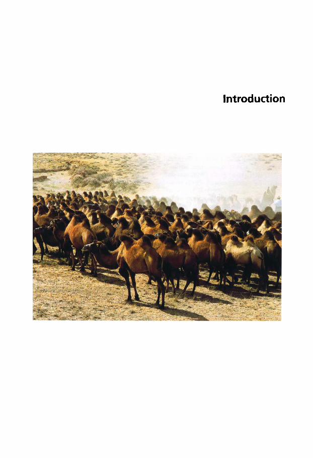
Camelids have served the needs of people for thousands of years and have provided them with food, fiber and fuel. In many parts of the world they have also served as beasts of burden. They secured trade and communication throughout wide arid and semiarid expanses. To the Bedouin of the Arabian Peninsula and North and East Africa, the dromedary was, and still is in some parts, vital for survival in a most in- hospitable environment. Bactrian camels inhabit the high deserts of Asia where they survive - 40°C temperatures. For hundreds of years they have carried goods along the Old Silk Route to China. A few wild Bactri- ans still roam the steppes of the Gobi des- ert in Mongolia and China. In South Amer- ica, the vicufia and guanaco remain wild species, while the llama and the alpaca are domesticated. They have adapted well to high altitude survival. In many countries camelids have now adapted to contained management, and in the last few years there has been a renaissance in both Old and New World camelids.
Until recently, scientific interest in cam- els and the majority of research projects in- volving camels have been concentrated in countries actively involved with the care and maintenance of the camel as a domes- ticated animal. A frequent opinion encoun- tered in those countries not involved with camel husbandry is that the camel is an anachronism, an animal of the past and without a future (Wilson, 1984). It is there- fore not surprising that many publications on camels appear in journals that are difficult to obtain or in lesser-known lan- guages. There was obviously an urgent need for a comprehensive compilation and evaluation of the published literature for all those involved with camels. An impoc- tant step in this direction was the publica- tion of the bibliography Sur le dromadaire et le chameau by Saint-Martin et al. (1990), in which approximately 5500 pre-1990 pub- lications regarding camels are catalogued by author and subject matter. Additional
bibliographies about the camel can be found under Farid (1981), Mukasa-Muger- wa (1981), and Wilson et al. (1990).
Prior to 1987, approximately 1000 New World camelid veterinary references were published. During the period from 1987 to 1996 one thousand four hundred new ref- erences appeared in the world literature (Wernery et al., 1999).
The camelid family has become the focus of increasing study in the last few years. This has become apparent not only through the increase in scientific publi- cations by, for example, Wilson (1984), Yagil (1985), Higgins (1986), Bitter (1986), Griindel (1988), Doose (1990), Saltin and Roose (1994), Wernery and Kaaden (1995), Manfield and Tinson (1996), Tibary and Anouassi (1997), Gauly (1997), Faye (1997), Wilson (1998), Fowler (1998), Beil (1999), Wernery et al. (1999), and Gahlot (2000), and the edition of a camel journal (Journal of Camel Practice and Research, editor Dr. T. K. Gahlot), but also through the increase in joint research projects between Euro- pean universities and institutions in and countries. This growing general interest in camelids also became evident when 300 camel experts from 30 countries took part in the First International Camel Conference in Dubai, United Arab Emirates, in Febru- ary 1992 (Allen et al., 1992). Further inter- national meetings and conferences took place in 1996 in Eilat, Israel, in 1997 in Al- Ain, UAE, and in 1999 in South and North America and in Morocco. Proceedings are available from most of these conferences.
As an important source of milk, meat and wool as well as transportation and la- bor, the camel should play a more impor- tant role than is currently the case in a world where food and energy reserves are dwindling (El-Gayoum, 1986). This is es- pecially true as the camel is, due to its physiological attributes, the most suitable domestic mammal for uses in climatic ex- tremes (Yagil, 1985; Wilson, 1989; George, 1992; Wernery, 1992).
4 Introduction
For a long time it has been incorrectly as- sumed that one and two-humped camels derive from a sole wild species, i.e. the two-humped wild camel - Camelus ferus. There were two main reasons for this be- lief. Firstly, both the one and two-humped camels pass through a two-hump embry- onic stage. Secondly, the crossbreeds be- tween dromedaries and Bactrians are fer- tile. However, the latest osteological in- vestigations on post-cranial skeletons of dromedaries (Camelus dromedarius) and Bactrian camels (Camelus buctrianus) have shown that they are in fact derived from two different species (Peters, 1997).
The tylopods originated in North Amer- ica 50-60 million years ago (Tertiary pe- riod) at which time they branched into eight different families (Zeuner, 1963). They were at that time the size of hares. Six of the eight families died out in the middle Miocene. Then, five million years ago, the
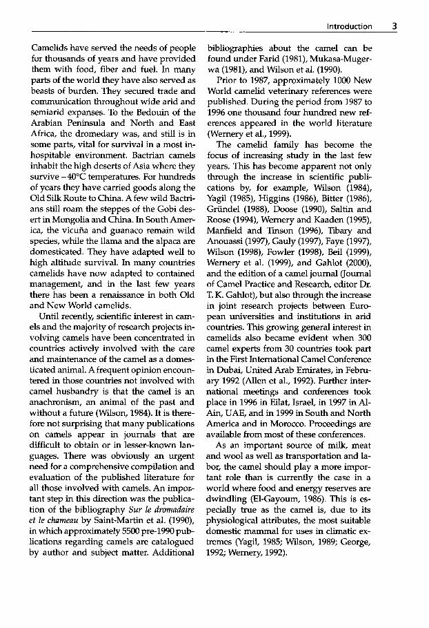
ancestors of the OWC migrated to north- east Asia across the isthmus today known as the Bering Strait. Today’s OWC evolved from these early camels, branching out westwards. They were most widely spread during the Pleistocene era (ending two mil- lion years ago) when they reached as far as Eastern Europe, North and East Africa and eastern Asia (Koehler, 1981; Koehler- Rollefson, 1988). After some time, they died out in some of these regions. When the OWC migrated eastwards after cross- ing the Bering Strait, the ancestors of the humpless NWC migrated south over the newly formed isthmus between the half continents of North and South America (Sielmann, 1982). They populated South America where the different types are known today as llama (Lama g l u m - do- mesticated), vicufia (Lama vicugna - wild), guanaco (Lama guanicoe - wild) and alpaca (Lama pacos - domesticated) (Fig. 1). The
Figure 1 The four different South American camelids (courtesy of Prof. M. E. Fowler, USA) (a) Llama, (b) Alpaca, (c) Guanaco, (d) VicuAa
Introduction 5
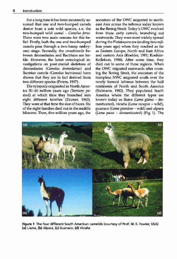
Table 1 Classification of camelids and other artiodactylids (Fowler, 1998)
Class Mammalia Order Artiodactyla Suborder Suiformes Hippopotamuses, swine, peccaries Suborder Tylopoda Camel ids
Old World
New Lama glama - llama World Lama pacos - alpaca
Camelus dromedarius - dromedary camel Camelus bactrianus - Bactrian camel
Lama guanicoe - guanaco Vicugna vicugna - vicuAa V: vicugna mensalis (Peruvian) V: vicugna vicugna (Argentinean)
Suborder Ruminantia Cattle, sheep, goats, water buffalo, giraffe, deer, antelooe. bison
OWC and NWC belong to the Camelidae (camel-like) family under the suborder Ty- Zopoda (Table 1).
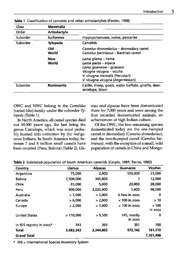
In North America, all camel species died out 10,000 years ago, the last being the genus Camelops, which was most proba- bly hunted into extinction by the indige- nous Indians. In South America today, be- tween 7 and 8 million small camels have been counted (Peru, Bolivia) (Table 2). Lla-
mas and alpacas have been domesticated there for 7,000 years and were among the first recorded domesticated animals, an achievement of high Indian culture.
Of the OWC, the two remaining species domesticated today are the one-humped camel or dromedary (Camelus dromedarius), and the two-humped camel (Camelus buc- trianus), with the exception of a small, wild population of camels in China and Mongo-
Table 2 Estimated population of South American camelids (Carpio, 1991; Torres, 1992)
Country Llamas Alpacas Guanacos Vicuiias Argentina Bolivia Chile Peru Austra I ia Canada Europe
United States
In lSlS registry in zoos* Total Grand Total
75,000 2,500,000
85,000 900,000 < 5,000 > 6,000 < 2,000
> 110,000
343 3,683,343
2,000 300,000
5,000
3,020,000 > 5,000 > 2,000 < 1,000
> 9,500
303
3,344.803
550,000 ?
20,000 1,400
A few in zoos < 100 in zoos < 100 in zoos
145, mostly in zoos
397 572,142
23,000
12,000 28,000 98,000
0 > 10
< 100 in zoos
0
100 161,210
7,761,498
* ISIS = International Species Inventory System
6 Introduction
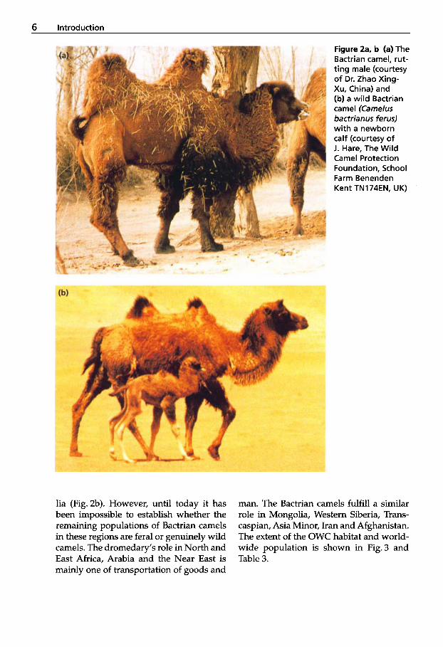
Figure 2a. b (a) The Bactrian camel, rut- ting male (courtesy of Dr. Zhao Xing- Xu, China) and (b) a wild Bactrian camel (Camelus bactrianus ferus) with a newborn calf (courtesy of J. Hare, The Wild Camel Protection Foundation, School Farm Benenden Kent TN174EN, UK)
lia (Fig. 2b). However, until today it has been impossible to establish whether the remaining populations of Bactrian camels in these regions are feral or genuinely wild camels. The dromedary’s role in North and East Africa, Arabia and the Near East is mainly one of transportation of goods and
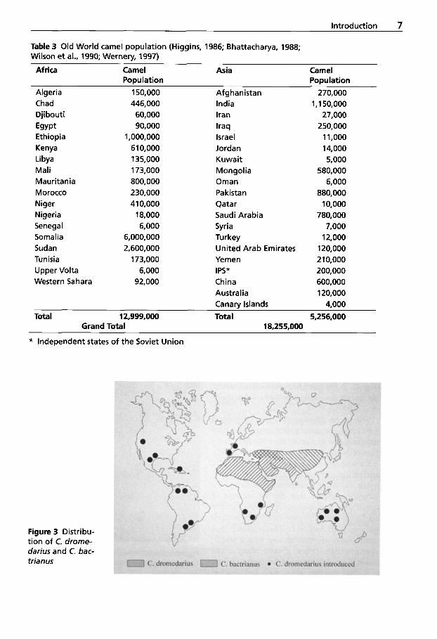
man. The Bactrian camels fulfill a similar role in Mongolia, Western Siberia, Trans- Caspian, Asia Minor, Iran and Afghanistan. The extent of the OWC habitat and world- wide population is shown in Fig. 3 and Table 3.
Introduction 7
Table 3 Old World camel population (Higgins, 1986; Bhattacharya, 1988; Wilson et al., 1990; Wernery, 1997)
Africa Camel Asia Camel
Algeria 150,000 Afghanistan 270,000 Population Population
Chad Djibouti Egypt Ethiopia Kenya Libya Mali Mauritania Morocco Niger Nigeria Senegal Somalia Sudan Tunisia Upper Volta Western Sahara
446,000 60,000 90,000
1,000,000 610,000 135,000 173,000 800,000 230,000 410,000 18,000 6,000
6,000,000 2,600,000 173,000 6,000 92,000
India Iran Iraq Israel Jordan Kuwait Mongolia Oman Pakistan Qatar Saudi Arabia Syria Turkey United Arab Emirates Yemen IPS* China Australia Canarv Islands
1,150,000 27,000 250,000 1 1,000 1 4,000 5,000
580,000 6,000
880,000 10,000
780,000 7,000 12,000 120,000 2 10,000 200,000 600,000 120,000 4,000
Total 12,999,000 Total 5,256,000 Grand Total 18,255,000
* Independent states of the Soviet Union
Figure 3 Distribu- tion of C. drome- darius and C. bac- trianus
8 Introduction
OWC have adapted marvelously to life in either hot or cold environments and NWC to life in high altitudes. Sophisticat- ed mechanisms have evolved that guaran- tee survival of this unique animal family under extreme conditions.
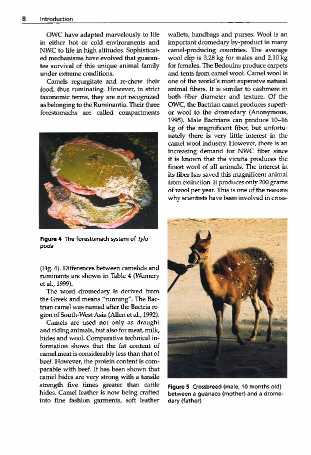
Camels regurgitate and re-chew their food, thus ruminating. However, in strict taxonomic terms, they are not recognized as belonging to the Ruminantia. Their three forestomachs are called compartments
wallets, handbags and purses. Wool is an important dromedary by-product in many camel-producing countries. The average wool clip is 3.28 kg for males and 2.10 kg for females. The Bedouins produce carpets and tents from camel wool. Camel wool is one of the world's most expensive natural animal fibers. It is similar to cashmere in both fiber diameter and texture. Of the OWC, the Bactrian camel produces superi- or wool to the dromedary (Anonymous, 1995). Male Bactrians can produce 10-16 kg of the magnificent fiber, but unfortu- nately there is very little interest in the camel wool industry. However, there is an increasing demand for NWC fiber since it is known that the vicufia produces the finest wool of all animals. The interest in its fiber has saved this magnificent animal from extinction. It produces only 200 grams of wool per year. This is one of the reasons why scientists have been involved in cross-
Figure 4 The forestomach system of Tylo- poda
(Fig. 4). Differences between camelids and ruminants are shown in Table 4 (Wernery et al., 1999).
The word dromedary is derived from the Greek and means "running". The Bac- trian camel was named after the Bactria re- gion of South-West Asia (Allen et al., 1992).
Camels are used not only as draught and riding animals, but also for meat, milk, hides and wool. Comparative technical in- formation shows that the fat content of camel meat is considerably less than that of beef. However, the protein content is com- parable with beef. It has been shown that camel hides are very strong with a tensile strength five times greater than hides. Camel leather is now being crafted into fine fashion garments, soft leather
Figure 5 Crossbreed (male, 10 months old) between a guanaco (mother) and a drome- dary (father)
Introduction 9
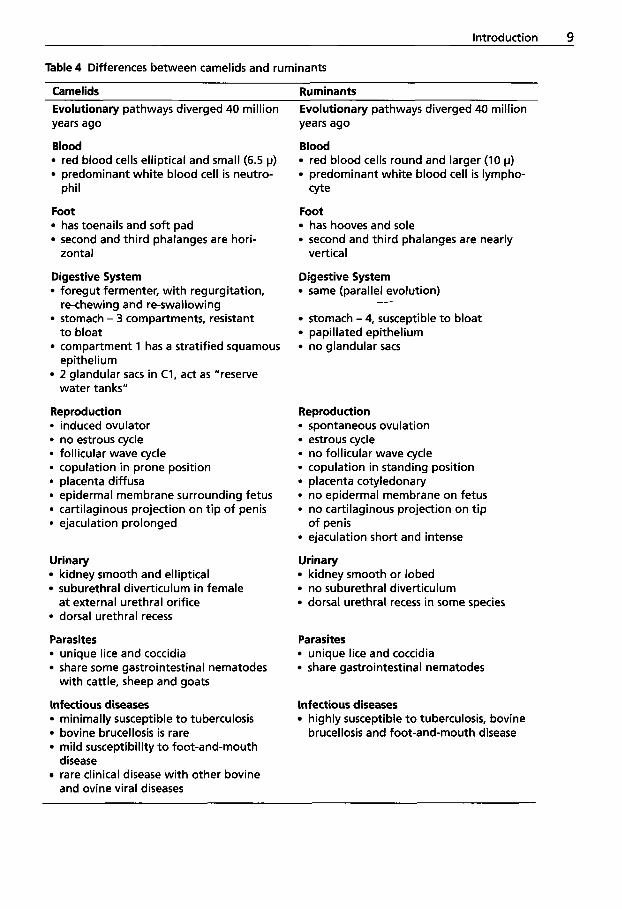
Table 4 Differences between camelids and ruminants
Camelids Evolutionary pathways diverged 40 million years ago
Blood red blood cells elliptical and small (6.5 p) predominant white blood cell is neutro-
Foot has toenails and soft pad second and third phalanges are hori-
Digestive System foregut fermenter, with regurgitation,
stomach - 3 compartments, resistant
compartment 1 has a stratified squamous
2 glandular sacs in C1, act as "reserve
Reproduction
phi1
zontal
re-chewing and re-swallowing
to bloat
epithelium
water tanks"
induced ovulator no estrous cycle follicular wave cycle copulation in prone position placenta diffusa epidermal membrane surrounding fetus cartilaginous projection on tip of penis ejaculation prolonged
Urinary kidney smooth and elliptical suburethral diverticulum in female at external urethral orifice dorsal urethral recess
Parasites unique lice and coccidia share some gastrointestinal nematodes
Infectious diseases minimally susceptible t o tuberculosis bovine brucellosis is rare mild susceptibility t o foot-and-mouth disease rare clinical disease with other bovine and ovine viral diseases
with cattle, sheep and goats
Ruminants Evolutionary pathways diverged 40 million years ago
Blood red blood cells round and larger (10 p) predominant white blood cell is lympho-
Foot has hooves and sole second and third phalanges are nearly
Digestive System same (parallel evolution)
stomach - 4, susceptible to bloat papillated epithelium no glandular sacs
cyte
vertical
-/
Reproduction spontaneous ovulation estrous cycle no follicular wave cycle copulation in standing position placenta cotyledonary no epidermal membrane on fetus no cartilaginous projection on tip of penis ejaculation short and intense
Urinary kidney smooth or lobed no suburethral diverticulum dorsal urethral recess in some species
Parasites unique lice and coccidia share gastrointestinal nematodes
infectious diseases highly susceptible t o tuberculosis, bovine brucellosis and foot-and-mouth disease
10 Introduction
breeding NWC with OWC. The first suc- cessful hybrid was produced in the Unit- ed Arab Emirates (UAE) between a male dromedary and a female guanaco (Fig. 5).
Although there is evidence of the Bactri- an camels’ ancestors discovered at pre-his- toric sites in Kazakhstan and Mongolia, lit- tle is known about the dromedary’s ances- try. An ancestor of the dromedary camel, the ”giant” camel, is known zoologically as Camelus thomasi (named after the French paleontologist Thomas). Camelus thomasi is now considered a possible ancestor of the domestic one-humped camel (Peters, 1998). These camels are presumed to have existed in a wild state during the last ice age in North Africa and in the Negev Desert, where they probably died out some 12,000 to 20,000 years ago during extremely cold temperatures coupled with drought. How- ever, no skeletal remains or rock paintings of camels in the Sahara mountains support this theory. Evidence of wild camels was only found once in South West Asia, at
the beginning of the Holocene era. The re- mains were found at Sihi, a village in Yemen, and were dated at 7000 BC.
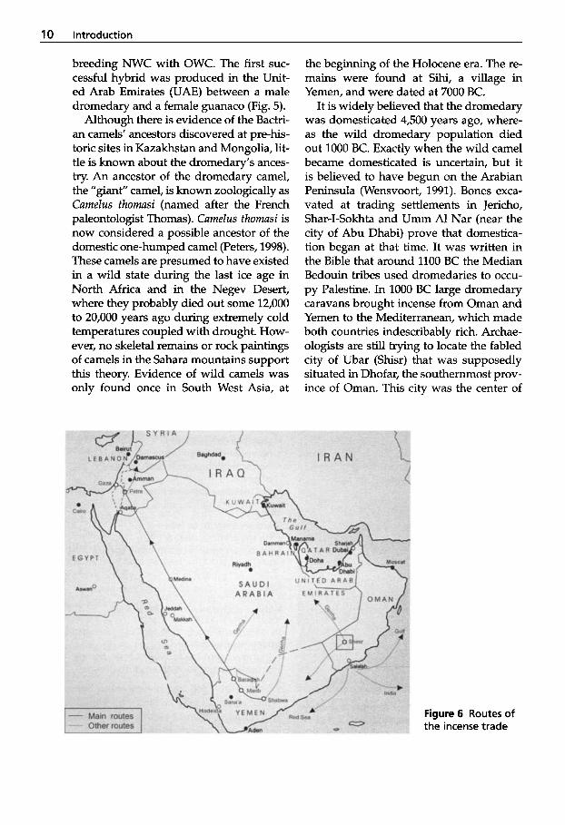
It is widely believed that the dromedary was domesticated 4,500 years ago, where- as the wild dromedary population died out 1000 BC. Exactly when the wild camel became domesticated is uncertain, but it is believed to have begun on the Arabian Peninsula (Wensvoort, 1991). Bones exca- vated at trading settlements in Jericho, Shar-I-Sokhta and Umm A1 Nar (near the city of Abu Dhabi) prove that domestica- tion began at that time. It was written in the Bible that around 1100 BC the Median Bedouin tribes used dromedaries to occu- py Palestine. In 1000 BC large dromedary caravans brought incense from Oman and Yemen to the Mediterranean, which made both countries indescribably rich. Archae- ologists are still trying to locate the fabled city of Ubar (Shisr) that was supposedly situated in Dhofar, the southernmost prov- ince of Oman. This city was the center of
Figure 6 Routes of the incense trade
Introduction 11
the incense trade, from where the camel caravans made their way through Marib, Medina and Petra towards Gaza and the Mediterranean. The other incense routes through the great Arabian deserts towards Gerrha on the Arabian Gulf could only be traversed with the help of the camel (Fig. 6).
Camel breeding may have increased be- cause of the lucrative incense trade. These heavily laden “ships of the desert” took about 50-70 days to cross the deadly stretch of land between Marib and Petra. The caravanserai reached its zenith during the reign of the Nabateen. Terracotta finds from Petra are richly decorated with drom- edaries. With the advent of Christianity the incense trade began to decline, and Arabia Felix reverted to the deserted Empty Quar- ter. After the caravans vanished, only the Bedouins continued to utilize the drome- dary.
When trade began with Arabia, drome- dary numbers increased in Africa. It is pre- sumed that between 1500-2000 BC, drom- edaries spread into Africa from the Ara- bian Peninsula via the Horn of Africa. Beyond Somalia, the country with the highest proportion of dromedaries per per- son, the “ship of the desert” spread north and westwards. However, it was not intro- duced into Tunisia and the Atlas countries before Hellenistic times.
Dromedaries were not only introduced into countries with temperate climates such as Europe, South America and the Carib- bean, but also into Australia and southern Africa, which have hot climates. An esti- mated 10,000-12,000 camels imported into Australia between 1860 and 1907 were used as draught and riding animals by people pioneering the dry interior (Viswa- nathan, 1991). The camels introduced into Australia were almost exclusively drome- daries, because they are highly suited to the Australian desert climate. Most of the camels were released in the mid-l920s, when motor vehicles began operating in the central areas of Australia. In the semi-
arid deserts of Australia they established free-ranging herds, which nowadays num- ber approximately 200,000 animals. These feral camels are scattered throughout the arid interior of Australia with an estimated 50% in Western Australia, 25% in the Nort- hem Territory, and 25% in western Queens- land and northern South Australia. In the late 1960s, there was renewed interest in camels, and by 1970, Australia had two camel tourist businesses with camel races being held around Australia (Anonymous, 1995). Several races were held in Sydney in August 1998 (with the support of the UAE) in preparation for the Olympic Games in 2000.
Dromedaries were also brought into southern Africa, mainly Namibia, around 1890. They were used by the German Schutztruppe in Namibia until the end of World War I for three reasons. Firstly, only dromedaries could survive in the Namib- ian and Kalahari deserts; secondly, oxen were eradicated by rinderpest and foot- and-mouth disease; and thirdly, horses were severely decimated by the devastat- ing African horse sickness virus. In 1906, Lorenz Hagenbeck shipped 2,000 Sudanese camels to the small outpost of Swakop- mund in Namibia. After the Versailles Peace Treaty (1919), the English police force then took possession of all remaining camels in Namibia. However, as in Australia, after motorized transport became popular, the camels were abandoned and it is believed that as a result of being eaten by lions and bushmen, they disappeared in southern Africa in the late 1960s (Massmann, 1981).
Dromedaries were also used in the Unit- ed States after the Mexican war of the 1840s, on mail express routes across the newly acquired arid regions, but they were later eradicated.
In Europe, camel societies have emerged during the last two decades and animals have been used to attract tourists. In Au- gust 1997, camel races were held at Berlin’s famous horse race course Hoppegarten in
12 introduction
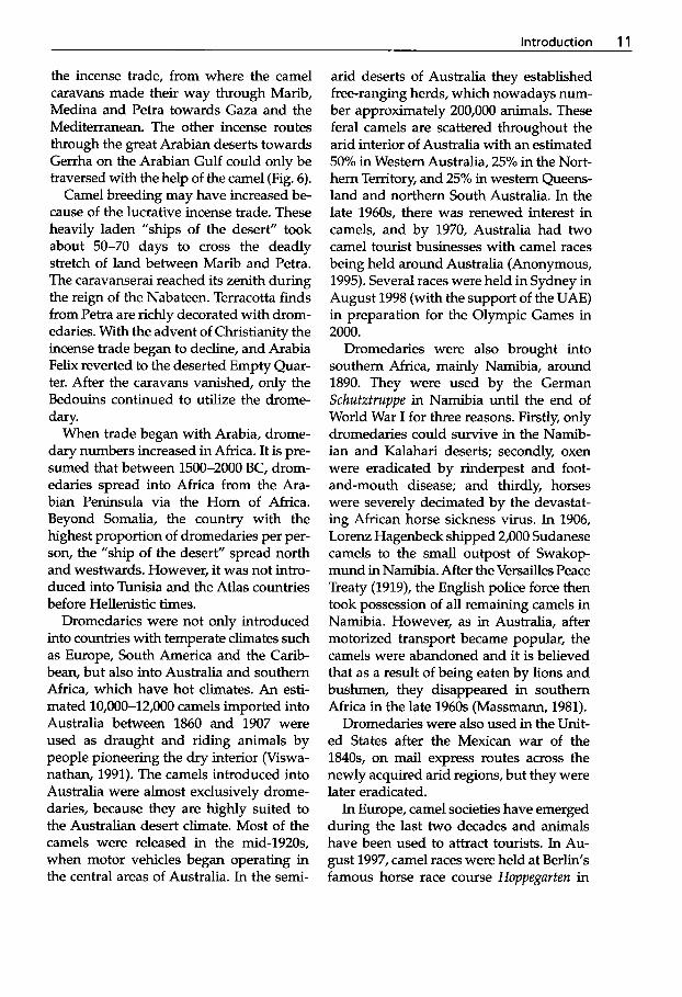
Figure 7 Geographical distribution of dromedary breeds (after Wilson, 1998)
front of 60,000 spectators. However, this was not the first camel race in Europe, as was then claimed. The first races took place in Cologne-Weidenpesch in 1969 with Moroccan camels (Leue, 1969).
Since the 1980s, the dromedary has again become popular, not only with scientists, but also in the countries where it is used for riding and transport. Its milk, skin and meat are all utilized and, additionally, it has become a tourist attraction. The future of the dromedary species is assured de- spite the competition of modem transport and other domesticated animals, and it of- fers no threat to domesticated animals or any endangered wildlife.
Scientists have recently intensified their study of the dromedary and are debating whether there are different dromedary "breeds". Until now, the dromedary has been classified in the following ways - by naming them after the tribes who rear them, or whether they are riding or trans- port camels, by their color, geographical background (Fig. 7), physical characteris- tics or their use for milk, meat, or racing (Wilson, 1998). This categorization has giv- en rise to the classification of dromedaries under 48 "breeds" in 9 regions and sub-re- gions, under 3 main groups and 8 sub- groups. The confusion is compounded be-
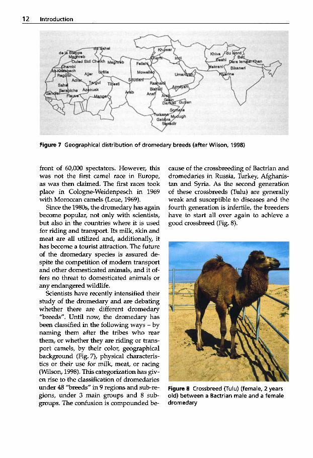
cause of the crossbreeding of Bactrian and dromedaries in Russia, Turkey, Afghanis- tan and Syria. As the second generation of these crossbreeds (Tulu) are generally weak and susceptible to diseases and the fourth generation is infertile, the breeders have to start all over again to achieve a good crossbreed (Fig. 8).
Figure 8 Crossbreed (Tulu) (female, 2 years old) between a Bactrian male and a female dromedary
Introduction 13
In its great genetic diversity, the drome- dary raises many questions which are not easily explained.
Thousands of years before the Pyramids were built, the Bedouins and their drome- dary herds wandered through the great Arabian deserts and lived undisturbed throughout the successive reigns of the Pharaohs, Sumerians, Assyrians, Phoeni- cians, Greeks, Romans and Turks. The tribes could only survive in the desert thanks to the dromedary, which the Bedouins call Ata Allah (God‘s Gift). Only through its in- dispensable patience and perseverance did it enable survival in the perpetual sands. Over this period, the desert played a big part in the evolution of the dromedary as the only domesticated animal to survive in such extreme conditions. Not only does the dromedary produce milk, meat and wool, but it is also used as transport over thousands of kilometers. Not only the ”ship of the desert’s” ability to survive in the hottest climates, but its natural resist- ance to such deadly animal diseases as rinderpest and African horse sicknes makes it indispensable to its owner.
In an effort to find new grazing areas, it was often necessary for the Bedouin tribes to cross enemy territory. This sometimes
resulted in feuds and skirmishes. Obvious- ly, the tribe with the quickest and most nimble dromedaries had the most chance of surviving. Sir Wilfred Thesiger, in his book Arabian Sands, described disputes that still occurred until some 30 years ago. However, not only were the drome- daries vital during tribal conflicts, but the Bedouins also used them for racing during social occasions, such as weddings or births. The quickest dromedaries were selected to run over short courses.
In the Arabian Desert, it was the Bedou- in who managed to breed the precious Ara- bian horse, the Saluki dog and the drome- dary. In its perseverance, intelligence and beauty, the Arabian dromedary, bred over hundreds of years in one of the hottest climates on earth, is comparable with the
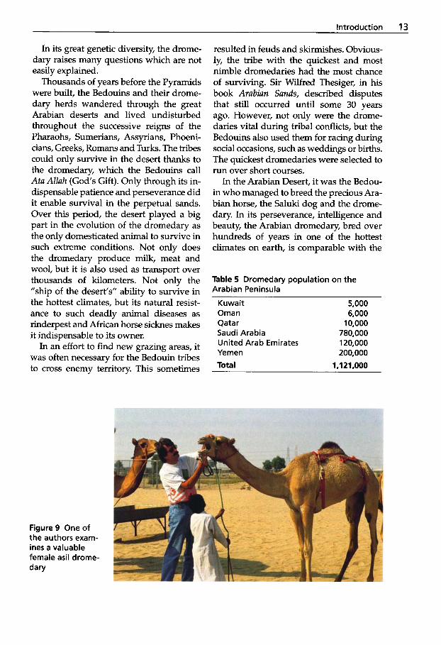
Table 5 Dromedary population on the Arabian Peninsula
Kuwait 5,000 Oman 6,000
Saudi Arabia 780,000 Qatar 10,000
United Arab Emirates 120,000 Yemen 200,000 Total 1.1 21,000
Figure 9 One of the authors exam- ines a valuable female asil drome- dary
14 Introduction
1.
2.
3.
4.
5.
7.
8.
9.
10.
11.
12.
13. 14.
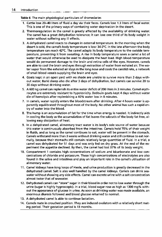
Table 6 The main physiological particulars of dromedaries
Cattle lose 20-40 liters of fluid a day via their feces. Camels lose 1.3 liters of fecal water. This is one of the primary ways of combating water deprivation in the desert. Thermoregulation in the camel is greatly affected by the availability of drinking water. The camel has a great dehydration tolerance; it can lose one third of i t s body weight in water without suffering any ill effects. A dehydrated camel reacts to changes in external temperature. In the morning when the desert is cold, the camel's body temperature is low: 34.0"C. In the late afternoon the body temperature can reach 42°C. The camel adapts i t s body temperature t o the outside tem- perature, preventing it from sweating. A rise in body temperature saves a camel a lot of water that would otherwise be used to dissipate the heat load. High blood temperature would do permanent damage to the brain and retina cells of the eyes. However, camels are able to cool the brain and eyes through extraction of water from exhaled air. The wa- ter vapor from the exhaled air stays in the long nose and cools the carotid rete, a network of small blood vessels supplying the brain and eyes. Goats kept in an open yard with no shade are unable t o survive more than 3 days with- out water; Barki sheep also die after 3 days of dehydration, but camels can survive 20 to 30 days without drinking water. A 600 kg camel can replenish i ts entire water deficit of 200 liters in 3 minutes. Camel eryth- rocytes are extremely resistant t o hypotonicity. Bedouin goats kept 4 days without water die of hemolysis after replenishing a 40% water loss in 8 minutes. In camels, water rapidly enters the bloodstream after drinking. After 4 hours water is ap- parently equilibrated throughout most of the body. No other animal has such a rapid en- try of water into the blood. The hump is an accumulation of fat for the time when energy is needed. It indirectly aids in cooling the body as the accumulation of fat leaves the subcutis of the body fat free, al- lowing easy dissipation of heat. In a dehydrated camel, alimentary tract water is i t s body's sole source of water because this water is continuously absorbed from the intestines. Camels hold 75% of their weight in fluids, and as long as the camel continues to eat, water will be present in the stomach. Camels withstand more than 3 weeks without drinking water and s t i l l continue t o eat nor- mally, because their stomachs s t i l l contain relatively large quantities of fluid. In a trial, a camel was dehydrated for 51 days and was only fed on dry grass. At the end of the ex- periment the appetite declined. By then, the camel had lost 37% of i t s body weight. Compartment 1 contains high concentrations of sodium and bicarbonate and low con- centrations of chloride and potassium. These high concentrations of electrolytes are also found in the saliva and intestines and play an important role in the camel's utilization of alimentary water. Camel kidneys have long loops of Henle, and urine production is greatly decreased in the dehydrated camel. Salt is also well handled by the camel kidneys. Camels can drink sea- water without showing any side effects. Camel can excrete urine with a salt concentration almost twice that of seawater. Dehydrated camels can "store" sugar in their blood in order not t o lose water through the urine (sugar is highly hygroscopic). In a trial, blood sugar rose as high as 1300 mg% with- out the appearance of glucose in urine. As soon as drinking water was made available, an enormous diuresis followed and blood glucose returned t o normal. A dehydrated camel is able t o continue lactation. Camels mate in crouched position. They are induced ovulators with a relatively short mat- ing period. Their gestation period is 13 months.
introduction 15
Arabian horse. There is no other drom- edary that compares with the Arabian. Through breeding, it has become an agile, fast, long-legged, slender, brown racing dromedary with fine limbs and a long head (Fig. 9). Although no studbooks exist, the Bedouin are extremely careful to keep the bloodlines pure. In the 30 years since the oil boom began, camel racing has gone through a fundamental change.
In the last few years, the worldwide camel population has risen from 17.5 mil- lion (Wilson et al., 1990) to 18.3 million (Table 3). The camel population has de- creased in only a few countries, such as Libya and the Gulf States, where oil has brought nomadism to a virtual standstill (Wilson, 1984). However, in recent years, an opposite trend has been observed in the UAE where the dromedary is experienc- ing a renaissance resulting in a revival of the old Bedouin tradition of camel racing. What was earlier seen as playful competi- tion and a pleasant pastime between the Bedouin has become a scientifically found- ed racing discipline following the oil boom of the 1960s. Based on this development, more than 100,000 racing camels are kept in the UAE. In the cooler months between September and May, competitions are held on 20 racetracks throughout the Emirates. Based on the age of the animal, the drome- dary competes at distances between 3 and 10 kilometers. A dromedary can cover the 10 km course in 17 to 18 minutes (Wernery, 1992).
Due to a number of specific anatomical and physiological characteristics, the dmm- edary can survive and perform tasks in the extreme climate of the desert that can be utilized by man (Schmidt-Nielsen, 1964) (Table 6).
A further advantage is the low suscepti- bility of the camelids to disease (Fazil and Hofmann, 1981). This is especially true of viral diseases, although bacterial ailments play a larger role. Both the camelids’ resist- ance to a number of pathogenic microor-
ganisms, as extensively examined by sci- entists in the Institute for Horticulture and Animal Hygiene in Goettingen (El-Gay- own, 1986; Margan, 1987), and the previ- ous lack of interest in the camel family in general, may have been decisive in the dearth of publications on infectious dis- eases of camelids. The second edition of this book will attempt to close this gap by surveying and compiling the published literature regarding bacterial, viral and fungal diseases as well as pathology and parasitology in the camelids as completely as possible. The majority of the literature encompasses the one-humped Camelus dromedarius as the available literature on the two-humped Camelus bactrianus is un- fortunately very difficult to obtain. As the exchange of scientific research with coun- tries where the Bactrian camel lives is now improving, it is hoped that more compre- hensive data will soon become accessible. New scientific findings of NWC are also included.
In addition to a compilation of the known literature, results of the authors’ personal research conducted since 1987 on a camel population of 30,000 racing dromedaries (including breeding animals) in the UAE, in conjunction with various research insti- tutes abroad, will also be presented.
References
Allen, W.R., A.J. Higgins, I.J. Mayhew, D.H. Snow and J.F. Wade 1992. Proc. 1st int. Camel Cod., Feb 24,1992. Published by R. and W. Publications. (Newmarket) Ltd.
Anonymous. 1995. The central Australian camel industry. Brochure of the Central Australian Camel Industry Association, PO Box 8760, Alice Springs, Australia: 1 4 .
Beil, Christiane. 1999. Reproduktion beim weib- lichen Kame1 (Camelus dromedarius und Ca- melus bactrianus). Eine gewichtete Literatur- studie. Thesis, Hannover.
Bhattacharya, A.N. 1988. Camel production re- search in northern Saudi Arabia: a mono- graph. Ministry of Agriculture and Water De-
16 Introduction
partment of Agricultural Research, UTFN/ SAU/OO8/SAU.
Bitter, H. 1986. Untersuchungen zur Resistenz von Kamelen (Camelus dromedarius) unter besonderer Beriicksichtigung der Mektion mit Trypanosoma evansi (Steel 1885). Thesis, H ann o v e r .
Carpio, M. 1991. Camelidos socio-economia An- dina (Camelids and Andean socio-econom- ics). Ed. Novoa, C. and Florez, M.: A produc- tion de Rumiantes Menores: Alpacas. Lima, Peru. Re rumen: 3-16.
Doose, Anette. 1990. Funktionen und Morpho- logie des Verdauungssystems des einhockri- gen Kamels (Camelus dromedarius). Thesis, Hannover.
El-Gayoum, S.E.A. 1986. Study on the mecha- nism of resistance to camel diseases. Thesis, Gottingen 22.
Farid, M.F.A. 1981. Camelids Bibliography.
Faye, B. 1997. Guide de l'devage du dro- madaire. Sanofi Sant6 Nutrition Animale, La Ballastiere - 813126, 33501 Libourne, Cedex, France: 115-116.
Fazil, M.A. and R.R. Hofmann. 1981. Haltung und Krankheiten des Kamels. Tieriirztl. Praxis
Fowler, M.E. 1998. Medicine and surgery of South American Camelids. Iowa State Uni- versity Press, Ames.
Gahlot, T.K. 2000. Selected topics on camelids. The Camelid Publishers, Sankhla Printers, Bikaner, India.
Gauly, M. 1997. Neuweltkamele. Parey Buch- verlag Berlin.
George, U. 1992. ijberleben. Geo Spezial, Sahara 6: 47.
Gruendel, M. 1988. Das Blut des einhockrigen Kamels (Camelus dromedarius). Eine Litera- turiibersicht. Thesis, Hannover.
Higgins, A. 1986. The camel in health and dis- ease. Bailliere Tindall.
Koehler, J. 1981. Zur Domestikation des Kamels. Thesis, Hannover.
Koehler-Rollefson, I. 1988. The introduction of the camel into Africa with special reference to Somalia. Working paper 24.
h u e , G. 1969. Erstmaliges Kamelrennen in Eu- ropa 1969 auf der Pferderennbahn in Koln aus veterinarphysiologischer, genetischer and biomechanischer Sicht. Dtsch. tierarztl. Wschr.
ACSAD-AS 15.
9: 389-402.
78 (18): 500-502.
Manefield, G.W. and A. Tinson. 1996. Camels. A compendium. The T.G. Hungerford Vade Mecum Series for Domestic Animals.
Margan, Ute. 1987. Vergleichende Untersuchun- gen zur Bedeutung der alternativen Komple- mentaktivierung bei Rindern und Kamelen. Thesis, Gottingen 33.
Massmann, Ursula. 1981. Kamele in Siidwest- afrika. Namib und Meer 9: 31-54.
Mukasa-Mugerwa, E. 1981. The camel (Cam- elus dromedarius): A bibliographical review. International Livestock Center for Africa. ILCA Monogr. 5: 4-119.
Peters, J. 1997. Das Dromedar: Herkunft, Dome- stikationsgeschichte und Krankheitsbehand- lung in friihgeschichtlicher Zeit. Tierurztl. Praxis 25: 559-565.
Peters, J. 1998. Camelus thomasi Pomel, 1893, a possible ancestor of the one-humped camel? Int. J. of Mammalian Biology 63: 372-376.
Saint-Martin, G., M.F. Nitcheman, D. Richard and M.A. Richard. 1990. Bibliographie sur le dromadaire et le chameau. 2nd edition, Tome 1, Tome 2: Index.
Saltin, 8. and R.J. Roose. 1994. The racing camel (Camelus dromedarius). Acta Physiol. Scand., Wernerssons Grafiska AB, Kumla/Chister Perssons Tryckeri AB, Koeping 150 (617).
Schmidt-Nielsen, K. 1964. Desert animals: phys- iological problems of heat and water. Claren- don Press, Oxford.
Sielmann, H. 1982. Weltreich der Tiere. Natura- lis Verlags- und Vertiebsgesellschaft mbH, Miinchen, Monchengladbach, Arbus.
Tibary, A. and A. Anouassi. 1997. Theriogenolo- gy in camelidae. Anatomy, Physiology, Pathol- ogy and Artificial Breeding. Abu Dhabi Print- ing and Publishing Co., Mina, Abu Dhabi, UAE.
Torres, H. 1992. South American Camelids: an action plan for their conservation. South Amer- ican Camelid Specialist Group, Gland, Swit- zerland. IUCN/CSE.
Viswanathan, L. 1991. More about camels. The Gazelle, Dubai Natural History Group 6: 6.
Wensvoort, J. 1991. Camels, camel nutrition and racing camels. The Gazelle, Dubai Natural History Group 6: 5.
Wernery, U. 1992. Dromedare, die Rennpferde Arabiens. Tierarztl. Umschau 4 7 801.
Wernery, U. 1997. Dromedare in Arabien. La- mas. Haltung and Zucht wn Neuweltkameliden 5 (1): 34-36.
Introduction 17
Wemery, U., M.E. Fowler and R. Wernery. 1999. Color Atlas of Camelid Hematology. Black- well Wissenschafts-Verlag, Berlin.
Wemery, U. and 0.-R. Kaaden. 1995. Infectious Diseases of Camelids. Blackwell Wissen- schafts-Verlag, Berlin.
Wilson, R.T. 1984. The camel. Longman, London and New York.
Wilson, R.T. 1989. Ecophysiology of the cameli- dae and desert ruminants. Springer Verlag.
Wilson, R.T., Astier Araya and Azeb Melaku. 1990. The one-humped camel. An analytical and annotated bibliography. The United Na- tions Sudano-Sahelian Office (UNSO), Tech- nical paper series 3.
Wilson, R.T. 1998. Camels. The Tropical Agricul- turalist, MacMillan: 106.
Yagil, R. 1985. The Desert Camel. Verlag Karger, Basel.
Zeuner, F.E. 1963. A history of domesticated an- imals. Hutchinson, London.
Further reading
Fowler, M.E. 1997. Evolutionary history and dif- ferences between camelids and ruminants. J. Camel Prac. and Res. 4 (2), 99-105.
Hare, J.N. 1997. Status and distribution of wild Bactrian camels (Camelus bactrianus ferus) in China. I. Camel Prac. and Res. 4 (2), 107- 110.
Hare, J.N. 1998. The lost camels of Tartary. Little Brown and Company, London.
Skidmore, J.A., M. Billah, M. Binns, R.V. Short and W.R. Allen. 1999. Hybridizing Old and New World camelids: Camelus dromedarius x Lama guanicoe. Proc. R. SOC. Lond. B 266, 649-656.
Bacterial Diseases
1.1 General Survey
1.1.1 Anaerobic Infections
Clostridial diseases are a constant threat to livestock in many parts of the world. Clo- stridia are all potent producers of exotox- ins upon which their pathogenicity de- pends. Clostridial organisms are common- ly present in soils and the intestinal tract of animals, including man, and cause disease only in special circumstances. The ubiqui- tous character of clostridial bacteria makes eradication of clostridiosis virtually im- possible and necessitates control by pro- phylaxis. Both W C and OWC may suffer from some of the clostridial diseases (Wern- ery and Kaaden, 1995; Fowler, 1998).
Clostridial diseases are caused by bacteria of the genus Clostridiurn. Clo- stridium bacteria are large, Gram-positive, anaerobic, endospore-producing rods. The spores bulge the mother cell. C. perfringens possesses a capsule in animal tissue and is non-motile. Clostridia are oxidase-nega- tive and catalase-negative and the anaero- bic requirements vary among the species.
Most of the pathogenic species produce one or more exotoxins of varying potency. The vegetative organism is capable of forming spores that are able to survive long periods of time in the soil. Contami- nated soil can contain up to 105 CZostridiurn perfringens spores per gram of soil (Seifert, 1992).
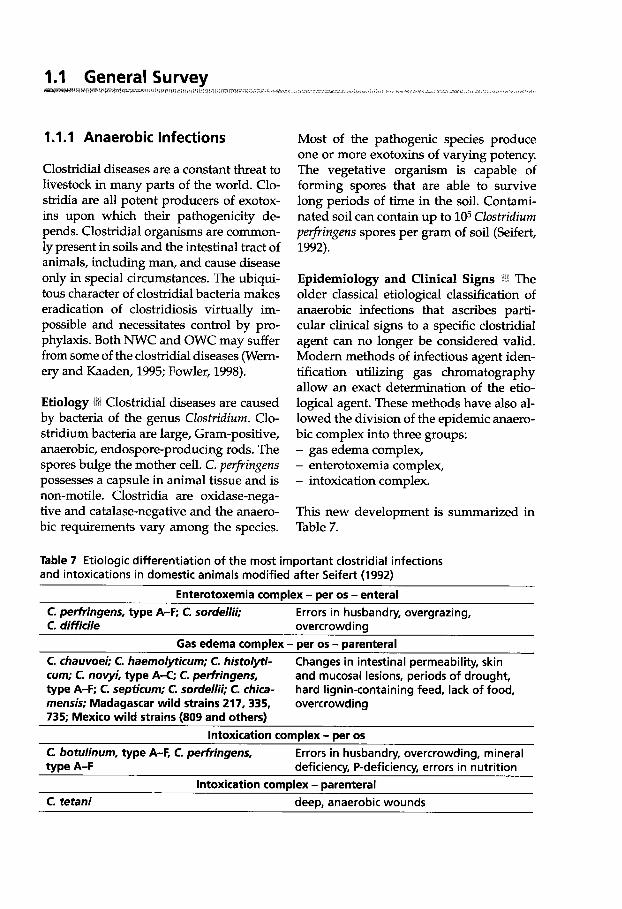
Epidemiology and Clinical Signs :Bi The older classical etiological classification of anaerobic infections that ascribes parti- cular clinical signs to a specific clostridial agent can no longer be considered valid. Modern methods of infectious agent iden- tification utilizing gas chromatography allow an exact determination of the etio- logical agent. These methods have also al- lowed the division of the epidemic anaero- bic complex into three groups: - gas edema complex, - enterotoxemia complex, - intoxication complex.
This new development is summarized in Table 7.
Table 7 Etiologic differentiation of the most important clostridial infections and intoxications in domestic animals modified after Seifert (1992)
Enterotoxemia complex - per 0s - enteral C. perfringens, type A-F; C. sordellii; C. di fficile overcrowding
C. chauvoei; C. haemolyticum; C. histolyti- cum; C. novyi, type A-C; C. perfringens, type A-F; C. septicum; C. sordellii; C. chica- mensis; Madagascar wild strains 217,335, 735; Mexico wild strains (809 and others)
Errors in husbandry, overgrazing,
Gas edema complex - per 0s - parenteral Changes in intestinal permeability, skin and mucosal lesions, periods of drought, hard lignin-containing feed, lack of food, overcrowding
Intoxication complex - per 0s C. botulinum, type A-F, C. perfringens, type A-F
C. tetani deep, anaerobic wounds
Errors in husbandry, overcrowding, mineral deficiency, P-deficiency, errors in nutrition
Intoxication complex - parenteral
22 Bacterial Diseases
Diseases caused by clostridia are often difficult to identify in the tropics due to indigenous ecological influences, making diagnosis a challenge. C. perfringens types A, B and C, C. novyi, C. chauvoei and C. sep- ticum have all been isolated from camelids.
Gas Edema Complex
The causative agents of the gas edema complex, which according to Seifert (1992) include the following diseases:
- black-quarter (blackleg), - malignant edema, - bacillary hemoglobinuria, - infectious necrotizing hepatitis
are seldom isolated from camelids. As most of the available literature is outdated, it is possible that these disorders were falsely diagnosed in the past due to the prevailing incomplete, traditional analytical methods used. Current techniques have identified the following causative agents of the gas edema complex (Seifert, 1992): - C. chauvoei, C. septicum, C. chicamensis,
wild strains that have been exactingly characterized (335 and 735 Madagascar, 805 Mexico);
- C. histolyticum, C. sordellii, C. novyi type AX, C. haemolyticum;
- C. perfringens type A-F and wild strains (217 Madagascar).
C. chauvoei infections in dromedaries have been reported as possibly occurring in North and East Africa, as well as in Chad and India (Gatt Rutter and Mack, 1963), but these reports are contradictory. With the exception of Cross (1919), Curasson (1947) believes that many previous authors have confused black-quarter with true an- thrax caused by Bacillus anthracis. The pro- gression of both disorders is similar, begin- ning with subcutaneous swellings on the shoulders that lead to the animal's death within 2 to 3 days. Hutyra et al. (1946) re- ported that camels were not susceptible to
gas edema; however, Cross (1919) was able to elicit the disorder experimentally in three dromedaries through intramuscular injection of C. chauvoei. The type of swell- ing should allow the differentiation be- tween gas edema and anthrax. Recent pub- lications regarding gas edema in camels are not known.
Blackleg has been produced experimen- tally in alpacas, but there is one report of natural infection in a female llama that died suddenly. The causative agent was C. novyi (Anonymous, 1998). It is believed that OWC and NWC are more resistant to blackleg infections than bovines.
Malignant edema is an economically im- portant disease in alpacas in Peru and has also been associated with rattlesnake bites in llamas in Colorado (Moro Sommo, 1956; Fowler, 1998). The disease in lamoids is caused by C. septicum with two types of syndromes: the typical wound infection and edema and the acute systemic disease, which may kill animals instantly.
The other two diseases of the gas edema complex, bacillary hemoglobinuria and in- fectious necrotizing hepatitis, have not been reported in Camelidae.
Enterotoxemia Corn plex
All types of C. perfringens as well as C. soy- dellii and C. spiroforme can cause the en- terotoxemia complex. C. perfringens, most frequently type A (Bisping and Amtsberg, 1988), is also found in the intestines of healthy animals so that cultural evidence of C. perfringens has little disease-pre- dictive value. Enterotoxemia caused by C. perfringens is found all over the world and is also found in all types of domestic animals. According to Seifert (1992), fac- tors predisposing to disease include di- etary errors, climatic influences, change of pasture, transportation, and weighing of animals.
Acute and subacute enterotoxemia as well as hemorrhagic enteritis due to C. per-
General Survev 23
fringens, types A, C and D have been de- scribed in camels by Moebuu et al. (1966)) Ipatenko (1974), Chauhan et al. (1985) and Gameel et al. (1986). Fowler (1998) has re- ported enterotoxemia due to C. perfringens types A, C and D in NWC.
Extensive studies of C. perfringens type A outbreaks in racing dromedaries in the UAE have been performed by Wernery et al. (1991), Seifert et al. (1992), Wernery et al. (1992b) and Wernery and Kaaden (1995). Peracute and acute enterotoxemia in breed- ing and racing dromedaries as well as se- vere myocardial degeneration and “pulpy kidney” in dromedary calves are known to occur. For all three age groups of drome- daries, predisposing etiological factors were proven to be responsible for the out- breaks.
In a herd of 90 breeding animals, 71% of the dromedaries were found to have an acute Typanosoma evansi infection. Try- panosomosis is known to be able to cause immune suppression in domesticated ani- mals (Losos, 1986), and may have been the predisposing factor for the peracute C. per- fringens outbreak in this group, since nutri- tional errors and environmental influences had been excluded. The camels affected ex- hibited the following clinical signs:
- perspiration - muscle tremor - ataxia - aggression - hyperexcitability - seizures
Affected animals died within one hour af- ter the onset of clinical signs.
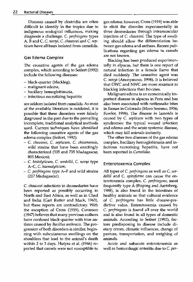
The pathological changes found in au- topsied animals were mild. They included - petechiae in the thoracic musculature, - petechiae in the cerebellum and brain-
- petechiae in the pharyngeal mucosa, - subpleural (Fig. 10) and subepicardial
petechiae, - petechiae in the mucosa of the third
compartment (Fig. 11) and the stomach, - hydropericardium with fibrinous exu-
date, - dark kidneys, with adherence of the cap-
sule to the parenchyma (Fig. 12).
stem,
In another incident, salmonella paved the way for the outbreak of C. perfringens type A in racing dromedaries. The animals de- veloped intractable diarrhea and died after 4 days. In those animals autopsied, even more severe pathological changes were
Figure 10 C. per- fringens entero- toxemia: subpleu- ral hemorrhages
24 Bacterial Diseases
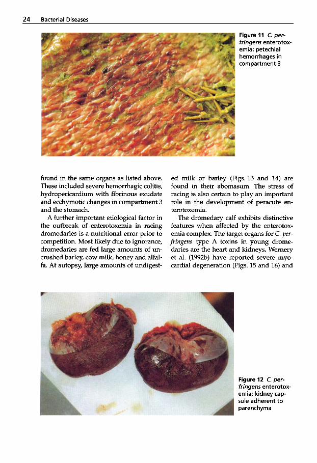
found in the same organs as listed above. These included severe hemorrhagic colitis, hydropericardium with fibrinous exudate and ecchymotic changes in compartment 3 and the stomach.
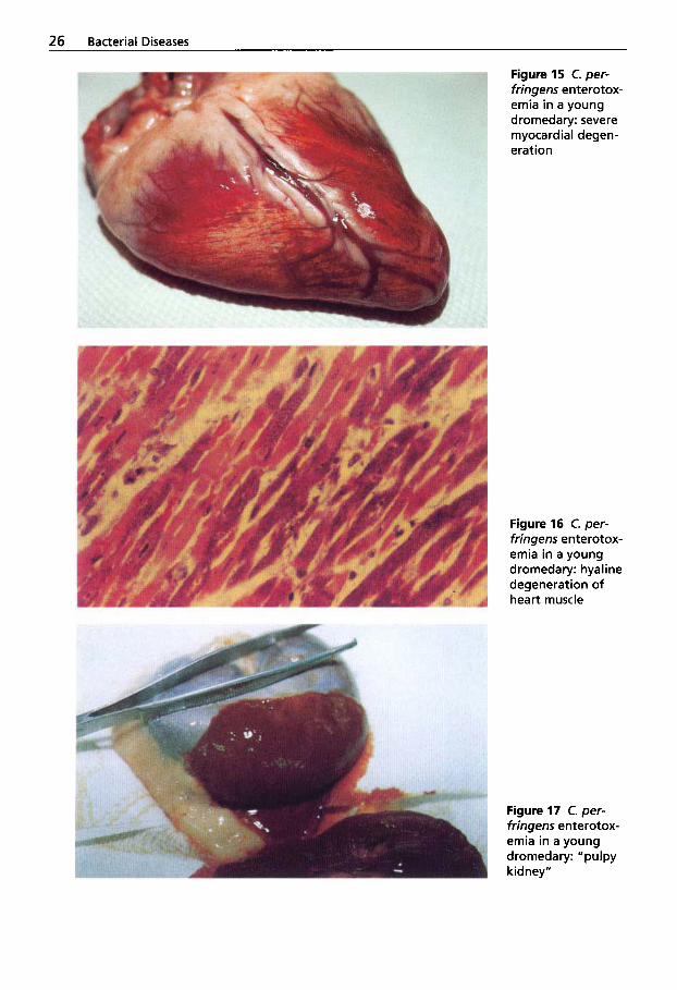
A further important etiological factor in the outbreak of enterotoxemia in racing dromedaries is a nutritional error prior to competition. Most likely due to ignorance, dromedaries are fed large amounts of un- crushed barley, cow milk, honey and alfal- fa. At autopsy, large amounts of undigest-
Figure 11 C. per- fringens enterotox- emia: petechial hemorrhages in Compartment 3
ed milk or barley (Figs. 13 and 14) are found in their abomasum. The stress of racing is also certain to play an important role in the development of peracute en- terotoxemia.
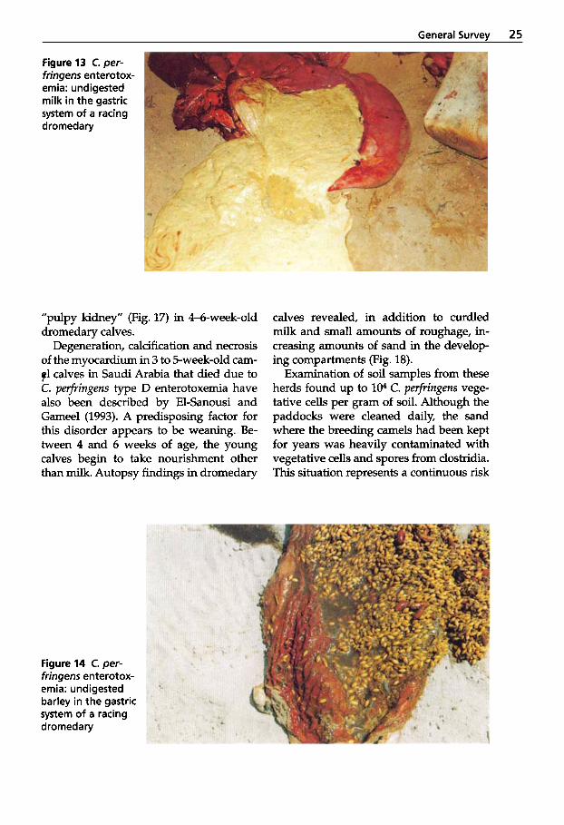
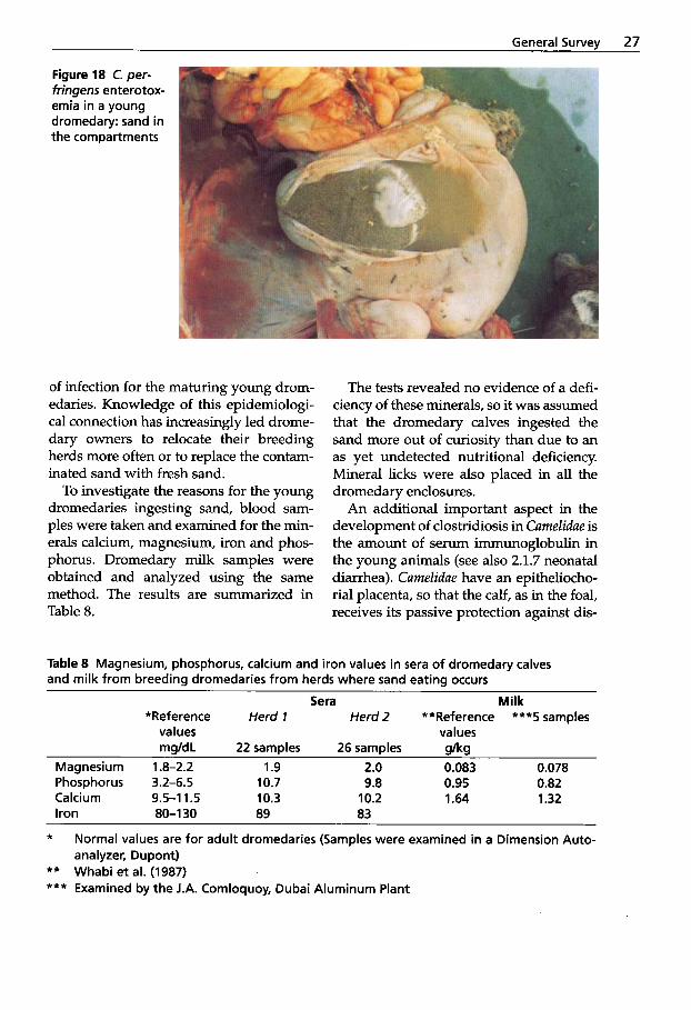
The dromedary calf exhibits distinctive features when affected by the enterotox- emia complex. The target organs for C. per- fringens type A toxins in young drome- daries are the heart and kidneys. Wernery et al. (1992b) have reported severe myo- cardial degeneration (Figs. 15 and 16) and
Figure 12 C. per- fringens enterotox- emia: kidney cap- sule adherent to parenchyma
General Survev 25
Figure 13 C. per- fringens enterotox- emia: undigested milk in the gastric system of a racing dromedary
”pulpy kidney” (Fig. 17) in 4-6-week-old dromedary calves.
Degeneration, calcification and necrosis of the myocardium in 3 to 5-week-old cam- 91 calves in Saudi Arabia that died due to C. perfringens type D enterotoxemia have also been described by El-Sanousi and Gameel (1993). A predisposing factor for this disorder appears to be weaning. Be- tween 4 and 6 weeks of age, the young calves begin to take nourishment other than milk. Autopsy findings in dromedary
calves revealed, in addition to curdled milk and small amounts of roughage, in- creasing amounts of sand in the develop- ing compartments (Fig. 18).
Examination of soil samples from these herds found up to 104 C. perfringens vege- tative cells per gram of soil. Although the paddocks were cleaned daily, the sand where the breeding camels had been kept for years was heavily contaminated with vegetative cells and spores from clostridia. This situation represents a continuous risk
Figure 14 C. per- fringens enterotox- emia: undigested barley in the gastric system of a racing dromedary
26 Bacterial Diseases
Figure 15 C. per- fringens enterotox- emia in a young dromedary: severe myocardial degen- eration
Figure 16 C. per- fringens enterotox- emia in a young dromedary: hyaline degeneration of heart muscle
Figure 17 C. per- fringens enterotox- emia in a young dromedary: "pulpy kidney "
General Survey 27
Figure 18 C. per- fringens enterotox- emia in a young dromedary: sand in the compartments
of infection for the maturing young drom- edaries. Knowledge of this epidemiologi- cal connection has increasingly led drome- dary owners to relocate their breeding herds more often or to replace the contam- inated sand with fresh sand.
To investigate the reasons for the young dromedaries ingesting sand, blood sam- ples were taken and examined for the min- erals calcium, magnesium, iron and phos- phorus. Dromedary milk samples were obtained and analyzed using the same method. The results are summarized in Table 8.
The tests revealed no evidence of a defi- ciency of these minerals, so it was assumed that the dromedary calves ingested the sand more out of curiosity than due to an as yet undetected nutritional deficiency. Mineral licks were also placed in all the dromedary enclosures.
An additional important aspect in the development of clostridiosis in Cumelidue is the amount of serum immunoglobulin in the young animals (see also 2.1.7 neonatal diarrhea). Cumelidue have an epitheliocho- rial placenta, so that the calf, as in the foal, receives its passive protection against dis-
Table 8 Magnesium, phosphorus, calcium and iron values in sera of dromedary calves and milk from breeding dromedaries from herds where sand eating occurs
Sera Milk *Reference Herd 1 Herd 2 **Reference ***5 samples
values values mgldL 22 samples 26 samples g/kg
Magnesium 1.8-2.2 1.9 2.0 0.083 0.078 Phosphorus 3.2-6.5 10.7 9.8 0.95 0.82 Ca Ici u m 9.5-1 1.5 10.3 10.2 1.64 1.32 Iron 80-1 30 89 83
~
*
** Whabi et al. (1987) *** Examined by the J.A. Comloquoy, Dubai Aluminum Plant
Normal values are for adult dromedaries (Samples were examined in a Dimension Auto- analyzer, Dupont)
28 Bacterial Diseases
ease through the intestinal reabsorption of immunoglobulins from the colostrum after birth. Although the newborn calf is im- munocompetent at birth, the endogenous antibody production is not sufficient to produce a protective immunoglobulin lev- el within the first month of life. The globu- lin fraction is naturally low at birth. Even after ingestion of colostrum, the globulin level declines after the seventh day and reaches the lowest level between the 20th and 30th day post partum. The highest losses due to C. perfringens enterotoxemia occul during this time.
Fowler (1998) made similar observations in NWC. He determined that the globulin content of NWC serum is very low at birth (< 5.2 mg/mL), increased following inges- tion of colostrum to 5.5-6.2 mg/mL within 4-5 days yet reached its lowest level 3 to 4 weeks post partum. C. perJrrngens type A is a very serious disease in alpaca crias in South America and it is named “MaZ de AZpucas” (Rath, 1950; Moro Sommo, 1963; Ramirez and Huaman, 1980-1981; Ramirez et al., 1983a and b; Huaman et al., 1981; Ellis et al., 1990; Fowler, 1996). The animal mortality rates vary between 10 and 70% and even on carefully managed farms may approach 50%. The disease occu~s in crias between 8 and 35 days of age with sudden death or a short disease period during which the crias are recumbent, showing nervous system disorders. The pathological changes in al- pacas are very similar to the lesions seen in OWC with petechiae in different organs, hyperemia, excess serosanguinous pericar- dial fluid and lesions in the intestinal tract.
Type C and D enterotoxemias are more common in lamoids than they are in OWC.
Specimens, including intes- tinal fluid, should be taken from freshly (less than 4 hours) dead animals, as clos- tridia are rapid postmortem invaders. Tox- ins are very labile and therefore small in- testinal contents should be frozen as soon as possible until processed. The laboratory
diagnosis of ”clostridial enterotoxemia” is made by identirylng clostridial toxins in the duodenum of recently expired animals. The intestinal contents are removed imme- diately post mortem and deep frozen. The next day the material is thawed, sterile fil- tered and tested for pathogenicity in mice. One milliliter of the sterile intestinal con- tents is injected intravenously into the tail vein of laboratory mice. In the presence of clostridial toxin, the mice expire within 2 to 8 hours, exhibiting seizures and the characteristic opisthotonus.
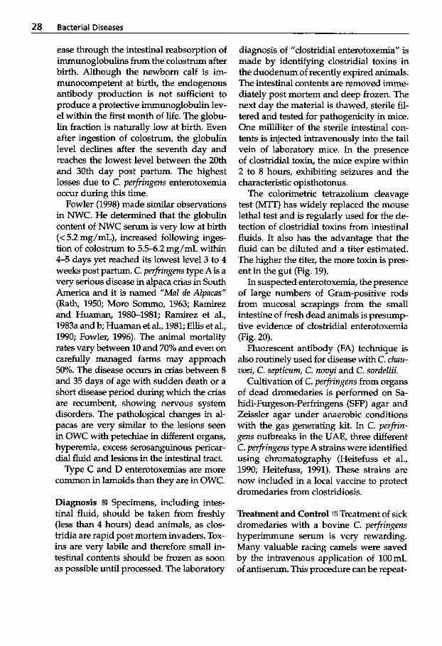
The colorimetric tetrazolium cleavage test (MTT) has widely replaced the mouse lethal test and is regularly used for the de- tection of clostridial toxins from intestinal fluids. It also has the advantage that the fluid can be diluted and a titer estimated. The higher the titer, the more toxin is pres- ent in the gut (Fig. 19).
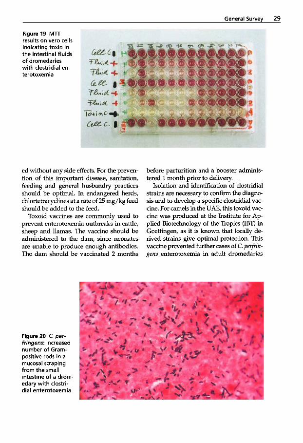
In suspected enterotoxemia, the presence of large numbers of Gram-positive rods from mucosal scrapings from the small intestine of fresh dead animals is presump- tive evidence of clostridial enterotoxemia (Fig. 20).
Fluorescent antibody (FA) technique is also routinely used for disease with C. chau- voei, C. septicum, C. novyi and C. sordellii.
Cultivation of C. perfringens from organs of dead dromedaries is performed on Sa- hidi-Furgeson-Perfringens (SFP) agar and Zeissler agar under anaerobic conditions with the gas generating kit. In C. perfrin- gens outbreaks in the UAE, three different C. perfringens type A strains were identified using chromatography (Heitefuss et al., 1990; Heitefuss, 1991). These strains are now included in a local vaccine to protect dromedaries from clostridiosis.
Treatment and Control Treatment of sick dromedaries with a bovine C. perfringens hyperimmune serum is very rewarding. Many valuable racing camels were saved by the intravenous application of 1OOmL of antiserum. This procedure can be repeat-
General Survev 29
Figure19 MlT results on vero cells indicating toxin in the intestinal fluids of dromedaries with clostridial en- terotoxemia
ed without any side effects. For the preven- tion of this important disease, sanitation, feeding and general husbandry practices should be optimal. In endangered herds, chlortetracyclines at a rate of 25 mg/kg feed should be added to the feed.
Toxoid vaccines are commonly used to prevent enterotoxemia outbreaks in cattle, sheep and llamas. The vaccine should be administered to the dam, since neonates are unable to produce enough antibodies. The dam should be vaccinated 2 months
Figure 20 C. per- fringens: increased number of Gram- positive rods in a mucosal scraping from the small intestine of a drom- edary with clostri- dial enterotoxemia
before parturition and a booster adminis- tered 1 month prior to delivery.
Isolation and identification of clostridial strains are necessary to confirm the diagno- sis and to develop a specific clostridial vac- cine. For camels in the UAE, this toxoid vac- cine was produced at the Institute for Ap- plied Biotechnology of the Tropics (IBT) in Goettingen, as it is known that locally de- rived strains give optimal protection. This vaccine prevented further cases of C. pefrin- gens enterotoxemia in adult dromedaries
30 Bacterial Diseases
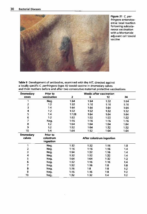
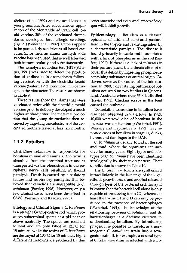
Figure 21 C. per- fringens enterotox- emia: local reaction following subcuta- neous vaccination with a Montanide adjuvant cell toxoid vaccine
Table 9 Development of antibodies, examined with the HIT, directed against a locally specific C. perfringens (type A) toxoid vaccine in dromedary calves and their mothers before and after two consecutive maternal protective vaccinations
Dromedary Prior t o Weeks after vaccination cows vaccination 2 6 12 24
1 Neg. 1 :64 1 :64 1 :32 1 :64 2 1 :2 1 :32 1:16 1:16 1:16 3 1 :4 1 :64 1 :64 1 :64 1 :64 4 1 :2 1:32 1 :32 1 :32 1 :32 5 1 :4 1:128 1 :64 1 :64 1 :64 6 1 :2 1 :32 1:32 1 :32 1:32 7 Neg. 1:16 1:16 1:16 1:16 8 1 :2 1 :64 1 :64 1 :64 1 :64 9 1 :2 1 :32 1 :64 1 :32 1 :32 10 1 :4 I :64 1:32 1 :64 1 :64
Dromedary Prior to
ingestion calves colostrum After colostrum ingestion
1 Neg. 1:32 1 :32 1:16 1 :8 2 Neg. 1:16 1:16 1:16 1 :4 3 Neg. 1 :64 1 :32 1:16 1 :4 4 Neg. 1 :32 1 :32 1 :32 1 :4
6 Neg. 1 :32 1:16 1:16 1 :4 7 Neg. 1 :32 1:16 1 :8 1 :8
5 Neg. 1 :64 1 :64 1 :32 1 :2
8 Neg. 1:16 1 :8 1 :8 1 :2 9 Neg. 1:16 1:16 1 :8 1 :2 10 Neg. 1 :32 1 :32 1 :4 1 :2
General Survey 31
(Seifert et al., 1992) and reduced losses in young animals. After subcutaneous appli- cation of the Montanide adjuvant cell tox- oid vaccine, 30% of the vaccinated drome- daries developed local allergic swellings (Fig. 21) (Seifert et al., 1992). Camels appear to be particularly sensitive to oil-based vac- cines. Since then, an aluminum hydroxide vaccine has been used that is well tolerated both intramuscularly and subcutaneously.
The hemolysis inhibition test (HIT) (Scha- per, 1991) was used to detect the produc- tion of antibodies in dromedaries follow- ing vaccination with the clostridia toxoid vaccine (Seifert, 1992) produced in Goettin- gen in the bioreactor. The results are shown in Table 9.
These results show that dams that were vaccinated twice with the clostridia toxoid vaccine prior to delivery developed a much higher antibody titer. The maternal protec- tion that the young dromedaries then re- ceived by ingesting the colostrum of the vac- cinated mothers lasted at least six months.
1.1.2 Botulism
CZostridium botulinum is responsible for botulism in man and animals. The toxin is absorbed from the intestinal tract and is transported via the bloodstream to the pe- ripheral nerve cells resulting in flaccid paralysis. Death is caused by circulatory failure and respiratory paralysis. It is be- lieved that camelids are susceptible to C. botulinum (Fowler, 1998). However, only a few clinical cases have been described in OWC (Wernery and Kaaden, 1995).
Etiology and Clinical Signs C. botulinum is a straight Gram-positive rod which pro- duces subterminal spores at a pH near or above neutrality. The spores are resistant to heat and are only killed at 121°C for 15 minutes while the toxins of C. botulinum are destroyed at 100°C for 15 minutes. Eight different neurotoxins are produced by this
strict anaerobe and even small traces of oxy- gen will inhibit growth.
Epidemiology Botulism is a classical epidemic of arid and semi-arid pasture- land in the tropics and is distinguished by a characteristic paralysis. The disease is found primarily in cattle and is associated with a lack of phosphorus in the soil (Sei- fert, 1992). If there is a lack of minerals in their pasture grass, the animals attempt to cover this deficit by ingesting phosphorus- containing substances of animal origin. Ca- davers serve as the source of the intoxica- tion. In 1990, a devastating outbreak of bot- ulism occurred on two feedlots in Queens- land, Australia where over 5500 bulls died (Jones, 1991). Chicken scraps in the feed caused the outbreak.
Devastating losses due to botulism have also been observed in waterfowl. In 1983, 40,000 waterfowl died of botulism in the marshes west of Hamburg (Westphal, 1991). Wernery and Haydn-Evans (1992) have re- ported cases of botulism in seagulls, ducks, herons and flamingos in the UAE.
C. botulinum is usually found in the soil and mud, where the organisms can sur- vive for many years. Eight types and sub- types of C. botulinum have been identified serologically by their toxin pattern. Their distribution is shown in Table 10.
The C. botulinurn toxins are synthesized intracellularly in the last stage of the loga- rithmic growth phase and are first released through lysis of the bacterial cell. Today it is known that the bacterial cell alone is only capable of producing toxin C2, whereas at least the toxins C1 and D can only be pro- duced in the presence of bacteriophages (Westphal, 1991). The knowledge of the relationship between C. botulinum and its bacteriophages is a decisive criterion in understanding botulism. By introducing phages, it is possible to transform a non- toxigenic C. botulinum strain into a toxi- genic strain. If, for example, a neutral type of C. botulinum strain is infected with a C1-
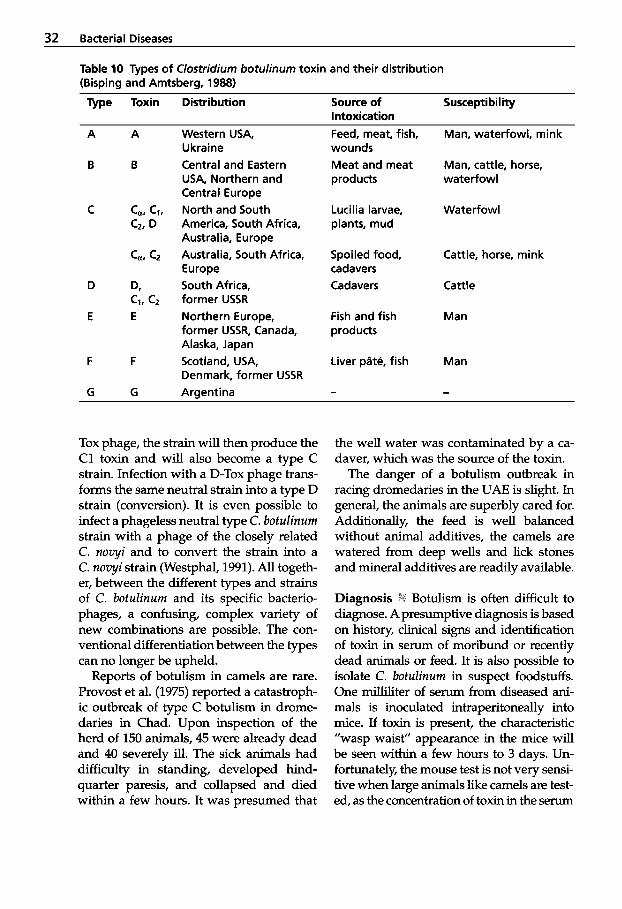
32 Bacterial Diseases
Table 10 Types of Clostridium botulinum toxin and their distribution (Bisping and Amtsberg, 1988)
Type Toxin Distribution Source of Susceptibility Intoxication
Western USA, Feed, meat, fish, Man, waterfowl, mink Ukraine Central and Eastern USA, Northern and Central Europe North and South America, South Africa, Australia, Europe Australia, South Africa, Europe South Africa, former USSR Northern Europe, former USSR, Canada, Alaska, Japan Scotland, USA, Denmark, former USSR Argentina
wounds Meat and meat products
Lucilia larvae, plants, mud
Spoiled food, cadavers Cadavers
Fish and fish products
Liver pdte, fish
Man, cattle, horse, waterfowl
Waterfowl
Cattle, horse, mink
Cattle
Man
Man
-
Tox phage, the strain will then produce the C1 toxin and will also become a type C strain. Infection with a D-Tox phage trans- forms the same neutral strain into a type D strain (conversion). It is even possible to infect a phageless neutral type C. botulinum strain with a phage of the closely related C. novyi and to convert the strain into a C. novyi strain (Westphal, 1991). All togeth- er, between the different types and strains of C. botulinum and its specific bacterio- phages, a confusing, complex variety of new combinations are possible. The con- ventional differentiation between the types can no longer be upheld.
Reports of botulism in camels are rare. Provost et al. (1975) reported a catastroph- ic outbreak of type C botulism in drome- daries in Chad. Upon inspection of the herd of 150 animals, 45 were already dead and 40 severely ill. The sick animals had difficulty in standing, developed hind- quarter paresis, and collapsed and died within a few hours. It was presumed that
the well water was contaminated by a ca- daver, which was the source of the toxin.
The danger of a botulism outbreak in racing dromedaries in the UAE is slight. In general, the animals are superbly cared for. Additionally, the feed is well balanced without animal additives, the camels are watered from deep wells and lick stones and mineral additives are readily available.
Diagnosis tli Botulism is often difficult to diagnose. A presumptive diagnosis is based on history, clinical signs and identification of toxin in serum of moribund or recently dead animals or feed. It is also possible to isolate C. botulinum in suspect foodstuffs. One milliliter of serum from diseased ani- mals is inoculated intraperitoneally into mice. If toxin is present, the characteristic ”wasp waist” appearance in the mice will be seen within a few hours to 3 days. Un- fortunately, the mouse test is not very sensi- tive when large animals like camels are test- ed, as the concentration of toxin in the serum
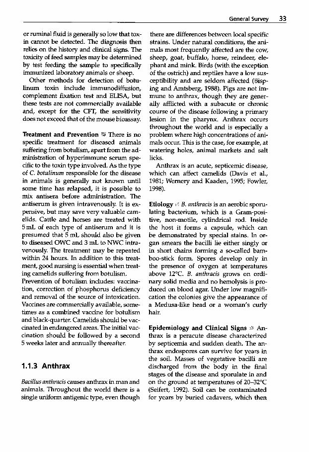
General Survey 33
or ruminal fluid is generally so low that tox- in cannot be detected. The diagnosis then relies on the history and clinical signs. The toxicity of feed samples may be determined by test feeding the sample to specifically immunized laboratory animals or sheep.
Other methods for detection of botu- h u m toxin include immunodiffusion, complement fixation test and ELISA, but these tests are not commercially available and, except for the CFT, the sensitivity does not exceed that of the mouse bioassay.
Treatment and Prevention specific treatment for diseased animals suffering from botulism, apart from the ad- ministration of hyperimmune serum spe- cific to the toxin type involved. As the type of C. botulinum responsible for the disease in animals is generally not known until some time has relapsed, it is possible to mix antisera before administration. The antiserum is given intravenously. It is ex- pensive, but may save very valuable cam- elids. Cattle and horses are treated with 5mL of each type of antiserum and it is presumed that 5 mL should also be given to diseased OWC and 3 mL to NWC intra- venously. The treatment may be repeated within 24 hours. In addition to this treat- ment, good nursing is essential when treat- ing camelids suffering from botulism. Prevention of botulism includes: vaccina- tion, correction of phosphorus deficiency and removal of the source of intoxication. Vaccines are commercially available, some- times as a combined vaccine for botulism and black-quarter. Camelids should be vac- cinated in endangered areas. The initial vac- cination should be followed by a second 5 weeks later and annually thereafter.
1.1.3 Anthrax
Bacillus anthracis causes anthrax in man and animals. Throughout the world there is a single uniform antigenic type, even though
there are differences between local specific strains. Under natural conditions, the ani- mals most frequently affected are the cow, sheep, goat, buffalo, horse, reindeer, ele- phant and mink. Birds (with the exception of the ostrich) and reptiles have a low sus- ceptibility and are seldom affected (Bisp- ing and Amtsberg, 1988). Pigs are not im- mune to anthrax, though they are gener- ally afflicted with a subacute or chronic course of the disease following a primary lesion in the pharynx. Anthrax occurs throughout the world and is especially a problem where high concentrations of ani- mals occur. This is the case, for example, at watering holes, animal markets and salt licks.
Anthrax is an acute, septicemic disease, which can affect camelids (Davis et al., 1981; Wernery and Kaaden, 1995; Fowler, 1998).
Etiology IK B. anthracis is an aerobic sporu- lating bacterium, which is a Gram-posi- tive, non-motile, cylindrical rod. Inside the host it forms a capsule, which can be demonstrated by special stains. In or- gan smears the bacilli lie either singly or in short chains forming a so-called bam- boo-stick form. Spores develop only in the presence of oxygen at temperatures above 12°C. B. anthracis grows on ordi- nary solid media and no hemolysis is pro- duced on blood agar. Under low magnifi- cation the colonies give the appearance of a Medusa-like head or a woman’s curly hair.
Epidemiology and Clinical Signs i b An- thrax is a peracute disease characterized by septicemia and sudden death. The an- thrax endospores can survive for years in the soil. Masses of vegetative bacilli are discharged from the body in the final stages of the disease and sporulate in and on the ground at temperatures of 2032°C (Seifert, 1992). Soil can be contaminated for years by buried cadavers, which then
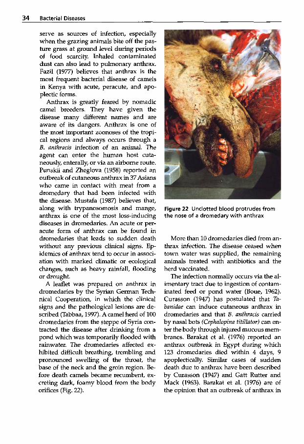
34 Bacterial Diseases
serve as sources of infection, especially when the grazing animals bite off the pas- ture grass at ground level during periods of food scarcity. Inhaled contaminated dust can also lead to pulmonary anthrax. Fazil (1977) believes that anthrax is the most frequent bacterial disease of camels in Kenya with acute, peracute, and apo- plectic forms.
Anthrax is greatly feared by nomadic camel breeders. They have given the disease many different names and are aware of its dangers. Anthrax is one of the most important zoonoses of the tropi- cal regions and always occurs through a B. anthracis infection of an animal. The agent can enter the human host cuta- neously, enterally, or via an airborne route. Punskii and Zheglova (1958) reported an outbreak of cutaneous anthrax in 37Asians who came in contact with meat from a dromedary that had been infected with the disease. Mustafa (1987) believes that, along with trypanosomosis and mange, anthrax is one of the most loss-inducing diseases in dromedaries. An acute or per- acute form of anthrax can be found in dromedaries that leads to sudden death without any previous clinical signs. Ep- idemics of anthrax tend to occur in associ- ation with marked climatic or ecological changes, such as heavy rainfall, flooding or drought.
A leaflet was prepared on anthrax in dromedaries by the Syrian German Tech- nical Cooperation, in which the clinical signs and the pathological lesions are de- scribed (Tabbaa, 1997). A camel herd of 100 dromedaries from the steppe of Syria con- tracted the disease after drinking from a pond which was temporarily flooded with rainwater. The dromedaries affected ex- hibited difficult breathing, trembling and pronounced swelling of the throat, the base of the neck and the groin region. Be- fore death camels became recumbent, ex- creting dark, foamy blood from the body orifices (Fig. 22).
Figure 22 Unclotted blood protrudes from the nose of a dromedary with anthrax
More than 10 dromedaries died from an- thrax infection. The disease ceased when town water was supplied, the remaining animals treated with antibiotics and the herd vaccinated.
The infection normally occurs via the al- imentary tract due to ingestion of contam- inated feed or pond water (Boue, 1962). Curasson (1947) has postulated that Ta- banidue can induce cutaneous anthrax in dromedaries and that B. anthracis carried by nasal bots (CqhaIopina titillator) can en- ter the body through injured mucous mem- branes. Barakat et al. (1976) reported an anthrax outbreak in Egypt during which 123 dromedaries died within 4 days, 9 apoplectically. Similar cases of sudden death due to anthrax have been described by Curasson (1947) and Gatt Rutter and Mack (1963). Barakat et al. (1976) are of the opinion that an outbreak of anthrax in
General Survev 35
a dromedary herd was due to migrating birds. The outbreak was controlled by strict hygienic measures, administration of pro- caine penicillin and 50 mL of anthrax anti- serum per dromedary over 5 days.
The clinical signs of anthrax in drome- daries are similar to those in the cow (Gatt Rutter and Mack, 1963): fever up to 42"C, extravasation of tar-like blood from the body orifices, diarrhea, colic, bloat and severe cardiovascular and pulmonary dis- turbances. Some dromedaries develop painful swellings on the throat and neck.
In NWC the clinical signs described for anthrax resemble those seen in OWC. Sud- den death without any signs may occur as well as subcutaneous swellings on various parts of the body. Bloody discharge may exude from all body orifices and lamoids may die after 1 to 3 days (Fowler, 1998).
Pathology The principal lesions in sep- ticemic anthrax in animals are hemor- rhages, edema and necrosis. In drome- daries, there is evidence of rapid post mortem decomposition (Tabbaa, 1997) of the carcass with oozing of bloodstained fluid from nose, mouth and anus. Dark- red, poorly clotted blood, petechiae and ecchymoses are observed throughout the carcass. An enlarged pulpy spleen, whch is the most characteristic feature at necrop- sy in ruminants, has also been described in camelids (Manefield and Tinson, 1996). There is no rigor mortis and the blood fails to clod. Splenomegaly with black tar- ry pulp, generalized congestion and lung edema were also observed by Boue (1962) and Richard (1975).
Diagnosis B. anthracis is easily cultured from blood and tissues. However, if an- thrax is suspected one should avoid a necropsy to exclude contamination of the soil with spores. A small quantity of blood is sufficient for the diagnosis. A smear or a culture as well as a fluorescent antibody test (FAT) will confirm the diagnosis. In
advanced autolysis, when no anthrax ba- cilli are demonstrable, the thermo-precipi- tation of Ascoli can be applied. For the cul- tivation of B. unthrucis in laboratory ani- mals, white mice are the animals of choice. They are subcutaneously infected and will die within 2 to 4 days. A gelatinous edema develops at the injection site.
Prevention and Control To prevent sporulation of B. unthrucis, carcasses should not be opened. They should be incinerated with the contaminated bedding. After con- tact, equipment must be properly disinfec- ted. The following disinfectant solutions can be used: - 10% hot caustic soda solution, - 4% formaldehyde solution, - 7% hydrogen peroxide, - 2% glutaraldehyde, - calcium hypochloride with 5% active
chlorine.
B. anthrucis is susceptible to many anti- biotics, including penicillin and tetracy- clines.
Pasteur developed the first effective B. unthrucis vaccine. It was replaced by the live, avirulent, spore vaccine devel- oped by Steme. This vaccine has been used worldwide with great economic value to the livestock industry and to wildlife. A single inoculation provides effective im- munity for 9 months, but annual booster vaccinations are recommended. Anthrax can be a serious danger to camelids and it is therefore recommended to vaccinate Cumelidue in endangered areas. However, anthrax vaccines should be carefully used in camelids and the dose adjusted to the weight of the animal, since bacteria-in- duced anthrax has been reported in young llamas (Cartwright et al., 1987). OWC should be given the dose of cattle and NWC should receive the dose that is rec- ommended for sheep. A half sheep dose is recommended for NWC weaners (Fowler, 1998).
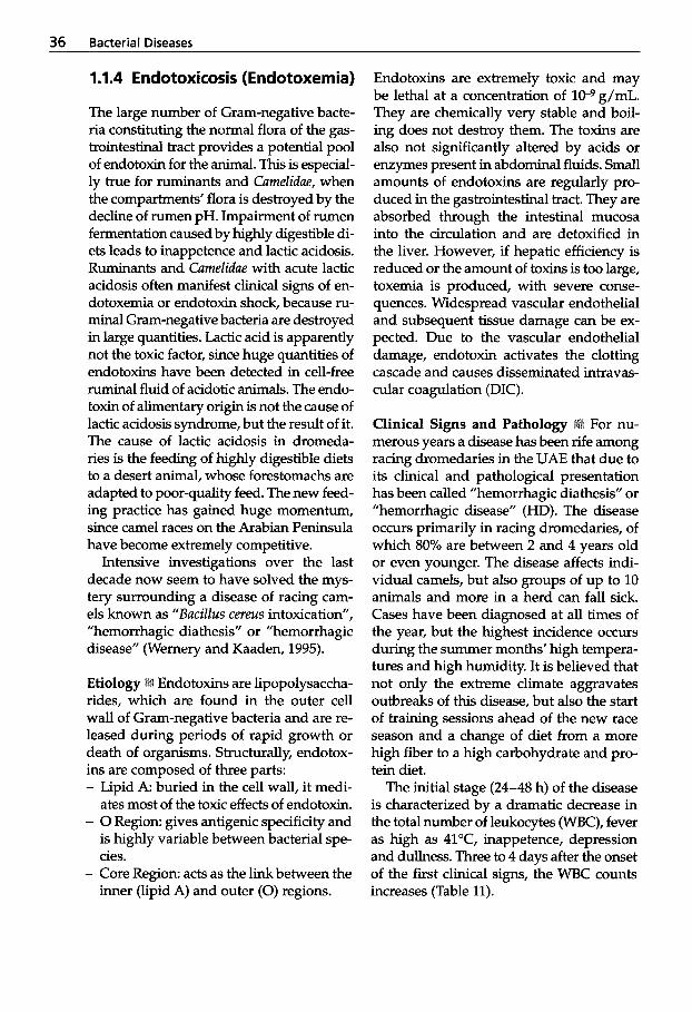
36 Bacterial Diseases
1.1.4 Endotoxicosis (Endotoxemia)
The large number of Gram-negative bacte- ria constituting the normal flora of the gas- trointestinal tract provides a potential pool of endotoxin for the animal. This is especial- ly true for ruminants and Cumelidue, when the compartments’ flora is destroyed by the decline of rumen pH. Impairment of rumen fermentation caused by highly digestible di- ets leads to inappetence and lactic acidosis. Ruminants and Cumelidue with acute lactic acidosis often manifest clinical signs of en- dotoxemia or endotoxin shock, because ru- minal Gram-negative bacteria are destroyed in large quantities. Lactic acid is apparently not the toxic factor, since huge quantities of endotoxins have been detected in cell-free ruminal fluid of acidotic animals. The endo- toxin of alimentary origin is not the cause of lactic acidosis syndrome, but the result of it. The cause of lactic acidosis in dromeda- ries is the feeding of highly digestible diets to a desert animal, whose forestomachs are adapted to poor-quality feed. The new feed- ing practice has gained huge momentum, since camel races on the Arabian Peninsula have become extremely competitive.
Intensive investigations over the last decade now seem to have solved the mys- tery surrounding a disease of racing cam- els known as “ B u d u s cereus intoxication”, “hemorrhagic diathesis” or ”hemorrhagic disease” (Wernery and Kaaden, 1995).
Endotoxins are lipopolysaccha- rides, which are found in the outer cell wall of Gram-negative bacteria and are re- leased during periods of rapid growth or death of organisms. Structurally, endotox- ins are composed of three parts: - Lipid A buried in the cell wall, it medi-
ates most of the toxic effects of endotoxin. - 0 Region: gives antigenic specificity and
is highly variable between bacterial spe- cies.
- Core Region: acts as the link between the inner (lipid A) and outer (0) regions.
Endotoxins are extremely toxic and may be lethal at a concentration of 10-9 g/mL. They are chemically very stable and boil- ing does not destroy them. The toxins are also not significantly altered by acids or enzymes present in abdominal fluids. Small amounts of endotoxins are regularly pro- duced in the gastrointestinal tract. They are absorbed through the intestinal mucosa into the circulation and are detoxified in the liver. However, if hepatic efficiency is reduced or the amount of toxins is too large, toxemia is produced, with severe conse- quences. Widespread vascular endothelial and subsequent tissue damage can be ex- pected. Due to the vascular endothelial damage, endotoxin activates the clotting cascade and causes disseminated intravas- d a r coagulation (DIC).
Clinical Signs and Pathology merous years a disease has been rife among racing dromedaries in the UAE that due to its clinical and pathological presentation has been called ”hemorrhagic diathesis” or ”hemorrhagic disease” (HD). The disease occurs primarily in racing dromedaries, of which 80% are between 2 and 4 years old or even younger. The disease affects indi- vidual camels, but also groups of up to 10 animals and more in a herd can fall sick. Cases have been diagnosed at all times of the year, but the highest incidence occurs during the summer months’ high tempera- tures and high humidity. It is believed that not only the extreme climate aggravates outbreaks of this disease, but also the start of training sessions ahead of the new race season and a change of diet from a more high fiber to a high carbohydrate and pro- tein diet.
The initial stage (24-48 h) of the disease is characterized by a dramatic decrease in the total number of leukocytes (WBC), fever as high as 41”C, inappetence, depression and dullness. Three to 4 days after the onset of the first clinical signs, the WBC counts increases (Table 11).
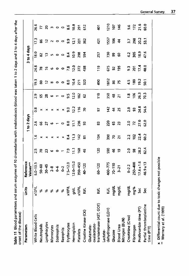
Tabl
e 11
Blo
od p
aram
eter
s an
d se
rum
enz
ymes
of
10 d
rom
edar
ies
wit
h e
ndot
oxic
osis
(blo
od w
as ta
ken
1 to
2 d
ays
and
3 to
4 d
ays
afte
r the
on
set o
f the
dis
ease
)
Par
amet
ers
Whi
te B
lood
Cel
ls
Neu
trop
hils
Ly
mph
ocyt
es
M o n
ocyt
es
Eos
inop
hils
B
asop
hils
E
ryth
rocy
tes
Hem
oglo
bin
Pla
tele
ts
Cre
atin
e K
inas
e (C
K)
Glu
tam
ate-
ox
alac
etat
e-
trans
amin
ase
(AST
, GO
T)
Lact
ate-
de
hydr
ogen
ase
(LD
H)
Glu
cose
B
lood
Ure
a N
itrog
en (B
UN
) C
reat
inin
e (C
rea)
Fi
brin
ogen
P
roth
rom
bin
time
(PT)
P
artia
l thr
ombo
plas
tine
time
(PIT
)
Uni
ts
XI 03
1~
%
%
%
%
%
XI O
VL
g/d
L
I U/L
XI
03
1~
I U/L
I U/L
m
g/dL
m
g/dL
mg/
dL
mg%
Se
c Se
c
Ref
eren
ce
Val
ues*
*
50-6
0 30
-45
2-8
6.0-
13.5
0-6
0-2
7.5-
12.0
12
.0-1
5.0
350-
450
40-1
20
60-1
20
400-
775
70-1
10
3-2
1
0-2.
2
250-
400
17.6
k1.6
46
.9 i 1
3
2.5 70
23
6 0 1 7.9
11.1
168
46
120
590
46
19
2.0 98
28.2
82
.4
1 to
2 d
ays
1.6
2.6
0.8
X
66
X
X
27
X
X
6 X
X
1 X
X
0 X
8.4
8.0
9.0
11.3
11
.1
12.2
142
236
116
81
93
70
104
83
97
390
220
142
70
65
44
21
23
25
2.2
2.0
2.0
102
72
93
22.4
27
.0
24.8
60
.2
62.0
54
.6
-
2.9 65
28
6 1 0 9.5
12.0
182
62
110
350
48
21
1.8
106
26.3
70
.3
3 to
4 d
ays
19.3
80
12
8 0 0 8.0
10.4
27 1
320
24.8
78
16
6 0 0 7.8
12.0
372
438
18.0
82
13
5 0 0 8.4
10.9
298
594
17.3
86
12
2 0 0 9.9
12.1
20
1
362
26.6
77
20
3 0 0 8.6
10.8
291
612
490
119
257
421
401
1812
67
5 73
0 15
57
1210
86
92
99
10
6 10
7 75
19
5 60
44
14
6
4.5
9.3
4.2
3.7
9.6
180
201
305
298
172
19.2
17
.4
18.9
20
.2
21.6
50
.1
48.0
47
.6
53.1
60
.0
x D
iffer
entia
l cou
nt d
ue t
o to
xic
chan
ges
no
t pos
sibl
e **
Wer
nery
et a
l. (1
999)
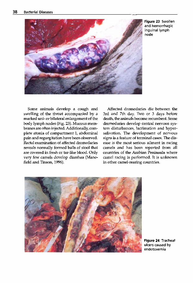
38 Bacterial Diseases
Figure 23 Swollen and hemorrhagic inguinal lymph node
Some animals develop a cough and swelling of the throat accompanied by a marked uni- or bilateral enlargement of the body lymph nodes (Fig. 23). Mucous mem- branes are often injected. Additionally, com- plete atonia of compartment 1, abdominal pain and regurgitation have been observed. Rectal examination of affected dromedaries reveals normally formed balls of stool that are covered in fresh or tar-like blood. Only very few camels develop diarrhea (Mane- field and Tinson, 1996).
Affected dromedaries die between the 3rd and 7th day. Two or 3 days before death, the animals become recumbent. Some dromedaries develop central nervous sys- tem disturbances, lacrimation and hyper- salivation. The development of nervous signs is a feature of terminal cases. The dis- ease is the most serious ailment in racing camels and has been reported from all countries of the Arabian Peninsula where camel racing is performed. It is unknown in other camel-rearing countries.
Figure 24 Tracheal ulcers caused by endotoxemia
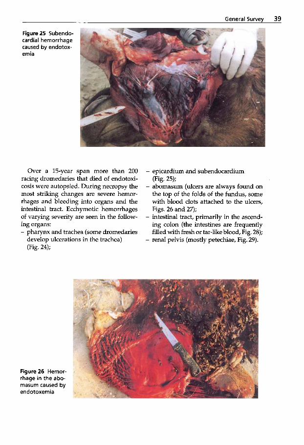
General Survev 39
Figure 25 Subendo- cardial hemorrhage caused by endotox- emia
Over a 15-year span more than 200 racing dromedaries that died of endotoxi- cosis were autopsied. During necropsy the most striking changes are severe hemor- rhages and bleeding into organs and the intestinal tract. Ecchymotic hemorrhages of varying severity are seen in the follow- ing organs: - pharynx and trachea (some dromedaries
develop ulcerations in the trachea) (Fig. 24);
- epicardium and subendocardium (Fig. 25);
- abomasum (ulcers are always found on the top of the folds of the fundus, some with blood clots attached to the ulcers, Figs. 26 and 27);
- intestinal tract, primarily in the ascend- ing colon (the intestines are frequently filled with fresh or tar-like blood, Fig. 28);
- renal pelvis (mostly petechiae, Fig. 29).
Figure 26 Hemor- rhage in the abo- masum caused by endotoxemia
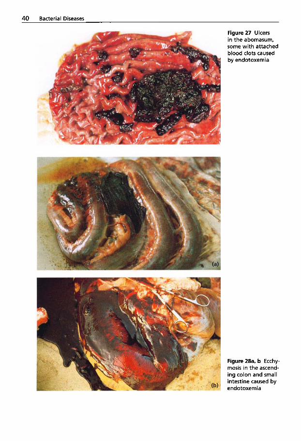
40 Bacterial Diseases
Figure 27 Ulcers in the abomasum, some with attached blood clots caused by endotoxemia
Figure 28a, b Ecchy- mosis in the ascend- ing colon and small intestine caused by endotoxemia
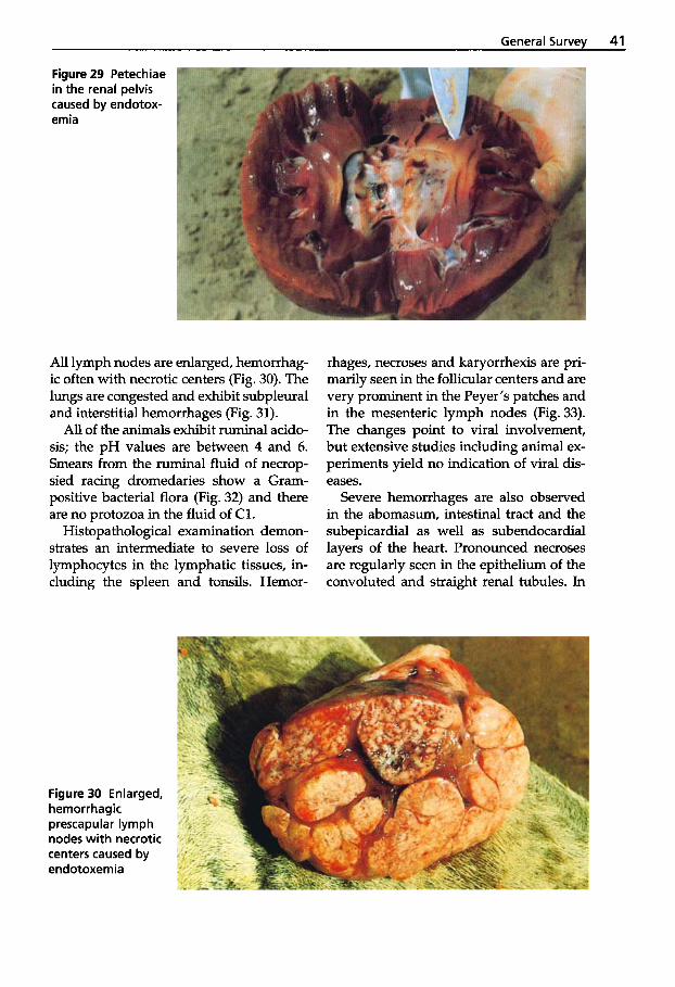
General Survev 41
Figure 29 Petechiae in the renal pelvis caused by endotox- emia
All lymph nodes are enlarged, hemorrhag- ic often with necrotic centers (Fig. 30). The lungs are congested and exhibit subpleural and interstitial hemorrhages (Fig. 31).
All of the animals exhibit ruminal acido- sis; the pH values are between 4 and 6. Smears from the ruminal fluid of necrop- sied racing dromedaries show a Gram- positive bacterial flora (Fig. 32) and there are no protozoa in the fluid of C1.
Histopathological examination demon- strates an intermediate to severe loss of lymphocytes in the lymphatic tissues, in- cluding the spleen and tonsils. Hemor-
rhages, necroses and karyorrhexis are pri- marily seen in the follicular centers and are very prominent in the Peyer’s patches and in the mesenteric lymph nodes (Fig.33). The changes point to viral involvement, but extensive studies including animal ex- periments yield no indication of viral dis- eases.
Severe hemorrhages are also observed in the abomasum, intestinal tract and the subepicardial as well as subendocardial layers of the heart. Pronounced necroses are regularly seen in the epithelium of the convoluted and straight renal tubules. In
Figure 30 Enlarged, hemorrhagic prescapular lymph nodes with necrotic centers caused by endotoxemia
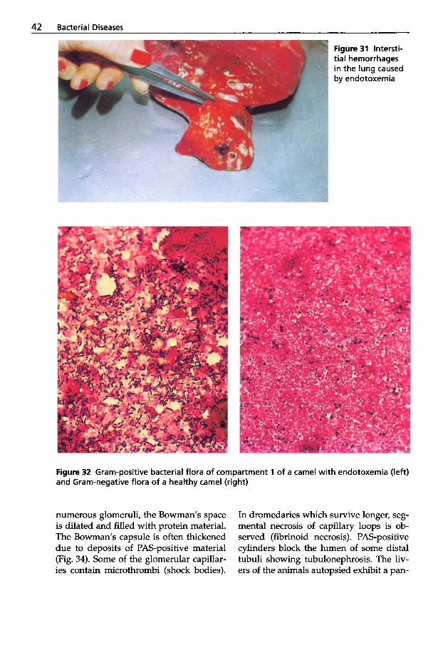
42 Bacterial Diseases
Figure 31 Intersti- tial hemorrhages in the lung caused by endotoxemia
Figure 32 Gram-positive bacterial flora of compartment 1 of a camel with endotoxemia (left) and Gram-negative flora of a healthy camel (right)
numerous glomeruli, the Bowman’s space is dilated and filled with protein material. The Bowman’s capsule is often thickened due to deposits of PAS-positive material (Fig. 34). Some of the glomerular capillar- ies contain microthrombi (shock bodies).
In dromedaries which survive longer, seg- mental necrosis of capillary loops is ob- served (fibrinoid necrosis). PAS-positive cylinders block the lumen of some distal tubuli showing tubulonephrosis. The liv- ers of the animals autopsied exhibit a pan-
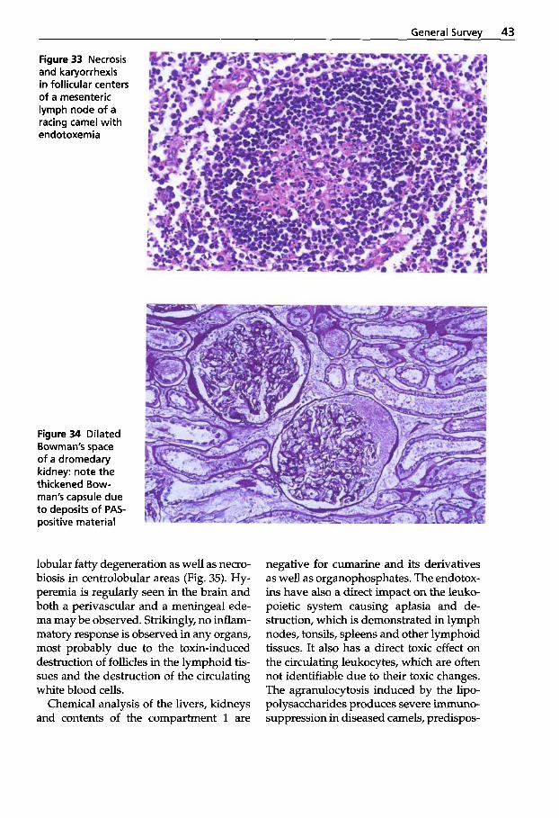
General Survev 43
Figure 33 Necrosis and karyorrhexis in follicular centers of a mesenteric lymph node of a racing camel with endotoxemia
Figure 34 Dilated Bowman's space of a dromedary kidney: note the thickened Bow- man's capsule due to deposits of PAS- positive material
lobular fatty degeneration as well as necro- biosis in centrolobular areas (Fig. 35). Hy- peremia is regularly seen in the brain and both a perivascular and a meningeal ede- ma may be observed. Strikingly, no inflam- matory response is observed in any organs, most probably due to the toxin-induced destruction of follicles in the lymphoid tis- sues and the destruction of the circulating white blood cells.
Chemical analysis of the livers, kidneys and contents of the compartment 1 are
negative for cumarine and its derivatives as well as organophosphates. The endotox- ins have also a direct impact on the leuko- poietic system causing aplasia and de- struction, which is demonstrated in lymph nodes, tonsils, spleens and other lymphoid tissues. It also has a direct toxic effect on the circulating leukocytes, which are often not identifiable due to their toxic changes. The agranulocytosis induced by the lipo- polysaccharides produces severe immuno- suppression in diseased camels, predispos-
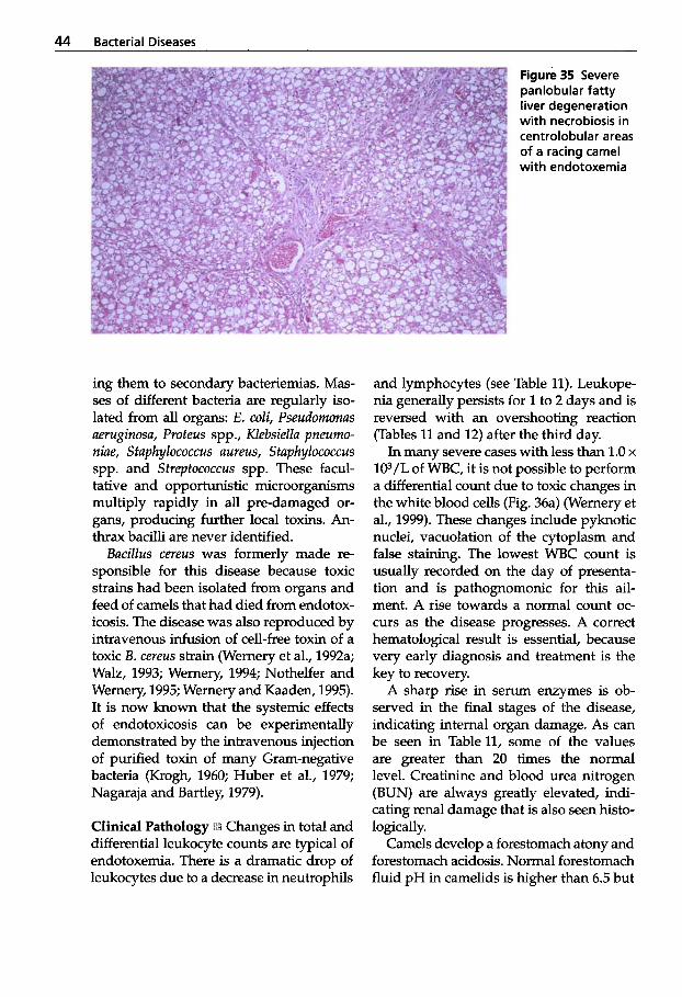
44 Bacterial Diseases
ing them to secondary bacteriemias. Mas- ses of different bacteria are regularly iso- lated from all organs: E . coli, Pseudomonas aeruginosa, Proteus spp., Klebsiella pneumo- niae, Staphylococcus aureus, Staphylococcus spp. and Streptococcus spp. These facul- tative and opportunistic microorganisms multiply rapidly in all pre-damaged or- gans, producing further local toxins. An- thrax bacilli are never identified.
Bacillus cereus was formerly made re- sponsible for this disease because toxic strains had been isolated from organs and feed of camels that had died from endotox- icosis. The disease was also reproduced by intravenous infusion of cell-free toxin of a toxic B. cereus strain (Wernery et al., 1992a; Walz, 1993; Wernery, 1994; Nothelfer and Wernery, 1995; Wernery and Kaaden, 1995). It is now known that the systemic effects of endotoxicosis can be experimentally demonstrated by the intravenous injection of purified toxin of many Gram-negative bacteria (Krogh, 1960; Huber et al., 1979; Nagaraja and Bartley, 1979).
Clinical Pathology Changes in total and differential leukocyte counts are typical of endotoxemia. There is a dramatic drop of leukocytes due to a decrease in neutrophils
Figure 35 Severe panlobular fatty liver degeneration with necrobiosis in centrolobular areas of a racing camel with endotoxemia
and lymphocytes (see Table 11). Leukope- nia generally persists for 1 to 2 days and is reversed with an overshooting reaction (Tables 11 and 12) after the third day.
In many severe cases with less than 1.0 x lO3/L of WBC, it is not possible to perform a differential count due to toxic changes in the white blood cells (Fig. 36a) (Wernery et al., 1999). These changes include pyknotic nuclei, vacuolation of the cytoplasm and false staining. The lowest WBC count is usually recorded on the day of presenta- tion and is pathognomonic for this ail- ment. A rise towards a normal count oc- curs as the disease progresses. A correct hematological result is essential, because very early diagnosis and treatment is the key to recovery.
A sharp rise in serum enzymes is ob- served in the final stages of the disease, indicating internal organ damage. As can be seen in Tablell, some of the values are greater than 20 times the normal level. Creatinine and blood urea nitrogen (BUN) are always greatly elevated, indi- cating renal damage that is also seen histo- logically.
Camels develop a forestomach atony and forestomach acidosis. Normal forestomach fluid pH in camelids is higher than 6.5 but
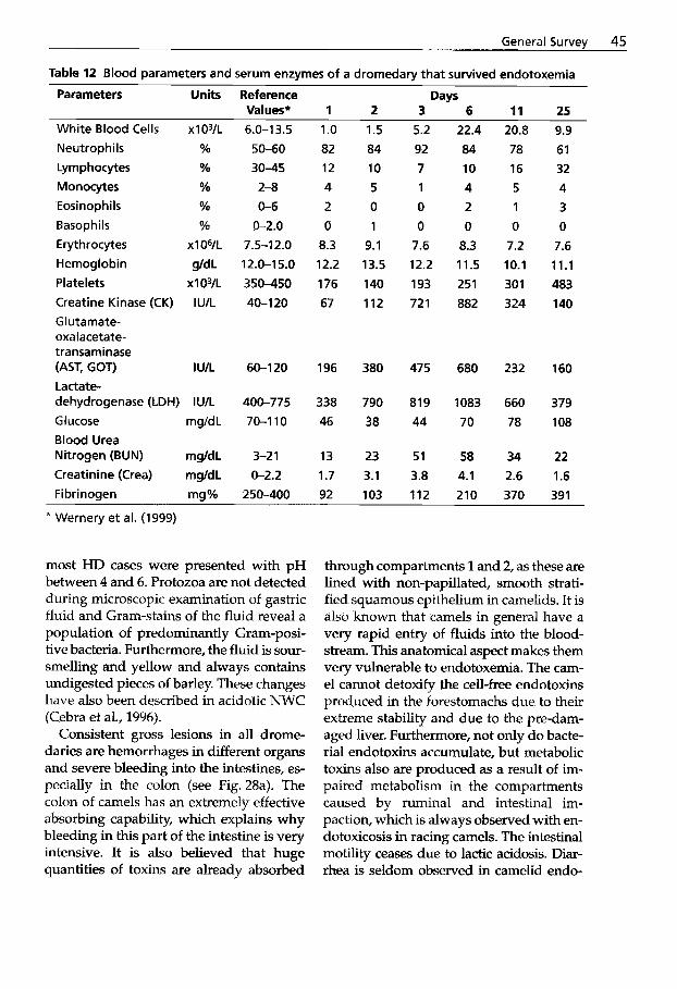
General Survey 45
Table 12 Blood parameters and serum enzymes of a dromedary that survived endotoxemia
Values* 1 2 3 6 11 25 Parameters Units Reference Days
White Blood Cells xlO3/L
Neutrophils % Lymphocytes % Monocytes Yo Eosinophils % Basoph i Is % Erythrocytes x 1 OYL Hemoglobin g/d L Platelets x 1 031~ Creatine Kinase (CK) IU/L Glutarnate- oxalacetate- transaminase (AST, GOT) I U/L Lactate- dehydrogenase (LDH) lU/L Glucose rng/dL Blood Urea Nitrogen (BUN) mg/dL Creatinine (Crea) mg/dL Fibrinoqen mq%
6.0-13.5
50-60 30-45 2-8 0-6
0-2.0 7.5-12.0 12.0-1 5.0
350-450 40-1 20
60-1 20
400-775
70-1 10
3-2 1
0-2.2 250-400
1 .o a2 12 4 2 0
8.3 12.2
176 67
196
338 46
13 1.7 92
1.5 84 10 5 0 1
9.1 13.5 140
112
380
790 38
23 3.1 103
5.2 92 7 1 0 0
7.6 12.2 193
721
475
819 44
51 3.8 112
* Wernery et al. (1999)
most HD cases were presented with pH between 4 and 6. Protozoa are not detected during microscopic examination of gastric fluid and Gram-stains of the fluid reveal a population of predominantly Gram-posi- tive bacteria. Furthermore, the fluid is sour- smelling and yellow and always contains undigested pieces of barley. These changes have also been described in acidotic NWC (Cebra et al., 1996).
Consistent gross lesions in all drome- daries are hemorrhages in different organs and severe bleeding into the intestines, es- pecially in the colon (see Fig. 28a). The colon of camels has an extremely effective absorbing capability, which explains why bleeding in this part of the intestine is very intensive. It is also believed that huge quantities of toxins are already absorbed
22.4 84 10 4 2
0 8.3 11.5
251 882
680
1083 70
58 4.1 210
20.8 78 16 5 1
0 7.2 10.1
301 324
232
660 78
34 2.6 370
9.9
61 32 4 3 0
7.6 11.1 483 140
160
379
108
22
1.6 39 1
through compartments 1 and 2, as these are lined with non-papillated, smooth strati- fied squamous epithelium in camelids. It is also known that camels in general have a very rapid entry of fluids into the blood- stream. This anatomical aspect makes them very vulnerable to endotoxemia. The cam- el cannot detoxify the cell-free endotoxins produced in the forestomachs due to their extreme stability and due to the pre-dam- aged liver. Furthermore, not only do bacte- rial endotoxins accumulate, but metabolic toxins also are produced as a result of im- paired metabolism in the compartments caused by ruminal and intestinal im- paction, which is always observed with en- dotoxicosis in racing camels. The intestinal motility ceases due to lactic acidosis. Diar- rhea is seldom observed in camelid endo-
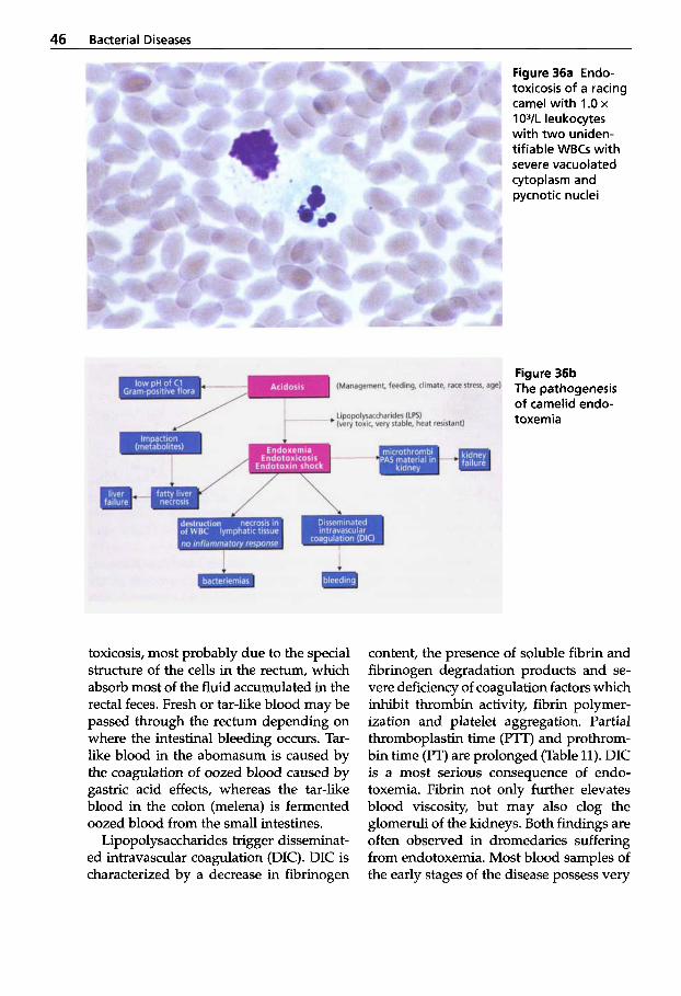
46 Bacterial Diseases
toxicosis, most probably due to the special structure of the cells in the rectum, which absorb most of the fluid accumulated in the rectal feces. Fresh or tar-like blood may be passed through the rectum depending on where the intestinal bleeding occurs. Tar- like blood in the abomasum is caused by the coagulation of oozed blood caused by gastric acid effects, whereas the tar-like blood in the colon (melena) is fermented oozed blood from the small intestines.
Lipopolysaccharides trigger disseminat- ed intravascular coagulation (DIC). DIC is characterized by a decrease in fibrinogen
Figure 36a Endo- toxicosis of a racing camel with 1.0 x 1 OYL leukocytes with two uniden- tifiable WBCs with severe vacuolated cytoplasm and pycnotic nuclei
Figure 36b The pathogenesis of camelid endo- toxemia
content, the presence of soluble fibrin and fibrinogen degradation products and se- vere deficiency of coagulation factors which inhibit thrombin activity, fibrin polymer- ization and platelet aggregation. Partial thromboplastin time (PTT) and prothrom- bin time (PT) are prolonged (Table 11). DIC is a most serious consequence of endo- toxemia. Fibrin not only further elevates blood viscosity, but may also clog the glomeruli of the kidneys. Both findings are often observed in dromedaries suffering from endotoxemia. Most blood samples of the early stages of the disease possess very
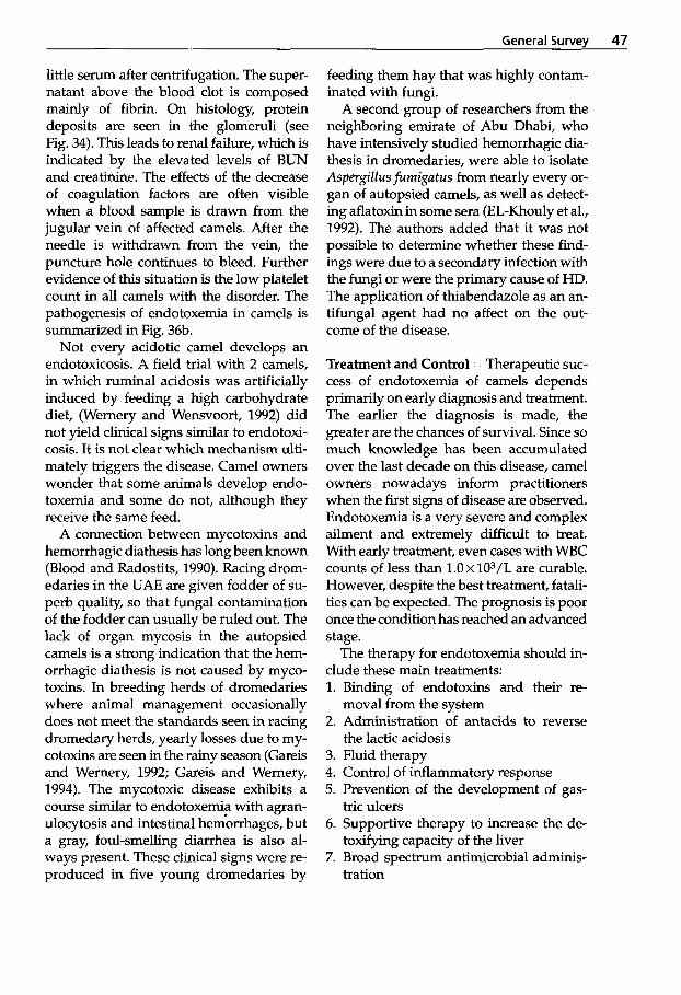
General Survev 47
little serum after centrifugation. The super- natant above the blood clot is composed mainly of fibrin. On histology, protein deposits are seen in the glomeruli (see Fig. 34). This leads to renal failure, which is indicated by the elevated levels of BUN and creatinine. The effects of the decrease of coagulation factors are often visible when a blood sample is drawn from the jugular vein of affected camels. After the needle is withdrawn from the vein, the puncture hole continues to bleed. Further evidence of this situation is the low platelet count in all camels with the disorder. The pathogenesis of endotoxemia in camels is summarized in Fig. 36b.
Not every acidotic camel develops an endotoxicosis. A field trial with 2 camels, in which ruminal acidosis was artificially induced by feeding a high carbohydrate diet, (Wernery and Wensvoort, 1992) did not yield clinical signs similar to endotoxi- cosis. It is not clear which mechanism ulti- mately triggers the disease. Camel owners wonder that some animals develop endo- toxemia and some do not, although they receive the same feed.
A connection between mycotoxins and hemorrhagic diathesis has long been known (Blood and Radostits, 1990). Racing drom- edaries in the UAE are given fodder of su- perb quality, so that fungal contamination of the fodder can usually be ruled out. The lack of organ mycosis in the autopsied camels is a strong indication that the hem- orrhagic diathesis is not caused by myco- toxins. In breeding herds of dromedaries where animal management occasionally does not meet the standards seen in racing dromedary herds, yearly losses due to my- cotoxins are seen in the rainy season (Gareis and Wernery, 1992; Gareis and Wernery, 1994). The mycotoxic disease exhibits a course similar to endotoxemi? with agran- ulocytosis and intestinal hemorrhages, but a gray, foul-smelling diarrhea is also al- ways present. These clinical signs were re- produced in five young dromedaries by
feeding them hay that was highly contam- inated with fungi.
A second group of researchers from the neighboring emirate of Abu Dhabi, who have intensively studied hemorrhagic dia- thesis in dromedaries, were able to isolate Aspergillus fumigatus from nearly every or- gan of autopsied camels, as well as detect- ing aflatoxin in some sera (EL-Khouly et al., 1992). The authors added that it was not possible to determine whether these find- ings were due to a secondary infection with the fungi or were the primary cause of HD. The application of thiabendazole as an an- tifungal agent had no affect on the out- come of the disease.
Treatment and Control Therapeutic suc- cess of endotoxemia of camels depends primarily on early diagnosis and treatment. The earlier the diagnosis is made, the greater are the chances of survival. Since so much knowledge has been accumulated over the last decade on this disease, camel owners nowadays inform practitioners when the first signs of disease are observed. Endotoxemia is a very severe and complex ailment and extremely difficult to treat. With early treatment, even cases with WBC counts of less than 1.0 x 10³/L are curable. However, despite the best treatment, fatali- ties can be expected. The prognosis is poor once the condition has reached an advanced stage.
The therapy for endotoxemia should in- clude these main treatments: 1. Binding of endotoxins and their re-
moval from the system 2. Administration of antacids to reverse
the lactic acidosis 3. Fluid therapy 4. Control of inflammatory response 5. Prevention of the development of gas-
6. Supportive therapy to increase the de-
7. Broad spectrum antimicrobial adminis-
tric ulcers
toxifying capacity of the liver
tration
48 Bacterial Diseases
8. Activation of the coagulation system 9. Prevention of cerebral corticonecrosis
(CCN).
To avoid endotoxicosis, special attention is to be paid to feeding. It is still common practice in the UAE to feed racing camels with cow milk, dates, excessive barley and fresh alfalfa. A better balance between high energetic diet and roughage has to be achieved. If carbohydrate overload occurs, a laxative such as liquid paraffin should be given by gastric tube to reduce the gas- trointestinal transit time and to avoid any impaction and bacterial proliferation and endotoxin absorption. A more stringent laxative may be used in severe impaction with magnesium sulfate at a dose of 500 to 1000 g per animal. It is believed that char- coal also administered through a gastric tube, at a dose of 500 g daily (for a 400 kg racing camel), may reduce the cell-free en- dotoxin by adsorption.
Polymyxin B is an antimicrobial drug with a good affinity for lipid, a portion of endotoxin. This drug is toxic in horses with severe side effects of renal damage, but should be tried in camels suffering from endotoxemia due to its known endotoxin- binding capacity. The suggested dose rate in equines is 6000 W/kg or 2.5 mg/kg poly- myxin B sulfate, diluted in up to 5 liters of saline and given by slow i.v. infusion.
It has been demonstrated in a number of studies in equines that the administration of an antiserum directed against the core region of the endotoxin reduces mortality. The recommended dose in horses is 1 to 2 mL/kg body weight (Gaffin, 1987). It is available from Veterinary Dynamics as Hypermune-J@ and from Immvac, Colum- bia, MO 65201 as Endoserum@. Ideally, the treatment with these sera should start in peracute cases. Stegantox 60@ (Schering- Plough Animal Health) is a freeze-dried, purified endotoxin-specific IgG and, in the presence of complement, the product is also bactericidal against many Gram-nega-
tive microorganisms. The content of one 60 mg vial provides a single dose for an an- imal of 200 kg body weight. This product has already been used in camels suffering from endotoxemia.
In endotoxemia, oxygen-free radicals lead to tissue damage and inflammation. Dimethyl sulfoxide (DMSO) should be given as a 10 to 20% solution with isotonic fluids or water through gastric tube. In horses a dose of 250 mg to 1 g/kg every 12 hours is recommended.
Non-steroidal anti-inflammatory drugs are very important in the control of the in- flammatory response, which always fol- lows the endotoxin shock.
Phenylbutazone, ketoprofen or flunixin should be used to block the formation of inflammatory mediators. Finadyne has been shown to be efficient in horses since it possesses an anti-endotoxic, analgesic, anti-inflammatory and anti-pyretic effect, but it is toxic for camels.
Correction of the circulating fluid deficits is an important procedure to save camels from endotoxic death. Great quantities of fluids are lost by internal bleeding due to the impairment of capillary wall integrity. Furthermore, to correct acidosis, a rigorous fluid replacement therapy should immedi- ately take place. A 5% sodium bicarbonate solution should be given i.v. at a dose of 5 l/camel in the very early stages of the ill- ness followed by an i.v. infusion of 601/ camel of a 1.3% sodium bicarbonate solu- tion in saline with dextrose. In addition to this therapy, antacids should be adminis- tered twice daily through a gastric tube in- cluding 500 g/camel of magnesium hydrox- ide dissolved in warm water. This treatment has to be repeated daily for several days. In severe cases of ruminal lactic acidosis and endotoxemia involving very valuable camels, a rumenotomy should be consid- ered. Compartment 1 should be emptied and washed out with a siphon and the com- partment ingesta replaced by ingesta from healthy ruminants such as sheep and goats.
General Survev 49
For the prevention of gastric ulcers, the substituted benzimidazole, omeprazole, should be orally given at a dose of 0.7 mg/ kg body weight. This drug prevents the se- cretion of gastric acid through blocking the H+ and K+ ATPase.
In endotoxemia, hepatic function is also compromised. Because of the role the liver plays in the storage, activation and synthe- sis of many vitamins and, because of its detoxifying capability, multiple vitamins (including K) and liver stimulants should be administered.
Consumption coagulopathy may be stopped by the administration of heparin, but this treatment has not been tried in camelids. To help prevention of polioen- cephalomalacia thiamine hydrochloride at a dose of 2.5 to 10 mg/kg should be given i.v. or i.m.
Endovac-Bov@, a vaccine against E. coli mastitis, has also been tried against en- dotoxicosis in camelids. The vaccine en- hances both the T- and B-lymphocytes, and in combination with the mutant Re-17 bac- terin it seems to protect against other en- dotoxin-mediated diseases. Its efficacy has not been proven in camelids.
Hundreds of valuable racing camels have succumbed to endotoxemia on the Arabian Peninsula. Since much is now known about the pathogenesis of this devastating dis- ease, research should be directed into the prevention and prophylaxis of endotox- emia. It has been shown that even modest grain feeding can cause severe acidosis with fatal consequences (Cebra et al., 1996). The practices of feeding cow milk, dates, hon- ey, excessive uncrushed barley and alfalfa should be carefully considered as well as the prophylactic administration of pro- biotics, antisera to endotoxins and para- munity inducer like Baypamun@. Further- more, training of very young racing cam- els should be avoided in order to reduce stress.
1.1.5 Pasteurellosis
Pasteurella species have a worldwide dis- tribution with a wide host spectrum. Most pasteurella organisms are commensals on mucous membranes of the upper respira- tory and intestinal tracts of animals. Pas- teurella multocida has been isolated from the respiratory tract of healthy NWC and no reports exist that pasteurella causes dis- ease in NWC (Fowler, 1998), except one from Fowler and Gillespie (1985) of a llama with osteitis of the ear. Pasteurella sp. was isolated from a slight exudation of the left external ear. OWC seem to be less suscep- tible to pasteurella than ruminants (Awad et al., 1976b) and few scientists have ob- served hemorrhagic septicemia (HS) caus- ed by I? multocida in dromedaries (Hassan and Mustafa, 1985).
Etiology The Pasteurella are small, Gram- negative rods or coccobacilli. They are non- mode, non-sporing and facultative anaer- obes. They are oxidase-positive and cata- lase-positive. Pasteurellae grow best on media enriched with serum or blood. The mechanism of disease production by Pas- teurella is not fully understood, but it is known that endotoxins are particularly im- portant in septicemic cases such as HS. It is not known if camelids harbor their own Pasteurella species. Some scientists believe that camelids are not susceptible to bovine Pasteurella species.
Types or serotypes of I? multocida have been identified based on differences in cap- sular substances (polysaccharides). These polysaccharides have been designated A, B, C, D and F. Somatic types (lipopolysac- charides) have also been identified and given numbers. A I? multocida serotype is identified by its serotype followed by its somatic type. For example: I? multocida E: 978 or B: 925, the first one being the cause of HS in Africa, the second in Southeast Asia. I? haemolytica has analogous capsular types, which are identified by numbers.
50 Bacterial Diseases
Epidemiology and Clinical Signs Pus- teurellu species can be found associated with numerous animal diseases, and al- though they are responsible for a few pri- mary diseases, their main role is as the causative agents of secondary disease. The nomenclature of diseases caused by pas- teurella organisms is non-uniform and con- fusing. Blood and Radostits (1990), Seifert (1992), De Alwis (1992), as well as Smith (1994, personal communication) differenti- ate the following diseases: - hemorrhagic septicemia (HS) in cattle
and buffalo caused by Pusteurella (P.) multocidu, serotype B (1) and E;
- pasteurellosis in cattle (shipping fever) accompanied by bronchopneumonia, caused by P. multocidu, serotype A (2) and Pasteurella (Munnheimiu) haemolytica (A1 and A2);
- pasteurellosis in sheep and goats (en- zootic pneumonia) caused by P. haemo- lytica, type A 2;
- ”fowl cholera”, a septicemic disease of chickens and waterfowl, caused by l? muI- tocidu;
- l? unutipestifer infection in ducks, geese, pheasants and quails.
Stress is thought to be of great importance in the initiation of pasteurellosis in large animals, i.e. in HS of cattle and buffalo and in transit fever (shipping fever) of cattle. Viruses occur along with the Pusteurellue in transit fever pneumonias (e.g. parainfluen- za 3, bovine herpes virus 1, mucosal dis- ease virus, bovine respiratory syncytial virus). The form of stress varies but often appears to be linked with overexertion and fatigue such as caused by working (e.g. use of Asian buffaloes for plowing at the beginning of the rainy season), trekking (hence the belief that ”change in pasture” is a cause), transportation in mechanical vehicles with associated fear and pro- longed muscle tension. The belief is that - once an index case occurs (in a stressed an- imal) - the organism undergoes a tempo-
rary increase in virulence, allowing it to pass to other less stressed individuals, es- pecially if the group is housed in a crowd- ed corral e.g. at night, or closely confined in a truck.
Different authors have documented Pus- teurellu infections in camels. However, there are discrepancies regarding the clini- cal presentation and the pathogenesis to a distinct species of Pusteurella. Mistaking outbreaks of Pusteurella with other diseases presenting similar clinical signs such as anthrax and salmonellosis (Donatien and Boue, 1944; Fazil and Hofmann, 1981; Mustafa, 1987) has led to uncertainty in defining this disease in camels. According to the WHO/FAO/ OIE (1961), HS occurs in Bactrians in the former Soviet Union, in dromedaries from Algeria, Sudan and So- malia, seasonally in Mauritania and is sus- pected to exist in Chad and the Sahara. Leese (1927) isolated Pusteurellu-like organ- isms from exudates of 2 camels in India that exhibited acute pleurisy, pericarditis and peritonitis.
According to Higgins (1986), l? multocidu exhibits three different clinical courses in camels: acute, peracute and an abdominal form. The latter is differentiated by diar- rhea that is frequently mixed with blood. Chauhan et al. (1986) reported that HS in camels is a highly contagious disease caused by l? multocidu. The disease spreads through contact and contaminated feed and water. Clinical signs are associated with fever, nasal discharge, lacrimation, dyspnea, congestion of mucous mem- branes, swelling of throat and neck, and pneumonia. Schwartz and Dioli (1992) suggest that the acute form is identical with HS. However, various authors believe the camel to be generally very resistant to HS (Leese, 1918; Cross, 1919; Gatt Rutter and Mack, 1963) and not susceptible to bovine pasteurellosis.
P. multocidu outbreaks in camels have been reported in various African countries, Russia, India and Iran (Table 13). Schwartz
General Survev 51
and Dioli (1992) have characterized HS as being associated with pyrexia (up to 40°C), tachycardia and tachypnea, anorexia and extremely painful swellings on the neck. The mandibular and cervical lymph nodes are swollen and, in nearly all cases, a hem- orrhagic enteritis occurs with tar-like feces. A further symptom of this disease is the occurrence of chocolate colored urine. Out- breaks of HS are mainly seen in the rainy season and in areas that are regularly flooded. The disease occurs primarily in adult camels but can be seen in all age groups. The morbidity is low, but the mor- tality can reach 80% (Schwartz and Dioli, 1992). Momin et al. (1987) reported an out- break of pasteurellosis in India in which 11 out of 14 dromedaries died. The ani- mals developed high fever, cervical edema with acute respiratory problems and sud- den death. Bipolar organisms were seen in blood smears that resembled P. multocida. No laboratory confirmation has been per- formed on any of these cases and it is be- lieved that the disease could have been confused with anthrax, since the clinical signs and lesions described resemble an- thrax.
Pasfeurella multocida, serotype B, was iso- lated by Hassan and Mustafa (1985) from the organs and bone marrow of Sudanese dromedaries that died during an HS out- break. The authors were able to prove that this strain causes HS in cattle calves. The application of a bouillon culture to rabbits led to their death within 24 hours. How- ever, the disease was not reproduced in dromedaries, but a bacterin vaccine used for cattle and sheep controlled the outbreak in the camels.
As in other animals, Pasteurella are also symbionts in camels. They are found on mucous membranes (mainly in the upper respiratory tract) assuming pathogenicity when the host's resistance is lowered due to a disturbance of the host-parasite bal- ance as in mange, trypanosomosis or heat stress (Higgins, 1986). Different authors
have reported on the clinical course fol- lowing experimental infection of drome- daries with cultures of Pasfeurella. Fayed (1973) isolated 6 P. multocida strains from 100 nasal swabs of healthy dromedaries in Egypt. All of the isolates were pathogenic for mice and rabbits. However, two drom- edaries that were infected intranasally with these strains recovered following a brief period of illness. Cross (1919) also inocu- lated two dromedaries with a bovine "HS culture" and observed no systemic disease other than minor local swelling.
Awad et al. (1976a and b) reported hap- petence, fever, hypersalivation, rapid pulse and respiration in dromedaries following intramuscular and nasal application of P. multocida, type 1. Pasfeurella was re-isolat- ed from the saliva, but not from the blood. The dromedaries infected by this route all recovered after 5 days.
In addition to the septicemic form of Pasteurella infections, different authors have described other clinical presentations. Do- natien and Larrieu (1922) observed pneu- monia, generalized myositis and diarrhea as well as exudative pericarditis and peri- tonitis (Donatien, 1921). Richard (1975) be- lieves that abortions occur more frequently in conjunction with Pasfeurella infections. None of these reports provide any infor- mation regarding the isolation or identifi- cation of the causative agent.
In a small field trial (Wernery et al. 1994, unpublished) two I? multocida strains (type B: 925; type E: 978), which are both highly virulent in cattle and buffalo (Smith, 1994, personal communication), were sprayed into the nostrils (5mL of nutrient broth containing 106CFU/mL) of two healthy 9-month-old camels. These camels devel- oped no signs of illness. Furthermore 5 mL of the same strains containing 106 CFU/mL were given intratracheally into four healthy 8-month-old camels (Fig. 37).
Two out of the four camels developed an increase in body temperature to 39.2"C, a slight rise in white blood cell count (WBC)
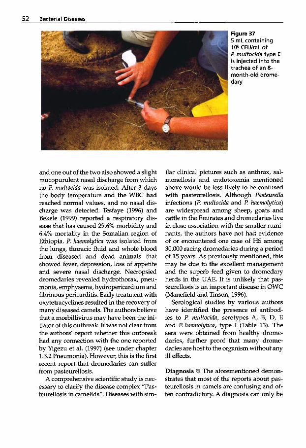
52 Bacterial Diseases
and one out of the two also showed a slight mucopurulent nasal discharge from which no P. multocida was isolated. After 3 days the body temperature and the WBC had reached normal values, and no nasal dis- charge was detected. Tesfaye (1996) and Bekele (1999) reported a respiratory dis- ease that has caused 29.6% morbidity and 6.4% mortality in the Somalian region of Ethiopia. P. haemolytica was isolated from the lungs, thoracic fluid and whole blood from diseased and dead animals that showed fever, depression, loss of appetite and severe nasal discharge. Necropsied dromedaries revealed hydrothorax, pneu- monia, emphysema, hydropericardium and fibrinous pericarditis. Early treatment with oxytetracyclines resulted in the recovery of many diseased camels. The authors believe that a morbillivirus may have been the ini- tiator of this outbreak. It was not clear from the authors’ report whether this outbreak had any connection with the one reported by Yigezu et al. (1997) (see under chapter 1.3.2 Pneumonia). However, this is the first recent report that dromedaries can suffer from pasteurellosis.
A comprehensive scientific study is nec- essary to clarify the disease complex “Pas- teurellosis in camelids”. Diseases with sim-
Figure 37 5 mL containing 1 O6 CFU/mL of t? multocida type E is injected into the trachea of an 8- month-old drome- dary
ilar clinical pictures such as anthrax, sal- monellosis and endotoxemia mentioned above would be less likely to be confused with pasteurellosis. Although Pasteurella infections (P. multocida and P. haemolytica) are widespread among sheep, goats and cattle in the Emirates and dromedaries live in close association with the smaller rumi- nants, the authors have not had evidence of or encountered one case of HS among 30,000 racing dromedaries during a period of 15 years. As previously mentioned, this may be due to the excellent management and the superb feed given to dromedary herds in the UAE. It is unlikely that pas- teurellosis is an important disease in OWC (Manefield and Tinson, 1996).
Serological studies by various authors have identified the presence of antibod- ies to P. multocida, serotypes A, B, D, E and P. haemolytica, type I (Table 13). The sera were obtained from healthy drome- daries, further proof that many drome- daries are host to the organism without any ill effects.
1 The aforementioned demon- strates that most of the reports about pas- teurellosis in camels are confusing and of- ten contradictory. A diagnosis can only be
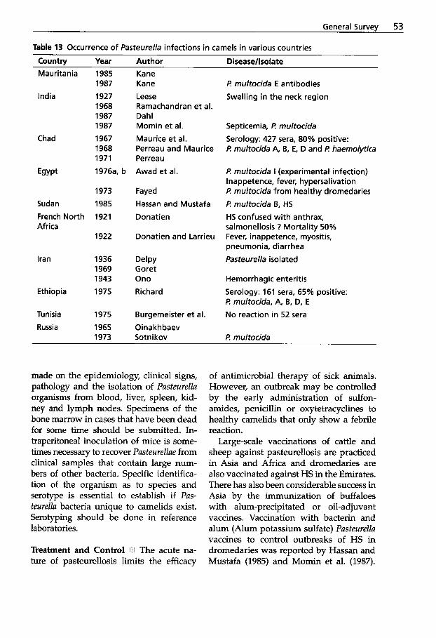
General Survey 53
Table 13 Occurrence of Pasteurella infections in camels in various countries
Country Year Author Disease/lsolate Mauritania 1985 Kane
1987 Kane India 1927 Leese
1968 Ramachandran et al. 1987 Dahl 1987 Momin et al.
1968 Perreau and Maurice 1971 Perreau
Chad 1967 Maurice et al.
Egypt 1976a, b Awad et al.
1973 Fayed
Sudan 1985 Hassan and Mustafa French North 1921 Donatien Africa
1922 Donatien and Larrieu
Iran 1936 Delpy 1969 Goret 1943 Ono
Ethiopia 1975 Richard
Tunisia 1975 Burgemeister et al. Russia 1965 Oinakhbaev
1973 Sotn i kov
t? multocida E antibodies Swelling in the neck region
Septicemia, R multocida Serology: 427 sera, 80% positive: t? multocida A, B, E, D and t? haemolytica
/? multocida I (experimental infection) Inappetence, fever, hypersalivation t? multocida from healthy dromedaries f? multocida B, HS HS confused with anthrax, salmonellosis ? Mortality 50% Fever, inappetence, myositis, pneumonia, diarrhea Pasteurella isolated
Hemorrhagic enteritis Serology: 161 sera, 65% positive: t? multocida, A, B, D, E
No reaction in 52 sera
t? multocida
made on the epidemiology, clinical signs, pathology and the isolation of Pusteurellu organisms from blood, liver, spleen, kid- ney and lymph nodes. Specimens of the bone marrow in cases that have been dead for some time should be submitted. In- traperitoneal inoculation of mice is some- times necessary to recover Pusteurellue from clinical samples that contain large num- bers of other bacteria. Specific identifica- tion of the organism as to species and serotype is essential to establish if Pus- teurellu bacteria unique to camelids exist. Serotyping should be done in reference laboratories.
Treatment and Control I t The acute na- ture of pasteurellosis limits the efficacy
of antimicrobial therapy of sick animals. However, an outbreak may be controlled by the early administration of sulfon- amides, penicillin or oxytetracyclines to healthy camelids that only show a febrile reaction.
Large-scale vaccinations of cattle and sheep against pasteurellosis are practiced in Asia and Africa and dromedaries are also vaccinated against HS in the Emirates. There has also been considerable success in Asia by the immunization of buffaloes with alum-precipitated or oil-adjuvant vaccines. Vaccination with bacterin and alum (Alum potassium sulfate) Pusteurellu vaccines to control outbreaks of HS in dromedaries was reported by Hassan and Mustafa (1985) and Momin et al. (1987).
54 Bacterial Diseases
Mohamed and Rahamtalla (1998) used an indirect hemagglutination (IHAT) and a mouse protection test (MPT) to assess the antibody response in dromedaries vacci- nated with HS type B plain bacterin, alum precipitated vaccine and the combination of the two vaccines. The authors could show that sera from camels vaccinated with vaccines containing type B P. multo- cidu antigen seroconverted and protected mice against challenge with P. mu2tocidu type B. However, no challenge experi- ments were performed in vaccinated and unvaccinated dromedaries.
1.1.6 Camel Plague
In previous centuries, Yersiniu pestis pro- duced pandemics which killed millions of people. It is said that the "Black Death killed 40 million Europeans before 1400 AC, cutting Europe's population by one third. Nowadays plague is still endemic in many countries of Africa, in the former So- viet Union, Indonesia, India, Vietnam, and in some parts of North and South America where natural foci exist. The recent out- break in humans in Zambia was linked to heavy rain and flooding, causing rats to in- vade higher grounds. Y. pestis is mainly transmitted by fleas from tolerant rodents. Cats are also susceptible to rabbit Y. pestis and can therefore pose a health hazard to humans in endemic areas. There are two forms of plague. In bubonic plague, the bacteria reach the regional lymph nodes, which become inflamed, soft and may sup- purate (buboes). Dissemination via the blood stream may lead to pneumonia and meningitis. The pneumonic plague is an airborne infection and droplets may allow aerosol infection between humans. This form of plague is fatal. Plague has been re- ported to occur in OWC and both Bactri- ans and dromedaries play an important role in the transmission to humans (Sot- nikov, 1973).
Etiology N Y. pestis is a short, oval coc- cobacillus with rounded ends, occurring singly or in pairs when directly stained from tissue or exudate. In fluid culture, the bacilli tend to form chains. Y. pestis is Gram-negative, non-motile, non-sporing and capsulated. In smears from tissues stained with methylene blue, the bacilli show characteristic bipolar staining. Y. pestis grows on nutrient, blood and McConkey agars. Great care must be taken during necropsy of an animal that might be infect- ed with plague.
Epidemiology and Clinical Signs 1 1 The camel's role in the epidemiology of plague has been known for hundreds of years (Curasson, 1947; Fedorov, 1960). Wu et al. (1936) and Pollitzer (1954) have reviewed past reports of camel plague and deter- mined that many scientists are skeptical about the earlier reports of plague out- breaks in camels. Fedorov (1960) consid- ered that Yersiniu pestis infections play an important role as anthropozoonoses as well as zooanthroponoses, even up to the present. Sotnikov (1973) reported out- breaks of plague in camels in Mongolia, China, India, Iran, Iraq, Africa and Russia. Plague outbreaks among Bactrian camels have been known in Russia since 1911 and various plague outbreaks in man were due to contact with Bactrian camels. One such outbreak in Russia affected numerous peo- ple following the consumption of infected camel meat (Kowalevsky, 1912). The last reported outbreak of plague in Russia oc- curred in 1926 (Strogov, 1959).
Plague as a zoonosis has played a role in the past, not only in Russia, but also in Mauritania and Libya where outbreaks of plague involving men and dromedaries have been recently reported by Alonso (1971) and Christie et al. (1980). Yersiniu pestis was isolated from buboes in drom- edaries. Bubonic plague, described by Sacquepee and Garcin (1913) as occur- ring among dromedaries of French North
General Survey 55
Africa, not only affected the lymph nodes, but also caused abscesses disseminated over the entire body. Y. pestis was isolated from these lesions as well as from pleural effu- sions. In addition to a cutaneous mani- festation, septicemic and pulmonary forms also occur in the camel (Lobanov, 1959 and 1967). The incubation time in camels is 1 to 6 days followed by death within 20 days. Martynchenko (1967), Alonso (1971) and Klein et al. (1975) described the clinical presentation of camel plague in drome- daries in Turkmenistan, Algeria and Mau- ritania. The authors also proved that the flea is the main vector of disease transmis- sion among camels. Ticks of the genus Hyalomrna and Ornithodoros are also able to transmit the disease mechanically (Fedo- rov, 1960).
Treatment and Control Prevention in- volves eliminating contact with infected rodents, cats and rabbits and their fleas. Before necropsy of a plague-suspected cam- el is carried out, the entire carcass should be sprayed with insecticides to destroy any ectoparasites.
Streptomycin and tetracyclines in com- bination are effective and, based on human cases, should be administered for at least 5 days. Sotnikov (1973) used a freeze-dried anti-plague vaccine for the immunization of camels; their immunity lasted for 6 months. A genetically modified vaccine against bu- bonic plaque has recently been developed in Britain, mainly to protect armed forces operating in countries where plaque oc- curs naturally and where Y. pestis may be used in biological warfare.
1 .I .7 Leptospirosis
Leptospirosis occurs worldwide and there are reports of leptospirosis in OWC as well as NWC (Wernery and Kaaden, 1995; Fowler, 1998). Leptospires are present in tubules of mammalian kidneys and are ex-
creted in urine, often for several months. Streams and ponds can be the source of in- fection as well as aerosols of urine in cow- sheds and milk from infected cows.
The order Spirochaetales in- cludes the families Spirochaetaceae and Lep- tospiraceae with the following genera, which are of significance to animals and humans: - Spirochaetaceae:
Serpulina (Brachyspira) Treponema Borrelia
Lep tospira - Leptospiraceae:
Leptospira are spirochetal organisms divid- ed into serotypes based on their antigenic structure. Within the genus Leptospirae, only the species L. interrogans is of medical importance. All of the pathogenic leptospi- rae are included under this designation. Due to a varying antigenic structure, L. in- terrogans consists of 19 serogroups and ap- proximately 180 serotypes (serovars).
Leptospires can be demonstrated in urine, body fluids and tissues by dark field mi- croscopy and fluorescent antibody tech- nique (FAT). Leptospires grow in special media like Stuart or Korthof broths.
Epidemiology * Leptospira are found ubiq- uitously around the world. All domesticat- ed animals, wild game, rodents in partic- ular, as well as man are susceptible to in- fection. In some animals, chronic renal involvement serves as a reservoir for the organism (Bisping and Amtsberg, 1988). Direct or indirect infection of man or ani- mal is possible from these reservoirs via contact with infected urine or ingestion of urine-contaminated food or water. A broad spectrum of manifestations, from inappe- tence to more severe clinical signs, can be expected. According to Seifert (1992), ro- dents and dogs serve as the most impor- tant epidemiological reservoirs in inten- sive cattle husbandry in the tropics. Man,
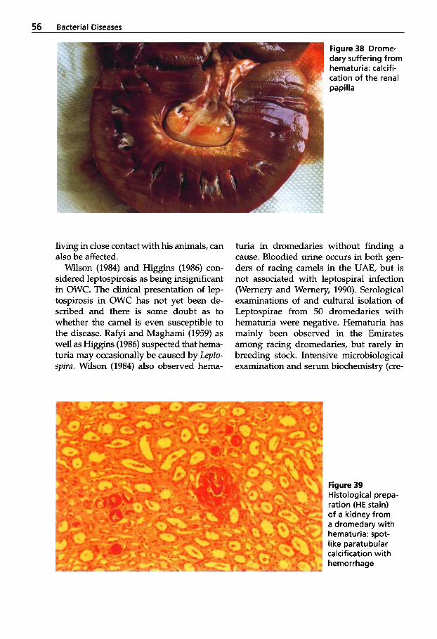
56 Bacterial Diseases
living in close contact with his animals, can also be affected.
Wilson (1984) and Higgins (1986) con- sidered leptospirosis as being insignificant in OWC. The clinical presentation of lep- tospirosis in OWC has not yet been de- scribed and there is some doubt as to whether the camel is even susceptible to the disease. Rafyi and Maghami (1959) as well as Higgins (1986) suspected that hema- turia may occasionally be caused by Lepto- spiru. Wilson (1984) also observed hema-
Figure 38 Drome- dary suffering from hematuria: calcifi- cation of the renal papilla
turia in dromedaries without finding a cause. Bloodied urine occurs in both gen- ders of racing camels in the UAE, but is not associated with leptospiral infection (Wernery and Wernery, 1990). Serological examinations of and cultural isolation of Leptospirae from 50 dromedaries with hematuria were negative. Hematuria has mainly been observed in the Emirates among racing dromedaries, but rarely in breeding stock. Intensive microbiological examination and serum biochemistry (cre-
Figure 39 Histological prepa- ration (HE stain) of a kidney from a dromedary with hematuria: spot- like paratubular calcification with hemorrhage
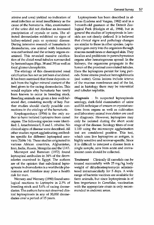
General Survev 57
atinine and urea) yielded no indication of renal infection or renal insufficiency as the cause of the hematuria. Also, examination of the urine did not disclose an increased precipitation of crystals or casts. The af- fected dromedaries exhibited no signs of kidney-related pain or systemic disease. During intensive research of hematuria in dromedaries, one animal with hematuria was euthanized and the urinary organs ex- amined. This revealed massive calcifica- tion of the distal renal tubules surrounded by hemorrhages (Figs. 38 and 39) as well as focal glomerulonephritis.
The etiology of this disseminated renal calcification has not as yet been elucidated. It has been surmised that these deposits re- sult from the higher mineral content of the feed given to the racing dromedaries. This would explain why hematuria has rarely been known to occur in breeding stock. Breeding animals are given a less well-bal- anced diet, consisting mostly of hay. Fur- ther studies should clarify possible con- nections in the etiology of the hematuria.
Krepkogorskaya (1956) is the only au- thor to have isolated Leptospira from camel organs. The following species were identi- fied: L. kazachstanica I, I1 and L. vitulina. No clinical signs of disease were described. All other studies report agglutinating antibod- ies specific for different leptospiral sero- vars (Table 14). These studies originated in various African countries, Afghanistan, Iran, India, Russia, Mongolia and the UAE.
Maronpot and Barsoum (1972) found leptospiral antibodies in 34% of the drom- edaries examined in Egypt. The authors are of the opinion that subclinical lepto- spirosis in dromedaries is a worldwide phe- nomena and therefore may pose a health risk for man.
Wernery and Wernery (1990) found sero- logical reactions to Leptospira in 2.5% of breeding stock and 5.6% of racing drome- daries. The authors have not observed clin- ical leptospirosis in any of 30,000 drome- daries over a period of 15 years.
Leptospirosis has been described in al- pacas (Ludena and Vargus, 1982) and in a 3-month-old guanaco at the Detroit Zoo- logical Park (Hodgin et al., 1984), but in general the studies of leptospirosis in lam- oids are not clearly defined. It is believed that clinical signs and pathology changes are similar to those in other species. Lepto- spires gain entry into the organism through mucous membranes or damaged skin. They localize and proliferate in parenchymatous organs after hematogenous spread. In the kidneys, the organisms propagate in the lumen of the proximal convoluted tubules. Here the leptospires persist for long peri- ods. Some strains produce hemoglobinuria (red water). Gross lesions include icterus of the mucous membranes and of the fat, and in histology there may be interstitial and tubular nephritis.
Diagnosis In suspected leptospirosis serology, dark-field examination of urine and FA technique of smears or cryostat sec- tions from organs as well as cultivation and laboratory animal inoculation are used for diagnosis. However, leptospires may only be isolated during the short acute stage of the disease. Serology titers of over 1 : l O O using the microscope agglutination test are considered positive. This test, which uses live leptospires as antigen, is highly sensitive and serovar-specific. Since it is difficult to interpret a disease from a single sample, sera from acute and conva- lescent cases should be collected.
Treatment Clinically ill camelids can be treated successfully with 25 mg/kg body weight of dihydrostreptomycin adminis- tered intramuscularly for 5 days. A wide range of bacterin vaccines are available for farm animals, but since leptospirosis is of less importance in Camelidae, vaccination with the appropriate strain is only recom- mended in endemic areas.
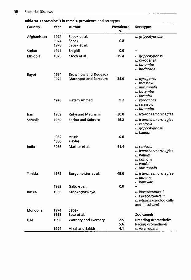
58 Bacterial Diseases
Table 14 Leptospirosis in camels, prevalence and serotypes
Country Year Author Prevalence Serotypes %
Afghanistan
Sudan Ethiopia
Egypt
Iran Somalia
India
Tunisia
Russia
Mongolia
UAE
1972 1974 1978
1974 1975
1964 1972
1976
1959
1960
1982 1986 1986
1975
1989 1956
1974 1988 1990
1994
Sebek et al. Sebek Sebek et al. Shigidi Moch et al.
Brownlow and Dedeaux Maronpot and Barsoum
Hatem Ahmed
Rafyi and Maghami Farina and Sobrero
Arush Hayles Mathur et al.
Burgemeister et al.
Gallo et al. Krepkogorskaya
Sebek Sosa et al. Wernery and Wernery
Afzal and Sakkir
0.8
0.0 15.4
34.0
9.2
20.0 16.2
0.0
51.4
48.0
0.0
2.5 5.6 4.1
L. grippotyphosa
- L. grippotyphosa L. pyrogenes L. butembo L. borincana
L. pyrogenes L. tarassovi L. autumnalis L. butembo L. javanica L. pyrogenes L. tarassovi L. butembo L. icterohaemorrhagiae L. icterohaemorrhagiae L. canicola L. grippotyphosa L. ballum -
L. canicola L. icterohaemorrhagiae L. ballum L. pomona L. wolfei L. autumnalis L. icterohaemorrhagiae L. pomona L. bataviae
L. kazachstanica I L. kazachstanica II L. vitulina (serologically and in culture)
-
Zoo camels Breeding dromedaries Racing dromedaries L. interrogans
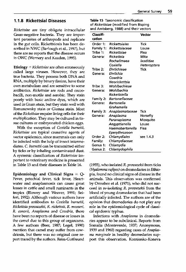
General Survey 59
1 .I .8 Rickettsia1 Diseases
Rickettsiae are tiny obligate intracellular Gram-negative bacteria. They are impor- tant parasites of arthropods and replicate in the gut cells. Rickettsiosis has been de- scribed in NWC (Barlough et al., 1997), but there are no reports that the disease occurs in OWC (Wernery and Kaaden, 1995).
Etiology ' I t Rickettsiae are often erroneously called large viruses. However, they are true bacteria. They possess both DNA and RNA, multiply by binary fission, have their own metabolism and are sensitive to some antibiotics. Rickettsiae are rods and cocco- bacilli, non-motile and aerobic. They stain poorly with basic aniline dyes, which are used in Gram stain, but they stain well with Romanowsky stain or Giemsa stain. Most of the Rickettsiae require living cells for their multiplication. They may be cultured in tis- sue cultures or embryonated chicken eggs.
With the exception of Coxiella burnetii, Rickettsiae are typical causative agents of vector epidemics, since mammals can only be infected with the help of insect interme- diates. C. burnetii can be transmitted either by ticks or by inhaling contaminated dust. A systemic classification of Rickettsiae im- portant to veterinary medicine is presented in Table 15 and their diseases in Table 16.
Epidemiology and Clinical Signs Q- Fever, petechial fever, tick fever, Heart- water and anaplasmosis can cause great losses in cattle and small ruminants in the tropics (Blowey and Weaver, 1991; Sei- fert, 1992). Although various authors have identified antibodies to Coxiella burnetii, Rickettsia prowazekii, R. rickettsii, R. mooseri, R. conorii, Anaplasma and Cowdria, there have been no reports of disease or losses in the camel due to this group of organisms. A few authors (Beer, 1987; Legel, 1990) mention that camel may suffer from cow- driosis, but there was no original case re- port traced by the authors. Reiss-Gutfreund
Table 15 Taxonomic classification of Rickettsiae (modified from Bisping and Amtsberg, 1988) and their vectors
Classifi- Vector cation Order 1: Rickettsiales Family 1: Rickettsiaceae Tribe 1: Rickettsiae Genera : Rickettsia
Rochalimaea Coxiella
Tribe 2: Ehrlichieae Genera: Ehrlichia
Co wdria Neorickettsia
Tribe 3: Wohlbachieae Genera: Wohlbachia
Rickettsiella Family 2: Bartonellaceae Genera: Bartonella
Grahamella Family 3: Anaplasmataceae Genera: Anaplasma
Para naplasma Aegyp tia nella Haemobartonella Eperythrozoon
Order 2: Chlamydiales Family: Chlamydiaceae Genus 1: Chlamydia Genus 2: Chlamydophila
Tick Louse Flea Mite lxodidae Heteroptera Tick
Tick Horsefly Mosquito Louse Flea
see 1.4.3
(1955), who isolated R. prowazekii from ticks (Hyalomma rufipes) on dromedaries in Ethio- pia, found no clinical signs of disease in the animals. This observation was confirmed by Ormsbee et al. (1971), who did not suc- ceed in re-isolating R. prowazekii from the blood of young dromedaries that had been artificially infected. The authors are of the opinion that dromedaries do not play any role in the epidemiological cycle of classi- cal epidemic typhus.
Infections with Anaplasma in dromeda- ries appear to be subclinical. Reports from Somalia (Monteverde, 1937; Anonymous, 1939 and 1960) regarding cases of Anaplas- ma marginale in healthy dromedaries sup- port this observation. Kornienko-Koneva
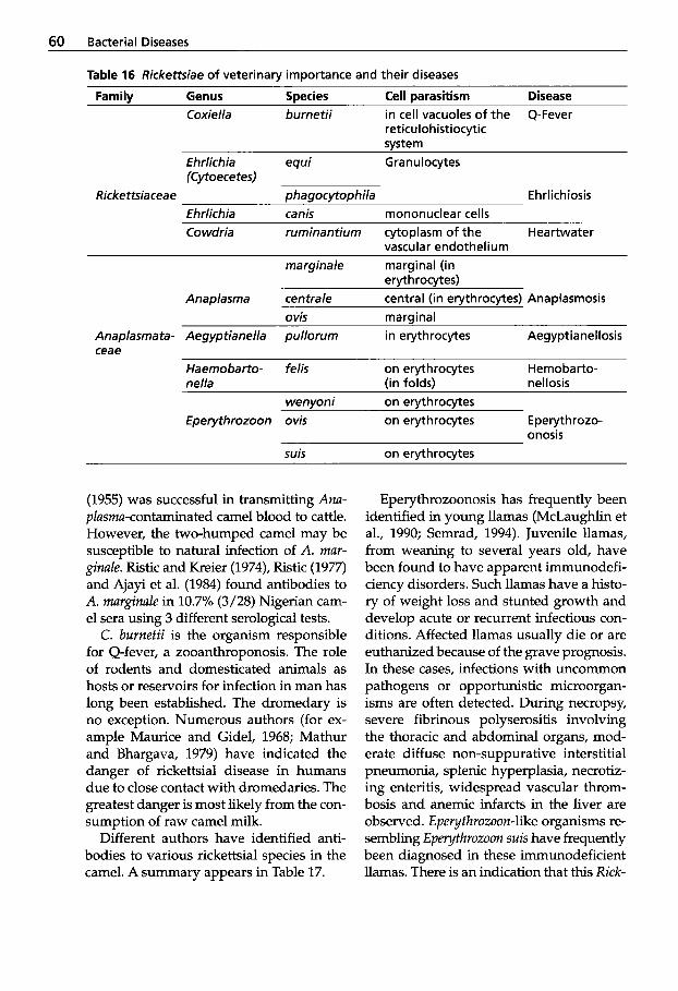
60 Bacterial Diseases
Table 16 Rickettsiae of veterinary importance and their diseases
Family Genus Species Cell parasitism Disease Coxiella burnetii in cell vacuoles of the Q-Fever
reticulohistiocytic system
Eh rlich ia equi G ra n u I ocytes (Cytoece tes)
Ehrlichia canis rnononuclear cells Rickettsia cea e phagocytophila Ehrlichiosis
Co wdria rurninantiurn cytoplasm of the Heartwater vascular endotheliurn
marginale marginal (in erythrocytes)
Anaplasrna centrale central (in erythrocytes) Anaplasmosis 0 vis marqinal
Anaplasrnata- Aegyptianella pullorurn in erythrocytes Aegyptianellosis ceae
Haernobafio- felis on erythrocytes Hemobarto- nella (in folds) nellosis
wenyoni on erythrocytes Eperythrozoon ovis on erythrocytes Eperythrozo-
onosis suis on ervthrocvtes
(1955) was successful in transmitting Ana- plasma-contaminated camel blood to cattle. However, the two-humped camel may be susceptible to natural infection of A. mar- ginale. Ristic and Kreier (1974), Ristic (1977) and Ajayi et al. (1984) found antibodies to A. marginale in 10.7% (3/28) Nigerian cam- el sera using 3 different serological tests.
C. burnetii is the organism responsible for Q-fever, a zooanthroponosis. The role of rodents and domesticated animals as hosts or reservoirs for infection in man has long been established. The dromedary is no exception. Numerous authors (for ex- ample Maurice and Gidel, 1968; Mathur and Bhargava, 1979) have indicated the danger of rickettsial disease in humans due to close contact with dromedaries. The greatest danger is most likely from the con- sumption of raw camel milk.
Different authors have identified anti- bodies to various rickettsial species in the camel. A summary appears in Table 17.
Eperythrozoonosis has frequently been identified in young llamas (McLaughlin et al., 1990; Semrad, 1994). Juvenile llamas, from weaning to several years old, have been found to have apparent immunodefi- ciency disorders. Such llamas have a histo- ry of weight loss and stunted growth and develop acute or recurrent infectious con- ditions. Affected llamas usually die or are euthanized because of the grave prognosis. In these cases, infections with uncommon pathogens or opportunistic microorgan- isms are often detected. During necropsy, severe fibrinous polyserositis involving the thoracic and abdominal organs, mod- erate diffuse non-suppurative interstitial pneumonia, splenic hyperplasia, necrotiz- ing enteritis, widespread vascular throm- bosis and anemic infarcts in the liver are observed. Eperytkrozoon-like organisms re- sembling Eperythrozoon suis have frequently been diagnosed in these immunodeficient llamas. There is an indication that this Rick-
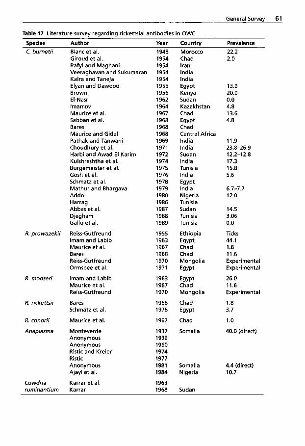
General Survev 61
Table 17 Literature survey reqardinq rickettsia1 antibodies in OWC
Species Author Year Country Prevalence C. burnetii Blanc et al.
Giroud et al. Rafyi and Maghani Veeraghavan and Sukumaran Kalra and Taneja Elyan and Dawood Brown El-Nasri lmamov Maurice et al. Sabban et al. Bares Maurice and Gidel Pathak and Tanwani Choudhury et al. Harbi and Awad E l Karim Kulshreshtha et al. Burgemeister et al. Gosh et al. Schmatz et al. Mathur and Bhargava Addo Harrag Abbas et al. Djegham Gallo et al.
R. prowazekii Reiss-Gutfreund Imam and Labib Maurice et al. Bares Reiss-G utf reund Ormsbee et al.
R. mooseri Imam and Labib Maurice et al. Reiss-G utf reu nd
R. rickettsii Bares Schmatz et al.
R. conorii Maurice et al.
Anaplasrna Monteverde Anonymous Anonymous Rist ic and Kreier Rist ic Anonymous Ajayi et al.
Co wdria Karrar et al. ruminantiurn Karra r
1948 1954 1954 1954 1954 1955 1956 1962 1964 1967 1968 1968 1968 1969 1971 1972 1974 1975 1976 1978 1979 1980 1986 1987 1988 1989
1955 1963 1967 1968 1970 1971
1963 1967 1970
1968 1978
1967
1937 1939 1960 1974 1977 1981 1984
1963 1968
Morocco Chad Iran India India Egypt Kenya Sudan Kazakhstan Chad Egypt Chad Central Africa India India Sudan India Tunisia India Egypt India Nigeria Tunisia Sudan Tunisia Tunisia
Ethiopia Egypt Chad Chad Mongolia Egypt
Egypt Chad Mongolia
Chad Egypt
Chad
Somalia
Somalia Nigeria
Sudan
22.2 2.0
13.9 20.0 0.0 4.8 13.6 4.8
11.9 23.8-26.9 12.2-12.8 17.3 15.8 5.6
6.7-7.7 12.0
14.5 3.06 0.0
Ticks 44.1 1.8 11.6 Experimental Experimental
26.0 11.6 Experimental
1.8 3.7
1 .o 40.0 (direct)
4.4 (direct) 10.7
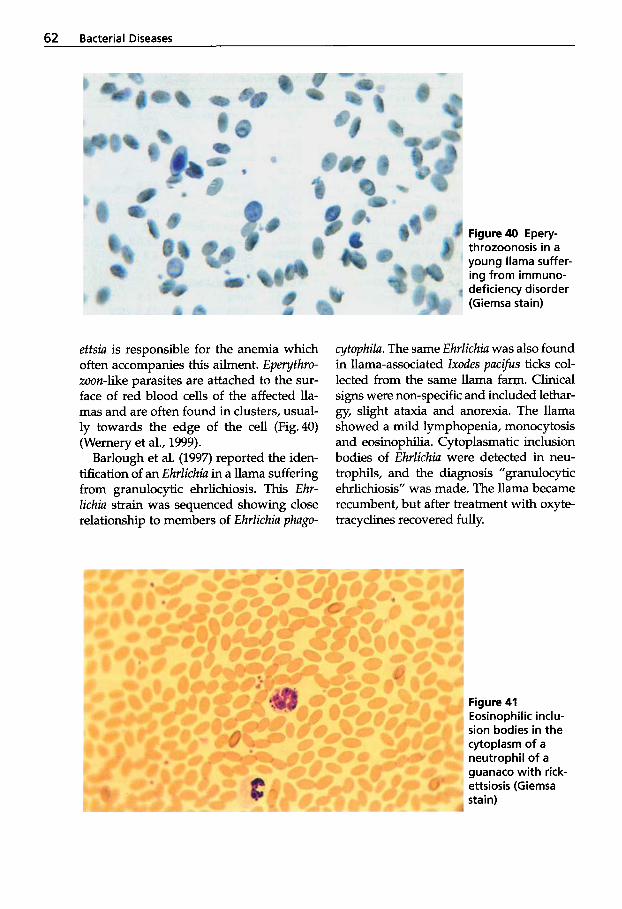
62 Bacterial Diseases
Figure 40 Epery- throzoonosis in a young llama suffer- ing from immuno- deficiency disorder (Giemsa stain)
ettsiu is responsible for the anemia which often accompanies this ailment. Epeythru- zoon-like parasites are attached to the sur- face of red blood cells of the affected lla- mas and are often found in clusters, usual- ly towards the edge of the cell (Fig.40) (Wernery et al., 1999).
Barlough et al. (1997) reported the iden- tification of an Ehrlichiu in a llama suffering from granulocyhc ehrlichiosis. This Ehr- lichia strain was sequenced showing close relationship to members of Ehrlichiu phagu-
cytuphilu. The same Ehrlichiu was also found in llama-associated lxudes pucifus ticks col- lected from the same llama farm. Clinical signs were non-specific and included lethar- gy, slight ataxia and anorexia. The llama showed a mild lymphopenia, monocytosis and eosinophilia. Cytoplasmatic inclusion bodies of Ehrlichiu were detected in neu- trophils, and the diagnosis ”granulocytic ehrlichiosis” was made. The llama became recumbent, but after treatment with oxyte- tracyclines recovered fully.
Figure 41 Eosinophilic inclu- sion bodies in the cytoplasm of a neutrophil of a guanaco with rick- ettsiosis (Giemsa stain)
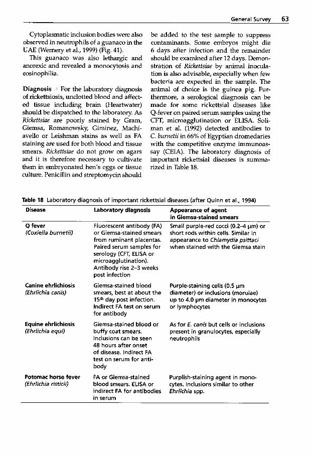
General Survey 63
Cytoplasmatic inclusion bodies were also observed in neutrophils of a guanaco in the UAE (Wernery et al., 1999) (Fig. 41).
This guanaco was also lethargic and anorexic and revealed a monocytosis and eosinophilia.
Diagnosis For the laboratory diagnosis of rickettsiosis, unclotted blood and affect- ed tissue including brain (Heartwater) should be dispatched to the laboratory. As Rickettsiae are poorly stained by Gram, Giemsa, Romanowsky, Giminez, Machi- avello or Leishman stains as well as FA staining are used for both blood and tissue smears. Rickettsiae do not grow on agars and it is therefore necessary to cultivate them in embryonated hen’s eggs or tissue culture. Penicillin and streptomycin should
be added to the test sample to suppress contaminants. Some embryos might die 6 days after infection and the remainder should be examined after 12 days. Demon- stration of Rickettsiae by animal inocula- tion is also advisable, especially when few bacteria are expected in the sample. The animal of choice is the guinea pig. Fur- thermore, a serological diagnosis can be made for some rickettsial diseases like Q-fever on paired serum samples using the CFT, microagglutination or ELISA. Soli- man et al. (1992) detected antibodies to C. burnetii in 66% of Egyptian dromedaries with the competitive enzyme immunoas- say (CEIA). The laboratory diagnosis of important rickettsial diseases is summa- rized in Table 18.
Table 18 Laboratory diagnosis of important rickettsial diseases (after Quinn et al., 1994)
Disease Laboratory diagnosis Appearance of agent
Q fever FI uorescent anti body (FA) in Giemsa-stained smears
(Coxiella burnetii) or Giemsa-stained smears from ruminant placentas. Paired serum samples for serology (CFT, ELISA or microagglutination). Antibody rise 2-3 weeks post infection
smears, best at about the 15th day post infection. Indirect FA test on serum for antibody
Equine ehrlichiosis Giemsa-stained blood or (Ehrlichia equi) bufFy coat smears.
Canine ehrlichiosis Giemsa-stained blood (Ehrlichia canis)
Inclusions can be seen 48 hours after onset of disease. Indirect FA test on serum for anti- body
FA or Giemsa-stained blood smears. ELISA or indirect FA for antibodies in serum
Potomac horse fever (Ehrlichia risticii)
Small purple-red cocci (0.2-4 pm) or short rods within cells. Similar in appearance to Chlamydia psittaci when stained with the Giemsa stain
Purple-staining cells (0.5 pm diameter) or inclusions (morulae) up to 4.0 pm diameter in monocytes or lymphocytes
As for E. canis but cells or inclusions present in granulocytes, especially neutrophils
Purplish-staining agent in mono- cytes. Inclusions similar t o other Ehrlichia spp.
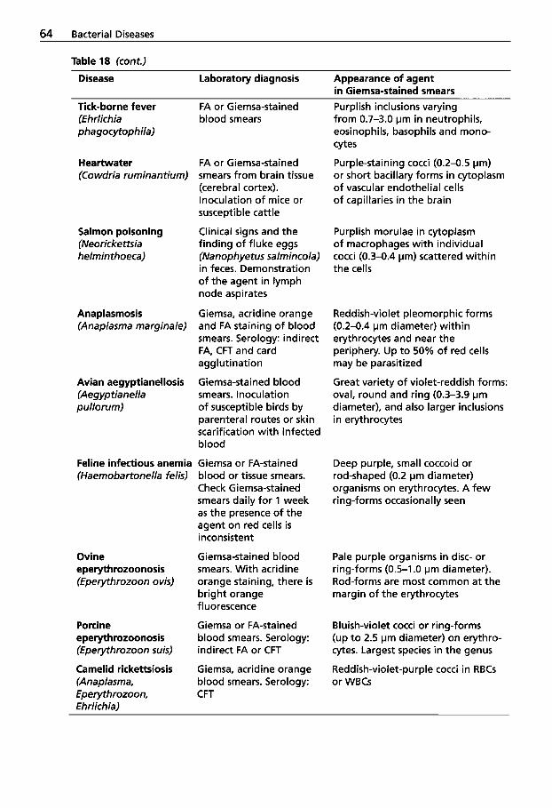
64 Bacterial Diseases
Table 18 (cont.)
Disease Laboratory diagnosis Appearance of agent in Giemsa-stained smears
Tick-borne fever (Ehrlichia phagocytophila)
Heartwater (Cowdria ruminantium)
Salmon poisoning (Neoricke ttsia helmin thoeca)
Anaplasmosis (Anaplasma marginale)
Avian aegyptianellosis (Aegyptianella pullorurn)
Feline infectious anemia (Haemobartonella felis)
Ovine eperythrozoonosis (Eperythrozoon o vis)
Porcine eperythrozoonosis (Eperythrozoon suis)
Camelid rickettsiosis (Anaplasma, Eperythrozoon, Ehrlichia)
FA or Giemsa-stained blood smears
FA or Giemsa-stained smears from brain tissue (cerebral cortex). Inoculation of mice or susceptible cattle
Clinical signs and the finding of fluke eggs (Nanophyetus salmincola) in feces. Demonstration of the agent in lymph node aspirates
Giemsa, acridine orange and FA staining of blood smears. Serology: indirect FA, CFT and card agglutination
Giemsa-stained blood smears. Inoculation of susceptible birds by parenteral routes or skin scarification with infected blood
Giemsa or FA-stained blood or tissue smears. Check Giemsa-stained smears daily for 1 week as the presence of the agent on red cells is inconsistent
Giemsa-stained blood smears. With acridine orange staining, there is bright orange fluorescence
Giemsa or FA-stained blood smears. Serology: indirect FA or CFT
Giemsa, acridine orange blood smears. Serology: CFT
Purplish inclusions varying from 0.7-3.0 pm in neutrophils, eosinophils, basophils and mono- cytes
Purple-staining cocci (0.2-0.5 pm) or short bacillary forms in cytoplasm of vascular endothelial cells of capillaries in the brain
Purplish morulae in cytoplasm of macrophages with individual cocci (0.3-0.4 pm) scattered within the cells
Reddish-violet pleomorphic forms (0.2-0.4 pm diameter) within erythrocytes and near the periphery. Up to 50% of red cells may be parasitized
Great variety of violet-reddish forms: oval, round and ring (0.3-3.9 pm diameter), and also larger inclusions in erythrocytes
Deep purple, small coccoid or rod-shaped (0.2 pm diameter) organisms on erythrocytes. A few ring-forms occasionally seen
Pale purple organisms in disc- or ring-forms (0.5-1 .O pm diameter). Rod-forms are most common at the margin of the erythrocytes
Bluish-violet cocci or ring-forms (up t o 2.5 pm diameter) on erythro- cytes. Largest species in the genus
Reddish-violet-purple cocci in RBCs or WBCs
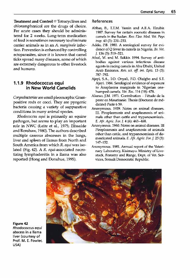
General Survev 65
Treatment and Control i ' Tetracyclines and chloramphenicol are the drugs of choice. For acute cases they should be adminis- tered for 2 weeks. Long-term medication of feed is sometimes necessary to eliminate carrier animals as in an A. marginale infec- tion. Prevention is enhanced by controlling ectoparasites, since it is known that camel ticks spread many diseases, some of which are extremely dangerous to other livestock and humans.
1 .I .9 Rhodococcus equi in New World Camelids
Corynebucteriae are small pleomorphic Gram- positive rods or cocci. They are pyogenic bacteria causing a variety of suppurative conditions in many animal species.
Rhodococcus equi is primarily an equine pathogen, but seems to play an important role in NWC (Leite et al., 1975; Elissalde and Renshaw, 1980). The authors described multiple caseous abscesses in the lungs, liver and spleen of llamas from North and South America from which A. equi was iso- lated (Fig. 42). A R. equi-associated necro- tizing lymphadenitis in a llama was also reported (Hong and Donahue, 1995).
References
Abbas, B., T.T.M. Yassin and A.E.A. Elzubir. 1987. Survey for certain zoonotic diseases in camels in the Sudan. Rev. Elev. Med. Vet. Pays
Addo, P.B. 1980. A serological survey for evi- dence of Q fever in camels in Nigeria. BY. Vet.
Afzal, M. and M. Sakkir. 1994. Survey of anti- bodies against various infectious disease agents in racing camels in Abu Dhabi, United Arab Emirates. Rev. sci. off. int. Epiz. 13 (3):
Ajayi, S.A., 1.0. Onyali, F.O. Oluigbo and S.T. Ajayi. 1984. Serological evidence of exposure to Anaplasma marginale in Nigerian one- humped camels. Vet. Rec. 114 (19): 478.
Alonso, J.M. 1971. Contribution - l'etude de la peste en Mauritanie. Thesis (Doctorat de me- dicine) Paris 6 59.
Anonymous. 1939. Notes on animal diseases. III. Piroplasmosis and anaplasmosis of ani- mals other than cattle and trypanosomiasis.
Anonymous. 1960. Notes on animal diseases. III Piroplasmosis and anaplasmosis of animals other than cattle, and trypanosomiasis of do- mesticated animals. E. A?. Agric. For. J. 25 (3):
Anonymous. 1981. Annual report of the Veteri- nary Laboratory, Kisimayo. Ministry of Live- stock, Forestry and Range, Dept. of Vet. Ser- vices, Somali Democratic Republic.
hop. 40 (3): 231-233.
J. 136 (5): 519-521.
787-792.
E. A?. AP~c . For. J. 4 (6): 463-468.
147-152.
Figure 42 Rhodococcus equi abscess in a llama liver (courtesy of Prof. M. E. Fowler, USA)
66 Bacterial Diseases
Anonymous. 1998. Blackleg. Vet. Rec. 143 (12): 322.
Arush, M.A. 1982. La situazione sanitaria del dromedario nella Repubblica Democratica Somala. Bollettino scientifica della facolt; di zoo- tecnia e veterinaria 3: 209-217.
Awad, EL, A.A. Salem and A.A. Fayed. 1976a. Studies on the viability of Pasteurella multo- cida Type 1 under simulated environmental conditions in Egypt. Egypt J. Vet. Sci. 13 (1):
Awad, F.J., A.A. Salem and A.A. Fayed. 1976b. Studies of clinical signs observed on experi- mentally infected animals with Pasteurel- la multocida type 1. Egypt. J. Vet. Sci. 13 (1): 53-56.
Barakat, A.A., E. Sayour and A.A. Fayed. 1976. Investigation of an outbreak of anthrax in camels in the western desert. J. Egypt Vet. Med. Assoc. 36 (1): 183-186.
Bares, J.F. 1968. Contribution a l'etude de la pathologie infectieuse du dromadaire au Tchad. Thesis, Toulouse.
Barlough, J.E., J.E. Medigan, D.R. Turoff, J.R. Clover, S.M. Shelly and J.S. Dumler. 1997. An Ehrlichia strain from a llama (Lama glama) and llama-associated ticks (Ixodes pacificus). J. Clinical Microbiol. 35 (4): 1005-1007.
Beer, J. 1987. Infektionskrankheiten der Haus- tiere. VEB Gustav Fischer Verlag, Jena, pp. 395-398.
Bekele, T. 1999. Studies on the respiratory dis- ease "Sonbole" in camels in eastern lowlands of Ethiopia. Trop. Anim. Hlth and Prod. 31: 333-345.
Bisping, W. and G. Amtsberg. 1988. Colour atlas for the diagnosis of bacterial pathogens in an- imals. Verlag Paul Parey, Berlin and Ham- burg.
Blanc, G.R., J. Bruneau, J.A. Martin and A. Mau- rice. 1948. Quelques dorm& nouvelles sur le virus de la Q fever marocaine. C.R. Scand. Acad. Sci. 226 (7): 607-608.
Blood, D.C. and O.M. Radostits. 1990. Veteri- nary Medicine. 7* ed. London: Bailliere Tin- dall.
Blowey, R.W. and A.D. Weaver. 1991. A colour atlas of diseases and disorders of cattle. Wolfe Publishing Limited.
Boue, A. 1962. L'initiation au dromadaire. Ser- vice biologique et vetkrinaire des armkes, centre &instruction du service. VeMrinaire de l'Arm6e Compiegne, 1957 R e d s a jour.
57-62.
Brown, R.D. 1956. La mise en evidence, par tests serologiques, de la fievre Q chez les animaux domestiques au Kenya. Bull. Epiz. Dis. Afi. 4:
Brownlow, W.J. and J.D. Dedeaux. 1964. Lep- tospirosis in animals of upper Egypt. Amer. J. Trop. Med. Hyg. 13: 311-318.
Burgemeister, R., W. Leyk and R. Goessler. 1975. Untersuchungen iiber Vorkommen von Para- sitosen, bakteriellen und viralen Infektions- krankheiten bei Dromedaren in Siidtunesien. Dtsch. Tierarztl. Wschr. 8 2 352-354.
Cartwright, M.E., A.E. McChesney and R.L. Jones. 1987. Vaccination-related anthrax in three llamas. JAVMA 191 (6): 715-716.
Cebra, C.K., M.L. Cebra, F.B. Garry and E.B. Belknap. 1996. Forestomach acidosis in six New World Camelids. JAVMA 208 (6):
Chauhan, R.S., R.C. Kulshreshtha and R.K. Kaushik. 1985. A report of enterotoxemia in camels in India. Indian Vet. J. 62 (10): 825-827.
Chauhan, R.S., R.K. Kaushik, S.C. Gupta, K.C. Satiya and R.C. Kulshreshta. 1986. Prevalence of different diseases in camels (Camelus dromedarius) in India. Camel Newsletter 3:
Choudhury, S., S. Balaya and L.N. Mohapatra. 1971. Serological evidence of Coxiella bur- netii infection in domestic animals in Delhi and surrounding areas. Indian J. Med. Res. 59:
Christie, A.B., T.H. Chen and S.S. Elberg. 1980. Plague in camels and goats: their role in hu- man epidemics. J. Infect. Dis. 141 (6): 724-726.
Cross, H.E. 1919. Are camels susceptible to blackquarter, haemorrhagic septicemia and rinderpest? Bull. Agric. Res. Inst. Pusa: 80.
Curasson, G. 1947. Le chameau et ses maladies. Vigot Freres, Editeurs: 86-88.
Dahl, G. 1987. %minaire national sur le droma- daire. S6minaire national sur le dromadaire, Dec. 2-9,1985, Gao. Camel forum 18: 1-111.
Davis, J.W., L.H. Karstad and E.D. Trainer. 1981. Infectious diseases of wild mammals. 2"d ed. Ames: Iowa State Univ. Press.
Del Alwis, M.C.L. 1992. Haemorrhagic sep- ticemia - a general review. Br. Vet. J. 148
Delpy, L. 1936. Sur les maladies contagieuses des animaux domestiques observees en Iran de 1930 a 1935. Bull. Ac. Vit . Fr. 9 (4):
115-119.
901-904.
10-14.
1194-1196.
99-112.
206-210.
General Survey 67
Djegham, M. 1988. A propos de l'avortement chez la chamelle en Tunisie. Maghreb Vt t . 3 (14): 60.
Donatien, A. 1921. El Ghedda, septicemie he- morragique des dromadaires. Archs Inst.
Donatien, A. and A. Boue. 1944. Une epizootie de ghedda dam la region d'Qued Guir (Sa- hara oranais). Arch. Inst. Pasteur Alger 22 (3):
Donatien, A. and M. Larrieu. 1922. Nouvelle epizootie de Ghedda a M Raier (Sahara) en 1921. Arch. Inst. Pasteur de I'Afrique du Nord 2
El-Khouly, A-Ba., F.A. Gadir, D.D. Cluer and G.W. Manefield. 1992. Aspergillosis in camels affected with a specific respiratory and en- teric syndrome. Austr. Vet. J. 69 (8): 182-186.
El-Nasri, M. 1962. A serological survey for the detection of Q fever antibodies in the sera of animals in the Sudan. Bull. epiz. Dis. Afr. 10: 55-57.
El-Sanousi, S.M. and A.A. Gameel. 1993. An outbreak of enterotoxaemia in suckling cam- els. J. Vet. Med. A-40: 525-532.
Elissalde, G.S. and H.W. Renshaw. 1980. Corynebacterium equi: An interhost review with emphasis on the foal. Comp. Immunol. Microbiol. Infect. Dis. 3: 433-435.
Ellis, R.P., R.J. Todd, L.A. Metelman-Alvis, A.L. Newton, T,J. Thomson, L.W. Johnson and A. Ramirez. 1990. Response of llamas to Clostridium perfringens type C and D vac- cines. Am. Ass. Small Ruminants Pract. and West. Reg. Coord. Comm. Symp., Corvallis, Ore- gon: 4-5.
Elyan, A and M.M. Dawood. 1955. A serological survey of Q fever in Egypt. J. Egypt Publ. Hlth.
Farina, R and L. Sobrero. 1960. Ricerche sierolo- giche sulla diffusione delle leptospirosi ani- mali in Somalia. Zooprofilassi 15 (12): 925-936.
Fayed, A.A. 1973. Studies on pasteurellosis in buffaloes in Egypt. Thesis Vet. Med., Fac. of Vet. Med. Cairo University.
Fazil, M.A. 1977. The Camel. Bull. Anim. Hlth. Prod. Afr. 25 (4): 435-442.
Fazil, M.A. and R.R. Hofmann. 1981. Haltung und JSrankheiten des Kamels. Tieriirzfl. Praxis
Fedorov, V.N. 1960. Plague in camels and its prevention in the USSR. Bull. Org. Mond. San-
Past. Afr. 1 (3): 242-249.
171-174.
(3): 316-319.
ASS. 29 (6): 185-190.
9: 389-402.
t i 23 (2-3): 275-281.
Fowler, M.E. 1996. Husbandry and diseases of camelids. Rat. sci. tech. Ofi int. Epiz. 15 (1):
Fowler, M.E. 1998. Medicine and surgery of South American Camelids. Iowa State Uni- versity Press, Ames.
Fowler, M.E. and D. Gillespie. 1985. Middle and inner ear infection in llamas. J. Zoo An. Med.
Gaffin, S.L. 1987. Endotoxins and anti-endotoxin antibodies. Equine Vet. 1. 3 2 76.
Gallo, C., G. Vesco, F. Campo, N. Haddad and H. Abdelmoula. 1989. EnquCte zoosanitaire chez les chevres et les dromadaires au Sud de la Tunisie. Maghreb Vi t . 4 (17): 15-17.
Gameel, A.A., S.M. El-Sanousi, B. Musa and E.E. El-Owni. 1986. Association of some pathogen- ic bacteria with haemorrhagic enteritis in cam- els. Camel research paper S.R.C. No. 12, Camel Res. Unit, University of Khartoum, Sudan: 50-55.
Gareis, M. and U. Wemery. 1992. Determination of mycotoxins in samples associated with cases of intoxications in camels. Proc. 1st int. Camel Conf.: 403-404. Eds: W.R. Allen, A.J. Higgens, I.G. Mayhew, O.H. Snow and J.F. Wade: R. and W. Publications, Newmarket, U.K.
Gareis, M. and U. Wernery. 1994. Determination of Gliotoxin in samples associated with cases of intoxication in camels. Mycotoxin Research 10: 2-8.
Gatt Rutter, T.E. and R. Mack. 1963. Diseases of camels. Part 1: Bacterial and fungal diseases. Vet. Bull. 33 (3): 119-124.
Ghosh, S.S., K.R. Mittal and G.P. Sen. 1976. Inci- dence of Q fever in man and animals. Indian J. Anim. Hlth. 15 (1): 79-80.
Giroud, P., F. Roger, N. Dumas, P. Vouilloux and E. Sacquet. 1954. Comportement des ani- maux domestiques de la region du Tchad vis- a-vis de l'antighe T13. Bull. SOC. Path. Exot. 47: 644-645.
Goret, l? 1969. Notes pour servir au cours sur les maladies bacterienne et virales - la pasteurel- lose bovo-bubaline. ENSAIl - 42. Maisons - Alfort, IEMVT.
Harbi, M.S.M.A. and M.H. Awad El Karim. 1972. Serological investigation into Q fever in Sudanese camels (Camelus dromedarius). Bull. epizoot. Dis. Afr. 20: 15-17.
Harrag, M. 1986. Contribution a l'etude serolo- gique de la fikvre Q chez le dromadaire en
155-169.
16: 9-15.

68 Bacterial Diseases
Tunisie. Thesis, Doctorat veterinaire, Sidi Thabet 1986: 283.
Hassan, A.K.M. and A.A. Mustafa. 1985. Isola- tion of Pasteurella multocida type B from an outbreak of haemorrhagic septicemia in cam- els in Sudan. Rev. Elev. Mid . ve't. Pays trop. 38
HatemAhmed, M.E. 1976. Studies on leptospira group of microorganisms with special refer- ence to purification and cultivation. M.V. Sc. Thesis, Fac. of Vet. Med., Cairo University.
Hayles, L.B. 1986. Proceedings of the first national veterinary symposium, Somalia, Oct. 12-15, 1986. Is* National Veterinary Sym- posium, Mogadishu, Rome, FAO.
Heitefuss, S., A. Heine and H.S.H. Seifert. 1990. Detection of non-volatile organic acids by head-space gas chromatography. J. Chroma- tog. Biomed. Appl. 532: 374-378.
Heitefuss, S. 1991. Untersuchung zur Identifi- zierung von aeroben, anaeroben und fakulta- tiv anaeroben Bakterien mit gaschromatogra- phischen Methoden. Thesis, Gottingen.
Higgins, A. 1986. The camel in health and dis- ease. BailliPre Tindall.
Hodgin, C., T.W. Schillhom, R. Fayer and N. Richter. 1984. Leptospirosis and coccidial infection in a guanaco. JAVMA 185 (11):
Hong, C.B. and J.M. Donahue. 1995. Rhodococ- cus equi - associated necrotizing lymphadeni- tis in a llama. 1. Comp. Path. 113 (1): 85-88.
Huaman, D., A. Ramirez and H. Samame. 1981. Produccih de alfa toxina de 3 cepas de Clostridium perfringens tip0 A aislados de alpacas. Resumenes 5th Congr. Peru. Microbiol. Parasitol. (Arequipa): 56.
Huber, T.L., M.C. Peed, R.C. Wilson and D.D. Goetsch. 1979. Endotoxin absorption in hay- fed and lactic acidotic sheep. Am. J. Vet. Res.
Hutyra, F., J. Marek and R. Manninger. 1946. Special pathology and therapeutics of the diseases of domestic animals. 5* English ed.: Balliere, Tindall and Cox, London.
Imam, I.Z.E. and A. Labib. 1963. Complement fixing antibodies against epidemic and murine typhus in domestic animals in U.A.R.
Imamov, E.D. 1964. La fi&vre Q chez les ani- maux domestiques de Kirghizie Frunce (cite par P.F. Zdrodowski dans Les rickettsioses en
(1): 31-33.
1442-1444.
10 (6): 792-794.
J. Egypt Publ. Hlth. ASS. 38: 101-109.
U.R.S.S.). Bull. O.M.S. 31: 33-43.
Ipatenko, N.G. 1974. Infectious enterotoxemia
Jones, T. 1991. Bovine botulism. In Practice 13
Kalra, S.L. and B.L. Taneja. 1954. Q-fever in India: A serological survey. Indian J. Med. Res.
Kane, M. 1985. Enquete serologique sur la pas- teurellose des dromadaires dans le cercle de Nara (Mali) et Abdel Bagron (Mauritanie). Se'minaire national sur le dromadaire, Gao (MLC).
Kane, M. 1987. La pasteurellose chez droma- daires maliens et mauritaniens. Bull. liaison
Karrar, G., M.N. Kaiser and H. Hoogstraal. 1963. Ecology and host relationship of ticks (Ixodoidea) infesting domestic animals in Kassala province, Sudan, with special refer- ence to Amblyomma lepidum Doenitz. Bull. Entom. Res. 5 4 509-523.
Karrar, G. 1968. No Title. Sudan J. Vet. Sci. and Anim. Husb. 9 (11): 328.
Klein, J.M., J.M. Alonso, G. Baranton, A.R. Poulet and H.H. Mollaret. 1975. La peste en Mauritanie. Med. Mal. infect. 5 (4): 198-207.
Kornienko-Koneva, Z.P. 1955. No Title. Disser- tation, Moscow (cited by Markov, A.A. et al; 1965,210).
Kowalesky, M.J.M. 1912. Le Chameau et ses maladies d'aprks les observations d'auteurs russes. J. Mid. Ve't. Zootechn., Lyon 15: 462-466.
Krepkogorskaja, T.A. 1956. La leptospirose des animaux domestiques dans le desert de Bet- pak-Dal. Izvest. Akad. Nauouk Kazakh. SSR (Physiol. Mid . ) 7 80-81.
Krogh, N. 1960. Studies on alterations in the N- men fluid of sheep, especially concerning the microbial composition, when readily avail- able carbohydrates are added to the food. 11. Lactose. Acta Vet. Scand. 1: 383-410.
Kulshreshtha, R.C., R.G. Arora and D.S. Kalra. 1974. Sero-prevalence of Q fever in camels, buffaloes and pigs. Ind. J. Med. Res. 62 (0):
Leese, A.S. 1918. "Tips" on camels for veterinary surgeons on active service. Bailliere Tindall and Cox, London 50.
Leese, A.S. 1927. A treatise on the one-humped camel in health and disease. Vigot FrPres, Pans II.
Legel, S. 1990. Nutztiere der Tropen und Subtro- pen. S. Hirzel Verlag Stuttgart, Leipzig, p. 194.
of camels. Vet. Bull 44 (4): 1481-1484.
(3): 83-86.
42 315-318.
ILCNGRPRC 9: 21-22.
1314-1316.
General Survev 69
Leite, R.C., H. Negrelli Filho and C.H. Langen- egger. 1975. Infectio por Corynebacterium equi em lhama (Lama glama). Pesqui. Agro- pecu. Bras. Ser. Vet. 10 (8): 57-59.
Lobanov, V.N. 1959. Pathology of experimental plague in camels. Arkh. Patol. 21 (7): 37-43.
Lobanov, V.N. 1967. La peste chez les chameaux. OMS Shinaire inter-rigional de L'0.M.S. pour la lutte contre la peste, Moscow.
Losos, G.J. 1986. Infectious tropical diseases of domestic animals. Avon, The Bath Press.
Ludena, J. and A. Vargus. 1982. Leptospirosis en alpacas. Adv. Vet. Sci. Comp. Med 2 (2): 27-28.
Manefield, G.W. and A. Tinson. 1996. Camels. A compendium. The T.G. Hungerford Vade Mecum Series for Domestic Animals: pp. 240, 298.
Maronpot, R.R. and J.S. Barsoum. 1972. Lep- tospiral microscopic agglutinating antibodies in sera of man and domestic animals in Egypt. Amer. J. trop. Med. Hyg. 21 (4): 467-472.
Martynchenko, V.A. 1967. Clinical picture of plague in camels infected by means of ecto- parasite carriers. In: Kovalenko, Y.R. Mal- oiznchennye Zabolevaniya Sel'-khoz zhivot- nykh, Moscow, Kolos 191-196. Vet. Bull. 1968 38 (9): 3431.
Mathur, K.N., V.K. Khanna and A.K. Purohit. 1986. Macroscopic plate agglutination results of serological examination of camels, cattle and goats for leptospirosis. Indian J. Publ.
Mathur, K.N. and S.C. Bhargava. 1979. Sero- prevalence of Q fever and brucellosis in cam- els of Jorbeer and Bikaner, Rajasthan State. Indian J. Med. Res. 70 (11): 391-393.
Maurice, Y., J.E Bares and Mme Bade. 1967. En- qu@te skrologique sur les rickettsioses chez le dromadaire au Tchad. Rev. Elev. Mid. vit. Pays trop. 20 (4): 543-550.
Maurice, Y. and R. Gidel. 1968. Incidence of Q fever in Central Africa. Bull. SOC. Path. exot. 61
McLaughlin, B.G., C.N. Evans, P.S. McLaughlin, L.W. Johnson, A.R. Smith and J.F. Zachary. 1990. An Eperythrozoon-like parasite in lla- mas. JAVMA 197 (9): 1170-1175.
Moch, R.W., E.E. Ebner, J.S. Barsoumand B.A.M. Botros. 1975. Leptospirosis in Ethiopia: a serological survey in domestic and wild ani- mals. J. Trop. Med. Hyg. 78 (2): 38-42.
Moebuu, Aynurzana, Dashdava and N.G. Ipa- tenko. 1966. Infectious enterotoxaemia of cam-
Hlth. 30 (3): 170-172.
(5): 721-736.
els in Mongolia, caused by C1. perfringens type C. Veterinariya, Moscow 43 (11): 32-35.
Mohamed, G.E. and M.H. Rahamtalla. 1998. Serological response of camel (Camelus dromedarius) to haemorrhagic septicaemia (Pasteurella multocida infections) vaccines. J. Camel Prac. and Res. 5 (2): 207-212.
Momin, R.R., D.K. Petkar, T.N. Jaiswal and V.M. Jhala. 1987. An outbreak of pasteurellosis in camels. Indian Vet. 1. 64 (10): 896-897.
Monteverde, G. 1937. Anaplasmosi nei cammel- li in Cirenaica. Clin. Vet. Milano 60 (2): 73-77.
Moro Sommo, M. 1956. Contribucih a1 estudio de las enfermedades de 10s auguenidos. Rev. Fac. Med. Vet. (Lima) 7 (11): 15-177.
Moro Sommo, M. 1963. Enfermedades infec- ciosas de las alpacas. V. Enterotoxemia odi- arrea bacilar producida por Clostridium welchii, tip0 A. Rev. Fac. Med. Vet. (Lima) 18
Mustafa, I.E. 1987. Bacterial diseases of the camel and dromedary. OIE 55e Session g h i - rale OIE, office intemationale des kpizooties, Paris, France 55: 18-22.
Nagaraja, T.G. and E.E. Bartley. 1979. Endotoxin shock in calves from intravenous injection of rumen bacterial endotoxin. J. Anim. Sci. 49
Nothelfer, H.B. and U. Wemery. 1995. Hemor- rhagic disease in dromedary camels (Came- lus dromedarius) - etiology and morphology. Proc. of the Intl. Con$ on Livestock Production in Hot Climates 66 A57.
Oinakhbaev, S. 1965. Study of aetiology of con- tagious cough in camels. Veterinariya, Moscow
Ono, Y. 1943. Haemorrhagic enteritis in camels. J. Vet. Sci 5: 113-114.
Ormsbee, R., W. Burgdorfer, M. Peacock and P. Hildebrandt. 1971. Experimental infections of Rickettsia prowazeki among domestic livestock and ticks. Amer. J. Trop. Med. Hyg. 20
Pathak, P.N. and S.K. Tanwani. 1969. Serological investigations in Q-fever. Indian Vet. J. 46
Perreau, P. 1971. Pasteurella. Cours de microbi- ologie systkmatique 1971-1972 (Bactkriolo- gie) de l'institut Pasteur Paris.
Perreau, P. and Y. Maurice. 1968. Epizootiologie de la pasteurellose des chameaux au Tchad. Enqu@te sbrologique. Rev. Elev. Mid. vet. Pays trops. 21 (4): 451-454.
(20): 85-87.
(2): 567-582.
42 (6): 105-106.
(1): 117-124.
551-553.
70 Bacterial Diseases
Pollitzer, R. 1954. Hosts of the infection. Plague: W.H.O., Geneva, Monogr. Ser. no. 22 305-308.
Provost, A., P. Haas and M. Dembelle. 1975. Premiers cas au Tchad de botulisme animal (type C): intoxication des dromadaires par l’eau d’un puit. Rev. Elev. Mkd. vkt. Pays trop.
Punskii, E.E. and D.V. Zheglova. 1958. No Title. J. Microbiol., Moscow 29 (2): 78.
Quinn, PJ., M.E. Carter, B.K. Markey and G.R. Carter. 1994. Clinical Veterinary Microbiolo- gy, Wolfe: pp. 381-421.
Rafyi, A. and C. Maghami. 1954. Sur la prksence de la fi&vre Q en Iran. Bull. SOC. Path. Exof. 6: 766.
Rafyi, A. and G. Maghami. 1959. Sur la frkquen- ce de la leptospirose en Iran: isolement de Leptospira grippotyphosa chez l’homme et chez les bovins. Bull. SOC. Path. Exot. 52 (5):
Ramachandran, P.K., S. Ramachandran and T.P. Joshi. 1968. An outbreak of haemorrhagic gastroenteritis in camels (Camelus drome- darius). Ann. Parasit. Hum. Comp. 43 (1): 5-14.
Ramirez, A. L. Lauerman, D Huaman and A. Vargas. 1983a. Inducci6n preliminar de la en- terotoxemia A Clostridium perfringens tip0 A en alpaca. Resumenes Proyecfos Invest. Real- izados (Lima) 3: 48-49.
Ramirez, A., H. Ludena and M. Acosta. 198313. Mortalidad en alpacas del centro pecuario La Raya - Pun0 en siete anos. Resumenes Proyec- fos Invest. Realizados (Lima) 3: 47-78.
Ramirez, A. and D. Huaman. 1980-1981. Evalu- aci6n de la enterotoxemia en crias de alpacas vacunados. Resumenes Proyectos Invest. Real- izados (Lima) 3: 48-49.
Rath, E. 1950. Sintomas y cuadros anatomo-pa- tologicos de las enfermedades de ganado en el Departamento de Puno. Rev. Agrop. Perus 1
Reiss-Gutfreund, R.J. 1955. Isolement de sou- ches de Rickettsia prowazeki a partir du sang des animaw domestiques d’Ethiopie et de leurs tiques. Bull. Soc. Path. Exot. 48 (2):
Reiss-Gutfreund, R. J. 1970. The serological re- sponse of Mongolian domestic animals to Rickettsia prowazeki and Rickettsia mooseri antigens. G. Batf. Virol. lmmun. 63 (9-10): 455-457.
Richard, B. 1975. Etude de la pathologie du dro- madaire dam la sous-province du Borana
28 (1): 9-12.
592-596.
(2): 68-70.
602-607.
(Ethiopie). These Doct. Vet., E.N.V. Alfort, Creteil, Paris 75.
Ristic, M. 1977. Parasitic Protozoa. Ed. J. P. Kreier, New York Acad. Press 4: 235.
Ristic, M. and J.P. Kreier. 1974. Bergeys Manual of Determinative Bacteriology. Eds. R.E. Bu- chanan, N.E. Gibbons, Baltimore, Williams and Wikens 8: p. 907.
Sabban, M.S., N. Hussien, B. Sadek and H. El Dahaby. 1968. Q fever in the United Arab Re- public. Bull. Of int. Epizoot. 69 (5-6): 745-760.
Sacquepee and Garcin. 1913. N/A. Arch. Med. Pharm. Abstract: Vet. Bull. 33 (3): 119-124.
Schaper, R. 1991. Methodische Untersuchungen zur Produktions- und Wirksamkeitskontrolle von Rauschbrandvakzinen. Thesis, GStfin- gen .
Schmatz, H.D., H. Krauss, P. Viertel, Abdel Sha- kour Ismail and Abdul Assiz Hussein. 1978. Seroepidemiologische Untersuchungen zum Nachweis von Antikorpern gegen Rickett- sien und Chlamydien bei Hauswiederkauern in Agypten, Somalia und Jordanien. Acta Tro- pica 35: 101-111.
Schwartz, H.J. and M. Dioli. 1992. The one- humped camel in Eastern Africa. A pictorial guide to diseases, health care and manage- ment. Verlag Josef Margraf.
Sebek, Z., V. Sery and A. Saboor. 1972. Results of the first leptospirological study carried out in Afghanistan. J. Hyg. Epidem. Microbiol. Im- mun. Prague 16 (3): 314-324.
Sebek, Z. 1974. Results of serologic examination of domestic animals for leptospirosis in the Mongolian People‘s Republic. Folia Parasit.
Sebek, Z., K. Blasek, M. Valova and A. Amin. 1978. Further results of serological examina- tion of domestic animals for leptospirosis in Afghanistan. Folia Parasit. 25 (1): 17-22.
Seifert, H.S.H., H. Boehnel, S. Heitefuss, J. Ren- gel, R. Schaper, U. Sukop and U. Wernery. 1992. Isolation of C. perfringens type A from enterotoxemia in camels and production of a locality-specific vaccine. Proc. 1st int. Camel Conf.: 65-68. Eds: W.R. Allen, A.J. Higgins, I.G. Mayhew, D.H. Snow and J.F. Wade: R. and W. Publications, Newmarket, U.K.
Seifert, H.S.H. 1992. Tropentierhygiene. Gustav Fischer Verlag Jena, Stuttgart.
Semrad, S.D. 1994. Septicemic listeriosis, throm- bocytopenia, blood parasitism and hepatopa- thy in a Llama. JAVMA 204 (2): 213-216.
21 (1): 21-28.
General Survev 71
Shigidi, M.T.A. 1974. Animal leptospirosis in the Sudan. BY. Vet. J. 130 (3): 528-531.
Soliman, A.K., B.A. Botros and D.M. Watts. 1992. Evaluation of a competitive enzyme immunoassay for detection of Coxiella bur- netii antibody in animal sera. J. Clin. Microbi-
Sosa, G., 0. Santos, C.L. Duarte, D. Hernandez and L. Delgado. 1988. Investigacih serologi- ca y bacteriologica de leptospirosis realizada en fauna exotica. Revta Cub. Cienc. Vet. 19 (3):
Sotnikov, M.I. 1973. Camel plague. In: Orlov, EM. Maloizvestnye zarazny bolezni zhivot- nykh, Izdatel'stvo Kolos, 213-222. Vet. Bull. 44: 937,1974.
Strogov, A.K. 1959. Plague in camels. Maloizvest- nye zaraznye bolezni zhivotnykh Moscow, SeY-khoz 262-280. Vet. Bull. 28: 2734,1958.
Tabbaa, D. 1997. Anthrax in camels. Pamphlet, Faculty of Veterinary Medicine, A1 Baath University, Syria.
Tesfaye, R. 1996. Report on the new camel dis- ease (FURROO) in Southem Rangeland De- velopment Project (SORDU), Borena, Ethio- pia. Ethiopian Vet. Assoc. Proc. of the loth Con- ference: 13-15.
Veeraghavan, N. and P.K. Sukumaran. 1954. Q- fever survey in the Nilgiris and Coimbatore districts of Madras state. Indian J. Med. Res.
Walz, A. 1993. Nachweis und Eigenschaften von Bacillus cereus-Stammen, isoliert von arabi- schen Kamelen (Camelus dromedarius). The- sis, Munich.
Wemery, U., H.H. Schimmelpfennig, H.S.H. Sei- fert and J. Pohlenz. 1992a. Bacillus cereus as a possible cause of haemorrhagic disease in dromedary camels (Camelus dromedarius). Proc. lSt int. Camel Cod.: 51-58. Eds: W.R. Allen, A.J. Higgins, I.G. Mayhew, D.H. Snow and J.F. Wade: R. and W. Publications, New- market, U.K.
Wernery, U., M. Ali, R. Wemery and H.S.H. Seifert. 1992b. Severe heart muscle degenera- tion caused by Clostridium perfringens type A in camel calves (Camelus dromedarius). Rev. Elm. Mid. v.3. Pays trop. 45 (3-4): 255-259.
Wernery, U. 1994. Neue Ergebnisse zur Diagno- se, Prophylaxe und Therapie wichtiger bak- terieller und viraler Krankheiten beim Kame1 (Camelus dromedarius). Habilitationsschrift zur Erlangung der Lehrbef;ihigung an der
01. 30 (6): 1595-1597.
219-225.
42: 5-7.
Tierarztlichen Fakultat der Ludwig-Maximi- lians-Universitat Miinchen.
Wernery, U., M.E. Fowler and R. Wernery. 1999. Color Atlas of Camelid Hematology. Black- well Wissenschafts-Verlag, Berlin.
Wernery, U., H.S.H. Seifert, A.M. Billah, and M. Ali. 1991. Predisposing factors in entero- toxemias of camels (Camelus dromedarius) caused by Clostridium perfringens type A. Rev. Elev. Mid. vit. Pays trop. 44 (2):
Wernery, U. and J. Haydn-Evans. 1992. Botulism in waterfowls in the U.A.E. Tribulus, Bull. of the Emir. Nut. Hist. Group 2. 1 (4): 18-19.
Wernery, U. and J. Wensvoort. 1992. Experimen- tally induced rumen acidosis in a one year old camel bull (Camelus dromedarius). A preliminary report. Brit. Vet. J. 148: 167-170.
Wernery, U. and 0.-R. Kaaden. 1995. Infectious Diseases of Camelids. Blackwell Wissen- schafts-Verlag, Berlin.
Wernery, U. and R. Wernery. 1990. Seroepide- miologische Untersuchungen zum Nachweis von Antikorpern gegen Brucellen, Chlamy- dien, Leptospiren, BVD/MD, IBR/ IPV - und Enzootischen Bovinen Leukosevirus (EBL) bei Dromedarstuten (Camelus dromedarius). Dtsch. tierarztl. Wschr. 9 7 134-135.
Westphal, U. 1991. Botulismus bei Vogeln. Aula Verlag Wiesbaden.
Whabi, A.A., S.E. Gadir, A. Awadelsied and O.F. Idris. 1987. Biochemical studies on Sudanese camel milk collected from Butana Area. Sud. J. Vet. Med. B 34: 340-342.
WHO, FAO, OIE. 1961. Animal Health Year Book. Food and Agricultural Organisation of the United Nations, Rome.
Wilson, R.T. 1984. The camel. Longman, London and New York.
Wu, L.T., J.W.H. Chu, R. Pollitzer and C.Y. Wu. 1936. Plague: a manual for medical and public health workers. Weishengshu Nation- al Quarantine Service, Shanghai: 232-235.
Yigezu, M., F. Roger, M. Kiredjian and S. Tariku. 1997. Isolation of Streptococcus equi sub- species equi (strangles agent) from an Ethio- pian camel. Vet. Rec. 140: 608.
147-152.
Further reading
Bornstein, S. 1993. Camel health and diseases: veterinary projects. The multi-purpose cam- e l interdisciplinary studies on pastoral pro-
72 Bacterial Diseases
duction in Somalia by Hjort af Omaes, A.
Davies, G.O. 1946. Gaiger and Davies Veteri- nary pathology and bacteriology. 31d ed. Bail- li&re, Tindall and Cox, London.
Eldisougi, I. 1984. A note on the diseases of camels in Saudi Arabia. The Camelid; an "all purpose" animal. W.R. Cockrill. Scandina- vian Institute of African Studies, Uppsala:
Greiner, V. 1985. Beitrag zum Spektrum der Krankheiten bei Ruminantia und Tylopoda im Miinchener Tierpark Hellabrunn. Tier- park Hellabrunn.
Guleed, H.A. and S. Bomstein. 1987. Pilot study of the health of Somali camel herds. Camel Forum working paper, Mogadishu and Upp- sala: SOMAC/SIAS 23.
Heller, M., D. Anderson and F. Silveira. 1998. Streptococcal peritonitis in a young drome- dary camel. Australian Vet. J. 76 (4): 253-254.
Hoste, C., 8. Peyre de Fabregues and D. Richard. 1985. Le dromadaire et son 6levage. Elm. Mkd. vet. Pays trop. 7 145-146.
Leese, A.S. 1909. Two diseases of young camels. J. Trop. Vet. Sci. 4: 1-7.
McGrane, J.J. and A.J. Higgins. 1985. Infectious diseases of the camel; viruses, bacteria and fungi. Br. Vet. J. 141: 529-547.
Odend'Hal, S. 1983. The geographical distribu- tion of animal viral diseases. Academic Press, New York p. 99.
Rahamtalla, M.H. 1994. Haemorrhagic septi- caemia (Pasteurella multocida) in camels: Im- munity status, serological and bacteriological studies. Thesis, Faculty Vet. Sci. University of Khartoum, Sudan.
Book: 189-206.
496-502.
Ramosvara, J.A., M. Kopcha, E. Richter, G.L. Watson, J.S. Patterson, C. Juansalles and B. Yamini. 1998. Actinomycotic splenitis and in- testinal volvulus in an Alpaca (Lama-Pacos). J. Zoo and Wildlife Med. 29 (2): 228-232.
Refai, M. 1992. Bacterial and mycotic diseases of camels in Egypt. Proc. lSt int. Camel Conf. Eds: W.R. Allen, A.J. Higgins, I.G. Mayhew, D.H. Snow and J.F. Wade: R. and W. Publica- tions, Newmarket, U K 59-64.
Richard, D. 1986. Manuel des maladies du dro- madaire. Projet de d6veloppement de l'6le- vage dans le Niger centre-est. Maisons Alfort, IEMVT.
Shommein, A.M. and A.M. Osman. 1987. Dis- eases of camels in the Sudan. Rm. sci. tech. Ofi int. Epiz. 6 (2): 481.
Wernery, R., M. Ali, J. Kinne, A.A. Abraham and U. Wernery. 2001. Mineral deficiency: a pre- disposing factor for septicemia in dromedary calves. 2nd lnternational Camelid Conference, Agroeconomics of Camelid Farming, Almaty, Kazakhstan, 8.-12.9.2000 (in press.).
Wemery, U. 1995. Viral infections in camels - a review. I. Camel Practice and Research 2 (1): 1-12.
Wemery, U. 1999. New aspects on infectious diseases in camelids. J. Camel Prac. and Res. 6
WHO, FAO, Om. 1990. Animal Health Year Book. Food and Agriculture Organisation of the United Nations, Rome.
Wilson, A.J., H.J. Schwartz, R. Dolan, C.R. Field and D. Roettcher. 1982. Epidemiologische As- pekte bedeutender Kamelkrankheiten in aus- gewahlten Gebieten Kenias. Der praktische Tierarzt 11: 974-987.
(1): 87-91.
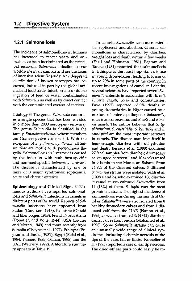
1.2.1 Salmonellosis
The incidence of salmonellosis in humans has increased in recent years and ani- mals have been incriminated as the princi- pal reservoir. Salmonella infections occur worldwide in all animals and are the focus of intensive scientific study. A widespread distribution of known serotypes has oc- curred, induced in part by the global ani- mal and food trade. Infections occur due to ingestion of feed or water contaminated with Salmonella as well as by direct contact with the contaminated excreta of carriers.
Etiology :!$I The genus Salmonella compris- es a single species that has been divided into more than 2000 serotypes (serovars). The genus Salmonella is classified in the family Enterobacteriaceae, whose members are Gram-negative coccobacilli. With the exception of S. gallinarumpullorum, all Sal- monellae are motile with peritrichous fla- gella. Salmonellosis in livestock is caused by the infection with both host-specific and non-host-specific Salmonella serovars. The disease is characterized by one or more of 3 major syndromes: septicemia, acute and chronic enteritis.
Epidemiology and Clinical Signs merous authors have reported salmonel- losis and Salmonella infections in camels in different parts of the world. Reports of Sal- monella infections have appeared from Sudan (Curasson, 1918), Palestine (Olitzki and Ellenbogen, 1943), French North Africa (Donatien and Boue, 1944), USA (Bruner and Moran, 1949) and more recently from Somalia (Cheyne et al., 1977), Ethiopia (Pe- gram and Tareke, 1981), Egypt (Refai et al., 1984; Yassien, 1985; Osman, 1995) and the UAE (Wernery, 1992). A literature summa- ry appears in Table 19.
In camels, Salmonella can cause enteri- tis, septicemia and abortion. Chronic sal- monellosis is characterized by diarrhea, weight loss and death within a few weeks (Fazil and Hofmann, 1981). Pegram and Tareke (1981) reported that salmonellosis in Ethiopia is the most important disease in young dromedaries, leading to losses of up to 20% in some parts of the country. In recent investigations of camel calf deaths, several scientists have reported severe Sal- monella enteritis in association with E. coli, Eimeria cameli, rota- and coronaviruses. Faye (1997) reported 68.3% deaths in young dromedaries in Niger caused by a mixture of enteric pathogens: Salmonella, rotavirus, coronavirus and E. coli and Eime- ria cameli. The author believes that S. ty- phimurium, S. enteritidis, S. kentucky and S. saint-Paul are the most important serovars in camels. The disease manifests itself in hemorrhagic diarrhea with dehydration and death. Berrada et al. (1998) examined 27 fecal samples from diarrheic dromedary calves aged between 1 and 10 weeks raised in 9 herds in the Moroccan Sahara. From 14.8% of the diseased calves, 5 different Salmonella strains were isolated. Salih et al. (1998 a and b), who examined 106 diarrhe- ic camel calves cultured Salmonellae from 14 (13%) of them. S. typhi was the most prominent strain. The highest incidence of salmonellosis was during the month of Oc- tober. Salmonellae were also isolated from 8 healthy dromedary calves and from 1 dis- eased calf from the UAE (Nation et al., 1996) as well as from 9.5% (4/42) diarrheic camel calves from Sudan (Mohamed et al., 1998). Some Salmonella strains can cause an unusually wide range of clinical syn- dromes including ischemic necrosis of the tips of the ears, tail or limbs. Nothelfer et al. (1995) reported a case of ear tip necrosis. The dried-off ear parts could easily be re-
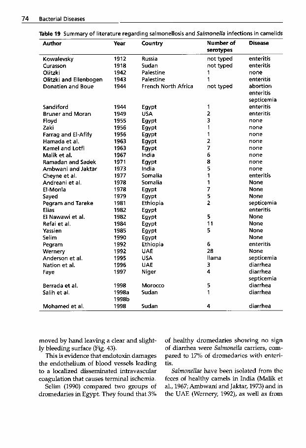
74 Bacterial Diseases
Table 19 Summary of literature regarding salmonellosis and Salmonella infections in camelids
Author Year Country Number of Disease serotypes
Kowa levsky Curasson Olitzki Olitzki and Ellenbogen Donatien and Boue
Sandiford Bruner and Moran Floyd Zaki Farrag and El-Afify Hamada et al. Kame1 and Lotfi Malik et al. Ramadan and Sadek Ambwani and Jaktar Cheyne et al. Andreani et al. El-Monla Sayed Pegram and Tareke Elias E l Nawawi et al. Refai et al. Yassien Selim Pegram Wernery Anderson et al. Nation et al. Faye
Berrada et al. Salih et al.
Mohamed et al.
1912 1918 1942 1943 1944
1944 1949 1955 1956 1956 1963 1963 1967 1971 1973 1977 1978 1978 1979 1981 1982 1982 1984 1985 1990 1992 1992 1995 1996 1997
1998 1998a 1998b 1998
Russia Sudan Palestine Pa lest i ne French North Africa
Egypt
Egypt Egypt Egypt Egypt Egypt India Egypt India Somalia Somalia Egypt Egypt
Egypt Egypt Egypt Egypt Egypt Ethiopia UAE USA UAE Niger
USA
Ethiopia
Morocco Sudan
Sudan
not typed not typed 1 1 not typed
1 2 3 1 1 2 7 6 8 5 1 1 7 5 2
5 11 5
6 28 llama 3 4
5 1
4
enteritis enteritis none enteritis abortion enteritis septicemia enteritis enteritis none none none none none none none none enteritis None None None septicemia enteritis None None None None enteritis None septicemia diarrhea diarrhea septicemia diarrhea diarrhea
diarrhea
moved by hand leaving a clear and slight- ly bleeding surface (Fig. 43).
This is evidence that endotoxin damages the endothelium of blood vessels leading to a localized disseminated intravascular coagulation that causes terminal ischemia.
Selim (1990) compared two groups of dromedaries in Egypt. They found that 3%
of healthy dromedaries showing no sign of diarrhea were Salmonella carriers, com- pared to 17% of dromedaries with enteri- tis.
Salmonellae have been isolated from the feces of healthy camels in India (Malik et al., 1967; Ambwani and Jaktar, 1973) and in the UAE (Wernery, 1992), as well as from
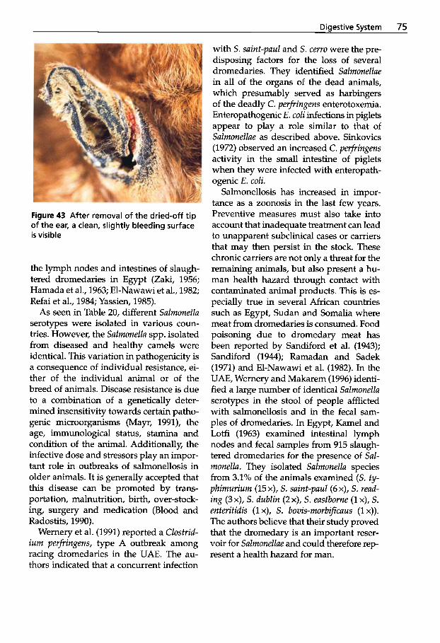
Digestive System 75
Figure 43 After removal of the dried-off tip of the ear, a clean, slightly bleeding surface is visible
the lymph nodes and intestines of slaugh- tered dromedaries in Egypt (Zaki, 1956; Hamada et al., 1963; El-Nawawi et al., 1982; Refai et al., 1984; Yassien, 1985).
As seen in Table 20, different Salmonella serotypes were isolated in various coun- tries. However, the Salmonella spp. isolated from diseased and healthy camels were identical. This variation in pathogenicity is a consequence of individual resistance, ei- ther of the individual animal or of the breed of animals. Disease resistance is due to a combination of a genetically deter- mined insensitivity towards certain patho- genic microorganisms (Mayr, 1991), the age, immunological status, stamina and condition of the animal. Additionally, the infective dose and stressors play an impor- tant role in outbreaks of salmonellosis in older animals. It is generally accepted that this disease can be promoted by trans- portation, malnutrition, birth, over-stock- ing, surgery and medication (Blood and Radostits, 1990).
Wemery et al. (1991) reported a Clostrid- ium perfringens, type A outbreak among racing dromedaries in the UAE. The au- thors indicated that a concurrent infection
with S. saint-paul and S. cerro were the pre- disposing factors for the loss of several dromedaries. They identified Salmonellae in all of the organs of the dead animals, which presumably served as harbingers of the deadly C. perfringens enterotoxemia. Enteropathogenic E. coli infections in piglets appear to play a role similar to that of Salmonellae as described above. Sinkovics (1972) observed an increased C. perfringens activity in the small intestine of piglets when they were infected with enteropath- ogenic E. coli.
Salmonellosis has increased in impor- tance as a zoonosis in the last few years. Preventive measures must also take into account that inadequate treatment can lead to unapparent subclinical cases or carriers that may then persist in the stock. These chronic carriers are not only a threat for the remaining animals, but also present a hu- man health hazard through contact with contaminated animal products. This is es- pecially true in several African countries such as Egypt, Sudan and Somalia where meat from dromedaries is consumed. Food poisoning due to dromedary meat has been reported by Sandiford et al. (1943); Sandiford (1944); Ramadan and Sadek (1971) and El-Nawawi et al. (1982). In the UAE, Wemery and Makarem (1996) identi- fied a large number of identical Salmonella serotypes in the stool of people afflicted with salmonellosis and in the fecal sam- ples of dromedaries. In Egypt, Kame1 and Lotfi (1963) examined intestinal lymph nodes and fecal samples from 915 slaugh- tered dromedaries for the presence of Sal- monella. They isolated Salmonella species from 3.1% of the animals examined (S. ty- phimurium (15 x), S. saint-paul (6 x), S. read- ing (3 x), s. dublin (2 x), S. eastborne (1 x), S. enteritidis (1 x), S. bovis-morbifcaus (1 x)). The authors believe that their study proved that the dromedary is an important reser- voir for Salmonellae and could therefore rep- resent a health hazard for man.
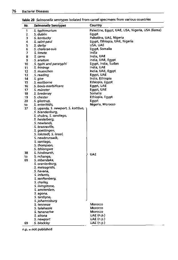
76 Bacterial Diseases
37 5. Uganda, 5. newport, 5. kottbus, ’ 5. brandenburg, 5. shubra, 5. sandiego, 5. heidelberg, 5. newlands, 5. brazzaville, 5. goettingen, 5. lokstedt, 5. israel, 5. newbrunswik, 5. Santiago, 5. thompson, 5. tshiongwe
38 5. hindmarsh, t o 5. nchanga, 69 5. mbandaka,
5. oranienburg, 5. meleagridis, 5. havana, 5. infantis, 5. senftenberg, 5. chailey, 5. livingstone, 5. amsterdam, 5. agona, 5. tarshyne, 5. johannisburg J
’ UAE
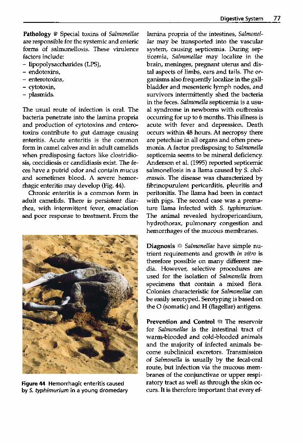
Digestive System 77
1 Special toxins of Salmonellae are responsible for the systemic and enteric forms of salmonellosis. These virulence factors include: - lipopolysaccharides (LPS), - endotoxins, - enterotoxins,
- plasmids. - cytotoxin,
The usual route of infection is oral. The bacteria penetrate into the lamina propria and production of cytotoxins and entero- toxins contribute to gut damage causing enteritis. Acute enteritis is the common form in camel calves and in adult camelids when predisposing factors like clostridio- sis, coccidiosis or candidiasis exist. The fe- ces have a putrid odor and contain mucus and sometimes blood. A severe hemor- rhagic enteritis may develop (Fig. 4.4).
Chronic enteritis is a common form in adult camelids. There is persistent diar- rhea, with intermittent fever, emaciation and poor response to treatment. From the
Figure 44 Hemorrhagic enteritis caused by S. typhimurium in a young dromedary
lamina propria of the intestines, Salmonel- lae may be transported into the vascular system, causing septicemia. During sep- ticemia, Salmonellae may localize in the brain, meninges, pregnant uterus and dis- tal aspects of limbs, ears and tails. The or- ganisms also frequently localize in the gall- bladder and mesenteric lymph nodes, and survivors intermittently shed the bacteria in the feces. Salmonella septicemia is a usu- al syndrome in newborns with outbreaks occurring for up to 6 months. This illness is acute with fever and depression. Death occurs within 48 hours. At necropsy there are petechiae in all organs and often pneu- monia. A factor predisposing to Salmonella septicemia seems to be mineral deficiency. Anderson et al. (1995) reported septicemic salmonellosis in a llama caused by S. chol- erasuis. The disease was characterized by fibrinopurulent pericarditis, pleuritis and peritonitis. The llama had been in contact with pigs. The second case was a prema- ture llama infected with S. typhimurium. The animal revealed hydropericardium, hydrothorax, pulmonary congestion and hemorrhages of the mucous membranes.
Salmonellae have simple nu- trient requirements and growth in vitro is therefore possible on many different me- dia. However, selective procedures are used for the isolation of Salmonella from specimens that contain a mixed flora. Colonies characteristic for Salmonellae can be easily serotyped. Serotyping is based on the 0 (somatic) and H (flagellar) antigens.
Prevention and Control for Salmonellae is the intestinal tract of warm-blooded and cold-blooded animals and the majority of infected animals be- come subclinical excretors. Transmission of Salmonella is usually by the fecal-oral route, but infection via the mucous mem- branes of the conjunctivae or upper respi- ratory tract as well as through the skin oc- curs. It is therefore important that every ef-
78 Bacterial Diseases
fort be undertaken to prevent introduction of carrier animals into a herd. It should also be ensured that feed supplies are free of Salmonellae. Certain procedures should be followed in a Salmonella outbreak on a camel farm: 1. Carrier animals should be identified,
isolated and treated vigorously. Treated camels must be re-examined several times before there can be confidence that they are not carriers.
2. Feed and water supplies must be pro- tected from fecal contamination (be- ware of pigeons and rodents).
3. Movement of animals around the farm should be restricted.
4. All persons should be aware of the health hazards of working with infected camels.
5. The use of vaccines should be consid- ered.
Supportive therapy and good nursing are important especially in camel calves with enteritis. This includes oral or parenteral rehydration, correction of electrolyte im- balances and stabilization of the acid-base equilibrium. It is also very important to avoid factors that lead to mineral deficien- cies in the dams and their offspring. Non- steroidal anti-inflammatory drugs, such as ketoprofen, may be of benefit since it is known that many camel calves subsequent- ly also suffer from endotoxic shock. In these cases, the treatment protocol described under the heading Endotoxicosis (1.1.4) should be followed.
Antimicrobial drug treatment of salmo- nellosis is controversial because it may cre- ate a carrier state in camelids and antibiot- ic resistant strains of Salmonellae. The drug should be administered parenterally, since oral treatment of an animal species that ruminates can create severe disturbances of the gastrointestinal flora. Antimicrobial drugs generally recommended for par- enteral use in salmonellosis are ampicil- lin, amoxycillin and trimethoprim-sulfon-
amide combinations. BaytriP is also a very effective drug, but the drug of choice should be based upon culture and sensitiv- ity. Treatment must start immediately and should be continued daily for up to 6 days.
Some commercial live, avirulent vac- cines are now available. However, autoge- nous Salmonella vaccines (see chapter 4) are of greater value because they include the Salmonella strains involved in the outbreak. These vaccines should be used in problem herds and should be administered twice before parturition in order to provide pro- tection against salmonellosis for the new- borns. The colostral immunity will last approximately 6 to 8 weeks. The autoge- nous vaccines are killed vaccines, which should be administered either intramuscu- larly or subcutaneously at a dosage of 5 to 8 mL depending on the weight of the ani- mal. It is of no use to vaccinate newborn camelids because their immune system is still immature. Camelid calves in problem herds whose dams have not been vaccinat- ed should receive up to 50 mL of the herd- specific vaccine orally for 14 days.
1.2.2 Colibacillosis
While Escherichia coli is the cause of various diseases of great economic magnitude, es- pecially in young animals, it also consti- tutes a large part of the normal commen- sally aerobic intestinal flora. Willinger (1981), Quinn et al. (1994), Wernery and Kaaden (1995) and Fowler (1998) ascribed the following diseases to E. coli: - colibacillosis (white scours) in bovine
calves less than 1 week old; - colisepticemia in bovine calves less than
1 week old; - joint ill in bovine calves surviving a col-
isepticemia; - neonatal diarrhea (colibacillosis), colisep-
ticemia in piglets less than 1 week old; - colibacillosis in piglets about 2 weeks
after weaning;
Digestive System 79
- E. coli enterotoxemia in weaned pigs; - mastitis in cattle and other animals
(ewes); - MMA-complex (metritis-mastitis-agalac-
tia syndrome) in sows; - colibacillosis and colisepticemia in neo-
natal lambs, "watery mouth in neona- tal lambs;
- colibacillosis and colisepticemia in cam- elids 2 to 4 weeks old;
- dysentery in rabbits; - diarrhea and pyometra in dogs; - septicemia, coli-granulomatosis in fowl; - inflammation of the urogenital and res-
- wound infections and healing impair- piratory tracts;
ment.
Colibacillosis and colisepticemia have been reported in OWC and NWC (Wernery and Kaaden, 1995; Fowler, 1998).
Etiology Escherichia coli is a straight Gram-negative motile rod that belongs like Salmonella to the Enterobacteriaceae family. Enteric infection caused by E. coli can be due to at least 5 different varieties of bac- teria operating through different mecha- nisms: 1. Enterotoxigenic E. coli (ETEC) with their
fimbrial adhesions K88, K99 and others - these strains cause the majority of neonatal colibacillosis.
2. Enteropathogenic E. coli (EPEC) do not produce enterotoxins, but can cause di- arrhea.
3. Enteroinvasive E. coli (EIEC) invade en- terocytes and produce virulence factors - they are responsible for colisepticemia and release endotoxins when dying.
4. Attaching and effacing E. coli (AEEC) produce verotoxins and destroy the microvilli - they produce enteric dis- eases.
5. Enterohemorrhagic E. coli (EHEC) cause hemorrhagic colitis and have been asso- ciated with the hemolytic-uremic syn- drome in children.
All five pathogens share the general prop- erties of demonstrating specific interac- tions with the intestinal mucosa, of elabo- rating various toxins, and of possessing plasmid-encoded virulence factors.
E. coli possess different antigens (K = capsular, 0 = cell wall or somatic, H = fla- gellar, F = fimbrial), which can be used to serotype strains. A plasmid, of which there may be more than one in an E. coli cell, may code for several virulence factors includ- ing antibiotic resistance.
Epidemiology and Clinical Signs E. coli is a natural inhabitant of some parts of the intestines of all mammals and is excreted in feces. The presence of E. coli in water and food samples is taken as evidence of fecal contamination.
E. coli infections in young animals are mostly due to errors in husbandry and, in calves, are frequently associated with Sal- monella, Clostridia, Pasteurella, corona- and rotaviruses as well as Cryptosporidia infec- tions. Different serotypes can cause differ- ent clinical signs. Enterotoxigenic E. coli strains produce enterotoxins, which cause enteritis with dehydration. Some strains, especially pathogenic ones, can cause he- molysis (Bisping and Amtsberg, 1988). Pathogenic E. coli strains possess a variety of virulence factors upon which their path- ogenicity depends. These factors include endotoxin, protein adhesions, a- and p-he- molysins, capsular polysaccharides, heat- labile and heat-stable enterotoxins and verotoxin. A considerable economic loss to the camel industry results from colibacillo- sis or colisepticemia in young camelids. These losses can reach 40%.
E. coli infections in dromedary calves have been described by various authors. Schwartz and Dioli (1992) reported a mor- bidity of 30% in neonatal dromedary calves in East Africa. Without immediate veteri- nary intervention, mortality can reach 100%. The authors believe that the unsanitary con- ditions in the breeding herds along with
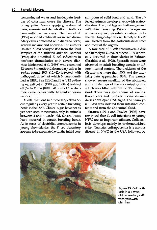
80 Bacterial Diseases
contaminated water and inadequate feed- ing of colostrum cause the disease. The calves suffer from dysentery abdominal pain, anorexia and dehydration. Death oc- curs within a few days. Chauhan et al. (1986) reported colibacillosis in two drom- edary calves presented with diarrhea, fever, general malaise and anorexia. The authors isolated E. coli serotype 083 from the fecal samples of the afflicted animals. Rombol (1942) also described E. coli infections in newborn dromedaries with severe diar- rhea. Mohamed et al. (1998) who examined 42 one to 3-month-old dromedary calves in Sudan found 40% (12/42) infected with pathogenic E. coli, of which 5 were identi- fied as EIEC, 2 as EPEC and 1 as VT2 patho- types. Salih et al. (1997 and 1998 a) isolated 69 (66%) E. coli (K88, F41) out of 106 diar- rheic camel calves with different adhesion factors.
E. coli infections in dromedary calves oc- cur regularly every year in certain breeding herds in the UAE. Clinical signs have not as yet been seen in neonates, only in animals between 2 and 4 weeks old. Severe losses have occurred in certain breeding herds. As in cases of clostridial enterotoxemia in young dromedaries, the E. coli dysentery appears to be associated with the initial con-
sumption of solid food and sand. The af- fected animals develop a yellowish watery diarrhea. The hind legs and tail are covered with dried feces (Fig. 45) and the eyes are sunken deep in their orbital cavities due to the resulting dehydration. Hemolytic E. coli are isolated from the gastrointestinal tract and most of the organs.
A rare case of E. coli enterotoxemia due to a hemolytic E. coli, serotype 0139 report- edly occurred in dromedaries in Bahrain (Ibrahim et al., 1998). Sporadic cases were observed in adult breeding camels at dif- ferent camel centers. The incidence of the disease was more than 50% and the mor- tality rate approached 90%. The camels showed severe swelling of the abdomen and a distention of the abdominal cavity, which was filled with 100 to 150 liters of fluid. There was also edema of eyelids, throat, ears and forehead. Some drome- daries developed CNS signs. The hemolyt- ic E. coli was isolated from intestinal con- tents and from the abdominal fluid.
Strauss (1991) and Fowler (1998) have remarked that E. coli infections in young NWC are an important ailment. Colibacil- losis develops mainly in undernourished crias. Neonatal colisepticemia is a serious disease in NWC in the USA followed by
Figure 45 Colibacil- losis in a 3-week- old dromedary calf with yellowish diarrhea
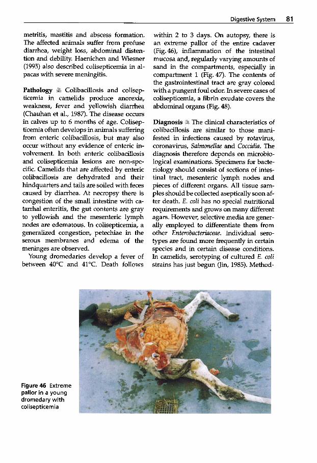
Digestive System 81
metritis, mastitis and abscess formation. The affected animals suffer from profuse diarrhea, weight loss, abdominal disten- tion and debility. Haenichen and Wiesner (1995) also described colisepticemia in al- pacas with severe meningitis.
Pathology Lit Colibacillosis and colisep- ticemia in camelids produce anorexia, weakness, fever and yellowish diarrhea (Chauhan et al., 1987). The disease occurs in calves up to 6 months of age. Colisep- ticemia often develops in animals suffering from enteric colibacillosis, but may also occur without any evidence of enteric in- volvement. In both enteric colibacillosis and colisepticemia lesions are non-spe- cific. Camelids that are affected by enteric colibacillosis are dehydrated and their hindquarters and tails are soiled with feces caused by diarrhea. At necropsy there is congestion of the small intestine with ca- tarrhal enteritis, the gut contents are gray to yellowish and the mesenteric lymph nodes are edematous. In colisepticemia, a generalized congestion, petechiae in the serous membranes and edema of the meninges are observed.
Young dromedaries develop a fever of between 40°C and 41°C. Death follows
within 2 to 3 days. On autopsy, there is an extreme pallor of the entire cadaver (Fig. 46), inflammation of the intestinal mucosa and, regularly varying amounts of sand in the compartments, especially in compartment 1 (Fig.47). The contents of the gastrointestinal tract are gray colored with a pungent foul odor. In severe cases of colisepticemia, a fibrin exudate covers the abdominal organs (Fig. 48).
The clinical characteristics of colibacillosis are similar to those mani- fested in infections caused by rotavirus, coronavirus, Salmonellae and Coccidia. The diagnosis therefore depends on microbio- logical examinations. Specimens for bacte- riology should consist of sections of intes- tinal tract, mesenteric lymph nodes and pieces of different organs. All tissue sam- ples should be collected aseptically soon af- ter death. E. coZi has no special nutritional requirements and grows on many different agars. However, selective media are gener- ally employed to differentiate them from other Enterobacteriaceae. Individual sero- types are found more frequently in certain species and in certain disease conditions. In camelids, serotyping of cultured E. coZi strains has just begun (Jin, 1985). Method-
Figure 46 Extreme pallor in a young dromedary with colisepticemia
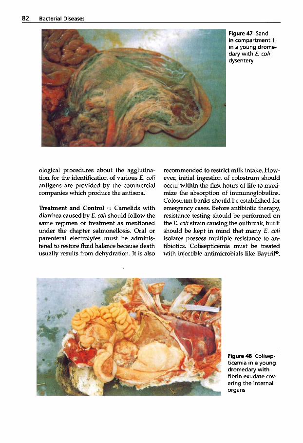
82 Bacterial Diseases
ological procedures about the agglutina- tion for the identification of various E. coli antigens are provided by the commercial companies which produce the antisera.
Treatment and Control . Camelids with diarrhea caused by E . coli should follow the same regimen of treatment as mentioned under the chapter salmonellosis. Oral or parenteral electrolytes must be adminis- tered to restore fluid balance because death usually results from dehydration. It is also
Figure47 Sand in compartment 1 in a young drome- dary with E. coli dysentery
recommended to restrict milk intake. How- ever, initial ingestion of colostrum should occur within the first hours of life to maxi- mize the absorption of immunoglobulins. Colostrum banks should be established for emergency cases. Before antibiotic therapy, resistance testing should be performed on the E. coli strain causing the outbreak, but it should be kept in mind that many E. coli isolates possess multiple resistance to an- tibiotics. Colisepticemia must be treated with injectible antimicrobials like BaytriP,
Figure 48 Colisep- ticemia in a young dromedary with fibrin exudate cov- ering the internal organs
Digestive System 83
trimethoprim/sulfonamide, kanamycin or colistin (Manefield and Tinson, 1996).
In order to reduce losses among young dromedaries, maternal vaccination with herd-specific E. coli vaccines should be ad- ministered annually or oral vaccinations in young camelids should be tried.
Due to intensive animal keeping, for ex- ample in animal parks and zoos, consider- able losses can arise due to E . coli. For this reason, Strauss (1991) applied a protective maternal vaccination (Colivac@) twice to all pregnant camels about 8 and then 4 weeks prior to the calculated date of parturition. After that, losses in young animals were greatly reduced.
1.2.3 Paratuberculosis (Johne’s Disease)
This disease is characterized by persistent and progressive diarrhea, weight loss, de- bilitation and eventually death. The dis- ease produces a chronic, contagious enteri- tis and affects cattle, sheep, goats, farmed deer and other domestic and wild rumi- nants. It has also been reported to occur in OWC (Wernery and Kaaden, 1995) and in NWC (Appleby and Head, 1954; Schwar- te, 1956; Belknap et al., 1994; Ridge et al., 1995).
Paratuberculosis occurs worldwide. In tropical areas with intensive dairy farm- ing, paratuberculosis presents a serious economic problem. Mycobacterium avium spp. paratuberculosis is excreted in the fe- ces of infected animals and so can then be ingested with contaminated food or water. The bacteria spread to the intestin- al mucosa or mesenteric lymph nodes where they can cause chronic inflamma- tion. M. avium spp. paratuberculosis is also able to cross the placenta to the fetus.
Etiology Mycobacterium avium spp. para- tuberculosis is a non-motile, non-sporing, aerobic and oxidative bacterium which
does not take up dyes of the Gram stain be- cause the cell wall is rich in lipids and my- colic acid. M. avium spp. paratuberculosis is acid-fast and the best stain is Ziehl- Neelsen. The disease can be diagnosed by the demonstration of the bacteria and by serological and allergic tests.
Epidemiology and Clinical Signs M. avium spp. paratuberculosis is shed in feces and the organisms are found within mac- rophages of the intestinal mucosa and ad- jacent lymph nodes. A cell-mediated im- mune response appears to be involved in the pathogenesis of this disease. Not all in- fected animals become clinical cases, but they remain excretors of M. avium spp. paratuberculosis. Following oral infection, M. avium spp. paratuberculosis enters the lymphatics through the tonsils and the in- testinal mucosa. Peyer’s patches take up the microorganisms from the intestinal lumen and transport them through the in- testinal mucosa. The incubation period is generally 18 to 24 months.
Paratuberculosis is one of the most im- portant and widespread diseases of the Bactrian camel in the former Soviet bloc, as reported by Ivanov and Skalinskii (1957), Ovdienko et al. (1985), Fassi-Fehri (1987) and Buchnev et al. (1987). The disease has also been diagnosed in Bactrian camels from Mongolia suffering from severe diar- rhea (Guake et al., 1964) and has been known in Turkmenistan since 1949 (Stro- gov, 1957). Strogov (1957) and Buchnev et al. (1987) reported that paratuberculosis is more prevalent in young Bactrians be- tween weaning and 4 years of age. The au- thors believe that older camels may recov- er from the disease. Recovery of adult in- fected camels takes place slowly over a period of 6 months. The annual incidence of infection in Bactrian camels in Turk- menistan between 1946 and 1952 was be- tween 0.3 and 1.5% (Strogov, 1957).
Paratuberculosis is also seen in drome- daries, but is less prevalent than in Bactri-
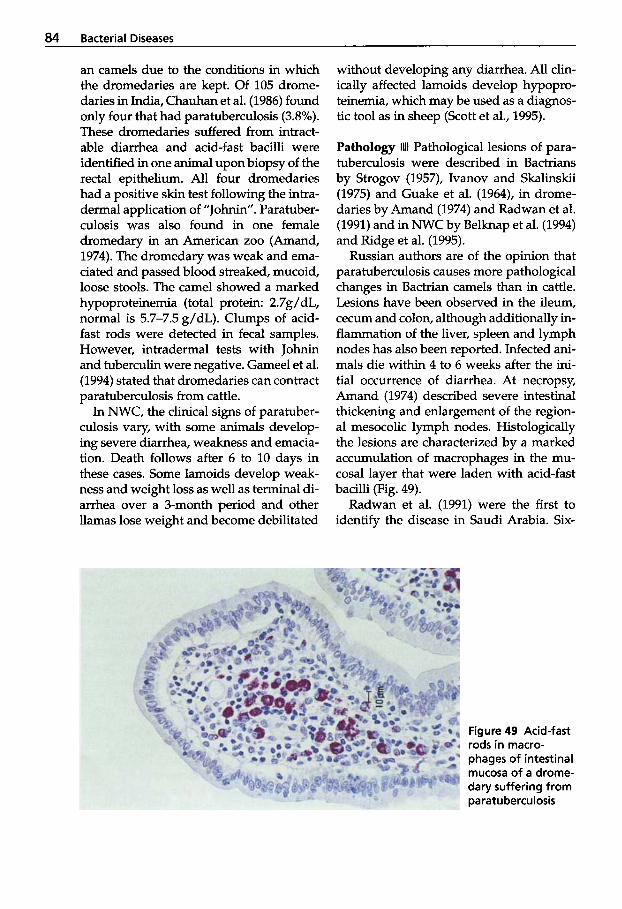
84 Bacterial Diseases
an camels due to the conditions in which the dromedaries are kept. Of 105 drome- daries in India, Chauhan et al. (1986) found only four that had paratuberculosis (3.8%). These dromedaries suffered from intract- able diarrhea and acid-fast bacilli were identified in one animal upon biopsy of the rectal epithelium. All four dromedaries had a positive skin test following the intra- dermal application of "Johnin". Paratuber- culosis was also found in one female dromedary in an American zoo (Amand, 1974). The dromedary was weak and ema- ciated and passed blood streaked, mucoid, loose stools. The camel showed a marked hypoproteinemia (total protein: 2.7g/dL, normal is 5.7-7.5 g/dL). Clumps of acid- fast rods were detected in fecal samples. However, intradermal tests with Johnin and tuberculin were negative. Gameel et al. (1994) stated that dromedaries can contract paratuberculosis from cattle.
In NWC, the clinical signs of paratuber- culosis vary, with some animals develop- ing severe diarrhea, weakness and emacia- tion. Death follows after 6 to 10 days in these cases. Some lamoids develop weak- ness and weight loss as well as terminal di- arrhea over a 3-month period and other llamas lose weight and become debilitated
without developing any diarrhea. All clin- ically affected lamoids develop hypopro- teinemia, which may be used as a diagnos- tic tool as in sheep (Scott et al., 1995).
Pathology 1111 Pathological lesions of para- tuberculosis were described in Bactrians by Strogov (1957), Ivanov and Skalinskii (1975) and Guake et al. (1964), in drome- daries by Amand (1974) and Radwan et al. (1991) and in NWC by Belknap et al. (1994) and Ridge et al. (1995).
Russian authors are of the opinion that paratuberculosis causes more pathological changes in Bactrian camels than in cattle. Lesions have been observed in the ileum, cecum and colon, although additionally in- flammation of the liver, spleen and lymph nodes has also been reported. Infected ani- mals die within 4 to 6 weeks after the ini- tial occurrence of diarrhea. At necropsy, Amand (1974) described severe intestinal thickening and enlargement of the region- al mesocolic lymph nodes. Histologically the lesions are characterized by a marked accumulation of macrophages in the mu- cosal layer that were laden with acid-fast bacilli (Fig. 49).
Radwan et al. (1991) were the first to identify the disease in Saudi Arabia. Six-
Figure 49 Acid-fast rods in macro- phages of intestinal mucosa of a drome- dary suffering from pa ratu bercu losis
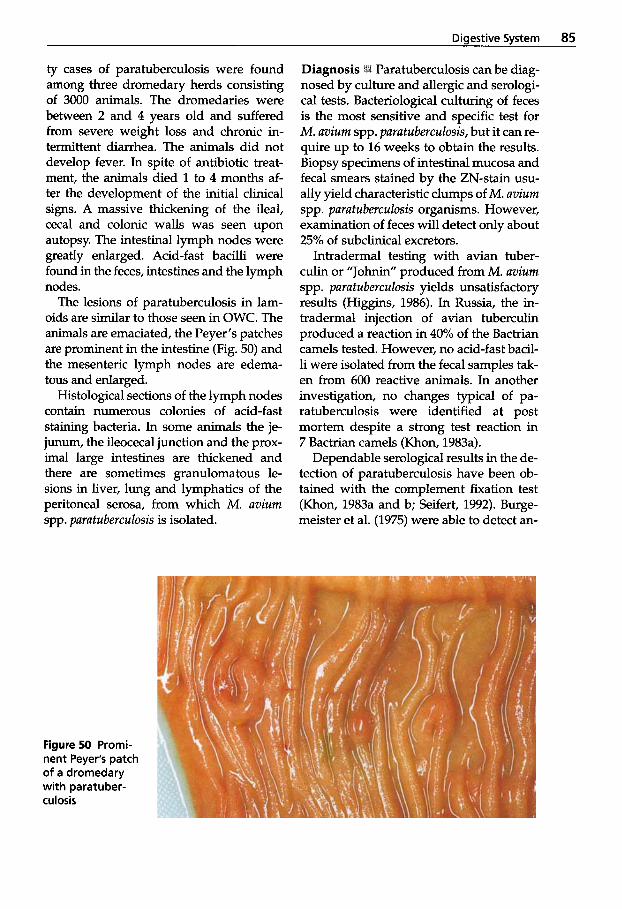
Digestive System 85
ty cases of paratuberculosis were found among three dromedary herds consisting of 3000 animals. The dromedaries were between 2 and 4 years old and suffered from severe weight loss and chronic in- termittent diarrhea. The animals did not develop fever. In spite of antibiotic treat- ment, the animals died 1 to 4 months af- ter the development of the initial clinical signs. A massive thickening of the ileal, cecal and colonic walls was seen upon autopsy. The intestinal lymph nodes were greatly enlarged. Acid-fast bacilli were found in the feces, intestines and the lymph nodes.
The lesions of paratuberculosis in lam- oids are similar to those seen in OWC. The animals are emaciated, the Peyer's patches are prominent in the intestine (Fig. 50) and the mesenteric lymph nodes are edema- tous and enlarged.
Histological sections of the lymph nodes contain numerous colonies of acid-fast staining bacteria. In some animals the je- junum, the ileocecal junction and the prox- imal large intestines are thickened and there are sometimes granulomatous le- sions in liver, lung and lymphatics of the peritoneal serosa, from which M . avium spp. paratuberculosis is isolated.
Paratuberculosis can be diag- nosed by culture and allergic and serologi- cal tests. Bacteriological culturing of feces is the most sensitive and specific test for M. avium spp. paratuberculosis, but it can re- quire up to 16 weeks to obtain the results. Biopsy specimens of intestinal mucosa and fecal smears stained by the ZN-stain usu- ally yield characteristic clumps of M. avium spp. paratuberculosis organisms. However, examination of feces will detect only about 25% of subclinical excretors.
Intradermal testing with avian tuber- culin or "Johnin" produced from M. avium spp. paratuberculosis yields unsatisfactory results (Higgins, 1986). In Russia, the in- tradermal injection of avian tuberculin produced a reaction in 40% of the Bactrian camels tested. However, no acid-fast bacil- li were isolated from the fecal samples tak- en from 600 reactive animals. In another investigation, no changes typical of pa- ratuberculosis were identified at post mortem despite a strong test reaction in 7 Bactrian camels (Khon, 1983a).
Dependable serological results in the de- tection of paratuberculosis have been ob- tained with the complement fixation test (Khon, 1983a and b; Seifert, 1992). Burge- meister et al. (1975) were able to detect an-
Figure 50 Promi- nent Peyer's patch of a dromedary with paratuber- culosis
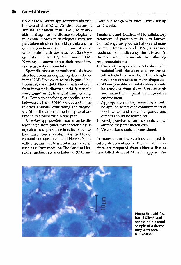
86 Bacterial Diseases
tibodies to M. avium spp. paratuberculosis in the sera of 11 of 52 (21.2%) dromedaries in Tunisia. Feldmann et al. (1981) were also able to diagnose the disease serologically in Kenya. However, serological tests for paratuberculosis on individual animals are often inconclusive, but they are of value when entire herds are screened. Serologi- cal tests include CFT, AGID and ELISA. Nothing is known about their specificity and sensitivity in camelids.
Sporadic cases of paratuberculosis have also been seen among racing dromedaries in the UAE. Five cases were diagnosed be- tween 1987 and 1993. The animals suffered from intractable diarrhea. Acid-fast bacilli were found in all five fecal samples (Fig. 51). Complement-fixing antibodies (titers between 1:64 and 1:256) were found in the infected animals, confirming the diagno- sis. All of the animals died in spite of an- tibiotic treatment within one year.
M. avium spp. paratuberculosis can be dif- ferentiated from other mycobacteria by its mycobactin-dependence in culture. Benza- lkonium chloride (Zephiran) is used to de- contaminate specimens and Herrold’s egg yolk medium with mycobactin is often used as culture medium. The slants of Her- rold’s medium are incubated at 37°C and
examined for growth, once a week for up to 16 weeks.
Treatment and Control < No satisfactory treatment of paratuberculosis is known. Control requires good sanitation and man- agement. Radwan et al. (1991) suggested methods of eradicating the disease in dromedaries. They include the following recommendations: 1. Clinically suspected camels should be
isolated until the disease is confirmed. All infected camels should be slaugh- tered and carcasses properly disposed.
2. Where possible, camelid calves should be removed from their dams at birth and reared in a paratuberculosis-free environment.
3. Appropriate sanitary measures should be applied to prevent contamination of food, water and soil; and ponds and ditches should be fenced off.
4. Newly purchased camels should be ex- amined for paratuberculosis.
5. Vaccination should be considered.
In many countries, vaccines are used in cattle, sheep and goats. The available vac- cines are prepared from either a live or heat-killed strain of M. avium spp. purutu-
Figure 51 Acid-fast bacilli (ZiehCNeel- sen stain) in a stool sample of a drome- dary with para- tuberculosis
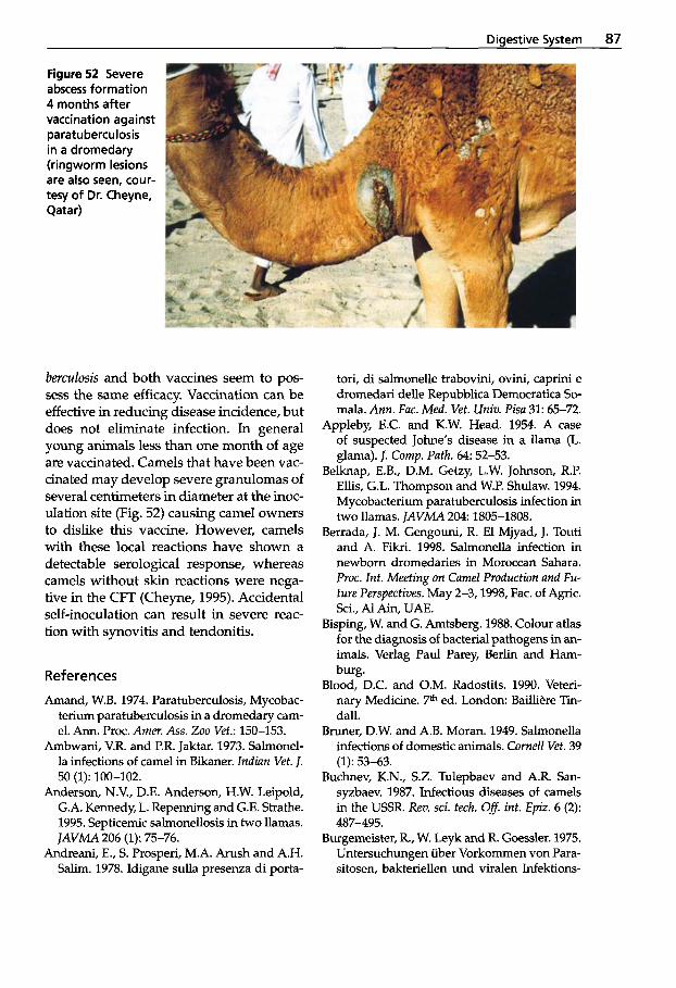
Digestive System 87
Figure 52 Severe abscess formation 4 months after vaccination against paratu berculosis in a dromedary (ringworm lesions are also seen, cour- tesy of Dr. Cheyne, Qatar)
berculosis and both vaccines seem to pos- sess the same efficacy. Vaccination can be effective in reducing disease incidence, but does not eliminate infection. In general young animals less than one month of age are vaccinated. Camels that have been vac- cinated may develop severe granulomas of several centimeters in diameter at the inoc- ulation site (Fig. 52) causing camel owners to dislike this vaccine. However, camels with these local reactions have shown a detectable serological response, whereas camels without skin reactions were nega- tive in the CFT (Cheyne, 1995). Accidental self-inoculation can result in severe reac- tion with synovitis and tendonitis.
References
Amand, W.B. 1974. Paratuberculosis, Mycobac- terium paratuberculosis in a dromedary cam- el. Ann. Proc. Amer. Ass. Zoo Vet.: 150-153.
Ambwani, V.R. and P.R. Jaktar. 1973. Salmonel- la infections of camel in Bikaner. Indian Vet. J .
Anderson, N.V., D.E. Anderson, H.W. Leipold, G.A. Kennedy, L. Repenning and G.E. Strathe. 1995. Septicemic salmonellosis in two llamas.
Andreani, E., S. Prosperi, M.A. Arush and A.H. Salim. 1978. Idigane sulla presenza di porta-
50 (1): 100-102.
JAVMA 206 (1): 75-76.
tori, di salmonelle trabovini, ovini, caprini e dromedari delle Repubblita Democratica So- mala. Ann. Fac. Med. Vet. Univ. Pisa 31: 65-72.
Appleby, E.C. and K.W. Head. 1954. A case of suspected Johne’s disease in a llama (L. glama). J. Comp. Path. 64: 52-53.
Belknap, E.B., D.M. Getzy, L.W. Johnson, R.P. Ellis, G.L. Thompson and W.P. Shulaw. 1994. Mycobacterium paratuberculosis infection in two llamas. JAVMA 204: 1805-1808.
Berrada, J. M. Gengouni, R. El Mjyad, J. Touti and A. Fikri. 1998. Salmonella infection in newborn dromedaries in Moroccan Sahara. Proc. Int. Meeting on Camel Production and Fu- ture Perspectives. May 2-3,1998, Fac. of Agric. Sci., Al Ah, UAE.
Bisping, W. and G. Amtsberg. 1988. Colour atlas for the diagnosis of bacterial pathogens in an- imals. Verlag Paul Parey, Berlin and Ham- burg.
Blood, D.C. and O.M. Radostits. 1990. Veteri- nary Medicine. 7*h ed. London: BailliPre Tin- dall.
Bruner, D.W. and A.B. Moran. 1949. Salmonella infections of domestic animals. Cornell Vet. 39
Buchnev, K.N., S.Z. Tulepbaev and A.R. San- syzbaev. 1987. Infectious diseases of camels in the USSR. Rev. sci. tech. 08 int. Epiz. 6 (2):
Burgemeister, R., W. Leyk and R. Goessler. 1975. Untersuchungen uber Vorkommen von Para- sitosen, bakteriellen und viralen Mektions-
(1): 53-63.
487-495.
88 Bacterial Diseases
krankheiten bei Dromedaren in Siidtunesien. Dtsch. Tierarztl. Wschr. 8 2 352-354.
Chauhan, R.S., R.K. Kaushik, S.C. Gupta, K.C. Satiya and R.C. Kulshreshta. 1986. Prevalence of different diseases in camels (Camelus dromedarius) in India. Camel Newsletter 3:
Chauhan, R.S., K.O. Satija, S.M. Tika Ram and R.K. Kaushik. 1987. Diseases of camels and their control. Indian Farming 36: 27-31.
Cheyne, LA., R.G. Pegram and C.F. Cartwright. 1977. An outbreak of salmonellosis in cam- els in the north-east of the Somali Democra- tic Republic. Trop. Anim. Hlth. Prod. 9 (4): 238-240.
Cheyne, LA. 1995. A brief summary of Paratu- berculosis (Johne's Disease) in domestic ani- mals with reference to recent reports of the disease in camels. Pers. report: 1-12.
Curasson, G. 1918. Une maladie du dromadaire analogue au farcin du boeuf. Bull. SOC. Cent. Mkd. Vet. (Supplement to Rec. Mkd. ve't. 94) 71:
Donatien, A. and A. Boue. 1944. Une epizootie de ghedda dam la region d'Qued Guir (Sa- hara oranais). Arch. Inst. Pasteur Alger 22 (3):
El-Monla, A. 1978. Incidence of zoonotic dis- ease (Salmonellosis) encountered in animals slaughtered in Egypt. M.V. Sc. Thesis, Fac. of Vet. Med., Cairo University.
El-Nawawi, F., H. El-Derea and A. Sayed. 1982. Salmonellae in slaughter animals. Arch f. Lebensmittelhygiene 33: 33-36.
Elias, S.S. 1982. Preliminary studies on salmo- nella microorganisms in camels in Egypt. M.V. Sc. Thesis, Fac. Vet. Med., Cairo Univer- sity.
Farrag, H. and A. El-A*. 1956. Salmonella in apparently normal camels. J. Egypt. Med. Ass.
Fassi-Fehri, M.M. 1987. Les maladies des came- lides. Rev. sci. tech. Ofi int. Epiz. 6 (2): 315-335.
Faye, B. 1997. Guide de l'elevage du droma- daire. Sanofi Sante Nutrition Animale, La Ballastiere - BP126, 33501 Libourne, Cedex, France: pp. 115-116.
Fazil, M.A. and R.R. Hofmann. 1981. Haltung und Krankheiten des Kamels. Tieriirztl. Praxis
Feldman, B.F., C.L. Keen, J.J. Kaneko and T.B. Parver. 1981. Husbandry and diseases of camels. Tieriintl. Praxis 9 (3): 389-402.
10-14.
491-496.
171-174.
39: 698-699.
9: 389-402.
Floyd, T.M. 1955. Salmonella in domestic ani- mals and fowls in Egypt. J. Egypt. Pub. Hlth.
Fowler, M.E. 1998. Medicine and surgery of South American Camelids. Iowa State Uni- versity Press, Ames.
Gameel, A.A.,A.S. Ali, S.A.Razig, J. Brown, S.A. Alhendi and S.M. El-Sanousi. 1994. A clinico- pathological study on spontaneous para- tuberculosis in camels (Camelus dromedar- ius) in Saudi Arabia. Pakistan Vet. J. 14 (1):
Guake, L.K., Z. Dubba, K.H. Tumba and R.M. Abugaliev. 1964. No title. Vet. Moscow 41:
Haenichen, T. and H. Wiesner. 1995. Erkran- kungs- und Todesursachen bei Neuweltka- meliden. Tierurztl. Praxis 23: 515-520.
Hamada, S., H. El-Sawah, I. Sherif, M. Joussef and M. Hidik. 1963. Salmonella of the mesen- teric lymph nodes of slaughtered cattle buf- faloes and camels. J. Arab. Vet. Med. Ass. 23 (4):
Higgins, A. 1986. The camel in health and dis- ease. Baillihre Tindall.
Ibrahim, A.M., A.A. Abdelghaffar, M.E. Fadlal- la, M.N. Nayel, B.A. Ibrahim and A.S. Adam. 1998. Oedema disease in female camels (Camelus dromedarius) in Bahrain. J. Camel Prac. and Res. 5 (1): 167-169.
Ivanov, B.G. and E.I. Skalinskii. 1957. Patholog- ical changes in paratuberculosis in camels. Trudy gos. Insf. eksp. Vet. 20: 186-206.
Jin, Y.C. 1985. Report on the diagnosis and treat- ment of colibacillosis in camels. Acta Agricul- tural College, Yanbian 2: 78-82.
Kamel, H. and Z. Lofti. 1963. Types of salmonel- la prevailing in apparently healthy camels slaughtered for meat. Proc. 4fk Arab. Ann. - Vet. Cong., Cairo, E.A.R.
Khon, F.K. 1983a. Allergical diagnosis of camel paratuberculosis. Bid. vses Inst. eksp. vet., Moskva 50: 26-28.
Khon, F.K. 1983b. Complement fixation test at camel paratuberculosis. Bid. vses Inst. eksp. vet., Moskva 50: 30-32.
Kowalesky, M.J.M. 1912. Le Chameau et ses maladies d'apres les observation d'auteurs russes.J. Mkd. V&. Zootechn., Lyon 15: 462-466.
Malik, ED., S.K. Datta, I.P. Singh and D.S. Kalra. 1967. Salmonella serotypes from camel in In- dia. J. Res. Punjab Agric. Univ., Ludhiana 4:
ASS. 30 (5): 177-183.
15-19.
115-116.
272-277.
123-126.
Digestive System 89
Manefield, G.W. and A. Tinson. 1996. Camels. A compendium. The TG Hungerford Vade Mecum series for domestic animals, series C, No. 22.
Mayr, A. 1991. Neue Erkenntnisse iiber Ent- wicklung, Aufbau und Funktion des Immun- systems. Tierarztl. Praxis 19: 235-240.
Mohamed, M.E.H., C.A. Hart and 0.-R. Kaaden. 1998. Agents associated with camel diarrhea in Eastern Sudan. Proc. Int. Meeting on Camel Production and Future Perspectives. May 2-3, 1998, Fac. of Agric. Sci., Al Ain, UAE.
Nation, G., J.E. Moore, A.H. Tinson, M. MacAl- mont, P.G. Murphy and D. Harron. 1996. Treatment of Salmonella and Campylobacter- associated diarrhoea in camels (Camelus dromedarius) with enrofloxacin. European Congress of Chemotherapy, Glasgow, Scotland, May 14-17,1996.
Nothelfer, H.B., U. Wernery and J. Akbar. 1994. Acral dry gangrene in a camel calf (Camelus dromedarius). J. Camel Prac. and Res. 1 (2):
Olitzki, L. 1942. Comparative studies on sal- monella strains isolated in Palestine from camels and human beings. J. Hyg., Camb. 42:
Olitzki, L. and V. Ellenbogen. 1943. A salmonel- la strain isolated from camels in Palestine. J. Comp. Physiol. Ther. 53 (1): 75-79.
Osman, A.H.A.E.R. 1995. Pathological study on intestinal affections in camels. Thesis, Facul- ty of Veterinary Medicine, Cairo University:
Ovdienko, N.P., F.K. Khon, V.A. Sharov and O.V. Yakusheva. 1985. Diagnosis of paratu- berculosis in camels. Veterinariya, Moscow,
Pegram, R.G. 1992. Camel salmonellosis in the horn of Africa. Proc. lSt int. Camel Cod. In: Allen, W.R., A.J. Higgins, I.G. Mayhew, D.H. Snow and J.F. Wade. R. and W. Publications, Newmarket/UK p. 402.
Pegram, R.G. and F. Tareke. 1981. Observation on the health of Afar livestock. Ethiopian Vet. J.
Quinn, P.J., M.E. Carter, B.K. Markey and G.R. Carter. 1994. Clinical Veterinary Microbiolo- gy, Wolfe: pp. 381-421.
Radwan, A.I., S. El-Magawry, A. Hawari, S.J. Al- Bekairi, S. Aziz and R.M. Rebleza. 1991. Paratuberculosis enteritis (Johne’s Disease) in camels in Saudi Arabia. Biol. Sci. 1: 57-66.
83-84,
547-548.
1-128.
USSR 4: 65-68.
5: 11-15.
Ramadan, EM. and I.M. Sadek. 1971. Parame- ters of salmonellosis in Egypt. J. Egypt. Vet. Med. Ass. 31 (3-4): 193-218.
Refai, M., W.G. El-Said, K. Osman, Z . Lotfi, E.E. Safwat and S. Elias. 1984. Salmonella in slaughtered camels in Egypt. Zagazig Vet. J. 9:
Ridge, S.E., J.T. Harkin, R.T. Badman, A.M. Mel- lor and J.W.A. Larsen. 1995. Johne’s disease in alpacas (Lama pacos) in Australia. Austr. Vet. J. 72 (4): 150-153.
Rombol, B. 1942. Enzootic bacterium coli infec- tion in new-born camels. Numa Vet. 2 0 85-93.
Salih, O., M.T. Shigidi, H.O. Mohamed, Mc- Doungh and Y. Chang. 1998a. The bacterial causes of camel-calf diarrhoea in Eastern Sudan. Proc. Int. Meeting on Camel Production and Future Perspectives. May 2-3,1998, Fac. of Agric. Sci., A1 Ain, UAE.
Salih, O., H.O. Mohamed and M.T. Shigidi. 1998b. The epidemiological factors associ- ated with camel-calf diarrhoea in eastern Sudan. Proc. Int. Meeting on Camel Production and Future Perspectives. May 2-3,1998, Fac. of Agric. Sci., Al Ain, UAE.
Salih, O.S.M., M.T. Shigidi, H.O. Mohammed, P. McDoungh and Y.F. Chang. 1997. Bacteria isolated from camel-calves (Camelus drome- darius) with diarrhea. Camel Newsletter 13 (9): 34-43.
Sandiford, B.R., M.G. El Gheriany, M. Abdul Ela and A.M. Keram. 1943. Food poisoning out- breaks in Egypt associated with Bacterium aertrycke. Lab. Med. Prog. 4 (1): 14-18.
Sandiford, B.R. 1944. Food poisoning due to Bacterium typhimurium (anaerogenes). J. Path. Bact. 56: 254-255.
Sayed, A.I.H. 1979. Studies on salmonella infec- tion in apparently healthy slaughtered ani- mals. M.V. Sc. Thesis, Fac. of Vet. Med., Cairo University.
Schwarte, L.H. 1956. Johne’s Disease suspected in a llama. Vet. Bull. cited in JAVMA: 354.
Schwartz, H.J. and M. Dioli. 1992. The one- humped camel in Eastern Africa. A pictorial guide to diseases, health care and manage- ment. Verlag Josef Margraf.
Scott, P.R., C.J. Clarke and T.J. King. 1995. Serum protein concentrations in clinical cases of ovine paratuberculosis (Johne’s disease). Vet. Rec. 137 173.
Seifert, H.S.H. 1992. Tropentierhygiene. Gustav Fischer Verlag Jena, Stuttgart.
266-267.
90 Bacterial Diseases
Selim, A.M. 1990. Salmonellosis in camel in Egypt. IIX Wardeh, M.F., R.T. Wilson and A.A. Zaied. Proc. Inter Con6 on Camel Prod. and Impr., Dec. 10-13,1990, Tobruk, Libya.
Sinkovics, G. 1972. Quantitative changes of clos- tridia in the intestine of early-weaned pigs diseased in Coli-enterotoxemia. Acta vet. hung. 22 (21): 133-139.
Strauss, G. 1991. Erkrankungen junger Neu- weltkamele im Tierpark Berlin-Friedrichs- felde 11. Arbeitstagung der Zootierarzte im deutschsprachigen Raum. Nov. 1-3 in Stuttgart, Tagungsbericht: 80-83.
Strogov, A.K. 1957. Paratuberculosis in camels. Trudy vses. Inst. eksp. Vet. 20: 120-131.
Wemery, U. 1992. The prevalence of salmonella infections in camels (Camelus dromedarius) in the United Arab Emirates. Br. Vet. J. 148 (5):
Wernery, U., H.S.H. Seifert, A.M. Billah, and M. Ali. 1991. Predisposing factors in en- terotoxemias of camels (Camelus dromedar- ius) caused by Clostridium perfringens type A. Rev. Elm. Me'd. ve't. Pays trop. 44 (2):
Wemery, U. and E.H. Makarem. 1996. Compar- ative study on salmonella serovars isolated from humans and camels in the United Arab Emirates. Camel Newsletter 12 (9): 55-59.
Wemery, U. and 0.-R. Kaaden. 1995. Infectious Diseases of Camelids. Blackwell Wissen- schafts-Verlag, Berlin.
Willinger, H. 1981. Escherichia cob. In: H. Blobel und Tr. Schliesser: Handbuch der bakteriel- len Infektionen bei Tieren. Jena: VEB Gustav Fischer 3: pp. 257-343.
Yassien, N.A. 1985. Salmonella in slaughtered camels. M.V. Sc. Thesis, Fac. of Vet. Med., Cairo University.
445-450.
147-152.
Zaki, O.A. 1956. The incidence of salmonella in- fections in camels. J. Egyp t . Publ. Hlth. Ass. 31 (2): 75-79.
Further reading Abubakr, M.I., M.N. Nayel, M.E. Fadlalla, A.O.
Abdelrahman, S.A. Abuobeida and Y.M. Al- gabara. 1999. The incidence of bacterial in- fection in young camels with reference to Escherichia coli. Int. Workshop on the young camel, Quarzazate, Morocco, 24-26 Oct., 40.
Bornstein, S., M. Younan and R. Feinstein. 1999. A case of neonatal camel colibacillosis. Int. Workshop on the young camel, Quarzazate, Mo- rocco, 24-26, Oct., 23.
Dia, M.L., A. Diop, Ahmed 0. Mohamed, C. Diop and El Hacen 0. Taleb. 1999. Diarrh6es du chamelon en Mauritanie: Rksultats d'en- qu@te au cnerv. Int. Workshop on the young camel, Quarzazate, Morocco, 24-26 Oct., 41.
Kane, Y. and B.C. Diallo. 1999. Donnees sur la pathologie du chamelon en Mauritanie. Int. Workshop on the young camel, Quarzazate, Mo- rocco, 24-26 Oct., 42.
Pal'gov, A.A. 1950. No title. Trud. nauchno-issled, Vet. Inst. Alma Ata 5: 29.
Saad, M.A.G. and A.M. Hussein. 1975. Isolation of salmonellae from camels in Sudan. Sudan Vet. Assoc. 7th con$ on meat industry, Khar- toum.
Salih, O.S.M. 1993. Aetiological and epidemio- logical factors associated with camel calf diarrhoea. Thesis, Faculty Vet. Sci. University Khartoum, Sudan.
Younan, M and S. Bornstein. 1999. Colisepti- cemia in a camel calf. Int. Workshop on the young camel, Quarzazate, Morocco, 24-26 Oct., 77.
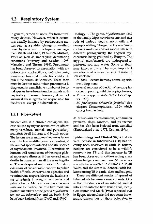
1.3 Respiratory System riiiiiiu I I W I
In general, camels do not suffer from respi- ratory disease. However, when it occurs, it is usually initiated by predisposing fac- tors such as a sudden change in weather, poor hygiene and inadequate manage- ment (Shah and Khan, 1935-1936; Mustafa, 1987) as well as underlying debilitating conditions (Wemery and Kaaden, 1995; Manefield and Tinson, 1996). Pneumonia has been observed in association with en- dotoxicosis, colibacillosis, enterotoxemia, leukemia, chronic skin infections and vita- min E/selenium deficiencies. These facts must be kept in mind when pneumonia is diagnosed in camelids. A number of bacte- rial species have been found in camels with respiratory disease. However, it is not known if these agents are responsible for the disease, except in tuberculosis.
1.3.1 Tuberculosis
Tuberculosis is a chronic contagious dis- ease caused by mycobacteria, which affects many vertebrate animals and particularly manifests itself in lungs and lymph nodes. The lesions are granulomas known as tuber- cles. The lesions differ greatly according to the animal species infected and the species of mycobacteria involved. Tuberculosis in humans still remains one of the major glob- al reportable diseases; it has caused more deaths in humans than all the wars togeth- er. The widespread outbreaks of M. tuber- culosis are of considerable concern to public health officials, conservation agencies and veterinarians responsible for the health sta- tus of animals in zoos, animal parks and private herds. Many strains have become resistant to medication. The two most im- portant members of the genus Mycobacteri- um are M. tuberculosis and M. bovis. Both have been isolated from OWC and NWC.
Etiology The genus Mycobacterium (M.) of the family Mycobacteriaceae are acid-fast rods of various lengths, non-motile and non-sporulating. The genus Mycobacterium contains multiple species (about 50) with different pathogenicity, the atypical my- cobacteria being grouped by Runyon. The atypical mycobacteria are widespread in pastures, soil and water. Some of them may infect animals. The most important mycobacterial species causing disease in livestock are: - M. bovis - occurs in many animal species
including man; - several serovars of the M. avium complex
occur in poultry, wild birds, pigs, horses; - M. avium spp. paratuberculosis (see chap-
ter 1.2.3); - M. farcinogenes (Nocardia farcinica? See
chapter Dermatophilosis, 1.5.3) which causes bovine farcy.
M. tuberculosis affects humans, non-human primates, dogs, canaries, and psittacines and has also been isolated from camelids (Elmossalami et al., 1971; Osman, 1974).
Epidemiology and Clinical Signs A se- vere increase in tuberculosis (TB) has re- cently been observed in cattle in Britain. Badgers are considered to be a wildlife reservoir for TB and this increase in TB has been observed in cattle-keeping areas where badgers are common. M . bovis has recently been isolated in 2 llamas in south Wales and the strain is identical with iso- lates causing TB in cattle, deer and badgers.
There are different modes of spread of tuberculosis between camelid herds. One is the introduction of an infected animal into a non-infected herd (Bush et al., 1990). Gatt Rutter and Mack (1963) reported that in Egypt, tuberculosis did not occur in no- madic camels but in those belonging to
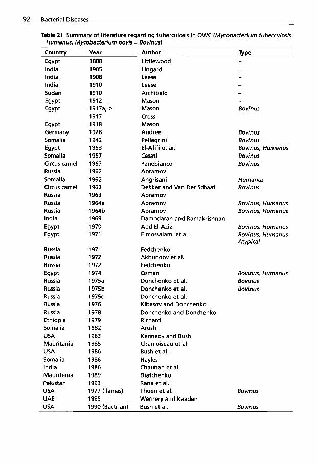
92 Bacterial Diseases
Table 21 Summary of literature regarding tuberculosis in OWC (Mycobacterium tuberculosis = Humanus, Mycobacterium bovis = Bovinus)
Country Year Author Type Egypt India India India Sudan Egypt Egypt
Egypt Germany Somalia Egypt Somalia Circus camel Russia Somalia Circus camel Russia Russia Russia India Egypt Egypt
Russia Russia Russia
Russia Russia Russia Russia Russia Ethiopia Somalia USA Mauritania USA Somalia India Mauritania Pakistan USA UAE USA
Egypt
1888 1905 1908 1910 1910 1912 1917a, b 1917 1918 1928 1942 1953 1957 1957 1962 1962 1962 1963 1964a 1964b 1969 1970 1971
1971 1972 1972 1974 1975a 1975b 1975c 1976 1978 1979 1982 1983 1985 1986 1986 1986 1989 1993 1977 (llamas) 1995
Littlewood Lingard Leese Leese Archibald Mason Mason Cross Mason Andree Pel legri n i El-Afifi et al. Casati Panebianco Abramov Angrisani Dekker and Van Der Schaaf Abramov Abramov Abramov Damodaran and Ramakrishnan Abd El-Aziz Elmossalami et al.
Fedchen ko Akhundov et al. Fedchen ko Osman Donchenko et al. Donchenko et al. Donchenko et al. Kibasov and Donchenko Donchenko and Donchenko Richard Arush Kennedy and Bush Chamoiseau et al. Bush et al. Hayles Chauhan et al. Diatchen ko Rana et al. Thoen et al. Wernery and Kaaden
1990 (Bactrian) Bush et al.
Bo vin us Bovinus Bovinus, Humanus Bovinus Bovinus
Humanus Bovinus
Bovinus, Humanus Bovinus, Humanus
Bovinus, Humanus Bovinus, Humanus Atypica I
Bovinus, Humanus Bovinus Bovinus
Bovinus
Bovinus
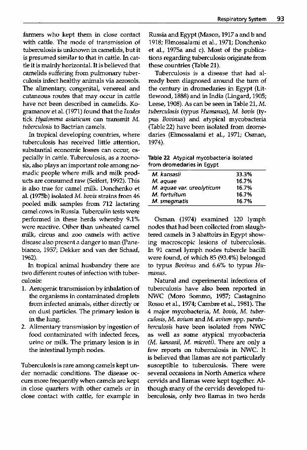
ResDiratorv Svstem 93
farmers who kept them in close contact with cattle. The mode of transmission of tuberculosis is unknown in camelids, but it is presumed similar to that in cattle. In cat- tle it is mainly horizontal. It is believed that camelids suffering from pulmonary tuber- culosis infect healthy animals via aerosols. The alimentary, congenital, venereal and cutaneous routes that may occur in cattle have not been described in camelids. Ko- gramanov et al. (1971) found that the Ixodes tick Hyalomma asiaticum can transmit M . tuberculosis to Bactrian camels.
In tropical developing countries, where tuberculosis has received little attention, substantial economic losses can occur, es- pecially in cattle. Tuberculosis, as a zoono- sis, also plays an important role among no- madic people where milk and milk prod- ucts are consumed raw (Seifert, 1992). This is also true for camel milk. Donchenko et al. (1975b) isolated M. bovis strains from 46 pooled milk samples from 712 lactating camel cows in Russia. Tuberculin tests were performed in these herds whereby 9.1% were reactive. Other than unheated camel milk, circus and zoo camels with active disease also present a danger to man (Pane- bianco, 1957; Dekker and van der Schaaf, 1962).
In tropical animal husbandry there are two different routes of infection with tuber- culosis: 1. Aerogenic transmission by inhalation of
the organisms in contaminated droplets from infected animals, either directly or on dust particles. The primary lesion is in the lung.
2. Alimentary transmission by ingestion of food contaminated with infected feces, urine or milk. The primary lesion is in the intestinal lymph nodes.
Tuberculosis is rare among camels kept un- der nomadic conditions. The disease oc- curs more frequently when camels are kept in close quarters with other camels or in close contact with cattle, for example in
Russia and Egypt (Mason, 1917 a and b and 1918; Elmossalami et al., 1971; Donchenko et al., 1975a and c). Most of the publica- tions regarding tuberculosis originate from these countries (Table 21).
Tuberculosis is a disease that had al- ready been diagnosed around the turn of the century in dromedaries in Egypt (Lit- tlewood, 1888) and in India (Lingard, 1905; Leese, 1908). As can be seen in Table 21, M . tuberculosis (typus Humanus), M . bovis (ty- pus Bovinus) and atypical mycobacteria (Table 22) have been isolated from drome- daries (Elmossalami et al., 1971; Osman, 1974).
Table 22 Atypical mycobacteria isolated from dromedaries in Egypt
M. kansasii 33.3% M. aquae 16.7% M. aquae var. ureolyticum 16.7% M. fortuitum 16.7% M. smeamatis 16.7%
Osman (1974) examined 120 lymph nodes that had been collected from slaugh- tered camels in 3 abattoirs in Egypt show- ing macroscopic lesions of tuberculosis. In 91 camel lymph nodes tubercle bacilli were found, of which 85 (93.4%) belonged to typus Bovinus and 6.6% to typus Hu- manus.
Natural and experimental infections of tuberculosis have also been reported in NWC (Moro Sommo, 1957; Castagnino Rosso et al., 1974; Cambre et al., 1981). The 4 major mycobacteria, M. bovis, M. tuber- culosis, M. avium and M. avium spp. paratu- berculosis have been isolated from NWC as well as some atypical mycobacteria (M. kansasii, M . microti). There are only a few reports on tuberculosis in NWC. It is believed that llamas are not particularly susceptible to tuberculosis. There were several occasions in North America where cervids and llamas were kept together. Al- though many of the cervids developed tu- berculosis, only two llamas in two herds
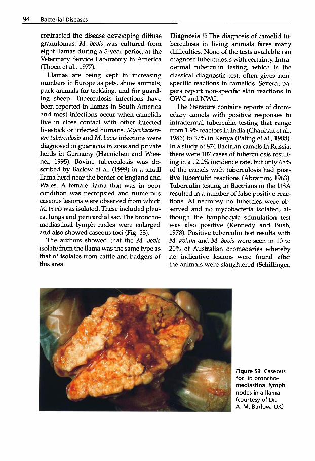
94 Bacterial Diseases
contracted the disease developing diffuse granulomas. M. bovis was cultured from eight llamas during a 5-year period at the Veterinary Service Laboratory in America (Thoen et al., 1977).
Llamas are being kept in increasing numbers in Europe as pets, show animals, pack animals for trekking, and for guard- ing sheep. Tuberculosis infections have been reported in llamas in South America and most infections occur when camelids live in close contact with other infected livestock or infected humans. Mycobacteri- um tuberculosis and M. bovis infections were diagnosed in guanacos in zoos and private herds in Germany (Haenichen and Wies- ner, 1995). Bovine tuberculosis was de- scribed by Barlow et al. (1999) in a small llama herd near the border of England and Wales. A female llama that was in poor condition was necropsied and numerous caseous lesions were observed from which M. bovis was isolated. These included pleu- ra, lungs and pericardial sac. The broncho- mediastinal lymph nodes were enlarged and also showed caseous foci (Fig. 53).
The authors showed that the M . bovis isolate from the llama was the same type as that of isolates from cattle and badgers of this area.
~
Diagnosis 1 The diagnosis of camelid tu- berculosis in living animals faces many difficulties. None of the tests available can diagnose tuberculosis with certainty. Intra- dermal tuberculin testing, which is the classical diagnostic test, often gives non- specific reactions in camelids. Several pa- pers report non-specific skin reactions in OWC and NWC.
The literature contains reports of drom- edary camels with positive responses to intradermal tuberculin testing that range from 1.9% reactors in India (Chauhan et al., 1986) to 37% in Kenya (Paling et al., 1988). In a study of 874 Bactrian camels in Russia, there were 107 cases of tuberculosis result- ing in a 12.2% incidence rate, but only 68% of the camels with tuberculosis had posi- tive tuberculin reactions (Abramov, 1963). Tuberculin testing in Bactrians in the USA resulted in a number of false positive reac- tions. At necropsy no tubercles were ob- served and no mycobacteria isolated, al- though the lymphocyte stimulation test was also positive (Kennedy and Bush, 1978). Positive tuberculin test results with M. avium and M. bovis were seen in 10 to 20% of Australian dromedaries whereby no indicative lesions were found after the animals were slaughtered (Schillinger,
Figure 53 Caseous foci in broncho- mediastinal lymph nodes in a llama (courtesy of Dr. A. M. Barlow, UK)
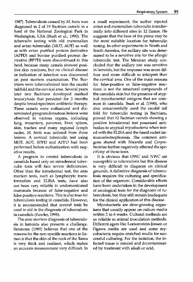
Respiratory System 95
1987). Tuberculosis caused by M . bovis was diagnosed in 2 of 19 Bactrian camels in a herd of the National Zoological Park in Washington, USA (Bush et al., 1990). The tuberculin testing with old mammalian and avian tuberculin (MOT, AOT) as well as with avian purified protein derivative (APPD) and bovine purified protein de- rivative (BPPD) were discontinued in this herd, because many camels showed posi- tive skin reactions, but no clinical disease or indication of infection was discovered on post mortem examination. The Bac- trians were tuberculinized into the caudal tail fold and the cervical area. Several years later two Bactrians developed marked leucocytosis that persisted for 3 months despite broad-spectrum antibiotic therapy. These camels were euthanized and dis- seminated pyogranulomatous lesions were observed in various organs, including lung, mesentery, pancreas, liver, spleen, skin, trachea and many regional lymph nodes. M. bovis was isolated from these lesions. A cervical tuberculin test using MOT, AOT, BPPD and APPD had been performed before euthanization with neg- ative results.
A program to control tuberculosis in camelids based only on intradermal tuber- culin tests will face severe deficiencies. Other than the intradermal test, the ante mortem tests, such as lymphocyte trans- formation and ELISA tests, have also not been very reliable in undomesticated mammals because of false-negative and false-positive reactions. This is also true for tuberculosis testing in camelids. However, it is recommended that several tests be used to aid in the diagnosis of tuberculosis in camelids (Fowler, 1998).
The ante mortem diagnosis of tuberculo- sis in lamoids also presents a challenge. Simmons (1989) believes that one of the reasons for the non-specific reactions in lla- mas is that the skin of the neck of the llama is very tluck and resilient, which makes an accurate measurement very difficult. In
a small experiment, the author injected avian and mammalian tuberculin intrader- mally into different sites in 12 llamas. He suggests that the base of the pinna may be the most suitable location for tuberculin testing. In other experiments in North and South America, the axillary site was deter- mined to be a sensitive site for the allergic tuberculin test. The Mexican study con- cluded that the axillary site was sensitive in lamoids, but the response was more dif- fuse and more difficult to interpret than the cervical area. One of the main reasons for false-positive or false-negative reac- tions is not the structural compounds of the camelids skin but the presence of atyp- ical mycobacterial antigens that are com- mon in camelids. Bush et al. (1990), who also unsuccessfully used the caudal tail fold for tuberculin testing in Bactrians, proved that 12 Bactrian camels showing a positive intradermal test possessed anti- bodies to atypical mycobacteria when test- ed with the ELISA and the fused rocket im- munoelectrophoresis. The common anti- gens shared with Nocardia and Coryne- bacteriae further negatively affected the spe- cificity of these tests.
It is obvious that OWC and NWC are susceptible to tuberculosis but this disease is very difficult to diagnose on clinical grounds. A definitive diagnosis of tubercu- losis requires the culturing and specifica- tion of the organism. Considerable efforts have been undertaken in the development of serological tests for the diagnosis of tu- berculosis, but they still remain inadequate for the clinical application of this disease.
Mycobacteria are slow-growing organ- isms that usually appear on culture media within 2 to 6 weeks. Cultural methods are as reliable as animal inoculation methods. Different agars like Loewenstein-Jensen or Ogawa media are used and some my- cobacteria require enriched media for suc- cessful culturing. For the isolation, the in- fected tissue is minced and decontaminat- ed by treatment with alkali or acid.
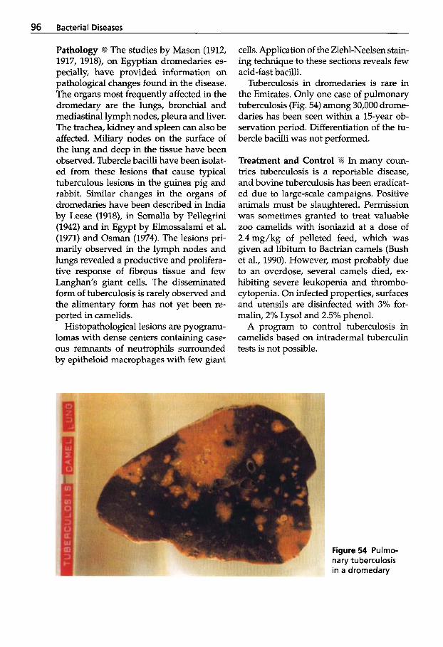
96 Bacterial Diseases
The studies by Mason (1912, 1917, 1918), on Egyptian dromedaries es- pecially, have provided information on pathological changes found in the disease. The organs most frequently affected in the dromedary are the lungs, bronchial and mediastinal lymph nodes, pleura and liver. The trachea, kidney and spleen can also be affected. Miliary nodes on the surface of the lung and deep in the tissue have been observed. Tubercle bacilli have been isolat- ed from these lesions that cause typical tuberculous lesions in the guinea pig and rabbit. Similar changes in the organs of dromedaries have been described in India by Leese (1918), in Somalia by Pellegrini (1942) and in Egypt by Elmossalami et al. (1971) and Osman (1974). The lesions pri- marily observed in the lymph nodes and lungs revealed a productive and prolifera- tive response of fibrous tissue and few Langhan’s giant cells. The disseminated form of tuberculosis is rarely observed and the alimentary form has not yet been re- ported in camelids.
Histopathological lesions are pyogranu- lomas with dense centers containing case- ous remnants of neutrophils surrounded by epitheloid macrophages with few giant
cells. Application of the Ziehl-Neelsen stain- ing technique to these sections reveals few acid-fast bacilli.
Tuberculosis in dromedaries is rare in the Emirates. Only one case of pulmonary tuberculosis (Fig. 54) among 30,000 drome- daries has been seen within a 15-year ob- servation period. Differentiation of the tu- bercle bacilli was not performed.
Treatment and Control I
tries tuberculosis is a reportable disease, and bovine tuberculosis has been eradicat- ed due to large-scale campaigns. Positive animals must be slaughtered. Permission was sometimes granted to treat valuable zoo camelids with isoniazid at a dose of 2.4mg/kg of pelleted feed, which was given ad libitum to Bactrian camels (Bush et al., 1990). However, most probably due to an overdose, several camels died, ex- hibiting severe leukopenia and thrombo- cytopenia. On infected properties, surfaces and utensils are disinfected with 3% for- malin, 2% Lysol and 2.5% phenol.
A program to control tuberculosis in camelids based on intradermal tuberculin tests is not possible.
Figure 54 Pulmo- nary tuberculosis in a dromedary
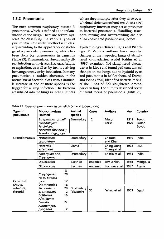
Respiratory System 97
1.3.2 Pneumonia where they multiply after they have over- whelmed defense mechanisms. Also a viral respiratory infection may act as precursor to bacterial pneumonia. Handling, trans- port, mixing and overcrowding are also often considered predisposing factors.
The most common respiratory disease is pneumonia, which is defined as an inflam- mation of the lungs. There are several sys- tems for classifying the various types of pneumonia. One useful method is to clas- s@ according to the appearance or etiolo- gy of a particular pneumonia, which has been done for pneumonias in camelids (Table 23). Pneumonia can be caused by di- rect infection with viruses, bacteria, fungus or aspiration, as well as by toxins arriving hematogenously or by inhalation. In many pneumonias, a sudden alteration in the normal nasal bacterial flora with a dramat- ic increase in one or more species is the trigger for a lung infection. The bacteria are inhaled into the lungs in large numbers
Epidemiology, Cl in ical Signs and Pathol- ogy Various authors have reported changes in the inspected lungs of slaugh- tered dromedaries. Abdel Rahim et al. (1990) examined 204 slaughtered drome- daries in Libya and found pathoanatomical changes in the lungs due to hydatid cysts and pneumonia in half of them. Al Darraji and Wajid (1990) identified bacteria in 56% of the lungs of 220 slaughtered drome- daries in Iraq. The authors described seven different forms of pneumonia (Table 23).
ineumonia in camelids (except tuberculosis) Table 23 Types oi
Type of pneumonia
- Year
1919 1927
-
Animal species Dromedary
Microorganisms isolated Streptothrix cameli (Actinomyces farcinicus, Nocardia farcinica?) Pseudotuberculosis
Authors
Mason Leese
Cases Country
Egypt Sudan Egypt
2
Granulomatous Histoplasma caosulatum
Dromedary 2 Chandel and Kher
1994 India
Nocardia asteroides
Llama 1 Ching-Dong Chaw et al.
1993 USA
Dromedary 1 Bhatia et al. 1983
1968 1987
- -
India Aspergillus and C. pyogenes Diplococcus Didococcus
Bactrian Bactrian
endemic endemic
Semushkin Buchnev et al
Mongolia Russia
% 20
12 16 20 2
16
22
2 -
C. pyogenes Hem. Strepto- cocci Diphtheroids Str. uiridans 5. enteritidis Coliforms Alcaligenes faecalis Actinomyces pyogenes
Catarrhal (Acute, subacute, chronic)
Dromedary (abattoir) 50 Farrag et al. 1953
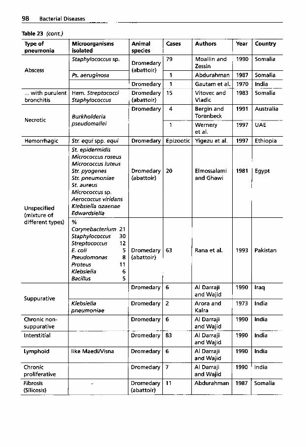
98 Bacterial Diseases
Table 23 (cont.)
Cases Authors Year Country Type of pneumonia
Microorganisms isolated Staphylococcus sp.
Animal species
Dromedary (abattoir)
79 Moallin and Zessin
1990 Somalia
Abscess Ps. aeruoinosa 1 Abdurahman 1987 Somalia
Dromedarv 1 Gautam et al. 1970 India ... with puruleni bronchitis
Hem. Streptococci Staphylococcus
Dromedary (abattoir)
15 Vitovec and Vladic
1983 Somalia
Dromedary 4 Bergin and Toren beck
1991 Australia Burkholderia pseudomallei Necrotic
1 Wernery et al.
1997 UAE
i t r . equi Spp. equi St. epidermidis Micrococcus roseus Micrococcus luteus Str. pyogenes Str. pneumoniae St. aureus Micrococcus sp. Aerococcus viridans Klebsiella ozaenae Edwardsiella
Yiaezu et al. 1997 -
198
Dromedary Ethiopia Hemorrhagic Epizootic
Dromedary (abattoir)
20 Elmossalami and Ghawi
Unspecified (mixture of different types) YO
Corynebacterium 21 Staphylococcus 30 Streptococcus 12 E. coli 5 Pseudomonas 8 Proteus 11 Kle bsiella 6 Bacillus 5
Dromedary (abattoir)
63 Rana et al. 1993 Pakistan
Dromedary 6 Al Darraji and Wajid
1990 Iraq
Suppurative Dromedary 2 1973
1990 -
India Klebsiella pneumoniae
Arora and Ka I ra Al Darraji and Waiid
Chronic non- swwrat ive
Dromedary 6 India
Interstitial Dromedary 83 Al Darraji and Wajid
1990 India
Lymphoid like MaediNisna Dromedary 6 1990
1990 -
India Al Darraji and Wajid Al Darraji and Waiid
Chronic Droliferative
Dromedary 7 India
Fibrosis (Si I icosis)
Dromedary (abattoir)
11 Abdurahman 1987 Somalia
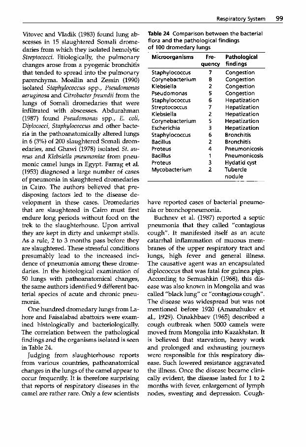
Respiratory System 99
Vitovec and Vladik (1983) found lung ab- scesses in 15 slaughtered Somali drome- daries from which they isolated hemolytic Streptococci. Etiologically, the pulmonary changes arose from a pyogenic bronchitis that tended to spread into the pulmonary parenchyma. Moallin and Zessin (1990) isolated Staphylococcus spp., Pseudomonas aeruginosa and Citrobacterfreundii from the lungs of Somali dromedaries that were infiltrated with abscesses. Abdurahman (1987) found Pseudomonas spp., E. coli, Diplococci, Staphylococcus and other bacte- ria in the pathoanatomically altered lungs in 6 (3%) of 200 slaughtered Somali drom- edaries, and Ghawi (1978) isolated St. au- reus and Klebsiella pneumoniae from pneu- monic camel lungs in Egypt. Farrag et al. (1953) diagnosed a large number of cases of pneumonia in slaughtered dromedaries in Cairo. The authors believed that pre- disposing factors led to the disease de- velopment in these cases. Dromedaries that are slaughtered in Cairo must first endure long periods without food on the trek to the slaughterhouse. Upon arrival they are kept in dirty and unkempt stalls. As a rule, 2 to 3 months pass before they are slaughtered. These stressful conditions presumably lead to the increased inci- dence of pneumonia among these drome- daries. In the histological examination of 50 lungs with pathoanatomical changes, the same authors identified 9 different bac- terial species of acute and chronic pneu- monia.
One hundred dromedary lungs from La- hore and Faisalabad abattoirs were exam- ined histologically and bacteriologcally. The correlation between the pathological findings and the organisms isolated is seen in Table 24.
Judging from slaughterhouse reports from various countries, pathoanatomical changes in the lungs of the camel appear to occur frequently. It is therefore surprising that reports of respiratory diseases in the camel are rather rare. Only a few scientists
Table 24 Comparison between the bacterial flora and the pathological findings of 100 dromedary lungs
Microorganisms Fre- Pathological quenw findinqs
Staphylococcus Corynebacteriurn Klebsiella Pseudomonas Staphylococcus Streptococcus Klebsiel la Corynebacterium Escherichia Staphylococcus Baci I Ius Proteus Bacillus Proteus Mycobacterium
7 8 2 5 6 7 2 5 3 6 2 4 1 3 2
Congestion Congestion Congestion Congestion Hepatization Hepatization Hepatization Hepatization Hepatization Bronchitis Bronchitis Pneumonicosis Pneumonicosis Hydatid cyst Tubercle nodule
have reported cases of bacterial pneumo- nia or bronchopneumonia.
Buchnev et al. (1987) reported a septic pneumonia that they called "contagious cough". It manifested itself as an acute catarrhal inflammation of mucous mem- branes of the upper respiratory tract and lungs, high fever and general illness. The causative agent was an encapsulated diplococcus that was fatal for guinea pigs. According to Semushkin (1968), this dis- ease was also known in Mongolia and was called "black lung" or "contagious cough. The disease was widespread but was not mentioned before 1920 (Amanzhulov et al., 1929). Oinakhbaev (1965). described a cough outbreak when 5000 camels were moved from Mongolia into Kazakhstan. It is believed that starvation, heavy work and prolonged and exhausting journeys were responsible for this respiratory dis- ease. Such lowered resistance aggravated the illness. Once the disease became clini- cally evident, the disease lasted for 1 to 2 months with fever, enlargement of lymph nodes, sweating and depression. Cough-
100 Bacterial Diseases
ing became steadily worse with prolonged attacks and difficult breathing (Kuznetsov, 1962; Voikulesku, 1963). Diatchenko (1989) compiled a summary of the literature of the different bacterial and viral respiratory diseases in the dromedary.
Arora and Kalra (1973) described cases of chronic bronchopneumonia in Indian dromedaries. The authors reported that the disease occurred only during the cold- er months and affected almost exclusively adult animals. The morbidity reached 30%, yet only a few camels died. The animals exhibited a protracted course of the illness, during which time they were unfit for work thereby causing economic losses. Klebsiellu pneumoniue and hemolytic Diplo- cocci were isolated from the lungs of two dromedaries that died of bronchopneu- monia.
Different authors have reported individ- ual cases of pneumonia in the dromedary. Leese (1927) attributed some isolated cases of respiratory disease to pulmonary ab- scesses. Gautam et al. (1970) described a pulmonary abscess in a 10-year-old drom- edary encompassing nearly the entire right lung. The authors failed to report the caus- ative agent. Pathoanatomical changes in the lung of a young Sudanese dromedary with pseudoactinomycosis similar to tu- berculosis were described by Mason (1919) and Hansen et al. (1987) described lung sil- icosis in dromedaries in Somalia. Kame1 (1939) reported pneumococcal pneumonia in Egyptian dromedaries and Agab et al. (1993) isolated a pathogenic Bacillus cougu- luns from a camel lung with pneumonia.
Streptococcus species seem to play an im- portant role in lung infections and other ailments, but they have also been isolated from the lungs of clinically healthy cam- elids (Shigidi, 1973; Mahmoud et al., 1988; Rana et al., 1993). A hemolytic Pneumococ- cus has been isolated from Bactrian camel lungs in the Gobi Desert (Oinakhbaev, 1965), Str. viriduns, Str. pneumoniue and Str. pyogenes from dromedaries (Thabet, 1994),
and a P-hemolytic Streptococcus also from dromedaries (Pal and Chandel, 1989). In Bahrain, Ibrahim et al. (1998) cultured Str. zooepidemicus serotype 2 from the nasal cavities of a dromedary. It had died from suffocation due to necrotic material com- pletely blocking its nasal passages and frontal sinuses. Str. zooepidemicus was also isolated from a septic peritonitis of a male Australian dromedary (Heller et al., 1998).
Of great importance is the report by Yigezu et al. (1997) who cultured Str. equi spp. equi from a sick dromedary during an epizootic outbreak in Ethiopia. The dis- ease was highly contagious with high morbidity and low mortality. The pre- dominant clinical signs were fever, lacri- mation, edema of the throat and supra- orbital fossa, loss of appetite, cough, dysp- nea and purulent nasal discharge. Upon necropsy the lungs revealed hemorrhages and thickened interlobular septae. This is the first report of the isolation of bacteria that causes equine strangles. It is assumed that donkeys were responsible for this out- break.
Streptococcal infections are also com- mon in NWC. Various streptococcal spe- cies have been isolated from NWC ab- scesses and septicemic enterococcus infec- tions in adult llamas have also occurred (Burkhardt et al., 1993). "Alpaca fever", a septicemia caused by Str. zooepidemicus, has been described by Thedford and John- son (1989). Stress is often a predisposing factor in the disease. Pneumonia is espe- cially common in NWC neonates (Fowler, 1998) and several bacterial agents are in- volved. As in other animal species (also in NWC calves), septicemic animals often de- velop pneumonia. E. coli is the most com- mon bacteria. Actinomyces lumue may also produce pneumonia with abscessation. A llama which was euthanized due to severe dyspnea and cyanosis revealed necrotic pneumonia from which Nocurdiu usteroides was isolated. Microscopically, the lung contained multiple small scattered pyo-
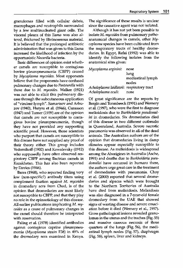
Respiratory System 101
granulomas filled with cellular debris, macrophages and neutrophils surrounded by a few multinucleated giant cells. The visceral pleura of this llama was also al- tered, thickened by fibrinoserous material. It is believed that the prolonged antibiotic administration that was given to this llama increased the likelihood of infection by the opportunistic Nocardia bacteria.
Basic differences of opinion exist wheth- er camels are susceptible to contagious bovine pleuropneumonia (CBPP) caused by Mycoplasma mycoides. Most opponents believe that the proponents have confused pulmonary changes due to Pasteurella with those due to M. mycoides. Walker (1921) was not able to elicit this pulmonary dis- ease through the subcutaneous application of "virulent lymph. Samartsev and Arbu- zov (1940), Hutyra et al. (1946), Curasson (1947) and Turner (1959) are of the opinion that camels are not susceptible to conta- gious bovine pleuropneumonia, though they have not provided any supporting scientific proof. However, those scientists who purport that camels are susceptible to this disease have not supplied any proof of their theory either. This group includes Vedernikoff (1902) and Kowalevsky (1912) who supposedly have often observed res- piratory CBPP among Bactrian camels in Kazakhstan. This has also been reported by Davies (1946).
Bares (1968), who reported finding very low (non-specific?) antibody titers using complement fixation against M. mycoides in dromedary sera from Chad, is of the opinion that dromedaries are most likely not susceptible to CBPP, and that they play no role in the epizootiology of this disease. All earlier publications implicating M. my- coides as a cause of pulmonary changes in the camel should therefore be interpreted with reservation.
Paling et al. (1978) identified antibodies against contagious caprine pleuropneu- monia (Mycoplasma strain F38) in 49% of the dromedary sera examined in Kenya.
The significance of these results is unclear since the causative agent was not isolated.
Although it has not yet been possible to isolate M. rnycoides from pulmonary patho- anatomical changes in camels, other My- coplasma species have been cultivated from the respiratory tracts of healthy drome- daries. In Egypt, Refai (1992) was able to identify the following isolates from the anatomical sites given:
Mycoplasma arginini:
Acholeplasma laidlawii: Acholeplasma oculi:
Of great significance
nose
mediastinal lymph nodes respiratory tract nose
are the reports by
lung
B e r s and forenbeeck (1991) andWerneG et al. (1997), who were the first to diagnose melioidosis due to Burkholderia pseudomal- lei in dromedaries. Six dromedaries died of this disease in two different outbreaks in Queensland, Australia. Severe necrotic pneumonia was observed in all of the dead animals. The Australian authors are of the opinion that dromedaries living in damp climates appear especially susceptible to this disease. As melioidosis is widespread among the Aborigines in Australia (Asche, 1991) and deaths due to Burkholderia pseu- domallei have occurred in humans there, the authors urge great care in the treatment of dromedaries with pneumonia. Choy et al. (2000) reported that several drome- daries and alpacas which were brought to the Northern Territories of Australia have died from melioidosis. Melioidosis was also diagnosed in a 7-year-old female dromedary from the UAE that showed signs of wasting disease and severe emaci- ation before it died (Wernery et al., 1997). Gross pathological lesions revealed granu- lomas in the uterus and the trachea (Fig. 55) and massive caseous necrosis of three quarters of the lungs (Fig.56), the medi- astinal lymph nodes (Fig. 57), diaphragm (Fig. 58), spleen, liver and kidneys.
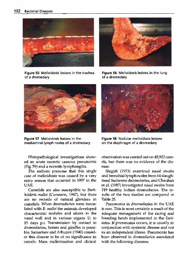
102 Bacterial Diseases
Figure 55 Melioidosis lesions in the trachea of a dromedary
Figure 56 Melioidosis lesions in the lung of a dromedary
Figure 57 Melioidosis lesions in the mediastinal lymph nodes of a dromedary
Figure 58 Nodular melioidosis lesions on the diaphragm of a dromedary
Histopathological investigations show- ed an acute necrotic caseous pneumonia (Fig. 59) and a necrotic lymphangitis.
The authors presume that this single case of melioidosis was caused by a very rainy season that occurred in 1997 in the UAE.
Camelids are also susceptible to Burk- holderia mallei (Curasson, 1947), but there are no records of natural glanders in camelids. When dromedaries were inocu- lated with B. mallei the animals developed characteristic nodules and ulcers in the nasal wall and in various organs 11 to 15 days p.i. Transmission by contact to dromedaries, horses and giraffes is possi- ble. Samartsev and Arbuzov (1940) consid- er this disease to be of no significance in camels. Mass malleinisation and clinical
observation was carried out on 45,922 cam- els, but there was no evidence of the dis- ease.
Shigidi (1973) examined nasal swabs and bronchial lymph nodes from 64 slaugh- tered Sudanese dromedaries, and Chauhan et al. (1987) investigated nasal swabs from 219 healthy Indian dromedaries. The re- sults of the two studies are compared in Table 25.
Pneumonia in dromedaries in the UAE is rare. This is most certainly a result of the adequate management of the racing and breeding herds implemented in the Emi- rates. If pneumonia occurs, it is usually in conjunction with systemic disease and not as an independent illness. Pneumonia has been observed in dromedaries associated with the following diseases:
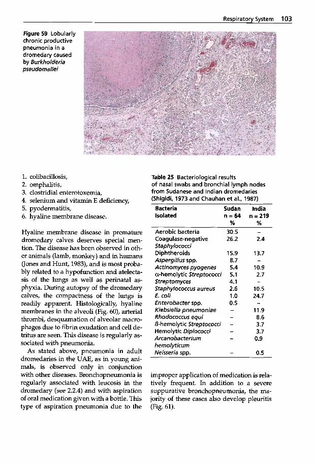
Respiratoty System 103
Figure 59 Lobularly chronic productive pneumonia in a dromedary caused by Burkholderia pseudomallei
1. 2. 3. 4. 5. 6.
colibacillosis, omphalitis, clostridial enterotoxemia, selenium and vitamin E deficiency, pyodermatitis, hyaline membrane disease.
Hyaline membrane disease in premature dromedary calves deserves special men- tion. The disease has been observed in oth- er animals (lamb, monkey) and in humans (Jones and Hunt, 1983), and is most proba- bly related to a hypofunction and atelecta- sis of the lungs as well as perinatal as- phyxia. During autopsy of the dromedary calves, the compactness of the lungs is readily apparent. Histologically, hyaline membranes in the alveoli (Fig. 60), arterial thrombi, desquamation of alveolar macro- phages due to fibrin exudation and cell de- tritus are seen. This disease is regularly as- sociated with pneumonia.
As stated above, pneumonia in adult dromedaries in the UAE, as in young ani- mals, is observed only in conjunction with other diseases. Bronchopneumonia is regularly associated with leucosis in the dromedary (see 2.2.4) and with aspiration of oral medication given with a bottle. This type of aspiration pneumonia due to the
Table 25 Bacteriological results of nasal swabs and bronchial lymph nodes from Sudanese and Indian dromedaries (Shigidi, 1973 and Chauhan et al., 1987)
Bacteria Isolated
Sudan India n = 6 4 n=219
% % Aerobic bacteria Coagulase-negative Staphylococci Diphtheroids Aspergillus spp. Actinomyces pyogenes a-hemolytic Streptococci Strep tomyces Staphylococcus a ureus E. coli Enterobacter spp. Klebsiella pneumoniae Rhodococcus equi B-hemolytic Streptococci Hemolytic Diplococci Arcanobacterium hemolyticum Neisseria spp.
30.5 - 26.2 2.4
15.9 13.7 8.7 - 5.4 10.9 5.1 2.7 4.1 - 2.6 10.5 1.0 24.7 0.5 - - 11.9 - 8.6
3.7 - 3.7 - 0.9
- 0.5
-
improper application of medication is rela- tively frequent. In addition to a severe suppurative bronchopneumonia, the ma- jority of these cases also develop pleuritis (Fig. 61).
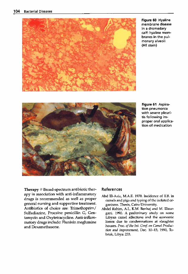
104 Bacterial Diseases
Figure 60 Hyaline membrane disease in a dromedary calf: hyaline mem- branes in the pul- monary alveoli (HE stain)
Figure 61 Aspira- tion pneumonia with severe pleuri- tis following im- proper oral applica- tion of medication
Broad-spectrum antibiotic ther- apy in association with anti-inflammatory drugs is recommended as well as proper general nursing and supportive treatment. Antibiotics of choice are: Trimethoprim/ Sulfadiazine, Procaine penicillin G, Gen- tamycin and Oxytetracycline. Anti-inflam- matory drugs include: Flunixin meglumine and Dexamethasone.
References
Abd El-Aziz, M.A.E. 1970. Incidence of T.B. in camels and pigs and typing of the isolated or- ganisms. Thesis, Cairo University.
Abdel Rahim, A.I., K.M. Benhaj and M. Elzur- gani. 1990. A preliminary study on some Libyan camel affections and the economic losses due to condemnations at slaughter houses. Proc. of the lnt. Con$ on Camel Produc- tion and improvement, Dec. 10-13, 1990, To- bruk, Libya: 233.
Respiratory System 105
Abdurahman, O.A. Sh. 1987. Pulmonary lesions among slaughtered camels in Mogadishu. Camel Forum 20.
berculosis in camels. J. Egypt Vet. Med. Asso.
Abramov, L.P. 1962. Diagnosis of tuberculosis in camels. J. Vet. Med. Ass. 4 0 26.
Abramov, L.P. 1963. Diagnosis of tuberculosis in camels by the ophthalmic and intradermal tuberculin tests. Veterinuriya, Moscow 40 (10):
Abramov, L.P. 1964a. The pathology of tuber- culosis in camels. Trudy II Vses. Patol. Anot. Zhivot. Mosk Vet. Akad.
Abramov, L.P. 1964b. Susceptibility of camels to various types of Mycobacterium tuberculo- sis. Veteriniya, Moscow 41: 19.
Agab, H.A.M., M.R. Bakhiet, H. El Jack and I.E. Mamoum. 1993. The pathogenicity of a Bacil- lus coagulans strain isolated from a camel (Camelus dromedarius) in Sudan. Bull. Anim. Prod. Afr. 41: 269-270.
Akhundov, A.A., G.N. Amanova and V.V. Dub- rovskaya. 1972. Some results of treating pul- monary tuberculosis patients with antibac- terial preparations together with "chal", a product of camel milk. Zdravookhr, Turkmen
Al Darraji, A.M. and SJ. Wajid. 1990. Etiological and pathological study of camel's lung le- sions in Iraq. Camel Newsletter 7 (12): 77.
Amanzhulov, S.A., L.N. Abruzov and A.M. Zhuravlev. 1929. About one infectious dis- ease of camels with unelucidated aetiology in the Ural province and the experience of ex- perimental challenge. Veterharnyi truzhenic 1, 2,3,4.
Andree, J. 1928. Ein Fall von generalisierter Tu- berkulose bei einem Kamel. Thesis, Hannover.
Angrisani, V. 1962. Considerazioni sul rapport0 tra l'allattamento con latte di bovini e di ca- melidi, in Somalia, el'eventuada infezione TBC. Archo ital. Sci. med. trop. Parasit. 43 (4):
Archibald, R.G. 1910. Acid-fast bacilli in a cam- el's lung, the gross lesions of which closely simulated miliary tuberculosis. J. Comp. Path.
Arora, R.G. and D.S. Kalra. 1973. Anote on iso- lation of Klebsiella pneumoniae and Diplo- cocci from cases of broncho-pneumonia in camels. Ind. J. Anim. Sci. 43 (12): 1095-1096.
Abou-Zaid, A.A. and H.M. HX~I~XII. 1993. TU-
53 (1 & 2): 243-250.
26-29.
16 (1): 36-38.
205-210.
Ther. 23 (1): 56-57.
Arush, M.A. 1982. La situazione sanitaria del dromedario nella Repubblica Democratica Somala. Bollettino scientifica della facoltli di zoo- tecnia e veterinaria 3: 209-217.
Asche, V. 1991. Melioidosis - a disease for all organs. Today's Life Science 6: 34-37.
Bares, J.F. 1968. Contribution a l'6tude de la pa- thologie infectieuse du dromadaire au Tchad. Thesis, Toulouse.
Barlow, A.M., K.A. Mitchell and K.H. Visram. 1999. Bovine tuberculosis in llama (Lama glama). Vet. Rec. 145: 639-640.
Bergin, T.J. and L.R. Torenbeeck. 1991. Melioi- dosis in camels. Australian Vet. J. 6 8 309.
Bhatia, K.C., R.C. Kulshreshtha and R.K. Paul Gupta. 1983. Pulmonary aspergdlosis in cam- el. Haryuna Vet. XXII: 118-119.
Buchnev, K.N., S.Z. Tulepbaev and A.R. San- syzbaev. 1987. Infectious diseases of camels in the USSR. Rev. sci. tech. Of. int. Epiz. 6 (2):
Burkhardt, J.E. E.B. Janovitz, T.L. Bowerstock and R. Higgins. 1993. Septicemic enterococ- cus infection in an adult llama. J. Vet. Diagn. Invest. 5: 106-109.
Bush, M., R.J. Montali, L.G. Phillips, D.K. Nichols and P.A. Holobaugh. 1986. Tubercu- losis in Bactrian camel. Proc. Meet. Am. Ass. Zoo. Vet.: 22-23.
Bush, M., R.J. Montali, L.G. Philipps and P.A. Holobaugh. 1990. Bovine tuberculosis in a Bactrian camel herd: clinical, therapeutic, and pathologic findings. J. Zoo and Wildl. Med. 21
Cambre, R., C. Thoen, W.O. Lang and W.L. Richards. 1981. Mycobacteria isolated from exotic animals. 8lSt Annual Meet. Am. Vet. SOC. Med. Microbiol.: 296.
Casati, R. 1957. Osservazione su di un caso di tubercolosi de cammello. Atti. SOC. Ital. Sci. Vet. 11: 551-554.
Castagnino Rosso, D., H. Ludena, D. Huaman and A. Ramirez. 1974. Linea de enfermeda- des infecciosas. Bol. Divulg. 15: 145-147.
Chamoiseau, G., S.O. Bah and S.M.O. Ahmed Vall. 1985. Un cas de tuberculose pulmonaire chez dromadaire. Rev. Elev. Mkd. vkt. Pays trop. 38 (1): 28-30.
Chandel, B.S. and H.N. Kher. 1994. Occurrence of histoplasmosis-like disease in camel (Cam- elus dromedarius). Ind. Vet. J. 71 (5): 521-523.
Chauhan, R.S., R.K. Kaushik, S.C. Gupta, K.C. Satiya and R.C. Kulshreshta. 1986. Prevalence
487-495.
(2): 171-179.
106 Bacterial Diseases
of different diseases in camels (Camelus drom- edarius) in India. Camel Newsletter 3: 10-14.
Chauhan, R.S., S.C. Gupta, K.C. Satija, R.C. Kul- shreshtha and R.K. Kaushik. 1987. Bacterial flora of upper respiratory tract in apparently healthy camels. Ind. J. Anim. Sci. 57 (5):
Ching-Dong Chang, T.R. Boosinger, P.D. Dowl- ing, E.E. McRae, J.W. Tyler and D.G. Pugh. 1993. Nocardiosis in a llama. J. Vet. Diagn. In- vest. 5: 631-634.
Choy, J.L., M. Mayo, A Janmaat and B.J. Currie. 2000. Animal melioidosis in Australia. Acta Tropica 74 (2-3): 153-158.
Cross, H.E. 1917. The camel and its diseases. Balliere, Tindall and Cox, London.
Curasson, G. 1947. Le chameau et ses maladies. Vigot Fr&es, Editeurs: 86-88.
Damodaran, S. and R. Ramakrishnan. 1969. Tu- berculosis in animals in Madras. 3rd Interna- tional Conference on the global impacts of applied microbiology, Bombay, India, s.I., s.n.: 1-86.
Davies, G.O. 1946. Gaiger and Davies Veteri- nary pathology and bacteriology. 31d ed. Bail- liere, Tindall and Cox, London.
Dekker, N.D.M. and A. van der Schaaf. 1962. Eengeval van open tuberculose bij een kame- el. Tijdschr. Diergeneesk 87 (17): 1133-1140.
Diatchenko, F. 1989. Contribution a l'ktude lksio- nelle des affections respiratoires du droma- daire. Thesis, Ecole Nationale Vet. d'Alfort 22.
Donchenko, A.S., V.N. Donchenko, E.A. Fatkee- va, M. Kibasov and L.A. Zemova. 1975a. De- struction of tubercle bacilli in camel's milk and "shubat", a lactic acid product. Veteri- nariya, Moscow 2: 24-26.
Donchenko, A.S., V.N. Donchenko, E.A. Fatkee- va and M. Kibasov. 1975b. Isolation of tuber- culosis mycobacteria in camel milk, their sur- vival in "shubat" and methods of decontam- ination of these products. Vest. Se1.'-khoz Nauki, Alma Ata 4 119-122.
Donchenko, A.S., V.N. Donchenko and S. Ken- zheev. 1975c. Effect of tuberculinization on the blood proteins of camels and cows. Vet- erinariya, Moscow 9 52-53.
Donchenko, A.S. and V.N. Donchenko. 1978. Change of proteins in blood serum of healthy and tuberculosis-diseased camels and cattle. Vest. Se1'-khoz. Nauki, Alma Ata 2: 73-76.
El-Af'ifi, A., R. Zaki and H.F. Farrag. 1953. Inci- dence and typing of tuberculosis in camels in Egypt. Vet. Med. J. 1: 1-6.
424-426.
Elmossalami, E., M.A. Siam and M. El Sergany. 1971. Studies on tuberculosis-like lesions in slaughtered camels. Zbl. Vet. Med. B. 18 (4):
Elmossalami, E. and A. Ghawi. 1981. Public health importance of camel lung affections. Egypt. J. Vet. Sci. 18 (1-2): 109-119.
Farrag, H., R. Zaki and M.R. El Hindawi. 1953. Pneumonia in camels. Brit. Vet. Rec. 59:
Fedchenko, V.A. 1971. Tuberculosis in camels. 11. Haematological changes. Trudy Kazakh. Nauchno-issled. Vet. Inst 1 4 57-61.
Fedchenko, V.A. 1972. Tuberculosis in camels. I. Epidemiology in Kazakhstan. Trudy Kazakh. Nauchno-issled. Vet. Inst 14: 51-56.
Fowler, M.E. 1998. Medicine and surgery of South American Camelids. Iowa State Uni- versity Press, Ames.
Gatt Rutter, T.E. and R. Mack. 1963. Diseases of camels. Part 1: Bacterial and fungal diseases. Vet. BUZZ. 33 (3): 119-124.
Gautam, O.P., R.L. Gulati and K.L. Gera. 1970. Pulmonary abscess (Malli) in a camel. Ind. Vet. J. 47 (4): 364-365.
Ghawi, A.M. 1978. Public health importance of camel lung affections. Thesis, Cairo University.
Haenichen, T. and H. Wiesner. 1995. Erkran- kungs- und Todesursachen bei Neuweltka- meliden. Tierarztl. Praxis 23: 515-520.
Hansen, H.J., EM. Jama and 0. Abdulkadir. 1987. Silicosis in camels. A preliminary re- port. SIDA Regional Seminar on Vet. Path., De- brezeit, Ethiopia.
Hayles, L.B. 1986. Proceedings of the first na- tional veterinary symposium, Somalia, Oct. 12-15, 1986. Zst National Veterinary Sympo- sium, Mogadishu, Rome, FAO.
Heller, M., D. Anderson and F. Silveira. 1998. Streptococcal peritonitis in a young drome- dary camel. Australian Vet. J. 76 (4): 253-254.
Hutyra, F., J. Marek and R. Manninger. 1946. Special pathology and therapeutics of the diseases of domestic animals. gfh English ed.: Balliere, Tindall and Cox, London.
Ibrahim, A.M., A.A. Abdelghaffar and M.E. Fadlalla. 1998. Streptococcus zooepidemicus infection in a female camel in Bahrain. J. Cum- el Prac. and Res. 5 (1): 165-176.
Jones, Th. C. and R.D. Hunt. 1983. Veterinary Pathology. 5& edition. Lea and Febiger.
Kamel, H. 1939. Pneumo-coccus in camels. Tech- nical and Scientific Service Bull.: 226.
253-261.
119-122.
Respiratory System 107
Kennedy, S. and M. Bush. 1978. Evaluation of tuberculin testing and lymphocyte transfor- mation in Bactrian camels. Montali, R.J. Mycobacterial infections in zoo animals. Washington DC: Smithsonian Inst. Press:
Kibasov, M. and A.S. Donchenko. 1976. Experi- mental determination of economic losses in camels due to tuberculosis. Vest. Se1.’-khoz. Nauki, Alma Ata 1 2 5-8.
Kogramanov, A.I., Ya. A. Blagogamy, N.M. Makarevitch, I.M. Blekhman and M.P. Yaku- nine. 1971. Ticks as possible carriers of tuber- cular infections. Pathology, pathomorphology and experimental tuberculosis. 9: 60-64.
Kowalesky, M.J.M. 1912. Le Chameau et ses maladies d’apres les observations d‘auteurs russes. J. Med. Vet. Zootechn., Lyon 15: 462-466.
Kuznetsov, S.V. 1962. Camels with infectious lung inflammation in the Turkmen SSR. Trudy turkmenskogo, Niskhi, Ashkhabad XI.
Leese, A.S. 1908. Camel tuberculosis. Annual re- port of officer investigating camel disease, India, s.1.s.n.
Leese, A.S. 1910. Acid-fast bacilli in camel’s lung with lesions resembling those of tuberculosis. J. Comp. Path. Ther. 23 (4): 358-359.
Leese, A.S. 1918. “Tips” on camels for veterinary surgeons on active service. Bailliere Tindall and Cox, London 50.
Leese, A.S. 1927. A treatise on the one-humped camel in health and disease. Vigot Frkres, Paris 11.
Lingard, A. 1905. Camel tuberculosis. Annual report of imperial bacteriologist, India, s.n. 190 tou.06.
Littlewood, W. 1888. Camel tuberculosis. Egypt. Off. Gaz.
Mahmoud, A.Z., S.I. Moustafa and A.H. El-Yas. 1988. No Title. Assiut Vet. Med. J. 2 0 93.
Manefield, G.W. and A. Tinson. 1996. Camels. A compendium. The T.G. Hungerford Vade Mecum Series for Domestic Animals: pp. 240, 298.
Mason, F.A. 1912. Some observations on tuber- culosis in camels in Egypt. J. Comp. Path. Ther.
Mason, F.A. 1917a. Tuberculosis in camels.
Mason, F.A. 1917b. Tuberculosis in camels. J.
Mason, F.A. 1918. Tuberculosis in the camel. J.
139-143.
25 (1): 109-111.
Agric. J. Egypt 7 2-11.
Comp. Path. Ther. 30 (1): 80-84.
Comp. Path. Ther. 31 (2): 100-102.
Mason, F.E. 1919. Pseudo-actinomycosis or Strep- totrichosis in the camel. J. Comp. Path. Ther. 32 (1): 34-42.
Moallin, A.S.M. and K.H. Zessin. 1990. Note on diseases of the dromedaries at Beletweyne abattoir of Central Somalia. Camel Newsletter 7 (12): 69.
Moro Sommo, M. 1957. Investigacion prelimi- nar de la brucelosis en alpacas. Rev. Fac. Med. Vet., Lima 12: 135-137.
Mustafa, I.E. 1987. Bacterial diseases of the cam- el and dromedary. OIE 55e Session gknkrale OIE, office intemationale des 6pizooties, Paris, France 55: 18-22.
Oinakhbaev, S. 1965. Study of aetiology of con- tagious cough in camels. Veterinariya, Moscow
Osman, K.M. 1974. Studies on acid fast microor- ganisms in some domesticated animals with special reference to a typical mycobacterium group. PhD Thesis, Fac. of Vet. Med., Cairo University.
Pal, M. and B.S. Chandel. 1989. No title. Ind. Vet. Med. J. 13: 277.
Paling, R.W., K.J. Macowan and L. Karstad. 1978. The prevalence of antibody to Conta- gious Caprine Pleuropneumonia (Mycoplas- ma strain F38) in some wild herbivores and camels in Kenya. 1. Wildl. Dis. Vol. 14 (7): 305-308.
Paling, R.W., S. Whaghela, K.J. Macowan and B.R. Heath. 1988. The occurrence of infec- tious diseases in mixed farming of domesti- cated and wild herbivores and livestock in Kenya. 11. Bacterial diseases. J. Wildl. Dis. 2 4
Panebianco, F. 1957. Su di caso di tubercolosi del cammello. Acta Med. Vet. 3 (3): 291-302.
Pellegrini, D. 1942. Tubercoli spontanea del cammello in Somalia. Ricerche diagnostiche esperimentali. Racc. Stud. Vet. Pat. Somali,
Rana, M.Z., A. Ahmed, S.T.A.K. Sindhu and G. Mohammed. 1993. Bacteriology of camel lungs. Camel Newsletter 10 (6): 30-32.
Refai, M. 1992. Bacterial and mycotic diseases of camels in Egypt. Proc. lSt int. Camel Conf. Eds: W.R. Allen, A.J. Higgins, I.G. Mayhew, D.H. Snow and J.F. Wade: R. and W. Publica- tions, Newmarket, UK: 59-64.
Richard, D. 1979. Study of the pathology of the dromedary in Borana Awraja (Ethiopia). The- sis, IEMVT.
42 (6): 105-106.
308-316.
1942-1945 1: 33-41.

108 Bacterial Diseases
Samartsev, A.A. and P.N. Arbuzov. 1940. The susceptibility of camels to glanders, rinder- pest and bovine contagious pleuro-pneumo- nia. Veterinariya, Moscow 4 59-63.
Schillinger, D. 1987. Kame1 (Camelus dromeda- rius). Seminar Sonderdruck Vet., Labhard Verlag Konstanz 9: 50-53.
Seifert, H.S.H. 1992. Tropentierhygiene. Gustav Fischer Verlag Jena, Stuttgart.
Semushkin, N.R. 1968. Diagnosis of camel dis- eases. Sel'khozgiz Moscow.
Shah, M.A. and G.S. Khan. 1935-1936. Pneumo- nia in camels. Ind. Vet. J. 12: 206-220.
Shigidi, M.T.A. 1973. Aerobic microflora of res- piratory tract of camels. Sudan J. Vet. Sci. and Anim. Husb. 14 (1): 9-14.
Simmons, A.G. 1989. Alternative site for the sin- gle intrademal comparative tuberculin test in the llama (Lama glama). Vet. Rec. 124: 17-18.
Thabet, A. El. R. 1994. No title. Assiut Vet. Med. J. 30: 188.
Thedford, R.R. and L.W. Johnson. 1989. In- fectious diseases of New-world camelids (NWC). Vet. Clin. North Am. Food Anim. Pract.
Thoen, C.O., W.D. Richards and J.L. Jamigan. 1977. Mycobacteria isolated from exotic ani- mals. JAVMA 170 (9): 987-990.
Turner, A.W. 1959. Pleuropneumonia group of diseases: Infectious diseases of animals. Stu- bleforth and Galloway I 1 437.
Vedernikov, V. 1902. cited from Curasson (1947). Vitovec, J. and P. Vladik. 1983. Bronchial disease
of camels in Somalia. Bull. Anim. Hlth. Prod.
Voikulesku, M. 1963. Streptococcus infection. Infectious diseases. 1. Meridiane, Bucharest
Walker, J. 1921. Experiments and observations in connection with pleuropneumonia conta- giosa bovum. Bull. Dept. Agric., Kenya 2.
Wernery, Renate, J. Kinne, J. Haydn-Evans and A. U1-Haq. 1997. Melioidosis in a seven year
5 (3): 145-157.
Afi. 31 (3): 291-294.
107-126.
old camel, a new disease in the United Arab Emirates (UAE). J. Camel Prac. and Res. 4 (2):
Wernery, U. and 0.-R. Kaaden. 1995. Infectious Diseases of Camelids. Blackwell Wissen- schafts-Verlag, Berlin.
Yigezu, M., F. Roger, M. Kiredjian and S. Tariku. 1997. Isolation of Streptococcus equi sub- species equi (strangles agent) from an Ethio- pian camel. Vet. Rec. 140: 608.
141-143.
Further reading
Agab, H. and B. Abbas. 1999. Epidemiological studies on camel diseases in the eastern Su-
Abu Elgasim, K.E.M. 1992. Study on clinical, ae- tiological and pathological aspects of pneu- monia in camels (Camelus dromedarius). Thesis, Faculty Vet. Sci. University Khar- toum, Sudan.
Elmossalami, E. and A. Ghawi. 1983. Public health importance of camel lung affections. Egypt. J. Vet. Sci. 18 (1-2): 109-119.
Graber, M. 1968. Region of veterinary and zootechnical research of central Africa. An- nual report, Farcha Laboratory. lSt research and products 2 pleuropneumonia. Quin- quennial report. Fort Lami, Chad. Vet. Bull. 38: 5265.
Hansen, H., F.M. Jama, C. Nilsson, L. Norrgren and 0.Sh.A. Abdurahman. 1989. Silicate pneumoconiosis in camel. J. Vet. Med., Series.
Kogramanov, A.I. 1967. Microbiology of tu- berculosis in the USSR during 60 years. 1:
Sechi, L.A., F. Roger, A. Diallo, L.M. Yigezu, S. Zanetti and G. Fadda 1999. Molecular charac- terisation of Streptococcus equi subspecies equi isolated from an Ethiopian camel by ri- botyping and PCR-ribotyping. Microbiologica
dan. WAR/RMZ. 92: 42-51.
A 36 (10): 789-796.
28-43.
22: 383-387.
1.4 Urogenital System , . ,
1.4.1 Brucellosis
Through intensive health control measures, many industrialized countries have suc- ceeded in eradicating brucellosis. In devel- oping countries, however, brucellosis re- mains widespread in domesticated and wild animal populations and presents a great economic problem for tropical animal husbandry (Seifert, 1992). Brucellosis is also one of the most important zoonoses in the tropics. On tropical dairy farms the rate of infection can reach 80%. In areas of exten- sive animal husbandry in the Sahel, the rate of contamination has been estimated be- tween 25 and 30% (Seifert, 1992). Dome- nech et al. (1982) estimated the yearly loss- es in stock due to brucellosis to be 6%.
Especially OWC are frequently infected with brucellosis, particularly when they are in contact with infected ruminants (Abo El-Hassan et al., 1991; Radwan et al., 1992; Barsoum et al., 1995; Al-Ani et al., 1998). Brucellosis in NWC is rare (Fowler, 1998). Humans are at risk through con- sumption of unheated milk (WHO/FAO, 1986; Kiel and Khan, 1987; Madkow, 1989; Radwan et al., 1995).
Etiology I Brucellosis is a contagious dis- ease caused by the bacteria of the genus Brucella. Brucella bacteria are Gram-nega- tive coccobacilli, which are non-motile and non-spore-forming. Except for B. ovis and B. abortus, biotype 2, which require media enriched with serum or blood, the growth of other Brucellae is enhanced by enriched media, but they are also able to grow on nu- trient agar. Extreme care must be exercised when working with Brucella organisms.
Epidemiology and Clinical Signs jc Bru- cellosis is characterized by abortion, and to a lesser extent by orchitis and infection of
the accessory sex glands in males. The dis- ease has a worldwide distribution and af- fects cattle, pigs, sheep, goats, camelids, dogs, and occasionally horses. In humans, the disease referred to as undulant fever or Malta fever is a serious public health prob- lem.
The infection occurs via the mucous membranes or skin or by ingestion of con- taminated foodstuffs, whereby the causa- tive agent then enters via the upper gas- trointestinal tract. Infections through the mucosa of the respiratory tract or the eyes are also possible. The spread of brucella during sexual activity plays a subordinate role.
Theoretically, all three known Brucella species can cause infection in camels (Hig- gins, 1986). However, it is surmised that B. melitensis is widespread in Africa and the Middle East and B. abortus is wide- spread in the former USSR. Solonitsyn (1949) reported mixed infections with var- ious Brucella species in Bactrian camels in Russia.
As can be seen in Table 26, the majority of reports on brucellosis utilize serological methods of identification. A survey of the prevalence of brucellosis in Africa was published by Chukwu (1985). The inci- dence of brucellosis in camel populations appears to be related to breeding and hus- bandry practices (Richard, 1980). The in- fection rate in some regions of the former USSR, where Bactrian camels are kept on large farms, is 15% (Pal'gov and Zhulo- bovski, 1964). In countries with more ex- tensive forms of husbandry, as in Chad or Ethiopia, the prevalence is 3.8% (Gra- ber, 1968) and 5.5% (Richard, 1980) respec- tively.
Similar differences in the seroprevalence have been reported from Saudi Arabia by Radwan et al. (1992) and Ghoneim and
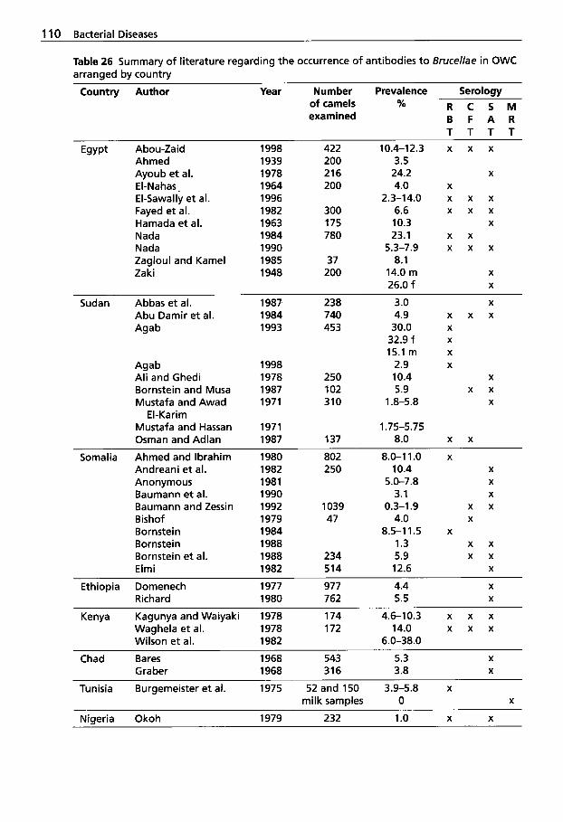
1 10 Bacterial Diseases
Table 26 Summary of literature regarding the occurrence of antibodies to Brucellae in OWC arranged by country
Country Author Year Number Prevalence Serology of camels % R C S M examined B F A R
T T T T Egypt Abou-Zaid 1998 422 10.4-12.3 x x x
Ahmed 1939 200 3.5 Ayoub et al. 1978 216 24.2 X
El-Nahas. 1964 200 4.0 X
El-Sawally et al. 1996 2.3-14.0 x x x Fayed et al. 1982 300 6.6 x x x Hamada et al. 1963 175 10.3 X
Nada 1984 780 23.1 x x Nada 1990 5.3-7.9 x x x Zagloul and Kame1 1985 37 8.1 Zaki 1948 200 14.0 m X
26.0 f X
Sudan Abbas et al. 1987 238 3.0 X
Abu Damir et al. 1984 740 4.9 x x x Agab 1993 453 30.0 X
Agab 1998 2.9 X
Ali and Ghedi 1978 250 10.4 X
Bornstein and Musa 1987 102 5.9 x x Mustafa and Awad 1971 310 1 .&5.8 X
Mustafa and Hassan 1971 1.75-5.75 Osman and Adlan 1987 137 8.0 x x
32.9 f X
15.1 m X
El-Karim
Somalia Ahmed and lbrahim Andreani et al. Anonymous Baumann et al. Baumann and Zessin Bishof Bornstein Bornstein Bornstein et al. Elmi
1980 1982 1981 1990 1992 1979 1984 1988 1988 1982
802 8.0-1 1 .o 250 10.4
5.0-7.8 3.1
47 4.0
1.3 234 5.9 514 12.6
1039 0.3-1.9
8.5-1 1.5
X
X
X
X
x x X
X
x x x x
X
Ethiopia Domenech 1977 977 4.4 Richard 1980 762 5.5
X
X
Kenya Kagunya and Waiyaki 1978 174 4.6-10.3 x x x Waghela et al. 1978 172 14.0 x x x Wilson et al. 1982 6.0-38.0
Chad Bares 1968 543 5.3 X
Graber 1968 316 3.8 X
Tunisia Burgemeister et al. 1975 52 and 150 3.9-5.8 x milk samples 0 X
Nigeria Okoh 1979 232 1.0 X X
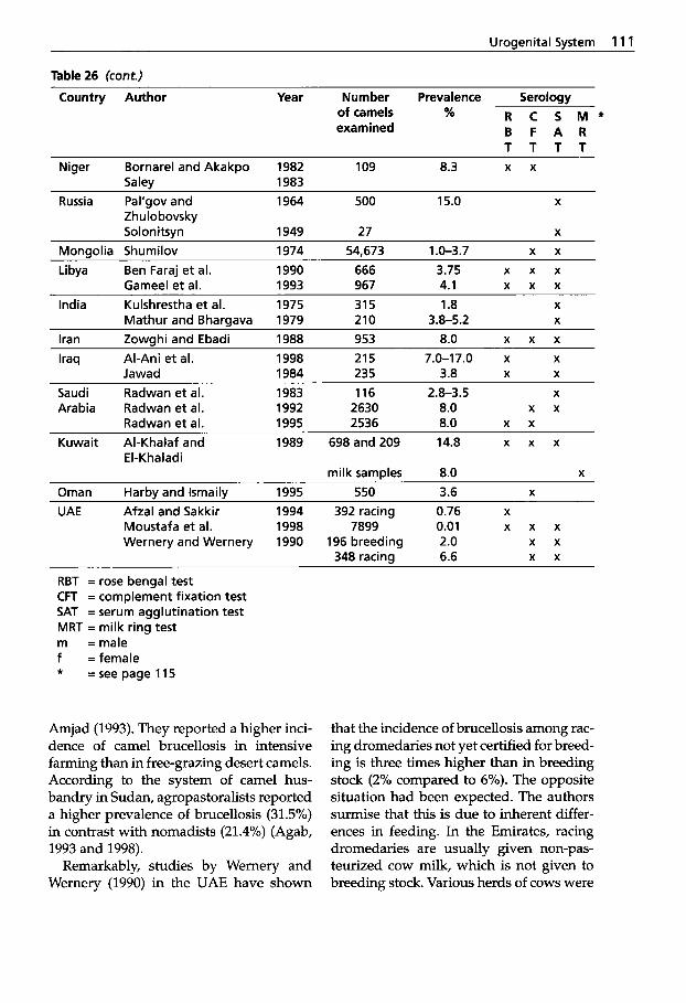
Uroaenital Svstem 11 1
Table 26 (cont.) Country Author Year Number Prevalence Serology
of camels % R C S M * examined B F A R
T T T T Niger Bornarel and Akakpo 1982 109 8.3 x x
Saley 1983 Russia Pal’gov and 1964 500 15.0 X
Z h u lo bovs ky Solonitsvn 1949 27 X
Monaolia Shumilov 1974 54,673 1 .O-3.7 x x Libya Ben Faraj et al. 1990 666 3.75 x x x
Gameel et al. 1993 967 4.1 x x x India Kulshrestha et al. 1975 315 1.8 X
Mathur and Bhargava 1979 210 3.8-5.2 X
Iran Zowghi and Ebadi 1988 953 8.0 x x x Iraq Al-Ani et al. 1998 215 7.0-17.0 x X
Jawad 1984 235 3.8 X X
Saudi Radwan et al. 1983 116 2.8-3.5 X
Arabia Radwan et al. 1992 2630 8.0 x x Radwan et al. 1995 2536 8.0 x x
Kuwait Al-Khalaf and 1989 698 and 209 14.8 x x x El-Khaladi
milk samples 8.0 X
Oman Harby and lsmaily 1995 550 3.6 X
UAE Afzal and Sakkir 1994 392 racing 0.76 X
Moustafa et al. 1998 7899 0.01 x x x Wernery and Wernery 1990 196 breeding 2.0 x x
348 racing 6.6 x x
RBT = rose bengal test CFT = complement fixation test SAT = serum agglutination test MRT = milk ring test m =male f =female * =see page 115
Amjad (1993). They reported a higher inci- dence of camel brucellosis in intensive farming than in free-grazing desert camels. According to the system of camel hus- bandry in Sudan, agropastoralists reported a higher prevalence of brucellosis (31.5%) in contrast with nomadists (21.4%) (Agab, 1993 and 1998).
Remarkably, studies by Wernery and Wernery (1990) in the UAE have shown
that the incidence of brucellosis among rac- ing dromedaries not yet certified for breed- ing is three times higher than in breeding stock (2% compared to 6%). The opposite situation had been expected. The authors surmise that this is due to inherent differ- ences in feeding. In the Emirates, racing dromedaries are usually given non-pas- teurized cow milk, which is not given to breeding stock. Various herds of cows were
1 12 Bacterial Diseases
shown to have an incidence of brucellosis
The lower incidence of infection in the breeding camel cows when compared to the racing dromedaries in the UAE indi- cates a spontaneous recovery among cur- rently non-reproductive dromedaries, as described by numerous authors (Ostrovi- dov, 1954 a and b; Gatt Rutter and Mack, 1963; Fazil and Hofmann; 1981). Interest- ingly, seropositive racing dromedaries ex- hibited no reduction in performance dur- ing the racing season. The hematology pa- rameters as well as the enzyme activity remained within normal limits.
Moustafa et al. (1998) reported on a sero- logical survey in dromedaries and a bru- cellosis eradication campaign in the east- ern region of the UAE during a 5-year pe- riod. The highest prevalence was in 1991 with 5.8% reactors, whereas the lowest was in 1996 with 0.01%. Since no camels have been culled due to brucellosis, it is be- lieved that the reduction in camel brucel- losis was caused by the reduction in bru- cellosis in sheep and goats.
According to various researchers, bru- cellosis in breeding camelids occurs in all of the known forms, whereby abortion is its most obvious manifestation (Acosta et al., 1972; WHO/FAO, 1986; Fazil and Hof- mann, 1981; Radwan et al., 1995; Agab et al., 1996). Infections may also cause still- born calves, retained placenta and reduced milk yield as is common in cattle and sheep. Retained placentas have not been described in Camelidae. This may be a re- sult of the difference in the placental at- tachment (Fowler, 1998). B. abortus and/or B. melitensis have been isolated from milk, vaginal swabs, aborted fetuses, lymph nodes and hygromas of infected camelids from different countries.
Although camels appear to be very sus- ceptible to BruceIZa infection, isolation of Brucella organisms from camel samples has proved less successful. Only recently have attempts at isolation of BruceZZa from milk
of Up to 40%. been successful. Brucella abortus biovars 1 and 3 were isolated from camels in Sene- gal (Verger et al., 1979). Radwan et al. (1992) were able to isolate B. melitensis, bio- var 1 and 2 twenty six times from a total of 100 milk samples from seropositive Saudi Arabian dromedaries. This poses a severe health risk for man, since camel milk is not pasteurized. Sharing this conviction, Gameel et al. (1993) were also able to iso- late B. melitensis, biovar 1 five times from the milk of Libyan dromedaries and four times from aborted fetuses and a vaginal swab. Zaki (1943) both inoculated guinea pigs with milk samples from seropositive dromedaries and cultured the milk sam- ples in vitro. Both tests were negative. Al- Khalaf and Al-Khaladi (1989) examined cultures of 209 milk samples from Kuwaiti dromedaries. The samples were obtained from herds with an increased incidence of abortion. The results were negative. How- ever, the authors were successful in isolat- ing B. abortus from the gastric fluids of five aborted fetuses. Pal'gov (1950) was able to isolate B. abortus from Bactrian camels in Russia. In the herds examined, 2% of all animals aborted in the first half of the pregnancy. Fifteen percent of the herds were seropositive to brucellosis. Zowghi and Ebadi (1988) cultured 3500 lymph nodes from 300 slaughtered dromedaries from Iran for Brucella organisms. B. meli- tensis, biovar 1 and 3 were isolated from these lymph nodes in 1% (3/300) of the camels. The authors are of the opinion that the B. melitensis infections in the drome- daries originated from neighboring sheep and goat herds.
Radwan et al. (1995), who examined a large camel herd with 2536 dromedaries in Saudi Arabia from which a 12% abortion rate and a Brucella seroprevalence of 8% were reported, isolated B. melitensis, bio- vars 1 ,2 and 3 from aborted camel fetuses. During their investigations, Malta fever was diagnosed in 30% of the camel hand- lers and milkers and the same B. melitensis
Urogenital System 113
biovars were cultured from aborted sheep and goats sharing the same premises.
B. abortus, biovar 3 was recovered from 3 different specimens obtained from free- ranging camels in eastern Sudan (Agab et al., 1994; Agab et al., 1996). The Brucella or- ganisms were isolated from a supramam- mary lymph node, a vagnal swab and an inguinal lymph node in dromedaries with histories of abortion, presence of hygromas or testicular lesions. It is worth mention- ing that both isolates of B. abortus biovar 3 from Senegal and Sudan were the only oxidase-negative biovars reported in the lit- erature. Ramadan et al. (1998) have recent- ly recovered B. melitensis from a hygroma of an Indian camel. B. melitensis was isolated twice from milk samples of seropositive camels in the UAE (Moustafa et al., 1998).
Non-pregnant dromedaries artificially infected with B. abortus (wild strain, 6 x lo6 bacteria) developed only mild clinical signs. Reduced appetite, slight lameness and bilateral lacrimation were observed. The bacteria were re-isolated 45 to 65 days later from the cranial and genital lymph nodes, which showed follicular hyperpla- sia of cortical and paracortical areas with active germinal centers, atrophy of medul- lary cords and sinusoidal congestion. A mild interstitial hepatitis was also observed (Abu Damir et al., 1984).
Brucellosis is not a major disease in NWC, but a severe B. melitensis outbreak occurred in a herd of alpacas in Peru. Near- ly 30% of the 1449 alpacas tested had a pos- itive plate agglutination titer. Over 25% of the alpaca handlers were seropositive to brucellosis and some developed Malta fe- ver. It was felt that sheep were the source of infection in this alpaca herd (Acosta et al., 1972). In an experimental infection trial in llamas in the United States, it was found that llamas are susceptible to B. abortus and that they develop positive serological titers and histological lesions similar to those found in cattle, sheep and goats (Fowler, 1998).
Pathology Very little is known about the pathological changes caused by Brucella organisms in camelids. The predilection organs for these bacteria are the pregnant uterus, udder, testicle, accessory male sex glands, lymph nodes, joint capsules and bursae. Lesions may be found in these tis- sues. Nada and Ahmed (1993) described lesions in non-pregnant dromedaries. They found inflammation of the uterus lining with reddening, edema and necrotic foci in the uterus epithelium, as well as fibrosis of the endometrium and atrophy of the uter- ine glands. The authors also observed an increased number of ovariobursal adhe- sions and hydrobursae. The adhesions oc- curred between the Bursa ovarica and the ovary and in several cases also between the Bursa ovarica and the salpinges, causing a severe induration of the latter. Hy- drobursitis was often observed in brucel- losis-positive dromedaries causing an en- largement of the bursa, which was then filled with a clear amber-colored fluid. No lesions have been described so far in aborted camelids and in brucellosis-posi- tive camelid males. A pregnant llama was infected by inoculating viable B. abortus bacteria into the conjunctival sac. Forty- three days p.i., the llama aborted an eight- month-old fetus. B. abortus was isolated from the placenta and all fetal specimens as well from the dam’s mammary gland numerous lymph nodes. Histologically there was a moderate, multifocal, lympho- cytic and histiocytic, subacute placentitis with marked loss of trophoblastic epithe- lial cells. The chorioallantoic stroma con- tained abundant necrotic and mineralized debris and the swollen capillaries were ex- panded by large numbers of Brucella or- ganisms (Gidlewski et al., 2000).
Diagnosis :i: Brucellosis is usually diag- nosed in the laboratory by culture of blood, milk or tissue or detection of antibodies in sera. Brucella organisms can be recovered from the placenta, but more conveniently
114 Bacterial Diseases
in pure culture from the stomach and lungs of aborted fetuses.
However, difficulties may arise in the diagnosis of brucellosis. Abortion and re- duced fertility in the camel frequently have other causes, such as salmonellosis, trypanosomosis, or infections with Campy- lobacter or Trichomonas fetus (Wernery and Amjad Ali, 1989; Wernery, 1991; Wernery and Wernery, 1992). An incorrect diagnosis of brucellosis may occur when based on serology alone. Sunaga et al. (1983) report- ed that five dromedaries imported into Japan had positive complement fixation (CFT) and slow agglutination reactions. The animals were immediately slaugh- tered. No brucella organisms were isolat- ed; however, Yersinia enterocolitica, sero- type 09 was identified. It is known that false-positive (unspecific) reactions with various other bacterial species can occur (Bisping and Amtsberg, 1988).
Many authors regard the CFT as being the most sensitive and specific test for brucellosis (Gatt Rutter and Mack, 1963; Pal'gov and Zhulobovski, 1964; Tseren- dash and Shumilov, 1970; Waghela et al., 1978). This is true for both acute and chron- ic infections. Shumilov (1974) determined that the CFT was four times more sensitive than the agglutination test. He tested Bac- trians in Mongolia where brucellosis is widespread among camels. He examined two herds with the following results: - Herd 1: 3751 head: CFT 4.3% and SAT
- Herd 2: 54,673 head: CFT 3.7% and SAT 0.6%;
1.0%.
In the serum agglutination test an end titer of 1:20 (40 IU) was regarded as suspicious according to different researchers (Arbu- sov, 1940; Pal'gov 1950; Zhulabovski and Pal'gov, 1954; Ghazi, 1996). Fayed et al., 1982; Salem et al., 1990; El-Sawaly et al., 1996 believe that the Serum or tube agglu- tination test detects a higher percentage of reactors to brucellosis than other assays
~
due to its greater sensitivity to IgM than IgG.
In order to eliminate unspecific reactions in the serum agglutination test, Wernery and Wernery (1990) utilized a 5% solution of phenol sodium chloride.
In addition to this cross-reactivity with other bacteria that make the serological di- agnosis of brucellosis more difficult, Zhu- lobovski and Pal'gov (1954) observed pro- zones in some sera of Bactrian camels in Russia and Nada (1984) in dromedaries from Egypt. The absence of a visual posi- tive reaction in low dilutions has also been observed in 1.5% of all positive dromedary sera in the UAE. The Coombs test is neces- sary to verify the diagnosis of brucellosis in these cases.
In an attempt to overcome the difficul- ties in the serological diagnosis of brucel- losis in camel sera using traditional meth- ods, the authors recently utilized a com- mercial brucellosis ELISA for cattle with good results. The labeled second antibody was produced in cooperation with the In- stitute for Medical Microbiology in Mu- nich, but nowadays anticamel IgG is com- mercially available. Other researchers have recently used ELISA for the detection of Brucella antibodies, not only in camel sera (Azwai et al., 1996; Abou-Zaid, 1998), but also in camel milk (Straten et al., 1997). The camel milk ELISA seems to be an impor- tant alternative to the conventional serodi- agnosis of camelid brucellosis.
Several researchers have evaluated the different serological tests for the diagnosis of camel brucellosis (Abo El-Hassan et al., 1991; Nada et al., 1992; Ghoneim et al., 1993; Abou Zaid, 1998). It was concluded that the elimination of non-specific reac- tions to Brucella in camelid sera is essential for the correct diagnosis. It is also impor- tant to apply more than one test, of which the tube agglutination test (TAT) using 5% NaCl phenolized solution must be includ- ed for the serological diagnosis of camelid brucellosis. Atwa (1997) and Abou Zaid
Urogenital System 11 5
(1998) found agreement between five dif- ferent serological tests ranged between 80.6% and 95.6%.
Mohammed (1996) evaluated the rose bengal plate test (RBPT), the tube aggluti- nation test (TAT), and the complement fix- ation test (CFT) for the diagnosis of brucel- losis in camels. He found that the RBPT and the CFT demonstrated equal ability in detecting positive and negative sera as well as prozone reactions. However, for optimal sensitivity, the RBPT has to be used with serum-antigen at a 3:l dilution. When using the CFT, the 1 : lO diluted sera have to be inactivated at 54°C for 30 min- utes and the cold fixation technique has to be applied. Using the TAT, the classical neutral pH antigen has to be replaced by a buffered (pH 3.5) antigen to achieve opti- mal results.
In llamas experimentally infected with B. abortus, the CFT, the standard test tube (STT) and the D-tec ELISA were less reli- able for the detection of antibodies in com- parison with the buffered acidified plate agglutination test (BAPAT), the card test, the standard plate test (SPT) and the ri- vanol test (Fowler, 1998).
Radwan et al. (1995) examined a large camel farm comprising 2536 dromedaries in Saudi Arabia for Brucella antibodies. The authors used a combination of two tests to identify seropositive dromedaries - the rose bengal test (RBT) and the standard United States of America buffered plate ag- glutination test. With these two methods, the authors successfully eradicated the dis- ease from the farm that caused 12% abor- tions. The authors adopted these tests due to their sensitivity, simplicity and applica- bility in the field.
In contrast to cattle milk, camel milk can- not be used to detect lacteal brucellosis an- tibodies using the conventional milk ring test (MRT) because camel milk lacks the agglutinating substance required to clus- ter fat globules (Straten et al., 1997). The MRT results summarized in Table 26 should
therefore be interpreted with great caution. Straten et al. (1997) established a MRT that can also be used to detect antibodies in camel milk. The researchers named this test a modified MRT because Brucella-neg- ative cow milk is added to the camel milk, producing a typical colored creamy ring when antibodies to Brucella bacteria are pre- sent. Selective Brucella medium was found to be the optimal culture medium for the growth of Brucella organisms from fresh camel milk and camel tissue (Radwan et al., 1995). During intensive investigations, it was found that on a camel farm in Saudi Arabia 34% of all Brucella-seropositive milk- ing dromedaries were Brucella shedders.
Treatment and Control For the eradica- tion of brucellosis in animals, the "test and slaughter" and "vaccination" policy is rec- ommended. This method should be imple- mented when the disease is serologically and bacteriologically confirmed. Seroposi- tive animals should be slaughtered and the entire herd tested until all reactors are eliminated. In Camelidae, as in other ani- mals, this will be achieved when two to three successive tests are negative. After this procedure, a vaccination program may then be implemented to protect the entire herd from re-infection. The greatest danger comes from replacement animals. Infected vaccinated animals remain a severe hazard to public health.
Radwan et al. (1995) treated 202 seropos- itive dromedaries with a combination of oxytetracycline (25 mg/kg body weight) every 2 days for 30 days and streptomycin (25 mg/kg body weight) every 2 days for 16 days. In addition to this parenteral treat- ment, milking camels received 10 mL oxy- tetracycline as intramammary infusions in each teat every 2 days for 8 days. This reg- imen of treatment was effective in elimi- nating the shedding of Brucella organisms through milk. All treated dromedaries also became negative within 16 months after treatment.
1 16 Bacterial Diseases
Both inactivated and attenuated Brucellu vaccines have been used successfully in OWC. Dromedaries were vaccinated with B. abortus strain Buck 19 (Chichibabin, 1971) and with B. melitensis Rev 1 (Radwan et al., 1995). Young dromedaries received a full dose of the vaccine and adults a reduced dosage. Both groups developed Brucellu an- tibodies after vaccination, which receded after 8 months in young stock and after 3 months in adult camels. After vaccination no further abortions were reported. Agab et al. (1995) vaccinated five dromedaries with a reduced dose (5 x 10⁸CFU in 2 mL) of B. abortus strain 19 (S 19) against brucel- losis. All five camels seroconverted after one week and their antibodies declined 6 to 7 weeks later. The camels tested negative 14 weeks later.
1.4.2 Infections of the Uterus
In Camelidae, the reproductive biology pre- sents some very important particularities not seen in other domesticated animal spe- cies. These special features were unknown for a long time and were discovered only during the last decade. Camelids have a unique ovarian cycle - they are induced ovulators, the fetuses possess an epider- mal membrane and they exclusively de- velop left-horn pregnancies. Intensive re- search has been carried out over the last 10 years on the reproductive physiology of Cumelidue. This was done on NWC by Fowler and Bravo (1998) and on drome- daries by two groups from the UAE (Skid- more, 1994; Tibari and Anouassi, 1997). A third group from the UAE investigated the causes of uterine infections (Wernery and Kaaden, 1995). A comprehensive compila- tion of scientific papers concerning the re- productive tract of OWC has recently been gathered by Beil(l999).
In the last decades there has been in- tensive research undertaken to clarify the causes of uterine infections in the horse
and cow and to identlfy a causal relation- ship between the bacteria isolated in the uterus and endometritis. A great number of bacterial species have been isolated from the equine and bovine uterus; how- ever, only a few of these microorganisms are primarily pathogenic. The majority of these bacteria are opportunistic. It is there- fore important to view all bacteriological results together with the clinical presenta- tion of the genital tract, such as uterine in- flammation and discharge. Further rele- vant information can be obtained through endometrial smear preparations and uter- ine biopsies (Ricketts, 1989).
In general, Cumelidue are very fertile ani- mals. Bactrians and dromedaries produce fertile hybrids and NWC interbreed as well. With advanced technology it is even possible to enter a completely new field - the production of hybrids between OWC and NWC (Skidmore et al., 1999).
According to Wilson (1989), the drome- dary birth rate under natural conditions is very low, although dromedaries are sup- posedly very fertile. The author estimates a number of reasons for the low birth rate. In a large field study encompassing many Asian and African countries, he deter- mined that only three calves are born per breeding female. This is due to the late first pregnancy (at five years of age), the long gestation period of 13 months, the long interbirth interval (>24 months) and the early slaughter of breeding stock.
The fertility rate of dromedaries in Sau- di Arabia lies between 80 and 90% with only 1% permanent sterility (Arthur et al., 1985). Yagil(l985) reported similar figures. His experimental dromedaries attained a fertility rate of 100%. However, when kept under conditions of intensive husbandry, Mukasa-Mugerwa (1981) reported a drom- edary fertility rate of only 50%, which could be improved up to 65% with corre- sponding improvements in management. Nutritional deficiencies, trypanosomosis, tuberculosis, ecto- and endoparasites as
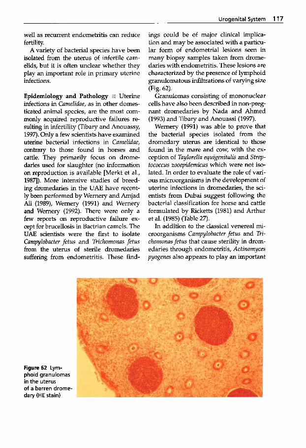
Urogenital System 117
well as recurrent endometritis can reduce fertility.
A variety of bacterial species have been isolated from the uterus of infertile cam- elids, but it is often unclear whether they play an important role in primary uterine infections.
Epidemiology and Pathology + Uterine infections in Camelidae, as in other domes- ticated animal species, are the most com- monly acquired reproductive failures re- sulting in infertility (Tibary and Anouassy, 1997). Only a few scientists have examined uterine bacterial infections in Camelidae, contrary to those found in horses and cattle. They primarily focus on drome- daries used for slaughter (no information on reproduction is available [Merkt et al., 1987). More intensive studies of breed- ing dromedaries in the UAE have recent- ly been performed by Wemery and Amjad Ali (1989), Wemery (1991) and Wemery and Wernery (1992). There were only a few reports on reproductive failure ex- cept for brucellosis in Bactrian camels. The UAE scientists were the first to isolate Campylobacter fetus and Trichomonas fetus from the uterus of sterile dromedaries suffering from endometritis. These find-
ings could be of major clinical implica- tion and may be associated with a particu- lar form of endometrial lesions seen in many biopsy samples taken from drome- daries with endometritis. These lesions are characterized by the presence of lymphoid granulomatous infiltrations of varying size (Fig. 62).
Granulomas consisting of mononuclear cells have also been described in non-preg- nant dromedaries by Nada and Ahmed (1993) and Tibary and Anouassi (1997).
Wernery (1991) was able to prove that the bacterial species isolated from the dromedary uterus are identical to those found in the mare and cow, with the ex- ception of Taylorella equigenitalis and StrqrJ- tococcus zooepidemicus which were not iso- lated. In order to evaluate the role of vari- ous microorganisms in the development of uterine infections in dromedaries, the sci- entists from Dubai suggest following the bacterial classification for horse and cattle formulated by Ricketts (1981) and Arthur et al. (1985) (Table 27).
In addition to the classical venereal mi- croorganisms Campylobacter fetus and Tri- chomonas fetus that cause sterility in drom- edaries through endometritis, Actinomyces pyogenes also appears to play an important
Figure 62 Lym- phoid granulomas in the uterus of a barren drome- dary (HE stain)
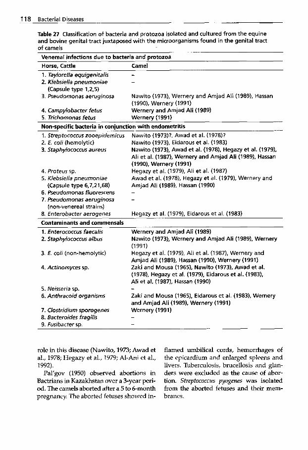
1 18 Bacterial Diseases
Table 27 Classification of bacteria and protozoa isolated and cultured from the equine and bovine genital tract juxtaposed with the microorganisms found in the genital tract of camels
Venereal infections due t o bacteria and protozoa Horse, Cattle Camel 1. Taylorella equigenitalis - 2. Klebsiella pneumoniae -
(Capsule type 1,2,5) 3. Pseudomonas aeruginosa
4. Campylobacter fetus 5. Trichomonas fetus Wernery (1991)
Nawito (1973), Wernery and Amjad Ali (1989), Hassan (1990), Wernery (1991) Wernery and Amjad Ali (1989)
Non-specific bacteria in coniunction with endometritis 1. Streptococcus zooepidemicus 2. E. coli (hemolytic) 3. Staphylococcus aureus
4. Proteus sp. 5. Klebsiella pneumonia e
(Capsule type 6,7,21,68) 6. Pseudomonas fluorescens 7. Pseudomonas aeruginosa
(non-venereal strains) 8. Enterobacter aeroqenes
Nawito (1973)?, Awad e t al. (1978)? Nawito (1973). Eidarous et al. (1983) Nawito (1973), Awad et al. (1978). Hegazy et at. (1979). Ali et al. (1987), Wernery and Amjad Ali (1989). Hassan (1990), Wernery (1991) Hegazy et al. (1979), Ali et al. (1987) Awad et al. (19781, Hegazy e t al. (1979), Wernery and Amjad Ali (1989). Hassan (1990)
Heqazy e t al. (19791, Eidarous et al. (1983) Contaminants and commensals 1. Enterococcus faecalis 2. Staphylococcus albus
3. E. coli (non-hemolytic)
4. Actinomyces sp.
Wernery and Amjad Ali (1989) Nawito (1 973), Wernery and Amjad Ali (1 989). Wernery (1991) Hegazy e t al. (1979), Ali et al. (1987), Wernery and Amjad Ali (1989), Hassan (1990). Wernery (1991) Zaki and Mousa (1965). Nawito (1973). Awad et al. (1978), Hegazy et al. (1979). Eidarous et al. (19831, Ali et al. (1987), Hassan (1990)
Zaki and Mousa (1965). Eidarous et al. (1983). Wernery and Amjad Ali (1989), Wernery (1991)
5. Neisseria sp. - 6. Anthracoid organisms
7. Clostridium sporogenes Wernery (1991) 8. Bacteroides fragilis - 9. Fusibacter sp. -
role in this disease (Nawito, 1973; Awad et aI., 1978; Hegazy et al., 1979; Al-Ani et al., 1992).
Pal'gov (1950) observed abortions in Bactrians in Kazakhstan over a 3-year peri- od. The camels aborted after a 5 to 6-month pregnancy. The aborted fetuses showed in-
flamed umbilical cords, hemorrhages of the epicardium and enlarged spleens and livers. Tuberculosis, brucellosis and glan- ders were excluded as the cause of abor- tion. Streptococcus pyogenes was isolated f rom the aborted fetuses and their mem- branes.
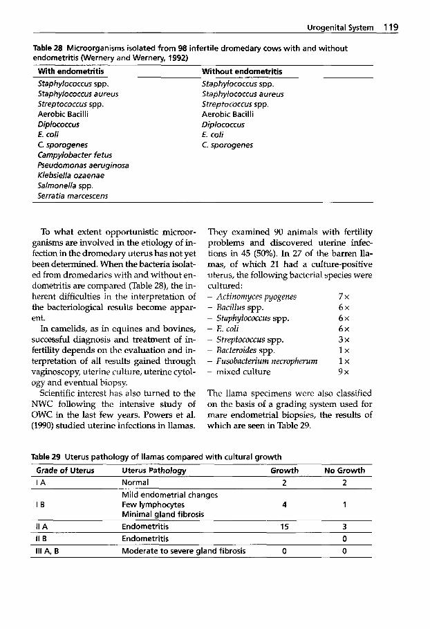
Uroaenital Svstem 119
Table 28 Microorganisms isolated from 98 infertile dromedary cows with and without endometritis Wernery and Wernery, 1992)
With endometritis Without endometritis Staphylococcus spp. Staphylococcus aureus Streptococcus spp. Aerobic Bacilli Diplococcus E. coli C. sporogenes Campylobacter fetus Pseudomonas aeruginosa K/ebsiella ozaenae Salmonella spp. Serratia marcescens
Staphylococcus spp, Staphylococcus a ureus Streptococcus spp. Aerobic Bacilli Diplococcus E. coli C. sporogenes
To what extent opportunistic microor- ganisms are involved in the etiology of in- fection in the dromedary uterus has not yet been determined. When the bacteria isolat- ed from dromedaries with and without en- dometritis are compared (Table 28), the in- herent difficulties in the interpretation of the bacteriological results become appar- ent.
In camelids, as in equines and bovines, successful diagnosis and treatment of in- fertility depends on the evaluation and in- terpretation of all results gained through vaginoscopy, uterine culture, uterine cytol- ogy and eventual biopsy.
Scientific interest has also turned to the NWC following the intensive study of OWC in the last few years. Powers et al. (1990) studied uterine infections in llamas.
They examined 90 animals with fertility problems and discovered uterine infec- tions in 45 (50%). In 27 of the barren lla- mas, of which 21 had a culture-positive uterus, the following bacterial species were cultured:
- Bacillus spp. 6 x - S t ~ ~ h ~ l O ~ O ~ ~ ~ s SPP. 6 x - E. coli 6 x
- Actinomyces pyogenes 7x
- Streptococcus spp. 3 x - Bacteroides spp. l x - Fusobacterium necropherurn 1 x - mixed culture 9 x
The llama specimens were also classified on the basis of a grading system used for mare endometrial biopsies, the results of which are seen in Table 29.
Table 29 Uterus pathology of llamas compared with cultural growth
Grade of Uterus Uterus Patholow Growth No Growth I A Normal 2 2
I B Mild endometrial changes Few lymphocytes Minimal gland fibrosis
4 1
II A Endometritis 15 3 II B Endometritis 0
111 A, B Moderate to severe gland fibrosis 0 0
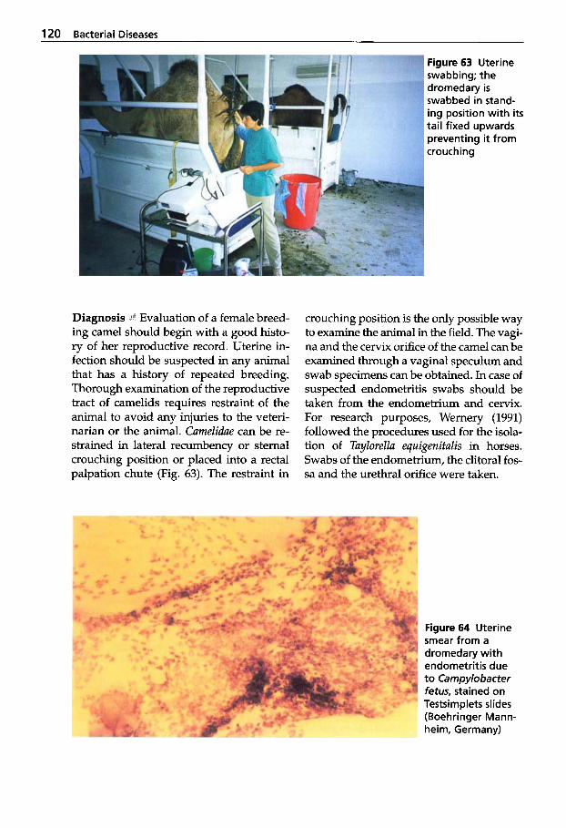
120 Bacterial Diseases
Figure 63 Uterine swabbing; the dromedary is swabbed in stand- ing position with i t s tai l fixed upwards preventing it from crouching
Diagnosis .l’! Evaluation of a female breed- ing camel should begin with a good histo- ry of her reproductive record. Uterine in- fection should be suspected in any animal that has a history of repeated breeding. Thorough examination of the reproductive tract of camelids requires restraint of the animal to avoid any injuries to the veteri- narian or the animal. CameZidae can be re- strained in lateral recumbency or sternal crouching position or placed into a rectal palpation chute (Fig. 63). The restraint in
crouching position is the only possible way to examine the animal in the field. The vagi- na and the cervix orifice of the camel can be examined through a vaginal speculum and swab specimens can be obtained. In case of suspected endometritis swabs should be taken from the endometrium and cervix. For research purposes, Wernery (1991) followed the procedures used for the isola- tion of TuyZoreZZu equigenifulis in horses. Swabs of the endometrium, the clitoral fos- sa and the urethral orifice were taken.
Figure 64 Uterine smear from a dromedary with endometritis due to Campylobacter fetus, stained on Testsimplets slides (Boehringer Mann- heim, Germany)
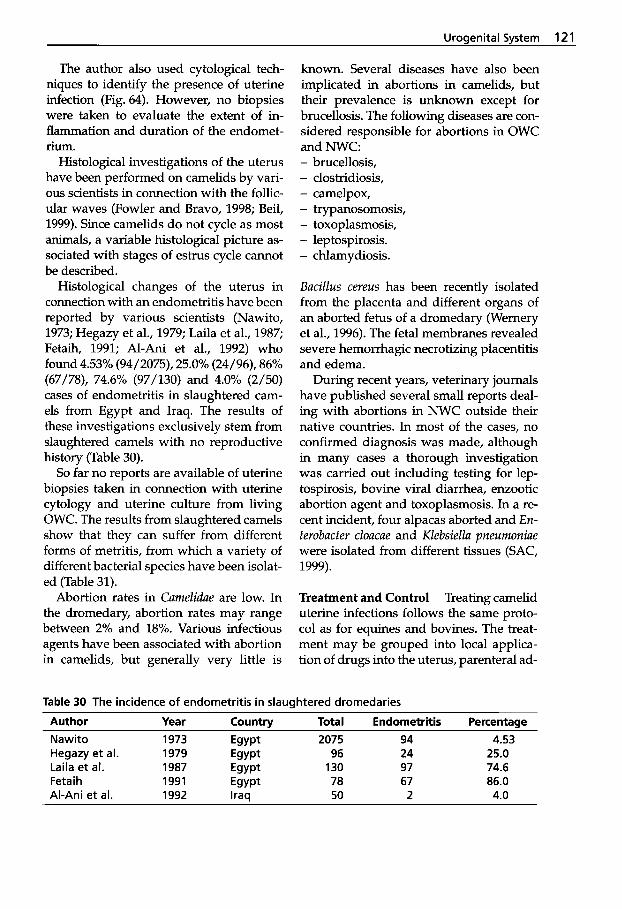
Urogenital System 121
The author also used cytological tech- niques to identify the presence of uterine infection (Fig. 64). However, no biopsies were taken to evaluate the extent of in- flammation and duration of the endomet- rium.
Histological investigations of the uterus have been performed on camelids by vari- ous scientists in connection with the follic- ular waves (Fowler and Bravo, 1998; Beil, 1999). Since camelids do not cycle as most animals, a variable histological picture as- sociated with stages of estrus cycle cannot be described.
Histological changes of the uterus in connection with an endometritis have been reported by various scientists (Nawito, 1973; Hegazy et al., 1979; Laila et al., 1987; Fetaih, 1991; Al-Ani et al., 1992) who
(67/78), 74.6% (97/130) and 4.0% (2/50) cases of endometritis in slaughtered cam- els from Egypt and Iraq. The results of these investigations exclusively stem from slaughtered camels with no reproductive history (Table 30).
So far no reports are available of uterine biopsies taken in connection with uterine cytology and uterine culture from living OWC. The results from slaughtered camels show that they can suffer from different forms of metritis, from which a variety of different bacterial species have been isolat- ed (Table 31).
Abortion rates in Camelidae are low. In the dromedary, abortion rates may range between 2% and 18%. Various infectious agents have been associated with abortion in camelids, but generally very little is
f o n d 4.53% (94/2075), 25.0% (24/96), 86%
known. Several diseases have also been implicated in abortions in camelids, but their prevalence is unknown except for brucellosis. The following diseases are con- sidered responsible for abortions in OWC and NWC: - brucellosis, - clostridiosis, - camelpox, - trypanosomosis, - toxoplasmosis, - leptospirosis. - chlamydiosis.
Bacillus cereus has been recently isolated from the placenta and different organs of an aborted fetus of a dromedary (Wernery et al., 1996). The fetal membranes revealed severe hemorrhagic necrotizing placentitis and edema.
During recent years, veterinary journals have published several small reports deal- ing with abortions in NWC outside their native countries. In most of the cases, no confirmed diagnosis was made, although in many cases a thorough investigation was carried out including testing for lep- tospirosis, bovine viral diarrhea, enzootic abortion agent and toxoplasmosis. In a re- cent incident, four alpacas aborted and En- terobacter cloacae and Klebsiella pneumoniae were isolated from different tissues (SAC, 1999).
Treatment and Control Treating camelid uterine infections follows the same proto- col as for equines and bovines. The treat- ment may be grouped into local applica- tion of drugs into the uterus, parenteral ad-
Table 30 The incidence of endometritis in slaughtered dromedaries Author Year Country Total Endometritis Percentage Nawito 1973 Egypt 2075 94 4.53 Hegazy et al. 1979 Egypt 96 24 25.0 Laila et al. 1987 Egypt 130 97 74.6 Fetaih 1991 Egypt 78 67 86.0 Al-Ani et al. 1992 Iraq 50 2 4.0
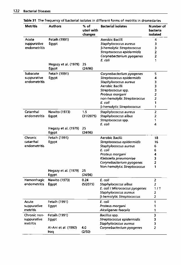
122 Bacterial Diseases
Table 31 The frequency of bacterial isolates in different forms of metritis in dromedaries
uteri with bacteria changes isolated
Metritis Authors % of Bacterial isolates Number of
Acute Fetaih (1991) Aerobic Bacilli 4 suppurative Egypt Staphylococcus aureus 3 endometritis P-hemolytic Streptococcus 3
Streptococcus epidermidis 2 Corynebacterium pyogenes 2 E. coli 1
Hegazy et al. (1979) 25 Egypt (24196)
Subacute Fetaih (1991) Corynebacterium pyogenes 5 suppurative Egypt Streptococcus epidermidis 4 endometritis Staphylococcus aureus 3
Aerobic Bacilli 3 Streptococcus spp. 3 Proteus morgani 2 non-hemolytic Streptococcus 2 E. coli 1 P-hemolytic Streptococcus 1
Catarrhal Nawito (1973) 1.5 Staphylococcus aureus 2 endometritis Egypt (31/2075) Staphylococcus albus 2
Streptococcus spp. 1 E. coli 4
Hegazy et al. (1979) 25 Egypt (24196)
Chronic Fetaih (1991) Aerobic Bacilli 18 catarrhal Egypt Streptococcus epidermidis 16 endometritis Staphylococcus a ureus 6
E. coli 6 Proteus morgani 4 Klebsiella pneumoniae 3 Corynebacterium pyogenes 2 Non-hemolytic 5treptococcus 1
Hegazy et al. (1979) 25 Egypt (24196)
Hemorrhagic Nawito (1973) 0.24 E. coli 2 endometritis Egypt (5/2075) Staphylococcus albus 2
Staphylococcus aureus 2 P-hemolytic Streptococcus 2
Acute Fetaih (1991) ' E. coli 1 suppurative Egypt Proteus morgani 1 metritis Alcaligenes faecalis 1 Chronic non- Fetaih (1991) Bacillus spp. 3 suppurative Egypt Streptococcus epidermidis 3 metritis Staphylococcus a ureus 3
Al-Ani et al. (1992) 4.0 Corynebacterium pyogenes 2 Iraq (a501
E. coli I Micrococcus pyogenes 1 I 1
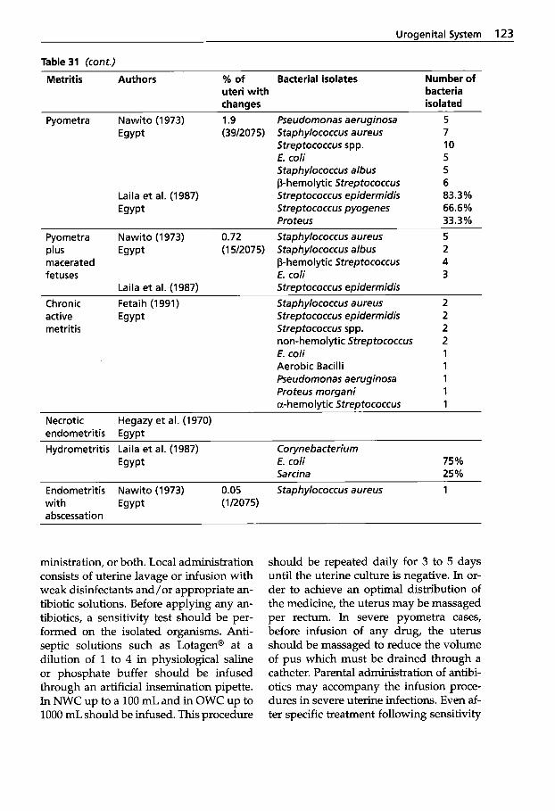
Uroaenital Svstem 123
Table 31 (cont.)
Metritis Authors % of Bacterial isolates Number of uteri with bacteria changes isolated
Pyometra Nawito (1973) 1.9 Egypt (39/2075)
Laila et al. (1987) Egypt
Pseudomonas aeruginosa Staphylococcus aureus Streptococcus spp. E. coli Staphylococcus albus P-hemolytic Streptococcus Streptococcus epidermidis Streptococcus pyogenes Proteus
5 7 10 5 5 6 83.3% 66.6% 33.3%
Pyometra Nawito (1973) 0.72 plus Egypt (1 5/2075) macerated fetuses
Chronic Fetaih (1991)
metritis
Laila et al. (1987)
active Egypt
Staphylococcus aureus 5 Staphylococcus albus 2 P-hemolytic Streptococcus 4 E. coli 3 Streatococcus eaidermidis Staphylococcus a ureus 2 Strep tococcus epidermidis 2 Streptococcus spp. 2 non-hemolytic Streptococcus 2 E. coli 1 Aerobic Bacilli 1 Pseudomonas aeruginosa 1 Proteus morgani 1 a-hemolytic Streptococcus 1
Necrotic Hegazy et al. (1970) endometritis Egypt Hydrometritis Laila et al. (1987) Corynebacterium
Egypt E. coli 75% Sarcina 25%
Endometritis Nawito (1973) 0.05 Staphylococcus aureus 1 with Egypt (1/2075) a bscessat ion
ministration, or both. Local administration consists of uterine lavage or infusion with weak disinfectants and/or appropriate an- tibiotic solutions. Before applying any an- tibiotics, a sensitivity test should be per- formed on the isolated organisms. Anti- septic solutions such as Lotagen@ at a dilution of 1 to 4 in physiological saline or phosphate buffer should be infused through an artificial insemination pipette. In NWC up to a 100 mL and in OWC up to 1000 mL should be infused. This procedure
should be repeated daily for 3 to 5 days until the uterine culture is negative. In or- der to achieve an optimal distribution of the medicine, the uterus may be massaged per rectum. In severe pyometra cases, before infusion of any drug, the uterus should be massaged to reduce the volume of pus which must be drained through a catheter. Parental administration of antibi- otics may accompany the infusion proce- dures in severe uterine infections. Even af- ter specific treatment following sensitivity
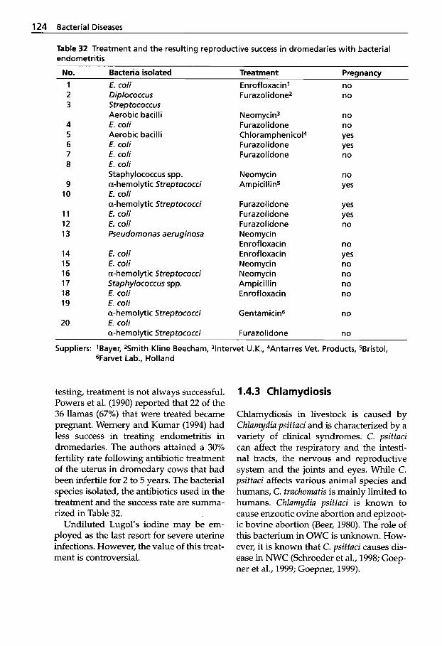
124 Bacterial Diseases
Table 32 Treatment and the resulting reproductive success in dromedaries with bacterial endometritis
No. Bacteria isolated Treatment ______
Preanancv 1 2 3
4 5 6 7 8
9 10
11 12 13
14 15 16 17 18 19
20
E. coli Diplococcus Streptococcus Aerobic bacil I i E. coli Aerobic bacilli E. coli E. coli E. coli Staphylococcus spp. a-hemolytic Streptococci E. coli a-hem0 I ytic Strep tococci E. coli E. coli Pseudomonas aeruginosa
E. coli E. coli a-hemolytic Streptococci Staphylococcus spp. E. coli E. coli a-hemolytic Streptococci E. coli a-hemolytic Streptococci
Enrofloxacin’ Furazolidonez
Neomycin3 Furazolidone Chloramphenicol4 Furazolidone Furazolidone
Neom yci n Ampicillin5
Furazolidone Furazolidone Furazolidone Neomycin Enrof loxacin En rof loxaci n Neomycin Neomycin Ampicillin Enrofloxacin
Gentamicin6
Furazolidone
no no
no no Yes Yes no
no Yes
Yes Yes no
no Yes no no no no
no
no
Suppliers: ’Bayer, 2Smith Kline Beecham, 3lntervet U.K., 4Antarres Vet. Products, SBristol, ‘jFarvet Lab., Holland
testing, treatment is not always successful. Powers et al. (1990) reported that 22 of the 36 llamas (67%) that were treated became pregnant. Wernery and Kumar (1994) had less success in treating endometritis in dromedaries. The authors attained a 30% fertility rate following antibiotic treatment of the uterus in dromedary cows that had been infertile for 2 to 5 years. The bacterial species isolated, the antibiotics used in the treatment and the success rate are summa- rized in Table 32.
Undiluted Lugol’s iodine may be em- ployed as the last resort for severe uterine infections. However, the value of this treat- ment is controversial.
1.4.3 Chlamydiosis
Chlamydiosis in livestock is caused by Chlamydia psittaci and is characterized by a variety of clinical syndromes. C. psittaci can affect the respiratory and the intesti- nal tracts, the nervous and reproductive system and the joints and eyes. While C. psittaci affects various animal species and humans, C. trachomatis is mainly limited to humans. ChZamydia psittaci is known to cause enzootic ovine abortion and epizoot- ic bovine abortion (Beer, 1980). The role of this bacterium in OWC is unknown. How- ever, it is known that C. psittaci causes dis- ease in NWC (Schroeder et al., 1998; Goep- ner et al., 1999; Goepner, 1999).
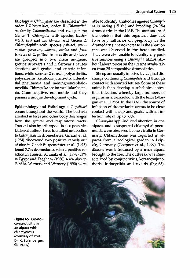
Urogenital System 125
Chlamydiae are classified in the order I Rickeftsiales, order I1 Chlamydiul- es, family Chlamydiaceae and two genera; Genus 1: Chlamydia with species tracho- mafis, suis and muridarum and Genus 2: Chlamydophila with species psittaci, pneu- moniae, pecorum, abortus, caviae and felis. Isolates of C. psittaci from cattle and sheep are grouped into two main antigenic groups: serovars 1 and 2. Serovar 1 causes abortions and genital and enteric infec- tions, while serovar 2 causes polyarthritis, polyserositis, keratoconjunctivitis, intersti- tial pneumonia and meningoencephalo- myelitis. Chlamydiae are intracellular bacte- ria, Gram-negative, non-motile and they possess a unique development cycle.
Epidemiology and Pathology 1 C. psittaci occurs throughout the world. The bacteria are shed in feces and other body discharges from the genital and respiratory tracts. Transmission by arthropods is also possible. Different authors have identified antibodies to Chlamydiae in dromedaries. Giraud et al. (1954) discovered two positive camels out of nine in Chad; Burgemeister et al. (1975) found 7.7% dromedaries with a positive re- action in Tunisia; Schmatz et al. (1978) 11% in Egypt and Djegham (1988) 4.4% also in Tunisia. Wemery and Wemery (1990) were
able to identdy antibodies against Chlamyd- ia in racing (15.0%) and breeding (24.0%) dromedaries in the UAE. The authors are of the opinion that this organism does not have any influence on pregnancy in the dromedary since no increase in the abortion rate was observed in the herds studied. They were also unable to identify any posi- tive reactors using a Chlamydia ELISA (Ab- bott Laboratories) on the uterine swabs tak- en from 28 seropositive dromedaries.
Sheep are usually infected by vaginal dis- charge containing Chlamydiae and through contact with aborted fetuses. Some of these animals then develop a subclinical intes- tinal infection, whereby large numbers of organisms are excreted with the feces (Mor- gan et al., 1988). In the UAE, the source of infection of dromedaries seems to be close contact with sheep and goats, with an in- fection rate of up to 50%.
Chlamydia spp.-induced abortion in one alpaca, and a suspected chlamydia1 pneu- monia were observed in one vicuiia in Ger- many. Chlamydiosis was reported in al- pacas from a zoological garden in Leip- zig, Germany (Goepner et al., 1999). The disease was introduced by a male alpaca brought to the zoo. The outbreak was char- acterized by conjunctivitis, keratoconjunc- tivitis, iridocyclitis and uveitis (Fig. 65).
Figure 65 Kerato- conjunctivitis in an alpaca with chlamydiosis (courtesy of Prof. Dr. K. Eulenberger, Germany)
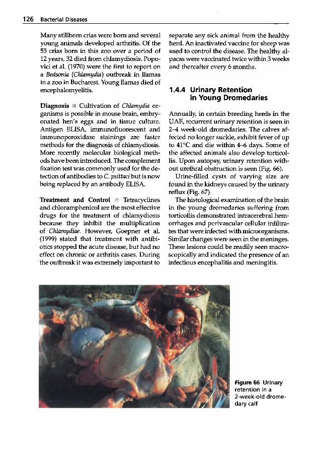
126 Bacterial Diseases
Many stillborn crias were born and several young animals developed arthritis. Of the 53 crias born in this zoo over a period of 12 years, 32 died from chlamydiosis. Popo- vici et al. (1970) were the first to report on a Bedsonia (ChZumydia) outbreak in llamas in a zoo in Bucharest. Young llamas died of encephalomyelitis.
Diagnosis !mi Cultivation of Chlamydia or- ganisms is possible in mouse brain, embry- onated hen’s eggs and in tissue culture. Antigen ELISA, immunofluorescent and immunoperoxidase stainings are faster methods for the diagnosis of chlamydiosis. More recently molecular biological meth- ods have been introduced. The complement fixation test was commonly used for the de- tection of antibodies to C. psitfaci but is now being replaced by an antibody ELISA.
Treatment and Control I Tetracyclines and chloramphenicol are the most effective drugs for the treatment of chlamydiosis because they inhibit the multiplication of Chlamydiae. However, Goepner et al. (1999) stated that treatment with antibi- otics stopped the acute disease, but had no effect on chronic or arthritis cases. During the outbreak it was extremely important to
separate any sick animal from the healthy herd. An inactivated vaccine for sheep was used to control the disease. The healthy al- pacas were vaccinated twice within 3 weeks and thereafter every 6 months.
1.4.4 Urinary Retention in Young Dromedaries
Annually, in certain breeding herds in the UAE, recurrent urinary retention is seen in 2 4 week-old dromedaries. The calves af- fected no longer suckle, exhibit fever of up to 41°C and die within 4-6 days. Some of the affected animals also develop torticol- lis. Upon autopsy, urinary retention with- out urethral obstruction is seen (Fig. 66).
Urine-filled cysts of varying size are found in the kidneys caused by the urinary reflux (Fig. 67).
The histological examination of the brain in the young dromedaries suffering from torticollis demonstrated intracerebral hem- orrhages and perivascular cellular infiltra- tes that were infected with microorganisms. Similar changes were seen in the meninges. These lesions could be readily seen macro- scopically and indicated the presence of an infectious encephalitis and meningitis.
Figure 66 Urinary retention in a 2-week-old drome- dary calf
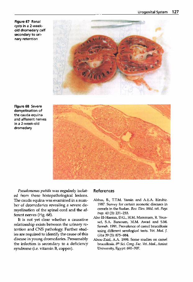
Uroaenital Svstem 127
Figure 67 Renal cysts in a 2-week- old dromedary calf secondary t o uri- nary retention
Figure 68 Severe demyelination of the cauda equina and afferent nerves in a 2-week-old dromedary
Pseudomonas putidu was regularly isolat- ed from these histopathological lesions. The cauda equina was examined in a num- ber of dromedaries revealing a severe de- myelination of the spinal cord and the af- ferent nerves (Fig. 68).
It is not yet clear whether a causative relationship exists between the urinary re- tention and CNS pathology. Further stud- ies are required to identify the cause of this disease in young dromedaries. Presumably the infection is secondary to a deficiency syndrome (i.e. vitamin B, copper).
References
Abbas, B., T.T.M. Yassin and A.E.A. Elzubir. 1987. Survey for certain zoonotic diseases in camels in the Sudan. Rev. Elev. Mid. vit. Pays
Abo El-Hassan, D.G., H.M. Mammam, R. Yous- sef, S.A. Barsoum, M.M. Awad and S.M. Sameh. 1991. Prevalence of camel brucellosis using different serological tests. Vet. Med. J. Giza 39 (3): 875-884.
Abou-Zaid, A.A. 1998. Some studies on camel brucellosis. gth Sci. Cong. Fac. Vet. Med., Assiut University, Egypt: 690-707.
trap. 40 (3): 231-233.
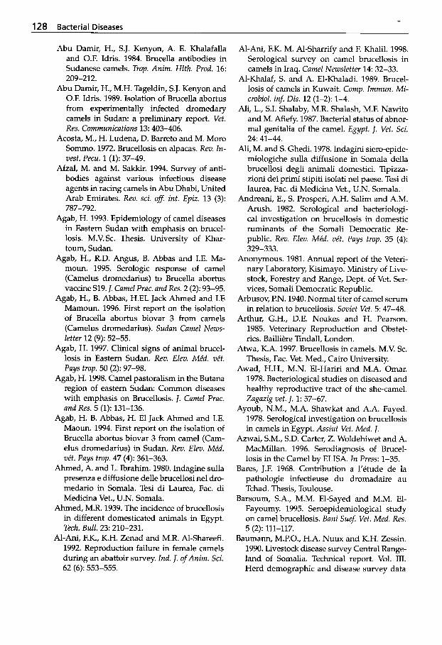
128 Bacterial Diseases
Abu Damir, H., S.J. Kenyon, A. E. Khalafalla and O.F. Idris. 1984. Brucella antibodies in Sudanese camels. Trop. Anim. Hlth. Prod. 16:
Abu Damir, H., M.H. Tageldin, S.J. Kenyon and O.F. Idris. 1989. Isolation of Brucella abortus from experimentally infected dromedary camels in Sudan: a preliminary report. Vet. Res. Communications 13: 403-406.
Acosta, M., H. Ludena, D. Barreto and M. Moro Sommo. 1972. Brucellosis en alpacas. Rev. In- vest. Pecu. 1 (1): 37-49.
Afzal, M. and M. Sakkir. 1994. Survey of anti- bodies against various infectious disease agents in racing camels in Abu Dhabi, United Arab Emirates. Rev. sci. of. int. Epiz. 13 (3):
Agab, H. 1993. Epidemiology of camel diseases in Eastern Sudan with emphasis on brucel- losis. M.V.Sc. Thesis. University of Khar- toum, Sudan.
Agab, H., R.D. Angus, B. Abbas and I.E. Ma- moun. 1995. Serologic response of camel (Camelus dromedarius) to Brucella abortus vaccine S19. J. Camel Prac. and Res. 2 (2): 93-95.
Agab, H., B. Abbas, H.EL Jack Ahmed and 1.E Mamoun. 1996. First report on the isolation of Brucella abortus biovar 3 from camels (Camelus dromedarius). Sudan Camel News- letter 12 (9): 52-55.
Agab, H. 1997. Clinical signs of animal brucel- losis in Eastern Sudan. Rev. Elev. Mid. ve‘t. Pays trop. 50 (2): 97-98.
Agab, H. 1998. Camel pastoralism in the Butana region of eastern Sudan: Common diseases with emphasis on Brucellosis. J. Camel Prac. and Res. 5 (1): 131-136.
Agab, H. B. Abbas, H. El Jack Ahmed and I.E. Maoun. 1994. First report on the isolation of Brucella abortus biovar 3 from camel (Cam- elus dromedarius) in Sudan. Rev. Elev. Mid. vit. Pays trop. 47 (4): 361-363.
Ahmed, A. and L. Ibrahim. 1980. Indagine sulla presenza e diffusione delle brucellosi nel dro- medario in Somala. Tesi di Laurea, Fac. di Medicina Vet., U.N. Somala.
Ahmed, M.R. 1939. The incidence of brucellosis in different domesticated animals in Egypt. Tech. Bull. 23: 210-231.
Al-Ani, F.K., K.H. Zenad and M.R. Al-Shareefi. 1992. Reproduction failure in female camels during an abattoir survey. Ind. J. of Anim. Sci.
209-212.
787-792.
62 (6): 553-555.
Al-Ani, F.K. M. Al-Sharrify and F. Khalil. 1998. Serological survey on camel brucellosis in camels in Iraq. Camel Newsletter 14: 32-33.
Al-Khalaf, S. and A. El-Khaladi. 1989. Brucel- losis of camels in Kuwait. Comp. Immun. Mi- crobiol. in5 Dis. 12 (1-2): 1-4.
Ali, L., S.I. Shalaby, M.R. Shalash, M.F. Nawito and M. Afiefy. 1987. Bacterial status of abnor- mal genitalia of the camel. Egypt. J. Vet. Sci.
Ali, M. and S. Ghedi. 1978. Indagini siero-epide- miologiche sulla diffusione in Somala della brucellosi degli animali domestici. Tipizza- zioni dei primi stipiti isolati nel paese. Tesi di laurea, Fac. di Medicina Vet., U.N. Somala.
Andreani, E., S. Prosperi, A.H. Salim and A.M. Arush. 1982. Serological and bacteriologi- cal investigation on brucellosis in domestic ruminants of the Somali Democratic Re- public. Rev. Elev. Mid. v&. Pays trop. 35 (4):
Anonymous. 1981. Annual report of the Veteri- nary Laboratory, Kisimayo. Ministry of Live- stock, Forestry and Range, Dept. of Vet. Ser- vices, Somali Democratic Republic.
Arbusov, P.N. 1940. Normal titer of camel serum in relation to brucellosis. Soviet Vet. 5: 47-48.
Arthur, G.H., D.E. Noakes and H. Pearson. 1985. Veterinary Reproduction and Obstet- rics. Bailliere Tindall, London.
Atwa, K.A. 1997. Brucellosis in camels. M.V. Sc. Thesis, Fac. Vet. Med., Cairo University.
Awad, H.H., M.N. El-Hariri and M.A. Omar. 1978. Bacteriological studies on diseased and healthy reproductive tract of the she-camel. Zagazig vet. J. 1: 57-67.
Ayoub, N.M., M.A. Shawkat and A.A. Fayed. 1978. Serological investigation on brucellosis in camels in Egypt. Assiut Vet. Med. J.
Azwai, S.M., S.D. Carter, Z. Woldehiwet and A. MacMillan. 1996. Serodiagnosis of Brucel- losis in the Camel by ELISA. In Press: 1-35.
Bares, J.F. 1968. Contribution a l’ktude de la pathologie infectieuse du dromadaire au Tchad. Thesis, Toulouse.
Barsoum, S.A., M.M. El-Sayed and M.M. El- Fayoumy. 1995. Seroepidemiological study on camel brucellosis. Bani Sue5 Vet. Med. Res.
Baumann, M.P.O., H.A. Nuux and K.H. Zessin. 1990. Livestock disease survey Central Range- land of Somalia. Technical report. Vol. III. Herd demographic and disease survey data
24: 41-44.
329-333.
5 (2): 111-117.
Urogenital System 129
from herds of camels. CRDP - Veterinary Component, Mogadishu, Somalia.
Baumann, M.P.O. and K.H. Zessin. 1992. Pro- ductivity and health of camels (Camelus dromedarius) in Somalia: associations with trypanosomiasis and brucellosis. Trop. Anim. Hlth. Prod. 24 (3): 145-156.
Beer, J. 1980. Infektionskrankheiten der Haus- tiere. VEB Gustav Fischer Verlag, Jena.
Beil, Christiane. 1999. Reproduktion beim weib- lichen Kame1 (Camelus dromedarius und Ca- melus bactrianus). Eine gewichtete Literatur- studie. Thesis, Hannover.
Ben Faraj, S.M., S.M. Azwai, S.E. Gameel, A.M. Shareha, K.M. Benhaj, H.M. Rayes and A.A. Nayil. 1990. Camel and human brucellosis in Libya. Proc. int. con5 on camel production and improvement, Dec. 10-13,1990, Tobruk, Libya.
Bishof, J. 1979. Serological examination of blood samples from dromedaries. Serum and Vac- cine Institute, Mogadishu, Somalia.
Bisping, W. and G. Amtsberg. 1988. Colour atlas for the diagnosis of bacterial pathogens in animals. Verlag Paul Parey, Berlin and Ham- burg.
Bomarel, P. and A.J. Akakpo. 1982. Brucelloses animales: Sondages serologiques dans quatre pays de l'Afrique de I'Ouest (Benin, Camer- oun, Haute-Volta, Niger). Mkdecine d'Afrique Noire 29 (12): 829-836.
Bornstein, S. 1984. Working paper No. 3, Camel Forum. Mogadishu, Somali Academy Science and Art.
Bornstein, S. 1988. A disease survey of the So- mali camel. SARE Report, Sweden.
Bornstein, S., B.E. Musa and EM. Jama. 1988. Comparison of seroepidemiological findings of antibodies to some infectious pathogens of cattle and camels of Sudan and Somalia with reference to findings in other countries of Africa. Proc. of International Symposium of De- velopment of Animal Resources in Sudan. Khar- toum: 28-34.
Bornstein, S. and B.E. Musa. 1987. Prevalence of antibodies to some viral pathogens, Brucella abortus and Toxoplasma gondii in serum from camels (Camelus dromedarius) in Su- dan. J. Vet. Med. B34 364-370.
Burgemeister, R., W. Leyk and R. Goessler. 1975. Untersuchungen iiber Vorkommen von Para- sitosen, bakteriellen und viralen Infektions- krankheiten bei Dromedaren in Siidtunesien. Dtsch. Tierarztl. Wschr. 8 2 352-354.
Chichibabin, E.S. 1971. Results of haemaggluti- nation test with the heat-inactivated sera from camels investigated for brucellosis. Proc. of Kazakh Res. Vet. Inst. 14: 29-30.
Chukwu, C.C. 1985. Brucellosis in Afrika. Part I The prevalence. Bull. Anim. Hlth. Prod. Afr. 33:
Djegham, M. 1988. A propos de l'avortement chez la chamelle en Tunisie. Magkreb Vet. 3 (14): 60.
Domenech, J. 1977. Enquete serologique sur la brucellose du dromadaire en Tchad. Rev. Elev. Mkd. vkt. Pays trop. 30 (2): 141-142.
Domenech, J., P. Lucet, B. Vallat, C. Stewart, J.B. Bonnet and A. Hentic. 1982. La brucellose bovine en Afrique centrale. III. Rksultats sta- tistique des enqugtes menbes au Tchad et au Cameroun. Rev. Elev. Mkd. vet. Pays trop. 35:
Eidarous, A., H. Mansour and A. Abdul Rahier. 1983. Bacterial flora of the genital system of male and female camel. Zagazig Vet. J. 4 24-27.
El-Nahas, H.M. 1964. Brucellosis in camels. Proc. 5th Arab. Vet. Cong., Cairo, UAR: 239-252.
El-Sawally, A.A., A.M. Montaser and L.G. Rizk. 1996. Diagnostic and biochemical evalua- tions of camel brucellosis. Vet. Med. J., Giza 44
Elmi, A.M. 1982. Thesis. University of Califor- nia, Davis, USA.
Fayed, A.A., S.A. Karmy, H.I. Yousef and M.M. Ayoub. 1982. Serological studies on brucello- sis in Aswan Province. Vet. Med. J. 30 491-497.
Fazil, M.A. and R.R. Hofmann. 1981. Haltung und Krankheiten des Kamels. Tierurztl. Praxis
Fetaih, A.A.H. 1991. Some pathological studies on the affections of genital system in she- camel. Thesis, Cairo University.
Fowler, M.E. 1998. Medicine and surgery of South American Camelids. Iowa State Uni- versity Press, Ames.
Fowler, M.E. and P.W. Bravo. 1998. Reproduc- tion in: Medicine and Surgery of South Amer- ican Camelids, Ed: M.E. Fowler, 2nd ed. Iowa State University Press, Ames: pp. 381-429.
Gameel, S.E.A., S.O. Mohamed, A.A. Mustafa and S.M. Azwai. 1993. Prevalence of camel brucellosis in Libya. Tmp. Anim. Prod. 25 (2):
Gatt Rutter, T.E. and R. Mack. 1963. Diseases of camels. Part 1: Bacterial and fungal diseases. Vet. Bull. 33 (3): 119-124.
193-198.
15-22.
(2): 323-329.
9: 389-402.
91-93.
130 Bacterial Diseases
Ghazi, Y.A. 1996. Studies on brucellosis in camels. PhD, Fac. Vet. Med., Cairo Univer- sity.
Ghoneim, N.A. and Amjad, A.M. 1993. Brucel- losis among sheep, goats and camels in Saudi Arabia in Al Joub region, incidence and comparison between Rose Bengal test and seroagglutination tube test. Proc. of 2ZSt Arab Vet. Med. Cong., Cairo, April 10-14,1993
Gidlewski, T, N.T. Cheville, J.C. Rhyan, L.D. Miller and M.J. Gildorf. 2000. Experimen- tal Brucella abortus induced abortion in a llama: Pathologic effects. Vet. Pathology 37 (1):
Giroud, P., F. Roger, N. Dumas, P. Vouilloux and E. Sacquet. 1954. Comportement des ani- maux domestiques de la region du Thad vis- h i s de l'antighe T13. Bull. SOC. Path. Exot.
Goepner, I. 1999. Analyse des Krankheitsge- schehens in der Alpakaherde des Zoologi- schen Gartens Leipzig unter besonderer Be- riicksichtigung der Chlamydiose. Vet. med. Thesis, Leipzig.
Goepner, Isabel, K. Eulenberger, A. Bernhard, Ute Schulz and A. Neubert. 1999. Chlamy- diose bei Alpakas (Lama guanacoe F. pacos). Verhandlber. Erkrg. Zootiere 39: 199-207.
Graber, M. 1968. Region of Veterinary and Zootechnical Research of Central Africa. An- nual report, Farcha Laboratory, ist Research and Products 2 Pleuropneumonia. Quini quennial Report. Fort Lamy, Chad. Veterinary Bulletin 38: 5265.
Hamada, S., M. El-Hidik, I. Sherif, H. El-Sawah and M. Yousef. 1963. Serological investiga- tions on brucellosis in cattle, buffaloes and camels. J. Arab. Vet. Med. 23: 173-178.
Harby, H.A.M. and S.L.N. Ismaily. 1995. The prevalence of Brucellosis among livestock in the Sultanate of Oman. Proc. of the Intl. Con5 on Livestock Production in Hot Climates: A46.
Hassan, M.S. 1990. Some studies on the bacteria of the uterus of the camel. M.V.Sc. Thesis, Fac. Vet. Med., Cairo University.
Hegazy, A., H.I. Youseff and S.A. Selim. 1979. Bacteriological and histopathological studies on endometritis of the camel. J. Egypt. Vet. Med. Ass. 39: 81-97.
Higgins, A. 1986. The camel in health and dis- ease. Bailli6re Tindall.
273-281.
77-82.
47 644-645.
Jawad, A.H. 1984. Brucellosis in camel in Iraq. Bull. endem. Dis.: 24-25,45-50.
Kagunya, D.K.J. and P.G. Waiyaki. 1978. A sero- logical survey of animal brucellosis in north- eastern province of Kenya. Kenya Vet. 2 (2):
Kiel, F.W. and M.Y. Khan. 1987. Analysis of 506 consecutive positive serological tests for bru- cellosis in Saudi Arabia. J. Clin. Microbiol. 25:
Kulshreshtha, R.C., R.G. Arora and D.S. Kalra. 1975. Brucellosis in camels and horses. Indian J. Anim. Sci. 45 (9): 673-675.
Laila, A.M., S.I.A. Shalaby, M.R. Shalash, M.F. Nawito and M.M. Afify. 1987. Bacterial status of abnormal genitalia of the camels. Egypt J. Vet. Sci. 24 (1): 41-44.
Madkow, M.M. 1989. Brucellosis. Buttenvorths, London.
Mathur, K.N. and S.C. Bhargava. 1979. Sero- prevalence of Q fever and brucellosis in cam- els of Jorbeer and Bikaner, Rajasthan State. Indian J. Med. Xes. 70 (11): 391-393.
Merkt, H., B. Mousa, M.A. El-Naggar and D. Rath. 1987. Reproduction in camels. A Re- view. FA0 Animal production health paper.
Mohammed, I.M. 1996. Development, optimiza- tion and evaluation of diagnostic immunoas- says for camel brucellosis. Thesis, Vet. Sci. Faculty, University Khartoum, Sudan.
Morgan, K.L., J.M. Wills, P. Howard and R.C. Williams. 1988. Isolation of Chlamydia psit- taci from the genital tract of lambs: a possible link with enzootic abortion of ewes. Vet. Rec.
Moustafa, T., E.A. Omar and S.M. Basyouni. 1998. Surveillance of brucella antibodies in camels of the eastern region of the United Arab Emirates. Proc. Int. Meeting on Camel Production and Future Perspectives. May 2-3, 1998, Fac. of Agric. Sci., A1 Ain, UAE.
Mukasa-Mugerwa, E. 1981. The camel (Cam- elus dromedarius): A bibliographical review. International Livestock Center for Africa. ILCA Monogr. 5: 4-119.
Mustafa, A.A. and A. Hassan. 1971. A prelimi- nary survey for the detection of brucella anti- bodies in camel sera. Sudan J. Vet Sci. and Anim. Husb. 12: 5.
Mustafa, A.A. and M.H. Awad El-Karim. 1971. A preliminary survey for the detection of brucella antibodies in camel sera. Sudan J. Vet. Sci. and Anim Husb. 12 (1): 5-8.
35-38.
1384-1387.
123: 399-400.
Urogenital System 131
Nada, A.R. 1984. Some studies on brucellosis in camels. M.V. Sc. Fac. Vet. Medicine, Cairo University.
Nada, A.R. 1990. Further studies on brucellosis in camels. PhD, Fac. Vet. Med., Cairo Univer- sity.
Nada, A.R., E.M. Ismail, M.E. Shawkat and S.A. Barsoum. 1992. Evaluation of serotests used in the diagnosis of camel brucellosis. J. Egypt Vet. Med. Ass. 52 (4): 435-442.
Nada, A.R. and W.M. Ahmed. 1993. Investiga- tions on Brucellosis in some genital abnor- malities of she-camels (C. dromedarius). Int. J. Anim. Sci. 8 (1): 37-40.
Nawito, M. 1973. Uterine infections in the cam- el. Egypt. J. Vet. Sci. 10: 17-22.
Okoh, A.E.J. 1979. A survey of brucellosis in camels in Kano, Nigeria. Trop. Anim. Hlth. Prod. 11 (4): 213-214.
Osman, A.M. and A.M. Adlan. 1987. Sudan. Brucellosis in domestic animals: prevalence, diagnosis and control. Tech. series, Ofice int. Epiz. 6: 67-72.
Ostrovidov, P.I. 1954a. Experiment on rearing healthy camels from dams infected with bru- cellosis. Dud. Inst. Vet., Alma-Ata 6: 62-68.
Ostrovidov, PI. 1954b. Development of resist- ance to brucellosis in camels. Trudy Inst. Vet., Alma Ata 6: 51-56.
Pal’gov, A.A. 1950. No title. Trud. naucho-issled, Vet. Inst., Alma Ata 5: 29.
Pal‘gov, A.A. and I.Z. Zhulobovski. 1964. Diag- nosis of brucellosis in camels and methods of eliminating infection from camel herds. Trudy Inst. Vet. Akudemiya Nauk Kuzakhskoi SSR, Alma Ata 6: 43-50.
Popovici, V., F. Hiastru, M. Cociu, D. Mastacan and G. Wagner. 1970. Bedsonia (chlamydia) infections in captive ruminants of the Bucha- rest Zoological Garden. Verhandlber. Erkrg. Zootiere 12 211-213.
Powers, B.E., L.W. Johnson, L.B. Linton, F. Gar- ry and J. Smith. 1990. Endometrial biopsy technique and uterine pathologic findings in llamas. JAVMA 197 1157-1162.
Radwan, A.I., J.A. Asmar, W.M. Frerichs, S.I. Bekairi and A.A. Al-Mukayel. 1983. Incidence of brucellosis in domestic livestock in Saudi Arabia. Trop. Anim. Hlth. Prod. 15: 139-143.
Radwan, A.I., S.J. Bekairi and P.V.S. Prasad. 1992. Serological and bacteriological study of brucellosis in camels in central Saudi Arabia. Rev. sci. tech. Ofi int. Epiz. 11 (3): 837-844.
Radwan, A.I., S.I. Bekairi, A.A. Mukayel, A.M. Albokmy, P.V.S. Prasad, EN. Azar and E.R. Coloyan. 1995. Control of Brucella melitensis infection in a large camel herd in Saudi Ara- bia using antibiotherapy and vaccination with Rev 1 vaccine. Bull. Ofi int. Epiz. 14 (3):
Ramadan, R.O., M.E. Hatem and M.R. Abdin Bey. 1998. Isolation of Brucella melitensis from carpal hygroma in camels. J. Camel Pruc. and Res. 5 (2): 239-241.
Richard, D. 1980. Dromedary pathology and productions. Provisional report No. 6. Camels. International Science Foundation (IFS), Khar- toum, Sudan and Stockholm 12 (18-20):
Ricketts, S.W. 1981. Bacterial examination of the mare’s cervix: techniques and interpretation of results. Vet. Rec. 108 46-51.
Ricketts, S.W. 1989. The barren mare. Diagnosis, prognosis, prophylaxis and treatment for genital abnormality. In Pract. 11: 119-125.
SAC, Vet. Sci. Div. 1999. Miscellaneous mam- mals, camelids. Vet. Rec. 145 (3): 66.
Salem, A.A., S.M. El-Gibaly, M.E. Shawkat, S.I. Ibrahim and A.R. Nada. 1990. Some studies on brucellosis in camels. Assiut Vet. Med. J. 23
Saley, H. 1983. Contribution a l’ktude des bru- celloses au Niger: rksultats d’une enquete s6- rologique dans 3 dkpartements. Thesis, Doc- torat Vktkrinaire, Dakar 6.
Schmatz, H.D., H. Krauss, P. Viertel, Abdel Sha- kour Ismail and Abdul Assiz Hussein. 1978. Seroepidemiologische Untersuchungen zum Nachweis von Antikorpem gegen Rickett- sien und Chlamydien bei Hauswiederkauern in Agypten, Somalia und Jordanien. Acta Tro-
Schroeder, H.-D., B. Seidel and G. Strauss. 1998. Chlamydial infections in ungulates kept in zoological gardens. Proc. EAZWV 2: 219-221.
Seifert, H.S.H. 1992. Tropentierhygiene. Gustav Fischer Verlag Jena, Stuttgart.
Shumilov, K.V. 1974. Diagnostic value of agglu- tination and complement fixation test for brucellosis in camels. Proc. All-Union Institute of Exp. Vet. Med. 42: 279-282.
Skidmore, J.A. 1994. Reproduction in the drom- edary camel. Thesis, Trinity Hall College, Cambridge, UK.
Skidmore, J.A., M. Billah, M. Binns, R.V. Short and W.R. Allen. 1999. Hybridizing Old and
719-732.
409-430.
(45): 139-145.
pica 35: 101-111.
132 Bacterial Diseases
New World camelids: Camelus dromedarius X Lama guanicoe. Proc. R. SOC. Lond. B 266:
Solonitsyn, M.O. 1949. Brucellosis in camels. Veterinariya Moscow 26 (6): 16-20.
Straten, van M., Z. Bercovich and Zia-Ur-Rah- man. 1997. The diagnosis of Brucellosis in female camels (Camelus dromedarius) using the milk ring test and milk Elisa: A pilot study. J. Camel Prac. and Res. 4 (2): 165-168.
Sunaga, Y. F. Tani and K. Mukai. 1983. Detection of Yersinia enterocolitica infection in camels serodiagnosed as brucellosis. Japanese J. of Vet. Sci. 45 (2): 247-250.
Tibary, A. and A. Anouassi. 1997. Theriogenol- ogy in camelidae. Anatomy, Physiology, Pathology and Artificial Breeding. Abu Dhabi Printing and Publishing Co., Mina, Abu Dhabi, UAE.
Tserendash, C. and K.V. Shumilov. 1970. Diag- nosis of brucellosis in camels. Veterinariya 1:
Verger, J.M. M. Grayon, M.P. Doutre and F. Sa- gna. 1979. Brucella abortus dorigine bovine au Senegal: identification et typage. Rev. Elev. Mid. vkt. Pays trop. 32 (1): 25-32.
Waghela, S., M.A. Fazil, J.M. Gathuma and D.K. Kagunya. 1978. A serological survey of brucellosis in camels in north-eastern province of Kenya. Trop. Anim. Hlth. Prod. 10
Wernery, U. 1991. The barren camel with endo- metritis. Isolation of Trichomonas fetus and different bacteria. J. Vet. Med. B38: 523-528.
Wernery, U., M. Ali, and J.E. Cooper. 1996. Bacil- lus cereus abortion in a nine year old drome- dary camel - A case report. J. Camel Prac. and Res. 3 (2): 153.
Wernery, U. and Amjad Ali. 1989. Bacterial in- fertility in camels (Camelus dromedarius). Isolation of Campylobacter fetus. Dtsch. Tier- arztl. Wschr. 96: 497-498.
Wernery, U. and B.N. Kumar. 1994. Reproduc- tive disorders in dromedary camels due to in- fectious causes and its treatment. J. Camel Prac. and Res. 1 (2): 85-87.
Wernery, U. and 0.-R. Kaaden. 1995. Infectious Diseases of Camelids. Blackwell Wissen- schafts-Verlag, Berlin.
Wernery, U. and R. Wernery. 1990. Seroepide- miologische Untersuchungen zum Nachweis von Antikorpern gegen Brucellen, Chlamy- dien, Leptospiren, BVD/MD, IBR/IPV - und
649-656.
116-117.
(1): 28-29.
Enzootischen Bovinen Leukosevirus (EBL) bei Dromedarstuten (Camelus dromedarius). Dtsch. tieriirztl. Wschr. 9 7 134-135.
Wernery, U. and R. Wernery. 1992. Uterine in- fections in the dromedary camel. A review. Proc. lSt Int. Camel Conf. Eds: W.R. Allen, A.J. Higgins, I.G. Mayhew, D.H. Snow and J.F. Wade: R. and W. Publications, Newmar- ket, UK: 155-158.
WHOIFAO. 1986.6th report of the expert com- mittee on Brucellosis. Tech. Rep. Ser., Geneva 740: 132.
Wilson, A.J., H.J. Schwartz, R. Dolan, C.R. Field and D. Roettcher. 1982. Epidemiologische Aspekte bedeutender Kamelkrankheiten in ausgewahlten Gebieten Kenias. Der praktische Tierarzt 11: 974-987.
Wilson, R.T. 1989. Reproductive performance of the one humped camel. The empirical base. Rev. Elev. Mid. vit. Pays trop. 42: 117-125.
Yagil, R. 1985. The Desert Camel. Verlag Karger, Basel.
Yagoub, LA., A.A. Mohamed and M.O. Salim. 1990. Serology survey for Br. abortus anti- body prevalence in the one humped camel (Camelus dromedarius) from Eastern Sudan. Rev. Elev. Mid. vit. Pays trop. 43 (2): 167-171.
Zagloul, A.H. and Y. Kamel. 1985. Incidence of brucellosis among farm animals in Assiut governorate. Assiut Vet. Med. J. 14: 117-122.
Zaki, K. and Mousa, 8. 1965. The bacterial flora of the cervical canal, uterine horn and fallo- pian tubes in native cows and she-camels. Fortpjl. Haust. 1: 229-232.
Zaki, R. 1943. Br. abortus infection in buffaloes, ewes and camels. Isolation of the organism from milk. M.V. Sc. Thesis, Fac. Vet. Medi- cine, Cairo University.
Zaki, R. 1948. Brucella infection among ewes, camels and pigs in Egypt. J. Comp. Path. 58:
Zhulobovski, I.L. and A.A. Palgov. 1954. No Ti- tle. Trud. Inst. Vet. Alma-Ata 6 17.
Zowghi, E. and A. Ebadi. 1988. Brucellosis in camels in Iran. Rev. sci. tech. Of. int. Epiz. 7
145-151.
(2): 383-386.
Further reading
Ahmed, M.S.H. 1996. Some studies on post par- tum period in the she camels. Camel Newslet- ter 12 (9): 27-28.
Uroaenital Svstem 133
Ajmal, M., M.D. Ahmad and A. Arshad. 1989. Sero-surveillance of brucellosis. Pakistan Vet.
Chen, J.N. 1988. A serological survey of camel brucellosis. GansuJ. Anim. Sci. and Vet. Med. 1: 8-9.
Dalafalla, E.N. and A. Khan. 1958. The occur- rence, epidemiology and control of animal brucellosis in the Sudan. Bull. Epiz. Dis. Aj?. 6
Fayza, A.O., O.H. El Sheikh, A.M. Zakia, M.O. Halima, H.B. Suliman and A. Y. Osman. 1989-1990. Survey of brucellosis among cat- tle, camels, goats and sheep in the Sudan. Su- dan 1. Vet. Res. 9: 36-40.
Gidlewski, T., N.F. Cheville, J.C. Rhyan, L.D. Miller and M.J. Gilsdorf. 2000. Experimental Brucella abortus induced abortions in a lla- ma: pathologic effects. Vet. Pathol. 3 7 77-82.
Hamid, A. 1993. Epidemiology of camel dis- eases in eastern Sudan, with emphasis on
I. 9: 115-117.
243-247.
Brucellosis. Thesis in Veterinary Science, Fac- ulty of Veterinary Science, University of Khartoum, Sudan: 184.
Moro Sommo, M. 1957. Investigacion prelimi- nar de la brucelosis en alpacas. Rev. Fac. Med. Vet., Lima 12: 135-137.
Mousa, A.M., K.M. Elhag, M. Khogali and A.A. Marafic. 1988. The nature of human brucel- losis in Kuwait: Study of 379 cases. Rev. Infect. Dis. 10: 211-217.
Pal'gov, A.A. 1954. Streptococcal abortion in camels. Proc. of Kazakh Res. Vet. Inst. 6
Richard, D., D. Planchenault and J.F. Giovannet- ti. 1985. Production cameline - Rapport final, Project de D6veloppement de l'6levage dans le Niger. Centre - Est, IEMVT.
Wang, J.L., H.S. Yie, Y.B. B a n g and Z.X. Wang. 1986. Comparison of four serological tests in diagnosis of camel brucellosis. Qinghai J Anim Sci b Vet Med. Special Issue on Camel: 87-89.
234-240.
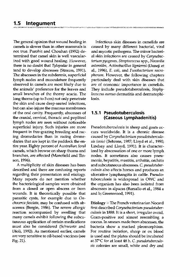
The general opinion that wound healing in camels is slower than in other mammals is not true. Purohit and Chouhan (1992) de- termined that camel skin is well vascular- ized with good wound healing. However, there is no doubt that Tylopodae in general tend to develop abscesses (Straws, 1991). The abscesses in the subdermis, superficial lymph nodes and musculature frequently observed in camels are most likely due to the animals' preference for the leaves and small branches of the thorny acacia. The long thorns (up to 5 cm) not only penetrate the skin and cause deep-seated infections, but can also injure the mucous membranes of the oral cavity. Frequently, abscesses of the cranial, cervical, thoracic and popliteal lymph nodes are seen without noticeable superficial injury. Such injuries are more frequent in free-grazing breeding and rac- ing dromedaries than in racing drome- daries that are kept in the paddock the en- tire year. Eighty percent of Australian feral camels, which browse on sharp thorns and branches, are affected (Manefield and Tin- son, 1996).
A multiplicity of skin diseases has been described and there are confusing reports regarding their presentation and etiology. Many reports do not mention whether the bacteriological samples were obtained from a closed or open abscess or from wounds. It is theoretically possible that parasitic cysts, for example due to On- chocerca fasciata, may be confused with ab- scesses (Bergin, 1986). The severe allergic reaction accompanied by swelling that many camels exhibit following the subcu- taneous application of certain medications must also be considered (Schwartz and Dioli, 1992). As mentioned earlier, camels are very sensitive to oil-based vaccines (see Fig. 21).
Infectious skin diseases in camelids are caused by many different bacterial, viral and mycotic pathogens. The minor bacteri- al skin infections are caused by Co ynebac- terium pyogenes, Streptococcus spp., Nocardia asteroides, ActinobaciZZus Zignieresi (Daneji et al., 1996), E. coli, and Fusobacterium necro- pherum. However, the following chapters particularly deal with skin diseases that are of economic importance in camelids. They include pseudotuberculosis, Staphy- Zococcus aureus dermatitis and dermatophi- losis.
1.5.1 Pseudotuberculosis (Caseous Lymphadenitis)
Pseudotuberculosis in sheep and goats oc- curs worldwide. It is a chronic disease caused by Co ynebacterium pseudotuberculo- sis (ovis) (Behrens, 1987; Lloyd et al., 1990; Lindsay and Lloyd, 1991). It is character- ized by abscessation of one or more lymph nodes. It sometimes also causes pneu- monia, hepatitis, mastitis, arthritis, orchitis and subcutaneous abscesses. C. pseudotuber- cuZosis also affects horses and produces an ulcerative lymphangitis in cattle. Pseudo- tuberculosis is widespread in OWC and the organism has also been isolated from abscesses in alpacas (Barsallo et al., 1984 a and b; Greenwood, 1991).
Etiology + The French veterinarian Nocard first described Co ynebacterium pseudotuber- culosis in 1888. It is a short, irregular ovoid, Gram-positive rod almost resembling a coccus. In smears made from abscesses, the bacteria show a marked pleomorphism. For routine isolation, sheep or ox blood is used and the plates should be incubated at 37°C for at least 48 h. C. pseudotuberculo- sis colonies are small, white and dry and
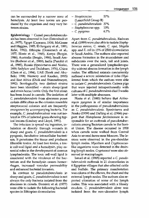
lnteaument 135
can be surrounded by a narrow zone of hemolysis. At least two toxins are pro- duced by the organism and may vary be- tween strains.
Epidemiology 1 I Camel pseudotuberculo- sis has been observed in Iran (Esterabadi et al., 1975), Egypt (Caprano, 1934; McGrane and Higgins, 1985; El-Sergany et al., 1991; Refai, 1992), Ethiopia (Domenech et al., 1977; Hoste et al., 1985), Kenya (Bergin, 1986), Australia (Bergin, 1986), Saudi Ara- bia (Radwan et al., 1989), India (Purohit et al., 1985), Russia (Spesivtseva and Nosko, 1959; Sadykov and Dadabaev, 1976), China (Chen et al., 1984), UAE (Tarek and Abu- Bakr, 1990; Wernery and Kaaden, 1995) and East Africa (Dioli and Stimmelmayer, 1992). Serologically, two distinct strains have been identified - strain sheep/goat and strain horse/cattle. Only the first strain has been found in camels. The isolation of C. pseudotuberculosis from abscesses poses certain difficulties as the colonies resemble streptococcal colonies and are frequently overgrown by accompanying bacteria. For example, C. pseudotuberculosis was not iso- lated in 15% of infected goats showing typ- ical lesions (Lindsay and Lloyd, 1991).
The infection is spread via ingestion, in- halation or directly through wounds in sheep and goats. C. pseudotuberculosis is a pyogenic, facultative intracellular bacteri- um. It penetrates the tissue and produces filterable toxins. At least two toxins, a tox- ic cell-wall lipid and a hemolysin, play es- sential roles in the development of caseous lymphadenitis. The toxic cell-wall lipid is associated with the virulence of the bac- terium and the hemolysin causes hemor- rhages, increased vascular permeability and enhanced bacterial invasion.
In contrast to pseudotuberculosis in sheep and goats, C. pseudotuberculosis is not always the only bacteria isolated from the abscesses in camels. Dominic et al. (1977) were able to isolate the following bacterial species in Ethiopian dromedaries:
- Streptococcus 57% (Lancefield Group B)
- C. pseudotuberculosis 37”/0 - stuphylococcus spp. 10% - C. pyogenes 6.7%
Apart from C. pseudotuberculosis, Radwan et al. (1989) were also able to isolate Stuphy- lococcus uureus, C. renule, C. equi, Shigella spp. and E. coli in 15% of 2500 dromedaries in Saudi Arabia. The authors also reported abscess formation in the musculature and subdermis over the neck, tail and joints. There was a generalized lymphadenopa- thy without abscess formation in the lymph nodes. The afflicted animals concurrently suffered a severe infestation of ticks (Hyu- lomma) from which the authors were able to isolate C. pseudotuberculosis. Guinea pigs that were injected intraperitoneally with cultures of C. pseudotuberculosis died 3 weeks later with multiple abscesses.
Hoste et al. (1985) believe that Actino- myces pyogenes is of similar importance in the pathogenesis of pseudotuberculosis as C. pseudotuberculosis. Spesivtseva and Nosko (1959) and Dalling et al. (1966) pur- port that Histoplusmu furciminosum is re- sponsible for an outbreak of pseudotuber- culosis among Bactrian camels in the Sovi- et Union. The disease occurred in 1958 when camels were walked from Central Asia to several farms near Moscow. The le- sions were observed in the pre-shoulder lymph nodes. Mycelium and Cryptococcus- like organisms were detected in the drain- ing lymph nodes. Cryptococci were also ob- served in macrophages.
Ismail et al. (1985) reported a C. pseudo- tuberculosis outbreak in 21 dromedaries in 6 Egyptian villages that also affected cattle and buffalo. The primary manifestation was edema of the elbows, the chest and the external lymph nodes. The authors also re- ported ulceration of some of the lymph nodes. This was associated with a bloody exudate. C. pseudotuberculosis alone was isolated from the non-ulcerative lymph
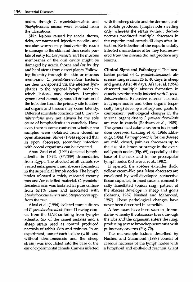
136 Bacterial Diseases
nodes, though C. pseudotuberculosis and Staphylococcus uureus were isolated from the ulcerations.
Skin lesions caused by acacia thorns, ticks, contaminated injection needles and nodular worms may inadvertently result in damage to the skin and thus create por- tals of entry for Corynebucteriu. The mucous membranes of the oral cavity might be damaged by acacia thorns and/or by dry and hard stems from desert plants. Follow- ing its entry through the skin or mucous membrane, C. pseudotuberculosis bacteria are then transported via the afferent lym- phatics to the regional lymph nodes in which lesions may develop. Lympho- genous and hematogenous distribution of the infection from the primary site to inter- nal organs and tissues may occur latently. Different scientists conclude that C. pseudo- tuberculosis may not always be the sole cause of lymphadenitis in camelids. How- ever, there is some confusion whether the samples were obtained from closed or open abscesses. Stowe (1984) reported that in open abscesses, secondary infection with coccal organisms can be expected.
Abou-Zaid et al. (1994) detected lymph- adenitis in 10.9% (37/339) dromedaries from Egypt. The affected adult camels re- vealed enlargement and abscess formation in the superficial lymph nodes. The lymph nodes released a thick, caseated creamy pus and/or calcified material. C. pseudotu- berculosis ovis was isolated in pure culture from 62.1% cases and associated with Staphylococcus uureus and Streptococcus spp. from the rest.
Afzal et al. (1996) isolated pure cultures of C. pseudotuberculosis from 11 racing cam- els from the UAE suffering from lymph- adenitis. Six of the camel isolates and a sheep strain used as control produced necrosis of rabbit skin and redness. In an experiment, one of each isolate (with and without dermonecrosis and the sheep strain) was inoculated into the base of the ear of experimental camels. Camels infected
with the sheep strain and the dermonecrot- ic isolate produced lymph node swelling only, whereas the strain without dermo- necrosis produced multiple abscesses in the experimental camels 40 days after in- fection. Re-infection of the experimentally infected dromedaries after they had recov- ered from the disease did not produce any lesions.
Clinical Signs and Pathology The incu- bation period of C. pseudotuberculosis ab- scesses ranges from 25 to 40 days in sheep and goats. After 40 days, Afzal et al. (1996) observed multiple abscess formation in camels experimentally infected with C. pseu- dotuberculosis. Extensive caseous necrosis in lymph nodes and other organs (espe- cially lung) develop in sheep and goats. In comparison, pathological changes in the internal organs due to C. pseudotuberculosis are rare in camels (Radwan et al., 1989). The generalized cutaneous form is also sel- dom observed (Dalling et al., 1966; Eldis- ougi, 1984). Pathognomonic for the disease are cold, closed, painless abscesses up to the size of a lemon or orange in the exter- nal lymph nodes (Fig. 69), especially at the base of the neck and in the prescapular lymph nodes (Schwartz et al., 1982).
If opened, the abscess extrudes thick, yellow cream-like pus. Most abscesses are enveloped by well-developed connective tissue capsules. In most cases a concentri- cally lamellated (onion ring) pattern of the abscess develops in sheep and goats (Behrens, 1987; Nashed and Mahmoud, 1987). These pathological changes have never been described in camelids.
A few cases have been seen in drome- daries whereby the abscesses break through the ribs and the organism enters the lung, producing severe bronchopneumonia with pulmonary caverns (Fig. 70).
The microscopic lesions described by Nashed and Mahmoud (1987) consist of caseous necroses of the lymph nodes with a lymphoid and epitheloid reaction. Giant
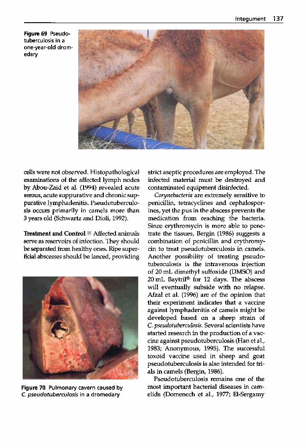
lnteaument 137
Figure 69 Pseudo- tuberculosis in a one-year-old drom- edary
cells were not observed. Histopathological examinations of the affected lymph nodes by Abou-Zaid et al. (1994) revealed acute serous, acute suppurative and chronic sup- purative lymphadenitis. Pseudotuberculo- sis occurs primarily in camels more than 3 years old (Schwartz and Dioli, 1992).
Treatment and Control @!I Affected animals serve as reservoirs of infection. They should be separated from healthy ones. Ripe super- ficial abscesses should be lanced, providing
Figure 70 Pulmonary cavern caused by C. pseudotuberculosis in a dromedary
strict aseptic procedures are employed. The infected material must be destroyed and contaminated equipment disinfected.
Corynebucteriu are extremely sensitive to penicillin, tetracyclines and cephalospor- ines, yet the pus in the abscess prevents the medication from reaching the bacteria. Since erythromycin is more able to pene- trate the tissues, Bergin (1986) suggests a combination of penicillin and erythromy- cin to treat pseudotuberculosis in camels. Another possibility of treating pseudo- tuberculosis is the intravenous injection of 20 mL dimethyl sulfoxide (DMSO) and 20mL Baytril@ for 12 days. The abscess will eventually subside with no relapse. Afzal et al. (1996) are of the opinion that their experiment indicates that a vaccine against lymphadenitis of camels might be developed based on a sheep strain of C. pseudotuberculosis. Several scientists have started research in the production of a vac- cine against pseudotuberculosis (Han et al., 1983; Anonymous, 1995). The successful toxoid vaccine used in sheep and goat pseudotuberculosis is also intended for tri- als in camels (Bergin, 1986).
Pseudotuberculosis remains one of the most important bacterial diseases in cam- elids (Domenech et al., 1977; El-Sergamy
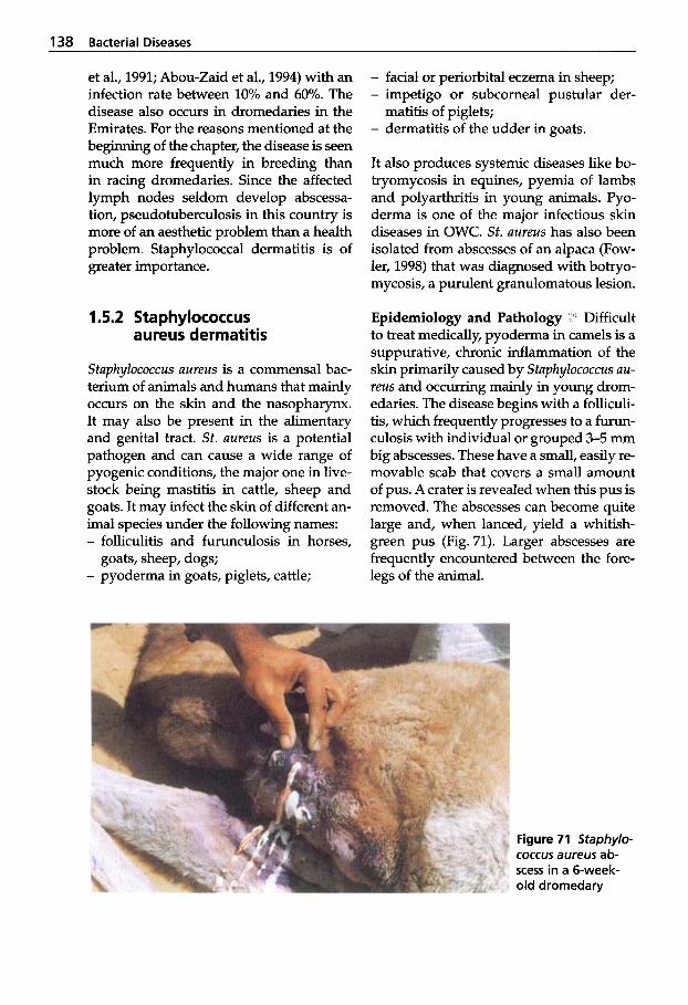
138 Bacterial Diseases
et al., 1991; Abou-Zaid et al., 1994) with an infection rate between 10% and 60%. The disease also occurs in dromedaries in the Emirates. For the reasons mentioned at the beginning of the chapter, the disease is seen much more frequently in breeding than in racing dromedaries. Since the affected lymph nodes seldom develop abscessa- tion, pseudotuberculosis in this country is more of an aesthetic problem than a health problem. Staphylococcal dermatitis is of greater importance.
1.5.2 Staphylococcus aureus dermatitis
Stuphylococcus uureus is a commensal bac- terium of animals and humans that mainly occurs on the skin and the nasopharynx. It may also be present in the alimentary and genital tract. St. uureus is a potential pathogen and can cause a wide range of pyogenic conditions, the major one in live- stock being mastitis in cattle, sheep and goats. It may infect the skin of different an- imal species under the following names: - folliculitis and furunculosis in horses,
- pyoderma in goats, piglets, cattle; goats, sheep, dogs;
- facial or periorbital eczema in sheep; - impetigo or subcorneal pustular der-
- dermatitis of the udder in goats. matitis of piglets;
It also produces systemic diseases like bo- tryomycosis in equines, pyemia of lambs and polyarthritis in young animals. Pyo- derma is one of the major infectious skin diseases in OWC. St. uureus has also been isolated from abscesses of an alpaca (Fow- ler, 1998) that was diagnosed with botryo- mycosis, a purulent granulomatous lesion.
Epidemiology and Pathology Difficult to treat medically, pyoderma in camels is a suppurative, chronic inflammation of the skin primarily caused by Staphylococcus uu- reus and occurring mainly in young drom- edaries. The disease begins with a folliculi- tis, which frequently progresses to a furun- culosis with individual or grouped 3-5 mm big abscesses. These have a small, easily re- movable scab that covers a small amount of pus. A crater is revealed when this pus is removed. The abscesses can become quite large and, when lanced, yield a whitish- green pus (Fig. 71). Larger abscesses are frequently encountered between the fore- legs of the animal.
Figure 71 Staphylo- coccus aureus ab- scess in a 6-week- old dromedary
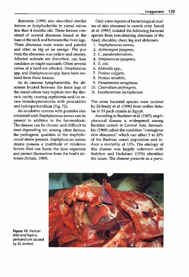
Integument 139
Bornstein (1995) also described similar lesions as lymphadenitis in camel calves less than 4 months old. These lesions con- sisted of several abscesses found at the base of the neck and between the front legs. These abscesses were warm and painful and often as big as an orange. The pus from the abscesses was yellow and creamy. Affected animals are disturbed, can lose condition or might succumb. Often several calves of a herd are affected. Streptococcus spp. and Staphylococcus spp. have been iso- lated from these lesions.
As in caseous lymphadenitis, the ab- scesses located between the front legs of the camel calves may rupture into the tho- racic cavity, causing septicemia and/or se- vere bronchopneumonia with pericarditis and hydropericardium (Fig. 72).
An exudative eczema with pustules also colonized with Staphylococcus aureus can be present in addition to the furunculosis. The disease can be chronic and difficult to treat depending on, among other factors, the pathogenic qualities of the staphylo- coccal strain present. Staphylococcus aureus strains possess a multitude of virulence factors that can harm the host organism and protect themselves from the host’s de- fenses (Schels, 1989).
Only a few reports of bacteriological stud- ies of skin abscesses in camels exist. Ismail et al. (1990) isolated the following bacterial species from non-draining abscesses of the head, shoulder, chest, leg and abdomen: 1. Staphylococcus aureus, 2. Actinomyces pyogenes, 3. C. pseudotuberculosis, 4. Streptococcus pyogenes, 5. E . coli, 6. Klebsiella spp., 7. Proteus vulgaris, 8. Proteus mirabilis, 9. Pseudomonas aeruginosa,
10. Clostridium perfringens, 11. Fusobacterium necrophorum.
The same bacterial species were isolated by El-Seedy et al. (1990) from wither fistu- lae in 93 pack camels in Egypt.
According to Buchnev et al. (1987), staph- ylococcal disease is widespread among Bactrian camels in Central Asia. Semush- kin (1968) called the condition “contagious skin abscesses” which can affect 5 to 20% of the Bactrian camel population and in- duce a mortality of 15%. The etiology of this disease was largely unknown until Sadykov and Dadabaev (1976) identified the cause. The disease presents as a puru-
Figure 72 Pericar- ditis and hydro- pericardium caused by St. aureus
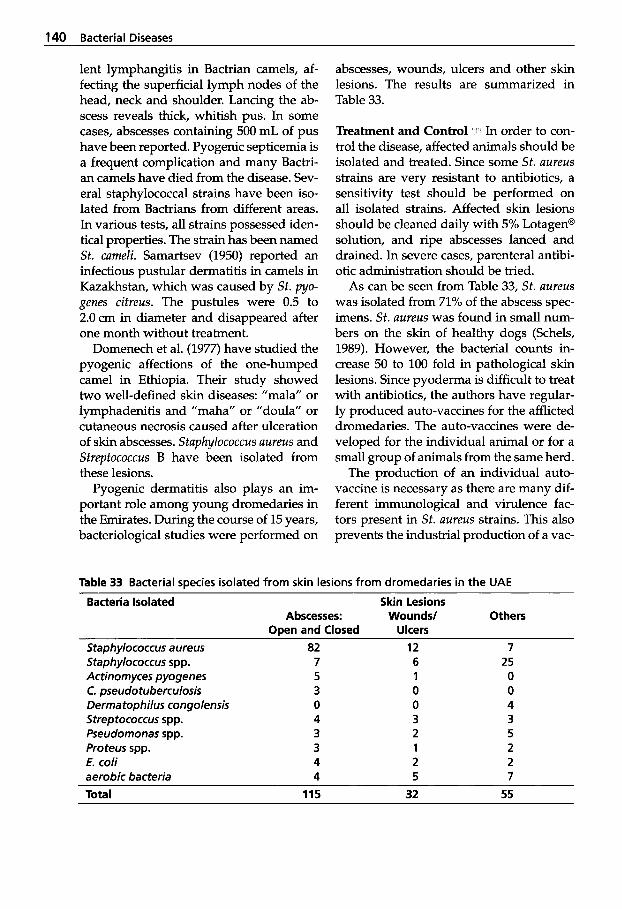
140 Bacterial Diseases
lent lymphangitis in Bactrian camels, af- fecting the superficial lymph nodes of the head, neck and shoulder. Lancing the ab- scess reveals thick, whitish pus. In some cases, abscesses containing 500 mL of pus have been reported. Pyogenic septicemia is a frequent complication and many Bactri- an camels have died from the disease. Sev- eral staphylococcal strains have been iso- lated from Bactrians from different areas. In various tests, all strains possessed iden- tical properties. The strain has been named St. cameli. Samartsev (1950) reported an infectious pustular dermatitis in camels in Kazakhstan, which was caused by Sf. pyo- genes citreus. The pustules were 0.5 to 2.0 cm in diameter and disappeared after one month without treatment.
Domenech et al. (1977) have studied the pyogenic affections of the one-humped camel in Ethiopia. Their study showed two well-defined skin diseases: "mala" or lymphadenitis and "maha" or "doula" or cutaneous necrosis caused after ulceration of skin abscesses. StaphyIococcus aureus and Streptococcus B have been isolated from these lesions.
Pyogenic dermatitis also plays an im- portant role among young dromedaries in the Emirates. During the course of 15 years, bacteriological studies were performed on
abscesses, wounds, ulcers and other skin lesions. The results are summarized in Table 33.
Treatment and Control In order to con- trol the disease, affected animals should be isolated and treated. Since some St. aureus strains are very resistant to antibiotics, a sensitivity test should be performed on all isolated strains. Affected skin lesions should be cleaned daily with 5% Lotagen@ solution, and ripe abscesses lanced and drained. In severe cases, parenteral antibi- otic administration should be tried.
As can be seen from Table 33, St. aureus was isolated from 71% of the abscess spec- imens. St. aureus was found in small num- bers on the skin of healthy dogs (Schels, 1989). However, the bacterial counts in- crease 50 to 100 fold in pathological skin lesions. Since pyoderma is difficult to treat with antibiotics, the authors have regular- ly produced auto-vaccines for the afflicted dromedaries. The auto-vaccines were de- veloped for the individual animal or for a small group of animals from the same herd.
The production of an individual auto- vaccine is necessary as there are many dif- ferent immunological and virulence fac- tors present in St. aureus strains. This also prevents the industrial production of a vac-
Table 33 Bacterial species isolated from skin lesions from dromedaries in the UAE
Bacteria Isolated Skin Lesions Abscesses: Wounds/ Others
Open and Closed Ulcers Staphylococcus aureus Staphylococcus spp. Actinornyces pyogenes C. pseudotuberculosis Derrnatophilus congolensis Streptococcus spp. Pseudornonas spp. Proteus spp. E. coli aerobic bacteria
82 7 5 3 0 4 3 3 4 4
12 6 1 0 0 3 2 1 2 5
7 25 0 0 4 3 5 2 2 7
Total 115 32 55
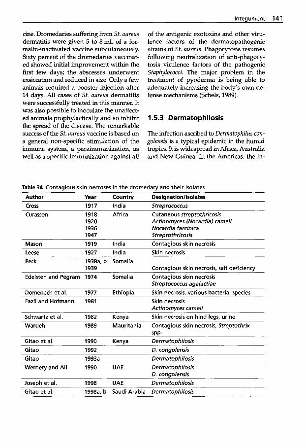
lnteaument 141
cine. Dromedaries suffering from St. aureus dermatitis were given 5 to 8 mL of a for- malin-inactivated vaccine subcutaneously. Sixty percent of the dromedaries vaccinat- ed showed initial improvement within the first few days; the abscesses underwent exsiccation and reduced in size. Only a few animals required a booster injection after 14 days. All cases of St. aureus dermatitis were successfully treated in this manner. It was also possible to inoculate the unaffect- ed animals prophylactically and so inhibit the spread of the disease. The remarkable success of the St. aureus vaccine is based on a general non-specific stimulation of the immune system, a paraimmunization, as well as a specific immunization against all
of the antigenic exotoxins and other viru- lence factors of the dermatopathogenic strains of St. aureus. Phagocytosis resumes following neutralization of anti-phagocy- tosis virulence factors of the pathogenic Staphylococci. The major problem in the treatment of pyoderma is being able to adequately increasing the body’s own de- fense mechanisms (Schels, 1989).
1.5.3 Dermatophilosis
The infection ascribed to Dermtophilus con- golensis is a typical epidemic in the humid tropics. It is widespread in Africa, Australia and New Guinea. In the Americas, the in-
Table 34 Contagious skin necroses in the dromedary and their isolates
Author Year Countw Desianationllsolates Cross 1917 India Strer, tococcus Curasson 1918 Africa Cutaneous streptothricosis
1920 Actinornyces (Nocardia) cameli 1936 Nocardia farcinica 1947 Strepto thricosis
Mason 1919 India Contaaious skin necrosis Leese 1927 India Skin necrosis Peck 1938a, b Somalia
Contagious skin necrosis, salt deficiency Edelsten and Pegram 1974 Somalia Contagious skin necrosis
Streptococcus asalactiae
1939
Domenech et al. 1977 Ethiopia Skin necrosis, various bacterial species Fazil and Hofmann 1981 Skin necrosis
Schwartz e t al. 1982 Kenva Skin necrosis on hind leas. urine Actinornyces carneli
Wardeh 1989 Mauritania Contagious skin necrosis, Streptothrix SPP.
Gitao et al. 1990 Kenya Derma tophilosis Gitao 1992 0. congolensis Gitao 1993a Derrna tophilosis Wernery and Ali 1990 UAE Derrna tophilosis
0. consolensis Joseph et al. 1998 UAE Derma top hilosis Gitao et al. 1998a, b Saudi Arabia Derrnatophilosis
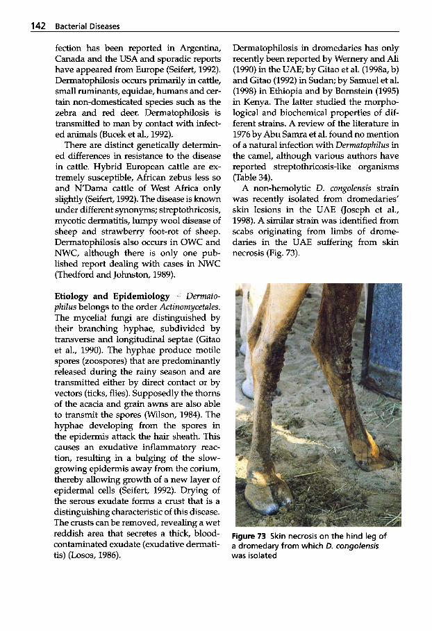
142 Bacterial Diseases
fection has been reported in Argentina, Canada and the USA and sporadic reports have appeared from Europe (Seifert, 1992). Dermatophilosis occurs primarily in cattle, small ruminants, equidae, humans and cer- tain non-domesticated species such as the zebra and red deer. Dermatophilosis is transmitted to man by contact with infect- ed animals (Bucek et al., 1992).
There are distinct genetically determin- ed differences in resistance to the disease in cattle. Hybrid European cattle are ex- tremely susceptible, African zebus less so and NDama cattle of West Africa only slightly (Seifert, 1992). The disease is known under different synonyms; streptothricosis, mycotic dermatitis, lumpy wool disease of sheep and strawberry foot-rot of sheep. Dermatophilosis also occurs in OWC and NWC, although there is only one pub- lished report dealing with cases in NWC (Thedford and Johnston, 1989).
Etiology and Epidemiology 1 Dermato- philus belongs to the order Actinomycetales. The mycelial fungi are distinguished by their branching hyphae, subdivided by transverse and longitudinal septae (Gitao et al., 1990). The hyphae produce motile spores (zoospores) that are predominantly released during the rainy season and are transmitted either by direct contact or by vectors (ticks, flies). Supposedly the thorns of the acacia and grain awns are also able to transmit the spores (Wilson, 1984). The hyphae developing from the spores in the epidermis attack the hair sheath. This causes an exudative inflammatory reac- tion, resulting in a bulging of the slow- growing epidermis away from the corium, thereby allowing growth of a new layer of epidermal cells (Seifert, 1992). Drying of the serous exudate forms a crust that is a distinguishing characteristic of this disease. The crusts can be removed, revealing a wet reddish area that secretes a thick, blood- contaminated exudate (exudative dermati- tis) (Losos, 1986).
Dermatophilosis in dromedaries has only recently been reported by Wernery and Ali (1990) in the UAE; by Gitao et al. (1998a, b) and Gitao (1992) in Sudan; by Samuel et al. (1998) in Ethiopia and by Bornstein (1995) in Kenya. The latter studied the morpho- logical and biochemical properties of dif- ferent strains. A review of the literature in 1976 by Abu Samra et al. found no mention of a natural infection with Dermatophilus in the camel, although various authors have reported streptothricosis-like organisms (Table 34).
A non-hemolytic D. congolensis strain was recently isolated from dromedaries' skin lesions in the UAE (Joseph et al., 1998). A similar strain was identified from scabs originating from limbs of drome- daries in the UAE suffering from skin necrosis (Fig. 73).
Figure 73 Skin necrosis on the hind leg of a dromedary from which D. congolensis was isolated
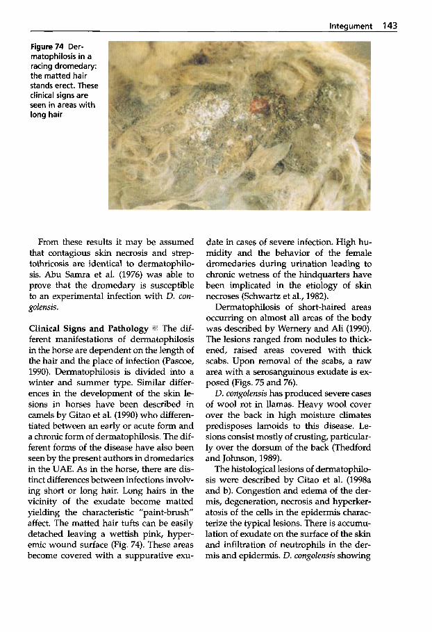
Integument 143
Figure 74 Der- rnatophilosis in a racing dromedary: the matted hair stands erect. These clinical signs are seen in areas with long hair
From these results it may be assumed that contagious skin necrosis and strep- tothricosis are identical to dermatophilo- sis. Abu Samra et al. (1976) was able to prove that the dromedary is susceptible to an experimental infection with D. con- golensis.
Clinical Signs and Pathology ill ferent manifestations of dermatophilosis in the horse are dependent on the length of the hair and the place of infection (Pascoe, 1990). Dermatophilosis is divided into a winter and summer type. Similar differ- ences in the development of the skin le- sions in horses have been described in camels by Gitao et al. (1990) who differen- tiated between an early or acute form and a chronic form of dermatophilosis. The dif- ferent forms of the disease have also been seen by the present authors in dromedaries in the UAE. As in the horse, there are dis- tinct differences between infections involv- ing short or long hair. Long hairs in the vicinity of the exudate become matted yielding the characteristic "paint-brush affect. The matted hair tufts can be easily detached leaving a wettish pink, hyper- emic wound surface (Fig. 74). These areas become covered with a suppurative exu-
date in cases of severe infection. High hu- midity and the behavior of the female dromedaries during urination leading to chronic wetness of the hindquarters have been implicated in the etiology of skin necroses (Schwartz et al., 1982).
Dermatophilosis of short-haired areas occurring on almost all areas of the body was described by Wernery and Ali (1990). The lesions ranged from nodules to thick- ened, raised areas covered with thick scabs. Upon removal of the scabs, a raw area with a serosanguinous exudate is ex- posed (Figs. 75 and 76).
D. congolensis has produced severe cases of wool rot in llamas. Heavy wool cover over the back in high moisture climates predisposes lamoids to this disease. Le- sions consist mostly of crusting, particular- ly over the dorsum of the back (Thedford and Johnson, 1989).
The histological lesions of dermatophilo- sis were described by Gitao et al. (1998a and b). Congestion and edema of the der- mis, degeneration, necrosis and hyperker- atosis of the cells in the epidermis charac- terize the typical lesions. There is accumu- lation of exudate on the surface of the skin and infiltration of neutrophils in the der- mis and epidermis. D. congolensis showing
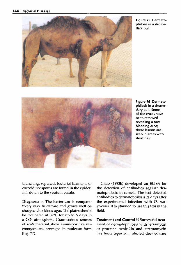
144 Bacterial Diseases
Figure 75 Dermato- philosis in a drome- dary bull
Figure 76 Dermato- philosis in a drome- dary bull. Some of the crusts have been removed revealing a raw bleeding area; these lesions are seen in areas with short hair
branching, septated, bacterial filaments or coccoid zoospores are found in the epider- mis down to the stratum basale.
Diagnosis . The bacterium is compara- tively easy to culture and grows well on sheep and ox blood agar. The plates should be incubated at 37°C for up to 5 days in a CO2 atmosphere. Gram-stained smears of scab material show Gram-positive mi- croorganisms arranged in rouleaux form (Fig. 77).
Gitao (1993b) developed an ELISA for the detection of antibodies against der- matophilosis in camels. The test detected antibodies to dermatophilosis 21 days after the experimental infection with D. con- golensis. It is planned to use this test in the field.
Treatment and Control 111 Successful treat- ment of dermatophilosis with terramycin or procaine penicillin and streptomycin has been reported. Infected dromedaries
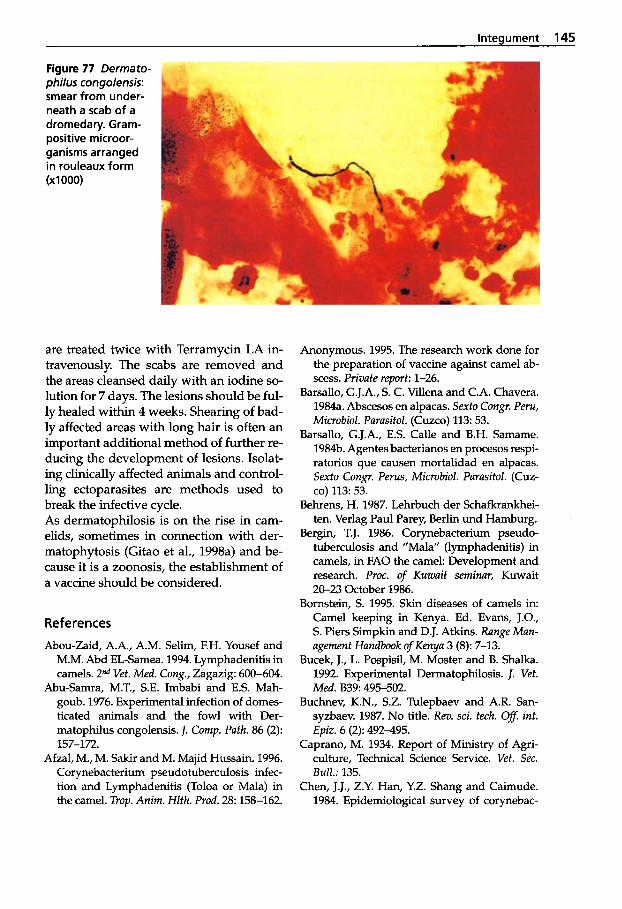
lnteaument 145
Figure 77 Dermato- philus congolensis: smear from under- neath a scab of a dromedary. Gram- positive rnicroor- ganisms arranged in rouleaux form (XI 000)
are treated twice with Terramycin LA in- travenously. The scabs are removed and the areas cleansed daily with an iodine so- lution for 7 days. The lesions should be ful- ly healed within 4 weeks. Shearing of bad- ly affected areas with long hair is often an important additional method of further re- ducing the development of lesions. Isolat- ing clinically affected animals and control- ling ectoparasites are methods used to break the infective cycle. As dermatophilosis is on the rise in cam- elids, sometimes in connection with der- matophytosis (Gitao et al., 1998a) and be- cause it is a zoonosis, the establishment of a vaccine should be considered.
References
Abou-Zaid, A.A., A.M. Selim, F.H. Yousef and M.M. Abd EL-Samea. 1994. Lymphadenitis in camels. 2nd Vet. Med. Cong., Zagazig: 600-604.
Abu-Samra, M.T., S.E. Imbabi and E.S. Mah- goub. 1976. Experimental infection of domes- ticated animals and the fowl with Der- matophilus congolensis. J. Comp. Path. 86 (2):
Afzal, M., M. Sakir and M. Majid Hussain. 1996. Corynebacterium pseudotuberculosis infec- tion and Lymphadenitis (Toloa or Mala) in the camel. Trop. Anim. Hlth. Prod. 28: 158-162.
157-172.
Anonymous. 1995. The research work done for the preparation of vaccine against camel ab- scess. Private report: 1-26.
Barsallo, G.J.A., S. C. Villena and C.A. Chavera. 1984a. Abscesos en alpacas. Sexto Congr. Peru, Microbiol. Purasitol. (Cuzco) 113: 53.
Barsallo, G.J.A., E.S. Calle and B.H. Samame. 1984b. Agentes bacterianos en procesos respi- ratorios que causen mortalidad en alpacas. Sexto Congr. Perus, Microbiol. Parasitol. (Cuz- co) 113: 53.
Behrens, H. 1987. Lehrbuch der Schafkrankhei- ten. Verlag Paul Parey, Berlin und Hamburg.
Bergin, T.J. 1986. Corynebacterium pseudo- tuberculosis and "Mala" (lymphadenitis) in camels, in FA0 the camel: Development and research. Proc. of Kuwait seminar, Kuwait 20-23 October 1986.
Bomstein, S. 1995. Skin diseases of camels in: Camel keeping in Kenya. Ed. Evans, J.O., S. Piers Simpkin and D.J. Atkins. Range Man- ugment Handbook of Kenya 3 (8): 7-13.
Bucek, J., L. Pospisil, M. Moster and B. Shalka. 1992. Experimental Dermatophilosis. J. Vet. Med. B39: 495-502.
Buchnev, K.N., S.Z. Tulepbaev and A.R. San- syzbaev. 1987. No title. Rev. sci. tech. Off. int. Epiz. 6 (2): 492495.
Caprano, M. 1934. Report of Ministry of Agri- culture, Technical Science Service. Vet. Sec. Bull.: 135.
Chen, J.J., Z.Y. Han, Y.Z. Shang and Caimude. 1984. Epidemiological survey of corynebac-
146 Bacterial Diseases
teriosis of Bactrian camel in Subei County, Gansu. Gansu J. Anim. Sci. Vet. Med. Suppl.:
Cross, H.E. 1917. The camel and its diseases. Balliere, Tindall and Cox, London.
Curasson, G. 1918. Une maladie du dromadaire analogue au farcin du beuf. Bull. SOC. Cent. Med. Vet. (Supplement to Rec. Med. vet. 94) 71:
Curasson, G. 1920. Hygiene et maladies du dro- madaire en Afrique occidentale franeaise Gor6e (SEN), Imprimerie du gouvemement g6n6ral.
Curasson, G. 1936. Trait6 de pathologie exo- tique v6t6rhaire et compare. Vigot Freres, Paris: LI.
Curasson, G. 1947. Le chameau et ses maladies. Vigot Freres, Editeurs: 86-88.
Dalling, T., A. Robertson, G. Boddie and J. Spru- ell. 1966. Diseases of camels. The Int. Ency- clopaedia of Veterina y Medicine, W. Green and Son, Edinburgh 1: 585.
Daneji, A.I., K.T.F. Djang and E.A. Ogunsan. 1996. Actinobacillus lignieresi infection in camels on the Sokoto plains, Nigeria. Trop. Anim. Hlth. Prod. 28: 315-316.
Dioli, M. and R. Stimmelmayr. 1992. Important camel diseases in the one-humped camel in Eastern Africa. A pictorial guide to diseases, health care and management. H.J. Schwartz and M. Dioli (Eds). Verlag Joseph Markgraf Scientific Books: pp. 155-164.
Domenech, J., T.G. Guidot and D. Richard. 1977. Les maladies pyogenes du dromadaire en Ethiopie. Symptomatologie - Etiologie. Rev. Elm. Mkd. v6t. Pays trop. 30 (3): 251-258.
Edelsten, R.M. and R.G. Pegram. 1974. Conta- gious skin necrosis of Somali camels associat- ed with Streptococcus agalactiae. Trop. Anim. Hlth. Prod. 6: 255-256.
El-Seedy, F.R., M. Ismail, Z. El-Sayed, M.E. Enany and M. Abdel-Ghani. 1990. Bacterial species implicated fistulous wither affecting one-humped camels in Egypt. J. Egypt Med.
El-Sergany, M.A., H. Soufy, M.M. Lotfi, M.A. Hassanain, A.M. Nasser, A. Laila and M.S. Shash. 1991. Lymphadenitis in Egyptian cam- els with special reference to bacteriological and parasitological affections. Egypt J. Comp. Path. Clinic Path. 4 (1): 25-45.
Eldisougi, I. 1984. Anote on the diseases of cam- els in Saudi Arabia. The Camelid; an "all pur-
51-54.
491-496.
ASS. 50: 81-92.
pose" animal. W.R. Cockrill. Scandinavian In- stitute of African Studies, Uppsala: 496-502.
Esterabadi, A.H., F. Entessar, H. Hedayati, A.A. Narimani and M. Sadri. 1975. Archives de l'Institut Razi 2 7 61-66.
Fazil, M.A. and R.R. Hofmann. 1981. Haltung und Krankheiten des Kamels. Tieriirztl. Praxis
Fowler, M.E. 1998. Medicine and surgery of South American Camelids. Iowa State Uni- versity Press, Ames.
Gitao, C.G., J.O. Evans and D.J. Atkins. 1990. Natural Dermatophilus congolensis infection in camels (Camelus dromedarius) from Kenya. J. comp. Path. 103: 307-312.
Gitao, C.G. 1992. Dermatophilosis in camels (Camelus dromedarius Linnaeus, 1758) in Kenya. Rev. sci. tech. Ofl int. Epiz. 11 (1-2):
Gitao, C.G. 1993a. The epidemiology and con- trol of camel dermatophilosis. Rev. Elev. Mkd. vkt. Pays. trop. 46 (1-2): 309-311.
Gitao, C.G. 1993b. An enzyme-linked immuno- sorbent assay for the epidemiological survey of Dermatophilus congolensis infection in camels (Camelus dromedarius). Rev. Sci. Tech.
Gitao, C.G., H. Agab and A.J. Khalafalla. 1998a. An outbreak of a mixed infection of Der- matophilus congolensis and Microsporum gypseum in camels (Camelus dromedarius) in Saudi Arabia. Rev. xi. tech. 08 int. Epiz. 17
Gitao, C.G., H. Agab and A.J. Khalafalla. 1998b. Outbreaks of Dermatophilus congolensis in- fection in camels (Camelus dromedarius) from the Butana region in Eastern Sudan. Rev. sci. tech. Ofl. int. Epiz. 17 (3): 743-748.
Greenwood, A.G. 1991. Control of pseudotuber- culosis in zoos. Vet. Rec. 128 (9): 215.
Han, Z.Y., J.G. Chen and Y.Z. Shang. 1983. Ex- periment on immunization against coryne- bacteriosis of the Bactrian camel. Acta Agri- culturae Universitatis, Gansu 2 47-58.
Hoste, C., B. P e p de Fabregues and D. Richard. 1985. Le dromadaire et son &levage. E l m Mkd. vkt. Pays trop.: 145-146.
Ismail, M., M. Enany, F.R. El-Seedy and M.T. Shouman. 1985. Oedematous skin disease of camel in El-Sharkia Governorate. Proc. 1st int. Conf Appl. Sci. Zagazig, Egypt.
Ismail, M., M. Ezzat, J. El-Jakee, Z.E. El-Sayed and M. Abd El-Rahmen. 1990. Microorgan-
9: 389-402.
309-311.
12 (2): 639-645.
(3): 749-755.
Integument 147
isms associated with closed abscesses of cam- els in Egypt. Vet. Med. J. Giza 38: 53-62.
Joseph, Sunitha, U. Wernery and M. Ali. 1998. Dermatophilosis caused by a nonhaemolybc Dermatophilus congolensis strain in drome- dary camels in the United Arab Emirates. J. Camel Prac. and Res. 5 (2): 247-248.
Leese, A.S. 1927. A treatise on the one-humped camel in health and disease. Vigot Freres, Paris II.
Lindsay, H.J. and S. Lloyd. 1991. Diagnosis of caseous lymphadenitis in goats. Vet. Rec. 128: 86.
Lloyd, S., H.S. Lindsay, J.D. Slater and P.G.G. Jackson. 1990. Caseous lymphadenitis in goats in England. Vet. Rec. 127 478.
Losos, G.J. 1986. Infectious tropical diseases of domestic animals. Avon, The Bath Press.
Manefield, G.W. and A. Tinson. 1996. Camels. A compendium. The T.G. Hungerford Vade Mecum Series for Domestic Animals: pp. 240, 298.
Mason, F.E. 1919. Pseudo-actinomycosis or St rep totrichosis in the camel. J. Comp. Path. Ther. 32
McGrane, J.J. and A.J. Higgins. 1985. Infectious diseases of the camel; viruses, bacteria and fungi. Br. Vet. J. 141: 529-547.
Nashed, S.M. and A.Z. Mahmoud. 1987. Micro- biological and histopathological diagnosis of rare cases of Corynebacterium infection in camels. Assiut. Vet. Med. J. 18: 82-86.
Pascoe, R.R. 1990. A colour atlas of equine der- matology. Wolfe Publishing Ltd.
Peck, E.F. 1938a. Notes relating to the camel. Vet. Rec. 33 (50): 1052-1054.
Peck, E.F. 1938b. The relationship of salt starva- tion to contagious necrosis and lameness in camels. The Vet. Rec. 14 (50): 409-410.
Peck, E.F. 1939. Salt intake in relation to cuta- neous necrosis and arthritis of one-humped camels (Camelus dromedarius, L.) in British Somaliland. Vet. Rec. 46 (51): 1355-1360.
Purohit, N.R., D.S. Chouhan and R.J. Choud- hary. 1985. Lymphangitis in the camel (two cases). Agr. Practice 6 (5): 23-24.
Purohit, N.R. and D.S. Chouhan. 1992. Wound healing in camels. Proc. 1 s t int. Camel Conf. Eds: W.R. Allen, A.J. Higgins, I.G. Mayhew, D.H. Snow and J.F. Wade: R. and W. Publica- tions, Newmarket, UK 365-370.
Radwan, A.I., S. El-Magawry, A. Hawari, S.I. Al- Bekairi and R.M. Rebleza. 1989. Corynebac-
(1): 34-42.
terium pseudotuberculosis infection in cam- els (Camelus dromedarius) in Saudi Arabia. Trop. Anim. Hlth. Prod. 21: 229-230.
Refai, M. 1992. Bacterial and mycotic diseases of camels in Egypt. Proc. lSt int. Camel Conf. Eds: W.R. Allen, A.J. Higgins, I.G. Mayhew, D.H. Snow and J.F. Wade: R. and W. Publica- tions, Newmarket, UK: 59-64.
Sadykov, R.G. and Zh. S. Dadabaev. 1976. On camels with pus lymphangitis (staphylococ- cosis) in Kazakh, SSR. Infectious and para- sitic diseases of farm animals. Alma Ata (SUN) s.n.: 73-78.
Samartsev, A.A. 1950. Infectious pustular der- matitis in camels. Proc. Kazakh Res. Vet. Insti- tute 5: 190-197.
Samuel, T., F. Tareke, G. Wirtu and T. Kiros. 1998. Bacteriological study of Ethiopian iso- lates of Dermatophilus congolensis. Trop. Anim. Hlth. Prod. 3 0 145-147.
Schels, H. 1989. Erfahrungen bei der Behand- lung der Pyodermie des Hundes mit Auto- vaccinen. Pro Veterinario 9 (1): 3.
Schwartz, H.J. and M. Dioli. 1992. The one- humped camel in Eastern Africa. A pictorial guide to diseases, health care and manage- ment. Verlag Josef Margraf.
Schwartz, Sabine, H.J. Schwartz and A.J. Wil- son. 1982. Eine fotographische Dokumenta- tion wichtiger Kamelkrankheiten in Kenia. Der prakt. Tierurzt 11: 985-989.
Seifert, H.S.H. 1992. Tropentierhygiene. Gustav Fischer Verlag Jena, Stuttgart.
Semushkin, N.R. 1968. Diagnosis of camel dis- eases. Sel'khozgiz Moscow.
Spesivtseva, N.A. and A.I. Noskov. 1959. Epi- zootic lymphangitis in camels. Trudy Vses. Inst. Vet. sanit. Ectoparasit. 14: 86.
Stowe, C.M. 1984. Antimicrobial drug interac- tions. JAVMA 185 (10): 1137-1141.
Strauss, G. 1991. Erkrankungen junger Neu- weltkamele im Tierpark Berlin-Friedrichs- felde 11. Arbeitstagung der Zootierarzte im deutschsprachigen Raum. Nov. 1-3 in Stuttgart, Tagungsbericht: 80-83.
Tarek, M. and Abu-Bakr. 1990. Bacteriological studies on dromedaries lymphadenitis in United Arab Emirates. Zag. Vet. J. 18 (1):
Thedford, R.R. and L.W. Johnson. 1989. Infec- tious diseases of New-world camelids (NWC). Vet. Clin. North Am. Food Anim. Pract. 5 (3):
77-90.
145-157.
148 Bacterial Diseases
Wardeh, M.F. 1989. Camel production in the Is- lamic Republic of Mauritania. Camel Newslet-
Wemery, U. and M. Ali. 1990. Dermatophilose in Renndromedaren Fallbericht. Tieriirztl. Umschau 45 (3): 209-210.
Wemery, U. and 0.-R. Kaaden. 1995. Infectious Diseases of Camelids. Blackwell Wissen- schafts-Verlag, Berlin.
Wilson, R.T. 1984. The camel. Longman, London and New York.
ter 5: 11-17.
Further reading Abubakr, M.I., M.N. Nayel and M.E. Fadlalla.
1999. Corynebacterium abscess in camels in Bahrain. J. Camel Prac. and Res. (1): 107-1009.
Shen, B.Y. 1986. Diagnosis of Corynaebacteri- um pseudotuberculosis infection in Bactrian camel with indirect hemagglutination test. Acta agriculturae Universitatis, Gansu 2: 53-58.
Shen, B.Y. and D.S. Huang. 1981. Comparison of biochemical reactions of some pseudotu- berculosis strain isolated from corynebacte- riotic camels in different districts of China. Xinjing Animal Science b Technology 4: 26- 30.
Wu, J.G. 1987. Pustules in camel. Gansu J Anim Sci b Vet Med. 3: 4-5.
Yagoub, S.O. and G.E. Mohamed. 1996. Inci- dence, clinical observation and etiology of contagious skin necrosis in camels (Camelus dromedarius) in the Sudan. J. Camel Prac. and Res. 3 (2): 95-98.
1.6.1 Infectious Mastitis
In the drought-stricken areas of the world where continuous severe drought deci- mates cattle, sheep and goat populations, only the camel survives and continues to produce milk. One of the most remarkable features of dehydrated camels is the ability to continue lactation and to secrete milk that is highly diluted with over 90% water content (Yagil and Etzion, 1980a,b). In true ruminants the reservoir for milk-water is lost for cooling and via fecal and urinary excretions. In cattle, sheep and goats, the lack of drinking leads to cessation of lacta- tion or to a very concentrated high fat and low water content milk after a short period. Camel can secrete 20 L of milk daily for at least 10 days without drinking water. Lac- tating camels will therefore guarantee am- ple food with the desired content for their offspring and humans alike. However, the let-down of milk must be stimulated by massage or calf suckling. It is of short du- ration and milking must be as fast as pos- sible. Nomads are aware of this fact and thus milking is carried out on both sides si- multaneously. It is estimated that good milk camels can produce 30 to 40 liters of milk daily, which can only be achieved by regular milkings (3 to 4 times daily), rapid milking (milker on each side), and reten- tion of the calves of the best milkers in the herd. The lactation period can last over 2 years.
In India, Pakistan and the Middle East, dromedaries are known to produce well over 20 liters daily with a lactation period lasting 8 to 18 months (Al-Sultan, 1996). Large concentrations of insulin and vita- min C have been found in camel milk. The milk is also unaffected by acid and will vir- tually pass untouched through the acid en- vironment of the human stomach to the in-
testines, where it is available for absorp- tion. These and many other features make the camel favorable over other ruminating domesticated animals. However, in many parts of the world the prejudice against the camel family still exists.
Inflammation of the udder occurs less frequently in the camelids than in other domesticated animals (Leese, 1927; Rama- dan et al., 1987; Fowler, 1998). This may ex- plain why there are so few publications re- garding mastitis in the camel. There might be several reasons why mastitis is more uncommon in camelids than in other do- mesticated animal species used for milk production. The mammary glands of both OWC and NWC possess four quarters and one teat per quarter. Each teat has two streak canals that enter into separate teat and gland cisterns. Each teat is associated with a non-communicating double gland. The streak canals are very narrow and a 1 mm tomcat catheter is required for pene- tration. This twin duct anatomy with its narrow streak canals might in some way protect against infection. Milking camels are often fitted with udder covers to re- strict suckling. These covers might reduce injuries to the teats and the udder and pro- tect against gross contamination. How- ever, the more likely explanation why ud- der infections in camelids are less frequent lies in the milk itself. Several scientists have found substances in camel milk that inhibit the growth of pathogenic bacteria (Kosparkov, 1975; Barbour et al., 1984; EL-Agamy et al., 1992; Farah, 1996; EL- Agamy, 1998; Kappeler, 1998). These in- hibitors are proteins and have been de- scribed as lysozyme, immunoglobulins, lactoferrin and lactoperoxidase, which are already well characterized. These proteins have been shown to have higher concen- trations or higher activity in camel milk
150 Bacterial Diseases
than in bovine milk. Kappeler (1998) found a novel minor whey protein, peptidogly- can recognition protein (PGRP), which has a beneficial influence on establishing fa- vorable gut microflora in the newborn and seems to especially inhibit the growth of Gram-positive bacteria.
Reports of inflammation of the camel udder have appeared from various countries, such as Egypt (Mostafa et al., 1987), India (Kapur et al., 1982), Saudi Ara- bia (Barbour et al., 1985; Hafez et al., 1987), Somalia (Arush et al., 1984; Abdurahman et al., 1991), Sudan (Obied, 1983) and the UAE (Quandil and Ouadar, 1984).
Peracute (Kapur et al., 1982), subacute (Quandil and Ouadar, 1984) and gan- grenous mastitis with lymph node enlarge- ment (Bolbol, 1982) have been described in the camel. In acute cases, the mammary secretions are watery, yellowish or blood- tinged (Tibary and Anouassi, 1997). Ra- madan et al. (1987) reported chronic uni- lateral mastitis in 3 dromedaries’ lactifer- ous ducts blocked by accumulations of keratin. This obstruction caused a reduc- tion in milk production, enlargement of the affected quarter and, in 2 cases, a sec- ondary bacterial infection with Pasteurella haernolytica and Staphylococcus aureus. Milk samples from the third dromedary were sterile.
Barbour et al. (1985) examined 205 milk samples from dromedaries in Saudi Arabia using the California mastitis test (CMT). They showed that in the majority of the dromedaries examined, an increase in so- matic cells in the milk samples occurred si- multaneously with a bacterial mastitis. As in cattle, a correlation between mastitis and the number of somatic cells in the drome- dary milk samples was confirmed. A simi- lar observation was made by Abdurah- man et al. (1992), Abdurahman et al. (1994) and Abdurahman et al. (1995), who exam- ined 391 udder quarters from Sudanese dromedaries. The 391 milk samples from
101 dromedaries from eastern Sudan were studied to evaluate the value of the CMT, the somatic cell count (SCC) and the adenosine triphosphate (ATP) tests for the detection of subclinical mastitis. It was found that the mean values of all three tests were generally higher for quarters in- fected with major pathogenic bacteria, al- though a significant number of quarter milk samples had elevated values from which no pathogenic bacteria were iso- lated, indicating that subclinical mastitis seems to occur more often than is realized. Bakhiet et al. (1992), who examined milk samples from 49 healthy dromedaries in Sudan, found bacteria in 45% (22/49). Staphylococcus spp. and Streptococcus spp. were the most frequently isolated udder microorganisms. Guliye (1996), who inves- tigated subclinical mastitis in dromedaries in the Negev desert, found that 81% of 86 milk samples from clinically healthy camels were positive for bacteria, with 40.7% revealing 2 or more bacterial spe- cies. Staphylococcus aureus, Micrococcus spp., Bacillus spp., Streptococcus dysagalactiae and E. coli were the most important organisms isolated. SCC ranged from 1.0 x O5 to 11.8 x 10⁶ cells/mL. Quarter milk samples with bacterial isolates had significantly higher mean SCC values. Quarter samples from which St. aureus was isolated showed the highest mean values. Similar results of subclinical mastitis in Bactrian camels were reported from Abdurahman (1996). Of 160 milk samples originating from 7 clinically healthy Bactrians, 22.5% were found to be positive for bacteria. St. aureus and coagulase-negative Staphylococci were the main organisms found. Quarters from which cocci were isolated had significantly higher SCC and CMT values. Both the SCC and the CMT are of value in predicting the infection status of the camel udder. Mody et al. (1998) investigated the preva- lence of mastitis in 146 adult Indian drom- edaries using the CMT and cultivation. Thirty subclinical cases of mastitis were
Udder 151
found, of which 28 possessed bacterial pathogens including Staphylococcus spp., Streptococcus spp. and Co ynebacterium. The authors also tested the pathogens for their antimicrobial susceptibility and found gentamycin and chloramphenicol highly effective.
Several scientists have studied the prop- erties and products of camel milk (Whabi et al., 1987; Hashi, 1989; Farah and Ruegg, 1991; Farah, 1996).
There are divergent opinions as to which bacteria are potentially the primary causal organisms of infectious mastitis in the camel. Barbour et al. (1985) views Micro- coccus as an important causative agent of mastitis whereas Obied (1983) did not con- sider this bacterium pathologically rele- vant. Obied et al. (1996) found Streptococ- cus, Staphylococcus, Micrococcus, Aerobacter and E . coli to be the main bacterial species causing mastitis. The authors did not find any correlation between the SCC and an udder infection. Al-Ani and Al-Shareefi (1997) found that 38% of lactating camels from three different herds in Iraq suffered from mastitis. St. aureus and Coynebac- terium pyogenes were the main causes of chronic mastitis, whereas St. epidermidis, Streptococcus spp., Pasteurella huemolytica, E. coli and Micrococcus spp. were responsi- ble for subclinical mastitis. Very little is known about fungal mastitis. Al-Ani and Al-Shareefi (1997) failed to isolate any fun- gi from 50 milk samples, but Quandil and Quadar (1984) cultured Candida albicans from milk originating from subclinical mas- titis samples.
From the multitude of bacteria isolated from milk samples taken from camels with mastitis, Staphylococcus aureus, Pasteurella haemolytica and Streptococci were found most frequently. Numerous authors be- lieve them to be primary causative organ- isms in the pathogenesis of mastitis in the camel (Barbour et al., 1985; Ramadan et al., 1987; Hafez et al., 1987). The following bac- teria were considered secondary agents:
- Micrococcus spp., - Actinomyces spp., - E. coli, - Pseudomonas aeruginosa, - Klebsiella pneumoniae, - Bacteroides spp., - C. perfringens.
These bacteria were isolated as both pure and mixed cultures from milk samples of camels with mastitis (Kapur et al., 1982; Hassanein et al., 1984; Quandil and Oua- dar, 1984; Barbour et al., 1985; Mostafa et al., 1987). It is also of great significance that BruceZZa organisms have been isolated from fresh camel milk (Radwan et al., 1992) and cases of human brucellosis have been attributed to the consumption of raw cam- el milk (Mousa et al., 1988) (see also chap- ter Brucellosis). In NWC, no specific bacte- ria that cause mastitis have been reported. Several bacteria species were isolated from peracute NWC mastitis including E. coli, Klebsiella pneumoniae, Aerobacter enterobac- terium. So far no mycoplasmas have been isolated from NWC (Fowler, 1998) and OWC udders.
Pathology s r l Very little is known about the pathological alterations occurring during infection of the mammary gland. The af- fected udders are often swollen, hard, reddened and painful to the animal on pal- pation. In chronic mastitis, necrosis and abscessation might be observed with dis- charge of greenish pus. Al-Ani and Al-Sha- reefi (1997) described some of the lesions observed histologically in mastitis.
Treatment 611 When mastitis occurs, prompt attention is necessary to avoid severe dam- age to the mammary gland or even loss of the animal. Mastitis treatment should be based on culture and sensitivity and the treating person must be fully aware of the anatomical particularities of the camelid’s mammary gland. The streak canals can be easily traumatized when using bovine an-
152 Bacterial Diseases
tibiotic mastitis ointments. The nozzles of the bovine infusion tubes are too big. Streak canals should only be penetrated with a 1 mm tomcat catheter to avoid any injuries. Before the infusion is carried out, the teats should be cleaned and disinfected with an alcohol wipe. Before infusion, the udder or the infected quarters should be emptied. In severe cases, the exudate should be removed from the gland three to five times daily by gently massaging the udder. This is sometimes difficult to do when there is a lot of pain. Camelids should be restrained and then rolled on their sides with the hind legs roped back. Commercial mastitis infusions are Ampiclox@, Orbenin LA@ and Mastalone@, which should be in- fused according to the manufacturers' rec- ommendations. It is also important to fol- low the withholding time of milk after treatment. Peracute and sometimes acute mastitis require parenteral treatment with antibiotics along with local infusion.
References
Abdurahman, O.A. Sh., S. Bornstein, Kh. Sh. Osman, A.M. Abdi and G. Zakrisson. 1991. Prevalence of mastitis among camels in Southern Somalia: a Pilot Study. Somali Acad. Arts and Science, Mogadishu, Camel fo- rum, Working Paper 3 7 1-9.
Abdurahman, O.A.Sh., H. Aqab and B. Abbas. 1994. Mastitis in camels (Camelus dromedar- ius) in the Sudan. Relationship between ud- der infection and inflammatory parameters in Sudanese camels (Camelus dromedarius). Br. vet. J. 4 71-76.
Abdurahman, O.A.S., H. Agab, B. Abbas and G. Astroem. 1995. Relations between udder in- fection and somatic cells in camel (Camelus dromedarius) milk. Acta Vet. Scand. 36 (4):
Abdurahman, O.A.S. 1996. The detection of subclinical mastitis in the Bactrian camel (Camelus bactrianus) by somatic cell count and California mastitis test. Vet. Res. Comrn.
Abdurahman, O.S., R. Cooray and S. Bornstein. 1992. The ultrastructure of cells and cell frag-
423-431.
20 (1): 9-14.
ments in mammary secretions of Camelus bactrianus. J. Vet. Med. A 39: 648-655.
Al-Ani, F.K. and M.R. Al-Shareefi. 1997. Studies on mastitis in lactating one humped camels (Camelus dromedarius) in Iraq. J. Camel Pruc. and Res. 4 (1): 47-49.
Al-Sultan, S. 1996. Veterinary care of camels in Saudi Arabia: Mastitis in camels. Prod. 3'6 meeting of British Vet. camelid society, 14-16 November 1996, UK 48-50.
Arush, M.A., C. Valente, M. Compagnucci and H. Hussein. 1984. Indagine sulla diffusione delle mastite del dromedario (Camelus drom- edarius) in Somalia. Bull. scient. Fac. Zoot. Vet.
Bakhiet, M.R., H. Agab and LE. Mamoun. 1992. Camel mastitis in Western Sudan. Short com- munication. Sud. J. Vet. Sc. Anim. Husb. 31 (1): 58-59
Barbour, E.K., N.H. Nabbut, W.M. Frerichs and H.M. Al-Nakhli. 1984. Inhibition of patho- genic bacteria by camel milk Relation to whey lysozyme and stage of lactation. J. of Food Protection 47 (11): 838-840.
Barbour, E.K., N.H. Nabbut, W.M. Frerichs, H.M. Al Nakhli and A.A. A1 Mukayel. 1985. Mastitis in Camelus dromedarius in Saudi Arabia. Trop. Anim. Hlth. Prod. 17 (3):
Bolbol, A. 1982. Mastectomy in she-camel. Assi- ut. vet. med. J. 10: 215.
El-Agamy, E.I. 1998. Camel's colostrum. Anti- microbial factors. Dromadaires et chameaux, animaux laitiers. Ed.: Bonnet, P. Actes du col- loque, 24-26 Octobre 1994, Noaukchott, Mauritanie: 177-1 79.
El-Agamy, E.I., R. Ruppanner, A. Ismail, C.P. Champagne and R. Assaf. 1992. Antibacterial and antiviral activity of camel milk protec- tive proteins. J. of Dairy Res. 59: 169-175.
Farah, Z. 1996. Camel milk. Properties and products. Laboratory of Dairy Scient, Instit. of Food Science, Swiss Federal Inst. of Tech., ETH-Zenhwn, LFO, CH-8092, Zurich.
Farah, Z. and M. Ruegg. 1991. The creaming properties and size distribution of fat glob- ules in camel milk. J. Dairy Sci. 7 4 2901-2904.
Fowler, M.E. 1998. Medicine and surgery of South American Camelids. Iowa State Uni- versity Press, Ames.
Guliye, A.Y. 1996. Studies on the compositional and hygienic quality of the milk of Bedouin camels (Camelus dromedarius) of the Negev
4: 99-104.
173-179.
Udder 153
desert. Thesis, University of Aberdeen, Scot- land.
Hafez, A.M., S.A. Fazig, S. El-Amrousi and R.O. Ramadan. 1987. Studies on mastitis in farm animals in Al-Hasa. ISt analytical studies. As- siut Vet. J. 19: 140-145.
Hashi, A.M. 1989. Preliminary observation on camel milk production and composition. Working paper. 30.
Hassanein, A., A.S. Soliman and M. Ismail. 1984. A clinical case of mastitis in she-camel (Cam- elus dromedarius) caused by Corynebac- terium pyogenes. Assiut vet. med. J. 12 (23):
Kappeler, S. 1998. Compositional and structural analysis of camel milk proteins with empha- sis on protective proteins. Diss. ETH No. 12947, Zurich, Switzerland.
Kapur, M.P., B.M. Khanna and R.P. Singh. 1982. A peracute case of mastitis in a she-camel associated with Klebsiella pneumoniae and Escherichia coli. lnd. Vet. J. 59 (8): 650-651.
Kosparkov, Zh. K. 1975. Antibacterial properties of camel’s milk. lnst. Veterinarnoi Sanitarri 51:
Leese, A.S. 1927. A treatise on the one-humped camel in health and disease. Vigot Freres, Paris 11.
Mody, S.K., l?R. Pate1 and C.B. Prajapati. 1998. A study on antimicrobial susceptibility of bacteria isolated from mastitic milk of rural camels in India. Proc. lnt. Meeting on camel prod. andfuture perspectives, A1 Ain, UAE, May
Mostafa, A.S., A.M. Ragab, E.E. Safwat, Z.A. El- Sayed, M. Abd-El-Rahman, N.A. El-Danaf and M.T. Shouman. 1987. Examination of raw she-camel milk for detection of subclinical mas- titis. J. Egypt Vet. med. Ass. 47 (1 & 2): 117-128.
Mousa, A.M., K.M. Elhag, M. Khogali and A.A. Marafic. 1988. The nature of human brucel- losis in Kuwait: Study of 379 cases. Rev. Infect. Dis. 10: 211-217.
Obied, A.I. 1983. Field investigation, clinical and laboratory findings of camel mastitis. M.V.Sc. Thesis, University of Khartoum.
Obied, A.I., H.O. Bagadi and M.M. Mukhtar. 1996. Mastitis in Camelus dromedarius and the somatic cell content of camels’ milk. Res. Vet. Sci. 61 (1): 55-58.
Quandil, S.S. and J. Ouadar. 1984. Etude bac- Mriologique de quelques cas de mammites chez la camelle (Camelus dromedarius) dans
239-241.
37-40.
2-3,1998.
les Emirats Arabes Unis. Rev. Elev. Mid. vit. Pays trop. 135 (11): 705-707.
Radwan, A.I., S.J. Bekairi and P.V.S. Prasad. 1992. Serological and bacteriological study of brucellosis in camels in central Saudi Arabia. Rev. sci. tech. Of. int. Epiz. 11 (3): 837-844.
Ramadan, R.O., A.M. El- Hassan, R. El-Abdin Bey, Y.A. Algasnawi, E.S.M. Abdalla and A.A. Fayed. 1987. Chronic obstructive mastitis in the camel, a clinical pathological study. Cor- nell Vet. 77 (2): 132-150.
Tibary, A. and A. Anouassi. 1997. Theriogenology in camelidae. Anatomy, Physiology, Patholo- gy and Artificial Breeding. Abu Dhabi Print- ing and Publishing Co., Mina, Abu Dhabi, UAE.
Whabi, A.A., S.E. Gadir, A. Awadelsied and O.F. Idris. 1987. Biochemical studies on Sudanese camel milk collected from Butana Area. Sud. J. Vet. Med. B 34: 340-342.
Yagd, R. and Z. Etzion. 1980a. Milk yields of camels (C. dromedarius) in drought areas. Comp. Biochem. Physiol. 67A: 207-209.
Yagil, R. and Z. Etzion. 1980b. The effect of drought conditions on the quality of camels’ milk. J. Dairy Res. 47 159-166.
Further reading
Almaw, G. and B. Molla. 2000. Prevalence and etiology of mastitis in camels (Camelus dromedarius) in eastern Ethiopia. J. Camel Prac. and Res. 7 (1): 97-100.
Chen, J.J., Z.Y. Han, Y.Z. Shang and Caimude. 1984. Epidemiological survey of corynebacte- riosis of Bactrian camel in Subei County, Gansu. Gansu J. Anim. Sci. and Vet. Med. Supp.:
Esterabadi, A.H., F. Entessar, H. Hedayati, A.A. Narimani and M. Sadri. 1975. Isolation of Corynebacterium pseudotuberculosis from camel in Iran. Arch. lnst. Razi. 2 7 61-66.
Han, Z.Y., J.G. Chen and Y.Z. Shang. 1983. Ex- periment on immunisation against Coryne- bacterium pseudotuberculosis of the Bactrian camel. Acta Agric. Univers. Gansu. 2: 47-58.
Mal, G., Suchitra Sena, D., V.K. Jain and M.S. Sa- hani. 1999. Utility of raw camel milk as nutri- tional supplement among chronic pumonary tuberculosis patients. Int. Workshop on the young camel, Quarzazate, Morocco, 24-26 Oct., 93.
Restani, l?, A. Gaiascha, A. Plebani, 8. Beretta, G. Cavagni, A. Fiocchi, C. Poiesi, T. Velona,
51-54.

154 Bacterial Diseases
A.G. Ugazio and C.L. Galli. 1999. Cross-reac- tivity between milk proteins from different animal species. Clinical and Experimental Al- lergy 29 (7): 997-1004.
Saad, N.M. and A.EI-R. Thabet. 1993. Bacterio- logical quality of camel's milk with special reference to mastitis. Assiut Vet. Med. J. 2 8
Shen, B.Y. and D.S. Huang. 1981. Comparison of biochemical reactions of some pseudotuber-
194-198.
culosis strains isolated from corynebacteriot- ic camels in the different districts of China. Xinjiang Anim. Sci. and Techn. 4: 26-30.
Wu, J.G. 1987. Pustules in camels. Gansu J. Anim. Sci. and Med. 3 4-5.
Younan, M. and J.W. Matofari. 1999. Streptococ- cus agalactiae infection in Kenyan camels (Camelus dromedarius). Int. Workshop on the young camel, Quarzazate, Morocco, 24-26 Oct., 73.
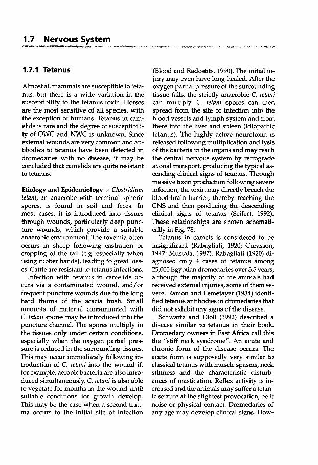
1.7.1 Tetanus
Almost all mammals are susceptible to teta- nus, but there is a wide variation in the susceptibility to the tetanus toxin. Horses are the most sensitive of all species, with the exception of humans. Tetanus in cam- elids is rare and the degree of susceptibili- ty of OWC and NWC is unknown. Since external wounds are very common and an- tibodies to tetanus have been detected in dromedaries with no disease, it may be concluded that camelids are quite resistant to tetanus.
Etiology and Epidemiology %I Clostridiurn tetani, an anaerobe with terminal spheric spores, is found in soil and feces. In most cases, it is introduced into tissues through wounds, particularly deep punc- ture wounds, which provide a suitable anaerobic environment. The toxemia often occurs in sheep following castration or cropping of the tail (e.g. especially when using rubber bands), leading to great loss- es. Cattle are resistant to tetanus infections.
Infection with tetanus in camelids oc- curs via a contaminated wound, and/or frequent puncture wounds due to the long hard thorns of the acacia bush. Small amounts of material contaminated with C. tetani spores may be introduced into the puncture channel. The spores multiply in the tissues only under certain conditions, especially when the oxygen partial pres- sure is reduced in the surrounding tissues. This may occur immediately following in- troduction of C. tetani into the wound if, for example, aerobic bacteria are also intro- duced simultaneously. C. tetani is also able to vegetate for months in the wound until suitable conditions for growth develop. This may be the case when a second trau- ma occurs to the initial site of infection
(Blood and Radostits, 1990). The initial in- jury may even have long healed. After the oxygen partial pressure of the surrounding tissue falls, the strictly anaerobic C. tetani can multiply. C. tetani spores can then spread from the site of infection into the blood vessels and lymph system and from there into the liver and spleen (idiopathic tetanus). The highly active neurotoxin is released following multiplication and lysis of the bacteria in the organs and may reach the central nervous system by retrograde axonal transport, producing the typical as- cending clinical signs of tetanus. Through massive toxin production following severe infection, the toxin may directly breach the blood-brain barrier, thereby reaching the CNS and then producing the descending clinical signs of tetanus (Seifert, 1992). These relationships are shown schemati- cally in Fig. 78.
Tetanus in camels is considered to be insigruficant (Rabagliati, 1920; Curasson, 1947; Mustafa, 1987). Rabagliati (1920) di- agnosed only 4 cases of tetanus among 25,000 Egyptian dromedaries over 3.5 years, although the majority of the animals had received external injuries, some of them se- vere. Ramon and Lemetayer (1934) identi- fied tetanus antibodies in dromedaries that did not exhibit any signs of the disease.
Schwartz and Dioli (1992) described a disease similar to tetanus in their book. Dromedary owners in East Africa call this the "stiff neck syndrome". An acute and chronic form of the disease occurs. The acute form is supposedly very similar to classical tetanus with muscle spasms, neck stiffness and the characteristic disturb- ances of mastication. Reflex activity is in- creased and the animals may suffer a tetan- ic seizure at the slightest provocation, be it noise or physical contact. Dromedaries of any age may develop clinical signs. How-
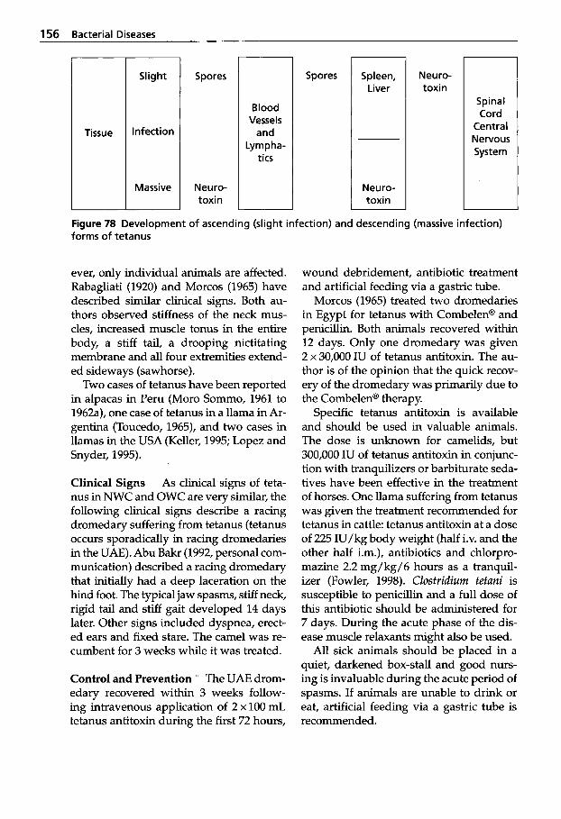
156 Bacterial Diseases
Tissue
Slight
Infection
Massive
Spores
Neuro- toxin
Blood Vesse I s
Lympha- tics
Spores Spleen, Liver
Neuro- toxin
Neuro- toxin
Spinal Cord
Central Nervous System
Figure 78 Development of ascending (slight infection) and descending (massive infection) forms of tetanus
ever, only individual animals are affected. Rabagliati (1920) and Morcos (1965) have described similar clinical signs. Both au- thors observed stiffness of the neck mus- cles, increased muscle tonus in the entire body, a stiff tail, a drooping nictitating membrane and all four extremities extend- ed sideways (sawhorse).
Two cases of tetanus have been reported in alpacas in Peru (Moro Sommo, 1961 to 1962a), one case of tetanus in a llama in Ar- gentina (Toucedo, 1965), and two cases in llamas in the USA (Keller, 1995; Lopez and Snyder, 1995).
Clinical Signs As clinical signs of teta- nus in NWC and OWC are very similar, the following clinical signs describe a racing dromedary suffering from tetanus (tetanus occurs sporadically in racing dromedaries in the UAE). Abu Bakr (1992, personal com- munication) described a racing dromedary that initially had a deep laceration on the hind foot. The typical jaw spasms, stiff neck, rigid tail and stiff gait developed 14 days later. Other signs included dyspnea, erect- ed ears and fixed stare. The camel was re- cumbent for 3 weeks while it was treated.
Control and Prevention The UAE drom- edary recovered within 3 weeks follow- ing intravenous application of 2 x 100 mL tetanus antitoxin during the first 72 hours,
wound debridement, antibiotic treatment and artificial feeding via a gastric tube.
Morcos (1965) treated two dromedaries in Egypt for tetanus with Combelen@ and penicillin. Both animals recovered within 12 days. Only one dromedary was given 2 x 30,000 IU of tetanus antitoxin. The au- thor is of the opinion that the quick recov- ery of the dromedary was primarily due to the Combelen@ therapy.
Specific tetanus antitoxin is available and should be used in valuable animals. The dose is unknown for camelids, but 300,000 IU of tetanus antitoxin in conjunc- tion with tranquilizers or barbiturate seda- tives have been effective in the treatment of horses. One llama suffering from tetanus was given the treatment recommended for tetanus in cattle: tetanus antitoxin at a dose of 225 IU/kg body weight (half i.v. and the other half i.m.), antibiotics and chlorpro- mazine 2.2 mg/kg/6 hours as a tranquil- izer (Fowler, 1998). Closfridium tetuni is susceptible to penicillin and a full dose of this antibiotic should be administered for 7 days. During the acute phase of the dis- ease muscle relaxants might also be used.
All sick animals should be placed in a quiet, darkened box-stall and good nurs- ing is invaluable during the acute period of spasms. If animals are unable to drink or eat, artificial feeding via a gastric tube is recommended.
Nervous System 157
Tetanus toxoid vaccines are readily avail- able and should be administered before any surgery. Llamas respond to toxoid vac- cination with a rise in titer.
1.7.2 Listeriosis
Listeria bacteria are widely distributed in the environment and can be isolated from soil, plants, decaying vegetation and silage with pH of over 5.5. In silage, Listeriu can multiply and it is commonly implicated in outbreaks of listeriosis in cattle and sheep. Listeria monocytogenes can also infect hu- mans via food including soft cheeses, milk and poultry meat and coleslaw. Listeriosis has been reported from NWC but not from OWC.
fi L. monocytogenes is a medium seized Gram-positive rod, non-spore-form- ing, measuring about 0.4 to 0.5 pm in di- ameter. Five serotypes and a number of subtypes have been identified. The bacte- ria are readily cultured on ordinary media and some strains grow only at 4°C.
Epidemiology Listeriosis has a world- wide distribution. It is, however, more common in regions with cold tempera- tures and it is often associated with the feeding of poor-quality silage with a pH higher than 5.5. Silage is not fed to OWC and this might be the reason why OWC do not contract listeriosis. The disease occurs most frequently in sheep, goats and cattle. It causes sporadic outbreaks in NWC (Fowler, 1998). The mortality rate can reach 100%. Only individual animals are com- monly affected. Haenichen and Wiesner (1995) described two cases of septic liste- riosis in 10 day-old alpacas after feeding poor-quality corn silage.
Clinical Signs and Pathology Listeriosis causes a meningoencephalitis in NWC with circling, trembling of the head, running into
objects and fever. Some cases develop uni- lateral facial nerve paralysis in association with drooping lips, ears, eyelids and paral- ysis of the jaw and pharynx, which inter- feres with mastication and swallowing. The course of the disease is usually 3 to 5 days (Moro Sommo, 1961 to 1962b; Tapia Cano, 1965; Mayer and Gehring, 1975; Butt et al., 1991). A listeriosis outbreak in a German zoo occurred in 1975 during which six lla- mas died. Three animals had developed en- cephalitis from which L. monocytogenes was isolated from the brain. The other three lla- mas died from septic listeriosis from which the organism was isolated from different organs. From this outbreak different sero- vars were cultured, three of them only through the cold incubation. It was men- tioned that heavy rains had flooded the zoo and there was an acute outbreak also in oth- er ungulates. An emergency vaccination with a live Listeriu vaccine was adminis- tered which stopped the further spread of the epizootic (Mayer and Gehring, 1975). Two adult llamas contracted encephalitic listeriosis in New York with abortion, atax- ia, depression, and facial paralysis followed by death (Butt et al., 1991). L. monocytogenes was isolated from one of the llamas, but both were positive in the immunofluores- cent test. Hamir and Moser (1998) de- scribed lesions found in a 2-year-old female llama at post mortem. They were confined to the brain and the spinal cord. The surface of the leptomeninges was rough, dark red and thickened with a thin layer of yellow- ish exudate. Not only meningoencephalitis is caused by L. monocytogenes, but also sep- ticemia with polyarthritis (alpaca) (Wisser, 1989), otitis media and intema with suppu- rative meningoencephalomyelitis (llama) (Van Metre et al., 1991), abortion (llama) (Mc Laughlin et al. 1993) and septicemia with thrombocytopenia and hepatopathy (llama) (Semrad, 1994).
Microscopic changes are confined to the white and/or gray matter of the brain stem, particularly the pons and the medulla ob-
158 Bacterial Diseases
longata. In the medulla oblongata, perivas- cular infiltrations of mononuclear cells and microabscesses can be detected. Hamir and Moser (1998), however, are of the opinion that the encephalitic form of listeriosis in NWC is not manifested as microabscesses in the brainstem, but as a suppurative menin- gitis. The authors also observed a multifocal acute necrotizing splenitis and on immuno- histochemistry there was a positive immu- noperoxidase reaction to L. monocytogenes.
Diagnosis '8 Listeriosis can be confirmed by isolation and identification of L. mono- cytogenes. Specimens of choice are brains from animals with CNS involvement, abort- ed placenta and fetuses. In the septicemic form, the liver and spleen should be cul- tured. If primary isolation attempts fail, samples should be incubated at 4°C for several weeks and re-cultured weekly. Im- munofluorescence and immunohistochem- istry (Hamir and Moser, 1998) are two fast methods for the diagnosis of listeriosis. Ra- bies must always be considered in the dif- ferential diagnosis.
Treatment and Control signs have developed the prognosis is poor. Penicillin at a dosage 44,000 IU/kg twice daily for 1 or 2 weeks may be tried. A live vaccine has been successfully used in a German zoo.
References
Blood, D.C. and O.M. Radostits. 1990. Veterinary Medicine. 7* edn. London: Bailliere Tindall.
Butt, M.T., A. Weldon, D. Step, A. dela Hunta and C.R. Huxtable. 1991. Encephalitic liste- riosis in two adult llamas (Lama glama): clin- ical presentations, lesions and immunofluo- rescence of Listeria monocytogenes in brain- stem lesions. Cornell Vet. 81: 251-258.
Curasson, G. 1947. Le chameau et ses maladies. Vigot FrPres, Editeurs: 86-88.
Fowler, M.E. 1998. Medicine and surgery of South American Camelids. Iowa State Uni- versity Press, Ames.
Haenichen, T. and H. Wiesner. 1995. Erkran- kungs- und Todesursachen bei Neuweltka- meliden. Tierk'rztl. Praxis 23: 515-520.
Hamir, A.N. and G. Moser. 1998. Immuno- histopathological findings in an adult llama with listeriosis. Vet. Rec. 143: 477-479.
Keller, D. 1995. Lockjaw in a llama. Culila News- letter Dec. 1995: 2-4.
Lopez, M.J. and J.R. Snyder. 1995. Tetanus in a llama. Equine Pruct. 17 (4): 26-31.
Mayer, H. and H. Gehring. 1975. Listeriose bei Lamas. Verhund. Ber. 17th lnt. Syrnp. Erkr. Zoo- tiere (Tunis) 1 7 307-312.
McLaughlin, B.G., S.C. Greer and S. Singh. 1993. Listerial abortion in a llama. J. Vet. Diugn. ln- vest. 5: 105-106.
Morcos, M.B. 1965. Treatment of tetanus in the camel. Vet. med. Rev. 2: 132-134.
Moro Sommo, M. 1961-1962a. Infectious dis- eases of alpacas. 111 Tetanus. Enfermedades infecciosas de las alpacas. I11 Tetanos. Rev. Fuc. Med. Vet. (Lima) 16-17 160-162.
Moro Sommo, M. 1961-1962b. Enfermedades infecciosas de las alpacas. An. Premier Congr. Nac. Med. Vet. (Lima): 129.
Mustafa, I.E. 1987. Bacterial diseases of the cam- el and dromedary. OIE 55e Session g6n6rule OIE, office internationale des epizooties, Pa- ris, France 55: 18-22.
Rabagliati, O.B.E. 1920. Tetanus in the camel. J. comp. Path. Ther. 33: 10-12.
Ramon, G. and E. Lemetayer. 1934. Sur l'immu- nit6 antitetanique naturellement acquise chez quelques especes de ruminants. C. r. Seunc. SOC. Biol. 116 (18): 275-277.
Schwartz, H.J. and M. Dioli. 1992. The one- humped camel in Eastern Africa. A pictorial guide to diseases, health care and manage- ment. Verlag Josef Margraf.
Seifert, H.S.H. 1992. Tropentierhygiene. Gustav Fischer Verlag Jena, Stuttgart.
Semrad, S.D. 1994. Septicemic listeriosis, thrombocytopenia, blood parasitism and hepatopathy in a Llama. JAVMA 204 (2): 213-216.
Tapia Cano, F. 1965. Investigacion de Listeria monocytogenes en la medula oblongata de alpacas aparentemente normales. B.Sc. the- sis, Fac. Med. Vet. Univ. Nac. Mayor, San Marcos (Lima): 1-45.
Toucedo, G.A. 1965. Infeccion a Clostridium tetani en una llama. Guc. Vet. (Argentina) 27 (13): 432-436.

Nervous System 159
Van Metre, D.C., G.M. Barrington, S.M. Parish and D.B. Tumas. 1991. Otitis media/interna and suppurative meningoencephalomyeltis associated with Listeria monocytogenes in- fection in a llama. JAVMA 199 (2): 236-240.
Wisser, J. 1989. Polyarthritis bei septikaemischer Listeriose eines Alpakas. R. Ippen, ed. Proc. Int. Symp. Erkrankung Wildtiere. Berlin: In- stitut fiir Zoo- und Wildtierforschung: 83-88.
Further reading
Higgins, A. 1986. The camel in health and dis- ease. Bailliere Tindall.
Leese, A.S. 1927. A treatise on the one-humped camel in health and disease. Vigot Frhres, Paris 11.
Oni, O.O., A.A. Adesiyun, J.O. Adekeye and S.N. Sai'du. 1989. Sero-prevalence of agglu- tinins to Listeria monocytogenes in Nigerian domestic animals. Rev. Elm. Mkd. vkt. Pays Trop. 42 (3): 383-388.
Viral Diseases
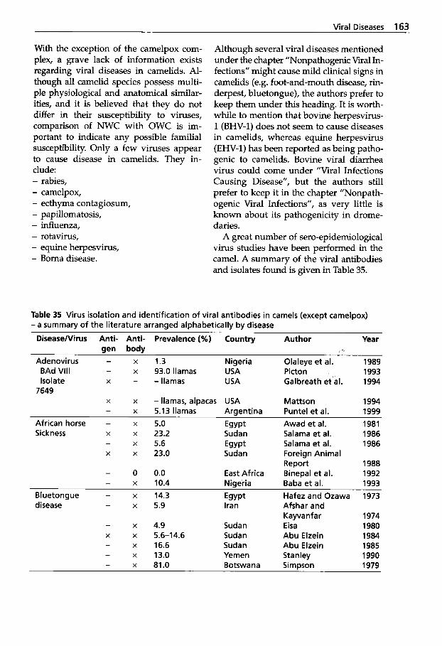
Viral Diseases 163 ~~
With the exception of the camelpox com- plex, a grave lack of information exists regarding viral diseases in camelids. Al- though all camelid species possess multi- ple physiological and anatomical similar- ities, and it is believed that they do not differ in their susceptibility to viruses, comparison of NWC with OWC is im- portant to indicate any possible familial susceptibility. Only a few viruses appear to cause disease in camelids. They in- clude: - rabies, - camelpox, - ecthyma contagiosum, - papillomatosis, - influenza, - rotavirus, - equine herpesvirus, - Borna disease.
Although several viral diseases mentioned under the chapter "Nonpathogenic Viral In- fections" might cause mild clinical signs in camelids (e.g. foot-and-mouth disease, rin- derpest, bluetongue), the authors prefer to keep them under this heading. It is worth- while to mention that bovine herpesvirus- 1 (BHV-1) does not seem to cause diseases in camelids, whereas equine herpesvirus (Em-1) has been reported as being patho- genic to camelids. Bovine viral diarrhea virus could come under "Viral Infections Causing Disease", but the authors still prefer to keep it in the chapter "Nonpath- ogenic Viral Infections", as very little is known about its pathogenicity in drome- daries.
A great number of sero-epidemiological virus studies have been performed in the camel. A summary of the viral antibodies and isolates found is given in Table 35.
Table 35 Virus isolation and identification of viral antibodies in camels (except camelpox) - a summary of the literature arranged alphabetically by disease
DiseaseIVirus Anti- Anti- Prevalence (%) Country Author Year gen body
Adenovirus - x 1.3 Nigeria Olaleye et al. 1989 BAd Vlll - x 93.0 llamas USA Picton 1993 Isolate X - -llamas USA Galbreath et'al. 1994
X x - llamas, alpacas USA Mattson 1994
African horse - x 5.0 Egypt Awad et al. 1981 Sickness X x 23.2 Sudan Salama et al. 1986
- x 5.6 Egypt Salama et al. 1986 X x 23.0 Sudan Foreign Animal
Report 1988 - 0 0.0 East Africa Binepal et al. 1992 - x 10.4 Nigeria Baba et al. 1993
Bluetongue - x 14.3 Egypt Hafez and Ozawa 1973 disease - x 5.9 Iran Afshar and
Kayva nf ar 1974 - x 4.9 Sudan Eisa 1980 X x 5.6-14.6 Sudan Abu Elzein 1984 - x 16.6 Sudan Abu Elzein 1985
- x 81.0 Botswana Simpson 1979
7649
- x 5.13 llamas Argentina Puntel et al. 1999
- x 13.0 Yemen Stanley 1990
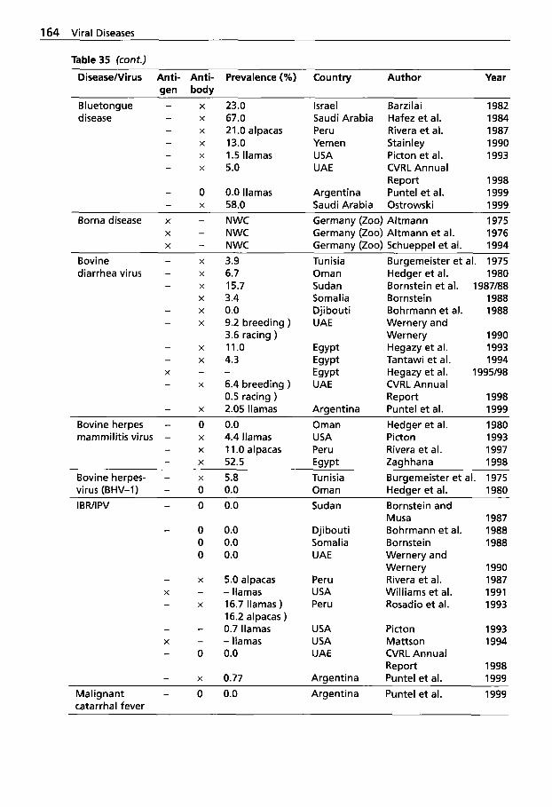
164 Viral Diseases
Table 35 (cont.)
DiseaselVirus Anti- aen
Prevalence (%) Country Author Year Anti- body
X
X
X
X
X
X
Bluetongue - disease -
- -
23.0 67.0 21.0 alpacas 13.0 1.5 llamas 5.0
Israel Saudi Arabia Peru Yemen USA UAE
Barzilai Hafez et al. Rivera et al. Stainley Picton et al. CVRL Annual Report Puntel et al. Ostrowski
1982 1984 1987 1990 1993
1998 1999 1999
0 X
0.0 llamas 58.0
Argentina Saudi Arabia
Borna disease x - NWC Germany (Zoo) Altmann 1975 X - NWC Germany (Zoo) Altmann et al. 1976 X - NWC Germany (Zoo) Schueppel et al. 1994
X Bovine - diarrhea virus - X
X
X
X
X
-
- -
3.9 6.7 15.7 3.4 0.0 9.2 breeding ) 3.6 racing ) 11.0 4.3
6.4 breeding ) 0.5 racing ) 2.05 llamas
-
Tun is i a Oman Sudan Somalia Djibouti UAE
Burgemeister e t al. 1975 Hedger et al. 1980 Bornstein et al. 1987188 Bornstein 1988 Bohrmann et al. 1988 Wernery and Wernery 1990 Hegazy e t al. 1993 Tantawi et al. 1994 Hegazy et al. 1995198 CVRL Annual Report 1998 Puntel et al. 1999
X
X
- -
- X
X -
Arsentina X -
Bovine herpes - 0 0.0 Oman Hedger et al. 1980 mammilitis virus - x 4.4 llamas USA Picton 1993
- x 11 .O alpacas Peru Rivera et al. 1997 - x 52.5 Egypt Zaghhana 1998
Bovine herpes- - x 5.8 Tunisia Burgemeister et al. 1975 virus (BHV-1) - 0 0.0 Oman Hedqer et al. 1980 IBWIPV - 0 0.0 Sudan Bornstein and
Musa Bohrmann et al. Bornstein Wernery and Wernery Rivera et al. Williams et al. Rosadio et al.
1987 1988 1988
- 0 0 0
0.0 0.0 0.0
Djibouti Somalia UAE
1990 1987 1991 1993
5.0 alpacas - llamas 16.7 llamas ) 16.2 alpacas ) 0.7 llamas - llamas 0.0
Peru USA Peru
X - - X
X -
USA USA UAE
Picton Mattson CVRL Annual Report Puntel et al.
1993 1994
1998 1999 0.77 Argentina X -
Malignant - 0 0.0 Argentina Puntel e t al. 1999 catarrhal fever
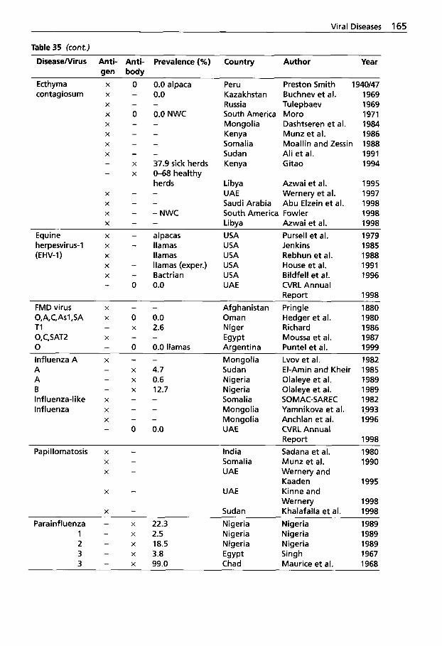
Viral Diseases 165
Table 35 (cont.)
Disease/Virus Anti- Anti- Prevalence (%) Country Author Year gen body
Ecthyma x o contagiosum x -
- X
x o - X
X
X
X
- - - X
X
- -
- X
X
X
X
- - -
0.0 alpaca 0.0
0.0 NWC -
- - - - 37.9 sick herds 0-68 healthy herds - - - NWC -
~
Peru Kazakhstan Russia South America Mongolia Kenya Somalia Sudan Kenya
Preston Smith 1940/47 Buchnev et al. 1969 Tulepbaev 1969 Moro 1971 Dashtseren et al. 1984 Munz et al. 1986 Moallin and Zessin 1988 Ali et al. 1991 Gitao 1994
Libya Azwai et al. 1995 UAE Wernery et al. 1997 Saudi Arabia Abu Elzein et al. 1998 South America Fowler 1998 Libya Azwai et al. 1998
Equine X - alpacas USA Pursell et al. 1979 herpesvirus-1 x - llamas USA Jenkins 1985 (EHV-1) X llamas USA Rebhun et al. 1988
X - Bactrian USA Bildfell et al. 1996 - 0 0.0 UAE CVRL Annual
Report 1998 FMD virus X - - Afghanistan Pringle 1880 O,A,C,Asl,SA x 0 0.0 Oman Hedger et al. 1980 TI - x 2.6 Niger Richard 1986 O.C.SAT2 X - - Egypt Moussa et al. 1987 0 - 0 0.0 llamas Arclentina Puntel et al. 1999
X - llamas (exper.) USA House et al. 1991
- - Influenza A X
A - x 4.7 x 0.6 A -
B - x 12.7 Influenza-like x - - Influenza X - -
- - X
- 0 0.0
Mongolia Sudan Nigeria Nigeria Somalia Mongolia Mongolia UAE
Lvov et al. 1982 El-Amin and Kheir 1985 Olaleye et al. 1989 Olaleye et al. 1989 SOMAC-SAREC 1982 Yamnikova et al. 1.993 Anchlan et al. 1996 CVRL Annual Report 1998
Papillomatosis x - India Sadana et al. 1980 - X
X -
- X
Somalia Munz et al. 1990 UAE Wernery and
Kaaden 1995 UAE Kinne and
Wernery 1998 X - Sudan Khalafaila et al. 1998
Parainfluenza - x 22.3 Nigeria Nigeria 1989 1 - x 2.5 Nigeria Nigeria 1989 2 - x 18.5 Nigeria Nigeria 1989 3 - x 3.8 Egypt Singh 1967 3 - x 99.0 Chad Maurice et al. 1968
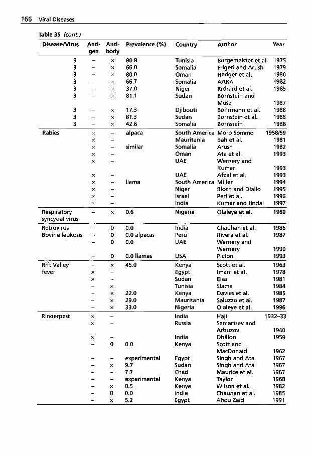
166 Viral Diseases
Table 35 (cont.)
DiseaselVirus Anti- Anti- Prevalence (%) Country Author Year gen body
3 - x 80.8 3 - x 66.0 3 - x 80.0 3 - x 66.7 3 - x 37.0 3 - x 81.1
3 - x 17.3 3 - x 81.3 3 - x 42.8
Rabies X - alpaca
X - similar - X
- X
X -
- X
X - llama - X
X
X
- -
Tunisia Burgemeister et al. 1975 Somalia Frigeri and Arush 1979
Somalia Arush 1982 Niger Richard et al. 1985 Sudan Bornstein and
Musa 1987 Djibouti Bohrmann et al. 1988 Sudan Bornstein et al. 1988 Somalia Bornstein 1988 South America Moro Sommo 1958/59 Mauritania Bah et al. 1981 Somalia Arush 1982 Oman Ata et al. 1993 UAE Wernery and
Kumar 1993 UAE Afzal et al. 1993 South America Miller 1994 Niger Bloch and Diallo 1995 Israel Per1 et al. 1996 India Kumar and Jindal 1997
Oman Hedger et al. 1980
Respiratory - x 0.6 Nigeria Olaleye et al. 1989 syncytial virus Retrovirus - 0 0.0 India Chauhan et al. 1986 Bovine leukosis - 0 0.0 alpacas Peru Rivera et al. 1987
- 0 0.0 UAE Wernery and
- 0 0.0 llamas USA Picton 1993 R i f t Valley - x 45.0 Kenya Scott et al. 1963 fever X - Egypt Imam et al. 1978
X - Sudan Eisa 1981 - X Tunisia Slama 1984 - x 22.0 Kenya Davies et al. 1985 - x 29.0 Mauritania Saluzzo et al. 1987 - x 33.0 Nigeria Olaleye et al. 1996
X - Russia Samartsev and Arbuzov 1940
X - India Dhillon 1959 - 0 0.0 Kenya Scott and
MacDonald 1962 - - experimental Egypt Singh and Ata 1967 - x 9.7 Sudan Singh and Ata 1967 - - 7.7 Chad Maurice et al. 1967 - - experimental Kenya Taylor 1968 - x 0.5 Kenya Wilson et al. 1982 - 0 0.0 India Chauhan et al. 1985 - x 5.2 E q w t Abou Zaid 1991
Wernery 1990
Rinderpest X - India Haji 1932-33
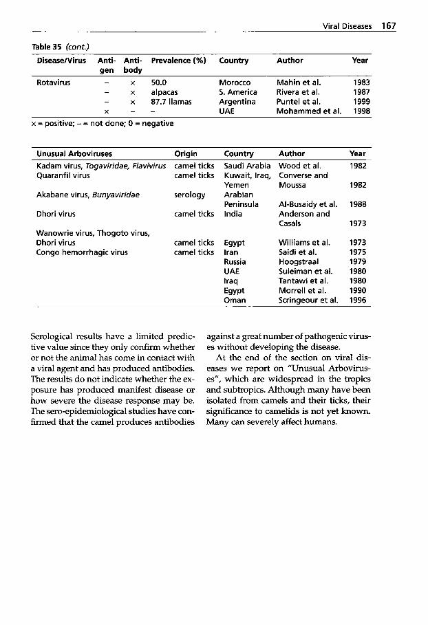
Viral Diseases 167
Table 35 (cont.)
DiseaseNirus Anti- Anti- Prevalence (%) Country Author Year gen body
Rotavirus - x 50.0 Morocco Mahin et al. 1983 - x alpacas 5. America Rivera et al. 1987
X - - UAE Mohammed et al. 1998 - x 87.7 llamas Argentina Puntel et al. 1999
x = positive; - = not done; 0 = negative
Unusual Arboviruses Origin Country Author Year Kadam virus, Togaviridae, Flavivirus camel ticks Saudi Arabia Quaranfil virus camel ticks Kuwait, Iraq,
Akabane virus, Bunyaviridae serology Arabian
Dhori virus camel ticks India
Yemen
Peninsula
Wanowrie virus, Thogoto virus, Dhori virus camel ticks Egypt Congo hemorrhagic virus camel ticks Iran
Russia UAE Iraq
Oman Egypt
Wood et al. Converse and Moussa
Al-Busaidy et al. Anderson and Casals
Williams et al. Saidi et al. Hoogstraal Suleiman et al. Tantawi et al. Morrell et al.
1982
1982
1988
1973
1973 1975 1979 1980 1980 1990
Scrinaeour et al. 1996
Serological results have a limited predic- tive value since they only confirm whether or not the animal has come in contact with a viral agent and has produced antibodies. The results do not indicate whether the ex- posure has produced manifest disease or how severe the disease response may be. The sero-epidemiological studies have con- firmed that the camel produces antibodies
against a great number of pathogenic virus- es without developing the disease.
At the end of the section on viral dis- eases we report on "Unusual Arbovirus- es", which are widespread in the tropics and subtropics. Although many have been isolated from camels and their ticks, their significance to camelids is not yet known. Many can severely affect humans.
JUIIII(1UIi 2.1 Viral Infections Causing Disease
2.1.1 Rabies
Rabies is a fatal disease for humans and all other warm-blooded vertebrates which is generally transmitted by the bite of a dis- eased animal. It causes encephalitis. Cam- elids are susceptible to rabies and the dis- ease has been extensively studied in NWC due to its zoonotic aspect.
Etiology The rabies virus belongs to the family Rhabdoviridae, which includes the genus Lyssavirus and the genus Vesiculo- virus. The genus Lyssavirus includes the rabies and bovine ephemeral fever sero- groups. Within the rabies serogroup, the rabies virus (Zyssavirus serotype 1) and the rabies-related viruses (Lagos bat, Mokola, Duvenhage = Zyssavirus serotypes 2, 3, 4) are biologically and antigenically different from seven other viruses of this group which are isolated from birds and hemato- phagus dipterids in Africa, South America and Australia. Rhabdoviruses are rod or bul- let-shaped. The genome consists of a single segment of single-stranded RNA and there are five structural proteins. Replication oc- curs in the cytoplasm of the infected cell and viral proteins accumulate here, consti- tuting the inclusions seen histologically as Negri bodies.
Epidemiology Rabies is an infectious disease transmitted via the saliva of infect- ed animals and is characterized by disturb- ances of the central nervous system, para- lysis and death. The transmission of the ra- bies virus from animal to animal and to man usually occurs through a bite. Herbi- vores and man are the final hosts and do not normally play a role as vectors. Carni- vores or vampire bats only sustain the cy- cle of infection.
Warm-blooded mammals as well as birds are susceptible to the rabies virus; however, there are substantial variations in susceptibility to the virus. Foxes, cotton rats and prairie wolves are extremely sus- ceptible; cattle, camels, rabbits and cats are very susceptible; dogs, sheep and goats are less susceptible. Opossums are most prob- ably not susceptible to rabies (Blood and Radostits, 1990).
Although rabies in dromedaries has sup- posedly been observed in many African and Asian countries (Richard 1980 and 1986), little has been published on this subject. Recent reports of rabies in camels have appeared from Morocco (Chevrier, 1959), Mauritania (Bah et al., 1981), Oman (Ata et al., 1993), the UAE (Wernery and Kumar, 1993; Afzal et al., 1993), Niger (Bloch and Diallo, 1995), India (Kumar and Jindal, 1997) and Israel (Per1 et al., 1996). Rabies-like diseases with hindquarter pare- sis have been reported by Arush (1982) and Somac/Sarec (1982) in Somalian drome- daries.
Little is known about the epidemiolog- ical interdependencies of rabies in the camel. Three types of rabies have been dif- ferentiated, depending on which animal species serves locally as the main reservoir and vector: the urban form, the sylvatic form and the bat form (paralyssa). The syl- vatic form plays the greatest role on the Arabian Peninsula. Rabies is most proba- bly transmitted by the red fox in the UAE and Oman (Wernery and Kumar, 1993; Ata et al., 1993) and by wild dogs in Yemen (Stanley, 1990). It can only be presumed that these animal species are the vectors of rabies on the Arabian Peninsula as there is very little available research.
In Niger, Bloch and Diallo (1995) report- ed that a rabid dog was responsible for a rabies outbreak in 7 camels in a herd of 40.
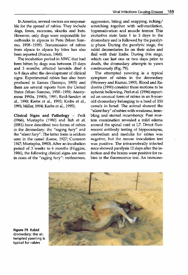
Viral Infections Causing Disease 169
In America, several vectors are responsi- ble for the spread of rabies. They include dogs, foxes, raccoons, skunks and bats. However, only dogs were responsible for outbreaks in alpacas in Peru (Moro Som- mo, 1958-1959). Transmission of rabies from alpaca to alpaca by bites has also been reported (Franco, 1968).
The incubation period in NWC that had been bitten by dogs was between 15 days and 3 months; affected lamoids died 6 to 8 days after the development of clinical signs. Experimental rabies has also been produced in llamas (Tamayo, 1905) and there are several reports from the United States (Moro Sommo, 1958-1959; Anony- mous 1990a, 1990b, 1991; Reid-Sanden et al., 1990; Krebs et al., 1992; Krebs et al., 1993; Miller, 1994; Krebs et al., 1995).
Clinical Signs and Pathology :. 1 Peck (1966), Mustapha (1980) and Bah et al. (1981) have described two forms of rabies in the dromedary: the "raging fury" and the "silent fury". The latter form is seldom seen in the camel (Leese, 1927; Curasson 1947; Mustapha, 1980). After an incubation period of 3 weeks to 6 months (Higgins, 1986), the following clinical signs are seen in cases of the "raging fury": restlessness,
aggression, biting and snapping, itching/ scratching together with self-mutilation, hypersalivation and muscle tremor. This excitative state lasts 1 to 3 days in the dromedary and is followed by the paralyt- ic phase. During the paralytic stage, the rabid dromedaries lie on their sides and flail with their limbs. During this stage, which can last one or two days prior to death, the dromedary attempts to yawn continuously (Fig. 79).
The attempted yawning is a typical symptom of rabies in the dromedary (Wernery and Kumar, 1993). Blood and Ra- dostits (1990) consider these motions to be aphonic bellowing. Per1 et al. (1996) report- ed an unusual form of rabies in an %year- old dromedary belonging to a herd of 150 camels in Israel. The animal showed the "silent fury" of rabies with weakness, trem- bling and sternal recumbency. Post mor- tem examination revealed a mild edema around the spinal cord at L7. Direct fluo- rescent antibody testing of hippocampus, cerebellum and medulla for rabies was negative, but the mouse inoculation test was positive. The intracerebrally infected mice showed paralysis 12 days after the in- fection and the brains were positive for ra- bies in the fluorescence test. An immuno-
Figure 79 Rabid dromedary: the at- tempted yawning is typical for rabies
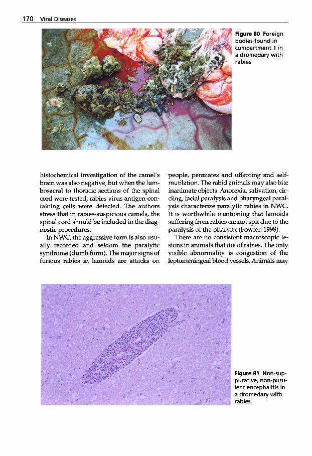
170 Viral Diseases
Figure 80 Foreign bodies found in compartment 1 in a dromedary with rabies
histochemical investigation of the camel's brain was also negative, but when the lum- bosacral to thoracic sections of the spinal cord were tested, rabies virus antigen-con- taining cells were detected. The authors stress that in rabies-suspicious camels, the spinal cord should be included in the diag- nostic procedures.
In NWC, the aggressive form is also usu- ally recorded and seldom the paralytic syndrome (dumb form). The major signs of furious rabies in lamoids are attacks on
people, penmates and offspring and self- mutilation. The rabid animals may also bite inanimate objects. Anorexia, salivation, cir- cling, facial paralysis and pharyngeal paral- ysis characterize paralytic rabies in NWC. It is worthwhile mentioning that lamoids suffering from rabies cannot spit due to the paralysis of the pharynx (Fowler, 1998).
There are no consistent macroscopic le- sions in animals that die of rabies. The only visible abnormality is congestion of the leptomeningeal blood vessels. Animals may
Figure 81 Non-sup- purative, non-puru- lent encephalitis in a dromedary with rabies
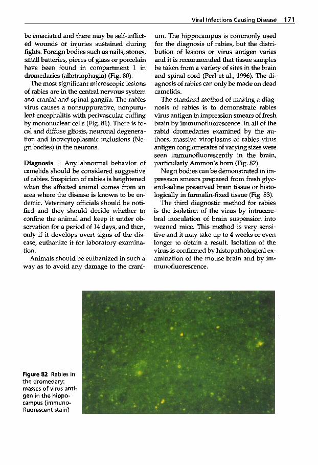
Viral Infections Causing Disease 171
be emaciated and there may be self-inflict- ed wounds or injuries sustained during fights. Foreign bodies such as nails, stones, small batteries, pieces of glass or porcelain have been found in compartment 1 in dromedaries (allotriophagia) (Fig. 80).
The most significant microscopic lesions of rabies are in the central nervous system and cranial and spinal ganglia. The rabies virus causes a nonsuppurative, nonpuru- lent encephalitis with perivascular cuffing by mononuclear cells (Fig. 81). There is fo- cal and diffuse gliosis, neuronal degenera- tion and intracytoplasmic inclusions (Ne- gri bodies) in the neurons.
Diagnosis Any abnormal behavior of camelids should be considered suggestive of rabies. Suspicion of rabies is heightened when the affected animal comes from an area where the disease is known to be en- demic. Veterinary officials should be noti- fied and they should decide whether to confine the animal and keep it under ob- servation for a period of 14 days, and then, only if it develops overt signs of the dis- ease, euthanize it for laboratory examina- tion.
Animals should be euthanized in such a way as to avoid any damage to the crani-
um. The hippocampus is commonly used for the diagnosis of rabies, but the distri- bution of lesions or virus antigen varies and it is recommended that tissue samples be taken from a variety of sites in the brain and spinal cord (Per1 et al., 1996). The di- agnosis of rabies can only be made on dead camelids.
The standard method of making a diag- nosis of rabies is to demonstrate rabies virus antigen in impression smears of fresh brain by immunofluorescence. In all of the rabid dromedaries examined by the au- thors, massive viroplasms of rabies virus antigen conglomerates of varying sizes were seen immunofluorescently in the brain, particularly Ammon’s horn (Fig. 82).
Negri bodies can be demonstrated in im- pression smears prepared from fresh glyc- erol-saline preserved brain tissue or histo- logically in formalin-fixed tissue (Fig. 83).
The third diagnostic method for rabies is the isolation of the virus by intracere- bra1 inoculation of brain suspension into weaned mice. This method is very sensi- tive and it may take up to 4 weeks or even longer to obtain a result. Isolation of the virus is confirmed by histopathological ex- amination of the mouse brain and by im- munofluorescence.
Figure 82 Rabies in the dromedary: masses of virus anti- gen in the hippo- campus (immuno- fluorescent stain)
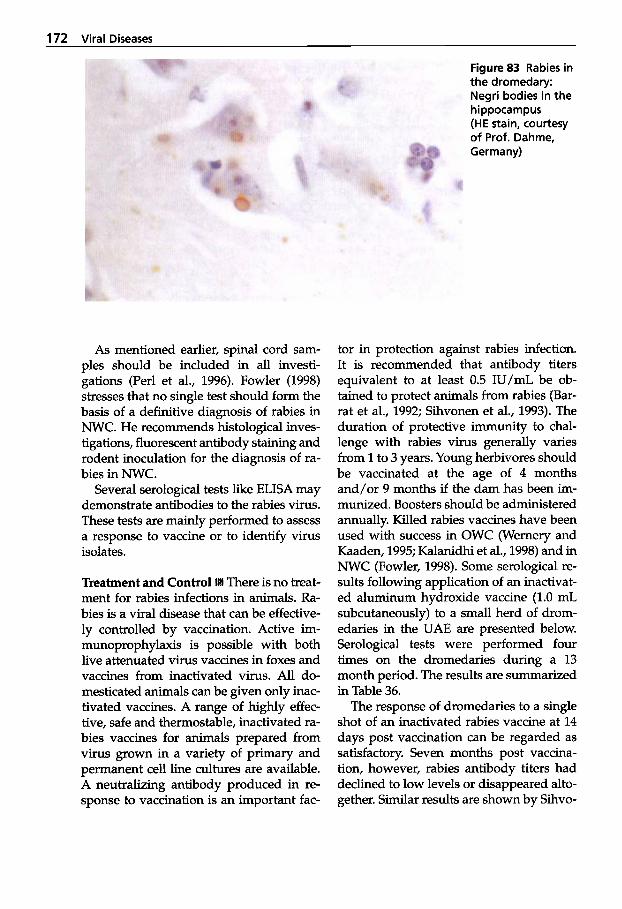
172 Viral Diseases
Figure 83 Rabies in the dromedary: Negri bodies in the hippocampus (HE stain, courtesy of Prof. Dahme, Germany)
As mentioned earlier, spinal cord sam- ples should be included in all investi- gations (Per1 et al., 1996). Fowler (1998) stresses that no single test should form the basis of a definitive diagnosis of rabies in NWC. He recommends histological inves- tigations, fluorescent antibody staining and rodent inoculation for the diagnosis of ra- bies in NWC.
Several serological tests like ELISA may demonstrate antibodies to the rabies virus. These tests are mainly performed to assess a response to vaccine or to identify virus isolates.
Treatment and Control There is no treat- ment for rabies infections in animals. Ra- bies is a viral disease that can be effective- ly controlled by vaccination. Active im- munoprophylaxis is possible with both live attenuated virus vaccines in foxes and vaccines from inactivated virus. All do- mesticated animals can be given only inac- tivated vaccines. A range of highly effec- tive, safe and thermostable, inactivated ra- bies vaccines for animals prepared from virus grown in a variety of primary and permanent cell line cultures are available. A neutralizing antibody produced in re- sponse to vaccination is an important fac-
tor in protection against rabies infection. It is recommended that antibody titers equivalent to at least 0.5 W/mL be ob- tained to protect animals from rabies (Bar- rat et al., 1992; Sihvonen et al., 1993). The duration of protective immunity to chal- lenge with rabies virus generally varies from 1 to 3 years. Young herbivores should be vaccinated at the age of 4 months and/or 9 months if the dam has been im- munized. Boosters should be administered annually. Killed rabies vaccines have been used with success in OWC (Wernery and Kaaden, 1995; Kalanidhi et al., 1998) and in NWC (Fowler, 1998). Some serological re- sults following application of an inactivat- ed aluminum hydroxide vaccine (1.0 mL subcutaneously) to a small herd of drom- edaries in the UAE are presented below. Serological tests were performed four times on the dromedaries during a 13 month period. The results are summarized in Table 36.
The response of dromedaries to a single shot of an inactivated rabies vaccine at 14 days post vaccination can be regarded as satisfactory. Seven months post vaccina- tion, however, rabies antibody titers had declined to low levels or disappeared alto- gether. Similar results are shown by Sihvo-
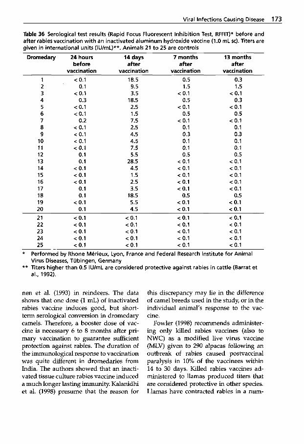
Viral Infections Causinu Disease 173
Table 36 Serological test results (Rapid Focus Fluorescent Inhibition Test, RFFIT)* before and after rabies vaccination with an inactivated aluminum hydroxide vaccine (1 .O mL sc). Titers are given in international units (IU/mL)**. Animals 21 to 25 are controls
Dromedary 24 hours 14 days 7 months 13 months before after after after
vaccination vaccination vaccination vaccination 1 c 0.1 18.5 0.5 0.3 2 0.1 9.5 1.5 1.5 3 c 0.1 3.5 c 0.1 < 0.1 4 0.3 18.5 0.5 0.3 5 < 0.1 2.5 < 0.1 c 0.1 6 c 0.1 1.5 0.5 0.5 7 0.2 7.5 < 0.1 < 0.1 8 < 0.1 2.5 0.1 0.1 9 < 0.1 4.5 0.3 0.3
10 < 0.1 4.5 0.1 0.1 11 c 0.1 7.5 0.1 0.1 12 0.1 5.5 0.5 0.5 13 0.1 28.5 c 0.1 c 0.1 14 < 0.1 4.5 < 0.1 c 0.1 15 c 0.1 1.5 < 0.1 < 0.1 16 < 0.1 2.5 < 0.1 < 0.1 17 0.1 3.5 < 0.1 < 0.1 18 0.1 18.5 0.5 0.5 19 < 0.1 5.5 < 0.1 < 0.1 20 0.1 4.5 c 0.1 < 0.1 21 c 0.1 < 0.1 < 0.1 c 0.1 22 < 0.1 c 0.1 c 0.1 < 0.1 23 c 0.1 c 0.1 < 0.1 < 0.1 24 < 0.1 < 0.1 c 0.1 c 0.1 25 < 0.1 c 0.1 < 0.1 < 0.1
* Performed by Rhone Merieux, Lyon, France and Federal Research Institute for Animal Virus Diseases, Tubingen, Germany
** Titers higher than 0.5 IU/mL are considered protective against rabies in cattle (Barrat et al., 1992).
nen et al. (1993) in reindeers. The data shows that one dose (1 mL) of inactivated rabies vaccine induces good, but short- term serological conversion in dromedary camels. Therefore, a booster dose of vac- cine is necessary 6 to 8 months after pri- mary vaccination to guarantee sufficient protection against rabies. The duration of the immunological response to vaccination was quite different in dromedaries from India. The authors showed that an inacti- vated tissue culture rabies vaccine induced a much longer lasting immunity. Kalanidhi et al. (1998) presume that the reason for
this discrepancy may lie in the difference of camel breeds used in the study, or in the individual animal's response to the vac- cine.
Fowler (1998) recommends administer- ing only killed rabies vaccines (also to NWC) as a modified live virus vaccine (MLV) given to 290 alpacas following an outbreak of rabies caused postvaccinal paralysis in 10% of the vaccinees within 14 to 30 days. Killed rabies vaccines ad- ministered to llamas produced titers that are considered protective in other species. Llamas have contracted rabies in a num-
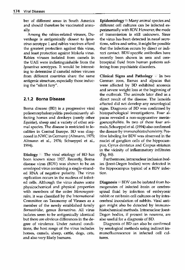
174 Viral Diseases
ber of different areas in South America and should therefore be vaccinated annu- ally.
Among the rabies-related viruses, Du- venhage is antigenically closest to Zyssa- virus serotype 1 and rabies vaccines afford the greatest protection against this virus, and least protection against Mokola virus. Rabies viruses isolated from camels in the UAE were indistinguishable from the Zyssavirus serotypes. It would be interest- ing to determine if camelid rabies viruses from different countries share the same antigenic structure, especially those induc- ing the "silent fury".
2.1.2 Borna Disease
Borna disease (BD) is a progressive viral polioencephalomyelitis predominantly af- fecting horses and donkeys (rarely other Equidae), sheep and a variety of other ani- mal species. The disease is restricted to lo- calities in Central Europe. BD was diag- nosed in NWC in Germany (Altmann, 1975; Altmann et al., 1976; Schueppel et al., 1994).
Etiology The viral etiology of BD has been known since 1927. Recently, Borna disease virus (BDV) was shown to be an enveloped virus containing a single-strand- ed RNA of negative polarity. The virus replication occurs in the nucleus of infect- ed cells. Although the virus shares some physicochemical and physical properties with members of the order Mononegavi- rules, it was classified by the International Committee on Taxonomy of Viruses as a member of the newly established family Bomaviridae, genus Bornavirus. All virus isolates seem to be antigenically identical but there are obvious differences in the de- gree of virulence. Under natural condi- tions, the host range of the virus includes horses, camels, sheep, cattle, dogs, cats, and also very likely humans.
Epidemiology i:ij Many animal species and different cell cultures can be infected ex- perimentally with BDV. However, the mode of transmission is still unknown. Since the virus has been detected in nasal secre- tions, saliva and urine, it might be possible that the infection occurs by direct or indi- rect contact. BDV-specific antibodies have recently been shown in sera and cere- brospinal fluid from human patients suf- fering from psychiatric disorders.
Clinical Signs and Pathology In two German zoos, llamas and alpacas that were affected by BD exhibited anorexia and severe weight loss at the beginning of the outbreak. The animals later died as a direct result of the disease. The lamoids affected did not develop any neurological signs. Diagnosis of BD was confirmed by histopathological investigations. Four al- pacas revealed a non-suppurative menin- goencephalitis. In two of these four ani- mals, Schueppel et al. (1994) also confirmed the disease by immunohistochemistry. Pos- itive labeling for BDV was observed in the nuclei of ganglion cells of the hippocam- pus, Gyrus dentatus and Corpus striatum in the vicinity of inflammatory infiltrates (Fig. 84).
Furthermore, intranuclear inclusion bod- ies (Joest-Degen bodies) were detected in the hippocampus typical of a BDV infec- tion.
Diagnosis i.i BDV can be isolated from ho- mogenates of infected brain or cerebro- spinal fluid by infection of embryonic rabbit or rat brain cell cultures or by intra- cerebral inoculation of rabbits. Viral anti- gen might also be detected by immuno- histochemical methods. Intranuclear Joest- Degen bodies, if present in neurons, are also useful for a diagnosis of BD.
Diagnosis of BD can also be confirmed by serological methods using indirect im- munofluorescence in infected cell cul- tures.
Viral Infections Causing Disease 175
Figure 84 Positive labeling for BDV of the hippocampus in an alpaca
Treatment and Prevention ji'i BD is a re- portable disease and controlled by a stamp- ing-out policy.
References Afzal, M., I.A. Khan and R. Salman. 1993. Clini-
cal signs and clinical pathology of rabies in the camel. Vet. Rec. 133: 220.
Altmann, D. 1975. Die wichtigsten Erkrankun- gen der Alt- und Neuweltkamele. Verhand- hngsber. 17fh lnt. Symp Erkr. Zootiere (Tunis)
Altmann, D., H. Kronberger, K.-F. Schueppel, R. Lippmann and I. Altmann. 1976. Bornasche Krankheit (meningo-encephalomyelitis sim- plex enzootica equonun) bei Neuwelttylo- poden und Equiden. Verhandlungsber. 18. lnt. Symp. Erkr. Zootiere, Innsbruck 127-132.
Anonymous. 1990a. Centers for Disease Con- trol. Rabies in a llama. Oklahoma Morb. Mort.
Anonymous. 1990b. Centers for Disease Con- trol. Rabies in a llama. Oklahoma J. Am. Vet. Med. Assoc. 263 (16): 2766.
Anonymous. 1991. Centers for Disease Control. Rabies in a llama. Wkly. Epidemiol. Rec. 65 (38): 294.
Arush, M.A. 1982. La situazione sanitaria del dromedario nella Repubblica Democratica Somala. Bollettino scient@ca della facoltd di zootecnia e veterinaria 3: 209-217.
1 7 53-60.
Wkly. Rep. 39 (12): 203-204.
Ata, F.A., M.H. Tageldin, H.S. A1 S u m r y and S.I. Al-Ismaily. 1993. Rabies in the Sultanate of Oman. Vet. Rec. 132 68-69.
Bah, S.O., G. Chamoiseau, M.L.O. Biha and S.M.O.A. Fall. 1981. Un foyer de rage Came- line en Mauritanie. Rev. Elm. Mkd. vkf. Pays trop. 34 (3): 263-265.
Barrat, J., F. Guillemin, A. Brun, F. Lacoste and P. Precausta. 1992. Cattle vaccination against rabies. Immunity duration and challenge three years after vaccination. Paper read at Proc. Pan-American Hlth. Org.: 24.10.91.
Bloch, N. and I. Diallo. 1995. A probable out- break of rabies in a group of camels in Niger. Vet. Microbiology 46 (1-3): 281-283.
Blood, D.C. and O.M. Radostits. 1990. Veteri- nary Medicine. 7th ed. London: Bailliere Tin- dall.
Chevrier, L. 1959. Epidemiologic de la rage au Maroc. Rev. Elm. Mkd. vet. Pays trop. 12 (2):
Curasson, G. 1947. Le chameau et ses maladies. Vigot Freres, Editeurs: 86-88.
Fowler, M.E. 1998. Medicine and surgery of South American Camelids. Iowa State Uni- versity Press, Ames.
El-Ahwal, A.M. 1969. Rabies problem and erad- ication in U.A.R. J. Egypt Vet. Med. Ass. 29
Franco, E. 1968. Brote de rabia en alpacas de m a hacienda del Departamento de Puno. Bol. Ex- traordinario 3: 59-60.
115-120.
(3-4): 121-129.
176 Viral Diseases
Higgins, A. 1986. The camel in health and dis- ease. Balliere Tindall.
Kalanidhi, AX, U.K. Bissa and V.A. Srinivasan. 1998. Seroconversion and duration of immu- nity in camels vaccinated with tissue-culture inactivated rabies vaccine. Veterinarski Arhiv
Krebs, J. W., R.C. Holman, U. Hines, T.W. Strine, E.J. Mandel and J.E. Childs. 1992. Rabies sur- veillance in the United States during 1991 (a llama). J. Am. Vet. Med. Assoc. 201 (12): 1839.
Krebs, J.W., T.W. Strine and J.E. Childs. 1993. Ra- bies surveillance in the United States during 1992 (a llama). J. Am. Vet. Med. Assoc. 203 (12): 1721.
Krebs, J.W., T.W. Strine, J.S. Smith, C.E. Rup- precht and J.E. Childs. 1995. Rabies surveil- lance in the United States during 1994. J. Am. Vet. Med. Assoc. 207 (12): 1562-1575.
Kumar, A. and N. Jindal. 1997. Rabies in a camel -A case report. Trop. Anim. Hlth. Prod. 29 (1): 34.
Leese, A.S. 1927. A treatise on the one-humped camel in health and disease. Vigot Freres, Paris H.
Miller, P. 1994. Rabies on rise. Llamas 8 (3): 49-55. Moro Sommo, M. 1958-59. Sobre un brote de
rabia en alpacas. Rev. Fac. Med. Vet. (Lima)
Mustapha, I.E. 1980. IFS Provisional report No. 6 on camels, 399. Stockholm: Int. Foundation for Science.
Peck, E.F. 1966. In Intern. Encyclopaedia of Vet. Med., ed. T. Dalling, A. Robertson, G.E. Bod- die and J.S. Spruell. lSt ed., Edinburgh, W. Green and Son: 577.
Perl, S., M. van Straten, B. Jakobson, I. Samina, N. Sheikhab and U. Orgad. 1996. Hind limb paralysis associated with rabies in a camel (Camelus dromedarius). 8tk Inst. Symposium of Vet. Lab. Diagnosticians, Jerusalem, Israel,
Reid-Sanden, EL. J.G. Dobbins, J.S. Smith and D.B. Fishbein. 1990. Rabies surveillance in the United States during 1989. J. Am. Vet. Med. Assoc. 197 (12): 1576.
Richard, D. 1980. Dromedary pathology and productions. Provisional report No. 6 camels. International Science Foundation (IFS), Khar- toum, Sudan and Stockholm 12 (18-20): 409-430.
Richard, D. 1986. Manuel des maladies du dro- madaire, Projet de developpement de l'ele-
68 (3): 81-84.
13-14 35-40.
4-8 Aug., 1996: 14.
vage dans le Niger centre-est. Maisons Alfort, IEMVT.
Schueppel, K.-F., J. Kinne und M. Reinacher. 1994. Bornavirus - Antigennachweis bei Al- pakas (Lama pacos) sowie bei einem Faultier (Choloepus didactylus) und einem Zwerg- fldpferd (Choeropsis liberiensis). Verh. ber. Erkrg. Zootiere 36: 189-193.
Sihvonen, L., K. Kulonen, T. Soveri, and M. Nie- minen. 1993. Rabies antibody titres in vacci- nated reindeer. Acta vet. scand. 34: 199-202.
Somac/Sarec. 1982. Camel research project re- port by a Somali/Swedish Mission, March
Stanley, M.J. 1990. Rabies in Yemen Arab Re- public, 1982 to 1986. Trop. Anim. Hlth. Prod. 22: 273-274.
Tamayo, M.D. 1905. La rabia experimental en la llama. Cron. Med. 22 269-272.
Wemery, U., and B.N. Kumar. 1993. Rabies in the U.A.E. Tribulus, Bulletin of the Emirates Natural History Group Vo13.1.: 5-21.
Wernery, U. and 0.-R. Kaaden. 1995. Infectious Diseases of Camelids. Blackwell Wissen- schafts-Verlag, Berlin.
10-26: 18-23.
Further reading
Sidya Ould Bah, G. Chamoiseau, Mohamed Lemine Ould Biha and Sidi Mohamed Ould Ahmed Fall. 1981. Un foyer de rage camline en Mauritanie. Rev. Elev. Mid. vkt. Pays trop. 34 (3): 263-265.
2.1.3 Camelpox
Camelpox occurs in the dromedary and the Bactrian camel and has also been experi- mentally induced in NWC (Kinne and Wernery, 1999; Wernery et al., 2000). The camelpox virus causes a proliferative skin disease that primarily affects younger ani- mals (Rohrer, 1970; Richard, 1979, 1980; Mahnel and Mum, 1987; Schwartz and Dioli, 1992). Pox-like lesions in camelids may also be induced by a yet-unnamed parapoxvirus and papillomavirus.
Viral Infections Causina Disease 177
Etiology li.* Poxviruses are classified in the family Poxviridae, which are divided into two subfamilies: Chordopoxvirinae, which infects vertebrates and Entornopoxvirinae, which are found in insects. Camelpox virus (CaPV) is a large, enveloped, doublestrand- ed DNA virus that represents 1 of 11 spe- cies currently assigned to the genus Or- thopoxvirus. Poxviruses are the largest and most complex viruses and have a brick- shaped appearance. The infective agent of camelpox is the Orthopoxvirus cameli.
Epidemiology Camelids may become infected with the poxvirus through small abrasions of the skin, by aerosol infec- tion of the respiratory tract or mechanical transmission by biting arthropods. Several scientists have reported an increase in camelpox outbreaks during wet seasons (e.g. Munz, 1992; Wernery et al., 1997a and b) when the disease becomes more severe. During the dry season, it usually follows a milder course (Pfahler and Munz, 1989). Since the camelpox virus has been isolated from the camel tick ffyalornrna dromedarii, it is now believed that a larger arthropod population builds up during rainy sea- sons, forcing a greater virus pressure and virus doses onto the camel populations. Differences in the virulence of camelpox strains have also been suggested (Munz, 1992; Otterbein, 1994; Otterbein et al., 1995; Pfeffer et al., 1996; Munz et al., 1997; Wer- nery and Zachariah, 1999), which may ex- plain the phenomenon that some strains produce generalized pox infections and oth- ers only a localized form (Wernery, 1994). DNA restriction enzyme analyses have shown that camelpox virus strains from different African countries possess differ- ent genomes, which may explain why virus strains differ in their virulence (Munz, 1992); an important factor in the produc- tion of vaccines and in performing test ex- posure experiments (Baxby et al., 1975).
Animals that have recovered from infec- tion appear to develop a lifelong immuni-
ty. Epidemics occur in regular cycles de- pendent on the rainy season and relation- ship of the density of the insect population to the number of immune camels in the population.
Camelpox is most probably not a zoono- sis, although clinical observations in vari- ous articles have reported the possibility of transmission of Orthopoxvirus carneli to humans. Even very recent reports of skin eruptions in camel herdsmen could not identify camelpox as the causative agent using current laboratory methods (Kriz, 1982; Jezek et al., 1983; Wemery and Kaa- den, 1995).
Pox is the most frequent infectious viral disease of the camel and therefore the most widely reported. The disease occurs wherever camel husbandry is practiced (Table 37). An exception is the Australian dromedary population where, so far, cam- elpox has not been observed (Doerges and Heucke, 1992, personal communication; Hafez et al., 1992).
In both localized and systemic poxvirus diseases, initial multiplication of the virus occurs at the site of entry. In those infec- tions characterized by systemic disease, further viral multiplication in the draining lymph nodes is followed by a primary viremia and multiplication of virus in or- gans and tissues. This results in a second- ary viremia and subsequent infection of the skin.
Serological studies in different countries have revealed a high prevalence of CaPV. Davies et al. (1985) showed, using the SNT, that there is a high prevalence of antibod- ies to camelpox in herds kept by nomadic pastoralists and by ranchers. Antibodies were found in five out of six camel herds in Kenya using SNT, although there was no clinical disease seen in the herds investi- gated. Munz et al. (1986) reported 95% positive cases in Sudan, which was con- firmed in 72.5% by Khalafalla et al. (1998). Pfeffer et al. (1998) found a prevalence be- tween 88% and 100% in 1,000 dromedaries
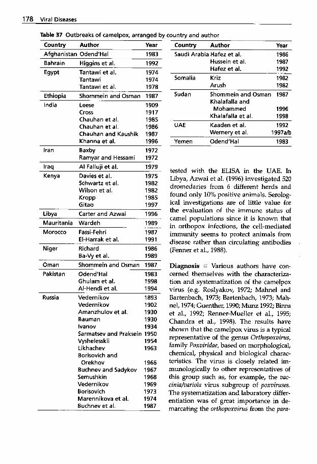
178 Viral Diseases
Table 37 Outbreaks of camelpox, arranged by country and author
Country Author Year Country Author Year Afghanistan Odend’Hal 1983 Bahrain Higgins et al. 1992 Egypt Tantawi et al. 1974
Tantawi 1974 Tantawi et al. 1978
Ethiopia Shommein and Osman 1987 India Leese
Cross Chauhan et al. Chauhan et al. Chauhan and Kaushik Khanna et al.
Ramvar and Hessami Iran Baxby
1909 1917 1985 1986 1987 1996 1972 1972
Iraq Al Falluii et al. 1979 Kenya Davies et al. 1975
Schwartz et al. 1982 Wilson et al. 1982 KroPP 1985 Gitao 1997
~
Libva Carter and Azwai 1996 Mauritania Wardeh 1989
~ ~~~~~
Morocco Fassi-Feh ri 1987 El-Harrak et al. 1991
Niger Richard 1986 Ba-Vy et al. 1989
Oman Shommein and Osman 1987 Pakistan Odend‘Hal 1983
Ghulam et al. 1998 Al-Hendi et al. 1994
Russia Vedernikov 1893 Vederni kov 1902 Amanzhulov et al. 1930 Bauman 1930 lvanov 1934 Sarmatsev and Praksein 1950
Likhachev 1963 Borisovich and Orekhov 1966
Buchnevand Sadykov 1967 Semushkin 1968 Vedernikov 1969 Borisovich 1973 Marennikova et al. 1974 Buchnev et al. 1987
Vyshelesskii 1954
Saudi Arabia Hafez et al. 1986 Hussein et al. 1987 Hafez et al. 1992
Somalia Kriz Arush
1982 1982
Sudan Shommein and Osman 1987 Khalafalla and Mohammed 1996
Khalafalla et al. 1998 UAE Kaaden et al. 1992-
Wernery et al. 1997ah Yemen Odend’Hal 1983
tested with the ELISA in the UAE. In Libya, Azwai et al. (1996) investigated 520 dromedaries from 6 different herds and found only 10% positive animals. Serolog- ical investigations are of little value for the evaluation of the immune status of camel populations since it is known that in orthopox infections, the cell-mediated immunity seems to protect animals from disease rather than circulating antibodies (Fenner et al., 1988).
Diagnosis f:i Various authors have con- cerned themselves with the characteriza- tion and systematization of the camelpox virus (e.g. Roslyakov, 1972; Mahnel and Bartenbach, 1973; Bartenbach, 1973; Mah- nel, 1974; Guenther, 1990; Munz 1992; Binns et al., 1992; Renner-Mueller et al., 1995; Chandra et al., 1998). The results have shown that the camelpox virus is a typical representative of the genus Orfhopoxvirus, family Poxviridae, based on morphological, chemical, physical and biological charac- teristics. The virus is closely related im- munologically to other representatives of this group such as, for example, the vuc- cinia/variola virus subgroup of poxviruses. The systematization and laboratory differ- entiation was of great importance in de- marcating the orthopoxvirus from the pura-
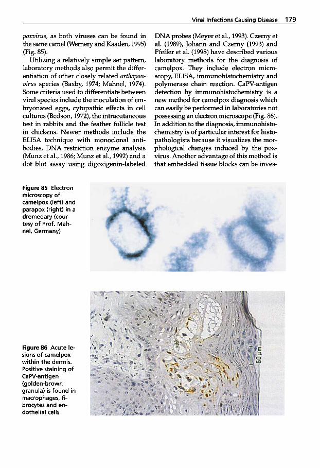
Viral Infections Causina Disease 179
pomirus, as both viruses can be found in the same camel (Wernery and Kaaden, 1995) (Fig. 85).
Utilizing a relatively simple set pattern, laboratory methods also permit the differ- entiation of other closely related orthopox- virus species (Baxby, 1974; Mahnel, 1974). Some criteria used to differentiate between viral species include the inoculation of em- bryonated eggs, cytopathic effects in cell cultures (Bedson, 1972), the intracutaneous test in rabbits and the feather follicle test in chickens. Newer methods include the ELISA technique with monoclonal anti- bodies, DNA restriction enzyme analysis (Munz et al., 1986; Munz et al., 1992) and a dot blot assay using digoxigenin-labeled
Figure 85 Electron microscopy of camelpox (left) and parapox (right) in a dromedary (cour- tesy of Prof. Mah- nel, Germany)
Figure 86 Acute le- sions of camelpox within the dermis. Positive staining of CaPV-antigen (golden-brown granula) is found in macrophages, fi- brocytes and en- dothelial cells
DNA probes (Meyer et al., 1993). Czerny et al. (1989), Johann and Czerny (1993) and Pfeffer et al. (1998) have described various laboratory methods for the diagnosis of camelpox. They include electron micro- scopy, ELISA, immunohistochemistry and polymerase chain reaction. CaPV-antigen detection by immunohistochemistry is a new method for camelpox diagnosis which can easily be performed in laboratories not possessing an electron microscope (Fig. 86). In addition to the diagnosis, immunohisto- chemistry is of particular interest for histo- pathologists because it visualizes the mor- phological changes induced by the pox- virus. Another advantage of this method is that embedded tissue blocks can be inves-
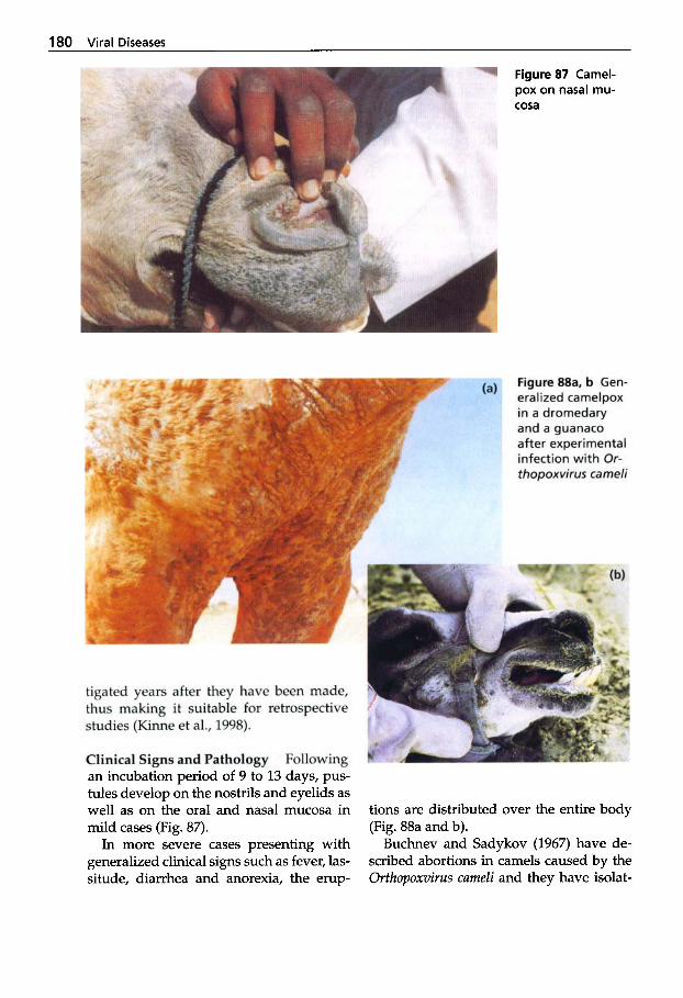
180 Viral Diseases
Figure 87 Camel- pox on nasal mu- cosa
Figure 88a. b Gen- eralized camelpox in a dromedary and a guanaco after experimental infection with Or- thopoxvirus cameli
tigated years after they have been made, thus making it suitable for retrospective studies (Kinne et al., 1998).
Clinical Signs and Pathology ” Following an incubation period of 9 to 13 days, pus- tules develop on the nostrils and eyelids as well as on the oral and nasal mucosa in mild cases (Fig. 87).
In more severe cases presenting with generalized clinical signs such as fever, las- situde, diarrhea and anorexia, the erup-
tions are distributed over the entire body (Fig. 88a and b).
Buchnev and Sadykov (1967) have de- scribed abortions in camels caused by the Orthopoxvirus cameli and they have isolat-
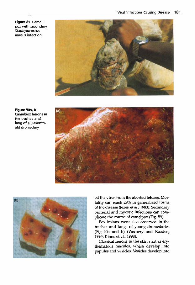
Viral Infections Causins Disease 181
Figure 89 Camel- pox with secondary Staphylococcus aureus infection
Figure 90a. b Camelpox lesions in the trachea and lung of a 9-month- old dromedary
ed the virus from the aborted fetuses. Mor- tality can reach 28% in generalized forms of the disease (Jezek et al., 1983). Secondary bacterial and mycotic infections can com- plicate the course of camelpox (Fig. 89).
Pox-lesions were also observed in the trachea and lungs of young dromedaries (Fig. 90a and b) (Wernery and Kaaden, 1995; Kinne et al., 1998).
Classical lesions in the skin start as ery- thematous macules, which develop into papules and vesicles. Vesicles develop into
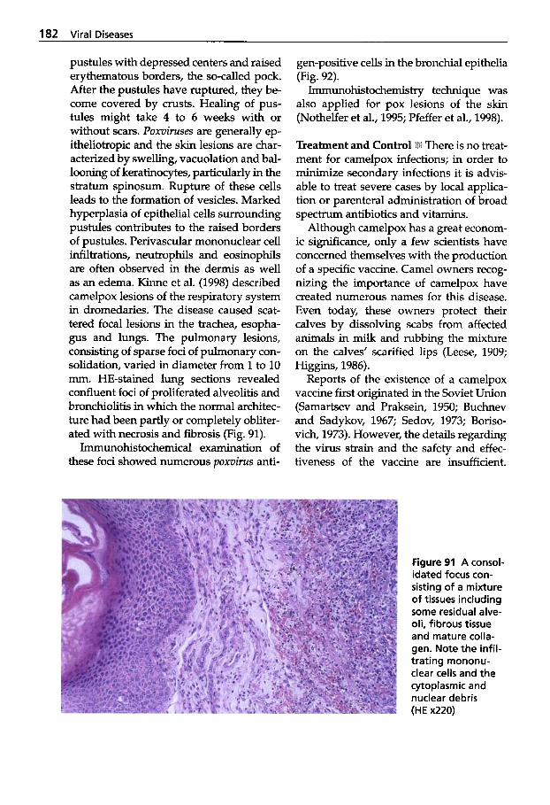
182 Viral Diseases
pustules with depressed centers and raised erythematous borders, the so-called pock. After the pustules have ruptured, they be- come covered by crusts. Healing of pus- tules might take 4 to 6 weeks with or without scars. Poxviruses are generally ep- itheliotropic and the skin lesions are char- acterized by swelling, vacuolation and bal- looning of keratinocytes, particularly in the stratum spinosum. Rupture of these cells leads to the formation of vesicles. Marked hyperplasia of epithelial cells surrounding pustules contributes to the raised borders of pustules. Perivascular mononuclear cell infiltrations, neutrophils and eosinophils are often observed in the dermis as well as an edema. Kinne et al. (1998) described camelpox lesions of the respiratory system in dromedaries. The disease caused scat- tered focal lesions in the trachea, esopha- gus and lungs. The pulmonary lesions, consisting of sparse foci of pulmonary con- solidation, varied in diameter from 1 to 10 mm. HE-stained lung sections revealed confluent foci of proliferated alveolitis and bronchiolitis in which the normal architec- ture had been partly or completely obliter- ated with necrosis and fibrosis (Fig. 91).
Immunohistochemical examination of these foci showed numerous poxvirus anti-
gen-positive cells in the bronchial epithelia (Fig. 92).
Immunohistochemistry technique was also applied for pox lesions of the skin (Nothelfer et al., 1995; Pfeffer et al., 1998).
Treatment and Control i There is no treat- ment for camelpox infections; in order to minimize secondary infections it is advis- able to treat severe cases by local applica- tion or parenteral administration of broad spectrum antibiotics and vitamins.
Although camelpox has a great econom- ic significance, only a few scientists have concerned themselves with the production of a specific vaccine. Camel owners recog- nizing the importance of camelpox have created numerous names for this disease. Even today, these owners protect their calves by dissolving scabs from affected animals in milk and rubbing the mixture on the calves’ scarified lips (Leese, 1909; Higgins, 1986).
Reports of the existence of a camelpox vaccine first originated in the Soviet Union (Samartsev and Praksein, 1950; Buchnev and Sadykov, 1967; Sedov, 1973; Boriso- vich, 1973). However, the details regarding the virus strain and the safety and effec- tiveness of the vaccine are insufficient.
Figure 91 A consol- idated focus con- sisting of a mixture of tissues including some residual alve- oli, fibrous tissue and mature colla- gen. Note the infil- trating mononu- clear cells and the cytoplasmic and nuclear debris (HE x220)
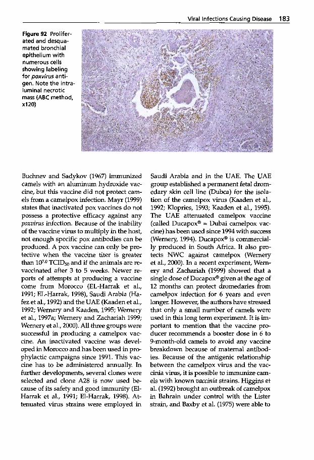
Viral Infections Causing Disease 183
Figure 92 Prolifer- ated and desqua- mated bronchial epithelium with numerous cells showing labeling for poxvirus anti- gen. Note the intra- lurninal necrotic mass (ABC method, X I 20)
Buchnev and Sadykov (1967) immunized camels with an aluminum hydroxide vac- cine, but this vaccine did not protect cam- els from a camelpox infection. Mayr (1999) states that inactivated pox vaccines do not possess a protective efficacy against any poxvirus infection. Because of the inability of the vaccine virus to multiply in the host, not enough specific pox antibodies can be produced. A pox vaccine can only be pro- tective when the vaccine titer is greater than 107.0 TCID50 and if the animals are re- vaccinated after 3 to 5 weeks. Newer re- ports of attempts at producing a vaccine come from Morocco (EL-Harrak et al., 1991; EL-Harrak, 1998), Saudi Arabia (Ha- fez et al., 1992) and the UAE (Kaaden et al., 1992; Wernery and Kaaden, 1995; Wernery et al., 1997a; Wernery and Zachariah 1999; Wernery et al., 2000). All three groups were successful in producing a camelpox vac- cine. An inactivated vaccine was devel- oped in Morocco and has been used in pro- phylactic campaigns since 1991. This vac- cine has to be administered annually. In further developments, several clones were selected and clone A28 is now used be- cause of its safety and good immunity (El- Harrak et al., 1991; El-Harrak, 1998). At- tenuated virus strains were employed in
Saudi Arabia and in the UAE. The UAE group established a permanent fetal drom- edary skin cell line (Dubca) for the isola- tion of the camelpox virus (Kaaden et al., 1992; Klopries, 1993; Kaaden et al., 1995). The UAE attenuated camelpox vaccine (called Ducapox@ = Dubai camelpox vac- cine) has been used since 1994 with success (Wernery, 1994). Ducapox@ is commercial- ly produced in South Africa. It also pro- tects NWC against camelpox (Wernery et al., 2000). In a recent experiment, Wem- ery and Zachariah (1999) showed that a single dose of Ducapox@ given at the age of 12 months can protect dromedaries from camelpox infection for 6 years and even longer. However, the authors have stressed that only a small number of camels were used in this long term experiment. It is im- portant to mention that the vaccine pro- ducer recommends a booster dose in 6 to 9-month-old camels to avoid any vaccine breakdown because of maternal antibod- ies. Because of the antigenic relationship between the camelpox virus and the vac- cinia virus, it is possible to immunize cam- els with known vaccinia strains. Higgins et al. (1992) brought an outbreak of camelpox in Bahrain under control with the Lister strain, and Baxby et al. (1975) were able to
184 Viral Diseases
show that dromedaries in Iran, vaccinated with the vaccinia strain EA8, were able to withstand test exposure to camelpox. It should be mentioned, however, that the vac- cination program against human variola using vaccinia virus was terminated world- wide by a recommendation implemented by the WHO in Geneva, Switzerland.
References
Al-Falluji, M.M., H.H. Tantawi and M.O. Shony. 1979. Isolation, identification and characteri- zation of camelpox virus in Iraq. J. Hyg. Camb. 83: 267-272.
Al-Hendi, A.B., E.M.E. Abuelzein, A.A. Gameel and M.M. Hassanien. 1994. A slow-spreading mild form of camelpox infection. J. Vet. Med.
Amanzhulov, S.A., A.A. Samarzev and L.N. Ar- buzov. 1930. S w la variole du chameau de la region d'Oural. Abstract in Bull., Inst. Pasteur 29: 96.
Arush, M.A. 1982. La situazione sanitaria del dromedario nella Repubblica Democratica Somala. Bollettino scientifica della facoltli di zootecnia e veterinaria 3: 209-217.
Azwai, S.M, S.D. Carter, Z . Woldehiwet and U. Wemery. 1996. Serology of Orthopoxvirus cameli infection in dromedary camels. Analy- sis by ELISA and Western Blotting. Comp. Im- mun. Microbiol. infect. Dis. 19 (1): 65-78.
Ba-Vy, N., D. Richard and J.P. Gillet. 1989. Pro- prikt6s d'une souche d'orthopoxvirus isol6e des dromadaires du Niger. Rev. Elm. Me'd. ve't. Pays. trop 42 (1): 19-25.
Bartenbach, G. 1973. Charakterisierung und Sy- stematisierung eines Kamelpockenvirus. Vet. med. Diss. Miinchen.
Bauman, V. 1930. The camel. Sel'khozgiz, Mos- cow and Leningrad.
Baxby, D. 1972. Smallpox-like viruses from cam- els in Iran. Lancet 2 1063-1065.
Baxby, D. 1974. Differentiation of smallpox and camelpox viruses in cultures of human and monkey cells. J. Hyg. Camb. 72: 251-254.
Baxby, D., H. Ramyar, M. Hessami and B. Gha- boosi. 1975. Response of camels to intradermal inoculation with smallpox and camel pox viruses. Infection and Immunity 11 (4): 617-621.
Bedson, H.S. 1972. Camelpox and smallpox. The Lancet 9: 1253.
B. 41: 71-73.
Binns, M., J. Mumford and U. Wemery. 1992. Analysis of the camel pox virus thymidine ki- nase gene. Br. Vet. J. 148: 541-546.
Borisovich, Y.F. 1973. Little-known infectious diseases in animals. Kolos, Moscow: 32-42.
Borisovich, Y.F. and M.D. Orekhov. 1966. Camel pox. Veterinayia, Moscow. Dated in Vet. Bull.
Buchnev, K.N., S.Z. Tulepbaev and A.R. San- syzbaev. 1987. Infectious diseases of camels in the USSR. Rev. sci. tech. Of. int. Epiz. 6 (2):
Buchnev, R.N. and R.G. Sadykov. 1967. Contri- bution to the study of camelpox. Proceedings of the 3rd All-Union Conference on Virology, 1967, Part 11: 152-153.
Carter, S.D. and S.M. Azwai. 1996. Immunity and infectious diseases in the dromedary camel. Proc. Brit. Vet. Camelid Soc., Burford, November 14-16,1996: 23-36.
Chandra, R., R.S. Chauhan and S.K. Garg. 1998. Camel pox: A review. Camel Newsletter 14: 34-45.
Chauhan, R.S., R.C. Kulshreshtha and R.K. Kaushik. 1985. Epidemiological studies of vi- ral diseases of livestock in Haryana State. Ind. J. Virol. l(1): 10-16.
Chauhan, R.S., R.K. Kaushik, S.C. Gupta, K.C. Satiya and R.C. Kulshreshta. 1986. Prevalence of different diseases in camels (Camelus dmm- edarius) in India. Camel Newsletter 3: 10-14.
Chauhan, R.S. and R.K. Kaushik. 1987. Isolation of camelpox virus in India. Br. Vet. 1.143 581-582.
Cross, H.E. 1917. The camel and its diseases. Balliere, Tindall and Cox, London.
Czerny, C.-P., H. Meyer and H. Mahnel. 1989. Establishment of an ELISA for the detection of orthopox viruses based on neutralizing monoclonal and polyclonal antibodies. J. Vet. Med. B 36: 537-546.
Davies, F.G., J.N. Mungai and T. Shaw. 1975. Characteristics of a Kenyan camelpox virus. J. Hyg. Camb. 75: 381-385.
Davies, F.G., H. Mbugna, C. Atema and A. Wil- son. 1985. The prevalence of antibody to cam- elpox virus in six different herds in Kenya. J. Comp. Path. 95: 633-635.
El-Harrak, M., C. Loutfi and F. Bertin. 1991. Isolement et identification du virus de la var- iole du dromadaire au Maroc. Ann. Rech. Vet.
El-Harrak, M. 1998. Isolation of camelpox virus, development of an inactivated vaccine and
1996,36,794 3: 50-52.
487-495.
22: 95-98.
Viral Infections Causina Disease 185
prophylactic application in Morocco. Int. meeting on camel production and future perspec- tives, May 2-3,1998, A1 Ain, UAE: 736.
Fassi-Fehri, M.M. 1987. Les maladies des camelides. Rev. Sci. Tech. Of. int. Epiz. 6 (2):
Fenner, F., R. Wittek and K. R. Dumbell. 1988. The orthopox viruses. Academic press, New York Book 100-133.
Ghulam, M., M. Z. Khan and M. Athar. 1998. An outbreak of generalised pox among draught camels in Faisalabad city. J. Camel Prac. and Res. 5 (1): 127-129.
Gitao, C.G. 1997. An investigation of camelpox outbreak in 2 principal camel (Camelus dmm- edarius) rearing areas of Kenya. Rev. Sci. Tech. Ofice Intl. des Epiz. 16 (3): 841-847.
Giinther, G. 1990. Isolierung und Charakterisie- rung des Kamelpockengenoms. Inst. Virolo- gie, Hannover.
Hafez, S.M., A. Al-Sukayran, D. dela Cruz, K.S. Mazloum, A.M. Al-Bokmy, A. Al-Mukayel and A.M. Amjad. 1992. Development of a live cell culture camelpox vaccine. Vaccine 10 (8): 533-537.
Hafez, S.M., Y.M. Eissa, A.M. Amjad, and A.K. Al-Sharif and A. Al-Sukayran. 1986. Prelimi- nary studies on camel pox in Saudi Arabia. Proceedings of the 9th Symp. on the biological as- pects of Saudi-Arabia, 24-27 March.
Higgins, A. 1986. The camel in health and dis- ease. Bailliere Tindall.
Higgins, A.J., R.E. Silvey, A.E. Abdelghafir and R.P. Kitching. 1992. The epidemiology and con- trol of an outbreak of camelpox in Bahrain. Proc. lSt int. Camel Cod. Eds.: W.R. Allen, A.J. Higgins, I.G. Mayhew, D.H. Snow and J.F. Wade: R. and W. Publications, Newmar- ket, UK: 10-104.
Hussein, M.F., S.M. Hafez and M. Gar El-Nabi. 1987. A clinico-pathological study of camel- pox in Saudi Arabia. loth Symp. on the biologi- cal aspects of Saudi Arabia, 20-24 April.
Ivanov, P.V. 1934. Camel breeding. Kazakhskoe kraevoe izdatel'stvo, Alma-Ata.
Jezek, Z., B. Kriz and V. Rothbauer. 1983. Camel- pox and its risk to the human population. J. Hyg. Epidem. Microbiol. Immun. 27 (1): 29-42.
Johann, S. and C.-P. Czerny. 1993. A rapid anti- gen capture ELISA for the detection of or- thopox viruses. J. Vet. Med. B. 40: 569-581.
Kaaden, 0.-R., A. Walz, C.-P. Czerny and U. Wer- nery. 1992. Progress in the development of a
315-335.
camel pox vaccine. Proc. of the lst int. Camel Conference. Eds.: W.R. Allen, A.J. Higgins, I.G. Mayhew, D.H. Snow and J.F. Wade: R. and W. Publications, Newmarket, UK 47-49.
Kaaden, 0.-R., U. Wernery and M. Klopries. 1995. Camel fibroblast cell line DUBCA and its use for diagnosis and prophylaxis of camel diseases. Proc. of the Intl. Con$ on Livestock Production in Hot Climates, Muscat, Oman: A49.
Khalafalla,A.I., M.E.M. Mohamed and B.H. Ali. 1998. Camelpox in the Sudan. Part I and Part II. J. Camel Prac. and Res. 5 (2): 229-238.
Khalafalla, A.I. and M.E.H. Mohamed. 1996. Clinical and epizootiological features of cam- elpox in Eastern Sudan. J. Camel Prac. and Res.
Khanna, N.D., P.K. Uppal, N. Sharma and B.N. Tripathi. 1996. Occurrence of pox infections in camels. Ind. Vet. J. 73 (8): 813-817.
Kinne, J., J.E. Cooper and U. Wernery. 1998. Pathological studies on camelpox lesions of the respiratory system in the United Arab Emirates (UAE). J. Comp. Path. 118: 257-266.
Kinne, J. and U. Wernery. 1999. Experimental camelpox infection. In: Meeting of the Euro- pean SOC. of vet. Pathol., Nantes, France, 14-17 September 1999.
Klopries, M. 1993. Etablierung und Charakteri- sierung einer Kamelhautzellinie (Dubca). Vet. med. Dissertation, Miinchen.
Kriz, B. 1982. A study of camelpox in Somalia. J. Comp. Path. 92: 1-8.
Kropp, E.M. 1985. Kamelpocken - eine synopti- sche Darstellung sowie der Nachweis von An- tikorpern in ostafrikanischen Dromedarseren mit einem ELISA. Vet. med. Diss., Miinchen.
Leese, AS. 1909. Two diseases of young camels. J. Trop. Vet. Sci. 4: 1-7.
Likhachev, N.V. 1963. Goats and sheep pox virus. Guidance on vet. virology. Sjurin V N .
Mahnel, H. 1974. Labordifferenzierung der Or- thopockenviren. Zbl. Vet. Med. B 21: 242-258.
Mahnel, H. and E. Munz. 1987. Zur derzeitigen epizootologischen Lage bei den Tierpocken. Tierurztl. Umschau 42 (1): 5-14.
Mahnel, H. and G. Bartenbach. 1973. Systemati- sierung des Kamelpockenvirus. Zbl. Vet. Med.
Marennikova, S.S., L.S. Shenkman., E.L. She- lukhina and N.N. Maltseva. 1974. Isolation of camelpox virus and investigation of its prop- erties. Acta. Virol. 18: 423-428.
3 (2): 99-102.
ED., Kolos, MOSCOW: 622-625.
B 20 572-576.
186 Viral Diseases
Mayr, A. 1999. Geschichtlicher iiberblick iiber die Menschenpocken (Variola), die Eradika- tion von Variola und den attenuierten Pocken- stamm MVA. Berl. Munch. Tierarztl. Wschr.
Meyer, H., N. Osterrieder and M. Pfeffer. 1993. Differentiation of species of genus Orthopox- virus in a dot blot assay using digoxigen- inlabeled DNA-probes. Vet. Microbiology 3 4 333-334.
Munz, E., E.-M. Kropp and M. Reimann. 1986. Der Nachweis von Antikorpem gegen Ortho- poxvirus cameli in ostafrikanischen Drome- darseren mit einem ELISA. J. Vet. Med. B 33:
Munz, E., S. Linckh and I.C.E. Renner-Mueller. 1992. Mektionen mit originarem Kuhpok- kenvirus und kuhpockenihlichen Erregern bei Mensch und Tier: Eine Literaturubersicht. J. Vet. Med. B 3 9 209-225.
Munz, E. 1992. Pox and pox-like diseases in camels. Proc. lSt int. Camel Conference. Eds.: W.R. Allen, A.J. Higgins, I.G. Mayhew, D.H. Snow and J.F. Wade: R. and W. Publications, Newmarket, UK: 43-46.
Munz, E., C.K. Otterbein, H. Meyer and I. Ren- ner-Mueller. 1997. Laboratory investigations to demonstrate a decreased virulence of two cell adapted African camelpox virus isolates as possible vaccine candidates. J. Camel Prac. and Res. 4 (2): 169-175.
Nothelfer, H.B., U. Wemery and C.P. Czemy. 1995. Camel Pox: Antigen detection within skin lesions-immunocytochemistry as a sim- ple method of etiological diagnosis. J. Camel Prac. and Res. 2 (2): 119-121.
Odend'Hal, S. 1983. The geographical distribu- tion of animal viral diseases. Academic Press, New York 99.
Otterbein, C., H. Meyer, I. Renner-Mueller and E. M u . 1995. Charakterisierung zwei- er afrikanischer Kamelpockenvirus-Isolate. Mitt. Oesterr. Ges. Tropenmed. Parasitol. 1 7
Otterbein, C.K. 1994. Phaeno- und genotypische Untersuchung zweier Kamelpockenvirusiso- late vor und nach Attenuierung durch Zell- kulturpassagen. Diss. Med. Vet. Ludwig- Maximilians-Universitat Miinchen.
Pfahler, W.H.E. and E. Munz. 1989. Camelpox. Int. J. Anim. Sci. 4: 109-114.
Pfeffer, M., H. Meyer, U. Wernery and 0.-R. Kaaden. 1996. Comparison of camelpox vi-
112 322-328.
221-230.
7-16.
~
ruses isolated in Dubai. Veterinary Microbiolo-
Pfeffer, M., U. Wemery, 0.-R. Kaaden and H. Meyer. 1998. Diagnostic procedures for pox- virus infections in camelids. J. Camel Prac. and Res. 5 (2): 189-195.
Ramyar, H and M. Hessami. 1972. Isolation, cul- tivation and characterization of camelpox virus. Zbl. Vet. Med. B 19: 182-189.
Renner-Mueller, I.C.E., H. Meyer and E. Munz. 1995. Characterization of camelpox virus iso- lates from Africa and Asia. Vet. Microbiol. 45
Richard, D. 1979. Study of the pathology of the dromedary in Borana Awraja (Ethiopia). Diss. Med. Vet., Universitaet Creteil.
Richard, D. 1980. Dromedary pathology and productions. Provisional report No. 6. Camels. International Science Foundation ( IFS) , Khar-
g~ 49: 135-146.
(4): 371-381.
toum, Sudan and Stockhoh 12 (18-20): 409-430.
Richard, D. 1986. Manuel des maladies du dro- madaire. Projet de dkveloppement de l'kleva- ge dam le Niger centre-est. Maisons Alfort, IEMVT.
Rohrer, H. 1970. Traite des maladies a virus des animaux. Paris, Vigot, France.
Roslyakov, A.A. 1972. Comparison of the ultra- structure of camel pox virus, the virus of pox- like disease of camels and contagious ecthy- ma virus. Voprosy Virusologii 17, Zoovet In- stitut, Alma-Ata, Kaz. SSR. Abstract: Vet. Bull.
Samartsev, A.A. and S.T. Praksein. 1950. Camel pox study. Proc. Kazakh Res. Vet. Institute 5:
Schwartz, H.J. and M. Dioli. 1992. The one- humped camel in Eastern Africa. A pictorial guide to diseases, health care and manage- ment. Verlag Josef Margraf.
Schwartz, Sabine, H.J. Schwartz and A.J. Wil- son. 1982. Eine fotographische Dokumenta- tion wichtiger Kamelkrankheiten in Kenia. Der prakt. Tierarzt 11: 985-989.
Sedov, V.A. 1973. Official communication: meas- ures for the prevention and eradication of camelpox. Veterinariya, Moscow. Vet. Bull.
Semushkin, N.R. 1968. Diagnosis of camel dis- eases. Sel'khozgiz Moscow.
Shommein, A.M. and A.M. Osman. 1987. Dis- eases of camels in the Sudan. Rev. sci. tech. Ofi int. Epiz. 6 (2): 481.
42,512 1: 26-30.
198-200.
1974,44,295 12: 63-64.
Viral Infections Causing Disease 187
Tantawi, H.H., M.S. Saban, I.M. Reda and H. El- Dahaby. 1974. Camel pox virus in Egypt. I. Isolation and characterization. Bull. Epiz. Dis. Afi. 22 315.
Tantawi, H.H. 1974. Comparative studies on camelpox, sheeppox and vaccinia viruses. Acta Virol. 18: 347-351.
Tantawi, H.H., H. El-Dahaby and L.S. Fahmy. 1978. Comparative studies on poxvirus strains isolated from camels. Acta Virol. 22: 451-457.
Vedernikov, V. 1893. Camel diseases. Archives of veterinary medicine, St. Petersburg I, V 149.
Vedernikov, V. 1902. cited from Curasson (1947). Vedemikov, V.A. 1969. Pox. Epizootiology. So-
sov R.F. ed. Kolos, Moscow: 158-164. Vyshelesskii, S.N. 1954. Pox. Particular epizooti-
ology. Sel'khozgiz: 195-212. Wardeh, M.F. 1989. Camel production in the Is-
lamic Republic of Mauritania. Camel Newslet-
Wernery, U. 1994. Neue Ergebnisse zur Diagno- se, Prophylaxe und Therapie wichtiger bak- terieller und viraler Krankheiten beim Kame1 (Camelus dromedarius). Habilitationsschrift zur Erlangung der Lehrbefahigung an der Tierarztlichen Fakultat der Ludwig-Maximil- ians-Universitat Miinchen.
Wernery, U. and 0.-R. Kaaden. 1995. Infectious Diseases of Camelids. Blackwell Wissen- schafts-Verlag, Berlin.
Wernery, U., H. Meyer and M. Pfeffer. 1997a. Camel pox in the United Arab Emirates and its prevention. J. Camel Prac. and Res. 4 (2):
Wernery, U., 0.-R. Kaaden and M. Ali. 199%. Orthopox virus infections in dromedary camels in United Arab Emirates (UAE) dur- ing winter season. J. Camel Prac. and Res. 4 (1):
Wernery, U. and R. Zachariah. 1999. Experimen- tal camelpox infection in vaccinated and un- vaccinated dromedaries. J. Vet. Med. B. 46
Wernery, U., J. Kinne and R. Zachariah. 2000. Experimental camelpox infection in vaccinat- ed and unvaccinated guanacos. J. Camel Prac. and Res. 7 (2): 153-157.
Wilson, A.J., H.J. Schwartz, R. Dolan, C.R. Field and D. Roettcher. 1982. Epidemiologische As- pekte bedeutender Kamelkrankheiten in aus- gewahlten Gebieten Kenias. Der praktische Tierarzt 11: 974-987.
ter 5: 11-17.
135-139.
51-55.
131-135.
Further reading
Binns, M., J. Mumford and U. Wernery. 1992. Development of camelpox virus as a vaccine vector. Proc. lSt Int. Camel Conf. Eds.: W.R. Allen, A. J. Higgins, I.G. Mayhew, D.H. Snow and J.F. Wade: R. and W. Publications, New- market, UK 97-99.
El-Harrak, M., C. Loutfi and B. Harif. 1994. Iso- lation and characterisation of camelpox virus in Morocco. 2nd int. conference on vaccines, new technologies and applications, March 21-33, 1994, Virginia, USA 2-8.
El-Harrak, M. and C. Loutfi. 1999. La variole du dromadaire chez le jeune. lnt. Workshop on the young camel, Quarzazate, Maroc, a t . , 24-26, 39.
Gitao, C.G., P.N. Nyaga and J.O. Evans. 1996. Pathogenicity of sheep skin-cell-propagated camel pox virus in camels (Camelus drome- darius). lnd. J. of Anim. Sci. 66 (6): 535-538.
Guake, L.K., Z . Dubba, K.H. Tumba and R.M. Abugaliev. 1964. No title. Vet. Moscow 41:
Meyer, H. and H.-J. Rziha. 1993. Characteriza- tion of the gene encoding the A-type inclu- sion protein of camelpox virus and sequence comparison with other Orthopoxviruses. J. Gen. Virol. 74: 1679-1684.
Munz, E. et al. 1986. Serological and etiological investigations on camelpox in African drom- edaries. Proc. Yh Con$ lnst. Trop. Vet. Med. Kuala Lumpur, Malaysia: 75-76.
Wernery, U. 1999. New aspects on infectious diseases of camelids. J. Camel Prac. and Res. 6
115-116.
(1): 87-91.
2.1.4 Contagious Ecthyma
Parapoxviruses are not related to orthopox- viruses and there is no crossimmunity as such between the two viral species. Conta- gious ecthyma (OW) causes a localized, vesiculo-pustular exanthema with a world- wide distribution. It is a disease in sheep, goats and wild ruminants (Buettner et al., 1995). In sheep, it is regarded as one of the most serious viral diseases.
Etiology Contagious ecthyma virus is a member of the genus Parapoxviridae. The
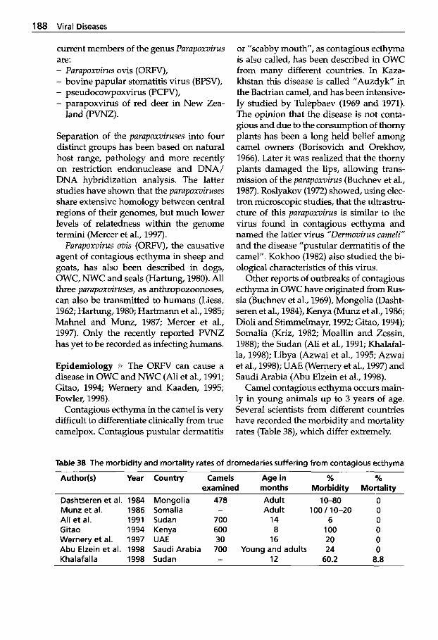
188 Viral Diseases
current members of the genus Parupoxvirus are: - Parupoxvirus ovis (ORFV), - bovine papular stomatitis virus (BPSV), - pseudocowpoxvirus (PCPV), - parapoxvirus of red deer in New Zea-
land (PVNZ).
Separation of the parupoxviruses into four distinct groups has been based on natural host range, pathology and more recently on restriction endonuclease and DNA/ DNA hybridization analysis. The latter studies have shown that the purupoxviruses share extensive homology between central regions of their genomes, but much lower levels of relatedness within the genome termini (Mercer et al., 1997).
Parupoxvirus ovis (ORFV), the causative agent of contagous ecthyma in sheep and goats, has also been described in dogs, OWC, NWC and seals (Hartung, 1980). All three purupoxviruses, as anthropozoonoses, can also be transmitted to humans (Liess, 1962; Hartung, 1980; Hartmann et al., 1985; Mahnel and Munz, 1987; Mercer et al., 1997). Only the recently reported PVNZ has yet to be recorded as infecting humans.
Epidemiology 1. The ORFV can cause a disease in OWC and NWC (Ali et al., 1991; Gitao, 1994; Wernery and Kaaden, 1995; Fowler, 1998).
Contagious ecthyma in the camel is very difficult to differentiate clinically from true camelpox. Contagious pustular dermatitis
or "scabby mouth", as contagious ecthyma is also called, has been described in OWC from many different countries. In Kaza- khstan this disease is called "Auzdyk" in the Bactrian camel, and has been intensive- ly studied by Tulepbaev (1969 and 1971). The opinion that the disease is not conta- gious and due to the consumption of thorny plants has been a long held belief among camel owners (Borisovich and Orekhov, 1966). Later it was realized that the thorny plants damaged the lips, allowing trans- mission of the parupoxvirus (Buchnev et al., 1987). Roslyakov (1972) showed, using elec- tron microscopic studies, that the ultrastru- cture of this parupoxvirus is similar to the virus found in contagious ecthyma and named the latter virus "Derrnovirus curneli" and the disease "pustular dermatitis of the camel". Kokhoo (1982) also studied the bi- ological characteristics of this virus.
Other reports of outbreaks of contagious ecthyma in OWC have originated from Rus- sia (Buchnev et al., 1969), Mongolia (Dasht- seren et al., 1984), Kenya (Munz et al., 1986; Dioli and Stimmelmayr, 1992; Gitao, 1994); Somalia (Kriz, 1982; Moallin and Zessin, 1988); the Sudan (Ali et al., 1991; Khalafal- la, 1998); Libya (Azwai et al., 1995; Azwai et al., 1998); UAE (Wernery et al., 1997) and Saudi Arabia (Abu Elzein et al., 1998).
Camel contagious ecthyma occurs main- ly in young animals up to 3 years of age. Several scientists from different countries have recorded the morbidity and mortality rates (Table 38), which differ extremely.
Table 38 The morbidity and mortality rates of dromedaries suffering from contagious ecthyma
Author(s) Year Country Camels Age in % % examined months Morbiditv Mortalitv
Dashtseren et al. 1984 Mongolia 478 Adult 10-80 0 Munz et al. 1986 Somalia - Adult 100 / 10-20 0 Ali et al. 1991 Sudan 700 14 6 0 Gitao 1994 Kenya 600 8 100 0 Wernery et al. 1997 UAE 30 16 20 0 Abu Elzein et al. 1998 Saudi Arabia 700 Young and adults 24 0 Khalafalla 1998 Sudan - 12 60.2 8.8
Viral Infections Causina Disease 189
Very little is known about the transmis- sion of the parapoxvirus. It is believed that natural transmission occurs by direct con- tact or indirectly from the environment or fomites. New findings indicate that the carrier animal is probably very important in the spread of the disease in sheep. Con- siderable evidence has shown that k- fected flocks grazing on pastures abundant in thistles, on which no sheep have grazed for many years, still succumb to the dis- ease (Lewis, 1996). Azwai et al. (1998) found that the seropositivity rate (ELISA) in Libyan camel herds with clinically affected dromedaries was 38% (and was related to clinical signs) and in apparently healthy herds was between 0% and 7Yo. Gitao (1994) believes that the common practice of keeping all camel calves in the same shel- ter at night could be responsible for the spread of the virus by contact, and he also proved that the outbreaks in camel calves occurred when parapoxvirus infections were also observed in goat kids raised nearby. Similar observations were made by Munz et al. (1986) and by Robertson (1976) who examined ORF infections in alpacas. Abu Elzein et al. (1998) reproduced camel con- tagious ecthyma experimentally in suscep- tible dromedaries, but experimentally-in- fected sheep were refractory to the camel virus. On the other hand, experimental in- fection with the ovine ORFV in drome- daries did not produce any disease in this animal species (Wemery, pers. corn.). Wer- nery and Kaaden (1995) reported three 8- month-old dromedaries in the UAE that developed and died from a mixed infec- tion of true camelpox and parapox during the course of an experimental camelpox vaccination program. The camels used as control animals were artificially infected with the camelpox virus. They might have developed a super infection with the con- tagious ecthyma virus or were latent carri- ers of the virus. Both viral species were seen situated next to one another upon electron microscopy (see Fig. 85).
NWC are also susceptible to contagious ecthyma virus (Preston Smith, 1940 and 1947; Moro, 1971; Ramirez, 1980; Thedford and Johnson, 1989; Fowler, 1998). Affected NWC develop typical proliferative lesions of the epidermis at the commissures of the mouth, which might spread to regions of the face and perineum. It is also possible that crias become infected when they suck- le their dams that have developed lesions on their teats. ORFV from lamoids has pro- duced severe ulcerating lesions on fingers, limbs and face in man (Fowler, 1998).
Clinical Signs and Pathology Two to 6 days post infection, primary lesions devel- op at the point of entry of the virus to the body. The lesions consist mainly of local- ized skin lesions of different magnitude, severity and location. Single or multiple primary pox lesions develop on the skin of the lips and muzzle. They frequently ex- tend to the skin of the eyelids and other parts of the head as well as to the buccal cavity, such as the palate and the gums be- low the incisor teeth. The lesions develop as reddish papules that change to yellow- ish pustules within a few days before be- coming nodular, ulcerated and hemor- rhagic. Secondary bacterial and fungal in- fections as well as myiasis may aggravate the lesions on the lips and mouth. Enlarge- ment of some superficial lymph nodes is also often observed.
Microscopic examination of the affected skin reveals parakeratosis, acanthosis, bal- looning degeneration of keratinocytes and inflammation and edema of the dermis. The lesions are often accompanied by focal ulcerations, neutrophilic and eosinophilic infiltrations and superficial bacterial and fungal colonies. Microscopic lesions are di- agnostic in early and acute stages, when cytoplasmic inclusion bodies are found in swollen epidermal cells but disappear in older lesions (6 days or more).
Contagious ecthyma in camels is usual- ly characterized by local pox-like lesions
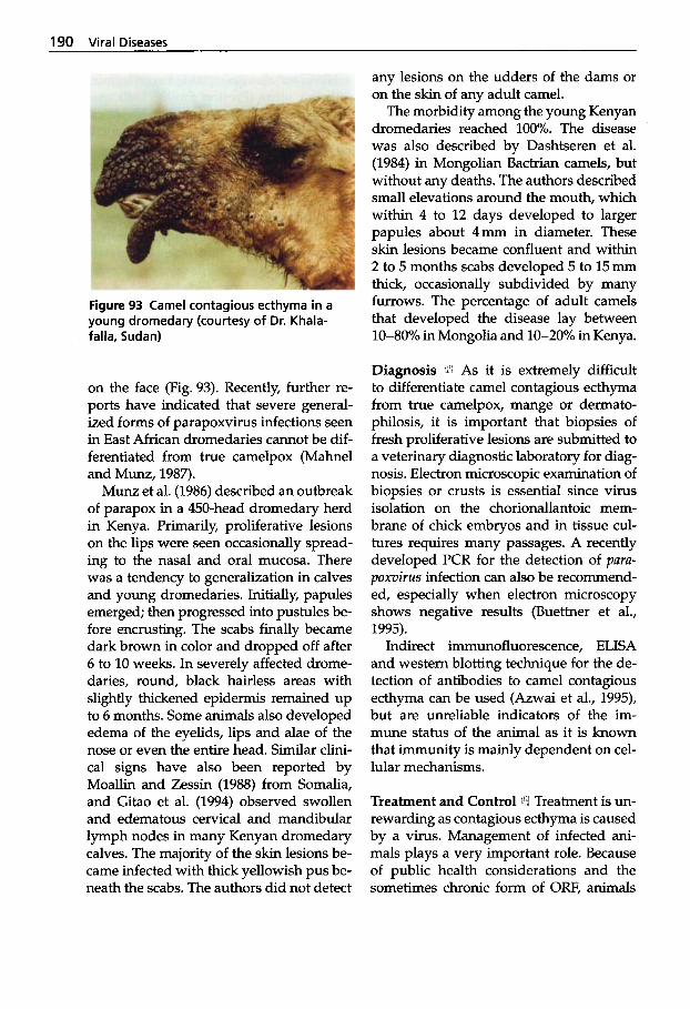
190 Viral Diseases
Figure 93 Camel contagious ecthyma in a young dromedary (courtesy of Dr. Khala- falla, Sudan)
on the face (Fig. 93). Recently, further re- ports have indicated that severe general- ized forms of parapoxvirus infections seen in East African dromedaries cannot be dif- ferentiated from true camelpox (Mahnel and Munz, 1987).
Munz et al. (1986) described an outbreak of parapox in a 450-head dromedary herd in Kenya. Primarily, proliferative lesions on the lips were seen occasionally spread- ing to the nasal and oral mucosa. There was a tendency to generalization in calves and young dromedaries. Initially, papules emerged; then progressed into pustules be- fore encrusting. The scabs finally became dark brown in color and dropped off after 6 to 10 weeks. In severely affected drome- daries, round, black hairless areas with slightly thickened epidermis remained up to 6 months. Some animals also developed edema of the eyelids, lips and alae of the nose or even the entire head. Similar clini- cal signs have also been reported by Moallin and Zessin (1988) from Somalia, and Gitao et al. (1994) observed swollen and edematous cervical and mandibular lymph nodes in many Kenyan dromedary calves. The majority of the skin lesions be- came infected with thick yellowish pus be- neath the scabs. The authors did not detect
any lesions on the udders of the dams or on the skin of any adult camel.
The morbidity among the young Kenyan dromedaries reached 100%. The disease was also described by Dashtseren et al. (1984) in Mongolian Bactrian camels, but without any deaths. The authors described small elevations around the mouth, which within 4 to 12 days developed to larger papules about 4mm in diameter. These skin lesions became confluent and within 2 to 5 months scabs developed 5 to 15 mm thick, occasionally subdivided by many furrows. The percentage of adult camels that developed the disease lay between 10-80% in Mongolia and 10-20% in Kenya.
Diagnosis iri As it is extremely difficult to differentiate camel contagious ecthyma from true camelpox, mange or dermato- philosis, it is important that biopsies of fresh proliferative lesions are submitted to a veterinary diagnostic laboratory for diag- nosis. Electron microscopic examination of biopsies or crusts is essential since virus isolation on the chorionallantoic mem- brane of chick embryos and in tissue cul- tures requires many passages. A recently developed PCR for the detection of para- poxvirus infection can also be recommend- ed, especially when electron microscopy shows negative results (Buettner et al., 1995).
Indirect immunofluorescence, ELISA and western blotting technique for the de- tection of antibodies to camel contagious ecthyma can be used (Azwai et al., 1995), but are unreliable indicators of the im- mune status of the animal as it is known that immunity is mainly dependent on cel- lular mechanisms.
Treatment and Control I/;\ Treatment is un- rewarding as contagious ecthyma is caused by a virus. Management of infected ani- mals plays a very important role. Because of public health considerations and the sometimes chronic form of OW, animals
Viral Infections Causina Disease 191
suspected of harboring the virus should be kept isolated from the herd until they re- cover fully. Stocking density should be re- duced as much as possible and attention directed at reducing any secondary infec- tion. Systemic treatment with high doses of synthetic penicillin against staphylococ- ci is probably the best approach. Veterinar- ians examining or treating camels suffer- ing from ORF should always wear gloves.
Neither vaccination nor natural infec- tion produces a long-lasting immunity. Re- covered sheep, for example, are only im- mune to re-infection for about 8 months after a primary infection. Attenuated vac- cines are routinely used in sheep and goats and might also be used in camelids. Stud- ies by Dashtseren et al. (1984) have shown that neither vaccinia virus nor the parapox- virus ovis vaccines protect camels against parapox disease. However, the authors have achieved protection against this disease using a camel parapoxvirus strain adapted in eggs. Vaccinated camels were protected for at least 6 months. It seems to be possi- ble that a bivalent vaccine against two of the most important viral diseases of camelids can be developed in the future.
References
Abu Elzein, E.M.E., E.R. Coloyan, A.A. Gameel, R.O. Ramadan and A.I. Al-Afaleq. 1998. Camel contagious ecthyma in Saudi Arabia. J. Camel Prac. and Res. 5 (2): 225-228.
Ali, O.A., S.A.M. Kheir, H. Abu Damir and M.E.S. Barri. 1991. Camel (Camelus drome- darius) contagious ecthyma in the Sudan. A case report. Rev. Elev. Mid. vit. Pays trop. 44
Azwai, S.M., S.D. Carter and Z. Woldehiwet. 1995. Immune responses of the camel (Cam- elus dromedarius) to contagious ecthyma (Orf) virus infection. Veterinary Microbiology
Azwai, S.M., S.D. Carter and Z . Woldehiwet. 1998. An immunological study of contagious pustular dermatitis in camels. Int. meeting on camel production and future perspectives. May
(2): 143-145.
47 (1-2): 119-131.
2-3,1998, A1 Ain, UAE: 108.
Borisovich, Y.F. and M.D. Orekhov. 1966. Camel pox. Veterinaryia, Moscow. Dated in Vet. Bull.
Buchnev, K.N., R.G. Sadykov, S. Zh. Tulepbayev and A.A. Roslyakov. 1969. Smallpox-like dis- ease of camels Auzdyk. Trudy Alma-Ata Tins- kogo Zootekhnicheskogo Instituta 16: 36-47.
Buchnev, K.N., S.Z. Tulepbaev and A.R. San- syzbaev. 1987. Infectious diseases of camels in the USSR. Rev. sci. tech. Of. int. Epiz. 6 (2):
Biittner, M., C. von Einem, C. Mclnnes and A. Oksanen. 1995. Klinik und Diagnostik einer schweren Parapocken-Epidemie beim Ren- tier in Finnland. Tierarztl. Praxis 23 (6): 614- 618.
Dashtseren, Ts., B.V. Solovyev, F. Varejka and A. Khokhoo. 1984. Camel contagious ecthyma (pustular dermatitis). Acta virol. 2 8 122-127.
Dioli, M. and R. Stimmelmayr. 1992. Important camel diseases in the one-humped camel in Eastem Africa. A pictorial guide to diseases, health care and management. H.J. Schwartz and M. Dioli (Eds.). Verlag Joseph Markgraf Scientific Books: pp. 155-164.
Fowler, M.E. 1998. Medicine and surgery of South American Camelids. Iowa State Uni- versity Press, Ames.
Gitao, C.G. 1994. Outbreaks of contagious ec- thyma in camels (Camelus dromedarius) in the Turkana District of Kenya. Rev. Sci. Tech.
Hartmann, A.A., M. Buettner, F. Stanka and P. Elner. 1985. Sero- und Immunodiagnostik bei Parapoxvirus - Infektionen des Menschen. Der Hautarzt 36: 663-669.
Hartung, J. 1980. Lippengrind des Schafes. Tier- arztl. Prax. 8: 435-438.
Khalafalla, A.I. 1998. Epizootiology of camel pox, camel contagious ecthyma and camel papillomatosis in the Sudan. Proc. Int. Meet- ing on camel prod. and future perspectives, Al Ain, UAE, May 2-3,1998: 105.
Khokhoo, A. 1982. Biological properties of camel contagious ecthyma virus. Thesis, Veterinary Institute, Bmo.
Kriz, 8.1982. A study of camelpox in Somalia. J. Comp. Path. 92: 1-8.
Lewis, C. 1996. Update on orf. In Practice 18 (8):
Liess, H. 1962. Lippengrind (Ecthyma contagio- sum) der Schafe als Zooanthroponose. Zbl. Bakteriol. Microbiol. Hyg. A183: 1969-1983.
1996,36,794 3: 50-52.
487-495.
13 (3): 939-945.
376-381.
192 Viral Diseases
Mahnel, H. and E. Munz. 1987. Zur derzeitigen epizootologischen Lage bei den Tierpocken. Tierarztl. Umschau 42 (1): 5-14.
Mercer, A., S. Fleming, A. Robinson, I? Nettleton and H. Reid. 1997. Molecular genetic analy- ses of parapoxviruses pathogenic for humans. Archives of Virology. (Suppl . 23) 13: 25-34.
Moallin, A.S.M. and K.H. Zessin. 1988. Out- break of camel contagious ecthyma in Central Somalia. Trup. Anim. Hlth. Prod. 2 0 185-186.
Moro, M. 1971. Ectima: En: La Alpaca. Enfer- medades Infecciosas y Parasitarias. Bol Di- vulgacion Instito Veteranario de Investiga- ciones Tropicales y de Altura. Unva Nac San Marcos, Lima, Peru: 30.
Munz, E., D. W i n g e r , M. Reimann and H. Mahnel. 1986. Electron microscopical diag- nosis of Ecthyma contagiosum in camels (Camelus dromedarius) First report of the disease in Kenya. 1. Vet. Med. B 33: 73-77.
Preston Smith, H. 1940. Los camello Peruanos, anguenidos, alpacas ectima. Ministr. Agric. Bol. (Lima).
Preston Smith, H. 1947. Ectima de 10s animales del Peru, dermatitis pustular contagiosa. Ganaderia (Peru) 1 (1): 27-32.
Ramirez, A. 1980. Ectuina contagioso en alpaca. En aspectos santarios en la alpaca. Curso siste- ma de production pecuaria en 10s altos Andes. Assoc. Peruana Prod. Anim., Lima, Peru: 94.
Robertson, A. 1976. Handbook on animal dis- eases in the Tropics. 31d ed. British Veterinary Association, London: 9-11.
Roslyakov, A.A. 1972. Comparison of the dtra- structure of camelpox virus, the virus of pox- like disease of camels and contagious ecthy- ma virus. Voprosy Virusologii 2 7, Zoovet Insti- tut, Alma-Ata, Kaz. SSR. Abstract: Vet. Bull.
Thedford, R.R. and L.W. Johnson. 1989. Infec- tious diseases of New-world camelids (NWC). Vet. Clin. North Am. Food Anim. Pract. 5 (3):
Tulepbaev, S. Zh. 1969. Sensitivity of domestic and laboratory animals to the virus of small- pox-like disease of camels (“Auzdyk”). Trudy Alma-atinskogo Zootekhnicheskogo Instituta 16:
Tulepbaev, S. Zh. 1971. Pox-like disease (”Auz- dyk”) of the camels in Kazakhstan. Diss. Kand., Alma-Ata.
Wemery, U., 0.-R. Kaaden and M. Ali. 1997. Orthopox virus infections in dromedary cam-
42,512 1: 26-30.
145-157.
41-42.
els in United Arab Emirates (UAE) during winter season. J. Camel Prac. and Res. 4 (1):
Wemery, U. and 0.-R. Kaaden. 1995. Infectious Diseases of Camelids. Blackwell Wissen- schafts-Verlag, Berlin.
51-55.
Further reading
Guo, S.Z. 1988. Serological comparison of the pathogens of aphthosis in camel, sheep and goat. Chinese J. Vet. Med. and Techn. 5: 35-37.
Khalafalla, A.I., H Agab and B. Abbas. 1994. An outbreak of contagious ecthyma in camels (Camelus dromedarius) in eastern Sudan. Trop. Anim. Hlth. Prod. 26: 253-254.
Khalafalla, A.I. and M.E.M. Mohamed. 1997. Epizootiology of camel contagious ecthyma in eastern Sudan. Rev. Elev. Mid. vkt. Pays trop. 50 (2): 99-103.
Khalafalla, A.I. 1999. Camel contagious ecthy- ma and its risk to young calves. Int. Workshop on the young camel, Quarzazate, Maroc, Oct.,
Mustapha, I.E. 1980. IFS Provisional report No. 6 on camels, 399. Stockholm: Int. Foundation for Science.
24-26,45.
2.1.5 Papillomatosis
Papillomas (warts) are benign neoplastic growths of the skin and mucous mem- branes and are observed worldwide in hu- mans and a variety of animals. They are caused by species-specific pupillornuviruses that have also been associated with the de- velopment of squamous cell carcinomas.
Cattle are more affected by warts than any other domestic animal species: 6 types of bovine papillomaviruses having been iden- tified. More than 70 pupillornuvirus sero- types are recognized in humans and cattle, while only 1 virus type has so far been iden- tified in each of the other animal species.
The papillornuvirus can also affect camels and cause typical skin lesions (Munz et al., 1990; Munz, 1992; Wernery and Kaaden, 1995; Khalafalla et al., 1998; Kinne and Wernery, 1998; Khalafalla, 1998).
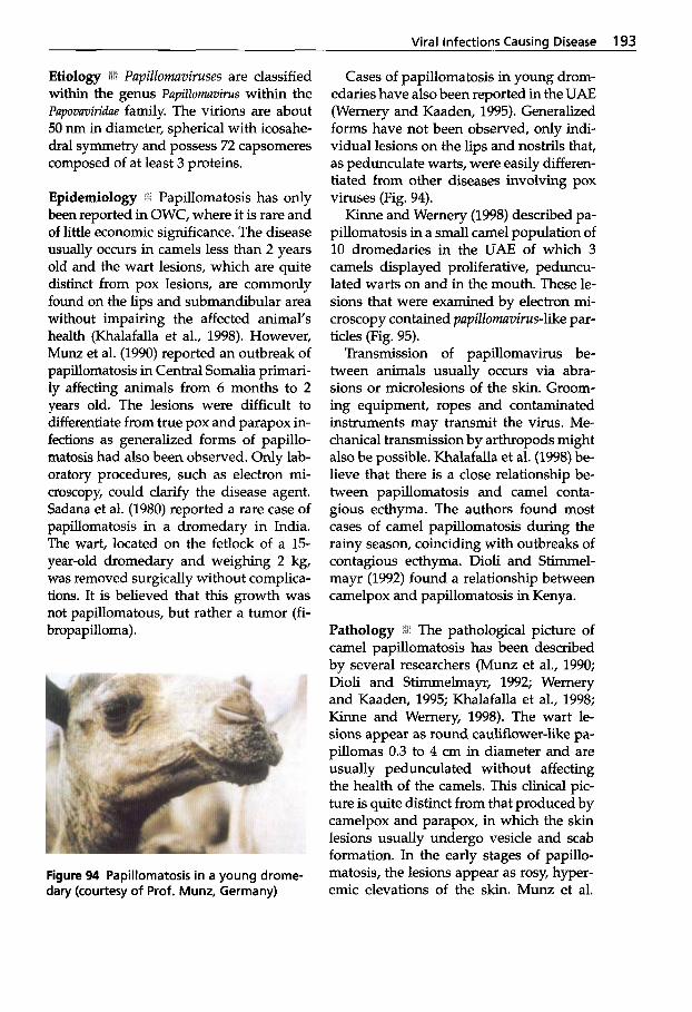
Viral Infections Causing Disease 193
Papillomaviruses are classified within the genus Papillomavirus within the Papovaviridae family. The virions are about 50 nm in diameter, spherical with icosahe- dral symmetry and possess 72 capsomeres composed of at least 3 proteins.
Epidemiology '1 Papillomatosis has only been reported in OWC, where it is rare and of little economic significance. The disease usually occurs in camels less than 2 years old and the wart lesions, which are quite distinct from pox lesions, are commonly found on the lips and submandibular area without impairing the affected animal's health (Khalafalla et al., 1998). However, Munz et al. (1990) reported an outbreak of papillomatosis in Central Somalia primari- ly affecting animals from 6 months to 2 years old. The lesions were difficult to differentiate from true pox and parapox in- fections as generalized forms of papillo- matosis had also been observed. Only lab- oratory procedures, such as electron mi- croscopy, could clarify the disease agent. Sadana et al. (1980) reported a rare case of papillomatosis in a dromedary in India. The wart, located on the fetlock of a 15- year-old dromedary and weighing 2 kg, was removed surgically without complica- tions. It is believed that this growth was not papillomatous, but rather a tumor (fi- bropapilloma).
Figure 94 Papillomatosis in a young drome- dary (courtesy of Prof. Munz, Germany)
Cases of papillomatosis in young drom- edaries have also been reported in the UAE (Wernery and Kaaden, 1995). Generalized forms have not been observed, only indi- vidual lesions on the lips and nostrils that, as pedunculate warts, were easily differen- tiated from other diseases involving pox viruses (Fig. 94).
Kinne and Wernery (1998) described pa- pillomatosis in a small camel population of 10 dromedaries in the UAE of which 3 camels displayed proliferative, peduncu- lated warts on and in the mouth. These le- sions that were examined by electron mi- croscopy contained papillomavirus-like par- ticles (Fig. 95).
Transmission of papillomavirus be- tween animals usually occurs via abra- sions or microlesions of the skin. Groom- ing equipment, ropes and contaminated instruments may transmit the virus. Me- chanical transmission by arthropods might also be possible. Khalafalla et al. (1998) be- lieve that there is a close relationship be- tween papillomatosis and camel conta- gious ecthyma. The authors found most cases of camel papillomatosis during the rainy season, coinciding with outbreaks of contagious ecthyma. Dioli and Stimmel- mayr (1992) found a relationship between camelpox and papillomatosis in Kenya.
The pathological picture of camel papillomatosis has been described by several researchers (Munz et al., 1990; Dioli and Stimmelmayr, 1992; Wernery and Kaaden, 1995; Khalafalla et al., 1998; Kinne and Wernery, 1998). The wart le- sions appear as round cauliflower-like pa- pillomas 0.3 to 4 c m in diameter and are usually pedunculated without affecting the health of the camels. This clinical pic- ture is quite distinct from that produced by camelpox and parapox, in which the skin lesions usually undergo vesicle and scab formation. In the early stages of papillo- matosis, the lesions appear as rosy, hyper- emic elevations of the skin. Munz et al.
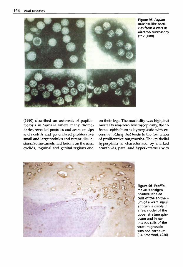
194 Viral Diseases
Figure 95 fapillo- mavirus-like parti- cles from a wart in electron microscopy (XI 25,000)
(1990) described an outbreak of papillo- matosis in Somalia where many drome- daries revealed pustules and scabs on lips and nostrils and generalized proliferative small and large nodules and tumor-like le- sions. Some camels had lesions on the ears, eyelids, inguinal and genital regions and
on their legs. The morbidity was high, but mortality was zero. Microscopically, the af- fected epithelium is hyperplastic with ex- cessive folding that leads to the formation of proliferative outgrowths. The epithelial hyperplasia is characterized by marked acanthosis, para- and hyperkeratosis with
Figure 96 fapillo- mavirus-antigen- positive labeled cells of the epitheli- um of a wart. Virus antigen is visible in a few nuclei of the upper stratum spin- osum and in nu- merous cells of the stratum granulo- sum and corneum (PAP-method, x220)
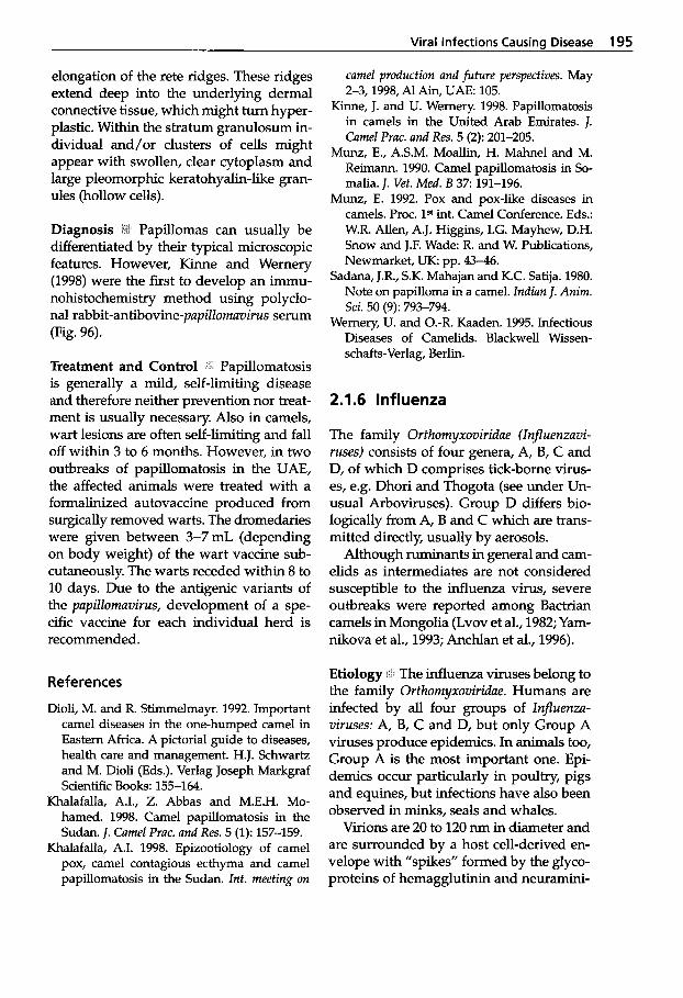
Viral infections Causina Disease 195
elongation of the rete ridges. These ridges extend deep into the underlying dermal connective tissue, which might turn hyper- plastic. Within the stratum granulosum in- dividual and/or clusters of cells might appear with swollen, clear cytoplasm and large pleomorphic keratohyalin-like gran- ules (hollow cells).
i Papillomas can usually be differentiated by their typical microscopic features. However, Kinne and Wernery (1998) were the first to develop an immu- nohistochemistry method using polyclo- nal rabbit-antibovine-papillomavirus serum (Fig. 96).
Treatment and Control I a Papillomatosis is generally a mild, self-limiting disease and therefore neither prevention nor treat- ment is usually necessary. Also in camels, wart lesions are often self-limiting and fall off within 3 to 6 months. However, in two outbreaks of papillomatosis in the UAE, the affected animals were treated with a formalinized autovaccine produced from surgically removed warts. The dromedaries were given between 3-7mL (depending on body weight) of the wart vaccine sub- cutaneously. The warts receded within 8 to 10 days. Due to the antigenic variants of the papillomavirus, development of a spe- cific vaccine for each individual herd is recommended.
References
Dioli, M. and R. Stimmelmayr. 1992. Important camel diseases in the one-humped camel in Eastern Africa. A pictorial guide to diseases, health care and management. H.J. Schwartz and M. Dioli (Eds.). Verlag Joseph Markgraf Scientific Books: 155-164.
Khalafalla, A.I., Z. Abbas and M.E.H. Mo- hamed. 1998. Camel papillomatosis in the Sudan. J. Camel Prac. and Res. 5 (1): 157-159.
Khalafalla, A.I. 1998. Epizootiology of camel pox, camel contagious ecthyma and camel papillomatosis in the Sudan. Int. meeting on
camel production and future perspectives. May
Kinne, J. and U. Wernery. 1998. Papillomatosis in camels in the United Arab Emirates. J. Camel Prac. and Res. 5 (2): 201-205.
Munz, E., A.S.M. Moallin, H. Mahnel and M. Reimann. 1990. Camel papillomatosis in So- malia. J. Vet. Med. B 37: 191-196.
Munz, E. 1992. Pox and pox-like diseases in camels. Proc. lSt int. Camel Conference. Eds.: W.R. Allen, A.J. Higgins, I.G. Mayhew, D.H. Snow and J.F. Wade: R. and W. Publications, Newmarket, UK: pp. 43-46.
Sadana, J.R., S.K. Mahajan and K.C. Satija. 1980. Note on papilloma in a camel. Indian J. Anim. Sci. 50 (9): 793-794.
Wernery, U. and 0.-R. Kaaden. 1995. Infectious Diseases of Camelids. Blackwell Wissen- schafts-Verlag, Berlin.
2-3,1998, A1 Ah, UAE: 105.
2.1.6 Influenza
The family Orthomyxoviridae (lnfuenzavi- ruses) consists of four genera, A, B, C and D, of which D comprises tick-borne virus- es, e.g. Dhori and Thogota (see under Un- usual Arboviruses). Group D differs bio- logically from A, B and C which are trans- mitted directly, usually by aerosols.
Although ruminants in general and cam- elids as intermediates are not considered susceptible to the influenza virus, severe outbreaks were reported among Bactrian camels in Mongolia (Lvov et al., 1982; Yam- nikova et al., 1993; Anchlan et al., 1996).
Etiology w The influenza viruses belong to the family Orthomyxoviridae. Humans are infected by all four groups of Influenza- viruses: A, B, C and D, but only Group A viruses produce epidemics. In animals too, Group A is the most important one. Epi- demics occur particularly in poultry, pigs and equines, but infections have also been observed in minks, seals and whales.
Virions are 20 to 120 nm in diameter and are surrounded by a host cell-derived en- velope with "spikes" formed by the glyco- proteins of hemagglutinin and neuramini-
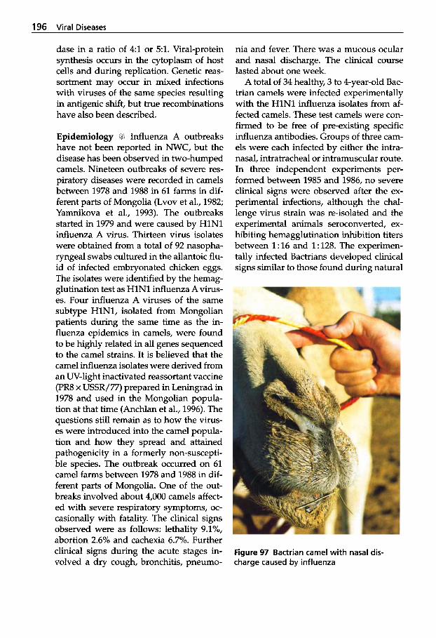
196 Viral Diseases
dase in a ratio of 4:l or 5:l. Viral-protein synthesis occurs in the cytoplasm of host cells and during replication. Genetic reas- sortment may occur in mixed infections with viruses of the same species resulting in antigenic shift, but true recombinations have also been described.
Epidemiology 81 Influenza A outbreaks have not been reported in NWC, but the disease has been observed in two-humped camels. Nineteen outbreaks of severe res- piratory diseases were recorded in camels between 1978 and 1988 in 61 farms in dif- ferent parts of Mongolia (Lvov et al., 1982; Yamnikova et al., 1993). The outbreaks started in 1979 and were caused by HlNl influenza A virus. Thirteen virus isolates were obtained from a total of 92 nasopha- ryngeal swabs cultured in the allantoic flu- id of infected embryonated chicken eggs. The isolates were identified by the hemag- glutination test as HlNl influenza A virus- es. Four influenza A viruses of the same subtype HlN1, isolated from Mongolian patients during the same time as the in- fluenza epidemics in camels, were found to be highly related in all genes sequenced to the camel strains. It is believed that the camel influenza isolates were derived from an UV-light inactivated reassortant vaccine (PR8 x USSR/77) prepared in Leningrad in 1978 and used in the Mongolian popula- tion at that time (Anchlan et al., 1996). The questions still remain as to how the virus- es were introduced into the camel popula- tion and how they spread and attained pathogenicity in a formerly non-suscepti- ble species. The outbreak occurred on 61 camel farms between 1978 and 1988 in dif- ferent parts of Mongolia. One of the out- breaks involved about 4,000 camels affect- ed with severe respiratory symptoms, oc- casionally with fatality. The clinical signs observed were as follows: lethality 9.1%, abortion 2.6% and cachexia 6.7%. Further clinical signs during the acute stages in- volved a dry cough, bronchitis, pneumo-
nia and fever. There was a mucous ocular and nasal discharge. The clinical course lasted about one week.
A total of 34 healthy, 3 to 4-year-old Bac- trian camels were infected experimentally with the HlNl influenza isolates from af- fected camels. These test camels were con- firmed to be free of pre-existing specific influenza antibodies. Groups of three cam- els were each infected by either the intra- nasal, intratracheal or intramuscular route. In three independent experiments per- formed between 1985 and 1986, no severe clinical signs were observed after the ex- perimental infections, although the chal- lenge virus strain was re-isolated and the experimental animals seroconverted, ex- hibiting hemagglutination inhibition titers between 1 : 16 and 1 : 128. The experimen- tally infected Bactrians developed clinical signs similar to those found during natural
Figure 97 Bactrian camel with nasal dis- charge caused by influenza
Viral Infections Causina Disease 197
infection (but milder): fever, coughing, bronchitis and discharge from nose and eyes (Fig. 97). All infected animals recov- ered. No further outbreaks among Bactrian camels have been reported since.
The influenza outbreak in Mongolian camels is convincing evidence that a reas- sortant between two human strains has caused severe epizootics among camels, which are not regarded as natural hosts for influenza A viruses. Safety requirements for cold-adapted reassortants must there- fore be adopted, because new strains may have a high pathogenicity for other species (Scholtissek, 1995).
Influenza-like epidemics in Somali cam- els have been reported by Auguadra (1958) and Somac-Sarec (1982) without any at- tempts to isolate the virus. The authors re- ported respiratory symptoms in conjunc- tion with rhinitis and conjunctivitis. Sero- logical studies to identdy antibodies to the influenza virus have been performed in various African countries. Olaleye et al. (1989) found 0.6% of the samples positive for the influenza A virus and 12.7% for the influenza B virus taken from slaughtered dromedaries in northeastern Nigeria. El- Amin and Kheir (1985) reported 7.89'0 of Sudanese camels positive for the influenza A virus.
Among influenza viruses, 12 different hemagglutinins (H) and 9 neuraminidases (N) have been identified. In the Mongo- lian influenza outbreak in Bactrian camels, only one combination HlNl occurred. Only two combinations have so far oc- curred in horses: H7N7 (influenza A/ equine-l/Prague/56) and H3N8 (influen- za/equine-2 /Miami/ 63). Major antigenic drift has been observed among H3N8 virus- es when mutations in the gene sequence result in amino acid substitutions, particu- larly in the hemagglutinin. One must be ex- tremely cautious in countries where horses and camels are kept in close vicinity, as not only antigenic drift occurs but also recom- binations of influenza viruses (antigenic
shift). Antigenic shift gives rise to new in- fluenza viruses which might result in pan- demics in susceptible populations. One of the countries where valuable horses and camels are kept in close vicinity is the UAE. In this country, the authors have ex- perienced annual outbreaks of respiratory disease in racing dromedaries caused by coccal infections. Furthermore, a serologi- cal survey on 500 UAE camels using the HIT with the equine strains Miami and Prague revealed no positive cases (CVRL Annual Report, 1998). However, the in- fluenza outbreaks in Mongolia proved that other influenza serotypes than Miami and Praha may infect camelids.
Diagnosis 11, In horses, for example, in- fluenza must be differentiated from other respiratory diseases like EHV-1 and EHV- 4, equine rhinoviruses, equine arteritis virus, Streptococcus equi equi (Strangles) and Rhodococcus equi. However, the rapid spread, the harsh cough and high temper- ature are sufficient to make a preliminary diagnosis. In vaccinated animals or in ani- mals that have overcome the disease, it is extremely difficult to diagnose. It is there- fore essential to carry out virus isolation and identification or serological tests.
Specimens for virus isolation should be collected after the onset of pyrexia and coughing, as virus excretion might be very short. Nasopharyngeal swabs have to be collected in virus transport medium and sent cooled to the laboratory as soon as possible. Influenza viruses should be cul- tured in embryonated eggs or in Madin- Darby canine kidney cells (MDCK). Sever- al passages may be required in order to iso- late the virus. The virus is identified by hemagglutination and subtyped using HIT with specific antisera. The HIT is also used for the detection of antibodies to the in- fluenza virus. A rapid diagnosis in equines is done with the Directogen FLU-A test kit (Becton Dickinson, USA) and should also be tried in camelids with influenza-like
198 Viral Diseases
clinical signs. However, the serological di- agnosis of infection in a vaccinated popu- lation is complicated by the presence of vaccine-induced antibodies.
Treatment and Control 1 ) The most effec- tive means of control in the face of an in- fluenza outbreak are vaccination and re- striction of movement of animals. No in- fluenza vaccines have been administered to camelids, but in case of an outbreak, vac- cination programs should be considered.
References Anchlan, D., S. Ludwig, P. Nymadawa, J. Mend-
saikhan and C. Scholtissek. 1996. Previous HlNl influenza A viruses circulating in the Mongolian population. Archives of Virology
Auguadra, P. 1958. Grippe 0 influenza del dromedario Somalo. Arch. ital. Sci. med. trop. Parasif. 34: 215-222.
CVRL. 1998. Annual Report. Central Veterinary Research Laboratory, Dubai, U.A.E.: 19.
El-Amin, M.A. and S.A. Kheir. 1985. Detection of influenza antibody in animal sera from Kas- sala region, Sudan, by agargel diffusion test. Rev. Elm. Mdd. vdf. Pays frop. 38 (2): 127-129.
Lvov, D.K., S.S. Yamnikova, I.G. Shemyakin, L.V. Agafonova, LA. Miyasnikova, E.A. Vla- dimirtseva, P. Nymadava, P. Dachtzeren, Z.H. Bel-Ochir and V.M. Zhadanov. 1982. Persistence of genes of epidemic influenza viruses. Voprosi Virus0127 401405.
Olaleye, O.D., S.S. Baba and S.A. Omolabu. 1989. Preliminary survey for antibodies against respiratory viruses among slaughter camels (Camelus dmmedarius) in northeastern Nige- ria. Rev. sci. tech. Of. int. Epiz. 8 (3): 779-783.
Scholtissek, C. 1995. Potential hazards associat- ed with influenza virus vaccines. Dev. Biol. Stand. 84: 55-58.
Somac/Sarec. 1982. Camel research project re- port by a Somali/Swedish Mission, March
Yamnikova, S.S., J. Mandler, Z.H. Bel-Ochir, P. Dachtzeren, S. Ludwig, D.K. Lvov and C. Scholtissek. 1993. A reassortant HlNl influen- za A virus caused fatal epizootics among camels in Mongolia. Virology 197 558-563.
141 (8): 1553-1569.
10-26: 18-23.
Further reading Wernery, U. 1999. New aspects on infectious
diseases of camelids. J. Camel Prac. and Res. 6 (1): 87-91.
2.1.7 Neonatal Diarrhea
Neonatal diarrhea in calves is one of the greatest sources of loss in animal breeding. Field and laboratory investigations have indicated that there is not a single etiology. The cause is complex and usually involves an interplay between enteropathogenic bacteria, viruses and parasites. On a clini- cal basis it is not usually possible to differ- entiate between the common known caus- es of diarrhea in newborns, which include enterotoxigenic E.coli (ENTEC), rotavirus, coronavirus, C yptosporidia spp. and Sal- monella spp. Rota- and coronaviruses have been identified as having characteristic lo- calizations on the mucosal epithelium of the jejunum, ileum (rotavirus) and colon (coro- navirus). The presence of viruses in the fe- ces is not always indicative of manifest dis- ease. Viral replication leads to the loss of function of the villous epithelium, causing the clinical signs (Freitag et al., 1984).
Etiology dl Rotaviruses are classified in the family Reoviridae, genus Rotavirus. Each ro- tavirus is named after the species in which it occurs.
Coronaviruses belong to the order Nido- virales, family Coronaviridae.
Epidemiology i Very little is known about the cause of neonatal diarrhea in camelids, but there is agreement that the most com- mon cause of death in camel calves up to 6 months of age is diarrhea (Khanna et al., 1992). In Sudan for example, Agab and Ab- bas (1998) reported a mortality rate higher than 30% in dromedary calves caused by diarrhea. There are only few reports that camelids might be susceptible to both ro-
Viral Infections Causina Disease 199
tavirus and coronavirus. Mattson (1994) believes that neonatal diarrhea in NWC oc- curs with a lower incidence than in cattle, pigs and sheep. Rotavirus has not been iso- lated from NWC, but Rivera et al. (1987) detected antibodies to rotavirus in alpacas. Coronaviruses have been seen by electron microscopic examination of feces in two llamas with diarrhea, but attempts to iso- late the virus in cell culture failed (Matt- son, 1994).
Rota- and coronaviruses were detected in fecal samples in UAE dromedary calves suffering from diarrhea using electron mi- croscopy (Mohamed et al., 1998; Ijaz et al., 2000 in prep.).
Rotaviruses were detected in a number of fecal samples from eastern Sudanese camel calves suffering from diarrhea using electron microscopy, ELISA and Latex ag- glutination (Khalafalla, unpublished). Eight out of 200 samples examined by Latex ag- glutination test, 11 out of 117 by ELISA and 4 out of 87 by electron microscopy were positive for group A rotaviruses. A season- ality of rotavirus infection in camel calves was observed. Most of the positive sam- ples were recorded in early winter (Octo- ber). Attempts to isolate rotavirus from eight positive fecal samples using MA 104 cell line (fetal monkey kidney) were un- successful up to the eighth blind passage. The genome of the camel rotavirus was an- alyzed by polyacrylamid gel electrophore- sis (PAGE), in comparison with group A human and equine rotavirus isolates. The results indicated that the profile of the camel rotavirus RNA was different.
Mahin et al. (1983) found in serological studies of Moroccan dromedaries that 50% of the animals (27/55) had antibodies to rotavirus. This proves that dromedaries are susceptible to rotavirus infection. Ro- tavirus antibodies were also detected by Puntel et al. (1999) in 390 llamas from 9 farms located in 3 different Argentinean provinces. The antibody prevalence was 87.7% (342/390), which indicates that this
species is highly susceptible to rotavirus. Chang-Say et al. (1985) showed that al- pacas are also susceptible to rotavirus.
In the UAE, corona-like agents were de- tected by electron microscopic investiga- tions in fecal samples of dromedary calves with diarrhea.
Diagnosis Rota- and coronavirus parti- cles can be demonstrated in preparations of fecal samples from diarrheic calves by transmission electron microscopy. How- ever, ELISA tests are more reliable and sen- sitive than electron microscopy and are nowadays widely used for the diagnosis of viral neonatal diarrhea. These tests also have the advantage of handling larger num- bers of specimens. Virus isolation in cell cultures is difficult and often fails.
Several methods are used for the detec- tion of antibodies to rota- and coronavirus- es such as SNT and HIT.
Neonatal diarrhea is often caused by a secondary immunoglobulin deficiency and it is therefore important to comment in this chapter on the passive transfer of immu-
Immunoglobulins (Ig) are divided into classes or isotypes (IgG, IgM, IgA, IgE, IgD) and further subdivided into subclasses. Most studies on animal immunology deal, however, with IgG, IgM or IgA (Table 39).
It is worthwhile mentioning that IgG antibodies in camelids differ from all other known antibodies and contradict all com- mon theories on antibody diversity. At present, three subclasses of camelid IgG have been identified (IgG 1,2,3), of which IgG2 and IgG3 lack the light chains (Fig. 98) (Hamers-Casterman et al., 1993; Azwai and Carter, 1995): - IgGl binding strongly to protein A and
G, composed of conventional antibod- ies, totaling 25% of serum IgG;
- IgG2 and IgG3 consisting of dimers of short heavy chains, which are character- ized by a normal Fc region without CH1 domain, totaling 75% of serum IgG.
nity.
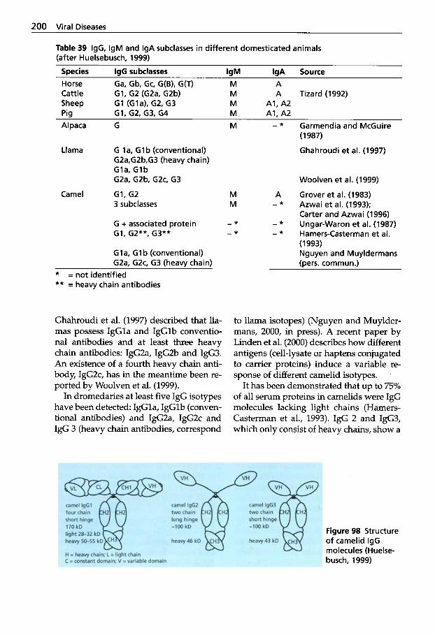
200 Viral Diseases
Table 39 IgG, IgM and IgA subclasses in different domesticated animals (after Huelsebusch, 1999)
Species IgG subclasses IgM IgA Source
Cattle GI, G2 (GZa, G2b) M A Tizard (1992) Sheep G1 (Gla), G2, G3 M Al, A2 Pig G1, G2, G3, G4 M Al, A2 Alpaca G M - * Garmendia and McGuire
Horse Ga, Gb, Gc, G(B), G(T) M A
(1 987)
Llama G la, G lb (conventional) G2a,G2b,G3 (heavy chain) Gla, G l b G2a, G2b. G2c. G3
Ghahroudi et al. (1997)
Woolven et al. (1999)
Camel G1, G2 M A Grover et al. (1983) 3 subclasses M - * Azwai et al. (1993);
Carter and Azwai (1996) G + associated protein - * - * Ungar-Waron et al. (1987) G1, G2**. G3** - * - * Hamers-Casterman et al.
Gla, G l b (conventional) GZa, G ~ c , G3 (heavy chain)
* = not identified ** = heavy chain antibodies
Ghahroudi et al. (1997) described that lla- mas possess IgGla and IgGlb conventio- nal antibodies and at least three heavy chain antibodies: IgG2a, IgG2b and IgG3. An existence of a fourth heavy chain anti- body, IgG2c, has in the meantime been re- ported by Woolven et al. (1999).
In dromedaries at least five IgG isotypes have been detected: IgGla, IgGlb (conven- tional antibodies) and IgG2a, IgG2c and IgG 3 (heavy chain antibodies, correspond
(1 993) Nguyen and Muyldermans (pers. commun.)
to llama isotopes) (Nguyen and Muylder- mans, 2000, in press). A recent paper by Linden et al. (2000) describes how different antigens (cell-lysate or haptens conjugated to carrier proteins) induce a variable re- sponse of different camelid isotypes.
It has been demonstrated that up to 75% of all serum proteins in camelids were IgG molecules lacking light chains (Hamers- Casterman et al., 1993). IgG 2 and IgG3, which only consist of heavy chains, show a
Figure 98 Structure of camelid IgG molecules (Huelse- busch, 1999)
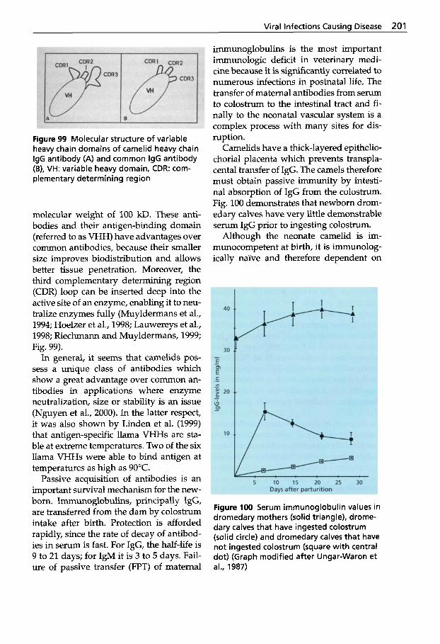
Viral Infections Causina Disease 201
Figure 99 Molecular structure of variable heavy chain domains of camelid heavy chain IgG antibody (A) and common IgG antibody (B), VH: variable heavy domain, CDR: com- plementary determining region
molecular weight of 100 kD. These anti- bodies and their antigen-binding domain (referred to as VHH) have advantages over common antibodies, because their smaller size improves biodistribution and allows better tissue penetration. Moreover, the third complementary determining region (CDR) loop can be inserted deep into the active site of an enzyme, enabling it to neu- tralize enzymes fully (Muyldermans et al., 1994; Hoelzer et al., 1998; Lauwereys et al., 1998; Riechmann and Muyldermans, 1999; Fig. 99).
In general, it seems that camelids pos- sess a unique class of antibodies which show a great advantage over common an- tibodies in applications where enzyme neutralization, size or stability is an issue (Nguyen et al., 2000). In the latter respect, it was also shown by Linden et al. (1999) that antigen-specific llama VHHs are sta- ble at extreme temperatures. Two of the six llama VHHs were able to bind antigen at temperatures as high as 90°C.
Passive acquisition of antibodies is an important survival mechanism for the new- born. Immunoglobulins, principally IgG, are transferred from the dam by colostrum intake after birth. Protection is afforded rapidly, since the rate of decay of antibod- ies in serum is fast. For IgG, the half-life is 9 to 21 days; for IgM it is 3 to 5 days. Fail- ure of passive transfer (FPT) of maternal
immunoglobulins is the most important immunologic deficit in veterinary medi- cine because it is significantly correlated to numerous infections in postnatal life. The transfer of maternal antibodies from serum to colostrum to the intestinal tract and fi- nally to the neonatal vascular system is a complex process with many sites for dis- ruption.
Camelids have a thick-layered epithelio- chorial placenta which prevents transpla- cental transfer of IgG. The camels therefore must obtain passive immunity by intesti- nal absorption of IgG from the colostrum. Fig. 100 demonstrates that newborn drom- edary calves have very little demonstrable serum IgG prior to ingesting colostrum.
Although the neonate camelid is im- munocompetent at birth, it is immunolog- ically na'ive and therefore dependent on
9 F
10
5 10 15 20 25 30 Days after parturition
Figure 100 Serum immunoglobulin values in dromedary mothers (solid triangle), drome- dary calves that have ingested colostrum (solid circle) and dromedary calves that have not ingested colostrum (square with central dot) (Graph modified after Ungar-Waron et al., 1987)
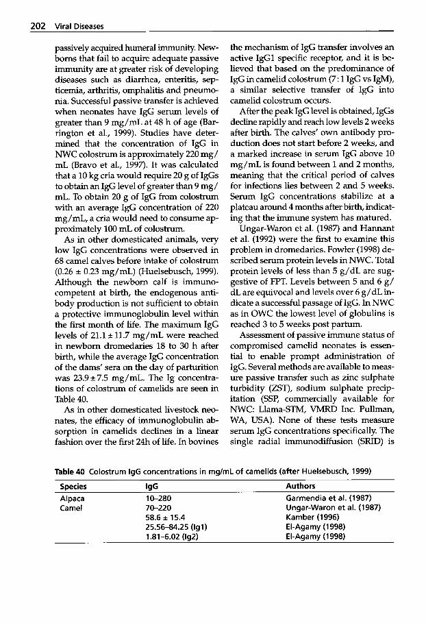
202 Viral Diseases
passively acquired humeral immunity. New- borns that fail to acquire adequate passive immunity are at greater risk of developing diseases such as diarrhea, enteritis, sep- ticemia, arthritis, omphalitis and pneumo- nia. Successful passive transfer is achieved when neonates have IgG serum levels of greater than 9 mg/mL at 48 h of age (Bar- rington et al., 1999). Studies have deter- mined that the concentration of IgG in NWC colostrum is approximately 220 mg/ mL (Bravo et al., 1997). It was calculated that a 10 kg cria would require 20 g of IgGs to obtain an IgG level of greater than 9 mg/ mL. To obtain 20 g of IgG from colostrum with an average IgG concentration of 220 mg/mL, a cria would need to consume ap- proximately 100 mL of colostrum.
As in other domesticated animals, very low IgG concentrations were observed in 68 camel calves before intake of colostrum (0.26 0.23 mg/mL) (Huelsebusch, 1999). Although the newborn calf is immuno- competent at birth, the endogenous anti- body production is not sufficient to obtain a protective immunoglobulin level within the first month of life. The maximum IgG levels of 21.1 2 11.7 mg/mL were reached in newborn dromedaries 18 to 30 h after birth, while the average IgG concentration of the dams’ sera on the day of parturition was 23.9 2 7.5 mg/mL. The Ig concentra- tions of colostrum of camelids are seen in Table 40.
As in other domesticated livestock neo- nates, the efficacy of immunoglobulin ab- sorption in camelids declines in a linear fashion over the first 24h of life. In bovines
the mechanism of IgG transfer involves an active IgGl specific receptor, and it is be- lieved that based on the predominance of IgG in camelid colostrum (7: 1 IgG vs IgM), a similar selective transfer of IgG into camelid colostrum occurs.
After the peak IgG level is obtained, IgGs decline rapidly and reach low levels 2 weeks after birth. The calves’ own antibody pro- duction does not start before 2 weeks, and a marked increase in serum IgG above 10 mg/mL is found between 1 and 2 months, meaning that the critical period of calves for infections lies between 2 and 5 weeks. Serum IgG concentrations stabilize at a plateau around 4 months after birth, indicat- ing that the immune system has matured.
Ungar-Waron et al. (1987) and Hannant et al. (1992) were the first to examine this problem in dromedaries. Fowler (1998) de- scribed serum protein levels in NWC. Total protein levels of less than 5 g/dL are sug- gestive of FPT. Levels between 5 and 6 g/ dL are equivocal and levels over 6 g/dL in- dicate a successful passage of IgG. In NWC as in OWC the lowest level of globulins is reached 3 to 5 weeks postpartum.
Assessment of passive immune status of compromised camelid neonates is essen- tial to enable prompt administration of IgG. Several methods are available to meas- ure passive transfer such as zinc sulphate turbidity (ZST), sodium sulphate precip- itation (SSP, commercially available for NWC: Llama-STM, VMRD Inc. Pullman, WA, USA). None of these tests measure serum IgG concentrations specifically. The single radial immunodiffusion (SRID) is
Table 40 Colostrum IgG concentrations in rng/rnL of carnelids (after Huelsebusch, 1999)
SDecies IQG Authors Alpaca 10-280 Garrnendia et at. (1987) Camel 70-220 Ungar-Waron et at. (1987)
58.6 i 15.4 Karnber (1996) 25.56-84.25 (lgl) El-Agarny (1998) 1.81-6.02 (192) El-Agarny (1998)
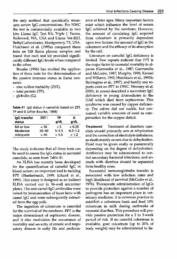
Viral Infections Causing Disease 203
the only method that specifically meas- ures serum IgG concentrations. For NWC the test is commercially available in two kits: Llama IgG Test Kit, Triple J. Farms, Redmond, WA, USA and Llama Vet-RID, Bethyl Laboratories, Montgomery, TX, USA. Hutchison et al. (1995a) compared these tests on 528 llama plasma samples and found that each test kit provided signifi- cantly different IgG levels when compared to the other.
Bourke (1996) has studied the applica- tion of three tests for the determination of the passive immune status in llama neo- nates: - zinc sulfate turbidity (ZST), - total protein (TP), - globulin (G).
Table 41 IgG status in carnelids based on ZST, TP and G (after Bourke, 1996)
IgG transfer ZST I TP G status gldL gIdL Nil or low c 30 c 5 c 0.25
Adequate >40 > 5.5 > 1.2 Moderate 30-40 5-5.5 0.3-1.2
The study indicates that all three tests can be used to assess the IgG status in neonatal camelids, as seen from Table 41.
An ELISA has recently been developed for the quantification of camelid IgG in blood serum; an important tool in tackling FPT (Huelsebusch, 1999; Erhard et al., 1999). This assay is designed as an indirect ELISA carried out in 96-well microtiter plates. The anti-camel-IgG antibodies were raised by immunization of layer hens with camel IgG and were subsequently extract- ed from the egg-yolk.
The ingestion of colostrum is essential for the survival of the newborn. FPT is the major determinant of septicemic disease, and it also modulates the occurrence of mortality and severity of enteric and respi- ratory disease in early life and perform-
ance at later ages. Many important factors exist which influence the level of serum IgG achieved by the newborn. However, the amount of circulating IgG acquired from colostrum is primarily dependent upon two factors: the amount of IgG in the colostrum and the efficacy of its absorption by the calf.
Literature on camelid IgG deficiency is limited. Few reports indicate that FPT is the major factor in neonatal mortality in al- pacas (Garmedia et al., 1987; Garmendia and McGuire, 1987; Murphy, 1998; Kennel and Wilkens, 1992; Hutchison et al., 1995b; Barrington et al., 1997), and hardly any re- ports exist on FPT in OWC. Wernery et al. (2001, in press) described a secondary IgG deficiency in young dromedaries in the UAE which died from septicemias. This syndrome was caused by copper deficien- cy. The calves did not suckle, but con- sumed variable amounts of sand as com- pensation for the copper deficit.
Treatment Treatment of diarrheic cam- elids should primarily aim at rehydration and the correction of electrolyte imbalance, as death mainly occurs due to dehydration. Fluid may be given orally or parenterally depending on the degree of dehydration. Antibiotics may be administered to con- trol secondary bacterial infections, and an- imals with diarrhea should be separated from healthy ones.
Successful immunoglobulin transfer is associated with low infection rates and high likelihood of survival (McGuire et al., 1976). Therapeutic administration of IgGs to provide protection against a number of pathogens has an important place in vet- erinary medicine. It is common practice to establish a colostrum bank and feed 10% colostrum in milk during outbreaks of neonatal diarrhea. This procedure will pro- vide passive protection for a 2 to 3-week period of risk. If no camelid colostrum is available, goat colostrum (up to 20% of body weight) may be administered to lla-
204 Viral Diseases
mas as a substitute (Pugh, 1992; Pugh and Belknap, 1997). For the treatment of FPT, it is also possible to give 20 to 40mL of camelid plasma intravenously. It should be given over a 30 to 60 min time frame and warmed to 37°C. Llama hyperimmunplas- ma is commercially available at Triple J. Farms, Redmond, WA, USA.
Viral Neonatal Diarrhea in calves is dif- ficult to control because the etiology is of- ten complex and the disease has a rapid course. Maternal vaccination may be an al- ternative approach. The use of commercial vaccines 1 to 3 months before calving can significantly reduce the prevalence of rota- and corona diarrhea in affected animals. In cases of viral Neonatal Diarrhea in camelid calves, vaccination programs should there- fore be considered.
References
Agab, H. and 8. Abbas. 1998. Epidemiological studies on camel diseases in eastern Sudan: 11. Incidence and causes of morality in pas- toral camels. Camel Newsletter 14 (4): 53-58.
Azwai, S.M., S.D. Carter and Z. Woldehiwet. 1993. The isolation and characterization of camel (Camelus dromedarius) immunoglob- ulin classes and subclasses. J. Comp. Path. 109:
Azwai, S.M. and S.D. Carter. 1995. Monoclonal- antibodies against camel (Camelus drome- darius) IgG, IgM and light chains. Vet. Im- munology and Imrnunopatk. 45 (1-2): 175-184.
Barrington, G.M., S.M. Parish, J.W. Tyler, D.G. Pugh and D.E. Anderson. 1997. Chronic weight loss in an immunodeficient adult lla- ma. J. Am. Vet. Med. Assoc. 211 (3): 295-298.
Barrington, G.M., S.M. Parish and F.B. Garry. 1999. Immunodeficiency in South American Camelids. J. Camel Prac. and Res. 6 (2): 185-190.
Bourke, D.A. 1996. Determination of passive immune status in llama neonates. Proceedings of the 3rd Br. Vet. Camelid SOC., Burford: 39-45.
Bravo, PW., J. Garnica and M.E. Fowler. 1997. Immunoglobulin G concentrations in peri- parturient llamas, alpacas and their crias. Small Ruminant Res. 26: 145-149.
Carter, S.D. and S.M. Azwai. 1996. Immunity and infectious diseases in the dromedary
187-195.
camel. Proc. Brit. Vet. Camelid SOC., Burford, November 14-16,1996 23-36.
Chang-Say, F., H. Rivera and H. Samame. 1985. Reporte preliminar sobre prevalencia de virus influenza tip0 A rotavirus en alpacas. Convencion Int. sobre Camelidos Sudameri- canos V, Cuzco, Peru: 37.
Erhard, M.H., S.A. Kouider, M.N. Dabbag, F. Schickel and M. Stangassinger. 1999. Deter- mination of serum IgG levels in camels by a bovine specific sandwich ELISA. J. Camel Prac. and Res. 6 (1): 15-18.
El-Agamy, E.I. 1998. Camel's colostrum. Anti- microbial factors. Dromadaires et chameaux, animaux laitiers. Ed. Bonnet, P. Actes du col- loque, 24-26 Octobre 1994, Noaukchott, Mauritanie: 177-179.
Freitag, H., H. Wetzel and E. Espenkotter. 1984. Aus der Praxis. Zur Prophylaxe der Rota- Corona-Virus-bedingten Kalberdiarrho. Tier- urztl. Umschau 39 (10): 731-736.
Fowler, M.E. 1998. Medicine and surgery of South American Camelids. Iowa State Uni- versity Press. Ames.
Garmendia, A.E. G.H. Palmer, J.C. DeMartini and T.C. McGuire. 1987. Failure of passive immunoglobulin transfer: a major determi- nant of mortality in newborn alpacas (Lama pacos). Am. J. Vet. Res. 4 8 1472-1476.
Garmendia, A.E. and T.C. McGuire. 1987. Mech- anism and isotypes involved in passive im- munoglobulin transfer to newborn alpaca (Lama pacos). Am. J. Vet. Res. 48: 1465-1471.
Ghahroudi, K.B., A. Desmyter, L. Wyns, R. Hamers and S. Muyldermans. 1997. Compar- ison of llama VH sequences from conven- tional and heavy-chain antibodies. EMBO J,
Grover, Y.P., Y.K. Kaura, S. Prasad and S.N. Sri- vastava. 1983. Preliminary studies on camel serum immunoglobulins. Ind. J. Biochem. Bio-
Hamers-Casterman, C., T. Atarhouch, S. Muyl- dermans, G. Robinson, C. Hamers, E. Bajyana Songa, N. Bendahman and R. Hamers. 1993. Naturally occurring antibodies devoid of light chains. Nature 363: 446-448.
Hannant, D., J.A. Mumford, U. Wernery and J.M. Bowen. 1992. ELISA for camel IgG and measurement of colostral transfer. Proc. 1 s t int. Camel Conf. Eds: W.R. Allen, A.J. Higgins, I.G. Mayhew, D.H. Snow and J.F. Wade: R. and W. Publications, Newmarket, UK: 93-95.
17 (13): 3512-3520.
pkys. 20 238-240.
Viral Infections Causing Disease 205
Hoelzer, W., S. Muyldermans and U. Wernery. 1998. Anote on camel IgG antibodies. J. Camel Prac. and Res. 5 (2): 187-188.
Huelsebusch, Chr. 1999. Immunoglobulin G sta- tus of camels during 6 months post natum. Hohenheim Tropical Agricultural Series, Mar- graf Verlag.
Hutchison, J.M., M.D. Salman, F.B. Garry. L.W. Johnson, J.K. Collins and T.Y. Keefe. 1995a. Comparison of two commercially available single radial immunodiffusion kits for quan- titation of llama immunoglobulin G. J. Vet. Diagn. Invest. 7 515-519.
Hutchison, J.M., F.B. Garry, E.B. Belknap, D.M. Geky, L.M. Johnson, R.P. Ellis, S.L. Quacken- busch, J. Ravnak, E.A. Hoover and G.L. Cockerell. 1995b. Prospective characterisa- tion of the clinicopathologic and immuno- logic features of an immunodeficiency syn- drome affecting juvenile llamas. Vet. lmmun. and Immunopath. 49: 209-227.
Ijaz, M.K., I. Ahmad, T.A. Alkarmi, F.K. Dar, J.A.R. Al-Masri, A.M.I. Al-Mugheryi, A.I.A. Mohammed, A.K. Gorde and S. Herlekar. 2000 (in prep.). Detection of coronavirus-like agents (CVLA) in diarrhoea of neonatal calves born to racing camels.
Kamber, R. 1996. Untersuchungen iiber die Ver- sorgung von neugeborenen Kamelfohlen (Camelus dromedarius) mit Immunglobulin- G. Thesis, Zurich University, Switzerland.
Kennel, A.J. and J. Wilkens. 1992. Causes of mortality in farmed llamas in North America. Necropsy findings from the database of a major insurer. Int. Llama Assoc. Res. Corn., Rochester, MN, USA.
Khanna, N.D., S.N. Tandon and M.S. Sahani. 1992. Calf mortality in Indian camels. Proc. 1st Int. Camel Cord. Eds: W.R. Allen, A.J. Higgins, I.G. Mayhew, D.H. Snow and J.F. Wade: R. and W. Publications, Newmarket,
Lauwereys, M., M.A. Ghahroudi, A. Desmyter, J. Kinne, W. Hoelzer, E. de Genst, L. Wyns and S. Muyldermans. 1998. Potent enzyme inhibitors derived from dromedary heavy- chain antibodies. EMBO J. 17 (13): 3512-3520.
Linden, van der R.H.J., L.G.J. Frenken, B. de Geus, M.M. Harmsen, R.C. Ruuls, W. Stok, L. de Ron, S. Wilson, I? Davis and C.T. Verrips. 1999. Comparison of physical chemical prop- erties of llama V-HH antibody fragments and mouse monoclonal antibodies. Biochemica et
U K 89-92.
Biophysica Acta-Protein Structure and Molecu- lar Enzymology 1431 (1): 3746.
Linden, R. van der, 8. de Geus, W. Stok, W. Bos, D. van Wassenaar, T. Verrips and 1. Frenken. 2000. Induction of immune responses and molecular cloning of the heavy chain anti- body repertoire of Lama glama. J. Immun. Methods 240 185-195.
McGuire, TC., N.E. Pfeiffer and J.M. Weikel. 1976. Failure of colostral immunoglobulin transfer in calves dying from infectious dis- eases. J. Am. Vet. Med. Assoc. 169: 713-718.
Mahin, L. A., Schwers, M., Chadli, M. Maen- houdt and P.P. Pastoret. 1983. R6ceptivit6 du dromadaire (Camelus dromedarius) a l'infec- tion par rotavirus. Rev. Elev. M6d. ve't. Pays. trop. 36 (3): 251-252.
Mattson, D.E. 1994. Viral Diseases. Vet. Clin. North America: Food and Animal Practice 10
Mohamed, M.E.H., C.A. Hart and 0.-R. Kaa- den. 1998. Agents associated with camel diar- rhea in Eastern Sudan. Proceed. Int. meeting on camel production and future perspectives, Al Ain, U.A.E., 2-3 May 1998.
Murphy, P.J. 1998. Obstetrics, neonatal care and congenital conditions. Vet. Clinics of North America: Food Animal Practice. 5: 1983-202.
Muyldermans, S., T. Atarhouch, J. Saldanha, J.A.R.G. Barbosa and R. Hamers. 1994. Se- quence and structure of VH domain from naturally occurring camel heavy immuno- globulins lacking light chains. Protein Engi- neering 7 1129-1131.
Nguyen, V.K., R. Hamers, L. Wyns and S. Muyl- dermans. 2000. Camel heavy-chain antibod- ies: diverse germline VHH and specific mech- anisms enlarge the antigen binding reper- toire. EMBO J. 19 (5): 921-930.
Pugh, G.H. 1992. Prepartum care of the preg- nant llama and neonatal care of the cria. Proc. Annual Meeting, SOC. for Theriogenology, Hast- ings, Neb., pp. 198-201.
Pugh, G.H., and E.B. Belknap. 1997. Perinatal and neonatal care of South American cam- elids. Vet. Med. 292-297.
Puntel, M., N.A. Fondevila, J. Blanco Viera, V.K. ODonnell, J.F. Marcovechio, B.J. Carillo and A.A. Schudel. 1999. Serological survey of vi- ral antibodies in llamas (Lama glama) in Ar- gentina. J. Vet. Med. B. 46: 157-161.
Riechmann, L. and S. Muyldermans. 1999. Sin- gle domain antibodies: comparison of camel
(2): 345-351.
206 Viral Diseases
VH and camelised human V H domains. J. Zmmunol. Methods 231 (1-2): 25-38.
Rivera, H., B.R. Madwell and E. Ameghina. 1987. Serological survey of viral antibodies in the Peruvian alpaca (Llama pacos). Am. J. Vet. Res. 4 8 189-191.
Tizard, I. 1992. Veterinary Immunology-An In- troduction. W.B. Saunders Co., Philadelphia, Penn., USA: p. 498.
Ungar-Waron, H., E. Elias, A. Gluckman and Z. Trainin. 1987. Dromedary Ig G: purification, characterization and quantitation in sera of dams and newborns. Zsr. J. Vet. Med. 43 (3):
Wernery, U., M. Ali, J. Kinne, A.A. Abraham and Renate Wernery. 2001, in press. Copper defi- ciency: A predisposing factor to septicemia in dromedary calves. Proc. of 2nd Camelid Con5 "Agroeconomics of Camelid Farming", Almaty, Kazakhstan, 8-12 Sept. 2000.
Woolven, B.P., L. Frenken, P. van der Logt and l?J. Nicholls. 1999. The structure of llama heavy-chain constant genes reveals a mecha- nism for heavy-chain antibody formation. J. lmmunogenetics 50: 98-101.
198-203.
Further reading Berrada, J., M. Bengoumi and K. Hidane. 1999.
Diarrhkes nkonatales du chamelon dans les provinces Sahariennes du sud du Maroc: Etude bactkriologique. 1999. Znt. Workshop on the young camel, Quarzazate, Maroc, Oct.,
Broadbent, R. 1994. The neonate Proceedings of the Br. Vet. Camelid SOC., Bristol: 50-64.
Dioli, M. 1999. Diseases and pathological condi- tions of camel calf in eastern Africa. lnt. Work- shop on the young camel, Quarzazate, Maroc, Oct., 24-26: 43.
Gandega, B.E., M. Bengoumi, A. Fikri, A. El Abrak, M. Aissa and B. Faye. 1999. Znt. Work- shop on the young camel, Quarzazate, Maroc, Oct., 24-26: 32.
Huelsebusch, Chr. G. 1999. Immunoglobulin-G status of camels during 6 months post natum. lnt. Workshop on the young camel, Quarzazate, Maroc, Oct., 24-26: 17.
Kataria, A.K. 1999. Molecular characterisation and immunobiology of camel serum and lacteal immunoglobulin classes and subclass- es and their role in immunity. Thesis, College of Vet. and Anim. Sci., Rajastan, India.
24-26: 53.
Kaufmann, B. 1999. Camel calf losses in pastoral herds of northern Kenya-a system compari- son. Znt. Workshop on the young camel, Quarza- zate, Maroc, Oct., 24-26: 33.
Leipold, H.W., T. Hiraga and L.W. Johnson. 1994. Congenital defects in the llama. In: Vet. Clinics of North America: Food Anim. Pract. 10 (2): 401402.
Nagpal, G.K. and G.N. Purohit. 1999. Disease prevalence and calf mortality in camel rear- ing areas in Bikaner. lnt. Workshop on the young camel, Quarzazate, Maroc, Oct., 24-26: 34.
Sheriff, St. and K.L. Constantine. 1996. Redefin- ing the minimal antigen-binding fragment. Nat. Struct. Biol. 3 (9): 733-736.
Spinelli, S., L. Frenken, D. Bourgeois, L. de Ron, W. Bos, T. Verrips, C. Anguille, C. Cambillau and M. Tegoni. 1996. The crystal structure of a llama heavy chain variable domain. Nat. Struct Biol. 3 (9): 752-757.
Tandon, S.N. and M.S. Sahani. 1999. Morbidity and mortality rate in dromedary calves. lnt. Workshop on the young camel, Quarzazate, Maroc, Oct., 24-26: 35.
Wernery, U. 1999. New aspects on infectious diseases of camels. J. Camel Prac. and Res. 6 (1): 87-91.
2.1.8 Equine Herpesvirus
Interestingly, no herpesvirus unique to camelids has been identified so far. Equine herpesvirus type 1 (EHV-I) produces rhinopneumonitis and abortion in horses and the disease has also been reported in OWC and NWC (Jenkins, 1985; Torres et al., 1985; Rebhun et al., 1988; Thedford and Johnson, 1989; House et al., 1991; Bildfell et al., 1996).
Etiology 1' At least seven different her- pesviruses were isolated from horses. The most important pathogens are EHV-1, the causative agent of equine abortion and neurological disorders, and EHV-4, the eti- ological agent of equine rhinopneumoni- tis. Both viruses are members of the genus Varicellovirus within the family Herpesviri- due.
Viral Infections Causing Disease 207
Epidemiology and Pathology 0 EHV-1 has worldwide distribution and although it is considered a disease of equids, has been isolated from other animal species including bovines, zebras and antelopes. In equines, it causes a respiratory disease most frequently seen in foals and year- lings. In pregnant mares, it is a major cause of abortions in late gestation. Additionally, EHV-1 can also cause neurological disturb- ances associated with encephalomyelitis. When herpesviruses infect a non-adapted host, serious disease or death is likely to result (Fowler, 1996). This has occurred in both NWC and OWC. EHV-1 infections have been described in a group of 100 al- pacas and llamas on an exotic animal farm (Purse11 et al., 1979; Jenkins, 1985; Rebhun et al., 1988). One hundred lamoids had been brought to the USA from Chile where they had been in close contact with other llamas, camels, gnus and various species of antelopes. A herpesvirus indistinguish- able from EHV-1 was isolated from dead llamas and alpacas that had suffered from blindness and central nervous system dis- turbances such as nystagmus, torticollis and paralysis. The blindness was believed to have been caused by chorioretinitis or optic neuritis.
EHV-1 appears to have followed an un- usual pattern in alpacas and llamas ex- posed to the virus. In a subsequent study (House et al., 1991) three llamas were ex- perimentally infected intranasally with EHV-1 isolated from the brain of an alpaca with severe neurological signs. Two of the three llamas developed severe neurologi- cal disorders: one died and one was eutha- nized. The third llama showed only mild neurological signs. EHV-1 was only re-iso- lated from a sample of the thalamus of the llama that had died acutely. These investi- gations demonstrate a difference between EHV-1 infection in equids and NWC. In equines, a viremia occurs after initial virus replication, whereas in NWC the v h s is believed to replicate in the cells of the mu-
cous membranes of the nasal cavity, where it infects the olfactory nerve and optic nerve and progresses to the central nervous sys- tem. There are no reports that EHV-1 in- duces abortions in camelids. EHV-1 infects not only NWC but also OWC. Bildfell et al. (1996) cultured EHV-1 from the brain of a Bactrian camel suffering from severe neu- rological disease prior to death.
Microscopic lesions in the brain of the Bactrian camel included a non-suppura- tive meningoencephalitis with vasculitis, necrosis and edema. These features are similar to those in EHV-l-induced neuro- logical disease in llamas and horses. How- ever, there was no ocular damage detected in this case. These investigations show that NWC and OWC can suffer from EHV-1 in- fections associated with nervous system signs, blindness and death. EHV-1 can cause seroconversion in NWC, but anti- bodies to EHV-1 have not been reported in OWC. Antibodies to EHV-1 were found in 21 serum samples from llamas and alpacas suffering an EHV-1 infection (Rebhun et al., 1988). Only 1 llama of 270 from Oregon possessed antibodies. Puntel et al. (1999) reported an antibody prevalence of 0.77% in 390 (3/390) llamas from 9 farms in 3 dif- ferent Argentinean provinces. No antibod- ies to EHV-1 were found in 500 UAE drom- edaries when tested with a sandwich ELISA (CVRL Annual Report, 1998).
Camelids are susceptible to EHV-1. With the increase in breeding of NWC in differ- ent countries and an increased opportuni- ty for both NWC and OWC to come into contact with equids, not only EHV-1 but also other equine viral infections should be considered in a differential diagnosis. Ef- forts should be undertaken to clarify the role of EHV-1 in abortions and neonatal diseases in camelids.
Diagnosis EHV-1 infections cannot be diagnosed solely on the basis of clinical signs. Confirmation of the disease can be achieved by virus isolation on a wide
208 Viral Diseases
range of cell cultures, including rabbit kidney (RK 13), Vero and EDMIN (equine dermal cells), by IFAT and by immunohis- tochemical staining for viral antigen in en- dothelial cells of the central nervous sys- tem. Serological testing of acute and con- valescent sera is also important for the diagnosis of EHV-1 in camelids.
Treatment and Control There is no spe- cific treatment for EHV-1 infection. Al- though steroids and antibiotics were ad- ministered to sick lamoids suffering from EHV-1, there was no response.
Vaccination with a killed vaccine to EHV-1 induces antibodies in llamas (Matt- son, 1994). However, so far no challenge infection has been conducted after vacci- nation to determine the efficacy of such a vaccine. EHV-1 vaccines have not been used in OWC. Live attenuated EHV-1 vac- cines are not recommended in camelids.
References
Bildfell, R., C. Yason, D. Haines and M. Mc- Gowan. 1996. Herpesvirus encephalitis in a camel (Camelus bactrianus). Journal of Zoo and Wildlife Medicine 27 (3): 409-415.
CVRL. 1998. Annual Report. Central Veterinary Research Laboratory, Dubai, U.A.E.: 19.
Fowler, M.E. 1996. Husbandry and diseases of camelids. Rev. x i . tech. Of. int. Epiz. 15 (1): 155-1 69.
House, J.A., D.A. Gregg, J. Lubroth, E.J. Dubovi and A. Torres. 1991. Experimental equine her- pesvirus-1 infection in llamas (Lama glama). J. Vet. Diagn. Invest. 3: 101-112.
Jenkins, D. 1985. Alpacas and llamas are sus- ceptible to an equine disease. Llama Magazine Nov./Dec. 1985: 15-16.
Mattson, D.E. 1994. Viral Diseases. Vet. Clin. North America.: Food and Animal Practice 10
Puntel, M., N.A. Fondevila, J. Blanco Viera, V.K. ODonnell, J.F. Marcovechio, B.J. Carillo and A.A. Schudel. 1999. Serological survey of viral antibodies in llamas (Lama glama) in Argentina. 1. Vet. Med. B. 46 157-161.
Pursell, A.R., L.T. Sangster, T.D. Byars et al. 1979. Neurological disease induced by equine her- pesvirus 1. J. Am. Vet. Med. Assoc. 175: 473- 474.
Rebhun, W.C., D.H. Jenkins, R.C. Riis, St.G. Dill, E.J. Dubovi and A. Torres. 1988. An epizootic of blindness and encephalitis asso- ciated with a herpes virus indistinguish- able from equine herpes virus I in a herd of alpacas and llamas. JAVMA 192 (4): 953-956.
Thedford, R.R. and L.W. Johnson. 1989. Infec- tious diseases of New-world camelids (NWC). Vet. Clin. North Am. Food Anim. Pracf. 5 (3): 145-157.
Torres, A., EJ . Dubovi, W.D. Rebhun and J.M. King. 1985. Isolation of a herpesvirus associated with an outbreak of blindness and encephalitis in a herd of alpacas and llamas. Abstr. 66th Con5 Xes. Workers Anim. Dis.
(2): 345-351.
2.2 Nonpathogenic Viral Infections
The growing interest in camelids is docu- mented in the rapidly increasing number of publications since the 1970s. More than 50% of all references appeared after 1970. More than 5,500 papers have been pub- lished on OWC and 2,400 NWC veterinary references appeared in the world literature (Wernery et al., 1999). For both NWC and OWC, less than 1,000 publications concern- ing microbiological subjects have been pub- lished, most of which are cited in this book.
Viruses such as African horse sickness, rinderpest, foot-and-mouth disease, Rift Valley fever, bovine viral diarrhea and blue- tongue have been isolated from camelids; some of them have caused mild diseases, especially through experimental infection. The authors therefore prefer to keep these viral infections under the chapter "Non- pathogenic Viral Infections".
As seen in Table 35, sero-epidemiological viral studies on camelids were primarily performed in the last 2 decades and inves- tigations have reported a number of posi- tive findings on viruses in camelids, indi- cating exposure and antigenic response. However, for several viral diseases only an- tibodies to the virus have been identified. Many of these reports were of OWC used for slaughter with little or no background regarding either their origin or condition.
Little is known about the unusual Ar- boviruses in camelids, which have been identified through serological investiga- tions or isolated from the camel tick. They will be examined in a separate chapter.
2.2.1 Respiratory Viruses
- Adenovirus - parainfluenza virus 1,2,3 - bovine respiratory syncytial virus (BRS) - infectious bovine rhinotracheitis virus
(BHV-1)
Antibodies to the respiratory viruses men- tioned here have been found in camelids all over the world. In northeastern Nigeria, Olaleye et al. (1989) found 1.3% positive re- actants to adenovirus among dromedaries kept for slaughter. The same authors have identified antibodies to parainfluenza vi- ruses 1, 2 and 3 (22.3%, 2.5%, 18.5%) and the respiratory syncytial virus (0.6%). The epidemiological significance of these re- sults is still unclear and requires further study.
In a serological survey involving 270 lla- mas from 21 ranches in Oregon, USA, the prevalence of one of the adenovirus spe- cies (isolate 7649) was 93% (Picton, 1993). The incidence of exposure in llamas ap- pears high, but the infection is mostly sub- clinical in nature. However, Galbreath et al. (1994) isolated an adenovirus from the lungs of a 5-month-old llama with pneu- monia and hepatitis. Intranuclear inclu- sion bodies characteristic of adenovirus were detected in the lung and liver. Ade- noviruses have been isolated from llamas and alpacas with diarrhea in the USA (Mattson, 1994). A llama that died revealed severe necrotizing enteritis and colitis. Be- cause it registered a very low IgG level, it was diagnosed as having an immunodefi- ciency syndrome as well as a secondary adenovirus infection. Puntel et al. (1999) found a prevalence of antibodies to bovine adenovirus (Bad VIII) of 5.13% (20/390) in llamas on a single farm in Argentina.
It is interesting to note the high preva- lence rate of antibodies to parainfluenza virus 3 in dry desert conditions (El-Amin and Kheir, 1985): 81% in Tunisia (Burge- meister et al., 1975), 99% in Chad (Maurice et al., 1968), 81% in Sudan (Bornstein and Musa, 1987) and 42.8% in Somalia (Born- stein, 1988). Only 5.6% of racing camels in the UAE were positive for parainfluenza 3.
21 0 Viral Diseases
The difference in the prevalence of parain- fluenza in different countries is probably due to different environmental conditions and management practices (Afzal and Sakkir, 1994). In spite of the high incidence rate, the parainfluenza virus has not yet been isolated.
NWC can also become infected with parainfluenza 3 and respiratory syncytial viruses, but there have been no reports that these viruses can cause clinical signs (Ri- vera et al., 1987; Picton, 1993).
The role of bovine herpesvirus type 1 (BHV-1) in diseases of NWC and OWC is not well established and is therefore re- ferred to in the chapter ”Nonpathogenic Viral Infections”.
The dromedary does not seem to be sus- ceptible to the BHV-1 virus. Hedger et al. (1980), Bornstein and Musa (1987), Born- stein et al. (1988), Bohrmann et al. (1988) and Wernery and Wemery (1990) were not able to detect any antibodies to the causa- tive bovine herpesvirus (BHV-l). Only Burgemeister et al. (1975) found low anti- body titers (1 : 5) in 5.8% of Tunisian drom- edaries. In a second serological survey conducted in the UAE (using a sandwich ELISA), no antibodies were found to BHV- 1 in 804 dromedaries (717 racing camels, 77 breeding camels, 10 yearlings) (CVRL An- nual Report, 1998). In an experimental tri- al, the authors infected two dromedaries intranasally with a BHV-1 strain that had a titer of lo5 TCID50/mL. Both camels and a control camel failed to develop any clinical signs and all three camels failed to sero- convert.
It seems that NWC are more susceptible to BHV-1 than OWC. NWC can become in- fected. BHV-1 was isolated from a 3-year- old llama revealing bronchopneumonia in association with Pasteurella haemolytica (Williams et al., 1991). It was not clear if the virus had caused the death of the animal. Histological changes revealed an acute, multifocal neutrophilic bronchopneumo- nia consistent with an early inflammatory
response to a bacterial infection. BHV-1 was also isolated from three separate cases of bronchopneumonia in llamas and also confirmed by immunofluorescent anti- body test (IFAT) (Mattson, 1994). The clini- cal signs of disease in these cases included progressive cough. BHV-1 was also isolat- ed by the same author from the brain tis- sue of a 1.5-year-old llama with acute neu- rological disease associated with diffuse nonsuppurative encephalitis.
BHV-1 antibodies were found by Rosa- dio et al. (1993) in Peruvian llamas and alpacas. The authors found the highest prevalence (16.7% in llamas and 16.2% in alpacas) when the herds grazed on the same pasture together with cattle, sheep and goats. When the alpacas were separat- ed from other ruminants, the prevalence was only 5.1%. In other serological surveys of alpacas, in Peru Rivera et al. (1987) detected 5% reactors, while only 0.7”/0 re- actors to BHV-1 of 270 llamas were diag- nosed in Oregon (Picton, 1993).
Since it is known that malignant ca- tarrhal fever virus (MCF), a gammaherpes- virus, can infect more than 150 species in the suborder Ruminantia, HongLi et al. (1996) tested 41 llama sera with a competi- tive-inhibition ELISA from the USA. All tested llamas were negative.
From all this data, it can be concluded that NWC are susceptible to BHV-1 and develop a disease, although the incidence of infection does not appear to be high. OWC, in contrast, seem resistant to BHV-1. However, the authors believe that addi- tional studies are needed to clarify this is- sue.
Prevention Adenovirus, parainfluenza- virus, bovine respiratory syncytialvirus and bovine herpesvirus 1 are of minor im- portance to Camelidae. However, intranasal vaccination with live virus vaccines has been shown to be very effective in con- trolling respiratory tract disease in cattle caused by bovine herpesvirus 1 and para-
Nonpathogenic Viral Infections 21 1
influenza 3. In case of outbreaks in cam- elids, these two vaccines may be used in these animal species.
References
Afzal, M. and M. Sakkir. 1994. Survey of anti- bodies against various infectious disease agents in racing camels in Abu Dhabi, United Arab Emirates. Rev. sci. of. int. Epiz. 13 (3):
Arush, M.A. 1982. La situazione sanitaria del dromedario nella Repubblica Democratica Somala. Bollettino scientifica della facoltd d i zootecnia e veterinaria 3: 209-217.
Bohrmann, R., H.R. Frey and B. Liess. 1988. Sur- vey on the prevalence of neutralizing anti- bodies to bovine viral diarrhea (BVD) virus, bovine herpes virus type 1 (BHV-1) and para- influenza virus type 3 (PI-3) in ruminants in the Djibouti Republic. Dtsch. tieriirztl. Wschr.
Bornstein, S. and B.E. Musa. 1987. Prevalence of antibodies to some viral pathogens, Brucella abortus and Toxoplasma gondii in serum from camels (Camelus dromedarius) in Su- dan. J. Vet. Med. B 34: 364-370.
Bornstein, S. 1988. A disease survey of the So- mali camel. Report to Sarec, Sweden.
Bornstein, S., B.E. Musa and EM. Jama. 1988. Comparison of seroepidemiological findings of antibodies to some infectious pathogens of cattle in camels of Sudan and Somalia with reference to findings in other countries of Africa. Proc. of International Symposium of De- velopment of Animal Resources in Sudan. Khar- toum: 28-34.
Burgemeister, R., W. Leyk and R. Goessler. 1975. Untersuchungen iiber Vorkommen von Para- sitosen, bakteriellen und viralen Infektions- krankheiten bei Dromedaren in Siidtunesien. Dtsch. Tierarztl. Wschr. 82: 352-354.
CVRL. 1998. Annual Report. Central Veterinary Research Laboratory, Dubai, U.A.E.: 19.
El-Amin, M.A. and S.A. Kheir. 1985. Detection of influenza antibody in animal sera from Kas- sala region, Sudan, by agargel diffusion test. Rev. Elm. Mkd. vet. Pays trop. 38 (2): 127-129.
Frigeri, F. and M.A. Arush. 1979. Ricerca di anti- corpi inibenti la emagglutinazione da virus parainfluenza 3 in sieri provenienti da ani- mali appartenenti a specie diverse (bovini,
787-792.
95: 99-102.
pecore, capre e dromedari) della Somala. Clin. Vet. 102 (5): 372-376.
Galbreath, E.J., R.E. Holland, A.L. Trapp, E. Baker-Behap, R.K. Maes, B. Yamini, F.A. Kennedy, A.K. Gilardy and D. Taylor. 1994. Adenovirus-associated pneumonia and hep- atitis in four llamas. JAVMA 204 (3): 424-426.
Hedger, R.S., T.R. Barnett and D.F. Gray. 1980. Some virus diseases of domestic animals in the Sultanate of Oman. Trop. Anim. Hlth. 12:
Hong Li, D.T. Shen, D.A. Jessup, D.P. Knowles, J.R. Gorham, T. Thorne, D. OToole and T.B. Crawford. 1996. Prevalence of antibody to malignant Catarrhal Fever in wild and do- mestic ruminants by competitive-inhibition ELISA. J. Wildl. Dis. 32 (3): 437-443.
Mattson, D.E. 1994. Viral Diseases. Vet. Clin. North America: Food and Animal Practice 10 (2):
Maurice, Y., R. Queval and J.F. Bares. 1968. En- qu@te sur l’infection a virus parainfluenza 3 chez le dromadaire tchadien. Rev. Elev. Mkd. vet. Pays trop. 21 (4): 443-449.
Olaleye, O.D., S.S. Baba and S.A. Omolabu. 1989. Preliminary survey for antibodies against respiratory viruses among slaughter camels (Camelus dromedarius) in north-east- ern Nigeria. Rev. sci. tech. Of. int. Epiz. 8 (3):
Picton, R. 1993. Serologic survey of llamas in Oregon for antibodies to viral diseases of livestock (MS thesis). Corvallis, Oregon State University.
Puntel, M., N.A. Fondevila, J. Blanco Viera, V.K. O’Donnell, J.F. Marcovechio, B.J. Carillo and A.A. Schudel. 1999. Serological survey of vi- ral antibodies in llamas (Lama glama) in Ar- gentina. J. Vet. Med. B 46: 157-161.
Rivera, H., B.R. Madwell and E. Ameghina. 1987. Serological survey of viral antibodies in the Peruvian alpaca (Llama pacos). Am. J. Vet. Res. 48: 189-191.
Rosadio, R.H., H. Rivera and A. Manchego. 1993. Prevalence of neutralising antibodies to bovine herpesvirus-1 in Peruvian livestock. Vet. Rec. 132 611-612.
Singh, K.V. 1967. Presence of antibodies against Parainfluenza 3 virus in camel and sheep sera. Vet. Rec. 7 84.
Wernery, U., M.E. Fowler and R. Wernery. 1999. Color Atlas of Camelid Hematology. Black- well Wissenschafts-Verlag, Berlin.
107-114.
345-351.
779-783.
212 Viral Diseases
Wernery, U. and R. Wernery. 1990. Seroepidemi- ologische Untersuchungen zum Nachweis von Antikorpem gegen Brucellen, Chlamydi- en, Leptospiren, BVD/MD, IBR/IPV- und Enzootischen Bovinen Leukosevirus (EBL) bei Dromedarstuten (Camelus dromedarius). Dtsch. tierarztl. Wschr. 9 7 134-135.
Williams, J.R., J.F. Evermann, R.F. Beede, E.S. Scott, P.M. Dilbeck, C.A. Whetstone and D.M. Stone. 1991. Association of bovine herpes- virus type 1 in a llama with bronchopneumo- nia. 1. Vet. Diagn. Invest. 3 258-260.
Further reading Eisa, M. and M.A.G. Amin. 1972. Adenovirus
precipitating antibodies in the sera of some domestic animal species in the Sudan. Sudan J. Vet. Sci. and Anim. Husb. 13 (2): 45-50.
Plowright, W. 1981. Herpesvirus of wild ungu- lates, including Malignant Catarrhal Fever. In: Infectious diseases of wild mammals ed. J.W. Davis, L.H. Karstad and D.O. Trainer:
Richard, D., D. Planchenault and J.F. Giovannet- ti. 1985. Production cameline - Rapport final, Project de DCveloppement de l'Clevage dans le Niger. Centre - Est, IEMVT.
Scott, S., P.M. Dilbeck, C.A. Whetstone and D.M. Stone. 1991. Association of bovine her- pesvirus type 1 in a llama with bronchopneu- monia. 1. Vet. Diagn. Invest. 3: 258-260.
Wernery, U. 1999. New aspects on infectious diseases of camelids. 1. Camel Prac. and Res. 6
pp. 126-146.
(1): 87-91.
2.2.2 African Horse Sickness
African horse sickness (AHS) is a highly fatal, insect-borne viral disease affecting horses, mules and donkeys. It has been known in Africa for hundreds of years. A H S is a disease of the vascular endotheli- um resulting in a variety of different forms of the disease. Different clinical presenta- tions are seen in the horse, depending on the virulence of the virus.
Etiology 9 : The African horse sickness vi- rus (AHSV) belongs to the genus Orbivirus
of the family Reoviridae. It shares many prop- erties with other orbiviruses such as blue- tongue and equine encephalosis. Virions of AHS contain 10 double-stranded M A genome segments encapsulated in a dou- ble-layered capsid. Nine different sero- types of AHS are known to give cross-reac- tions between the serotypes. The size of plaques produced in cell culture indicates the virulence: small plaque viruses are more virulent than large plaque variants. The virus grows in BHK 21, Vero and MS cell cultures with a cytopathic effect (CPE).
Epidemiology A H S is endemic in east- ern and central Africa from where it regu- larly spreads southwards. The disease has also been seen in North Africa, the Middle East and Spain. The spread of the disease is greatly influenced by favorable climatic conditions for the breeding of Culicoides midges, the main vector of AHS. It has now been confirmed that there is a strong link between the timing of epizootics of AHS in South Africa and the climatic changes brought about by El Niiio (Baylis et al. 1999). With the increasing impact of El Niiio as a result of global warming, there is real concern about important in- sect-borne diseases spreading worldwide. Serotype 9 of AHS is already widespread and occurs in North and West Africa as well as in the Middle East. Serotypes 1 to 8 are highly virulent for horses and cause up to 95% fatalities, whereas with serotype 9, the mortality rate reaches 70%. AHSVs af- fects all equines as well as canines. Horses followed by mules are most susceptible to the disease. Donkeys and zebras are re- sistant and the disease is often subclinical in these equids. The virus is transmitted by Culicoides spp., of which C. imicola is the most significant vector. These midges can travel hundreds of kilometers on air cur- rents. It is believed that the outbreak in Spain in 1992 was caused by midges from Morocco (Coetzer et al., 1994). Ticks do not play an important role in the transmission
Nonpathogenic Viral Infections 213
of the virus. However, the AHSV can repli- cate in Hyalomma dromedarii, which is usu- ally parasitic to camels (Awad et al., 1981a). The reservoir host of AHSV is unknown.
Clinical Signs and Pathology Serologi- cal studies identifying antibodies to the AHSV in African dromedaries yielded a prevalence of 5% in Egypt (Awad et al., 1981b) and 23% in Sudan (Foreign Animal Disease Report, 1988). Salama et al. (1986), who examined 134 Sudanese and 266 Egyp- tian camels serologically, found 23.2% pos- itive and 5.6%, respectively. In Nigeria, Baba et al. (1993) detected 10.4% positive dromedaries out of 96 with the HIT. The authors believe that camels and dogs are an important reservoir of AHSV. In 24 East African dromedaries, there were no anti- bodies detected by Binepal et al. (1992) and serological studies performed by Wernery (unpublished, 1992) on 500 dromedaries in the UAE also yielded a negative result for antibodies using the AGID. There are no reports of serological investigations of AHS in NWC.
Salama et al. (1986) isolated two AHSV strains, serotype 9, from blood of two healthy camels in suckling mice. Mouse brain suspensions of the fifth passage of these viruses were injected into two sus- ceptible horses that subsequently devel- oped typical clinical signs of AHS.
The AHSV was also isolated in Egypt from dromedary ticks (Hyalomma drome- darii). Those animals infested with infected ticks showed no signs of illness. Of 2089 ticks, 17% carried the AHSV, type 9, con- firmed by the mouse inoculation test (Sala- ma et al., 1979 and 1980). The infected lar- vae and nymphs of Hyalomma dromedarii transmitted the causative agent to suscep- tible animals that then developed the AHS. Infected nymphs are also able to transmit the disease later in the adult stage (Foreign Animal Disease Report, 1988). All these studies appear to prove that the drome- dary can serve as a reservoir for the AHSV.
There are four clinical and pathological disease forms seen in equines. The pul- monaryforrn occurs after an incubation pe- riod of 3 to 5 days and is associated with fever and severe respiratory distress. Patho- logically, there is a marked pulmonary edema with widened, edematous interlob- ular septa. The cardiac form occurs after a slightly longer incubation period and is characterized by intermittent fever and heart failure. At necropsy, there is enor- mous subcutaneous edema throughout the anterior portion of the body with petechial and ecchymotic hemorrhages on organ sur- faces accompanied by hydropericardium. A rare third form of AHS, the mixedform, is a mixture of the pulmonary and cardiac forms. Lastly, a mild form of AHS, the horse sickness fever, can be observed in partially immune animals with an influenza-like syndrome followed by total recovery. This form occurs in species such as donkeys and zebras that are resistant to the devel- opment of clinical disease. No clinical signs of AHS have been described in camelids, although the virus has been iso- lated from the blood and ticks of drome- daries.
Diagnosis Clinical signs and macro- scopic lesions of AHS are often sufficiently specific to allow a preliminary diagnosis in equines. However, to confirm the diagno- sis virological investigations must be per- formed. AHSV should be isolated from he- parinized blood during the febrile stage and from the lung, spleen and lymph nodes of necropsied horses. Virus isolation should be done on BHK 21, Vero, MS and by intracerebral inoculation of suckling mice. Virus isolates are identified by group- specific tests such as CFT, AGID, IFA or ELISA. Serotyping of AHSVs is carried out by virus neutralization using type-specific antisera. ELISA is the best serological test for the detection of antibodies. Antibodies in horses vaccinated 9 years prior were still positive when tested in the ELISA (CVRL
214 Viral Diseases
Annual Report, 1998). AGID, CFT and HIT have been used for the detection of anti- bodies to AHS in camels. A sandwich ELISA used at CVRL, Dubai, showed no antibodies to AHSV in 293 UAE drome- daries (CVRL Annual Report, 1998). New techniques, like polymerase chain reac- tions (PCR) or genomic probes that are more rapid, sensitive and specific, will soon become available for the diagnosis of AHS.
Treatment and Control i cific therapy for AHS. Horses suffering from AHS should be euthanized and dis- posed of properly.
Since there are many serotypes, the use of a polyvalent vaccine is recommended to protect horses from AHS in endemic re- gions. Infection of susceptible horses can be prevented to a large degree by stabling them some hours before sunset and letting them out a few hours after sunrise. Culi- coides midges are nocturnal and will not enter buildings. The application of insecti- cides will also have a positive effect on the control of AHS. Racehorses are generally not vaccinated against AHS, because they might be excluded from international trade or racing. Since camels seem to be re- sistant to AHSV there is no necessity for vaccination.
References
Awad, EI., M.M. Amin, S.A. Salama and S. Knide. 1981a. The role played by Hyalomma dromedarii in the transmission of African horse sickness virus in Egypt. Egypt Bull. Anim. Hlth. Prod. in Africa 29E: 337-340.
Awad, EL, M.M. Amin, S.A. Salama and M.M. Aly. 1981b. The incidence of African Horse Sickness antibodies in animals of various species in Egypt. Bull. Anim. Hlth. Prod. Afr.
Baba, S.S., O.D. Olaleye and O.A. Ayanbadejo. 1993. Haemagglutination-Inhibiting antibod- ies against African Horse Sickness virus in domestic animals in Nigeria. Veterinary Re- search 24 (6): 483-487.
2 9 285-287.
Baylis, M., P.S. Mellor and R. Meiswinkel. 1999. Horse sickness and ENS0 in South Africa. Nature 397 (2): 574.
Binepal, V.S., B.N. Wariru, EG. Davies, R. Soi and R. Olubayo. 1992. An attempt to define the host range for African horse sickness virus (Orbivirus, Reoviridae) in East Africa, by a serological survey in some Equidae, Camelidae, Loxodontidae and Carnivore. Vet. Microbiology 31 (1): 19-23.
Coetzer, J.A.W., G.R. Thomson and R.C. Tustin. 1994. Infectious diseases of livestock with special reference to Southern Africa. Oxford University Press 2 pp. 1518-1535.
CVRL. 1998. Annual Report. Central Veterinary Research Laboratory, Dubai, U.A.E.: 19.
Foreign Animal Disease Report. 1988. United States Department of Agriculture, Animal and Plant, Health Inspection Service. Veterinary Services 16 (4).
Salama, S.A., M.M. El-Husseini and S.K. Abdal- la. 1979 and 1980. No Title. 3rd & 4*h Ann. Rep. US AHS Project 169, Cairo: 55-69,91-98.
Salama, S.A., S.K. Abdallah, M. El-Bakry and M.M. Hassanein. 1986. Serological studies on African Horse Sickness virus in camels. Assi- ut Vet. Med. J. 16 (31): 379-390.
2.2.3 Bluetongue
Bluetongue (BT) is an acute arthropod- borne viral infection of sheep, cattle and wild ruminants. The virus is transmitted by Culicoides species. Twenty-four sero- types of the virus are known. The disease is characterized by cyanosis of the mucous membranes of the oronasal cavity, lamini- tis, coronitis, edema of head and neck, in- flammation and ulceration of the mouth. The disease, originally confined to Africa and only affecting sheep, has spread dur- ing the last decades to America and Aus- tralia.
BT virus (BTV) belongs to the genus Orbivirus in the family Reoviridae. BTV was the first domestic animal virus to possess a double stranded RNA genome. All 24 serotypes possess cross-immunity. A
Nonpathogenic Viral Infections 21 5
large number of related orbiviruses, (main- ly from insects in Australia), as well as epi- zootic hemorrhagic disease (EHD) virus in deer, have also been detected.
Epidemiology The distribution of BT is mainly confined to the tropics and sub- tropics and to areas with high rainfall in association with a sufficient number of game and cattle. With these conditions, large numbers of Culicoides transmit the virus to sheep, which are the most suscep- tible species. BTV has been isolated from various parts of the world from a variety of Culicoides, of which C. irnicolu is the most important. After a female gnat has ingest- ed blood, the virus replicates in its salivary glands. Infected midges remain infective for the rest of their lives. Ten days after be- coming infected with BTV, the midges can transmit the virus to animals (by biting). Midges live 30 days and feed every 3 to 5 days.
Although reports of BTV seropositive NWC and OWC exist, there is only one statement (Fowler, 1998) of a suspected BT case in a llama associated with respiratory distress followed by abortion. Paired serum samples taken after the abortion demon- strated a fourfold increase in BTV antibody titer. However, it remains unknown what role camelids play in the epizootiology of BT.
Reports of BTV seropositive drome- daries have appeared from many different countries. In Sudan, where BT is endemic, Eisa et al. (1979) and Eisa (1980) identified 4.9%, Abu Elzein (1984) 14.6% and Abu Elzein (1985a) 16.6% positive dromedaries. According to Abu Elzein (1985b), that is a very small prevalence in a country where 93% of the cattle, 86% of the goats and 73% of the sheep exhibit positive titers. This small percentage may be explained by the slight susceptibility of the dromedary to the BTV. In Egypt, where the virus is also endemic, Hafez and Ozawa (1973) were only able to identify 14.3% reactors. How-
ever, Hafez et al. (1984) found 67% reactors in Saudi Arabian dromedaries, although it should be noted that the authors only ex- amined three animals. In Botswana, Simp- son (1979) found a prevalence of 81% of the dromedary population showing antibod- ies to the BTV. Seropositive dromedaries were also diagnosed on the Arabian Penin- sula. Stainley (1990) found that 13% of the dromedaries in Yemen had antibodies to this virus. In a serological survey conduct- ed in the UAE, less than 1% of 1023 drom- edaries were found positive to BTV with the agar gel immunodiffusion test, and 5% of 211 camels positive with the competitive ELISA, although 35% of sheep from the same area reacted positively to the virus (CVRL Annual Report, 1998). No BTV was isolated from sheep and camels from this region. It is worthwhile mentioning that Ostrowski (1999) found 58% serological re- actors in Saudi Arabian camels (an arid country almost identical to the UAE). The reason for the great difference in the preva- lence of BT between these countries is un- known. Afshar and Kayvanfar (1974) diag- nosed 5.9% reactors in Iran. Antibodies to the BTV have also been found in Israeli dromedaries (Barzilai, 1982). Twenty-three percent of the dromedaries examined had positive titers to type 4.
Abu Elzein (1984) was the first to identi- fy BT antigens in Sudanese dromedaries using the immunodiffusion test. In 5.6% of 89 animals, BT antigen was detected. Based on these results, the author is of the opinion that the dromedary might play a role in the spread of BT.
Several serological studies have also been conducted in NWC. In a seropreva- lence study carried out in Peru by Rivera et al. (1987), 21% of 114 alpacas possessed an- tibodies to BTV, and in Oregon, USA, 1.5% of 270 llamas reacted positively (Picton, 1993). However, most of the llamas origi- nating from Oregon were from areas where BT is not enzootic in livestock. Puntel et al. (1999) did not detect any antibodies to BTV
21 6 Viral Diseases
in 390 llama sera from 9 farms in 3 differ- ent Argentinean provinces.
Clinical Signs and Pathology No clini- cal signs or pathological lesions caused by BTV have been described in camelids, except in one llama with a respiratory syn- drome and abortion (Fowler, 1998). Clini- cal manifestation of BT possesses an ex- treme variability not only between differ- ent ruminant species, but also between different breeds of sheep. BT in sheep in the USA is much milder than in Africa and the authors have not seen any clinical cases in sheep in the UAE, although more than 30% of the sheep population have an- tibodies to BTV. In sheep the disease is characterized by fever, dyspnea, and hy- peremia of the muzzles, lips and ears. 0th- er signs include a swollen, cyanotic “blue” tongue, lameness and muscle necrosis. U1- ceration, erosions and necrosis of the mouth mucosa and of the dental pad may appear. The coronary bands may become inflamed and swollen. Lameness is an ear- ly sign of infected flocks and might be con- fused with FMD. Cattle are commonly la- tently infected, but some may develop clinical signs similar to those seen in in- fected sheep.
Diagnosis BT is often misdiagnosed as photosensitization, FMD, BVD/MD, IBR, MCF, EHD and Orf, and it is therefore often necessary to confirm the disease by either virus isolation or serology. Direct isolation of the virus can be done in em- bryonated chicken eggs, certain cell cul- tures or susceptible sheep. The virus can also be propagated in suckling mice by intracerebral inoculation. Viruses are then identified by serum neutralization or by FA. Serological tests include ELISA, CFT, AGID and SNT.
Prevention and Control Measures to re- duce the Culicoides populations in endan-
bution towards the control of BT. This can be done by the use of insecticides and ster- ilization of Culicoides males by irradiation. However, the most effective and practical approach to endemic BT is prophylactic immunization. Attenuated vaccines are highly effective, but problems might arise in areas where several serotypes exist. No BT vaccines have been used in camelids.
References
Abu Elzein, E.M.E. 1984. Rapid detection of Bluetongue virus antigen in the sera and plasma of camels, sheep and cattle in the Su- dan, using the Gel Immunodiffusion test. Archives of Virology 79: 131-134.
Abu Elzein, E.M.E. 1985a. Bluetongue in cam- els: a serological survey of the one-humped camel (Camelus dromedarius) in the Sudan. Rev. Elev. Mid. vet. Pays trop. 38 (4): 438-442.
Abu Elzein, E.M.E. 1985b. Bluetongue in the Sudan. Rev. sci. tech. Of. int. Epiz. 4 (4):
Afshar, A. and H. Kayvanfar. 1974. Occurrence of precipitating antibodies to bluetongue virus in sera of farm animals in Iran. Vet. Rec.
Barzilai, E. 1982. Bluetongue antibodies in Cam- els’ sera in Israel. Refuah Vet. 39 (3): 90-93.
CVRL. 1998. Annual Report. Central Veterinary Research Laboratory, Dubai, U.A.E.: 19.
Eisa, M., A.E. Karrer and A.H. Abd Elrahim. 1979. Incidence of bluetongue virus precipi- tating antibodies in sera of some domestic animals in the Sudan. I. Hyg. Camb. 83 (3): 539-545.
Eisa, M. 1980. Considerations on bluetongue in the Sudan. Bull. Of. int. Epiz. 92 (7-8): 491 -500. XVLIII Session Gherale, Rapport No. 2.4. Bull. Off. Int.
Fowler, M.E. 1998. Medicine and surgery of South American Camelids. Iowa State Uni- versity Press, Ames.
Hafez, S.M., A.I. Radwan, S.I. Beharairi and A.A. Al-Mukayel. 1984. Serological evidence for the occurrence and prevalence of blue- tongue among ruminants in Saudi Arabia. Arab GulfJ. Sci. Res. 2 (1): 289-295.
Hafez, S.M. and Y. Ozawa. 1973. Serological sur- vey of Bluetonme in Egypt. Bull. Epizoot. Dis.
795-801.
94: 233-235.
-_ - - - gered areas can make a significant contri- Ah. 21 (3): 2971303.
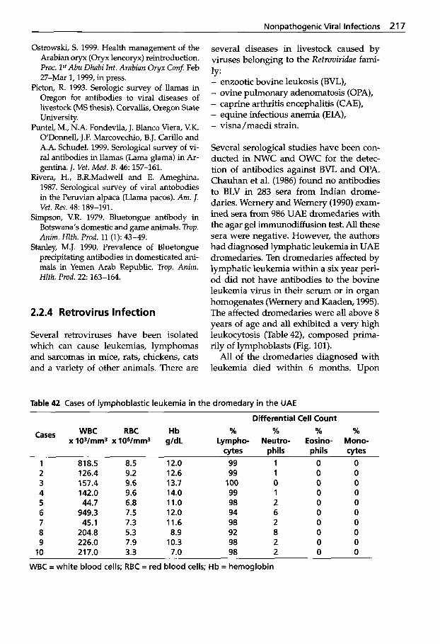
Nonoathoaenic Viral Infections 217
Ostrowski, S. 1999. Health management of the Arabian oryx (Oryx lencoryx) reintroduction. Proc. P A b u Dhabi Int. Arabian Oryx Con5 Feb 27-Mar 1,1999, in press.
Picton, R. 1993. Serologic survey of llamas in Oregon for antibodies to viral diseases of livestock (MS thesis). Corvallis, Oregon State University.
Puntel, M., N.A. Fondevila, J. Blanco Viera, V.K. ODonnell, J.F. Marcovechio, B.J. Carillo and A.A. Schudel. 1999. Serological survey of vi- ral antibodies in llamas (Lama glama) in Ar- gentina. I. Vet. Med. B. 46: 157-161.
Rivera, H., B.R.Madwel1 and E. Ameghina. 1987. Serological survey of viral antobodies in the Peruvian alpaca (Llama pacos). Am. I. Vet. Res. 48: 189-191.
Simpson, V.R. 1979. Bluetongue antibody in Botswana's domestic and game animals. Trop. Anim. Hlfh. Prod. 11 (1): 43-49.
Stanley, M.J. 1990. Prevalence of Bluetongue precipitating antibodies in domesticated ani- mals in Yemen Arab Republic. Trop. Anim. Hlth. Prod. 2 2 163-164.
2.2.4 Retrovirus Infection
Several retroviruses have been isolated which can cause leukemias, lymphomas and sarcomas in mice, rats, chickens, cats and a variety of other animals. There are
several diseases in livestock caused by viruses belonging to the Retroviridae fami-
- enzootic bovine leukosis (BVL), - ovine pulmonary adenomatosis (OPA), - caprine arthritis encephalitis (CAE), - equine infectious anemia (EIA), - visna /maedi strain.
ly:
Several serological studies have been con- ducted in NWC and OWC for the detec- tion of antibodies against BVL and OPA. Chauhan et al. (1986) found no antibodies to BLV in 283 sera from Indian drome- daries. Wernery and Wernery (1990) exam- ined sera from 986 UAE dromedaries with the agar gel immunodiffusion test. All these sera were negative. However, the authors had diagnosed lymphatic leukemia in UAE dromedaries. Ten dromedaries affected by lymphatic leukemia within a six year peri- od did not have antibodies to the bovine leukemia virus in their serum or in organ homogenates (Wernery and Kaaden, 1995). The affected dromedaries were all above 8 years of age and all exhibited a very high leukocytosis (Table 42), composed prima- rily of lymphoblasts (Fig. 101).
All of the dromedaries diagnosed with leukemia died within 6 months. Upon
Table 42 Cases of lymphoblastic leukemia in the dromedary in the UAE
Differential Cell Count WBC RBC Hb % % % %
x 103/mm3 x lO~/mm3 g/dL Lympho- Neutro- Eosino- Mono- cytes phils phils cytes
Cases
1 818.5 8.5 12.0 99 1 0 0 2 126.4 9.2 12.6 99 1 0 0 3 157.4 9.6 13.7 100 0 0 0 4 142.0 9.6 14.0 99 1 0 0 5 44.7 6.8 11.0 98 2 0 0 6 949.3 7.5 12.0 94 6 0 0 7 45.1 7.3 11.6 98 2 0 0 8 204.8 5.3 8.9 92 8 0 0 9 226.0 7.9 10.3 98 2 0 0
10 217.0 3.3 7.0 98 2 0 0
WBC = white blood cells; RBC = red blood cells; Hb = hemoglobin
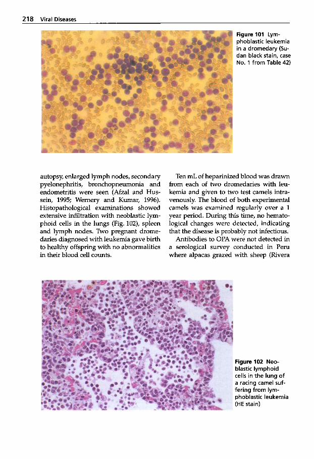
218 Viral Diseases
autopsy, enlarged lymph nodes, secondary pyelonephritis, bronchopneumonia and endometritis were seen (Afzal and Hus- sein, 1995; Wernery and Kumar, 1996). Histopathological examinations showed extensive infiltration with neoblastic lym- phoid cells in the lungs (Fig. 102), spleen and lymph nodes. Two pregnant drome- daries diagnosed with leukemia gave birth to healthy offspring with no abnormalities in their blood cell counts.
Figure 101 Lym- phoblastic leukemia in a dromedary (Su- dan black stain, case No. 1 from Table 42)
Ten mL of heparinized blood was drawn from each of two dromedaries with leu- kemia and given to two test camels intra- venously. The blood of both experimental camels was examined regularly over a 1 year period. During this time, no hemato- logical changes were detected, indicating that the disease is probably not infectious.
Antibodies to OPA were not detected in a serological survey conducted in Peru where alpacas grazed with sheep (Rivera
Figure 102 Neo- blastic lymphoid cells in the lung of a racing camel suf- fering from lym- phoblastic leukemia (HE stain)
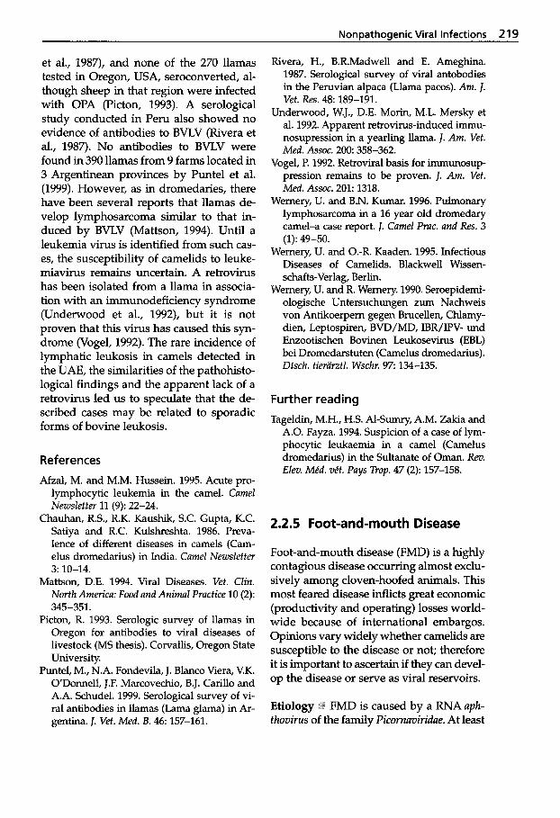
NonDathoaenic Viral Infections 219
et al., 1987), and none of the 270 llamas tested in Oregon, USA, seroconverted, al- though sheep in that region were infected with OPA (Picton, 1993). A serological study conducted in Peru also showed no evidence of antibodies to BVLV (Rivera et al., 1987). No antibodies to BVLV were found in 390 llamas from 9 farms located in 3 Argentinean provinces by Puntel et al. (1999). However, as in dromedaries, there have been several reports that llamas de- velop lymphosarcoma similar to that in- duced by BVLV (Mattson, 1994). Until a leukemia virus is identified from such cas- es, the susceptibility of camelids to leuke- miavirus remains uncertain. A retrovirus has been isolated from a llama in associa- tion with an immunodeficiency syndrome (Underwood et al., 1992), but it is not proven that this virus has caused this syn- drome (Vogel, 1992). The rare incidence of lymphatic leukosis in camels detected in the UAE, the similarities of the pathohisto- logical findings and the apparent lack of a retrovirus led us to speculate that the de- scribed cases may be related to sporadic forms of bovine leukosis.
References Afzal, M. and M.M. Hussein. 1995. Acute pro-
lymphocytic leukemia in the camel. Camel Newsletter 11 (9): 22-24.
Chauhan, R.S., R.K. Kaushik, S.C. Gupta, K.C. Satiya and R.C. Kulshreshta. 1986. Preva- lence of different diseases in camels (Cam- elus dromedarius) in India. Camel Newsletter
Mattson, D.E. 1994. Viral Diseases. Vet. Clin. North America: Food and Animal Practice 10 (2):
Picton, R. 1993. Serologic survey of llamas in Oregon for antibodies to viral diseases of livestock (MS thesis). Corvallis, Oregon State University.
Puntel, M., N.A. Fondevila, J. Blanco Viera, V.K. ODonnell, J.F. Marcovechio, B.J. Carillo and A.A. Schudel. 1999. Serological survey of vi- ral antibodies in llamas (Lama glama) in Ar- gentina. J. Vet. Med. B. 46: 157-161.
3: 10-14.
345-351.
Rivera, H., B.R.Madwell and E. Ameghina. 1987. Serological survey of viral antobodies in the Peruvian alpaca (Llama pacos). Am. J. Vet. Res. 4 8 189-191.
Underwood, W.J., D.E. Morin, M.L. Mersky et al. 1992. Apparent retrovirus-induced immu- nosupression in a yearling llama. J. Am. Vet. Med. Assoc. 200 358-362.
Vogel, P. 1992. Retroviral basis for immunosup- pression remains to be proven. J. Am. Vet. Med. Assoc. 201: 1318.
Wernery, U. and B.N. Kumar. 1996. Pulmonary lymphosarcoma in a 16 year old dromedary camel-a case report. J. Camel Prac. and Res. 3
Wernery, U. and 0.-R. Kaaden. 1995. Infectious Diseases of Camelids. Blackwell Wissen- schafts-Verlag, Berlin.
Wernery, U. and R. Wernery. 1990. Seroepidemi- ologische Untersuchungen zum Nachweis von Antikoerpern gegen Brucellen, Chlamy- dien, Leptospiren, BVD/MD, IBR/IPV- und Enzootischen Bovinen Leukosevirus (EBL) bei Dromedarstuten (Camelus dromedarius). Dtsch. tierarztl. Wschr. 9 7 134-135.
(1): 49-50.
Further reading Tageldin, M.H., H.S. Al-Sumry, A.M. Zakia and
A.O. Fayza. 1994. Suspicion of a case of lym- phocytic leukaemia in a camel (Camelus dromedarius) in the Sultanate of Oman. Rev. Elm. Me'd. ve't. Pays Trop. 47 (2): 157-158.
2.2.5 Foot-and-mouth Disease
Foot-and-mouth disease (FMD) is a highly contagious disease occurring almost exclu- sively among cloven-hoofed animals. This most feared disease inflicts great economic (productivity and operating) losses world- wide because of international embargos. Opinions vary widely whether camelids are susceptible to the disease or not; therefore it is important to ascertain if they can devel- op the disease or serve as viral reservoirs.
FMD is caused by a RNA uph- fhovirus of the family Picornuviridue. At least
220 Viral Diseases
seven immunologically distinct serotypes of FMDV have been identified, of which A, 0 and C are the three most common strains in Europe. 0 appears to be the most com- mon and C the least common. There is no crossimmunity between strains. Within the 7 serotypes, over 60 subtypes have been identified.
Epidemiology :i FMD was enzootic in parts of Europe, and still is in the Middle East, India, the Far East and South America. North America, Australia, New Zealand, and many countries in western Europe are free of the disease. These countries have stringent regulations preventing the intro- duction of the FMDV. FMD is of great in- terest with regard to NWC and OWC, be- cause the disease is enzootic in many coun- tries where camelids are reared. Saudi Arabia, for example (with a camel popula- tion of 800,000), imports approximately 6.5 million live animals, mainly sheep and goats, from Africa, Asia and Australasia. Animals from Africa and Asia bring their own FMDV strains, which spread within the nomadic herds of Saudi Arabia and neighboring countries. It would be of great importance to know if camels play a role in transmitting FMDV. The natural hosts of the virus are artiodactylids including cattle, sheep, swine and goats, but also many dif- ferent wild animals. Most transmission is via aerosols, usually when animals are in close contact with each other, although under cer- tain circumstances it may spread over long distances. Some ungulates can harbor the virus for a long period without developing vesicular lesions. The study of the epidemi- ology of FMD has been transformed by the use of molecular techniques to characterize individual strains of virus. By these meth- ods, it has been possible to trace the move- ment of individual strains of FMDV from one country to another (Kitchmg, 1998).
Clinical Signs and Pathology @ Earlier studies have reported FMD epizootics in
camels. This observation was based on clin- ical manifestations of the disease. After observing thousands of Afghani drome- daries, Pringle (1880) reported that the dis- ease was widespread and that foot lesions were the most prevalent symptom. Steele (cited from Curasson, 1947) also described outbreaks similar to FMD in dromedaries.
According to Kowalevsky (1912), who observed outbreaks of the disease among Bactrian camels in Kazakhstan, the disease affects the lips, the buccal mucosa and the feet. Rohrer (1970) also believes that all artiodactyl ungulates can develop FMD. However, Leese (1918) and Curasson (1947) are skeptical of the earlier reports. They be- lieve that these epizootics were all out- breaks of camelpox. Leese (1927) was not able to confirm FMD in dromedaries fol- lowing inoculation attempts with material from bovine aphthae.
Recent studies by Moussa et al. (1987) in Egypt have shown that dromedaries are susceptible to FMD. The authors described ruptured vesicles and ulcera on the upper lips of four dromedaries. Additionally, ul- cerations were seen between the teeth and on the teats. Two viral strains were isolated from these lesions and both were identi- fied as FMDV, serotype 0. A calf, a ram, a goat and a dromedary were artificially in- fected with this FMDV, type 0. The calf and the goat did not develop any lesions, the ram developed hyperemia of the eyes and buccal cavity and the dromedary de- veloped ulcera on the scarification site and two to three vesicles on the inside of the upper lip. The dromedary did not develop any antibodies to the artificial infection. However, the other animals seroconverted 3 weeks later. After an additional 5 weeks, all four animals were infected with a known bovine FMDV, type 0. The calf de- veloped a vesicle at the inoculation site and the goat developed ulcera on the dorsum of the tongue and the upper lip. Again the dromedary did not develop any antibodies following the second viral exposure.
NonDathoaenic Viral Infections 221
Additional experimental studies on the dromedary by Nasser et al. (1980) and Moussa (1988), following intranasal infec- tion with serotype 0 FMDV strains, yield- ed only slight or clinically inapparent man- ifestations. However, the virus was re-iso- lated from the pharynx and the feces over the course of 6 days following infection, whereby the highest titers were observed between the 3rd and 4th day p.i. Four weeks later, a second excretion of the virus was noted lasting for a week. Thereafter, virus detection was negative. Again there was no evidence of humoral antibody produc- tion. Similar reports in Saudi Arabia from Hafez et al. (1993) again showed that, fol- lowing intranasal infection with the Egyp- tian strain Sharkia 0/2/72, neither clinical signs nor seroconversion were seen in the infected dromedaries.
Only Richard (1986) was able to idenhfy antibodies to the FMDV serotypes 0, C and SAT2 in 2.6% of the sera from Nigerian dromedaries. However, Moussa (1988) is of the opinion that the substances identi- fied were nonspecific inhibitory substances in the sera, frequently seen in camel serum, as opposed to specific antibodies.
Dromedaries kept for weeks in close contact with severe cases of FMD in cattle, sheep and goats in various FMD epizootics in Ethiopia (Richard, 1979), Oman (Hedger et al., 1980), Niger (Richard, 1986), Saudi Arabia and Egypt (Hafez et al., 1993) did not develop any signs of clinical signs. Further studies carried out by Abou Zaid (1991) revealed that dromedaries could contract FMD after experimental infection. Three camels were infected with FMDV strain 01/3/87 Egypt IDL intradermolin- gually and the fourth intradermally in the footpad. One camel and three bovine calves were kept as contact animals. At the same time, three bovine calves were inocu- lated with the same FMDV strain and one bovine calf and two camels were kept as contact animals. The three infected camels showed signs of FMD and the virus was
isolated from blood, esophageal-pharyn- geal (OP) fluid, feces and ruptured vesi- cles. The fourth camel that was inoculated with FMDV into the footpad did not de- velop any FMD symptoms. The contact camel did not show any lesions and no virus was isolated, but the three bovine calves contracted the disease and the virus was isolated. In the second group, the three infected bovine calves as well as the con- tact bovine calf developed FMD, but the contact camel did not show any clinical signs. These investigations showed that camels in contact with cattle or camels with FMD did not contract the disease. However, camels could contract FMD when intradermolingually infected. These exper- iments also showed that infected, diseased camels seroconverted to FMDV during the first week, but the antibodies disappeared after 6 weeks. The camel not showing any lesions did not develop antibodies to FMD.
Several studies of NWC susceptibility to FMDV have been carried out (Mancini, 1952; Konigshofer, 1971; Moro, 1971; Mous- sa et al., 1979; Lubroth and Yedloutschnig 1987; Lubroth et al., 1990; Callis and Craig, 1992). As with OWC, the opinions on whether NWC are susceptible to FMD or not vary widely. A few surveys suggest that NWC are resistant to natural FMD in- fection (Mancini, 1952; Paling et al., 1979; Tantawi et al., 1984) while others describe the susceptibility of NWC to experimental infection with FMDV (Moussa et al., 1979; Lubroth et al., 1990) in a limited number of animals and with variable results. Unlike cattle, which are known to carry FMDV for long periods of time, little is known about the carrier ability of camelids. In a well- executed study by Fondevila et al. (1995), further evidence was provided that llamas are resistant to FMD infection. The authors conducted an experimental trial with FMDV serotypes A79, 0 3 and 01 to evaluate the ability of FMD to infect susceptible llamas exposed either directly to affected live- stock or indirectly to llamas that had been
222 Viral Diseases
directly exposed to affected livestock. Six pigs were inoculated with three different types of FMDV by different routes. Thirty llamas were placed together with the in- fected pigs and later interspersed with an additional 30 llamas after the exposure to the pigs. Forty susceptible livestock (pigs, bovine calves, goats and sheep) were then added to the entire group of 60 llamas to detect possible transmission of FMDV from llamas. Only 2 of 30 llamas directly ex- posed to the FMD pigs developed minor lesions, seroconverted and yielded virus in blood or OP fluid. A third llama from this group also seroconverted, but showed no lesions and did not shed the virus. All con- trol animals introduced to the 30 contact- exposed llamas failed to develop lesions or antibodies and failed to yield any FMDV.
The divergent results from scientists of various countries underline the impor- tance of examining and qualifying the pathogenesis of FMDVs in camelids. There is now agreement that NWC and drome- daries can contract the disease after exper- imental infection and by very close contact with FMD-diseased livestock. NWC and dromedaries are not very susceptible to FMD and do not present a risk in transmit- ting FMDVs to other susceptible animal species. However, all FMD antibody-posi- tive animals should be considered to be potentially infected, as it is known that im- mune animals in contact with live FMDV can become carriers. The pandemic sero- type 0 virus (now named the PanAsia strain) has currently reached a global ex- tension and has out-competed all other strains of FMD. It has also caused disease in Bactrians in Mongolia. It is now believed that NWC and dromedaries are more or less resistant to FMD infection, and that they play no role in transmitting the virus to domestic livestock. Cumelus buctriunus, however, is susceptible to FMD and may transmit the virus to other artiodactylids (Indian elephants are susceptible to FMD, Africans not).
Stehman et al. (1998) reported a picor- nuvirus infection in llamas that caused abortion in 15 llamas. This occurred over a three and a half month period at an aver- age 220 days gestation. Along with the picornuvirus infection, diabetes mellitus was observed in the adult llamas. The virus was isolated from two fetuses; serum neutralizing antibodies to the picornuvirus were found in the fetal fluids as well as in two llama herds with a similar clinical syn- drome of diabetes mellitus.
An encephalomyocarditis virus (EMCV), which is a picornuvirus, has been isolated from a 2-year-old dromedary in an Ameri- can zoological collection (Wells et al., 1989). Gross pathology consisted of excessive peri- cardial fluid, epicardial hemorrhages and pale foci within the myocardium. The virus was isolated from the heart. It is believed that rodents may have transmitted the EMCV.
Diagnosis The clinical signs of FMD are indistinguishable from vesicular stomati- tis, vesicular exanthema of swine (culici- virus) and vesicular disease in pigs (en- terovirus of the Picornuviridue family). Lab- oratory methods are therefore necessary for diagnosis. These methods include com- plement fixation test, ELISA, virus neutral- ization and agar gel precipitation. The virus can be isolated on different cell lines, including fetal camel kidney (Farid et al., 1974).
Treatment and Prevention 1*1: There is no cure for FMD. The most effective preven- tive measure is to prohibit introduction of animals or animal products into FMD- free countries from countries that have the disease. Many European countries have banned routine vaccinations against FMD because most of the outbreaks have been traced to improperly inactivated vaccines or escape of the virus from the production site. Furthermore, ruminants (in particular cattle), continue to carry live FMDV in their

NonDathoaenic Viral Infections 223
pharynx after contact. Animals immune to FMD after vaccination can still become car- riers after contact with field strains during outbreaks. Cattle can harbor FMDV for up to 3 years. FMD vaccine is an inactivated preparation; attempts to take advantage of new molecular biological technology to produce better FMD vaccines have been unsuccessful. The duration of immunity after FMD vaccination is rarely longer than 6 months (Kitching, 1998). In countries where vaccines are used, the virus from the outbreak must be isolated and typed to determine whether the field strain is homologous to the vaccine strain being used. FMD vaccines have not been used in camelids.
2.2.6 Vesicular Stomatitis
Vesicular stomatitis (VS) is another vesicu- lar disease which is indistinguishable from FMD. VS is caused by a rhabdovirus and there are two major types: New Jersey and Indiana. Few studies involving the suscep- tibility of NWC to VSV have been conduct- ed. It is believed that natural infection rarely occurs, as llamas that had been in close contact with diseased cattle did not contract VS (Thedford and Johnson, 1989). The llamas even shared the same watering and feeding facilities with the diseased cat- tle and they did not seroconvert to VSV. Two hundred and seventy llamas which were serologically tested to strains Indiana and New Jersey in Oregon were also nega- tive (Picton, 1993). However, one natural case of VS in lamoids has been reported (Fowler, 1998). Alpacas and llamas have been shown to be susceptible to an experi- mental infection with VSV. Vesicles ap- peared at the inoculation site at the dor- sum of the tongue and the animals devel- oped fever and anorexia (Gomes, 1964). Fluids taken from these vesicles of NWC caused disease in cattle. No reports exist on VS in OWC.
References
Abou-Zaid, A.A. 1991. Studies on some diseases of camels. PhD thesis (Infectious Diseases). Fac. of Vet. Med., Zagazig Uni., Egypt.
Callis, J.J. and D.A. Craig. 1992. Foot-and- mouth disease. In: Castro, A.E. and W.P. Heu- schele (eds.): Veterinary Diagnostic Virology, A Practitioner's Guide St. Louis, Mosby Year
Curasson, G. 1947. Le chameau et ses maladies. Vigot Freres, Editeurs: pp. 86-88.
Farid, F., H.H. Tantawi, G.A. Abd El Galil, M.S. Saber and M.A. Shalaby. 1974. Multiplication and Titration of foot and mouth disease virus in the Foetal Camel kidney tissue culture. J. Egypt vet. med. Ass. 34 (3-4): 384-392.
Fondevila, N.A., F.J. Marcovechio, J. Blanco Viera, V. K. ODonnell, B. J. Carrillo, A. A. Schudel, M. David, A. Torres and C. A. Me- bus. 1995. Susceptibility of Llamas (Lama glama) to Infection with Foot-and-mouth- disease Virus. J. Vet. Med. B 42: 595-599.
Fowler, M.E. 1998. Medicine and surgery of South American Camelids. Iowa State Uni- versity Press, Ames.
Gomez, D. 1964. Tests on the sensitivity of Camelids to vesicular stomatitis. Anales li Cong. Nuc Med Vet y Zoot, Lima, Peru: 403406.
Hafez, S.M., M.A. Farag and A. Al-Mukayel. 1993. Are camels susceptible to natural infec- tion with Foot-and-Mouth Disease virus? In- ternal paper, National Agriculture and Water Research Centre, P.O. Box 17285, Riyadh, Saudi Arabia.
Hedger, R.S., T.R. Barnett and D.F. Gray. 1980. Some virus diseases of domestic animals in the Sultanate of Oman. P o p . Anim. Hlth. 12:
Kitching, R.P. 1998. A recent history of foot and mouth disease. J. Comp. Puth. 118 89-108.
Konigshofer, H.O. 1971. Foot and Mouth Dis- ease in Peru. Anim. Health Yearbook. FAO-
Kowaleski, M.J.M. 1912. Le Chameau et ses mal- adies d'aprhs les observation d'auteurs russ- es. J. Mtd. ve't. Zootechn., Lyon 15: 462-466.
Leese, A.S. 1918. "Tips" on camels for veterinary surgeons on active service. Bailliere, Tmdall and Cox, London 50.
Leese, A.S. 1927. A treatise on the one-humped camel in health and disease. Vigot Frkres, Paris II.
Book: pp. 100-103.
107-114.
WHO-OIE: 178.
224 Viral Diseases
Lubroth, J. and R.J. Yedloutschnig. 1987. Foot and mouth disease studies in the llama (Lama glama). Mexican US Commission FMD, Culle Hegal 713, Colonia Polanco, 11560, Mexico. Proc. US Anim. Health Assoc. 91: 313-316.
Lubroth, J., R.J. Yedloutschnig, U.K. Culhane and P.E. Mikiciu. 1990. Foot and mouth dis- ease virus in the llama (Lama glama): Diag- nosis, transmission, and susceptibility. J. Vet. Diagn. Invest. 2 (3): 197-203.
Mancini, A. 1952. Tests on susceptibility of South American camelids to foot-and-mouth disease. Ensayos sobre la receptividad de 10s anguenidos a la fiebre aftosa. Bol. Inst. Nac. Antiaftosa (Lima) 1 (2): 127-145.
Mattson, D.E. 1994. Viral Diseases. Vet. Clin. North America: Food and Animal Practice 10 (2):
Moro, M. 1971. Ectima: En: La Alpaca. Enfer- madades Infecciosas y Parasitarias. Bol Di- vulgacion Instito Veteranario de Investiga- ciones Tropicales y de Altura. Unva Nac San Marcos, Lima, Peru: 30.
Moussa, A.A., M.M. Arafa, A. Daoud, M. Amer and S. Taswfik. 1979. Susceptibility of camel and sheep to infection with foot-and-mouth disease. Agr. Res. Rev. 5 7 1-18.
Moussa, A.A.M., A. Daoud, A. Omar, N. Met- wally, M. El-Nimr and J.W. McVicar. 1987. Isolation of Foot and Mouth disease virus from camels with ulcerative disease syndromes. J. Egypt Vet. Med. Ass. 47 (1,2): 219-229.
Moussa, A.A.M. 1988. The role of camels in the epizootiology of FMD (Foot and Mouth disease) in Egypt In: FAO. The camel devel- opment research. Proc. of Kuwait seminar, Kuwait: Oct 20-23,1986,162-173.
Nasser, M., A.A. Moussa, M. Abdeir Metwally and R. El S. Saleh. 1980. Secretion and per- sistence of foot and mouth diseases virus in faeces of experimentally infected camels and ram. I. Egypt. Vet. Med. Ass. 40 (4): 5-13.
Paling, R.W., D.M. Jesset and B.R. Heath. 1979. The occurrence of infectious diseases in mixed farming of domesticated wild herbi- vores and domestic herbivores including camels in Kenya: Viral diseases. A serological survey with special reference to foot and mouth disease. J. Wild. Dis. 15: 351-358.
Picton, R. 1993. Serologic survey of llamas in Oregon for antibodies to viral diseases of livestock (MS thesis). Corvallis, Oregon State University.
345-351.
Pringle, R. 1880. Foot and mouth disease in camels. Br. Vet. J.
Richard, D. 1979. Study of the pathology of the dromedary in Borana Awraja (Ethiopia). Diss. Med. Vet., Universitaet Creteil.
Richard, D. 1986. Manuel des maladies du dro- madaire. Projet de d6veloppement de l'kle- vage dans le Niger centre-est. Maisons Alfort, IEMVT.
Rohrer, H. 1970. Traite des maladies a virus des animaux. Paris, Vigot, France.
Steele, S. cited from Curasson, 1947. Stehman, S.M., L.I. Morris, L. Weisensel, W.
Freeman, F. Del Piero, N. Zglich and E.J. Dubovi. 1998. Case report: Picomavirus in- fection associated with abortion and adult onset diabetes mellitus in a herd of llamas. Am. Vet. Pathology meeting.
Tantawi, H.H., A.A. Moussa, A. Omar and M.H. Arafa. 1984. Detection of foot-and-mouth disease virus carriers among farm animals. Assiut Vet. Med. J. 13: 65-79.
Thedford, R.R. and L.W. Johnson. 1989. Mec- tious diseases of New-world camelids (NWC). Vet. Clin. North Am. Food Anim. Pract. 5 (3):
Wells, S.K., A.E. Gulter, K.F. Soike and G.B. Baskin. 1989. Encephalomycocarditis virus: epizootic in a zoological collection. J. Zoo and Wildl. Med. 20 (3): 291-296.
145-157.
Further reading Abu Elzein, E.M.E., B.J. Neumann, E.A. Omer
and B. Haroon. 1984-1985. Prevalence of serum antibodies to the Foot-and-mouth dis- ease virus infection associated antigen (via) in camels, sheep and goats of the Sudan. Su- dan J. Vet. Res. 6: 58-60.
Guo, S.Z. 1988. Serological comparison of the pathogens of aphthosis in camel, sheep, and goat. Chinese J Vet Med b Tech 5: 35-37.
Sutmoller, l? 1999. Risk of disease transmission by llama embryos. Rev. Sci. Techn. de I'Office Int. Epiz. 18 (3): 719-728.
2.2.7 Bovine Viral Diarrhea
Bovine viral diarrhea (BVD) and mucosal disease (MD) are epidemiologically differ- ent diseases of cattle that have different
NonDathoaenic Viral Infections 225
pathogeneses, although both are caused by the same virus. BVD can occur at any age in postnatal life as a result of an acute mild infection. However, severe clinical disease with agalactia and diarrhea may also oc- cur. In the late 1980s, an acute and fatal syndrome of calves was reported in h e r - ica. The disease was characterized by a profound thrombocytopenia and hemor- rhages. In contrast, MD is a severe disease with fatal consequences in cattle 6 months to 2 years of age. It occurs only in those cat- tle that have suffered a non-cytopathic vi- ral infection in the early stages (40-120 days) of fetal life and in which the virus has persisted as a result of immunological tolerance of the fetus (Thiel et al., 1999). Su- perinfection, either through mutation of the non-cytopathic to a cytopathic BVDV or through an exogenic cytopathic infec- tion, is believed to trigger MD. Brownlie et al. (2000) suggest the following definition for MD "MD is a fatal condition, mainly of young cattle aged 6 to 18 months, with characteristic erosive pathology in the oral/intestinal mucosa from which the cy- topathogenic biotype of BVDV can be iso- lated. The clinical disease is typically rapid in onset, although chronic debilitating forms can occur." After the persistently infected (p. i.) calf is born, it excretes the virus dur- ing its entire life. It is remarkable that both the non-cytopathic and the cytopath- ic forms can be isolated from MD cases. The virus is widespread in cattle popula- tions worldwide and has also been isolated from NWC and OWC (Evermann et al., 1993; Mattson, 1994; Hegazy et al., 1998).
Etiology 1 Bovine viral diarrhea virus (BVDV) is a small RNA virus of the FZa- viviridae. Together with the viruses of bor- der disease and classical swine fever virus it forms the genus Pestivirus. The three vi- ruses are antigenically related. Strains iso- lated from newborn calves and persistent- ly infected cattle are generally non-cyto- pathic (BVDVnc), while those from tissues
of cattle suffering from MD are usually cytopathic (BVDVc). Today two genotypes of BVDV are recognized: BVDV-1 and BVDV-2. BVDV-1 has a worldwide distri- bution, whereas BVDV-2 is largely restrict- ed to the USA and Canada.
Epidemiology Postnatal infection with the virus is acquired by ingestion or in- halation of contaminated material and re- sults in the development of serum neutral- izing antibodies. This is usually a clinical- ly unrecognizable infection. On the other hand, with infection of a non-immune pregnant animal, the virus is capable of crossing the placental barrier and invading the fetus. While the dam seroconverts without showing signs of disease, the fetus is immunotolerant in the early stages of pregnancy. This congenital infection can result in a wide spectrum of abnormalities: fetal death, congenital defects, or a persist- ent lifelong infection without clinical signs. The outcome is mainly dependent on the stage of fetal development during which infection takes place.
Clinical Signs and Pathology Serologi- cal studies indicate that NWC and OWC are susceptible to infection with the BVDV. The results of serological studies identify- ing BVDV antibodies in the dromedary have appeared from Tunisia with 3.9% positive (Burgemeister et al., 1975), from Oman with 6.7% (Hedger et al., 1980), from Sudan with 15.5% and 15.7% (Bornstein and Musa, 1987; Bornstein et al., 1989) and Somalia with 3.4% (Bornstein, 1988). Bohr- mann et al. (1988) did not identify any an- tibodies to BVDV in Qibouti. Using the serum neutralization test, Wernery and Wernery (1990) explained the higher inci- dence of BVD in UAE breeding camels (9.2%) when compared to racing drome- daries (3.6%) with their larger breeding herds and closer contact with cattle herds. In a later survey (CVRL Annual Report, 1998), these findings were confirmed using
226 Viral Diseases
an antibody ELISA. The incidence of BVDV antibodies in 552 camels tested was 0.5% in racing camels and 6.4% in breeding cam- els. The presence of neutralizing antibod- ies to BVDV was 11% in Egypt with a peak of 23% in one area (Hegazy et al., 1993). In another Egyptian survey, Tantawi et al. (1994) detected 4.3% BVDV positive drom- edaries and Zaghhana (1998) found that camels from Egypt exhibited an even high- er prevalence (52.5%) of neutralizing anti- bodies to BVDV. In a serological survey conducted in Peru involving 117 alpacas that grazed with cattle and sheep, the preva- lence of antibodies to BVDV was 11% (Rivera et al., 1987) and Picton (1993) re- ported a prevalence of 4.4% in 270 llamas from Oregon in the USA. A recent study by Puntel et al. (1999) found 2.05% (8/390) re- actors to the BVDV in llamas from nine farms located in three different provinces in Argentina.
Cattle suffering from BVD and MD show lesions in the alimentary tract. The pathological changes in MD are much more severe than in BVD. The MD lesions are often found only in the upper alimen- tary tract. In both BVD and MD, patholog- ical changes consist mainly of erosions and ulcers of varying severity. In camels these lesions have not been described.
BVD infections have been described in dromedary calves from Egypt (Hegazy et al., 1998) causing intrauterine death, still- births, weak calf syndrome with congeni- tal deformities, neonatal respiratory distress syndrome and acute hemorrhagic gastroen- teritis. BVDV was isolated from lymphoid tissues, spleen, brain and kidney on bovine kidney cells causing a cytopathic effect (CPE). The virus was also demonstrated by immunofluorescence in different organs. In another publication, Hegazy et al. (1995) state that the main cause of abortions in dromedaries is the BVDV, which can reach 50% in some herds.
In the UAE, adult dromedaries and calves that have died of other causes are routine-
ly virologically screened, including the flu- orescence test for the presence of the BVDV. So far the results have always been nega- tive (Wernery et al., 1992). VDV has been isolated from dead llamas that suffered ex- cessive nasal discharge and diarrhea (Matt- son, 1994), indicating an MD-like disease.
Over the last years our knowledge about BVD in camelids has increased and it seems that both NWC and OWC can contract the disease. However, extensive studies are nec- essary to elucidate the entire disease pat- tern in this animal species, as with bovines, through extensive field observations and laboratory studies. Investigations in bovines have led to a new understanding of the complex epidemiology and pathogenesis of BVD and MD and one can hope that this will also be the case in the camelid family. Since only one publication on BVD has been published each on NWC and OWC, the authors prefer to keep this chapter un- der “Nonpathogenic Viral Infections”.
Diagnosis i s i Diagnosis of BVD and MD requires laboratory support in the form of virus isolation, virus antigen detection and serum antibody determination. Skin biopsies are the tissues of choice for the diagnosis of BVDV using immunohisto- logical techniques and are always positive in persistently infected animals (Braun et al., 1999). This method should also be ap- plied in the diagnosis of this disease in camelids.
Treatment and Prevention ’ Economic losses caused by BVD/MD mainly arise from prenatal infections. It is therefore es- sential to remove all persistently infected animals and to vaccinate heifers prior to first breeding. Since it is known that BVDV also causes abortions in camels, it may be necessary to adopt control and vaccination strategies similar to those carried out in cattle. Live and inactivated vaccines have been widely used in several countries. Live vaccines are not recommended in camelids
NonDathoaenic Viral Infections 227
because a variety of adverse effects have been observed using live BVDV vaccines in cattle. Inactivated vaccines are safer and can provide good protection. It has been shown that NWC seroconverted after a regimen of three vaccinations using a inac- tivated-virus preparation (Mattson, 1994).
References
Bohrmann, R., H.R. Frey and B. Liess. 1988. Sur- vey on the prevalence of neutralizing anti- bodies to bovine viral diarrhea (BVD) virus, bovine herpes virus type 1 (BHV-1) and parain- fluenza virus type 3 (PI-3) in ruminants in the Djibouti Republic. Dtsch. Tierarztl. Wschr. 95:
Bornstein, S. 1988. A disease survey of the So- mali camel. SARE report, Sweden.
Bornstein, S., B.E. Musa and EM. Jama. 1989. Comparison of seroepidemiological findings of antibodies to some infectious pathogens of cattle in camels of Sudan and Somalia with reference to findings in other countries of Africa. Proc. of International Symposium of De- velopment of Animal Resources in Sudan. Khar-
Bornstein, S. and B.E. Musa. 1987. Prevalence of antibodies to some viral pathogens, Brucella abortus and Toxoplasma gondii in serum from camels (Camelus dromedarius) in Su- dan. J. Vet. Med. B 34: 364-370.
Braun, U., M. Schoenmann, F. Ehrensberger, M. Hilbe and M. Strasser. 1999. Intrauterine in- fection with bovine virus diarrhoea virus on alpine communal pastures in Switzerland. J. Vet. Med. 46: 13-17.
Brownlie, J., I. Thompson and A. Cumen. 2000. Bovine virus diarrhoea virus-strategic deci- sions for diagnosis and control. In Practice 22
Burgemeister, R., W. Leyk and R. Goessler. 1975. Untersuchungen uber Vorkommen von Para- sitosen, bakteriellen und viralen Infektions- krankheiten bei Dromedaren in Siidtunesien. Dtsch. Tierarztl. Wschr. 82: 352-354.
CVRL. 1998. Annual Report. Central Veterinary Research Laboratory, Dubai, U.A.E.: 19.
Evermann, J.F., E.S. Berry, T.V. Baszler et al. 1993. Diagnostic approaches for the detection of bovine virus diarrhea (BVD) virus and relat- ed pesti-viruses. J. Vet. Diagn. Invest. 5 265-269.
99-102.
toum: 28-34.
(4): 176-187.
Hedger, R.S., T.R. Barnett and D.F. Gray. 1980. Some virus diseases of domestic animals in the Sultanate of Oman. Trop. Anim. Hlth. 12:
Hegazy, A.A., S.F. Lotfia and M.S. Saber. 1993. Prevalence of antibodies common in viral diseases of domestic animals among camels in Egypt. In Project (91-H-2-4) NAlU? Epi- demiological, clinical and pathological stud- ies on some diseases in camel in Egypt.
Hegazy, A.A., A.A. El Sanousi, M.M. Lotfy and T.A. Aboellail. 1995. Pathological and viro- logical studies on calf mortality: B- mortali- ties associated with bovine virus diarrhea virus infection. J. Egypt Med. Assoc. Proceed- ings of 22& Arab Vet. Med. Cng., March 19-23, Cairo, Egypt 55 (Nos. 1 & 2): 493-503.
Hegazy, A.A., L.S. Fahmy, M.S. Saber, T.A. Aboellail, A.A. Yousif and C.C.L. Chase. 1998. Bovine virus diarrhea infection causes reproductive failure and neonatal mortality in the dromedary camel. Int. Meeting on camel production and future perspectives. Al Ain, UAE, 2-3 May 1998.
Mattson, D.E. 1994. Viral Diseases. Vet. Clin. North America: Food and Animal Practice 10 (2):
Picton, R. 1993. Serologic survey of llamas in Oregon for antibodies to viral diseases of livestock (MS thesis). Corvallis, Oregon State University.
Puntel, M., N.A. Fondevila, J. Blanco Viera, V.K. ODonnell, J.F. Marcovechio, B.J. Carillo and A.A. Schudel. 1999. Serological survey of vi- ral antibodies in llamas (Lama glama) in Ar- gentina. J. Vet. Med. B. 4 6 157-161.
Rivera, H., B.R. Madwell and E. Ameghina. 1987. Serological survey of viral antibodies in the Peruvian alpaca (Llama pacos). Am. J. Vet. Res. 48: 189-191.
Tantawi, H.W., R.R. Youssef, R.M. Arab, M.S. Marzouk and R.H. Itman. 1994. Some studies on bovine viral diarrhea disease in camel. Vet. Med. J. 32 (3): 9-15.
Thiel, H.-J., P. Becker, M. Baroth, M. Koenig and M. Orlich. 1999. Auftreten von MD nach Imp- fung. 3. Berlin-Brandenburgische Rindertag 10/ 2998 (in press).
Wemery, U., H.H. Schimmelpfennig, H.S.H. Seifert and J. Pohlenz. 1992. Bacillus cereus as a possible cause of haemorrhagic disease in dromedary camels (Camelus dromedarius). Proc. l S t int. Camel Conf. In: Allen, W.R., A. J.
107-114.
345-351.

228 Viral Diseases
Higgins, I.G. Mayhew, D.H. Snow and J.F. Wade. W. and W. Publications, Newmarket,
Wernery, U. and R. Wernery. 1990. Seroepidemi- ologische Untersuchungen zum Nachweis von Antikorpem gegen Brucellen, Chlamy- dien, Leptospiren, BVD/MD, IBR/IPV- und Enzootischen Bovinen Leukosevirus (EBL) bei Dromedarstuten (Camelus dromedarius). Dtsch. tieriirztl. Wschr. 9 7 134-135.
Zaghana, A. 1998. Prevalence of antibodies to Bovine Viral Diarrhoea Virus and/or Border Disease Virus in domestic ruminants. J. Vet. Med. B 45: 345-351.
UK. 51-58.
Further reading
Abou-Zaid, A.A. 1991. Studies on some diseases of camels. PhD thesis (Infectious Diseases). Fac. of Vet. Med., Zagazig Uni., Egypt.
Eisa, M.I. 1998. Serological survey of some viral diseases in camels in Sharkia Governorate, Egypt. Proceedings of the Int. Meeting on camel production and future perspectives, Al Ain, U.A.E., 2-3 May 1998.
Fowler, M.E. 1998. Medicine and surgery of South American Camelids. Iowa State Uni- versity Press, Ames.
Hegazy, A.A. and L.S. Fahmy. 1997. Epidemio- logical, clinical and pathological studies on some diseases of camel. Camel Newsletter 13
Thedford, R.R. and L.W. Johnson. 1989. Infec- tious diseases of New-world camelids (NWC). Vet. Clin. North Am. Food Anim. Pract. 5 (3): 145-157.
Wernery, U. 1999. New aspects on infectious diseases of camelids. J. Camel Prac. and Res.
(9): 21-22.
6(1): 87-91.
2.2.8 Rift Valley Fever
Rift Valley fever (RVF) is an arthropod- borne viral disease of animals including humans, but mostly found in ruminants. Infection in humans is primarily due to contact with material from infected car- casses (Hoogstraal et al., 1979). In addition to the human health hazards, RVF epidem- ics regularly cause serious economic dam- age to animal owners through the loss in
production and fatalities, exacerbated by the 100% abortion rate at all stages of preg- nancy. Strikingly, all of the RVF epizootics described to date have followed unusually severe rainy seasons, probably indicating a very large insect population as a vector prerequisite (Huebschle, 1983). RVF does not occur in very arid areas.
Etiology The Rift Valley Fever Virus (RVFV) is a member of the Phlebovirus genus of the family Bunyuviridue. The bun- yaviruses are spherical, 80 to 120 pn in dia- meter, and have a host cell-derived, bilipid layer envelope through which virus-coded glycoprotein spikes project. No significant antigenic differences have been detected between RVF isolates, but differences in virulence have been demonstrated.
Epidemiology For more than 70 years, RVF epidemics have occurred at prolonged intervals in eastern and southern Africa. It has been accepted that the virus is endem- ic in indigenous forests, where it circulates in mosquitoes and vertebrates spreading to livestock-rearing areas when heavy rains favor the breeding of mosquito vectors. Many different mosquito species can serve as vectors. The virus was first isolated in 1931 in livestock on a farm located in the Rift Valley of Kenya. The virus is now en- demic in much of sub-Saharan Africa with epidemics in West Africa. It has also spread into Egypt and clearly has the potential to spread elsewhere.
Epidemiologic studies of RVF have al- ways been performed during epizootics or immediately afterwards. This was the case following epidemics in Sudan, Kenya and Egypt. Several studies also included the respective local dromedary populations. Scott et al. (1963) reported outbreaks of RVF in cattle following severe rainfall in Kenya, parallel to a drastic increase in abortions in dromedaries. Antibodies to RVF were found in 45% of the dromedaries examined during this outbreak. The authors incrimi-
Nonpathogenic Viral Infections 229
nate the RVFV for the increased rate of abortions; however, no virological studies were performed to substantiate this suppo- sition. Meegan et al. (1979) also observed an increased abortion rate in dromedaries during a RVF epizootic in Egypt. In this case, the epidemic was supposedly carried by Sudanese dromedaries to Egypt (Hoog- straal et al., 1979), as severe epidemics were raging in northern Sudan at the time (Eisa et al., 1977). During this period, Hoogstraal et al. (1979) registered 31 RVF reactors in dromedaries. Other than the increased abortion rate during outbreaks of RVF, no other clinical signs have been so far ob- served in camels (Davies et al., 1985). Aly (1979) found antibodies with the HI-test in 15.6% dromedaries in Egypt and Walker (1975) described abortions and deaths in young one-humped camels during RVF out- breaks. Peters and Meegan (1981), however, observed only a subclinical form of RVF. Olaleye et al. (1996) examined 180 drome- daries with the hemagglutination inhibi- tion test and serum neutralization test in Nigeria and detected 3.3% positive cases. The authors stressed the involvement of camels in the transmission cycle of RVFV.
Imam et al. (1978) and Eisa (1981) were able to isolate the virus from a healthy, nat- urally infected dromedary. Experimental infections with the RVFV have induced no clinical signs in non-pregnant dromedaries (Davies et al., 1985). In spite of high RVF antibody titers, the same authors were not able to determine an increased rate of abor- tion in infected dromedaries.
Severe RVF epidemics have recently oc- curred in East Africa (Anonymous, 1998). Many domestic animals and humans had been affected in vast areas of Kenya, south- ern Sudan and northern Tanzania in De- cember 1997 and January 1998.
Clinical Signs During the last RVF out- breaks in East Africa, the WHO received many reports of high mortality in camels throughout the affected area. Some de-
scriptions of morbidity and mortality were highly suggestive of camelpox or parapox (Ecthyma contagiosum), with ballooning of the head and upper neck, swollen eyes and huge mucoid membrane sloughs in the mouth covering some ulcers.
However, the general disease pattern was that of fever and abortion, which were the predominant features, but early neona- tal death and jaundice have also been ob- served. Since no RVFV was isolated from camels during these outbreaks it is not clear if the disease was caused by RVF. The au- thors therefore prefer to keep this part of RVF under the overall chapter "Nonpathogenic Viral Infections" until proven otherwise.
Diagnosis Definitive diagnosis of RVF depends on virological and serological in- vestigations, since other arthropod-borne virus diseases tend to occur under the same climatic conditions. This is especially true for Wesselsbron disease, which can also cause mortality in lambs, kids and calves and abortion in ewes. However, RVF is associated with higher mortality and abortion rates. Lesions in the livers of young animals also differ in both RVF and Wesselsbron disease. Hepatic changes are usually less extensive in RVF compared to Wesselsbron disease. Specimens for lab- oratory confirmation should include he- parinized blood, liver, spleen, kidney, lymph nodes and brain from aborted fetuses for virus isolation on Vero and BHK 21 cells or suckling and weaned mice. Antibodies to RVF can be demonstrated by CFT, AGID, HIT and ELISA. Viral antigen can also be detected by impression smears of infected tissues by immunofluorescence.
Treatment and Prevention Measures such as chemical control of vectors, move- ment of livestock to higher altitudes, or the confinement of animals to mosquito-proof stables are usually impractical or too late. Immunization remains the only effective way to protect livestock.
230 Viral Diseases
Although it has still not been deter- mined decisively whether dromedaries ac- tually develop RVF, Guillaud and Lancelot (1989) have concerned themselves with the production of a vaccine. The authors determined that the attenuated vaccine strain (MVP-22) has yielded satisfactory results in the dromedary. Following a single subcutaneous vaccination, 18 of 22 dromedaries developed neutralizing anti- bodies. A challenge infection with the R W was not performed. As in other viral diseases already described, the camel ap- pears to be susceptible to RWV. Further intensive research, however, is necessary to clarify the pathogenicity of this virus in the camel.
References
Aly, R.R. 1979. Study of Rift Valley Fever in camels in Egypt. M.V.Sc. Thesis (Micro.), Vet. Med. Faculty, Cairo University, Egypt.
Anonymous. 1998. Rift Valley fever in Africa. Vet. Rec. 143 (2): 34.
Davies, F.G., J. Koros and H. Mbugua. 1985. Rift Valley fever in Kenya: the presence of anti- body to the virus in camels (Camelus drome- darius). J. Hyg. Camb. 9 4 241-244.
Eisa, M. 1981. Rift Valley Fever. Technical Report Series 1: 2-13.
Eisa, M., H.M.A. Abeid and A.S.A. El Sawi. 1977. Rift Valley Fever in the Sudan. 1. Re- sults of field investigations of the epizooty in Kosti district, 1973. Bull. SantiProd. anim. Afr.
Guillaud, M. and R. Lancelot. 1989. Essais de vaccination des ruminants domestiques (bovines, ovins, caprins, camelides) contre la fievre de la vallee du Rift avec la souche MVP-12 en Shegal et en Mauritanie. Rap- port d'execution, IEMVT-CIRAD.
Hoogstraal, H., J.M. Meegan, G.M. Khalil and F.K. Adham. 1979. The Rift Valley Fever epi- zootic in Egypt 1977-78.2. Ecological and en- tomological studies. Transactions of the Royal Society of Tropical Medicine and Hygiene 73 (6):
Huebschle, O.J.B. 1983. Exotische Virusseuchen der Wiederkauer II. Rift-Tal-Fieber. Tierarztl. Umschau 38: 268-273.
25 (4): 356-367.
624-629.
Imam, I.Z.E., R. Karamany and M.A. Danvish. 1978. Epidemic of RVF in Egypt. Isolation of RVF virus from animals. J. Egypt Publ. Health
Meegan, J.M., H. Hoogstraal and M.I. Moussa. 1979. An epizootic of Rift Valley Fever in Egypt in 1977. Vet. Rec. 105: 124-125.
Olaleye, O.D., 0. Tomori and H. Schmitz. 1996. Rift Valley fever in Nigeria: infections in do- mestic animals. Rev. sci. tech. Of. int. Epiz. 15
Peters, C.J. and Meegan, J.M. 1981. RVF in CRC handbook series in zoonoses. Beran G. Ed., CRC Press, Boca Raton, Flu. 403.
Saluzzo, J.F., C. Chartier, R. Bada, D. Martinez and J.P. Digoutte. 1987. La f ihre de la vallee du Rift en Afrique de l'Quest. Rev. Eleu. Mid. vit. Pays trop. 40 (3): 215-223.
Scott, G.R., W. Coakley, R.W. Roach and N.R. Cowdy. 1963. Rift Valley fever in camels. J . Path. Bact. 86: 229-231.
Slama, K. 1984. Contribution a l'etude &roepi- demiologique de la fikvre de la vallee du Rift chez les dromadaires du Sud Tunisien. Th. Doct. Vet. Sidi Thabet 246.
Walker, J.S. 1975. RVF, foreign animal diseases, their prevention, diagnosis and control com- mittee on foreign animal diseases of United States. Animal Health Assoc. 6: 209-221.
ASS. 2 3 265-269.
(3): 937-946.
2.2.9 Rinderpest
The clinical presentation and dangers of rinderpest, the scourge of cattle husbandry, have been known for centuries. This dis- ease led to the foundation of the first Euro- pean veterinary faculties and the imple- mentation of laws governing contagious diseases. Rinderpest is an acute or sub- acute, highly contagious, febrile viral dis- ease of cloven-hoofed mammals, the artio- dactylids. The disease is characterized by high mortality and is typified by hemor- rhagic/septicemic symptoms and mucosal erosions of the entire alimentary tract. Among the larger domesticated animals, cattle, water buffaloes and yaks are sus- ceptible to the virus. Different breeds of cattle have varying degrees of resistance to
Nonpathogenic Viral Infections 231
the virus. Of the smaller domesticated ani- mals, sheep, goats and swine are vulnera- ble to the disease under natural conditions. Wild animals exhibit varying susceptibility and play an important role in the epizooti- ology of rinderpest as virus reservoirs. There is evidence that the primary means of infection is via a virus-containing aerosol (Munz, 1983).
Etiology iii The rinderpest virus belongs to the order Mononegavirales, subfamily Pararnyxoviridae, genus Morbillivirus. It is closely related to the viruses that cause ca- nine distemper, phocine distemper, measles and peste-des-petits-ruminants. Individual rinderpest virus strains vary in their path- ogenicity in various species.
Epidemiology : 3 The natural hosts for the rinderpest virus are all members of the or- der Artioductylu. Natural rinderpest has never been reported in NWC, but experi- mental studies have shown that llamas de- velop a mild febrile response with a short clinical course of 3-5 days (Fowler, 1998). As in foot-and-mouth disease, there are differing opinions as to whether OWC are susceptible to the rinderpest virus or not.
Clinical Signs and Pathology 4 from the turn of the century mention se- vere outbreaks of rinderpest among cam- els. Curasson (1947), who reported the big rinderpest epizootic in Niger in 1892, learned that dromedaries developed se- vere diarrhea and hematuria. Vedernikov (1902) and Tschegis (1902) (cited from Curasson, 1947) saw cases of rinderpest among Bactrian camels in the region around Baku in 1898. Tartakowsky (1899) produced rinderpest experimentally in four Bactrian camels, one of which died. However, he could only elicit slight clini- cal reactions in two dromedaries. Lingard (1905) inoculated five Indian dromedaries with cattle blood infected with the rinder- pest virus. The dromedaries developed the
following clinical signs: fever, vesicles and ulcera in the mouth, eruptive skin lesions and, in one case, diarrhea. One bull (bovine) developed rinderpest after being given blood from one of these dromedaries. Cross (1919) injected the rinderpest virus into three Indian dromedaries, one of which lat- er died. Conti (1913) too diagnosed rinder- pest symptoms in dromedaries in Eritrea. The author believed that the epidemic in dromedaries was more of a prophylactic problem and that disease control measures should be instituted for the Eritrean ani- mal population.
According to Haji (1932-1933), drome- daries were also affected during outbreaks of rinderpest among cattle and buffalo in India. The affected animals had fever, m- minal atony, ocular discharge, depression, severe diarrhea occasionally mixed with blood, as well as vesicles on the lips and hard palate that developed into ulcera. The mortality was 20 to 40%. Dhillon (1959) reported similar discoveries in In- dia. Between 1948 and 1958, the author ob- served more than 15 outbreaks of rinder- pest among dromedaries with mortality rates of up to 100%. The dromedaries’ clin- ical signs were similar to those in cattle. Srinivasan (1940) was successful in con- t r o h g an outbreak of rinderpest among dromedaries after ”goat blood virus” inoc- ulation from infected goats.
Contrary to all these statements, various groups have reported that the camel is not susceptible to rinderpest. Littlewood (1905) in Egypt, Pecaud (1924) in Chad and Samartsev and Arbuzov (1940) in the Asi- atic region of Russia have reported that camels are not susceptible to natural infec- tions. Leese (1927), traveling around India, neither observed nor heard of outbreaks of rinderpest in dromedaries. The author does not exclude the fact that slight clinical signs may be possible and that these may be overlooked.
Until the middle of the twentieth centu- ry, epidemics with clinical signs similar to
232 Viral Diseases
rinderpest were diagnosed on the basis of clinical signs as well as the tendency to spread among other ungulates living in close proximity with the dromedaries. Lab- oratory methods in the diagnosis of rinder- pest were introduced later. Scott and Mac- Donald (1962) confirmed a severe outbreak of rinderpest among wild animals in north- ern Kenya in 1960. Dromedaries in this re- gion did not develop the disease and anti- body studies on 60 dromedary sera with lapinized rinderpest antigen were negative. Chauhan et al. (1986) examined 283 drom- edary sera from India serologically and did not detect any antibodies. However, Mau- rice et al. (1967) found rinderpest antibod- ies in 7.7% of the dromedary sera exam- ined from Chad. Singh and Ata (1967) also detected antibodies to the rinderpest virus in 10% of the Sudanese and Egyptian drom- edaries examined, and Abou-Zaid (1991) found rinderpest-neutralizing antibodies in 5.2% of 536 dromedaries in Egypt.
Experimental infections in dromedaries with the rinderpest virus have yielded fur- ther information regarding the susceptibil- ity of this species to rinderpest. Only one out of ten dromedaries infected experi- mentally with an aerosol of the rinderpest virus developed signs of an asymptomatic, non-contagious infection. Leukopenia and antibodies to the rinderpest virus were ob- served in the serum of this animal. Zebus, serving as contact animals, did not devel- op the disease and also developed no anti- bodies to rinderpest (Provost et al., 1968). Singh and Ata (1967) utilized two virulent and two attenuated (vaccine) rinderpest strains in their experimental trials. Drome- daries that were infected with these strains subcutaneously did not develop rinder- pest. A slight increase in body temperature was observed following inoculation with the virulent strains. Dromedaries given the attenuated vaccine developed only a low antibody titer, whereby high neutralizing antibody titers were observed 28 days after experimental infection with the virulent
strains. This experiment also showed that infected dromedaries did not transmit the virus to susceptible cattle. Taylor (1968) confirmed these results through further trials and performed additional experi- ments on dromedaries using the rinder- pest virus. The results of these experiments were as follows: 1. Following experimental intravenous in-
fection with a virulent rinderpest strain (Kabete 0), the virus was re-isolated be- tween the 3rd and gth day from the blood of the infected dromedary. This animal also developed neutralizing antibodies.
2. One of two dromedaries infected sub- cutaneously with the virulent rinder- pest strain RGK/1 developed a slight viremia lasting 6 days, though both an- imals developed neutralizing antibod- ies.
3. Slight viremia occurred in two out of three dromedaries that were in close contact to a bull infected with rinder- pest. One dromedary developed slight pyrexia. This was the only clinical man- ifestation that was observed during the experiments.
4. Although the rinderpest infection origi- nated in cattle, it was not possible to transmit the rinderpest virus from in- fected dromedaries to cattle or other dromedaries.
In order to determine the susceptibility of camels to experimental rinderpest infec- tion, further experiments were carried out by Chauhan et al. (1985). The authors inoc- ulated 10mL of a 10% spleen suspension collected from a buffalo calf suffering from rinderpest, into two healthy 8 to 12-month- old camels. One camel was given subcuta- neous and the other intravenous inocula- tion. No distinct clinical signs of rinder- pest lesions were detected except a slight hyperemia of visible mucous membranes and mild diarrhea. A post mortem exami- nation of one of the camels infected with rinderpest virus did not reveal any lesions.
Nonpathogenic Viral Infections 233
Chauhan et al. (1985) further showed that blood which was collected from the exper- imentally inoculated camels at the height of febrile reaction and injected into two susceptible buffalo calves caused the de- velopment of typical clinical signs and le- sions of rinderpest in these calves within 6 days of inoculation.
From all these experiments it can be as- sumed that OWC are susceptible to rinder- pest, and might develop mild clinical signs, especially through contact with infected cattle. It is less likely that they serve as vec- tors for the rinderpest virus; therefore they do not appear to play a major role in the epizootiology of rinderpest.
A new epizootic disease has affected thousands of camels in Ethiopia in 1995 and 1996 characterized by a febrile, highly contagious respiratory syndrome. The mor- bidity rate reached over 90% with a mor- tality ranging between 5 and 70%. The ma- jor clinical signs were sero-mucopurulent nasal discharge, lacrimation, coughing, dys- pnea and abdominal breathing. Swelling of the submandibular area and diarrhea was reported in some cases. Two morbil- Zivirus strains, closely related to the peste- des-petits-ruminants (PPR) virus, were iso- lated from diseased camels and a similar disease was reproduced in goats and sheep after inoculation of the camel viruses. Strep- tococcus equi spp. equi was also isolated from diseased camels. Further investiga- tions are currently being undertaken to re- produce the disease in camels (Roger et al., 2000).
Diagnosis LA Several handbooks and scien- tific papers detail the diagnosis of rinder- pest. A presumptive diagnosis of rinder- pest can be made on the basis of the clini- cal signs and gross pathology in cattle, but might be very difficult in camelids. In are- as where the disease is not prevalent, it is essential to obtain laboratory confirma- tion of the diagnosis as soon as possible. Mirchamsy et al. (1971) reported that the
rinderpest virus can be readily grown on camel kidney cells.
Treatment and Prevention * Confirmed rinderpest outbreaks are controlled by the slaughter and disposal of all infected and contact animals as well as by the imposi- tion of rigid quarantine and animal move- ment controls.
Prevention of rinderpest in endemic ar- eas requires annual vaccination of all calves up to 2 years of age with the attenu- ated Kabete "0 strain. This is an inexpen- sive, freeze dried vaccine that is highly ef- fective (Coetzer et al., 1994). It has not been used in camelids.
References
Abou-Zaid, A.A. 1991. Studies on some diseases of camels. PhD thesis (Infectious Diseases). Fac. of Vet. Med., Zagazig Uni., Egypt.
Chauhan, R.S., R.C. Kulshreshtha and R.K. Kaushik. 1985. Epidemiological studies of viral diseases of livestock in Haryana State. Ind. J. Virol. l (1) : 10-16.
Chauhan, R.S., R.K. Kaushik, S.C. Gupta, K.C. Satiya and R.C. Kulshreshta. 1986. Preva- lence of different diseases in camels (Cam- elus dromedarius) in India. Camel Newsletter
Coetzer, J.A.W., G.R. Thomson and R.C. Tustin. 1994. Infectious Diseases of livestock with special reference to Southern Africa. Oxford University Press 2: pp. 1518-1535.
Conti, G. 1913. A serious prophylactic problem: Rinderpest in camel. Moderna Zooiatro, Tori- no 24: 215.
Cross, H.E. 1919. Are camels susceptible to blackquarter, haemorrhagic septicemia and rinderpest? Bull. agric. Res. Inst. Pusa.
Curasson, G. 1947. Le chameau et ses maladies. Vigot Freres, Editeurs: pp. 86-88.
Dhillon, S.S. 1959. Incidence of Rinderpest in camels in Hissar district. Indian Vet. J. 36:
Fowler, M.E. 1998. Medicine and surgery of South American Camelids. Iowa State Uni- versity Press, Ames.
Haji, C.S.G. 1932-33. Rinderpest in camels. Ind. Vet. 1.9: 13-14.
3: 10-14.
603-607.
234 Viral Diseases
Leese, A.S. 1927. A treatise on the one-humped camel in health and disease. Vigot Frhres, Paris II.
Lingard, A. 1905. Report on the preparation of Rinderpest Serum, Calcutta. Abst. BUZZ. Inst. Pasteur 4 235.
Littlewood, W. 1905. Camels are not susceptible to Rinderpest. J. Comp. Path. 18: 312.
Maurice, Y., A. Provost and C. Borredon. 1967. Presence d'anticorps antibovipestiques chez le dromadaire du Tchad. Rev. Elm. Mid . vit. Pays trop. 20 (4): 537-542.
Mirchamsy, H., B. Bahrami, M. Amighi and A. Shafyi. 1971. Development of a camel kidney cell strain and its use in virology. Arch. Inst. Razi 23: 15-18.
Munz, E. 1983. Die heutige Situation auf dem Gebiet der tropischen Tierseuchen, ihre der- zeitige Gefahr fiir die Landwirtschaft der Bundesrepublik Deutschland und ihre Be- deutung fiir den Tierarzt. Der prakt. Tierarzt
Pecaud, G. 1924. Contribution a l'etude de la pathologie veterinaire de la colonie du Tchad. Bull. SOC. Path. Exot. 17 (3): 196-207.
Provost, A., Y. Maurice and C. Borredon. 1968. Note sur la peste bovine experimentale du dromadaire. Rev. Elm. Mid . vit. Pays trop. 21
Roger, F., L.M. Yigezu, C. Hurard, G. Libeau, G.Y. Mebratu and A. Diallo. 2000 in press. In- vestigations on a new pathology of camels in Ethiopia.
Samartsev, A.A. and EN. Arbuzov. 1940. The susceptibility of camels to glanders, rinder- pest and bovine contagious pleuro-pneumo- nia. Veterinariya Moscow 4: 59-63.
Scott, G.R. and J. MacDonald. 1962. Kenya cam- els and Rinderpest. Bull. epizoot. Dis. Afr. 10
Singh, K.V. and F. Ata. 1967. Experimental Rinderpest in camels. A preliminary report. Bull. epizoot. Dis. Afr. 15: 19-23.
Srinivasan, V. 1940. Active immunisation of camels against Rinderpest with goat blood virus. lnd. Vet. J. 16: 259-260.
Tartakowsky, M.M. 1899. No Title. Arch. Sci. biol., St. Petersburg 8: 11.
Taylor, W.P. 1968. The susceptibility of the one- humped camel (Camelus dromedarius) to in- fection with Rinderpest virus. Bull. epizoot. Dis Afi. 16: 405-410.
11: 993-1006.
(3): 293-296.
(4): 495-497.
Tschegis. 1902. cited from Curasson (1947).
Vedernikov, V. 1902. cited from Curasson (1947). Wilson, A.J., H.J. Schwartz, R. Dolan, C.-R. Field
and D. Bottcher. 1982. Epidemiologische As- pekte bedeutender Kamelkrankheiten in aus- gewahlten Gebieten Kenias. Der prakt. Tier- arzt 11: 974-987.
Further reading
Alonso, J.M. 1971. Contribution l'btude de la peste en Mauritanie. Thhe (Doctorat de me- decine) Paris 6 No 59.
Klein, J.M., J.M. .Alonso, G. Baranton, A.R. Poulet and H.H. Mollaret. 1975. La peste en Mauritanie. Med. Mal. infect. 5 (4): 198-207.
Lobanov, V.N. 1967. La peste chez les chameaux. In: OMS %minaire inter-regional de L'0.M.S. pour la lutte contre la peste, Moscow.
2.2.10 Unusual Arboviruses
Arboviruses (arthropod-born viruses) are primarily vector viruses that multiply in blood-sucking insects and/or are transmit- ted to vertebrates via the insect's bite or sting. They are widespread in the tropics and subtropics, but their significance in camelids is not known.
Wood et al. (1982) isolated the Kadam virus from HyaZomma dromedarii ticks col- lected from the immediate vicinity of a dead dromedary in Saudi Arabia. It was not possible to determine whether this an- imal's death was due to the Kadam virus. The pathogenicity of the Kadam virus for the dromedary, cattle and humans has not yet been determined.
Five strains of the Quaranfil virus were isolated by Converse and Moussa (1982) from HyaZomma dromedarii ticks collected in Kuwait, Iraq and Yemen. The significance of this finding has not yet been deter- mined.
The Akabane virus can cause epizootics and spontaneous abortions, premature births and congenital deformities in cattle, sheep and goats. The virus is widespread in Africa and Asia and appears to be en-
NonDathoaenic Viral Infections 235
demic to the Arabian Peninsula. Al Bu- saidy et al. (1988) found that 50% of the dromedaries examined in Oman had neu- tralizing antibodies to the Akabane virus. It has not been ascertained whether the virus causes abortions in the dromedary.
Anderson and Casals (1973) isolated four strains of Dhori virus from ticks (Hyulomma dromedurii) found on Indian dromedaries. Additionally, the author dis- covered neutralizing antibodies to the virus in 48 out of 50 dromedary sera. No clinical signs of disease were observed in the seropositive dromedaries. The virus has also been isolated from dromedary ticks in southwest Asia and Africa. Williams et al. (1973) found three non-classified Arboviruses in Hyulomrnu ticks from Egypt- ian dromedaries: Wanowrie, Thogoto and Dhori viruses. These viruses have been widely spread by the migratory patterns of the indigenous animals, including the camel caravans. The veterinary importance of these viruses is unknown.
A survey for antibodies against flavi- viruses in 269 slaughter camels was carried out in Nigeria (Baba et al., 1990). The anti- body prevalence against flaviviruses was noted as follows: Wesselsbron: 60.2%, Yellow Fever: 54.0%, Potiskum: 66.2%, Dengue type 1: 4.5%, Banzi: 5.4% and Uganda S: 0%. The importance of the high prevalence of some of the flavivirus anti- body in camels was not evaluated, but the authors believe that there is a potential for infected camels to play an important epidemiological role in the spread of these viruses to humans and livestock. Similar findings were reported by Kemp et al. (1973) who isolated the following viruses from camel blood injected into infant mice: Thogoto, West Nile and Wesselbron.
Crimean-Congo hemorrhagic fever virus (C-CHFV) is widely distributed through- out the arid regions of Africa, the Middle East, southern and eastern Europe and Asia (Hoogstral, 1979). The infection is en- zootic, but mainly asymptomatic in many
animal species such as cattle, sheep, goats, camels and hares (Schwarz et al., 1996). Thirty species of ticks, particularly the genus Hyulommu, act both as reservoir and vector. Humans become infected by tick bites or through close contact with infected animals or humans. Several reports deal with the detection of C-CHF-antibodies from different animal species as well as with the isolation of the virus from ani- mals. Causey et al. (1970) isolated 35 virus strains in Nigeria from cattle blood, from liver and spleen of a hedgehog and from four species of ticks and Culicoides spp. C-CHF-antibodies were found in Iranian men (13%), sheep (38%), goats (36%), cattle (18%) and small mammals (3%) with the agar gel immunodiffusion test, but no pos- itive cases were detected in camels (Saidi et al., 1975). However, C-CHF viral antibody was demonstrated in 14% (600/4301) of camels imported into Egypt by the agar gel diffusion and the indirect fluorescent anti- body techniques (Morrill et al., 1990). Has- sanein et al. (1997), who performed a sero- logical survey on humans and livestock in Saudi Arabia using the reversed passive hemagglutination inhibition test, found antibodies in humans (0.8%), sheep (3.2%), goats and cattle (O.6%), but no positive cases in horses and camels. The Hyulomrnu tick was most probably responsible for epidemics in Iraq (Tantawi et al., 1980), the UAE (Suleiman et al., 1980) and Oman (Scrimgeour et al., 1996). However, C- CHFV was not isolated from camels dur- ing an epidemiological survey on ticks conducted in Saudi Arabia (El-Azazy and Scrimgeour, 1997), although camels had the highest rate of tick infestation. The im- portance of C-CHFV for the camel is un- known. Serological examination for the virus in a small number of dromedaries suffering from hemorrhagic diathesis in the UAE (see 1.1.4) was negative (Wernery et al., 1992).
236 Viral Diseases
References
A1 Busaidy, S.M., P.S. Mellor and W.P. Tayler. 1988. Prevalence of neutralizing antibodies to Akabane virus in the Arabian Peninsula. Vet. Microbiol. 17 (2): 141-149.
Anderson, C.R. and J. Casals. 1973. Dhori virus, a new agent isolated from Hyalomma drom- edarii in India. Ind. J. Med. Res. 61 (10):
Baba, S.S., S.A. Omilabu, A.H. Fagbami and O.D. Olaleye. 1990. Survey for antibodies against flaviviruses in slaughter camels (Camelus dromedarius) imported to Nigeria. Preventive Veterina y Medicine 10: 97-103.
Causey, O.R., G.E. Kemp, M.H. Madbouly and T.S. David-West. 1970. Congo virus from domestic livestock, African hedgehog, and arthropods in Nigeria. Am. J. Trop. Med. and
Converse, J.D. and M.I. Moussa. 1982. Quaran- fil virus from Hyalomma dromedarii (Acari: Ixodoidea) collected in Kuwait, Iraq and Yemen. J. Med. Entomol. 19 (2): 209-210.
El-Azazy, O.M.E. and E.M. Scrimgeour. 1997. Crimean-Congo haemorrhagic fever virus in- fection in the Western Province of Saudi Ara- bia. Transactions of the Royal Society of Tropical Medicine and Hygiene 91: 275-278.
Hassanein, K.M., O.M.E. Elazazy and H.M. Yousef. 1997. Detection of Crimean-Congo Haemorrhagic Fever virus antibodies in hu- mans and imported livestock in Saudi Ara- bia. Transactions of the Royal Society of Tropical Medicine 8 Hygiene 91 (5): 536-537.
Hoogstraal, H. 1979. The epidemiology of tick- borne Crimean Congo Hemorrhagic fever in Asia, Europe and Africa. J. med. Entomol. 15
Kemp, G.E., O.R. Causey, D.L. Moore and E.H. OConnor. 1973. Viral isolates from livestock in Northern Nigeria: 1966-1970. Am. J. Vet. Res. 34 (5): 707-710.
Morrill, J.C., A.K. Soliman, I.Z. Imam, B.A.M. Botros, M.I. Moussa and D.M. Watts. 1990. Serological evidence of CrimeanCongo haem- orrhagic fever viral infection among camels imported into Egypt. J. Trop. Med. Hygiene 93:
1416-1420.
Hyg. 19 (5): 846-850.
(4): 307-417.
201-204.
Saidi, S., J. Casals and M.A. Faghih. 1975. Crimean Hemorrhagic Fever-Congo (CHF-C) virus antibodies in man and in domestic and small mammals in Iran. Am. J. Trop. Med. Hyg.
Schwarz, T.F, H. Nsanze, M. Longson, H. Nitschko, S. Gilch, H. Shurie, A. Ameen, A.R.M. Zahir, U.G. Acharaya and G. Jager. 1996. Polymerase chain reaction for diagno- sis and identification of distinct variants of Crimean-Congo hemorrhagic fever virus in the United Arab Emirates. Am. J. Trop. Med.
Scrimgeour, E.M., A. Zaki, F.R. Mehta, A.K. Abraham, S. Al-Busaidy, H. El-Khatim, S.F.S. Al-Rawas, A.M. Kamal and A.J. Mohammed. 1996. Crimean-Congo hemorrhagic fever in Oman. Transactions of the Royal Society of Tropical Medicine and Hygiene 9 0 290-291.
Suleiman, M.N.H., J.M. Muscat-Baron, J.R. Har- ries, A.G.O. Satti, G.S. Platt, E.T.W. Bowen and D.I.H. Simpson. 1980. Congo/Crimean Haemorrhagic Fever in Dubai. An outbreak at the Rashid Hospital. The Lancet 11: 939-941.
Tantawi, H.H., M.I. Al-Moslih, N.Y. Al-Janabi, A.S. AL-Bana, M.I.A. Mahmud, F. Jurji, M.S. Yonan, F. Al-Ani and S.K. Al-Tikriti. 1980. Crimean Congo Hemorrhagic Fever virus in Iraq: isolation, identification and electron mi- croscopy. Acta virologica 24: 464-467.
Wernery, U., H.H. Schimmelpfennig, H.S.H. Seifert and J. Pohlenz. 1992. Bacillus cereus as a possible cause of haemorrhagic disease in dromedary camels (Camelus dromedarius). Proc. lSt int. Camel Conf. In: Allen, W.R., A.J. Higgins, I.G. Mayhew, D.H. Snow and J.F. Wade. R. and W. Publications, Newmarket,
Williams, R.E., H. Hoogstraal, J. Casals, M.N. Kaiser and M.I. Moussa. 1973. Isolation of Wanowrie, Thogoto and Dhori viruses from Hyalomma ticks infesting camels Egypt. J. Med. Entomology. 10 (2): 143-146.
Wood, O.L., M.I. Moussa, H. Hoogstraal and W. Buettiker. 1982. Kadam virus (Togaviridae, Flavivirus) infecting camel-parasitizing Hya- lomma dromedarii ticks (Acari: Ixodoidea) in Saudi Arabia. J. Med Entomol. 19 (2): 207- 208.
24 (2): 353-357.
Hyg. 55 (2): 190-196.
UK 51-58.
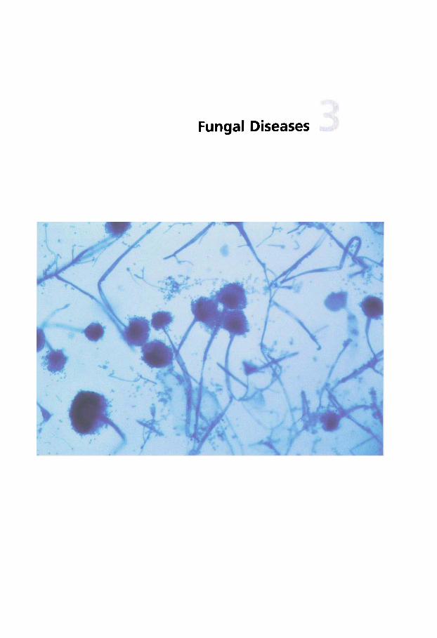
Fungal Diseases
Funaal Diseases 239
Most agents of mycoses exist as sapro- phytes in soil, decaying vegetables and dung. The soil reservoir is the primary source of most fungal infections, which can be contracted by inhalation, ingestion or by contact with infected individuals or equipment. Pathogenic fungi establish in- fection in apparently healthy hosts and such diseases as histoplasmosis, coccid- ioidomycosis and blastomycosis are re- garded as primary systemic mycoses. Op- portunistic fungi usually require a host that is debilitated by stress, metabolic aci- dosis, malnutrition or neoplasia to estab- lish infection. Prolonged exposure to an- timicrobials or immunosuppressive sub- stances can increase the likelihood of infec- tion by opportunistic fungi like Aspergillus, Mucor, Cryptococcus and Candida.
Dermatophytosis (ringworm) is an in- fection of keratinized tissue (skin, hair, nails) by several genera of fungi called der- matophytes. All domestic animals are sus- ceptible and the fungi are found world- wide. A few dermatophytes (Microsporum [M.] gypseum) normally inhabit soil (geo- philic) and can cause disease in animals and humans. Some dermatophytes (M. au- douinii) are adapted to humans and seldom infect animals (anthropophilic) and others are primarily animal pathogens (M. canis, Trichophyton fT.1 equinum), but can also cause disease in man (zoophilic).
Very little is known about fungal dis- eases in camelids. This chapter tries to summarize current knowledge of these mi- croorganisms.
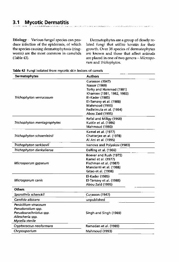
3.1 Mycotic Dermatitis I* ' I
Etiology Various fungal species can pro- duce infection of the epidermis, of which the species causing dermatophytosis (ring- worm) are the most common in camelids (Table 43).
Dermatophytes are a group of closely re- lated fungi that utilize keratin for their growth. Over 38 species of dermatophytes are known and those that affect animals are placed in one of two genera - Microspo- rum and Trichophyton.
Table 43 Fungi isolated from mycotic skin lesions of camels
Derrnatoph ytes Authors
Trichophyton verrucosum
Curasson (1947) Nasser (1 969) Torky and Hammad (1981) Khamiev (1981, 1982, 1983) El-Kader (1985) El-Tamavy et al. (1988) Mahmoud (1993) Fadlelmula et al. (1994) Abou Zaid (1995) Refai and Miligy (1968) Kuttin et al. (1986) Mahmoud (1993) Kame1 et al. (1977) Chatterjee et al. (1978) A1 Ani et al. (1995)
Trichophyton rnentagrophytes
Trichophyton schoenleinii
Trichophvton sarkisovii lvanova and Polvakov (1983) Trichophyton dankaliense Dalling et al. (1966)
Boever and Rush (1 975) Kame1 et al. (1977) Fischman et al. (1987) Mancianti et al. (1988) Gitao et al. (1998)
Microsporum gypseurn
El-Kader (1985) El-Tamavy et al. (1988) Abou Zaid (1995)
Microsporum canis
Others Sporothrix schenckii Curasson (1947) Candida albicans unpublished Penicillium vinaceum Pseudorotium spp. Pseudoarachniotus spp. Allescheria spp. Mycelia sterile Crvptococcus neoformans
Singh and Singh (1969)
Ramadan et al. (1989) Chwsosporium Mahmoud (1993)
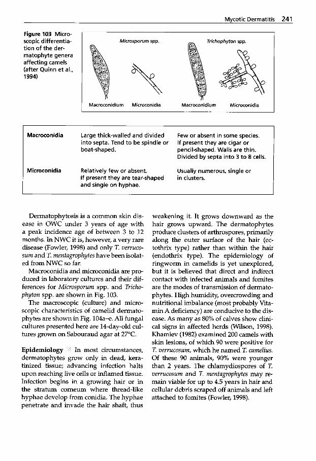
Mycotic Dermatitis 241
Figure 103 Micro- scopic differentia- tion of the der- matophyte genera affecting camels (after Quinn et al., 1994)
Microsporum spp. Trichophyton spp.
Macroconidium Microconidia
% Macroconidiurn Microconidia
' Macroconidia Large thick-walled and divided into septa. Tend to be spindle or boat-shaped. pencil-shaped. Walls are thin.
Few or absent in some species. If present they are cigar or
Divided by septa into 3 to 8 cells.
Microconidia Relatively few or absent. Usually numerous, single or If present they are tear-shaped and single on hyphae.
in clusters.
Dermatophytosis is a common skin dis- ease in OWC under 3 years of age with a peak incidence age of between 3 to 12 months. In NWC it is, however, a very rare disease (Fowler, 1998) and only T. verruco- sum and T. mentagrophytes have been isolat- ed from NWC so far.
Macroconidia and microconidia are pro- duced in laboratory cultures and their dif- ferences for Microsporum spp. and Tricho- phyton spp. are shown in Fig. 103.
The macroscopic (culture) and micro- scopic characteristics of camelid dermato- phytes are shown in Fig. 104a-e. All fungal cultures presented here are 14-day-old cul- tures grown on Sabouraud agar at 27°C.
Epidemiology In most circumstances, dermatophytes grow only in dead, kera- tinized tissue; advancing infection halts upon reaching live cells or inflamed tissue. Infection begins in a growing hair or in the stratum comeum where thread-like hyphae develop from conidia. The hyphae penetrate and invade the hair shaft, thus
weakening it. It grows downward as the hair grows upward. The dermatophytes produce clusters of arthrospores, primarily along the outer surface of the hair (ec- tothrix type) rather than within the hair (endothrix type). The epidemiology of ringworm in camelids is yet unexplored, but it is believed that direct and indirect contact with infected animals and fomites are the modes of transmission of dermato- phytes. High humidity, overcrowding and nutritional imbalance (most probably Vita- min A deficiency) are conducive to the dis- ease. As many as 80% of calves show clini- cal signs in affected herds (Wilson, 1998). Khamiev (1982) examined 200 camels with skin lesions, of which 90 were positive for T. verrucosum, which he named T. camelius. Of these 90 animals, 90% were younger than 2 years. The chlamydiospores of T. verrucosum and T. mentagrophytes may re- main viable for up to 4.5 years in hair and cellular debris scraped off animals and left attached to fomites (Fowler, 1998).
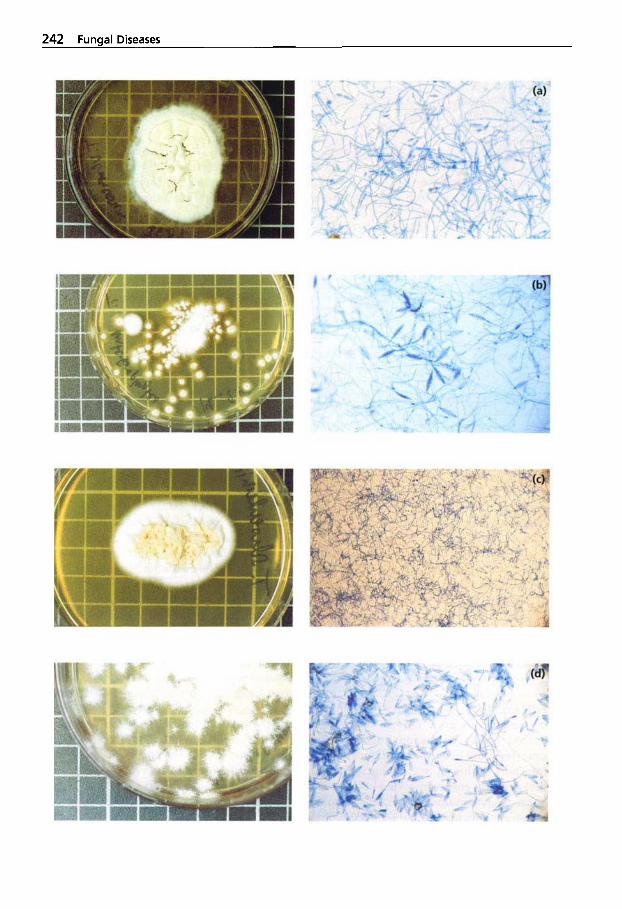
242 Fungal Diseases
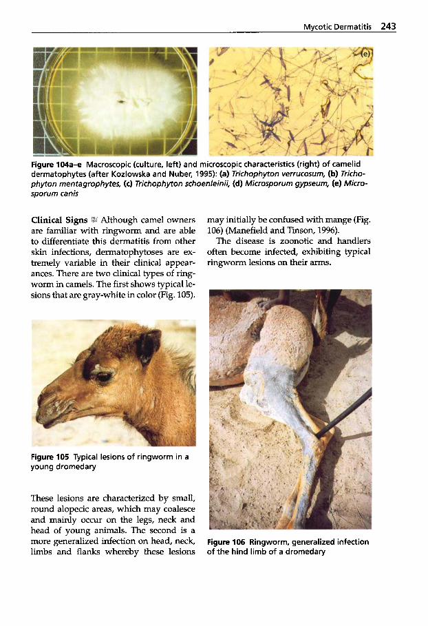
Mvcotic Dermatitis 243
Figure 104- Macroscopic (culture, left) and microscopic characteristics (right) of camelid dermatophytes (after Kozlowska and Nuber, 1995): (a) Trichophyton verrucosum, (b) Tricho- phyton mentagrophytes, (c) Trichophyton schoenleinii, (d) Microsporum gypseum, (e) Micro- sporum canis
!I Although camel owners are familiar with ringworm and are able to differentiate this dermatitis from other skin infections, dermatophytoses are ex- tremely variable in their clinical appear- ances. There are two clinical types of ring- worm in camels. The first shows typical le- sions that are gray-white in color (Fig. 105).
may initially be confused with mange (Fig. 106) (Manefield and Tinson, 1996).
The disease is zoonotic and handlers often become infected, exhibiting typical ringworm lesions on their arms.
Figure 105 Typical lesions of ringworm in a young dromedary
These lesions are characterized by small, round alopecic areas, which may coalesce and mainly OCCUT on the legs, neck and head of young animals. The second is a more generalized infection on head, neck, limbs and flanks whereby these lesions
Figure 106 Ringworm, generalized infection of the hind limb of a dromedary
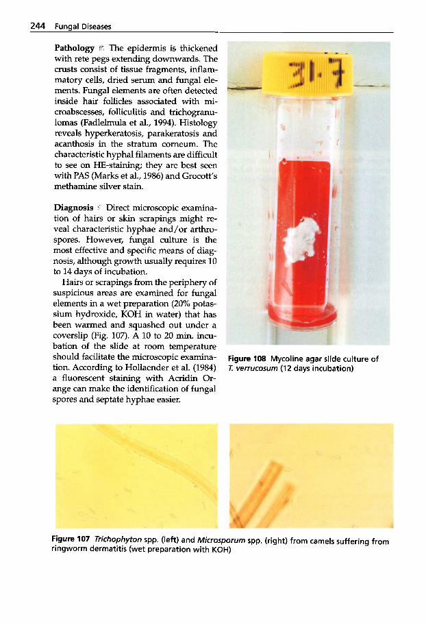
244 Funaal Diseases
Pathology lri The epidermis is thickened with rete pegs extending downwards. The crusts consist of tissue fragments, inflam- matory cells, dried serum and fungal ele- ments. Fungal elements are often detected inside hair follicles associated with mi- croabscesses, folliculitis and trichogranu- lomas (Fadlelmula et al., 1994). Histology reveals hyperkeratosis, parakeratosis and acanthosis in the stratum comeum. The characteristic hyphal filaments are difficult to see on HE-staining; they are best seen with PAS (Marks et al., 1986) and Grocott’s methamine silver stain.
Diagnosis Direct microscopic examina- tion of hairs or skin scrapings might re- veal characteristic hyphae and/or arthro- spores. However, fungal culture is the most effective and specific means of diag- nosis, although growth usually requires 10 to 14 days of incubation.
Hairs or scrapings from the periphery of suspicious areas are examined for fungal elements in a wet preparation (20% potas- sium hydroxide, KOH in water) that has been warmed and squashed out under a coverslip (Fig. 107). A 10 to 20 min. incu- bation of the slide at room temperature should facilitate the microscopic examina- tion. According to Hollaender et al. (1984) a fluorescent staining with Acridin Or- ange can make the identification of fungal spores and septate hyphae easier.
Figure 108 Mycoline agar slide culture of T: verrucosurn (12 days incubation)
Figure 107 Trichophyton spp. (left) and Microsporurn spp. (right) from camels suffering from ringworm dermatitis (wet preparation with KOH)
Mycotic Dermatitis 245
Microsporum spp. and Trichophyton spp. as well as other fungi should be cultured on Sabouraud dextrose agar and on Myco- line agar slide (bioM&ieux) and incubated for 10 to 14 days at 27°C (Fig. 108). Definite diagnosis and species identification re- quires removal of hyphae and macroconi- diae from the surface of the colony with acetate tape and microscopic examination with Lactophenol Cotton Blue (LPCB) stain. Culture on Mycoline agar slide is es- pecially helpful when saprophytic contam- ination is expected.
A number of keratin-proliferative der- matoses have been seen in camelids, and not all are caused by dermatophytes (Table 43). It is therefore essential that any skin le- sions should be carefully investigated and multiple deep skin scrapings (containing blood) should be dispatched for laboratory diagnosis.
Treatment and Prevention 5h' The spread of ringworm can be limited by early diag- nosis and separation of infected from unin- fected camels. To avoid recurrence of infec- tion, it is also essential that stables and equipment be properly disinfected.
Lesions should firstly be scrubbed clean with warm soapy water and all scabs removed. A variety of common fungicidal and fungistatic agents such as iodine, 5% sulfur in sesame oil (w/v), 5% salicylic acid, coal tar phenols (3.25%) with cop- per acetate (0.58%) and hydroxyquinolines may be applied topically as ringworm ointments onto the affected areas.
Captan@ is a fungicide for ornamental plants. The use of Captan@ has been ad- vocated (Ainsworth and Austwick, 1973) when sprayed on infected animals as a so-
lution of 1:200. The mixture is stable for one week after mixing and the solution should be applied to the lesions and sur- rounding areas for 2 weeks.
Treatment of dermatophytoses with gri- seofulvin is very effective in cattle (Coetzer et al., 1994), but it causes side effects in camels such as nausea and diarrhea and is therefore not recommended (Schwartz and Dioli, 1992).
Successful vaccination programs against Trichophyton spp. and Microsporum spp. in camels have been reported from Kazakh- stan (Toleutajewa, 1994).
Camelvac Tricho@ (IDT Dessau-Tornau, Germany) has been used in the Republic of Kazakhstan, where 34,302 Bactrians from 12 farms were investigated. In these herds the following incidents of ringworm were found: 5-day to 4-month-old Bactrians: 21.5%
13-month to 3-year-old Bactrians: 17.1% 5 to 12-month-old Bactrians: 60.1%
4 years and older Bactrians: 1.3%
In these herds, 3,300 camel calves were vaccinated with Camelvac Tricho@ and no ringworm cases reoccurred for several years. This vaccine is used with very good success not only for prophylactic but also for therapeutic purposes. Camels suffering from dermatophytoses were healed after one or two injections with Camelvac Tri- cho@ (Toleutajewa, 1994). Camelvac Tricho@ has also recently been successfully used by the authors in several camel herds in the UAE. Young dromedaries with ringworm lesions (see Fig. 105) were vaccinated once. The lesions receded within 14 days and disappeared after 4 weeks.
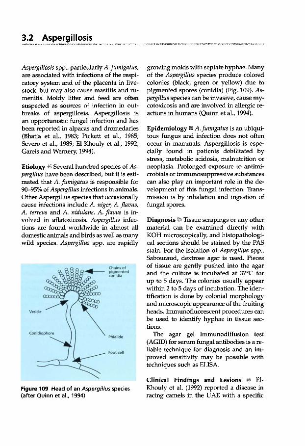
Aspergillosis spp., particularly A . furnigutus, are associated with infections of the respi- ratory system and of the placenta in live- stock, but may also cause mastitis and ru- menitis. Moldy litter and feed are often suspected as sources of infection in out- breaks of aspergillosis. Aspergillosis is an opportunistic fungal infection and has been reported in alpacas and dromedaries (Bhatia et al., 1983; Pickett et al., 1985; Severo et al., 1989; El-Khouly et al., 1992, Gareis and Wernery, 1994).
Etiology ti(#' Several hundred species of As- pergillus have been described, but it is esti- mated that A . furnigutus is responsible for 90-95% of Aspergillus infections in animals. Other Aspergillus species that occasionally cause infections include A . niger, A. fluvus, A . terreus and A. niduluns. A . flavus is in- volved in aflatoxicosis. Aspergillus infec- tions are found worldwide in almost all domestic animals and birds as well as many wild species. Aspergillus spp. are rapidly
Figure 109 Head of an Aspergillus species (after Quinn et at., 1994)
growing molds with septate hyphae. Many of the Aspergillus species produce colored colonies (black, green or yellow) due to pigmented spores (conidia) (Fig. 109). As- pergillus species can be invasive, cause my- cotoxicosis and are involved in allergic re- actions in humans (Quinn et al., 1994).
Epidemiology IN1 A. furnigutus is an ubiqui- tous fungus and infection does not often occur in mammals. Aspergillosis is espe- cially found in patients debilitated by stress, metabolic acidosis, malnutrition or neoplasia. Prolonged exposure to antimi- crobials or immunosuppressive substances can also play an important role in the de- velopment of this fungal infection. Trans- mission is by inhalation and ingestion of fungal spores.
li Tissue scrapings or any other material can be examined directly with KOH microscopically, and histopathologi- cal sections should be stained by the PAS stain. For the isolation of Aspergillus spp., Sabouraud, dextrose agar is used. Pieces of tissue are gently pushed into the agar and the culture is incubated at 37°C for up to 5 days. The colonies usually appear within 2 to 5 days of incubation. The iden- tification is done by colonial morphology and microscopic appearance of the fruiting heads. Immunofluorescent procedures can be used to identify hyphae in tissue sec- tions.
The agar gel immunodiffusion test (AGID) for serum fungal antibodies is a re- liable technique for diagnosis and an im- proved sensitivity may be possible with techniques such as ELISA.
Clinical Findings and Lesions El- Khouly et al. (1992) reported a disease in racing camels in the UAE with a specific
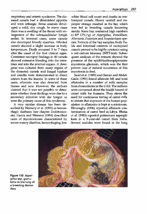
Aspergillosis 247
respiratory and enteric syndrome. The dis- eased camels had a diminished appetite and were lethargic. Some animals devel- oped a mild, dry cough. In many cases there was a swelling of the throat with en- largement of the submandibular lymph nodes. In terminal cases, some camels also developed bloody diarrhea. Affected camels showed a slight increase in body temperature. Death occurred 5 to 7 days after the onset of the first clinical signs. Consistent necropsy findings in 40 camels showed extensive bleeding into the intes- tines and into the internal organs. A. fumi- gatus was cultured from many organs of the dissected camels and fungal hyphae and conidia were demonstrated in direct smears from the lesions. In some of these cases, aflatoxin was also detected from tissues and sera. However, the authors claimed that it was not possible to deter- mine whether these findings were due to a secondary infection with the fungus or were the primary cause of this syndrome.
A very similar disease has been de- scribed by Wernery et al. (1992) as hemor- rhagic diathesis (see chapter Endotoxico- sis). Gareis and Wernery (1994) described cases of mycotoxicoses characterized by severe watery diarrhea, hemorrhaging, low
white blood cell count and deaths in one- humped camels. Heavy rainfall and im- proper storage resulted in the hay which was fed to breeding camels becoming moldy. Some hay contained high numbers (> lo8 CFU/g) of Aspergillus, Penicillium, Alternaria, Fusariurn and Scopulariopsis spe- cies. Extracts of the hay samples, body flu- ids and intestinal contents of necropsied camels proved to be highly cytotoxic using a cell-culture bioassay (MTT-test). Subse- quent analyses of the extracts showed the presence of the epidithiodioxopiperazine mycotoxin gliotoxin, which was the first proven case of natural occurrence of this mycotoxin in feed.
Saad et al. (1989) and Osman and Abdel- Gadir (1991) found aflatoxin M1 and total aflatoxins in a number of milk samples from dromedaries in the UAE. The authors were concerned about the health hazard of camel milk for humans. They stress the need for continuous testing of camel milk to ensure that exposure of the human pop- ulation to aflatoxins is kept at a minimum. Elmaraghy (1996) reported aflatoxin con- tamination of camel feed in Libya. Bhatia et al. (1983) reported pulmonary aspergl- losis in a 9-year-old camel from India. Several nodules were found in the lung
Figure 110 Asper- gillus spp. granu- loma in the lung of a breeding drorne- dary
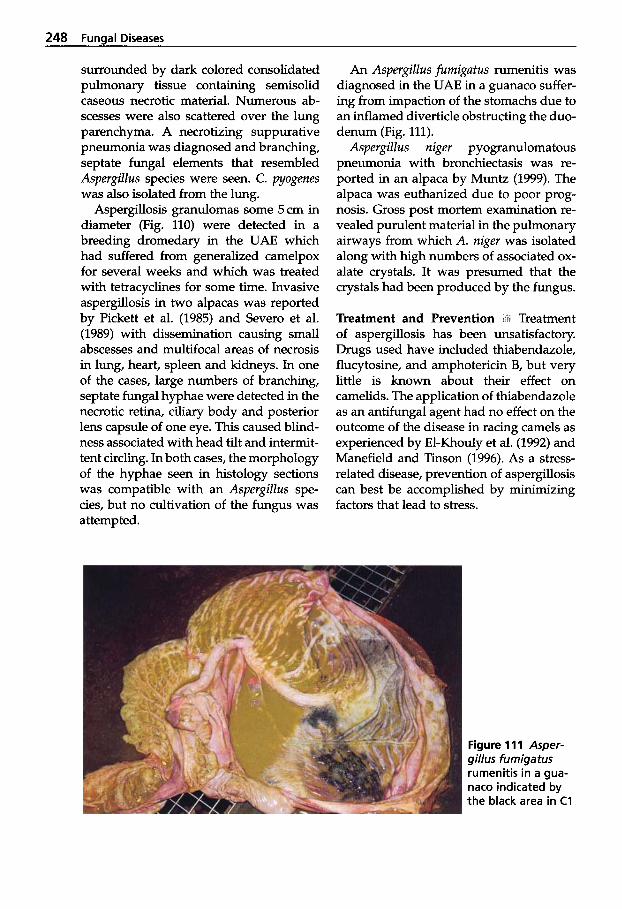
248 Funaal Diseases
surrounded by dark colored consolidated pulmonary tissue containing semisolid caseous necrotic material. Numerous ab- scesses were also scattered over the lung parenchyma. A necrotizing suppurative pneumonia was diagnosed and branching, septate fungal elements that resembled Aspergillus species were seen. C. pyogenes was also isolated from the lung.
Aspergillosis granulomas some 5cm in diameter (Fig. 110) were detected in a breeding dromedary in the UAE which had suffered from generalized camelpox for several weeks and which was treated with tetracyclines for some time. Invasive aspergillosis in two alpacas was reported by Pickett et al. (1985) and Severo et al. (1989) with dissemination causing small abscesses and multifocal areas of necrosis in lung, heart, spleen and kidneys. In one of the cases, large numbers of branching, septate fungal hyphae were detected in the necrotic retina, ciliary body and posterior lens capsule of one eye. This caused blind- ness associated with head tilt and intermit- tent circling. In both cases, the morphology of the hyphae seen in histology sections was compatible with an Aspergillus spe- cies, but no cultivation of the fungus was attempted.
An Aspergillus fumigatus rumenitis was diagnosed in the UAE in a guanaco suffer- ing from impaction of the stomachs due to an inflamed diverticle obstructing the duo- denum (Fig. 111).
Aspergillus niger pyogranulomatous pneumonia with bronchiectasis was re- ported in an alpaca by Muntz (1999). The alpaca was euthanized due to poor prog- nosis. Gross post mortem examination re- vealed purulent material in the pulmonary airways from which A. niger was isolated along with high numbers of associated ox- alate crystals. It was presumed that the crystals had been produced by the fungus.
Treatment and Prevention Ill! Treatment of aspergillosis has been unsatisfactory. Drugs used have included thiabendazole, flucytosine, and amphotericin B, but very little is known about their effect on camelids. The application of thiabendazole as an antifungal agent had no effect on the outcome of the disease in racing camels as experienced by El-Khouly et al. (1992) and Manefield and Tinson (1996). As a stress- related disease, prevention of aspergillosis can best be accomplished by minimizing factors that lead to stress.
Figure 111 Asper- gillus fumigatus rumenitis in a gua- naco indicated by the black area in C1
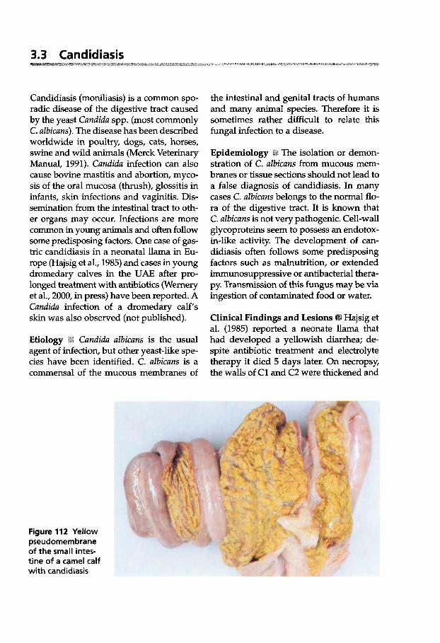
Candidiasis (moniliasis) is a common spo- radic disease of the digestive tract caused by the yeast Cundidu spp. (most commonly C. ulbicuns). The disease has been described worldwide in poultry, dogs, cats, horses, swine and wild animals (Merck Veterinary Manual, 1991). Cundidu infection can also cause bovine mastitis and abortion, myco- sis of the oral mucosa (thrush), glossitis in infants, skin infections and vaginitis. Dis- semination from the intestinal tract to oth- er organs may OCCUI: Infections are more common in young animals and often follow some predisposing factors. One case of gas- tric candidiasis in a neonatal llama in Eu- rope (Hajsig et al., 1985) and cases in young dromedary calves in the UAE after pro- longed treatment with antibiotics (Wemery et al., 2000, in press) have been reported. A Cundidu infection of a dromedary calf's skin was also observed (not published).
Etiology l id Candidu albicuns is the usual agent of infection, but other yeast-like spe- cies have been identified. c. ulbicuns is a commensal of the mucous membranes of
the intestinal and genital tracts of humans and many animal species. Therefore it is sometimes rather difficult to relate this fungal infection to a disease.
Epidemiology air The isolation or demon- stration of C. ulbicuns from mucous mem- branes or tissue sections should not lead to a false diagnosis of candidiasis. In many cases C. albicuns belongs to the normal flo- ra of the digestive tract. It is known that C. ulbicuns is not very pathogenic. Cell-wall glycoproteins seem to possess an endotox- in-like activity. The development of can- didiasis often follows some predisposing factors such as malnutrition, or extended immunosuppressive or antibacterial thera- py. Transmission of this fungus may be via ingestion of contaminated food or water.
Clinical Findings and Lesions al. (1985) reported a neonate llama that had developed a yellowish diarrhea; de- spite antibiotic treatment and electrolyte therapy it died 5 days later. On necropsy, the walls of C1 and C2 were thickened and
Figure 112 Yellow pseudomembrane of the small intes- tine of a camel calf with candidiasis
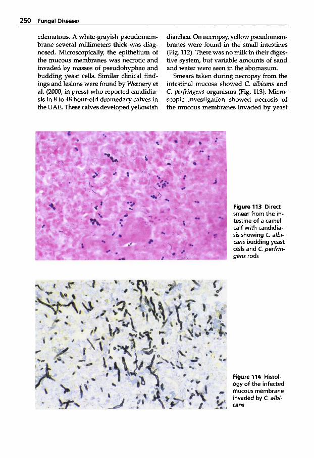
250 Fungal Diseases
edematous. A white-grayish pseudomem- brane several millimeters thick was diag- nosed. Microscopically, the epithelium of the mucous membranes was necrotic and invaded by masses of pseudohyphae and budding yeast cells. Similar clinical find- ings and lesions were found by Wernery et al. (2000, in press) who reported candidia- sis in 8 to 48 hour-old dromedary calves in the UAE. These calves developed yellowish
diarrhea. On necropsy, yellow pseudomem- branes were found in the small intestines (Fig. 112). There was no milk in their diges- tive system, but variable amounts of sand and water were seen in the abomasum.
Smears taken during necropsy from the intestinal mucosa showed C. albicans and C. perfringens organisms (Fig. 113). Micro- scopic investigation showed necrosis of the mucous membranes invaded by yeast
Figure 113 Direct smear from the in- testine of a camel calf with candidia- sis showing C. albi- cans budding yeast cells and C. perfrin- gens rods
Figure 114 Histol- ogy of the infected mucous membrane invaded by C. albi- cans
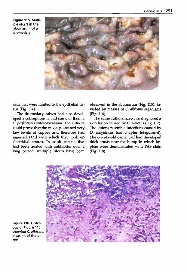
Candidiasis 251
Figure 115 Multi- ple ulcers in the abomasum of a dromedary
cells that were limited to the epithelial tis- sue (Fig. 114).
The dromedary calves had also devel- oped a colisepticemia and some of them a C. perfringens enterotoxaemia. The authors could prove that the calves possessed very low levels of copper and therefore had ingested sand with which they took up clostridial spores. In adult camels that had been treated with antibiotics over a long period, multiple ulcers have been
observed in the abomasum (Fig. 115), in- vaded by masses of C. ulbicuns organisms (Fig. 116).
The same authors have also diagnosed a skin lesion caused by C. ulbicuns (Fig. 117). The lesions resemble infections caused by D. congolensis (see chapter Integument). The 6-week-old camel calf had developed thick crusts near the hump in which hy- phae were demonstrated with PAS stain (Fig. 118).
Figure 116 Histol- ogy of Figure 1 15 showing C. albicans invasion of the ul- cers
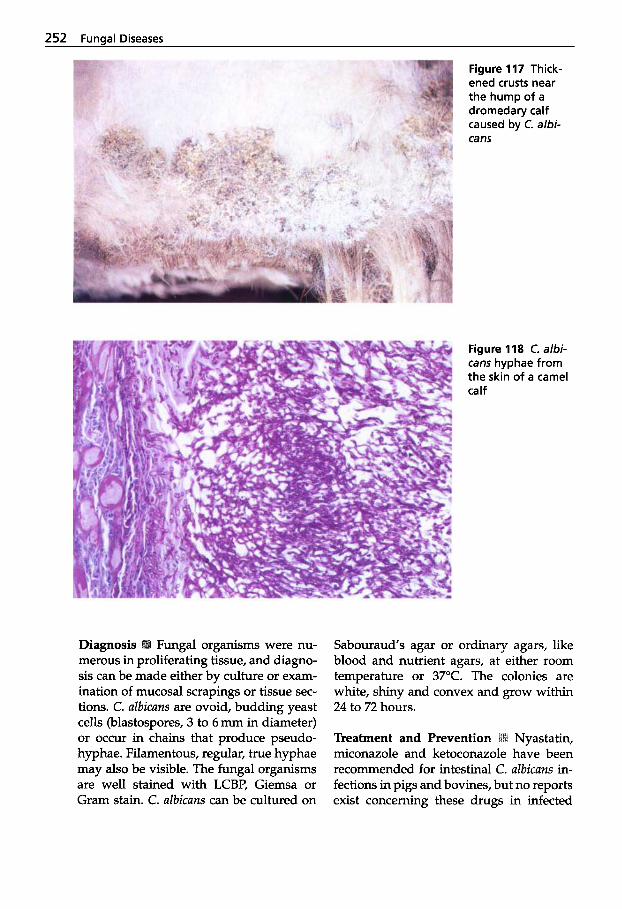
252 Funaal Diseases
Fungal organisms were nu- merous in proliferating tissue, and diagno- sis can be made either by culture or exam- ination of mucosal scrapings or tissue sec- tions. C. albicans are ovoid, budding yeast ceUs (blastospores, 3 to 6 mm in diameter) or occur in chains that produce pseudo- hyphae. Filamentous, regular, true hyphae may also be visible. The fungal organisms are well stained with LCBP, Giemsa or Gram stain. C. albicans can be cultured on
Figure 117 Thick- ened crusts near the hump of a dromedary calf caused by C. albi- can5
Figure 118 C. albi- can5 hyphae from the skin of a camel ca If
Sabouraud's agar or ordinary agars, like blood and nutrient agars, at either room temperature or 37°C. The colonies are white, shiny and convex and grow within 24 to 72 hours.
Treatment and Prevention miconazole and ketoconazole have been recommended for intestinal C. albicans in- fections in pigs and bovines, but no reports exist concerning these drugs in infected

Candidiasis 253
camelids. In our cases, the camel mothers received copper and selenium treatment and the calves were given lOmL of an E. coli autovaccine orally, 20 mL of a C. per- fringens antiserum i.v. (Rhone Merieux), and 10 mg StegantoxB (Schering-Plough Animal Health) i.v. twice within 24 hours. The camels did not receive any antibiotics.
Prevention of candidiasis can best be achieved by minimizing predisposing fac- tors. It is therefore essential to detect and to remove them. Optimal management of breeding herds, including vaccination (see chapter Vaccination Program), and proper mineral supplementation are crucial for the survival of young camelids.
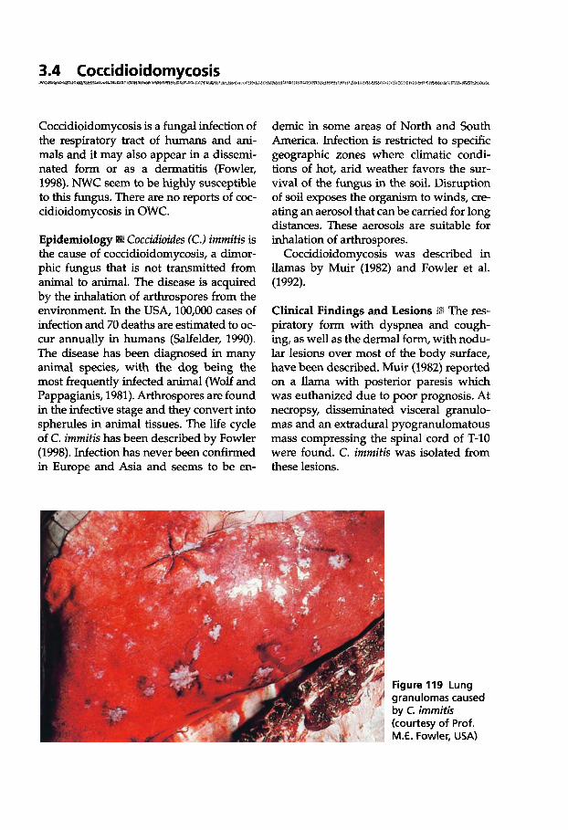
3.4 Coccidioidomycosis
Coccidioidomycosis is a fungal infection of the respiratory tract of humans and ani- mals and it may also appear in a dissemi- nated form or as a dermatitis (Fowler, 1998). NWC seem to be highly susceptible to this fungus. There are no reports of coc- cidioidomycosis in OWC.
Coccidioides (C.) immitis is the cause of coccidioidomycosis, a dimor- phic fungus that is not transmitted from animal to animal. The disease is acquired by the inhalation of arthrospores from the environment. In the USA, 100,000 cases of infection and 70 deaths are estimated to oc- cur annually in humans (Salfelder, 1990). The disease has been diagnosed in many animal species, with the dog being the most frequently infected animal (Wolf and Pappagianis, 1981). Arthrospores are found in the infective stage and they convert into spherules in animal tissues. The life cycle of C. immitis has been described by Fowler (1998). Infection has never been confirmed in Europe and Asia and seems to be en-
demic in some areas of North and South America. Infection is restricted to specific geographic zones where climatic condi- tions of hot, arid weather favors the sur- vival of the fungus in the soil. Disruption of soil exposes the organism to winds, cre- ating an aerosol that can be carried for long distances. These aerosols are suitable for inhalation of arthrospores.
Coccidioidomycosis was described in llamas by Muir (1982) and Fowler et al. (1992).
Clinical Findings and Lesions piratory form with dyspnea and cough- ing, as well as the dermal form, with nodu- lar lesions over most of the body surface, have been described. Muir (1982) reported on a llama with posterior paresis which was euthanized due to poor prognosis. At necropsy, disseminated visceral granulo- mas and an extradural pyogranulomatous mass compressing the spinal cord of T-10 were found. C. immitis was isolated from these lesions.
Figure 119 Lung granulomas caused by C. immitis (courtesy of Prof. M.E. Fowler, USA)
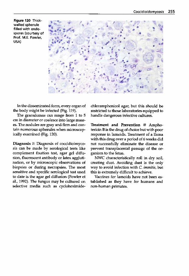
Coccidioidomycosis 255
Figure 120 Thick- walled spherule filled with endo- spores (courtesy of Prof. M.E. Fowler, USA)
In the disseminated form, every organ of the body might be infected (Fig. 119).
The granulomas can range from 1 to 5 cm in diameter or coalesce into large mass- es. The nodules are gray and firm and con- tain numerous spherules when microscop- ically examined (Fig. 120).
Diagnosis of coccidioimyco- sis can be made by serological tests like complement fixation test, agar gel diffu- sion, fluorescent antibody or latex aggluti- nation, or by microscopic observations of biopsies or during necropsies. The most sensitive and specific serological test used to date is the agar gel diffusion (Fowler et al., 1992). The fungus may be cultured on selective media such as cycloheximide-
chloramphenicol agar, but this should be restricted to those laboratories equipped to handle dangerous infective cultures.
Treatment and Prevention tericin B is the drug of choice but with poor response in lamoids. Treatment of a llama with this drug over a period of 6 weeks did not successfully eliminate the disease or prevent transplacental passage of the or- ganism to the fetus.
NWC characteristically roll in dry soil, creating dust. Avoiding dust is the only way to avoid infection with C. immitis, but this is extremely difficult to achieve.
Vaccines for lamoids have not been es- tablished as they have for humans and non-human primates.
Etiology l i i There are 11 genera of the order Mucorales with 22 species. The most im- portant genera are Mucor, Absidia, Rhizopus and Mortierella and the most pathogenic thermotolerant Mucor spp. are now classi- fied in a new genus: Rhizomucor. The dis- ease produced by any of these genera is called "mucormycosis".
These ubiquitous fungi are common in- habitants of soil, manure and rotting vege- tation. Infections are secondary to other disorders and might cause granulomatous lesions in several organs of various animal species. Mucormycosis is particularly im- portant as a cause of placentitis and abor- tion in cattle.
Only one genus, Rhizopus spp., has been isolated from a llama (Fowler, 1998).
The llama suffered from a disseminated, multisystemic infection in
association with a facial paralysis of cra- nial nerve w. During the course of the disease, swallowing became impossible and the llama began to lose weight. An endoscopic examination of the nasal cavity revealed a black membrane with white patches.
Diagnosis M Mucormycosis can be diag- nosed microscopically by demonstrating broad, branching, aseptate and irregular hyphae. Fungi can be identified in tissue sections by FA techniques with fluorescein antiglobulins specific for each genus of the Mucorales. In the reported case of the llama, filamentous growth was present on the surface of a necrotic rhinitis, the meninges on the ventral aspect of the brain were inflamed, and granulomas were pres- ent in the area of the cranial nerves.
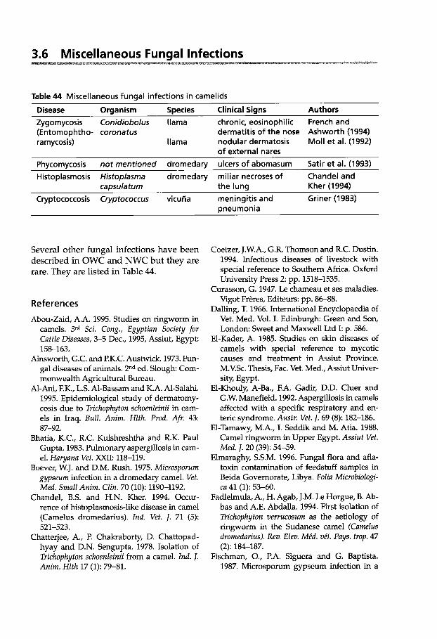
3.6 Miscellaneous Fungal Infections
Table 44 Miscellaneous fungal infections in camelids
Disease Oraanism SDecies Clinical Sians Authors Zygomycosis Conidiobolus llama chronic, eosinophilic French and (Entomophtho- coronatus dermatitis of the nose Ashworth (1994) ramycosis) llama nodular dermatosis Moll et al. (1992)
Phycomycosis not mentioned dromedary ulcers of abomasum Satir et al. (1993) Histoplasmosis Histoplasma dromedary miliar necroses of Chandel and
of external nares
capsula tum the lung Kher (1994) Cryptococcosis Cryptococcus vicuiia
Several other fungal infections have been described in OWC and NWC but they are rare. They are listed in Table 44.
References
Abou-Zaid, A.A. 1995. Studies on ringworm in camels. 3rd Sci. Cong., Egyptian Society for Cattle Diseases, 3-5 Dec., 1995, Assiut, Egypt: 158-163.
Ainsworth, G.C. and P.K.C. Austwick. 1973. Fun- gal diseases of animals. Znd ed. Slough: Com- monwealth Agricultural Bureau.
Al-Ani, F.K., L.S. Al-Bassam and K.A. Al-Salahi. 1995. Epidemiological study of dermatomy- cosis due to Trichophyton schoenleinii in cam- els in Iraq. Bull. Anim. Hlth. Prod. Afi. 43:
Bhatia, K.C., R.C. Kulshreshtha and R.K. Paul Gupta. 1983. Pulmonary aspergllosis in cam- el. Haryuna Vet. XXII: 118-119.
Boever, W.J. and D.M. Rush. 1975. Microsporum gypseum infection in a dromedary camel. Vet. Med. Small Anim. Clin. 70 (10): 1190-1192.
Chandel, B.S. and H.N. Kher. 1994. Occur- rence of histoplasmosis-like disease in camel (Camelus dromedarius). Ind. Vet. J. 71 (5):
Chattejee, A., P. Chakraborty, D. Chattopad- hyay and D.N. Sengupta. 1978. Isolation of Trichophyton schoenleinii from a camel. Ind. I. Anim. Hlth 17 (1): 79-81.
87-92.
521-523.
meningitis and Griner (1983) pneumonia
Coetzer, J.W.A., G.R. Thomson and R.C. Dustin. 1994. Infectious diseases of livestock with special reference to Southern Africa. Oxford University Press 2: pp. 1518-1535.
Curasson, G. 1947. Le chameau et ses maladies. Vigot Fr&es, Editeurs: pp. 86-88.
Dalling, T. 1966. International Encyclopaedia of Vet. Med. Vol. I. Edinburgh Green and Son, London: Sweet and Maxwell Ltd I: p. 586.
El-Kader, A. 1985. Studies on skin diseases of camels with special reference to mycotic causes and treatment in Assiut Province. M.V.Sc. Thesis, Fac. Vet. Med., Assiut Univer- sity, Egypt.
El-Khouly, A-Ba., F.A. Gadir, D.D. Cluer and G.W. Manefield. 1992. Aspergillosis in camels affected with a specific respiratory and en- teric syndrome. Austr. Vet. J. 69 (8): 182-186.
El-Tamawy, M.A., I. Seddik and M. Atia. 1988. Camel ringworm in Upper Egypt. Assiut Vet. Med. J. 20 (39): 54-59.
Elmaraghy, S.S.M. 1996. Fungal flora and afla- toxin contamination of feedstuff samples in Beida Governorate, Libya. Foliu Microbiologi- ca 41 (1): 53-60.
Fadlelmula, A., H. Agab, J.M. Le Horgue, 8. Ab- bas and A.E. Abdalla. 1994. First isolation of Trichophyton zlerrucosum as the aetiology of ringworm in the Sudanese camel (Camelus drornedarius). Rev. Elm. Mkd. vkt. Pays. trop. 47
Fischman, O., P.A. Siguera and G. Baptista. 1987. Microsporum gypseum infection in a
(2): 184-187.
258 Funaal Diseases
grey wolf (Canis lupus) and a camel (Came- lus bactrianus) in a zoological garden. Myko-
Fowler, M.E., D. Pappagianis and I. Irvin. 1992. Coccidioidomycosis in llamas in the United States: 19 cases (1981-1989). JAVMA 201 (10):
Fowler, M.E. 1998. Medicine and surgery of South American Camelids. Iowa State Uni- versity Press, Ames.
French, R.A. and C.D. Ashworth. 1994. Zy- gomycosis caused by Conidiobolus corona- tus in a llama (Lama glama). Vet. Pathol. 31: 120-122.
Gareis, M. and U. Wemery. 1994. Determination of Gliotoxin in samples associated with cases of intoxication in camels. Mycotoxin Research
Gitao, C.G., H. Agab and A.J. Khalifalla. 1998. An outbreak of a mixed infection of Der- matophilus congolensis and Microsporum gyp- seum in camels (Camelus dromedarius) in Sau- di Arabia. Rev. sci. tech. Of. int. Epiz. 17 (3): 749-755.
Griner, L.A. 1983. Camelidae. In L.A. Griner, ed. Pathology of 200 animals. San Diego Zool. SOC., San Diego: 501-505.
Hajsig, M., T. Naglio, D. Hajsig and M. Herceg. 1985. Systemic mycoses in domestic and wild ruminants. I. Candidiasis of forestomachs in the lamb, calf, kid and newborn llama. Vet. Arch. 55 (2): 53-58.
Hollaender, H., W. Keilig, J. Bauer and E. Rothe- mund. 1984. A reliable fluorescent stain for fungi in tissue sections and clinical speci- mens. Mycopathologia 88: 131-134.
Ivanova, L.G. and I.D. Polyakov. 1983. Tricho- phyton sarkisovii Ivanova and Polyakov sp. nov., a new species of the pathogenic fungus inducing dermatomycosis in camels. Mikol. fitopatologiya 17 (5): 363-366.
Kamel, Y.Y., M.A. Ahmed and A.A. Ismail. 1977. Dermatophytes in animals, birds and man. Animals a potential reservoir of dermato- phytes to man. Assiut Vet. Med. 4 (7): 149-159.
Khamiev, S.K. 1981. Camel ringworm. Buyll. Vses. Inst. Eksp. Vet. 42: 14-17.
Khamiev, S.K. 1982. Epidemiology of ringworm (Trichophyton infection) among camels in Kazakhstan. Veterinariya 9 42.
Khamiev, S.K. 1983. Clinical symptoms of tri- chophytosis in camels. Rev. Med. Vet. Mycolo- g y 17 (1): 147 (abstract).
S ~ Y I 30 (7): 295-297.
1609-1614.
1 0 2-8.
Kozlowska, E.A. and D. Nuber. 1995. Leitfaden der praktischen Mykologie. Einfiihrung in die mykologische Diagnostik. Blackwell Wis- senschafts-Verlag, Berlin, Wien: pp. 44-57.
Kuttin, E.S., E. Al-Hanaty, M. Feldman, M. Chaimovits and J. Muller. 1986. Dermatophy- tosis of camels. Rev. Med. Mycol. 24: 341-344.
Mahmoud, A.L.E. 1993. Dermatophytes and other associated fungi isolated from ring- worm lesions of camels. Folia Microbiologica
Mancianti, F., R. Papini and P. Cavicchio. 1988. Dermatofizia da Microsporum gypseum in un Cammello (Camelus dromedarius). Ann. Fac. Med. Vet. Univ. Pisa 4: pp. 233-237.
Manefield, G.W. and A. Tinson. 1996. Camels. A compendium. The T.G. Hungerford Vade Mecum Series for Domestic Animals: 240, 298.
Marks, R., A. Knight and P. Laidler. 1986. Atlas of skin pathology. MTP Press Limited: pp. 36, 39.
Merck, Veterinary Manual. 1991. The Merck Vet- erinary Manual. Merck and Co. Inc., Rahway,
Moll, H.D., J. Schumacher and T.R. Hoover. 1992. Entomophthormycosis conidiobolae in a llama. JAVMA 200 (7): 969-970.
Muir, Susie. 1982. Coccidioidomycosis in the llama: Case report and epidemiologic survey.
Muntz F.H.A. 1999. Oxalate-producing pul- monary aspergillosis in an alpaca. Vet. Path.
Nasser, M. 1969. The zoonotic importance of dermatophytes in U.A.R. PhD Thesis. Facul- ty of Vet. Med., Cairo University.
Osman, N.A. and F. Abdel-Gadir. 1991. Survey of total aflatoxins in camel sera by enzyme- linked immunosorbent assay (ELISA). Myco- toxin Research 7 35-38.
Pickett, J.P., C.P. Moore, B.A. Beehler, A. Gen- dron-Fitzpatrick and R.R. Dubielzig. 1985. Bi- lateral chorioretinitis secondary to dissemi- nated aspergdlosis in an alpaca. JAVMA 187
Quinn, P.J., M.E. Carter, B.K. Markey and G.R. Carter. 1994. Clinical Veterinary Microbiolo- gy. Wolfe: pp. 381-421.
Ramadan, R.O., A.A. Fayed and A.M. El-Has- san. 1989. Textbook of dermatology. Vol. 2. 4'h ed. Blackwell Scientific Publications, Oxford
38 (6): 505-508.
N. J., U.S.A.: pp. 342-343.
JAVMA 181 (11): 1334-1337.
36 (6): 631-632.
(11): 1241-1243.
2 (4): pp. 911-915.
Miscellaneoous Funaal Infections 259
Refai, M. and M. Miligy. 1968. Soil as a reservoir of Trichophyton mentagrophytes. J. Egypt . Vet. Med. Ass. 28: 47-52.
Saad, A.M., A.M. Abdelgadir and M.O. Moss. 1989. Aflatoxin in human and camel milk in Abu Dhabi, United Arab Emirates. Mycotoxin Research 5: 57-60.
Salfelder, K. 1990. Atlas of fungal pathology. Kluwer Academic Publishers: 101.
Satir, A.A., M.I. Abu Bakr, A. Abalkheil, A.E. Abdel Ghaffar and A.E. Babiker. 1993. Phy- comycosis of the abomasum in Camelus dromedarius. J. Vet. Med. Ass. 40: 672-675.
Schwartz, H.J. and M. Dioli. 1992. The one- humped camel in Eastern Africa. A pictorial guide to diseases, health care and manage- ment. Verlag Josef Margraf.
Severo, L.C., J.C. Bohrer, G.R. Geyer and L. Fer- reiro. 1989. Invasive aspergillosis in an alpaca (Lama pacos). J . Med. and Vet. Mycol. 2 7
Singh, M.P. and C.M. Singh. 1969. Mycotic der- matitis in camels. Ind. Vet. J. 46 (10): 855.
Toleutajewa, S.T. 1994. Widerstandsfahigkeit des Erregers der Trichophytie der Kamele in der Umwelt und vergleichende Aktivitat von Vakzinen bei dieser Erkrankung. Thesis, Rus- sische Akademie der Landwirtschaftswissen- schaften, Moskau.
Torky, H.A. and H.A.S. Hammad. 1981. Tri- chophytosis in farm animals and trials for treatment. Bull. Anim. Hlth. Prod. Afr. 29 (2):
Wernery, U., H.H. Schimmelpfennig, H.S.H. Seifert and J. Pohlenz. 1992. Bacillus cereus
193-195.
143-147.
as a possible cause of haemorrhagic disease in dromedary camels (Camelus dromedar- ius). Proc. lst int. Camel Conf. In: Allen, W.R., A.J. Higgins, I.G. Mayhew, D.H. Snow and J.F. Wade, R. and W. Publications, Newmar- ket, UK 51-58.
Wernery, R., M. Ali, J. Kinne, A.A. Abraham and U. Wemery. 2000. Mineral deficiency: a pre- disposing factor for septicemia in dromedary calves. Proc. of 2nd Camelid Conf. Agroeconom- ics of Camelid Farming, Almaty, Kazakhstan, 8-12 Sept 2000, in press.
Wilson, R.T. 1998. Camels. The Tropical Agricul- turalist, MacMillan: 106.
Wolf, A.M. and D. Pappagianis. 1981. Ca- nine coccidioidomycosis-treatment with new agent. Cal$ Vet. 5: 25-27.
Further reading Abdel Samed, G.H. 1983. Yeast flora in the di-
gestive tract of the one-humped camel. The- sis, Faculty of Vet. Sci. University Khartoum, Sudan.
Connole, M.D. 1990. Review of animal mycosis in Australia. Mycopathologica. III (3): 133-164.
Glawischnig, W. and D. Khaschabi. 1999. Gen- eralisierte Aspergdlose bei einem juvenilen Alpaca (Lama pacos). Wen. Tiearztl. Mschr.
Holmes, L.A., N.W. Frame, R.K. Frame, J.P. Duff and G.C. Lewis. 1999. Suspected tremor- genic mycotoxicosis (ryegrass staggers) in alpacas (Llama pacos) in the UK. Vet. Rec. 145
86: 317-319.
462-463.
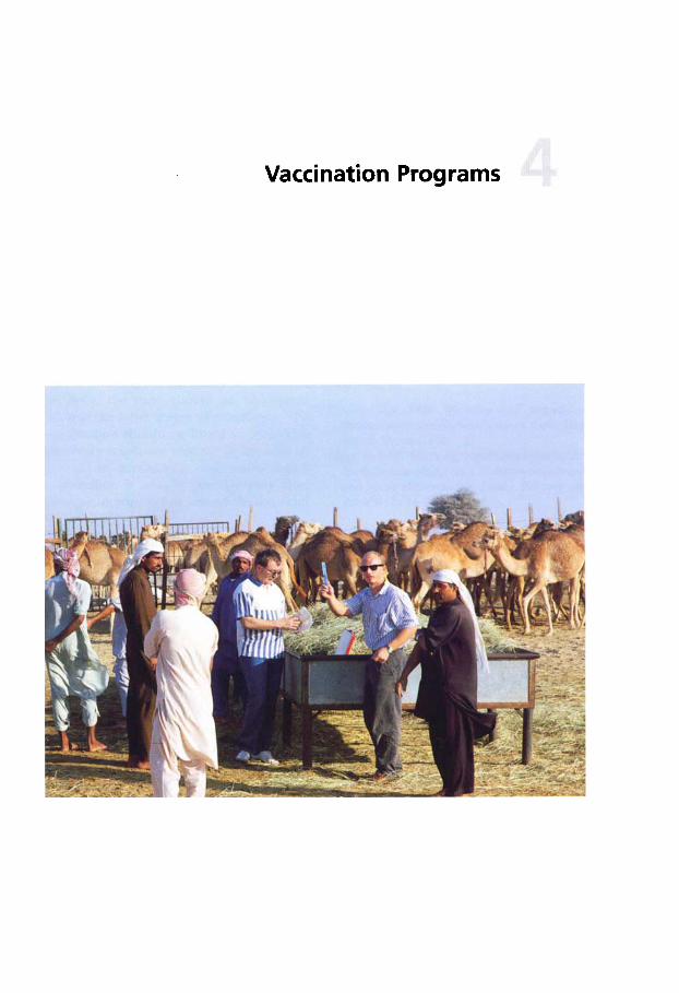
Vaccination Programs
Vaccination Proarams 263
The methods of reducing infection of economically important animals include a wide range of management practices, such as testing and slaughter, hygiene and sani- tation and immunization. Preventing and controlling a large number of animal dis- eases by immunization is probably the out- standing achievement of veterinary medi- cine in the last century.
Although it is impossible to give exact schedules for each vaccine, certain princi- ples are common to all methods of active immunization. As maternal antibodies may passively protect newborn animals, vacci- nation is usually not successful early in life. If immunity is necessary for newborn animals, the dam should be vaccinated during the latter stages of pregnancy. The vaccination should be timed so that peak antibody levels are reached at the time of colostrum production. Successful active vaccination is usually possible only after passive immunity has waned. As the exact
time of maternal immunity loss cannot be predicted, young animals must be vacci- nated at least twice to ensure successful immunization.
Very little is known about the efficacy of vaccines in camelids. In the United States of America for example no vaccines have been approved for use in camelids (Fowler, 1998). However, Fowler (1998) and Mayr (1998) recommend some vaccines in NWC. The following vaccine programs for viral, bacterial and fungal diseases are based on their recommendations and our own expe- rience (Tables 45 and 46).
References
Fowler, M.E. 1998. Medicine and surgery of South American Camelids. Iowa State Uni- versity Press, Ames.
Mayr, A. 1998. Nutzung des Immunsystems fur die Schutzimpfung und Paraimmunisierung von Neuweltkameliden. Lamas, Haltung und Zucht 6 (2): 14-23.
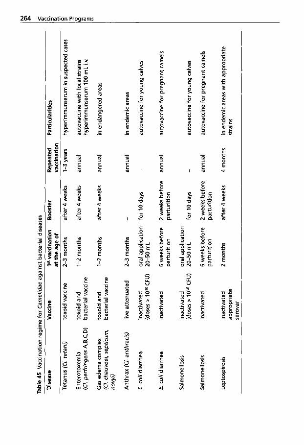
Tabl
e 45
Vac
cina
tion
reai
me
for
Car
nelid
ae a
gain
st b
acte
rial d
iseas
es
Dis
ease
V
acci
ne
1" v
acci
natio
n B
oost
er
Rep
eate
d P
artic
ular
ities
at
the
aae
of
vacc
inat
ion
Teta
nus (C/. te
tani
)
Ent
erot
oxem
ia
(Cl.
perf
ringe
ns A
,B,C
,D)
Gas
ede
ma
com
plex
(C
l. ch
auvo
ei,
sept
icur
n,
novy
i)
Ant
hrax
(C/. an
thra
cis)
E. c
oli d
iarr
hea
E. c
oli d
iarr
hea
Sal
mon
ello
sis
Sal
mon
ello
sis
Lept
ospi
rosi
s
toxo
id v
acci
ne
2-3
mo
nth
s af
ter 4 w
eeks
1-3
year
s
toxo
id a
nd
1-2
mon
ths
afte
r 4 w
eeks
an
nual
ba
cter
ial v
acci
ne
toxo
id a
nd
1-2
mon
ths
afte
r 4 w
eeks
an
nual
ba
cter
ial v
acci
ne
live
atte
nuat
ed
2-3
mon
ths
-
annu
al
inac
tivat
ed
oral
app
licat
ion
for 10 d
ays
- (d
oses
> 1O"JCFU)
20-50
mL
inac
tivat
ed
6 w
eeks
bef
ore
2 w
eeks
bef
ore
annu
al
part
uriti
on
part
uriti
on
inac
tivat
ed
oral
app
licat
ion
(dos
es >
10'0
CFU)
20-50
mL
for 10 d
ays
-
inac
tivat
ed
6 w
eeks
bef
ore
2 w
eeks
bef
ore
annu
al
part
uriti
on
part
uriti
on
inac
tivat
ed
2 m
onth
s af
ter 4 w
eeks
4
mon
ths
appr
opria
te
sero
var
hype
rimm
unse
rum
in s
uspe
cted
case
s
auto
vacc
ine
wit
h lo
cal s
train
s hy
perim
mun
seru
m 100 m
L i.v
.
in e
ndan
gere
d ar
eas
in e
ndem
ic a
reas
auto
vacc
ine
for y
oung
cal
ves
auto
vacc
ine
for
preg
nant
cam
els
auto
vacc
ine
for y
oung
cal
ves
auto
vacc
ine
for
preg
nant
cam
els
in e
ndem
ic a
reas
wit
h a
ppro
pria
te
stra
ins
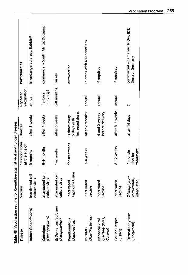
Tabl
e 46
Vac
cina
tion
regi
me
for
Cam
elid
ae a
gain
st v
iral a
nd fu
ngal
dis
ease
s
Dis
ease
V
acci
ne
1" v
acci
natio
n B
oost
er
Rep
eate
d P
artic
ular
ities
a
t the
age
of
vacc
inat
ion
Rab
ies
(Rha
bdov
irus)
Cam
el po
x (O
rthop
oxvi
rus)
Ect
hym
a co
n tag
iosu
m
(Par
apox
viru
s)
Pap
illom
atos
is
(Pap
ovav
irus)
BV
DIM
D
(Fla
vilP
estiv
irus)
Neo
nata
l vira
l di
arrh
ea (R
ota,
C
oron
a)
Equ
ine
Her
pes
(E H
V- 1)
Der
mat
op hy
tose
s (R
ingw
orm
)
inac
tivat
ed c
ell
cultu
re v
irus
atte
nuat
ed c
ell
cultu
re v
irus
atte
nuat
ed c
ell
cultu
re v
irus
inac
tivat
ed
Pap
illom
a tis
sue
inac
tivat
ed
vacc
ine
inac
tivat
ed
vacc
ine
inac
tivat
ed
vacc
ine
Tric
hop h
yton
ve
rruco
sum
, at
tenu
ated
3 m
onth
s
6-9
mon
ths
1-2
wee
ks
for t
reat
men
t
2-4
wee
ks
- 8-12
wee
ks
4 m
onth
s al
so fo
r tr
eatm
ent
afte
r 3
wee
ks
annu
al
in e
ndan
gere
d ar
eas,
Rab
isin
"
afte
r 4 w
eeks
lif
e-lo
ng
com
mer
cial
- S
outh
Afri
ca,
Duc
apox
im
mun
ity?
afte
r 6
wee
ks
6-8
mon
ths
Turk
ey
3tim
esev
ery
- au
tova
ccin
e 5
days
wit
h
incr
ease
d do
ses
afte
r 2
mon
ths
annu
al
in a
reas
wit
h M
D a
borti
ons
4 an
d 2
wee
ks
annu
al
if re
quire
d be
fore
del
iver
y
afte
r 3-
4 w
eeks
an
nual
if
requ
ired
afte
r 14
day
s ?
com
mer
cial
- C
amel
vac
Tric
ho,
IDT,
D
essa
u, G
erm
any
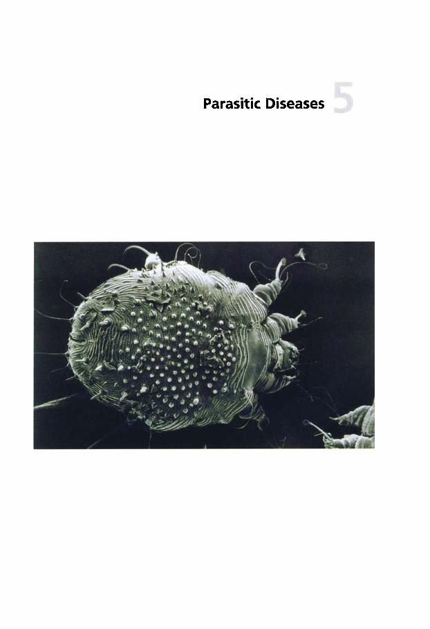
Parasitic Diseases
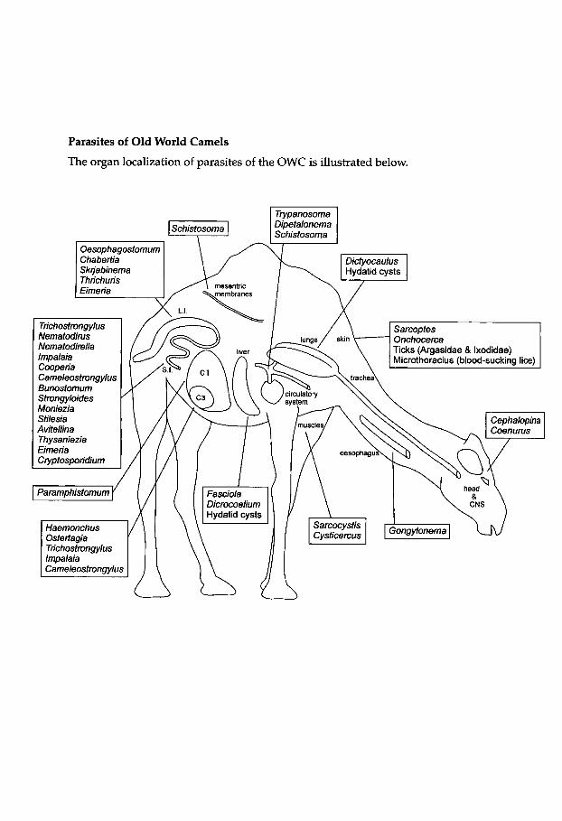
Parasites of Old World Camels
The organ localization of parasites of the OWC is illustrated below.
Trypanosoma Schistosoma Dipetalonema
Oesophagostomum Chabertia Sktja binema Thrichuris
Trichostrongylus Nematodims Nematodirella
Cameleostrongylus Bunostomum
Moniezia Stilesia Avitellina Thysaniezia Eimeria Cryptosporidium
Paramphistomum
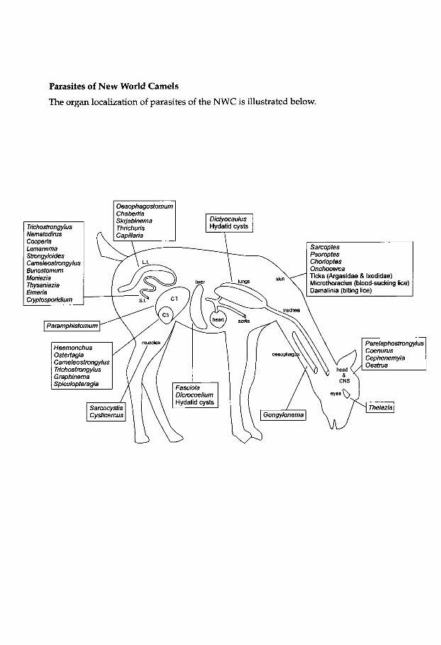
Parasites of New World Camels
The organ localization of parasites of the NWC is illustrated below.
Oesophagostomum Chabertia Skjabinema Dictyocaulus
Tnchostmngylus Thrichuris Hydatid cysts
Stmngyloides Cameleostrongylus 6unostomum Moniezia Thysaniezia
Ticks (Argasidae & Ixodidae) Microthoracius (blood-sucking lice) Damalinia (biting lice)
Paramphistomum
Haemonchus
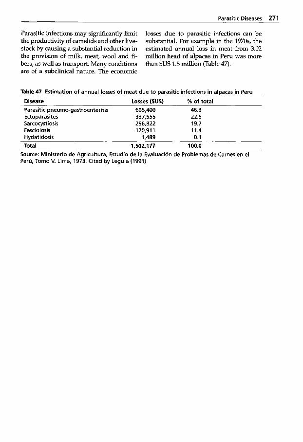
Parasitic Diseases 271
Parasitic infections may significantly limit the productivity of camelids and other live- stock by causing a substantial reduction in the provision of milk, meat, wool and fi- bers, as well as transport. Many conditions are of a subclinical nature. The economic
losses due to parasitic infections can be substantial. For example in the 1970s, the estimated annual loss in meat from 3.02 million head of alpacas in Peru was more than $US 1.5 million (Table 47).
Table 47 Estimation of annual losses of meat due to parasitic infections in alpacas in Peru
Disease losses ($US) % of total Parasitic pneumo-gastroenteritis 695,400 46.3 Ectoparasites 337,555 22.5 Sarcocystiosis 296,822 19.7 Fasciolosis 170,911 11.4 Hvdatidosis 1.489 0.1 Total 1,502.1 77 100.0
Source: Ministerio de Agricultura, Estudio de la Evaluacion de Problemas de Carnes en el Peru, Tom0 V. Lima, 1973. Cited by Leguia (1991)
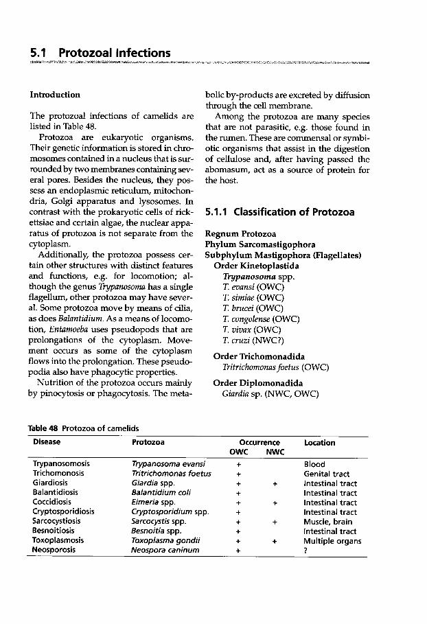
5.1 Protozoal Infections ruduu
Introduction
The protozoal infections of camelids are listed in Table 48.
Protozoa are eukaryotic organisms. Their genetic information is stored in chro- mosomes contained in a nucleus that is sur- rounded by two membranes containing sev- eral pores. Besides the nucleus, they pos- sess an endoplasmic reticulum, mitochon- dria, Golgi apparatus and lysosomes. In contrast with the prokaryotic cells of rick- ettsiae and certain algae, the nuclear appa- ratus of protozoa is not separate from the cytoplasm.
Additionally, the protozoa possess cer- tain other structures with distinct features and functions, e.g. for locomotion; al- though the genus Typunosomu has a single flagellum, other protozoa may have sever- al. Some protozoa move by means of cilia, as does Buluntidium. As a means of locomo- tion, Entamoebu uses pseudopods that are prolongations of the cytoplasm. Move- ment occurs as some of the cytoplasm flows into the prolongation. These pseudo- podia also have phagocytic properties.
Nutrition of the protozoa occurs mainly by pinocytosis or phagocytosis. The meta-
Table 48 Protozoa of camelids
Disease Protozoa
bolic by-products are excreted by diffusion through the cell membrane.
Among the protozoa are many species that are not parasitic, e.g. those found in the rumen. These are commensal or symbi- otic organisms that assist in the digestion of cellulose and, after having passed the abomasum, act as a source of protein for the host.
5.1.1 Classification of Protozoa
Regnum Protozoa Phylum Sarcomastigophora Subphylum Mastigophora (Flagellates)
Order Kinetoplastida Typanosoma spp. T. evansi (OWC) T. simiue (OWC) T. brucei (OWC) T. congolense (OWC) T. vivux (OWC) T. cruzi (NWC?)
Tritrichornonus foetus (OWC)
Giurdiu sp. (NWC, OWC)
Order Trichomonadida
Order Diplomonadida
Occurrence Location OWC NWC
Try panoso mosis Trichomonosis Giardiosis Balantidiosis Coccidiosis Cryptosporidiosis Sarcocystiosis Besnoitiosis Toxoplasmosis Neosporosis
Trypanosoma evansi Tritrichomonas foetus Giardia spp. Balantidium coli Eimeria spp. Cryptosporidium spp. Sarcocystis spp. Besnoitia spp. Toxoplasma gondii Neospora caninum
+ + + + + + -I-
+ + +
Blood Genital tract
+ Intestinal tract Intestinal tract
+ Intestinal tract Intestinal tract
+ Muscle, brain Intestinal tract
+ Multiple organs ?
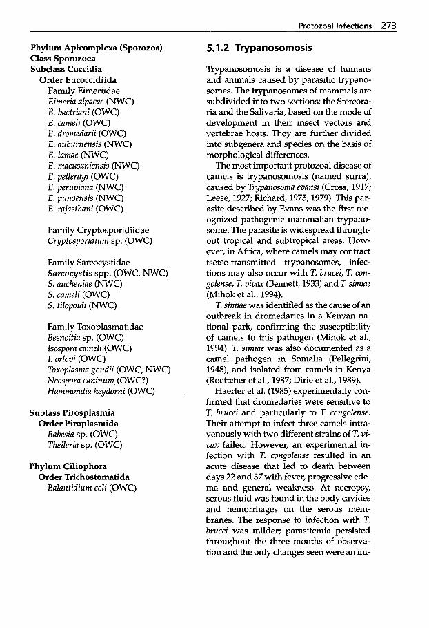
Protozoal Infections 273
Phylum Apicomplexa (Sporozoa) Class Sporozoea Subclass Coccidia
Order Eucoccidiida Family Eimeriidae Eimeriu ulpucue (NWC) E. buctriuni (OWC) E. cumeIi (OWC) E. dromedurii (OWC) E. auburnensis (NWC) E. lumae (NWC) E. macusuniensis (NWC) E. pellerdyi (OWC) E. peruviunu (NWC) E. punoensis (NWC) E. rujusthuni (OWC)
Family Cryptosporidiidae Cryptosporidium sp. (OWC)
Family Sarcocystidae
S. uucheniue (NWC) S. cumeli (OWC) S. tilopoidi (NWC)
Sarcocystis spp.(OWC,NWC)
Family Toxoplasmatidae Besnoitiu sp. (OWC) Isospora cumeli (OWC) I. orlovi (OWC) Toxoplusma gondii (OWC, NWC) Neosporu cuninum (OWC?) Hurnrnondiu heydomi (OWC)
Sublass Pirosplasmia Order Piroplasmida
Bubesiu sp. (OWC) Theileriu sp. (OWC)
Phylum Ciliophora Order Trichostomatida
Buluntidium coIi (OWC)
5.1.2 Trypanosomosis
Trypanosomosis is a disease of humans and animals caused by parasitic trypano- somes. The trypanosomes of mammals are subdivided into two sections: the Stercora- ria and the Salivaria, based on the mode of development in their insect vectors and vertebrae hosts. They are further divided into subgenera and species on the basis of morphological differences.
The most important protozoal disease of camels is trypanosomosis (named surra), caused by Ttypanosomu evunsi (Cross, 1917; Leese, 1927; Richard, 1975,1979). This par- asite described by Evans was the first rec- ognized pathogenic mammalian trypano- some. The parasite is widespread through- out tropical and subtropical areas. How- ever, in Africa, where camels may contract tsetse-transmitted trypanosomes, infec- tions may also occur with T. brucei, T. con- golense, T. vivux (Bennett, 1933) and T. simiue (Mihok et al., 1994).
T. simiue was identified as the cause of an outbreak in dromedaries in a Kenyan na- tional park, confirming the susceptibility of camels to this pathogen (Mihok et al., 1994). T. simiue was also documented as a camel pathogen in Somalia (Pellegrini, 1948), and isolated from camels in Kenya (Roettcher et al., 1987; Dirie et al., 1989).
Haerter et al. (1985) experimentally con- firmed that dromedaries were sensitive to T. brucei and particularly to T. congolense. Their attempt to infect three camels intra- venously with two different strains of T. vi- vux failed. However, an experimental in- fection with T. congolense resulted in an acute disease that led to death between days 22 and 37 with fever, progressive ede- ma and general weakness. At necropsy, serous fluid was found in the body cavities and hemorrhages on the serous mem- branes. The response to infection with T. brucei was milder; parasitemia persisted throughout the three months of observa- tion and the only changes seen were an ini-
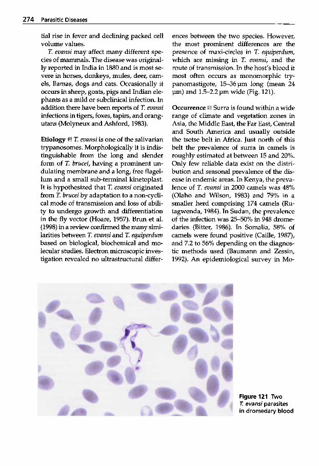
274 Parasitic Diseases
tial rise in fever and declining packed cell volume values.
T. evunsi may affect many different spe- cies of mammals. The disease was original- ly reported in India in 1880 and is most se- vere in horses, donkeys, mules, deer, cam- els, llamas, dogs and cats. Occasionally it occurs in sheep, goats, pigs and Indian ele- phants as a mild or subclinical infection. In addition there have been reports of T. evunsi infections in tigers, foxes, tapirs, and orang- utans (Molyneux and Ashford, 1983).
T. evunsi is one of the salivarian trypanosomes. Morphologically it is indis- tinguishable from the long and slender form of T. brucei, having a prominent un- dulating membrane and a long, free flagel- lum and a small sub-terminal kinetoplast. It is hypothesized that T. evunsi originated from T. brucei by adaptation to a non-cycli- cal mode of transmission and loss of abili- ty to undergo growth and differentiation in the fly vector (Hoare, 1957). Brun et al. (1998) in a review confirmed the many simi- larities between T. evunsi and T. equiperdum based on biological, biochemical and mo- lecular studies. Electron microscopic inves- tigation revealed no ultrastructural differ-
ences between the two species. However, the most prominent differences are the presence of maxi-circles in T. equiperdum, which are missing in T. evunsi, and the route of transmission. In the host's blood it most often occurs as monomorphic try- panomastigote, 15-36 pm long (mean 24 pm) and 1.5-2.2pm wide (Fig. 121).
Occurrence i f t i Surra is found within a wide range of climate and vegetation zones in Asia, the Middle East, the Far East, Central and South America and usually outside the tsetse belt in Africa. Just north of this belt the prevalence of surra in camels is roughly estimated at between 15 and 20%. Only few reliable data exist on the distri- bution and seasonal prevalence of the dis- ease in endemic areas. In Kenya, the preva- lence of T. evunsi in 2000 camels was 48% (Olaho and Wilson, 1983) and 79% in a smaller herd comprising 174 camels (Ru- tagwenda, 1984). In Sudan, the prevalence of the infection was 2550% in 948 drome- daries (Bitter, 1986). In Somalia, 58% of camels were found positive (Caille, 1987), and 7.2 to 56% depending on the diagnos- tic methods used (Baumann and Zessin, 1992). An epidemiological survey in Mo-
Figure 121 Two T. evansi parasites in dromedary blood
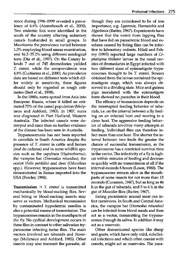
Protozoal Infections 275
rocco during 1996-1999 revealed a preva- lence of 6.6% (Atarorhouch et al., 2000). Two endemic foci were identified in the south of the country affecting sedentary camels husbanded in small groups. In Mauritania the prevalence varied between 1.3% employing blood smear examination, and 16.2-25.2% using different serological tests (Dia et al., 1997). On the Canary Is- lands 7 out of 745 dromedaries yielded T. evunsi, while the seroprevalence was 4.8% (Guitierrez et al., 2000). As prevalence data are based on different tests which dif- fer widely in sensitivity, these figures should only be regarded as rough esti- mates (Butt et al., 1998).
In the 188Os, surra spread from Asia into European Russia, where it killed an esti- mated 70% of the camel population (Moly- neux and Ashford, 1983). In 1907, surra was diagnosed in Port Hedland, Western Australia. The infected camels were de- stroyed and since then no further evidence of the disease has been seen in Australia.
Trypanosomosis has not been reported in camelids in South America despite the presence of T. evunsi in cattle and horses (ma1 de caderas) and in some wildlife spe- cies such as the capybara (Hydvochoerus), the vampire bat (Desmodus rotundus), the ocelot (Felis purdulis) and deer (Odocoileus spp.). However, trypanosomes have been demonstrated in llamas imported into the USA (Fowler, 1998).
Transmission i84l T. evunsi is transmitted mechanically by blood-sucking flies. Sev- eral biting or blood-sucking insects may serve as vectors. Mechanical transmission by contaminated hypodermic needles is also a potential means of transmission. The trypanosomes remain in the mouthparts of the fly. No cyclical development occurs in these flies in contrast to other salivarian try- panosome infecting tsetse flies. The main vectors involved are tabanids and Stomo- xys (Molyneux and Ashford, 1983). Other insects may also transmit the parasite, al-
though they are considered to be of less importance, e.g. Lyperosiu, Huemutobiu and Hypoboscu (Rutter, 1967). Experiments have shown that the vomit from lapping flies that have fed on parasitemic blood and ex- udates caused by biting flies can be infec- tive to laboratory rodents. Hilali and Fah- my (1993) reported large numbers of Ce- phalopinu titillator larvae in the nasal cav- ities of dromedaries in Egypt infected with two different sizes of epimastigote trypa- nosomes thought to be T. evunsi. Smears obtained from the larvae contained the epi- mastigote stage, which was always ob- served in a dividing state. Mice and guinea pigs inoculated with the epimastigote form showed no parasites in their blood.
The efficacy of transmission depends on the interrupted feeding behavior of taba- nids, i.e. on the interval between a fly feed- ing on an infected host and moving to a clean host. The aggressive feeding behav- ior of tabanids involves many attempts at feeding. Individual flies can therefore in- fect more than one host. The shorter the in- terval between two feeds the greater the chance of successful transmission, as the trypanosome has a restricted survival time in the vector. The infectivity of a fly is high- est within minutes of feeding and decreas- es quickly with no transmission at all if the interval exceeds 8 hours (Losos, 1980). The trypanosomes remain alive in the mouth- parts of some insects for not more than 15 seconds (Curasson, 1947), but as long as 44 h in the gut of tabanids, and 5 to 6 h in the gut of Muscidue flies (Rutter, 1967).
Eating parasitemic animal meat can in- fect carnivores. In South and Central Ame- rica, the vampire bat (Desmodus rotundus) can be infected from blood meals and then act as a vector, transmitting the trypano- somes through its saliva. In addition it may act as a reservoir.
Other domesticated species like sheep and goats, which have only mild, subclini- cal infections and which often coexist with camels, might act as reservoirs. The para-
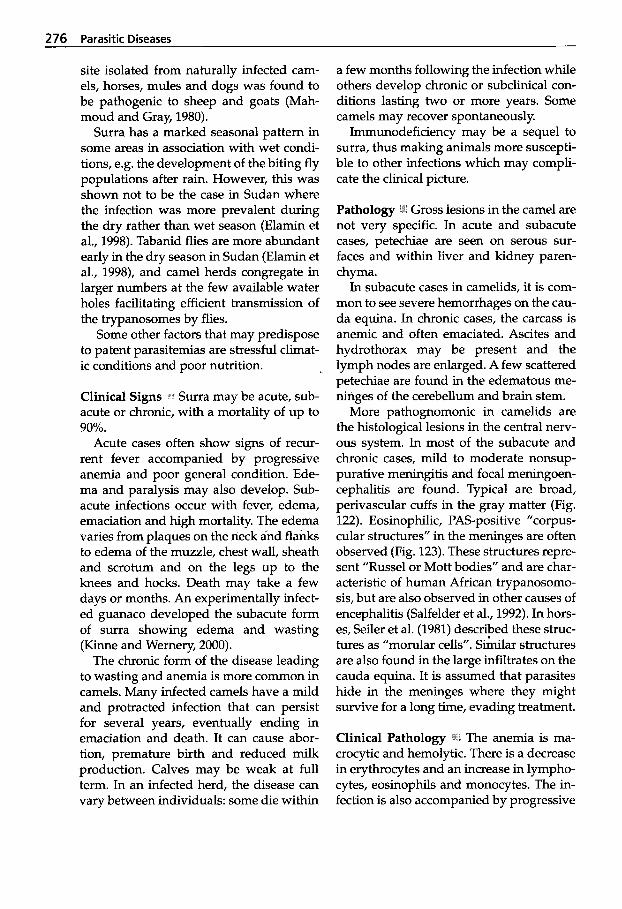
276 Parasitic Diseases
site isolated from naturally infected cam- els, horses, mules and dogs was found to be pathogenic to sheep and goats (Mah- moud and Gray, 1980).
Surra has a marked seasonal pattern in some areas in association with wet condi- tions, e.g. the development of the biting fly populations after rain. However, this was shown not to be the case in Sudan where the infection was more prevalent during the dry rather than wet season (Elamin et al., 1998). Tabanid flies are more abundant early in the dry season in Sudan (Elamin et al., 1998), and camel herds congregate in larger numbers at the few available water holes facilitating efficient transmission of the trypanosomes by flies.
Some other factors that may predispose to patent parasitemias are stressful climat- ic conditions and poor nutrition.
Clinical Signs + Surra may be acute, sub- acute or chronic, with a mortality of up to
Acute cases often show signs of recur- rent fever accompanied by progressive anemia and poor general condition. Ede- ma and paralysis may also develop. Sub- acute infections occur with fever, edema, emaciation and high mortality. The edema varies from plaques on the neck and flanks to edema of the muzzle, chest wall, sheath and scrotum and on the legs up to the knees and hocks. Death may take a few days or months. An experimentally infect- ed guanaco developed the subacute form of surra showing edema and wasting (Kinne and Wemery, 2000).
The chronic form of the disease leading to wasting and anemia is more common in camels. Many infected camels have a mild and protracted infection that can persist for several years, eventually ending in emaciation and death. It can cause abor- tion, premature birth and reduced milk production. Calves may be weak at full term. In an infected herd, the disease can vary between individuals: some die within
90%.
a few months following the infection while others develop chronic or subclinical con- ditions lasting two or more years. Some camels may recover spontaneously.
Immunodeficiency may be a sequel to surra, thus making animals more suscepti- ble to other infections which may compli- cate the clinical picture.
Pathology i i k Gross lesions in the camel are not very specific. In acute and subacute cases, petechiae are seen on serous sur- faces and within liver and kidney paren- chyma.
In subacute cases in camelids, it is com- mon to see severe hemorrhages on the cau- da equina. In chronic cases, the carcass is anemic and often emaciated. Ascites and hydrothorax may be present and the lymph nodes are enlarged. A few scattered petechiae are found in the edematous me- ninges of the cerebellum and brain stem.
More pathognomonic in camelids are the histological lesions in the central nerv- ous system. In most of the subacute and chronic cases, mild to moderate nonsup- purative meningitis and focal meningoen- cephalitis are found. Typical are broad, perivascular cuffs in the gray matter (Fig. 122). Eosinophilic, PAS-positive ”corpus- cular structures” in the meninges are often observed (Fig. 123). These structures repre- sent “Russel or Mott bodies” and are char- acteristic of human African trypanosomo- sis, but are also observed in other causes of encephalitis (Salfelder et al., 1992). In hors- es, Seiler et al. (1981) described these struc- tures as ”morular cells”. SiEnilar structures are also found in the large infiltrates on the cauda equina. It is assumed that parasites hide in the meninges where they might survive for a long time, evading treatment.
Clinical Pathology 1% The anemia is ma- crocytic and hemolytic. There is a decrease in erythrocytes and an increase in lympho- cytes, eosinophils and monocytes. The in- fection is also accompanied by progressive
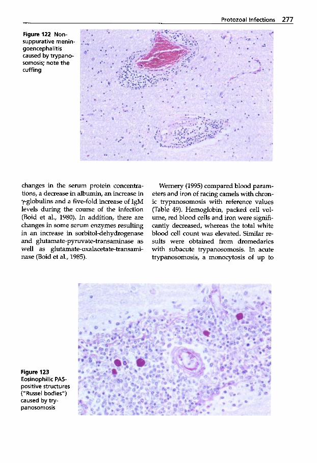
Protozoal Infections 277
Figure 122 Non- suppurative menin- goencephalitis caused by trypano- somosis; note the cuffing
changes in the serum protein concentra- tions, a decrease in albumin, an increase in y-globulins and a five-fold increase of IgM levels during the course of the infection (Boid et al., 1980). In addition, there are changes in some serum enzymes resulting in an increase in sorbitol-dehydrogenase and glutamate-pyruvate-transaminase as well as glutamate-oxalacetate-transami- nase (Boid et al., 1985).
Wernery (1995) compared blood param- eters and iron of racing camels with chron- ic trypanosomosis with reference values (Table 49). Hemoglobin, packed cell vol- ume, red blood cells and iron were sign&- cantly decreased, whereas the total white blood cell count was elevated. Similar re- sults were obtained from dromedaries with subacute trypanosomosis. In acute trypanosomosis, a monocytosis of up to
Figure 123 Eosinophilic PAS- positive structures ("Russel bodies") caused by try- panosomosis
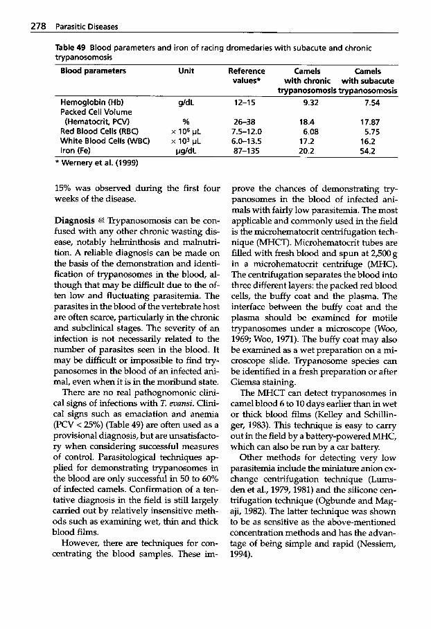
278 Parasitic Diseases
Table 49 Blood parameters and iron of racing dromedaries with subacute and chronic trypanosomosis
Blood parameters Unit Reference Camels Camels values* with chronic with subacute
trypanosomosis trypanosomosis Hemoglobin (Hb) gld L 12-1 5 9.32 7.54
Red Blood Cells (RBC) x 106 pL 7.5-12.0 6.08 5.75
Packed Cell Volume (Hematocrit, PW) % 26-38 18.4 17.87
White Blood Cells WBC) x lo3 pL 6.0-13.5 17.2 16.2 Iron (Fe) WJdL 87-1 35 20.2 54.2
* Wernery et al. (1999)
15% was observed during the first four weeks of the disease.
Trypanosomosis can be con- fused with any other chronic wasting dis- ease, notably helminthosis and malnutri- tion. A reliable diagnosis can be made on the basis of the demonstration and identi- fication of trypanosomes in the blood, al- though that may be difficult due to the of- ten low and fluctuating parasitemia. The parasites in the blood of the vertebrate host are often scarce, particularly in the chronic and subclinical stages. The severity of an infection is not necessarily related to the number of parasites seen in the blood. It may be difficult or impossible to find try- panosomes in the blood of an infected ani- mal, even when it is in the moribund state.
There are no real pathognomonic clini- cal signs of infections with T. evunsi. Clini- cal signs such as emaciation and anemia (PCV < 25%) (Table 49) are often used as a provisional diagnosis, but are unsatisfacto- ry when considering successful measures of control. Parasitological techniques ap- plied for demonstrating trypanosomes in the blood are only successful in 50 to 60% of infected camels. Confirmation of a ten- tative diagnosis in the field is still largely carried out by relatively insensitive meth- ods such as examining wet, thin and thick blood films.
However, there are techniques for con- centrating the blood samples. These im-
prove the chances of demonstrating try- panosomes in the blood of infected ani- mals with fairly low parasitemia. The most applicable and commonly used in the field is the microhematocrit centrifugation tech- nique (MHCT). Microhematocrit tubes are filled with fresh blood and spun at 2,500 g in a microhematocrit centrifuge (MHC). The centrifugation separates the blood into three different layers: the packed red blood cells, the buffy coat and the plasma. The interface between the buffy coat and the plasma should be examined for motile trypanosomes under a microscope (Woo, 1969; Woo, 1971). The buffy coat may also be examined as a wet preparation on a mi- croscope slide. Trypanosome species can be identified in a fresh preparation or after Giemsa staining.
The MHCT can detect trypanosomes in camel blood 6 to 10 days earlier than in wet or thick blood films (Kelley and Schillin- ger, 1983). This technique is easy to carry out in the field by a battery-powered MHC, which can also be run by a car battery.
Other methods for detecting very low parasitemia include the miniature anion ex- change centrifugation technique (Lums- den et al., 1979,1981) and the silicone cen- trifugation technique (Ogbunde and Mag- aji, 1982). The latter technique was shown to be as sensitive as the above-mentioned concentration methods and has the advan- tage of being simple and rapid (Nessiem, 1994).
Protozoal Infections 279
Inoculation of laboratory rodents with blood from suspected infectious camels is a very sensitive method for detecting low parasitemia caused by T. evansi (Boid et al., 1985) and T. brucei (Godfrey and Killick- Kendrick, 1962). Mouse or rat inoculation increases the number of camels found pos- itive by approximately 50% compared with blood film techniques (Molyneux and Ashford, 1983). However, this method is time-consuming, expensive and inappro- priate for use in large-scale surveys. Devel- opment of patent parasitemia is 5 to 9 days in mice and 3 to 9 days in rats.
Despite improvements in parasitologi- cal techniques for the detection of trypa- nosomes, a high proportion of infections are never detected. One major reason for this is the constant antigen variations that occur in T. evansi (as in other salivarian try- panosomes) (Jones and M c h e l l , 1984). This phenomenon makes it difficult to de- tect circulating antigens and antibodies - a tremendous advantage for the parasite, keeping it ahead of an attack by specific antibodies directed against the previous surface antigens. The identification of cir- culating variable antigen types (VSG) would be of great value in developing more sensitive diagnostic tests. The anti- genic variation is also a major constraint for immunoprophylactic control methods.
The development of enzyme immunoas- says (ELISA) detecting circulating antigens in animal sera provides an opportunity for an early diagnosis of trypanosomosis. This was an important breakthrough in the diagnosis of this disease (Rae and Luckins, 1984; Nantulya et al., 1987). How- ever, the present ELISA has proven to be unsatisfactory in sensitivity as well as specificity (Antigen ELISAs for trypanoso- mosis - Evaluation of the performance: Proc. Workshop ILRI, Nairobi, Kenya 1996). A simpler test established for use under field conditions was the latex agglutina- tion technique (Suratex@) (Nantulya, 1989). T. evunsi antigen may also be detected in
blood by the polymerase chain reaction (PCR) (Masiga and Gibson, 1992; Wuyts et al., 1994). In the near future, well- equipped laboratories may more efficient- ly use DNA-amplification technologies in the diagnosis of T. evunsi in animals, while pastoralists still traditionally diagnose try- panosome infections by the smell of the in- fected animal's urine.
In the diagnosis of surra, antibody tech- niques like flocculation assays (including the formol gel and mercuric chloride tests) (Pegram and Scott, 1976) measure an in- crease in the level of serum globulins. How- ever, these tests are non-specific and have yielded many inconsistencies (Luckins et al., 1979; Boid et al., 1980). As early as 1924, Schoening described a complement fixa- tion test demonstrating antibodies to T. evunsi (Schoening, 1924). This test has nev- er been routinely used as a diagnostic test because it is too difficult to perform, and procedure standardization is not possible.
Another promising assay for the diagno- sis of T. evunsi antibodies (an indirect he- magglutination test) was developed by Jaktar and Singh (1971). However, this as- say also had difficulties with the standard- ization of antigens and the presence of in- terfering, non-specific antibodies. Wilson et al. (1983) successfully used this assay to demonstrate antibodies to T. evansi in a serological survey of Kenyan camels. An- other agglutination test also available is the modified card agglutination test (CATT/ T. evansi), which was initially developed for T. brucei gumbiense (Dialli et al., 1994). This test is not specific to T. evunsi antibod- ies but can also detect antibodies to other salivarian trypanosomes, thereby compli- cating the interpretation of positive results when other salivarian trypanosomes are present.
Even the improved indirect fluorescent antibody test (Luckins et al., 1978) had in- herent drawbacks. The development of an enzyme-linked immunosorbent antibody assay (ELISA) was a major breakthrough.
280 Parasitic Diseases
The ELISA has been used with good results in the serodiagnosis of T. evunsi (Luckins et al., 1979; Boid et al., 1980; Rae et al., 1989). In the United Arab Emirates (UAE), where surra in dromedaries is endemic, a decrease in the seroprevalence was achiev- ed (from 12.5% in 1990 to 2.5% in 1999) due to the treatment of positive cases (by the use of antibody ELISA) and the control of vectors (CVRL Annual Report, 1999).
Antibodies to T. munsi infections in cam- els as demonstrated by ELISA do not dif- ferentiate between acute and chronic infec- tions (Rae et al., 1989). Antibody responses to T. evunsi infections may vary and the levels may stay high for a considerable time after effective treatment (Luckins et al., 1978).
Treatment and Control Only a few drugs, e.g. Cymelaman@ (melarsomine, Merial), Triquin@ (quinapyramine sulfate, quinapyramine chloride, distributor Wock- hardt Ltd.) and Trypamidium-Samorin@ (isometamidiurn chloride, Merial) have been approved by appropriate authorities for use in OWC or NWC. It is known that the pharmacokinetic behavior of drugs dif- fers significantly among different species. Therefore it is important that drugs should be studied carefully in every species. This is especially true for camelids due to their unique physiological characteristics. As there are only very limited pharmacoki- netic data available on camelids, drugs should be used with great caution. This also applies to the use of vaccines. They should undergo testing by regulatory agen- cies for safety and efficacy before they are used on camelids.
Monitoring for drug resistance is impor- tant and there are techniques available (Kaminsky and Zweygarth, 1989; B a n g et al., 1993; Brun and Lun, 1994) that should be employed when suspicion of resistance arises.
Surra is endemic in most countries where camels are reared. Chemotherapy
alone will not have a permanent effect on the cycle of the disease, regionally or glob- ally. The use of chemotherapy is often in- adequate: e.g., underdosing is common. As a result of the limited number of drugs available for therapeutic or prophylactic use, the dependence on trypanocidal drugs for the control of surra is alarming. Also, not all compounds effective against T. evunsi are suitable for use in camels. Many of the drugs used for cattle are either not curative or too toxic for camels: e.g. dimi- nazene aceturate (Berenil@, Hoechst AG - production, however, was stopped recent- ly), which is toxic to camels at doses of >3.5mg/kg and should not be used in dromedaries. Berenil@ has been success- fully used in Bactrian camels with doses as high as 5 mg/kg bodyweight (Luckins, 1992). Alternative drugs are Trypan@, Ata- rosP, Veriben@, and Sanofi@ (Rommel, pers. commun.).
In the early 1970s, Imperial Chemical In- dustries Ltd (ICI) stopped their production of the quinapyramines, AntrycideB and Antrycide@ Pro-Salt, at that time the com- monly used and successful trypanocides against T. munsi (Schillinger and Roettcher, 1984). Production of Antrycide@ has been resumed by a few drug companies and it is available today. This meant that only one curative drug remained available for cam- els - NaganoP (Bayer AG, Leverkusen, Germany - production, however, was stopped recently), in use since 1925. After nearly 75 years of use, the effectiveness of NaganoP is decreasing in some areas due to drug resistance. However, Suramin (Na- ganol@) administered at a dose rate of 10- 12 mg/kg by slow intravenous injection is still used in the treatment of camels. Due to its slow elimination from the body, it also has a prophylactic effect for between 6 to 12 weeks (Kaufmann, 1996). The preven- tive effect depends on the dosage used and the degree of the trypanosome challenge (Luckins, 1992). Leakage of the drug into the tissues may cause phlebitis.
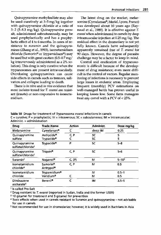
Protozoal Infections 281
Quinapyramine methylsulfate may also be used curatively at 3-5 mg/kg together with quinapyramine chloride at a ratio of 3:2 (5-8.3 mg/kg). Quinapyramine pros- alt, administered subcutaneously, may be used prophylactically and has a prophy- lactic effect of 4 to 6 months. In cases of re- sistance to suramin and the quinapyra- mines (Zhang et al., 1993), isometamidium chloride (Samorin@ or Trypamidiurn@) may be used but with great caution (0.5-0.7 mg/ kg intravenously administered as a 2% so- lution). This drug is only curative when the trypanosomes are present intravascularly. Overdosing quinapyramines can cause side effects in camels such as tremors, sali- vation and collapse leading to death.
There is in vitro and in vivo evidence that most isolates tested for T. evunsi are resist- ant (innate) or non-responsive to isometa- midium.
The latest drug on the market, melar- somine (CymelarsanB, Merial, Lyons, France) was developed about 10 years ago (Ray- naud et al., 1989). It is effective against T. evunsi when administered to camels by deep intramuscular injection at 0.25 mg/kg. The residual effect in the dromedary is not yet fully known. Camels have subsequently apparently remained free of T. evunsi for 90 days. However, the degree of parasite challenge may be a factor to keep in mind.
Control and eradication of trypanoso- mosis is difficult because of the develop- ment of drug resistance; even more diffi- cult is the control of vectors. Regular mon- itoring of infections is necessary to prevent large losses in endemic areas. Employing frequent (monthly) PCV estimations on well-managed herds has proven useful in keeping losses low. Some farm managers treat any camel with a PCV of < 25%.
Table 50 Drugs for treatment of Trypanosoma evansi infections in camels C = curative; P = prophylactic; IV = intravenous; SC = subcutaneous; IM = intramuscular; Administ. = administration
Drug Trade Name Action Administ. Dose mglkg Melarsomine Cymelarsan" C deep IM 0.25 Quinapyramine Antrycide@ c, p sc 5 sulfate Trypacidem C sc 5 Quinapyramine Trypacide" P sc sulfatekhloridel
5-8 ~ ~
Quinapyramine Triquin" c, p SC sulfatekhloridel
5 6
Suramin2 NaganoP c, (PI IV 5-1 O3 lsometamidium Samorin" c, p IV 0.5 chloride4 Antrypol@ lsometamidium Trypamidiurn" IV 0.5-1 chloride Ve r i d i u ma C IV 0.5 Diminazene Berenil@ C IM 3.5-5 accturate5
1 Is called Pro-Salt Drug resistant t o T: evansi (reported in Sudan, India and the former USSR) 10 gkamel for treatment and 5 gkamel for prevention
for use in camels 4 Toxic effects when used in camels resistant t o Suramin and quinapyramine - not advisable
5 Not recommended for use in dromedaries; however, it is widely used in Bactrians in Asia
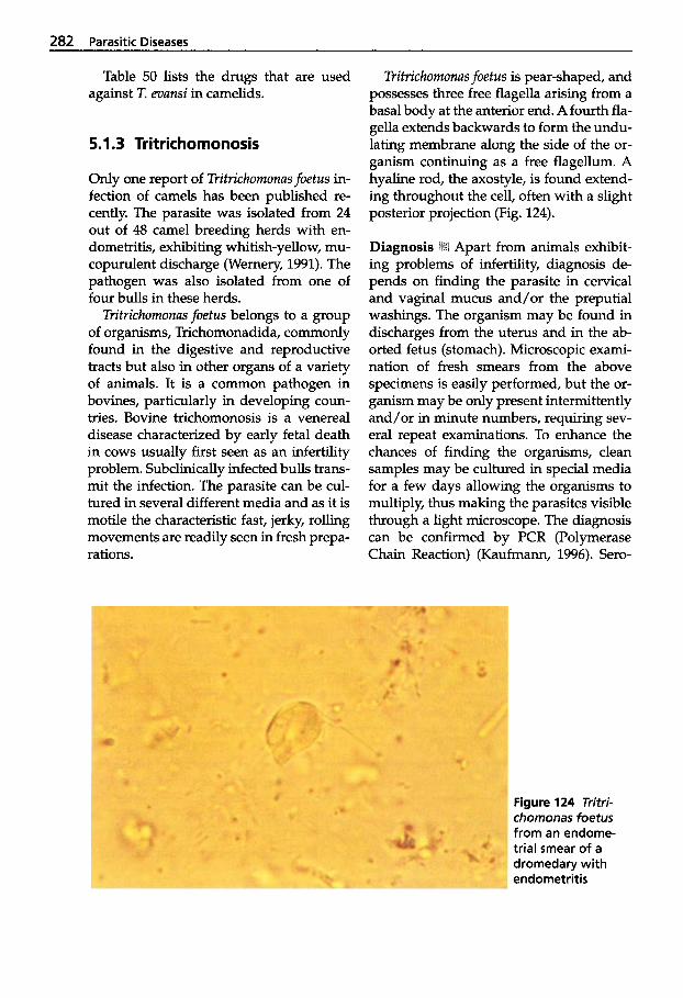
282 Parasitic Diseases
Table 50 lists the drugs that are used against T. evansi in camelids.
5.1.3 Tritrichomonosis
Only one report of Tritrichomonas foetus in- fection of camels has been published re- cently. The parasite was isolated from 24 out of 48 camel breeding herds with en- dometritis, exhibiting whitish-yellow, mu- copurulent discharge (Wemery, 1991). The pathogen was also isolated from one of four bulls in these herds.
Tritrichomonas foetus belongs to a group of organisms, Trichomonadida, commonly found in the digestive and reproductive tracts but also in other organs of a variety of animals. It is a common pathogen in bovines, particularly in developing coun- tries. Bovine trichomonosis is a venereal disease characterized by early fetal death in cows usually first seen as an infertility problem. Subclinically infected bulls trans- mit the infection. The parasite can be cul- tured in several different media and as it is motile the characteristic fast, jerky, rolling movements are readily seen in fresh prepa- rations.
Tritrichomonas foetus is pear-shaped, and possesses three free flagella arising from a basal body at the anterior end. A fourth fla- gella extends backwards to form the undu- lating membrane along the side of the or- ganism continuing as a free flagellum. A hyaline rod, the axostyle, is found extend- ing throughout the cell, often with a slight posterior projection (Fig. 124).
Apart from animals exhibit- ing problems of infertility, diagnosis de- pends on finding the parasite in cervical and vaginal mucus and/or the preputial washings. The organism may be found in discharges from the uterus and in the ab- orted fetus (stomach). Microscopic exami- nation of fresh smears from the above specimens is easily performed, but the or- ganism may be only present intermittently and/or in minute numbers, requiring sev- eral repeat examinations. To enhance the chances of finding the organisms, clean samples may be cultured in special media for a few days allowing the organisms to multiply, thus making the parasites visible through a light microscope. The diagnosis can be confirmed by PCR (Polymerase Chain Reaction) (Kaufmann, 1996). Sero-
Figure 124 Tritri- chomonas foetus from an endome- trial smear of a dromedary with endometritis
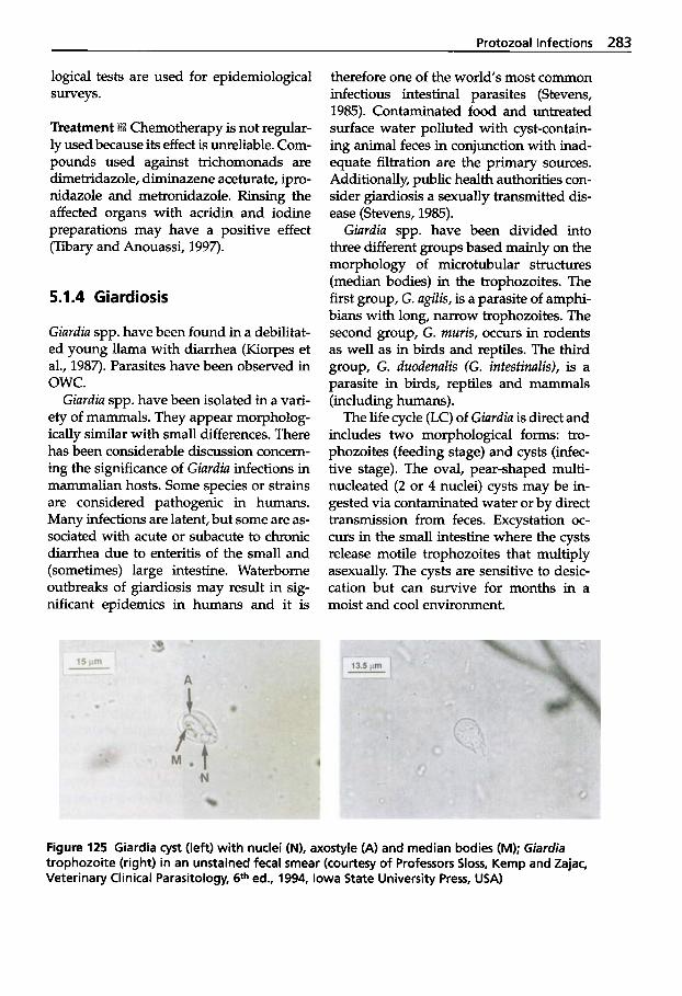
Protozoal Infections 283
logical tests are used for epidemiological surveys.
Chemotherapy is not regular- ly used because its effect is unreliable. Com- pounds used against trichomonads are dimetridazole, diminazene aceturate, ipro- nidazole and metronidazole. Rinsing the affected organs with acridin and iodine preparations may have a positive effect (Tibary and Anouassi, 1997).
5.1.4 Giardiosis
Giurdia spp. have been found in a debilitat- ed young llama with diarrhea (Kiorpes et al., 1987). Parasites have been observed in OWC.
Giurdiu spp. have been isolated in a vari- ety of mammals. They appear morpholog- ically similar with small differences. There has been considerable discussion concern- ing the significance of Giurdiu infections in mammalian hosts. Some species or strains are considered pathogenic in humans. Many infections are latent, but some are as- sociated with acute or subacute to chronic diarrhea due to enteritis of the small and (sometimes) large intestine. Waterborne outbreaks of giardiosis may result in sig- nificant epidemics in humans and it is
therefore one of the world's most common infectious intestinal parasites (Stevens, 1985). Contaminated food and untreated surface water polluted with cyst-contain- ing animal feces in conjunction with inad- equate filtration are the primary sources. Additionally, public health authorities con- sider giardiosis a sexually transmitted dis- ease (Stevens, 1985).
Giurdiu spp. have been divided into three different groups based mainly on the morphology of microtubular structures (median bodies) in the trophozoites. The first group, G. ugilis, is a parasite of amphi- bians with long, narrow trophozoites. The second group, G. rnuris, occurs in rodents as well as in birds and reptiles. The third group, G. duodenuZis (G. intestinulis), is a parasite in birds, reptiles and mammals (including humans).
The life cycle (LC) of Giardia is direct and includes two morphological forms: tro- phozoites (feeding stage) and cysts (infec- tive stage). The oval, pear-shaped multi- nucleated (2 or 4 nuclei) cysts may be in- gested via contaminated water or by direct transmission from feces. Excystation oc- curs in the small intestine where the cysts release motile trophozoites that multiply asexually. The cysts are sensitive to desic- cation but can survive for months in a moist and cool environment.
Figure 125 Giardia cyst (left) with nuclei (N), axostyle (A) and median bodies (MI; Giardia trophozoite (right) in an unstained fecal smear (courtesy of Professors Sloss, Kemp and Zajac, Veterinary Clinical Parasitology, 6th ed., 1994, Iowa State University Press, USA)
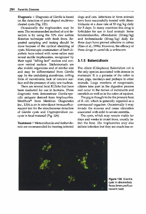
284 Parasitic Diseases
1 Diagnosis of Giurdiu is based on the detection of pear shaped multi-nu- cleated cysts (Fig. 125).
Occasionally the trophozoites may be seen. The recommended method of cyst de- tection is by using the 33% zinc sulfate flotation technique with fresh feces. Re- peated sampling and testing should be done because of the cyclical shedding of cysts. Microscopic examination of fresh di- arrheic feces mixed with some saline may reveal motile trophozoites, recognized by their rapid "falling leaf" motion and con- cave ventral surface. Trichomonads are also mobile organisms and of similar size and may be differentiated from Giurdiu spp. by the undulating membrane, rolling form of movements, lack of concave sur- face and the presence of only one nucleus.
There are several fecal ELISAs that have been marketed for use in humans. These diagnostic tests demonstrate Giurdiu-spe- cific antigens derived from trophozoites. Merifluorm from Meridian Diagnostics Inc., USA is an in vitro-direct immunofluo- rescent test for the simultaneous detection of Giurdiu cysts and Cryptosporidium oo- cysts in fecal material (Fig. 126).
Treatment ilil Metronidazole and fenbenda- zole are recommended for treating infected
dogs and cats. Infections in farm animals have been successfully treated with dime- tridazole at a dose rate of 50 mg/kg daily for 5 days. In many countries this drug is forbidden for use in food animals. Some benzimidazoles, albendazole (20 mg/ kg) and fenbendazole (10mg/kg) daily for three days have proved effective in calves (Xiao et al., 1996). However, the efficacy of these drugs in camelids is unknown.
5.1.5 Balantidiosis
The ciliate (Ciliophora) Buluntidium coli is the only species associated with disease in mammals. It is a parasite of the colon in man, pigs, monkeys and perhaps in other animals. Large numbers of nonparasitic ciliates take part in the digestive process and occur in the rumen of ruminants and camelids as well as in the colon of equines.
The pig is thought to be the primary host of B. coli, which is generally regarded as a commensal organism. Occasionally it may invade the mucosa and cause ulceration associated with mild to severe enteritis.
The cysts, which may remain viable for days and weeks in moist feces, usually in- fect the host. The trophozoites may also initiate infection but they are much less re-
Figure 126 Giardia cysts in dromedary feces (irnrnunofluo- rescent test)
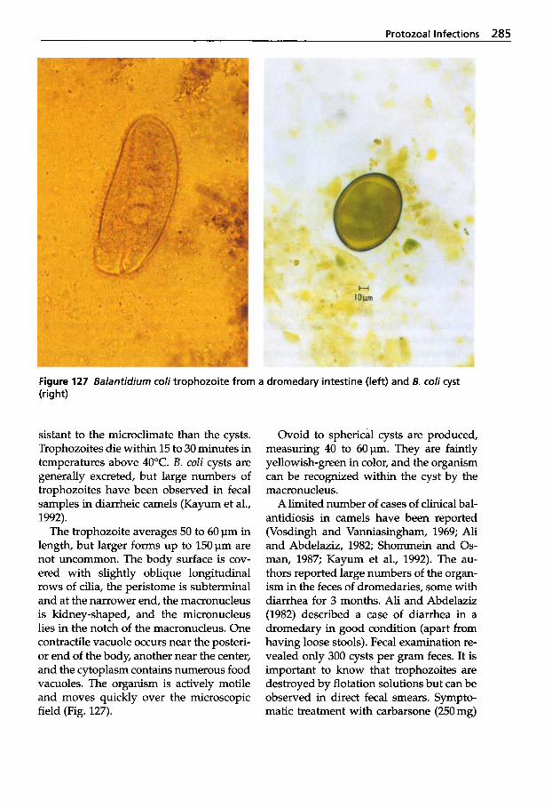
Protozoal Infections 285
Figure 127 Balantidium coli trophozoite from a dromedary intestine (left) and B. coli cyst (right)
sistant to the microclimate than the cysts. Trophozoites die within 15 to 30 minutes in temperatures above 40°C. B. coli cysts are generally excreted, but large numbers of trophozoites have been observed in fecal samples in diarrheic camels (Kayum et al., 1992).
The trophozoite averages 50 to 60 pn in length, but larger forms up to 150pm are not uncommon. The body surface is cov- ered with slightly oblique longitudinal rows of cilia, the peristome is subterminal and at the narrower end, the macronucleus is kidney-shaped, and the micronucleus lies in the notch of the macronucleus. One contractile vacuole occurs near the posteri- or end of the body, another near the center, and the cytoplasm contains numerous food vacuoles. The organism is actively motile and moves quickly over the microscopic field (Fig. 127).
Ovoid to spherical cysts are produced, measuring 40 to 60pm. They are faintly yellowish-green in color, and the organism can be recognized within the cyst by the macronucleus.
A limited number of cases of clinical bal- antidiosis in camels have been reported (Vosdingh and Vanniasingham, 1969; Ali and Abdelaziz, 1982; Shommein and 0 s - man, 1987; Kayum et al., 1992). The au- thors reported large numbers of the organ- ism in the feces of dromedaries, some with diarrhea for 3 months. Ali and Abdelaziz (1982) described a case of diarrhea in a dromedary in good condition (apart from having loose stools). Fecal examination re- vealed only 300 cysts per gram feces. It is important to know that trophozoites are destroyed by flotation solutions but can be observed in direct fecal smears. Sympto- matic treatment with carbarsone (250 mg)
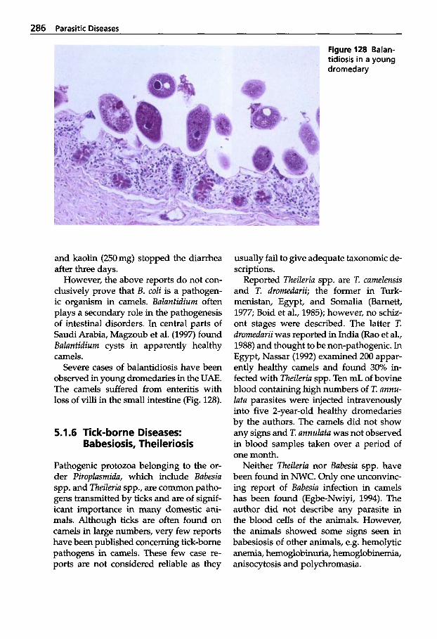
286 Parasitic Diseases
Figure 128 Balan- tidiosis in a young dromedary
and kaolin (250mg) stopped the diarrhea after three days.
However, the above reports do not con- clusively prove that B. coli is a pathogen- ic organism in camels. Baluntidium often plays a secondary role in the pathogenesis of intestinal disorders. In central parts of Saudi Arabia, Magzoub et al. (1997) found Buluntidium cysts in apparently healthy camels.
Severe cases of balantidiosis have been observed in young dromedaries in the UAE. The camels suffered from enteritis with loss of villi in the small intestine (Fig. 128).
5.1.6 Tick-borne Diseases: Babesiosis, Theileriosis
Pathogenic protozoa belonging to the or- der Piroplusmidu, which include Babesiu spp. and Theileria spp., are common patho- gens transmitted by ticks and are of signif- icant importance in many domestic ani- mals. Although ticks are often found on camels in large numbers, very few reports have been published concerning tick-borne pathogens in camels. These few case re- ports are not considered reliable as they
usually fail to give adequate taxonomic de- scriptions.
Reported Theileria spp. are T. camelensis and T. dromedurii; the former in Turk- menistan, Egypt, and Somalia (Barnett, 1977; Boid et al., 1985); however, no schiz- ont stages were described. The latter T. dromedurii was reported in India (Rao et al., 1988) and thought to be non-pathogenic. In Egypt, Nassar (1992) examined 200 appar- ently healthy camels and found 30% in- fected with Theileria spp. Ten mL of bovine blood containing high numbers of T. annu- lata parasites were injected intravenously into five 2-year-old healthy dromedaries by the authors. The camels did not show any signs and T. annulutu was not observed in blood samples taken over a period of one month.
Neither Theileria nor Babesia spp. have been found in NWC. Only one unconvinc- ing report of Bubesia infection in camels has been found (Egbe-Nwiyi, 1994). The author did not describe any parasite in the blood cells of the animals. However, the animals showed some signs seen in babesiosis of other animals, e.g. hemolytic anemia, hemoglobinuria, hemoglobinemia, anisocytosis and polychromasia.
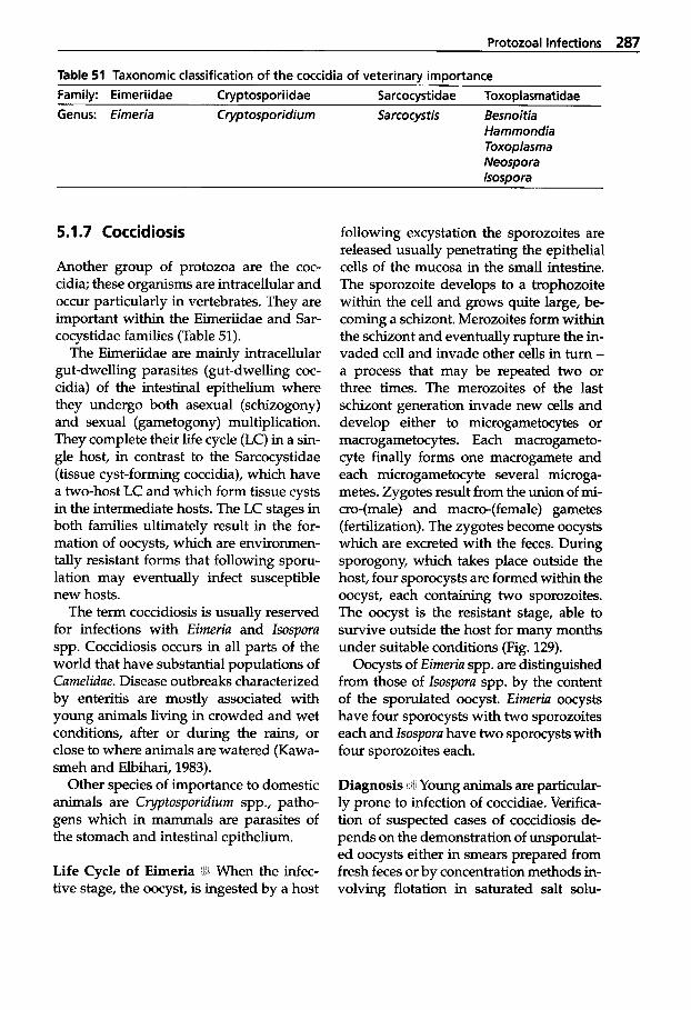
Protozoal Infections 287
Table 51 Taxonomic classification of the coccidia of veterinary importance Family: Eimeriidae Cryptosporiidae Sarcocystidae Toxoplasmatidae Genus: Eimeria Cryp tosporidium sarcocystis Besnoitia
Hammondia Toxoplasma Neospora Isospora
5.1.7 Coccidiosis
Another group of protozoa are the coc- cidia; these organisms are intracellular and occur particularly in vertebrates. They are important within the Eimeriidae and Sar- cocystidae families (Table 51).
The Eimeriidae are mainly intracellular gut-dwelling parasites (gut-dwelling coc- cidia) of the intestinal epithelium where they undergo both asexual (schizogony) and sexual (gametogony) multiplication. They complete their life cycle (LC) in a sin- gle host, in contrast to the Sarcocystidae (tissue cyst-forming coccidia), which have a two-host LC and which form tissue cysts in the intermediate hosts. The LC stages in both families ultimately result in the for- mation of oocysts, which are environmen- tally resistant forms that following sporu- lation may eventually infect susceptible new hosts.
The term coccidiosis is usually reserved for infections with Eimeria and Isospora spp. Coccidiosis occurs in all parts of the world that have substantial populations of Camelidae. Disease outbreaks characterized by enteritis are mostly associated with young animals living in crowded and wet conditions, after or during the rains, or close to where animals are watered (Kawa- smeh and Elbihari, 1983).
Other species of importance to domestic animals are Cyptosporidium spp., patho- gens which in mammals are parasites of the stomach and intestinal epithelium.
Life Cycle of Eimeria Mi When the infec- tive stage, the oocyst, is ingested by a host
following excystation the sporozoites are released usually penetrating the epithelial cells of the mucosa in the small intestine. The sporozoite develops to a trophozoite within the cell and grows quite large, be- coming a schizont. Merozoites form within the schizont and eventually rupture the in- vaded cell and invade other cells in turn - a process that may be repeated two or three times. The merozoites of the last schizont generation invade new cells and develop either to microgametocytes or macrogametocytes. Each macrogameto- cyte finally forms one macrogamete and each microgametocyte several microga- metes. Zygotes result from the union of mi- cro-(male) and macro-(female) gametes (fertilization). The zygotes become oocysts which are excreted with the feces. During sporogony, which takes place outside the host, four sporocysts are formed within the oocyst, each containing two sporozoites. The oocyst is the resistant stage, able to survive outside the host for many months under suitable conditions (Fig. 129).
Oocysts of Eimeria spp. are distinguished from those of Isospora spp. by the content of the sporulated oocyst. Eimeria oocysts have four sporocysts with two sporozoites each and Isospora have two sporocysts with four sporozoites each.
Diagnosis I!. Young animals are particular- ly prone to infection of coccidiae. Verifica- tion of suspected cases of coccidiosis de- pends on the demonstration of unsporulat- ed oocysts either in smears prepared from fresh feces or by concentration methods in- volving flotation in saturated salt solu-
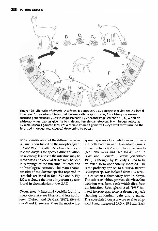
288 Parasitic Diseases
Figure 129 Life cycle of Eimeria: A = feces; B = oocyst; C1, C2 = oocyst sporulation; D = initial infection; E = invasion of intestinal mucosal cells by sporozoites; F = schizogony, several schizont generations; F, = first stage schizont; F2 = second stage schizont; GI, G2 = end of schizogony; merozoites give rise to male and female gametocytes; H = microgametocyte; I = male (micro-) gamete fertilizes a female (macro-) gamete; J = cyst wall forms around the fertilized macrogamete (zygote) developing to oocyst
tions. Identification of the different species is usually conducted on the morphology of the oocysts. It is often necessary to sporu- late the oocysts for species differentiation. At necropsy, lesions in the intestine may be recognized and asexual stages may be seen in scrapings of the intestinal mucosa and on histological sections. The main charac- teristics of the Eimeria species reported in camelids are listed in Table 52 a and b. Fig. 130 a-c shows the most important species found in dromedaries in the UAE.
Occurrence , Intestinal coccidia found to infect Camelidae are Eimeria and less so lso- spora (Ouhelli and Dakkak, 1987). Eimeria cameli and E. dromedarii are the most wide-
spread species of camelid Eimeria, infect- ing both Bactrian and dromedary camels. There are five Eimeria spp. found in camels (see Table 52a) and two lsopora spp.: 1. orlovi and 1. cameli. I. orlovi (Zigankoff, 1950) is thought by Pkllerdy (1965) to be an avian form accidentally ingested. The same probably applies to I. cameli. Recent- ly lsospora sp. was isolated from 1-3-week- old calves in a dromedary herd in Kenya. The calves exhibited profuse diarrhea. One isolation was from a calf which died from the infection. Raisinghani et al. (1987) iso- lated Isospora spp. from a dromedary calf showing abdominal pain and diarrhea. The sporulated oocysts were oval to ellip- soidal and measured 29.5 x 18.4pm. Each
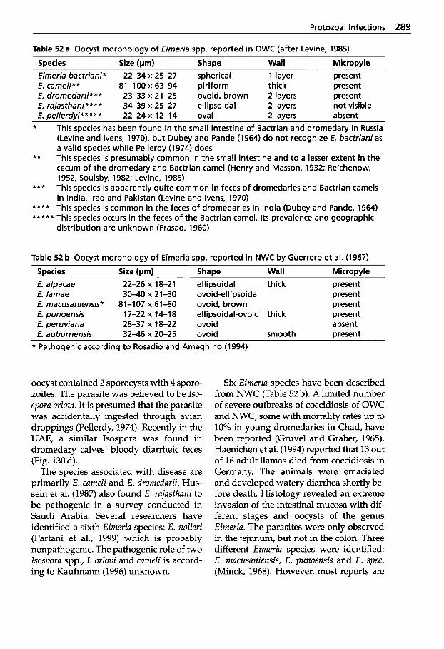
Protozoal Infections 289
Table 52 a Oocyst morphology of Eimeria spp. reported in OWC (after Levine, 1985)
Species Size (vm) Shape Wall Micropyle Eimeria bactriani* 22-34 x 25-27 spherical 1 layer present E. cameli** 81-100 x 63-94 piriform thick present E. dromedarii*** 23-33 x 21-25 ovoid, brown 2 layers present E. rajasthani**** 34-39 x 25-27 ellipsoidal 2 layers not visible E. pellerdyi***** 22-24 x 12-14 oval 2 layers absent
* This species has been found in the small intestine of Bactrian and dromedary in Russia (Levine and Ivens, 1970), but Dubey and Pande (1964) do not recognize E. bactrianias a valid species while Pellerdy (1974) does This species is presumably common in the small intestine and to a lesser extent in the cecum of the dromedary and Bactrian camel (Henry and Masson, 1932; Reichenow, 1952; Soulsby, 1982; Levine, 1985)
*** This species is apparently quite common in feces of dromedaries and Bactrian camels in India, Iraq and Pakistan (Levine and Ivens, 1970)
**** This species is common in the feces of dromedaries in India (Dubey and Pande, 1964) ***** This species occurs in the feces of the Bactrian camel. I t s prevalence and geographic
distribution are unknown (Prasad, 1960)
**
Table 52 b Oocyst morphology of Eimeria spp. reported in NWC by Guerrero et al. (1967)
Species Size (pm) Shape Wall Micropyle E. alpacae 22-26 x 18-21 ellipsoidal thick present E. lamae 30-40 x 21-30 ovoid-ellipsoidal present E. macusaniensis* 81-107 x 61-80 ovoid, brown present E. punoensis 17-22 x 14-18 ellipsoidal-ovoid thick present E. peruviana 28-37 x 18-22 ovoid absent E. auburnensis 32-46 x 20-25 ovoid smooth Dresent
* Pathogenic according to Rosadio and Ameghino (1994)
oocyst contained 2 sporocysts with 4 sporo- zoites. The parasite was believed to be Iso- spora orlovi. It is presumed that the parasite was accidentally ingested through avian droppings (Pellerdy, 1974). Recently in the UAE, a similar Isospora was found in dromedary calves’ bloody diarrheic feces (Fig. 130 d).
The species associated with disease are primarily E. cameli and E. dromedarii. Hus- sein et al. (1987) also found E. rajasthani to be pathogenic in a survey conducted in Saudi Arabia. Several researchers have identified a sixth Eimeria species: E. nolleri (Partani et al., 1999) which is probably nonpathogenic. The pathogenic role of two lsospora spp., 1. orlovi and cameli is accord- ing to Kaufmann (1996) unknown.
Six Eimeria species have been described from NWC (Table 52 b). A limited number of severe outbreaks of coccidiosis of OWC and NWC, some with mortality rates up to 10% in young dromedaries in Chad, have been reported (Gruvel and Graber, 1965). Haenichen et al. (1994) reported that 13 out of 16 adult llamas died from coccidiosis in Germany. The animals were emaciated and developed watery diarrhea shortly be- fore death. Histology revealed an extreme invasion of the intestinal mucosa with dif- ferent stages and oocysts of the genus Eimeria. The parasites were only observed in the jejunum, but not in the colon. Three different Eimeria species were identified: E. macusaniensis, E. punoensis and E. spec. (Minck, 1968). However, most reports are
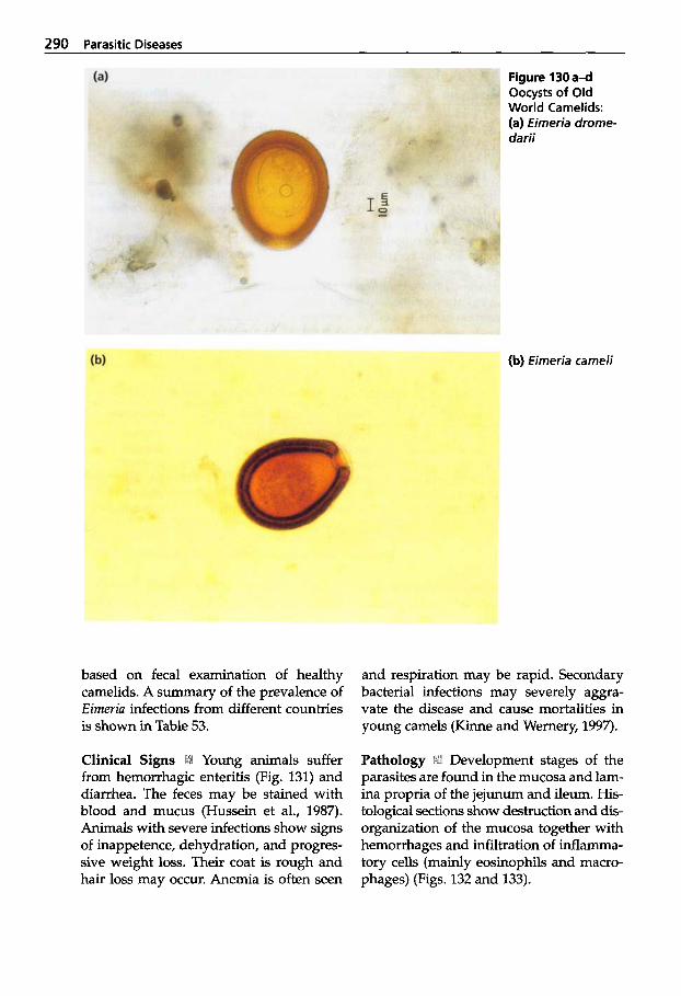
290 Parasitic Diseases
Figure 130 a 4 Oocysts of Old World Camelids: (a) Eimeria drome- darii
(b) Eimeria cameli
based on fecal examination of healthy camelids. A summary of the prevalence of Eimeria infections from different countries is shown in Table 53.
Clinical Signs :L Young animals suffer from hemorrhagic enteritis (Fig. 131) and diarrhea. The feces may be stained with blood and mucus (Hussein et al., 1987). Animals with severe infections show signs of inappetence, dehydration, and progres- sive weight loss. Their coat is rough and hair loss may occur. Anemia is often seen
and respiration may be rapid. Secondary bacterial infections may severely aggra- vate the disease and cause mortalities in young camels (Kinne and Wernery, 1997).
Pathology ii:l Development stages of the parasites are found in the mucosa and lam- ina propria of the jejunum and ileum. His- tological sections show destruction and & organization of the mucosa together with hemorrhages and infiltration of inflamma- tory cells (mainly eosinophils and macro- phages) (Figs. 132 and 133).
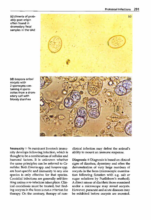
Protozoal Infections 291
(c) Eimeria of prob- ably goat origin often found in dromedary fecal samples in the UAE
(d) lsospora orlovi oocysts with 2 sporocysts con- taining 4 sporo- zoites from a drom- edary calf with bloody diarrhea
Immunity *$I' In ruminant livestock immu- nity develops following infection, which is thought to be a combination of cellular and humoral factors. It is unknown whether the same principles can be referred to Ca- melidae. Both Eimeria spp. and lsospora spp. are host-specific and immunity to any one species is only effective for that species. Coccidial infections are generally self-lim- iting unless a re-infection takes place. Clin- ical coccidiosis must be treated, but find- ing oocysts in the feces is not a criterion for therapy. On the contrary, therapy of non-
clinical infection may defeat the animal's ability to mount an immune response.
Diagnosis is based on clinical signs of diarrhea, dysentery and often the demonstration of very large numbers of oocysts in the feces (microscopic examina- tion following flotation with e.g. salt or sugar solutions by Fuellebom's method). A direct smear of diarrheic feces examined under a microscope may reveal oocysts. However, peracute and acute diseases may be exhibited before oocysts are excreted.
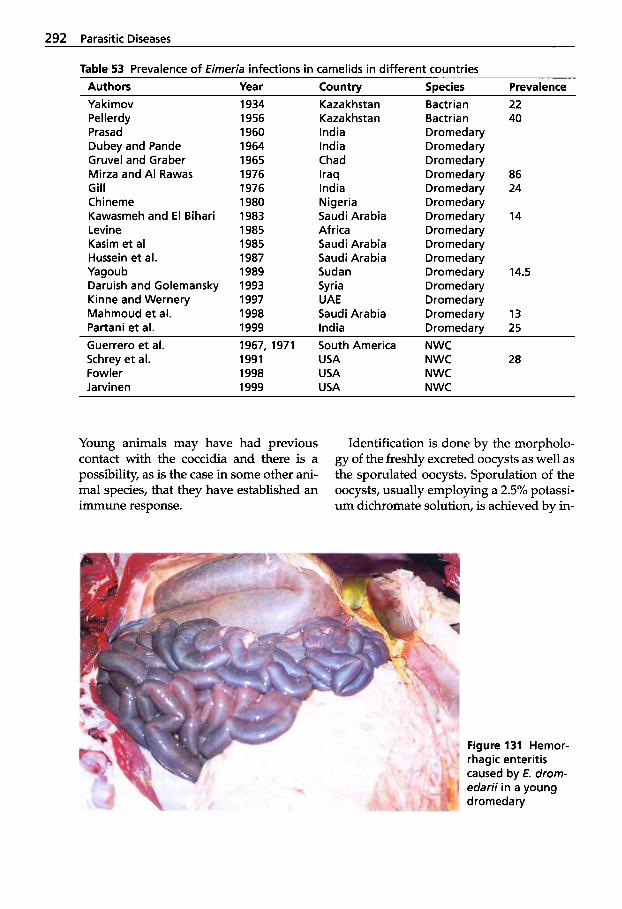
292 Parasitic Diseases
Table 53 Prevalence of Eimeria infections in camelids in different countries Authors Year Countrv Species Prevalence Yakimov Pellerdy Prasad Dubey and Pande Gruvel and Graber Mirza and A1 Rawas Gill Chineme Kawasmeh and E l Bihari Levine Kasim et a1 Hussein et al. Yagoub Daruish and Golemansky Kinne and Wernery Mahmoud et al. Partani et al.
22 40
86 24
14
14.5
13 25
1934 1956 1960 1964 1965 1976 1976 1980 1983 1985 1985 1987 1989 1993 1997 1998
Kazakhstan Kazakhstan India India Chad Iraq India Nigeria Saudi Arabia Africa Saudi Arabia Saudi Arabia Sudan Syria UAE Saudi Arabia
Bactrian Bactrian Dromedary Dromedary Dromedary Dromedary Dromedary Dromedary Dromedary Dromedary Dromedary Dromedary Dromedary Dromedary Dromedary Dromedarv
1999 India Dromedary ~-
Guerrero et al. 1967, 1971 South America NWC
Fowler 1998 USA NWC Jarvinen 1999 USA NWC
Schrey et al. 1991 USA NWC 28
Young animals may have had previous contact with the coccidia and there is a possibility, as is the case in some other ani- ma1 species, that they have established an immune response.
Identification is done by the morpholo- gy of the freshly excreted oocysts as well as the sporulated oocysts. Sporulation of the oocysts, usually employing a 2.5% potassi- um dichromate solution, is achieved by in-
Figure 131 Hemor- rhagic enteritis caused by E. drom- edarii in a young dromedary
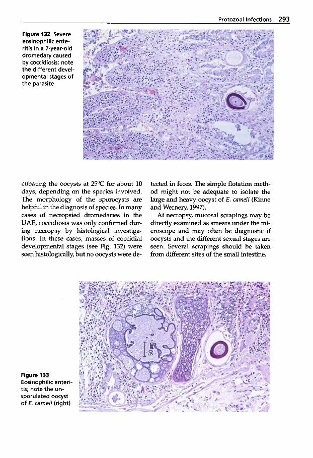
Protozoal Infections 293
Figure 132 Severe eosinophilic ente- ritis in a 7-year-old dromedary caused by coccidiosis; note the different devel- opmental stages of the parasite
cubating the oocysts at 25°C for about 10 days, depending on the species involved. The morphology of the sporocysts are helpful in the diagnosis of species. In many cases of necropsied dromedaries in the UAE, coccidiosis was only confirmed dur- ing necropsy by histological investiga- tions. In these cases, masses of coccidial developmental stages (see Fig. 132) were seen histologically, but no oocysts were de-
tected in feces. The simple flotation meth- od might not be adequate to isolate the large and heavy oocyst of E. cameli (Kinne and Wernery, 1997).
At necropsy, mucosal scrapings may be directly examined as smears under the mi- croscope and may often be diagnostic if oocysts and the different sexual stages are seen. Several scrapings should be taken from different sites of the small intestine.
Figure 133 Eosinophilic enteri- tis; note the un- sporulated oocyst of E. cameli (right)
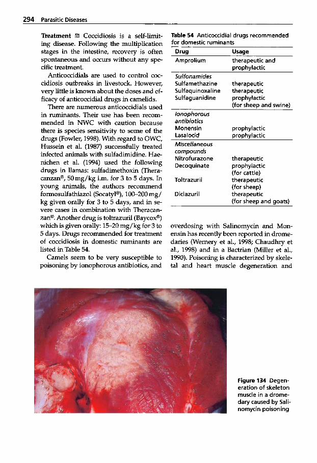
294 Parasitic Diseases
Coccidiosis is a self-limit- ing disease. Following the multiplication stages in the intestine, recovery is often spontaneous and occurs without any spe- cific treatment.
Anticoccidials are used to control coc- cidiosis outbreaks in livestock. However, very little is known about the doses and ef- ficacy of anticoccidial drugs in camelids.
There are numerous anticoccidials used in ruminants. Their use has been recom- mended in NWC with caution because there is species sensitivity to some of the drugs (Fowler, 1998). With regard to OWC, Hussein et al. (1987) successfully treated infected animals with sulfadimidine. Hae- nichen et al. (1994) used the following drugs in llamas: sulfadimethoxin (Thera- canzan@, 50mg/kg i.m. for 3 to 5 days. In young animals, the authors recommend formosulfathiazol (SocatyP), 100-200 mg/ kg given orally for 3 to 5 days, and in se- vere cases in combination with Theracan- zan@. Another drug is toltrazuril (Baycox@) which is given orally: 15-20 mg/kg for 3 to 5 days. Drugs recommended for treatment of coccidiosis in domestic ruminants are listed in Table 54.
Camels seem to be very susceptible to poisoning by ionophorous antibiotics, and
Table 54 Anticoccidial drugs recommended for domestic ruminants
Drug Usage Amprolium therapeutic and
DroDhvlactic Sulfonamides Sulfamethazine therapeutic Sulfaquinoxaline therapeutic Sulfaguanidine prophylactic
(for sheep and swine) lonophorous antibiotics Monensin prophylactic Lasalocid DroDhvlactic Miscellaneous compounds N itrof u razone therapeutic Decoq u i nate prophylactic
(for cattle) Toltrazuril therapeutic
(for sheep) Diclazuril therapeutic
(for sheep and soats)
overdosing with Salinomycin and Mon- ensin has recently been reported in drome- daries (Wernery et al., 1998; Chaudhry et al., 1998) and in a Bactrian (Miller et al., 1990). Poisoning is characterized by skele- tal and heart muscle degeneration and
Figure 134 Degen- eration of skeleton muscle in a drome- dary caused by Sali- nomycin poisoning
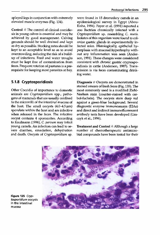
Protozoal Infections 295
splayed legs in conjunction with extremely elevated muscle enzymes (Fig. 134).
The control of clinical coccidio- sis in young calves is essential and may be achieved by good management. Calving grounds should be well drained and kept as dry as possible. Stocking rates should be kept to an acceptable level so as to avoid overcrowding, reducing the risk of a build- up of infections. Feed and water troughs must be kept free of contamination from feces. Frequent rotation of pastures is a pre- requisite for keeping most parasites at bay.
5.1.8 Cryptosporidiosis
Other Coccidia of importance to domestic animals are Cryptosporidium spp., patho- gens of mammals that are usually confined to the microvilli of the intestinal mucosa of the host. The small oocysts ( 4 . 0 4 . 5 ~ ) sporulate within the host and are infective when released in the feces. The infective oocyst contains 4 sporozoites. According to Kaufmann (1996), C. parvum may infect young camels. An infection can lead to se- vere diarrhea, emaciation, dehydration and death. Oocysts of Cryptosporidium sp.
were found in 15 dromedary camels in an epidemiological survey in Egypt (Abou- Eisha, 1994). Fayer et al. (1991) reported a zoo Bactrian chronically infected with a Cryptosporidium sp. resembling C. muris. Isolates of this organism were found to col- onize gastric glands in experimentally in- fected mice. Histologically, epithelial hy- perplasia with mucosal hypertrophy with- out any inflammation was seen (Ander- son, 1991). These changes were considered consistent with chronic gastric cryptospo- ridiosis in cattle (Anderson, 1987). Trans- mission is via feces contaminating drink- ing water.
Oocysts are demonstrated in stained smears of fresh feces (Fig. 135). The most commonly used is a modified Ziehl- Neelsen stain (counter-stained with car- bol-fuchsin). The oocysts stain deep red against a green-blue background. Several diagnostic enzyme immunoassays (EIAs) and direct and indirect immunofluorescent antibody tests have been developed (Gra- czyk et al., 1996).
Treatment and Control Although a large number of chemotherapeutic antimicro- bial compounds have been tested for their
Figure 135 Cryp- tosporidium oocysts in the intestinal mucosa
296 Parasitic Diseases
efficacy against cryptosporidiosis, no real- ly effective compound for therapy or pro- phylaxis has been found (Fayer et al., 1991). However, recently a few antiproto- zoal drugs have been recognized as having some therapeutic and prophylactic proper- ties, e.g. halofuginate lactate (Yvore and Naciri, 1989; Peeters et al., 1993), and paro- momycin (Fayer and Ellis, 1993) in dairy calves. Halofuginate lactate is commercial- ly available as Halocur@ vet (Intervet). An- other compound, lasalocid, showed prom- ising anticryptosporidial effect both in in vitro and in vivo trials when employed at a low dosage (Castro Hermida et al., 2000). Lasalocid is an ionophoric antibiotic pro- duced by Streptomyces lasaliensis. It is not known whether these drugs are tolerated by camels.
Rehydration may help in mild to moder- ate cases. Oocysts are resistant to most dis- infectants except formalin (5%) and ammo- nia. They can survive for months in the en- vironment if kept cool and moist. Optimal management with well-drained calving grounds and the avoidance of overcrowd- ing will prevent infection.
5.1.9 Sarcocystiosis
Species of Sarcocystidae have a two-host LC: an asexual stage in an intermediate host and a sexual in the final host.
Etiology b; Sarcocysfis spp. are parasites using two hosts to complete their LC. Car- nivores commonly act as final hosts and herbivores as intermediate hosts. Surcocys- tis spp. are mostly host-specific for their in- termediate hosts, but less so for their final hosts (Dubey et al., 1989). The general de- velopmental cycle in the final hosts in which sexual stages occur is similar to that of the Eimeria spp., except that there is no asexual multiplication, sporulation takes place in the intestinal wall and sporocysts are excreted for several weeks. In the inter-
mediate host, the infective sporocysts, fol- lowing ingestion of contaminated feces of the final host, release sporozoites into the intestine. The sporozoites invade many or- gans via the blood stream. Schizogony oc- curs in the endothelial cells of blood ves- sels of many organs before typical cysts in the striated muscles develop.
Clinical Signs Usually the definitive hosts carry the infection without showing any signs of disease. Most infections by Sarcocystis spp. in the intermediate hosts are subclinical; however, some infections may cause losses (recorded in domestic an- imals). Acute clinical disease may occur (referred to in cattle as Dalmeny disease) causing abortion, reduced milk produc- tion, wool breakage, lameness, suboptimal growth rate, and sometimes death in cases of heavy infection. Experimental studies have shown that even subclinical infec- tions may have a negative effect on growth and blood parameters in young animals (Leek et al., 1977; Giles et al., 1980).
According to Fowler (1998), light infec- tions give no clinical signs in NWC, but in heavily infected animals the schizogony cycles in endothelial cells may give acute febrile disease, resulting in abortion and death. Also, mild myositis with myalgia may be seen interfering with muscular function. Some llamas have shown clinical signs similar to those in horses with proto- zoal myeloencephalitis caused by S. fulcut- ula (Fowler, 1998). Recently, La Perle et al. (1999) described Dalmeny disease in an al- paca caused by S. aucheniue. It revealed an eosinophilic myositis associated with ma- croscopic sarcocysts and aborted two hours before death. The animal had been imported five years earlier to the USA from Peru.
Myocardial lesions have been attributed to Surcocystis spp. in cam- els (El-Etreby, 1970). Mason (1910) who found the cysts primarily in the myocardi-
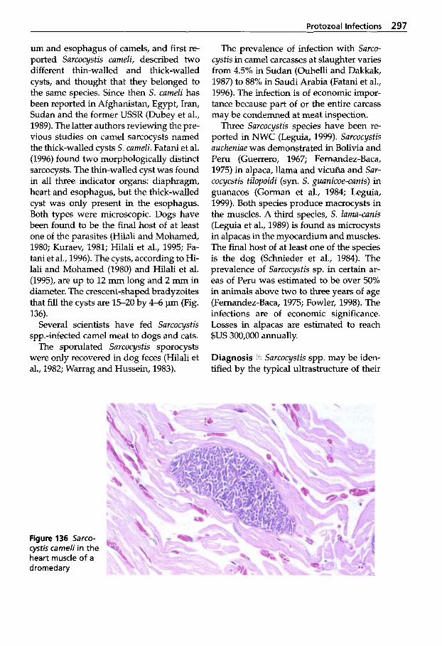
Protozoal Infections 297
um and esophagus of camels, and first re- ported Sarcocystis cameli, described two different thin-walled and thick-walled cysts, and thought that they belonged to the same species. Since then S. cameli has been reported in Afghanistan, Egypt, Iran, Sudan and the former USSR (Dubey et al., 1989). The latter authors reviewing the pre- vious studies on camel sarcocysts named the thick-walled cysts S. cameli. Fatani et al. (1996) found two morphologically distinct sarcocysts. The *-walled cyst was found in all three indicator organs: diaphragm, heart and esophagus, but the thick-walled cyst was only present in the esophagus. Both types were microscopic. Dogs have been found to be the final host of at least one of the parasites (Hilali and Mohamed, 1980; Kuraev, 1981; Hilali et al., 1995; Fa- tani et al., 1996). The cysts, according to Hi- lali and Mohamed (1980) and Hilali et al. (1995), are up to 12 mm long and 2 mm in diameter. The crescent-shaped bradyzoites that fill the cysts are 15-20 by 4-6 pm (Fig. 136).
Several scientists have fed Sarcocystis spp.-infected camel meat to dogs and cats.
The sporulated Sarcocystis sporocysts were only recovered in dog feces (Hilali et al., 1982; Warrag and Hussein, 1983).
The prevalence of infection with Sarco- cystis in camel carcasses at slaughter varies from 4.5% in Sudan (Ouhelli and Dakkak, 1987) to 88% in Saudi Arabia (Fatani et al., 1996). The infection is of economic impor- tance because part of or the entire carcass may be condemned at meat inspection.
Three Sarcocystis species have been re- ported in NWC (Leguia, 1999). Sarcocystis aucheniae was demonstrated in Bolivia and Peru (Guerrero, 1967; Fernandez-Baca, 1975) in alpaca, llama and vicufia and Sar- cocycstis tilopoidi (syn. S . guunicoe-canis) in guanacos (Gorman et al., 1984; Leguia, 1999). Both species produce macrocysts in the muscles. A third species, S. lama-canis (Leguia et al., 1989) is found as microcysts in alpacas in the myocardium and muscles. The final host of at least one of the species is the dog (Schnieder et al., 1984). The prevalence of Sarcocystis sp. in certain ar- eas of Peru was estimated to be over 50% in animals above two to three years of age (Fernandez-Baca, 1975; Fowler, 1998). The infections are of economic significance. Losses in alpacas are estimated to reach $US 300,000 annually.
Diagnosis Sarcocystis spp. may be iden- tified by the typical ultrastructure of their
Figure 136 Sarco- cystis carneli in the heart muscle of a dromedary
298 Parasitic Diseases
cyst wall (Dubey et al., 1989). The oocysts of the Sarcocystis spp. lack a micropyle and they have a fine colorless wall. Sporulated oocysts contain two sporocysts each with four sporozoites. The sporocysts are re- leased in the intestine before being shed through the feces.
Macroscopic cysts are seen during meat inspection or at necropsy. Microscopic cysts are often found accidentally in a his- tologic section of muscle, including the myocardium. Cysts or free bradyzoites may be demonstrated in squash prepara- tions of small pieces of fresh meat samples followed by stereomicroscopy (magnifica- tion 10-60) (Gut, 1982). Peptic digestion of minced muscular tissue followed by exam- ination by a light microscope preferably equipped with phase contrast may be used to diagnose released bradyzoites (Dubey et al., 1989). This method can also be used to extract the bradyzoites for further anti- genic or molecular biological investigation (Lunde and Fayer, 1977). It is not possible to identify the particular species based only on the morphology of the brady- zoites. Histological examination is often used to demonstrate the presence of micro- scopic sarcocysts in the tissues of a host.
There are several serological tests that have been developed for the diagnosis of Sarcocystis infections in different interme- diate hosts. Reported techniques so far are usually based on crude antigens that are not species specific (Uggla and Buxton, 1990).
Molecular techniques mostly based on PCR have proved to be useful in detecting infection and species identification.
Oocysts and sporocysts may be found during fecal examinations of the final host by using traditional flotation techniques based on saturated sodium chloride, su- crose or zinc sulfate solutions. Additional- ly, the organisms may also be found in mu- cosal scrapings of the small intestine by a flotation-concentration technique (Dubey et al., 1989). However, species differen-
tiation based on the morphology of the oocysts or sporocysts is not possible.
Treatment and Control 311 Sarcocystis spp., with few exceptions, are considered to be non-pathogenic. Some infections may be subclinical and only detected after slaugh- ter during meat inspection. Although ex- perimental studies have shown that sub- clinical infections may also have negative effects on the growth of young animals (Leek et al., 1977; Giles et al., 1980), treat- ment and control are very seldom ap- plied.
The only way to control Sarcocystis infec- tions is to break the LC of the parasite. Do- mestic dogs and cats should not receive uncooked meat or offal. Therefore, at abat- toirs it is important to keep offal away from predators. Bradyzoites are readily killed by freezing and by heating to ap- prox. 65°C. The organisms may remain infective in uncooked or poorly cooked meat. Freezing to - 18°C and cooking were effective for inactivating Sarcocystis in guanaco meat (Gorman et al., 1984).
5.1.10 Besnoitiosis
In India, Kharole et al. (1981) reported finding Besnoitia cysts at the base of the lamina propria in the intestine of a drome- dary. Numerous different-sized cysts were found, ranging from 10pm to several 100 pm. There was no systemic or cutaneous involvement as was reported by Fazil and Hofmann (1981) as is often the case in oth- er animal species. No inflammation was seen although the intestinal tissue was damaged by pressure from the large cysts. Similar parasitic infections had been re- ported in buffalo calves, cattle and sheep in the same area. Fazil and Hofmann (1981) stated that besnoitiosis (which they called globidiosis) often occurred in camels with typical skin lesions on the distal part of the legs. The infection often became general-
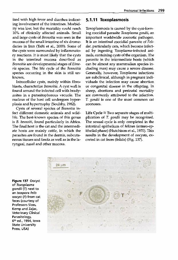
Protozoal Infections 299
ized with high fever and diarrhea indicat- ing involvement of the intestines. Morbid- ity was low, but the mortality could reach 10% of clinically affected animals. Small and large cysts of Besnoitiu was seen in the mucosa of the small intestine of six drome- daries in Iran (Tafti et al., 2000). Some of the cysts were surrounded by inflammato- ry reactions. It is most likely that the cysts in the intestinal mucosa described as Besnoitiu are developmental stages of Eime- ria species. The life cycle of the Besnoitiu species occurring in the skin is still un- known.
Intracellular cysts, mainly within fibro- blasts, characterize Besnoitiu. A cyst wall is found around the infected cell with brady- zoites in a parasitophorous vacuole. The nucleus of the host cell undergoes hyper- plasia and hypertrophy (Soulsby, 1982).
Cysts of several species of Besnoitiu in- fect different domestic animals and wild- life. The best-known species of this genus is B. besnoiti, found particularly in Africa. The final host is the cat and the intermedi- ate hosts are mainly cattle, in which the parasites are found in the dermis, subcuta- neous tissues and fascia as well as in the la- ryngeal, nasal and other mucosa.
Figure 137 Oocyst of Toxoplasma gondii (T) next to an lsospora felis oocyst (F) from cat feces (courtesy of Professors Sloss, Kemp and Zajac, Veterinary Clinical Parasitology, 6th ed., 1994, Iowa State University Press, USA)
5.1.1 1 Toxoplasmosis
Toxoplasmosis is caused by the cyst-form- ing coccidial parasite Toxoplusmu gondii, an important worldwide zoonotic pathogen. It is an intestinal coccidial parasite of FeZi- due, particularly cats, which become infect- ed by ingesting ToxopZusmu-infected ani- mals, containing cysts of the organism. The parasite in the intermediate hosts (which can be almost any mammalian species in- cluding man) may cause a severe disease. Generally, however, Toxoplusmu infections are subclinical, although in pregnant indi- viduals the infection may cause abortion or congenital disease in the offspring. In sheep, abortions and perinatal mortality are commonly attributed to the infection. T. gondii is one of the most common cat zoonoses.
Two separate stages of multi- plication of T. gondii may be recognized. The sexual cycle is only completed in the intestinal epithelium of felines (entero-ep- ithelial phase) (Hutchison et al., 1970). This results in the development of oocysts, ex- creted in cat feces (felids) (Fig. 137).
300 Parasitic Diseases
The oocysts are highly resistant when sporulated and can stay infective for a year or longer (Yilmaz and Hopkins, 1972). When the sporulated oocyst is ingested by a susceptible host (over 200 spp. have been recorded, including rodents, lagomorphs, insectivores, carnivores, marsupials, pri- mates and many birds species (Levine, 1985)), the sporozoites-emerge and enter tissues via the blobd and lymph. Any type of cell may be invarq-dducing tachy- zoites by endodyogjny, an asexual multi- plication (extra-intestinal phase). This may also occur in the final host - the cat - par- allel to the entero-epithelial phase.
In the acute infection, the tachyzoites rapidly multiply within any nucleated cell. New cells are invaded after rupture of the infected cell, which contains large num- bers of tachyzoites. As the infection pro- ceeds, cysts within cells are formed con- taining hundreds of organisms named bradyzoites. The tissue cysts measuring up to 300 pm may be found in any tissue, but are most commonly found in the brain, skeletal and heart muscle. These cyst for- mations are characteristic of the chronic in- fection.
Transmission % Human infection may re- sult from ingestion of cysts with brady- zoites, oocysts from cat feces (e.g. on vegetables) as well as the transplacental spread of tachyzoites to the fetus during acute infection in pregnancy. Camels con- tract the infection by ingesting feed con- taminated with oocysts. Cats given camel meat excreted oocysts of Cystoisospora felis, C. rivoltu and T. gondii (Hilali et al., 1995). C.felis and C. rivoltu are coccidia of cats.
Occurrence Only one case of acute toxo- plasmosis in camels has been reported (Hagemoser et al., 1990). The authors de- scribed a six-year-old female dromedary showing signs of mild dyspnea associated with pyothorax. Twenty-four liters of tur- bid fluid were drained from the pleural
~~
cavity and Toxoplasma tachyzoites were found in macrophages and neutrophils in smears. The camel had become anorectic a month earlier and aborted a near-term fe- tus. Several serological tests, including the Sabin-Feldman dye test, performed on the pleural fluid and the serum showed anti- body titers to T. gondii.
Several seroepidemiological toxoplas- mosis surveys in Camelidae have been re- ported (Table 55).
Both in India and Saudi Arabia there was a higher prevalence of antibodies in adults than in younger animals similar to findings in other hosts. This was attributed to a longer period of exposure to the para- site in older animals (Gill and Prakash, 1969). Hussein et al. (1988) found an asso- ciation between husbandry methods and the seroprevalence of T. gondii infections. Housed camels had a much higher preva- lence due to exposure to the final hosts (cats) than camels in the desert. This has been confirmed by a serological survey in the UAE (unpublished) using the recently established T. gondii ELISA (Chekit Toxo- test? Dr. Bommeli, Switzerland). A sero- prevalence of 38% in 521 racing drome- daries was detected. Other researchers us- ing an indirect hemagglutination test also confirmed this result. Afzal and Zakkir (1994) found 36.4% reactors in dromeda- ries in the UAE.
It is difficult to determine the signifi- cance of the results from these surveys. How- ever, the presence of antibodies shown in camels is indicative of past or present in- fections with T. gondii. It still needs to be established whether the T. gondii infection has any clinical significance in camels. Clinical toxoplasmosis-like signs were ex- perimentally induced in three camels by subcutaneous injections of peritoneal exu- date from mice infected by a pathogenic strain of T. gondii (Galuzo, 1965 cited by Gill and Prakash, 1969). However, earlier similar trials failed to produce the clinical disease in camels (Blanc et al., 1951).
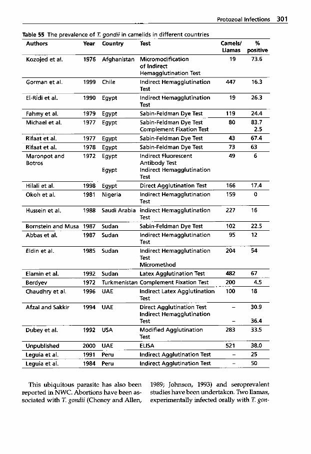
Protozoal Infections 301
Table 55 The Drevalence of I uondii in camelids in different countries Authors Year Country Test Camels/ %
Llamas aositive Kozojed et al. 1976 Afghanistan Micromodification 19 73.6
of Indirect Hemagglutination Test
Gorman et al. 1999 Chile Indirect Hemagglutination 447 16.3 Test
El-Ridi et al. 1990 Egypt Indirect Hemagglutination 19 26.3 Test
~~~~ ~
Fahmv et al. 1979 EavDt Sabin-Feldman Dve Test 119 24.4 Michael et al. 1977 Egypt Sabin-Feldman Dye Test 80 83.7
Complement Fixation Test 2.5 Rifaat et al. 1977 Egypt Sabin-Feldman Dye Test 43 67.4 Rifaat et al. 1978 Egypt Sabin-Feldman Dye Test 73 63 Maronpot and 1972 Egypt Indirect Fluorescent 49 6 Botros Antibody Test
Egypt Indirect Hemagglutination Test
Hilali et al. 1998 Egypt Direct Agglutination Test 166 17.4
Test Okoh et al. 1981 Nigeria Indirect Hemagglutination 159 0
Hussein et al. 1988 Saudi Arabia Indirect Hemagglutination 227 16 Test
Bornstein and Musa 1987 Sudan Sabin-Feldman Dye Test 102 22.5
Test Abbas et al. 1987 Sudan Indirect Hemagglutination 95 12
Eldin et al. 1985 Sudan Indirect Hemagglutination 204 54 Test Micromethod
Elamin et al. 1992 Sudan Latex Agglutination Test 482 67
Chaudhry et al. 1996 UAE Indirect Latex Agglutination 100 18 Berdyev 1972 Turkmenistan Complement Fixation Test 200 4.5
Test Afzal and Sakkir 1994 UAE Direct Agglutination Test - 30.9
Test - 36.4 Indirect Hemagglutination
Dubey et al. 1992 USA Modified Agglutination 283 33.5 Test
Unwblished 2000 UAE ELISA 52 1 38.0 Leauia et al. 1991 Peru Indirect Aaalutination Test - 25 Leauia et al. 1984 Peru Indirect Aaalutination Test - 50
This ubiquitous parasite has also been reported in NWC. Abortions have been as- sociated with T. gondii (Cheney and Allen,
1989; Johnson, 1993) and seroprevalent studies have been undertaken. Two llamas, experimentally infected orally with T. gon-
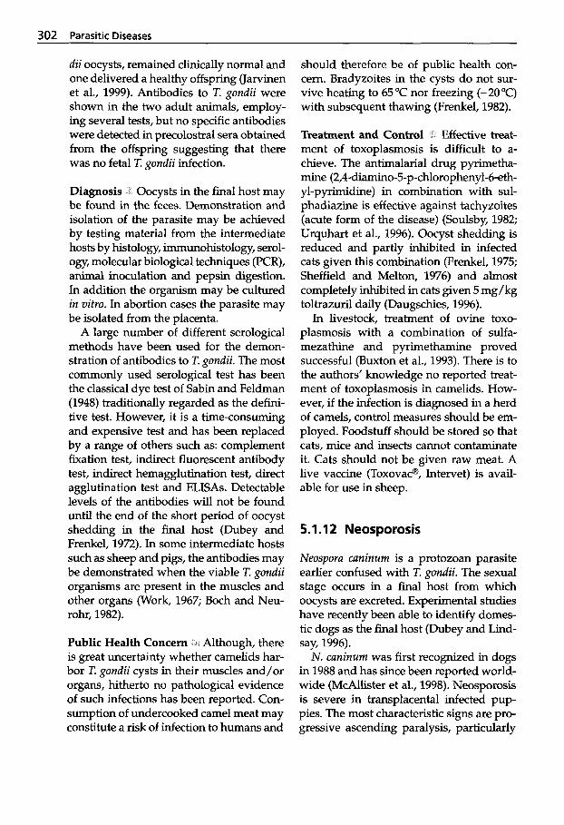
302 Parasitic Diseases
dii oocysts, remained clinically normal and one delivered a healthy offspring (Jarvinen et al., 1999). Antibodies to T. gondii were shown in the two adult animals, employ- ing several tests, but no specific antibodies were detected in precolostral sera obtained from the offspring suggesting that there was no fetal T. gondii infection.
Diagnosis .:i Oocysts in the final host may be found in the feces. Demonstration and isolation of the parasite may be achieved by testing material from the intermediate hosts by histology, immunohistology, serol- ogy, molecular biological techniques (PCR), animal inoculation and pepsin digestion. In addition the organism may be cultured in vitro. In abortion cases the parasite may be isolated from the placenta.
A large number of different serological methods have been used for the demon- stration of antibodies to T. gondii. The most commonly used serological test has been the classical dye test of Sabin and Feldman (1948) traditionally regarded as the defini- tive test. However, it is a time-consuming and expensive test and has been replaced by a range of others such as: complement fixation test, indirect fluorescent antibody test, indirect hemagglutination test, direct agglutination test and ELISAs. Detectable levels of the antibodies will not be found until the end of the short period of oocyst shedding in the final host (Dubey and Frenkel, 1972). In some intermediate hosts such as sheep and pigs, the antibodies may be demonstrated when the viable T. gondii organisms are present in the muscles and other organs (Work, 1967; Boch and Neu- rohr, 1982).
Public Health Concern A Although, there is great uncertainty whether camelids har- bor T. gondii cysts in their muscles and/or organs, hitherto no pathological evidence of such infections has been reported. Con- sumption of undercooked camel meat may constitute a risk of infection to humans and
should therefore be of public health con- cern. Bradyzoites in the cysts do not sur- vive heating to 65 "C nor freezing (- 20 "C) with subsequent thawing (Frenkel, 1982).
Treatment and Control i Effective treat- ment of toxoplasmosis is difficult to a- chieve. The antimalarial drug pyrimetha- mine (2,4-diamino-5-p-chlorophenyl-6-eth- yl-pyrimidine) in combination with sul- phadiazine is effective against tachyzoites (acute form of the disease) (Soulsby, 1982; Urquhart et al., 1996). Oocyst shedding is reduced and partly inhibited in infected cats given this combination (Frenkel, 1975; Sheffield and Melton, 1976) and almost completely inhibited in cats given 5 mg/kg toltrazuril daily (Daugschies, 1996).
In livestock, treatment of ovine toxo- plasmosis with a combination of sulfa- mezathine and pyrimethamine proved successful (Buxton et al., 1993). There is to the authors' knowledge no reported treat- ment of toxoplasmosis in camelids. How- ever, if the infection is diagnosed in a herd of camels, control measures should be em- ployed. Foodstuff should be stored so that cats, mice and insects cannot contaminate it. Cats should not be given raw meat. A live vaccine (Toxovac@, Intervet) is avail- able for use in sheep.
5.1 .I 2 Neosporosis
Neosporu caninurn is a protozoan parasite earlier confused with T. gondii. The sexual stage occurs in a final host from which oocysts are excreted. Experimental studies have recently been able to identify domes- tic dogs as the final host (Dubey and Lind- say, 1996).
N. caninurn was first recognized in dogs in 1988 and has since been reported world- wide (McAllister et al., 1998). Neosporosis is severe in transplacental infected pup- pies. The most characteristic signs are pro- gressive ascending paralysis, particularly
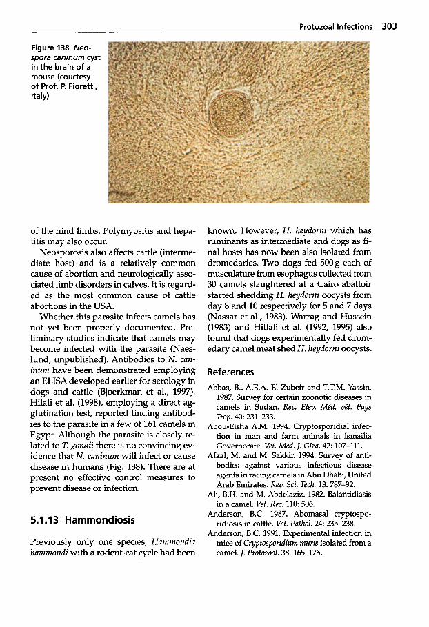
Protozoal Infections 303
Figure 138 Neo- spora caninurn cyst in the brain of a mouse (courtesy of Prof. P. Fioretti, Italy)
of the hind limbs. Polymyositis and hepa- titis may also occur.
Neosporosis also affects cattle (interme- diate host) and is a relatively common cause of abortion and neurologically asso- ciated limb disorders in calves. It is regard- ed as the most common cause of cattle abortions in the USA.
Whether this parasite infects camels has not yet been properly documented. Pre- liminary studies indicate that camels may become infected with the parasite (Naes- lund, unpublished). Antibodies to N. can- inum have been demonstrated employing an ELISA developed earlier for serology in dogs and cattle (Bjoerkman et al., 1997). Hilali et al. (1998), employing a direct ag- glutination test, reported finding antibod- ies to the parasite in a few of 161 camels in Egypt. Although the parasite is closely re- lated to T. gondii there is no convincing ev- idence that N. caninurn will infect or cause disease in humans (Fig. 138). There are at present no effective control measures to prevent disease or infection.
5.1.13 Hammondiosis
Previously only one species, Hammondia hammondi with a rodent-cat cycle had been
known. However, H. heydorni which has ruminants as intermediate and dogs as fi- nal hosts has now been also isolated from dromedaries. Two dogs fed 500g each of musculature from esophagus collected from 30 camels slaughtered at a Cairo abattoir started shedding H. heydorni oocysts from day 8 and 10 respectively for 5 and 7 days (Nassar et al., 1983). Warrag and Hussein (1983) and Hillali et al. (1992, 1995) also found that dogs experimentally fed drom- edary camel meat shed H. heydorni oocysts.
References
Abbas, B., A.E.A. El Zubeir and T.T.M. Yassin. 1987. Survey for certain zoonotic diseases in camels in Sudan. Rev. Elev. Mkd. vkt. Pays
Abou-Eisha A.M. 1994. Cryptosporidial infec- tion in man and farm animals in Ismailia Governorate. Vet. Med. J. Giza. 42: 107-111.
Afzal, M. and M. Sakkir. 1994. Survey of anti- bodies against various infectious disease agents in racing camels in Abu Dhabi, United Arab Emirates. Rev. Sci. Tech. 13: 787-92.
Ali, B.H. and M. Abdelaziz. 1982. Balantidiasis in a camel. Vet. Rec. 110 506.
Anderson, B.C. 1987. Abomasal cryptospo- ridiosis in cattle. Vet. Pathol. 2 4 235-238.
Anderson, B.C. 1991. Experimental infection in mice of C yptosporidium muris isolated from a camel. J. Protozool. 38 165-175.
TYOP. 40: 231-233.
304 Parasitic Diseases
Atarorhouch, T., A. Dakkak, M. Rami and R. Az- laf. 2000. Survey of camel trypanosomosis in six regions in the south of Morocco. 21. An- nual Meeting of the OIE ad hoc group on non tsetse transmitted animal trypanosomosis (NT- TAT), Paris, France 24. May, 2000.
Barnett, S.F. 1977. Theileria. In: J.P. Kreier, ed.: Parasitic Protozoa, Vol. 4. Academic Press, New York, p. 77.
Baumann, M.P.O. and K.H. Zessin. 1992. Pro- ductivity and health of camels (Camelus drom- edarius) in Somalia: Association with trypa- nosomosis and brucellosis. Trop. Anim. Hlth. Prod. 24 145-156.
Bennett, S.C.J. 1933. The control of camel try- panosomosis. 1. Comp. Pathol. 46: 67-77,174- 185.
Berdyev, A.S. 1972. Present position of toxoplas- mosis in Turkmenia. Izvestiya Akademii Nauk Turkmenskoi SSR. Seriya Biologicheskikh Nauk. No. 6: 46-51 (in Russian).
Bitter, H. 1986. Untersuchungen zur Resistenz von Kamelen (Camelus dromedarius) unter besonderer Beriicksichtigung der Infektion mit Trypanosoma evansi (Steel 1885). (Disease resistance in dromedaries with particular reference to Typanosoma evansi (Steel 1885) infection). Thesis, Tierarztliche Hochschule, Hannover, Germany.
Bjoerkman, C., O.J.M. Holmdal and A. Uggla. 1997. An indirect enzyme-linked immunoas- say (ELISA) for demonstration of antibodies to Neospora caninum in serum and milk of cat- tle. Vet. Parasitol. 68: 251-260.
Blanc, G., J. Bruneau and A. Chaubaud. 1951. Quelques essais de transmission de la toxo- plasmose par arthropodes piqueurs. Archs. Inst. Pasteur. Maroc 4 298-303.
Boch, J. and B. Neurohr. 1982. Vorkommen la- tenter Toxoplasma Infektionen bei Schweinen in Siiddeutschland und deren Nachweis mit IFAT und IHA. Tierurztl. Umschau 3 7 820- 826.
Boid, R., A.G., Luckins, P.F., Rae, A.R. Gray, M.M. Mahmoud and K.H. Malik. (1980): Serum immunoglobulin levels and electro- phoretic patterns of serum proteins in camels infected with Trypanosoma evansi. Vet. Para- sitol. 6 333-345.
Boid, R., T.W. Jones and A.G. Luckins. 1985. Proto- zoal diseases of camels. Brit. Vet. 1.111: 87-105.
Bornstein, S. and B.E. Musa. 1987. Prevalence of antibodies to some viral pathogens, Brucella
abortus and Toxoplasma gondii in serum from camels (Camelus dromedarius) in Sudan. 1. Vet. Med. B. 34: 364-370.
Brun, R. and Z.R. Lun. 1994. Drug sensitivity of Chinese Trypanosoma evansi and Trypanosoma equiperdum isolates. Vet. Parasitol. 5 2 37-46.
Brun, H., H. Hecker and Z.-R. Lun. 1998. T y - panosoma evansi and T. equiperdum: distribu- tion, biology, treatment and phylogenetic re- lationship (a review). Vet. Parasitol. 79: 95- 107.
Butt, A.A., G. Muhammed, M. Athar, M.Z. Khan and M. Anwar. 1998. Evaluation of different tests for the diagnosis of trypanosomiasis and dipetalonemiasis. 1. Camel Prac. and Res.
Buxton, D., K.M. Thomson and S. Maley. 1993. Treatment of ovine toxoplasmosis with a com- bination of sulphamezathine and pyrime- thamine. Vet. Rec. 132: 409-411.
Caille, J.Y. 1987. Serological survey of the preva- lence and seasonal incidence of haemoproto- zoa in livestock in Somalia. Thesis, Freie Uni- versitat Berlin, Germany.
Castro Hermida, J.A., F. Freire Santos, A.M. Oteiza Lopes, C.A. Vergara Castblanco and M.E. Ares-Mazas. 2000. In vitro and in vivo efficacy of lasalocid for treatment of experi- mental cryptosporidiosis. Vet. Parasitol. 90:
Chaudhry, Z.I., J. Iqbal, M. Raza and M.I. Qan- dil. 1996. Haematological and biochemical studies on toxoplasmosis in racing camels - a preliminary report. 1. Camel Prac. and Res. 3 7-9.
Chaudhry, Z.I., J. Iqbal, M. Raza and M.I. Qan- dil. 1998. Acute monensin toxicity in drome- dary camels. 1. Camel Prac. and Res. 5: 271- 273.
Cheney, J.M. and G.T. Allen. 1989. Parasitism in llama. Vet. Clin. North Amer., Food Anim. Pract. 5: 217-225.
Chineme, C.N. 1980. A case report of coccidiosis caused by Eimeria cameli in a camel (Camelus dromedarius) in Nigeria. J. Wildl. Dis. 16: 377- 380.
Cross, H.E. 1917. The Camel and its Diseases. BailliPre, Tindall and Cox, London.
Curasson, G.G. 1947. Le chameaux et ses ma- ladies. Vigot Freres, Paris.
CVRL Annual Report. 1999. Daruish, A.I. and V.G. Golemansky. 1993. Coc-
cidia (Apicomplexa, Eucoccidiida) in camels
5: 261-266.
265-270.
Protozoal Infections 305
(Camelus dromedarius L.) from Syria. Acta Zo- olog. Bulgarica 46: 10-15.
Daugschies, A. 1996. Prevention of excretion of Toxoplasma oocysts by medication of cats with toltrazuril. Parassitologia 38: 458.
Dia, M.L., C. Diop, M. Aminetou, P. Jacquit and A. Thiam. 1997. Some factors affecting the prevalence of Trypanosoma evansi in camels in Mauritania. Vet. Parasitol. 72: 111-120.
Dialli, O., E. Bajyana-Songo, E. Magnus, B. Kou- yate, B. Diallo, N. van Meirvenne and R. Ha- mers. 1994. Evaluation d'un test serologique &agglutination directe sur carte dans le diag- nostic de la trypanosornose cam6lie B Try- panosoma evansi. Rev. Sci. Tech. Ofice lnt. Epiz.
Dirie, M.F., K.R. Wallbanks, A.A. Aden, S. Born- stein and M.D. Ibrahim. 1989. Camel trypa- nosomiasis and its vectors in Somalia. Vet. Parasitol. 32: 285-291.
Dubey, J.P. 1977. Toxoplasma, Besnoitia, Sarcocys- tis and other tissue cyst-forming coccidia of man and animals. In J. P. Kreier (ed.): Para- sitic Protozoa. - Vol. 111. Academic Press, Inc., New York.
Dubey, J.P. and B.P. Pande. 1964. Eimerian oocysts recovered from Indian camel (Came- lus dromedarius). lndian J. Vet. Sci. 34: 28-34.
Dubey, J.P. and J.K. Frenkel. 1972. Cyst-induced toxoplasmosis in cats. J. Protozool. 19: 155-177.
Dubey, J.P., C.A. Speer and R. Fayer. 1989. Sar- cocystosis of animals and man. CRC Press, Boca Raton, Florida.
Dubey, J.P., L.G. Rickard, G.L. Zimmerman and D.M. Mulrooney. 1992. Seroprevalence of Toxoplasma gondii in llamas (Lama glama) in the northwest USA. Vet. Parasitol. 44: 295-298.
Dubey, J.P. and D.S. Lindsay. 1996. A review of Neospora caninum and neosporosis. Vet. Para- sitol. 6 7 1-59.
Egbe-Nwiyi, T.N. 1994. Haematological and pathological studies of camel babesiosis in Nigeria. Bull. Anim. Hlth. Prod. Afi. 42: 287- 290.
Elamin, E.A., M.O.A. El Bashir and E.M.A. Saeed. 1998. Prevalence and infection pattern of Trypanosoma evansi in camels in mid-west- ern Sudan. Trop. Anim. Hlth. Prod. 30: 107-114.
Elamin, E.A., S. Elias, A. Daugschies and M. Rommel. 1992. Prevalence of Toxoplasma gon- dii antibodies in pastoral camels (Camelus dromedarius) in the Butana plains, mid-east- ern Sudan. Vet. Parasitol. 43 (34) : 171-175.
1 3 793-800.
Eldin, E.A.Z., S.E. El Khawad, H.S.M. Kheir and E.A. Zain Eldin. 1985. A serological survey for Toxoplasma antibodies in cattle, sheep, goats and camels (Camelus dromedarius) in the Sudan. Rev. Elev. Mid . ve't. Pays Trop. 3 8 247- 249.
El-Etreby, M.F. 1970. Myocardial sarcosporidio- sis in the camel. Pathol. Vet. 7 7-11.
El-Ridi, A.M.S., S.M.M. Nada, A.S. Aly, S.M. Habeeb and M.M. Abdul Fattah. 1990. Sero- logical studies on toxoplasmosis in Zagazig slaughterhouse. J. Egyptian SOC. Parasitol. 20:
Fahmy, M. A., A.M. Mandour, M. S. Arafa and B. M. Abdel Rahman. 1979. Toxoplasmosis of camels in Assiut Governorate. J. Egypt. Vet. Med. Assoc. 39: 27-31.
Fatani, A., M. Hilali, S. Al-Atiya and S. Al-Sha- mi. 1996. Prevalence of Sarcocystis in camels (Camelus dromedarius) from Al-Ahsa, Saudi Arabia. Vet. Parasitol. 62: 241-245.
Fayer, R., L. Phillips, B.C. Anderson and M. Bush. 1991. Chronic cryptosporidiosis in a Bactrian camel. J. Zoo Wldl. Med. 22: 228-232.
Fayer, R. and W. Ellis. 1993. Paromomycin is ef- fective as prophylaxis for cryptosporidiosis in dairy calves. J. Parasitol. 79: 771-774.
Fazil, M.A. and R.R. Hofmann. 1981. Haltung und Krankheiten des Kamels. Tieriirztl. Prax.
Fernandez-Baca, S. 1975. Alpaca raising in the high Andes. World Anim. Rev. 14: 1-8.
Fowler, M.E. 1998. Medicine and Surgery of South American Camelids: Llama, Alpaca, Vicuiia, Guanaco. 2nd ed. Iowa State Univer- sity Press, Ames, USA.
Frenkel, J.K. 1975. Toxoplasmosis in cats and mice. Feline Pract. 5: 28-41.
Frenkel, J.K. 1982. Common questions on tox- oplasmosis: Veterinary medical, and public health considerations. Vet. Med. Small. Anim. Clin. 77: 1188-1196.
Giles, R.C., R. Tramontin, W.L. Kadel, K. Whit- aker, D. Miksch, D.W. Bryant and R. Fayer. 1980. Sarcocystosis in cattle in Kentucky. J. Am. Vet. Med. Assoc. 176 543-548.
Gill, H.S. and 0. Prakash. 1969: Toxoplasmosis in India: prevalence of antibodies in camels. Ann. Trop. Med. Parasitol. 63: 265-267.
Gill, H.S. 1976. Incidence of Eimeria and Infin- dibulorium in camel. Indian Vet. 1. 53: 897-898.
Godfrey, D.G. and R. Killick-Kendrick. 1962. Trypanosoma evansi of camels in Nigeria: A
677-681.
9: 389-402.
306 Parasitic Diseases
high incidence demonstrated by the inocula- tion of blood into rats. Ann. Trop. Med. Para- sitol. 5 6 14.
Gorman, T.R., J.P. Arancibia, M. Lorca, D. Hird and H. Alcaino. 1999. Seroprevalence of Tox- oplasma gondii in sheep and alpacas (Lama pa- cos) in Chile. Preu. Vet. Med. 40: 143-149.
Gorman, T.R., H.A. Alcaino, H. Munoz and C. Cunazza. 1984. Sarcocystis in guanaco (Lama guanacoe) and effect of temperature on its vi- ability. Vet. Parasitol. 15: 95-101.
Graczyk, T.K., M.C. Cransfield and R. Fayer. 1996. Evaluation of commercial immunoas- say (EM) and immunofluorescent antibody (FA) test kit for detection of Cyptosporid- ium oocysts of species other than Cypto- sporidium paruum. Am. J. Trop. Med. Hyg. 54:
Gruvel, J. and M. Graber. 1965. Quelques rksul- tats d’enquetes rhcentes sur la globidiose du dromadaire au Tchad. Rev. Eleu. Mid. uit. Pays Trop. 18: 897-898.
Guerrero, C.A., H. Bazalar and J. Alva. 1971. Eimeria macusaniensis n. sp. (Protozoa: Eime- riidae) of the alpaca (Lama pacos). J. Protozool.
Guerrero, C.A., J. Hernhdez and J. Alva. 1967. Coccidiosis en alpacas. Rev. Fac. Med. Vet. Lima. 21: 59-68.
Guerrero, D., J. Hernhdez and J. Alva. 1967. Sarcocystis en alpaca. Rev. Fac. Med. Vet. Lima.
Guitierrez, C., M.C. Juste, J.A. Corbera, E. Mag- nus, D. Verloo and J.A. Montoya. 2000. Cam- el trypanosomosis in the Canary Islands: Assessment of seroprevalence and infection rates, using the c a d agglutination test (CATT/ T. evansi) and parasite detection tests. Vet. Parasitol. 90: 155-159.
Gut, J. 1982. Effectiveness of methods used for the detection of sarcosporidiosis in farm ani- mals. Folia Parasitol. 29: 289-295.
Haerter, G.H., D. Rottcher, D. Schillinger and E. Zweygarth. 1985. Experimentelle Nagana- Infektionen beim Kame1 (Camelus dromedar- ius). Berl. Munch. Tierarztl. Wschr. 98: 346-350.
Hagemoser, W.A., J.P. Dubey and J.R. Thomp- son. 1990. Acute toxoplasmosis in a camel. J. Am. Vet. Med. Assoc. 196: 347.
Haenichen, T., H. Wiesner und E. Goebel. 1994. Zur Pathologie, Diagnostik und Therapie der Kokzidiose bei Wiederkauern im Zoo. Verh. ber. Erkrg. Zootiere 36: 375-380.
274-279.
18: 162-163.
69-76.
Hilali, M. and A. Mohamed. 1980. The dog (Ca- nis familiaris) as the final host of Sarcocystis cameli (Mason 1910). Tropenmed. Parasitol. 31: 213-214.
Hilali, M., E.L. Imam and A. Hassan. 1982. The endogenous stages of Sarcocystis cameli (Ma- son, 1910). Vet. Parasitol. ll: 127-129.
Hilali, M. and M.M. Fahmy. 1993. Trypanozoon- like epimastigotes in the larvae of Cephalop- ina titilattor (Diptera: Oestridae) infesting camels (Camelus dromedarius) infected with Trypanosoma evansi. Vet. Parasitol. 45: 327- 329.
Hilali, M., A. Fatani and S. Al-Atiya. 1995. Isola- tion of tissue cysts of Toxoplasma, Isospora, Hammondia and Sarcocystis from camel (Cu- melus dromedarius) meat in Saudi Arabia, Vet. Parasitol. 58: 353-356.
Hilali, M., A.M. Nassar and A. El-Ghaysh. 1992. Camel (Camelus dromedarius) and sheep (Ouis aries) meat as a source of dog infection with some coccidian parasites. Vet. Parasit. 43: 3743.
Hilali, M., S. Romand, P. Thulliez, O.C. Kwok and J.P. Dubey. 1998. Prevalence of Neospora caninum and Toxoplasma gondii antibodies in sera from camels from Egypt. Vet. Parasitol.
Hoare, C.A. 1957. The spread of African try- panosomes beyond their natural range. Z. Tropenmed. Parasitol. 8: 156-161.
Hussein, H.S., A.A. Kasim and Y.R. Shawa. 1987. The prevalence and pathology of Eime- ria infections in camels in Saudi Arabia. J. Comp. Pathol. 9 7 293-297.
Hussein, M.F., M.N. Bakkar, S.M. Basmaeil and A.R. Gar’el Nabi. 1988. Prevalence of toxo- plasmosis in Saudi Arabian camels (Camelus dromedarius). Vet. Parasitol. 28: 175-178.
Hutchison, W.M., J.F. Dunachie, J.F. Siim and K. Work. 1970. Coccidian-like nature of Toxo- plasma gondii. Brit. Med. J. 1: 142-144.
Jarvinen, J.A. 1999. Prevalence of Eimeria macu- saniensis (Apicomplexa: Eimeriidae) in mid- western USA. J. Parasitol. 85: 373-376.
Jarvinen, J.A., J.P. Dubey and G.C. Althouse. 1999. Clinical and serological evaluation of two llamas (Lama glama) infected with Toxo- plasma gondii during gestation. J. Parasitol. 85:
Jaktar, P.R. and Singh, M. 1971. Diagnosis of Surra in camels by the passive haemaglutina- tion test. Brit. Vet. J. 127 283-288.
75: 269-271.
142-145.
Protozoal Infections 307
Johnson, LW. 1993. Abortions in llamas. In: M.E. Fowler (ed.): Zoo and Wild Animal Medicine Current therapy 3. W.B. Saunders, Philadel- phia, Pennsylvania, pp. 541-544.
Jones, T.W. and C.D. McKinnell. 1984. Antigenic variation in Typanosoma evansk isolation and characterization of variable antigen type populations from rabbits infected with a stock of T. ewansi. Tropenmed. Parasitol. 35:
Kaminsky, R. and E. Zweygarth. 1989. Feeder layer-free in vitro assay for screening antitry- panosomal compounds against Typanosoma brucei and T. ewansi. Antimicrob. Agents Chemo- ther. 33 881-885.
Kasim, A.A., H.S. Hussein and Y.R. A1 Shawa. 1985. Coccidia in camels (Camelus dromedar- ius) in Saudi Arabia. y. Protozool. 3 2 202-203.
Kaufmann, J. 1996. Parasitic Infections of Do- mestic Animals - A Diagnostic Manual. Birk- hauser Verlag, Basel, Boston, Berlin.
Kawasmeh, Z.A. and S. El Bihari. 1983. Eimeria cameli (Henry and Masson, 1932); Reichenow, 1952 Re-description and prevalence in the eastern province of Saudi Arabia. Cornell Vet.
Kayum, A., M. Afzal and R. Salman. 1992. Gas- trointestinal parasites in racing camels: prev- alence and evaluation of different methods of faecal examination. In: W.R. Allen, A.J. Hig- gins, LG. Mayhew, D.H. Snow and J.F. Wade (eds.): Proceedings of the 1st Int. Camel Conf. R. and W. Publications, Newmarket, UK, pp.
Kelley, S. and D. Schillinger. 1983. Improved field diagnostic technique for trypanosomia- sis by use of a minicentrifuge. Vet. Rec. 113 219.
Kharole, M.U., S.K. Gupta and J. Singh. 1981. Note on besnoitiosis in a camel. Indian J. Anim. Sci. 51: 802-804.
h e , J. and U. Wernery. 1997. Severe outbreak of camel coccidiosis in the United Arab Emi- rates. J. Camel Prac. and Res. 4: 261-265.
h e , J. and U. Wemery. 2000. Comparative study on surra in equine and camelids. Euro- pean Society of Veterinary Pathology - con- ference, Amsterdam, Holland.
Kiorpes, A.L., C.E. Kirkpatrick and D. D. Bow- man. 1987. Isolation of Giardia from a llama and from sheep. Can. 1. Vet. Res. 51: 277-280.
Kozojed, V., K. Blazek and A. Amin. 1976. Inci- dence of toxoplasmosis in domestic animals in Afghanistan. Folia Parasitol. 23: 273-275.
237-241.
73: 58-66.
85-87.
Kuraev, G. T. 1981. Sarcocystis infection in drom- edaries and Bactrian camels in Kazakhstan (in Russian). Veterinariya No. 7 41-42.
La Perle, K.M.D., F. Silveria, D.E. Anderson and E.A.G. Blomme. 1999. Dalmeny disease in an alpaca (Lama pacos): Sarcocystosis, eosino- philic myositis and abortion. J. Comp. Pathol.
Leek, R.G., R. Fayer and A.J. Johnson. 1977. Sheep experimentally infected with Sarco- cystis from dogs. I. disease in young lamb. J. Parasitol. 63: 642-650.
Leguia, G., H. Samame, C. Guerrero, M. Rojas and A. Nuiiez. 1984. Prevalencia de anticuer- pos contra Toxoplasma gondii en alpacas. Rev. Cien. Vet. 3: 19-21.
Leguia, G., C. Guerrero, R. Sam and A. Chavez. 1989. Infecih experimental de perros y galos con microquistes de Sarcocysfis de alpacas (Lama pacos). M Y Rev. Cien. Vet. 5: 10-13.
Leguia, G. 1991. The epidemiology and eco- nomic impact of llama parasites. Parasitol. To-
Leguia, G. 1999. Enfermedades parasitarias de camelidos Sudamericanos. Editorial De Mar. Lima, Peru.
Leese, A.S. 1927. A treatise on the One-humped Camel in Health and Disease. Haynes and Son, Standford, Lincolnshire, UK.
Levine, N.D. 1985. Veterinary Protozology. Iowa State University Press, Ames.
Levine, N.D. and V. Ivens. 1970. The Coccidian Parasites (Protozoa, Sporozoa of Ruminants). Urbana, University of Illinois Press.
Losos, G.J. 1980. Diseases caused by Typanoso- ma evansi, a review. Vet. Res. Comm. 4 165-181.
Luckins, A.G. 1992. Protozoal diseases of cam- els. In: W.R. Allen, A. J. Higgins, I.G. Mayhew, D.H. Snow and J.F. Wade (eds.): Proceedings of the lSt Int. Camel Cod. R. and W. Publica- tions, Newmarket, UK, pp. 23-27.
Luckins, A.G., A.R. Gray and P. Rae. 1978. Com- parison of the diagnostic value of serum im- munoglobulin levels, an enzyme linked im- munosorbent assay and a fluorescent anti- body test in experimental infections with Trypanosoma ewansi in rabbits. Ann. Trop. Med. Parasitol. 72 429-441.
Luckins, A.G., R. Boid, P. Rae, M.M. Mahmoud, K.H. El Malik and A.R. Gray. 1979. Serodiag- nosis of infection with Trypanosoma ewansi in camels in the Sudan. Trop. Anim. Hlth. Prod. 11: 1-12.
121: 287-293.
day. 7 54-56.
308 Parasitic Diseases
Lumsden, W.H.R., Kimber, C.D., D.A. Evans and S.J. Doig. 1979. Tympanosoma brucei Min- iature anion-exchange centrifugation tech- nique for detection of low parasitaemias: Adaptation for field use. Trans. Roy. SOC. Trop. Med. Hyg. 73: 312-317.
Lumsden, W.H.R., Kimber, C.D., Dukes, P. Haller, A. Stanghellini and G. Duvallet. 1981. Field diagnosis of sleeping sickness in Ivory Coast. 1: Comparison of the miniature anion- exchange centrifugation technique with oth- er protozoological methods. Trans. Roy. SOC. Trop. Med. Hyg. 76: 242-250.
Lunde, M.N. and R. Fayer. 1977. Serological tests for antibody to Sarcocystis in cattle. J. Parasitol. 63: 222-225.
Magzoub, M., O.H. Omar, E.M. Haroun, O.M. Mahmoud and Y.M.A. Hamid. 1997. Gastro- intestinal parasites of dromedary camels in Gassim region, Saudi Arabia. Indian Vet. J. 74
Mahmoud, M.M. and A.R. Gray. 1980: Trypa- nosomiasis due to Trypanosoma evansi (Steel, 1885) Balbiani, 1888: A review of recent re- search. Trop. Anim. Hlth. Prod. 12: 1-12.
Mahmoud, O.M., E.M. Haroum, M. Magzoub, O.H. Omer and A. Sulman. 1998. Coccidial in- fection in camels of Gassim region, central Sau- di Arabia. J. Camel Prac. and Res. 5 257- 260.
Maronpot,R. R. and B. A.M. Botros. 1972. Toxo- plasma serologic survey in man and domes- tic animals in Egypt. J. Egypt. Pub. Hlth. Assoc.
Masiga, D. and W. Gibson. 1992. Sensitive detec- tion of trypanosomes by DNA amplifica- tions. First international seminar on non-Tsetse transmitted trypanosomes (NTTAT), Annecy,
Mason, F.P. 1910. Sarcocysts in the camel in Egypt. J. Comp. Pathol. Therap. 23: 168-176.
McAllister, M.M., J.P. Dubey, D. S. Lindsay, W.R. Jolley, R.A. Wills and A.M. McGuire. 1998. Dogs are definite hosts of Neospora caninum. lnt. J. Parasitol. 28: 1473-1478.
Michael, S.A., A.H. El-Refaii and T.A. Morsy. 1977. Incidence of Toxoplasma antibodies among camels in Egypt. J. Egypt. SOC. Parasitol. 7 129- 132.
Mihok, S., E. Zweygarth, E.N. Munyoki, J. Wam- bua and R. Kock. 1994. Typanosomasimiae in the white rhinoceros (Ceratotherium simum) and the dromedary camel (Camelus dromedar- ius). Vet. Parasitol. 53: 191-196.
373-376.
47 58-67.
Oct. 14-16.
Miller, R. E., W. J. Boever, R.E. Junge, L.P. Thorn- burg, M.F. Raisbeck. 1990. Acute monensin toxicosis in stone sheep (Ovis dalli stonei), blesbok (Damaliscus dorcus phillipsi) and a Bac- trian camel (Camelus bactrianus). J. Am. Vet. Med. Assoc. 196: 131-134.
Minck, K. 1968. Untersuchungen uber Kokzidi- en bei Zoowiederkauern. Vet. Med. Diss. Mu- nich, p. 29.
Mirza, M.Y. and A.Y. Al-Rawas. 1976. Coccidia (Protozoa: Eimeriidae) from camels (Camelus dromedarius) in Iraq. Bull. Biol. Res. Centre, Baghdad. 7 24-31.
Molyneux, D.H. and R.W. Ashford. 1983. The Biology of Trypanosoma and Leishmania, Para- sites of Man and Domestic Animals. Taylor and Francis, London, UK.
Nantulya, V.M. 1989. Suratex: A simple latex ag- glutination antigen test for diagnosis of T y - panosoma evansi infections (Surra). Tropenmed. Parasitol. 45: 9-11.
Nantulya, V.M., A.J. Musoke, F.R. Rurangirwa, N. Saigor and S.H. Minga. 1987. Monoclonal antibodies that distinguish Trypanosoma con- golense, T. vivax and T. brucei. Parasite lm- munol. 9: 421-431.
Nassar, A.M. 1992. Theileria infection in camels (Camelus dromedarius) in Egypt. Vet. Parasitol.
Nassar, A.M., M. Hilali and M. Rommel. 1983. Hammondia heydorni infection in camels (Cam- elus dromedarius) and water buffaloes (Buba- lus bubalus) in Egypt. Z. Parasitenkd. 69: 693- 694.
Nessiem, M.G. 1994. Evaluation of the silicone centrifugation technique in the detection of Trypanosoma evansi infection in camels and experimental animals. Trop. Anim. Hlth. Prod.
Ogbunde, P.O.J. and Y. Magaji. 1982. A silicone centrifugation technique for the detection of low parasitaemias of salivarian trypanoso- mes. Trans. Roy. SOC. Trop. Med. Hyg. 76: 317- 318.
Okoh, A.E.J., D.E. Agbonlahor and M. Momoh. 1981. Toxoplasmosis in Nigeria - a serologi- cal survey. Trop. Anim. Hlth. Prod. 13: 137-140.
Olaho, W. and A.J. Wilson. 1983. The prevalence of camel trypanosomiasis in selected areas of Kenya. 17th Meeting of the International Sci- entific Council for Trypanosomiasis Research and Control. Arusha, Tanzania, pp. 246-253.
43: 147-149.
26: 227-229.
Ouhelli, H. and A. Dakkak. 1987. Protozoal dis- eases of dromedaries. Rev. Sci. Tech. O f . Int. Epiz. 6: 417-422.
Partani, A.K., D. Kumar and G.S. Manohar. 1999. Prevalence of Eimeria infection in cam- els (Camelus dromedarius) at Bikaner (Rajas- than). J. Camel Prac. and Res. 6: 69-71.
Pegram, R.G. and J.M. Scott. 1976. The preva- lence and diagnosis of Trypanosoma evansi infection in camels in Ethiopia. Trop. Anim. Hlth. Prod. 8: 20-27.
Pellegrini, D. 1948. Trypanosoma simiae (Bruce) infection of the camel. E. Afr. Agric. J. 13: 207- 209.
Pellerdy, L.P. 1956. Catalogue of the genus Eime- ria (Protozoa, Eimeriidae). Acta Vet. Acad. Sci. Hung. 6: 75-102.
Pellerdy, L.P. 1974. Coccidia and Coccidiosis. 2nd ed. Paul Parey, Berlin.
Peeters, J.E., I. Vallacorta, M. Naciri and E. Va- nopdenbosch. 1993. Specific serum and local antibody response against Cryptosporidium parvum during medication of calves with halofuginone lactate. Infect. Immun. 61: 4440- 4445.
Prasad, H. 1960. Studies on the coccidia of the families Bovidae, Cervidae and Camelidae. Z . Parasitenkde. 20: 202-203.
Rae, P.F. and A.G. Luckins. 1984. Detection of circulating trypanosomal antigens by en- zyme immunoassays. Ann. Trop. Med. Para- sitol. 78: 587-596.
Rae, P.F., M.V. Thrusfield, C.G.D. Aitken, T.W. Jones and A.G. Luckins. 1989. Evaluation of enzyme immunoassays in the diagnosis of camel (Camelus dromedarius) trypanosomia- sis: a preliminary investigation. Epidem. In- fect. 102: 297-307.
Raisinghani, P.M., G.S. Monahar and J.S. Yadav. 1987. Isospora infection in the Indian camel Camelus dromedarius. Ind. J. Parasitol. 11 (1): 93- 94.
Rao, J.R., A.K. Mishra, N.N. Sharma, Kalicharan and M.C. Prasad. 1988. Biochemical studies on sera of camels (Camelus drornedarius) natu- rally infected with Theileria dromedarii n. sp. Riv. Parassitol. 49: 63-66.
Raynaud, J.P., K.R. Sones and E.A.H. Fried- heem. 1989. A review of Cymelarsan - a new treatment proposed for animal trypanosomi- asis due to T. evansi and other trypanosomes of the T. brucei group. ISCTRC, 20th Meeting, Mombassa, Kenya, Pub. No 115.
Protozoal Infections 309
Richard, D. 1975. Study on the pathology of the dromedary in Borana Awraja (Ethiopia). The- sis, IEMTV, Paris.
Richard, D. 1979. Pathology and production of camels. In: Camels, IFS Report 6: 409-430.
Rifaat, M.A., T.A. Morsy, M.S.M. Sadek, M.L.M. Khalid, M.E. Azab, M.K. Makled, E.H. Safar and O.M.N. El-Din. 1977. Incidence of toxo- plasmosis among farm animals in Suez Canal Governorates. J. Egypt. SOC. Parasitol. 7 135- 140.
Rifaat, M.A., T.A. Morsy, M.S.M. Sadek, M.L.M. Khalid, M.E. Azab and E.H. Safar. 1978. Prevalence of Toxoplasma antibodies among slaughtered animals in Lower Egypt. J. Egypt. SOC. Parasitol. 8: 339-345.
Roettcher, D., D. Schillinger and E. Zweygarth. 1987. Trypanosomiasis in the camel (Camelus dromedarius). Rev. Sci. Tech. Ofice. lnt. Epiz. 6:
Rosadio, R.H. and E.F. Ameghino. 1994. Coc- cidial infections in neonatal Peruvian alpa- cas. Vet. Rec. 135: 459-460.
Rutagwenda, T. 1984. A study of important camel diseases in Northern Kenya with spe- cial emphasis on their control. Camel Nezuslet- ter 1: 12-14.
Rutter, T.E.G. 1967. Diseases of camels. Vet. Bull.
Sabin, A.D. and H.A. Feldman. 1948. Dyes as microchemical indicators of a new immunity phenomenon affecting a protozoon parasite (Toxoplasma). Science 18: 660-663.
Salfelder, K., T.R. de Liscano and E. Sauerteig. 1992. Atlas of Parasitic Pathology. Kluwer Academic Publishers, Dordrecht/Boston/ London, pp. 23-26.
Schillinger, D. and D. Roettcher. 1984. The cur- rent state of chemotherapy of T. evansi in- fection in camels. In: W.R Cockrill (ed.): The Camelid - An All-purpose Animal. Vol. 1. Proceedings of the Khartoum Workshop on Cam- els. Dec. 1979, pp. 509-518.
Schnieder, T., F.-J. Kaup, W. Drommer, W. Thiel und M. Rommel. 1984. Zur Feinstruktur und Entwicklung von Sarcocystis aucheniae beim Lama. Z. Parasitenk. 70: 451-458.
Schoening, H.W. 1924. Trypanosomiasis in cam- els. J. Infect. Dis. 34: 608-613.
Schrey, C.F., T.A. Abbott, V.A. Stewart and C.W. Marquardt. 1991. Coccidia of the llama, Lama glama, in Colorado and Wyoming. Vet. Para- sitol. 40: 21-28.
463-470.
37: 611-618.
310 Parasitic Diseases
Seiler, R.J., S. Omar and A.R.B. Jackson. 1981. Meningoencephalitis in naturally occurring Trypanosoma evansi infection (Surra) in hors- es. Vet. Pathol. 18: 120-122.
Sheffield, H.G. and M.L. Melton. 1976. Effect of pyrimethamine and sulfadiazine on the in- testinal development of Toxoplasma gondii in cats. Am. J. Trop. Med. Hyg. 25: 379-383.
Shommein, A.M. and A.M. Osman. 1987. Dis- eases of camels in the Sudan. Rev. Sci. Tech. Ofice. Int. Epizoot. 6 (2): 481-486.
Soulsby, E.J.L. 1982. Helminths, Arthropods and Protozoa of Domesticated Animals, 7* ed. Bailliere Tiidall, London.
Stevens, D.P. 1985. Selective primary health care: strategies for control of disease in the developing world. XIX. Giardiasis. Rev. In- fect. Dis. 7 530-535.
Tibary, A. and A. Anouassi. 1997. Theriogenolo- gy in camelidae. Abu Dhabi Printing Press, Mina, Abu Dhabi, U.A.E.
Tufli, A. K., M. Maleki, A. Oryan and A.A. Mozafari. 2000. Pathological study of diges- tive system lesions of camels (Camelus drome- darius) slaughtered in Iran. Wh Meeting of the European Society of Veterina y Pathology, Am- sterdam, The Netherlands, 19-2Znd Septem- ber, p. 245.
Uggla, A. and D. Buxton. 1990. Immune re- sponse against Toxoplasma and Sarcocystis in- fections in ruminants: diagnosis and pros- pects for vaccination. Rev. Sci. Tech. Ofice Int. Epiz. 9: 441-462.
Urquhart, G.M., J. Armour, J.L. Duncan, A.M. Dunn and F.W. Jennings. 1996. Veterinary Parasitology, Znd ed. Blackwell Science, Ox- ford, UK.
Vosdingh, R.A. and J.A. Vanniasingham. 1969. Balantidiasis in a camel. J. Am. Vet. Med. As-
Warrag, M. and H.S. Hussein. 1983. The camel (Camelus dromedarius) as an intermediate host for Hammondia heydorni. J. Parasitol. 69:
Wemery, U. 1991. The barren camel with en- dometritis - Isolation of Trichomonas fetus and different bacteria. J. Vet. Med. B. 3 8 523- 528.
Wernery, U. 1995. Blutparameter und Enzym- werte von gesunden und kranken Rennkame- len (Camelus dromedarius). Tierarztl. Praxis. 23:
SOC. 155: 1077-1079.
926-929.
187-191.
Wernery, U., A. Tinson, J. Kinne and J. A1 Masri. 1998. Salinomycin poisoning in a dromedary breeding herd in the United Arab Emirates. J. Camel Prac. and Res. 5: 275-279.
Wernery, U., M.E. Fowler and R. Wernery. 1999. Color Atlas of Camelid Hematology. Black- well Wissenschafts-Verlag, Berlin, Wien, p. 7.
Wilson, A.J., H.J. Schwartz, R. Dolan and W.M. Olahu. 1983. A simple classification of differ- ent types of trypanosomiasis occurring in four camel herds in selected areas of Kenya. Tropenmed. Parasitol. 34: 220-224.
Woo, P.T.K. 1969. The haematocrit centrifuga- tion technique for the detection of trypano- somes in blood. Can. J. Zool. 4 7 921-923.
Woo, P.T.K. 1971. Evaluation of the haematocrit centrifuge and other techniques for the field diagnosis of human trypanosomiasis and fi- lariasis. Acta Tropica 28: 298-303.
Work, K. 1967. Isolation of Toxoplasma gondii from the flesh of sheep, swine and cattle. Acta Pathol. Microbiol. Scand. 71: 296-306.
Wuyts, N., N. Chokesajjawatee and S. Panyim. 1994. A simplified and highly sensitive detec- tion of Trypanosoma evansi by DNA amplifica- tion. Southeast Asian J. Pop. Med. Public Health
Xiao, L., K. Saeed and R.P. Herd. 1996. Efficacy of albendazole and fenbendazole against Gi- ardia infection in cattle. Vet. Parasitol. 16: 165- 170.
Yagoub, I.A. 1989. Coccidiosis in Sudanese cam- els (Camelus dromedarius): First record and de- scription of Eimeria spp. harboured by camels in the eastern regions of Sudan. J. Protozool.
Yakimov, W.L. 1934. Zur Frage der Coccidien der Kamele. Arch. Wiss. Prakt. Tierheilk. 68:
Yilmaz, S.M. and S.H. Hopkins. 1972. Effects of different conditions on duration of infectivity of Toxoplasma gondii oocysts. J. Parasitol. 58:
Yvore, P. and M. Naciri. 1989. Halofuginone lac- tate in the treatment of cryptosporidiosis in ruminants. In: Coccidia and Intestinal Cocci- diemorphs. Procedings 5. International Cocci- diosis Conference, Tours, France, 17-20 Oct. Les Colloques de YINRA. 49 (2): 475-478.
Zhang, Z.O., C. Giroud and T. Baltz. 1993. T y - panosoma evansi: in vivo and in vitro determi- nation of trypanocide resistance profiles. Exp. Parasitol. 7 7 387-394.
25: 266-271.
36: 422-423.
134-137.
938-939.
Protozoal infections 31 1
Further reading Boch, J. 1967. Toxoplasma infections in domestic
animals and their importance in meat inspec- tion. Fleischwirtschaft 9: 971-973.
Bornstein, S., B.E. Musa and EM. Jama. 1988. Comparison of seroepidemiological findings of antibodies to some infectious pathogens of cattle in camels of Sudan and Somalia with reference to findings in other countries of Africa. In: The International Symposium on the Development of Animal Resources in the Sudan. Khartoum, Sudan, pp. 28-34.
Buxton, D., D.A. Blewett, A.J. Trees, C. McCol- gan and C. Finlayson. 1988. Further studies in the use of monensin in the control of ex- perimental toxoplasmosis. J. Comp. Pathol. 98:
Connor, R.J. 1994. African animal trypanosomi- asis. In: J.A.W. Coetzer, G.R. Thomson and R.C. Tustin (eds.): Infectious Diseases of Live- stock with Special Reference to Southern Africa, Vol. 2. Oxford University Press, Cape Town, pp. 167-205.
Dubey, J.P. 1993. Toxoplasma, Neospora, Sarcocys- tis, and other tissue cyst-fonning coccidia of humans and animals. In: J.P. Kreier and J.R. Baker (eds.): Parasitic Protozoa, vol. 6. Aca- demic Press Inc., San Diego, California, USA.
Fayer, R. and A.J. Johnson. 1975. Effect of am- prolium on acute sarcocystosis in experimen- tally infected calves. J. Parasitol. 61: 932-936.
Foreyt, W.J. 1986. Evaluation of decoquinate, lasalocid and monensin against experimen- tally induced sarcocystosis in calves. Am. J . Vet. Res. 4 7 1674-1676.
Heydorn, A.O., S. Haralambidis and F.R. Ma- tuschka. 1981. Zur Chemopmphylaxe und The- rapie der akuten Sarkosporidiose. (Chemo- prophylaxis and therapy of acute sarcospori- diosis). Berl. Munch. Tieriirztl. Wochenschr. 9 4
225-236.
229-234.
Hodgin, C., T.W. Shillhorn van Veen, R. Fayer and N. Richter. 1984. Leptospirosis and coc- cidial infection in a guanaco. J. Am.Vet. Med.
Hornby, H.E. 1947. Trypanosomiasis in Eastern Africa. H.M. Stationary Office.
Jordan, A.M. 1986. Trypanosomiasis Control and African RuraI Development. Longman, London and New York, p. 78.
Lappin, M. 1999. Feline toxoplasmosis. In Prac- tice 21: 578-587.
Pellerdy, L.P. 1965. Coccidia and Coccidiosis. Akademiai Kiado, Publishing House of the Hungarian Academy of Sciences, Budapest,
Rifaat, M.A., T.A. Morsy, A. Salem Shafia, H.M. Khalil and P.C.C. Garnham. 1964. In: A. Cor- radetti (ed.): Proceedings of the 1st Interna- tional Congress of Parasitology. Rome, Italy. Pergamon Press, London, p. 171.
Rommel, M. 1983. Integrated control of proto- zoan diseases of livestock. In: J.D. Dunsmore (ed.): Tropical Parasitoses and Parasitic Zoo- noses. Proceedings of the 10th Conference of the World Association for the Advancement of Veterinary Parasitology, Perth, Australia,
Rommel, M., A. Schwerdtfeger and S. Blewaska. 1981. The Sarcocystis murk infection as a mod- el for research on the chemotherapy of acute sarcocystosis of domestic animals. Zentralbl. Bakteriol. Hyg. Abt. 2 Orig. A. 250 268- 276.
Sharma, S.P. and O.P. Gautam. 1974. A note on the prevalence of Toxoplasma antibodies among camels and pigs in Hissar, India. Indi- an J . Anim. Sci. 44: 214-215.
Stephen, L.E. 1970. Clinical manifestation of the trypanosomiasis in livestock and other do- mestic animals. In: H.W. Mulligan (ed.): The African Trypanosomiasis. Georg Allen, Un-
ASSOC. 185: 1442-1444.
1965, p. 510-516.
pp. 9-30.
win, pp. 784-786.
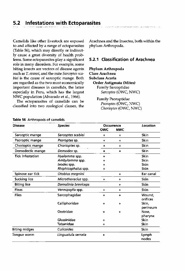
5.2 Infestations with EctoDarasites I , I . . I
Camelids like other livestock are exposed to and affected by a range of ectoparasites (Table 56), which may directly or indirect- ly cause a great diversity of health prob- lems. Some ectoparasites play a significant role in many disorders. For example, some biting insects are vectors of disease agents such as T. evansi, and the mite Sarcoptes sca- biei is the cause of sarcoptic mange. Both are regarded as the two most economically important diseases in camelids, the latter especially in Peru, which has the largest NWC population (Alvarado et al., 1966).
The ectoparasites of camelids can be classified into two zoological classes, the
Table 56 Arthropods of camelids
Arachnea and the Insectea, both within the phylum Arthropoda.
5.2.1 Classification of Arachnea
Phylum Arthropoda Class Arachnea Subclass Acaria
Order Astigmata (Mites) Family Sarcoptidae
Family Psoroptidae
Sarcoptes (OWC, NWC)
Psoroptes (OWC, NWC) Choriopfes (OWC, NWC)
Disease Species Occurrence Location OWC NWC
Sarcoptic mange Sarcoptes scabiei + + Skin Psoroptic mange Psoroptes sp. + + Skin Chorioptic mange Chorioptes sp. + + Skin Demodectic manae Demodex SD. + + Skin
~____________
Tick infestation Hyalomma spp. + Skin Ambylomma spp. + Skin lxodes spp. + Skin Rhiphicephalus spp. + Skin
Spinose ear tick Otobius megnini + Ear canal Sucking lice Microthoracius spp. + + Skin Biting lice Damalinia breviceps + Skin Fleas Vermimvlla SDD. + + Skin Flies
~ ~~
Sarcophagidae + + Wound, orif ices
perineum
pharynx
Calliphoridae + + Skin,
Oestridae + + Nose,
Glossinidae + Skin Tabanidae + Skin
Biting midges Culicoides Skin Tongue worm Linguatula serrata + Lymph
nodes
Infestations with Ectooarasites 31 3
Order Prostigmata Family Demodicidae
Demodex (OWC, NWC)
Order Metastigmata (Ticks) Family Argasidae (Soft ticks)
Ornithodoros savigny (OWC) 0. lahorensis (OWC) 0. tholozani (OWC) Otobius megnini (NWC)
Family Ixodidae (Hard ticks) Hyalomma spp. H. asiaticum (OWC) H. dromedarii (OWC) H. scupense (OWC) H. franchini (OWC) H. rufpes (OWC) H. anatolicum (OWC) H. detritum (OWC) H. impressum (OWC)
Amblyomma spp. A. lepidum (OWC) A. gemma (OWC) A. variegaturn (OWC)
Boophilus spp. B. decoloratus (OWC)
Rhipicephalus spp. R. pulchellus (OWC) R. appendiculatus (OWC) R. sanguineus (OWC)
Dermacentor spp. (OWC, NWC)
lxodes spp. r. ~ O Z O C ~ ~ Z U S (owc)
5.2.2 Sarcoptic Mange
Sarcoptic mange occurs in more than 100 species of mammals including humans. The disease in humans is generally refer- red to as scabies. The causative mite is Sar- coptes scabiei. The mite is thought to have a
number of subspecies or variants, each designated according to which host it has been isolated from S. scabiei var. hominis, S. scabiei var cameli, S. scabiei var. aucheniae etc. However, the host-specificity is not complete and transmission from one host species to another may occur. The different isolates or subspecies are morphologically indistinguishable.
Morphology Sarcoptes scabiei belongs to the burrowing mites (Fain, 1978). It has an oval, ventrally flattened and dorsally con- vex tortoise-like body (Fig. 139).
Life Cycle The developmental cycle of S. scabiei consists of egg, larval, protonym- phal and tritonymphal stages. The sarcop- tic mites differ from most other mange mites; they inhabit the epidermis of the skin excavating tunnels in the outer cell layers. The mites burrow in the stratum corneum through the dead cell layers until they reach living cells in the stratum gran- ulosum and stratum spinosum. Due to the continual outgrowth of the epidermis the burrows containing the mites and eggs are mostly found in the comeum. The mites are rarely found beneath the stratum ger- minativum.
The fertilized female lays her eggs in tunnels. Her lifespan is about four weeks and the development time from egg to adult is about 12 to 16 days. The eggs are produced at a rate of three to four daily. The eggs hatch in 3-5 days and larvae with three pairs of legs emerge (Fig. 140).
Epidemiology and Transmission I Mec- tion is mainly through direct contact. All three developmental stages (including the adults) are capable of migrating on the skin surface. However, infection occurs when the mites become dislodged by their host scratching or rolling on the ground, whereby infection may take place indirect- ly. Fomites also play an important part in the transmission of the mites. Sarcoptic
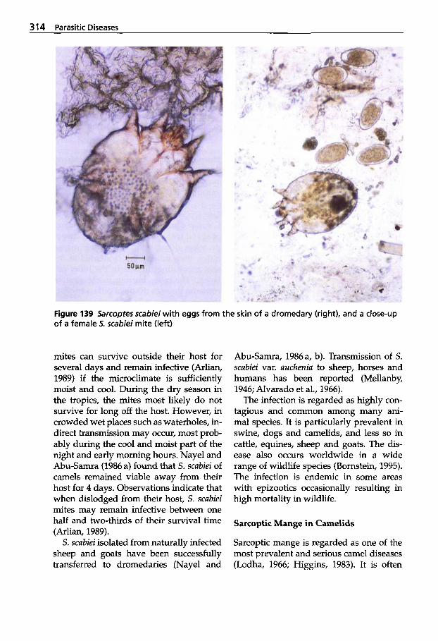
314 Parasitic Diseases
Figure 139 Sarcopres scabiei with eggs from the skin of a dromedary (right), and a close-up of a female 5. scabiei mite (left)
mites can survive outside their host for several days and remain infective (Arlian, 1989) if the microclimate is sufficiently moist and cool. During the dry season in the tropics, the mites most likely do not survive for long off the host. However, in crowded wet places such as waterholes, in- direct transmission may occur, most prob- ably during the cool and moist part of the night and early morning hours. Nayel and Abu-Samra (1986 a) found that S. scubiei of camels remained viable away from their host for 4 days. Observations indicate that when dislodged from their host, S. scubiei mites may remain infective between one half and two-thirds of their survival time (Arlian, 1989).
S. scubiei isolated from naturally infected sheep and goats have been successfully transferred to dromedaries (Nayel and
Abu-Samra, 1986a, b). Transmission of S. scubiei var. aucheniu to sheep, horses and humans has been reported (Mellanby, 1946; Alvarado et al., 1966).
The infection is regarded as highly con- tagious and common among many ani- mal species. It is particularly prevalent in swine, dogs and camelids, and less so in cattle, equines, sheep and goats. The dis- ease also occurs worldwide in a wide range of wildlife species (Bornstein, 1995). The infection is endemic in some areas with epizootics occasionally resulting in high mortality in wildlife.
Sarcoptic Mange in Camelids
Sarcoptic mange is regarded as one of the most prevalent and serious camel diseases (Lodha, 1966; Higgins, 1983). It is often
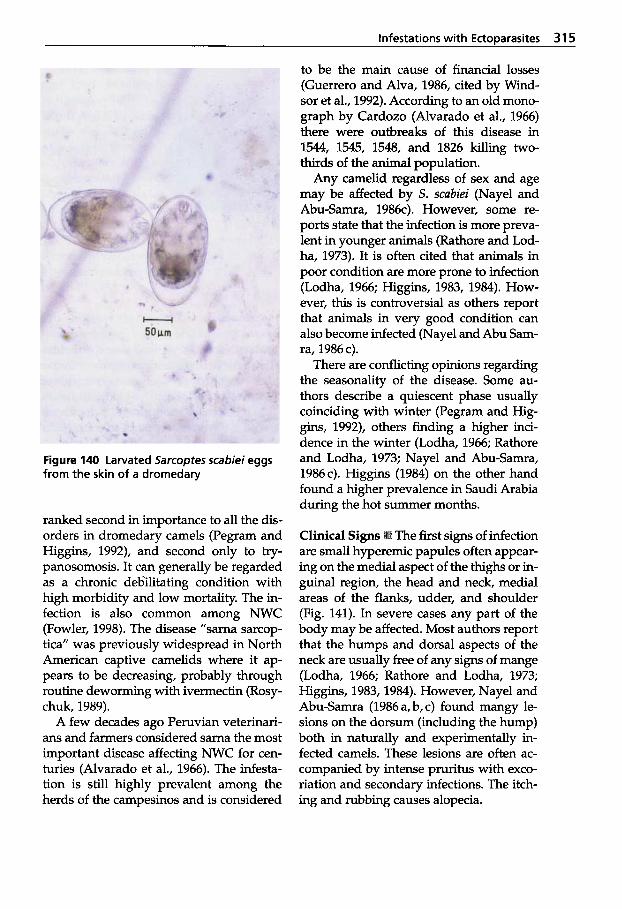
Infestations with EctoDarasites 31 5
Figure 140 Larvated Sarcoptes scabiei eggs from the skin of a dromedary
ranked second in importance to all the dis- orders in dromedary camels (Pegram and Higgins, 1992), and second only to try- panosomosis. It can generally be regarded as a chronic deb'ilitating condition with high morbidity and low mortality. The in- fection is also common among NWC (Fowler, 1998). The disease "sarna sarcop- tica" was previously widespread in North American captive camelids where it ap- pears to be decreasing, probably through routine deworming with ivermectin (Rosy- chuk, 1989).
A few decades ago Peruvian veterinari- ans and farmers considered sarna the most important disease affecting NWC for cen- turies (Alvarado et al., 1966). The infesta- tion is still highly prevalent among the herds of the campesinos and is considered
to be the main cause of financial losses (Guerrero and Alva, 1986, cited by Wind- sor et al., 1992). According to an old mono- graph by Cardozo (Alvarado et al., 1966) there were outbreaks of this disease in
thirds of the animal population. Any camelid regardless of sex and age
may be affected by S. scabiei (Nayel and Abu-Samra, 1986~). However, some re- ports state that the infection is more preva- lent in younger animals (Rathore and Lod- ha, 1973). It is often cited that animals in poor condition are more prone to infection (Lodha, 1966; Higgins, 1983, 1984). How- ever, this is controversial as others report that animals in very good condition can also become infected (Nayel and Abu Sam- ra, 1986 c).
There are conflicting opinions regarding the seasonality of the disease. Some au- thors describe a quiescent phase usually coinciding with winter (Pegram and Hig- gins, 1992), others finding a higher inci- dence in the winter (Lodha, 1966; Rathore and Lodha, 1973; Nayel and Abu-Samra, 1986~). Higgins (1984) on the other hand found a higher prevalence in Saudi Arabia during the hot summer months.
1544, 1545, 1548, and 1826 killing two-
The first signs of infection are small hyperemic papules often appear- ing on the medial aspect of the thighs or in- guinal region, the head and neck, medial areas of the flanks, udder, and shoulder (Fig. 141). In severe cases any part of the body may be affected. Most authors report that the humps and dorsal aspects of the neck are usually free of any signs of mange (Lodha, 1966; Rathore and Lodha, 1973; Higgins, 1983,1984). However, Nayel and Abu-Samra (1986 a, b, c) found mangy le- sions on the dorsum (including the hump) both in naturally and experimentally in- fected camels. These lesions are often ac- companied by intense pruritus with exco- riation and secondary infections. The itch- ing and rubbing causes alopecia.
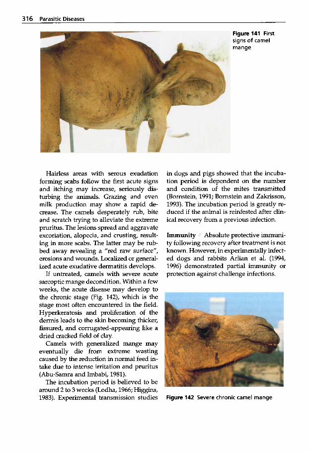
31 6 Parasitic Diseases
Figure 141 First signs of camel mange
Hairless areas with serous exudation forming scabs follow the first acute signs and itching may increase, seriously dis- turbing the animals. Grazing and even milk production may show a rapid de- crease. The camels desperately rub, bite and scratch trying to alleviate the extreme pruritus. The lesions spread and aggravate excoriation, alopecia, and crusting, result- ing in more scabs. The latter may be rub- bed away revealing a "red raw surface", erosions and wounds. Localized or general- ized acute exudative dermatitis develops.
If untreated, camels with severe acute sarcoptic mange decondition. Within a few weeks, the acute disease may develop to the chronic stage (Fig. 142), which is the stage most often encountered in the field. Hyperkeratosis and proliferation of the dermis leads to the skin becoming thicker, fissured, and corrugated-appearing like a dried cracked field of clay.
Camels with generalized mange may eventually die from extreme wasting caused by the reduction in normal feed in- take due to intense irritation and pruritus (Abu-Samra and Imbabi, 1981).
The incubation period is believed to be around 2 to 3 weeks (Lodha, 1966; Higgins, 1983). Experimental transmission studies
in dogs and pigs showed that the incuba- tion period is dependent on the number and condition of the mites transmitted (Bomstein, 1991; Bomstein and Zakrisson, 1993). The incubation period is greatly re- duced if the animal is reinfested after clin- ical recovery from a previous infection.
Immunity Absolute protective immuni- ty following recovery after treatment is not known. However, in experimentally infect- ed dogs and rabbits Arlian et al. (1994, 1996) demonstrated partial immunity or protection against challenge infections.
Figure 142 Severe chronic camel mange
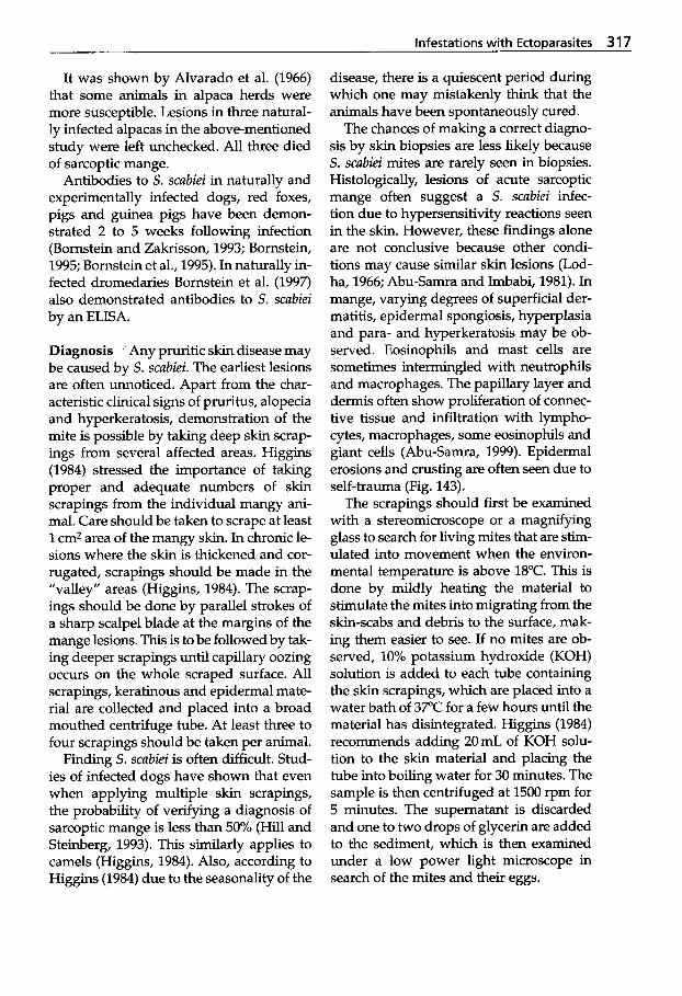
Infestations with Ectoparasites 31 7
It was shown by Alvarado et al. (1966) that some animals in alpaca herds were more susceptible. Lesions in three natural- ly infected alpacas in the above-mentioned study were left unchecked. All three died of sarcoptic mange.
Antibodies to S. scubiei in naturally and experimentally infected dogs, red foxes, pigs and guinea pigs have been demon- strated 2 to 5 weeks following infection (Bornstein and Zakrisson, 1993; Bornstein, 1995; Bornstein et al., 1995). In naturally in- fected dromedaries Bornstein et al. (1997) also demonstrated antibodies to S. scubiei by an ELISA.
Diagnosis Any pruritic skin disease may be caused by S. scubiei. The earliest lesions are often unnoticed. Apart from the char- acteristic clinical signs of pruritus, alopecia and hyperkeratosis, demonstration of the mite is possible by taking deep skin scrap- ings from several affected areas. Higgins (1984) stressed the importance of taking proper and adequate numbers of skin scrapings from the individual mangy ani- mal. Care should be taken to scrape at least 1 cm2 area of the mangy skin. In chronic le- sions where the skin is thickened and cor- rugated, scrapings should be made in the "valley" areas (Higgins, 1984). The scrap- ings should be done by parallel strokes of a sharp scalpel blade at the margins of the mange lesions. This is to be followed by tak- ing deeper scrapings until capillary oozing occurs on the whole scraped surface. All scrapings, keratinous and epidermal mate- rial are collected and placed into a broad mouthed centrifuge tube. At least three to four scrapings should be taken per animal.
Finding S. scubiei is often difficult. Stud- ies of infected dogs have shown that even when applying multiple skin scrapings, the probability of vedying a diagnosis of sarcoptic mange is less than 50% (Hill and Steinberg, 1993). This similarly applies to camels (Higgins, 1984). Also, according to Higgins (1984) due to the seasonality of the
disease, there is a quiescent period during which one may mistakenly think that the animals have been spontaneously cured.
The chances of making a correct diagno- sis by skin biopsies are less likely because S. scubiei mites are rarely seen in biopsies. Histologically, lesions of acute sarcoptic mange often suggest a S. scabiei infec- tion due to hypersensitivity reactions seen in the skin. However, these findings alone are not conclusive because other condi- tions may cause similar skin lesions (Lod- ha, 1966; Abu-Samra and Imbabi, 1981). In mange, varying degrees of superficial der- matitis, epidermal spongiosis, hyperplasia and para- and hyperkeratosis may be ob- served. Eosinophils and mast cells are sometimes intermingled with neutrophils and macrophages. The papillary layer and dermis often show proliferation of connec- tive tissue and infiltration with lympho- cytes, macrophages, some eosinophils and giant cells (Abu-Samra, 1999). Epidermal erosions and crusting are often seen due to self-trauma (Fig. 143).
The scrapings should first be examined with a stereomicroscope or a magnifying glass to search for living mites that are stim- ulated into movement when the environ- mental temperature is above 18°C. This is done by mildly heating the material to stimulate the mites into migrating from the skin-scabs and debris to the surface, mak- ing them easier to see. If no mites are ob- served, loo% potassium hydroxide (KOH) solution is added to each tube containing the skin scrapings, which are placed into a water bath of 37°C for a few hours until the material has disintegrated. Higgins (1984) recommends adding 20mL of KOH solu- tion to the skin material and placing the tube into boiling water for 30 minutes. The sample is then centrifuged at 1500 rpm for 5 minutes. The supernatant is discarded and one to two drops of glycerin are added to the sediment, which is then examined under a low power light microscope in search of the mites and their eggs.
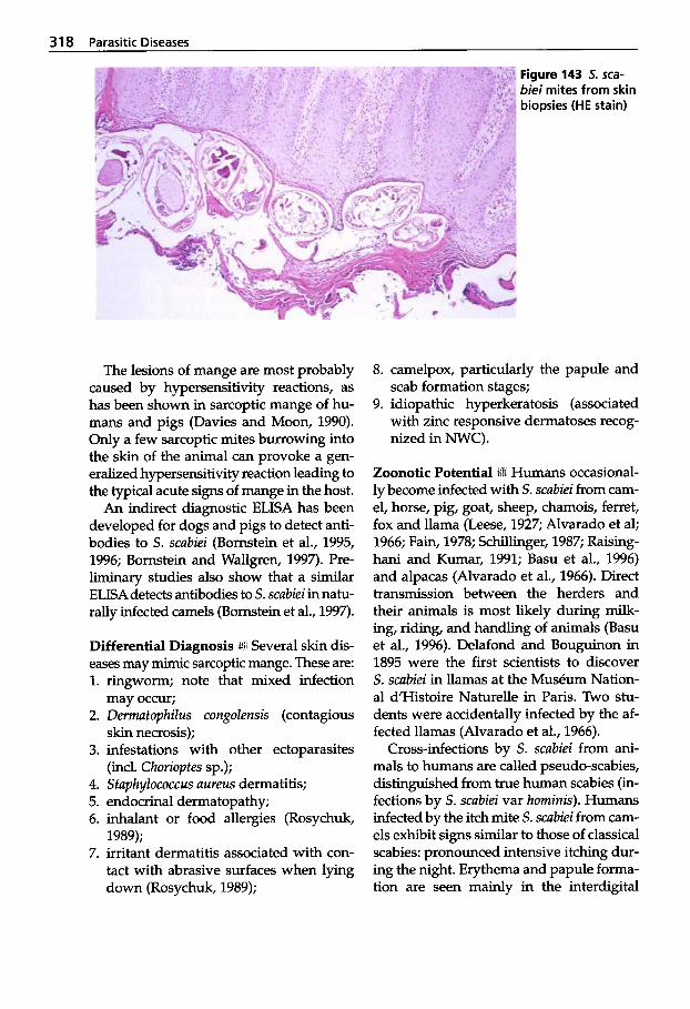
31 8 Parasitic Diseases _.~
Figure 143 5. sca- biei mites from skin biopsies (HE stain)
The lesions of mange are most probably caused by hypersensitivity reactions, as has been shown in sarcoptic mange of hu- mans and pigs (Davies and Moon, 1990). Only a few sarcoptic mites burrowing into the skin of the animal can provoke a gen- eralized hypersensitivity reaction leading to the typical acute signs of mange in the host.
An indirect diagnostic ELISA has been developed for dogs and pigs to detect anti- bodies to s. scabiei (Bornstein et al., 1995, 1996; Bornstein and Wallgren, 1997). Pre- liminary studies also show that a similar ELISA detects antibodies to S. scabiei in natu- rally infected camels (Bornstein et al., 1997).
Differential Diagnosis Several skin dis- eases may mimic sarcoptic mange. These are: 1. ringworm; note that mixed infection
2. Demzatopkilus congolensis (contagious
3. infestations with other ectoparasites
4. Staphylococcus aureus dermatitis; 5. endocrinal dermatopathy; 6. inhalant or food allergies (Rosychuk,
1989); 7. irritant dermatitis associated with con-
tact with abrasive surfaces when lying down (Rosychuk, 1989);
may occur;
skin necrosis);
(incl. Ckorioptes sp.);
8. camelpox, particularly the papule and scab formation stages;
9. idiopathic hyperkeratosis (associated with zinc responsive dermatoses recog- nized in NWC).
Zoonotic Potential iin Humans occasional- ly become infected with S. scabiei from cam- el, horse, pig, goat, sheep, chamois, ferret, fox and llama (Leese, 1927; Alvarado et al; 1966; Fain, 1978; Schillinger, 1987; Raising- hani and Kumar, 1991; Basu et al., 1996) and alpacas (Alvarado et al., 1966). Direct transmission between the herders and their animals is most likely during milk- ing, riding, and handling of animals (Basu et al., 1996). Delafond and Bouguinon in 1895 were the first scientists to discover S. scabiei in llamas at the MusCum Nation- al d’Histoire Naturelle in Paris. Two stu- dents were accidentally infected by the af- fected llamas (Alvarado et al., 1966).
Cross-infections by S. scabiei from ani- mals to humans are called pseudo-scabies, distinguished from true human scabies (in- fections by s. scabiei var horninis). Humans infected by the itch mite S. scabiei from cam- els exhibit signs similar to those of classical scabies: pronounced intensive itching dur- ing the night. Erythema and papule forma- tion are seen mainly in the interdigital
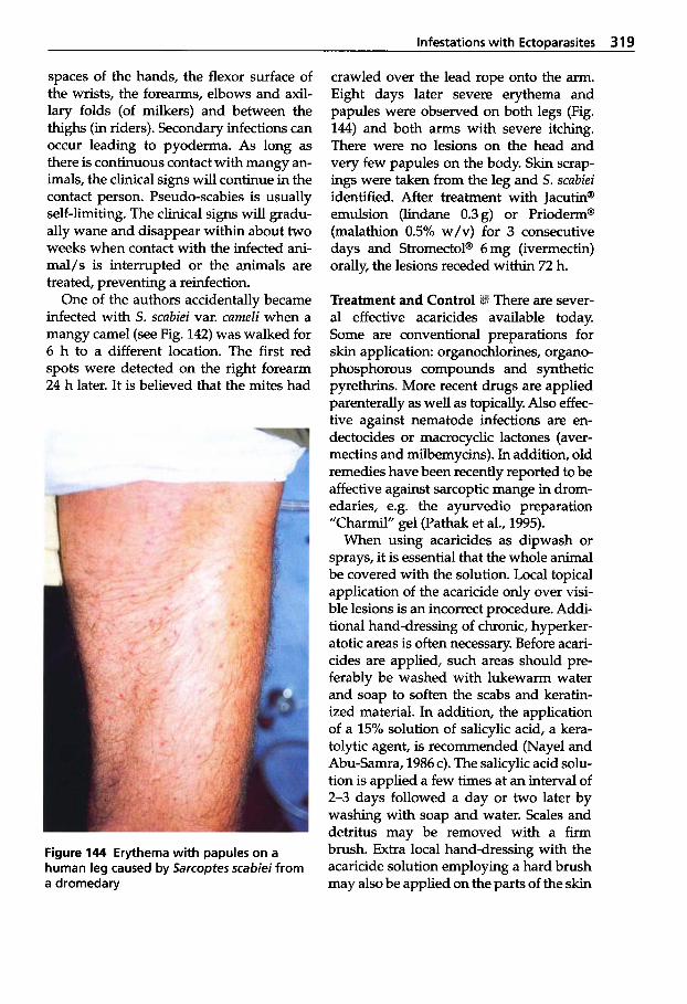
Infestations with Ectoparasites 31 9
spaces of the hands, the flexor surface of the wrists, the forearms, elbows and axil- lary folds (of milkers) and between the thighs (in riders). Secondary infections can occur leading to pyoderma. As long as there is continuous contact with mangy an- imals, the clinical signs will continue in the contact person. Pseudo-scabies is usually self-limiting. The clinical signs will gradu- ally wane and disappear within about two weeks when contact with the infected ani- mal/s is interrupted or the animals are treated, preventing a reinfection.
One of the authors accidentally became infected with S. scabiei var. cameli when a mangy camel (see Fig. 142) was walked for 6 h to a different location. The first red spots were detected on the right forearm 24 h later. It is believed that the mites had
Figure 144 Erythema with papules on a human leg caused by Sarcoptes scabiei from a dromedary
crawled over the lead rope onto the arm. Eight days later severe erythema and papules were observed on both legs (Fig. 144) and both arms with severe itching. There were no lesions on the head and very few papules on the body. Skin scrap- ings were taken from the leg and s. scabiei identified. After treatment with JacutinB emulsion (lindane 0.3g) or PriodermB (malathion 0.5% w/v) for 3 consecutive days and StromectoF 6mg (ivermectin) orally, the lesions receded within 72 h.
Treatment and Control a1 effective acaricides available today. Some are conventional preparations for skin application: organochlorines, organo- phosphorous compounds and synthetic pyrethrins. More recent drugs are applied parenterally as well as topically. Also effec- tive against nematode infections are en- dectocides or macrocyclic lactones (aver- mectins and milbemycins). In addition, old remedies have been recently reported to be affective against sarcoptic mange in drom- edaries, e.g. the ayurvedio preparation "Charmil" gel (Pathak et al., 1995).
When using acaricides as dipwash or sprays, it is essential that the whole animal be covered with the solution. Local topical application of the acaricide only over visi- ble lesions is an incorrect procedure. Addi- tional hand-dressing of chronic, hyperker- atotic areas is often necessary. Before acari- cides are applied, such areas should pre- ferably be washed with lukewarm water and soap to soften the scabs and keratin- ized material. In addition, the application of a 15% solution of salicylic acid, a kera- tolytic agent, is recommended (Nayel and Abu-Samra, 1986 c). The salicylic acid solu- tion is applied a few times at an interval of 2-3 days followed a day or two later by washing with soap and water. Scales and detritus may be removed with a firm brush. Extra local hand-dressing with the acaricide solution employing a hard brush may also be applied on the parts of the skin

320 Parasitic Diseases
particularly thickened, scabby and corru- gated (Higgins, 1983).
The animals should be treated 3 times within an interval of 7 to 10 days, but some- times 4 or more applications are needed until a cure is reached.
Nayel and Abu-Samra (1986 c) using the acaricide 0.1% hexachlorocyclohexane (Gam- matox@) on chronically infected camels found that 3 days following the first wash with GammatoxB, most of the treated cam- els (75%) became calm with reduced signs of pruritus. Two days after the second wash most of the scales had been shed, the cracks and fissures started to heal, and the edema on the legs had subsided. Six days after the second wash, there was no pruri- tus and the restless animals behaved nor- mal. Hair began to grow 5 days after the third wash.
The topical application of acaricides is very laborious and difficult to carry out under nomadic conditions, but may more easily be applied in sedentary herds.
The injectible modem endectocides or macrocyclic lactones (like ivermectin, do- ramectin) have made the treatment of sar- coptic mange much easier. Ivermectin@ has been shown to be effective and safe in Camelidae and cattle when the same dose and regime is employed (Ibrahim et al., 1981; Boyce et al., 1984; Raisinghani et al., 1989; Kumar and Yadav, 1993; Kuntze and Kuntze, 1991; Oukessou et al., 1996). The recommended dose is 200 mg/kg given sub- cutaneously and repeated after 15 days. The subcutaneous injection is painful to camelids and some diffuse swelling at the injection site may appear after 24 h. Cam- elids need to be well-restrained in the couched position. After treatment, clinical improvement is gradual. Pruritus com- pletely ceases after one week to 10 days following the second injection. Four weeks after the second injection all previously alopectic areas are covered with growing hair (Hashim and Wasfi, 1986; Raisinghani et al., 1989). Complete healing of skin le-
sions was reached on day 145 (Raisinghani et al., 1989). Unfortunately, this treatment protocol is not always successful. Depend- ing on the severity of lesions, a combina- tion of topical and injectible treatments is necessary.
New endectocides have recently reach- ed the market. Some have a longer period of bioavailability in the animal than the ivermectins. There are indications that one injection of these new drugs (e.g. mox- idectin, doramectin) may cure sarcoptic mange in pigs and cattle. If the same ap- plies to camelids, these drugs would be of great advantage to nomadic camel owners. One intramuscular injection of doramectin (Dectomax@, Pfizer, NY, USA) was enough to successfully eradicate sarcoptic mange in a herd of mangy pigs (Jacobsson et al., 1998).
In a trial on 15 camels (9 juveniles, 6 adults) showing mild to severe sarcoptic mange, doramectin was applied intramus- cularly at a dose of 200 pg/kg. Only two of the severe cases had to be treated twice. All 15 camels were cured (Mumin, pers. comm., 1999).
Abu-Samra (1999) reported even better results with 0.1% solution of phoxim (Se- b a d @ E.C., Bayer) applied topically three times, one week apart, following thorough application of 15% salicylic acid solution, resulting in complete recovery from chron- ic mange after 3 weeks.
Another promising form of endecto- cides is the pour-ons, which are poured onto the skin of the dorsal part of the body. The drug is absorbed through the skin. Both ivermectin and moxidectin are mar- keted for use as pour-ons for cattle with very good acaricidal as well as nematodici- dal properties.
5.2.3 Psoroptic Mange
Psoroptic mange mites spend their entire life on the skin, feeding superficially. They
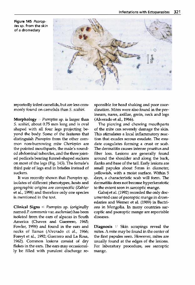
Infestations with EctoDarasites 321
Figure 145 Psorop- tes sp. from the skin of a dromedary
reportedly infest camelids, but are less com- monly found on camelids than S. scubiei.
Morphology 8 Psoroptes sp. is larger than S. scabiei, about 0.75 mm long and is oval shaped with all four legs projecting be- yond the body. Some of the features that distinguish Psoroptes from the other com- mon non-burrowing mite Chorioptes are the pointed mouthparts, the male’s round- ed abdominal tubercles, and the three joint- ed pedicels bearing funnel-shaped suckers on most of the legs (Fig. 145). The female’s third pair of legs end in bristles instead of suckers.
It was recently shown that Psoroptes sp. isolates of different phenotypes, hosts and geographic origins are conspecific (Zahler et al., 1998) and therefore only one species is mentioned in the text.
Clinical Signs i c a t Psoroptes sp. (originally named P. communis var. uucheniue) has been isolated from the ears of alpacas in South America (Chavez and Guerrero, 1965; Fowler, 1998) and found in the ears and necks of llamas (Alverado et al., 1966; Foreyt et al., 1992; Guerrero and La Rosa, 1962). Common lesions consist of dry flakes in the ears. The ears may occasional- ly be filled with purulent discharge re-
sponsible for head shaking and poor coor- dination. Mites were also found in the per- ineum, nares, axillae, groin, neck and legs (Alverado et al., 1966).
The piercing and chewing mouthparts of the mite can severely damage the skin. This stimulates a local inflammatory reac- tion that exudes serous exudate. The exu- date coagulates forming a crust or scab. The dermatitis causes intense pruritus and fiber loss. Lesions are generally found around the shoulder and along the back, flanks and base of the tail. Early lesions are small papules about 5mm in diameter, yellowish, with a moist surface. Within 5 days, a characteristic scab will form. The dermatitis does not become hyperkeratotic to the extent seen in sarcoptic mange.
Gabaj et al. (1992) recorded the only doc- umented case of psoroptic mange in drom- edaries and Werner et al. (1989) in Bactri- ans in Mongolia. In many countries sar- coptic and psoroptic mange are reportable diseases.
Diagnosis , ’ Skin scrapings reveal the mites. A mite may be found in the center of the first papules seen. However, mites are usually found at the edges of the lesions. For laboratory procedure, see sarcoptic mange.
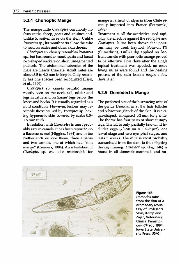
322 Parasitic Diseases
5.2.4 Chorioptic Mange
The mange mite Chorioptes commonly in- fests cattle, sheep, goats and equines and, unlike S. scubiei, lives on the skin. Unlike Psoroptes sp., its mouthparts allow the mite to feed on scales and other skin debris.
Chorioptes sp. closely resembles Psoroptes sp., but has rounder mouthparts and tarsal cup-shaped suckers on short unsegmented pedicels. The abdominal tubercles of the male are clearly truncate. Adult mites are about 3.5 to 4.0 mm in length. Only recent- ly has one species been recognized (Essig et al., 1999).
Chorioptes sp. causes pruritic mange mostly seen on the neck, tail, udder and legs in cattle and on horses’ legs below the knees and hocks. It is usually regarded as a mild condition. However, lesions may re- semble those caused by Psoroptes sp. hav- ing hyperemic skin covered by scabs 0.5- 1.5 mm thick.
Infestation with Chorioptes is most prob- ably rare in camels. It has been reported on a Bactrian camel (Higgins, 1984) and in the Netherlands on one llama, three alpacas and two camels, one of which had ”foot mange” (Cremers, 1984). An infestation of Chorioptes sp. was also responsible for
mange in a herd of alpacas from Chile re- cently imported into France (Petrowski, 1998).
All the acaricides used topi- cally are effective against the Psoroptes and Chorioptes. It has been shown that pour- ons may be used. Bayticol, Pour-on 1% (flumethrin), 1 mL/10 kg applied on Bac- trian camels with psoroptic mange proved to be effective. Five days after the single topical treatment was applied, no more living mites were found and the healing process of the skin lesions began a few days later.
5.2.5 Demodectic Mange
The preferred site of the burrowing mite of the genus Demodex is at the hair follicles and sebaceous glands of the skin. It is a ci- gar-shaped, elongated 0.2 mm long mite. The thorax has four pairs of short stumpy legs. The LC is only partially known. It in- cludes eggs (70-90pm x 19-25pm), one larval stage and two nymphal stages, and lasts 3 weeks. The mite is most probably transmitted from the dam to the offspring during nursing. Demodex sp. (Fig. 146) is found in all domestic mammals and hu-
Figure 146 Demodex mite from the skin of a dromedary (cour- tesy of Professors Sloss, Kemp and Zajac, Veterinary Clinical Parasitol- ogy, 6th ed., 1994, Iowa State Univer- sity Press, USA)
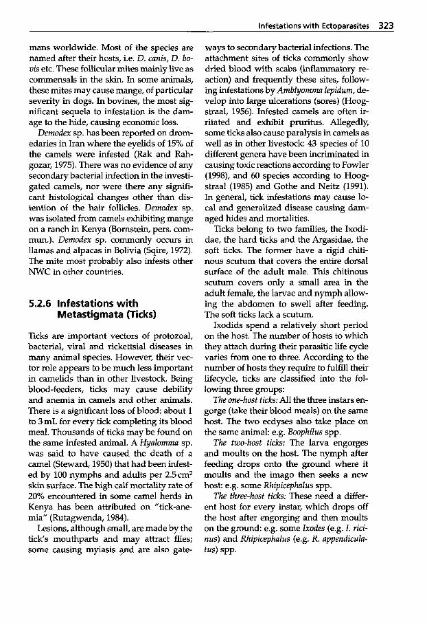
infestations with EctoDarasites 323
mans worldwide. Most of the species are named after their hosts, i.e. D. canis, D. bo- vis etc. These follicular mites mainly live as commensals in the skin. In some animals, these mites may cause mange, of particular severity in dogs. In bovines, the most sig- nificant sequela to infestation is the dam- age to the hide, causing economic loss.
Demodex sp. has been reported on drom- edaries in Iran where the eyelids of 15% of the camels were infested (Rak and Rah- gozar, 1975). There was no evidence of any secondary bacterial infection in the investi- gated camels, nor were there any signifi- cant histological changes other than dis- tention of the hair follicles. Demodex sp. was isolated from camels exhibiting mange on a ranch in Kenya (Bornstein, pers. com- mun.). Demodex sp. commonly occurs in llamas and alpacas in Bolivia (Sqire, 1972). The mite most probably also infests other NWC in other countries.
5.2.6 Infestations with Metastigmata (Ticks)
Ticks are important vectors of protozoal, bacterial, viral and rickettsia1 diseases in many animal species. However, their vec- tor role appears to be much less important in camelids than in other livestock. Being blood-feeders, ticks may cause debility and anemia in camels and other animals. There is a significant loss of blood: about 1 to 3 mL for every tick completing its blood meal. Thousands of ticks may be found on the same infested animal. A Hyalomma sp. was said to have caused the death of a camel (Steward, 1950) that had been infest- ed by 100 nymphs and adults per 2.5cm2 skin surface. The high calf mortality rate of 20% encountered in some camel herds in Kenya has been attributed on ”tick-ane- mia” (Rutagwenda, 1984).
Lesions, although small, are made by the tick’s mouthparts and may attract flies; some causing myiasis y d are also gate-
ways to secondary bacterial infections. The attachment sites of ticks commonly show dried blood with scabs (inflammatory re- action) and frequently these sites, follow- ing infestations by Amblyomma lepidum, de- velop into large ulcerations (sores) (Hoog- straal, 1956). Infested camels are often ir- ritated and exhibit pruritus. Allegedly, some ticks also cause paralysis in camels as well as in other livestock 43 species of 10 different genera have been incriminated in causing toxic reactions according to Fowler (1998), and 60 species according to Hoog- straal (1985) and Gothe and Neitz (1991). In general, tick infestations may cause lo- cal and generalized disease causing dam- aged hides and mortalities.
Ticks belong to two families, the Ixodi- dae, the hard ticks and the Argasidae, the soft ticks. The former have a rigid chiti- nous scutum that covers the entire dorsal surface of the adult male. This chitinous scutum covers only a small area in the adult female, the larvae and nymph allow- ing the abdomen to swell after feeding. The soft ticks lack a scutum.
Ixodids spend a relatively short period on the host. The number of hosts to which they attach during their parasitic life cycle varies from one to three. According to the number of hosts they require to fulfill their lifecycle, ticks are classified into the fol- lowing three groups:
The one-host ticks: All the three instars en- gorge (take their blood meals) on the same host. The two ecdyses also take place on the same animal: e.g. Boophilus spp.
The two-host ticks: The larva engorges and moults on the host. The nymph after feeding drops onto the ground where it moults and the imago then seeks a new host: e.g. some Rhipicephalus spp.
The three-host ticks: These need a differ- ent host for every instar, which drops off the host after engorging and then moults on the ground: e.g. some Ixodes (e.g. I . rici- nus) and Rhipicephalus (e.g. R. appendicula- tus) spp.
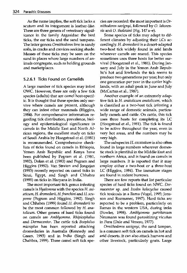
324 Parasitic Diseases
As the name implies, the soft tick lacks a scutum and its integument is leather-like. There are three genera of veterinary sigruf- icance in the family Argasidae: the bird ticks, the ear ticks and the sand tampans. The latter genera Ornithodoros live in sandy soils, in cracks and crevices seeking shade. Masses of these ticks may be seen on the sand in places where large numbers of an- imals congregate, such as holding grounds and marketplaces.
5.2.6.1 Ticks Found on Camelids
A large number of tick species may infest OWC. However, there are only a few tick species (adults) that are camel host-specif- ic. It is thought that these species only sur- vive where camels are present, although they can infest other mammals (Higgins, 1984). For comprehensive information re- garding tick distribution, prevalence, biol- ogy and epidemiological significance in camels in the Middle East and North Af- rican regions, the excellent study on ticks of Saudi Arabia by Hoogstraal et al. (1981) is recommended. Comprehensive check- lists of ticks found on camels in Ethiopia, Yemen Arab Republic and Kenya have been published by Pegram et al. (1981, 1982), Dolan et al. (1983) and Pegram and Higgins (1992). Van Straten and Jongejan (1993) recently reported on camel ticks in Sinai, Egypt, and Singh and Chhabra (1999) on ticks in Haryana in India.
The most important tick genus infesting camels is Hyalomma with the species H. asi- aticum, H. dromedarii, H.franchini and H. scu- pense (Pegram and Higgins, 1992). Singh and Chhabra (1999) found H. dromedarii to be the most common followed by H. and- tolicum. Other genera of hard ticks found on camels are Amblyomma, Rhipicephalus and Dermacentor. The cattle tick Boophilus microplus has been reported attacking dromedaries in Australia (Kennedy and Green, 1993) and in India (Singh and Chahbra, 1999). Three camel soft tick spe-
cies are recorded: the most important is Or- nithodoros savignyi, followed by 0. lahoren- sis and 0. tholozani (Fig. 147 a-e).
Some species of ticks may adapt to dif- ferent climates by adjusting their LCs ac- cordingly. H. dromedarii is a desert-adapted two-host tick widely found in arid lands wherever camels are reared. This species sometimes uses three hosts for better sur- vival (Hoogstraal et al., 1981). During Jan- uary and July in the Yemen Arab Repub- lic’s hot arid lowlands the tick seems to produce two generations per year, but only one generation per year in the cooler high- lands, with an adult peak in June and July (McCartan et al., 1987).
Another example of an extremely adap- tive tick is H. anatolicum anatolicurn, which is classified as a two-host tick infesting a wide range of domestic animals, particu- larly camels and cattle. On cattle, this tick uses three hosts for completing its LC (Hoogstraal et al., 1981). The tick is found to be active throughout the year, even in very hot areas, and the numbers may be very high.
The subspecies H. excavatum is also often found in large numbers wherever domes- tic stock is plentiful in the Middle East and northern Africa, and is found on camels in large numbers. It is reported that it may employ either a two-host or a three-host LC (Higgins, 1984). The immature stages are found in rodent burrows.
There are few reports that list particular species of hard ticks found on NWC. Der- macentor sp. and Ixodes holocyclus caused tick toxicosis in a llama (Vogel, 1995; Jons- son and Rozmanec, 1997). Hard ticks are reported to be a problem, particularly on llamas in the western USA, during treks (Fowler, 1998). Amblyomma parvitarsum Neumann was found parasitizing vicuiias in Peru (Dale and Venero, 1977).
Ornithodoros savignyi, the sand tampan, is a common soft tick on camels in hot and arid deserts. It can also attack humans and other livestock, particularly goats. Large
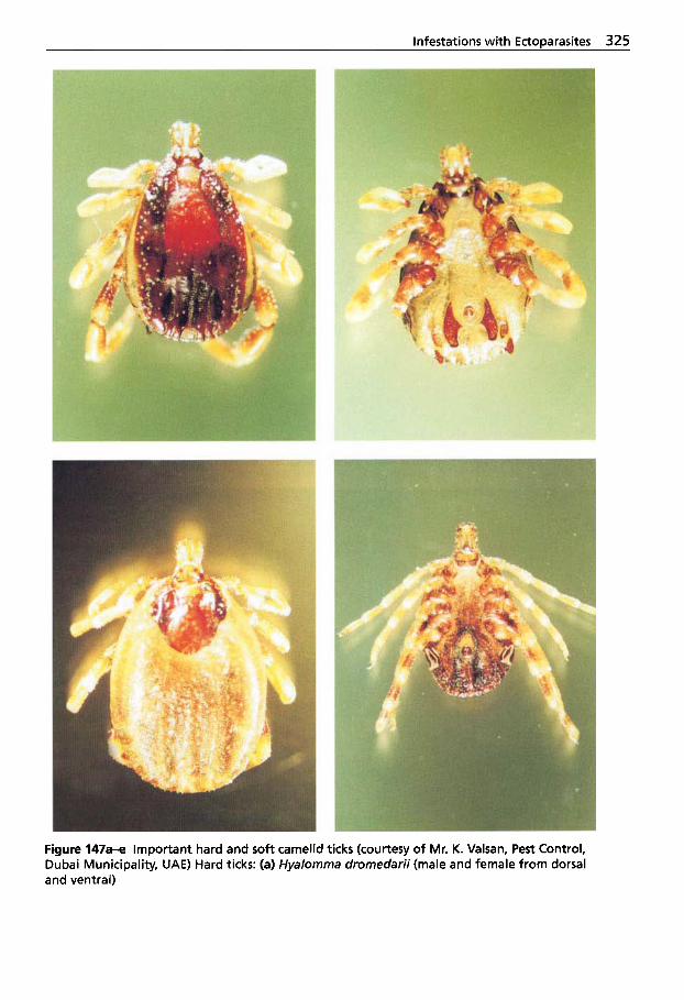
infestations with Ectoparasites 325
Figure 147- important hard and soft camelid ticks (courtesy of Mr. K. Valsan, Pest Control, Dubai Municipality, UAE) Hard ticks: (a) Hyalomma dromedarii (male and female from dorsal and ventral)
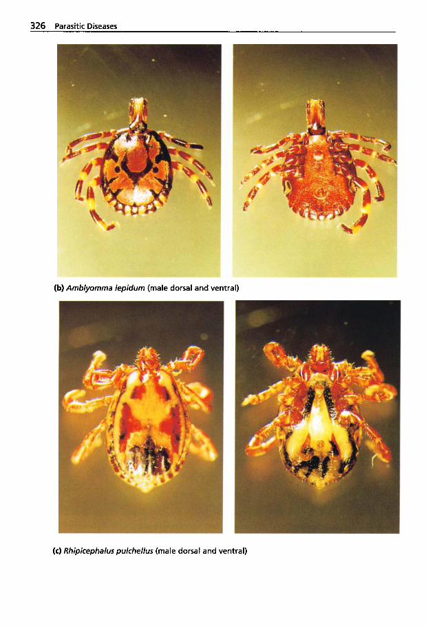
326 Parasitic Diseases
(b) Amblyomma lepidum (male dorsal and ventral)
(c) Rhipicephalus pulchellus (male dorsal and ventral)
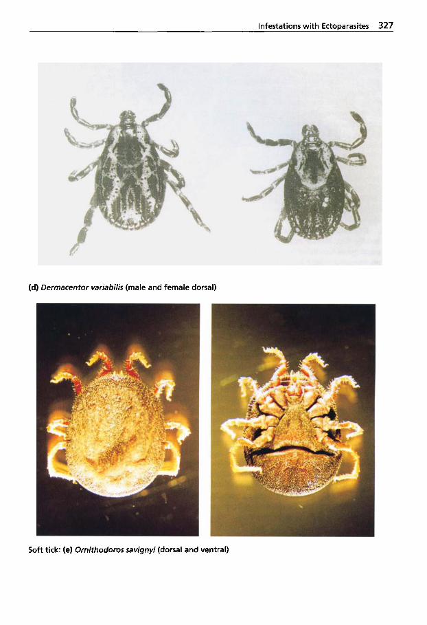
Infestations with Ectoparasites 327
(d) Dermacentor variabilis (male and female dorsal)
Soft tick: (el Ornithodoros savignyi (dorsal and ventral)
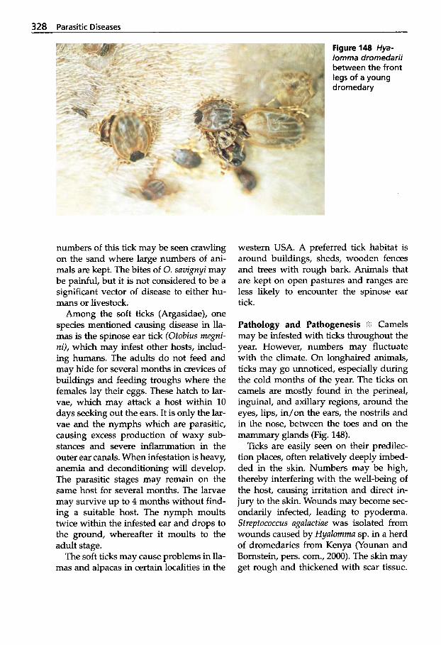
328 Parasitic Diseases
Figure 148 Hya- lomma dromedarii between the front legs of a young dromedary
numbers of this tick may be seen crawling on the sand where large numbers of ani- mals are kept. The bites of 0. suvignyi may be painful, but it is not considered to be a significant vector of disease to either hu- mans or livestock.
Among the soft ticks (Argasidae), one species mentioned causing disease in lla- mas is the spinose ear tick (Otobius megni- ni), which may infest other hosts, includ- ing humans. The adults do not feed and may hide for several months in crevices of buildings and feeding troughs where the females lay their eggs. These hatch to lar- vae, which may attack a host within 10 days seeking out the ears. It is only the lar- vae and the nymphs which are parasitic, causing excess production of waxy sub- stances and severe inflammation in the outer ear canals. When infestation is heavy, anemia and deconditioning will develop. The parasitic stages may remain on the same host for several months. The larvae may survive up to 4 months without find- ing a suitable host. The nymph moults twice within the infested ear and drops to the ground, whereafter it moults to the adult stage.
The soft ticks may cause problems in lla- mas and alpacas in certain localities in the
western USA. A preferred tick habitat is around buildings, sheds, wooden fences and trees with rough bark. Animals that are kept on open pastures and ranges are less likely to encounter the spinose ear tick.
Pathology and Pathogenesis J Camels may be infested with ticks throughout the year. However, numbers may fluctuate with the climate. On longhaired animals, ticks may go unnoticed, especially during the cold months of the year. The ticks on camels are mostly found in the perineal, inguinal, and axillary regions, around the eyes, lips, in/on the ears, the nostrils and in the nose, between the toes and on the mammary glands (Fig. 148).
Ticks are easily seen on their predilec- tion places, often relatively deeply imbed- ded in the skin. Numbers may be high, thereby interfering with the well-being of the host, causing irritation and direct in- jury to the skin. Wounds may become sec- ondarily infected, leading to pyoderma. Streptococcus uguluctiue was isolated from wounds caused by Hyulomma sp. in a herd of dromedaries from Kenya (Younan and Bornstein, pers. com., 2000). The skin may get rough and thickened with scar tissue.
Infestations with EctoDarasites 329
Sores are often seen at dermato-mucosal borders on the nose, lips and vulva. Tick bites may predispose to myiasis. It is not known whether ticks play any part in in- troducing secondary bacterial infections to the mammary glands. It is suggested that heavy tick loads contribute to reduced growth rates and calf mortality (Dolan et al., 1983). Sizable numbers of ticks may lead to anemia.
Vectors of Disease Pathogens I The im- portance of camel ticks as vectors of dis- ease pathogens for livestock has been de- scribed. There is evidence that Amblyomma lepidum or A. gemma may transmit Cowdria ruminantium (Heartwater) to cattle (Karrar et al., 1963) and that H. dromedarii is the vector of Theileria camelensis (Hoogstraal et al., 1981).
Camel ticks are also vectors of viruses infecting humans: Hyalomma anatolicum is an important vector of Crimean-Congo he- morrhagic fever (CCHF) virus, which was reported in the former USSR, Pakistan and Nigeria (Hoogstraal, 1979). This virus has also been isolated from the ticks common- ly found on camels, H. dromedarii and H. impeltaturn (see also under 2.2.10 Unusual Arboviruses) (Table 57).
Table 57 Zoonosis associated with carnel- infesting ticks (Pegrarn and Higgins, 1992)
Vector Agent H. anatolicum Thogoto virus H. excavatum Rickettsia prowazeki H. dromedarii Dhori virus
Khadam virus CCHF virus Q-fever (Coxiella burnetii)
H. impeltatum Wanowrie virus CCHF virus
H. marginatum CCHF virus H. scupense ? virus (Paralysis) H. truncatum CCHF virus
? virus (Paralysis) R. pulchellus Rickettsia prowazeki R. praetextatus Thogoto virus
5.2.6.2 Tick Paralysis
Many species of ticks have been incriminat- ed in causing tick paralysis, as distinct from tick toxicosis. The latter occurs in suscepti- ble ruminants, pigs and avians through toxins from adult ticks. Toxicosis is charac- terized by sweating, generalized hyperemia and a severe moist eczema primarily caused by Hyalomma spp. (Urquhart et al., 1996).
More than 60 of 869 known tick species are capable of causing paralysis (Hoog- straal, 1985; Gothe and Neitz, 1991). Tick paralysis occurs in OWC as well as NWC. In OWC, it is the larva of H. dromedarii that is thought to be the main cause of paraly- sis. It caused high mortality (above 24%) in calves in the Sudan (Agab and Abbas, 1998). Epidemics of suspected tick paraly- sis incriminating both Hyalomma spp. and Rhicephalus spp. have also been reported in Sudan (Musa and Osman, 1990).
Clinical Signs 8 In NWC, individual fe- males of several species of hard ticks, un- der certain unknown circumstances, pro- duce neurotoxins which are injected by the tick when it ingests a blood meal. A bite from a single tick, e.g. Dermacentor spp., may kill an animal. Studies of other animal species have shown that there is variable host susceptibility and most probably also a seasonal or annual variability. Dermacen- tor spp. were identified as causing tick paralysis in two young llamas in the USA (Barrington and Parish, 1995) and also in seven llamas and one alpaca in the USA (Cebra et al., 1996). Seven of the diseased animals showed generalized muscle flac- cidity. They recovered following treatment including removal of the ticks. The female llama recovered after 3 days after being clipped to remove all the ticks. A llama in Australia exhibiting typical signs of tick paralysis thought to be caused by lxodes holocyclus did not survive in spite of inten- sive treatment and removal of the ticks (Jonsson and Rozmanec, 1997).
330 Parasitic Diseases
According to Fowler (1998), the patho- genesis and clinical manifestation of the disease in NWC is similar to that in other animal species. Most cases of tick paralysis in NWC have been reported from non-in- digenous regions (Barrington and Parish, 1995). The tick paralysis in North America is most likely due to a salivary neurotoxin, which is thought to act on the end plates of the motor neurons, preventing acetylcho- line release into the synapses of the neuro- muscular junctions (Gothe et al., 1979).
Signs are usually not apparent until 5 to 7 days after the tick has begun to feed. Ac- cording to Musa and Osman (1990), the clinical signs may appear earlier and the first deaths may already occur 3 days fol- lowing the tick invasion. The first signs of paresis and paralysis are seen in the hind- quarters, and they progressively increase in severity as they move toward the crani- um. The ability to rise is lost in 12 to 36 hours. Loss of all motor functions occurs, preceded by ataxia. Stretch reflexes are also impaired and pain perception re- mains. The signs may develop rapidly within a few hours or may take 24 to 48 hours until the victim dies of respiratory arrest from involvement of the respiratory centers in the brain.
Diagnosis $9 Diagnosis is based on clinical signs and the finding of ticks known to cause paralysis. Analysis of cerebrospinal fluid may help in distinguishing tick paral- ysis from other causes of paralytic diseas- es. There is a strong indication that the di- agnosis of tick paralysis is correct if the pa- tient recovers rapidly (within a few days) following removal of the ticks.
5.2.6.3 Tick Control
Chemical Control '13 Routine prophylactic tick control is not practiced in camelids as in cattle. However, control of significant numbers of ticks attacking camels is rec- ommended. This can be done by applying
appropriate acaricides to the predilection sites (chlorinated hydrocarbons, organo- phosphates, carbamates, synthetic pyre- throids or the macrocyclic lactones), as used for cattle. It should be noted whether the ticks in the area have developed resist- ance to a particular acaricide. A 1% flume- thrin (BayticoP Bayer) pour-on formula- tion was successfully used (El-Azazy, 1996) in controlling H. dromedarii infestations on camels. The drug (1 to 2mL/10kg) was poured from the shoulder along the mid- dle of the back over the hump to the tail.
Subcutaneous injections of ivermectin (10mg/50 kg) are effective in controlling both larvae and nymphs of the spinose ear tick (Fowler, 1998). The ear canals may be cleaned manually and solutions of insecti- cides or acaricides instilled.
Reinfestation can occur because of the difficulty of eradicating the ticks from the environment. Regular inspection of the outer ear canals followed by treatment is recommended to avoid a build-up of infes- tation. The recently available endectocides possess an extended period of bioavailabil- ity but their pharmacokinetics are un- known in camelids.
There is no effective treatment that can neutralize the tick paralysis toxin. How- ever, Ixodes hoZocyclus canine hyperimmune serum is used in affected small animals and calves (cattle) at a dose rate of 0.5 mL/ kg (Jonsson and Rozmanec, 1997) curing about 75% of cases. This hyperimmune serum was used without success in tick paralysis of a llama caused by I. holocycZus.
Vaccination ii?l The hosts of hematophagous arthropods may stimulate immune defens- es that react with tissues and saliva of the parasite. This can disrupt blood meal acqui- sition, impair physiological responses and/ or kill the arthropod (Wikel, 1982,1996).
Since Trager (1939) showed that guinea pigs immunized with whole larval extract of Dermacentor variabilis were resistant to the challenge of the larvae, numerous in-
Infestations with EctoDarasites 331
vestigators have been trying to develop anti-tick vaccines. Today there are two types of tick vaccines available for cattle. One crude vaccine is made from extracts of the partly engorged adult female B. mi- croplus. Antibodies produced in the host destroy the cells lining the tick's gut and blood escapes into the hemocele. A certain percentage of the ticks die and the fertility of those remaining may be reduced by up to 70% (Willadsen et al., 1989). The fertility of males is also affected. Recombinant vac- cines have also been developed and are commercially available. However, these vaccines have a limited application and have not yet been developed for Cumelidue.
5.2.7 Insects Found on Camelids
5.2.7.1 Classification of Insects
Among the class Insectea there are several orders of particular veterinary interest: the Anoplurida (sucking lice), the Mallophagi- da (biting lice), the Siphonapterida (fleas), and the Dipterida (flies).
Phylum Arthropoda Class Insectea
Order Anoplurida (Sucking lice) Microthorucius cumeli (OWC) M. muzzui (NWC) M. minor (NWC) M. pruelongiceps (NWC)
Order Mallophagida (Biting lice) Dumuliniu breviceps (NWC)
Order Siphonapterida (Fleas) Vermipsyllu spp. (OWC, NWC)
Order Dipterida (Flies) Suborder Brachycerina Family Sarcophagidae (Flesh flies)
Wohlfuhrtiu mugnificu (OWC) Wohlfuhrtiu nubu (OWC) Sarcophugu dux (OWC)
Family Calliphoridae (Blowflies) Luciliu cuprinu (OWC) Chrysomyu bezziunu (OWC) Culliphoru spp. (NWC) CochZiomyia hominivorux (OWC,
Phormia spp. (NWC) NWC)
Family Oestridae (Bot flies) Cephulopinu titillator (OWC) Oestrus ovis (OWC, NWC) Cephenomyiu spp. (NWC)
Family Muscidae (Flies) Muscu domesticu (OWC, NWC) M . autumnalis (OWC, NWC) Stomoxys culcitruns (OWC, NWC) Hydrotea spp. Huemutobiu spp.
Family Glossinidae (Tsetse flies) Glossinu spp.
Family Tabanidae (Horse flies) Tubanus spp. (OWC) Huemutopotu spp. (OWC) Chrysops spp.(OWC)
Suborder Nematocera Family Ceratopogonidae (Midges)
Culicoides spp.
Phylum Pentastomida Linguutulu serrata
5.2.7.2 Infestation with Lice
There are two orders: the Anoplurida, the sucking lice, and Mallophagida, the biting lice. The latter have not yet been reported on OWC. Llamas may suffer from both bit- ing and sucking lice and both may be found on the same individual. Biting lice have a blunt broad head that is distinctly different from the elongated mouthparts of the sucking lice (Fig. 149).
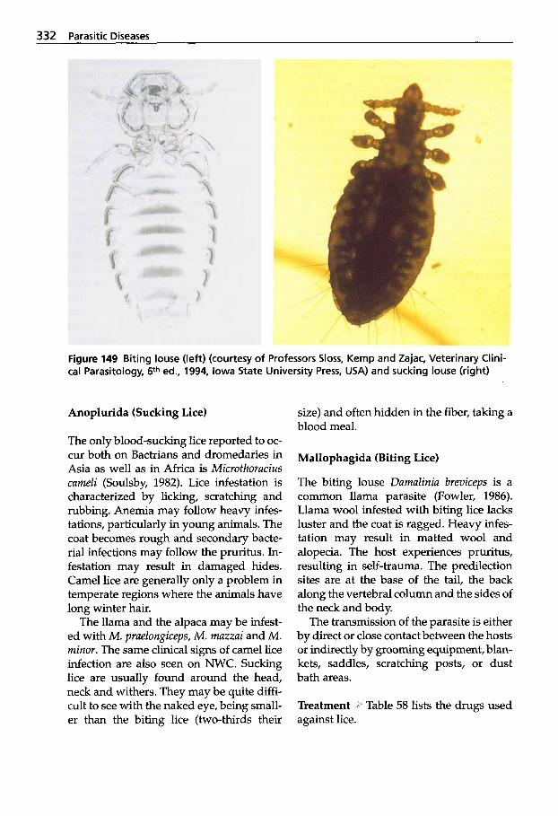
332 Parasitic Diseases
Figure 149 Biting louse (left) (courtesy of Professors Sloss, Kemp and Zajac, Veterinary Clini- cal Parasitology, 6th ed., 1994, Iowa State University Press, USA) and sucking louse (right)
Anoplurida (Sucking Lice)
The only blood-sucking lice reported to oc- cur both on Bactrians and dromedaries in Asia as well as in Africa is Microthoracius cameli (Soulsby, 1982). Lice infestation is characterized by licking, scratching and rubbing. Anemia may follow heavy infes- tations, particularly in young animals. The coat becomes rough and secondary bacte- rial infections may follow the pruritus. In- festation may result in damaged hides. Camel lice are generally only a problem in temperate regions where the animals have long winter hair.
The llama and the alpaca may be infest- ed with M . praelongiceps, M . mazzai and M . minor. The same clinical signs of camel lice infection are also seen on NWC. Sucking lice are usually found around the head, neck and withers. They may be quite diffi- cult to see with the naked eye, being small- er than the biting lice (two-thirds their
size) and often hidden in the fiber, taking a blood meal.
Mallophagida (Biting Lice)
The biting louse Damalinia breviceps is a common llama parasite (Fowler, 1986). Llama wool infested with biting lice lacks luster and the coat is ragged. Heavy infes- tation may result in matted wool and alopecia. The host experiences pruritus, resulting in self-trauma. The predilection sites are at the base of the tail, the back along the vertebral column and the sides of the neck and body.
The transmission of the parasite is either by direct or close contact between the hosts or indirectly by grooming equipment, blan- kets, saddles, scratching posts, or dust bath areas.
Treatment ig Table 58 lists the drugs used against lice.
Infestations with EctoDarasites 333
Table 58 Treatment against lice in carnelids
Trade Name Generic Name Formulation Application AsuntoP Cournaphos 50% wettable powder Apply at a concentration of
0.05% (500 pprn) directly onto skin, wet hair coat thoroughly
Methoxychlor Dusting powder 50% directly onto skin Severin@ Severin Dusting powder As above
Ivornec@* lverrnectin 0.5 rna/ka Pour on Ivornec@* lverrnectin Inj. 0.2 rng/kg S.C.
* Internationally registration approval for lvornec injectable exists for Sarcoptes scabiei var. cameli only. Reports are available for sucking lice and endoparasites. Withdrawal period is 28 days for meat. Camels producing milk for human consumption should not be treated. lvornex Pour-on is used in carnelids (as field reports say successfully) without market au- thorization.
5.2.7.3 Infestation with Siphonapterida (Fleas)
Vermipsyllidae
Fleas (Vermipsylla alacurt, V ioffi), like lice, infest camelids in cooler countries (Zedev, 1976). They have also been reported affect- ing Bactrian camels in zoos (Pegram and Higgins, 1992) and llamas (Fowler, 1998). In addition, several other species may at- tack camelids, as is the case of Ctenocephu- Iides felis fezis (Yeruham et al., 1997). Al- though fleas are important vectors of in- fectious pathogens, such as Typhus-like rickettsia, Yersiniu pestis, and as an interme- diate host for filarids and cestodes, no in- stances of pathogen transmission by these insects have been reported in camelids.
Treatment lice.
Treatment is the same as for
5.2.7.4 Infestation with Flies
Some Dipteridu as adults are external para- sites, while some parasitize the tissues of the hosts as larvae, causing myiasis. Many members of this order are also important vectors of pathogens. The order is divided into two suborders: Brachycerina, Nema- tocerina (Table 59).
Myiasis
Myiasis is defined as the invasion of living mammalian tissue by larvae of dipterous flies (Urquhart et al., 1996) that, at least during a certain period of their life, feed on dead or living tissue. There are larvae of six fly species known to cause myiasis in camels. Five of these belong to the blow- flies (CalZiphoridae) and one to the Oestridae (Zumpt, 1965; Higgins, 1986). Obligate para- sites such as Chrysomya bezziunu and facul- tative parasites such as Lucilia cuprina may also cause myiasis. Musca domestica can also cause myiasis. Myiasis may be cuta- neous (e.g. caused by Lucilia spp.), nasal (e.g. caused by Oestrus), or somatic (e.g. caused by Hypodermu spp.).
Sarcophagidae Producing Myiasis (Flesh Flies) Wohlfahrtiu magnifica, Wohlfahrtiu nuba, Surcophugu dux As an obligate parasite, WohZjiuhrtiu mag- nifica is the most important fly causing myiasis in camels. It occurs in the Mediter- ranean basin (James, 1947; Hadani et al., 1971), southern Russia, Turkey, Iran, the Far East (James, 1947), Spain (Ruiz-Mar- tinez et al., 1987), and Mongolia (Yasuda, 1940; Valentin et al., 1997) (Fig. 150). The female fly deposits larvae near any skin
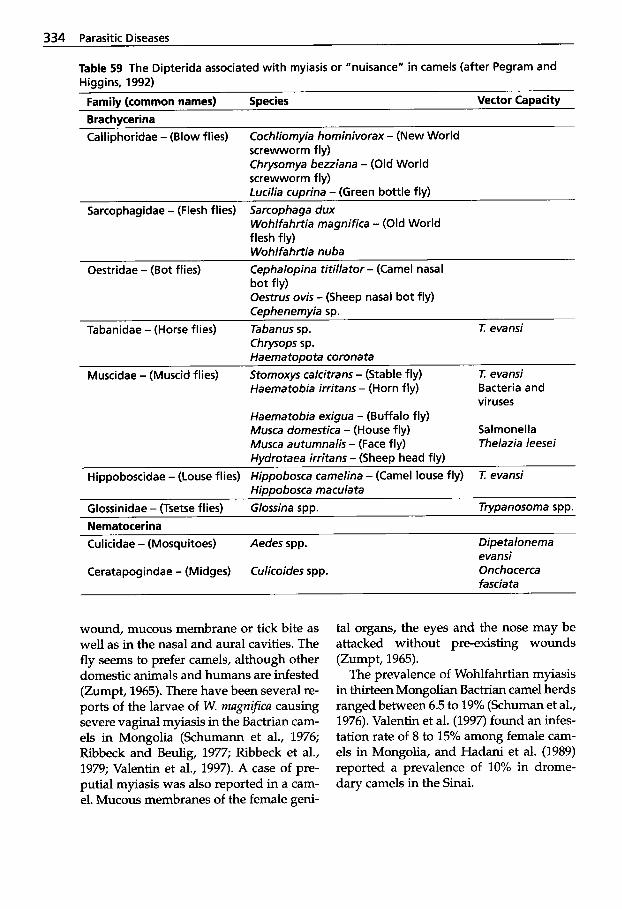
334 Parasitic Diseases
Table 59 The Dipterida associated with myiasis or "nuisance" in camels (after Pegram and Higgins, 1992)
Family (common names) Species Vector Capacity
Brachycerina Calliphoridae - (Blow flies) Cochliomyia hominivorax - (New World
screwworm fly) Chrysomya bezziana - (Old World screwworm fly) Lucilia cuprina - (Green bottle fly) Sarcophaga dux Wohlfahrtia magnifica - (Old World flesh fly) Wohlfahrtia nuba Cephalopina titillator - (Camel nasal bot fly) Oestrus ovis - (Sheep nasal bot fly) Cephenemyia sp. Tabanus sp. Chrysops sp. Haematopota coronata Stomoxys calcitrans - (Stable fly) Haematobia irritans - (Horn fly)
Haematobia exigua - (Buffalo fly) Musca domestica - (House fly) Musca autumnalis - (Face fly) Hydrotaea irritans - (Sheep head fly)
Hippoboscidae - (Louse flies) Hippobosca camelina - (Camel louse fly) Himobosca maculata
Sarcophagidae - (Flesh flies)
Oestridae - (Bot flies)
T: evansi Tabanidae - (Horse flies)
Muscidae - (Muscid flies) T: evansi Bacteria and viruses
Salmonella Thelazia leesei
T: evansi
~
Glossinidae - (Tsetse flies) Glossina spp. Trypanosoma spp.
Nematocerina Culicidae - (Mosquitoes) Aedes spp. Dipetalonema
Ceratapogindae - (Midges) Culicoides spp. Onchocerca evansi
fascia ta
wound, mucous membrane or tick bite as well as in the nasal and aural cavities. The fly seems to prefer camels, although other domestic animals and humans are infested (Zumpt, 1965). There have been several re- ports of the larvae of W. mugnifcu causing severe vaginal myiasis in the Bactrian cam- els in Mongolia (Schumann et al., 1976; Ribbeck and Beulig, 1977; Ribbeck et al., 1979; Valentin et al., 1997). A case of pre- putial myiasis was also reported in a cam- el. Mucous membranes of the female geni-
tal organs, the eyes and the nose may be attacked without pre-existing wounds (Zumpt, 1965).
The prevalence of Wohlfahrtian myiasis in thirteen Mongolian Bactrian camel herds ranged between 6.5 to 19% (Schuman et al., 1976). Valentin et al. (1997) found an infes- tation rate of 8 to 15% among female cam- els in Mongolia, and Hadani et al. (1989) reported a prevalence of 10% in drome- dary camels in the Sinai.
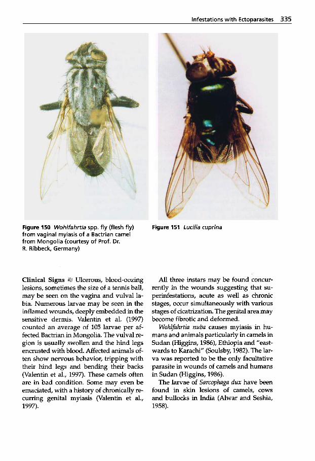
Infestations with EctoDarasites 335
Figure 150 Wohlfahrtia spp. fly (flesh fly) from vaginal myiasis of a Bactrian camel from Mongolia (courtesy of Prof. Dr. R. Ribbeck, Germany)
Figure 151 Lucilia cuprina
Ulcerous, blood-oozing lesions, sometimes the size of a tennis ball, may be seen on the vagina and vulval la- bia. Numerous larvae may be seen in the inflamed wounds, deeply embedded in the sensitive dermis. Valentin et al. (1997) counted an average of 105 larvae per af- fected Bactrian in Mongolia. The vulval re- gion is usually swollen and the hind legs encrusted with blood. Affected animals of- ten show nervous behavior, tripping with their hind legs and bending their backs (Valentin et al., 1997). These camels often are in bad condition. Some may even be emaciated, with a history of chronically re- curring genital myiasis (Valentin et al., 1997).
All three instars may be found concur- rently in the wounds suggesting that su- perinfestations, acute as well as chronic stages, occur simultaneously with various stages of cicatrization. The genital area may become fibrotic and deformed.
Wohlfahrtia nuba causes myiasis in hu- mans and animals particularly in camels in Sudan (Higgins, 1986), Ethiopia and "east- wards to Karachi" (Soulsby, 1982). The lar- va was reported to be the only facultative parasite in wounds of camels and humans in Sudan (Higgins, 1986).
The larvae of Sarcophaga dux have been found in skin lesions of camels, cows and bullocks in India (Alwar and Seshia, 1958).
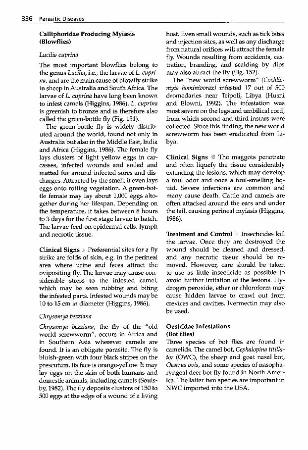
336 Parasitic Diseases
Calliphoridae Producing Myiasis (Blowflies)
Luciliu cuprinu The most important blowflies belong to the genus Luciliu, i.e., the larvae of L. cupri- nu, and are the main cause of blowfly strike in sheep in Australia and South Africa. The larvae of L. cuprinu have long been known to infest camels (Higgins, 1986). L. cuprinu is greenish to bronze and is therefore also called the green-bottle fly (Fig. 151).
The green-bottle fly is widely distrib- uted around the world, found not only in Australia but also in the Middle East, India and Africa (Higgins, 1986). The female fly lays clusters of light yellow eggs in car- casses, infected wounds and soiled and matted fur around infected sores and dis- charges. Attracted by the smell, it even lays eggs onto rotting vegetation. A green-bot- tle female may lay about 1,000 eggs alto- gether during her lifespan. Depending on the temperature, it takes between 8 hours to 3 days for the first stage larvae to hatch. The larvae feed on epidermal cells, lymph and necrotic tissue.
Clinical Signs -‘ Preferential sites for a fly strike are folds of skin, e.g. in the perineal area where urine and feces attract the ovipositing fly. The larvae may cause con- siderable stress to the infested camel, which may be seen rubbing and biting the infested parts. Infested wounds may be 10 to 15 cm in diameter (Higgins, 1986).
Ch ysornyu bezziunu Chrysornyu bezziunu, the fly of the “old world screwworm”, occurs in Africa and in Southern Asia wherever camels are found. It is an obligate parasite. The fly is bluish-green with four black stripes on the prescutum. Its face is orange-yellow. It may lay eggs on the skin of both humans and domestic animals, including camels (Souls- by, 1982). The fly deposits clusters of 150 to 500 eggs at the edge of a wound of a living
host. Even small wounds, such as tick bites and injection sites, as well as any discharge from natural orifices will attract the female fly. Wounds resulting from accidents, cas- tration, branding, and scalding by dips may also attract the fly (Fig. 152).
The “new world screwworm” (Cochlio- myiu horninivorax) infested 17 out of 500 dromedaries near Tripoli, Libya (Husni and Elowni, 1992). The infestation was most severe on the legs and umbilical cord, from which second and third instars were collected. Since this finding, the new world screwworm has been eradicated from Li- bya.
Clinical Signs -/i The maggots penetrate and often liquefy the tissue considerably extending the lesions, which may develop a foul odor and ooze a foul-smelling liq- uid. Severe infections are common and many cause death. Cattle and camels are often attacked around the ears and under the tail, causing perineal myiasis (Higgins, 1986).
Treatment and Control Insecticides kill the larvae. Once they are destroyed the wound should be cleaned and dressed, and any necrotic tissue should be re- moved. However, care should be taken to use as little insecticide as possible to avoid further irritation of the lesions. Hy- drogen peroxide, ether or chloroform may cause hidden larvae to crawl out from crevices and cavities. Ivermectin may also be used.
Oestridae Infestations (Bot flies) Three species of bot flies are found in camelids. The camel bot, Cephulopinu titilla- tor (OWC), the sheep and goat nasal bot, Oestrus ovis, and some species of nasopha- ryngeal deer bot fly found in North Amer- ica. The latter two species are important in NWC imported into the USA.
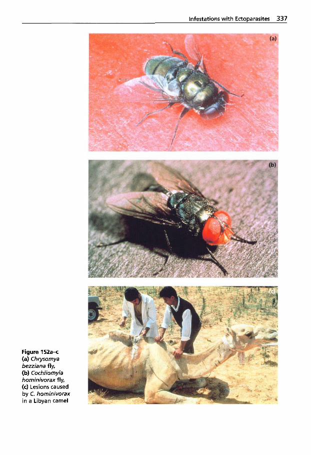
Infestations with EctoDarasites 337
Figure 152a-c (a) Chrysomya bezziana fly, (b) Cochliomyia hominivorax fly, (c) Lesions caused by C. hominivorax in a Libyan camel
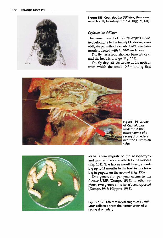
338 Parasitic Diseases
Figure 153 Cephalopina titillator, the camel nasal bo t f l y (courtesy of Dr. A. Higgins, UK)
Cephalopina titillator The camel nasal bot fly Cephalopina titilla- tor, belonging to the family Oestridae, is an obligate parasite of camels. OWC are com- monly infected with C. titillator larvae.
The fly has a reddish, dark brown thorax and the head is orange (Fig. 153).
The fly deposits its larvae in the nostrils from which the small, 0.7mm-long first
Figure 154 Larvae of Cephalopina titil lator in the nasopharynx of a racing dromedary near the Eustachian tube
stage larvae migrate to the nasopharynx and nasal sinuses and attach to the mucosa (Fig. 154). The larvae moult twice, spend- ing up to 11 months in the host before leav- ing to pupate on the ground (Fig. 155).
One generation per year occurs in the former USSR (Zumpt, 1965). In other re- gions, two generations have been reported (Zumpt, 1965; Higgins, 1986).
Figure 155 Different larval stages o f C. titi/- lator collected from the nasopharynx of a racing dromedary
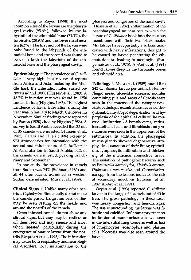
infestations with Ectoparasites 339
According to Zayed (1998) the most common sites of the larvae are the pharyn- geal cavity (95.6%), followed by the la- byrinth of the ethmoidal bone (71.1y0), the turbinates (28.9%) and the lower nasal mea- tus (6.7%). The first molt of the larvae were only found in the labyrinth of the eth- moidal bone and the second was found to occur in both the labyrinth of the eth- moidal bone and the pharyngeal cavity.
Epidemiology 1% The prevalence of C. titil- lator is very high. In a review of reports from Africa and Asia, including the Mid- dle East, the infestation rates varied be- tween 47 and 100% (Hussein et al., 1983). A 46.7% infestation rate was found in 1250 camels in Iraq (Higgins, 1986). The highest incidence of larval infestation during the year was in January to March, the lowest in November. Similar findings were reported by Patton (1920) cited by Higgins (1986). A survey in Saudi Arabia revealed that 32 out of 35 camels were infested (Hussein et al., 1982). Fatani and Hilali (1994) examined 923 dromedaries for infestation with the second and third instars of C. titillator at Al-Asha abattoir in Saudi Arabia; 52% of the camels were infested, peaking in Feb- ruary and September.
In one study, the prevalence in camels from Sudan was 74% ( S w a n , 1965) and all 44 dromedaries examined in western Sudan were infested (Musa et al., 1989).
Clinical Signs * I Unlike many other oes- trids, Cephalopina flies usually do not make the camels panic. Large numbers of flies may be seen resting on the heads and around the nostrils of the camels.
Often infested camels do not show any clinical signs, but they may be restless or off their feed and may sneeze and snort when infested, particularly during the emergence of mature larvae from the nos- trils (Urquhart et al., 1996). The infestation may cause both respiratory and neurologi- cal disorders, local inflammation of the
pharynx and congestion of the nasal cavity (Hussein et al., 1982). Inflammation of the nasopharyngeal mucosa occurs when the larvae of C. titillator hook into the mucous membranes with their two black hooks. Mortalities have reportedly also been asso- ciated with heavy infestations, thought to be caused by larvae penetrating the eth- moturbinates leading to meningitis (Bur- gemeister et al., 1975). Al-Ani et al. (1991) found larvae deep in the turbinate bones and ethmoid area.
Pathology Musa et al. (1989) found 8 to 243 C. titillator larvae per animal. Hemor- rhagic areas, ulcer-like erosions, nodules containing pus and areas of fibrosis were seen in the mucosa of the nasopharynx. Histopathologic examinations revealed des- quamation, hydropic degeneration and hy- perplasia of the epithelial cells of the mu- cosa. Infiltration of lymphocytes, reticu- loendothelial cells and fibroblasts and gra- nulomas were seen in the upper part of the submucosa. In addition, the pharyngeal mucus glands showed degenerative atro- phy, desquamation of their lining epitheli- um, lymphocytic infiltration and thicken- ing of the interacinar connective tissue. The isolation of pathogenic bacteria such as Pasteurella haemolytica, Klebsiella ozaenae, Diplococcus pneumoniae and Co ynebacteri- urn spp. from the lesions indicates the risk of secondary infections (Hussein et al., 1982; Al-Ani et al., 1991).
Oryan et al. (1993) reported C. titillator larvae in the lungs of 4 camels out of 40 in Iran. The gross pathology in these cases was heavy congestion and hemorrhages. The tissue surrounding the larvae was fi- brotic and calcified. Inflammatory reaction infiltration of mononuclear cells was seen in the interstitial lung tissue as well as foci of lymphocytes, eosinophils and plasma cells. Necrosis was also seen around the larvae.
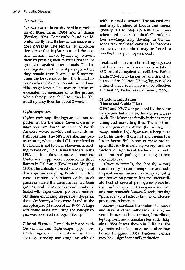
340 Parasitic Diseases
Oestrus ovis
Oestrus ovis has been observed in camels in Egypt (Kaufmann, 1996) and in llamas (Fowler, 1998). Commonly found world- wide, the fly and its larvae are sheep and goat parasites. The female fly produces live larvae that it places around the nos- trils. Llamas attacked by flies try to avoid them by pressing their muzzles close to the ground or against other animals. The lar- vae migrate into the nasal passages where they remain from 2 weeks to 9 months. Then the larvae move into the frontal si- nuses where they develop into second and third stage larvae. The mature larvae are evacuated by sneezing onto the ground where they pupate for 3 to 9 weeks. The adult fly only lives for about 2 weeks.
Cephenemyia spp. Cephenernyia spp. findings are seldom re- ported in the literature. Several Cephene- myia spp. are found in areas of North America where cervids and camelids co- habit pastures. The NWC are aberrant par- asite hosts; whether the LC is completed in the llamas is not known. However, accord- ing to Fowler (1998), llama breeders in the USA consider these parasites important. Cephenemyia spp. were reported in three llamas in California (Fowler and Murphy, 1985). The animals showed sneezing, nasal discharge and coughing. White-tailed deer were common co-habitants of livestock pastures where the three llamas had been grazing, and these deer are commonly in- fected with Cephenernyia spp. In a %month- old llama exhibiting inspiratory dyspnea, three Cephenemyia bots were found in the nasopharynx (Mattoon et al., 1997). A large soft tissue mass occluding the nasophar- ynx was observed radiographically.
Clinical Signs - Camelids infested with Oestrus ovis and Cephenemyia spp. show similar signs, such as restlessness, head shaking, sneezing and coughing with or
without nasal discharge. The affected ani- mal may be short of breath and conse- quently fail to keep up with the others when used as a pack animal. Granuloma- tous swellings may develop in the na- sopharynx and nasal cavities. If it becomes obstructive, the animal may be forced to breathe through an open mouth.
Treatment Ivermectin (0.2 mg/kg, s.c.) has been used with some success (about 85% effective against C. titillator). Rafox- anide (7.5-10 mg/kg per 0s) as a drench or bolus and trichlorfon (75 mg/kg, per 0s) as a drench have been shown to be effective, eliminating the larvae (Kaufmann, 1996).
Muscidae Infestation (House and Stable Flies) OWC and NWC are pestered by the same fly species that irritate other domestic live- stock. The Muscidae family includes many biting and non-biting flies. The most im- portant genera are Musca (house fly), Sto- moxys (stable fly), Hydrotaea (sheep-head fly), Haematobia (horn fly) and Fannia (the lesser house fly). Many of these are re- sponsible for livestock “fly-worry” and are vectors of significant bacterial, helminth and protozoal pathogens causing disease (see Table 59).
Musca auturnnalis, the face fly, a very common fly in some temperate and sub- tropical areas, causes fly-worry to cattle and horses on pasture. It is the intermedi- ate host of several pathogenic parasites, e.g. Thelazia spp. and Paraflaria bovicola, and may transmit Moraxella bovis, causing “pink eye” or infectious bovine keratocon- junctivitis in bovines.
Stomoxys calcitrans is a vector of T. evansi and several other pathogens causing se- vere diseases such as anthrax, brucellosis, leptospirosis and vesicular stomatitis (Hig- gins, 1986). It was shown in India that the fly preferred to feed on camels rather than horses (Higgins, 1986). Pestered camels may have significant milk reduction.
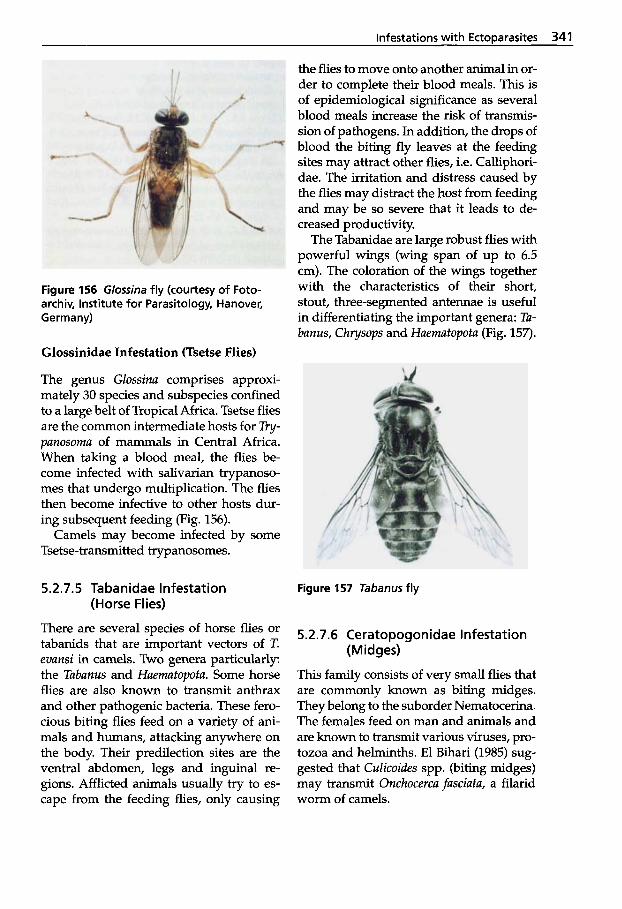
Infestations with Ectoparasites 341
Figure 156 Glossina fly (courtesy of Foto- archiv, Institute for Parasitology, Hanover, Germany)
Glossinidae Infestation (Tsetse Flies)
The genus Glossina comprises approxi- mately 30 species and subspecies confined to a large belt of Tropical Africa. Tsetse flies are the common intermediate hosts for Try- panosoma of mammals in Central Africa. When taking a blood meal, the flies be- come infected with salivarian trypanoso- mes that undergo multiplication. The flies then become infective to other hosts dur- ing subsequent feeding (Fig. 156).
Camels may become infected by some Tsetse-transmitted trypanosomes.
5.2.7.5 Tabanidae Infestation (Horse Flies)
There are several species of horse flies or tabanids that are important vectors of T. evansi in camels. Two genera particularly: the Tabanus and Haematopota. Some horse flies are also known to transmit anthrax and other pathogenic bacteria. These fero- cious biting flies feed on a variety of ani- mals and humans, attacking anywhere on the body. Their predilection sites are the ventral abdomen, legs and inguinal re- gions. Afflicted animals usually try to es- cape from the feeding flies, only causing
the flies to move onto another animal in or- der to complete their blood meals. This is of epidemiological significance as several blood meals increase the risk of transmis- sion of pathogens. In addition, the drops of blood the biting fly leaves at the feeding sites may attract other flies, i.e. Calliphori- dae. The irritation and distress caused by the flies may distract the host from feeding and may be so severe that it leads to de- creased productivity.
The Tabanidae are large robust flies with powerful wings (wing span of up to 6.5 cm). The coloration of the wings together with the characteristics of their short, stout, three-segmented antennae is useful in differentiating the important genera: Ta- banus, Chrysops and Haematopota (Fig. 157).
Figure 157 Tabanus fly
5.2.7.6 Ceratopogonidae Infestation
This family consists of very small flies that are commonly known as biting midges. They belong to the suborder Nematocerina. The females feed on man and animals and are known to transmit various viruses, pro- tozoa and helminths. El Bihari (1985) sug- gested that Culicoides spp. (biting midges) may transmit Onchocerca fasciafa, a filarid worm of camels.
(Midges)
342 Parasitic Diseases
5.2.8 Linguatula serrata Infection (Tongue Worm)
Linguatula serrata is a cosmopolitan para- site found in the nasal and respiratory pas- sages of canines such as dogs, foxes and wolves. It may also attack humans, horses, goats and sheep. Camels may serve as an intermediate host. The parasite was found in 27% of 11 camels surveyed in Jordan (Sherkov and Rabie, 1976). In a survey of 40 camels in Iran (Oryan et al., 1993), nymphs of L. serrata were found in 12.5% of the camels in the portal mesenteric lymph nodes. No adults were seen.
L. serrata is tongue-shaped and the adults of this strange class of arthropods resemble annelid worms rather than arthropods.
Life Cycle The eggs contain larvae that hatch in the intermediate host’s intestine. The larvae penetrate the intestinal wall and reach the mesenteric glands via the blood stream and develop into the infective nymphal stage. The nymph lies in a small cyst surrounded by a viscid fluid. Consum- ing infected viscera completes the cycle.
Parasites in large numbers may cause significant irritation of the host, manifest- ed by sneezing and coughing. Fits of diffi- cult breathing and restlessness may occur and a mucous nasal discharge, often blood- stained, may be observed.
Diagnosis I Clinical signs and eggs in the feces or in the nasal discharge help diagno- sis. The clinical signs described may be seen in any Camelidae respiratory disease, including C. titillator infestations.
References
Abu-Samra, M.T. 1999. The efficacy of Sebacil E.C. 50%, Gammatox and Ivomec in the treat- ment of sarcoptic mange in camel (Camelus dromedarius). 1. Camel Prac. and Res. 6 61-67.
Abu-Samra, M.T. and S.E. Imbabi. 1981. Mange in domestic animals in the Sudan. Ann. Trop. Med. Parasitol. 75: 627-637.
Agab, H. and B. Abbas. 1998. Epidemiological studies on camel diseases in Eastern Sudan: II. Incidence and causes of mortality in pas- toral camels. Camel Newsletter 1 4 53-56.
Al-Ani,F.K., W.A. Khamas,K.H. 2knadandM.R. Al-Shareefi. 1991. Camel nasal myiasis: Clini- cal, epidemiological and pathological studies in Iraq. Indian I . Anim. Sci. 61 (6): 576-578.
Alvarado, J., R.G. Astrom and G.B.S. Heath. 1966. An investigation into remedies of sarna (sarcoptic mange) of alpacas in Peru. Expl. Agric. 2: 245-254.
Arlian, L.G. 1989. Biology, host relations and ep- idemiology of Sarcoptes scabiei. Ann. Rev. En- tomol. 3 4 139-161.
Arlian, L.G., M.S. Morgan, D.L. Vyszenski-Mo- her and B.L. Sterner. 1994. Sarcoptes scabiei: the circulating antibody response and induced immunity to scabies. E x p . Parasitol. 7 8 37-50.
Arlian, L.G., M.S. Morgan, D.L., C.M. Rapp and D.L. Vyszenski-Moher. 1996. The develop- ment of protective immunity in canine sca- bies. Vet. Parasitol. 62: 133-140.
Alwar, V.S. and S. Seshiah. 1958. Studies on the life-history and bionomics of Sarcophaga dux Thomson, 1868. Indian Vet. J. 35: 559-565.
Barrington, G.M. and S.M. Parish. 1995. Tick paralysis in two llamas. J. Am. Vet. Med. As-
Basu, A.K., A.L. Aliyu and A. Mohammed. 1996. Sarcoptic mange of camels infects man. I. Camel Prac. and Res. 3: 51.
Bornstein, S. 1991. Experimental infection of dogs with Sarcoptes scabiei derived from natu- rally infected wild red foxes (Vulpes vulpes): Clinical observation. Vet. parasitol. 2 151-159.
Bornstein, S. and G. Zakrisson. 1993. Clinical picture and antibody response in pigs infect- ed by Sarcoptes scabiei var. suis. Vet. Dermatol.
Bornstein, S. 1995. Sarcoptes scabiei infections of the domestic dog, red fox and pig. Thesis. Uppsala, Sweden.
Bornstein, S., G. Zakrisson and P. Thebo. 1995. Clinical picture and antibody response to ex- perimental Sarcoptes scabiei var. vulpes infec- tion in red foxes Vulpes vulpes. Acta Vet. Scand.
Bornstein, S., P. Thebo and G. Zakrisson. 1996. Evaluation of enzyme linked immunosor- bent assay (ELISA) for the serological diag- nosis of canine sarcoptic mange. Vet. Derma-
SOC. 207 476-477.
4: 123-131.
36: 509-519.
t01. 7 21-28.
Infestations with EctoDarasites 343
Bornstein, S., P. Thebo, G. Zakrisson, M.T. Abu- Samra and G.E. Mohamed. 1997. Demonstra- tion of serum antibody to Sarcoptes scabiei in naturally infected camels: A pilot study. J. Camel Prac. and Res. 4 183-185.
Bornstein, S. and P. Wallgren. 1997. Serodiagnosis of sarcoptic mange in pigs. Vet. Rec. 73: 315-324.
Boyce, W., G. Kollias, C.H. Courtney, J. Allen and E. Charmers. 1984. Efficacy of ivermectin against gastrointestinal nematodes in drome- dary camels. J. Am. Vet. Med. Assoc. 185: 1307- 1308.
Burgemeister, R., W. Leik and R. Goessler. 1975. Untersuchungen iiber Vorkommen von Para- sitosen, bakteriellen und viralen Infektions- krankheiten bei Dromedaren in Siidtunesien. Dtsch. Tierarztl. Wschr. 82: 352-354.
Cebra, C.K., F.B. Carry and M.L. Cebra. 1996. Tick paralysis in eight New World camelids. Food Animal Practice/Vet. Med. 91: 673-676.
Chavez, C.E. and C.A. Guerrero. 1965. Parasites and parasitic diseases of Lama pacos (alpacas in Peru). Foreign Agr. Res. Grant Project, School of Vet. Med., Univ. San Marcos, Lima, Peru: 1-8.
Cremers, H.J.M. 1984. The incidence of Chori- optes bovis (Acarina; Psoroptidae) in domesti- cated ungulates. Trop. Geogr. Med. 36: 105.
Dale, W.E. and J.L. Venero. 1977. Ectoparasitic insects and mites on vicuiia in Pampa Galeras, Ayacucho. Rev. Peruana Entomol. 20: 93-99.
Davies, D.P. and R.D. Moon. 1990. Density of itch mite, Sarcoptes scabiei (Acari; Sarcoptidae) and temporal development of cutaneous hy- persensitivity in swine mange. Vet. Parasitol.
Dolan, R., A.J. Wilson, H.J. Schwartz, R.M. Newson and C.R. Field. 1983. Camel produc- tion in Kenya and its constraints. II. Tick in- festation. Trop. Anim. Hlth. Prod. 15: 179-185.
El-Azazy, O.M.E. 1996. Camel tick (Acari: Ixodi- dae) control with pour-on application of flu- methrin. Vet. Parasitol. 6 7 281-284.
El-Bihari, S. 1985. Helminths of the camel: A re- view. Br. Vet. J. 141: 315-326.
Essig, A. H. Rinder. R. Gothe and M. Zahler. 1999. Genetic differentiation of mites of the genus Chorioptes (Acari: Psoroptidae). Exp. Appl. Acarol. 2 3 309-318.
Fain, A. 1978. Epidemiological problems in sca- bies. Int. J. Dermatol. 1 7 20-30.
Fatani, A. and M. Hilali. 1994. Prevalence and monthly variations of the 2nd and 3d instars of Cephalopina titillator (Diptera, Oestridae)
36: 285-293.
infesting camels (Camelus dromedarius) in the eastern province of Saudi Arabia. Vet. Para- sitol. 5 3 145-151.
Foreyt, W.J., L.G. Rickard and W. Boyce. 1992. Psoroptes sp. in two llamas (Lama glarna) in Washington. J. Parasitol. 7 8 153-155.
Fowler, M.E. and J. P. Murphy. 1985. Cephene- myia sp. infestation in the llama. Calq Vet. Dec/Nov: 10-12.
Fowler, M.E. 1986. Lice in llamas. Avian/Exotic Practice - Exotic Parasitol. 3 22-25.
Fowler, M.E. 1998. Medicine and Surgery of South American Camelids: Llama, Alpaca, Vicuiia, Guanaco, 2nd ed. Iowa State Univer- sity Press. Ames, USA.
Gabaj, M.M., W.N. Beesley and M.A.Q. Awan. 1992. A survey of mites on farm animals in Libya. Ann. Trop. Med. Parasitol. 86: 537-542.
Gothe, R., K. Kunze and H. Hogstraal. 1979. The mechanisms of pathogenicity in the tick pa- ralysis. J. Med. Entomol. 16: 357-360.
Gothe, R. and A.W.H. Neitz. 1991. Tick paraly- sis; pathogenesis and etiology. Adv. Dis. Vect. Res. 8 177-204.
Guerrero, C.A. and G.V. La Rosa. 1962. Sarna psoroptica en alpacas (Psoroptic mange in al- pacas). J. Microsc. Parasitol. Ann. Trujillo, Peru:
Guerrero, C.A., J. Hernandez and J. Alva. 1967. Coccidiosis en alpacas. Rev. Fac. Med. Vet. Lima. 21: 59-68.
Hadani, A., B. Ben-Yaakov and S. Rosen. 1989. Myiasis caused by Wohlfahrtia magnifica (Schiner, 1862) in the Arabian camel (Camelus dromedarius) in the Peninsula of Sinai. Rev. Elev. Mid. vit. Pays Trop. 42: 33-38.
Hadani, A., R. Rabinsky, A. Shimshoni and Y. Vishinsky. 1971. Myiasis caused by Wohlfahr- tia magnifica (Schiner, 1862) in sheep in Golan Heights. Israel 1. Vet. Med. 28: 25-33.
Hashim, N.H. and LA. Wasfi. 1986. Ivermectin treatment of camels naturally infected with sarcoptic mange. World Anim. Rev. 5 7 26-29.
Higgins, A.J. 1983. Observations on the diseases of the Arabian camel (Camelus dromedarius) and their control (A review). Vet. Bull. 53:
Higgins, A.J. 1984. Sarcoptic mange in the Ara- bian camel. World Anim. Rev. 49: 2-5.
Higgins, A.J. 1986. Common ectoparasites of the camel and their control. In: A.J. Higgins (ed.): The Camel in Health and Disease. Bailli&re, Tindall and Cox, London.
13-14.
1089-1097.
344 Parasitic Diseases
Hill, P.B. and H. Steinberg. 1993. Difficult der- matological diagnosis. J. Am. Vet. Med. Assoc.
Hoogstraal, H. 1956. African Ixodoidea. I. Ticks of the Sudan. Cairo US Naval Medical Res. Unit No. 3 (Res. Report NM 005.29.07).
Hoogstraal, H. 1979. The epidemiology of tick- borne Crimean-Congo fever in Asia, Europe and Africa. J. Med. Entomol. 15 307-417.
Hoogstraal, H., H.Y. Wassef and W. Buttiker. 1981. Ixodoidea In: W. Wittmer, W. Buttiker (eds.): Fauna of Saudi Arabia, Vol. 3. Pro En- tomologica, Natural History Museum, Basel.
Hoogstraal, H. 1985. Argasid and nuttalliellid ticks as parasites and vectors. Adv. Parasitol.
Husni, M.M. and E.E. Elowni. 1992. New World screwworm (Cqchliomyia hominovorax) infes- tations in the Arabian camel. In: W.R. Allen, A.J. Higgins, I.G. Mayhew, D.H. Snow and J.F. Wade (eds.): Proceedings of the lSt Camel Conference. R. and W. Publications, New- market, UK, p. 401.
Hussein, M.F., EM. El Amin, N.T. El-Tayeb and S.M. Basmaeil. 1982. The pathology of naso- pharyngeal myiasis in Saudi Arabian camels (Camelus dromedarius). Vet. Parasitol. 9: 253- 260.
Hussein, M.F., H.A.R. Hassan, H.K. Bilal, S.M. Basmaeil, T.M. Younis, A.A.R. Al-Motlaq and M.A. Al-Scheikh. 1983. Cephalopina titillator (Clark, 1797) infection in Saudi Arabian cam- els. Zbl. Vet. Med. B. 30: 553-558.
Ibrahim, M.S., A.R. Mohamed, F.A. Balkemy, H. Omran and M.F. El-Mekkawi. 1981. Res. Bull. Zagazig UNv. Fac. Agri. 375: 1-8.
Jacobsson, M., S. Bornstein, E. Palmer and P. Wallgren. 1998. Eradication of Sarcoptes sca- biei using one and two treatments of dora- mectin in swine herds with natural infesta- tions of the disease. Proc. 15th Int. Pig Vet. Sur- geons Congr., Birmingham, England, 5-9 July 1998, p. 253.
James, M.T. 1947. The flies that cause myiasis in man. Miscellaneous Publications 631. Wash- ington D.C. United States Department of Agriculture.
Jonsson, N.N. and M. Rozmanec. 1997. Tick paralysis and hepatic lipidosis in a llama. Austr. Vet. J. 75: 250-253.
Karrar, G., M.N. Kaiser and H. Hoogstraal. 1963. Ecology and host-relationships of ticks (Ixodoidea) infesting domestic animals in
202: 873-874.
24: 135-238.
Kassala Province, Sudan, with special refer- ence to Amblyomma lepidum Donitz. Bull. En- tomol. Res. 54: 509-522.
Kaufmann, J. 1996. Parasitic Infections of Do- mestic Animals - A Diagnostic Manual. Birk- haeuser Verlag. Basel, Boston, Berlin.
Kennedy, T.P. and P.E. Green. 1993. The camel, Camelus dromedarius, as a host of the cattle tick, Boophilus microplus. Austr. Vet. J. 70: 267- 268.
Kumar, S. and C.L. Yadav. 1993. Establishment and pathogenesis of gastrointestinal nema- todes of camel and sheep. Int. J. Anim. Sci. 8: 113-118.
Kuntze, A. and 0. Kuntze. 1991. Erfaluungen mit Ivermectin bei exotischen Tieren: Raude bei Kameliden (Camelus bactrianus, Lama gua- nicoe, Lama glama) sowie Raude und Spul- wurmbefall bei Baren (Thalarctos maritimus und Ursus arctos). Bwl. Munch. Tiwarztl. Wschr. 2 4648.
Leese, A.S. 1927. A Treatise on the One-humped Camel in Health and Disease. Haynes & Son, Maiden Lane, Standford, Lincolnshire, UK.
Lodha, K.R. 1966. Studies on sarcoptic mange in camels (Camelus drornedarius). Vet. Rec. 79: 4143.
Mattoon, J.S., T.C. Gerros, J.E. Parker, C.A. Carter and R.M. LaMarche. 1997. Upper air- way obstruction in a llama caused by aber- rant nasopharyngeal bots (Cephenomyia sp.). Vet. Radiol. Ultrasound. 38: 384-386.
McCartan, B.M., A.G. Hunter, R.G. Pegram and AS. Bourne. 1987. Tick infestations on live- stock in the Yemen Arab Republic and their potentials as vectors of livestock diseases. Trop. Anim. Hlth. Prod. 19: 21-31.
Mellanby, K. 1946. Sarcoptic mange in the al- paca. Trans. Royal SOC. Trop. Med. Hyg. 40: 359.
Musa, M.T., M. Harrison, A.M. Ibrahim and T.O. Taha. 1989. Observation on Sudanese camel nasal myiasis caused by the larvae of Cephalopina titillator. Rev. Elev. Mkd. vkt. Pays Trop. 42: 27-32.
Musa, M.T. and O.M. Osman. 1990. An outbreak of suspected tick paralysis in one-humped camels (Camelus dromedarius) in the Sudan. Rev. Elev. Mid. vit. Pays Trop. 43: 505-510.
Nayel, N.M. and M.T. Abu-Samra. 1986a. Ex- perimental infection of the one humped cam- el (Camelus dromedarius) and goats with Sar- coptes scabiei var cameli and S . scabiei var. ovis. Ann. Trop. Med. Parasitol. 80: 553-561.
Infestations with Ectoparasites 345
Nayel, N.M. and M.T. Abu-Samra. 1986b. Ex- perimental infection of the one humped cam- el (Camelus dromedarius) and goats with Sar- coptes scabiei var cameli and S. scabiei var cap- rue. Brit. Vet. J. 142 264-269.
Nayel, N.M. and M.T. Abu-Samra. 1986c. Sar- coptic mange in the one-humped camel (Ca- melus dromedarius). A clinico-pathological and epizootiological study of the disease and its treatment. 1. Arid Environm. 10: 199-211.
Oryan, A., N. Moghaddar and M.R. Hanifepour. 1993. Arthropods recovered from the visceral organs of camel with special reference to their incidence and pathogenesis in Fars pro- vince of Iran. Indian J. Anim. Sci. 6 3 290-293.
Oukessou, M., M. Badri, J.F. Sutra, I? Galtier and M. Alvinerie. 1996. Pharmacokinetics of iver- mectin in the camel (Camelus dromedarius). Vet. Rec. 139: 424-425.
Pathak, K.M.L., M. Kapoor and R.C. Shulkla, 1995. Efficacy of Charmil gel against sarcop- tic mange in dromedary camel. lnd. Vet. J. 72:
Pegram, R.G. and A.J. Higgins. 1992. Camel ec- toparasites: a review. In: W.R. Allen, A.J. Hig- gins, I.G. Mayhew, D.H. Snow and J.F. Wade (eds.): Proceedings of the lst Camel Confer- ence. R. and W. Publications, Newmarket,
Pegram, R.G., H. Hoogstraal and H.Y. Wassef. 1981. Ticks (Acari, Ixodoidea) of Ethiopia. I. Distribution, ecology and host relationships of species infesting livestock. Bull. Entomol. Res. 71: 339-359.
Pegram, R.G., H. Hoogstraal and H.Y. Wassef. 1982. Ticks (Acari, Ixodoidea) of the Yemen Arab Republic. I. Species infesting livestock. Bull. Entomol. Res. 72: 215-227.
Petrowski, M. 1998. Chorioptic mange in an al- paca herd. In: Kwochka, K.W., T. Willemse and C. von Tscharner (eds.): Advances in vet- erinary dermatology. Vol. 3. Proceedings of the Third World Congress on Veterina y Dermatol- ogy. Edinburgh, Scotland, 11-14 Sept. 1996.
Raisinghani, P.M., D. Kumar and M.S. Rathore. 1989. Efficacy if ivermectin against Sarcoptes scabiei var cameli infestation in Indian camel (Camelus dromedarius). Indian Vet. J. 66: 1160- 1163.
Raisinghani, P.M. and D. Kumar. 1991. Sarcoptic mange in Indian camel. In: C.V. Ischamer, R.E.W. Halliwell (eds.): Advances in Veterinary
494-496.
UK, pp. 69-82.
450-451.
Dermatology. Bailliere Tindall, London, pp.
Rak, H. and R. Rahgozar, 1975. Demodectic mange in the eyelid of domestic ruminants in Iran. Bull. SOC. Pathol. Exot. 68: 591-593.
Rathore, M.S. and K.R. Lodha, 1973. Observa- tion on sarcoptic mange in camels (Camelus dromedarius). Indian Vet. J. 50: 1083-1088.
Ribbeck, R. and W. Beulig. 1977. Vaginale Mya- sis beim Kamel. Monatshefte Vet. Med. 3 2 354.
Ribbeck, R., H. Splisteser, H. Rausch and Th. Hiepe. 1979. Probleme der Ektoparasitenbe- kampfung in der Mongolischen Volksrepub- lik. Angezuandte Parasitologie 20: 221-229.
Rosychuk, R.A.W. 1989. Llama dermatology. Vet. Clin. North Am. Food Anim. Pract. 5: 203- 215.
Ruiz-Martinez, I., M.D. Soler-Cruz, R. Benitez- Rodriguez, M. Diaz-Lopez and A. Florido- Navio. 1987. Myiasis caused by Wohlfahrtia rnagnifica in Southern Spain. Israel J. Vet. Med.
Rutagwenda, T. 1984. The state of our knowl- edge on camel diseases in northem Kenya. Camel Pastoralism Seminar, Marsabit, Kenya, April. 4-6.
Schillinger, D. 1987. Mange in camels - an im- portant zoonosis. Rev. Sci. Tech. Ofice lnt. Epiz. 6: 479-480.
Schumann, H., R. Ribbeck and W. Beuling. 1976. Wohlfahrtia magn@ca (Schiner 1862) (Diptera: Sarcophagidae) causing a vaginal myiasis in domesticated two-humped camels in the Mongolian People's Republic. Arch. Exp. Vet. Med. 30: 799-806.
Sherkov, S.N. and Y. El. Rabie. 1976. A survey of Linguatula serrata (Pentostomum denticulatum) in domestic animals in Jordan. Egypt. J. Vet. Sci. 13: 89-97.
Singh, S. and M.B. Chhabra. 1999. A note on ticks in Haryana (India). J. Camel Prac. and Res. 6: 77-78.
Soulsby, E.J.L. 1982. Helminths, Arthropods and Protozoa of Domesticated Animals, 7ul ed. Bailliere Tindall, London.
Sqire, F.A. 1972. Entomological problems in Bo- livia. PANS. 18: 249-268.
Steward, J.S. 1950. Notes on some parasites of camels in the Sudan. Vet. Rec. 6 2 835-837.
Suliman, K.N. 1965. Parasites of the camel, Cam- elus dromedarius in Egypt with special refer- ence to same in Sudan. 3d Ann. Vet. Confr. Cairo, Egypt. J. Vet. Med. Assoc. 4: 385-396.
470-471.
43: 34-42.
346 Parasitic Diseases
Urquhart, G.M., J. Armour, J.L. Duncan, A.M. Dunn and F.W. Jennings. 1996. Veterinary Parasitology, 2nd ed. Blackwell Science, Ox- ford, London, Edinburgh, UK.
Valentin, A., M.P.O. Baumann, E. Schein and S. Bajanbileg. 1997. Genital myiasis (Wohlfahr- tiosis) in camel herds in Mongolia. Vet. Pura- sitol. 73: 335-346.
Van Straten, M. and F. Jongejan. 1993. Ticks (Acari: Ixodidae) infesting the Arabian camel (Cumelus dromedarius) in the Sinai, Egypt with a note on the acaricidal efficacy of Ivermec- tin. Exp. Appl. Acurol. 1 7 605-616.
Vogel, J. 1995. Tick paralysis. Alpacas. Summer: 21-22.
Werner, G., G. Porsch, G. Ilchmann and Th. Hiepe. 1989. Exploratory studies on the effi- cacy of Bayticol Pour-on in sheep, cattle and camels in the People's Republic of Mongolia. Vet. Med. Rev. 60: 40-42.
Wikel, S.K. 1982. Immune responses to arthro- pods and their products. Ann. Rev. Entomol. 2 7 2148.
Wikel, S.K. 1996. Host immunity to ticks. Ann. Rev. Entomol. 41: 1-21.
Willadsen, P., G.A. Riding, R.V. McKenna, D.H. Kemp, R.L. Tellam, J.L. Nielsen, J. Lahnstein, G.S. Cobon and J.M. Gough. 1989. Immuno- logic control of a parasitic arthropod. Identi- fication of a protective antigen from Boophilus microplus. J. Immunol. 143: 1346-1351.
Windsor, R.H.S., M. Teran and R.S. Windsor. 1992. Effects of parasitic infestation on the productivity of alpacas (Lamu pucos). Trop. Anim. Hlth. Prod. 24: 57-62.
Yasuda, M. 1940. On the morphology of the lar- va of Wohlfahrtiu mugnifica (Schiner) found in the wound of camel in Inner Mongolia. J. Chosen Natural History SOC. 7 27-36.
Yeruham, I., S. Rosen and S. Perl. 1997. An ap- parent flea allergy dermatitis in kids and lambs. J. Vet. Med. Assoc. 44: 391-397.
Zahler, M., A. Essig, R. Gothe and H. Rinder. 1998. Genetic evidence suggests that Psorop- tes isolates of different phenotypes, host and geographic origins are conspecific. Int. 1. Par- usitol. 28: 1713-1719.
Zayed, A.A. 1998. Localization and migration route of Cephlopinu titillutor (Diptera: Oestri- dae) larvae in the head of infested camels (Camelus dmmedurius). Vet. Purasitol. 8 0 65-70.
Zedev, B. 1976. Untersuchungen uber Biologie, Vorkommen und Verbreitung von Vermipsyllu spp. (Siphonaptera, Vermipsyllidae) bei Nutz- und Wildtieren in der Mongolischen Volksre- publik [Studies into biology, occurrence, and distribution of Vermipsyllu spp. (Siphanap- tera, Vermipsyllidae) in farm and wild ani- mals in the Mongolian People's Republic]. Monatsh. Vet. Med. 31: 788-791.
Zumpt, F. 1965. Myiasis in Man and Animals in the Old World. Butterworth and Co. London.
Further reading
Al-Qudah, K.M., L.A. Sharif, O.F. Al-Rawash- deh and EK. Al -h i . 1999. Efficacy of closan- tel plus albendazole against natural infection of gastrointestinal parasites in camels. Vet. Parasitol. 82: 173-178.
Arlian, L.G. and D.L. Vyszenski-Moher. 1988. Life-cycle of Sarcoptes scubiei var. cunis. J. Pur- asitol. 7 4 427-430.
Bates, P.G. 1999. Inter- and intra-specific varia- tion within the genus Psoroptes (Acari: Pso- roptidae). Vet. Parusitol. 83: 201-217.
Elamin, F.A., G.E. Mohammed, M. Fadl, Seham Elias, M.S. Saleem and M.O.A. Elbashir. 1993. An outbreak of cameline filariosis in the Su- dan. Br. Vet. J. 149: 195-200.
Fain, A. 1968. Etude de variabilite de Sarcoptes scabiei avec une r6vision des Sarcoptidae. Actu Zool. Puthol. Antwerp. 7 1-196.
Karrar, G. 1968. Epizootiological studies on heartwater disease in the Sudan. Sudan J. Vet. Sci. Anim. Husb. 9, Suppl. Part 11: 328-334.
Nasher, A.K. 1986. Incidence and intensity of Onchocercafusciutu Railliet and Henry, 1910 in local camels in Saudi-Arabia. Ann. Parasitol. Hum. Comp. 61: 77-80.
Thompson, R.C.A. 1995. Biology and systemat- ics of Echinococcus. In: Thompson, R.C.A. and A.J. Lymbery (eds.): Echinococcus and Hy- datid disease. Wallingford CAB Intemation- al, pp. 3349.
Trager, W. 1939. Acquired immunity to ticks. J. Purasitol. 25: 57-81.
Wachia, T.M., J. Bowles, E. Zeyhle and D.P. Mc- Manus, 1993. Molecular examination of the sympatry and distribution of sheep and cam- el strains of Echinococcus granulosus in Kenya. Am. J. Trop. Med. Hyg. 48: 473-479.
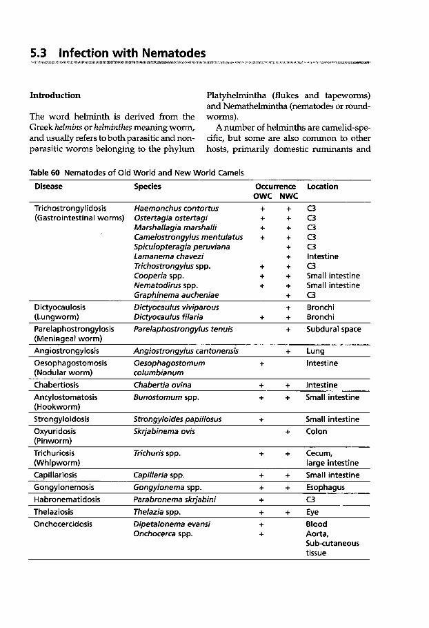
5.3 Infection with Nematodes W W I t
Introduction Platyhelmintha (flukes and tapeworms) and Nemathelmintha (nematodes or round-
The word helminth is derived from the worms). Greek helrnins or helrninfhes meaning worm, A number of helminths are camelid-spe- and usually refers to both parasitic and non- cific, but some are also common to other parasitic worms belonging to the phylum hosts, primarily domestic ruminants and
Table 60 Nematodes of Old World and New World Camels
Disease Species Occurrence location OWC NWC
Trichostrongylidosis Haemonchus contortus + + (Gastrointestinal worms) Ostertagia ostertagi + +
Marshallagia manhalli + + Camelostrongylus mentulatus + + Spiculopteragia peruviana + Lamanema chavezi + Trichostrongylus spp. Cooperia spp.
+ + + +
Nematodirus spp. + + Graphinema aucheniae +
Dictyocaulosis Dictyocaulus viviparous + (Lungworm) Dictyocaulus filaria + +
c3 c3 c3 c3 c3 Intestine c3 Small intestine Small intestine c3 Bronchi Bronchi
Parelaphostrongylosis Parelaphostrongylus tenuis + Subdural space (Meninseal worm) Ansiostronclvlosis Anuiostronuvlus cantonensis + Luns Oesophagostomosis Oesophagostomum + Intestine (Nodular worm) columbianum Chabertiosis Chabertia ovina + + Intestine Ancylostomatosis (Hookworm)
~ ~~
Bunostomum spp. + + Small intestine
Strongyloidosis Strongyloides papillosus + Small intestine Oxyuridosis Skrjabinema ovis (Pinworm)
+ Colon
Trichuriosis Trichuris spp. + + Cecum, (Whipworm) large intestine Capillariosis Capillaria spp. + + Small intestine Gonavlonemosis Gonavlonema SDD. + + EsoDhaaus
~
Habronematidosis Parabronema skriabini + c3 Thelaziosis Thelazia SDD. + + Eve Onchocercidosis Dipetalonema evansi + Blood
Onchocerca spp. + Aorta, Sub-cutaneous tissue
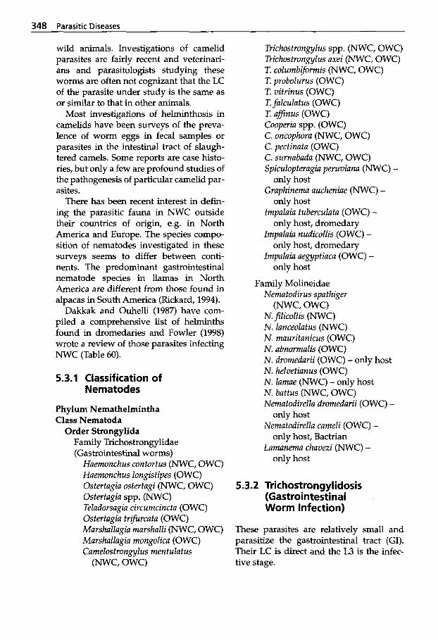
348 Parasitic Diseases
wild animals. Investigations of camelid parasites are fairly recent and veterinari- ans and parasitologists studying these worms are often not cognizant that the LC of the parasite under study is the same as or similar to that in other animals.
Most investigations of helminthosis in camelids have been surveys of the preva- lence of worm eggs in fecal samples or parasites in the intestinal tract of slaugh- tered camels. Some reports are case histo- ries, but only a few are profound studies of the pathogenesis of particular camelid par- asites.
There has been recent interest in defin- ing the parasitic fauna in NWC outside their countries of origin, e.g. in North America and Europe. The species compo- sition of nematodes investigated in these surveys seems to differ between conti- nents. The predominant gastrointestinal nematode species in llamas in North America are different from those found in alpacas in South America (Rickard, 1994).
Dakkak and Ouhelli (1987) have com- piled a comprehensive list of helminths found in dromedaries and Fowler (1998) wrote a review of those parasites infecting NWC (Table 60).
5.3.1 Classification of Nematodes
Phylum Nemathelmintha Class Nematoda
Order Strongylida Family Trichostrongylidae (Gastrointestinal worms)
Huemonchus contortus (NWC, OWC) Huemonchus longistipes (OWC) Ostertugia ostertagi (NWC, OWC) Ostertugiu spp. (NWC) Teladorsagiu circumcinctu (OWC) Ostertugia trzfurcatu (OWC) Murshallugia murshalli (NWC, OWC) MurshaZlagia mongolicu (OWC) Camelostronmlus mentulutus
Trichostrongylus spp. (NWC, OWC) Trichostrongylus uxei (NWC, OWC) T. columbiformis (NWC, OWC) T. probolurus (OWC) T. vitrinus (OWC)
T affinus (OWC) Cooperia spp. (OWC) C. oncophora (NWC, OWC) C. pectinata (OWC) C. surnabada (NWC, OWC) Spiculopteragiu peruviana (NWC) -
only host Graphinemu aucheniae (NWC) -
only host Impalaiu tuberculuta (OWC) -
only host, dromedary Impalaiu nudicollis (OWC) -
only host, dromedary Impalaia aegyptiuca (OWC) -
only host
Family Molineidae
T. falculatus (OWC)
Nematodirus spathiger
N. filicollis (NWC) N. Zunceolutus (NWC) N. mauritunicus (OWC) N. ubnormulis (OWC) N. dromedurii (OWC) - only host N. helvetiunus (OWC) N. lamae (NWC) - only host N. battus (NWC, OWC) Nematodirella dromedurii (OWC) -
Nematodirella cumeli (OWC) -
Lamunema chuvezi (NWC) -
(NWC, OWC)
only host
only host, Bactrian
only host
5.3.2 Trichostrongylidosis (Gastrointestinal Worm Infection)
These parasites are relatively small and parasitize the gastrointestinal tract (GI). Their LC is direct and the L3 is the infec-
(NWC, OWC) tive stage.
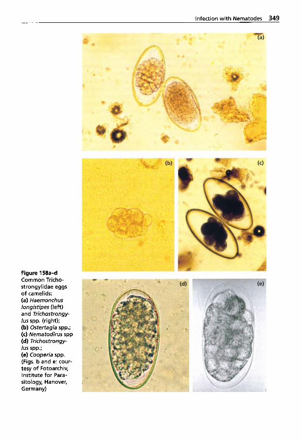
Infection with Nematodes 349
Figure 158a-d Common Tricho- strongylidae eggs of camelids: (a) Haemonchus longistipes (left) and Trichostrongy- /us spp. (right); (b) Ostertagia spp.; (c) Nematodirus spp (d) Trichostrongy- /us spp.; (e) Cooperia spp. (Figs. b and e: cour- tesy of Fotoarchiv, Institute for Para- sitology, Hanover, G e r m a n y)
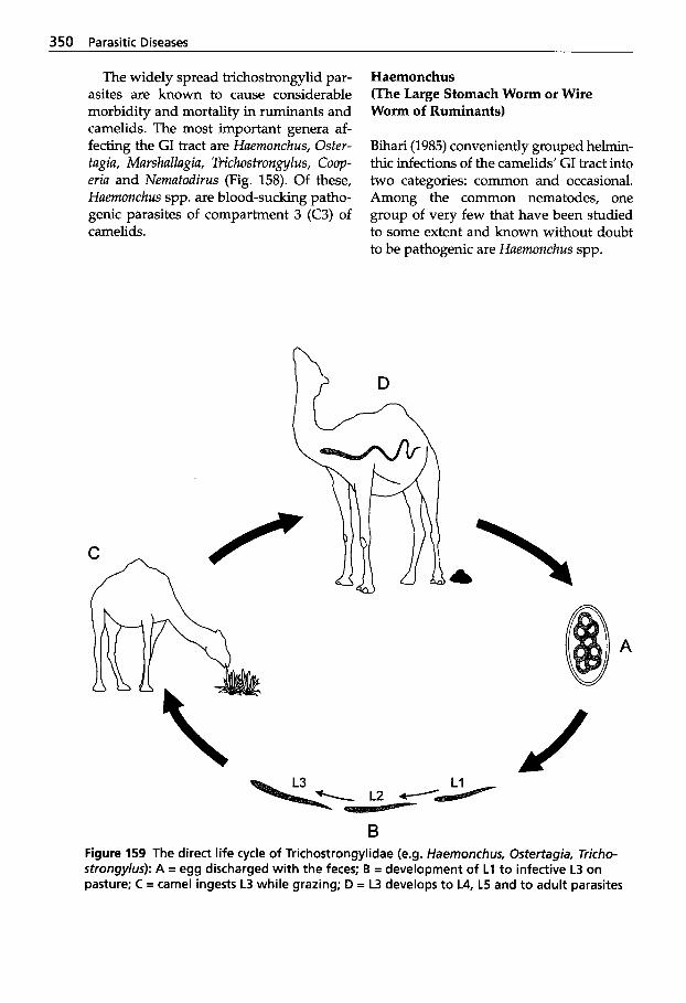
350 Parasitic Diseases
The widely spread trichostrongylid par- asites are known to cause considerable morbidity and mortality in ruminants and camelids. The most important genera af- fecting the GI tract are Haemonchus, Oster- tugia, Marshallagia, Trichostrongylus, Coop- eriu and Nernatodirus (Fig. 158). Of these, Haemonchus spp. are blood-sucking patho- genic parasites of compartment 3 (C3) of camelids.
Haemonchus (The Large Stomach Worm or Wire Worm of Ruminants)
Bihari (1985) conveniently grouped helmin- thic infections of the camelids’ GI tract into two categories: common and occasional. Among the common nematodes, one group of very few that have been studied to some extent and known without doubt to be pathogenic are Haernonchus spp.
A
B Figure 159 The direct life cycle of Trichostrongylidae (e.g. Haernonchus, Ostertagia, Tricho- strongylus): A = egg discharged with the feces; B = development of L1 to infective L3 on pasture; C = camel ingests L3 while grazing; D = L3 develops to L4, L5 and to adult parasites
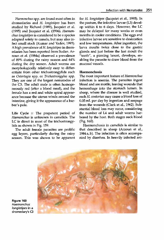
Infection with Nematodes 351
Huemonchus spp. are found most often in dromedaries and H. Zongistipes has been studied by Richard (1989), Jacquiet et al. (1995) and Jacquiet et al. (1996). Huernon- chus Zongistipes is considered to be a species adapted solely to camels, but may also in- fect small stock (Kumar and Yadev, 1993). A high prevalence of H. Zongistipes in drom- edaries has been reported from Sudan. Ar- zoun et al. (1984a) observed a prevalence of 89% during the rainy season and 64% during the dry season. Adult worms are morphologically relatively easy to differ- entiate from other trichostrongylids such as Ostertugia spp. or Trichostrongylus spp. They are one of the largest nematodes of the C3. The adult male is often homoge- neously red (after a blood meal), and the female has a red and white spiral appear- ance because the uterus winds around the intestine, giving it the appearance of a bar- ber's pole.
Life Cycle P: The prepatent period of Huernonchus is unknown in camelids. The LC is direct in most of the trichostrongy- lids as shown in Fig. 159.
The adult female parasites are prolific egg layers, particularly during the rainy season. This was shown to be apparent
for H. Zongistipes (Jacquiet et al., 1995). In the pasture, the infective larvae (L3) devel- op within 4 to 6 days. However, the LC may be delayed for many weeks or even months in cooler conditions. The eggs and infective larvae are sensitive to desiccation and low temperatures. After ingestion, the larva moults twice close to the gastric glands and just before the last moult the "tooth", a piercing lancet, develops, en- abling the parasite to draw blood from the mucosal vessels.
Haemonchosis The most important feature of Huemonchus infection is anemia. The parasites ingest blood and are motile, leaving wounds that hemorrhage into the stomach lumen. In sheep, where the disease is well studied, each H. contortus may cause a blood loss of 0.05 mL per day by ingestion and seepage from the wounds (Clark et al., 1962). Sub- stantial blood loss may occur, considering the number of L4 and adult worms har- bored by the host. Both stages suck blood (Fig. 160).
Haemonchosis in camelids is similar to that described in sheep (Arzoun et al., 1984a,b). The infection is often accompa- nied by diarrhea. In heavily infected ani-
Figure 160 Haemonchus longistipes in a dromedary's C3
352 Parasitic Diseases
mals, progressive deterioration occurs with marked anemia seen in 10 to 45% of cases (Faye, 1997), eventually leading to emacia- tion and death. The chronic form may be difficult to differentiate from other chronic camel diseases, e.g. trypanosomosis.
Acute haemonchosis in experimentally infected camels induced clinical signs of mucoid diarrhea, anorexia, anemia, loss of body weight, edema of the lower limbs, general malaise and death after 8 to 10 weeks (Arzoun et al., 198413).
Haemonchosis in the camel is often as- sociated with hypoproteinemia, including hypoalbuminemia and hypoglubulinemia, as well as leucocytosis, including neu- trophilia and eosinophilia (Graber et al., 1967; Queval et al., 1967; Richard 1979 and 1989; Arzoun et al., 1984 b; Jacquiet et al., 1995). Low PCV, calcium, phosphate, mag- nesium and copper levels were also diag- nosed by Kaufmann (1996).
Other Common Trichostrongylids
The common stomach worms of Ostertu- giu spp., such as 0. ostertugi, 0. lyrutu, Telu- dorsugru circumcinctu, and 0. trifurcutu, are highly adapted to cattle, small stock and wild ruminants. However, they are also found in camelids with an LC similar to Huemonchus spp.
Different climates produce differences in the epidemiology of the parasite. In tem- perate regions, the larvae become arrested (hypobiosis) in early autumn and develop- ment starts again in spring. In other areas of the world where the summers are hot, the larvae may survive the hot unfavorable environmental conditions during the sum- mer in hypobiosis. The LC varies accord- ing to climate and host species.
Significant numbers of the parasites in the C3 may give rise to extensive patho- logical and biochemical changes, which in turn create severe clinical signs. These are most evident when the larvae emerge from the glands. The larvae in the host’s glands
stimulate the formation of grayish white nodules, which are readily seen in the mu- cosa of C3 at necropsy.
Windsor (1997) reported three cases of ostertagiosis in llamas in northern Scot- land south and southwestern England. The affected animals died despite treat- ment with ivermectin.
There are some other parasites found in the abomasum or C3 of camelids closely resembling Ostertagia: Murshullugiu mur- shulli, M . mongolicu, Teludorsugiu sp./ Cume- lostrongylus mentulutus, and Spiculopterugiu peruviunu, which was first described in alpacas, llamas and vicufias from Titicaca in Peru (Guerrero and Chavez, 1967). At the same time, Ostertugiu lyrutu and Huem- onchus contortus were first found in al- pacas, together with two other species: Trichostrongylus longispiculuris and Cumelo- strongylus mentulutus. A few years later, 0. ostertugiu and 0. lyrutu were first found in llamas in Peru (Vasques and Marchinares, 1971).
The L3 of Murshullugiu marshalli pene- trate the gastric glands of C3 and are even- tually surrounded by a 2 to 4 mm diame- ter large nodule, each containing two to three larvae that mature in 15 to 18 days. The prepatent period is usually about 3 weeks, but arrested development may oc- cur (Fowler, 1998). The eggs may easily be confused with Nemutodirus spp. eggs. The parasite has limited distribution. It occurs in llamas in the western USA. It is a com- mon parasite in sheep in the Mediter- ranean, and has also been reported in camels in India and Russia (Dakkak and Ouhelli, 1987). Murshullugiu mongolicu has only been reported in Mongolia (Dakkak and Ouhelli, 1987), and unidentified Mur- shullugiu sp. in guanacos in Argentina (Na- vone and Merino, 1989).
Cumelostrongylus mentulutus is a com- mon camelid stomach worm, particularly in animals sharing grazing with sheep (Dakkak and Ouhelli, 1987). The parasite also infects sheep, goats, antelope and lla-
Infection with Nematodes 353
mas (Soulsby, 1982). C. mentulatus is com- monly found in the Middle East and in areas north of the African continent (Kaufmann, 1996), but less commonly in South America and the USA. It was first described in llamas in Argentina (Led and Boero, 1972). According to Kaufmann (1996), C. mentulatus may cause significant disease in camels. It seldom occurs in sin- gle infections.
Trichostrongylus
Trichostrongylus spp. are considered to be one of the most important causes of para- sitic gastroenteritis in ruminants. Tricho- strongylus axei is found in the abomasum of ruminants, in C3 in camelids and in the stomach of horses, donkeys, pigs and hu- mans. Other Trichostrongylus spp., such as T. colubriformis, T. vitrinus, T. probolurus, are found mainly in the small ruminants’ in- testines but also frequently in camelids. Occasionally T. falculatus and T. aflnus have been recorded in camelids. The Tri- chostrongylus spp. are small and thin, about 7mm long and difficult to see with the naked eye.
Cooperia
Cooperia spp. are small nematodes similar in size to Ostertagia. They are parasites of the small intestines of ruminants and camelids throughout the world.
C. oncophora and C. pectinata are found in OWC and C. oncophora and C. zurnabada in NWC.
Graphinema aucheniae
This parasite is only found in C3 in NWC. Its LC and epidemiology is similar to tri- chostrongvles.
5.3.3 Infections with Molineidae
Nematodirus
These parasites are found worldwide, par- ticularly in temperate zones. They are small intestinal parasites. The adults are slender, about 2mm long, and relatively easy to differentiate from other tricho- strongyles. The eggs are large and twice the size of other trichostrongyle eggs (see Fig. 158a-e). Nematodirus battus is the most pathogenic species in temperate areas.
Severe damage to the villi and erosion of the mucosa resulting in villous atrophy, co- incide with the parasitic phases of the lar- vae while in the mucosa. Young animals may exhibit rapid progressive dehydration following diarrhea, leading to death. At necropsy, the carcass is dehydrated and en- teritis is often evident in the ileum. N. lamae, N. battus, N. spathiger, N. filicollis and N. lanceolatus are species found in NWC (Fowler, 1998).
The following species are reported in OWC: N. spathiger, N. mauritanicus, N. ab- normalis, N. dromedarii and N. helvetianus. N. cameli is reported in the Bactrian camel of the former USSR. In addition, species closely related to Nematodirus spp. are found parasitizing dromedaries: Nematodirella dromedarii, lmpalaia tuberculata and 1. nudi- collis. The latter two species are parasites of the C3, occasionally of the small intestine (Kaufmann, 1996), and are mostly found in camels in Africa (Dakkak and Ouhelli, 1987). Gibbons et al. (1977) discussed the classification of these seldom-mentioned species. Nematodirella dromedarii was first reported in India and described by Lodha and Raisinghani (1979). It was found in the districts of Bikaner and Jodhpur in Ra- iasthan with a Prevalence of over 42%.
354 Parasitic Diseases
Lamanema chavezi
One of the most important NWC nema- tode pathogens is Lamanema chuvezi. It is thought to be a parasite of the mountain viscacha Lagidium viscuciu boxi. Llamas and alpacas are believed to be aberrant hosts, in which the infection may be very se- vere. Particularly vulnerable are recently weaned NWC.
The LC is poorly understood. The infec- tive larvae develop within the eggs, giving them excellent resistance to adverse cli- matic conditions (Leguia, 1991). Ingested larvae penetrate the intestinal wall and pass to the liver and lungs. When matura- tion is completed the parasites migrate back to the small intestine via the trachea. The prepatent period is about 30 days (Guerrero et al., 1973).
Heavy infection causes hepatic and res- piratory failure and death may follow. This was shown experimentally: a $-month old alpaca given 200,000 larvae orally died after 20 days, exhibiting severe anemia (Guerrero et al., 1973).
The migration of the larvae causes ca- tarrhal and hemorrhagic enteritis with areas of mucosal necrosis. In acute infec- tions, the liver is congested, showing mul- tiple small foci of coagulative necrosis and petechial hemorrhages. Areas of lung congestion are also seen. When the larvae have returned to the intestine, the liver lesions become fibrotic and may calcify (Fowler, 1998) showing a characteristic mottled appearance (Leguia, 1991). Five young alpaca given 10,000 L. chavezi larvae showed increased levels of glutamate-ox- alacetate-transaminase 14 days later, indi- cating liver damage (Guerrero et al., 1973). The liver is often condemned.
5.3.4 Dictyocaulosis (Lungworm Infection) Parelaphostrongylosis (Meningeal Worm Infection) Ang iostrongylosis
Family Dictyocaulidae DictyocuuIus vivipurus (NWC) D. filuriu (NWC, OWC)
Family Protostrongylidae Pureluphostrongylus tenuis (NWC, llama aberrant host)
Angiostrongylus cuntonensis (NWC, alpaca aberrant host)
Family Angiostrongylidae
Dictyocaulus
The parasite belonging to the family Dicty- ocaulidae occurs in the respiratory pas- sages of the lungs and is the major cause of parasitic bronchitis in domestic animal species. The parasites are found world- wide, particularly in temperate climates. The adult parasite is long and slender, about 8cm long, and is found in the tra- chea and bronchi. Their LC is direct. The females are ovo-viviparous, laying eggs containing fully developed larvae L1. The eggs are coughed up and swallowed. Hatching may already begin in the lungs, but usually occurs while the eggs pass through the gut of the host. Some eggs may be expelled via nasal discharges.
Life Cycle I The preparasitic (free) stages feed on food reserves stored in their intes- tinal cells, unlike those of other tricho- strongyle larvae, which actively feed on microorganisms in the environment. The L3 stage is reached in 5 to 7 days. Approx- imately 4 days following the infection, the ingested L3 penetrate the intestinal mu- cosa of the host and pass into the mesen- teric lymph nodes where they moult into L4. The L4 reach the lungs via the blood and lymph within a week of infection. The
infection with Nematodes 355
last molt occurs in the bronchioles and the L5 move up the bronchi and mature into the adult form. The prepatent period in cattle is about 3-4 weeks.
Clinical Signs ~1 Parasitic bronchitis is a problem, particularly in areas with a mild climate, high rainfall and permanent pas- tures. The disease is mostly seen in young animals, but it can affect any age group. Affected animals may cough, with dys- pnea and nasal discharge. Heavily infected animals may die due to respiratory failure following the development of interstitial emphysema and pulmonary edema. Many animals gradually recover, but this may take months. Superimposed bacterial in- fections might occur, hindering recovery. Body temperature is usually normal unless secondary pneumonia develops.
Epidemiology In endemic areas with temperate climates, the L3 may hibernate (over winter) on pasture in sufficient num- bers to initiate infection the following spring. An infection may also persist from year to year by carrier animals; i.e. small numbers of adult parasites may survive in the bronchi of affected animals.
In Europe generally only calves in their first grazing season show clinical disease. In endemic situations, older animals ac- quire a strong immunity.
Larvae require a moist environment to survive; thus infections are not considered a problem in hot and dry climates. How- ever, Dictyocaulus filaria in OWC has been reported in several African and Asian countries as well as in Europe. D. cameli (Boev, 1952) has been described in camels in Asia and Europe. Some authors consid- er it to be synonymous with D. viviparus (Soulsby, 1982). Both D. viviparus and D. filaria are commonly found parasitizing Camelidae in South America (Fowler, 1998).
/ I Diagnosis is based on clinical signs of respiratory distress. It is usually a
herd problem. Several young animals may simultaneously show signs of the infec- tion. Coughing may be evident during the prepatent period when no eggs are yet laid. Demonstration of larvae in feces us- ing the Baemann method is an important diagnostic tool. Furthermore, specific anti- body tests are commercially available for the diagnosis in cattle.
Parelaphostrongylus tenuis
Adult P. tenuis are found in the cranial ve- nous sinuses and subdural spaces of the white-tailed deer (Odocoileus virginianus) in which the infection is sub-clinical. The parasite may also infect and cause neuro- logical disease in several other Cervidae and domestic livestock such as sheep, goats and llamas. It is a small, hair-like nema- tode.
Life Cycle The llama is an aberrant host and it is not known whether the LC of this parasite is the same in the llama as in its natural host. In deer, eggs are laid and eventually hatch on the meninges. The larvae are then carried via the circulation to the lungs. Eggs may also be deposited in the venous circulation and then carried to the lungs, where they embryonate and hatch (Soulsby, 1982). The larvae then reach the bronchi and trachea and are coughed up and swallowed, ending up in the feces. Intermediate hosts such as terrestrial snails and slugs eat the larvae. The final host in- gests the infected snails and after the lar- vae are released into the stomach, they penetrate the wall, reaching the spinal cord in 10 days. They then migrate to the spinal subdural space and further on to the brain where they enter their final location, the venous sinuses, by penetrating the dura mater.
Clinical Signs There are usually no clin- ical signs of disease but fatal neurological disease may occur in the aberrant hosts,
356 Parasitic Diseases
such as the llama. Migration of the larvae in the spinal cord produces lesions like en- cephalomalacia, manifested as lameness, ataxia, stiffness, circling, blindness, hyper- metria, paraplegia, paralysis and abnor- mal position of the head (Fowler, 1998). Lo- cal hemorrhages in the spinal cord often lead to death (Cheney and Allen, 1989).
Epidemiology Llamas cohabiting with white-tailed deer are at risk. Other ungu- lates may be aberrant hosts, e.g. elk (Cer- vus canadensis), moose (Alces alces), caribou (Rangifer tarandus) and red deer (Cervus ela- phus), as well as sheep and goats. The in- fection may cause death in llamas, alpacas and guanacos (Rickard, 1994).
Diagnosis k a At necropsy the thin, slender parasites are difficult to find. Larvae may be found in the feces by the Baermann technique. However, identification of the larvae to species is not possible. A recently developed ELISA has shown promising re- sults in demonstrating meningeal worm infestations in domestic goats (Rickard, 1994).
Angiostrongylus cantonensis
A lungworm, Angiostrongylus cantonensis, normally a parasite of rats in the Pacific Basin, was found in an alpaca during quarantine. The alpaca died and the nema- todes were found in the lungs at necropsy (Fowler, 1998).
5.3.5 Oesophagostomosis and Chabertiosis (Nodular Worm Infection)
Family Chabertiidae Oesophagostomurn spp. (NWC) 0. columbianum (OWC, NWC) 0. venulosum (OWC) 0. vigintimembrum (OWC) Chabertia ovina (OWC, NWC)
Oesophagostomum spp. are stout round- worms 1-2cm long. These nematodes, found in the large intestine of ruminants, camelids, pigs and primates, are often called nodular parasites because many cause nodules in the wall of the intestine.
The parasites are distributed world- wide, but are more important in tropical and subtropical regions. The two species, 0. columbianum and 0. venulosum, essen- tially sheep and goat parasites, have also been reported in camels in Africa and Asia (Kaufmann, 1996). None of the nodular worms in NWC have been identified as to species (Fowler, 1998).
Life Cycle ’ The LC of these nematodes has not been established in camelids. How- ever, it is assumed that the LC in Camelidae is similar to that in ruminants. Thin-walled eggs are passed in the feces. The develop- ment and bionomics of the preparasitic stages are similar to those of the Strongylus spp. The infective stage, the L3, will pene- trate the intestinal mucosa where the third ecdysis takes place.
In some species, the development occurs within the nodules. 0. columbianum and 0. venulosum can cause severe enteritis. However, the latter species has fewer tendencies to cause nodules, i.e. inflam- mation in the wall of the intestine, and is less pathogenic. The L3 of 0. columbia- num may migrate deep into the intestinal mucosa, provoking inflammation. The nodules formed are visible to the naked eye.
Chabertia ovina
Another parasite of the cecum and colon, Chabertia ovina, also called the ”large- mouthed bowel worm” is found in domes- tic and wild ruminants and in camelids throughout the world. It is particularly common in sheep and goats, but rarely causes clinical disease. The worm is rarely reported in dromedaries and NWC. The
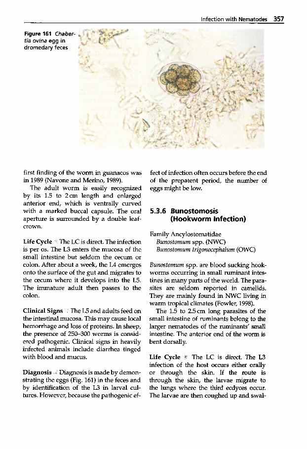
Infection with Nematodes 357
Figure 161 Chaber- tia ovina egg in dromedary feces
first finding of the worm in guanacos was in 1989 (Navone and Merino, 1989).
The adult worm is easily recognized by its 1.5 to 2cm length and enlarged anterior end, which is ventrally curved with a marked buccal capsule. The oral aperture is surrounded by a double leaf- crown.
Life Cycle The LC is direct. The infection is per 0s. The L3 enters the mucosa of the small intestine but seldom the cecum or colon. After about a week, the L4 emerges onto the surface of the gut and migrates to the cecum where it develops into the W. The immature adult then passes to the colon.
Clinical Signs The L5 and adults feed on the intestinal mucosa. This may cause local hemorrhage and loss of proteins. In sheep, the presence of 250-300 worms is consid- ered pathogenic. Clinical signs in heavily infected animals include diarrhea tinged with blood and mucus.
Diagnosis 8 Diagnosis is made by demon- strating the eggs (Fig. 161) in the feces and by identification of the L3 in larval cul- tures. However, because the pathogenic ef-
fect of infection often occurs before the end of the prepatent period, the number of eggs might be low.
5.3.6 Bunostomosis (Hookworm Infection)
Family Ancylostomatidae Bunostomum spp. ( W C ) Bunostomum trigonocephalum (OWC)
Bunostomum spp. are blood sucking hook- worms occurring in small ruminant intes- tines in many parts of the world. The para- sites are seldom reported in camelids. They are mainly found in NWC living in warm tropical climates (Fowler, 1998).
The 1.5 to 2.5cm long parasites of the small intestine of ruminants belong to the larger nematodes of the ruminants’ small intestine. The anterior end of the worm is bent dorsally.
Life Cycle 3 The LC is direct. The L3 infection of the host occurs either orally or through the skin. If the route is through the skin, the larvae migrate to the lungs where the third ecdyces occur. The larvae are then coughed up and swal-
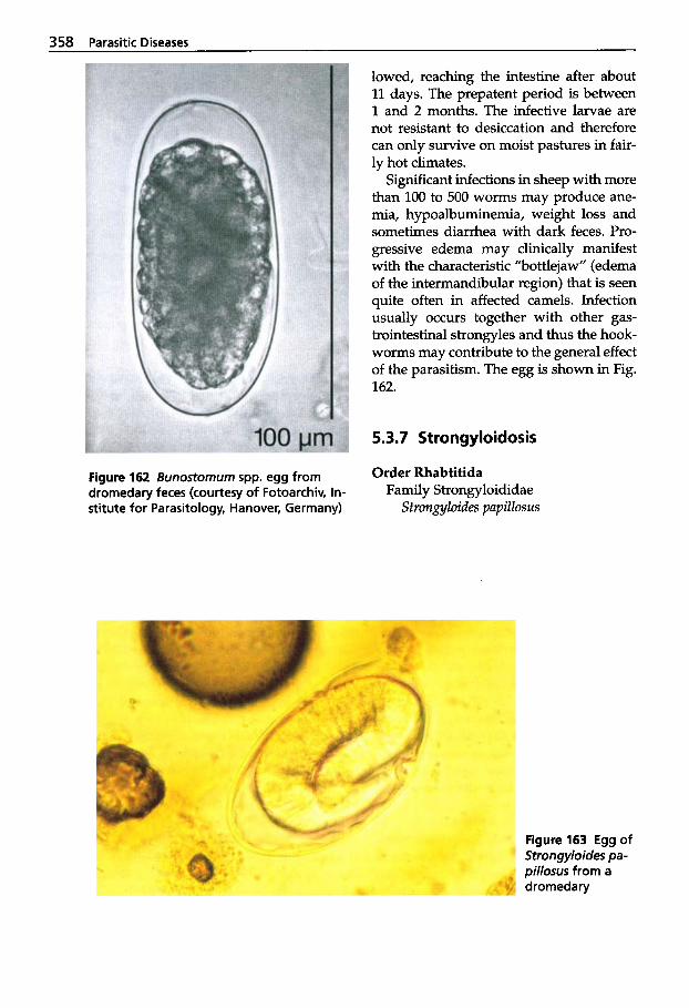
358 Parasitic Diseases
lowed, reaching the intestine after about 11 days. The prepatent period is between 1 and 2 months. The infective larvae are not resistant to desiccation and therefore can only survive on moist pastures in fair- ly hot climates.
Significant infections in sheep with more than 100 to 500 worms may produce ane- mia, hypoalbuminemia, weight loss and sometimes diarrhea with dark feces. Pro- gressive edema may clinically manifest with the characteristic "bottlejaw" (edema of the intermandibular region) that is seen quite often in affected camels. Infection usually occurs together with other gas- trointestinal strongyles and thus the hook- worms may contribute to the general effect of the parasitism. The egg is shown in Fig. 162.
5.3.7 Strongyloidosis
Order Rhabtitida Figure 162 Bunostornurn spp. egg from dromedary feces (courtesy of Fotoarchiv, In- stitute for Parasitology, Hanover, Germany)
Family Strongyloididae Strongyloides papillosus
Figure 163 Egg of Strongyloides pa- pillosus from a dromedary
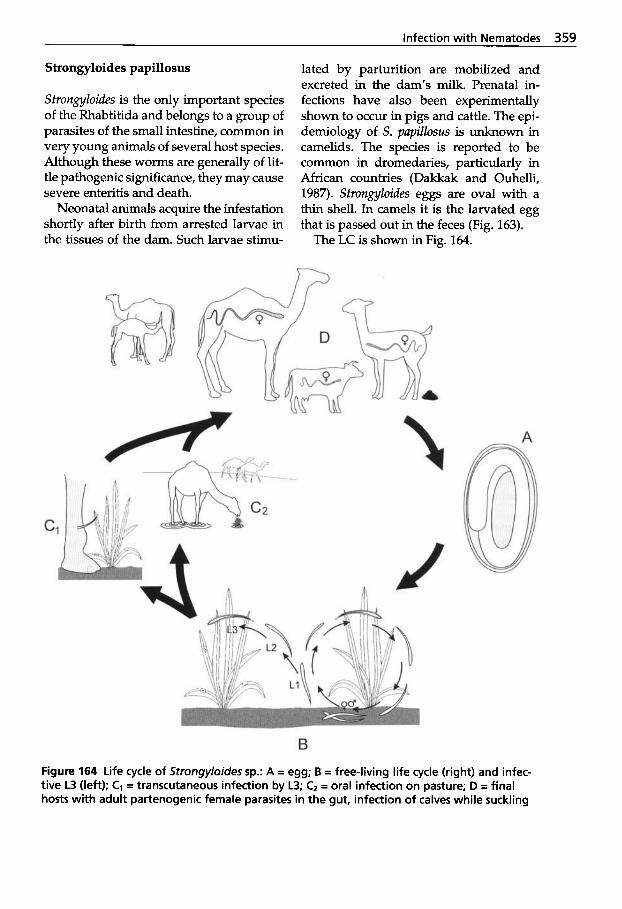
Infection with Nematodes 359
Strongyloides papillosus
Strongyloides is the only important species of the Rhabtitida and belongs to a group of parasites of the small intestine, common in very young animals of several host species. Although these worms are generally of lit- tle pathogenic significance, they may cause severe enteritis and death.
Neonatal animals acquire the infestation shortly after birth from arrested larvae in the tissues of the dam. Such larvae stimu-
lated by parturition are mobilized and excreted in the dam’s milk. Prenatal in- fections have also been experimentally shown to occur in pigs and cattle. The epi- demiology of S. papillosus is unknown in camelids. The species is reported to be common in dromedaries, particularly in African countries (Dakkak and Ouhelli, 1987). Strongyloides eggs are oval with a thin shell. In camels it is the larvated egg that is passed out in the feces (Fig. 163).
The LC is shown in Fig. 164.
A
B Figure 164 Life cycle of Strongyloides sp.: A = egg; B = free-living life cycle (right) and infec- tive L3 (left); C1 = transcutaneous infection by L3; Cz = oral infection on pasture; D = final hosts with adult partenogenic female parasites in the gut, infection of calves while suckling
360 Parasitic Diseases
5.3.8 Oxyuridosis (Pinworm Infection)
Order Oxyurida Family Oxyuridae
Skrjubinm ovis (NWC)
The sheep pinworm, Skrjubinemu ovis, has been found in the guanaco in Argentina (Fowler, 1998). It is a small parasite meas- uring between 3 to 8 mm. The eggs are flat- tened on one side.
Life Cycle I The LC is direct. Adults live in the colon and migrate to the rectum from where the female traverses the anal sphincter to deposit her fully embryonated eggs around the anus. The eggs drop off and are ingested with water and food. The L3 hatch in the small intestine and the lar- vae migrate to the large intestine where the parasites mature within 25 days (Schad, 1959).
Epidemiology The worm has also been found in goats and antelopes. It is consid- ered a benign parasite, although it might cause irritation and pruritus in and around the anus.
Diagnosis The eggs are usually not seen in the regular fecal flotation analysis. Scotch tape applied around the anus and then applied to a glass slide is the recom- mended diagnostic method.
5.3.9 Trichuriosis (Whipworm Infection) Capillariosis
Order Enoplida Family Trichuridae
Trichuris tenuis (NWC) T. ovis (NWC, OWC) T. globulosu (OWC) T. ufinus (OWC) T. ruoi (OWC)
T. skrjabini (OWC) T. curneli (OWC)
Family Capillariidae Cupillaria spp. (NWC)
Nematodes belonging to the genus Tri- churis are common parasites of several mammals, particularly ruminants. Several species are found in camelids and some authors consider Trichuris spp. significant parasites in these animal species (Boyce et al., 1984; Hayat et al., 1998; Partani et al., 1998). T. globulosu is the most prevalent in dromedaries in Africa and Asia (Kauf- mann, 1996). However, as the different species of Trichuris are difficult to distin- guish, most authors do not identify the par- ticular species. Other Trichuris spp. have occasionally been reported to occur in camels: T. ovis, T. curneli, T. ruoi, T. skrjubini and T. ufinus (Kaufmann, 1996).
T. ovis is considered to be the species af- fecting NWC in South America (Fowler, 1998) while T. tenuis has been more fre- quently reported in animals in the north- west Pacific regions (Rickard and Bishop, 1991 a, b). The latter worm was suggested to be the typical whipworm of the llamas (Rickard and Bishop, 1991 b; Rickard, 1994). Recently T. tenuis was found during a sur- vey in llamas and vicuiias in northwestern Argentina (Cafrune et al., 1999). Whip- worm eggs were found in over 50% of the animals surveyed and T. tenuis was demon- strated during necropsy of one llama and one vicuiia in each of the herds. The au- thors were convinced that T. tenuis is close- ly associated with camelids (Cafrune et al., 1999).
Trichuris spp. are between 3 to 8cm long and are easily identified by the long and much thinner anterior body portion that becomes shorter and thicker posteri- orly.
Life Cycle The LC is direct. On pasture, the eggs may reach the infective stage after 3 weeks (Soulsby, 1982). However, devel-
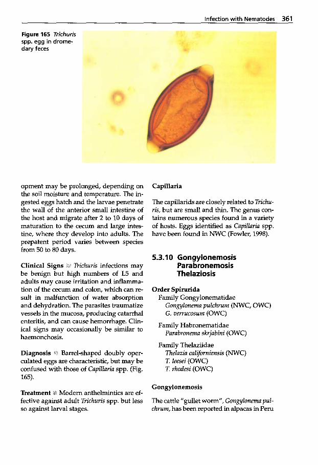
Infection with Nematodes 361
Figure 165 Trichuris spp. egg in drome- dary feces
opment may be prolonged, depending on the soil moisture and temperature. The in- gested eggs hatch and the larvae penetrate the wall of the anterior small intestine of the host and migrate after 2 to 10 days of maturation to the cecum and large intes- tine, where they develop into adults. The prepatent period varies between species from 50 to 80 days.
Clinical Signs h i 1 Trichuris infections may be benign but high numbers of W and adults may cause irritation and inflamma- tion of the cecum and colon, which can re- sult in malfunction of water absorption and dehydration. The parasites traumatize vessels in the mucosa, producing catarrhal enteritis, and can cause hemorrhage. Clin- ical signs may occasionally be similar to haemonchosis.
Diagnosis Barrel-shaped doubly oper- culated eggs are characteristic, but may be confused with those of Cupillaria spp. (Fig. 165).
Treatment ilii Modern anthelmintics are ef- fective against adult Trichuris spp. but less so against larval stages.
Capillaria
The capillarids are closely related to Trichu- ris, but are small and thin. The genus con- tains numerous species found in a variety of hosts. Eggs identified as Cupillaria spp. have been found in NWC (Fowler, 1998).
5.3.1 0 Gongylonemosis Para bronemosis Thelaziosis
Order Spirurida Family Gongylonematidae
Gongylonernu pulchrum (NWC, OWC) G. verrucosum (OWC)
Family Habronematidae Purubronemu skrjabini (OWC)
Family Thelaziidae Theluziu culiforniensis (NWC) T. leesei (OWC) T. rhodesi (OWC)
Gongylonemosis
The cattle "gullet worm", Gongylonema pul- chrum, has been reported in alpacas in Peru
362 Parasitic Diseases
(Fowler, 1998) as well as in dromedaries, which are rarely infected also with the ru- men gullet worm G. zlerrucosum (Kauf- mann, 1996).
G. pulchrum OCCUTS more often in sheep, goats, cattle, pigs and buffaloes than in horses, donkeys, wild boars and camelids. It may also infect humans in the oral ep- ithelium or sometimes subcutaneously.
The adult parasite is found in the esoph- agus within the mucosa or submucosa in a zigzag pattern. In ruminants, it may also inhabit the rumen wall.
Eggs are passed in the feces and hatch when ingested by an intermedi- ate host, one of 70 different species of bee- tles, including cockroaches. The larvae are liberated in the stomach of the final host and migrate to the esophagus.
The infection is usually subclinical and diagnosis is made by chance during necropsy.
Parabronemosis
Parabronema skrjabini is rarely found in dromedaries, sheep, goats and cattle in the abomasum and C3.
Life Cycle itit Stomyoxys and Haematobia flies are intermediate hosts that deposit the infective L3 on the final host, which in turn ingests it.
Thelaziosis
TheZazia spp. are 1 to 2cm long thin par- asites mainly found in or around the eyes of numerous animals (as well as hu- mans).
Thelazia californiensis has been found in llamas’ conjunctival sac (Fowler, 1998), and T. Zeesei is considered to be the drome- dary eye worm (Drobynin, 1974; Kauf- mann, 1996). T. Zeesi is reported to occur in Africa and Asia. Also T. rhodesi, a species
usually found in cattle, has occasionally been found in camels.
The TheZuzia spp. are ovoviviparous. The adult female parasite deposits eggs con- taining L1 into the host’s lachrymal se- cretions, which are taken up by the feed- ing intermediate host: mainly different species of Musca flies. Development to L3 occurs in the ovaries of various flies with- in a month. The infective larvae then mi- grate to the mouthparts of the fly from which they are transmitted to a new host. This occurs when the intermediate host takes a meal from secretions of the eye of the host.
Life Cycle (Fig. 166) l i t The development of larval stages to maturity takes place in the conjunctival sac. The L3 of T. Zeesei in dromedaries penetrates the conjunctival sac and from there migrates into the lacrimal duct, where the final develop- ment to adult worm occurs (Dobrynin, 1974). Adult worms may also be found on the cornea.
i One or both eyes may of- ten be infected without clinical signs. Oc- casionally the infection may cause irrita- tion, resulting in lacrimation with mild conjunctivitis, which may progress to ker- atitis. In severe cases the whole cornea may be opaque. Flies may often be seen cluster- ing around affected eyes.
Diagnosis Y’!~ Diagnosis is based on finding the parasites in the eye, usually in the lacrimal duct. Local anesthetics are a use- ful help in demonstrating the worm. Eggs or L1 may be found in lacrimal secre- tions.
1% The parasites may be phy- sically removed using topical anesthetic drops. Ivermectin drops or diethylcarba- mazine (2 mg/L) may also be instilled into the conjunctival sac (Kaufmann, 1996).
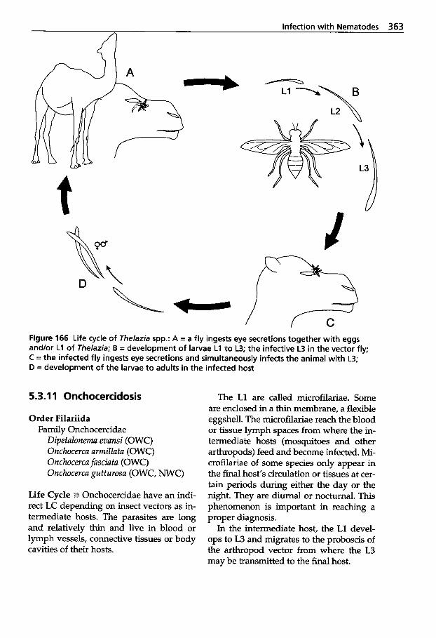
Infection with Nematodes 363
----
Figure 166 Life cycle of Thelazia spp.: A = a fly ingests eye secretions together with eggs and/or L1 of Thelazia; B = development of larvae L1 to L3; the infective L3 in the vector fly; C =the infected fly ingests eye secretions and simultaneously infects the animal with L3; D = development of the larvae to adults in the infected host
5.3.1 1 Onchocercidosis
Order Filariida Family Onchocercidae
Dipetalonema evansi (OWC) Onchocerca armillata (OWC) Onchocerca fasciatu (OWC) Onchocercu gutturosa (OWC, NWC)
Onchocercidae have an indi- rect LC depending on insect vectors as in- termediate hosts. The parasites are long and relatively thin and live in blood or lymph vessels, connective tissues or body cavities of their hosts.
The L1 are called microfilariae. Some are enclosed in a thin membrane, a flexible eggshell. The microfilariae reach the blood or tissue lymph spaces from where the in- termediate hosts (mosquitoes and other arthropods) feed and become infected. Mi- crofilariae of some species only appear in the final host’s circulation or tissues at cer- tain periods during either the day or the night. They are diurnal or nocturnal. This phenomenon is important in reaching a proper diagnosis.
In the intermediate host, the L1 devel- ops to L3 and migrates to the proboscis of the arthropod vector from where the L3 may be transmitted to the final host.
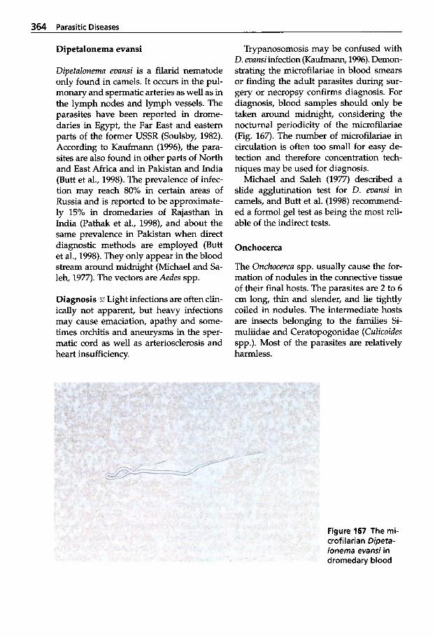
364 Parasitic Diseases
Dipetalonema evansi
Dipetalonema evansi is a filarid nematode only found in camels. It occurs in the pul- monary and spermatic arteries as well as in the lymph nodes and lymph vessels. The parasites have been reported in drome- daries in Egypt, the Far East and eastern parts of the former USSR (Soulsby, 1982). According to Kaufmann (1996), the para- sites are also found in other parts of North and East Africa and in Pakistan and India (Butt et al., 1998). The prevalence of infec- tion may reach 80% in certain areas of Russia and is reported to be approximate- ly 15% in dromedaries of Rajasthan in India (Pathak et al., 1998), and about the same prevalence in Pakistan when direct diagnostic methods are employed (Butt et al., 1998). They only appear in the blood stream around midnight (Michael and Sa- leh, 1977). The vectors are Aedes spp.
Diagnosis Lf Light infections are often clin- ically not apparent, but heavy infections may cause emaciation, apathy and some- times orchitis and aneurysms in the sper- matic cord as well as arteriosclerosis and heart insufficiency.
Trypanosomosis may be confused with D. evansi infection (Kaufmann, 1996). Demon- strating the microfilariae in blood smears or finding the adult parasites during sur- gery or necropsy confirms diagnosis. For diagnosis, blood samples should only be taken around midnight, considering the nocturnal periodicity of the microfilariae (Fig. 167). The number of microfilariae in circulation is often too small for easy de- tection and therefore concentration tech- niques may be used for diagnosis.
Michael and Saleh (1977) described a slide agglutination test for D. evansi in camels, and Butt et al. (1998) recommend- ed a formol gel test as being the most reli- able of the indirect tests.
Onchocerca
The Onchocerca spp. usually cause the for- mation of nodules in the connective tissue of their final hosts. The parasites are 2 to 6 cm long, thin and slender, and lie tightly coiled in nodules. The intermediate hosts are insects belonging to the families Si- muliidae and Ceratopogonidae (Culicoides spp.). Most of the parasites are relatively harmless.
Figure 167 The mi- crofilarian Dipeta- lonema evansi in dromedary blood
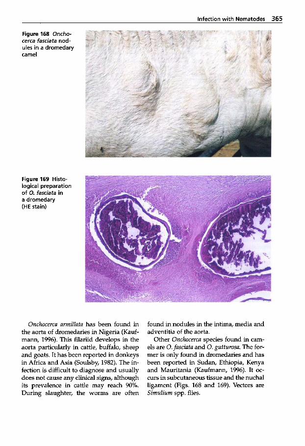
Infection with Nematodes 365
Figure 168 Oncho- cerca fasciata nod- ules in a dromedary camel
Figure 169 Histo- logical preparation of 0. fasciata in a dromedary (HE stain)
Onchocercu armillutu has been found in the aorta of dromedaries in Nigeria (Kauf- mann, 1996). This filariid develops in the aorta particularly in cattle, buffalo, sheep and goats. It has been reported in donkeys in Africa and Asia (Soulsby, 1982). The in- fection is difficult to diagnose and usually does not cause any clinical signs, although its prevalence in cattle may reach 90%. During slaughter, the worms are often
found in nodules in the intima, media and adventitia of the aorta.
Other Onchocercu species found in cam- els are 0.fusciutu and 0. gutturosu. The for- mer is only found in dromedaries and has been reported in Sudan, Ethiopia, Kenya and Mauritania (Kaufmann, 1996). It oc- curs in subcutaneous tissue and the nuchal ligament (Figs. 168 and 169). Vectors are SimuIium spp. flies.
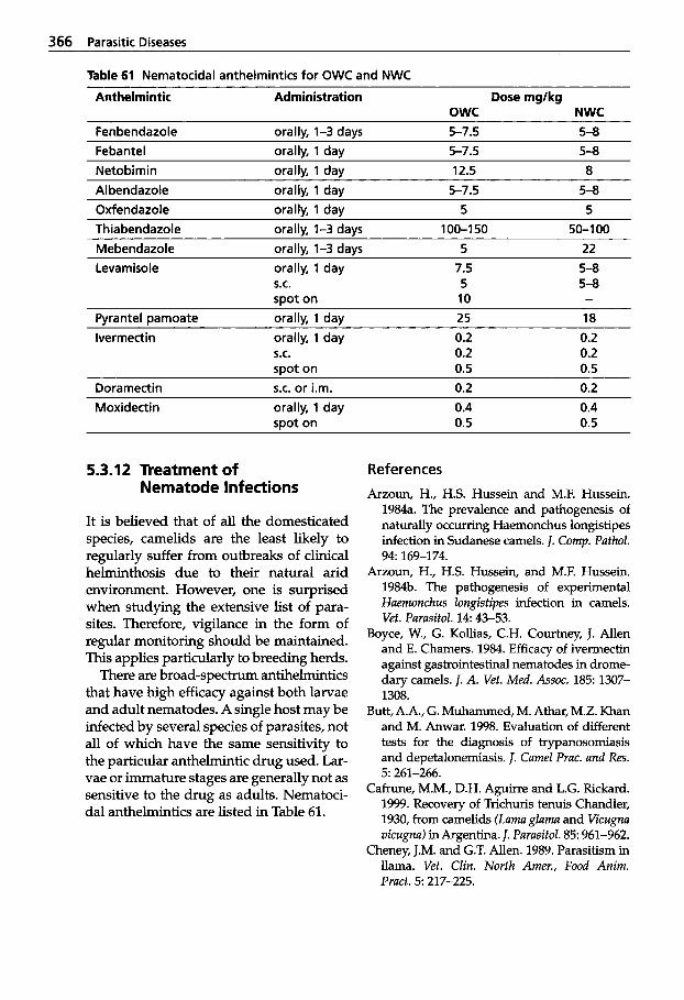
366 Parasitic Diseases
Table 61 Nematocidal anthelmintics for OWC and NWC
Anthelmintic Administration Dose rnglkg OWC NWC
Fenbendazole orallv, 1-3 davs 5-7.5 5-8 Febantel orally, 1 day 5-7.5 5-8 Netobimin orally, 1 day 12.5 8 A1 bendazole orally, 1 day 5-7.5 5-8 Oxfendazole orally, 1 day 5 5 Thiabendazole orally, 1-3 days 100-1 50 50-1 00 Mebendazole orally, 1-3 days 5 22 Levam isole orally, 1 day 7.5 5-8
S.C. 5 5-8 spot on 10 -
Pyrantel pamoate orally, 1 day 25 18 lvermectin orally, 1 day 0.2 0.2
S.C. 0.2 0.2 mot on 0.5 0.5
Doramectin S.C. or i.m. 0.2 0.2 Moxidectin orally, 1 day
spot on 0.4 0.5
0.4 0.5
5.3.12 Treatment of Nematode Infections
It is believed that of all the domesticated species, camelids are the least likely to regularly suffer from outbreaks of clinical helminthosis due to their natural arid environment. However, one is surprised when studying the extensive list of para- sites. Therefore, vigilance in the form of regular monitoring should be maintained. This applies particularly to breeding herds.
There are broad-spectrum antihelmintics that have high efficacy against both larvae and adult nematodes. A single host may be infected by several species of parasites, not all of which have the same sensitivity to the particular anthelmintic drug used. Lar- vae or immature stages are generally not as sensitive to the drug as adults. Nematoci- dal anthelmintics are listed in Table 61.
References Arzoun, H., H.S. Hussein and M.F. Hussein.
1984a. The prevalence and pathogenesis of naturally occurring Haemonchus longistipes infection in Sudanese camels. I. Comp. Pathol.
Arzoun, H., H.S. Hussein, and M.F. Hussein. 1984b. The pathogenesis of experimental Haemonchus longistipes infection in camels. Vet. Parasitol. 14: 43-53.
Boyce, W., G. Kollias, C.H. Courtney, J. Allen and E. Chamers. 1984. Efficacy of ivermectin against gastrointestinal nematodes in drome- dary camels. J. A. Vet. Med. Assoc. 185: 1307- 1308.
Butt, A.A., G. Muhammed, M. Athar, M.Z. Khan and M. Anwar. 1998. Evaluation of different tests for the diagnosis of trypanosomiasis and depetalonemiasis. J. Camel Prac. and Res.
Cafrune, M.M., D.H. Aguirre and L.G. Rickard. 1999. Recovery of Trichuris tenuis Chandler, 1930, from camelids (Lama glama and Vicugna vicugna) in Argentina. I. Parasitol. 85: 961-962.
Cheney, J.M. and G.T. Allen. 1989. Parasitism in llama. Vet. Clin. North Amer., Food Anim. Pract. 5 217-225.
94: 169-174.
5: 261-266.
Infection with Nematodes 367
Clark, C.H., G.K. Kiesel and C.H. Goby. 1962. Measurements of blood loss caused by Haemonckus contortus infection in sheep. Amer. J. Vet. Res. 23: 977-980.
Dakkak, A. and H. Ouhelli. 1987. Helminths and helminthoses of the dromedary. A re- view of the literature. Rev. Bibliogr. Rev. Sci. Tech. Ofice Int. Epiz. 6: 423-445,447-461.
Dobrynin, M.I. 1974. The development of Tke- lazia leesei Railliet and Henry, 1910, in the body of the intermediate host. lzvestiya Aka- demii Nauk Turkmeneskoi SSR, Biologcheskikk Nauk. 5: 39-45.
El-Bihari, S. 1985. Helminths of the camel: A Re- view. Br. Vet J. 141: 315-326.
Faye, B. 1997. Guide de l'elkvage du dromadaire. Sanofi Sante Nutrition Animale, Cedex, France.
Fowler, M.E. 1998. Medicine and Surgery of South American Camelids: Llama, Alpaca, Vicuiia, Guanaco, 2nd ed. Iowa State Univer- sity Press, Ames. USA.
Gibbons, L.M., M.C. Durette-Desset and P. Daynes. 1977. A review of the genus Impalaia Monnig, 1923 (Nematoda: Trichostrongyloi- dea). Ann. Parasitol. Hum. Comp. 52: 435-446.
Graber, M., R. Tab0 and J. Service. 1967. Enqu6te sur les helminthes du dromadaire tchadien. Etude des strongyloses gastro-intestinales et de l'haemonchose 2 Haemoncus longistipes. Rev. Elev. Mid . vet. Pays Trop. 20: 227-254.
Guerrero, C.A. and C.A. Chavez. 1967. New parasitic nematodes reported in alpacas (lama pacos) from Peru, with description of Spicu- lopteragia peruvianus n. sp. Bol. Ckileno Para- sitol. 22: 147-150.
Guerrero, C.A., J. Aha, I. Vega, J. Hemhdez and M. Rojas. 1973. Biological and pathologi- cal features of Lamanema chavezi in alpacas (Lama pacos). Revista de Investigaciones Pecuar- ias IVITA Universidad Nacional Mayor de San Marcos. 2: 29-42.
Hayat, C.S., A. Maqbool, N. Badar, H.A. Hash- mi and I. Hussein. 1998. Common gastroin- testinal helminths of camels of Pakistan. J. Camel Prac. and Res. 5: 251-254.
Jacquiet, P., J. Cabaret, M.L. Dia, D. Cheikh and E. Thiam 1996. Adaptation to arid environ- ment: Haemonckus longistipes in dromedaries of Saharo-Sahelian areas of Mauritania. Vet. Parasitol. 66: 193-204.
Jacquiet, P., J.F. Humbert, A.M. Comes, J. Cabaret, A. Thiam and D. Cheikh. 1995. Eco-
logical morphological and genetic characteri- zation of Haemonchus spp. parasites of do- mestic ruminants in Mauritania. Parasitology
Kaufmann, J. 1996. Parasitic Infections of Do- mestic Animals - A Diagnostic Manual. Birkhauser Verlag. Basel, Boston, Berlin.
Kumar, S. and C.L. Yadav. 1993. Establishment and pathogenesis of gastrointestinal nema- todes of camel and sheep. Int. J. Anim. Sci. 8:
Led, J.E. and J.J. Boero. 1972. Camelostrongylus mentulatus (Railliet and Henry, 1909), Orlov, 1933. (Nematoda-Trichostrongylidae). First report from the Republic of Argentina and of a new host Lama glama Cuvier. Gaceta Veteri- naria 34: 187-190.
Leguia, G. 1991. The epidemiology and econom- ic impact of llama parasites. Parasitol. Today.
Lodha, K.R. and M. Raisinghani. 1979. Report of Nematodirella dromedarii (Nematoda: Tricho- strongylidae) on the Indian camel (Camelus dromedarius) with remarks on the genus Ne- matodirella Yorke and Maplestone, 1926. Indi- an J. Anim. Sci. 49: 817-822.
Michael, S.A. and S.M. Saleh. 1977. The slide ag- glutination test for the diagnosis of filariasis in camels. Trop. Anim. Hlth. Prod. 9: 241-244.
Navone, G.T. and M.L. Merino. 1989. Contribu- tion to the knowledge of the endoparasitic fauna of Lama guanicoe Muller, 1776, from the Mitre Peninsula, Tierra del fuego, Argentina. Bol. Ckileno Parasitol. 44: 46-51.
Partani, A.K., D. Kumar, G.S. Manohar and A.K. Bhan. 1998. Clinical manifestation of natural infection with gastrointestinal nematodes in camel. J. Camel Prac. and Res. 5: 255-256.
Queval, R., M. Graber and M. Brunet. 1967. Serum proteins and haematological values in camels in relation to helminth infection. Rev. Elev. Mid . vtt. Pays Trop. 20: 437-449.
Richard, D. 1979. Dromedary pathology and productions. In: W.R. Cockrill (ed.): The Camelid - An All-purpose Animal. Proceed- ings of the Khartoum Workshop on Camels, Dec. 1979. Khartoum, Sudan.
Richard, D. 1989. L'haemonchose du dromadaire. Rev. Elev. Mtd . vtt. Pays Trop. 42: 45-53.
Rickard, L.G. and J.K. Bishop. 1991a. Helminth parasites of llamas (Lama glama) in the Pa- cific Northwest. J. Helminthol. SOC. Wash. 58:
110: 483-492.
113-118.
7 54-56.
110-115.
368 Parasitic Diseases
Rickard, L.G. and J.K. Bishop. 1991b. Redescrip- tion of Trichuris tenuis Chandler, 1930, from llamas (Lama glama) in Oregon with a key to the species of Trichuris present in North American ruminants. J. Parasitol. 77: 70-75.
Rickard, L.G. 1994. Parasites. Vet. Clin. N . Amer. Food. Anim. Pract. 1 0 239-247.
Schad, G.A. 1959. A revision of the North Amer- ican species of the genus Skrjabinema (Nema- toda, Oxyuroidea). Proc. Helminthol. SOC. Wash.
Soulsby, E.J.L. 1982. Helminths, Arthropods and Protozoa of Domesticated Animals, 7* ed. Bailli6re Tindall, London.
Vasques, D.M.S. and A.C. Marchinares. 1971. Parasitological survey in sheep and other an- imal species in the Pun0 Department, Peru. Revista del Instituto de Zoonosis e Investigation Pecuaria. Lima, 1: 25-109.
Windsor, R.S. 1997. Type I1 ostertagiasis in lla- mas. Vet. Rec. 141: 608.
26: 138-147.
Further reading Chakraborty, A. 1994. Occurrence and patholo-
gy of Gongylonema infection in captive herbi- vores. Vet. Parasitol. 52 163-167.
Diaz, D. C.A. 1961-62. Cooperia mcmasteri in al- pacas and vicuiia (Cooperia mcmasteri en alpacas y vicuiia). Rev. Fac. Med. Vet. Lima.
Dobrynin, M.I. 1972. The discovery of the inter- mediate host of Thelazia leesei Railliet and Henry, 1910. Izvestiya Akademii Nauk Turkme- neskoi SSR, Biologcheskikh Nauk. 3: 73-77.
16-17 131-137.
El-Bihari, S., Z.A., Kawasmeh, N.A. Ashour and A.H. Elnaiem. 1984. Experimental infection of sheep by camel stomach worm, Haemon- chus longistipes. Vet. Parasitol. 15: 257-261.
Jacquiet, P. 1995. Adaptations des Haemonchi- nae des ruminants domestiques au milieu subdesertique (Mauritanie). Thesis, Acade'mie de Monfpellier, France.
Leguia, G. 1997. Acute and subacute fasciolosis of alpacas (Lama pacos) and treatment with triclabendazole. Trop. Anim. Hlth. Prod. 29:
Michael, S.A. and S.M. Saleh. 1977. The slide ag- glutination test for the diagnosis of filariasis in camels. Trop. Anim. Hlth. Prod. 9: 241-244.
Sharma, I.K. 1991. Efficacy of some anthelmin- tics against gastrointestinal nematodes in camels (Camelus dromedarius). Ind. Vet. J. 68:
Soulsby, E.J.L. 1965. Textbook of Veterinary Clinical Parasitology, I. Helminths. Oxford. Blackwell Scientific.
Sudesh, K. and C.L. Yadav. 1993. Establishment and pathogenesis of gastrointestinal nema- todes of camel and sheep. Int. J. Anim. Sci. 8
Tager-Kagan, P. 1984. Resultats d'enquetes sur les helminthiases du dromadaire dam le de- partement de Zinder (Rep. du Niger), leur evolution dans l'annke - moyens de lutte. Rev. Elev. Mkd. Ve't. Pays Trop. 37 19-25.
Urquhart, G.M., J. Armour, J.L., Duncan, A.M. Dunn and F.W. Jennings. 1996. Veterinary Parasitology, Znd ed. Blackwell Science, Ox- ford, UK.
51-52.
1069-1072.
113-118.
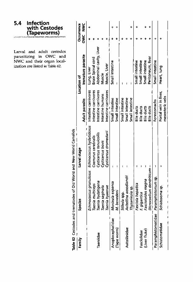
Tabl
e 62
Ces
tode
s an
d tre
mat
odes
of O
ld W
orld
and
New
Wor
ld C
amel
ids
Fam
ily
Spe
cies
La
rval
sta
ge
Loca
tion
of
Occ
urre
nce
Adu
lt pa
rasi
te
Imm
atur
e pa
rasi
te
OW
C
NW
C
Ech
inoc
occu
s gra
nulo
sus
Ech
inoc
occu
s hy
da tid
osus
In
test
ine
carn
ivor
es
Lung
, Li
ver
+ +
Taen
ia m
ultic
eps
Coe
n uru
s cer
ebra
lis
Inte
stin
e ca
rniv
ores
B
rain
Spi
nal c
ord
+
Taen
ia s
agin
ata
Cys
ticer
cus b
ovis
In
test
ine
hum
ans
Mus
cle
+ Ta
enia
hya
enae
C
ystic
ercu
s dro
med
arii
Inte
stin
e ca
rniv
ores
M
uscl
e, L
iver
+
Taen
iidae
Ta
enia
hyd
atig
ena
Cys
ticer
cus
tenu
icol
lis
Inte
stin
e ca
rniv
ores
A
bdom
inal
cav
ity,
Live
r +
Ano
ploc
epha
lidae
M
onie
zia
expa
nsa
-
Sm
all i
ntes
tine
Sm
all i
ntes
tine
+ +
(Tap
e w
orm
) M
. ben
eden
i -
Sm
all i
ntes
tine
-
Thyz
anie
zia
sp.
Sm
all i
ntes
tine
-
Fasc
iola
hep
atic
a -
Bile
duc
ts
Sm
all i
ntes
tine
+ +
Fasc
iolid
ae
E gi
gant
ica
- B
ile d
ucts
S
mal
l int
estin
e +
(Liv
er fl
uke)
Fa
scio
loid
es m
agna
-
Bile
duc
ts
Sm
all i
ntes
tine
+ +
Dic
roco
eliu
m d
endr
iticu
m -
Bile
duc
ts
Per
itone
um, l
iver
+
+ P
aram
phis
tom
atid
ae
Par
amph
isto
mum
sp.
- Fo
rest
omac
hs
Sm
all i
ntes
tine
+ S
chis
tom
atid
ae
Sch
isto
som
a sp
. -
Por
tal v
ein
in liv
er,
Hea
rt, lu
ng
+ + +
+ + + +
- S
mal
l int
estin
e -
Stil
esia
spp
. A
vite
llina
woo
dlan
dii
-
Sm
all i
ntes
tine
- A
vite
llini
dae
mes
ente
ric ve
in
370 Parasitic Diseases
5.4.1 Classification of Cestodes
Phylum Platyhelmintha (Flatworms) Class Cestodea Subclass Eucestodia
Order Cyclophyllida Family Taeniidae
Hydatids of Echinococcus grunulosus (OWC)
Coenurus cerebralis, cysts of Tueniu multiceps (OWC)
Cysticercus tenuicollis, cysts of Tueniu hydutigenu (OWC, NWC)
Larval stage of T. helicometru
Cysticercus bovis, larval stage of
Cysticercus dromedurii, larval stage
(NWC)
T. suginutu (OWC, NWC)
of T. hyuenue (OWC)
Family Anoplocephalidae Monieziu expunsu (OWC, NWC) Monieziu benedeni (OWC, NWC)
Stilesiu centripunctutu (OWC) S. globipunctutu (OWC) S. vittutu (OWC) Avitellinu woodlundi (OWC) Thyzunieziu sp. (NWC) T. ovillu (OWC)
Family Avitellinidae
5.4.2 Tapeworm Infection
Tapeworms have an elongated flat body without alimentary canal or body cavity. They are hermaphroditic and the body is segmented. Each segment or proglottid contains one or two sets of male and female reproductive organs, which are formed at the neck - the growth region of the worm. These proglottids mature as they are pushed further away from the head or scolex, and the fully gravid proglottid eventually contains a residual of branched uterus packed with eggs.
The typical LC of the cestodes belonging to the order Cyclophyllidea is indirect,
containing one intermediate host. Adult tapeworms are found in the small intestine of the final host, in which the prepatent period lasts between 40 to 50 days. Once the egg is ingested by the intermediate host, it finds its way to its predilection site where it develops to a larval stage named metacestode. The metacestodes have dif- ferent forms depending on the species. Some of these larval stages are character- ized as:
Cysticercus - A fluid-filled cyst containing one in-
vaginated scolex.
Coenurus - A metacestode larva similar to cys-
ticercus, but containing numerous in- vaginated scolices.
Hydatid cyst - Another metacestode larva consisting
of a large cyst filled with fluid. The germinal epithelium lining the cyst produces invaginated scolices, brood capsules.
When the final host ingests the intermedi- ate host containing the metacestode, the scolex attaches to the mucosa of the small intestine. Proglottids start to grow from the base of the scolex. The main families found parasitizing camelids are the Taeni- idae and Anoplocephalidae.
5.4.2.1 Cestode Larvae
Taeniidae
in Internal Organs
Hydatid disease Hydatids, caused by the tapeworms be- longing to the genus Echinococcus, are found worldwide in numerous mammalian spe- cies, including humans. Echinococcosis is recognized as one of the world’s most im- portant zoonoses. Hydatid cysts are fre- quently found in OWC as well as in NWC, particularly in lungs and liver. Hydatido-
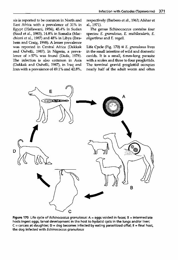
Infection with Cestodes (TaDeworms) 371
sis is reported to be common in North and East Africa with a prevalence of 31% in Egypt (Hallawani, 1956), 45.4% in Sudan (Saad et al., 1983), 14.8% in Somalia (Mac- chioni et al., 1987) and 48% in Libya (Ibra- hem and Craig, 1998). A lesser prevalence was reported in Central Africa (Dakkak and Ouhelli, 1987). In Nigeria, a preva- lence of >57% was found (Dada, 1978). The infection is also common in Asia (Dakkak and Ouhelli, 1987), in Iraq and Iran with a prevalence of 49.1% and 42.8%,
respectively (Barber0 et al., 1963; Afshar et al., 1971).
The genus Echinococcus contains four species: E. granulosus, E. rnultilocularis, E. oligarthrus and E. vogeli.
Life Cycle (Fig. 170) E. granulosus lives in the small intestine of wild and domestic canids. It is a small, 6mm-long parasite with a scolex and three to four proglottids. The terminal gravid proglottid occupies nearly half of the adult worm and often
D
b- Figure 170 Life cycle of Echinococcus granulosus: A = eggs voided in feces; B = intermediate hosts ingest eggs; larval development in the host t o hydatid cysts in the lungs and/or liver; C = carcass at slaughter; D = dog becomes infected by eating parasitized offal; E = final host, the dog infected with Echinococcus granulosus
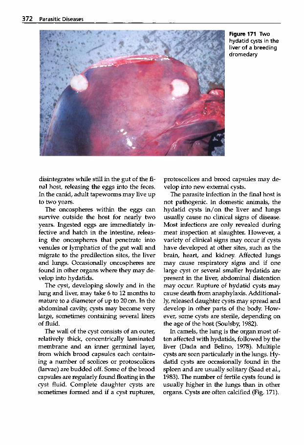
372 Parasitic Diseases
disintegrates while still in the gut of the fi- nal host, releasing the eggs into the feces. In the canid, adult tapeworms may live up to two years.
The oncospheres within the eggs can survive outside the host for nearly two years. Ingested eggs are immediately in- fective and hatch in the intestine, releas- ing the oncospheres that penetrate into venules or lymphatics of the gut wall and migrate to the predilection sites, the liver and lungs. Occasionally oncospheres are found in other organs where they may de- velop into hydatids.
The cyst, developing slowly and in the lung and liver, may take 6 to 12 months to mature to a diameter of up to 20 cm. In the abdominal cavity, cysts may become very large, sometimes containing several liters of fluid.
The wall of the cyst consists of an outer, relatively thick, concentrically laminated membrane and an inner germinal layer, from which brood capsules each contain- ing a number of scolices or protoscolices (larvae) are budded off. Some of the brood capsules are regularly found floating in the cyst fluid. Complete daughter cysts are sometimes formed and if a cyst ruptures,
Figure 171 Two hydatid cysts in the liver of a breeding dromedary
protoscolices and brood capsules may de- velop into new external cysts.
The parasite infection in the final host is not pathogenic. In domestic animals, the hydatid cysts in/on the liver and lungs usually cause no clinical signs of disease. Most infections are only revealed during meat inspection at slaughter. However, a variety of clinical signs may occur if cysts have developed at other sites, such as the brain, heart, and kidney. Affected lungs may cause respiratory signs and if one large cyst or several smaller hydatids are present in the liver, abdominal distention may occur. Rupture of hydatid cysts may cause death from anaphylaxis. Additional- ly, released daughter cysts may spread and develop in other parts of the body. How- ever, some cysts are sterile, depending on the age of the host (Soulsby, 1982).
In camels, the lung is the organ most of- ten affected with hydatids, followed by the liver (Dada and Belino, 1978). Multiple cysts are seen particularly in the lungs. Hy- datid cysts are occasionally found in the spleen and are usually solitary (Saad et al., 1983). The number of fertile cysts found is usually higher in the lungs than in other organs. Cysts are often calcified (Fig. 171).
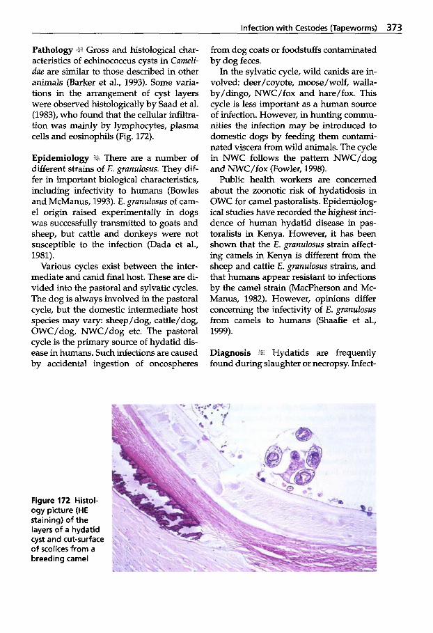
Infection with Cestodes (Tapeworms) 373
Pathology 19 Gross and histological char- acteristics of echinococcus cysts in Cumeli- due are similar to those described in other animals (Barker et al., 1993). Some varia- tions in the arrangement of cyst layers were observed histologically by Saad et al. (1983), who found that the cellular infiltra- tion was mainly by lymphocytes, plasma cells and eosinophils (Fig. 172).
i There are a number of different strains of E. grunulosus. They dif- fer in important biological characteristics, including infectivity to humans (Bowles and McManus, 1993). E. grunulosus of cam- el origin raised experimentally in dogs was successfully transmitted to goats and sheep, but cattle and donkeys were not susceptible to the infection (Dada et al., 1981).
Various cycles exist between the inter- mediate and canid final host. These are di- vided into the pastoral and sylvatic cycles. The dog is always involved in the pastoral cycle, but the domestic intermediate host species may vary: sheep/dog, cattle/dog, OWC/dog, NWC/dog etc. The pastoral cycle is the primary source of hydatid dis- ease in humans. Such infections are caused by accidental ingestion of oncospheres
Figure 172 Histol- ogy picture (HE staining) of the layers of a hydatid cyst and cut-surface of scolices from a breeding camel
from dog coats or foodstuffs contaminated by dog feces.
In the sylvatic cycle, wild canids are in- volved: deer/coyote, moose/wolf, walla- by/dingo, NWC/fox and hare/fox. This cycle is less important as a human source of infection. However, in hunting commu- nities the infection may be introduced to domestic dogs by feeding them contami- nated viscera from wild animals. The cycle in NWC follows the pattern NWC/dog and NWC/fox (Fowler, 1998).
Public health workers are concerned about the zoonotic risk of hydatidosis in OWC for camel pastoralists. Epidemiolog- ical studies have recorded the highest inci- dence of human hydatid disease in pas- toralists in Kenya. However, it has been shown that the E . grunulosus strain affect- ing camels in Kenya is different from the sheep and cattle E. grunulosus strains, and that humans appear resistant to infections by the camel strain (MacPherson and Mc- Manus, 1982). However, opinions differ concerning the infectivity of E. grunulosus from camels to humans (Shaafie et al., 1999).
1 Hydatids are frequently found during slaughter or necropsy. Infect-
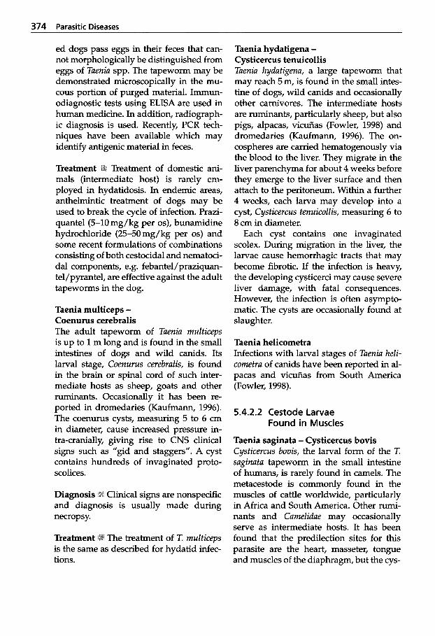
374 Parasitic Diseases
ed dogs pass eggs in their feces that can- not morphologically be distinguished from eggs of Tueniu spp. The tapeworm may be demonstrated microscopically in the mu- cous portion of purged material. Immun- odiagnostic tests using ELISA are used in human medicine. In addition, radiograph- ic diagnosis is used. Recently, PCR tech- niques have been available which may identify antigenic material in feces.
Treatment of domestic ani- mals (intermediate host) is rarely em- ployed in hydatidosis. In endemic areas, anthelmintic treatment of dogs may be used to break the cycle of infection. Prazi- quantel(5-10mg/kg per os), bunamidine hydrochloride (25-50 mg/kg per 0s) and some recent formulations of combinations consisting of both cestocidal and nematoci- dal components, e.g. febantel/praziquan- tel/pyrantel, are effective against the adult tapeworms in the dog.
Taenia multiceps - Coenurus cerebralis The adult tapeworm of Tueniu multiceps is up to 1 m long and is found in the small intestines of dogs and wild canids. Its larval stage, Coenurus cerebralis, is found in the brain or spinal cord of such inter- mediate hosts as sheep, goats and other ruminants. Occasionally it has been re- ported in dromedaries (Kaufmann, 1996). The coenurus cysts, measuring 5 to 6 cm in diameter, cause increased pressure in- tra-cranially, giving rise to CNS clinical signs such as "gid and staggers". A cyst contains hundreds of invaginated proto- scolices.
Diagnosis iliii Clinical signs are nonspecific and diagnosis is usually made during necropsy.
Treatment The treatment of T. multiceps is the same as described for hydatid infec- tions.
Taenia hydatigena - Cysticercus tenuicollis Tueniu hydutigenu, a large tapeworm that may reach 5 m, is found in the small intes- tine of dogs, wild canids and occasionally other carnivores. The intermediate hosts are ruminants, particularly sheep, but also pigs, alpacas, vicufias (Fowler, 1998) and dromedaries (Kaufmann, 1996). The on- cospheres are carried hematogenously via the blood to the liver. They migrate in the liver parenchyma for about 4 weeks before they emerge to the liver surface and then attach to the peritoneum. Within a further 4 weeks, each larva may develop into a cyst, Cysticercus tenuicollis, measuring 6 to 8 cm in diameter.
Each cyst contains one invaginated scolex. During migration in the liver, the larvae cause hemorrhagic tracts that may become fibrotic. If the infection is heavy, the developing cysticerci may cause severe liver damage, with fatal consequences. However, the infection is often asympto- matic. The cysts are occasionally found at slaughter.
Taenia helicometra Infections with larval stages of Tueniu heli- cometru of canids have been reported in al- pacas and vicuiias from South America (Fowler, 1998).
5.4.2.2 Cestode Larvae Found in Muscles
Taenia saginata - Cysticercus bovis Cysticercus bovis, the larval form of the T. suginutu tapeworm in the small intestine of humans, is rarely found in camels. The metacestode is commonly found in the muscles of cattle worldwide, particularly in Africa and South America. Other rumi- nants and Cumelidue may occasionally serve as intermediate hosts. It has been found that the predilection sites for this parasite are the heart, masseter, tongue and muscles of the diaphragm, but the cys-
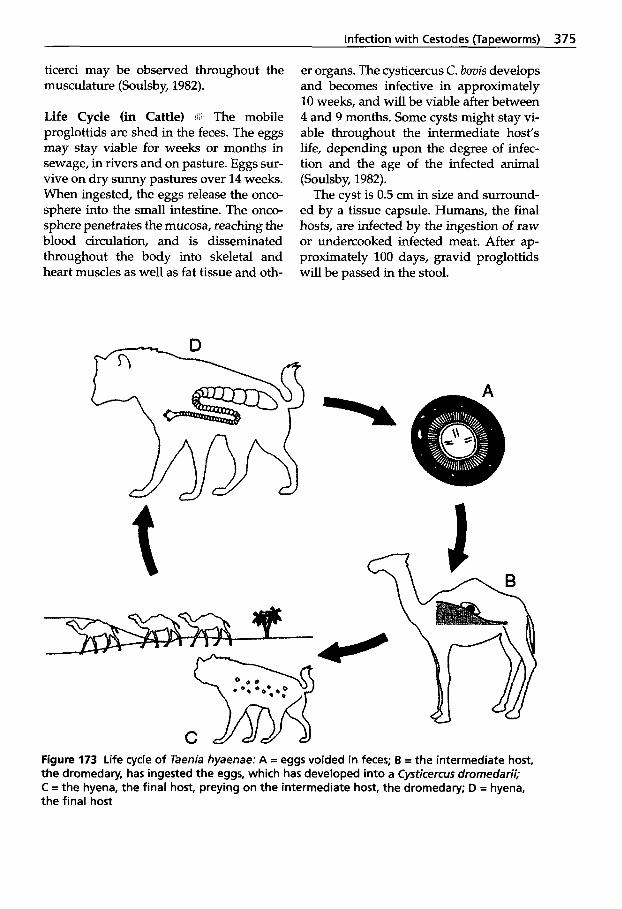
Infection with Cestodes (Tapeworms) 375
ticerci may be observed throughout the musculature (Soulsby, 1982).
Life Cycle (in Cattle) '2. The mobile proglottids are shed in the feces. The eggs may stay viable for weeks or months in sewage, in rivers and on pasture. Eggs sur- vive on dry sunny pastures over 14 weeks. When ingested, the eggs release the onco- sphere into the small intestine. The onco- sphere penetrates the mucosa, reaching the blood circulation, and is disseminated throughout the body into skeletal and heart muscles as well as fat tissue and oth-
er organs. The cysticercus C. bovis develops and becomes infective in approximately 10 weeks, and will be viable after between 4 and 9 months. Some cysts might stay vi- able throughout the intermediate host's life, depending upon the degree of infec- tion and the age of the infected animal (Soulsby, 1982).
The cyst is 0.5 cm in size and surround- ed by a tissue capsule. Humans, the final hosts, are infected by the ingestion of raw or undercooked infected meat. After ap- proximately 100 days, gravid proglottids will be passed in the stool.
Figure 173 Life cycle of Taenia hyaenae: A = eggs voided in feces; B = the intermediate host, the dromedary, has ingested the eggs, which has developed into a Cysticercus dromedarii; C = the hyena, the final host, preying on the intermediate host, the dromedary; D = hyena, the final host
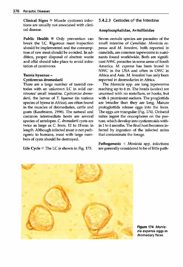
376 Parasitic Diseases
I Muscle cysticerci infec- tions are usually not associated with clini- cal disease.
Only prevention can break the LC. Rigorous meat inspection should be implemented and the consump- tion of raw meat should be avoided. In ad- dition, proper disposal of abattoir waste and offal should take place to avoid infes- tation of carnivores.
Taenia hyaenae - Cysticercus dromedarii There are a large number of taeniid ces- todes with an unknown LC in wild car- nivores’ small intestine. Cysficercus drome- darii, the larvae of T. hyaenae (in various species of hyena in Africa), are often found in the muscles of dromedaries, cattle and goats (Kaufmann, 1996). The natural and common intermediate hosts are several species of antelopes. C. dromedarii cysts are twice as large as C. bovis, 12 to 18mm in length. Although infected meat is not path- ogenic to humans, meat with large num- bers of cysts should be destroyed.
Life Cycle !hi The LC is shown in Fig. 173.
5.4.2.3 Cestodes of the Intestine
Anoplocephalidae, Avitellinidae
Seven cestode species are parasites of the small intestine of Camelidae. Moniezia ex- pansa and M. benedeni, both reported in camelids, are common tapeworms in rumi- nants found worldwide. Both are signifi- cant NWC parasites in some areas of South America. M. expansa has been found in NWC in the USA and often in OWC in Africa and Asia. M . benedeni has only been reported in dromedaries in Africa.
The Moniezia spp. are long tapeworms reaching up to 6 m. The heads (scolex) are unarmed with no rostellum, or hooks, but with 4 prominent suckers. The proglottids are broader than they are long. Mature protoglottids release eggs into the feces. The eggs are triangular (Fig. 174). Oribatid mites ingest the oncospheres on the pas- ture, which develop into cysticercoids with- in 1 to 4 months. The final host becomes in- fected by ingestion of the infected mites that contaminate the forage.
Pathogenesis ! i i0 Moniezia spp. infections are generally considered to be of little path-
Figure 174 Monie- zia expansa eggs in dromedary feces
Infection with Cestodes (TaDeworms) 377
ogenic importance. However, there are indications that heavy infections may im- pair nutrition and cause diarrhea, debility, and sometimes obstruction of the intes- tine.
Stilesia
Three Stilesiu spp. are found in dromeda- ries. S. globipunctutu and S. centripunctutu are widely reported in Africa and Asia, whereas S. vittutu has only occasionally been observed (Dakkak and Ouhelli, 1987).
S. globipunctutu occurs primarily in the small intestine of ruminants in southern Europe, Africa and Asia.
Life Cycle 1 1 Little is known about the LC of Stilesia parasites. Oribatid mites may play an important role, as shown by Souls- by (1982) in Chad and India.
The immature worms penetrate the mu- cosa of the duodenum and jejunum. Nod-
ules are formed with proliferate inflamma- tion and epithelial desquamation (Arnjadi, 1971). The head and the anterior part of the parasite are embedded in the nodule and the posterior proglottids of the worm move freely in the intestinal lumen. The in- fection may lead to death.
Other tapeworms belonging to Avitel- linidae have been reported in camelids. Avitellinu woodlundi has been found in cam- els in Africa (Dakkak and Ouhelli, 1987), and according to Kaufmann (1996) Avitelli- na centripunctutu is widespread in these an- imals in Africa and Asia. Thyzunieziu spe- cies are reported in llamas and T. ovillu in camels in Chad (Graber, 1966; Graber et al., 1967). Little is known of the LC and epide- miology of the latter species. They seem to have no pathogenic significance.
Treatment Praziquantel has been shown to be effective (15mg/kg in sheep and goats).
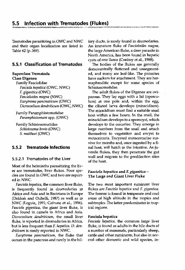
Trematodes parasitizing in OWC and NWC and their organ localization are listed in Table 62 (p. 369).
5.5.1 Classification of Trematodes
Superclass Trematoda Class Digenea
Family Fasciolidae FuscioZu heputica (OWC, NWC) F. gigunficu (OWC) Fuscioloides mugna (NWC) Eu ytremu puncreuticum (OWC) Dicrocoelium dendrificum (OWC, NWC)
Purumphisfomum spp. (OWC)
Schisfosomu bovis (OWC) S. mutfheei (OWC)
Family Paramphistomatidae
Family Schistosomatidae
5.5.2 Trematode Infections
5.5.2.1 Trematodes of the Liver
Most of the helminths parasitizing the liv- er are trematodes, liver flukes. Four spe- cies are found in OWC and two are report- ed in NWC.
Fasciola hepuficu, the common liver fluke, is frequently found in dromedaries in Africa and Asia and in Bactrians in Europe (Dakkak and Ouhelli, 1987) as well as in NWC (Leguia, 1991; C a h e et al., 1996). Fusciolu gigunficu, the giant liver fluke, is also found in camels in Africa and Asia. Dicrocoelium dendriticum, the small liver fluke, is reported in dromedaries in Africa, but is less frequent than E hepuficu. D. den- driticum is rarely reported in NWC.
Eurytremu puncreuticum, the fluke that occurs in the pancreas and rarely in the bil-
iary ducts, is rarely found in dromedaries. An immature fluke of FascioZoides mugnu, the large American fluke, a deer parasite in North America, has been found in hepatic cysts of one llama (Conboy et al., 1988).
The bodies of the flukes are generally dorsoventrally flattened and unsegment- ed, and many are leaf-like. The parasites have suckers for attachment. They are her- maphroditic except for some species of Schistosomatidae.
The adult flukes of the Digenea are ovi- parous. They lay eggs with a lid (opercu- lum) at one pole and, within the egg, the ciliated larva develops (miracidium). The miracidium must find a suitable snail host within a few hours. In the snail, the miracidium develops to a sporocyst, which develops to the cercariae. They emerge in large numbers from the snail and attach themselves to vegetation and encyst to metacercaria. Encysted metacercariae sur- vive for months and, once ingested by a fi- nal host, will hatch in the intestine. As ju- venile flukes, they then penetrate the gut wall and migrate to the predilection sites of the host.
Fasciola hepatica and E gzgantica - The Large and Giant Liver Fluke
The two most important ruminant liver flukes are Fasciola hepatica and E giguntica. The former is found in temperate and cool areas of high altitude in the tropics and subtropics. The latter predominates in trop- ical regions.
Fasciola hepatica Fusciolu heputicu, the common large liver fluke, is found as adults in the bile ducts of a number of mammals, particularly sheep, cattle and other ruminants, but also in sev- eral other domestic and wild species, in-
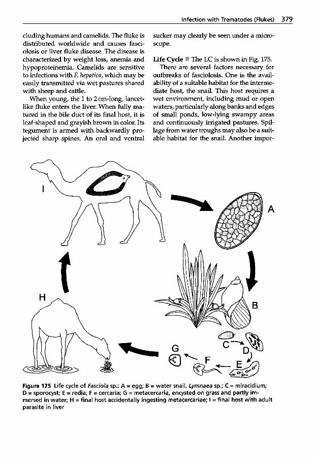
Infection with Trematodes (Flukes) 379
cluding humans and camelids. The fluke is distributed worldwide and causes fasci- olosis or liver fluke disease. The disease is characterized by weight loss, anemia and hypoproteinemia. Camelids are sensitive to infections with E hepatica, which may be easily transmitted via wet pastures shared with sheep and cattle.
When young, the 1 to 2 cm-long, lancet- like fluke enters the liver. When fully ma- tured in the bile duct of its final host, it is leaf-shaped and grayish brown in color. Its tegument is armed with backwardly pro- jected sharp spines. An oral and ventral
sucker may clearly be seen under a micro- scope.
Life Cycle I The LC is shown in Fig. 175. There are several factors necessary for
outbreaks of fasciolosis. One is the avail- ability of a suitable habitat for the interme- diate host, the snail. This host requires a wet environment, including mud or open waters, particularly along banks and edges of small ponds, low-lying swampy areas and continuously irrigated pastures. Spil- lage from water troughs may also be a suit- able habitat for the snail. Another impor-
Figure 175 Life cycle of Fasciola sp.: A = egg; B = water snail, Lymnaea sp.; C = miracidium; D = sporocyst; E = redia; F = cercaria; G = metacercaria, encysted on grass and partly im- mersed in water; H = final host accidentally ingesting metacercariae; I = final host with adult parasite in liver
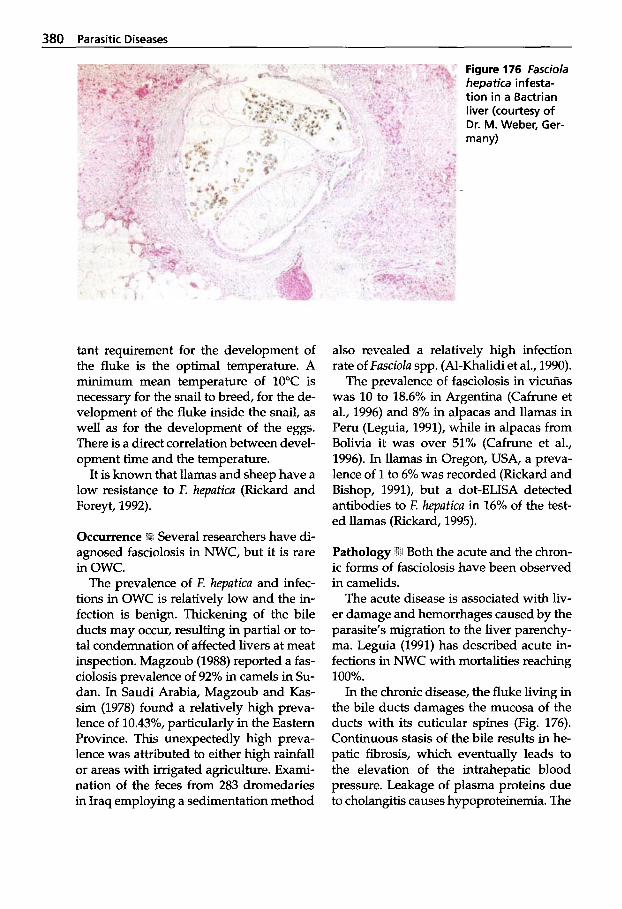
380 Parasitic Diseases
tant requirement for the development of the fluke is the optimal temperature. A miniium mean temperature of 10°C is necessary for the snail to breed, for the de- velopment of the fluke inside the snail, as well as for the development of the eggs. There is a direct correlation between devel- opment time and the temperature.
It is known that llamas and sheep have a low resistance to E hepatica (Rickard and Foreyt, 1992).
Several researchers have di- agnosed fasciolosis in NWC, but it is rare in OWC.
The prevalence of E hepatica and infec- tions in OWC is relatively low and the in- fection is benign. Thickening of the bile ducts may occur, resulting in partial or to- tal condemnation of affected livers at meat inspection. Magzoub (1988) reported a fas- ciolosis prevalence of 92% in camels in Su- dan. In Saudi Arabia, Magzoub and Kas- sim (1978) found a relatively high preva- lence of 10.43%, particularly in the Eastern Province. This unexpectedly high preva- lence was attributed to either high rainfall or areas with irrigated agriculture. Exami- nation of the feces from 283 dromedaries in Iraq employing a sedimentation method
Figure 176 Fasciola hepatica infesta- tion in a Bactrian liver (courtesy of Dr. M. Weber, Ger- many)
also revealed a relatively high infection rate of Fasciola spp. (Al-Khalidi et al., 1990).
The prevalence of fasciolosis in vicuiias was 10 to 18.6% in Argentina (Cafrune et al., 1996) and 8% in alpacas and llamas in Peru (Leguia, 1991), while in alpacas from Bolivia it was over 51% (Cafrune et al., 1996). In llamas in Oregon, USA, a preva- lence of 1 to 6% was recorded (Rickard and Bishop, 1991), but a dot-ELISA detected antibodies to F. hepatica in 16% of the test- ed llamas (Rickard, 1995).
Pathology ilki Both the acute and the chron- ic forms of fasciolosis have been observed in camelids.
The acute disease is associated with liv- er damage and hemorrhages caused by the parasite’s migration to the liver parenchy- ma. Leguia (1991) has described acute in- fections in NWC with mortalities reaching 100%.
In the chronic disease, the fluke living in the bile ducts damages the mucosa of the ducts with its cuticular spines (Fig. 176). Continuous stasis of the bile results in he- patic fibrosis, which eventually leads to the elevation of the intrahepatic blood pressure. Leakage of plasma proteins due to cholangitis causes hypoproteinemia. The
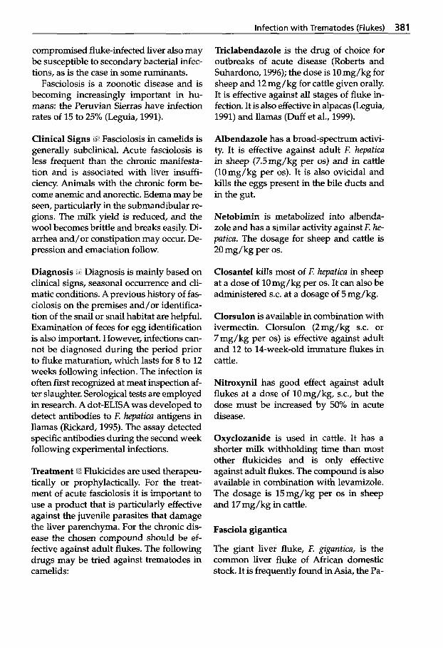
Infection with Trematodes (Flukes) 381
compromised fluke-infected liver also may be susceptible to secondary bacterial infec- tions, as is the case in some ruminants.
Fasciolosis is a zoonotic disease and is becoming increasingly important in hu- mans: the Peruvian Sierras have infection rates of 15 to 25% (Leguia, 1991).
Clinical Signs ?Kt Fasciolosis in camelids is generally subclinical. Acute fasciolosis is less frequent than the chronic manifesta- tion and is associated with liver insuffi- ciency. Animals with the chronic form be- come anemic and anorectic. Edema may be seen, particularly in the submandibular re- gions. The milk yield is reduced, and the wool becomes brittle and breaks easily. Di- arrhea and/or constipation may occur. De- pression and emaciation follow.
Diagnosis GI& Diagnosis is mainly based on clinical signs, seasonal occurrence and cli- matic conditions. A previous history of fas- ciolosis on the premises and/or identifica- tion of the snail or snail habitat are helpful. Examination of feces for egg identification is also important. However, infections can- not be diagnosed during the period prior to fluke maturation, which lasts for 8 to 12 weeks following infection. The infection is often first recognized at meat inspection af- ter slaughter. Serological tests are employed in research. A dot-ELISA was developed to detect antibodies to E hepatica antigens in llamas (Rickard, 1995). The assay detected specific antibodies during the second week following experimental infections.
Flukicides are used therapeu- tically or prophylactically. For the treat- ment of acute fasciolosis it is important to use a product that is particularly effective against the juvenile parasites that damage the liver parenchyma. For the chronic dis- ease the chosen compound should be ef- fective against adult flukes. The following drugs may be tried against trematodes in camelids:
Triclabendazole is the drug of choice for outbreaks of acute disease (Roberts and Suhardono, 1996); the dose is 10 mg/kg for sheep and 12 mg/kg for cattle given orally. It is effective against all stages of fluke in- fection. It is also effective in alpacas (Leguia, 1991) and llamas (Duff et al., 1999).
Albendazole has a broad-spectrum activi- ty. It is effective against adult E hepatica in sheep (7.5mg/kg per 0s) and in cattle (10mg/kg per 0s). It is also ovicidal and kills the eggs present in the bile ducts and in the gut.
Netobimin is metabolized into albenda- zole and has a similar activity against E he- patica. The dosage for sheep and cattle is 20mg/kg per 0s.
Closantel kills most of E heputica in sheep at a dose of 10 mg/kg per 0s. It can also be administered S.C. at a dosage of 5 mg/kg.
Clorsulon is available in combination with ivermectin. Clorsulon (2 mg/kg S.C. or 7mg/kg per 0s) is effective against adult and 12 to 14-week-old immature flukes in cattle.
Nitroxynil has good effect against adult flukes at a dose of 10mg/kg, s.c., but the dose must be increased by 50% in acute disease.
Oxyclozanide is used in cattle. It has a shorter milk withholding time than most other flukicides and is only effective against adult flukes. The compound is also available in combination with levamizole. The dosage is 15mg/kg per 0s in sheep and 17mglkg in cattle.
Fasciola gigantica
The giant liver fluke, F. gigantica, is the common liver fluke of African domestic stock. It is frequently found in Asia, the Pa-
382 Parasitic Diseases
cific Islands, southern USA, southern Eu- rope and the Middle East. The epidemiolo- gy is similar to E hepatica but it is larger. It can reach 7.5 cm in length.
Life Cycle i4t The intermediate hosts are snails belonging to the genus Lyrnnaea and are primarily aquatic, found in streams, ir- rigation channels and wet and swampy areas. The LC is similar to F. hepatica with
the exception that the different stages and the total cycle are longer. The prepatent pe- riod is 13 to 16 weeks.
Fascioloides magna - The Large American Liver Fluke
Fascioloides rnagna, the large American liver fluke, is a large liver fluke mainly para- sitizing moose and deer in North America,
q H A
G - U Y - 7
\ J
Figure 177 Life cycle of Dicrocoelium dendriticum: A = egg; B = intermediate host, land snail ingesting embryonated egg; C, = sporocyst; Cz = daughter sporocyst; D = cercariae released by the snail in clusters; E = cercariae eaten by ants (Formica); F = development of metacer- cariae in infected ants, second intermediate host; G = final host accidentally ingesting infect- ed ants; H = final host with adult parasite
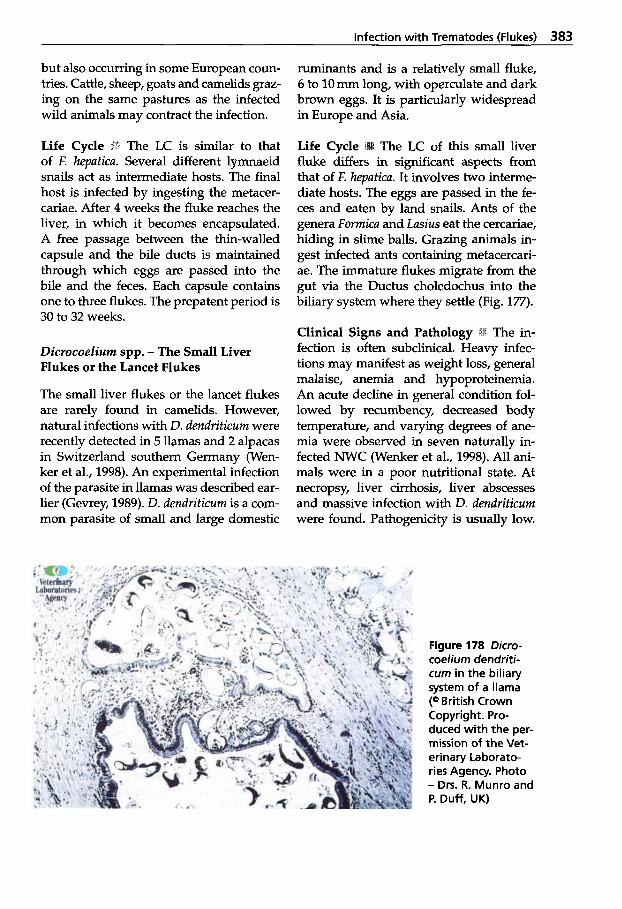
Infection with Trematodes (Flukes) 383
but also occurring in some European com- tries. Cattle, sheep, goats and camelids graz- ing on the same pastures as the infected wild animals may contract the infection.
Life Cycle Pi The LC is similar to that of E hepatica. Several different lymnaeid snails act as intermediate hosts. The final host is infected by ingesting the metacer- cariae. After 4 weeks the fluke reaches the liver, in which it becomes encapsulated. A free passage between the thin-walled capsule and the bile ducts is maintained through which eggs are passed into the bile and the feces. Each capsule contains one to three flukes. The prepatent period is 30 to 32 weeks.
Dicrocoelium spp. - The Small Liver Flukes or the Lancet Flukes
The small liver flukes or the lancet flukes are rarely found in camelids. However, natural infections with D. dendrificum were recently detected in 5 llamas and 2 alpacas in Switzerland southern Germany (Wen- ker et al., 1998). An experimental infection of the parasite in llamas was described ear- lier (Gevrey, 1989). D. dendrificum is a com- mon parasite of small and large domestic
ruminants and is a relatively small fluke, 6 to 10 mm long, with operculate and dark brown eggs. It is particularly widespread in Europe and Asia.
The LC of this small liver fluke differs in significant aspects from that of F. hepatica. It involves two interme- diate hosts. The eggs are passed in the fe- ces and eaten by land snails. Ants of the genera Formica and Lasius eat the cercariae, hiding in slime balls. Grazing animals in- gest infected ants containing metacercari- ae. The immature flukes migrate from the gut via the Ductus choledochus into the biliary system where they settle (Fig. 177).
Clinical Signs and Pathology fection is often subclinical. Heavy infec- tions may manifest as weight loss, general malaise, anemia and hypoproteinemia. An acute decline in general condition fol- lowed by recumbency, decreased body temperature, and varying degrees of ane- mia were observed in seven naturally in- fected NWC (Wenker et al., 1998). All ani- mals were in a poor nutritional state. At necropsy, liver cirrhosis, liver abscesses and massive infection with D. dendrificum were found. Pathogenicity is usually low.
Figure 178 Dicro- coelium dendriti- cum in the biliary system of a llama (Q British Crown Copyright. Pro- duced with the per- mission of the Vet- erinary Laborato- ries Agency. Photo - Drs. R. Munro and P. Duff, UK)
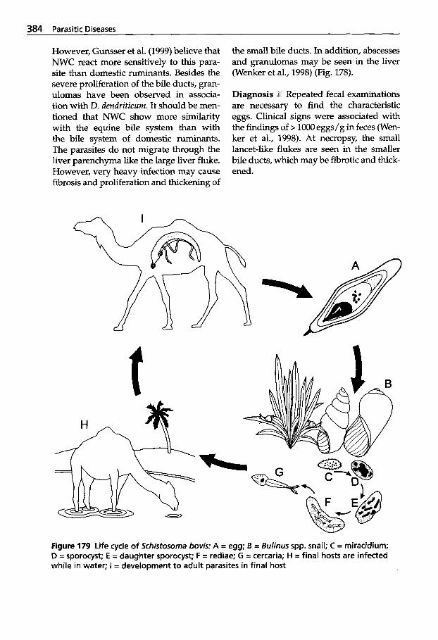
384 Parasitic Diseases
However, Gunsser et al. (1999) believe that NWC react more sensitively to this para- site than domestic ruminants. Besides the severe proliferation of the bile ducts, gran- ulomas have been observed in associa- tion with D. dendriticum. It should be men- tioned that NWC show more similarity with the equine bile system than with the bile system of domestic ruminants. The parasites do not migrate through the liver parenchyma like the large liver fluke. However, very heavy infection may cause fibrosis and proliferation and thickening of
the small bile ducts. In addition, abscesses and granulomas may be seen in the liver (Wenker et al., 1998) (Fig. 178).
Diagnosis (L Repeated fecal examinations are necessary to find the characteristic eggs. Clinical signs were associated with the findings of > 1000 eggs/g in feces (Wen- ker et al., 1998). At necropsy, the small lancet-like flukes are seen in the smaller bile ducts, which may be fibrotic and thick- ened.
Figure 179 Life cycle of Schistosorna bovis: A = egg; B = Bulinus spp. snail; C = miracidium; D = sporocyst; E = daughter sporocyst; F = rediae; G = cercaria; H = final hosts are infected while in water; I = development to adult parasites in final host
Infection with Trematodes (Flukes) 385
Treatment 'k Albendazole at a dose rate of 15 mg/kg per 0s and netobimin at 20 mg/ kg per 0s are effective. Praziquantel at a dose rate of 50mg/kg per 0s was well tolerated by a few llamas and alpacas, but achieved only a 90% reduction of eggs in the feces (Wenker et al., 1998).
5.5.2.2 Paramphistomatidae - Rumen Flukes
Different species of rumen flukes, Purumphis- tomum, have been found in camels (Kauf- mann, 1996). They are not considered harmful to the host unless there is a mas- sive invasion of immature flukes attached to the intestinal mucosa.
5.5.2.3 Schistosomatidae
Schistosomu bovis and S. muttheei causing bilharziosis are rarely found in OWC and are considered occasional parasites. They are not found in NWC.
Schistosomatidae trematodes are dimor- phic. They inhabit their hosts' blood ves- sels. The female worm is slender and in some species longer than the male, which harbors the female in a ventral, gutter-like groove: the gynaecophoric canal. Several species of Schistosoma cause severe disease in humans. Animals may act as reservoirs of the infection.
Life Cycle shown in Fig. 179.
The LC of Schistosorna bovis is
5.6.1 Classification of Hirudinea
Phylum Annelida Class Hirudinea
Limnatis nilotica
5.6.2 Infection with Leeches
Leeches are occasional parasites feeding on various animals. After their blood meal, they engorge and drop off their host. Leeches have pharyngeal glands from which they secrete an anticoagulant sub- stance when piercing the skin or mucosa for a blood meal. Bleeding may continue for some time after the parasites have engorged.
Eight Limnatis nilotica leeches were found attached to the pharyngeal mucosa of a 2-year-old male dromedary in Iraq ( A l - h i and Al-Shareefi, 1995). The camel had difficulty in breathing and exhibited edema of the face and neck. It released snoring sounds, and had difficulty in swal- lowing food and water.
References Afshar, A., I. Nazariani and B. Baghbannaseer.
1971. A survey of the incidence of hydatid disease in camels in South Iran. Brit. Vet. J.
Al-Ani, F.K. and M.R. Al-Shareefi. 1995. Obser- vation on medical leech (Limnatis nilotica) in a camel in Iraq. J. Camel Prac. and Res. 2 145.
Al-Khalidi, N.W., M.A. Hassan and A.F. Al- Taee. 1990. Faecal incidence of Fasciola spp. and E u y t r e m pancreaticurn eggs in camels (Camelus dromedarius) in Iraq. 1. Vet. Parasitol.
Arnjadi, A.R. 1971. Studies on histopathology of Stilesia globipunctata infections in Iran. Vet. Rec. 88: 486-488.
Barbero, B., M. Al Dabagh, A.A. Al Safar and EM. Ali. 1963. The zoonosis of animal para-
127: 544-546.
4: 75-76.
sites in Iraq. W. Hydatid disease. Ann. Trop. Med. Parasitol. 5 7 499-510.
Barker, I.K., A.A. Van Dreumel and N. Palmer. 1993. The alimentary system. In: K.V.F. Jubb, P.C. Kennedy and N. Palmer (eds.): Patholo- gy of Domestic Animals, V012.4~ ed. Acade- mic Press, New York, pp. 289-292.
Bowles, J. and D.P. McManus. 1993. Molecular variation in Echinococcus. Acta Trop. 53:
Cafrune, M.M., G.E. Rebuffi, A.B. Gaido and D.H. Aguirre. 1996. Fasciola hepatica in semi- captive vicuiias (Vicugna vicugna) in north- west Argentina. Vet. Rec. 139: 97.
Conboy, G.A., T.D. OBrien and D.L. Stevens. 1988. A natural infection of Fasciola magna in a llama (Lama glama). J, Parasitol. 7 4 345- 346.
Dada, B.J.O. 1978. Incidence of hydatid disease in camels slaughtered at Kano abattoir. Trop. Anim. Hlth. Prod. 1 0 204.
Dada, B.J.O. and E.D. Belino. 1978. Prevalence of hydatidosis and cysticercosis in slaugh- tered livestock at city abattoir, Kano, Nigeria. Vet. Rec. 103: 311-312.
Dada, B.J.O., E.D. Beliio, D.S. Adegboye and A.N. Mohammed. 1981. Experimental trans- mission of Echinococcus granulosus of camel- dog origin to goats, sheep, cattle and don- keys. lnt. J. Zoon. 8 33-43.
Dakkak, A. and H. Ouhelli. 1987. Helminths and helminthoses of the dromedary. A re- view of the literature. Rev. Sibliogr. Rev. Sci. Tech. Ofice lnt. Epiz. 6 423-445,447-461.
Duff, J.P., A.J. Maxwell and J.R. Claxton. 1999. Chronic and fatal fascioliasis in llamas in the UK. Vet. Rec. 145: 315-316.
Fowler, M.E. 1998. Medicine and Surgery of South American Camelids: Llama, Alpaca, Vicuiia, Guanaco, 2nd ed. Iowa State Univer- sity Press, Ames, USA.
Gevrey, J. 1989. Lamas et moutons: Observation d'intertransmissibilit6 helminthique. Bull. SOC. FranF. Parasitol. 7: 245-247.
Graber, M. 1966. Study under certain conditions in Africa of the action of thiabendazole against various helminths of domestic ani- mals. II. Rev. El&. Mid. vit. Pays Trop. 19:
291-305.
527-543.
Infection with Hirudinea (Leeches) 387
Graber, M., R. Tab0 and J. Service. 1967. Enqu6te sur les helminthes du dromadaire tchadien. Etude des strongyloses gastro-intestinales et de l'haemonchose h Haemoncus longistipes. Rev. Elev. Mid. vet. Pays Trop. 20: 227-254.
Gunsser, I., T. Haenichen und J. Maierl. 1999. Leberegelbefall bei Neuweltkameliden. Tier- urztl. Prax. 27 (G): 187-192.
Hallawani, A. 1956. Hydatid disease in Egypt. Arch. lnt. Hydatid. 15: 374-375.
Ibrahem, M.M. and P.S. Craig. 1998. Prevalence of cystic echinococcosis in camels (Camelus dromedarius) in Libya. I. Helminthol. 72: 27-31.
Kaufmann, J. 1996. Parasitic Infections of Do- mestic Animals - A Diagnostic Manual. Birk- hauser Verlag. Basel, Boston, Berlin.
Leguia, G. 1991. The epidemiology and eco- nomic impact of llama parasites. Parasitol. To- day 7 54-56.
Macchioni, G., M. Arispici, P. Lanfranchi and F. Testi. 1987. Experimental infection of sheep and monkeys with the camel strain of Echi- nococcus granulosus. In: S. Geerts, V. Kumar and J. Brandt (eds.): Helminth Zoonosis. Dor- drecht, Netherlands, Martinus Nijhoff Pub- lishers. pp. 24-28.
Magwub, M. and AA. Kassim. 1978. The pma- lence of fasciolosis in Saudi Arabia. Trop. Anim. Hlth Prod. 10: 205-206.
Magzoub, M. 1988. The prevalence of fasciolosis in camels in Sudan. Sudan Vet. J. 12: 23-26.
MacPherson C.N.L. and D.P. McManus. 1982. A comparative study of Echinococcus granulosus from human and animal hosts in Kenya us- ing isoelectric focusing and isoenzyme analy- sis. Int. J. Parasitol. 12: 515-521.
Rickard, L.G. and W.J. Foreyt. 1992. Experimen- tal fasciolosis in llamas. J. Helminthol. SOC. Wash. 59: 140-144.
Rickard, L.G. 1995. Development of a dot- ELISA test for the detection of serum anti- bodies to Fasciola hepatica antigens in llamas. I. Helminthol. SOC. of Wash. 59: 9.
Roberts, J.A. and X. Suhardono. 1996. Approach- es to the control of fascioliosis in ruminants. J. Parasitol. 26 971-981.
Saad, M.B., E.A. Zien Eldin and M.H. Tag El Din. 1983. Some observations on the preva- lence and pathology of hydatidosis in Su- danese camels (Camelus dromedarius). Rev. Elev. Mid. vet. Pays Trop. 36: 359-363.
Shaafie, LA., A.H. Khan and K. Rambabu. 1999. Biochemical profiles of hydatid cyst fluids of Echinococcus granulosus of human and animal origin in Libya. 1. Helminthol. 73: 255-258.
Soulsby, E.J.L. 1982. Helminths, Arthropods and Protozoa of Domesticated Animals, 7* ed. Bailliere Tindall, London.
Wenker, C., J.M. Hatt, H. Hertzberg, P. Ossent, T. Hhichen, A. Brack and E. Isenbuegel. 1998. Dicrocoeliosis in South American camelids. Tierurztl. Prax. 2 6 355-361.
Further reading
Comick, J.L. 1988. Gastric squamous cell carci- noma and fascioliasis in a llama. Cornell Vet.
Navone, G.T. and M.L. Merino. 1989. Contribu- tion to the knowledge of the endoparasitic fauna of Lama guanicoe Muller, 1776, from the Mitre Peninsula, Tierra del fuego, Argentina. Bol. Chileno Parasitol. 44: 46-51.
Urquhart, G.M., J. Armour, J.L., Duncan, A.M. D m and F.W. Jennings. 1996. Veterinary Parasitology, 2nd ed. Blackwell Science, Ox- ford, UK.
78: 235-241.
Index
Page numbers in bold type refer to figures and/or tables.
A abortion 109,206 abscesses 99,134 Absidia 256 acaricides 319 acid-fast bacilli 84 Actinomyces -,lamae 100 -, pyogenes 117,135 Actinomycetales hyphae 142 adenosine triphosphate (ATP) 150 adenovirus 209 Aedes spp. 334 Aegyptianella 60 African horse sickness 212 -, cardiac form 213 -, cytopathic effect 212 -, equine encephalosis 212 -,midges 212 -, mixed form 213 -, Orbivirus 212 -, pulmonary form 213 -, Reoviridae 212 agranulocyte(s) 43 alpaca 3 Alpaca fever 100 Alternaria 247 Amblyomma 324 -,gemma 329 -, lepidum 329 -, parvitarsum 324
anaerobic infections 21 -,antiserum 28 -, clostridial enterotoxemia 28 -, colorimetric tetrazolium cleavage test
-, hyperimmune serum 28 Anaplasma 59,60,64 -, marginale 59f, 65 anaplasmosis 59 Ancylostomatidae 357 ancylostomatosis 347 Angiostrongylidae 354 angiostrongylosis 347,354 Angiostrongylus cantonensis 347,356 Annelida 386 Anoplocephalidae 369,370,376
-,SPP. 312
(MTT) 28
Anoplurida (sucking lice) 331 anthelmintic -,broad-spectrum 366
-, nematocidal 366 anthrax 33,50 -,bacilli 33 - -, vegetative 33 -, ecchymoses 35 -,edema 35 -, Gram-positive 33 -,hemorrhages 35 -, necrosis 35 -, petechiae 35 -,pulmonary 34 -, septicemic disease 33 -,spores 33 antibiotic(.$ 59 -, resistance 79 antigens 79 antimicrobials 82 Arachnea 312 -, classification 312 Arbovirus (arthropod-borne virus) 209,
-, Akabane virus 234 -, Dhori virus 235 -,flavivirus 235 --,Bani 235 - -, Dengue 235
- -, Wesselsbron 235 - -, Yellow fever 235 -, Kadam virus 234 -, Quaranfil virus 234 Argasidae (soft ticks) 313,323 Arthropoda 312 aspergillosis 246 -, aflatoxicosis 246 -, aflatoxin 247 -, amphotericin I3 248 -, fungal hyphae 247 -, gliotoxin 247 -, granulomas 248 -, immunosuppressive substances 246 -, mycotoxin 247 -, rumenitis 248 -, thiabendazole 248
-,drug 366
234
- -, P o t i ~ k ~ m 235
390 Index
Aspergill us -,Farms 246 -,fumigatus 47,246 -, nidulans 246 -, niger 246 -,terreus 246 atonia 38 auto-vaccines 140 Avitellina -, centripunctata 377 -, woodlandi 369,377 Avitellinidae 369,370,376
B Babesia 286 babesiosis 286 bacillary hemoglobinuria 22 Bacillus -, anthracis 33 -, cereus 44,121 bacterial diseases 264 bacteriemia(s) 44 Bactrian camelids 3 balantidiosis 272,284 -,cilia 285 Balantidium coli 272,284 Bedsonia (Chlamydia) 126 Besnoitia 287 -, besnoiti 299
besnoitiosis 272,298 biting lice 312 biting midges 312 black-quarter (blackleg) 22 blood parameters 37 blowfly 334 bluetongue 214 -, abortion 215,216 -, cyanosis 214 -, epizootic hemorrhagic disease 215 -, lameness 216 -, respiratory syndrome 216 Boophilus microplus 324 booster(s) 172 -, vaccination 35 Boma disease 174 -, stamping-out policy 175 Bornaviridae 174 Bornavirus 174 botfly 334 botryomycosis 138 botulism 31 -, antisera 33
-,spp. 272
-, bacteriophages 31 - -, Tox phage 32 -, complement fixation test 33
-, immunodiffusion 33 -,minerals 31 -, mouse bioassay 33 -, phosphorus 31 -, ruminal fluid 33 -,toxicity 33 bovine respiratory syncytial virus (BE)
bovine viral diarrhea 224 -, agalactia 225 -, congenital defects 225 -, diarrhea 225 -,hemorrhages 225 -, immunotolerant fetus 225 -, non-cytopathic viral infection 225 -, persistently infected calf 225 -, thrombocytopenia 225 Brachycerina 334 bronchitis 99 bronchopneumonia 99 Brucella -,abortus 109 -, melitensis 109 -,ovis 109 Brucellae 109 brucellosis 109,118,121 -, Brucella vaccines 116
-, complement fixation test (CET) 115 -, eradication 115 -, milk ring test (MRT) 115 -, non-specific reactions 114 -, placentitis 113 -, prozones 114 -,Rev1 116 -, rose bengal plate test (RBPT) 115 -, serum agglutination test 114 -, tube agglutination test (TAT) 114f bubonic plague 54 Buffalo fly 334 Bulinus 384 bunostomosis 357 -, bottlejaw 358 Bunostomum spp. 347,357 Bunyaviridae 230 Burkholderia -, mallei 102 -, pseudomallei 101
-,ELISA 33
50,209
-,Buck19 116
Index 391
C calcification 56,57 California mastitis test (CMT) 150 Calliphoridae 312,331,333,334 camel plague 54 -, airborne infection 54 -, anthropozoonosis 54 -, biological warfare 55 -, endemic area(s) 54 -,immunity 55 -, immunization 55 -, insecticide(s) 55 -, louse fly 334
-, nasal bot fly 334 -, pandemic(s) 54 -, pox - -, Chordopoxvirinae 177 --,Dubca 183 - -, Entornopoxvirinae 177 - -, erythematous macules 181 - -, immune status 178 - -, papules 181 - -, pustules 182 - -, systemic poxvirus 177 - -, vaccine 182 - -, vaccinia/variola virus subgroup 178 - -, vesicles 181 -, zooanthroponosis 54 camelid IgG 203 camelid rickettsiosis 64 Camelidae 5'27 -, classification of artiodactylids 5 -, classification of camelids 5 -, estimated population 5
- -, Camelops 5 camelids 3 Camelostrongylus mentulatus 347,352 camelpox 121,176 Camelus -, bactrianus 4 -, dromedarius 4 -, ferus 4 Campylobacter 114 -,fetus 117 Candida albicans 151,249 candidiasis 77,249 -, moniliasis 249 -, mucosal scrapings 252 -, pseudomembranes 250 -, pseudohyphae 250 -,yeast 250
-, milk 112,149
-, Tylopoda
Capillaria 361
Capillariidae 360 capillariosis 347,360 caseous lymphadenitis 134 CBPP see contagious bovine pleuropneu-
Cephalopina titillator 34,275,334,336 Cephenemyia spp. 334,340 Ceratopogonidae (midges) 331,334 -, filarid worm 341 -, infestation Cervidae 355 Cestodes 369 -, classification 370 Chabertia ovina 347,356 Chabertiidae 356 chabertiosis 347,356 Chlamydia -, psittaci 124 -, trachomatis 124 Chlamydiae 125 Chlamydiales 125 chlamydiosis 121,124 -, chloramphenicol 126 -, encephalomyelitis 126 -, keratoconjunctivitis 125 -,stillborn 126 -, tetracyclines 126 Chorioptes 322
chorioptic mange 312,322 -,Baytic01 322 chronicmastitis 151 Ch ysomya bezziana 333,334,336 Chysops 341 -,sp. 334 clostridial diseases 21 clostridial enterotoxemia 103 clostridiosis 77,121 Clostridium -, botulinum 21 - -, flaccid paralysis 31 -,chauvoei 21 -, chicamensis 21 -, diBcile 21 -, haemolyticum 21 -, histolyticum 21 - ,noyi 21,32 -, perfringens 21,21 - -, Ma1 de Alpacas 28 --,typeA-F 21 -, septicum 21
-,spp. 347
monia
-,sP. 312
392 Index
-, sordellii 21 -, tetani 21,155 - -, anaerobe 155 -- ,spores 155 - -, toxemia 155 Coccidia 273 Coccidioides immitis 254 coccidiomycosis 254 -, arthrospores 250 -, granulomas 255 -, spherules 255 coccidiosis 77,272,287 -, anticoccidial drugs 294 -, anticoccidials 294 -, coccidial developmental stages 293 -, Eimeridae 287 - -, gametogony 287 - -, gut-dwelling coccidia 287 - -, schizogony 287 - -, tissue cyst-forming coccidia 287 -, flotation method 293 -, life cycle of Eimeria 288 -, macrogametocytes 287 -, merozoites 287 -, microgametocytes 287 -,Monensin 294 -, oocysts - -, sporulation 287 -, Salinomycin 294 -, Sarcocystidae 287 -, schizont 287 -, sporocytes 287 -, sporozoite 287 -, trophozoite 287 Cochliomyia hominivorax 34,336 Coenurus -, cerebralis 369,374 -, metacestode larva 370 colibacillosis 78f, 103 -, electrolytes 82 -, enterohemorrhagic E. coli 79 -, enteroinvasive E. coli 79 -, enteropathogenic E. coli 79 -, enterotoxigenic E. coli 79 -,serotyping 81 colibacillosis see also Escherichia coli -, adhesion factors 80 -,anorexia 80 -,CNSsigns 80 -, dehydration 80 colisepticemia 78,79 colostrum 28,82,203 compartment(s) 25,45
complement fixation test (CFT) 33,85,110,
contagious ecthyma 189,190 -, acanthosis 189
-, contagious pustular dermatitis 188 -, cytoplasmatic inclusion bodies 189 -, parakeratosis 189 -, scabby mouth 188 -,scabs 190 -, vesiculo-pustular exanthema 187
contagious skin necrosis 143 control 35 Coombs test 114 Cooperia 350,353 -, oncophora 353 -, pectinata 353
-, zurnabada 353 Coronaviridae 198 Co rynebacteriae -, pyogenic bacteria 65 Co ynebacterium pseudotuberculosis 134 Cowdria 60 -, ruminantiurn (Heartwater) 329 Coxidla 60 -, bumetii 59f Crimean-Congo hemorrhagic fever virus
Cryptococcus 135 Cryptosporidiidae 273 cryptosporidiosis 272,295 -, anticryptosporidial effect 296 -, gastric cryptosporidiosis 295 -, ionophoric antibiotic 296 -, Ziehl-Neelsen 295 Cryptosporidium 287,295 -,muris 295 -,pamum 295
Cryptosporiidae 287 Ctenocephalides felis felis 333 Culicidae 334 Culicoides 212,214,312,341,364 -, imicola 212 -,spp. 334 Cycloph yllidea -, metacestodes 370 Cysticercus -, bovis 369,374f -, dromedarii 369,376 -,scolex 370
114f, 126
-,Auzdyk 188
-,virus 189
-,spp. 347
235
-,spp. 272
Index 393
269,374 a
00 77
vviceps 312,332 mange 312 822 3 3
313 'sp. 324 losis 142
301 disease 142 lermatitis 142 y foot-rot 142 icosis 142 us congolensis 141f rtosis (ringworm) 240 cameli 188 44 ~ s p p . 383 m 369,378,383 e 382 ae 354 sis 347,354 iosts 356 technique 356 : 354 54 L7,355
347,354 count 37 stem 73 na evansi 347,364
'da 272 ies) 331,333 solutions 35
sd intravascular coagulation (DIC)
100
35
6 homasi 10 d 8 on 7 in 7,13
183
E echinococcosis 370 Echinococcus 370 -, granulosus 369,371 - -, life cycle 371 -, multilocularis 371 -, hydatidosus 369 -, oligoarthrus 371 -, vogeli 371 ectoparasites 55,312 Ehrlichia 60,62,64 -, phagocytophila 62 Eimeria 287,288 -, alpacae 289 -, auburnensis 289 -, bactriani 289 -, cameli 288f, 289 -, dromedarii 288,289 -,lamae 289 -, macusaniensis 289 -, nolleri 289 -, pellerdyi 289 -, peruviana 289 -, punoensis 289 -, rajasthani 289,289
Eimeriidae 287 electron microscopy 179 emaciation 77 endometritis 116f endotoxemia see endotoxicosis endotoxicosis 36 -, antacid(s) 48 -, antimicrobial drug 48 -,Bacillus cereus intoxication 36 -,bleeding 45 -, carbohydrate 48 -, cerebral corticonecrosis (CCN) 48 -, coagulation factors 46
- -, partial thromboplastin time 46 - -, prothrombin time 46 -, coagulation system 48 -, diarrhea 38,45 -, endothelial damage 36 -, endotoxin shock 36 -,fibrinogen 46 -, forestomach acidosis 44 -, forestomach atony 44 -, fungal contamination 47 -, gastric ulcers 49 -, hemorrhagic diathesis 36 -, hemorrhagic disease 36
-,SPP. 272
--,fibrin 46
394 Index
-, histopathological examination 41 -, intestinal motility 45 -, laxative 48 -, mycosis 47 -, mycotoxic disease 47 -, opportunistic microorganisms 44 -, paramunity inducer 49 -,probiotics 49 -, regurgitation 38 -,ruminalfluid 36 -, secondary infection 47 -, tar-like blood 38 -,ulcers 39 endotoxin(s) 36,77 Enterobacteriaceae 73,79 enterotoxemia complex 21 enterotoxigenic E. coli 79 -, verotoxin 79 enterotoxins 77 Epeythrozoon 60,62,64 -, suis 60 eperythrozoonosis 60 epidemiology 33 equine Herpesvirus 206
-, central nervous system disturbances
- -, vaccines 208
-, encephalomyelitis 207 -, Herpesviridiae 206 -, herpesviruses 207 -, neurological disease 207 -, paralysis 207 -, Varicellovhs 206 Escherichia coli see also colibacillosis 78 -, enterotoxemia 79 Eu ytrema pancreaticum 378 exotoxins 21 exudative dermatitis 142
-,blindness 207
-,EHV-l 206
-,EHV-4 206
F facefly 334 Fannila 340 Fasciola -, gigantica 369,378,381 -,hepatica 369,378 - -, bile ducts 378 -, sp. - -, life cycle 379 - -, miracidium 379 Fasciolidae 369,378 Fascioloides m a p a 369,378,382
fasciolosis 380 fever 77 Filariida 363 flatworms 370 Flavivirus 235 Flavoviridae 225 flea(s) 55,312 flesh fly 334 fly 312 fluke(@ 378 -, biliary system 383 -, Ductus choledochus 383 -, lancet-like 383 foot-and-mouth disease 219 -, aphthae 220 -,carrier 222 -, encephalomyocarditis virus 222 -, humoral antibody 221 -,immunity 223 -, Picornaviridae 219 -, Picomavirus 222 -, RNA aphthovirus 219 -, seroconversion 221 -, serotype(s) 220 - -, serotype 0 220 -,subtypes 220 -, vesicular exanthema 222 -, vesicular stomatitis 222 forestomach 45 Formica 383 fungi 47 Fusarium 247
G gangrenous mastitis 150 gas edema complex 21 gastrointestinal worm(s) 347,348 -, infection 348 generalized pox infections 177 giant liver fluke 378 Giardia -, agilis 283 -, duodenalis 283 -,infections 283 -, intestinalis 283 -, muris 283
giardiosis 272,283 -, albendazole 284
-, dimetridazole 284 -, trichomonads 284 -, trophozoites 283
-,spp. 272
-,Cysts 283
Index 395
-, undulating membrane 284 -,waterborne outbreaks 283 globulin 28 Glossina 341
Glossinidae (tsetse flies) 312,331,334 Gongylonema -, pulchrum 361
-, verrucosum 362 Gongylonematidae 361 gongylonemosis 347,361 -, gullet worm 361 Gram-negative microorganisms 48 granulocytic ehrlichiosis 62 Graphinema aucheniae 347,353 Green bottle fly 334 guanaco 3 gut microflora 150
H Habronematidae 361 habronematidosis 347 Haematobia 275,340,362 -,exigua 334 -, irritans 334 Haematopota 341 -, coronata 334 hematuria 56 Haemobartonella 60 Haemonchus 350 -, contortus 347,351 -, longistipes 351 Hammondia 287 -, hammondi 303 -,heydomi 303 hammondiosis 303 hemorrhages 45 hemorrhagic diathesis 47 hemorrhagic septicemia (HS) 49 Herpesvirus see equine Herpesvirus hindquarter paresis 168 Hippobosca -, camelina 334 -, maculata 334 Hippoboscidae 334 Hirudinea 386 -, classification 386 Histoplasma farciminosum 135 hookworm 347 -, infection 357 hornfly 334 horsefly 334
-,spp. 334
-,spp. 347
housefly 334 hyaline membrane disease 103 Hyalomma 55,324 -, anatolicum 324,329 -, anatolicum anatolicum 324 -, asiaticum 93,324 -, dromedarii 177,213,324,329 -, excavatum 324 -,franchini 324 -, impeltatum 329 -,rufipes 59 -, scupense 324
hyatid cyst -,brood capsule 370 hydatid disease 373 -,brood capsules 372 -, cestodical components 374 -, hydatid cysts 372 -, nematocidal components 374 -, oncosphere 372 -, pastoral cycles 373 -, protoscolices 372 -, sylvatic cycles 373 hydatidosis 370 Hydrotaea 340 -, irritans 334 Hypoderma 333 hyperimmunesenun 33 Hypobosca 275
-,SPP. 312
I idiopathic tetanus 155 IgGstatus 203 immunity 263 immunodeficiency disorders 60 immunofluorescence 171 immunoglobulin(s) 28,82,149 immunohistochemistry 179 impaction 45 lmpalaia -, nudicollis 353 -, tuberculata 353 infection -, (with) cestodes 369 -, (with) leeches 386 -, (with) Molineidae - -, Nematodirus 353 -, (with) nematodes 347 - -, classification 348 --,flukes 347 --,helminth 347 - -, Nemathelmintha 347
396 Index
- -, Platyhelmintha 347 - -, roundworms 347 - -, tapeworms 347 -, (with) trematodes 378 - -, flukicides 381 -, (of the) uterus --,barren llamas 119 - -, catarrhal endometritis 122 - -, discharge 116 - -, endometrial smear 116 - -, fertility rate 116 - -, follicular waves 121 - -, genital tract 116 - -, glanders 118 - -, granulomas 117 - -, hemorrhagic endometritis 122 - -, hybrids 116 --,infertility 117 - -, lymphoid granulomatous infiltration 117 - -, metritis 121 - -, necrotizing placenta 121 - -, pregnancy 116 - -, pyometra 123 - -, reproductive failure 117 - -, suppurative endometritis 122 - -, uterine biopsies 116 - -, uterine culture 119 - -, uterine cytology 119 - -, uterine infections 116 - -, uterine inflammation 116 infectious bovine rhinotracheitis virus (BHV-1)
infectious necrotizing hepatitis 22 infestation -, (with) ectoparasites 312 -, (with) flies 333 - -, blowflies 336 - -, Brachycerina 333 - -, calliphoridae producing myiasis
(blowflies) 336 - -, camel nasal bot fly 338 - -, cervids 340 - -, chloroform 336 - -, dipterous fly 333 --,face fly 340
- -, genital myiasis 335 - -, Glossinidue infestation (tsetse flies) 341 --,hornfly 340
- -, hydrogen peroxide 336 - -, infectious bovine keratoconjunctivitis
209
--,fly-worry 340
- -, house fly 340
340
- -, insecticides 336 - -, ivermectin 336 - -, (the) lesser house fly 340 - -, Muscidae infestation (house and stable flies)
340 -- ,myiasis 333 - -, Nematocerina 333 - -, new world screwworm 336 - -, Oestidae infestations (bot flies) 336 - -, old world ScTeWworm 336 - -, perineal myiasis 336 --,pinkeye 340 - -, Sarcophagidae producing myiasis 333 - -, sheep-head fly 340 - -, stable fly 340 - -, white-tailed deer 340 -, (with) lice - -, Anoplurida 331f - -, biting lice 331f - -, drugs (against) 332 - -, Mallophagida 331f - -, sucking lice 331f -, (with) Siphonapterida (fleas) 333 - -, Vermipsyllidae 333 -, (with) ticks 323 - -, Crimean-Congo hemorrhagic fever 329 - -, hard ticks 323 - -, ixodids 323 - -, larvae 323,328 - -, nymph(s) 323,328 - -, one-host tick 323 --,scutum 323
- -, spinose ear tick 328 - -, three-host tick 323 - -, ticks found on camelids 324 - -, two-host tick 323 - -, vectors 329 --,viruses 329 inflammation of the udder 149 influenza -, allantoic fluid 196 -, antigenic drift 197 -, antigenic shift 196
-, embryonated chicken eggs 196 -, hemagglutination test 196 -, reassortant 197 -,Thogota 195
insects -, (found on) camelids 331 - -, Anoplurida 331
- -, Soft tick 323
-,Dhori 195
-, virus A, B, C, D 195
Index 397
- -, biting lice 331 - -, Dipterida 331 --,fleas 331 - -, Mallophagida 331 - -, Siphonapterida 331 - -, sucking lice 331 --/ classification 331 Insectea 312 integument 134 intoxication 31 -,complex 21 intracytoplasmatic inclusions 171 Isospora 287,287 -, cameli 288f, 289 -, orlovi 188,289 Ixodes 93
-,pacifus 62
lxodidae (hard ticks) 313,323
-, holocyclus 329,423
-,SPP. 312
J Joest-Degen bodies 174 Johne’s disease see also paratuberculosis 83 Johnin 84
K Klebsiella pneumoniae 100
L lactic acidosis 36 Lama -,glama 4 -,guanicoe 4 -,paces 4 -,vicugna 4 Lamanema chavezi 347,354 lancetfluke 383 large American liver fluke 382 large liver fluke 378 large-mouthed bowel worm 356 Lasius 383 Leptospira 55 -, interrogans 55 Leptospiraceae 55 leptospirosis 55,57,121 -, agglutination 57 -,casts 57 -,crystals 57 -, dark field microscopy 55 -, fluorescent antibody technique (FAT) 55 -, glomerulonephritis 57 -, hematogenous spread 57
-, hemoglobinuria 57 -, microbiological examination 56 -, parenchymatous organs 57 -,rodents 55 -, serovar-specifity 57 -, serum biochemistry 56 leukopenia 44 Limnatis nilotica 386 Linguatula serrata 312,342 -, infection (tongue worm) 342 lipopolysaccharide(s) 36,46,77 Listeria 157 -, rnonocytogenes 157 listeriosis -, encephalitic 157 -, encephalitis 157 -, hepatopathy 157 -, immunohistochemistry 158 -, meningoencephalomyelitis 157 -, microabscesses 158 -, septic listeriosis 157 -,silage 157 liverfluke 369 llama 3 louse fly 334 Lucilia cuprina 333,334,336
lungworm 347 -,infection 354 Lymnaea 379,392 lymphatic leukemia 217 Lyperosia 275 Lyssavirus 168
lungs 99
M malignant edema 22 Mallophagida (biting lice) 331 Malta fever 109 Marshallagia 350 -, marshalli 347,352 -, mongolica 352 mastitis 150 -, antibiotic mastitis ointments 152 -, lactoferrin 149 -, lactoperoxidase 149 -, lysozyme 149 -, streak canal 151 -, udder microorganisms 150 maternal vaccination 83 melioidosis 101 meningeal worm 347 -,infection 354 meningoencephalitis 157
398 index
Microsporum 240 -, canis 240 -,gypseum 240 Microthoracius -,cameli 332 -,mazzai 332 -,minor 332 -, praelongiceps 332
midges 334 mineral deficiency 77 miscellaneous fungal infections 257 -, cryptococcosis 257 -, histoplasmosis 257 -, phycomycosis 257 -, zygomycosis 257 Molineidae 348 Moniezia -, benedeni 369,376 -, cysticercoids 376 -, expansa 369,376 -, oribatid mites 376 -, rostellum 376 Mononegavirales 174,231 morbidity 52 Morbillivirus 231,233 mortality 52 mosquitoes 334 Motierella 256 Mucor 256 Mucorales 256 mucormycosis 256 mucosal disease 224 -,virus 50 Musca 340 -, autumnalis 334,340 -, dornestica 333,334 Muscidae (flies) 331,334 muscidfly 334 Mycobacteriaceae 91 Mycobacterium -, avium spp. paratuberculosis 83 -, bovis = Bovinus 91,92 -, paratuberculosis 83 -, tuberculosis = Humanus 91,92 Mycoplasma mycoides 101 mycotic dermatitis 240 -, camelid dermatophytes 241 -, Camelvac TrichoB 24 -,conidia 241 -, dermatophytes 240 - -, arthrospores 241 - -, chlamydiospores 241
- ,~pp . 312
- -, macroconidia 241,241 - -, microconidia 241,241 --,septa 241 -, griseofulvin 245 -,hyphae 241 -, hyphal filaments 244 -, Mycoline agar slide 245 -, ringworm dermatitis 243 -, Sabouraud dextrose agar 245 -, scrapings 244 -, silver stain 244 mycotic infection 181 mycotoxins 47
N Negri bodies 171 Nematocerina 334,341 nematocidal anthelmintics 366 Nematoda 348 nematodes of Old World and New World
camels 347 Nematodirella -, dromedarii 353 Nematodirus 350 -, abnormalis 353 -,battus 353 -, drornedarii 353 -, filicollis 353 -, helvetianus 353 -, lamae 353 -, lanceolatus 353 -, mauritanicus 353 -, spathiger 353 -,spp. 347 neonatal camelids 203 neonatal colisepticemia 80 neonatal diarrhea 203 -, anti-camel IgG 203 -, camelid colostrum 202 -, coronavirus 198 -, dehydration 203 -, electron microscopy 199 -, enteropathogenic bacteria 198 -, failure of passive transfer (FPT) 201
-, humeral immunity 202 -, hyperimmunplasma 204 -, immunocompetence 201 -, immunoglobulin(s) - -, complementary determining region 201 - -, heavy chain 199
- -, molecular weight 201
-,globulin 203
--,light chain 199
Index 399
- -, transfer 203 -, maternal immunglobulins 201 -, neonatal mortality 203 -, passive immune status - -, single radial immunodiffusion 202 --, sodium sulphate precipitation 202 - -, zinc sulphate turbidity 202 -, placenta 201 -, rotavirus 198 -, serum immunoglobulin 201 -, total protein 203 -, zinc sulfate turbidity 203 Neospora 287 -, caninum 272,302 neosporosis 272,302 nervous system 155 neurotoxin 155 New World camelids 3,5 New World screwworm fly 334 Nidovirales 198 Nocardia asteroides 100 nodular worm 347 -,infection 356 nonsuppurative encephalitis 171 nonpathogenic viral infections 163,209 NWC see New World camelids
0 oesophagostomosis 347,356 Oesophagostomum -, columbianum 347,356 -, venulosum 356 Oestridae (botflies) 312,331,333,334 Oestrus 333 -, ovis 334,336,340 Old World camelids 3ff, 369 Old World flesh fly 334 Old World screwworm fly 334 omphalitis 103 Onchocerca -, armillata 365 -, fasciata 134,341,365 -, gutturosa 365
Onchocercidae 363 onchocercidosis 347,363 -, arthropod vector 363 -, intermediate hosts 363 -, microfilariae 363 -, nuchal ligament 365 Orbivirus 214 ORF 187 oribatid mites 377
-, spp. 347,364
Ornithodoros 55,324 -, lahorensis 324 -, savignyi 324,328 -, tholozani 324 Orthomyxovirihe 195 Orthopoxvirus cameli 177 Ostertagia 350,353 -, lyrata 352 -, ostertagi 347,352
-, trifurcata 352 Otobius megnini 312,328 OWC see Old World camelids Oxyurida 360 Oxyuridae 356 Oxyuridosis 347,360
-,spp. 352
P papillomatosis 192 -, acanthosis 194 -, hyperkeratosis 194 -, papillomas 195 -, parakeratosis 194
papillomavirus 176,192 Papoviridae 193 Parabronemia skrjabini 347,362 parabronemosis 360,362 Parafilaria bovicola 340 paraimmunization 141 parainfluenza virus 1,2,3 209 paralysis 157 Paramphistomatidae 369,378,385 Paramphistomum 385
Paramyxovirihe 231 Parapoxviridae 187 Parapoxvirus 176,187 -,ovis 188 parasitic diseases 267 -, parasites of New World camels 269 -, parasites of Old World camels 268 paratuberculosis 83 -,biopsy 85 -, emaciation 84 -, hypoproteinemia 84 -,lamoids 84 -, Peyer’s patches 83 parelaphostrongylosis 347,354 Parelaphostrongylus tenuis 347,355 passive immune status 203 Pasteurella 49 -, haemolytica 150
-,warts 193
-,sP. 369
400 Index
-, multocida 49 -,sp. 49 Pasteurellae 49,50 -, bovine herpes virus 1 50 -, parainfluenza 3 50 -, polysaccharides 49 -,serotypes 49 pasteurellosis 49,50 -, abortion(s) 51 -, contagious disease 50 -, indirect hemagglutination test (IHAT) 54 -, morbillivirus 52 -, mouse protection test (MPT) 54 -, oxytetracyclines 52 -,ruminants 49 -, septic form 51 -,serotypes 49
-, virulence 50 pathogenesis 50 Pentastomida 331 Pestivirus 225 Phlebovirus 230 phosphorus deficiency 33 Picornaviridae 219
-,infection 360 Piroplasmida 273,286 plasmids 77 platyhelmintha 370 pneumonia 97 -, anti-inflammatory drugs 104 -, caseous necrosis 101
-, clostridial enterotoxemia 103 -, colibacillosis 103 -, contagious bovine pleuropneumonia
-, contagious caprine pleuropneumonia
-, contagious cough 99 -, hyaline membrane disease 103 -, malleinisation 102 -, Mycoplasma -, nasal bacterial flora 97 -, necrotic lymphangitis 102 -, omphalitis 103 -,pleura- 101 -, pneumococcal 100 -, pyodermatitis 103 -, selenium and vitamin E deficiency 103 -, strain F38 101 -, wasting disease 101
-,stress 50
pinworm 347
-,CBPP 101
(CBPP) 101
101
pneumonic plague 54 pock 182 polymerase chain reaction 179 pox-lesions 181 Poxvirus 182 prevention 33,35 Protostrongylidae 354 Protozoa 272,287 protozoal infections 272 -, classification of protozoa 272 Pseudomonas putida 127 pseudotuberculosis 134 -, caseous necrosis 136 -, hemolysin 135 -,lipid 135
-, ulcerative lymphangitis in cattle 1% Psoroptes 321 -, communis var. aucheniae 321
psoroptic mange 312,320 -, non-burrowing mite 321 Psoroptidae 312 pulpy kidney 23 pyoderma 138 pyodermatitis 103
-,PUS 136
-,sP, 312
R rabies 168 -, allotriophagy 171 -, dumb form 170 -, hypersalivation 169 -, killed rabies vaccines 173 -, muscle tremor 169 -, paralytic stage 169 -, postvaccinal paralysis 173 -, self-mutilation 169 ragingfury 169 Reoviridae 198,214 resistance 75 respiratory disease 52 respiratory system 91 respiratory viruses 209 -, bronchopneumonia 210 -, immunodeficiency syndrome 209 -, malignant catarrhal fever virus (MCF)
- -, gammaherpesvirus 210 -, nonsuppurative encephalitis 210 -, Ruminantia 210 retrovirus infection -,bovine leukosis 219 -, caprine arthritis encephalitis 217
210
Index 401
-, enzootic bovine leukosis 217 -, equine infectious anemia 217 -, leucocytosis 217 -, leukemia virus 219 -, lymphoblastic leukemia 217 -, lymphoblasts 217 -, lymphosarcoma 219 -, ovine pulmonary adenomatosis 217 -, Retroviridae 217 -, visna/maedi strain 217 Rhabdoviridae 168 Rhabtitida 358 rhinopneumonitis 206 Rhipicephalus 312,324 Rhizornucor 256 Rhizopus 256 Rhodococcus e9ui 65 Rickettsia -, conorii 59 -,rnooseri 59 -, prowazekii 59 -, rickettsii 59 Rickettsiae 59 Rickettsia1 disease(s) 59f -,camelmilk 60 -, carrier animals 65 -, cowdriosis 59 -, cytoplasmatic inclusion bodies 63 -, eosinophilia 62 -, Giemsa stain 59 -, Heartwater 59 -, opportunistic microorganisms 60 -, peiechial fever 59 -,Q-Fever 59 -, tick fever 59 -, tissue culture 63 Rickettsiales 125 rickettsiosis 59 Rift Valley fever 228 -, arthropod-borne viral disease -, attenuated vaccine 230 -, epizootics 228 -, Wesselsbron disease 229 Rinderpest 230 -, canine distemper 231 -, measles 231 -, pestes-des-petits-ruminants -, phocine distemper 231 -, -neutralizing antibodies 232 -,viremia 232 Rotavirus 198 rumen flukes 385 Rurninantia 210
228
31,Z 3
S Salivaria 273 salmonellosis 50,73 -, acute enteritis 73 -, antimicrobial drugs 78 -, chronic enteritis 73 -, diarrhea 73 -, electrolyte imbalance 78 -, endotoxic shock 78 -, pericarditis 77 -, pleuritis 77 -, rehydration 78 -, septicemia 73 Sarcocystidae 273,287 sarcocystiosis 272,296 -, bradyzoites 297 -, Dalmeny disease 296 -, flotation techniques 298 -, myalgia 296 -, myositis 296 -, peptic digestion 298 -, sarcocystidae 296 -, schizogony 296 Sarcocystis 287,296 -, aucheniae 296 -,carneli 297 -, falcatula 296 -, guanicoe-canis 297 -, lama-canis 297
-, tilopoidi 297 Sarcophaga dux 333,334 Sarcophagidae (flesh flies) 331,312,334 Sarcoptes scabiei 313 -, var. horninis 318 sarcoptic mange 312 -, alopecia 315 -, burrowing mites 313 -, (in) camelids 314 -, endectocides 319 -, erosions 315 -, excoriation 315 -,itching 315 -, ivermectin 315 -, macrocyclic lactones 319 -,mite 313 -, organochlorines 319 -, organophosphorus compounds 319
-,pruritus 315f -,rubbing 315 -, sarna sarcoptica 315 -, skin biopsies 317
-,spp. 272
-,POW-O~S 320
402 Index
-, skin scrapings 317 -, synthetic pyrethrins 319 Sarcoptidae 312 Sarcoptes scabiei 312 Schistomatidae 369 Schistosoma 385 -, bovis 385 - -, life cycle 384 -, mattheei 385
Schistosomatidae 378,385 selenium and vitamin E deficiency 103 sensitivity 78 septicemia 77 serovm 73 seruinenzymes 37 sheep head fly 334 sheep nasal bot fly 334 silentfury 169 Simulium fly 365 Siphonapterida (fleas) 331 skin disease($ 134 Skrjabinema ovis 347,360 slow agglutination reactions 114 small liver flukes 383 somatic cell count (SCT) 150 Spiculopteragia peruviana 347,352 spinose ear tick 312 Spirochaetaceae 55 Spirochaetales 55 stable fly 334 Staphylococcus -, aureus 134,138,150 - -, dermatitis 138 - -, eczema 138 - -, exudative eczema 139 - -, folliculitis 138 - -, furunculosis 138f - -, lymphadenitis 139 - -, phagocytosis 141 - -, polyarthritis 138 - -, pyoderma 138,140 - -, rouleaux form 144 - -, skin necrosis 142 -, cameli 140 Stercoraria 273 Stilesia -, centripunctata 377 -, globipunctata 377
-, vittata 377 Stomoxys 275,340 -, calcitrans 334,340
-,sP. 369
-,spp. 369
Stomyoxys 362 streptococcal infections 100 Streptococcus 100 -, equi spp. equi 100,233 -, pyogenes 118 -, zooepidemicus 100 Strongylida 348 Strongyloides 359 -, life cycle 359 -, papillosus 347,359 Strongyloididae 358 strongyloidosis 347,358 subclinical mastitis 150 suckinglice 312 sylvatic form 168
T Tabanidae (horse flies) 34,312,331,334 -, infestation - -, biting flies 341 - -, pathogenic bacteria 341 Tabanus 341 -,sp. 334 Taenia -, helicometra 374 -, hyaenae 369,374,376 - -, life cycle 375 -, hydatigena 369 -, multiceps 369,374 -, saginata 369,374 Taeniidae 369,370 -, hydatid disease 370 tapeworm(s) 369f, 369 -, proglottids 370 Teladorsagia -, circumcincta 352
tetanus 155 -, anaerobe 155
-, sawhorse 156 -, stiff neck syndrome 155
-, toxoid vaccines 157 Theileria -, camelensis 286,329 -, dromedarii 286 theileriosis 286 Thelazia 340 -, californiensis 362 -, leesei 362 -, life cycle 363 -, rhodesi 362
-,sP. 352
-,antitoxin 156
-,toxin 155
index 403
-,spp. 347 Thelaziidae 361 thelaziosis 347,360,362 therapy for endotoxemia 47 Thyzaniezia ovilla 377
tick@) see also infestation 286 -,-anemia 323 -, -borne diseases 286 -,control 330 - -, acaricides 330 - -, hematophagous arthropods 330 - -, ivermectin 330 - -, recombinant vaccines 331 -, infestation 312 -, paralysis 329 - -, acetylcholine 330 - -, ataxia 330 - -, muscle flaccidity 329 - -, paralysis 329 - -, salivary neurotoxin 330 - -, tick toxicosis 329 tongueworm 312 toxins 135 toxoid vaccines 29 Toxoplasma 287 -, gondii 272,299 Toxoplasmatidae 273,287 toxoplasmosis 121,272,299 -, antimalarial drug 302 -, direct agglutination test 302 -, entero-epithelial phase 299 -, extra-intestinal phase 300 -, Sabin-Feldman dye test 300 -, Toxovac@ 302 -, zoonoses 299 treatment 33 -, (of) nematode infections 366 Trematoda 378 trematode infection -, liver flukes 378 - -, cercariae 378 - -, Digenea 378 - -, metacercariae 378 - -, miracidium 378 - -, oviparous 378 --,snail 378 trematodes 369 -, classification 378 -, infection 378 -,liver 378 Trichomonadida 272 Trichomonas fetus 114,117
-,sp. 369
trichomonosis 272 Trichophyton 240 -, carnelius 241 -, mentagrophytes 240 -, sarkisovii 240 -, schoenleinii 240 -, verrucosum 240 Trichostomatida 273 trichostrongylid parasites 350 Trichostrongylidae 348 -, life cycle 250 trichostrongylidosis 347,348 -, blood-sucking parasites 350 -, haemonchosis 351 -, haemonchus 350 -, hypobiosis 352 -, large stomach worm 350 -, nematodes 350 -, prepatent period 351 -, wire worm of ruminants 350 Trichostrongylus 359 -,afFnus 353 -,axei 353 -, colubriformis 353 -, falculatus 353 -, longispicularis 352 -, probolurus 353 -,spp. 347 -, vitrinus 353 Trichuridae 360 trichuriosis 347,360 -, anthelmintics 361 -, capillarids 360 Trichuris 360 -,afinus 360 -, cameli 360 -, globulosa 360 -, ovis 360 -,raoi 360 -, skrjabini 360
-, tenuis 360 Tritrichomonas foetus 272,282 tritrichomonosis 282 -, flagella 282 -, trichomonads 283 Trypanosoma -, equiperdum 274 -, evansi 23,272 trypanosomosis 121,272,273 -,acute 277 -,anemia 276 -, Antrycide@ 280
-,spp. 347
404 Index
-, ascites 276 -,biting flies 275 -,blood smear 275 -, card agglutination test (CATT) 279
-, complement fixation test 279 -, Cymelarsan@ 280 -, drug resistance 280 -, drugs for treatment 281 -, encephalitis 276 -, enzyme immunoassay (ELISA) 279 -, epimastigote 275 -, flagellum 274 -, hydrothorax 276 -, immunodeficiency 276 -, indirect fluorescent antibody test 279 -, indirect hemagglutination test 279 -, insect vector 273 -, kinetoplast 274 -,larvae 275 -, microhematocrit centrifugation technique
(MHC) 278 -, mouse inoculation 279 -, NaganoP 280 -, parasitemia 273 -, polymerase chain rection (PCR) 279 -, protozoal disease 273 -, subacute 277 -,surra 273 -, transmission 275 - -, tsetse flies 275 -, tsetse-transmitted trypanosomes 273 -, vampire bat 275 tsetse flies 334 tuberculosis 91,118 -, atypical mycobacteria 93 -, caseous foci 94 -, granulomas 91 -, Hyalomma asiaticum 93 -, isoniazid 96 -,Ixodes 93 -, leukopenia 96 -, pulmonary 96 -, pyogranulomas 96 -, thrombocytopenia 96 -, tubercle bacilli 96 -, tuberculin test 94f Tylopodae 134 tylopod(s) 4
-,&oNc 278
U udder 149 undulant fever 109 urbanform 168 urinary retention -, encephalitis 126 -, meningitis 126 urogenital system 109
V vaccination regime 264f vaccine 35 -,program 263 Vaccinia 183 vampire 168 veneral microorganisms 117 Vemipsylla -, alacurt 333 -, iofi 333
vesicular stomatitis -, rhabdovirus 223 -, vesicles 223 Vesiculovirus 168 vicuiia 3 viral and fungal diseases 265 viral infections causing disease 163 virulence 177 -, factors 140
- ,~pp . 312
W wasp waist 32 whipworm 347 -, infection 360 white blood cell count (WBC) 51 white-tailed deer 355 Wohlfahrtia -, magnifica 333,334 -, nuba 333,334
Y Yersinia -, enterocolitica 114 -, pestis 54,333
Z Ziehl-Neelsen 83 zoonosis 34,54,177
Related Documents