JVI02738-13 revised version 1 Infection of pericytes in vitro by Japanese encephalitis virus disrupts the integrity of 1 endothelial barrier 2 3 Chun-Jung Chen 1,2,3,4 *, Yen-Chuan Ou 5 , Jian-Ri Li 5 , Cheng-Yi Chang 6 , Hung-Chuan Pan 7 , 4 Ching-Yi Lai 1 , Su-Lan Liao 1 , Shue-Ling Raung 1 , Chen-Jung Chang 8 5 6 1 Department of Education and Research, 5 Division of Urology, 7 Department of Neurosurgery, 7 Taichung Veterans General Hospital, Taichung, Taiwan 8 2 Center for General Education, Tunghai University, Taichung, Taiwan 9 3 Institute of Biomedical Sciences, National Chung Hsing University, Taichung, Taiwan 10 4 Graduate School of Nursing, HungKuang University, Taichung, Taiwan 11 6 Department of Surgery, Fong-Yuan Hospital, Taichung, Taiwan 12 8 Department of Medical Imaging and Radiological Sciences, Central Taiwan University of 13 Sciences and Technology, Taichung, Taiwan 14 15 Running title: JEV disrupts endothelial barrier 16 17 *Corresponding author: 18 Chun-Jung Chen: Department of Education and Research, Taichung Veterans General 19 Hospital 20 No. 160, Sec. 3, Taichung-Kang Rd., Taichung 407, Taiwan 21 Phone: (886)-4-23592525; Fax: (886)-4-23592705; E-mail: [email protected] 22 23 Word count for the abstract: 227 24 Word count for the text: 5715 25 26 JVI Accepts, published online ahead of print on 6 November 2013 J. Virol. doi:10.1128/JVI.02738-13 Copyright © 2013, American Society for Microbiology. All Rights Reserved.

Welcome message from author
This document is posted to help you gain knowledge. Please leave a comment to let me know what you think about it! Share it to your friends and learn new things together.
Transcript

JVI02738-13 revised version
1
Infection of pericytes in vitro by Japanese encephalitis virus disrupts the integrity of 1
endothelial barrier 2
3
Chun-Jung Chen1,2,3,4
*, Yen-Chuan Ou5, Jian-Ri Li
5, Cheng-Yi Chang
6, Hung-Chuan Pan
7, 4
Ching-Yi Lai1, Su-Lan Liao
1, Shue-Ling Raung
1, Chen-Jung Chang
8 5
6
1Department of Education and Research,
5Division of Urology,
7Department of Neurosurgery, 7
Taichung Veterans General Hospital, Taichung, Taiwan 8
2Center for General Education, Tunghai University, Taichung, Taiwan 9
3Institute of Biomedical Sciences, National Chung Hsing University, Taichung, Taiwan 10
4Graduate School of Nursing, HungKuang University, Taichung, Taiwan 11
6Department of Surgery, Fong-Yuan Hospital, Taichung, Taiwan 12
8Department of Medical Imaging and Radiological Sciences, Central Taiwan University of 13
Sciences and Technology, Taichung, Taiwan
14
15
Running title: JEV disrupts endothelial barrier 16
17
*Corresponding author: 18
Chun-Jung Chen: Department of Education and Research, Taichung Veterans General 19
Hospital 20
No. 160, Sec. 3, Taichung-Kang Rd., Taichung 407, Taiwan 21
Phone: (886)-4-23592525; Fax: (886)-4-23592705; E-mail: [email protected] 22
23
Word count for the abstract: 227 24
Word count for the text: 5715 25
26
JVI Accepts, published online ahead of print on 6 November 2013J. Virol. doi:10.1128/JVI.02738-13Copyright © 2013, American Society for Microbiology. All Rights Reserved.

JVI02738-13 revised version
2
Abstract 27
Though the compromised blood-brain barrier (BBB) is a pathological hallmark of 28
Japanese encephalitis-associated neurological sequelae, the underlying mechanisms and the 29
specific cell types involved are not understood. BBB characteristics are induced and 30
maintained by crosstalk between brain microvascular endothelial cells and neighbouring 31
elements of the neurovascular unit. In this study, we show a potential mechanism of 32
disruption of endothelial barrier integrity during the course of Japanese encephalitis virus 33
(JEV) infection through the activation of neighbouring pericytes. We found that cultured 34
brain pericytes were susceptible to JEV infection but were without signs of remarkable 35
cytotoxicity. JEV-infected pericytes were found to release biologically active molecules 36
which activated ubiquitin proteasome, degraded zonula occludens-1 (ZO-1), and disrupted 37
endothelial barrier integrity in cultured brain microvascular endothelial cells. Infection of 38
pericytes with JEV was found to elicit elevated production of interleukin-6 (IL-6), which 39
contributed to the aforementioned endothelial changes. We further demonstrated that 40
ubiquitin-protein ligase E3 component, n-recognin-1 (Ubr 1) was a key upstream regulator 41
which caused proteasomal degradation of ZO-1 downstream of IL-6 signaling. During JEV 42
central nervous system trafficking, endothelial cells rather than pericytes are directly exposed 43
to cell-free viruses in the peripheral blood stream. Therefore, the results of this study suggest 44
that subsequent to primary infection of endothelial cells, JEV infection of pericytes might 45
contribute to the initiation and/or augmentation of Japanese encephalitis-associated BBB 46
breakdown in concerted action with other unidentified barrier disrupting factors. 47
48

JVI02738-13 revised version
3
Introduction 49
The blood-brain barrier (BBB) acts as an interface between the central nervous system 50
(CNS) and the systemic compartments of the body and is a unique diffusion barrier which 51
plays an important role in the maintenance of CNS homeostasis by restricting immune cell 52
migration and diffusion of soluble molecules from the blood to the brain parenchyma. 53
Endothelial cells in the brain microvasculature line the intraluminal portion of brain 54
capillaries closely interconnected by continous tight junctions and represent the cellular basis 55
of the structural and functional integrity of the BBB. In addition to brain microvascular 56
endothelial cells, the neurovascular unit of BBB is also composed of the capillary basement 57
membrane, neurons, astrocytic end-feet ensheathing the vessels, and pericytes embedded 58
within the basement membrane. Brain microvascular endothelial cells have a dynamic 59
interaction with those neighboring cells. The crosstalk between the cells of the neurovascular 60
unit and their cooperation are crucial for the formation of complex tight junctions and the 61
maintenance of functional barrier integrity (1, 2). 62
The disruption of BBB integrity is a feature of several acute and chronic neurological 63
disorders and plays a critical role in disease progression, including viral pathogenesis. 64
Neurotropic virus-associated neuropathy is characterized by the presence of infectious virus 65
particles, immune cells, inflammatory mediators, and eventual neuronal 66
dysfunction/destruction in the parenchymal tissues of the CNS. Generally, BBB integrity is 67
compromised during infection and this BBB disruption dictates the aforementioned 68
alterations and brain injury in several neurotropic viruses (3-6). Though most studies 69
demonstrated the detrimental consequences of BBB breakdown during neurotropic virus 70
infection, the opening of the BBB also prevents certain lethal viral CNS infections (7). 71
Currently, the mechanisms of BBB disruption during neurotropic virus-associated 72
pathologies are not fully understood. 73

JVI02738-13 revised version
4
Japanese encephalitis virus (JEV), an enveloped, single-stranded, positive-sense, 74
neurotropic flavivirus, is an important human pathogen transmitted by the mosquito and may 75
cause severe, even lethal encephalitis (8, 9). Neurological complications such as 76
inflammation and neuronal death contribute to the mortality and morbidity associated with 77
JEV-induced encephalitis and a high proportion of survivors have serious neurological and 78
psychiatric sequelae (10, 11). During the course of JEV infection, the neuronal death and the 79
mortality rate increase in patients with elevated levels of inflammatory mediators in the 80
serum and cerebrospinal fluids (12, 13). The increased production of inflammatory mediators 81
is also associated with high virus titers in the brain and increased mortality in Japanese 82
encephalitis animal models (10, 11, 14). Although the exact mechanisms of neurotropic 83
virus-associated CNS invasion and encephalitis are yet to be clearly defined, increasing 84
evidence suggests the crucial role of BBB in controlling viral entry and immune cell 85
infiltration into the nervous tissues. Several clinical and experimental studies demonstrated 86
the dysfunction and/or disruption of the BBB in Japanese encephalitis subjects and these 87
alterations were positively correlated with the severity of encephalitis (10, 15-18). 88
Since BBB endothelial cells are directly exposed to cell-free viruses in the peripheral 89
blood stream, they are highly expected to play a determinant role in neurotropic 90
virus-associated BBB disruption. This hypothesis is supported by the finding that direct 91
infection of BBB endothelial cells with Semiliki Forest virus caused disruption of endothelial 92
barrier integrity (6). Further evidence has demonstrated that the BBB is not intrinsic to the 93
endothelial cells, but is regulated by interactions with neighboring cells. Brain pericytes, the 94
nearest neighbors of brain microvascular endothelial cells sharing a common basal 95
membrane in cerebral capillaries, have a regulatory effect on BBB integrity (19-21). 96
Virus-infected or stressed pericytes produced elevated levels of proinflammatory cytokines 97
and compromised the integrity of the BBB in vitro (22-24). Although the viruses can be 98

JVI02738-13 revised version
5
detected in BBB endothelial cells after systemic infection (17), the results of a brain 99
microvascular endothelial cell monoculture model study showed that the increased vascular 100
permeability during JEV infection could not solely be produced by endothelial infection (25). 101
The mechanisms of BBB disruption during JEV-associated pathologies are not fully 102
understood. To extend the scope of understanding of cellular mechanisms associated with 103
JEV-induced BBB disruption, our aim was to study the impact of pericytes on the barrier 104
properties of brain microvascular endothelial cells during the course of JEV infection. We 105
found that JEV infection resulted in compromised integrity of an in vitro BBB model 106
coculturing of brain microvascular endothelial cells and pericytes. Soluble bioactive 107
interleukin-6 (IL-6) derived from JEV-infected pericytes contributed to endothelial zonula 108
occludens-1 (ZO-1) degradation leading to barrier disruption. These endothelial changes 109
were accompanied by activation of IL-6-induced ubiquitin-proteasome-dependent 110
degradation machinery. 111
112
Materials and Methods 113
Virus. JEV NT113 was propagated in C6/36 cells (BCRC-60114, Bioresource 114
Collection and Research Center, Hsinchu, Taiwan) utilizing Dulbecco’s modified Eagle 115
medium (DMEM) containing 5% fetal bovine serum (FBS). For virus inactivation, JEV 116
stocks were incubated at 94°C for 15 min (JEV/heat-inactivated). Baby hamster kidney cells 117
(BHK21, BCRC-60041, Bioresource Collection and Research Center, Hsinchu, Taiwan) 118
were used to determine viral titers. To conduct viral infection, cells were adsorbed with JEV 119
for 1 h at 37°C as described in our previous report (25). After adsorption, the unbound 120
viruses were removed by gentle washing with phosphate-buffered saline (PBS). Fresh 121
medium was added to each plate for further incubation at 37°C. 122

JVI02738-13 revised version
6
Brain microvascular endothelial cells and pericytes. The protocol for this animal 123
study was approved by the Animal Experimental Committee of Taichung Veterans General 124
Hospital. Brain microvascular endothelial cells and pericytes were isolated from adult female 125
Sprague-Dawley rats (BioLASCO Taiwan Co., Ltd.) and cultured according to previously 126
reported methods with some modifications (26). Briefly, the gray matter was minced and 127
digested for 2 h at 37°C with 1 mg/ml collagenase in DMEM. The cell pellets were separated 128
by centrifugation for 20 min at 1,000 x g in 20% bovine serum albumin in DMEM. The 129
microvessels obtained in the pellets were digested further with 1 mg/ml collagenase-dispase 130
in DMEM for 1.5 h at 37°C. The digested microvessel solution was centrifuged at 700 x g 131
and 4°C for 6 min. Percoll was mixed in a 9:1 ratio with 10× concentrated PBS. This solution 132
was diluted 1:3 in PBS containing 5% FBS. The mixture was sterilized using a 0.2-ȝm 133
syringe filter and centrifuged in a fixed-angle rotor for 60 min at 30,000 x g and 4°C for 134
Percoll gradient formation. The pellets were resuspended and layered over a 33% continuous 135
Percoll gradient and centrifuged at 1000 x g for 10 min at 4°C. Subsequently, the microvessel 136
layer was removed and diluted into DMEM. After centrifugation at 700 x g for 10 min, cell 137
pellets were resuspended and used for cultivation. For pericyte preparation, the obtained cells 138
were seeded onto uncoated dishes and cultured in DMEM containing 10% FBS for 10 days. 139
For endothelial cells, another set of cells were seeded onto collagen-coated dishes. Cells were 140
cultured in DMEM containing 20% horse serum, 40 µg/ml of endothelial cell growth 141
supplements, and 4 µg/ml of puromycin. Two days after the initial plating, cells were fed 142
with culture medium without puromycin and fed every two days afterwards (7-10 days). The 143
resultant cells were microvascular endothelial cells. To measure the integrity of endothelial 144
barrier, brain microvascular endothelial cells (1 x 10⁵) were seeded onto collagen-coated 145
Transwell filter inserts (24-well, BD, San Jose, CA) 3 days prior to experiments. Two 146
experimental conditions were designed to establish the coculture system. Brain 147
JVI02738-13 revised version
7
microvascular endothelial cells (9 x 10⁴) were first seeded onto collagen-coated Transwell 148
filter inserts. Twenty-four hours later, pericytes (1 x 10⁴) were seeded onto the same 149
Transwell filter inserts grown with monolayers of brain microvascular endothelial cells. 150
These cocultured cells were used two additional days later. In another set, brain 151
microvascular endothelial cells (9 x 10⁴) and pericytes (1 x 10
4) were seeded onto 152
collagen-coated Transwell filter inserts and 24-well plates, respectively. Three days later, the 153
coculture was constructed by putting Transwell filter inserts into 24-well plates and was then 154
used for experiments. 155
Cell viability assessment. 156
[3-(4,5-Dimethylthiazol-2-yl)-5-(3-carboxymethoxyphenyl)-2-(4-sulfophenyl)-2H-tetrazoliu157
m] (MTS, Promega, Madison, WI) assay was performed to measure cell viability in a 96-well 158
plate according to the manufacturer’s instructions. 159
Immunofluorescence staining. The cells were washed twice with PBS, fixed with 4% 160
paraformaldehyde in phosphate buffer (PB) (0.1 M Na2HPO4 and 0.1 M NaH2PO4) for 10 161
min, permeabilized with 0.1% Triton X-100 for 15 min, and washed with PBS. The cells 162
were blocked with 5% nonfat milk in PBS for 30 min and then incubated with antibody 163
against occludin (Santa Cruz Biotechnology, Santa Cruz, CA) or zonula occludens-1 (ZO-1, 164
Invitrogen, Carlsbad, CA) overnight at 4°C, followed by washing with PBS. After washing, 165
the cells were incubated with rhodamine- or fluorescein isothiocyanate (FITC)-conjugated 166
secondary antibody for 1 h at room temperature. The nuclei were counterstained with 167
Hoechst 33342. The fluorescent signals were observed under a fluorescence microscope. 168
Flow cytometry measurement. For the detection of CD31-positive cells, the detached 169
cells were washed in PBS and stained with monoclonal antibody against CD31 (GeneTex, 170
Irvine, CA). Antibody-labeled cells were washed and fixed in PBS with 0.37% formaldehyde. 171
To identify cells expressing α-smooth muscle actin (α-SMA), the cells were then incubated 172

JVI02738-13 revised version
8
with permeabilization buffer (0.5% saponin, 0.005% Tween-20, 0.2% FBS, and 0.1% NaN3 173
in PBS), stained with anti-α-SMA antibody (Dako, Carpinteria, CA), washed in PBS, and 174
resuspended in PBS-formaldehyde. These cells were then incubated with FITC-conjugated 175
secondary antibody. Characterization of antibody-labeled cells was performed on a BD 176
FACScalibur flow cytometer. 177
Western blot analysis. Cells were washed twice with PBS and harvested in Laemmli 178
SDS sample buffer. Protein extracts were separated by SDS-PAGE and electrophoretically 179
transferred to polyvinylidene difluoride membranes. After blocking, the membranes were 180
incubated with antibodies against the following: ZO-1 (Invitrogen, Carlsbad, CA), ZO-2, 181
occludin, claudin-1, claudin-5, ubiquitin-protein ligase E3 component, n-recognin-1 (Ubr 1, 182
Santa Cruz Biotechnology, Santa Cruz, CA), JEV NS3, and β-tubulin (BD, San Diego, CA). 183
After washing, a 1:10,000 (v/v) dilution of horseradish peroxidase-labeled IgG was added at 184
room temperature for 1 h. Finally, the blots were developed using enhanced 185
chemiluminescence Western blotting reagents. The intensity of each signal was determined 186
by a computer image analysis system (IS1000; Alpha Innotech Corporation). 187
Transendothelial electrical resistance (TEER). The culture medium was aspirated, 188
then washed three times with medium. After the insert was dropped into medium, the barrier 189
function of the endothelial monolayer was estimated by measuring the transendothelial 190
electrical resistance with a Millicell ERS ohmmeter (Millipore, Billerica, MA), as previously 191
reported (27). The values were corrected for the background resistance measured across the 192
filter without cells. 193
Transendothelial permeability assay. Transendothelial permeability assay was 194
carried out according to previously reported methods with some modifications (28). Brain 195
microvascular endothelial cells were grown on 3-µm pore Transwell filter inserts until 196
confluent. After treatments, dextran-FITC was applied apically at 0.1 µg/ml for 30 min. 197

JVI02738-13 revised version
9
Samples were removed from the lower chamber for fluorescence measurements and 198
compared to control monolayers. Fluorescence was measured using a fluorometer (Ex 492 199
nm and Em 520 nm). 200
RNA isolation and quantitative real-time reverse transcriptase polymerase chain 201
reaction (RT-PCR). Total cellular RNAs were extracted from the cells using a TriZol RNA 202
isolation reagent (Invitrogen, Carlsbad, CA) and subjected to complementary DNA synthesis 203
using random primers and MMLV reverse transcriptase (Epicentre Biotechnologies, Madison, 204
WI). Quantitative real-time PCR was performed on ABI StepOneTM
(Applied Biosystems, 205
Foster City, CA), as previously reported (29). Relative gene expression was determined by 206
the ǻǻCT method. Primers used for amplifications were as follows: ZO-1, 207
5’-CAGGTCTCTGTCACGCTTCT and 5’-AGTATTCATGGAAGGGAATA; JEV, 208
5'-AGAGCACCAAGGGAATGAAATAGT and 5'-AATAGGTTGTAGTTGGGCACTCTG; 209
and β-actin, 5’-AAGTCCCTCACCCTCCCAAAAG and 210
5’-AAGCAATGCTGTCACCTTCCC. 211
Enzyme-linked immunosorbent assay (ELISA). The levels of tumor necrosis 212
factor-α (TNF-α), interleukin-1β (IL-1β), IL-6, and vascular endothelial growth factor 213
(VEGF) in the supernatants were measured using an ELISA kit according to the 214
manufacturer’s instructions (R&D Systems, Minneapolis, MN). 215
Small interfering RNA (siRNA) transfection. The Ubr 1 and control siRNAs were 216
purchased from Santa Cruz Biotechnology (Santa Cruz, CA). Microvascular endothelial cells 217
were transfected with siRNAs using INTERFERinTM
siRNA transfection reagent (Polyplus 218
Transfection Inc., New York, NY) according to the manufacturer’s instructions. The resultant 219
cells were used 4 h after transfection. 220
Proteasome activity assay. After treatment, cells were homogenized on ice in a lysis 221
buffer containing 50 mM Tris-HCl (pH 7.4), 5 mM MgCl2, and 250 mM sucrose. The 222

JVI02738-13 revised version
10
homogenates were centrifuged at 10,000 x g for 20 min at 4°C and the resultant supernatants 223
were re-centrifuged at 100,000 x g for 1 h at 4°C. The final pellet, containing proteasomes, 224
was resuspended in buffer containing 50 mM Tris-HCl (pH 7.4), 5 mM MgCl2, and 20% 225
glycerol. The MG132-inhibitable proteasome activity was measured by incubating the 226
supernatants in reaction buffer containing 50 mM Tris-HCl (pH 8.0), 10 mM MgCl2, 1 mM 227
1,4-dithiothreitol, and fluorogenic peptide substrates Suc-LLVY-AMC (chymotrypsin-like 228
activity) or Suc-LLE-AMC (trypsin-like activity) (Calbiochem, San Diego, CA) for 45 min at 229
37°C. The levels of released AMC moiety were measured at an excitation of 380 nm and an 230
emission of 460 nm. The arbitrary unit was expressed as the fluorescence change per amount 231
of protein. 232
Caspase-3 activity assay. After treatment, cells were homogenized on ice in a lysis 233
buffer containing 20 mM HEPES, pH 7.4, 4 mM EDTA, 1 mM EGTA, 5 mM MgCl2, and 1 234
mM DTT. An aliquot of 50 µl of supernatants was incubated with an equal volume of the 235
reaction buffer containing 20 mM HEPES, pH 7.4, 4 mM EDTA, 0.2% CHAPS, 10 mM 236
DTT and caspase-3-specific fluorogenic peptide substrates (BioVision, Mountain View, CA). 237
The levels of released AMC moiety were measured at an excitation of 380 nm and an 238
emission of 460 nm. The arbitrary unit was expressed as the fluorescenc change per amount 239
of protein. 240
Gelatinase zymography. Supernatants (15 µl) were assayed for gelatinase activity by 241
zymography and underwent electrophoresis in polyacrylamide gels containing 0.5 mg/ml 242
gelatin in the presence of SDS under nonreducing conditions. After electrophoresis, the gels 243
were washed twice in 2.5% Triton X-100 for 1 h, rinsed briefly, and incubated at 37°C for 24 244
h in 100 mM Tris-HCl, pH 7.4, and 10 mM CaCl2. Thereafter, gels were stained with 245
Coomassie Brilliant R-250 and destained in a solution of 7.5% acetic acid and 5% methanol. 246
Zones of enzymatic activity appeared as clear bands against a blue background. The zone 247

JVI02738-13 revised version
11
areas were measured using a computer image analysis system (Alpha Innotech Corporation, 248
IS1000). 249
Statistical analysis. The data are expressed as mean values ± standard deviation. 250
Statistical analysis was carried out using one-way analysis of variance (ANOVA), followed 251
by Dunnett’s test to assess the statistical significance between treated and untreated groups in 252
all experiments. A level of p < 0.05 was considered statistically significant. 253
254
Results 255
JEV-infected pericytes disrupted the integrity of endothelial barrier. Pericytes used 256
in this study were prepared from the same brain microvascular vessels from which 257
endothelial cells were obtained. Confluent monolayers of brain microvascular endothelial 258
cells (Fig. 1A) and pericytes (Fig. 1B) obtained from adult Sprague-Dawley rats were 259
examined under a light microscope. Phenotypic characteristics of endothelial cells were 260
elucidated by the positivity of CD31 immunoreactivity (Fig. 1C, left panel) and the 261
negativity of α-SMA immunoreactivity (Fig. 1C, right panel). Pericyte cultures were found 262
to be negative for CD31 (Fig. 1D, left panel) and positive for α-SMA (Fig. 1D, right panel). 263
More than 95% of cultured cells were identified to be endothelial cells and pericytes, 264
respectively. Previously, we found that cultured brain microvascular endothelial cells were 265
susceptible to JEV infection with limited amplification (25). As with endothelial cells, JEV 266
infection had a negligible effect on the viability of pericytes (Fig. 2A). Although JEV (20 267
multiplicity of infection, MOI) replicated in pericytes, the amplification of viral RNA (Fig. 268
2B), the expression of viral nonstructural protein NS3 (Fig. 2C), and the production of 269
infectious virus particles (Fig. 2D) were not as elevated as those in BHK21 cells (5 MOI). As 270
a quantitative measurement of the impact of pericytes on the endothelial barrier integrity 271
during the course of JEV infection, we monitored the TEER (Figs. 3A-3E, upper panel) and 272

JVI02738-13 revised version
12
permeability to dextran-FITC (Figs. 3A-3E, lower panel) of the brain microvascular 273
endothelial cell monoculture and the coculture of brain microvascular endothelial cells and 274
pericytes. As shown in a previous report (25), JEV infection had negligible effects on the 275
established electrical resistance and impermeability (Fig. 3A) in monoculture. When 276
pericytes were grown over the established monolayers of endothelial cells, the endothelial 277
barrier integrity was compromised in response to JEV infection (Fig. 3B). To further 278
demonstrate the potential disrupting effect of pericytes on endothelial barrier integrity during 279
JEV infection, coculture of monolayers of endothelial cells and pericytes was established by 280
separation with microporous Transwell filter insert. Infection of pericytes with JEV in the 281
lower chambers also caused disruption of endothelial barrier integrity (Fig. 3C). This 282
coculture enables the endothelial cells and pericytes to interact via soluble factors. To verify 283
whether the barrier disruption consequence is mediated indirectly through soluble bioactive 284
molecules released by JEV-infected pericytes, the supernatants from infected pericytes were 285
collected. In comparison with mock-infected control, the exposure of endothelial cell 286
monoculture with supernatants obtained from JEV-infected pericytes compromised 287
endothelial barrier integrity and the disruption was apparently increased with the progression 288
of infection (Fig. 3D). The supernatants collected from pericytes 48 h after JEV infection 289
decreased TEER and increased permeability to dextran-FITC in endothelial cell monoculture 290
and remarkable disruption started to occur 12 h after exposure (Fig. 3E). These results 291
suggest that pericyte-derived bioactively soluble molecules caused by JEV infection play a 292
role in disrupting endothelial barrier integrity during the course of infection. 293
JEV-infected pericytes caused selective degradation of tight junction proteins. 294
Since the expression and subcellular distribution of tight junction proteins such as claudin, 295
occludin, and ZO play a key role in the physiology of endothelial barrier integrity (1, 2), we 296
first examined their expression in endothelial cell monoculture after exposure to supernatants 297

JVI02738-13 revised version
13
collected from pericytes 48 h after infection. The supernatants collected from pericytes 48 h 298
after mock and JEV infection were mixed with an equal volume of fresh DMEM, referred to 299
as mock-conditioned medium and JEV-conditioned medium, respectively, and were used for 300
the following experiments. The data of Western blotting revealed that the additions of 301
JEV-conditioned medium to endothelial cells caused a reduction of endothelial ZO-1 protein 302
(p < 0.01, n = 4). Unlike ZO-1, the protein levels of ZO-2, claudin-1, claudin-5, and occludin 303
remained relatively constant (Fig. 4A). There was no remarkable difference in the levels of 304
ZO-1 mRNA was detected (Fig. 4B). To examine whether the reduction seen in the total 305
amounts of ZO-1 protein could be visualized by immunofluorescence staining, a set of 306
immunofluorescence experiments was performed. In comparison with the mock control, an 307
apparent reduction in the amounts of surface staining of ZO-1 was observed in cells exposed 308
to JEV-conditioned medium. There was still no apparent difference in the amounts of surface 309
staining of occludin between these two groups (Fig. 4C). The findings of these experiments 310
suggest that a protein degradation mechanism might be involved in the ZO-1 protein 311
reduction seen in this study. Thus, the potential involvement of proteases was evaluated by 312
addition of pharmacological inhibitors to endothelial cells during exposure periods. As 313
shown in figure 4D, inhibition of ubiquitin-proteasome activity by MG132 (p < 0.01, n = 4) 314
and lactacystin (p < 0.01, n = 4) attenuated JEV-conditioned medium-induced ZO-1 315
degradation. However, this reversal was not observed by inhibiting metalloproteinase activity 316
(GM6001) or caspase-3 activity (Z-DEVD). Parallel studies also showed that only MG132 317
and lactacystin alleviated JEV-conditioned medium-induced endothelial barrier disruption 318
(Fig. 4E). To further verify the potential involvement of proteases, endothelial proteasome, 319
caspase, and metalloproteinase activity was measured. No apparent difference in proteasome 320
activity was detected in endothelial cells directly infected with mock- or JEV-, or exposed to, 321
mock-conditioned medium (Fig. 4F). However, the exposure of JEV-conditioned medium 322

JVI02738-13 revised version
14
increased trypsin-like and chymotrypsin-like proteasome activities (Fig. 4F). There was no 323
remarkable difference in activity of caspase-3 (Fig. 4G) or metalloproteinase (Fig. 4H) 324
among the groups. These results suggest that the activation of endothelial 325
ubiquitin-proteasome activity and consequent ZO-1 degradation might play an active role in 326
endothelial barrier disruption caused by JEV-infected pericytes. 327
JEV infection induced expression of IL-6 and contributed to barrier disruption. 328
There is evidence showing that microvascular endothelial cells exposed to TNF-α, IL-1β, 329
IL-6, or VEGF have shown increased paracellular permeability (30, 31). We assessed 330
whether JEV infection induces pericytes to express elevated levels of cytokines, which 331
participate in endothelial barrier disruption. Our results showed that wild-type but not 332
heat-inactivated JEV or mock infection caused robust IL-6 release from pericytes (Fig. 5A). 333
In contrast, there was no remarkable production of TNF-α (Fig. 5B), IL-1β (Fig. 5C), and 334
VEGF (Fig. 5D) during the course of JEV infection. To elucidate whether IL-6 plays a role in 335
JEV-conditioned medium-induced permeability induction, the conditioned media were 336
pretreated with IL-6 neutralizing antibody. Pretreatment with IL-6 neutralizing antibody had 337
an inhibitory effect on JEV-conditioned medium-induced trypsin-like (Fig. 6A, left panel) 338
and chymotrypsin-like (Fig. 6A, right panel) proteasome activation, ZO-1 protein reduction 339
(p < 0.05, n = 4) (Fig. 6B), and endothelial barrier disruption (Fig. 6C). In parallel, the 340
addition of exogenous IL-6 caused activation of trypsin-like (Fig. 6A, left panel) and 341
chymotrypsin-like (Fig. 6A, right panel) proteasome activities, reduction of ZO-1 protein (p 342
< 0.01, n = 4) (Fig. 6B), and disruption of endothelial barrier integrity (Fig. 6C) in 343
monoculture of endothelial cells. Another set of experiments further showed that the 344
production of IL-6 (Fig. 7A) and the disruption of endothelial barrier integrity (Fig. 7B) were 345
positively correlated with infectious virus doses. Evidence suggests that the Janus kinase 346
(Jak)/signal transducers and activators of transcription (STAT) activation plays an important 347

JVI02738-13 revised version
15
role in the signal transduction cascade event after the engagement of IL-6 and its action can 348
be blocked by pharmacological inhibitor AG490 (32). The results showed that AG490 was 349
able to inhibit JEV-conditioned medium- and IL-6-induced trypsin-like (Fig. 6A, left panel) 350
and chymotrypsin-like (Fig. 6A, right panel) proteasome activation, ZO-1 protein reduction 351
(p < 0.01, n = 4) (Fig. 6B), and endothelial barrier disruption (Fig. 6C). These results suggest 352
that IL-6 is crucial in triggering proteasomal degradation of ZO-1 and disruption of 353
endothelial barrier integrity, and that the pericytes actively produce IL-6 during JEV 354
infection. 355
Upregulation of ubiquitin E3 ligase contributed to barrier disruption. E3 ubiquitin 356
ligases play a crucial role and determine the substrate specificity in ubiquitin-proteasome 357
degradation machinery. The activation of IL-6 signaling has been demonstrated to induce 358
Ubr 1 expression, one E3 ubiquitin ligase (32-34). To elucidate the upstream regulatory 359
mechanism of proteasomal degradation of ZO-1, the expression of Ubr 1 was examined. The 360
results of Western blotting showed that the additions of JEV-conditioned medium to 361
endothelial cells induced endothelial Ubr 1 expression (Fig. 8A) and the elevation was 362
attenuated by the pretreatment of IL-6-neutralizing antibody (p < 0.01, n = 4) or the addition 363
of AG490 (p < 0.01, n = 4) (Fig. 8B). The potential involvement of elevated Ubr 1 in 364
mediating the accompanying ZO-1 degradation and endothelial barrier disruption was 365
evaluated by silencing Ubr 1 expression in endothelial cells before conditioned medium or 366
IL-6 treatments. In comparison with scrambled control, the silencing of Ubr 1 gene made 367
endothelial cells more refractory to JEV-conditioned medium- and IL-6-induced Ubr 1 368
upregulation (p < 0.01, n = 4) as well as ZO-1 reduction (p < 0.01, n = 4) (Fig. 8C), 369
trypsin-like (Fig. 8D, upper panel) and chymotrypsin-like (Fig. 8D, lower panel) proteasome 370
activation, and barrier disruption (Fig. 8E). These results suggest that Ubr 1 is an active E3 371
ubiquitin ligase involved in triggering proteasomal degradation of ZO-1 and consequent 372

JVI02738-13 revised version
16
disruption of endothelial barrier integrity in response to JEV-conditioned medium or IL-6 373
treatments. 374
375
Discussion 376
JEV-associated neurotoxicity, characterized by neuronal dysfunction and 377
neuroinflammation, has been well demonstrated in clinical and animal studies. Peripheral 378
JEV infection ultimately results in central neurodegeneration by a mechanism that is not yet 379
fully understood, but it is known that the structural and functional integrity of the BBB is 380
severely compromised and these alterations have impacts on the development of Japanese 381
encephalitis (15). Currently, there are few data on the relative contributions of the specific 382
BBB cell types and mechanisms underlying the disruption of endothelial barrier integrity in 383
Japanese encephalitis. Our previous study showed that direct infection of endothelial cells 384
with JEV is not the determining event in regulating endothelial viability and barrier activity. 385
Instead, JEV infection switches endothelial cells to the proinflammatory phenotype which 386
promotes recruitment of leukocytes and adhesion (25). Here, we showed that brain pericytes, 387
another cell type of the BBB component, can be a target for JEV infection and plays a role in 388
a mechanism which contributes to the disruption of endothelial barrier integrity. In 389
comparison with monoculture of endothelial cells, coculture with pericytes, either in cell-cell 390
contact or out of contact, caused proteasomal degradation of endothelial tight junction 391
protein ZO-1 and decreased the tightness of endothelial monolayers in response to JEV 392
infection. JEV infection of pericytes induced robust production of IL-6 and its elevation in 393
the cultured supernatants correlated well with barrier disruption ability. In parallel with the 394
activaton of IL-6 signaling after JEV-conditioned medium exposure, endothelial cells 395
upregulated E3 ubiquitin ligase Ubr 1 expression leading to proteasomal degradation of ZO-1 396
and causing disruption of endothelial barrier integrity. The findings from the relevant studies 397

JVI02738-13 revised version
17
described above suggest a potential indirect mechanism in Japanese encephalitis-associated 398
BBB breakdown involving pericytes. 399
The formation and maintenance of BBB integrity depends critically on the interaction 400
of endothelial cells with other cell types of the neurovascular unit. Therefore, 401
cell-culture-based in vitro BBB models have been developed using monoculture of brain 402
microvascular endothelial cells or coculture of brain microvascular endothelial cells with 403
other BBB component cells (35). In this study, monoculture of endothelial cells and 404
coculture of endothelial cells with pericytes in a direct contact manner orchestrated 405
functional barrier integrity as evidenced by the establishment of electrical resistance and 406
impermeability, particularly the former. Both endothelial cells (25) and pericytes were 407
susceptible to JEV infection but with a limited efficacy and a negligible cytotoxicity under 408
experimental conditions when compared with BHK21 cell line. The significance of 409
JEV-induced endothelial barrier disruption was observed in in vitro coculture but not in a 410
monoculture model. Increasing evidence demonstrates the specific role and contribution of 411
endothelial cells, astrocytes, and pericytes in neurotropic virus-associated BBB breakdown (6, 412
23, 24, 28, 36). In a Japanese encephalitis animal model, electron microscopic examination 413
revealed the presence of virions in BBB-associated endothelial cells and pericytes (16, 17), 414
suggesting the potential involvement of endothelial cells or pericytes in accompanying BBB 415
breakdown. Although pericytes possess permeability inducing and reducing effects (19, 20), 416
we found that pericytes acquired barrier disruption ability in response to JEV infection. A 417
similar permeability inducing effect was demonstrated in pericytes infected with human 418
immunodeficiency virus type 1 (23). Thus, our current findings suggest that the bystander 419
effects from pericytes might play an active role in Japanese encephalitis-associated BBB 420
breakdown. 421
The aforementioned results and those of relevant studies suggest that direct infection of 422

JVI02738-13 revised version
18
endothelial cells with JEV has a negligible effect in regulating barrier activity. Other events 423
such as cell-cell interaction and soluble molecule bioactivity might be involved. The results 424
of coculture with separated cell layers and exposure to conditioned medium further 425
emphasize the crucial role of biologically active molecules released by JEV-infected 426
pericytes. The regulation of BBB integrity by pericytes is of increasing interest due to the 427
fact that these cells are in close proximity to brain endothelium and release a large number of 428
endothelial permeability-regulating molecules. Pericytes are known to secrete elevated levels 429
of permeability-inducing factors such as TNF-α, IL-1β, IL-6, metalloproteinases, and VEGF 430
in different conditions (19-22, 24). These factors were elevated in JEV-infected animals and 431
cultured glial cells (10, 11, 15, 31, 37-39). However, in this experimental model, there was 432
no apparent induction of TNF-α, IL-1β, VEGF, and metalloproteinases, and only induction 433
of IL-6 in JEV-infected pericytes. During JEV infection, the permeability-inducing effect of 434
IL-6 was supported by the finding of the inhibitory effect of IL-6 neutralizing antibody 435
against JEV-conditioned medium-induced barrier disruption and the barrier disrupting effect 436
of exogenous recombinant IL-6. The corresponding compromised endothelial barrier 437
integrity by infected pericytes and accompanying IL-6 production was also noted in cases of 438
HIV-1 and human cytomegalovirus infection (22, 23). In addition to macrophage-derived 439
neutrophil chemotactic factor (15), our findings suggest that IL-6 released by brain 440
microvascular endothelium neighboring pericytes is of paramount importance in Japanese 441
encephalitis-associated BBB breakdown. 442
Breakdown of the BBB has been previously demonstrated in JEV-infected animals. The 443
demise of endothelial cells and/or the degradation/dissociation of tight junction proteins can 444
in most cases be attributed to BBB disruption (10, 15-18). Tight junction proteins, including 445
occludin and claudins that are joined to the cytoskeleton by the cytoplasmic proteins such as 446
ZOs in particular, play a key role in restricting paracellular permeability. The events of their 447

JVI02738-13 revised version
19
transcription, translation, degradation, phosphorylation, and subcellular distribution control 448
the formation and activity of tight junctions. Of particular importance are the matrix 449
metalloproteinases, which are a protease family crucial to the degradation of tight junction 450
proteins. The activation of metalloproteinases causes a degradation of tight junction proteins, 451
including ZO-1 leading to the disruption of barrier integrity (1, 2, 28, 36, 40, 41). However, 452
metalloproteinases seemed to play a negligible role in JEV-infected pericyte-induced barrier 453
disruption. No apparent induction of metalloproteinase activity was detected in endothelial 454
cells infected with JEV and exposed to JEV-conditioned medium. An elevated expression of 455
metalloproteinase was demonstrated in rat astrocytes in response to JEV infection (42). That 456
is, despite the crucial role of metalloproteinases in endothelial barrier integrity, their 457
inductive expression varies and depends on cell types, stress, and microenviroments. Despite 458
the successful detection of ZO-1, ZO-2, claudin-1, claudin-5, and occludin in our cultured 459
endothelial cells, our data clearly showed an association between proteasomal degradation of 460
ZO-1 and JEV-infected pericyte-induced disruption of endothelial barrier integrity. The 461
degradation of ZO-1 and its reduction of surface presentation were accompanied by elevated 462
proteasome activity in compromised endothelial cells. Other interesting findings in this study 463
were that IL-6 participated in E3 ubiquitin ligase Ubr 1 expression and the consequent 464
activation of ubiquitin proteasome and degradation of ZO-1 in brain microvascular 465
endothelial cells. E3 ubiquitin ligases such as Itch and Nedd4 involve in the proteasomal 466
degradation of cytoplasmic proteins including occludin (43, 44). Evidence suggests that the 467
expression of Ubr 1 is dependent on the STAT activity induced by the IL-6/gp130 signaling 468
pathway (32, 45). By extending the scope of these studies, we have demonstrated that the 469
expression of Ubr 1 via stimulation with IL-6 is an alternative regulatory mechanism of 470
cytoplasmic ZO-1 degradation. The presence of elevated level of IL-6, activation of STAT 471
pathway, deformation of tight junctions, and disruption of the BBB have been observed in a 472

JVI02738-13 revised version
20
mouse model of Japanese encephalitis (10, 15-18, 38, 39). The results of this in vitro study 473
showed parallel changes and demonstrated their execution and potential crosstalk in 474
endothelial cells and pericytes during the course of JEV infection. It should be noted that 475
astrocytes and microglia are also capable of inducing IL-6 expression in response to JEV 476
infection (37). Therefore, the crosstalk between endothelial cells and other cells such as 477
astrocytes and microglia through IL-6 is highly expected but not addressed in current study. 478
Brain homeostasis is maintained by the structure and function of the BBB, which plays 479
a key role in the pathogenesis of neurotropic viruses by regulating the entry of circulating 480
molecules, immune cells, or viruses into the CNS. Endothelial cells, which line the 481
intraluminal portion of brain capillaries in close contact with basement membrane-embedded 482
pericytes, are the direct targets of blood-borne materials. Previously, we found that JEV 483
infection could activate brain microvascular endothelial cells and modify their 484
proinflammatory characteristics without compromising the barrier integrity (25). In this study, 485
we show a potential mechanism of disruption of endothelial barrier integrity during the 486
course of JEV infection through the activation of neighboring pericytes. JEV infection 487
selectively triggers pericyte release of IL-6. Under pathophysiological conditions, the 488
consequences of the released IL-6 are to turn on gene expression and induce 489
proinflammatory responses. Our data demonstrate that IL-6 released by JEV-infected 490
pericytes is critical for proteasomal degradation of ZO-1 and the accompanying disruption of 491
endothelial barrier integrity through the induction of Ubr 1 in brain microvascular endothelial 492
cells. Our findings show that pericytes can be a target for JEV infection and appears to be 493
one of the mechanisms by which the integrity of endothelial barrier is compromised. 494
Collectively, these data suggest that JEV infection could activate pericytes and release IL-6, 495
thereby contributing, in concert with other unidentified barrier-disrupting factors, to the 496
induction of Japanese encephalitis-associated BBB breakdown. 497

JVI02738-13 revised version
21
498
Acknowledgments 499
This work was supported by grants from the National Science Council 500
(NSC100-2314-B-075A-004 and NSC101-2314-B-075A-007) and a joint grant from 501
Taichung Veterans General Hospital and Central Taiwan University of Sciences and 502
Technology (TCVGH-CTUST987701), Taiwan. The authors have no conflicts of interest to 503
declare. 504
505

JVI02738-13 revised version
22
References 506
1. Hawkins BT, Davis TP. 2005. The blood-brain barrier/neurovascular unit in health and 507
disease. Pharmacol. Rev. 57:173-185. 508
2. Persidsky Y, Ramirez SH, Haorah J, Kanmogne GD. 2006. Blood-brain barrier: 509
structural components and function under physiologic and pathologic conditions. J. 510
Neuroimmune Pharmacol. 1:223-236. 511
3. Afonso PV, Ozden S, Cumont MC, Seilhean D, Cartier L, Rezaie P, Mason S, 512
Lambert S, Huerre M, Gessain A, Couraud PO, Pique C, Ceccaldi PE, Romero IA. 513
2008. Alteration of blood-brain barrier integrity by retroviral infection. PLoS Pathog. 514
4:e1000205. 515
4. Chaturvedi UC, Dhawan R, Khanna M, Mathur A. 1991. Breakdown of the 516
blood-brain barrier during dengue virus infection of mice. J. Gen. Virol. 72:859-866. 517
5. Schäfer A, Brooke CB, Whitmore AC, Johnston RE. 2011. The role of the blood-brain 518
barrier during Venezuelan equine encephalitis virus infection. J. Virol. 85:10682-10690. 519
6. Soilu-Hänninen M, Erälinna JP, Hukkanen V, Röyttä M, Salmi AA, Salonen R. 520
1994. Semliki Forest virus infects mouse brain endothelial cells and causes blood-brain 521
barrier damage. J. Virol. 68:6291-6298. 522
7. Roy A, Hooper DC. 2007. Lethal silver-haired bat rabies virus infection can be 523
prevented by opening the blood-brain barrier. J. Virol. 81:7993-7998. 524
8. Chambers TJ, Hahn CS, Galler R, Rice CM. 1990. Flavivirus genome organization, 525
expression, and replication. Ann. Rev. Microbiol. 44:649-688. 526
9. Solomon T, Dung NM, Kneen R, Gainsborough M, Vaughn DW, Khanh VT. 2000. 527
Japanese encephalitis. J. Neurol. Neurosurg. Psych. 68:405-415. 528
10. German AC, Myint KS, Mai NT, Pomeroy I, Phu NH, Tzartos J, Winter P, Collett J, 529
Farrar J, Barrett A, Kipar A, Esiri MM, Solomon T. 2006. A preliminary 530

JVI02738-13 revised version
23
neuropathological study of Japanese encephalitis in humans and a mouse model. Trans. R. 531
Soc. Trop. Med. Hyg. 100:1135-1145. 532
11. Ghoshal A, Das S, Ghosh S, Mishra MK, Sharma V, Koli P, Sen E, Basu A. 2007. 533
Proinflammatory mediators released by activated microglia induce neuronal death in 534
Japanese encephalitis. Glia 55:483-496. 535
12. Ravi V, Parida S, Desai A, Chandramuki A, Gourie-Devi M, Grau GE. 1997. 536
Correlation of tumor necrosis factor levels in the serum and cerebrospinal fluid with clinical 537
outcome in Japanese encephalitis patients. J. Med. Virol. 51:132-136. 538
13. Winter PM, Dung NM, Loan HT, Kneen R, Wills B, Thu le T, House D, White NJ, 539
Farrar JJ, Hart CA, Solomon T. 2004. Proinflammatory cytokines and chemokines in 540
humans with Japanese encephalitis. J. Infect. Dis. 190:1618-1626. 541
14. Saxena V, Mathur A, Krishnani N, Dhole TN. 2008. Kinetics of cytokine profile during 542
intraperitoneal inoculation of Japanese encephalitis virus in BALB/c mice model. Microbes 543
Infect. 10:1210-1217. 544
15. Mathur A, Khanna N, Chaturvedi UC. 1992. Breakdown of blood-brain barrier by 545
virus-induced cytokine during Japanese encephalitis virus infection. Int. J. Exp. Pathol. 546
73:603-611. 547
16. Liu TH, Liang LC, Wang CC, Liu HC, Chen WJ. 2008. The blood-brain barrier in the 548
cerebrum is the initial site for the Japanese encephalitis virus entering the central nervous 549
system. J. NeuroVirol. 14:514-521. 550
17. Liou ML, Hsu CY. 1998. Japanese encephalitis virus is transported across the cerebral 551
blood vessels by endocytosis in mouse brain. Cell Tissue Res. 293:389-394. 552
18. Mishra MK, Dutta K, Saheb SK, Basu A. 2009. Understanding the molecular 553
mechanism of blood-brain barrier damage in an experimental model of Japanese encephalitis: 554

JVI02738-13 revised version
24
correlation with minocycline administration as a therapeutic agent. Neurochem. Int. 555
55:717-723. 556
19. Nakagawa S, Deli MA, Nakao S, Honda M, Hayashi K, Nakaoke R, Kataoka Y, 557
Niwa M. 2007. Pericytes from brain microvessels strengthen the barrier integrity in primary 558
cultures of rat brain endothelial cells. Cell Mol. Neurobiol. 27:687-694. 559
20. Thanabalasundaram G, Pieper C, Lischper M, Galla HJ. 2010. Regulation of the 560
blood-brain barrier integrity by pericytes via matrix metalloproteinases mediated activation 561
of vascular endothelial growth factor in vitro. Brain Res. 1347:1-10. 562
21. Thanabalasundaram G, Schneidewind J, Pieper C, Galla HJ. 2011. The impact of 563
pericytes on the blood-brain barrier integrity depends critically on the pericyte differentiation 564
stage. Int. J. Biochem. Cell Biol. 43:1284-1293. 565
22. Alcendor DJ, Charest AM, Zhu WQ, Vigil HE, Knobel SM. 2012. Infection and 566
upregulation of proinflammatory cytokines in human brain vascular pericytes by human 567
cytomegalovirus. J. Neuroinflamm. 9:95. 568
23. Nakagawa S, Castro V, Toborek M. 2012. Infection of human pericytes by HIV-1 569
disrupts the integrity of the blood-brain barrier. J. Cell. Mol. Med. 16:2950-2957. 570
24. Vandenhaute E, Culot M, Gosselet F, Dehouck L, Godfraind C, Franck M, Plouët J, 571
Cecchelli R, Dehouck MP, Ruchoux MM. 2012. Brain pericytes from stress-susceptible 572
pigs increase blood-brain barrier permeability in vitro. Fluids Barriers CNS 9:11. 573
25. Lai CY, Ou YC, Chang CY, Pan HC, Chang CJ, Liao SL, Su HL, Chen CJ. 2012. 574
Endothelial Japanese encephalitis virus infection enhances migration and adhesion of 575
leukocytes to brain microvascular endothelia via MEK-dependent expression of ICAM1 and 576
the CINC and RANTES chemokines. J. Neurochem. 123:250-261. 577

JVI02738-13 revised version
25
26. András IE, Pu H, Deli MA, Nath A, Hennig B, Toborek M. 2003. HIV-1 Tat protein 578
alters tight junction protein expression and distribution in cultured brain endothelial cells. J. 579
Neurosci. Res. 74:255-265. 580
27. Tedelind S, Ericson LE, Karlsson JO, Nilsson M. 2003. Interferon-γ down-regulates 581
claudin-1 and impairs the epithelial barrier function in primary cultured human thyrocytes. 582
Eur. J. Endocrinol. 149:215-221. 583
28. Xu R, Feng X, Xie X, Zhang J, Wu D, Xu L. 2012. HIV-1 Tat protein increases the 584
permeability of brain endothelial cells by both inhibiting occludin expression and cleaving 585
occludin via metalloproteinase-9. Brain Res. 1436:13-19. 586
29. Wang YY, Chen CJ, Lin SY, Chuang YH, Sheu WH, Tung KC. 2013. 587
Hyperglycemia is associated with enhanced gluconeogenesis in a rat model of permanent 588
cerebral ischemia. Mol. Cell. Endocrinol. 367:50-56. 589
30. Candelario-Jalil E, Taheri S, Yang Y, Sood R, Grossetete M, Estrada EY, Fiebich 590
BL, Rosenberg GA. 2007. Cyclooxygenase inhibition limits blood-brain barrier disruption 591
following intracerebral injection of tumor necrosis factor-alpha in the rat. J. Pharmacol. Exp. 592
Ther. 323:488-498. 593
31. de Vries HE, Blom-Roosemalen MC, van Oosten M, de Boer AG, van Berkel TJ, 594
Breimer DD, Kuiper J. 1996. The influence of cytokines on the integrity of the blood-brain 595
barrier in vitro. J. Neuroimmunol. 64:37-43. 596
32. Ozawa Y, Nakao K, Kurihara T, Shimazaki T, Shimmura S, Ishida S, Yoshimura A, 597
Tsubota K, Okano H. 2008. Roles of STAT3/SOCS3 pathway in regulating the visual 598
function and ubiquitin-proteasome-dependent degradation of rhodopsin during retinal 599
inflammation. J. Biol. Chem. 283:24561-24570. 600
33. Eisele F, Wolf DH. 2008. Degradation of misfolded protein in the cytoplasm is mediated 601
by the ubiquitin ligase Ubr 1. FEBS Lett. 582:4143-4146. 602

JVI02738-13 revised version
26
34. Heck JW, Cheung SK, Hampton RY. 2010. Cytoplasmic protein quality control 603
degradation mediated by parallel actions of the E3 ubiquitin ligases Ubr1 and San1. Proc. 604
Natl. Acad. Sci. USA 107:1106-1111. 605
35. Nakagawa S, Deli MA, Kawaguchi H, Shimizudani T, Shimono T, Kittel A, Tanaka 606
K, Niwa M. 2009. A new blood-brain barrier model using primary rat brain endothelial cells, 607
pericytes, and astrocytes. Neurochem. Int. 54:253-263. 608
36. Verma S, Kumar M, Gurjav U, Lum S, Nerurkar VR. 2010. Reversal of West Nile 609
virus-induced blood-brain barrier disruption and tight junction proteins degradation by matrix 610
metalloproteinase inhibitor. Virology 397:130-138. 611
37. Chen CJ, Ou YC, Lin SY, Raung SL, Liao SL, Lai CY, Chen SY, Chen JH. 2010. 612
Glial activation involvement in neuronal death by Japanese encephalitis virus infection. J. 613
Gen. Virol. 91:1028-1037. 614
38. Gupta N, Rao PV. 2011. Transcriptomic profile of host response in Japanese 615
encephalitis virus infection. Virol. J. 8:92. 616
39. Yang Y, Ye J, Yang X, Jiang R, Chen H, Cao S. 2011. Japanese encephalitis virus 617
infection induces changes of mRNA profile of mouse spleen and brain. Virol. J. 8:80. 618
40. Xie H, Xue Y, Liu L, Liu Y. 2010. Endothelial-monocyte-activating polypeptide II 619
increases blood-brain barrier permeability by down-regulating the expression levels of tight 620
junction associated proteins. Brain Res. 1319:13-20. 621
41. Yamamoto M, Ramirez SH, Sato S, Kiyota T, Cerny RL, Kaibuchi K, Persidsky Y, 622
Ikezu T. 2008. Phosphorylation of claudin-5 and occludin by Rho kinase in brain endothelial 623
cells. Am. J. Pathol. 172:521-533. 624
42. Tung WH, Tsai HW, Lee IT, Hsieh HL, Chen WJ, Chen YL, Yang CM. 2010. 625
Japanese encephalitis virus induces matrix metalloproteinase-9 in rat brain astrocytes via 626

JVI02738-13 revised version
27
NF-κB signaling dependent on MAPKs and reactive oxygen species. Br. J. Pharmacol. 627
161:1566-1583. 628
43. Traweger A, Fang D, Liu YC, Stelzhammer W, Krizbai IA, Fresser F, Bauer HC, 629
Bauer H. 2002. The tight junction-specific protein occludin is a functional target of the E3 630
ubiquitin-protein ligase Itch. J. Biol. Chem. 277:10201-10208. 631
44. Wang C, An J, Zhang P, Xu C, Gao K, Wu D, Wang D, Yu H, Liu JO, Yu L. 2012. 632
The Nedd4-like ubiquitin E3 ligases target angiomotin/p130 to ubiquitin-dependent 633
degradation. Biochem. J. 444:279-289. 634
45. Sasaki T, Kojima H, Kishimoto R, Ikeda A, Kunimoto H, Nakajima K. 2006. 635
Spatiotemporal regulation of c-Fos by ERK5 and the E3 ubiquitin ligase UBR1, and its 636
biological role. Mol. Cell. 24:63-75. 637
638
JVI02738-13 revised version
28
Figure legends 639
Fig. 1. Characterization of cultured brain microvascular endothelial cells and pericytes. 640
Confluent monolayers of brain microvascular endothelial cells (A) and pericytes (B) were 641
observed under a light microscope. Scale bar = 50 µm. The dissociated brain microvascular 642
endothelial cells (C) and pericytes (D) were subjected to immunofluorescence staining with 643
isotype IgG and IgG against CD31 or α-SMA. Characterization of antibody-labeled cells was 644
performed on a BD FACScalibur flow cytometer. 645
646
Fig. 2. Characterization of JEV replication in pericytes. (A) Pericytes were infected with 647
mock or JEV (20 MOI) over time. Cell viability was measured by MTS reduction and 648
expressed as arbitraty units. N = 4. Pericytes (20 MOI) and BHK21 cells (5 MOI) were 649
infected with mock or JEV over time. (B) Total RNAs were isolated and subjected to 650
quantitative real-time RT-PCR for the measurement of JEV genome and β-actin. Relative 651
JEV genome content was determined by the ǻǻCT method and expressed as arbitrary unit. N 652
= 4. (C) Total cellular proteins were isolated and subjected to Western blot with antibodies 653
against JEV NS3 and β-tubulin. Total cellular proteins obtained from mock-infected cells at 654
the indicated time were used for control. One representative blot of three independent 655
experiments is shown. (D) The supernatants were collected and subjected to plaque assay for 656
the determination of viral titers. N = 4. 657
658
Fig. 3. Effects of JEV infection on TEER and transendothelial permeability. (A) 659
Confluent monolayers of brain microvascular endothelial cells were infected with mock or 660
JEV (20 MOI) over time. The TEER (upper panel) and transendothelial permeability to 661
dextran-FITC (lower panel) were measured at the indicated times. The coculture of brain 662
microvascular endothelial cells and pericytes seeded together (B) and separated by Transwell 663

JVI02738-13 revised version
29
filter insert (C) was infected with mock or JEV (20 MOI) over time. The TEER (upper panel) 664
and transendothelial permeability to dextran-FITC (lower panel) were measured at the 665
indicated times. (D) Pericytes were infected with mock or JEV (20 MOI) over time. The 666
supernatants were collected at the indicated times after infection and mixed with an equal 667
volume of fresh medium. The manipulated media were added to brain microvascular 668
endothelial cells for 24 h. The TEER (upper panel) and transendothelial permeability to 669
dextran-FITC (lower panel) were measured. (E) Pericytes were infected with mock or JEV 670
(20 MOI) for 48 h. The supernatants were collected and mixed with an equal volume of fresh 671
medium. The manipulated media were added to brain microvascular endothelial cells over 672
time. The TEER (upper panel) and transendothelial permeability to dextran-FITC (lower 673
panel) were measured at the indicated times. The values of TEER were given in ohm.cm2 and 674
the relative levels of dextran-FITC were expressed as arbitrary unit. *p < 0.05 and **p < 0.01 675
vs. each mock control, n = 4. 676
677
Fig. 4. Effects on tight junction protein expression. Pericytes were infected with mock 678
(Mock CM) or JEV (20 MOI, JEV CM) for 48 h. The supernatants were collected and mixed 679
with an equal volume of fresh medium. The manipulated media were added to brain 680
microvascular endothelial cells over time. (A) Total cellular proteins were isolated and 681
subjected to Western blot with antibodies against ZO-1, ZO-2, claudin-1, claudin-5, occludin, 682
and β-tubulin. One representative blot of four independent experiments is shown. The 683
content in Mock CM at each time point was defined as 100%. (B) Total RNAs were isolated 684
and subjected to quantitative real-time RT-PCR for the measurement of ZO-1 and β-actin. 685
Relative gene expression was determined by the ǻǻCT method and the level in Mock CM at 686
4 h was defined as 1. N = 4. (C) The manipulated media (Mock CM and JEV CM) were 687
added to brain microvascular endothelial cells for 24 h. The cells were subjected to 688

JVI02738-13 revised version
30
immunofluorescence staining with antibodies against occludin (FITC) or ZO-1 (rhodamine) 689
and counterstained with Hoechst 33342. The manipulated media (Mock CM and JEV CM) 690
were added to brain microvascular endothelial cells in the absence or presence of MG132 (5 691
µM), lactacystin (50 µM), GM6001 (10 µM), or Z-DEVD (20 µM) for 24 h. Untreated cells 692
were used as the control. (D) Total cellular proteins were isolated and subjected to Western 693
blot with antibodies against ZO-1 and β-tubulin. One representative blot of four independent 694
experiments is shown. The content in control was defined as 100%. (E) The TEER (left panel) 695
and transendothelial permeability to dextran-FITC (right panel) were measured. N = 4. Brain 696
microvascular endothelial cells were infected with mock or JEV (20 MOI) or exposed to the 697
manipulated media (Mock CM and JEV CM) for 24 h. (F) Cellular proteins were isolated and 698
subjected to fluorogenic assay for the determinations of trypsin-like and chemotrypsin-like 699
proteasome activity. N = 4. (G) Cellular proteins were isolated and subjected to fluorogenic 700
assay for the determination of caspase-3 activity. N = 4. (H) The supernatants were collected 701
and subjected to zymography for the determinations of MMP-2 and MMP-9 activities. One 702
representative blot of four independent experiments is shown. **p < 0.01 vs. Mock CM and 703
##p < 0.01 vs. JEV CM. 704
705
Fig. 5. Effects on gene production. Pericytes were infected with mock, wild-type JEV (20 706
MOI), or heat-inactivated JEV over time. Supernatants isolated from infected cells were 707
subjected to ELISA for the measurement of IL-6 (A), TNF-α (B), IL-1β (C), and VEGF (D). 708
**p < 0.01 vs. each mock control, n = 4. 709
710
Fig. 6. Role of IL-6. Pericytes were infected with mock (Mock CM) or JEV (20 MOI, JEV 711
CM) for 48 h. The supernatants were collected and mixed with an equal volume of fresh 712
medium. Brain microvascular endothelial cells were exposed to the manipulated media 713

JVI02738-13 revised version
31
(Mock CM and JEV CM) or treated with IL-6 (20 ng/ml) in the absence or presence of 714
AG490 (50 µM) for 24 h. One set of manipulated medium (JEV CM) was modified by 715
neutralization with IL-6 neutralizing antibody (10 µg/ml) for 30 min before being subjected 716
to exposure. Untreated cells were used as the control. (A) Cellular proteins were isolated and 717
subjected to fluorogenic assay for the determinations of trypsin-like (left panel) and 718
chemotrypsin-like (right panel) proteasome activity. N = 4. (B) Total cellular proteins were 719
isolated and subjected to Western blot with antibodies against ZO-1 and β-tubulin. One 720
representative blot of four independent experiments is shown. The content in control was 721
defined as 100%. (C) The TEER (left panel) and transendothelial permeability to 722
dextran-FITC (right panel) were measured. N = 4. **p < 0.01 vs. medium control, ##p < 0.01 723
vs. JEV CM, and &&p < 0.01 vs. IL-6 control. 724
725
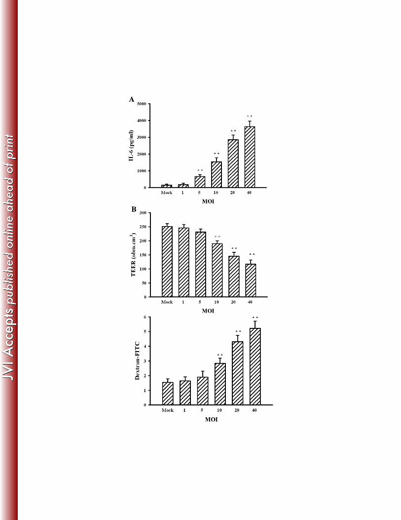
Fig. 7. Effects of JEV infection on IL-6 expression. Pericytes were infected with mock or 726
JEV (1, 5, 10, 20, and 40 MOI) for 48 h. (A) Supernatants isolated from infected cells were 727
subjected to ELISA for the measurement of IL-6. N = 4. (B) The supernatants were collected 728
and mixed with an equal volume of fresh medium. The manipulated media were added to 729
brain microvascular endothelial cells for 24 h. The TEER (upper panel) and transendothelial 730
permeability to dextran-FITC (lower panel) were measured. N = 4. **p < 0.01 vs. mock. 731
732
Fig. 8. Role of Ubr 1. Pericytes were infected with mock (Mock CM) or JEV (20 MOI, JEV 733
CM) for 48 h. The supernatants were collected and mixed with an equal volume of fresh 734
medium. (A) The manipulated media were added to brain microvascular endothelial cells 735
over time. Total cellular proteins were isolated and subjected to Western blot with antibodies 736
against Ubr 1 and β-tubulin. One representative blot of four independent experiments is 737
shown. (B) Brain microvascular endothelial cells were exposed to the manipulated media 738

JVI02738-13 revised version
32
(Mock CM and JEV CM) in the absence or presence of AG490 (50 µM) for 24 h. One set of 739
manipulated medium (JEV CM) was modified by neutralization with IL-6 neutralizing 740
antibody (10 µg/ml) for 30 min before being subjected to exposure. Total cellular proteins 741
were isolated and subjected to Western blot with antibodies against Ubr 1 and β-tubulin. One 742
representative blot of four independent experiments is shown. The content in Mock CM was 743
defined as 100%. Brain microvascular endothelial cells were transfected with mock, control 744
siRNA (1 nM), or Ubr 1 siRNA (1 nM) for 4 h. The resultant cells were treated with IL-6 (20 745
ng/ml) or exposed to JEV CM for 24 h. Untreated cells were as control. (C) Total cellular 746
proteins were isolated and subjected to Western blot with antibodies against Ubr 1, ZO-1, 747
and β-tubulin. One representative blot of four independent experiments is shown. The 748
content in control was defined as 100%. (D) Cellular proteins were isolated and subjected to 749
fluorogenic assay for the determinations of trypsin-like (upper panel) and chemotrypsin-like 750
(lower panel) proteasome activity. N = 4. (E) The TEER (upper panel) and transendothelial 751
permeability to dextran-FITC (lower panel) were measured. N = 4. **p < 0.01 vs. medium 752
control, ##p < 0.01 vs. IL-6 control, and &&p < 0.01 vs. JEV CM control. 753


Related Documents