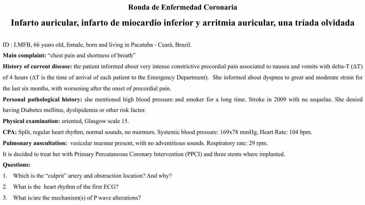
Ronda de Enfermedad Coronaria ID : LMFB, 66 years old, female, born and living in Pacatuba - Ceará, Brazil. Main complaint: “chest pain and shortness of breath” History of current disease: the patient informed about very intense constrictive precordial pain associated to nausea and vomits with delta-T (ΔT) of 4 hours (ΔT is the time of arrival of each patient to the Emergency Department). She informed about dyspnea to great and moderate strain for the last six months, with worsening after the onset of precordial pain. Personal pathological history: she mentioned high blood pressure and smoker for a long time. Stroke in 2009 with no sequelae. She denied having Diabetes mellitus, dyslipidemia or other risk factor. Physical examination: oriented, Glasgow scale 15. CPA: Split, regular heart rhythm, normal sounds, no murmurs. Systemic blood pressure: 169x78 mmHg, Heart Rate: 104 bpm. Pulmonary auscultation: vesicular murmur present, with no adventitious sounds. Respiratory rate: 29 rpm. It is decided to treat her with Primary Percutaneous Coronary Intervention (PPCI) and three stents where implanted. Questions: 1. Which is the “culprit” artery and obstruction location? And why? 2. What is the heart rhythm of the first ECG? 3. What is/are the mechanism(s) of P wave alterations? Infarto auricular, infarto de miocardio inferior y arritmia auricular, una tríada olvidada

Welcome message from author
This document is posted to help you gain knowledge. Please leave a comment to let me know what you think about it! Share it to your friends and learn new things together.
Transcript
Ronda de Enfermedad Coronaria
ID : LMFB, 66 years old, female, born and living in Pacatuba - Ceará, Brazil.
Main complaint: “chest pain and shortness of breath”
History of current disease: the patient informed about very intense constrictive precordial pain associated to nausea and vomits with delta-T (ΔT)
of 4 hours (ΔT is the time of arrival of each patient to the Emergency Department). She informed about dyspnea to great and moderate strain for
the last six months, with worsening after the onset of precordial pain.
Personal pathological history: she mentioned high blood pressure and smoker for a long time. Stroke in 2009 with no sequelae. She denied
having Diabetes mellitus, dyslipidemia or other risk factor.
Physical examination: oriented, Glasgow scale 15.
CPA: Split, regular heart rhythm, normal sounds, no murmurs. Systemic blood pressure: 169x78 mmHg, Heart Rate: 104 bpm.
Pulmonary auscultation: vesicular murmur present, with no adventitious sounds. Respiratory rate: 29 rpm.
It is decided to treat her with Primary Percutaneous Coronary Intervention (PPCI) and three stents where implanted.
Questions:
1. Which is the “culprit” artery and obstruction location? And why?
2. What is the heart rhythm of the first ECG?
3. What is/are the mechanism(s) of P wave alterations?
Infarto auricular, infarto de miocardio inferior y arritmia auricular, una tríada olvidada
Sessão coronariana
ID : L.M.F.B., 66 anos, natural e residente em Pacatuba-CE.
Queixa Principal: “dor no peito e falta de ar”
História da doença atual: paciente refere dor precordial de forte intensidade associada a náuseas e vômitos com delta-T de 4 horas. Relata dispneia
aos grandes e moderados esforços há 6 meses com piora após o início da dor precordial.
Antecedentes pessoais patológicos : refere HAS e tabagismo de longa data. AVC em 2009 sem sequelas. Nega DM.
Ex. físico: orientada, Glasgow 15.
ACP: ritmo cardíaco regular, em 2 tempos, bulhas normofonética, sem sopros. PAS 169x78mmHg, FC 104bpm.
Ausculta pulmonar: murmúrio vesicular presente sem ruídos adventícios. Frequência respiratória 29 irpm.
Se decide tartar com angioplastia primaria e se implantam stent na CD
Perguntas:
1. Qual a artéria culpada e local da obstrução? E por quê?
2. Qual o ritmo cardíaco do primeiro ECG?
3. Qual(s) o(s) mecanismo(s) das alterações da onda P?
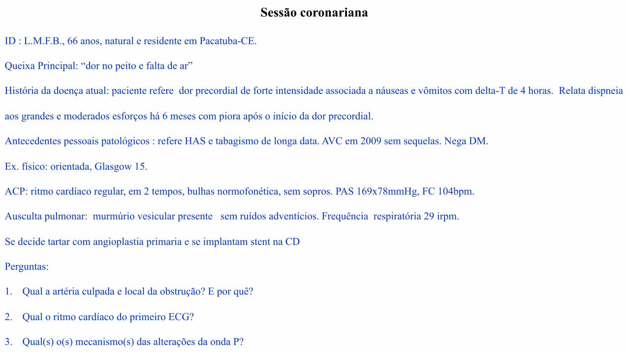
ECG-1 performed at admission 11:00 AM
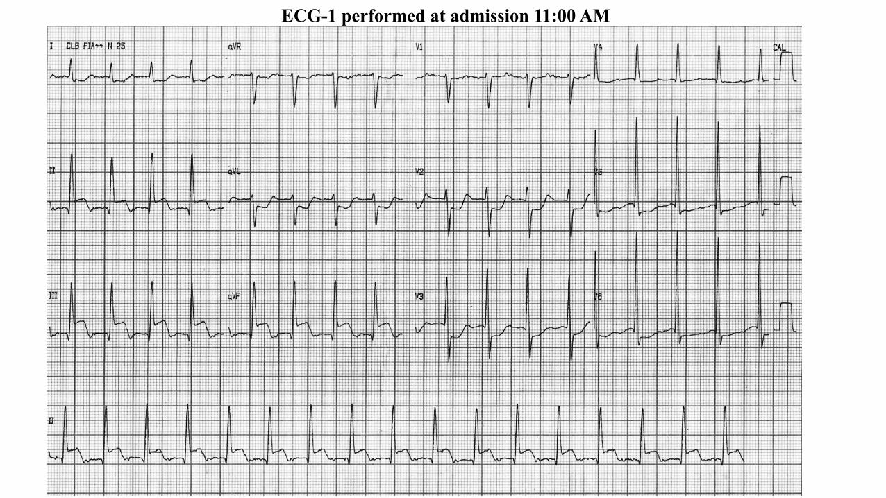
ECG-2 performed one hour and twenty minutes after admission 12:20 PM
II
III
aVL
aVF
III aVF
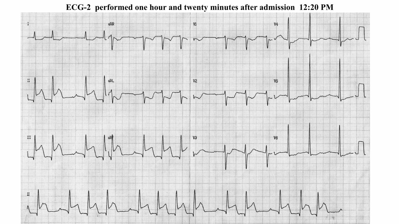
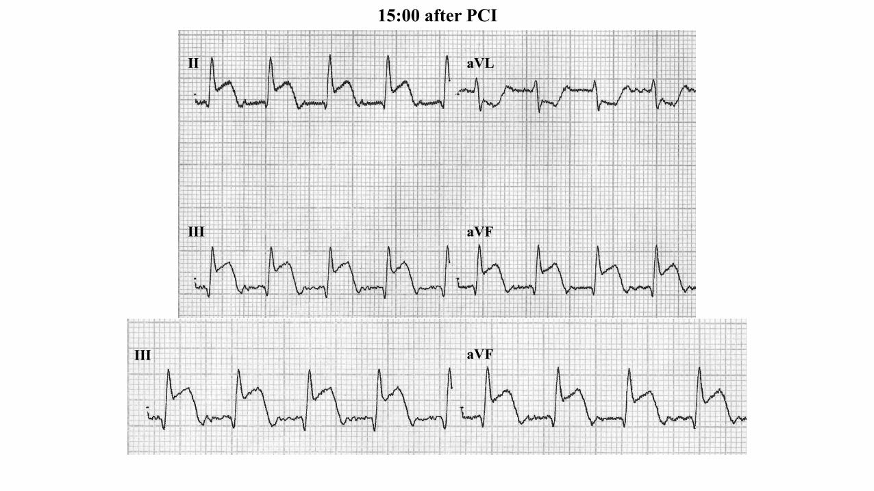
15:00 after PCI

Colleagues opinions
Spanish: Estimado Potro: El paciente presenta infarto hiperagudo de cara inferior con supradesnivel DIII mayor a DII y AVF, con infradesnivel del segmento ST máximo en V1 y V2 que se extiende V3 y V4, así como DI y AVL. La arteria culposa es la CD proximal, porque no la arteria circunfleja? esperaría encontrar supra ST en V5 y V6 que no observo. El ritmo es un ritmo acelerado de la unión por las ondas P negativas en DII, DIII y AVF. y evoluciona en el segundo ECG presentando latidos conducidos donde se observa onda P con signos de isquemia auricular y BAV de primer grado y ritmo de la union acelardo, continuando con IAM de cara inferior. Este patron de infarto tiene mal pronostico de ingreso, con aumento de la mortalidad en su evolución. Un cordial saludo Martín Ibarrola MD Argentina
English: Dear Potro, The patient presents hyperacute inferior infarction with DIII elevation greater than DII and aVF, with maximum ST segment depression in V1 and V2 extending to V3 and V4, as well as DI and aVL. The culprit artery is the proximal RCA, and why not the circumflex artery? I would expect to find ST elevation in V5 and V6 that I don’t see. The rhythm is accelerated rhythm of the junction, due to negative P waves in DII, DIII and aVF, and it evolved in the second ECG, presenting conducted beats where P wave is observed with signs of atrial ischemia and 1st degree AVB, and accelerated junction rhythm, continuing with inferior MI. This infarction pattern has a poor admission prognosis, with increase in mortality in its evolution. Kind regards, Martín Ibarrola MD Argentina
Portuguese: Interessante caso! Ao meu ver a artéria acometida é a coronária direita (supradesnivelamento do segmento ST em parede inferior, com supra de ST em DIII maior que em DII, com provável acometimento da parede ínfero-basal (antiga posterior): infradesnível em V2 e V3 representando, possivelmente, imagem em espelho da corrente de lesão na parede ínfero-basal) O nível de obstrução provavelmente foi no terço proximal, antes da artéria do nó sinoatrial, levando em conta a extensão do acometimento (paredes inferior e posterior), além de que parece haver comprometimento do nó sinusal, já que é difícil caracterizar onda P no primeiro ECG, que interpretei tendo como ritmo uma taquicardia juncional. Os mecanismos de alteração de onda P no infarto da coronária direta podem ser vários. Porém, levando-se em conta que após uma hora e vinte minutos espera-se que o paciente já tenha sido submetido à angioplastia, o aparecimento de ondas P esporádicas no segundo ECG pode ser devido à reperfusão do nó sinoatrial, porém, ainda doente, já que não consegue assumir o ritmo cardíaco. Outro motivo pode ser o desbalanço entre descargas simpáticas e parassimpáticas. Grande abraço, Gabriel Afonso Dutra Kreling Residente de Clínica Médica da Faculdade de Medicina da Universidade de São Paulo [email protected]
English: Interesting case! The way I see it, the affected artery is the RCA (ST segment elevation in inferior wall, with ST elevation in DIII greater than DII, with probable involvement of the inferobasal wall (old posterior): depression in V2 and V3, possibly representing mirror image of lesion current in the inferobasal wall). The level of obstruction is probably on the proximal third, before the sinoatrial node artery, taking into account the extension of the involvement (inferior and posterior wall), besides involvement of the sinus node, since it is difficult to characterize the P wave in the first ECG, which I interpreted as junctional tachycardia rhythm. There could be several mechanisms of P wave alteration in RCA infarction. However, taking into account that after one hour and twenty minutes it is expected for the patient to have already undergone angioplasty, the appearance of sporadic P waves in the second ECG could be due to sinoatrial node reperfusion; however, still sick, since it does not achieve resuming cardiac rhythm. Another reason could be the imbalance between sympathetic and parasympathetic loads. Warm regards, Gabriel Afonso Dutra Kreling MD
Hello, this is inferolateral STEMI. When ST elevation in III is higher than in II, ST depression deeper in aVL than in aVR and there is no ST elevation in V1, I think it is not possible to differentiate RCA from LCX occlusion (Tahvanainen M et al. JECG 2001). According to Samuel’s and Yochai’s group (Mager A et al. Coron Artery Dis 2000), ST depression in the lateral precordial leads in these cases indicates multivessel disease. In Finland, 14-lead ECG is standard in suspected acute MI and we would have recorded also V4R and V8. They might have helped to differentiate RCA from LCX. To me the rhythm in ECG 1 looks like accelerated junctional rhythm or ectopic atrial rhythm from the inferior part of the right atrium. In the second ECG I see atrial ectopic beats. Best regards Kjell Nikus M.D. Ph.D. Tampere, Finland h"ps://www.researchgate.net/profile/Kjell_Nikus
Spanish: Hola foristas, Este paciente presenta una oclusión de la CD probablemente proximal: ST descendido en I, STII<III y digo proximal a la arteria de ventrículo derecho porque el ST de V1 es isoelectrico.No tiene ningún criterio para obstrucción de Cx. En el primer ECG es difícil analizar la onda P por la taquicardia pero podría ser una taquicardia nodal con bloqueo AV de 1º En el segundo el ritmo es sinusal con una morfología de P algo distorsionada (isquemia ) y bloqueo AV de 2º tipo I Dado que el ST esta descendido de V1 a V6 debe pensarse en enfermedad multivasos. Saludos Miquel Fiol Sala, MD, PhD nickname: “The ambassador of the Balearic Islands” Director Científico del Instituto de Investigación Sanitaria de Palma (IdISPa) IP del CIBERobn Hospital Son Espases Ctra de Valldemossa, 79 07010 Palma de Mallorca Teléfno: 871205987/871205050, externo 75987 e.mail: [email protected]
English: Hello, members of the Forum, This patient presents RCA occlusion; probably it is proximal: ST decreased in I, ST II<III, and I say proximal to the artery of the right ventricle, because the ST of V1 is isoelectric. There is no criterion for Cx. In the first ECG, it is difficult to analyze the P wave because of the tachycardia, but it could be nodal tachycardia with 1° degree AV block. The second rhythm is sinus, with a somewhat distorted P morphology (ischemia) and type I, 2° degree AV block. Since ST is depressed from V1 through V6, we should consider multivessel disease. Regards, Miquel Fiol Sala
Addendum from Dr. Fiol-Sala: With regard to the differential diagnosis of RCA occlusion versus LCx I want to point out the following:
- Have a high sensitivity both ST depression in aVR (1;2) and aVL (3;4) because in RCA occlusion the injury vector falls on the negative hemi field of both leads aVR and aV(see slide 19). The few cases that are due to occlusion of the LCx are due to: very dominant LCx or a distal occlusion of very dominant RCA. In both cases what counts for the ECG pattern is the Ischemic Area more than the occluded artery.
I think ahead of the forum members who follow us should practice more electrocardiography clinical practice virtuosity in the electrocardiographic interpretation. Moreover, I agree with Dr Samuel that excessive use of aVR lead as "magic" is not recommended.
Miquel Fiol Sala, MD, PhD
1. Fiol M, Cygankiewicz I, Carrillo A, et al. Value of electrocardiographic algorithm based on "ups and downs" of ST in assessment of a culprit artery in evolving inferior wall acute myocardial infarction. Am J Cardiol. 2004;94(6):709-14.
2. Fiol M, Carrillo A.Culprit artery in evolving inferior wall acute myocardial infarction: RCA vs LCx. Europace. 201;12(5):758; author reply 758-9.
3. Birnbaum Y, Sclarovsky S, Mager A, et al.ST segment depression in aVL: a sensitive marker for acute inferior myocardial infarction. Eur Heart J. 1993;14(1):4-7
4. Birnbaum Y, Herz I, Sclarovsky S, et al. Prognostic significance of precordial ST segment depression on admission electrocardiogram in patients with inferior wall myocardial infarction. J Am Coll Cardiol. 1996;28(2):313-8.
Balearic islands: Mediterranean Paradise This wonderful group of islands is divided into Gimnesias (Mallorca, Menorca and Cabrera) in the North, and Pitiusas (Ibiza and Formentera) in the South-West. Their excellent climate with some 300 days of sun per year, wonderful beaches, a rich cultural offer and the unusual hospitality of their inhabitants make Balearic Islands one of the preferred holiday destinations in Spain. There are more passengers at the airport of Palma de Mallorca than at the airport of any other Spanish city. Hardly anybody who has spent holidays here didn't wish to return, in fact, many visitors have made the islands their permanent home. Each of the islands has a strong personality all of its own, and each of them offers much more but the possibility to spend nice holidays at the beach ..Dr Miguel Fiol is “The ambassador of the Balearic Islands”.
Spanish: Obstruccion aguda de la arteria coronária derecha ST- . T DIIII> DIII, distal a la arteria marginal primera y de la conal ,porque el ST está deprimido y la onda T invertida em V1,sugeriendo que el septo derecho posterior esta involucrado en el proceso isquémico . V1 expresa la remodelacion fisiologia del septo derecho anterior. El ST deprimido con onda T invertida en V2 sugiere que la remodelcion isquemica es fisiologica. V2 expresa la remodelacion fisiologica de la isquemia de la pared posterior que se manifiesta en el segment elevado ST y en la onda T positiva en DII, por ende pareceria que no hay complicaciones con obstrucciones criticas de las otras arterias coronarias izquierdas El q/ R en DIIII ,DII y AVF sugiere isquemia aguda en el músculo papilar póstero-inferior irrigada el 80% por la arteria descendente postero-inferior rama de la coronaria derecha sugeriendo que que el musculo papilar anterosuperior se depolariza antes que el músculo postero inferior ,similar a lo que ocurre en el hemibloqueo antero-superior no isquemico. Esto se debe a que las izquemias inducen una diminución severa en concetración de la conexina 43 “down regulacion” (buscar en internet conexina43 e isquemia ). Las derivaciones izquierdas sugieren una hipertrofia hipertensiva crónica con sobrecarga sistolica El bloqueo de primer grado y las extrasistoles atriales seguidas (coupled APC'S ,podria ser debido a un proceso crónico, como complicacion de la hypertension cronica en particular en mujeres postemenopausicas hypertensivas ( en este caso las ondas P tienen la misma polaridad que las P sinusales )_ ,pero una posibilidad menos probable, podria ser debido a la isquemia aguda ,debido a una obstrucci)on proximal a la arteria nodal. La dimension del arteria derecha es codominante, Porque? Porque DII interviene en el proceso ,pero descarta que sea CD dominante ,ya que v5,v6 no intervienen en el proceso isquemico agudo El grado de isquemia es 3,sugeriendo que el miocardio infero-posterior no esta protegido por un mecanismo precondicionado , o circulacion colateral Este critero fisiopatologica es importante ,por que en reperfusion ,ya sea quimica o mecanica puede aparecer taquicardia polimorfica por reperfusion Quien quiera saber de este tema, recomiendo leer el capitulo 10 de mi libro en espaniol en un capitulo escrito en el libro de los profesores J Milei y J Lerman Los infartos en presencia de hipertrofia lateral inferior con ST deprimido sugieren fibrosis en el area apical lateral y alto de riesgo No veo nos demas electro ,pero el grado 3 de isquemia e hipertrofia con fibrosis revisten un alto riego y en particular en mujeres post menopáusicas Un fraternal abrazo Samuel Sclarovky Lean con paciencia ya que esta es una explicación moderna del este tema
English: Acute RCA obstruction – ST-T DIII>DII, distal to the first marginal and conal arteries, because ST is depressed and T wave is inverted in V1, suggesting that the posterior right septum is involved in the ischemic process. V1 expresses the physiological remodeling of the right anterior septum. The depressed ST with inverted T wave in V2 suggests that ischemic remodeling is physiological. V2 expresses the physiological remodeling of posterior wall ischemia, that manifests in the elevated ST segment and positive T wave in DII; therefore it would seem that there are no complications with critical obstructions of the other left coronary arteries. q/R in DIII, DII and aVF suggests acute ischemia in the postero-inferior papillary muscle irrigated in 80% by the posterior-inferior branch of the RCA, suggesting that the anterior-superior papillary muscle depolarizes before the posterior-inferior, similar to what happens in non-ischemic anterior-superior hemiblock. This is because ischemias induce a severe decrease in the concentration of connexin 43 (downregulation; investigate on the Internet connexin 43 and ischemia). The left leads suggest chronic hypertensive hypertrophy with systolic overload. First-degree block and continuous premature atrial contractions (coupled APC) could be due to a chronic process, such as chronic hypertension complication, particularly in hypertensive post-menopausal women (in this case, P waves have the same polarity as sinus P), but a less likely possibility is they could be due to acute ischemia, due to obstruction proximal to the nodal artery. The dimension of RCA is co-dominant. Why? Because DII intervenes in the process, but it rules out RCA as dominant, since V5 and V6 do not intervene in the acute ischemic process. The degree of ischemia is 3, suggesting that the inferior-posterior myocardium is not protected by a pre-conditioned mechanism, or collateral circulation. This pathophysiological criterion is important, because in reperfusion, whether chemical or mechanical, polymorphic tachycardia may appear by reperfusion. To anyone who knows about this topic, I advice reading chapter 10 of my book in Spanish, in the chapter written in the book by professors J. Milei and J. Lerman. Infarctions in the presence of inferior lateral hypertrophy with depressed ST suggest fibrosis in the lateral apical area and high risk. I don’t see anything in the other ECGs, but degree 3 of ischemia and hypertrophy with fibrosis imply a high risk, and particularly in post-menopausal women. Warm regards, Samuel Sclarovky, MD, Israel
Sinus tachycardia with 1st degree AV block ,acute IMI with occlusion of the RCA Sinus tach with AV Wenkebach evolving IMI Melvin M. Scheinman Department of Cardiac Electrophysiology, University of California San Francisco, San Francisco, California, USA. Professor of Medicine Address: UCSF Electrophysiology Service 500 Parnassus Avenue San Francisco, CA 94143-1354 Phone: (415) 476-5706 email: [email protected]
Final comments by
Andrés Ricardo Pérez-Riera, M.D. Ph.D. Design of Studies and Scientific Writing Laboratory at the ABC School of Medicine, Santo André, São Paulo, Brazil
ECG / VCG Pérez-Riera | my cardiology site of scientific interests: https://ekgvcg.wordpress.com
Raimundo Barbosa-Barros, M.D. Coronary Center of the Hospital de Messejana Dr. Carlos Alberto Studart Gomes, Fortaleza, CE, Brazil
Luiz Carlos de Abreu, Ph.D.
Design of Studies and Scientific Writing Laboratory at the ABC School of Medicine, Santo André, São Paulo, Brazil
Atrial infarction, inferior myocardial infarction atrial arrhythmia a forgotten triad
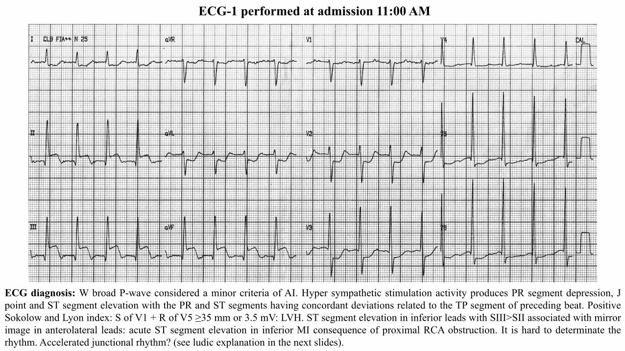
ECG-1 performed at admission 11:00 AM
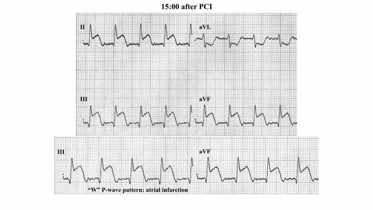
ECG diagnosis: W broad P-wave considered a minor criteria of AI. Hyper sympathetic stimulation activity produces PR segment depression, J point and ST segment elevation with the PR and ST segments having concordant deviations related to the TP segment of preceding beat. Positive Sokolow and Lyon index: S of V1 + R of V5 ≥35 mm or 3.5 mV: LVH. ST segment elevation in inferior leads with SIII>SII associated with mirror image in anterolateral leads: acute ST segment elevation in inferior MI consequence of proximal RCA obstruction. It is hard to determinate the rhythm. Accelerated junctional rhythm? (see ludic explanation in the next slides).
II III
aVF
X I
Y
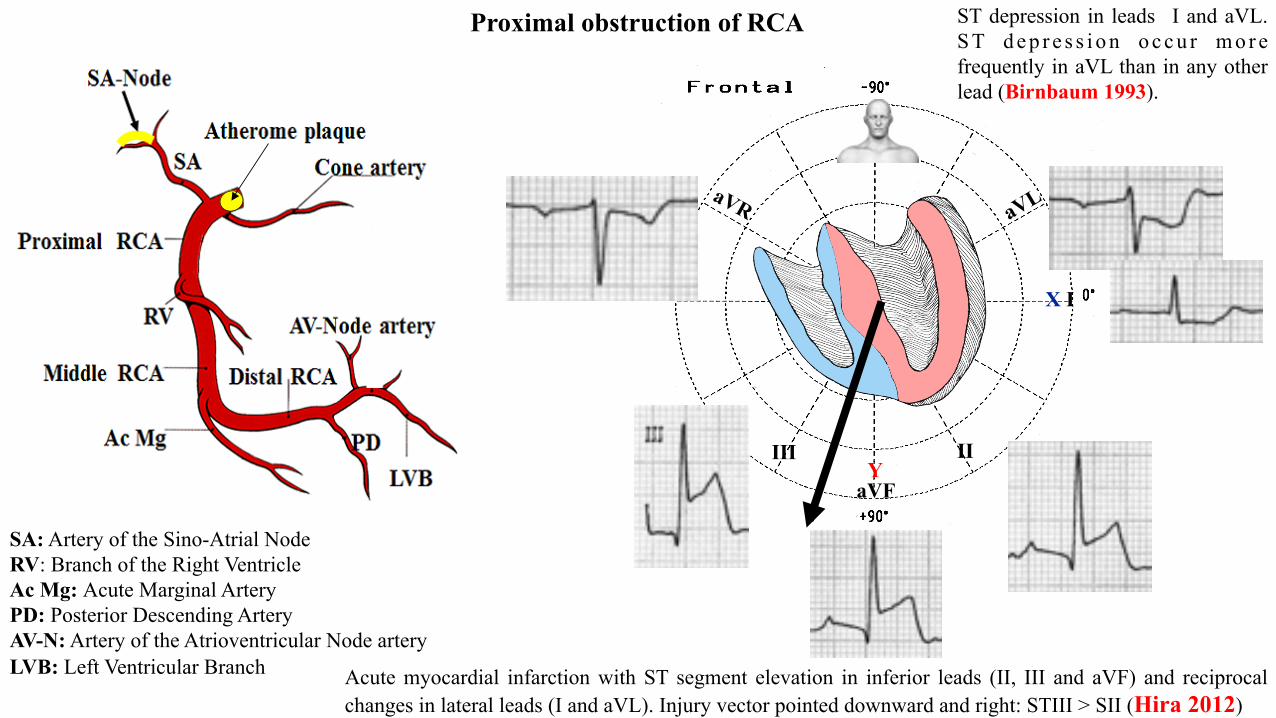
Acute myocardial infarction with ST segment elevation in inferior leads (II, III and aVF) and reciprocal changes in lateral leads (I and aVL). Injury vector pointed downward and right: STIII > SII (Hira 2012)
SA: Artery of the Sino-Atrial Node RV: Branch of the Right Ventricle Ac Mg: Acute Marginal Artery PD: Posterior Descending Artery AV-N: Artery of the Atrioventricular Node artery LVB: Left Ventricular Branch
Proximal obstruction of RCA ST depression in leads I and aVL. S T d e p r e s s i o n o c c u r m o r e frequently in aVL than in any other lead (Birnbaum 1993).
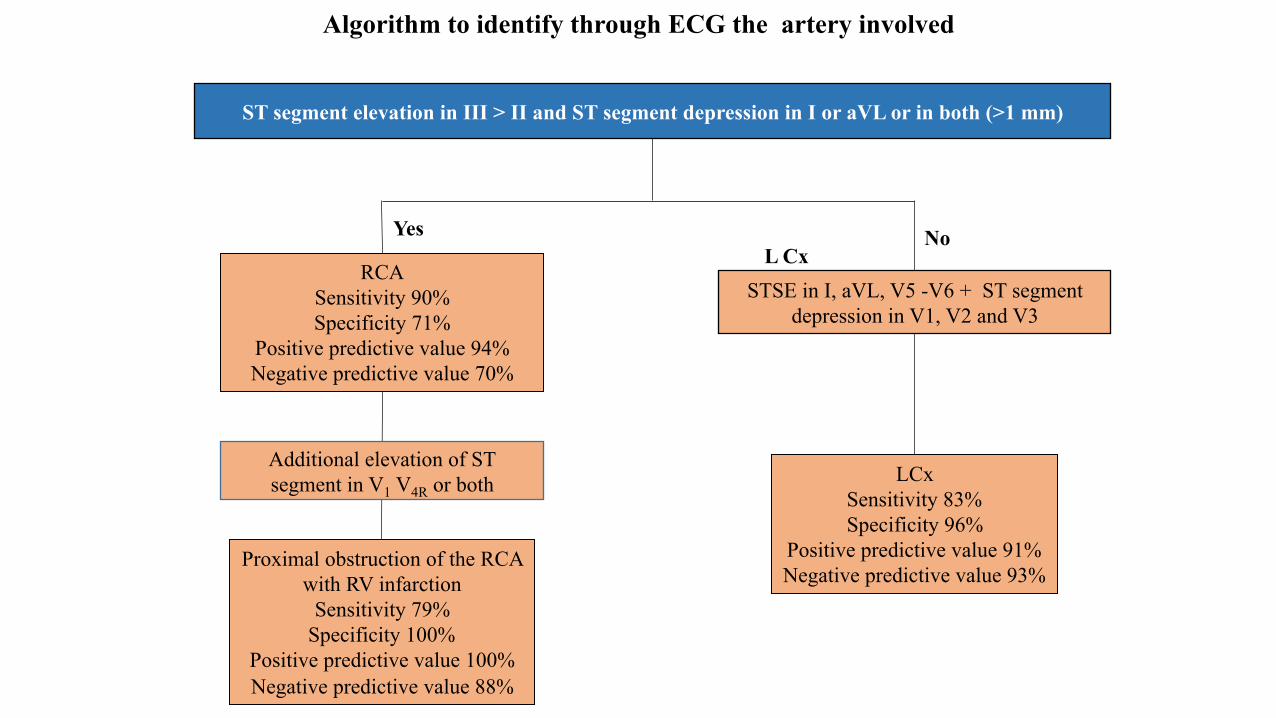
Algorithm to identify through ECG the artery involved
Yes No
LCx Sensitivity 83% Specificity 96%
Positive predictive value 91% Negative predictive value 93%
Proximal obstruction of the RCA with RV infarction
Sensitivity 79% Specificity 100%
Positive predictive value 100% Negative predictive value 88%
RCA Sensitivity 90% Specificity 71%
Positive predictive value 94% Negative predictive value 70%
L Cx
STSE in I, aVL, V5 -V6 + ST segment depression in V1, V2 and V3
ST segment elevation in III > II and ST segment depression in I or aVL or in both (>1 mm)
Additional elevation of ST segment in V1 V4R or both
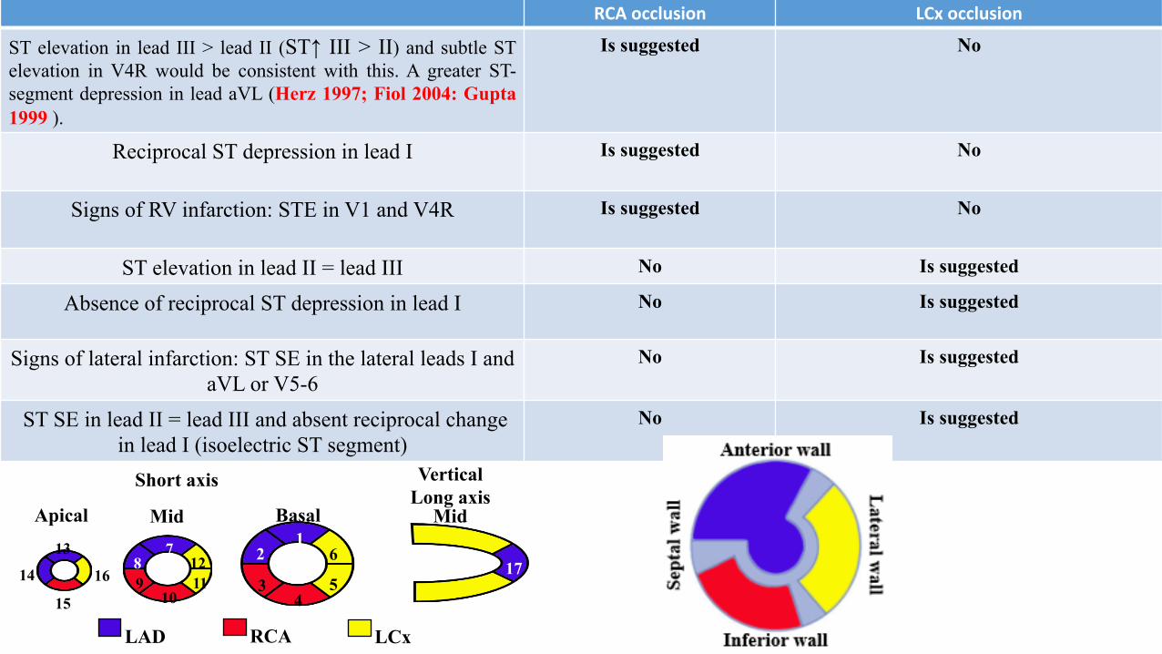
RCAocclusion LCxocclusion
ST elevation in lead III > lead II (ST↑ III > II) and subtle ST elevation in V4R would be consistent with this. A greater ST-segment depression in lead aVL (Herz 1997; Fiol 2004: Gupta 1999 ).
Is suggested No
Reciprocal ST depression in lead I
Is suggested
No
Signs of RV infarction: STE in V1 and V4R
Is suggested
No
ST elevation in lead II = lead III No Is suggested
Absence of reciprocal ST depression in lead I No Is suggested
Signs of lateral infarction: ST SE in the lateral leads I and aVL or V5-6
No Is suggested
ST SE in lead II = lead III and absent reciprocal change in lead I (isoelectric ST segment)
No Is suggested
LAD RCA LCx
Short axis Vertical Long axis
Basal Mid Apical Mid 1
2
3 4
5
6 7 8 9
10 11 12
15
14
13 17 16
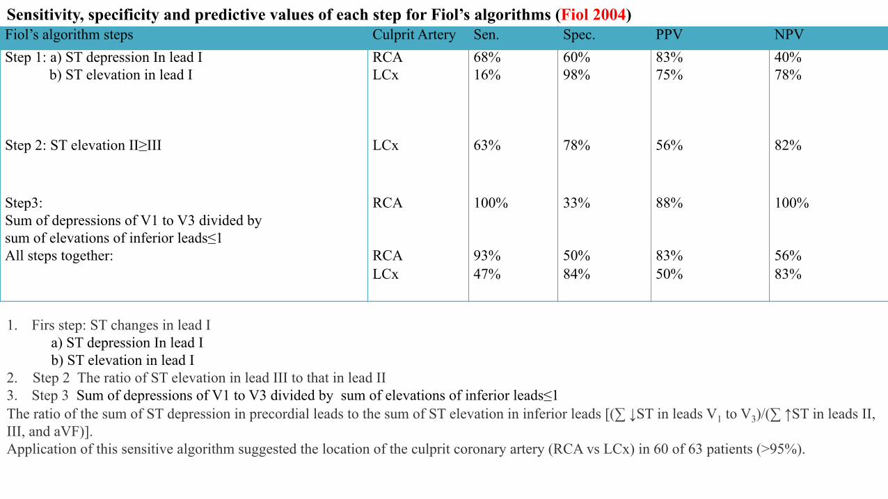
Fiol’s algorithm steps Culprit Artery Sen. Spec. PPV NPV Step 1: a) ST depression In lead I b) ST elevation in lead I
RCA LCx
68% 16%
60% 98%
83% 75%
40% 78%
Step 2: ST elevation II≥III LCx 63% 78% 56% 82%
Step3: Sum of depressions of V1 to V3 divided by sum of elevations of inferior leads≤1 All steps together:
RCA RCA LCx
100% 93% 47%
33% 50% 84%
88% 83% 50%
100% 56% 83%
Sensitivity, specificity and predictive values of each step for Fiol’s algorithms (Fiol 2004)
1. Firs step: ST changes in lead I a) ST depression In lead I b) ST elevation in lead I 2. Step 2 The ratio of ST elevation in lead III to that in lead II 3. Step 3 Sum of depressions of V1 to V3 divided by sum of elevations of inferior leads≤1 The ratio of the sum of ST depression in precordial leads to the sum of ST elevation in inferior leads [(∑ ↓ST in leads V1 to V3)/(∑ ↑ST in leads II, III, and aVF)]. Application of this sensitive algorithm suggested the location of the culprit coronary artery (RCA vs LCx) in 60 of 63 patients (>95%).
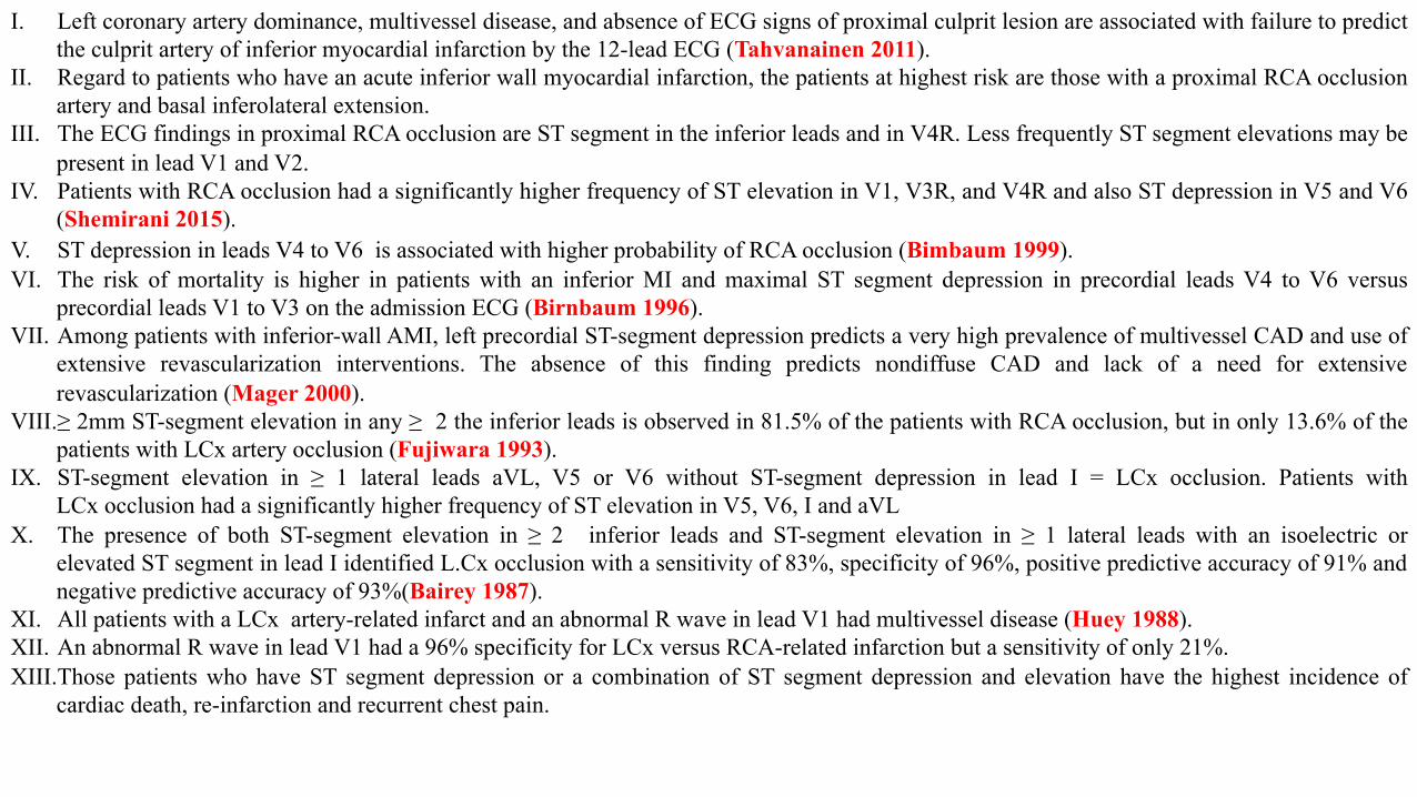
I. Left coronary artery dominance, multivessel disease, and absence of ECG signs of proximal culprit lesion are associated with failure to predict the culprit artery of inferior myocardial infarction by the 12-lead ECG (Tahvanainen 2011).
II. Regard to patients who have an acute inferior wall myocardial infarction, the patients at highest risk are those with a proximal RCA occlusion artery and basal inferolateral extension.
III. The ECG findings in proximal RCA occlusion are ST segment in the inferior leads and in V4R. Less frequently ST segment elevations may be present in lead V1 and V2.
IV. Patients with RCA occlusion had a significantly higher frequency of ST elevation in V1, V3R, and V4R and also ST depression in V5 and V6 (Shemirani 2015).
V. ST depression in leads V4 to V6 is associated with higher probability of RCA occlusion (Bimbaum 1999).VI. The risk of mortality is higher in patients with an inferior MI and maximal ST segment depression in precordial leads V4 to V6 versus
precordial leads V1 to V3 on the admission ECG (Birnbaum 1996). VII. Among patients with inferior-wall AMI, left precordial ST-segment depression predicts a very high prevalence of multivessel CAD and use of
extensive revascularization interventions. The absence of this finding predicts nondiffuse CAD and lack of a need for extensive revascularization (Mager 2000).
VIII. ≥ 2mm ST-segment elevation in any ≥ 2 the inferior leads is observed in 81.5% of the patients with RCA occlusion, but in only 13.6% of the patients with LCx artery occlusion (Fujiwara 1993).
IX. ST-segment elevation in ≥ 1 lateral leads aVL, V5 or V6 without ST-segment depression in lead I = LCx occlusion. Patients with LCx occlusion had a significantly higher frequency of ST elevation in V5, V6, I and aVL
X. The presence of both ST-segment elevation in ≥ 2 inferior leads and ST-segment elevation in ≥ 1 lateral leads with an isoelectric or elevated ST segment in lead I identified L.Cx occlusion with a sensitivity of 83%, specificity of 96%, positive predictive accuracy of 91% and negative predictive accuracy of 93%(Bairey 1987).
XI. All patients with a LCx artery-related infarct and an abnormal R wave in lead V1 had multivessel disease (Huey 1988). XII. An abnormal R wave in lead V1 had a 96% specificity for LCx versus RCA-related infarction but a sensitivity of only 21%. XIII. Those patients who have ST segment depression or a combination of ST segment depression and elevation have the highest incidence of
cardiac death, re-infarction and recurrent chest pain.
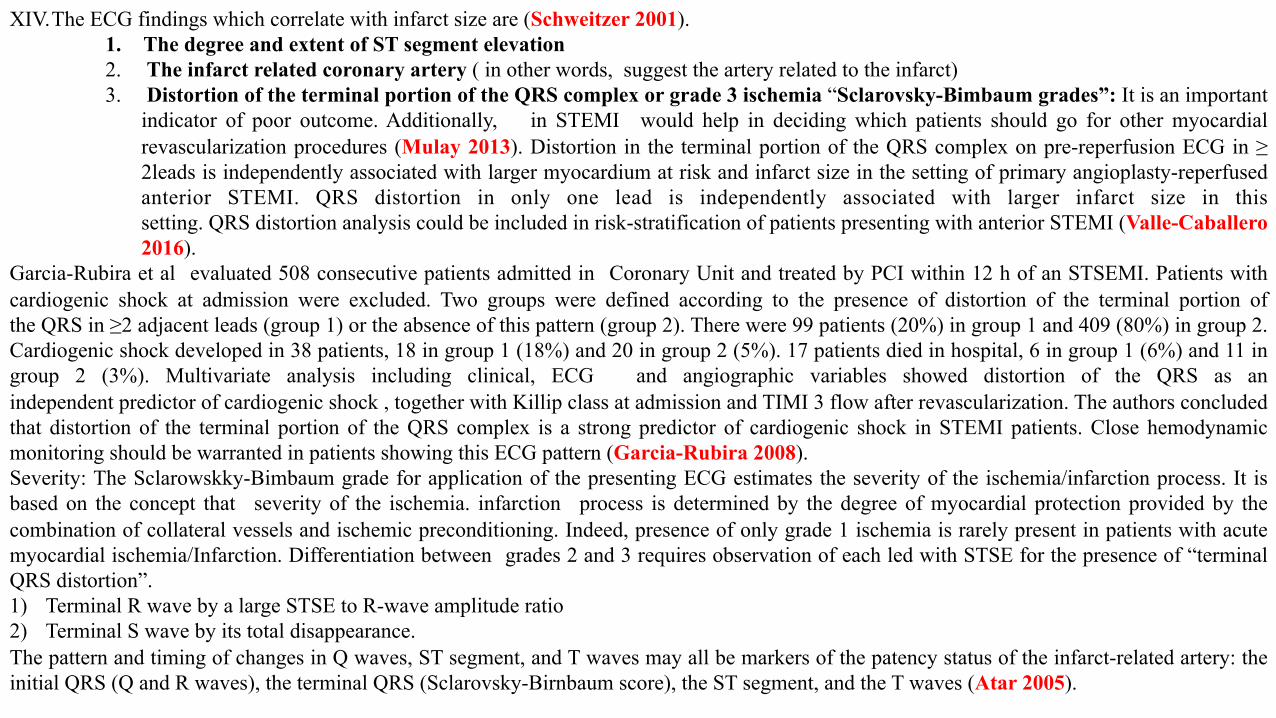
XIV. The ECG findings which correlate with infarct size are (Schweitzer 2001). 1. The degree and extent of ST segment elevation 2. The infarct related coronary artery ( in other words, suggest the artery related to the infarct) 3. Distortion of the terminal portion of the QRS complex or grade 3 ischemia “Sclarovsky-Bimbaum grades”: It is an important
indicator of poor outcome. Additionally, in STEMI would help in deciding which patients should go for other myocardial revascularization procedures (Mulay 2013). Distortion in the terminal portion of the QRS complex on pre-reperfusion ECG in ≥ 2leads is independently associated with larger myocardium at risk and infarct size in the setting of primary angioplasty-reperfused anterior STEMI. QRS distortion in only one lead is independently associated with larger infarct size in this setting. QRS distortion analysis could be included in risk-stratification of patients presenting with anterior STEMI (Valle-Caballero 2016).
Garcia-Rubira et al evaluated 508 consecutive patients admitted in Coronary Unit and treated by PCI within 12 h of an STSEMI. Patients with cardiogenic shock at admission were excluded. Two groups were defined according to the presence of distortion of the terminal portion of the QRS in ≥2 adjacent leads (group 1) or the absence of this pattern (group 2). There were 99 patients (20%) in group 1 and 409 (80%) in group 2. Cardiogenic shock developed in 38 patients, 18 in group 1 (18%) and 20 in group 2 (5%). 17 patients died in hospital, 6 in group 1 (6%) and 11 in group 2 (3%). Multivariate analysis including clinical, ECG and angiographic variables showed distortion of the QRS as an independent predictor of cardiogenic shock , together with Killip class at admission and TIMI 3 flow after revascularization. The authors concluded that distortion of the terminal portion of the QRS complex is a strong predictor of cardiogenic shock in STEMI patients. Close hemodynamic monitoring should be warranted in patients showing this ECG pattern (Garcia-Rubira 2008). Severity: The Sclarowskky-Bimbaum grade for application of the presenting ECG estimates the severity of the ischemia/infarction process. It is based on the concept that severity of the ischemia. infarction process is determined by the degree of myocardial protection provided by the combination of collateral vessels and ischemic preconditioning. Indeed, presence of only grade 1 ischemia is rarely present in patients with acute myocardial ischemia/Infarction. Differentiation between grades 2 and 3 requires observation of each led with STSE for the presence of “terminal QRS distortion”. 1) Terminal R wave by a large STSE to R-wave amplitude ratio 2) Terminal S wave by its total disappearance. The pattern and timing of changes in Q waves, ST segment, and T waves may all be markers of the patency status of the infarct-related artery: the initial QRS (Q and R waves), the terminal QRS (Sclarovsky-Birnbaum score), the ST segment, and the T waves (Atar 2005).
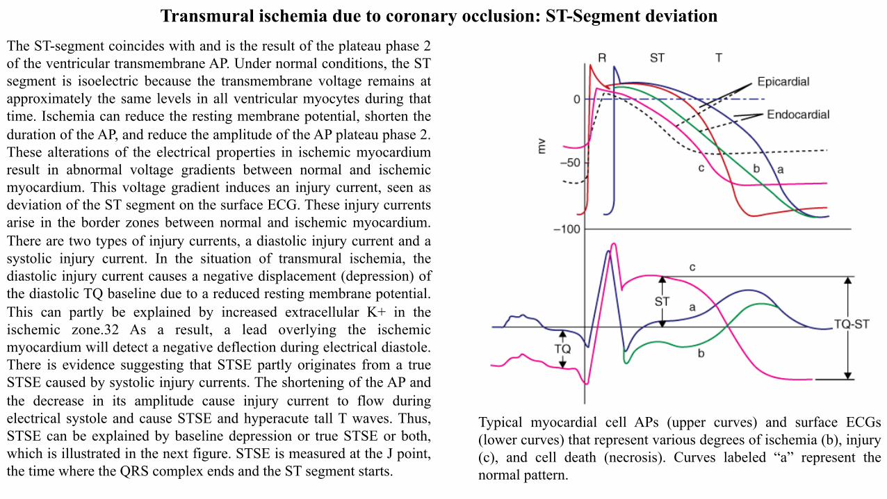
The ST-segment coincides with and is the result of the plateau phase 2 of the ventricular transmembrane AP. Under normal conditions, the ST segment is isoelectric because the transmembrane voltage remains at approximately the same levels in all ventricular myocytes during that time. Ischemia can reduce the resting membrane potential, shorten the duration of the AP, and reduce the amplitude of the AP plateau phase 2. These alterations of the electrical properties in ischemic myocardium result in abnormal voltage gradients between normal and ischemic myocardium. This voltage gradient induces an injury current, seen as deviation of the ST segment on the surface ECG. These injury currents arise in the border zones between normal and ischemic myocardium. There are two types of injury currents, a diastolic injury current and a systolic injury current. In the situation of transmural ischemia, the diastolic injury current causes a negative displacement (depression) of the diastolic TQ baseline due to a reduced resting membrane potential. This can partly be explained by increased extracellular K+ in the ischemic zone.32 As a result, a lead overlying the ischemic myocardium will detect a negative deflection during electrical diastole. There is evidence suggesting that STSE partly originates from a true STSE caused by systolic injury currents. The shortening of the AP and the decrease in its amplitude cause injury current to flow during electrical systole and cause STSE and hyperacute tall T waves. Thus, STSE can be explained by baseline depression or true STSE or both, which is illustrated in the next figure. STSE is measured at the J point, the time where the QRS complex ends and the ST segment starts.
Transmural ischemia due to coronary occlusion: ST-Segment deviation
Typical myocardial cell APs (upper curves) and surface ECGs (lower curves) that represent various degrees of ischemia (b), injury (c), and cell death (necrosis). Curves labeled “a” represent the normal pattern.
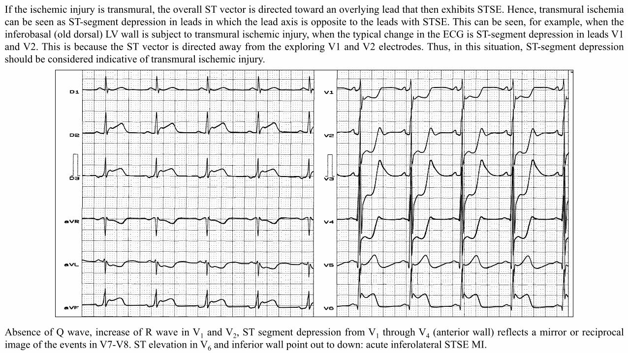
If the ischemic injury is transmural, the overall ST vector is directed toward an overlying lead that then exhibits STSE. Hence, transmural ischemia can be seen as ST-segment depression in leads in which the lead axis is opposite to the leads with STSE. This can be seen, for example, when the inferobasal (old dorsal) LV wall is subject to transmural ischemic injury, when the typical change in the ECG is ST-segment depression in leads V1 and V2. This is because the ST vector is directed away from the exploring V1 and V2 electrodes. Thus, in this situation, ST-segment depression should be considered indicative of transmural ischemic injury.
Absence of Q wave, increase of R wave in V1 and V2, ST segment depression from V1 through V4 (anterior wall) reflects a mirror or reciprocal image of the events in V7-V8. ST elevation in V6 and inferior wall point out to down: acute inferolateral STSE MI.

Threshold values for ST-Segment changes STSE ST-segment depression
For men 40 years of age and older, the threshold value for abnormal J-point elevation should be 0.2 mV (2 mm) in leads V2 and V3 and 0.1 mV (1 mm) in all other leads.
For men and women of all ages, the threshold value for abnormal J-point depression should be -0.05 mV (-0.5 mm) in leads V2 and V3 and -0.1 mV (-1 mm) in all other leads.
For men < 40 years of age, the threshold values for abnormal J-point elevation in leads V2 and V3 should be 0.25 mV (2.5 mm)
For women, the threshold value for abnormal J-point elevation should be 0.15 mV (1.5 mm) in leads V2 and V3 and >0.1 mV (1 mm) in all other leads.
For men and women, the threshold for abnormal J-point elevation in V3R and V4R should be 0.05 mV (0.5 mm), except for males <30 years of age, for whom 0.1 mV (1 mm) is more appropriate.
For men and women, the threshold value for abnormal j-point elevation in V7 through V9 should be 0.05 mV (0.5 mm).
The predetermined threshold for STSE (age, sex, and ECG lead dependent) should be exceeded in ≥ two anatomically contiguous ECG leads in order to be indicative of acute transmural myocardial ischemia. The currently recommended threshold values of ST-segment changes are listed in the table below.
Grade ECG Changes Severity
1 T wave Least
2 ST segment Moderate
3 QRS complex Most
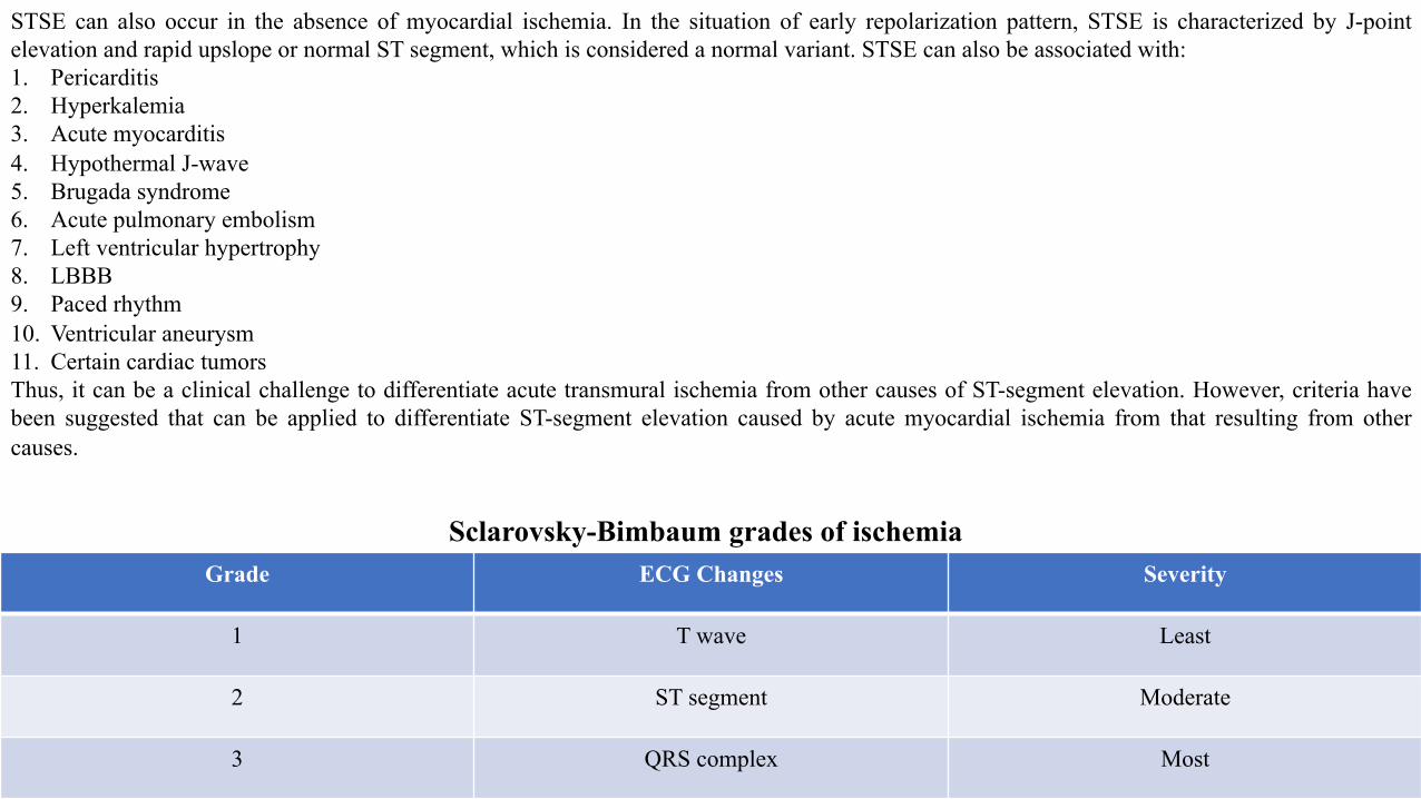
STSE can also occur in the absence of myocardial ischemia. In the situation of early repolarization pattern, STSE is characterized by J-point elevation and rapid upslope or normal ST segment, which is considered a normal variant. STSE can also be associated with: 1. Pericarditis 2. Hyperkalemia 3. Acute myocarditis 4. Hypothermal J-wave 5. Brugada syndrome 6. Acute pulmonary embolism 7. Left ventricular hypertrophy 8. LBBB 9. Paced rhythm 10. Ventricular aneurysm 11. Certain cardiac tumors Thus, it can be a clinical challenge to differentiate acute transmural ischemia from other causes of ST-segment elevation. However, criteria have been suggested that can be applied to differentiate ST-segment elevation caused by acute myocardial ischemia from that resulting from other causes.
Sclarovsky-Bimbaum grades of ischemia
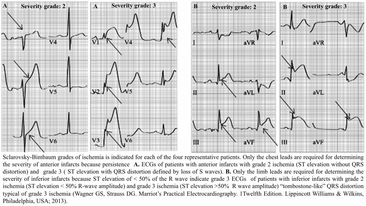
Sclarovsky-Bimbaum grades of ischemia is indicated for each of the four representative patients. Only the chest leads are required for determining the severity of anterior infarcts because persistence A. ECGs of patients with anterior infarcts with grade 2 ischemia (ST elevation without QRS distortion) and grade 3 ( ST elevation with QRS distortion defined by loss of S waves). B. Only the limb leads are required for determining the severity of inferior infarcts because ST elevation of < 50% of the R wave indicate grade 3 ECGs of patients with inferior infarcts with grade 2 ischemia (ST elevation < 50% R-wave amplitude) and grade 3 ischemia (ST elevation >50% R wave amplitude) “tombostone-like” QRS distortion typical of grade 3 ischemia (Wagner GS, Strauss DG. Marriot’s Practical Electrocardiography. 1Twelfth Edition. Lippincott Williams & Wilkins, Philadelphia, USA; 2013).
A Severity grade: 2 Severity grade: 3 B Severity grade: 2 Severity grade: 3 A B
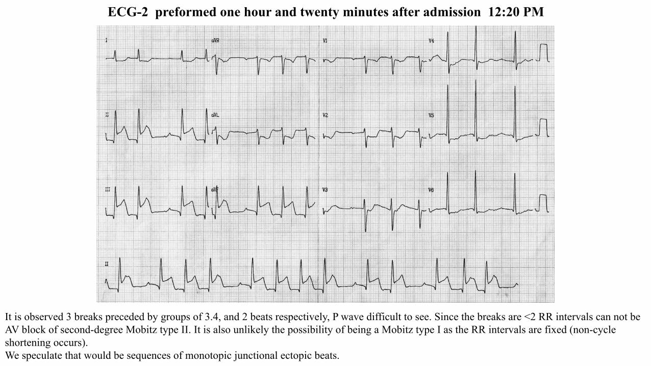
ECG-2 preformed one hour and twenty minutes after admission 12:20 PM
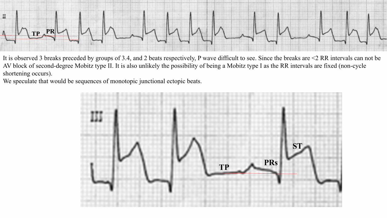
It is observed 3 breaks preceded by groups of 3.4, and 2 beats respectively, P wave difficult to see. Since the breaks are <2 RR intervals can not be AV block of second-degree Mobitz type II. It is also unlikely the possibility of being a Mobitz type I as the RR intervals are fixed (non-cycle shortening occurs). We speculate that would be sequences of monotopic junctional ectopic beats.
PRTP
It is observed 3 breaks preceded by groups of 3.4, and 2 beats respectively, P wave difficult to see. Since the breaks are <2 RR intervals can not be AV block of second-degree Mobitz type II. It is also unlikely the possibility of being a Mobitz type I as the RR intervals are fixed (non-cycle shortening occurs). We speculate that would be sequences of monotopic junctional ectopic beats.
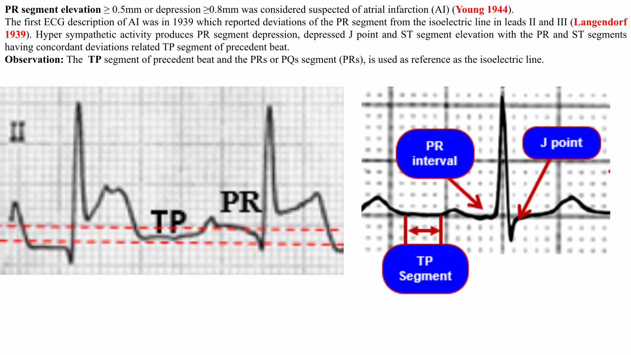
PR segment elevation ≥ 0.5mm or depression ≥0.8mm was considered suspected of atrial infarction (AI) (Young 1944). The first ECG description of AI was in 1939 which reported deviations of the PR segment from the isoelectric line in leads II and III (Langendorf 1939). Hyper sympathetic activity produces PR segment depression, depressed J point and ST segment elevation with the PR and ST segments having concordant deviations related TP segment of precedent beat. Observation: The TP segment of precedent beat and the PRs or PQs segment (PRs), is used as reference as the isoelectric line.
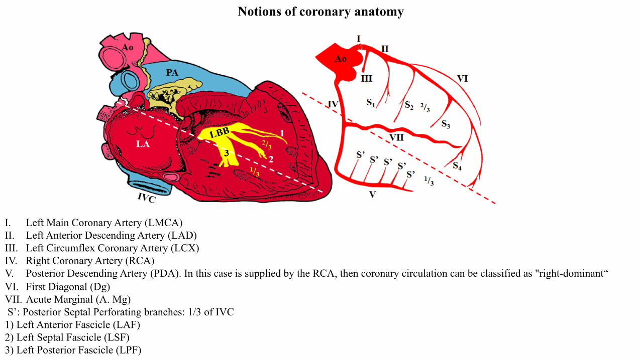
I. Left Main Coronary Artery (LMCA) II. Left Anterior Descending Artery (LAD) III. Left Circumflex Coronary Artery (LCX) IV. Right Coronary Artery (RCA) V. Posterior Descending Artery (PDA). In this case is supplied by the RCA, then coronary circulation can be classified as "right-dominant“ VI. First Diagonal (Dg) VII. Acute Marginal (A. Mg) S’: Posterior Septal Perforating branches: 1/3 of IVC 1) Left Anterior Fascicle (LAF) 2) Left Septal Fascicle (LSF) 3) Left Posterior Fascicle (LPF)
Notions of coronary anatomy
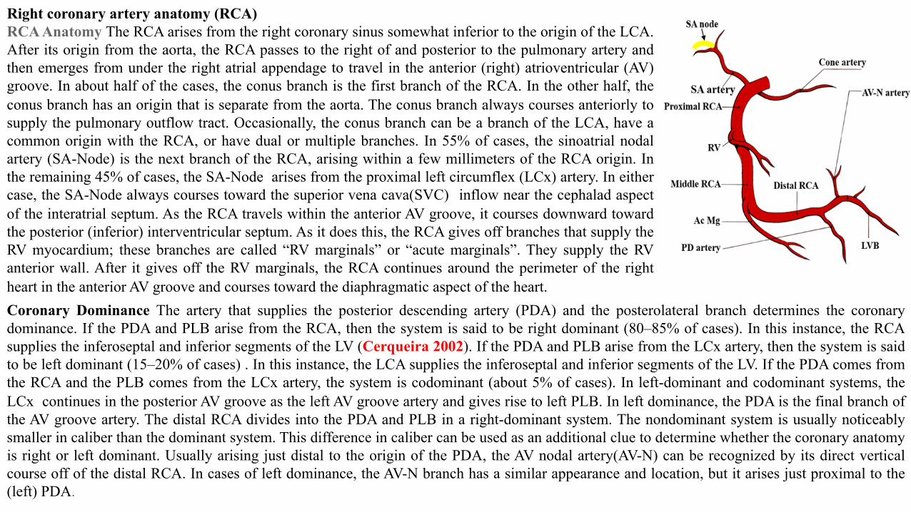
Right coronary artery anatomy (RCA) RCA Anatomy The RCA arises from the right coronary sinus somewhat inferior to the origin of the LCA. After its origin from the aorta, the RCA passes to the right of and posterior to the pulmonary artery and then emerges from under the right atrial appendage to travel in the anterior (right) atrioventricular (AV) groove. In about half of the cases, the conus branch is the first branch of the RCA. In the other half, the conus branch has an origin that is separate from the aorta. The conus branch always courses anteriorly to supply the pulmonary outflow tract. Occasionally, the conus branch can be a branch of the LCA, have a common origin with the RCA, or have dual or multiple branches. In 55% of cases, the sinoatrial nodal artery (SA-Node) is the next branch of the RCA, arising within a few millimeters of the RCA origin. In the remaining 45% of cases, the SA-Node arises from the proximal left circumflex (LCx) artery. In either case, the SA-Node always courses toward the superior vena cava(SVC) inflow near the cephalad aspect of the interatrial septum. As the RCA travels within the anterior AV groove, it courses downward toward the posterior (inferior) interventricular septum. As it does this, the RCA gives off branches that supply the RV myocardium; these branches are called “RV marginals” or “acute marginals”. They supply the RV anterior wall. After it gives off the RV marginals, the RCA continues around the perimeter of the right heart in the anterior AV groove and courses toward the diaphragmatic aspect of the heart. Coronary Dominance The artery that supplies the posterior descending artery (PDA) and the posterolateral branch determines the coronary dominance. If the PDA and PLB arise from the RCA, then the system is said to be right dominant (80–85% of cases). In this instance, the RCA supplies the inferoseptal and inferior segments of the LV (Cerqueira 2002). If the PDA and PLB arise from the LCx artery, then the system is said to be left dominant (15–20% of cases) . In this instance, the LCA supplies the inferoseptal and inferior segments of the LV. If the PDA comes from the RCA and the PLB comes from the LCx artery, the system is codominant (about 5% of cases). In left-dominant and codominant systems, the LCx continues in the posterior AV groove as the left AV groove artery and gives rise to left PLB. In left dominance, the PDA is the final branch of the AV groove artery. The distal RCA divides into the PDA and PLB in a right-dominant system. The nondominant system is usually noticeably smaller in caliber than the dominant system. This difference in caliber can be used as an additional clue to determine whether the coronary anatomy is right or left dominant. Usually arising just distal to the origin of the PDA, the AV nodal artery(AV-N) can be recognized by its direct vertical course off of the distal RCA. In cases of left dominance, the AV-N branch has a similar appearance and location, but it arises just proximal to the (left) PDA.
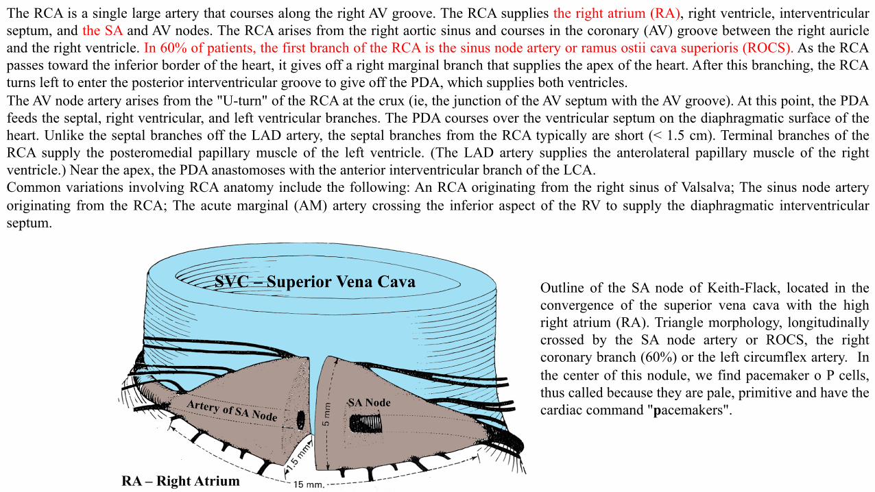
The RCA is a single large artery that courses along the right AV groove. The RCA supplies the right atrium (RA), right ventricle, interventricular septum, and the SA and AV nodes. The RCA arises from the right aortic sinus and courses in the coronary (AV) groove between the right auricle and the right ventricle. In 60% of patients, the first branch of the RCA is the sinus node artery or ramus ostii cava superioris (ROCS). As the RCA passes toward the inferior border of the heart, it gives off a right marginal branch that supplies the apex of the heart. After this branching, the RCA turns left to enter the posterior interventricular groove to give off the PDA, which supplies both ventricles. The AV node artery arises from the "U-turn" of the RCA at the crux (ie, the junction of the AV septum with the AV groove). At this point, the PDA feeds the septal, right ventricular, and left ventricular branches. The PDA courses over the ventricular septum on the diaphragmatic surface of the heart. Unlike the septal branches off the LAD artery, the septal branches from the RCA typically are short (< 1.5 cm). Terminal branches of the RCA supply the posteromedial papillary muscle of the left ventricle. (The LAD artery supplies the anterolateral papillary muscle of the right ventricle.) Near the apex, the PDA anastomoses with the anterior interventricular branch of the LCA. Common variations involving RCA anatomy include the following: An RCA originating from the right sinus of Valsalva; The sinus node artery originating from the RCA; The acute marginal (AM) artery crossing the inferior aspect of the RV to supply the diaphragmatic interventricular septum.
Outline of the SA node of Keith-Flack, located in the convergence of the superior vena cava with the high right atrium (RA). Triangle morphology, longitudinally crossed by the SA node artery or ROCS, the right coronary branch (60%) or the left circumflex artery. In the center of this nodule, we find pacemaker o P cells, thus called because they are pale, primitive and have the cardiac command "pacemakers".
RA – Right Atrium
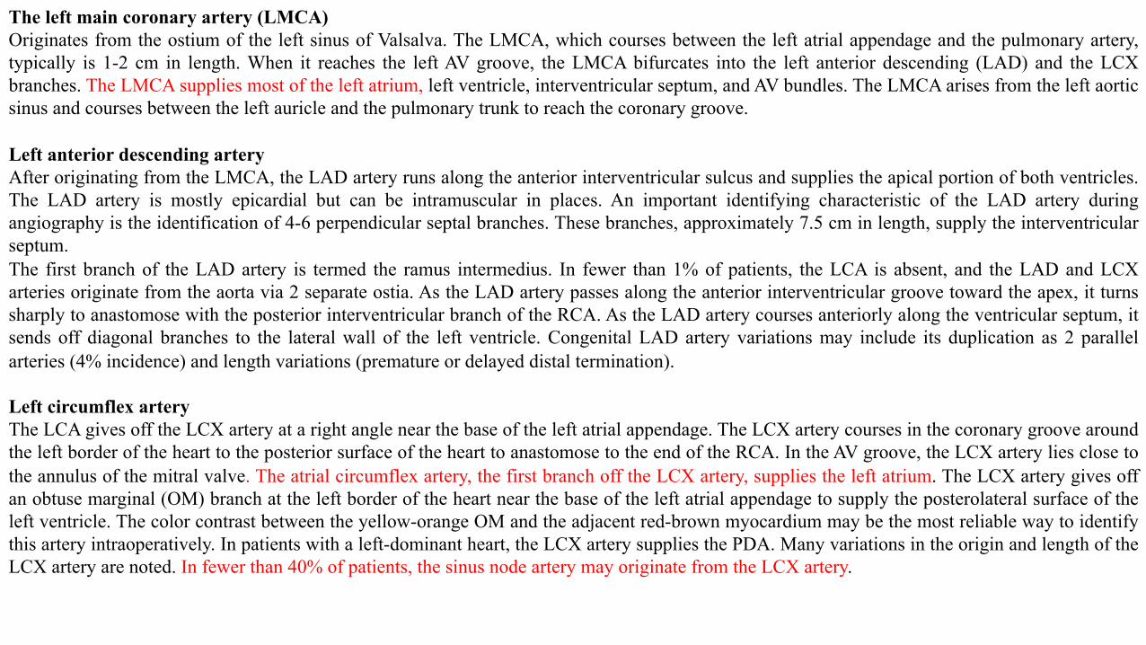
The left main coronary artery (LMCA) Originates from the ostium of the left sinus of Valsalva. The LMCA, which courses between the left atrial appendage and the pulmonary artery, typically is 1-2 cm in length. When it reaches the left AV groove, the LMCA bifurcates into the left anterior descending (LAD) and the LCX branches. The LMCA supplies most of the left atrium, left ventricle, interventricular septum, and AV bundles. The LMCA arises from the left aortic sinus and courses between the left auricle and the pulmonary trunk to reach the coronary groove. Left anterior descending artery After originating from the LMCA, the LAD artery runs along the anterior interventricular sulcus and supplies the apical portion of both ventricles. The LAD artery is mostly epicardial but can be intramuscular in places. An important identifying characteristic of the LAD artery during angiography is the identification of 4-6 perpendicular septal branches. These branches, approximately 7.5 cm in length, supply the interventricular septum. The first branch of the LAD artery is termed the ramus intermedius. In fewer than 1% of patients, the LCA is absent, and the LAD and LCX arteries originate from the aorta via 2 separate ostia. As the LAD artery passes along the anterior interventricular groove toward the apex, it turns sharply to anastomose with the posterior interventricular branch of the RCA. As the LAD artery courses anteriorly along the ventricular septum, it sends off diagonal branches to the lateral wall of the left ventricle. Congenital LAD artery variations may include its duplication as 2 parallel arteries (4% incidence) and length variations (premature or delayed distal termination). Left circumflex artery The LCA gives off the LCX artery at a right angle near the base of the left atrial appendage. The LCX artery courses in the coronary groove around the left border of the heart to the posterior surface of the heart to anastomose to the end of the RCA. In the AV groove, the LCX artery lies close to the annulus of the mitral valve. The atrial circumflex artery, the first branch off the LCX artery, supplies the left atrium. The LCX artery gives off an obtuse marginal (OM) branch at the left border of the heart near the base of the left atrial appendage to supply the posterolateral surface of the left ventricle. The color contrast between the yellow-orange OM and the adjacent red-brown myocardium may be the most reliable way to identify this artery intraoperatively. In patients with a left-dominant heart, the LCX artery supplies the PDA. Many variations in the origin and length of the LCX artery are noted. In fewer than 40% of patients, the sinus node artery may originate from the LCX artery.
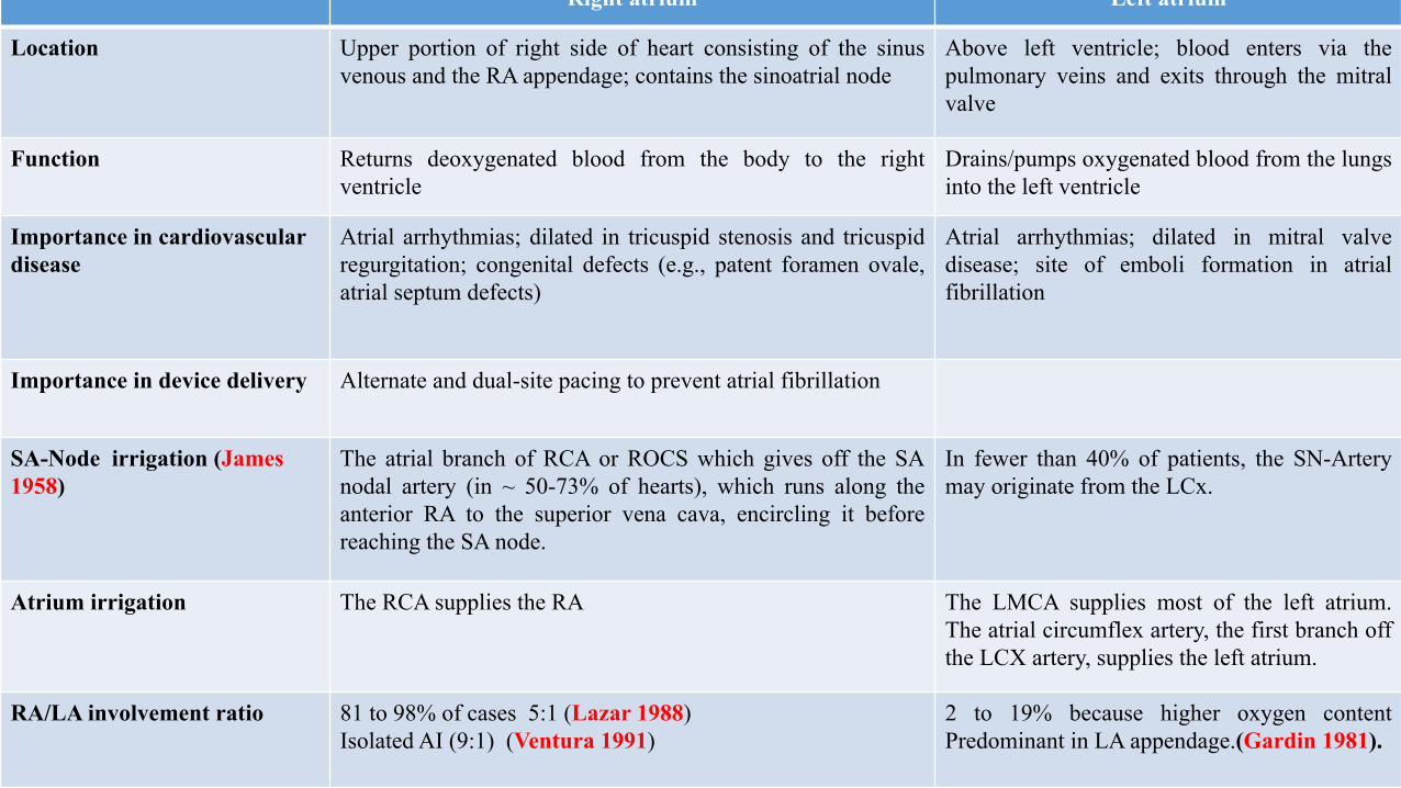
Right atrium Left atrium
Location Upper portion of right side of heart consisting of the sinus venous and the RA appendage; contains the sinoatrial node
Above left ventricle; blood enters via the pulmonary veins and exits through the mitral valve
Function Returns deoxygenated blood from the body to the right ventricle
Drains/pumps oxygenated blood from the lungs into the left ventricle
Importance in cardiovascular disease
Atrial arrhythmias; dilated in tricuspid stenosis and tricuspid regurgitation; congenital defects (e.g., patent foramen ovale, atrial septum defects)
Atrial arrhythmias; dilated in mitral valve disease; site of emboli formation in atrial fibrillation
Importance in device delivery Alternate and dual-site pacing to prevent atrial fibrillation
SA-Node irrigation (James 1958)
The atrial branch of RCA or ROCS which gives off the SA nodal artery (in ~ 50-73% of hearts), which runs along the anterior RA to the superior vena cava, encircling it before reaching the SA node.
In fewer than 40% of patients, the SN-Artery may originate from the LCx.
Atrium irrigation The RCA supplies the RA The LMCA supplies most of the left atrium. The atrial circumflex artery, the first branch off the LCX artery, supplies the left atrium.
RA/LA involvement ratio 81 to 98% of cases 5:1 (Lazar 1988) Isolated AI (9:1) (Ventura 1991)
2 to 19% because higher oxygen content Predominant in LA appendage.(Gardin 1981).
1
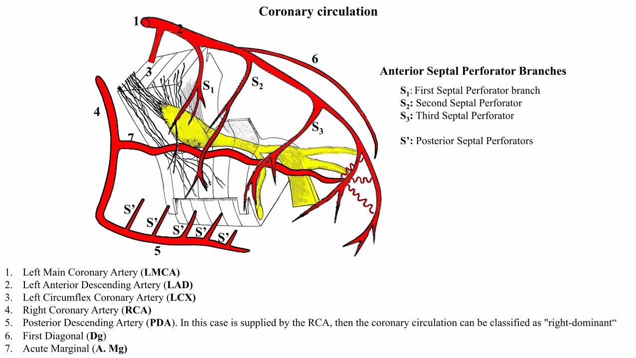
1. Left Main Coronary Artery (LMCA) 2. Left Anterior Descending Artery (LAD) 3. Left Circumflex Coronary Artery (LCX) 4. Right Coronary Artery (RCA) 5. Posterior Descending Artery (PDA). In this case is supplied by the RCA, then the coronary circulation can be classified as "right-dominant“ 6. First Diagonal (Dg) 7. Acute Marginal (A. Mg)
2
3
4
5
S1 S2
S3
S1: First Septal Perforator branch S2: Second Septal Perforator S3: Third Septal Perforator S’: Posterior Septal Perforators
6
7
Coronary circulation
Anterior Septal Perforator Branches
S’ S’ S’ S’ S’
15 10
4 17
13
7 1
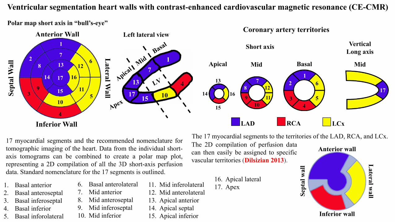
Ventricular segmentation heart walls with contrast-enhanced cardiovascular magnetic resonance (CE-CMR)
1. Basal anterior 2. Basal anteroseptal 3. Basal inferoseptal 4. Basal inferior 5. Basal inforolateral
11. Mid inferolateral 12. Mid anterolateral 13. Apical anterior 14. Apical septal 15. Apical inferior
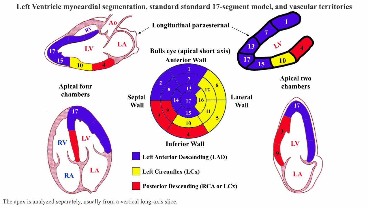
17 myocardial segments and the recommended nomenclature for tomographic imaging of the heart. Data from the individual short-axis tomograms can be combined to create a polar map plot, representing a 2D compilation of all the 3D short-axis perfusion data. Standard nomenclature for the 17 segments is outlined.
Coronary artery territories
LAD RCA LCx
Short axis Vertical Long axis
Basal Mid Apical Mid
1 2
3 4
5
6 7 8
9 10
11
12
15
14
13
17 16
6. Basal anterolateral 7. Mid anterior 8. Mid anteroseptal 9. Mid inferoseptal 10. Mid inferior
16. Apical lateral 17. Apex
The 17 myocardial segments to the territories of the LAD, RCA, and LCx.
Left lateral view
Polar map short axis in “bull’s-eye”
Anterior wall
Inferior wall
Sept
al w
all L
ateral wall
The 2D compilation of perfusion data can then easily be assigned to specific vascular territories (Dilsizian 2013).
Inferior Wall
Sept
al W
all L
ateral Wall
Anterior Wall 1
7
13
17
15
10
4
2 8
9 3
14
12 6
11 5
16
15 10 4
LV LA
Ao Longitudinal paraesternal
Bulls eye (apical short axis)
LV LV
LA LA
15 10
4 17
13
7 1
RV
RA
Apical four chambers
Apical two chambers
Left Anterior Descending (LAD)
Left Circunflex (LCx)
Posterior Descending (RCA or LCx)
Inferior Wall
Septal Wall
Lateral Wall
Anterior Wall 1
7
13
17
15
10
4
2 8
9 3
14
12 6
11 5
16
17
17 17
9
3
Left Ventricle myocardial segmentation, standard standard 17-segment model, and vascular territories
The apex is analyzed separately, usually from a vertical long-axis slice.
II
III
aVL
aVF
III aVF
15:00 after PCI
“W” P-wave pattern: atrial infarction
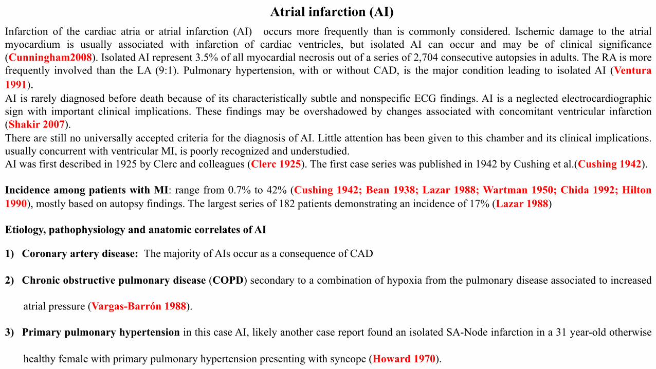
Infarction of the cardiac atria or atrial infarction (AI) occurs more frequently than is commonly considered. Ischemic damage to the atrial myocardium is usually associated with infarction of cardiac ventricles, but isolated AI can occur and may be of clinical significance (Cunningham2008). Isolated AI represent 3.5% of all myocardial necrosis out of a series of 2,704 consecutive autopsies in adults. The RA is more frequently involved than the LA (9:1). Pulmonary hypertension, with or without CAD, is the major condition leading to isolated AI (Ventura 1991). AI is rarely diagnosed before death because of its characteristically subtle and nonspecific ECG findings. AI is a neglected electrocardiographic sign with important clinical implications. These findings may be overshadowed by changes associated with concomitant ventricular infarction (Shakir 2007). There are still no universally accepted criteria for the diagnosis of AI. Little attention has been given to this chamber and its clinical implications. usually concurrent with ventricular MI, is poorly recognized and understudied. AI was first described in 1925 by Clerc and colleagues (Clerc 1925). The first case series was published in 1942 by Cushing et al.(Cushing 1942). Incidence among patients with MI: range from 0.7% to 42% (Cushing 1942; Bean 1938; Lazar 1988; Wartman 1950; Chida 1992; Hilton 1990), mostly based on autopsy findings. The largest series of 182 patients demonstrating an incidence of 17% (Lazar 1988) Etiology, pathophysiology and anatomic correlates of AI
1) Coronary artery disease: The majority of AIs occur as a consequence of CAD
2) Chronic obstructive pulmonary disease (COPD) secondary to a combination of hypoxia from the pulmonary disease associated to increased
atrial pressure (Vargas-Barrón 1988).
3) Primary pulmonary hypertension in this case AI, likely another case report found an isolated SA-Node infarction in a 31 year-old otherwise
healthy female with primary pulmonary hypertension presenting with syncope (Howard 1970).
Atrial infarction (AI)
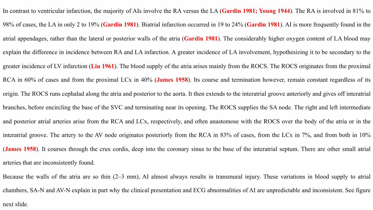
In contrast to ventricular infarction, the majority of AIs involve the RA versus the LA (Gardin 1981; Young 1944). The RA is involved in 81% to
98% of cases, the LA in only 2 to 19% (Gardin 1981). Biatrial infarction occurred in 19 to 24% (Gardin 1981). AI is more frequently found in the
atrial appendages, rather than the lateral or posterior walls of the atria (Gardin 1981). The considerably higher oxygen content of LA blood may
explain the difference in incidence between RA and LA infarction. A greater incidence of LA involvement, hypothesizing it to be secondary to the
greater incidence of LV infarction (Liu 1961). The blood supply of the atria arises mainly from the ROCS. The ROCS originates from the proximal
RCA in 60% of cases and from the proximal LCx in 40% (James 1958). Its course and termination however, remain constant regardless of its
origin. The ROCS runs cephalad along the atria and posterior to the aorta. It then extends to the interatrial groove anteriorly and gives off interatrial
branches, before encircling the base of the SVC and terminating near its opening. The ROCS supplies the SA node. The right and left intermediate
and posterior atrial arteries arise from the RCA and LCx, respectively, and often anastomose with the ROCS over the body of the atria or in the
interatrial groove. The artery to the AV node originates posteriorly from the RCA in 83% of cases, from the LCx in 7%, and from both in 10%
(James 1958). It courses through the crux cordis, deep into the coronary sinus to the base of the interatrial septum. There are other small atrial
arteries that are inconsistently found.
Because the walls of the atria are so thin (2–3 mm), AI almost always results in transmural injury. These variations in blood supply to atrial
chambers, SA-N and AV-N explain in part why the clinical presentation and ECG abnormalities of AI are unpredictable and inconsistent. See figure
next slide.
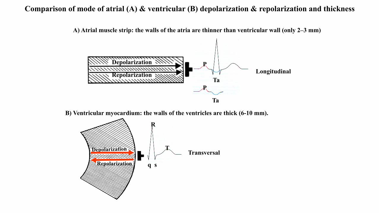
Comparison of mode of atrial (A) & ventricular (B) depolarization & repolarization and thickness
Depolarization
Repolarization
Depolarization
Repolarization
A) Atrial muscle strip: the walls of the atria are thinner than ventricular wall (only 2–3 mm)
B) Ventricular myocardium: the walls of the ventricles are thick (6-10 mm).
T
s q
R
P
P Ta
Ta
Longitudinal
Transversal
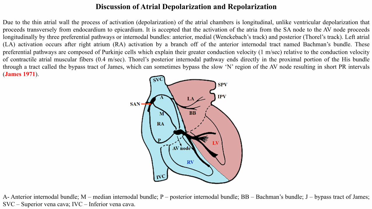
Discussion of Atrial Depolarization and Repolarization Due to the thin atrial wall the process of activation (depolarization) of the atrial chambers is longitudinal, unlike ventricular depolarization that proceeds transversely from endocardium to epicardium. It is accepted that the activation of the atria from the SA node to the AV node proceeds longitudinally by three preferential pathways or internodal bundles: anterior, medial (Wenckebach’s track) and posterior (Thorel’s track). Left atrial (LA) activation occurs after right atrium (RA) activation by a branch off of the anterior internodal tract named Bachman’s bundle. These preferential pathways are composed of Purkinje cells which explain their greater conduction velocity (1 m/sec) relative to the conduction velocity of contractile atrial muscular fibers (0.4 m/sec). Thorel’s posterior internodal pathway ends directly in the proximal portion of the His bundle through a tract called the bypass tract of James, which can sometimes bypass the slow ‘N’ region of the AV node resulting in short PR intervals (James 1971).
A- Anterior internodal bundle; M – median internodal bundle; P – posterior internodal bundle; BB – Bachman’s bundle; J – bypass tract of James; SVC – Superior vena cava; IVC – Inferior vena cava.
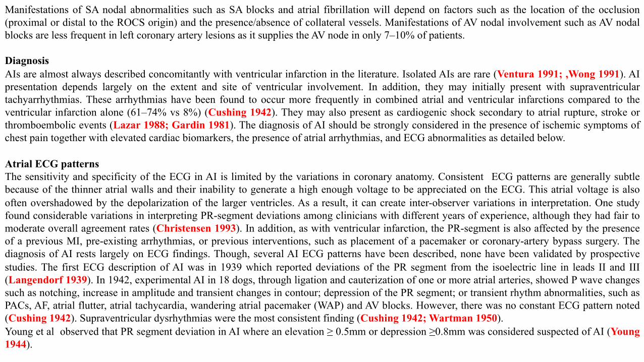
Manifestations of SA nodal abnormalities such as SA blocks and atrial fibrillation will depend on factors such as the location of the occlusion (proximal or distal to the ROCS origin) and the presence/absence of collateral vessels. Manifestations of AV nodal involvement such as AV nodal blocks are less frequent in left coronary artery lesions as it supplies the AV node in only 7–10% of patients. Diagnosis AIs are almost always described concomitantly with ventricular infarction in the literature. Isolated AIs are rare (Ventura 1991; ,Wong 1991). AI presentation depends largely on the extent and site of ventricular involvement. In addition, they may initially present with supraventricular tachyarrhythmias. These arrhythmias have been found to occur more frequently in combined atrial and ventricular infarctions compared to the ventricular infarction alone (61–74% vs 8%) (Cushing 1942). They may also present as cardiogenic shock secondary to atrial rupture, stroke or thromboembolic events (Lazar 1988; Gardin 1981). The diagnosis of AI should be strongly considered in the presence of ischemic symptoms of chest pain together with elevated cardiac biomarkers, the presence of atrial arrhythmias, and ECG abnormalities as detailed below. Atrial ECG patterns The sensitivity and specificity of the ECG in AI is limited by the variations in coronary anatomy. Consistent ECG patterns are generally subtle because of the thinner atrial walls and their inability to generate a high enough voltage to be appreciated on the ECG. This atrial voltage is also often overshadowed by the depolarization of the larger ventricles. As a result, it can create inter-observer variations in interpretation. One study found considerable variations in interpreting PR-segment deviations among clinicians with different years of experience, although they had fair to moderate overall agreement rates (Christensen 1993). In addition, as with ventricular infarction, the PR-segment is also affected by the presence of a previous MI, pre-existing arrhythmias, or previous interventions, such as placement of a pacemaker or coronary-artery bypass surgery. The diagnosis of AI rests largely on ECG findings. Though, several AI ECG patterns have been described, none have been validated by prospective studies. The first ECG description of AI was in 1939 which reported deviations of the PR segment from the isoelectric line in leads II and III (Langendorf 1939). In 1942, experimental AI in 18 dogs, through ligation and cauterization of one or more atrial arteries, showed P wave changes such as notching, increase in amplitude and transient changes in contour; depression of the PR segment; or transient rhythm abnormalities, such as PACs, AF, atrial flutter, atrial tachycardia, wandering atrial pacemaker (WAP) and AV blocks. However, there was no constant ECG pattern noted (Cushing 1942). Supraventricular dysrhythmias were the most consistent finding (Cushing 1942; Wartman 1950). Young et al observed that PR segment deviation in AI where an elevation ≥ 0.5mm or depression ≥0.8mm was considered suspected of AI (Young 1944).
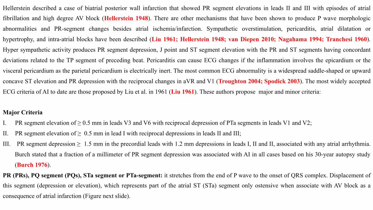
Hellerstein described a case of biatrial posterior wall infarction that showed PR segment elevations in leads II and III with episodes of atrial fibrillation and high degree AV block (Hellerstein 1948). There are other mechanisms that have been shown to produce P wave morphologic
abnormalities and PR-segment changes besides atrial ischemia/infarction. Sympathetic overstimulation, pericarditis, atrial dilatation or
hypertrophy, and intra-atrial blocks have been described (Liu 1961; Hellerstein 1948; van Diepen 2010; Nagahama 1994; Tranchesi 1960).
Hyper sympathetic activity produces PR segment depression, J point and ST segment elevation with the PR and ST segments having concordant
deviations related to the TP segment of preceding beat. Pericarditis can cause ECG changes if the inflammation involves the epicardium or the
visceral pericardium as the parietal pericardium is electrically inert. The most common ECG abnormality is a widespread saddle-shaped or upward
concave ST elevation and PR depression with the reciprocal changes in aVR and V1 (Troughton 2004; Spodick 2003). The most widely accepted
ECG criteria of AI to date are those proposed by Liu et al. in 1961 (Liu 1961). These authors propose major and minor criteria:
Major Criteria
I. PR segment elevation of ≥ 0.5 mm in leads V3 and V6 with reciprocal depression of PTa segments in leads V1 and V2;
II. PR segment elevation of ≥ 0.5 mm in lead I with reciprocal depressions in leads II and III;
III. PR segment depression ≥ 1.5 mm in the precordial leads with 1.2 mm depressions in leads I, II and II, associated with any atrial arrhythmia.
Burch stated that a fraction of a millimeter of PR segment depression was associated with AI in all cases based on his 30-year autopsy study
(Burch 1976).
PR (PRs), PQ segment (PQs), STa segment or PTa-segment: it stretches from the end of P wave to the onset of QRS complex. Displacement of this segment (depression or elevation), which represents part of the atrial ST (STa) segment only ostensive when associate with AV block as a
consequence of atrial infarction (Figure next slide).
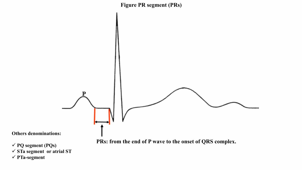
Figure PR segment (PRs)
Others denominations:
ü PQ segment (PQs) ü STa segment or atrial ST ü PTa-segment
PRs: from the end of P wave to the onset of QRS complex.
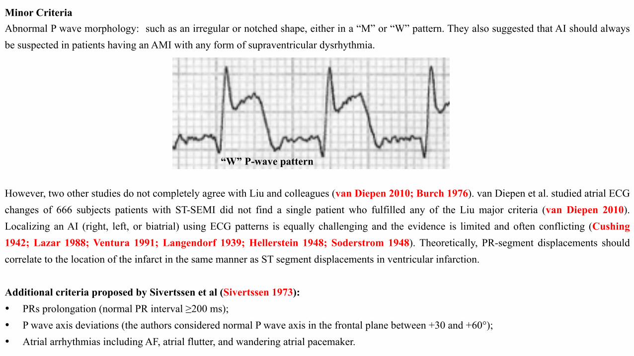
Minor Criteria Abnormal P wave morphology: such as an irregular or notched shape, either in a “M” or “W” pattern. They also suggested that AI should always be suspected in patients having an AMI with any form of supraventricular dysrhythmia. However, two other studies do not completely agree with Liu and colleagues (van Diepen 2010; Burch 1976). van Diepen et al. studied atrial ECG changes of 666 subjects patients with ST-SEMI did not find a single patient who fulfilled any of the Liu major criteria (van Diepen 2010). Localizing an AI (right, left, or biatrial) using ECG patterns is equally challenging and the evidence is limited and often conflicting (Cushing 1942; Lazar 1988; Ventura 1991; Langendorf 1939; Hellerstein 1948; Soderstrom 1948). Theoretically, PR-segment displacements should correlate to the location of the infarct in the same manner as ST segment displacements in ventricular infarction. Additional criteria proposed by Sivertssen et al (Sivertssen 1973): • PRs prolongation (normal PR interval ≥200 ms); • P wave axis deviations (the authors considered normal P wave axis in the frontal plane between +30 and +60°); • Atrial arrhythmias including AF, atrial flutter, and wandering atrial pacemaker.
“W” P-wave pattern
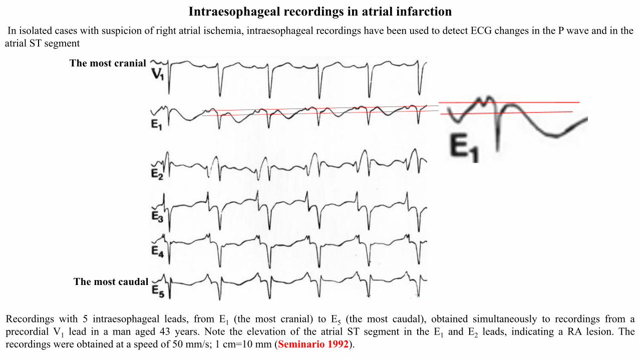
In isolated cases with suspicion of right atrial ischemia, intraesophageal recordings have been used to detect ECG changes in the P wave and in the atrial ST segment
Intraesophageal recordings in atrial infarction
Recordings with 5 intraesophageal leads, from E1 (the most cranial) to E5 (the most caudal), obtained simultaneously to recordings from a precordial V1 lead in a man aged 43 years. Note the elevation of the atrial ST segment in the E1 and E2 leads, indicating a RA lesion. The recordings were obtained at a speed of 50 mm/s; 1 cm=10 mm (Seminario 1992).
The most caudal
The most cranial
Cardio Command Systems for easy access to all the diagnostic benefits of the Esophageal ECG The close proximity of the esophageal electrodes to the posterior atrium and left ventricle provides greater signal strength, resulting in larger P-waves (atrial activation) as well as an amplified QRS complex (ventricular activity). Because the atrium is positioned behind the ventricles relative to the chest wall, P-waves are often obscured by ventricular electrical activity in ECG recordings acquired from surface electrodes. Thus esophageal ECG monitoring is essential for accurate diagnosis of cardiac arrhythmias for which a clearly identifiable P-wave is needed. Ambulatory esophageal ECG monitoring is feasible and when compared to QRST subtraction can reveal LA foci as initiators of paroxysmal AF. Documentation of early esophageal lead activation for LA premature beats that initiate AF is highly predictive of pulmonary vein triggers, and may serve as a useful screening test for candidates for focal AF ablation. The capsule with enclosed electrodes was voluntarily swallowed, and the recording electrodes were positioned posterior to the LA. A high-quality esophageal ECG recording is obtained within one to 10 minutes with minimal patient discomfort. In 25 of 48 study patients (52%), the original diagnosis, based on the surface ECG recording, was incorrect after review of the esophageal recording. Results of esophageal recording altered management in 19 of 48 (40%) patients ( Shaw 1987). The esophageal lead is safe, simple to use, and provides valuable information for detection or diagnosis of dysrhythmias and myocardial ischemia during anesthesia (Kates 1982). Abstract. Atrial flutter may be difficult to diagnose in infants with tachycardia unless transesophageal electrogram recording is utilized for evaluation (Dunnigan 1985). Example: Twenty-six of 68 (pediatric cardiac surgery) patients exhibited rhythms during reperfusion that required the Esophageal ECG for definitive diagnosis (Bushman 1989). Heart rate variability (HRV) analysis has been an established method for assessment of the activities of autonomic nervous system. A-A intervals in the intraatrial ECG and P-P intervals in surface ECG were poorly correlated in AV block patients. However, intraatrial ECG was well correlated with esophageal ECG. HRV with time domain decreased significantly in patients with AV block. The esophageal lead recording is a non-invasive, easy and safe method to detect HRV of AV block patients whose vagal activity is abnormal (Hsiao 1997). Risk stratification of post MI patients suggests that heart rate variability is the single most important predictor of those patients who are at high risk of SCD or serious ventricular arrhythmias (Malik 1990). This new high-resolution Esophageal-ECG device detected in a beat-to-beat technique more atrial activity during cardioplegic arrest than a Surface-ECG and offered the advantages of artifact exclusion and better prediction of postoperative supraventricular dysrhythmias (Maechler 1997). During acute myocardial ischemia and infarction in sheep, the esophageal electrocardiograph has visually apparent ST alternans of amplitude in the millivolt range. This is therefore one very promising technique for better evaluation of electrocardiographic changes of ischemia (Mächler 1998). The precession of the left atrial electrogram recorded by the esophageal electrode on the P wave in lead V1 during supraventricular tachycardia (SVT) was a specific criterion of reentry through a left accessory AV connection, and this technique could be useful for preliminary localization of the accessory connection before EPS (Brembilla-Perrot 1991).
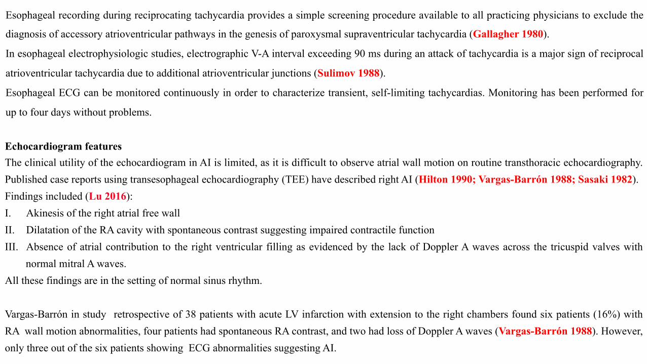
Esophageal recording during reciprocating tachycardia provides a simple screening procedure available to all practicing physicians to exclude the
diagnosis of accessory atrioventricular pathways in the genesis of paroxysmal supraventricular tachycardia (Gallagher 1980).
In esophageal electrophysiologic studies, electrographic V-A interval exceeding 90 ms during an attack of tachycardia is a major sign of reciprocal
atrioventricular tachycardia due to additional atrioventricular junctions (Sulimov 1988).
Esophageal ECG can be monitored continuously in order to characterize transient, self-limiting tachycardias. Monitoring has been performed for
up to four days without problems.
Echocardiogram features The clinical utility of the echocardiogram in AI is limited, as it is difficult to observe atrial wall motion on routine transthoracic echocardiography. Published case reports using transesophageal echocardiography (TEE) have described right AI (Hilton 1990; Vargas-Barrón 1988; Sasaki 1982). Findings included (Lu 2016): I. Akinesis of the right atrial free wall II. Dilatation of the RA cavity with spontaneous contrast suggesting impaired contractile function III. Absence of atrial contribution to the right ventricular filling as evidenced by the lack of Doppler A waves across the tricuspid valves with
normal mitral A waves. All these findings are in the setting of normal sinus rhythm. Vargas-Barrón in study retrospective of 38 patients with acute LV infarction with extension to the right chambers found six patients (16%) with RA wall motion abnormalities, four patients had spontaneous RA contrast, and two had loss of Doppler A waves (Vargas-Barrón 1988). However, only three out of the six patients showing ECG abnormalities suggesting AI.
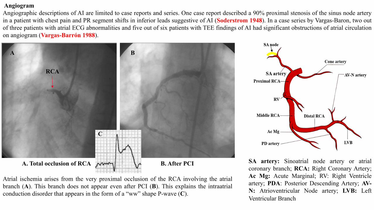
Angiogram Angiographic descriptions of AI are limited to case reports and series. One case report described a 90% proximal stenosis of the sinus node artery in a patient with chest pain and PR segment shifts in inferior leads suggestive of AI (Soderstrom 1948). In a case series by Vargas-Baron, two out of three patients with atrial ECG abnormalities and five out of six patients with TEE findings of AI had significant obstructions of atrial circulation on angiogram (Vargas-Barrón 1988).
SA artery: Sinoatrial node artery or atrial coronary branch; RCA: Right Coronary Artery; Ac Mg: Acute Marginal; RV: Right Ventricle artery; PDA: Posterior Descending Artery; AV-N: Atrioventricular Node artery; LVB: Left Ventricular Branch
A B
A. Total occlusion of RCA B. After PCI
Atrial ischemia arises from the very proximal occlusion of the RCA involving the atrial branch (A). This branch does not appear even after PCI (B). This explains the intraatrial conduction disorder that appears in the form of a “ww” shape P-wave (C).
C
RCA
Other diagnostic studies One case report described akinesis and enlargement of the RA using a radionuclide gated cardiac blood pool study in a patient suffering from inferior wall myocardial infarction that developed episodes of atrial flutter, atrial fibrillation and complete AV block (Gordon 1984). which corresponds to the LA will result in PR (PTa) segment elevation in leads II and III with reciprocal depression in lead I (Hellerstein 1948). Likewise, involvement of the anterior or anterolateral wall which corresponds to the RA will produce PR (PTa) segment elevation in lead I with reciprocal depression in leads II, III and the anterior precordial leads V2–V4 (Cushing 1942; Liu 1961; Langendorf 1939). There remain no universally accepted criteria. Classical signs of AI include: • Changes in wide and shape of the P waves; • Depression or elevation of the PR segment; • Previously unrecorded convexity of the first portion of the PR segment. ECG signs of atrial rupture inter setting of an acute MI Marafioti et al (Marafioti 2013) report the case of a 79 year old man with history of CAD and complaints of dyspnea and typical chest pain who died shortly after admitted at ED. The patient’s ECG shows two components: the first presumably representing the atrial activity and the second the ventricular activity. The second activity is actually compatible with an evolving acute inferolateral MI. However, one is struck by the giant waves, especially in the anterolateral precordial leads assumed to represent the atrial activity. The first impression form that tracing is that these giant P waves represent an artefact. However, a more careful analysis suggest that they represent a major injury pattern affecting the atrium in the same way ST segment elevation reflects a ventricular injury pattern. Such atrial disorders could suggest the imminence of atrial rupture, just as persistent mayor ST elevation could indicate the imminence of ventricular rupture. Unfortunately, the lack of postmortem examination did not enable a definitive diagnostic confirmation. Nonetheless, the authors agree that these impressive and uncommon prominent ECG changes at the atrial level, combined with the patient’s history and the clinical presentation, strongly suggest that atrial wall rupture had occurred in the course of an inferolateral MI and resulted in the patient’s death. The ECG tracing presented by Marafioti et al (Marafioti 2013) exhibits striking similarities with that reported by van Veldhuisen and van den Berg (van Veldhuisen 1999) in a previously healthy 20 year old man brought to hospital after he was run over by a car. The patient died shortly after admission to hospital and a right atrial rupture with massive intrathoracic bleeding was found during autopsy. The patient’s ECG showed abnormalities that were even more pronounced than those described by Marafioti et al (Marafioti 2013). 2:1 AV block was present, which made it possible to individualize the giant P waves that mimic the classical Pardee waves observed during the first hours of an AMI. The similarity in the P wave morphology in both reports is remarkable, supporting the hypothesis that the atrial rupture
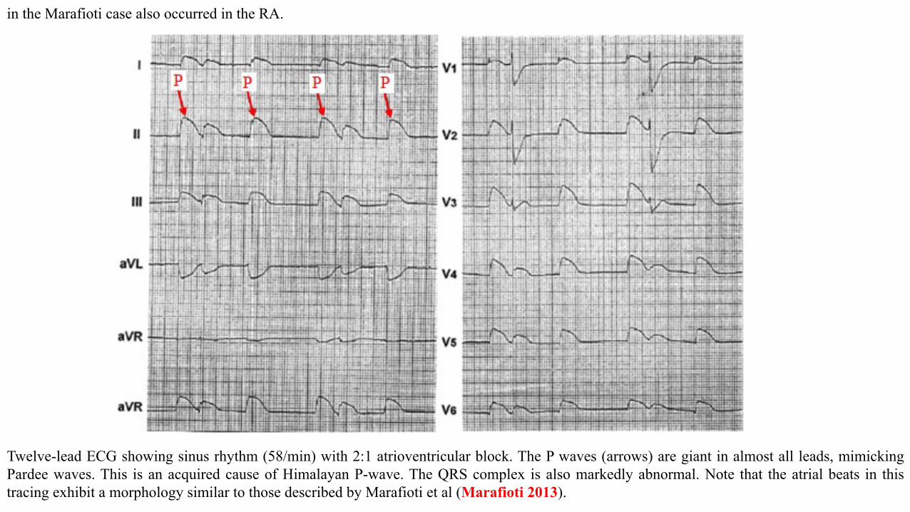
in the Marafioti case also occurred in the RA.
Twelve-lead ECG showing sinus rhythm (58/min) with 2:1 atrioventricular block. The P waves (arrows) are giant in almost all leads, mimicking Pardee waves. This is an acquired cause of Himalayan P-wave. The QRS complex is also markedly abnormal. Note that the atrial beats in this tracing exhibit a morphology similar to those described by Marafioti et al (Marafioti 2013).
Himalayan P-wave causes I. Congenital
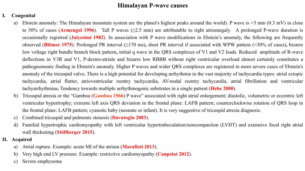
a) Ebstein anomaly: The Himalayan mountain system are the planet's highest peaks around the world). P wave is >3 mm (0.3 mV) in close to 50% of cases (Armengol 1996). Tall P waves (≥2.5 mm) are attributable to right atriomegaly. A prolonged P-wave duration is occasionally registred (Jaiyesimi 1982). In association with P wave modifications in Ebstein’s anomaly, the following are frequently observed (Blömer 1975): Prolonged PR interval (≥170 ms), short PR interval if associated with WPW pattern (≈30% of cases), bizarre low voltage right bundle branch block pattern, initial q wave in the QRS complexes of V1 and V2 leads. Reduced amplitude of R-wave deflections in V3R and V1, P-dextro-atriale and bizarre low RBBB without right ventricular overload almost certainly constitutes a pathognomonic finding in Ebstein's anomaly. Higher P waves and wider QRS complexes are registered in more severe cases of Ebstein's anomaly of the tricuspid valve. There is a high potential for developing arrhythmia in the vast majority of tachycardia types: atrial ectopic tachycardia, atrial flutter, atrioventricular reentry tachycardia, AV-nodal reentry tachycardia, atrial fibrillation and ventricular tachyarrhythmias. Tendency towards multiple arrhythmogenic substrates in a single patient (Hebe 2000).
b) Tricuspid atresia or the “Gamboa (Gamboa 1966) P wave” associated with right atrial enlargement; diastolic, volumetric or eccentric left ventricular hypertrophy; extreme left axis QRS deviation in the frontal plane: LAFB pattern; counterclockwise rotation of QRS loop in the frontal plane: LAFB pattern; cyanotic baby (neonate or infant). It is very suggestive of tricuspid atresia diagnosis.
c) Combined tricuspid and pulmonic stenosis (Davutoglu 2003). d) Familial hypertrophic cardiomyopathy with left ventricular hypertrabeculation/noncompaction (LVHT) and extensive focal right atrial
wall thickening (Stöllberger 2015). II. Acquired
a) Atrial rupture. Example: acute MI of the atrium (Marafioti 2013). b) Very high end LV pressure. Example: restrictive cardiomyopathy (Canpolat 2012). c) Severe emphysema
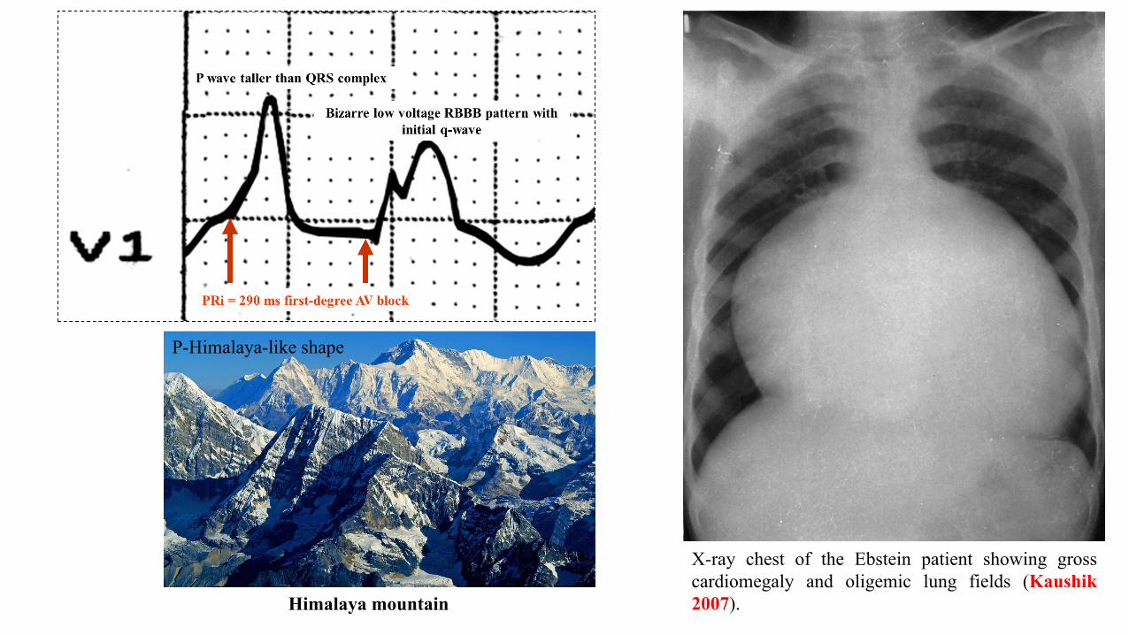
X-ray chest of the Ebstein patient showing gross cardiomegaly and oligemic lung fields (Kaushik 2007). Himalaya mountain
P-Himalaya-like shape
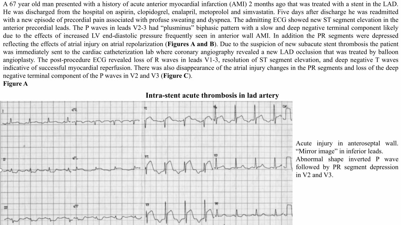
A 67 year old man presented with a history of acute anterior myocardial infarction (AMI) 2 months ago that was treated with a stent in the LAD. He was discharged from the hospital on aspirin, clopidogrel, enalapril, metoprolol and simvastatin. Five days after discharge he was readmitted with a new episode of precordial pain associated with profuse sweating and dyspnea. The admitting ECG showed new ST segment elevation in the anterior precordial leads. The P waves in leads V2-3 had “plusminus” biphasic pattern with a slow and deep negative terminal component likely due to the effects of increased LV end-diastolic pressure frequently seen in anterior wall AMI. In addition the PR segments were depressed reflecting the effects of atrial injury on atrial repolarization (Figures A and B). Due to the suspicion of new subacute stent thrombosis the patient was immediately sent to the cardiac catheterization lab where coronary angiography revealed a new LAD occlusion that was treated by balloon angioplasty. The post-procedure ECG revealed loss of R waves in leads V1-3, resolution of ST segment elevation, and deep negative T waves indicative of successful myocardial reperfusion. There was also disappearance of the atrial injury changes in the PR segments and loss of the deep negative terminal component of the P waves in V2 and V3 (Figure C). Figure A
Intra-stent acute thrombosis in lad artery
Acute injury in anteroseptal wall. “Mirror image” in inferior leads. Abnormal shape inverted P wave followed by PR segment depression in V2 and V3.
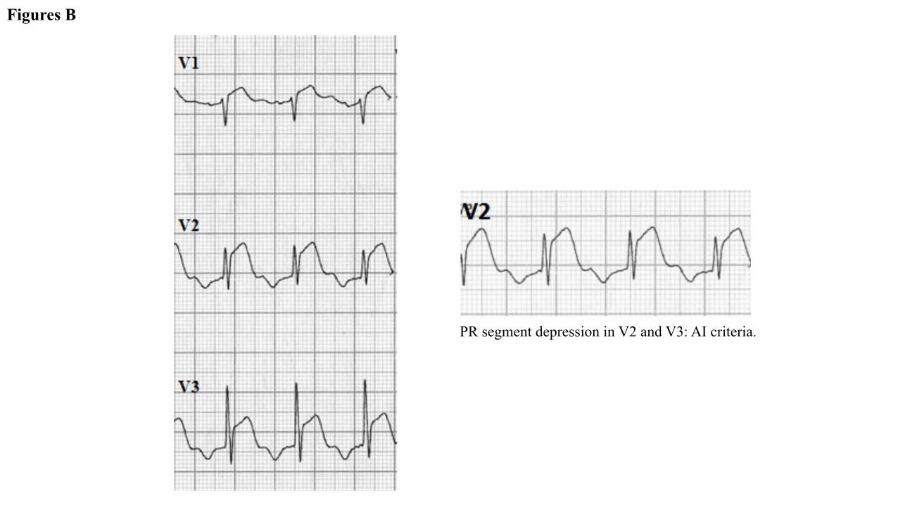
Figures B
PR segment depression in V2 and V3: AI criteria.
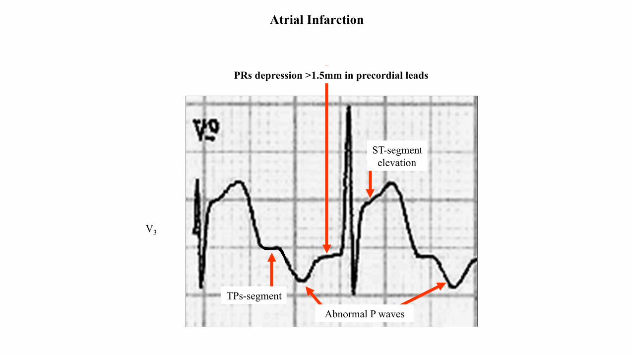
Atrial Infarction
Abnormal P waves
PRs depression >1.5mm in precordial leads
V3
ST-segment elevation
TPs-segment
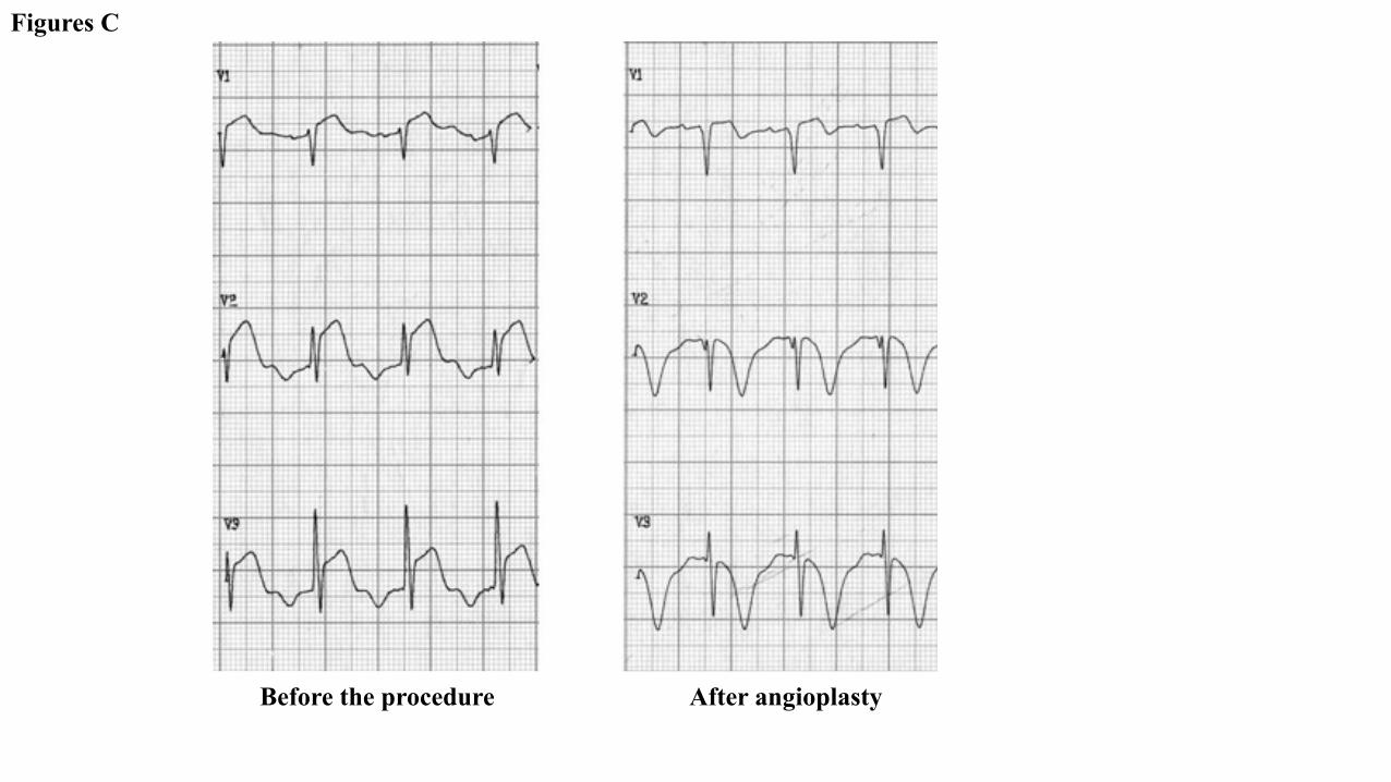
Figures C
Before the procedure After angioplasty
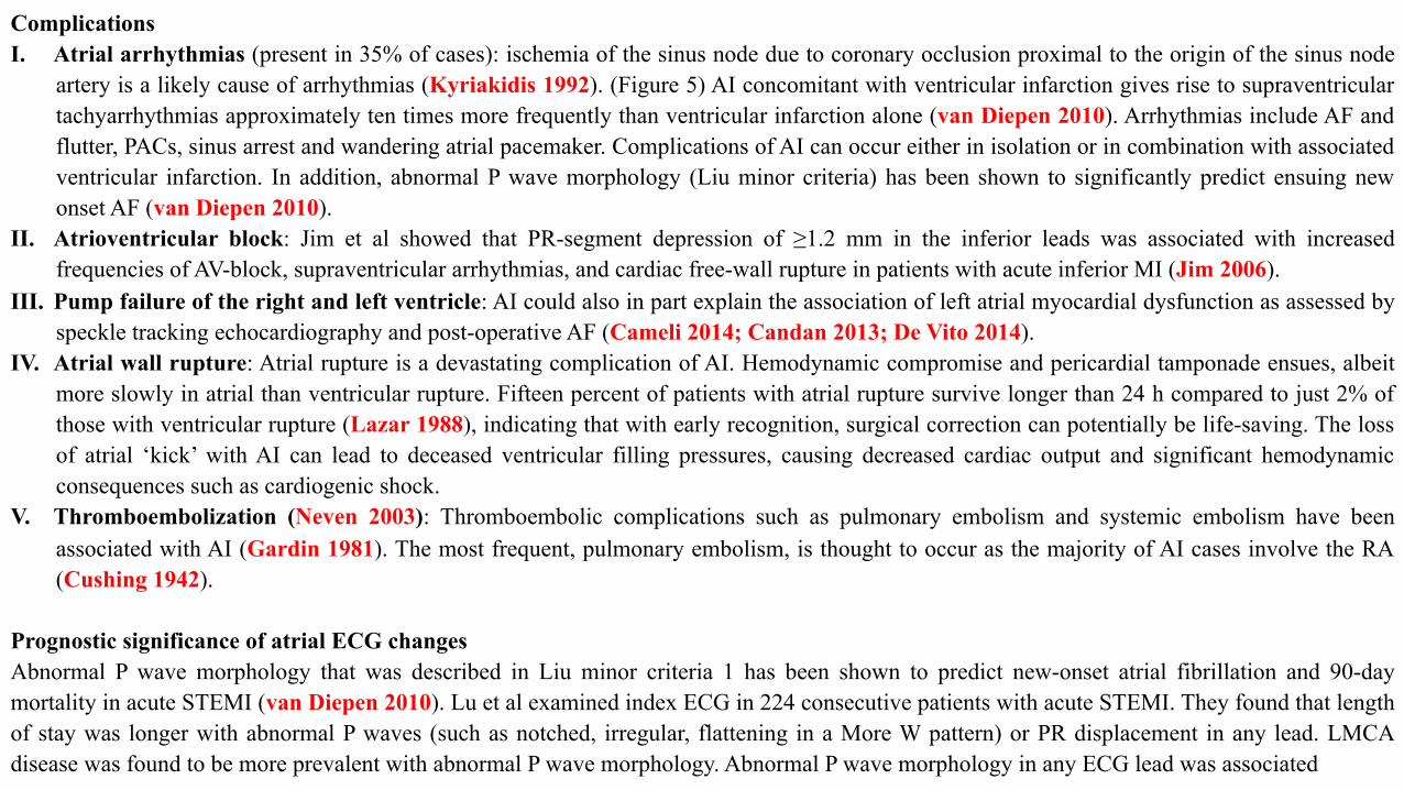
Complications I. Atrial arrhythmias (present in 35% of cases): ischemia of the sinus node due to coronary occlusion proximal to the origin of the sinus node
artery is a likely cause of arrhythmias (Kyriakidis 1992). (Figure 5) AI concomitant with ventricular infarction gives rise to supraventricular tachyarrhythmias approximately ten times more frequently than ventricular infarction alone (van Diepen 2010). Arrhythmias include AF and flutter, PACs, sinus arrest and wandering atrial pacemaker. Complications of AI can occur either in isolation or in combination with associated ventricular infarction. In addition, abnormal P wave morphology (Liu minor criteria) has been shown to significantly predict ensuing new onset AF (van Diepen 2010).
II. Atrioventricular block: Jim et al showed that PR-segment depression of ≥1.2 mm in the inferior leads was associated with increased frequencies of AV-block, supraventricular arrhythmias, and cardiac free-wall rupture in patients with acute inferior MI (Jim 2006).
III. Pump failure of the right and left ventricle: AI could also in part explain the association of left atrial myocardial dysfunction as assessed by speckle tracking echocardiography and post-operative AF (Cameli 2014; Candan 2013; De Vito 2014).
IV. Atrial wall rupture: Atrial rupture is a devastating complication of AI. Hemodynamic compromise and pericardial tamponade ensues, albeit more slowly in atrial than ventricular rupture. Fifteen percent of patients with atrial rupture survive longer than 24 h compared to just 2% of those with ventricular rupture (Lazar 1988), indicating that with early recognition, surgical correction can potentially be life-saving. The loss of atrial ‘kick’ with AI can lead to deceased ventricular filling pressures, causing decreased cardiac output and significant hemodynamic consequences such as cardiogenic shock.
V. Thromboembolization (Neven 2003): Thromboembolic complications such as pulmonary embolism and systemic embolism have been associated with AI (Gardin 1981). The most frequent, pulmonary embolism, is thought to occur as the majority of AI cases involve the RA (Cushing 1942).
Prognostic significance of atrial ECG changes Abnormal P wave morphology that was described in Liu minor criteria 1 has been shown to predict new-onset atrial fibrillation and 90-day mortality in acute STEMI (van Diepen 2010). Lu et al examined index ECG in 224 consecutive patients with acute STEMI. They found that length of stay was longer with abnormal P waves (such as notched, irregular, flattening in a More W pattern) or PR displacement in any lead. LMCA disease was found to be more prevalent with abnormal P wave morphology. Abnormal P wave morphology in any ECG lead was associated
with higher 30-day and 1-year mortality. PR displacement in any lead was also associated with increased 30-day and 1-year mortality. Abnormal P wave, PR depression in II III and aVF, and elevation in aVR or aVL were associated with increased 1-year mortality as was PR depression in the precordial leads. After adjusting for age, LVEF, peak troponin I, and LMCA disease, the authors found that PR displacement (present in 31% of STEMI in this series) in any lead was associated with increased 1-year mortality (Lu 2015). Management There are no specific recommendations in the management guidelines of myocardial infarction if concomitant AI is suspected (Amsterdam 2014; O'Gara 2013). Restoration of perfusion to the ventricular myocardium remains the main objective. One case of a patient with an inferior ST elevation myocardial infarction (STEMI) complicated by atrial fibrillation observed that the ECG immediately converted to sinus rhythm after balloon dilatation and stenting of the large left atrial branch of the left circumflex artery (Blanton 2010). The role of anti-arrhythmics in AI has yet to be explored. One large randomized control study found that the use of metoprolol in patients with acute myocardial infarction significantly reduced the incidence of supraventricular tachyarrhythmia, among its other benefits (Amsterdam 2014). If supraventricular tachyarrhythmias such as atrial fibrillation occur, initial treatment should include rate control with a beta blocker. Direct current cardioversion should be strongly considered if the patient is unstable, and can usually be done without the need for anticoagulation except when there is suspicion that the arrhythmia is not associated with the acute myocardial infarction. Conclusion Though the entity “AI” has been reported for over a century ,its recognition remains elusive. The diagnosis of AI should be suspected in any patient who presents with typical chest pain, elevated cardiac biomarkers and ECG changes consistent with AI; either the presence of abnormal P-wave morphologies, PR segmentdeviations, or supraventricular tachyarrhythmias. Patientsshould be monitored for complications such as arrhythmias, thromboembolic events and heart failure. Management is focused on coronary reperfusion and maintenance of sinus rhythm. With ischemic heart disease being the most common cause of death worldwide, adequate knowledge and further investigation of this disease entity may be one key to improved survival.
References 1. Amsterdam EA, Wenger NK, Brindis RG, et al. 2014 AHA/ACC guideline for the management of patients with non-ST-elevation acute
coronary syndromes: executive summary: a report of the American College of Cardiology/American Heart Association Task Force on Practice Guidelines. J Am Coll Cardiol. 2014;64(24)2645-87.
2. Armengol Rofes AJ, Serrano Durán M, Albert Brotons DC, Sánchez López C, Casaldáliga Ferrer J, Girona Comas JM. Ebstein's anomaly of the tricuspid valve. Apropos 35 cases. An Esp Pediatr. 1996;44(2):139-44.
3. Atar S, Bimbaum Y. Ischemia-induced ST-segment elevation: classification, prognosis, and therapy. J Electrocardiol. 2005;38(Supp):1-7. 4. Bairey CN, Shah PK, Lew AS, Hulse S.Electrocardiographic differentiation of occlusion of the left circumflex versus the right coronary artery
as a cause of inferior acute myocardial infarction.Am J Cardiol. 1987;60(7):456-9. 5. Bean WB. Infarction of the heart. Ann Intern Med. 1938;12:71–94. 6. Birnbaum Y, Sclarovsky S, Mager A, et al.ST segment depression in aVL: a sensitive marker for acute inferior myocardial infarction. Eur
Heart J. 1993;14(1):4-7 7. Birnbaum Y, Herz I, Sclarovsky S, et al. Prognostic significance of precordial ST segment depression on admission electrocardiogram in
patients with inferior wall myocardial infarction.J Am Coll Cardiol. 1996;28(2):313-8. 8. Birnbaum Y, Wagner GS, Barbash GI, Gates K, Criger DA, Sclarovsky S, et al. Correlation of angiographic findings and right (V1 to V3)
versus left (V4 to V6) precordial ST-segment depression in inferior wall acute myocardial infarction. Am J Cardiol. 1999;83(2):143–8. 9. Blanton RM, Nappi A, Kimmelstiel CD. Conversion of infarction-associated atrial fibrillation by restoration of atrial perfusion. Clin Cardiol.
2010;33(12):E79-81. 10. Blömer H. Electrocardiographic and phonocardiographic findings in Ebstein's anomaly with special regard to its severity. Med Klin.
1975;70(27):1175-8. 11. Brembilla-Perrot B. Study of P wave morphology in lead V1 during supraventricular tachycardia for localizing the reentrant circuit. Am Heart
J. 1991;121(6 Pt 1):1714-20. 12. Burch GE. Of the p-r segment depression and atrial infarction. Am Heart J. 1976;91(1):129-30. 13. Bushman GA. Clinical correlates of dysrhythmias requiring an esophageal ECG for accurate diagnosis in patients with congenital heart
disease. J Cardiothorac Anesth. 1989;3(3):290-4. 14. Cameli M, Lisi M, Reccia R, et al. Pre-operative left atrial strain predicts post-operative atrial fibrillation in patients undergoing aortic valve
replacement for aortic stenosis. Int J Cardiovasc Imaging. 2014;30(2):279-86.
15. Candan O, Ozdemir N, Aung SM, et al. Left atrial longitudinal strain parameters predict postoperative persistent atrial fibrillation following mitral valve surgery: a speckle tracking echocardiography study. Echocardiography. 2013;30(9):1061-8.
16. Canpolat U, Dural M, Aytemir K, Tokgözoğlu L. The importance of Himalayan P-wave in differentiation of cardiomyopathies. Anadolu Kardiyol Derg. 2012;12(4):368-9.
17. Cerqueira MD, Weisman NJ, Dilsizian V, et al. Standardized myocardial segmentation and nomenclature for tomographic imaging of the heart. A statement for healthcare professionals from the Cardiac Imaging Committee of the Council on Clinical Cardiology of the American Heart Association. Circulation 2002; 105(4):539–7.
18. Chida K, Ohkawa S, Maeda S, et al. A clinicopathologic study of atrial infarction complicating left ventricular posterior myocardial infarction. J Cardiol. 1992;22(1):1-10.
19. Clerc A, Levy R. Infarctus auriculaire: tachyarrhythmia terminale. Bull Mem Soc Med Hop Paris. 1925;41:1603–7. 20. Christensen JH, Nielsen FE, Falstie-Jensen N, Schmidt EB. Interobserver variation in interpretation of electrocardiographic signs of atrial
infarction. Clin Cardiol. 1993;16(8):603-6. 21. Cunningham KS, Chan KL, Veinot JP.Pathology of isolated atrial infarction: case report and review of the literature.Cardiovasc Pathol.
2008;17(3):183-5. 22. Cushing EH, Feil HS, Stanton EJ, Wartman WB. Infarction of the cardiac auricles (atria): clinical, pathological, and experimental studies. Br
Heart J. 1942;4(1-2):17–34. 23. Davutoglu V, Kilinc M, Dinckal MH. Himalayan P waves in a patient with combined tricuspid and pulmonic stenosis. Heart. 2003;89(10):
1216. 24. De Vito R, Di Giovanni A, Reccia R, et al. Left atrial strain predicts postoperative atrialfibrillation in patient undergoing major orthopaedic
surgery. Int Cardiovasc Forum J. 2014;1(4):189–92. 25. Dunnigan A, Benson W Jr, Benditt DG. Atrial flutter in infancy: diagnosis, clinical features, and treatment. Pediatrics. 1985;75(4):725-9. 26. Fiol M, Cygankiewicz I, Carrillo A, et al. Value of electrocardiographic algorithm based on "ups and downs" of ST in assessment of a culprit
artery in evolving inferior wall acute myocardial infarction.Am J Cardiol. 2004;94(6):709-14. 27. Fujiwara M, Horimoto M, Shiokoshi T,et al,Electrocardiographic ST-segment deviation in acute inferoposterior myocardial infarction caused
by obstruction of the left circumflex coronary arteryJ Cardiol. 1993;23(3):249-56. 28. Gallagher JJ, Smith WM, Kasell J, Smith WM, Grant AO, Benson DW Jr. Use of the esophageal lead in the diagnosis of mechanisms of
reciprocating supraventricular tachycardia. Pacing Clin Electrophysiol. 1980;3(4):440-51.
29. Gamboa R, Gersony WM, Nadas AS. The electrocardiogram in tricuspid atresia and pulmonary atresia with intact ventricular septum. Circulation. 1966;34(1):24-37.
30. Garcia-Rubira JC, Garcia-Borbolla R, Nuñez-Gil,et al. Distortion of the terminal portion of the QRS is predictor of shock after primary percutaneous coronary intervention for acute myocardial infarction. Int J Cardiol. 2008;130(2):241-5.
31. Gupta A, Lokhandwala YY, Kerkar PG, Vora AM. Electrocardiographic differentiation between right coronary and left circumflex coronary arterial occlusion in isolated inferior wall myocardial infarction. Indian Heart J. 1999;51(3):281–4.
32. Hebe J. Ebstein's anomaly in adults. Arrhythmias: diagnosis and therapeutic approach. Thorac Cardiovasc Surg. 2000;48(4):214-9. 33. Herz I Assali AR, Adler Y, Solodky A, Sclarovsky S.New electrocardiographic criteria for predicting either the right or left circumflex artery of
the culprit coronary artery in inferior wall acute myocardial infarction.Am J Cardiol. 1997;80(10):1343-5. 34. Hira RS, Wilson JM, Birnbaum Y.Introducing a new algorithm in inferior ST-segment elevation myocardial infarction to predict the culprit
artery and distinguish proximal versus distal lesions.Coron Artery Dis. 2011;22(3):165-70. 35. Hsiao HC, Chiu HW, Lee SC, Kao T, Chang HY, Kong CW. Heart rate variability in patients with atrioventricular block. Zhonghua Yi Xue Za
Zhi (Taipei). 1997;60(2):81-5. 36. Huey BL, Beller GA, Kaiser DL, Gibson RS.A comprehensive analysis of myocardial infarction due to left circumflex artery occlusion:
comparison with infarction due to right coronary artery and left anterior descending artery occlusion.J Am Coll Cardiol. 1988;12(5):1156-66. 37. Jaiyesimi F. Observations on the so-called non-specific electrocardiographic changes in endomyocardial fibrosis. East Afr Med J. 1982;59(1):
56-69. 38. Jim MH, Siu CW, Chan AO, et al. Prognostic implications of PR-segment depression in inferior leads in acute inferior myocardial infarction.
Clin Cardiol. 2006;29(8):363-8. 39. Kates RA, Zaidan JR, Kaplan JA.Esophageal lead for intraoperative electrocardiographic monitoring. Anesth Analg. 1982;61(9):781-5. 40. Kaushik ML, Sharma M, Kashyap R. 'Himalayan' p wave. J Assoc Physicians India. 2007;55:856. 41. Lazar EJ, Goldberger J, Peled H, et al.Atrial infarction: diagnosis and management.Am Heart J. 1988;116(4):1058-63. 42. Lu ML, De Venecia T, Patnaik S, Figueredo VM. Atrial myocardial infarction: A tale of the forgotten chamber. Int J Cardiol. 2016;202:904-9. 43. Mächler HE, Lueger A, Rehak P, et al. A new high-resolution esophageal electrocardiography recording technique: an experimental approach
for the detection of myocardial ischemia. Anesth Analg. 1998;86(1):34-9. 44. Maechler HE, Lueger A, Bergmann P, et al. A high-resolution esophageal electrocardiogram for monitoring atrial activity in the hypothermic
potassium-arrested heart. Anesth Analg. 1997;84(3):484-90.
45. Mager A, Sclarovsky S, Herz I, et al.Value of the initial electrocardiogram in patients with inferior-wall acute myocardial infarction for prediction of multivessel coronary artery disease. Coron Artery Dis. 2000;11(5):415-20.
46. Malik M, Camm AJ. Heart rate variability. Clin Cardiol. 1990;13(8):570-6. 47. Marafioti V, Carbone V, Manara F, Oreto G. Suspected atrial wall rupture associated with acute myocardial infarction: a pathognomonic ECG
pattern? Isr Med Assoc J. 2013;15(2):121-2. 48. Mulay DV, Mukhedkar SM.Prognostic significance of the distortion of terminal portion of QRS complex on admission electrocardiogram in
ST segment elevation myocardial infarction.Indian Heart J. 2013 ;65(6):671-7. 49. O'Gara PT, Kushner FG, Ascheim DD, et al. 2013 ACCF/AHA guideline for the management of ST-elevation myocardial infarction: executive
summary: a report of the American College of Cardiology Foundation/American Heart Association Task Force on Practice Guidelines. Circulation. 2013;127(4):529-55.
50. Sasaki T, Matsuzaki M, Anno Y, et al. Diagnosis of right atrial infarction by esophageal echocardiography. J Cardiogr. 1982;12(3):595-604. 51. Schweitzer P1, Keller S. The role of the initial 12-lead ECG in risk stratification of patients with acute coronary syndrome. Bratisl Lek Listy.
2001;102(9):406-11. 52. Seminario Origgi MA, Godoy RA. Intraesophageal electrocardiography: an old technic under a new concept. Arch Inst Cardiol Mex.
1992;62(3):277-88. 53. Shakir DK, Arafa SO. Right atrial infarction, atrial arrhythmia and inferior myocardial infarction form a missed triad: a case report and review
of the literature. Can J Cardiol. 2007;23(12):995-7. 54. Shaw M, Niemann JT, Haskell RJ, et al. Esophageal electrocardiography in acute cardiac care. Efficacy and diagnostic value of a new
technique. Am J Med. 1987;82(4):689-96. 55. Shemirani H, Nayeri-Torshizi E. Electrocardiographic characteristics of posterior myocardial infarction in comparison to angiographic
findings. ARYA Atheroscler. 2015;11(1):30-5. 56. Soderstrom N. Myocardial infarction and mural thrombosis in the atria of the heart. Acta Med Scand. 1948;27(Suppl.217):1–114. 57. Spodick DH. Acute pericarditis: current concepts and practice. JAMA. 2003;289(9):1150-3. 58. Stöllberger C, Avanzini M, Siostrzonek P, et al.Familial Himalayan p wave and left ventricular hypertrabeculation/noncompaction.Ann
Noninvasive Electrocardiol. 2015;20(2):181-6.
59. Sulimov VA, Papakin GM, Preobrazhenskiĭ VIu, Kuz'menkov DV, Syrkin AL. Comparative assessment of the diagnostic value of intracardiac and transesophageal electrophysiological examination of patients with reciprocal atrioventricular tachycardia caused by additional atrioventricular junctions. Kardiologiia. 1988;28(2):11-7.
60. Tahvanainen M, Nikus KC, Holmvang L, et al. Factors associated with failure to identify the culprit artery by the electrocardiogram in inferior ST-elevation myocardial. J Electrocardiol. 2011 S;44(5):495-501.
61. Tranchesi J, Adelardi V, de Oliveira J. Atrial repolarization--its importance in clinical electrocardiography. Circulation. 1960;22(4):635-44. 62. Troughton RW, Asher CR, Klein AL. Pericarditis. Lancet. 2004;363(9410):717-27. 63. van Diepen S, Siha H, Fu Y, Westerhout CM, et al. Do baseline atrial electrocardiographic and infarction patterns predict new-onset atrial
fibrillation after ST-elevation myocardial infarction? Insights from the Assessment of Pexelizumab in Acute Myocardial Infarction Trial. J Electrocardiol. 2010;43(4):351-8.
64. Valle-Caballero MJ, Fernández-Jiménez R, Díaz-Munoz R, et al. QRS distortion in pre-reperfusion electrocardiogram is a bedside predictor of large myocardium at risk and infarct size (a METOCARD-CNIC trial substudy). Int J Cardiol. 2016;202:666-73.
65. van Veldhuisen DJ, van den Berg MP. The electrocardiogram in traumatic right atrial rupture. Int J Cardiol. 1999;71(2):179-80. 66. Vargas-Barrón J, Espinola-Zavaleta N, Romero-Cárdenas A, et al. Clinical-Echocardiographic Correlation of Myocardial Infarction with
Extension to Right Chambers. Echocardiography. 1998;15(2):171-80. 67. Ventura T, Colantonio D, Leocata P, et al. Isolated atrial myocardial infarction: pathological and clinical features in 10 cases. Cardiologia.
1991;36(5):345-50. 68. Wartman WB, Sanders JC. Localization of myocardial infarcts with respect to the muscle bundles of the heart. Arch Pathol Lab Med.
1950;50:321-64. 69. Wong AK, Marais HJ, Jutzy K, Capestany GA, Marais GE. Isolated atrial infarction in a patients with single vessel disease of the sinus node
artery. Chest. 1991;100(1):255-6. 70. Young EW, Koenig A. Auricular infarction. Am Heart J. 1944;28(3):287–94.
Related Documents