8 Infants Born with Intrauterine Growth Restriction: Renal and Cardiovascular Follow-Up Silvia Visentin, Martina Bertin, Michela Rampon, Daniele Trevisanuto, Vincenzo Zanardo and Erich Cosmi * Department of Woman and Child Health, University of Padua School of Medicine, Padua, Italy 1. Introduction Intrauterine growth restriction (IUGR) is a abnormal fetal growth pattern, occurrung in 8% of pregnancies (Mandruzzato & al., 2008), but its prevalence changes in different categories of pregnant women, with rates of 3-5% for healthy mothers and 25% or more in some high- risk groups such as hypertensive mothers. This condition is the leading cause of fetal mortality and perinatal morbidity and significantly increased short-and long-term mortality (Froen & al., 2004). The modern purpose of Obstetrics is “To ensure that every baby born with a potential psychological and physical development as much as possible unchanged compared to the one inherited by the two parental gametes at the time of their union” (JP Greenhill, 1976). The identification of intrauterine growth restriction is important not only to choose the timing of delivery, but also, in cooperation with neonatologists, an appropriate follow-up of cardiovascular complications, renal, neurological and endocrinological that may arise in these infants (McIntire & al., 1999). Several factors have contributed to the confusion in terminology associated to IUGR (Battaglia & al., 1967). By definition, 10% of infants in any population, has a birth weight less than or equal to 10 th percentile (Figure 1). Often the distinction between pathological and normal growth cannot be performed reliably in clinical practice, especially before the fetus is born. Furthermore, although define a condition based on the 10th percentile seems to have a statistical significance, may prove to be clinically relevant. Although fetal growth curves for specific ethnic origin and geographical are increasingly used to assess the weight at birth, remains unclear whether or not they are appropriate (Zhang & al., 1995 ). The correct use of the terms “small for gestational age” (SGA) and “intrauterine growth restriction “(IUGR) is still unclear and often the two words are switched. Given these difficulties, the American College of Obstetricians and Gynecologists (ACOG) Practice Bulletin, in January 2000, advised to apply * Corresponding Author

Welcome message from author
This document is posted to help you gain knowledge. Please leave a comment to let me know what you think about it! Share it to your friends and learn new things together.
Transcript

8
Infants Born with Intrauterine Growth Restriction:
Renal and Cardiovascular Follow-Up Silvia Visentin, Martina Bertin,
Michela Rampon, Daniele Trevisanuto, Vincenzo Zanardo and Erich Cosmi*
Department of Woman and Child Health, University of Padua School of Medicine, Padua,
Italy
1. Introduction Intrauterine growth restriction (IUGR) is a abnormal fetal growth pattern, occurrung in 8% of pregnancies (Mandruzzato & al., 2008), but its prevalence changes in different categories of pregnant women, with rates of 3-5% for healthy mothers and 25% or more in some high-risk groups such as hypertensive mothers. This condition is the leading cause of fetal mortality and perinatal morbidity and significantly increased short-and long-term mortality (Froen & al., 2004). The modern purpose of Obstetrics is “To ensure that every baby born with a potential psychological and physical development as much as possible unchanged compared to the one inherited by the two parental gametes at the time of their union” (JP Greenhill, 1976). The identification of intrauterine growth restriction is important not only to choose the timing of delivery, but also, in cooperation with neonatologists, an appropriate follow-up of cardiovascular complications, renal, neurological and endocrinological that may arise in these infants (McIntire & al., 1999). Several factors have contributed to the confusion in terminology associated to IUGR (Battaglia & al., 1967). By definition, 10% of infants in any population, has a birth weight less than or equal to 10 th percentile (Figure 1). Often the distinction between pathological and normal growth cannot be performed reliably in clinical practice, especially before the fetus is born. Furthermore, although define a condition based on the 10th percentile seems to have a statistical significance, may prove to be clinically relevant. Although fetal growth curves for specific ethnic origin and geographical are increasingly used to assess the weight at birth, remains unclear whether or not they are appropriate (Zhang & al., 1995 ). The correct use of the terms “small for gestational age” (SGA) and “intrauterine growth restriction “(IUGR) is still unclear and often the two words are switched. Given these difficulties, the American College of Obstetricians and Gynecologists (ACOG) Practice Bulletin, in January 2000, advised to apply * Corresponding Author

Contemporary Pediatrics 184
the first term to infants and the second to fetuses (American College of Obstetricians and Gynecologists [ACOG] 2001).
Fig. 1. Curve of fetal weight percentiles throughout the gestation.
The infants defined SGA are fetuses whose birth weight corresponding to lower ends of the curve of normality and, especially in the United States of America, the infants whose weight is below 10th percentile for gestational age, are called SGA. The fetus whose estimated weight is lower than expected, lower than the classically 10th percentile, is classified IUGR, so this definition will include fetuses with normal growth corresponding to lower values of the curves normality, but also those with specific clinical conditions which prevent them from reaching their growth potential. In the second case may be influenced by the extrinsic conditions (eg, mother smoker), or inherent genetic defects (eg aneuploidy).

Infants Born with Intrauterine Growth Restriction: Renal and Cardiovascular Follow-Up 185
1.1 Etiology of IUGR
Growth retardation in utero is not a specific disease caused by a single etiology, but the result of the simultaneous presence of multiple and diverse maternal and fetal abnormalities. Among the factors that determine fetal growthdistinguish intrinsic ones, or fetal and extrinsic ones, both maternal and placental (Figure 2). IUGR is not a specific entity but is the result of idiopathic, maternal, fetal and placental disorders. The management and neonatal outcome depends in part on the etiology of delayed fetal growth and thus is always important to try to identify it, but this makes it possible in only 40% of IUGR fetuses. In the remaining 60% of cases the factors etiologic known to date do not appear to be involved: idiopathic IUGR fetuses.
Fig. 2.
Fetal factors may contribute up to 15 to 20% of IUGR (Baschat, 2004). There is a strong correlation between chromosome disorders, congenital malformation and IUGR, which constitutes about 7% of all fetal growth restriction ( FGR). Fetuses with aneuploidy such as trisomy 13, 18, 21, as well as with other disorders (multifactorial congenital malformation, X-linked disorders or other autosomal disorders) are usually growth restricted (Snijders & al., 1993) Fetal infection
may be responsible up to 10% of fetal growth restriction. These infections are usually acquired in utero congenital incurred by agents viral infections (rubella, CMV, HSV, HIV), protozoa (Toxoplasma gondii, Plasmodium falciparum) and bacteria (Listeria, Treponema pallidum). In the placenta, the infection can replicate and be transmitted to the fetus, thus causing a delay in organogenesis which will be a direct or an indirect consequence of fetal response to infection. Maternal causes associated with a decrease in uteroplacental perfusion, is responsible to account for 25-30% of all IUGR cases (Lin & Santolaya-Forgas, 1998). The placental factors explain 25-40% of cases of growth retardation and include: inadequate trophoblastic invasion of the endometrium, multiple placental infarcts and abnormalities in size or shape of the placenta. It is observed that the placenta of IUGR fetuses was smaller than the normality and that a ratio of placental weight greater than the value of 10 can be considered an index diagnosis of IUGR, irrespective of fetal weight. Thus chronic vascular disease secondary to hypertension, renal disease, diabetes mellitus, and
Contemporary Pediatrics 186
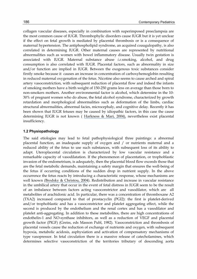
collagen vascular diseases, especially in combination with superimposed preeclampsia are the most common cause of IUGR. Thrombophylic disorders cause IUGR but it is yet unclear if the effect on fetal growth is mediated by placental thrombosis or is a consequence of maternal hypertension. The antiphospholipid syndrome, an acquired coaugulopathy, is also correlated in determining IUGR. Other maternal causes are represented by nutritional abnormalities such as women with bowel inflammatory disease. Usually twin gestation is associated with IUGR. Maternal substance abuse i.e.smoking, alcohol, and drug consumption is also correlated with IUGR. Placental factors, such as abnormality in size and/or function are implied in IUGR. Between the exogenous toxic substances consider firstly smoke because it causes an increase in concentration of carboxyhemoglobin resulting in reduced maternal oxygenation of the fetus. Nicotine also seems to cause arched and spiral artery vasoconstriction, with subsequent reduction of placental flow and indeed the infants of smoking mothers have a birth weight of 150-250 grams less on average than those born to non-smokers mothers. Another environmental factor is alcohol, which determine in the 10-30% of pregnant women who do abuse, the fetal alcohol syndrome, characterized by growth retardation and morphological abnormalities such as deformation of the limbs, cardiac structural abnormalities, abnormal facies, microcephaly, and cognitive delay. Recently it has been shown that IUGR fetuses may be caused by idiopathic factors, in this case the cause determining IUGR is not known ( Harkness & Mari, 2004), nevertheless exist placental insufficiency.
1.2 Physiopathology
The said etiologies may lead to fetal pathophysiological three paintings: a abnormal placental function, an inadequate supply of oxygen and / or nutrients maternal and a reduced ability of the fetus to use such substances, with subsequent loss of its ability to adapt. Uteroplacental circulation is characterized by low vascular resistance and a remarkable capacity of vasodilatation. If the phenomenon of placentation, or trophoblastic invasion of the endometrium, is adequately, then the placental blood flow exceeds those that are the fetal metabolic demands, maintaining a safety margin that ensures the well-being of the fetus if occurring conditions of the sudden drop in nutrient supply. In the above occurrence the fetus reacts by introducing a characteristic response, whose mechanisms are well known (Brodsky & Christou, 2004). Redistribution and increase in vascular resistance in the umbilical artery that occur in the event of fetal distress in IUGR seem to be the result of an imbalance between factors acting vasoconstrictor and vasodilator, which are all metabolites of arachidonic acid. In particular, there was a concentration of thromboxane A2 (TXA2) increased compared to that of prostacyclin (PGI2): the first is platelet-derived and/or trophoblastic and has a vasoconstrictor and platelet aggregating effect, while the second is produced by the endothelium and the renal cortex and has a vasodilator and platelet anti-aggregating. In addition to these metabolites, there are high concentrations of endothelin-1 and NO-synthase inhibitors, as well as a reduction of VEGF and placental growth factor (PlGF) (Greiss, eds Masson Publ, 1982). Vasoconstriction and thrombosis of placental vessels cause the reduction of exchange of nutrients and oxygen, with subsequent hypoxia, metabolic acidosis, asphyxiation and activation of compensatory mechanisms of type vasopressor. In fetal circulation there is a massive release of catecholamines, which determines selective vasoconstriction of the territories tributary of descending aorta

Infants Born with Intrauterine Growth Restriction: Renal and Cardiovascular Follow-Up 187
(especially in the districts splanchnic and skeletal muscle) and vasodilation in brachio-cephalic vessels to facilitate the flow to the brain. The persistence of this compensatory mechanism creates an overload of the ventricle left with a high risk of myocardial ischemia, and contemporary overloading of the right ventricle, due to splanchnic vasoconstriction. One therefore understand the reason for the reduced fetal myocardial contractility, which accompanies the reduction in cardiac output, the peak rate in large vessels and ventricular filling, resulting in increased central venous pressure and subdiaphragmatic venous pulsatility. The increase of indices of resistance in the umbilical artery indicates worsening of placental impairment, while the concomitant reduction of the same in middle cerebral artery and cerebro-placental ratio inversion (ratio umbilical and cerebral resistance) indicates that there began the phenomenon of brain-sparing effect. The caliber of the middle cerebral artery (MCA) continues to increase gradually until it reaches a zenith, before the onset of an abnormal fetal heart rate patterns, a sign of imminent death of the fetus. The ability to protect the brain parenchyma, with vasodilatation of ACM, just before the exitus, is lost, probably because the vessels lose their sensitivity to hypoxia, or due to the reduction of output fetal heart. At this point flow changes and appear similar to those in the umbilical artery also in the peripheral vessels (Krebs & al., 1996).
1.3 Classification
The IUGR fetus is usually divided into two clinical patterns: symmetric and asymmetric. This classification is related to the morphometric ultrasonography measurements of the head circumference (HC) and abdominal circumference (AC). In the moment that exist a reduction in the growth rate of both the above measurements the IUGR fetus is named symmetric; if only the AC is compromised the IUGR is asymmetric. The clinical pattern of IUGR is related to the nature of the causative factor, the stage of gestation in which the insult acts, and its duration. The above patterns are classified on the basis of antepartum and postpartum morphometric measurements of the head and abdomen of the fetus and newborn respectively. Approximately 20 to 30% of IUGR fetuses are symmetric type and 70 to 80% asymmetric. The clinical pattern divided into symmetric and asymmetric IUGR depends on the time the insult on the fetal growth occurs. In fact, during intrauterine life, fetal growth can be classified into three dynamic stages (Winick, 1971) . The first phase is represented by cellular hyperplasia and occurs during the first 16 weeks of gestation determining a rapid increase in cell number; in the second stage a concomitant cellular hyperplasia and hypertrophy involves an increase in both cell number and size occurring during the mid trimester. The last phase is characterized by a rapid increase in cell size as cellular hypertrophy dominates up to 32 weeks of gestation. Thus fetal growth rate in these three different dynamic stages is from 5g/day at 15 weeks, 15 to 20 g/day at 24 weeks, and 30 to 35 g/day at 34 weeks gestation. There have been many controversies surrounding the prognosis of the two patterns of growth-restricted fetuses. Many studies suggested that the symmetrically growth restricted fetuses were at greater risk of adverse outcome. This was particularly correlated to the high incidence of malformation and infections in the symmetric IUGR fetuses. A more recent study reports that the outcome of asymmetric IUGR is poorer if compared to the symmetrical one, also if the abnormalities in the first group were higher. So far, It is unclear if body proportionality (asymmetric / symmetric IUGR) is important with respect to etiology and neonatal outcome.

Contemporary Pediatrics 188
2. Diagnosis and management of IUGR The detection of the IUGR fetuses is essential because of the high mortality and morbidity associated with this condition. In a study with a 10 years follow up, it was reported that in IUGR fetuses there is an incidence of 10 % of physical handicap and a 5% of neurodevelopment delay.
2.1 Diagnosis
Accurate pregnancy dating is the first step that should be considered in order to detect accurately IUGR fetuses. In the moment that pregnancy age is accurately dated, the diagnosis of IUGR consists essentially on clinical history, physical examination, ultrasonography and Doppler ultrasonography. The correct dating of the pregnancy (it is considered that the gestational age is uncertain at the 20-40% of pregnancies) is the key point of diagnosis, as only once in possession of this last, we compare the measurements with those obtained by us of reference curves. The determination of gestational age is possible through the date of last menstrual period (Last Menstrual Period - LMP), which is reported by pregnant or with ultrasound evaluation of fetal biometric parameters or during the clinical examination of the newborn immediately after birth. The LMP is often not correct, as reported by pregnant women, and vitiated by any menstrual irregularities. In contrast, the ultrasound parameters almost always show a high accuracy, especially if you use the measurement of the length of the top-sacred (Crown Rump Length - CRL) trans vaginal ultrasound during the first trimester of pregnancy (Soothill & al., 1999). Clinical history is important to find out the cause of IUGR, which is important for the clinical management. In this condition a detailed fetal echo is indicated. Physical examination consists in fundal height measurements of the uterus that can detect the small for gestational age fetus. A single measurement at 32 to 34 weeks of gestation has a sensitivity of 69% to 85% combined with a specificity of up to 96%. The diagnostic accuracy in predicting an IUGR fetus measuring the fundal height is similar if compared to AC measurement. Ultrasonography is able to predict the EFW of a fetus and several morphometric parameters should be used ( Shepard & al., 19824). The error in the prediction of EFW may reach up to 25% ( Hadlock & al., 1984). The most accurate parameter in the estimation of fetal weight is the AC, which is the most sensitive parameter for the detection of IUGR. In fact the AC measurements fall within 10% of the actual weight. It can be considered affected by intrauterine growth restriction fetus that shows (Cosmi & al., 2005): abdominal stunted controls seriated, CA decline from normal values to values below 10 th percentile and AC <10 th percentile and head circumference in the norm. The slight delay is defined if the AC is <10 °, and severe if <2SD, equal to 2.5 th percentile for the age gestationa. A normal CA excludes the diagnosis of IUGR with a false negative rate of about 40 to 10%. Dudley has completed a review of ultrasonographic fetal weight estimation and concluded that the method is inaccurate, with low sensitivity and predictive value for fetal compromise, but by its method of analysis does not reveal any significantly better (Dudley, 2005). In conclusion, since fetal growth is a dynamic process, the "serial do biometrics in order to obtain a longitudinal analysis of the actual rate of growth of each fetus, improves the sensitivity of diagnosis of IUGR.
2.2 Management
Current management of IUGR fetuses consists of serial ultrasound measurements to assess the growth rate, amniotic fluid, fetal heart rate (FHR) monitoring and Doppler velocimetry.

Infants Born with Intrauterine Growth Restriction: Renal and Cardiovascular Follow-Up 189
The fetal non stress test (NST) is used as the primary fetal surveillance method for the assessment of fetal well-being, its interpretation can recognize fetuses compromised or not (Cagnon & al., 1987). In the current practice an abnormal NST is usually followed by the evaluation of the biophysical profile score (BPP). The latter assess 5 parameters being the FHR, fetal tone, fetal gross body movements, fetal breathing movements (FBM), and the AF. To each parameter assessed a numeral scale from 0 to 2 is given. Thus the BPP is considered normal when the score is from 8 to 10, and the test is repeated at weekly interval because the occurrence of fetal death is very rare within 1 week; a score less than 6 is considered abnormal as chronic asphyxia is suspected. In the latter situation the BPP should be repeated in 4-6 hrs. and delivery should be considered if oligohydramnios is present (Dayal & al., 1999). The Non stress test (NST) and BPP are useful screening tool in the evaluation of fetal well being. At present, there is not enough evidence from randomized trials to evaluate the use of biophysical profile score as a test of fetal well being in high-risk pregnancies (Manning & al., 1980). Furthermore, the NST become abnormal too late if compared to the Doppler velocimetry changes. Doppler investigation should be used for the antenatal surveillancein IUGR fetuses, as it select which fetus could benefit from a more intense antenatal surveillance. In fact Doppler velocimetry in IUGR fetuses versus NST is superior in identifying the need of intervention. Doppler investigation is very useful in order to find out the possibility of fetal hypoxia and/or hypercarbia. Doppler meta-analysis has shown the use of the umbilical artery in high-risk pregnancies reduces the number of antenatal admissions (44%), inductions of labor (29%), cesarean sections for fetal distress (52%), and perinatal mortality (38%). The ACOG recommends the use of Doppler investigation in IUGR fetuses. Many studies have focused their attention on the temporal sequence of changes in fetal parameters in the IUGR fetus (ACOG, 1997). Thus an IUGR fetus is considered that with an EFW < 10th percentile, with an abnormal Doppler investigation and oligohydramnios. This group of IUGR fetuses constitute about 35-40% of all IUGR fetus and the antenatal surveillance has to be increased; the remaining IUGR fetuses, those with EFW < 10th percentile, normal Doppler investigation and amniotic fluid could be managed as AGA fetuses as have the same outcome. Fetuses whose weight is less than 10th percentile are usually managed with weekly NST or BPP to search for evidence of fetal distress. Doppler investigation of the umbilical artery (UA) is important to find out abnormalities, such as resistence increase, absent of end diastolic flow, reversed flow, in the UA, which constitutes pathological findings. Fetal growth restriction is a significant factor for increasing Doppler indices and Doppler abnormality identifies those fetuses that are at greater risk for adverse perinatal outcome (Arduini & Rizzo, 1993). In addiction the middle cerebral artery evaluation and thus cerebroplacental ratio should be performed, as it improves the prediction of perinatal outcome compared with UA velocimetry alone. Changes in the cerebral vascular diastolic flow, are progressive after the presence of the brain-sparing effect, indicative of blood redistribution to vital organ, and usually reach a nadir just prior to the onset of an abnormal FHR pattern. Other vessels that should be investigated are that of the venous system such as the ductus venosus and umbelical vein (UV) which abnormalities, reversed flow and pulsations, respectively, reflect an advanced status of fetal compromise. Studies on the temporal sequence of changes in IUGR fetus showed that when Doppler changes occur, aggressive management of the fetus (delivery) is suggested despite the NST is normal. Ozcan et al. pointed out that in compromised IUGR fetuses Doppler changes occur as follow: abnormality in UA, MCA, UV pulsation, reversed flow in the ductus venosus and at last abnormalities in NST (Ozcan & al., 1998). Thus the IUGR fetus

Contemporary Pediatrics 190
should be investigated by doppler velocimetry of the arterial system and venous system and by NST and BPP. Senat et al. shown that prior the evidence of fetal distress by means of NST, significant changes occur in the arterial (UA, MCA) and venous system (DV, UV) showing that other hemodynamic changes were not statistically significant (Senat & al., 2000). Other studies confirm the above findings, thus Doppler investigation is an important tool to use in the management of IUGR fetuses, as according to its findings conservative or aggressive management should be opted. Arterial and venous flow changes are the first to become evident prior to abnormal findings in FHR, and BPP, in the latter the first abnormality are represented by fetal breathing movements, AF, body movements and at least fetal tone. Moreover a recent study shown that there are early and late changes in Doppler velocimetry. The first are represented by abnormality in the UA (increased resistance, absent end diastolic flow) and MCA; the latter are changes in DV, reversed flow in UA, pulmonary vein abnormalities and DV reversed flow. The last changes in those fetuses were in FHR tracing while abnormality in the AF were not statistically significant. The early stage changes were correlated with a less rate of mortality if compared to the late one.
3. Outcome of IUGR infants IUGR is the second leading cause of perinatal morbidity and mortality with a 10-fold greater risk of fetal death if compared to normal fetuses which are appropriate for gestational age (AGA) (Pallotto & Kilbride, 2006). Moreover the mortality and morbidity rates are correlated to the EFW of the fetus: the less is the percentile at a given gestational age, the higher is the morbidity and mortality rate. In fact the mortality and morbidity are increased among infants born at term whose birth weights are at or below the 5th percentile with an increase below the 3rd percentile for a given gestational age. In fact those fetuses share a greater risk for fetal demise, fetal distress, and could develop short term (hypoglycemia, hypocalcemia, hypothermia, polycythemia, necrotizing enterocolitis, pulmonary hypertension), and long-term (ischemic heart disease, stroke hypertension, Non-Insulin Dependent Diabetes) complications.
3.1 Short-term outcome
Infants with low birth weight as those born with IUGR have a risk significantly increased to provide a low Apgar score associated with respiratory, neurological and metabolic sequelae in the long term, thanks to the condition of acute and chronic deprivation of oxygen and nutrients. The risk of sudden death in utero in fetuses with EFW below the 10th percentile increased 2 to 6 times compared to fetuses with appropriate weight for gestational age, mainly due to the hepato-renal impairment (Baschat & al., 2007). Hence the importance of an early intrauterine diagnosis in order to programme for the best fetal monitoring and to identify the right time for the induction of labor. The negative outcome does not seem to depend on the underlying cause of the lack of growth, but appears rather "dose-related," as worse with the worsening of the disease, to generate a cascade mechanism of physiological effects chained together. Several authors have focused attention on the temporal sequence of hemodynamic changes that characterize the IUGR fetuses and affecting first the artery and then the venous compartment. Initially there is the increase >2SD of the pulsatility index (PI) of the umbelical artery (UA), then the reduction of middle cerebral artery PI (MCA), the absence of diastolic flow (AEDF) in UA, the reverse flow (RF) in UA and changes in ductus

Infants Born with Intrauterine Growth Restriction: Renal and Cardiovascular Follow-Up 191
venosus (DV), with increased PI and absent or reverse flow of wave A (corresponding to atrial contraction) (Baschat & al., 2001). Observational studies carried out and especially the multicenter one performed at our Department of Gynecology and Obstetrics Sciences, has shown that the optimal timing for the delivery is the time when the fetus has no diastolic flow in the umbilical artery, gestational age> 28 weeks and fetal weight >600g. The morbidity due to IUGR occur both in the short and long term (Hecher & al., 2001). The first group includes the problems that the child must face in the post-partum (delivery outcomes), including the condition of prematurity, with its related complications. Among the complications of delivery outcomes appear: high risk of perinatal asphyxia (due to placental insufficiency and intrauterine chronic hypoxia), greater frequency of resuscitation’s intervention in the delivery room, increased risk of developing hypothermia, greater incidence of hypoglycemia (blood glucose <40 mg/ dL) and other metabolic sequelae, especially the hypocalcemia ([Ca2 +] <7 mg/dL) and hyperbilirubinemia, due to both impaired hepatic detoxifying function and hyperhemolysis. The morbidity related to them are respiratory, digestive, neurological or renal, and they complicate the neonatal period of these patients. All other changes are manifest in childhood or adulthood are considered rather long complications term.
3.2 Long-term outcome
The long term complications are numerous and involve several systems: first of all the cardiovascular one because these children frequently have hypertension, with a significant increase in systolic pressure and in intima-media thickness of the abdominal aortic wall. The “developmental origins of adult disease” hypothesis, often called “the Barker hypothesis”(Barker, 2006) proposes that these diseases originate through adaptations of the fetus when it is undernourished. These adaptations may be cardiovascular, metabolic or endocrine and they may permanently change the structure and function of the body, increasing coronary heart disease risk factors, such as hypertension, type 2 diabetes mellitus, insuline resistance and hyperlipidemia (Gillman, 2005). This hypothesis originally envolved from observation by Barker and colleagues that the regions in England with the highest rates of infant mortality in the early 20th century also had the highest rates of mortality from coronary heart disease decades later. As the most commonly registered cause of infant death at the start of 20th century was low birth weight, these observations led to the hypothesis that low birth weight babies who survived infancy and childhood might be at increased risk of coronary heart disease in later life (Barker, 2000). As Barker and his colleagues reported in several epidemiological and anthropological studies, in fetal life tissues and organs go through the so called “critical” periods of development. These may coincide with periods of rapid cell division. Although the fetal growth is influenced by its genes, several studies suggest that it is usually limited by intrauterine environment, in particular the nutrients and oxygen received from the mother. Influence linked to fetal and placental growth have an important effect on the risk of coronary heart disease and stroke. Thus this theory focusing on intrauterine life, offers a new point of departure for research in cardiovascular disease. According to the thrifty phenotype hypothesis, deficient fetal supply may be followed by a programming which includes circulatory adjustment and insulin resistance in liver and muscular tissue in order to spare the brain. In addiction post-natal over nutrition following intrauterine growth restriction may be pathogenetic for the development of obesity and type 2 diabetes, whereas the elevated cardiovascular mortality rate may be associated with rapid
Contemporary Pediatrics 192
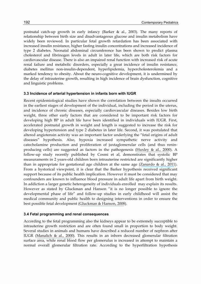
postnatal catch-up growth in early infancy (Barker & al., 2003). The many reports of relationship between birth size and disadvantageous glucose and insulin metabolism have widely been reviewed. In particular fetal growth retardation has been associated with increased insulin resistance, higher fasting insulin concentrations and increased incidence of type 2 diabetes. Neonatal abdominal circumference has been shown to predict plasma cholesterol and fibrinogen levels in adult in later life, which are both risk factors for cardiovascular disease. There is also an impaired renal function with increased risk of acute renal failure and metabolic disorders, especially a great incidence of insulin resistance, diabetes mellitus non-insulin-dependent, hyperlipidemia, hypercholesterolemia and a marked tendency to obesity. About the neuro-cognitive development, it is undermined by the delay of intrauterine growth, resulting in high incidence of brain dysfunction, cognitive and linguistic problems.
3.3 Incidence of arterial hypertension in infants born with IUGR
Recent epidemiological studies have shown the correlation between the insults occurred in the earliest stages of development of the individual, including the period in the uterus, and incidence of chronic diseases, especially cardiovascular diseases. Besides low birth weight, three other early factors that are considered to be important risk factors for developing high BP in adult life have been identified in individuals with IUGR. First, accelerated postnatal growth in weight and length is suggested to increase the risk for developing hypertension and type 2 diabetes in later life. Second, it was postulated that altered angiotensin activity was an important factor underlying the “fetal origins of adult diseases” hypothesis. Also, hypoxia increased sympathetic nerve activity and catecholamine production and proliferation of juxtaglomerular cells (and thus renin-producing cells) are suggested as factors in the pathogenesis (Huxley & al., 2000). A follow-up study recently published by Cosmi et al, demonstrates that systolic BP measuraments in 2 years-old children born intrauterine restricted are significantly higher than in appropriate for gestational age children at the same age (Zanardo & al., 2011). From a hystorical viewpoint, it is clear that the Barker hypothesis received significant support because of its public health implication. However it must be considered that may confounders are known to influence blood pressure in adult life apart from birth weight. In addiction a larger genetic heterogeneity of individuals enrolled may explain its results. However as stated by Gluckman and Hanson “it is no longer possible to ignore the developmental phase of life” and follow-up studies in early childhood will assist the medical community and public health to designing interventions in order to ensure the best possible fetal development (Gluckman & Hanson, 2008).
3.4 Fetal programming and renal consequences
According to the fetal programming also the kidneys appear to be extremely succeptible to intrauterine growth restriction and are often found small in proportion to body weight. Several studies in animals and humans have described a reduced number of nephron after IUGR (Manalich & al., 2000). This results in an inborn decreased glomerular filtration surface area, while renal blood flow per glomerulus is increased in attempt to maintain a normal overall glomerular filtration rate. According to the hyperfiltration hypothesis

Infants Born with Intrauterine Growth Restriction: Renal and Cardiovascular Follow-Up 193
explained by Brenner and co-workers (Brenner & al., 1996), this leads to glomerular hypertension and hypertrophy, which causes systemic hypertension and higher sodium reabsorption and glomerular damage resulting in albuminuria (Figure 3) and glomeruloclerosis. Therefore IUGR can lead to impairment of renal function (Luyckx & Brenner, 2005). A kidney with a reduced nephron number has less renal reserve to adapt to dietary excesses or to compensate for renal injury. The pathway leading from small kidney to hypertension may include the renin – angiotensin system, which has been demonstrated to be altered in the early phase of primary hypertension. An increased activity of the renin – angiotensin system could be a compensatory mechanism in a decreased number of nephrons in order to maintain normal filtration.
Fig. 3. Importance of microalbuminuria.
These mechanisms are well described in experimental study including IUGR male rats, whose presented higher blood pressure and fewer glomeruli at 22 weeks of age (Wlodek & al., 2008). In the last five years more clinical data are available regarding maturation of renal function in IUGR infants. Keijzer-Veen and colleagues in 2005 identified a positive association of birth weight and glomerular filtration rate (GFR) (Keijzer-Veen & al., 2005). The investigators also detected a negative association of birth weight and serum creatinine, suggesting that IUGR individuals are at greater risk to develop hypertension and progressive renal failure. This work made a significant contribution to understanding mechanism associated with the progression of cardiovascular disease and intrauterine retardation. In contrary Rakow at al found that glomerular filtration rate and urinary protein patterns were similar between IUGR 12 years-old children and controls, but kidney volume was smaller in the first group. A meta-analysis published by Teeninga et al in 2008, consisted in 201 patients (25 SGA, 176 AGA), showed that low birth weight has a strong influence in glomerular filtration rate and proteinuria, associated with a higher chance of developing several complications, including hypertension (Teeninga & al., 2008). A recent follow-up study demonstrated that 5 years- old children born IUGR showed higher BP, increased albuminuria, lower GRF and different urinary sodium excretion rate than controls. These observations support the contention that extrinsic renal injury is not a

Contemporary Pediatrics 194
prerequisite for the initiation and perpetuation of renal injury and that certain circumstances, prenatally derived, intrinsic deficiencies in functioning renal mass may be sufficient to contribute to renal functional decline occurring with advancing age.
3.5 Endothelial dysfunction
The endothelium controls vascular tone, coagulation and inflammatory responses. Endothelial dysfunction is an early event of atherosclerosis (Figure 4), preceding structural changes in the vascular wall. Atherosclerosis is thought to begin in childhood and to develop silently before clinical events such as myocardial infarction or stroke occur (Jarvisalo & al., 2001). As with major cardiovascular risk factors, impaired growth in utero is associated with functional (endothelial dysfunction) and structural (increased wall thickness) changes to the arterial vasculare consistent with early atherosclerosis.
Fig. 4. Sequence in progression of atherosclerosis.

Infants Born with Intrauterine Growth Restriction: Renal and Cardiovascular Follow-Up 195
Impaired fetal growth in infants is associated with increased sympathetic tone and lipid concentration, and reduced concentration of insulin-like growth factor-1, all of which might contribute to arterial wall thickening. However, these findings are difficult to interpret because of potential confounding by, or interaction with, postnatal influences, so this hypothesis need further clarification. Tracy et al demonstrated atheromatous changes in the aorta of children (McGill & al., 2000). Atheromatous changes have been documented histopathologically by early childhood, and it has been assessed that the first atherosclerotic lesions actually begin to develop in the abdominal aorta. Nowadays many studies demonstrated atherosclerotic wall thickening in the arteries of children with cardiovascular risk factors using ultrasound imaging assessing noninvasively early vascular changes. In 2005 Skilton and co-workers add new interesting information to the Barker’s hypothesis (Skilton & al., 2005). They compared intima-media thickness of the aortic wall in newborn infants with low birth weight with normal controls. In IUGR newborns aIMT was significantly greater than in the controls. On the basis of these finding they described the ultrasound-based measurement of abdominal aortic intima-media thickness in children as a feasible, accurate and sensitive marker of atherosclerosis risk and, as there was no confounding from childhood and adult exposures, they provide clear indications for a fetal contribution to later cardiovascular disease. Also Koklu et al in 2007 evaluated the potential use of aIMT in the study of high-risk neonates, concluding that aIMT measurement in IUGR newborns can help in the early identification of asymptomatic vascular dysfunction (Koklu & al., 2007). Recently Cosmi studied aIMT in fetuses for the first time and than in the same infants after a mean follow-up of 18 months. He found that aIMT measurements in IUGR fetuses are inversely related to estimated fetal weight, showing that low birth weight and Doppler abnormalities may be correlated to an altered vascular structure causing possible endothelial damage, consistent with the finding that atherosclerosis begins to develop first in the intima of the aorta. These differences remains also at the time of follow-up. Similarly, carotid intima-media thickness has been shown to be greater in children with low-birth weight. In a recent study Gratacos et al confirms the presence of increased carotid wall thickness in children with IUGR and that these changes persist into childhood (Crispi & al., 2010). The increased arterial wall thickness could be the result of vascular remodelling linked to metabolic programming in intrauterine life. They also highlighted that children with IUGR show changes in cardiac morphology, subclinical cardiac longitudinal dysfunction and hypertension, all of which increase linearly with the severity of growth restriction and are independent of gestational age at delivery, lipid profile or body mass index. The importance of early identification and intervention in pediatric risk factors for cardiovascular disease is now well recognized and this could open new opportunities for monitoring and intervention in newborns and children affected with intrauterine growth restriction.
3.6 IUGR Infant and their neurodevelopment
As said before intrauterine growth restriction plays a significant role in short and long term outcome ad reflected in high incidence of brain dysfunction and neurodevelopmental impairment, as well as in somatic growth failure. These clinical consequences could not be apparent until later in childhood development and could involve poor academic

Contemporary Pediatrics 196
performance, memory, visuomotor and language difficulties and executive function problems. Several longitudinal studies have addressed the question of correlation between cognitive development and somatic growth in IUGR, using different questionnaire dealing with aspects of reading, writing, spelling and mathematics. An increased risk of cognitive impairment has been demonstrated in children with small head circumference. Shenkin et al found that birth weight is significantly related to IQ at age 11 (Shenkin & al., 2001). In other studies IUGR children have lower nonverbal and verbal IQ than controls. According to data from the National Collaborative Perinatal Project (1959-1976) the IQ scores of 2719 IUGR children tested at age 7 were 6 point lower than AGA children. Besides neurosensory handicaps, also behavioural sequelae are of serious concern. Behavioural problems, which might become manifested only at school age, can have a great impact on school performance and social competence and have a negative influence on quality of life (Van de Broek & al., 2010). Learning difficulties and behavioural problems are reported more often in IUGR preterm infants compared to AGA (Padilla & al., 2010). Management of these infants is controversial. They have an increased perinatal mortality and morbility including behavioural problems, minor developmental delay, poor neurosensory development and spastic cerebral palsy. Definition of these important long-term relationships invites research of pathological mechanism. Recent advanced magnetic resonance imaging studies have shown that IUGR is associated with structural differences that can be identified very early in life, such as reduction in cerebral cortical grey matter and hippocampal volume. These macrostructural alterations have been associated with microstructural and metabolic changes. As well explained from Baschat et al, the IUGR fetus “enables” sparing of the head while growing in a low-nutrient intrauterine environment and has neuroadaptative modification aimed at preserving brain oxygen supply in presence of chronic hypoxia (Baschat & al., 2009) . This process is identified clinically by a reduced Doppler pulsatility index (PI) in the middle cerebral artery (MCA). When multiple parameters are considered, such as gestational age and birth weight, umbilical artery reversed end diastolic velocity (UA-REDV) is identified as an important contributor to neurodevelopment. No similar effect could be demonstrated for abnormal venous Doppler findings. Brain sparing and cerebral arteries Doppler results are objects of study as predictors of adverse neurological outcome, but these relationships are not well assessed. These findings have important implications for the prognosis and the management of intrauterine growth restricted fetuses and children, who should be closely followed-up to identified individuals at risk.
4. Conclusion The importance of IUGR in understanding adverse pregnancy outcome is relevant not only for clinicians, but also for public health service. During the last decade, knowledge of the short-term and long-term consequences of impaired fetal growth has increased at a very rapid rate and has involved lots of clinical aspects. At present, it is most important not only to develop efficient methods of preventing and diagnosing IUGR, recognizing at-risk fetuses, and screening fetal growth restriction among low-risk pregnancies, but to work out followup and adequate treatment programs for the control of the disorders which may follow this conditions. Programming the right time to deliver is the best method to avoid

Infants Born with Intrauterine Growth Restriction: Renal and Cardiovascular Follow-Up 197
adverse perinatal outcome; indeed, proper postnatal feeding and infant growth may be essential for long-term outcome.
5. References American College of Obstetricians and Gynecologists. ACOG practice bulletin. Intrauterine
growth restriction. Obstet Gynecol 2001; 72: 85-96. American College of Obstetricians and Gynecologists .ACOG. Utility of antepartum
Umbelical artery Doppler velocimetry in intrauterine growth restriction. Committee Opinion. 1997; 188.
Arduini D, Rizzo G. Doppler studies of deteriorating growth retarded fetuses. Curr Opin Obstet Gynecol. 1993; 5: 195-203.
Barker DJ. In utero programming of cardiovascular disease. Therionol, 2000; 15;53(2):555-74. Barker DJP, Eriksson JG, Forsen T, Osmond C. Fetal origins of adult disease:strength of
effects and biological basis. Int J Epidemiology 2003; 31:1235-9. Barker DJP: Adult consequences of fetal growth restriction. Clinical Obstetrics and
Gynecology 2006; 49: 270–283. Baschat AA, Viscardi RM, Hussey-Gardner B, Hashmi N, Harman C. Infant
neurodevelopment following fetal growth restriction: relationship with antepartum surveillance parameters. Ultrasound Obstet Gynecol 2009;33:44-50.
Baschat AA, Cosmi E, Bilardo CM, Wolf H, Berg C, Rigano S, Germer U, Moyano D, Turan S, Hatung J, Bhide A, Muller T, Bower S, Nicolaides KH, Thilaganathan B, Gembruch U, Ferrazzi E, Hecher K, Galan HL, Harman CR: Predictors of Neonatal Outcome in Early-Onset Placental Dysfunction. Obstet Gynecol 2007; 109: 253-261.
Baschat AA: Pathophysiology of fetal growth restriction: implications for diagnosis and surveillance. Obstet Gynecol Survey 2004; 59: 617-627.
Baschat AA, Gembruch U, and Harman CR. The sequence of changes in Doppler and biophysical parameters as severe growth restriction worsens. Ultrasound Obstet Gynecol. 2001; 18 : 571-577.
Battaglia FC, Lubchenco LO: A practical classification of newborn infants by weight and gestational age. J Pediatr 1967; 71: 159-163.
Brenner BM, Lawler EV, Mackenzie HS. The hyperfiltration theory: a paradigm shift in nephrology. Kidney Int 1996;49:1774-1777.
Brodsky D, Christou H: Current concepts in intrauterine growth restriction. J Intensive Care Med 2004; 19: 307-319.
Cagnon R, Campbell K, Hunse C, Patrick J. patterns of human fetal heart rate acceleration from 26 week to term. Am J Obstet Gynecol. 1987; 157: 743-8.
Cosmi E, Ambrosini G, D’Antona D, Saccardi C, Mari G: Doppler, cardiotocography, and biophysical profile changes in growth-restricted fetuses. Obstet Gynecol 2005; 106: 1240-1245.
Crispi F, Bijnens B, Figueras F, Bartrons J, Eixarch E, Le Noble F, Ahmed A, Gratacos E. Fetal growth restriction results in remodelled and less efficient hearths in children. Circulation 2010;121:2427.36.

Contemporary Pediatrics 198
Dayal AK, Manning FA, Berk DJ, Mussalli JM, Avila C, Harman CR. Et al Fetal death after normal biophysical profile score: An eighteen year experience. Am J Obstet Gynecol. 1999; 181: 1231.
Dudley NJ: A systematic review of the ultrasound estimation of fetal weight. Ultrasound Obstet Gynecol 2005; 25: 80–89.
Froen JF, Gardosi JO, Thurmann A, Francis A, Stray-Pedersen B: Restricted fetal growth in sudden intrauterine unexplained death. Acta Obstet Gynecol Scand 2004; 83: 801–807.
Gillman MW: Developmental origins of health and disease. N Engl J Med 2005; 353: 1848-1850.
Gluckman PD, Hanson MA. Develompmental origins of health and disease: new insights. Basic Clin Pharmacol Toxicol. 2008 Feb;102(2):90-3.
Greiss FC Jr: Uterine blood flow in pregnancy: an overview. In: Uterine and placental blood flow. Moawad AH, Lindheimer MD eds Masson Publ 1982; pp 19-26.
Hadlock FP, Deter RL, Harrist RB, Park SK: Estimating fetal age: computerassisted analysis of multiple fetal growth parameters. Radiology 1984; 152: 497- 501.
Harkness U, Mari G: Diagnosis and management of intrauterine growth restriction. Clin in Perinatol 2004; 31: 743-764.
Hecher K, Bilardo CM, Stigter RH, Ville Y, Hackeoler BJ, Kok HJ, Senat MV and Visser GHA: Monitoring of fetuses with intrauterine growth restriction: a longitudinal study. Ultrasound Obstet Gynecol 2001; 18: 564-570.
Huxley RR, Shiell AW, Law CM. The role of size at birth and postnatal catch-up growth in determining systolic blood pressure: a systematic review in literature. J Hypertens 2000;18:815-31.
Jarvisalo MJ, Jartti L, Nanto-Salonen K. Increased aortic intima-media thickness: a marker of preclinical atherosclerosis in high-risk children. Circulation 2001;104:2943-7.
Keijzer-Veen MG, Schrevel M, Finken MJJ, Dekker FW, Nauta J, Hille ETM, Frolich M, van der Heijden BJ. Microalbuminuria and lower glomerular filtration rate at young adult age in subjects born very premature after intrauterine growth retardation. J Am Soc Nephrol 2005; 16:2762-2768.
Koklu E, Ozturk MA, Gunes T, Akcakus M, Kurtoglu S. Is increased intima-media thickness associated with preatherosclerotic changes in intrauterine growth restricted newborns? Acta Paediatr. 2007;96(12):1858.
Krebs C, Macara LM, Leiser R, Bowman AW, Greer IA, Kingdom JCP: Intrauterine growth restriction with absent end-diastolic flow velocity in the umbilical artery is associated with maldevelopment of the placental villous tree. Am J Obstet Gynecol 1996; 175: 1534-1542.
Lin CC, Santolaya-Forgas J: Current concepts of fetal growth restriction: part I. Causes, classification and pathophysiology. Obstet Gynecol 1998; 92: 1044-1055.
Luyckx VA, Brenner BM. Low birth weight, nephron number, and kidney disease. Kidney Int 2005;68:68-77.
Manalich R, Reyes L, Herrera M. Relationship between weight at birth and the number and the size of renal glomeruli in humans: a histomorphometric study. Kidney Int 2000;58: 770-773.

Infants Born with Intrauterine Growth Restriction: Renal and Cardiovascular Follow-Up 199
Mandruzzato G, Antsaklis A, Botet F, Chervenak FA, Figueras F, Grunebaum A, Puerto B, Skupski D, Stanojevic M: Intrauterine Growth Restriction (IUGR). J Perinat Med 2008; 36: 277–281.
Manning FA, Platt LD, Sipos L. Antepartum fetal evaluation : development of a biophysical profile score. Am J Obstet Gynecol. 1980; 136: 787-95.
McGill HCJ, McMahu CA, Herderick EE, Tracy RE, Malcom GT, Zieske AW, Strong JP. Effect of coronary heart disease risk factors on atherosclerosis of selected regions of the aorta and right coronary artery. PDAY Research Group. Pathobiological Detetrminants of Atherosclerosis in Youth. Ateriscler Thromb Vasc Biol. 2000; 20: 836,45.
McIntire DD, Bloom SL, Casey BM, Leveno KJ: Birth weight in relation with morbidity and mortality among newborn infants. N Engl J Med 1999; 340:1234-1238.
Ozcan T, Sbarcia M, d’Ancona L, Copel JA, and Mari G. Arterial and venous Doppler velocimetry in the severely growth-restricted fetus and associations with adverse perinatal outcome. Ultrasound Obstet Gynecol. 1998; 12: 39-44.
Padilla N, Perapoch J, Carrascosa A, Acosta-Rojas R, Botet F, Gratacos E. Twelve month neurodevelopmental out come in preterm infants with and without intrauterine growth restriction. Acta Pediatrica 2010;99:1498-1503.
Pallotto EK, Kilbride HW: Perinatal outcome and later implications of intrauterine growth restriction. Clin Obstet Gynecol 2006; 49: 257-269.
Senat MV, Schwarzler P, Alcais A, and Ville Y. Longitudinal changes in the ductus venosus, cerebral transverse sinus and cardiotocogram in fetal growth restriction. Ultrasound Obstet Gynecol. 2000; 16: 19-24.
Shenkin SD, Starr JM, Pattie A, Rush MA, Whalley LJ, Deary IJ. Birth weight and cognitive function at age 11: the Scottish Mental Survey 1932. Arch Dis Child 2001; 85(3):189-197.
Shepard MJ, Richards VA, Berkowitz RL, Warsof SL, Hobbins JC: An evaluation of two equations for predicting fetal weight by ultrasound. Am J Obste Gynecol 1982; 142: 47-54.
Skilton MR, Evans N, Griffiths KA, Harmer JA, Celermajer DS. Aortic wall thickness in newborns with intrauterine growth restriction. Lancet. 2005;365:1484-6.
Snijders RJ, Sherrod C, Golden CM, Nicolaides KH: Fetal growth retardation: associated malformations and chromosomal abnormalities. Am J Obstet Gynecol 1993; 168: 547-555.
Soothill PW, Bobrow CS, Holmes R: Small for gestational age is not a diagnosis (Editorial), Ultrasound Obstet Gynecol 1999; 33: 225–228.
Teeninga N, Schreuder M, Bokenkamp A, Delemarre-vam der Waal H, van Wijk J. influence of low birth weight on minimal change nephrotic syndrome in children, including a meta-analysis. Nephrol Dial Transplant 2008;23:1615-1620.
Van de Broek AJM, Kok JH, Houtzager A, Scherjon SA. Behavioural problems at the age of eleven years in preterm-born children with or without fetal brain sparing: A prospective cohort study. Early Human Development 2010;86:379-384.
Winick M: Cellular growth during early malnutrition. Pediatrics 1971; 47: 969-978.

Contemporary Pediatrics 200
Wlodek M, Westcott K, Siebel A, Owens J, Moritz K. Growth restriction before or after birth reduces nephron number and increases blood pressure in male rats. Kidney International 2008;74:187-195.
Zanardo V, Fanelli T, Weiner G, Fanos V, Zaninotto M, Visentin S, Cavallin F, Trevisanuto D, Cosmi E. Intrauterine growth restriction is associated with persistent aortic wall thickening and glomerular proteinuria during infancy. Kidney Int. 2011 Jul;80(1):119-23.
Zhang J, Bowes WA: Birth-weight-for-gestational-age patterns by race, sex and parity in the United States population. Obstet Gynecol 1995; 86: 200-208.
Related Documents