Induced autophagy reduces virus output in dengue infected monocytic cells Mingkwan Panyasrivanit a , Michael P. Greenwood b , David Murphy b , Ciro Isidoro c , Prasert Auewarakul d , Duncan R. Smith a, ⁎ a Molecular Pathology Laboratory, Institute of Molecular Biosciences, Mahidol University, Thailand b Laboratories of Integrative Neurosciences and Endocrinology (LINE), University of Bristol, Bristol, UK c Laboratorio di Patologia Molecolare, Dipartimento di Scienze Mediche, Università del Piemonte Orientale ‘A. Avogadro’, Via Solaroli 17, 28100 Novara, Italy d Department of Microbiology, Siriraj Hospital, Mahidol University, Bangkok, Thailand abstract article info Article history: Received 8 June 2011 Returned to author for revision 3 July 2011 Accepted 13 July 2011 Available online 2 August 2011 Keywords: ADE Autophagy Dengue ER Monocytic cells While several studies have shown a role for autophagy in the replication of dengue virus (DENV), these studies have been performed in directly infected cells. However, in severe cases of DENV infection the critical cell in the disease is believed to be monocytes which are poorly infected directly, but are highly susceptible to antibody enhanced infection. This study sought to determine the involvement of autophagy in the DENV infection of monocytic cells, using U937 cells as a model system. While the induction of autophagy was seen in response to DENV-2 infection, biochemical induction of autophagy resulted in a significant decrease in virus output. Down regulation of autophagy resulted in only a very slight increase in intracellular virus levels. In monocytic cells autophagy is not a significant part of the DENV replication mechanism, and there are distinct cell type specific differences in the DENV–autophagy interaction. © 2011 Elsevier Inc. All rights reserved. Introduction Dengue viruses (DENVs) are transmitted to humans by Aedes mosquitoes and can cause an acute febrile illness with retro-orbital pain, myalgia, arthralgia and hemorrhagic manifestation called dengue fever (DF). In some infections the individual may develop massive bleeding, thrombocytopenia, evidence of plasma leakage such as pleural effusion, ascites and a rise of hematocrit called dengue hemorrhagic fever (DHF). If the plasma leakage leads to hypovolemic shock the syndrome is called dengue shock syndrome (DSS) which has a high mortality rate (Gubler, 1998; Kurane, 2007). At present specific treatments or a preventive vaccine for dengue infection is not available, although appropriate supportive treatment significantly reduces the mortality rate. However, the social and economic cost from dengue infections is onerous due to the high incidence of infection around the world (Mathers et al., 2007). DENVs are enveloped positive single-stranded RNA viruses, consist- ing of four heterologous serotypes termed DENV serotypes 1, 2, 3 and 4 (DENV-1 to 4). The viral genome is translated as a single polyprotein which is co- and post-translationally processed into 3 structural (envelope (E), pre-membrane (prM) and capsid (C)) proteins and 7 non-structural (NS1, NS2A, NS2B, NS3, NS4A, NS4B and NS5) proteins (Henchal and Putnak, 1990; Perera and Kuhn, 2008). The virus enters the host cells by receptor-mediated endocytosis (Krishnan et al., 2007; Suksanpaisan et al., 2009; van der Schaar et al., 2007) where low-pH conditions in the endosome induce uncoating of the viral genome followed by translation and replication of the virus (Modis et al., 2004; Mukhopadhyay et al., 2005). The reason why some cases of DENV infection, but not all, develop severe symptoms (DHF and DSS) has been widely studied, and both virus and host factors have been proposed to contribute to the pathogenesis of severe dengue (Murgue et al., 2000; Rico-Hesse, 2007; Vaughn et al., 2000; Wang et al., 2006). While previous reports have implicated a role for specific genotypes or lineages in determining severity (Murgue et al., 2000; Vaughn et al., 2000; Wang et al., 2006), numerous studies have implicated second infections with a heterologous DENV as being a major cause of a more severe disease presentation, through the process termed antibody dependent enhancement (ADE) of infection (Halstead et al., 2010; Halstead and O'Rourke, 1977a; Halstead et al., 1980). While lifelong immunity is generated against subsequent infections with a homotypic virus, only transient protection is provided against heterotypic infections (Guzman et al., 2000; Sangkawibha et al., 1984) and it is believed that the presence of pre-existing sub-neutralizing antibodies from a previous heterotypic infection facilitate entry of the virus to Fc receptor-bearing cells such as monocytes, leading to increased virus uptake and replication in these cells (Goncalvez et al., 2007; Kliks et al., 1988). This model is strongly supported by a retrospective seroepidemiologic study in Cuba of the 1981 DENV-2 Virology 418 (2011) 74–84 ⁎ Corresponding author at: Molecular Pathology Laboratory, Institute of Molecular Biosciences, Mahidol University, 25/25 Phuttamonthon 4 Road, Salaya, Nakhon Pathom, 73170, Thailand. Fax: + 66 2441 1013. E-mail address: [email protected] (D.R. Smith). 0042-6822/$ – see front matter © 2011 Elsevier Inc. All rights reserved. doi:10.1016/j.virol.2011.07.010 Contents lists available at ScienceDirect Virology journal homepage: www.elsevier.com/locate/yviro

Welcome message from author
This document is posted to help you gain knowledge. Please leave a comment to let me know what you think about it! Share it to your friends and learn new things together.
Transcript
Virology 418 (2011) 74–84
Contents lists available at ScienceDirect
Virology
j ourna l homepage: www.e lsev ie r.com/ locate /yv i ro
Induced autophagy reduces virus output in dengue infected monocytic cells
Mingkwan Panyasrivanit a, Michael P. Greenwood b, David Murphy b, Ciro Isidoro c, Prasert Auewarakul d,Duncan R. Smith a,⁎a Molecular Pathology Laboratory, Institute of Molecular Biosciences, Mahidol University, Thailandb Laboratories of Integrative Neurosciences and Endocrinology (LINE), University of Bristol, Bristol, UKc Laboratorio di Patologia Molecolare, Dipartimento di Scienze Mediche, Università del Piemonte Orientale ‘A. Avogadro’, Via Solaroli 17, 28100 Novara, Italyd Department of Microbiology, Siriraj Hospital, Mahidol University, Bangkok, Thailand
⁎ Corresponding author at: Molecular Pathology LabBiosciences, Mahidol University, 25/25 Phuttamonthon 473170, Thailand. Fax: +66 2441 1013.
E-mail address: [email protected] (D.R.
0042-6822/$ – see front matter © 2011 Elsevier Inc. Aldoi:10.1016/j.virol.2011.07.010
a b s t r a c t
a r t i c l e i n f oArticle history:Received 8 June 2011Returned to author for revision 3 July 2011Accepted 13 July 2011Available online 2 August 2011
Keywords:ADEAutophagyDengueERMonocytic cells
While several studies have shown a role for autophagy in the replication of dengue virus (DENV), thesestudies have been performed in directly infected cells. However, in severe cases of DENV infection the criticalcell in the disease is believed to be monocytes which are poorly infected directly, but are highly susceptible toantibody enhanced infection. This study sought to determine the involvement of autophagy in the DENVinfection of monocytic cells, using U937 cells as a model system.While the induction of autophagy was seen inresponse to DENV-2 infection, biochemical induction of autophagy resulted in a significant decrease in virusoutput. Down regulation of autophagy resulted in only a very slight increase in intracellular virus levels. Inmonocytic cells autophagy is not a significant part of the DENV replication mechanism, and there are distinctcell type specific differences in the DENV–autophagy interaction.
oratory, Institute of MolecularRoad, Salaya, Nakhon Pathom,
Smith).
l rights reserved.
© 2011 Elsevier Inc. All rights reserved.
Introduction
Dengue viruses (DENVs) are transmitted to humans by Aedesmosquitoes and can cause an acute febrile illness with retro-orbitalpain, myalgia, arthralgia and hemorrhagic manifestation calleddengue fever (DF). In some infections the individual may developmassive bleeding, thrombocytopenia, evidence of plasma leakagesuch as pleural effusion, ascites and a rise of hematocrit called denguehemorrhagic fever (DHF). If the plasma leakage leads to hypovolemicshock the syndrome is called dengue shock syndrome (DSS) whichhas a high mortality rate (Gubler, 1998; Kurane, 2007). At presentspecific treatments or a preventive vaccine for dengue infection is notavailable, although appropriate supportive treatment significantlyreduces the mortality rate. However, the social and economic costfrom dengue infections is onerous due to the high incidence ofinfection around the world (Mathers et al., 2007).
DENVs are enveloped positive single-stranded RNA viruses, consist-ing of four heterologous serotypes termed DENV serotypes 1, 2, 3 and 4(DENV-1 to 4). The viral genome is translated as a single polyproteinwhich is co- and post-translationally processed into 3 structural(envelope (E), pre-membrane (prM) and capsid (C)) proteins and 7
non-structural (NS1, NS2A, NS2B, NS3, NS4A, NS4B and NS5) proteins(Henchal and Putnak, 1990; Perera and Kuhn, 2008). The virus entersthe host cells by receptor-mediated endocytosis (Krishnan et al., 2007;Suksanpaisan et al., 2009; van der Schaar et al., 2007) where low-pHconditions in the endosome induce uncoating of the viral genomefollowed by translation and replication of the virus (Modis et al., 2004;Mukhopadhyay et al., 2005).
The reason why some cases of DENV infection, but not all, developsevere symptoms (DHF and DSS) has been widely studied, and bothvirus and host factors have been proposed to contribute to thepathogenesis of severe dengue (Murgue et al., 2000; Rico-Hesse,2007; Vaughn et al., 2000; Wang et al., 2006). While previous reportshave implicated a role for specific genotypes or lineages indetermining severity (Murgue et al., 2000; Vaughn et al., 2000;Wang et al., 2006), numerous studies have implicated secondinfections with a heterologous DENV as being a major cause of amore severe disease presentation, through the process termedantibody dependent enhancement (ADE) of infection (Halsteadet al., 2010; Halstead and O'Rourke, 1977a; Halstead et al., 1980).While lifelong immunity is generated against subsequent infectionswith a homotypic virus, only transient protection is provided againstheterotypic infections (Guzman et al., 2000; Sangkawibha et al., 1984)and it is believed that the presence of pre-existing sub-neutralizingantibodies from a previous heterotypic infection facilitate entry of thevirus to Fc receptor-bearing cells such as monocytes, leading toincreased virus uptake and replication in these cells (Goncalvez et al.,2007; Kliks et al., 1988). This model is strongly supported by aretrospective seroepidemiologic study in Cuba of the 1981 DENV-2

75M. Panyasrivanit et al. / Virology 418 (2011) 74–84
epidemic, which showed that 98% of patients with symptomaticdengue infectionwere the consequence of secondary infections, while97% of patients without illness (but with a positive serology test) wereprimary infection (Guzman et al., 1990).
Several recent studies have shown that DENV infection activatesthe cellular autophagy pathway (Heaton and Randall, 2010; Khakpooret al., 2009; Lee et al., 2008; Panyasrivanit et al., 2009). Autophagy is aconserved lysosomal degradation pathway responsible for degrada-tion of long life proteins and organelles tomaintain the homeostasis ofmacromolecules in eukaryotic cells (Eskelinen and Saftig, 2009;Levine and Klionsky, 2004; Meijer and Codogno, 2006). Autophagybegins with sequestration of cytoplasmic materials inside a doublemembrane vesicle called an autophagosome (Dunn, 1990a). Thisstructure can either directly fuse with lysosomes to form autophago-lysosomes, or with endosomes to form amphisomes (Gordon andSeglen, 1988) prior to fusion with lysosomes (Dunn, 1990b). Fusion ofautophagic vesicles with lysosomes leads to degradation of thecytoplasmic materials (Mizushima, 2007). Two ubiquitin-like conju-gations of autophagy (Atg) proteins are essential for autophagosomeformation. Firstly the covalent linkage between Atg5 and Atg12 iscrucial for elongation of the pre-autophagosomal membrane andformation of the autophagosome (Mizushima et al., 1998) andsecondly the conjugation of microtubule-associated protein 1 lightchain 3 (LC3; Atg8) with phosphatidylethanolamine converts thecytosolic form of LC3 (LC3-I) to the membrane bound LC3 (LC3-II)form which integrates into the autophagosomal membrane (Ferraroand Cecconi, 2007; Levine and Klionsky, 2004;Mizushima, 2007; Yangand Klionsky, 2010). LC3 is commonly used as a marker to monitorautophagy where the amount of LC3-II reflects the existence ofautophagosomes (Mizushima, 2004, 2007).
While studies on the interaction of DENV and the autophagicmachinery to date all propose that autophagy is activated duringDENV infection and that autophagy is required for efficient DENVreplication (Heaton and Randall, 2010; Khakpoor et al., 2009; Leeet al., 2008; Panyasrivanit et al., 2009) the mechanics of theinteraction remain controversial. While we have previously proposedthat autophagic vacuoles can act as sites of replication for DENV(Khakpoor et al., 2009; Panyasrivanit et al., 2009), other authors haverecently suggested that the induction of autophagy helps virusreplication through alterations in lipid metabolism and increase inβ-oxidation and generation of ATP (Heaton et al., 2010; Heaton andRandall, 2010). However all of these studies have been undertakenprimarily on liver cell lines (Heaton and Randall, 2010; Khakpooret al., 2009; Lee et al., 2008; Panyasrivanit et al., 2009) and while theliver and hepatocytes in particular are generally seen as a viable targetfor DENV (reviewed in Smith and Khakpoor, 2009), monocytes are ofgreater significance, particularly in the severe forms of dengue(Halstead et al., 2010; Halstead and O'Rourke, 1977b; Kurane andEnnis, 1992; Rothman and Ennis, 1999).
In this study we sought to characterize the interaction betweenDENV and autophagy in monocytic cells, using the cell line U937 as amodel. These cells are poorly infected directly but are highlypermissive under ADE infection conditions, and are thus believed tobe a suitable model for secondary infection of monocytes (O'Sullivanand Killen, 1994). While our results support the induction ofautophagy by DENV infection under ADE conditions, we found that,in marked contrast to previous studies on directly infected cells(Heaton and Randall, 2010; Khakpoor et al., 2009; Lee et al., 2008;Panyasrivanit et al., 2009) induction of autophagy reduced viral yieldwhile inhibition of autophagy only slightly increased levels of thevirus. These results are somewhat similar to a defense interactionbetween DENV and autophagy (but with significant differences) asopposed to the subversive interaction seen in liver cells (Heaton andRandall, 2010; Khakpoor et al., 2009; Lee et al., 2008; Panyasrivanitet al., 2009), and indicate that the DENV/autophagy interaction has asignificant cell type specific component.
Results
Induction of autophagy in U937 cells
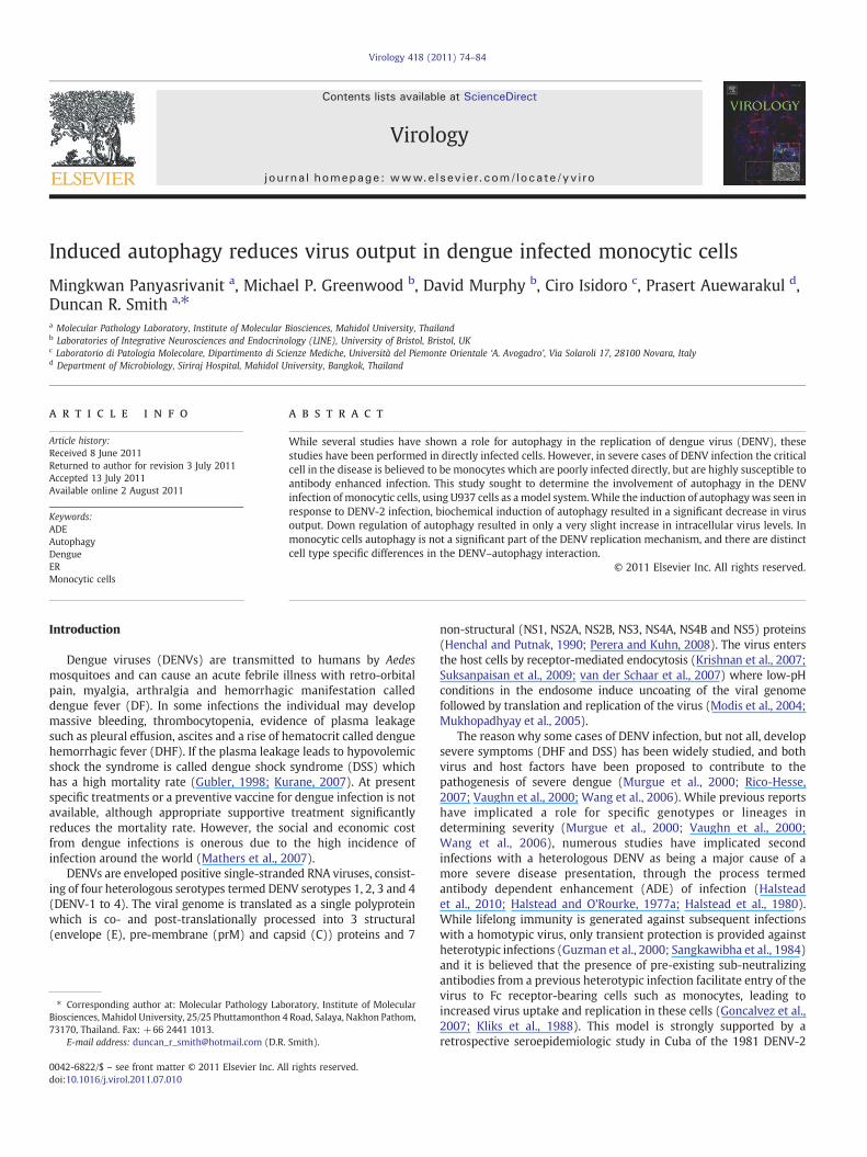
This study aimed to investigate the relationship betweenautophagy and DENV-2 in monocytic cells, and used the humanmonocytic cell line U937 as a model (O'Sullivan and Killen, 1994). Toconfirm that the autophagy pathway was inducible in these cells,cells were subjected to two well characterized autophagy inductionconditions, namely starvation and treatment with rapamycin (Nodaand Ohsumi, 1998). The induction of autophagy was observed by co-immunofluorescence using antibodies against LC3 and a marker ofendosomal and lysosomal membranes, LAMP1, with co-localizationof these two markers indicating maturation of autophagic vacuolesduring autophagy. The cellular localization of these markers wasexamined in control cells cultured under normal conditions, in cellsincubated in Earle's Balanced Salt Solution for 1 h (starvationcondition) and in cells cultured in the presence of 100 nM rapamycinfor 1 h. A significantly higher degree of co-localization between LC3and LAMP1 was observed in both starved (mean Pearson correlationcoefficient 0.54, 95% confidence interval (CI) 0.51–0.57; Pb0.001)and rapamycin (mean Pearson correlation coefficient 0.4, 95% CI0.37–0.43; Pb0.001) treated cells compared to control cells (meanPearson correlation coefficient−0.01, 95% CI−0.05–0.03; Fig. 1(a)).
Induction of autophagy in response to DENV-2 infection in U937 cells
To determine whether autophagy was induced in response toDENV infection, U937 cells were mock-infected or infected in thepresence of dilutions of the monoclonal antibody HB-114 (Henchalet al., 1982) previously shown to result in at least 80% infection of cells(20 p.f.u. per cell/1:200 final antibody dilution; Klomporn et al.,2011). Cells were harvested at 1 to 3 days post infection (d.p.i) forprotein extraction and samples were analyzed for LC3 expression byWestern blotting. The autophagic membrane associated form of LC3(LC3-II) in DENV-2 infected U937 cells increased on day 1, day 2 andday 3 post infection in comparison to mock-infected cells (Fig. 1(b)),suggesting induction of autophagy in DENV-2 infection. To show thatinduction of autophagy results from DENV-2 infection, not physicalinteraction of the viruses and cells, Western blot of LC3 wasperformed in U937 cells infected with DENV-2 in the absence ofenhancing antibody (no ADE). Result showed no increase of LC3-IIlevel in non-ADE-DENV-2 infection as compared to mock-infection(Fig. 1(c)). Localization of LC3 and LAMP1 was examined in mock-infected and (ADE) DENV-2 infected U937 cells. A significant increasein co-localization was observed in (ADE) DENV-2 infected U937 cells(mean Pearson correlation coefficient 0.27, 95% CI 0.22–0.32;P=0.021) compared to mock-infected cells (mean Pearson correla-tion coefficient 0.13, 95% CI 0.04–0.22; Fig. 1(d)). Collectively theseresults indicate that autophagy was induced in response to DENV-2ADE-mediated infection of U937 cells.
Effect of autophagy induction and inhibition of autophagosome-lysosome fusion on DENV-2 production
To further investigate the interaction between autophagy andDENV,two autophagymodulatorswere used namely rapamycin, an autophagyinducer (Noda and Ohsumi, 1998) and L-Asparagine (L-Asn), aninhibitor of autophagosome–lysosome fusion (Hoyvik et al., 1991).First, the effects of these two autophagy modulators in U937 cells wereconfirmed. U937 cells were treated with either 100 nM rapamycin or30 mML-Asn for 24 h and total proteinswere extracted and subjected toWestern blot analysis of LC3. An increase of LC3-II was observedfollowing rapamycin treatment compared to control indicating theinduction of autophagy, consistent with the LC3 and LAMP1 co-localization analysis. LC3-II expression also increased following L-Asn
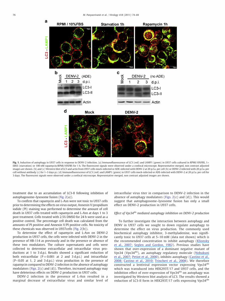
Fig. 1. Induction of autophagy in U937 cells in response to DENV-2 infection. (a) Immunofluorescence of LC3 (red) and LAMP1 (green) in U937 cells cultured in RPMI/10%FBS, 1×EBSS (starvation) or 100 nM rapamycin/RPMI/10%FBS for 1 h. The fluorescent signals were observed under a confocal microscope. Representative merged, non contrast adjustedimages are shown. (b) and (c)Western blot of LC3 and actin from U937 cells mock-infected or ADE-infected with DENV-2 at 20 p.f.u. per cell (b) or DENV-2 infectedwith 20 p.f.u. percell without antibody (c) for 1–3 days p.i. (d) Immunofluorescence of LC3 (red) and LAMP1 (green) in U937 cells mock-infected or ADE-infected with DENV-2 at 20 p.f.u. per cell for3 days. The fluorescent signals were observed under a confocal microscope. Representative merged, non contrast adjusted images are shown.
76 M. Panyasrivanit et al. / Virology 418 (2011) 74–84
treatment due to an accumulation of LC3-II following inhibition ofautophagosome–lysosome fusion (Fig. 2(a)).
To confirm that rapamycin and L-Asn were not toxic to U937 cellsprior to determining the effects on virus output, Annexin V/propidiumiodide (PI) staining was performed to determine the amount of celldeath in U937 cells treated with rapamycin and L-Asn at days 1 to 3post treatment. Cells treated with 2.5% DMSO for 24 h were used as apositive control. The percentage cell death was calculated from theamounts of PI positive and Annexin V/PI positive cells. No toxicity ofthese chemicals was observed in U937cells (Fig. 2(b)).
To determine the effect of rapamycin and L-Asn on DENV-2production in U937 cells, the cells were infected with DENV-2 in thepresence of HB-114 as previously and in the presence or absence ofthese two modulators. The culture supernatants and cells werecollected to determine extracellular and intracellular virus pro-ductions at 1 to 3 d.p.i. Results showed a significant reduction ofboth extracellular (P=0.001 at 2 and 3 d.p.i.) and intracellular(Pb0.05 at 1, 2 and 3 d.p.i.) virus production in the presence ofrapamycin compared to DENV-2 infection in the absence of autophagymodulators (Figs. 2(c) and (d)). Therefore, increased autophagy mayhave deleterious effects on DENV-2 production in U937 cells.
DENV-2 infection in the presence of L-Asn resulted in amarginal decrease of extracellular virus and similar level of
intracellular virus titer in comparison to DENV-2 infection in theabsence of autophagy modulators (Figs. 2(c) and (d)). This wouldsuggest that autophagosome–lysosome fusion has only a smalleffect on DENV-2 production in U937 cells.
Effect of Vps34dn mediated autophagy inhibition on DENV-2 production
To further investigate the interaction between autophagy andDENV in U937 cells we sought to down regulate autophagy todetermine the effect on virus production. The commonly usedbiochemical autophagy inhibitor, 3-methyladenine, was signifi-cantly toxic to U937 cells at 5–10 mM (data not shown) which isthe recommended concentration to inhibit autophagy (Klionskyet al., 2007; Seglen and Gordon, 1982). Previous studies haveshown that over-expression of a dominant negative mutant ofVps34 (Vps34dn), an autophagy regulatory molecule (Nobukuniet al., 2007; Petiot et al., 2000), inhibits autophagy (Castino et al.,2008; Castino et al., 2010; Trincheri et al., 2008). We thereforeconstructed a lentiviral expression vector expressing Vps34dn
which was transduced into HEK293T/17 and U937 cells, and theinhibition effect of over-expression of Vps34dn on autophagy wasinvestigated by Western blot analysis of LC3. The results showed areduction of LC3-II form in HEK293T/17 cells expressing Vps34dn
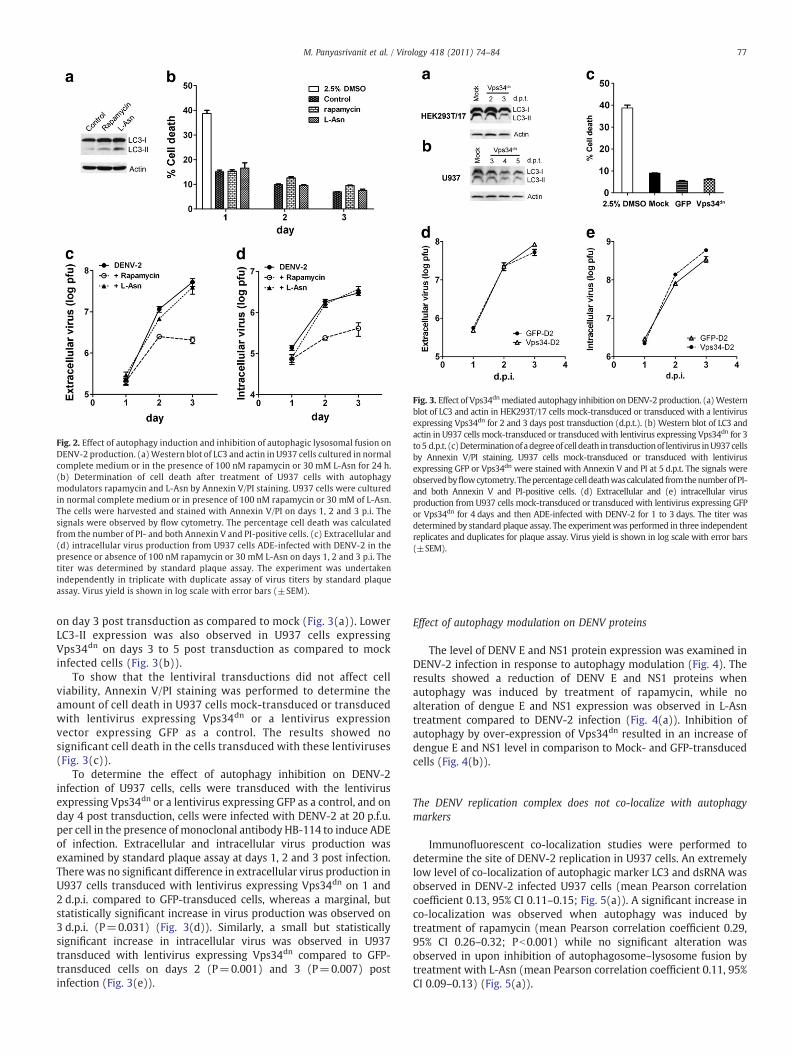
Fig. 2. Effect of autophagy induction and inhibition of autophagic lysosomal fusion onDENV-2 production. (a)Western blot of LC3 and actin in U937 cells cultured in normalcomplete medium or in the presence of 100 nM rapamycin or 30 mM L-Asn for 24 h.(b) Determination of cell death after treatment of U937 cells with autophagymodulators rapamycin and L-Asn by Annexin V/PI staining. U937 cells were culturedin normal complete medium or in presence of 100 nM rapamycin or 30 mM of L-Asn.The cells were harvested and stained with Annexin V/PI on days 1, 2 and 3 p.i. Thesignals were observed by flow cytometry. The percentage cell death was calculatedfrom the number of PI- and both Annexin V and PI-positive cells. (c) Extracellular and(d) intracellular virus production from U937 cells ADE-infected with DENV-2 in thepresence or absence of 100 nM rapamycin or 30 mM L-Asn on days 1, 2 and 3 p.i. Thetiter was determined by standard plaque assay. The experiment was undertakenindependently in triplicate with duplicate assay of virus titers by standard plaqueassay. Virus yield is shown in log scale with error bars (±SEM).
Fig. 3. Effect of Vps34dnmediated autophagy inhibition onDENV-2 production. (a)Westernblot of LC3 and actin in HEK293T/17 cells mock-transduced or transduced with a lentivirusexpressing Vps34dn for 2 and 3 days post transduction (d.p.t.). (b) Western blot of LC3 andactin in U937 cells mock-transduced or transduced with lentivirus expressing Vps34dn for 3to5 d.p.t. (c)Determinationofadegreeof cell death in transductionof lentivirus inU937cellsby Annexin V/PI staining. U937 cells mock-transduced or transduced with lentivirusexpressing GFP or Vps34dn were stained with Annexin V and PI at 5 d.p.t. The signals wereobservedbyflowcytometry. Thepercentagecell deathwascalculated fromthenumberof PI-and both Annexin V and PI-positive cells. (d) Extracellular and (e) intracellular virusproduction from U937 cells mock-transduced or transduced with lentivirus expressing GFPor Vps34dn for 4 days and then ADE-infected with DENV-2 for 1 to 3 days. The titer wasdetermined by standard plaque assay. The experimentwas performed in three independentreplicates and duplicates for plaque assay. Virus yield is shown in log scale with error bars(±SEM).
77M. Panyasrivanit et al. / Virology 418 (2011) 74–84
on day 3 post transduction as compared to mock (Fig. 3(a)). LowerLC3-II expression was also observed in U937 cells expressingVps34dn on days 3 to 5 post transduction as compared to mockinfected cells (Fig. 3(b)).
To show that the lentiviral transductions did not affect cellviability, Annexin V/PI staining was performed to determine theamount of cell death in U937 cells mock-transduced or transducedwith lentivirus expressing Vps34dn or a lentivirus expressionvector expressing GFP as a control. The results showed nosignificant cell death in the cells transduced with these lentiviruses(Fig. 3(c)).
To determine the effect of autophagy inhibition on DENV-2infection of U937 cells, cells were transduced with the lentivirusexpressing Vps34dn or a lentivirus expressing GFP as a control, and onday 4 post transduction, cells were infected with DENV-2 at 20 p.f.u.per cell in the presence of monoclonal antibody HB-114 to induce ADEof infection. Extracellular and intracellular virus production wasexamined by standard plaque assay at days 1, 2 and 3 post infection.Therewas no significant difference in extracellular virus production inU937 cells transduced with lentivirus expressing Vps34dn on 1 and2 d.p.i. compared to GFP-transduced cells, whereas a marginal, butstatistically significant increase in virus production was observed on3 d.p.i. (P=0.031) (Fig. 3(d)). Similarly, a small but statisticallysignificant increase in intracellular virus was observed in U937transduced with lentivirus expressing Vps34dn compared to GFP-transduced cells on days 2 (P=0.001) and 3 (P=0.007) postinfection (Fig. 3(e)).
Effect of autophagy modulation on DENV proteins
The level of DENV E and NS1 protein expression was examined inDENV-2 infection in response to autophagy modulation (Fig. 4). Theresults showed a reduction of DENV E and NS1 proteins whenautophagy was induced by treatment of rapamycin, while noalteration of dengue E and NS1 expression was observed in L-Asntreatment compared to DENV-2 infection (Fig. 4(a)). Inhibition ofautophagy by over-expression of Vps34dn resulted in an increase ofdengue E and NS1 level in comparison to Mock- and GFP-transducedcells (Fig. 4(b)).
The DENV replication complex does not co-localize with autophagymarkers
Immunofluorescent co-localization studies were performed todetermine the site of DENV-2 replication in U937 cells. An extremelylow level of co-localization of autophagic marker LC3 and dsRNA wasobserved in DENV-2 infected U937 cells (mean Pearson correlationcoefficient 0.13, 95% CI 0.11–0.15; Fig. 5(a)). A significant increase inco-localization was observed when autophagy was induced bytreatment of rapamycin (mean Pearson correlation coefficient 0.29,95% CI 0.26–0.32; Pb0.001) while no significant alteration wasobserved in upon inhibition of autophagosome–lysosome fusion bytreatment with L-Asn (mean Pearson correlation coefficient 0.11, 95%CI 0.09–0.13) (Fig. 5(a)).
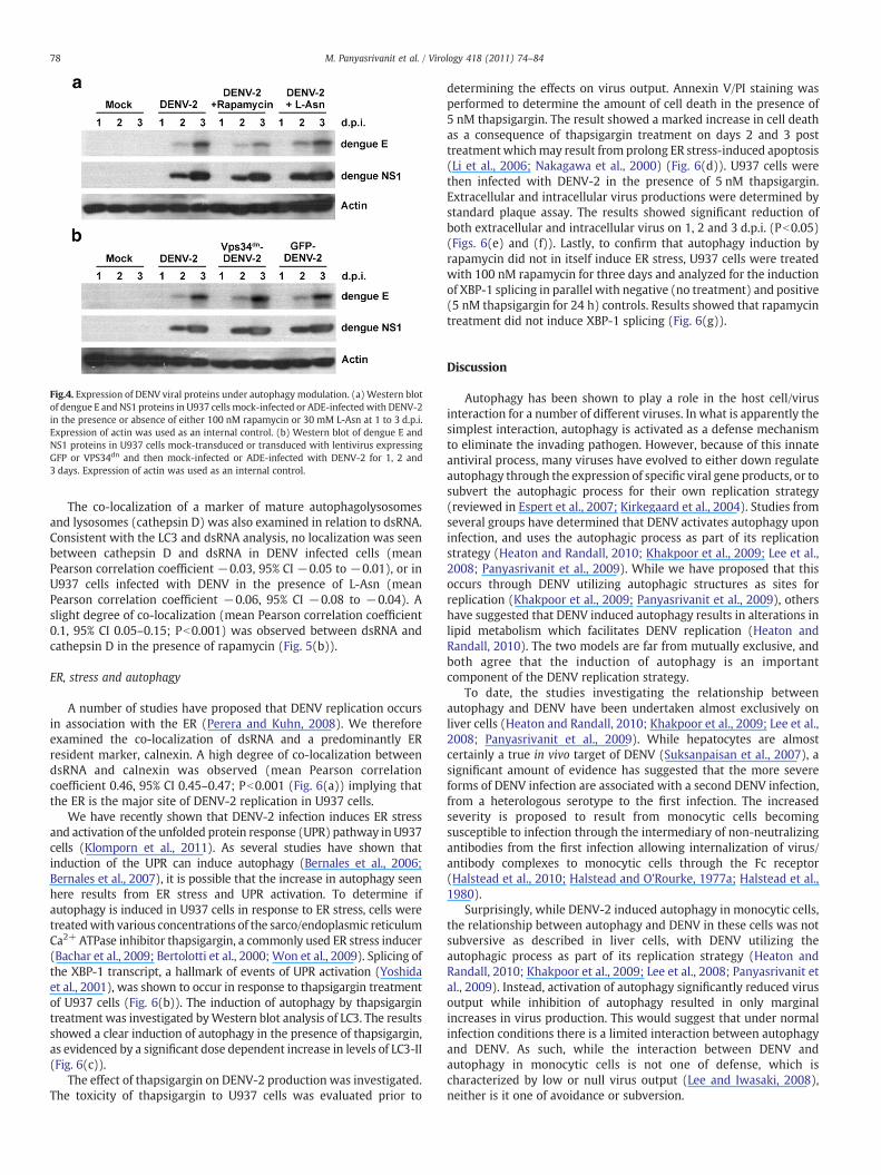
Fig.4. Expression of DENV viral proteins under autophagy modulation. (a) Western blotof dengue E and NS1 proteins in U937 cells mock-infected or ADE-infected with DENV-2in the presence or absence of either 100 nM rapamycin or 30 mM L-Asn at 1 to 3 d.p.i.Expression of actin was used as an internal control. (b) Western blot of dengue E andNS1 proteins in U937 cells mock-transduced or transduced with lentivirus expressingGFP or VPS34dn and then mock-infected or ADE-infected with DENV-2 for 1, 2 and3 days. Expression of actin was used as an internal control.
78 M. Panyasrivanit et al. / Virology 418 (2011) 74–84
The co-localization of a marker of mature autophagolysosomesand lysosomes (cathepsin D) was also examined in relation to dsRNA.Consistent with the LC3 and dsRNA analysis, no localization was seenbetween cathepsin D and dsRNA in DENV infected cells (meanPearson correlation coefficient −0.03, 95% CI −0.05 to −0.01), or inU937 cells infected with DENV in the presence of L-Asn (meanPearson correlation coefficient −0.06, 95% CI −0.08 to −0.04). Aslight degree of co-localization (mean Pearson correlation coefficient0.1, 95% CI 0.05–0.15; Pb0.001) was observed between dsRNA andcathepsin D in the presence of rapamycin (Fig. 5(b)).
ER, stress and autophagy
A number of studies have proposed that DENV replication occursin association with the ER (Perera and Kuhn, 2008). We thereforeexamined the co-localization of dsRNA and a predominantly ERresident marker, calnexin. A high degree of co-localization betweendsRNA and calnexin was observed (mean Pearson correlationcoefficient 0.46, 95% CI 0.45–0.47; Pb0.001 (Fig. 6(a)) implying thatthe ER is the major site of DENV-2 replication in U937 cells.
We have recently shown that DENV-2 infection induces ER stressand activation of the unfolded protein response (UPR) pathway in U937cells (Klomporn et al., 2011). As several studies have shown thatinduction of the UPR can induce autophagy (Bernales et al., 2006;Bernales et al., 2007), it is possible that the increase in autophagy seenhere results from ER stress and UPR activation. To determine ifautophagy is induced in U937 cells in response to ER stress, cells weretreatedwith various concentrations of the sarco/endoplasmic reticulumCa2+ ATPase inhibitor thapsigargin, a commonly used ER stress inducer(Bachar et al., 2009; Bertolotti et al., 2000; Won et al., 2009). Splicing ofthe XBP-1 transcript, a hallmark of events of UPR activation (Yoshidaet al., 2001), was shown to occur in response to thapsigargin treatmentof U937 cells (Fig. 6(b)). The induction of autophagy by thapsigargintreatment was investigated byWestern blot analysis of LC3. The resultsshowed a clear induction of autophagy in the presence of thapsigargin,as evidenced by a significant dose dependent increase in levels of LC3-II(Fig. 6(c)).
The effect of thapsigargin on DENV-2 production was investigated.The toxicity of thapsigargin to U937 cells was evaluated prior to
determining the effects on virus output. Annexin V/PI staining wasperformed to determine the amount of cell death in the presence of5 nM thapsigargin. The result showed a marked increase in cell deathas a consequence of thapsigargin treatment on days 2 and 3 posttreatmentwhichmay result from prolong ER stress-induced apoptosis(Li et al., 2006; Nakagawa et al., 2000) (Fig. 6(d)). U937 cells werethen infected with DENV-2 in the presence of 5 nM thapsigargin.Extracellular and intracellular virus productions were determined bystandard plaque assay. The results showed significant reduction ofboth extracellular and intracellular virus on 1, 2 and 3 d.p.i. (Pb0.05)(Figs. 6(e) and (f)). Lastly, to confirm that autophagy induction byrapamycin did not in itself induce ER stress, U937 cells were treatedwith 100 nM rapamycin for three days and analyzed for the inductionof XBP-1 splicing in parallel with negative (no treatment) and positive(5 nM thapsigargin for 24 h) controls. Results showed that rapamycintreatment did not induce XBP-1 splicing (Fig. 6(g)).
Discussion
Autophagy has been shown to play a role in the host cell/virusinteraction for a number of different viruses. In what is apparently thesimplest interaction, autophagy is activated as a defense mechanismto eliminate the invading pathogen. However, because of this innateantiviral process, many viruses have evolved to either down regulateautophagy through the expression of specific viral gene products, or tosubvert the autophagic process for their own replication strategy(reviewed in Espert et al., 2007; Kirkegaard et al., 2004). Studies fromseveral groups have determined that DENV activates autophagy uponinfection, and uses the autophagic process as part of its replicationstrategy (Heaton and Randall, 2010; Khakpoor et al., 2009; Lee et al.,2008; Panyasrivanit et al., 2009). While we have proposed that thisoccurs through DENV utilizing autophagic structures as sites forreplication (Khakpoor et al., 2009; Panyasrivanit et al., 2009), othershave suggested that DENV induced autophagy results in alterations inlipid metabolism which facilitates DENV replication (Heaton andRandall, 2010). The two models are far from mutually exclusive, andboth agree that the induction of autophagy is an importantcomponent of the DENV replication strategy.
To date, the studies investigating the relationship betweenautophagy and DENV have been undertaken almost exclusively onliver cells (Heaton and Randall, 2010; Khakpoor et al., 2009; Lee et al.,2008; Panyasrivanit et al., 2009). While hepatocytes are almostcertainly a true in vivo target of DENV (Suksanpaisan et al., 2007), asignificant amount of evidence has suggested that the more severeforms of DENV infection are associated with a second DENV infection,from a heterologous serotype to the first infection. The increasedseverity is proposed to result from monocytic cells becomingsusceptible to infection through the intermediary of non-neutralizingantibodies from the first infection allowing internalization of virus/antibody complexes to monocytic cells through the Fc receptor(Halstead et al., 2010; Halstead and O'Rourke, 1977a; Halstead et al.,1980).
Surprisingly, while DENV-2 induced autophagy in monocytic cells,the relationship between autophagy and DENV in these cells was notsubversive as described in liver cells, with DENV utilizing theautophagic process as part of its replication strategy (Heaton andRandall, 2010; Khakpoor et al., 2009; Lee et al., 2008; Panyasrivanit etal., 2009). Instead, activation of autophagy significantly reduced virusoutput while inhibition of autophagy resulted in only marginalincreases in virus production. This would suggest that under normalinfection conditions there is a limited interaction between autophagyand DENV. As such, while the interaction between DENV andautophagy in monocytic cells is not one of defense, which ischaracterized by low or null virus output (Lee and Iwasaki, 2008),neither is it one of avoidance or subversion.
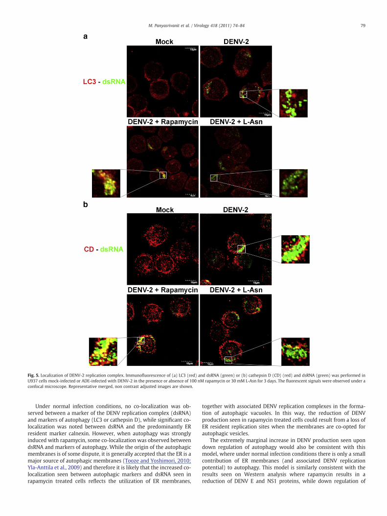
Fig. 5. Localization of DENV-2 replication complex. Immunofluorescence of (a) LC3 (red) and dsRNA (green) or (b) cathepsin D (CD) (red) and dsRNA (green) was performed inU937 cells mock-infected or ADE-infected with DENV-2 in the presence or absence of 100 nM rapamycin or 30 mM L-Asn for 3 days. The fluorescent signals were observed under aconfocal microscope. Representative merged, non contrast adjusted images are shown.
79M. Panyasrivanit et al. / Virology 418 (2011) 74–84
Under normal infection conditions, no co-localization was ob-served between a marker of the DENV replication complex (dsRNA)and markers of autophagy (LC3 or cathepsin D), while significant co-localization was noted between dsRNA and the predominantly ERresident marker calnexin. However, when autophagy was stronglyinduced with rapamycin, some co-localization was observed betweendsRNA and markers of autophagy. While the origin of the autophagicmembranes is of some dispute, it is generally accepted that the ER is amajor source of autophagic membranes (Tooze and Yoshimori, 2010;Yla-Anttila et al., 2009) and therefore it is likely that the increased co-localization seen between autophagic markers and dsRNA seen inrapamycin treated cells reflects the utilization of ER membranes,
together with associated DENV replication complexes in the forma-tion of autophagic vacuoles. In this way, the reduction of DENVproduction seen in rapamycin treated cells could result from a loss ofER resident replication sites when the membranes are co-opted forautophagic vesicles.
The extremely marginal increase in DENV production seen upondown regulation of autophagy would also be consistent with thismodel, where under normal infection conditions there is only a smallcontribution of ER membranes (and associated DENV replicationpotential) to autophagy. This model is similarly consistent with theresults seen on Western analysis where rapamycin results in areduction of DENV E and NS1 proteins, while down regulation of

Fig. 6. ER stress induced autophagy in DENV-2 infected U937 cells. (a) Immunofluorescence of calnexin (red) and dsRNA (green) was performed in U937 cells mock-infected or ADE-infected with DENV-2 for 3 days. The fluorescent signals were observed under a confocal microscope. Representativemerged, non contrast adjusted images are shown. (b) Splicing of XBP-1transcriptswas examined inU937 cells cultured in RPMI/10%FBS (control) or in thepresence of 1, 5 and 25 nMof thapsigargin for 24 hbyRT-PCR.H: heteroduplex; U: unspliced; S: sliced formofXBP-1. (c)Westernblot of LC3andactin inU937 cells cultured innormal completemedium(control) or in thepresenceof 1, 5 and25 nMof Thapsigargin for 1 to3 days. (d)Determinationofcell death in U937 cells treated with 5 nM thapsigargin by Annexin V/PI staining. The signals were observed by flow cytometry on days 1 to 3 post treatment. Percentage of cell death wascalculated from thenumber of PI- and bothAnnexinV andPI-positive cells. (e) Extracellular and (f) intracellular virus production fromU937 cells ADE-infectedwithDENV-2 in thepresence orabsence of 5 nM thapsigargin on days 1, 2 and 3 post infection. The titerwas determined by standard plaque assay. The experimentwas undertaken independently in triplicatewith duplicateassay of virus titers by standard plaque assay. Virus yield is shown in log scale with error bars (±SEM). * indicates statistically significant compared to control (Pb0.05). (g) U937 cells wereeither not treated (Control) or treatedwith either 100 nM rapamycin for 1, 2 or 3 days (rapamycin 1 to 3) or with 5 nM thapsigargin for 24 h as a positive control (+ve) and analyzed for thesplicing of the XBP-1 transcript by RT-PCR. H: heteroduplex; U: unspliced; S: sliced form of XBP-1.
80 M. Panyasrivanit et al. / Virology 418 (2011) 74–84
autophagy results in a slight increase in the levels of these proteins.Again, the ER membranes being co-opted under increased autophagywould serve to reduce the amount of protein translation occurring inthe ER.
While the data suggests a minimal interaction between autophagyand DENV production under normal conditions, the induction ofautophagy was observed in response to infection.
We have recently shown that infection of monocytic cells by DENVresults in the activation of multiple ER stress pathways, which
ultimately result in apoptosis of the cells (Klomporn et al., 2011).Several studies have suggested a direct link between ER stress andautophagy (Bernales et al., 2006; Bernales et al., 2007), and it is likelythen that the induction of autophagy seen in response to DENVinfection occurs as a result of activation of ER stress and activation ofthe unfolded protein response, rather than a direct DENV mediatedactivation of autophagy. Indeed, treatment of U937 cells withthapsigargin showed the induction of ER stress as well as theinduction of autophagy. Under these conditions, DENV output was
81M. Panyasrivanit et al. / Virology 418 (2011) 74–84
reduced in a similar manner to the reduction seen with rapamycin.While we note that long term treatment with thapsigargin inducedcell death, a significant reduction in virus production was observedprior to significant levels of cell death.
In this way it can be hypothesized that in U937 cells biochemicalactivation of autophagy (either directly through rapamycin, orindirectly through induction of the UPR) reduces virus output as aconsequence of recruitment of ER membranes to autophagic vacuoles(Bernales et al., 2006; Bernales et al., 2007) thus reducing indirectlythe number of available sites of replication for DENV.
Inhibition of autophagy did result in a small, but statisticallysignificant increase in both intracellular and extracellular viruses.This would suggest that under normal conditions there is some(albeit small) interaction between DENV and autophagy. Apart fromits functions tomaintain homeostasis in the cell, autophagy also has arole in the immune system where it mediates endogenous majorhistocompatibility complex (MHC) class II antigen processing(Deretic, 2006; Nimmerjahn et al., 2003; Paludan et al., 2005).Since cells of amonocyte/macrophage lineage are antigen presentingcells, autophagic degradation of DENV-2 in these cells may alsoinvolve antigen processing. In this case, the proportion of DENVproduced that would normally be lost during the process ofautophagy mediated antigen presentation is no longer lost, and isdetected on analysis of virus levels.
Given that DENV infectedmonocytic cells undergo apoptosis withina few days, it is difficult to see that the marginally increased cellularlevels of virus would have profound effects on the cell. However, apossible reduction in antigen presentation as a consequence of downregulation of autophagy could have serious consequences. Thustreatment approaches that seek to down regulate autophagy, basedupon the subversion reaction observed in other cell types (Heaton andRandall, 2010; Khakpoor et al., 2009; Lee et al., 2008; Panyasrivanit et al.,2009) could have serious long term immune consequences by mutingthe efficacy of the immune response.
Overall, the interaction between autophagy and DENV in mono-cytic cells described here does not fit any of the classical models ofvirus/host cell autophagy interaction (Espert et al., 2007; Kirkegaardet al., 2004), but does show that the interaction between DENV andautophagy is mediated in a cell type specific manner.
Materials and methods
Cells and viruses
U937 cells (human monocytic cell line, ATCC CRL-1593.2) werecultured in RPMI 1640 medium (RPMI; GibcoTM Invitrogen)supplemented with 10% heat-inactivated fetal bovine serum (FBS;GibcoTM Invitrogen) and 100 unit/ml of penicillin–streptomycin(PAA Laboratories GmbH). LLC-MK2 cells (rhesus monkey kidneycell line, ATCC CCL-7) were cultured in Dulbecco's Modified EagleMedium (DMEM; GibcoTM Invitrogen) supplemented with 5% FBSand 100 unit/ml of penicillin–streptomycin. HEK293T/17 cells(human embryonic kidney cell line, ATCC CRL-11268) were culturedin DMEM supplemented with 10% FBS and 100 unit/ml of penicillin–streptomycin. All cells were incubated at 37 °C in a humidifiedincubator with 5% CO2. C6/36 cells (the Aedes albopictus cell line,ATCC CRL-1660) were cultured in minimum essential medium(MEM; GibcoTM Invitrogen) supplemented with 10% FBS and thesame antibiotics as the cell lines above at 28 °C.
Dengue virus serotype 2 (DENV-2) strain 16681was propagated inC6/36 cells. The medium containing virus was collected at the dayproviding a maximum virus titer (Sakoonwatanyoo et al., 2006) andsubsequently centrifuged to remove cell debris. The virus was storedat −80 °C until use. The virus titer was determined by standardplaque assay as previously described (Sithisarn et al., 2003) on LLC-MK2 cells.
Cell starvation and chemical treatments
U937 cells were centrifuged at 400×g for 5 min to remove culturemedium. The cell pellets were washed once with PBS (for starvation)and then resuspended with 1× EBSS for starvation condition,complete culture medium containing 100 nM rapamycin (SigmaChemical Company), 30 mM L-Asparagine (L-Asn; Sigma) or 1, 5and 25 nM Thapsigargin (Sigma). The cells were then incubated understandard conditions until harvesting.
Infection of U937 cells
DENV-2 infection of U937 cells was undertaken exactly as describedelsewhere (Klomporn et al., 2011). Briefly, a pre-calculated 20 p.f.u. percell of DENV-2 and a 1:200 dilution of a pan specific mousemonoclonalanti-dengue E protein antibody (HB114) produced from mousehybridoma cell line D3-2H2-9-21 (ATCC HB114, isotype IgG2a) wereincubated for 1 hat4 °C inRPMImedium.U937 cellswere centrifugedat400×g for 5 min to remove culture medium and resuspended with theantibody–virus mixture. The cells were then incubated at 37 °C, 5% CO2
for 2 h with constant agitation. After 2 h, complete mediumwas addedto give a final cell density of 3×10⁵ cells/ml. For infection in thepresence of autophagy modulators, U937 cells were pre-treated for 1 hwith 100 nM rapamycin, 30 mM L-Asn or 5 nM thapsigargin (Sigma) inRPMI/10%FBS and infectionwas performed as described above. The cellswere incubated under standard conditions until harvesting of the cellsor culture medium. For non-ADE-DENV-2 infection, U937 cells weredirectly incubated with DENV-2 at 20 p.f.u. per cell at 37 °C, 5% CO2 for2 h with constant agitation and then complete medium was added togive a final cell density of 3×10⁵ cells/ml.
Intracellular and extracellular virus titration by standard plaque assay
Extracellular and intracellular virus titers were determinedessentially as described elsewhere (Thepparit and Smith, 2004).Extracellular virus titers were determined directly from cell superna-tants. To determine the intracellular virus titer, cells in each samplewere harvested and centrifuged at 400×g for 5 min to remove culturemedium. The cells were washed with RPMI and resuspended in BA-1medium (1× medium199/Earle's balanced salts (HyClone), 0.05 MTris–HCl pH 7.6, 1% BSA fraction V (PAA), 0.075% NaHCO3, 100 units/ml penicillin–streptomycin). The cell suspensions were mixed andintracellular virus was released from the cells by one freeze–thawcycle and sonicating at 4 °C for 5 min. The extracellular virus andintracellular virus titers were determined by standard plaque assay onLLC-MK2 cells as previously described (Sithisarn et al., 2003). Thevirus titer values were derived from three-independent experimentsassayed in duplicate.
Indirect immunofluorescence
50,000–100,000 U937 cells from each experimental conditionwere collected and centrifuged at 400×g for 5 min to remove culturemedium. The cells were washed once with RPMI and spun downonto glass cover slips using StatSpin® CytoFuge 2 (Iris SampleProcessing). The cells were fixed in ice-cold absolute methanol for20 min and washed twice with PBS. The subsequent steps wereundertaken exactly as previously described (Panyasrivanit et al.,2009). Primary antibodies used were a rabbit polyclonal anti-MAP-LC3 antibody (sc-28266, Santa Cruz Biotechnology), a mousemonoclonal anti-CD107a antibody (LAMP1, 555798, BD Bioscience),a mouse monoclonal anti-dsRNA antibody (J2-0702, Scicons,Hungary), a rabbit polyclonal anti-calnexin antibody (ab10286,Abcam) and a rabbit polyclonal anti-cathepsin D antibody (Ab-2, IM16, Calbiochem). All primary antibodies were used at a concentra-tion of 1:50 with the exception of the anti-dsRNA antibody which
82 M. Panyasrivanit et al. / Virology 418 (2011) 74–84
was used at a dilution of 1:100. Secondary antibodies used were arhodamine redTM-X-conjugated goat anti-rabbit IgG antibody (111-295-144, Jackson ImmunoResearch Laboratories) (1:50), a FITC-conjugated goat anti-mouse IgG antibody (02-18-06, KPL) (1:20),and an Alexa™ 647-conjugated donkey anti-rabbit IgG antibody(A31573, Molecular Probes) (1:100). The cells were viewed underan Olympus FluoView 1000 confocal microscope as describedelsewhere (Panyasrivanit et al., 2009).
The degree of co-localization was analyzed in non-contrastadjusted pictures using ImageJ software with PSC co-localizationplug-in as described elsewhere (Panyasrivanit et al., 2009). A degreeof co-localization was determined as Pearson correlation coefficients,which represent the linear relationship of the signal intensity from thegreen and red channels. At least 20 cells were analyzed for eachcondition. Statistical significance (Pb0.05) between data sets wasdetermined by independent sample t tests using SPSS software (SPSSInc., Chicago, IL).
Protein extraction and Western blot assay
Cells were harvested and centrifuged to remove culture medium.The cell pellets were washed with cold RPMI and resuspended in RIPAbuffer (1% Nonidet P-40, 0.5% sodium deoxycholate, 0.1% SDS in PBS).The cell lysates were incubated on ice for 30 min with periodic mixingand sonicated twice at 4 °C for 5 min. The cell lysates were thencentrifuged at 10,000×g for 10 min and supernatants were collected.Proteins were separated by SDS-PAGE and transferred to PVDFmembranes. The membranes were blocked in blocking solution (5%skimmed milk/0.05% tween20/TBS) for 2 h at room temperature orovernight at 4 °C and incubated with primary antibodies: a rabbitpolyclonal anti-LC3B antibody (ab48394, Abcam) (1:2000 dilution), agoat polyclonal anti-actin (I-19) antibody (sc-1616, Santa CruzBiotechnology) (1:500 dilution), a mouse monoclonal anti-dengueNS1 antibody (Puttikhunt et al., 2003) (1:200 dilution) or a mousemonoclonal anti-dengue virus type 1–4 antibody (MA1-27093,Pierce) (1:1000 dilution) diluted in blocking solution at roomtemperature for at least 3 h (for LC3) or 4 °C overnight (for all otherantibodies) followed by incubation with an appropriate secondaryantibody conjugatedwith HRP at room temperature for 1 h. The signalwas visualized using Pierce® ECL Western blotting substrate (ThermoFisher Scientific).
Annexin V and Propidium iodide staining
2×10⁵ cells were harvested from each condition at the indicatedtime points. The cells were washed and resuspended in 1×ApoAlertTM Annexin V binding buffer (630202, Clontech). 1 μl ofApoAlertTM Annexin V conjugated FITC (630201, Clontech) and 10 μlof 50 μg/ml Propidium iodide (PI; Sigma) were added to the cells andsamples were incubated in darkness at room temperature for 15 min.The signals were detected using a FACS Calibur flow cytometer. Thepositive control in all experiments was U937 cells treated with 2.5%DMSO in RPMI/10%FBS for 24 h. The percentage cell death wascalculated from the amounts of PI-positive and Annexin V/PI-positivecells. All experiments were undertaken as three independentreplicates.
Construction of lentiviruses
AcDNAcloneencoding akinasedeficient (dominantnegative)mutantof rat Vps34 was the kind gift of Dr. HW Davidson (University ofCambridge) (Row et al., 2001). The full length cDNA of Vps34 whichincluded two point mutations (Asp743-Ala, Asn748-Ile) was excised fromplasmidpcDNA3puro andexpressed inplaceofGFP in the lentiviral vectorpRRL.SIN.CPPT.CMV.GFP.WPRE (engineered from Addgene Plasmid12252). The lentiviral transfer vectors pRRL.SIN.CPPT.CMV.GFP.WPRE
and pRRL.SIN.CPPT.CMV.Vps34.WPRE were propagated in Stbl3 compe-tent cells (Invitrogen) to reduce homologous recombination. All plasmidconstructs were purified by two rounds of CsCl ultracentrifugation.Viruses were generated by transient transfection of the transfer vectortogether with 3 separate packaging plasmids (pMDLg/pRRE, pRSV-Rev,PMD2.G (Addgene)) into HEK293T/17 cells by the calcium phosphatemethod. After 8 h in the presence of the transfection precipitate theculture medium was changed and replaced with fresh media. Culturesupernatant containing lentiviruseswere collected 48 h after transfection,cell debris was removed by centrifugation, and the supernatant wasfiltered. Virus stocks were stored at−80 °C until used.
Lentivirus transduction and DENV-2 infection in U937 cells over-expressing Vps34dn
4×10⁵ HEK293T/17 cells were seeded in 12-well tissue cultureplates for 24 h prior to viral transduction. For U937 cells, 5×10⁵ cellswere centrifuged at 420×g for 5 min to remove culture medium. Thecells then were incubated with 400 μl crude lentivirus expressing GFPor Vps34dn in the presence of 8 μg/ml polybrene (hexadimethrinebromide; H9268, Sigma) for 1 h at 37 °C 5% CO2. Control experimentswere performed with 400 μl DMEM/10%FBS. After an hour ofincubation, RPMI/10%FBS was added to a final volume of 1 ml. Thecells were incubated under standard conditions and fresh mediumwas added daily. A high transduction efficiency (N70%) of the GFPlentivirus was demonstrated in U937 cells by fluorescent microscopyand flow cytometry (data not shown).
To perform DENV-2 ADE-infection in U937 cells transduced withlentiviruses, transduced U937 cells were harvested at day 4 posttransduction and infected DENV-2 at 20 p.f.u. per cell in thepresence of dilutions of anti-dengue E protein monoclonal antibodyHB114 as described elsewhere (Klomporn et al., 2011). Extracellularand intracellular virus production was determined as previouslydescribed. Experiments were performed as three independentreplicates with duplicate assay of virus titers by standard plaqueassay.
Semi-quantitative RT-PCR
Semi-quantitative RT-PCR was performed as previously describedelsewhere (Klomporn et al., 2011). Briefly, U937 cells were harvestedby centrifugation and the cells were washed once with cold RPMI.RNA extraction was performed using TRI Reagent® (MolecularResearch Center, Inc.). 2 μg of total RNA was reverse transcribed tocDNA by using ImpromIITM reverse transcriptase (Promega) and oligo(dT). The cDNA was amplified by PCR using specific primer for XBP-1(Yoshida et al., 2001): sense primer 5′-CCTTGTAGTTGAGAACCAGG-3′,antisense primer 5′-GGGGCTTGGTATATATGTGG-3′ and actin: senseprimer 5′-GAAGATGACCCCAGATCATGT-3′, antisense primer 5′-ATCTCTTGCTCGAAGTCCAG-3′ (Lithanatudom et al., 2010). PCRconditions were denaturation at 94 °C for 10 s (XBP-1) and 20 s(actin), annealing at 55 °C for 20 s (XBP-1) and 60 °C for 15 s (actin)and extension at 72 °C for 30 s (XBP-1) and 20 s (actin). PCR-amplified fragment size for XBP-1 are 416 (spliced form) and 442 bp(unspliced form), and for actin is 330 bp. PCR products wereseparated by electrophoresis on 2% agarose gel. Bands were visualizedby ethidium bromide staining.
Acknowledgments
This work was supported by grants from the Thailand ResearchFund and Mahidol University. M.P. is supported by a Thai RoyalGolden Jubilee Research Scholarship.
83M. Panyasrivanit et al. / Virology 418 (2011) 74–84
References
Bachar, E., Ariav, Y., Ketzinel-Gilad, M., Cerasi, E., Kaiser, N., Leibowitz, G., 2009. Glucoseamplifies fatty acid-induced endoplasmic reticulum stress in pancreatic beta-cellsvia activation of mTORC1. PLoS One 4, e4954.
Bernales, S., McDonald, K.L., Walter, P., 2006. Autophagy counterbalancesendoplasmic reticulum expansion during the unfolded protein response. PLoSBiol. 4, e423.
Bernales, S., Schuck, S., Walter, P., 2007. ER-phagy: selective autophagy of theendoplasmic reticulum. Autophagy 3, 285–287.
Bertolotti, A., Zhang, Y., Hendershot, L.M., Harding, H.P., Ron, D., 2000. Dynamicinteraction of BiP and ER stress transducers in the unfolded-protein response. Nat.Cell Biol. 2, 326–332.
Castino, R., Thepparit, C., Bellio, N., Murphy, D., Isidoro, C., 2008. Akt induces apoptosisin neuroblastoma cells expressing a C98X vasopressin mutant following autophagysuppression. J. Neuroendocrinol. 20, 1165–1175.
Castino, R., Bellio, N., Follo, C., Murphy, D., Isidoro, C., 2010. Inhibition of PI3k class III-dependent autophagy prevents apoptosis and necrosis by oxidative stress indopaminergic neuroblastoma cells. Toxicol. Sci. 117, 152–162.
Deretic, V., 2006. Autophagy as an immune defense mechanism. Curr. Opin. Immunol.18, 375–382.
Dunn Jr., W.A., 1990a. Studies on the mechanisms of autophagy: formation of theautophagic vacuole. J. Cell Biol. 110, 1923–1933.
Dunn Jr., W.A., 1990b. Studies on the mechanisms of autophagy: maturation of theautophagic vacuole. J. Cell Biol. 110, 1935–1945.
Eskelinen, E.L., Saftig, P., 2009. Autophagy: a lysosomal degradation pathway with acentral role in health and disease. Biochim. Biophys. Acta 1793, 664–673.
Espert, L., Codogno, P., Biard-Piechaczyk, M., 2007. Involvement of autophagy in viralinfections: antiviral function and subversion by viruses. J. Mol. Med. 85, 811–823.
Ferraro, E., Cecconi, F., 2007. Autophagic and apoptotic response to stress signals inmammalian cells. Arch. Biochem. Biophys. 462, 210–219.
Goncalvez, A.P., Engle, R.E., St Claire, M., Purcell, R.H., Lai, C.J., 2007. Monoclonalantibody-mediated enhancement of dengue virus infection in vitro and in vivo andstrategies for prevention. Proc. Natl. Acad. Sci. U. S. A. 104, 9422–9427.
Gordon, P.B., Seglen, P.O., 1988. Prelysosomal convergence of autophagic and endocyticpathways. Biochem. Biophys. Res. Commun. 151, 40–47.
Gubler, D.J., 1998. Dengue and dengue hemorrhagic fever. Clin. Microbiol. Rev. 11,480–496.
Guzman, M.G., Kouri, G.P., Bravo, J., Soler, M., Vazquez, S., Morier, L., 1990. Denguehemorrhagic fever in Cuba, 1981: a retrospective seroepidemiologic study. Am. J.Trop. Med. Hyg. 42, 179–184.
Guzman, M.G., Kouri, G., Valdes, L., Bravo, J., Alvarez, M., Vazques, S., Delgado, I.,Halstead, S.B., 2000. Epidemiologic studies on dengue in Santiago de Cuba, 1997.Am J Epidemiol 152, 793–799 discussion 804.
Halstead, S.B., O'Rourke, E.J., 1977a. Antibody-enhanced dengue virus infection inprimate leukocytes. Nature 265, 739–741.
Halstead, S.B., O'Rourke, E.J., 1977b. Dengue viruses and mononuclear phagocytes. I.Infection enhancement by non-neutralizing antibody. J. Exp. Med. 146, 201–217.
Halstead, S.B., Porterfield, J.S., O'Rourke, E.J., 1980. Enhancement of dengue virusinfection in monocytes by flavivirus antisera. Am. J. Trop. Med. Hyg. 29, 638–642.
Halstead, S.B., Mahalingam, S., Marovich, M.A., Ubol, S., Mosser, D.M., 2010. Intrinsicantibody-dependent enhancement of microbial infection in macrophages: diseaseregulation by immune complexes. Lancet Infect. Dis. 10, 712–722.
Heaton, N.S., Randall, G., 2010. Dengue virus-induced autophagy regulates lipidmetabolism. Cell Host Microbe 8, 422–432.
Heaton, N.S., Perera, R., Berger, K.L., Khadka, S., Lacount, D.J., Kuhn, R.J., Randall, G., 2010.Dengue virus nonstructural protein 3 redistributes fatty acid synthase to sites of viralreplication and increases cellular fatty acid synthesis. Proc. Natl. Acad. Sci. U. S. A. 107,17345–17350.
Henchal, E.A., Putnak, J.R., 1990. The dengue viruses. Clin. Microbiol. Rev. 3, 376–396.Henchal, E.A., Gentry, M.K., McCown, J.M., Brandt, W.E., 1982. Dengue virus-specific and
flavivirus group determinants identified with monoclonal antibodies by indirectimmunofluorescence. Am. J. Trop. Med. Hyg. 31, 830–836.
Hoyvik, H., Gordon, P.B., Berg, T.O., Stromhaug, P.E., Seglen, P.O., 1991. Inhibition ofautophagic-lysosomal delivery and autophagic lactolysis by asparagine. J. Cell Biol.113, 1305–1312.
Khakpoor, A., Panyasrivanit, M., Wikan, N., Smith, D.R., 2009. A role for autophagolyso-somes in dengue virus 3 production in HepG2 cells. J. Gen. Virol. 90, 1093–1103.
Kirkegaard, K., Taylor, M.P., Jackson, W.T., 2004. Cellular autophagy: surrender,avoidance and subversion by microorganisms. Nat. Rev. Microbiol. 2, 301–314.
Kliks, S.C., Nimmanitya, S., Nisalak, A., Burke, D.S., 1988. Evidence that maternal dengueantibodies are important in the development of dengue hemorrhagic fever ininfants. Am. J. Trop. Med. Hyg. 38, 411–419.
Klionsky, D.J., Cuervo, A.M., Seglen, P.O., 2007. Methods for monitoring autophagy fromyeast to human. Autophagy 3, 181–206.
Klomporn, P., Panyasrivanit, M., Wikan, N., Smith, D.R., 2011. Dengue infection ofmonocytic cells activates ER stress pathways, but apoptosis is induced throughboth extrinsic and intrinsic pathways. Virology 409, 189–197.
Krishnan, M.N., Sukumaran, B., Pal, U., Agaisse, H., Murray, J.L., Hodge, T.W., Fikrig, E.,2007. Rab 5 is required for the cellular entry of dengue and West Nile viruses.J. Virol. 81, 4881–4885.
Kurane, I., 2007. Dengue hemorrhagic fever with special emphasis on immunopatho-genesis. Comp. Immunol. Microbiol. Infect. Dis. 30, 329–340.
Kurane, I., Ennis, F.E., 1992. Immunity and immunopathology in dengue virusinfections. Semin. Immunol. 4, 121–127.
Lee, H.K., Iwasaki, A., 2008. Autophagy and antiviral immunity. Curr. Opin. Immunol. 20,23–29.
Lee, Y.R., Lei, H.Y., Liu, M.T., Wang, J.R., Chen, S.H., Jiang-Shieh, Y.F., Lin, Y.S., Yeh, T.M.,Liu, C.C., Liu, H.S., 2008. Autophagic machinery activated by dengue virus enhancesvirus replication. Virology 374, 240–248.
Levine, B., Klionsky, D.J., 2004. Development by self-digestion: molecular mechanismsand biological functions of autophagy. Dev. Cell 6, 463–477.
Li, J., Lee, B., Lee, A.S., 2006. Endoplasmic reticulum stress-induced apoptosis: multiplepathways and activation of p53-up-regulated modulator of apoptosis (PUMA) andNOXA by p53. J. Biol. Chem. 281, 7260–7270.
Lithanatudom, P., Leecharoenkiat, A., Wannatung, T., Svasti, S., Fucharoen, S., Smith, D.R., 2010. A mechanism of ineffective erythropoiesis in beta-thalassemia/Hb Edisease. Haematologica 95, 716–723.
Mathers, C.D., Ezzati, M., Lopez, A.D., 2007. Measuring the burden of neglected tropicaldiseases: the global burden of disease framework. PLoS Negl. Trop. Dis. 1, e114.
Meijer, A.J., Codogno, P., 2006. Signalling and autophagy regulation in health, aging anddisease. Mol. Aspects Med. 27, 411–425.
Mizushima, N., 2004. Methods for monitoring autophagy. Int. J. Biochem. Cell Biol. 36,2491–2502.
Mizushima, N., 2007. Autophagy: process and function. Genes Dev. 21, 2861–2873.Mizushima, N., Noda, T., Yoshimori, T., Tanaka, Y., Ishii, T., George, M.D., Klionsky, D.J.,
Ohsumi, M., Ohsumi, Y., 1998. A protein conjugation system essential forautophagy. Nature 395, 395–398.
Modis, Y., Ogata, S., Clements, D., Harrison, S.C., 2004. Structure of the dengue virusenvelope protein after membrane fusion. Nature 427, 313–319.
Mukhopadhyay, S., Kuhn, R.J., Rossmann, M.G., 2005. A structural perspective of theflavivirus life cycle. Nat. Rev. Microbiol. 3, 13–22.
Murgue, B., Roche, C., Chungue, E., Deparis, X., 2000. Prospective study of the durationand magnitude of viraemia in children hospitalised during the 1996–1997 dengue-2 outbreak in French Polynesia. J. Med. Virol. 60, 432–438.
Nakagawa, T., Zhu, H., Morishima, N., Li, E., Xu, J., Yankner, B.A., Yuan, J., 2000. Caspase-12 mediates endoplasmic-reticulum-specific apoptosis and cytotoxicity by amy-loid-beta. Nature 403, 98–103.
Nimmerjahn, F., Milosevic, S., Behrends, U., Jaffee, E.M., Pardoll, D.M., Bornkamm, G.W., Mautner, J., 2003. Major histocompatibility complex class II-restrictedpresentation of a cytosolic antigen by autophagy. Eur. J. Immunol. 33,1250–1259.
Nobukuni, T., Kozma, S.C., Thomas, G., 2007. hvps34, an ancient player, enters a growinggame: mTOR Complex1/S6K1 signaling. Curr. Opin. Cell Biol. 19, 135–141.
Noda, T., Ohsumi, Y., 1998. Tor, a phosphatidylinositol kinase homologue, controlsautophagy in yeast. J. Biol. Chem. 273, 3963–3966.
O'Sullivan, M.A., Killen, H.M., 1994. The differentiation state of monocytic cells affectstheir susceptibility to infection and the effects of infection by dengue virus. J. Gen.Virol. 75 (Pt 9), 2387–2392.
Paludan, C., Schmid, D., Landthaler, M., Vockerodt, M., Kube, D., Tuschl, T., Munz, C.,2005. Endogenous MHC class II processing of a viral nuclear antigen afterautophagy. Science 307, 593–596.
Panyasrivanit, M., Khakpoor, A., Wikan, N., Smith, D.R., 2009. Co-localization ofconstituents of the dengue virus translation and replication machinery withamphisomes. J. Gen. Virol. 90, 448–456.
Perera, R., Kuhn, R.J., 2008. Structural proteomics of dengue virus. Curr. Opin. Microbiol.11, 369–377.
Petiot, A., Ogier-Denis, E., Blommaart, E.F., Meijer, A.J., Codogno, P., 2000. Distinctclasses of phosphatidylinositol 3′-kinases are involved in signaling pathways thatcontrol macroautophagy in HT-29 cells. J. Biol. Chem. 275, 992–998.
Puttikhunt, C., Kasinrerk, W., Srisa-ad, S., Duangchinda, T., Silakate, W., Moonsom, S.,Sittisombut, N., Malasit, P., 2003. Production of anti-dengue NS1 monoclonalantibodies by DNA immunization. J. Virol. Meth. 109, 55–61.
Rico-Hesse, R., 2007. Dengue virus evolution and virulence models. Clin. Infect. Dis. 44,1462–1466.
Rothman, A.L., Ennis, F.A., 1999. Immunopathogenesis of dengue hemorrhagic fever.Virology 257, 1–6.
Row, P.E., Reaves, B.J., Domin, J., Luzio, J.P., Davidson, H.W., 2001. Overexpression of arat kinase-deficient phosphoinositide 3-kinase, Vps34p, inhibits cathepsin Dmaturation. Biochem. J. 353, 655–661.
Sakoonwatanyoo, P., Boonsanay, V., Smith, D.R., 2006. Growth and production of thedengue virus in C6/36 cells and identification of a laminin-binding protein as acandidate serotype 3 and 4 receptor protein. Intervirology 49, 161–172.
Sangkawibha, N., Rojanasuphot, S., Ahandrik, S., Viriyapongse, S., Jatanasen, S., Salitul,V., Phanthumachinda, B., Halstead, S.B., 1984. Risk factors in dengue shocksyndrome: a prospective epidemiologic study in Rayong, Thailand. I. The 1980outbreak. Am. J. Epidemiol. 120, 653–669.
Seglen, P.O., Gordon, P.B., 1982. 3-Methyladenine: specific inhibitor of autophagic/lysosomal protein degradation in isolated rat hepatocytes. Proc. Natl. Acad. Sci. U. S. A.79, 1889–1892.
Sithisarn, P., Suksanpaisan, L., Thepparit, C., Smith, D.R., 2003. Behavior of the denguevirus in solution. J. Med. Virol. 71, 532–539.
Smith, D.R., Khakpoor, A., 2009. Involvement of the liver in dengue infections. DengueBull. 33, 75–86.
Suksanpaisan, L., Cabrera-Hernandez, A., Smith, D.R., 2007. Infection of human primaryhepatocytes with dengue virus serotype 2. J. Med. Virol. 79, 300–307.
Suksanpaisan, L., Susantad, T., Smith, D.R., 2009. Characterization of dengue virus entryinto HepG2 cells. J. Biomed. Sci. 16, 17.
Thepparit, C., Smith, D.R., 2004. Serotype-specific entry of dengue virus into liver cells:identification of the 37-kilodalton/67-kilodalton high-affinity laminin receptor as adengue virus serotype 1 receptor. J. Virol. 78, 12647–12656.

84 M. Panyasrivanit et al. / Virology 418 (2011) 74–84
Tooze, S.A., Yoshimori, T., 2010. The origin of the autophagosomal membrane. Nat. CellBiol. 12, 831–835.
Trincheri, N.F., Follo, C., Nicotra, G., Peracchio, C., Castino, R., Isidoro, C., 2008.Resveratrol-induced apoptosis depends on the lipid kinase activity of Vps34 and onthe formation of autophagolysosomes. Carcinogenesis 29, 381–389.
van der Schaar, H.M., Rust, M.J.,Waarts, B.L., van der Ende-Metselaar, H., Kuhn, R.J.,Wilschut,J., Zhuang, X., Smit, J.M., 2007. Characterization of the early events in dengue virus cellentry by biochemical assays and single-virus tracking. J. Virol. 81, 12019–12028.
Vaughn, D.W., Green, S., Kalayanarooj, S., Innis, B.L., Nimmannitya, S., Suntayakorn, S.,Endy, T.P., Raengsakulrach, B., Rothman, A.L., Ennis, F.A., Nisalak, A., 2000. Dengueviremia titer, antibody response pattern, and virus serotype correlate with diseaseseverity. J. Infect. Dis. 181, 2–9.
Wang, W.K., Chen, H.L., Yang, C.F., Hsieh, S.C., Juan, C.C., Chang, S.M., Yu, C.C., Lin, L.H.,Huang, J.H., King, C.C., 2006. Slower rates of clearance of viral load and virus-
containing immune complexes in patients with dengue hemorrhagic fever. Clin.Infect. Dis. 43, 1023–1030.
Won, J.C., Jang, P.G., Namkoong, C., Koh, E.H., Kim, S.K., Park, J.Y., Lee, K.U., Kim, M.S.,2009. Central administration of an endoplasmic reticulum stress inducer inhibitsthe anorexigenic effects of leptin and insulin. Obesity (Silver Spring) 17,1861–1865.
Yang, Z., Klionsky, D.J., 2010. Mammalian autophagy: core molecular machinery andsignaling regulation. Curr. Opin. Cell Biol. 22, 124–131.
Yla-Anttila, P., Vihinen, H., Jokitalo, E., Eskelinen, E.L., 2009. 3D tomography revealsconnections between the phagophore and endoplasmic reticulum. Autophagy 5,1180–1185.
Yoshida, H., Matsui, T., Yamamoto, A., Okada, T., Mori, K., 2001. XBP1 mRNA is inducedby ATF6 and spliced by IRE1 in response to ER stress to produce a highly activetranscription factor. Cell 107, 881–891.
Related Documents