Independent Component Analysis: Applications in ECG signal processing Jakub Kuzilek Department of Cybernetics Faculty of Electrical Engineering Czech Technical University in Prague Supervisor: Lenka Lhotska Study Programme No. P2612-Electrotechnics and Informatics Domain No. 3902V035-Artificial Intelligence and Biocybernetics A thesis submitted for the degree of Doctor of Philosophy (PhD) 2013

Welcome message from author
This document is posted to help you gain knowledge. Please leave a comment to let me know what you think about it! Share it to your friends and learn new things together.
Transcript
Independent Component Analysis:
Applications in ECG signal
processing
Jakub Kuzilek
Department of Cybernetics
Faculty of Electrical Engineering
Czech Technical University in Prague
Supervisor: Lenka Lhotska
Study Programme No. P2612-Electrotechnics and Informatics
Domain No. 3902V035-Artificial Intelligence and Biocybernetics
A thesis submitted for the degree of Doctor of Philosophy (PhD)
2013

I would like to dedicate this thesis to my loving beautiful wife, my parents
and my brother for their support during my whole academic career.
Acknowledgements
And I would like to thank to my supervisor Lenka Lhotska for her help with
thesis and all problems I ever dealt with. I would like to thank my colleagues
and Department of Cybernetics for stimulating workspace.

Abstract
Our work aims at processing of ECG signals using Independent Component
Analysis (ICA). ICA serves in many biomedical applications as a preprocessing
or feature extraction technique. These applications are listed in state-of-the-
art chapter, which describes ICA algorithms in general and also presents basic
algorithms and their applications in processing of various biomedical signals.
ICA represents a solution of the Blind Source Separation (BSS) problem, which
can be stated as extraction of signals based merely on their mixtures. In our
work we used JADE algorithm for solving two problems: de-noising of elec-
trocardiographic (ECG) signals and beat detection. The de-noising algorithm,
which we have developed, is an automatic method capable to deal with strong
uncommon noises, present in nearly every holter ECG recording. The method
is based on detection of noisy components and their removal from ECG data.
The results show that our method is able to reduce both normal and uncom-
mon noises. The algorithm for beat detection is based on extraction of ECG
activity from noisy recording using JADE algorithm. The method is an exten-
sion of the well-known Christov’s beat detection algorithm, which detects beats
using combined adaptive threshold on transformed ECG signal (complex lead).
Our extension adds estimation of independent components of measured signal
into the transformation of ECG creating a signal called complex component,
which enhances ECG activity and enables beat detection in presence of strong
noises. This makes the beat detection algorithm much more robust in cases of
unpredictable noise appearances typical for holter ECGs and telemedical appli-
cations of ECG. Methods were tested and compared with other state-of-the-art
methods using standard databases.

iv

Contents
Nomenclature xi
1 Introduction 1
2 Aims of the Thesis 3
3 Independent Component Analysis and its applications in biomedical en-
gineering 5
3.1 The principles of ICA . . . . . . . . . . . . . . . . . . . . . . . . . . . . . . 5
3.2 Algorithms . . . . . . . . . . . . . . . . . . . . . . . . . . . . . . . . . . . . 7
3.2.1 FastICA . . . . . . . . . . . . . . . . . . . . . . . . . . . . . . . . . 8
3.2.2 FOBI . . . . . . . . . . . . . . . . . . . . . . . . . . . . . . . . . . 9
3.2.3 JADE . . . . . . . . . . . . . . . . . . . . . . . . . . . . . . . . . . 10
3.2.4 AMUSE . . . . . . . . . . . . . . . . . . . . . . . . . . . . . . . . . 10
3.2.5 SOBI . . . . . . . . . . . . . . . . . . . . . . . . . . . . . . . . . . . 11
3.3 Example . . . . . . . . . . . . . . . . . . . . . . . . . . . . . . . . . . . . . 12
3.4 Applications . . . . . . . . . . . . . . . . . . . . . . . . . . . . . . . . . . . 14
3.4.1 ECG applications . . . . . . . . . . . . . . . . . . . . . . . . . . . . 15
3.4.2 EEG applications . . . . . . . . . . . . . . . . . . . . . . . . . . . . 19
3.4.3 Other biomedical applications . . . . . . . . . . . . . . . . . . . . . 22
3.4.3.1 EMG applications . . . . . . . . . . . . . . . . . . . . . . 22
3.4.3.2 fMRI and other medical image processing applications . . 23
3.4.3.3 Abdominal phonograms application . . . . . . . . . . . . . 24
3.4.3.4 EGG applications . . . . . . . . . . . . . . . . . . . . . . . 24
3.4.3.5 Measuring HR and temperature from video . . . . . . . . 24
v

CONTENTS
4 Electrocardiography and signal processing 25
4.1 Anatomy and function of human heart . . . . . . . . . . . . . . . . . . . . 25
4.2 The conduction system of the heart . . . . . . . . . . . . . . . . . . . . . . 27
4.3 Generation and recording of ECG . . . . . . . . . . . . . . . . . . . . . . . 27
4.3.1 ECG wave form description . . . . . . . . . . . . . . . . . . . . . . 31
5 Independent Component Analysis for ECG de-noising: proposed method 33
5.1 Introduction . . . . . . . . . . . . . . . . . . . . . . . . . . . . . . . . . . . 33
5.2 Proposed Algorithm . . . . . . . . . . . . . . . . . . . . . . . . . . . . . . 34
5.2.1 Preprocessing and component estimation . . . . . . . . . . . . . . . 35
5.2.2 Feature computation . . . . . . . . . . . . . . . . . . . . . . . . . . 35
5.2.3 Noise component detection using CART algorithm . . . . . . . . . . 36
5.2.4 Removing noisy components and backward transform . . . . . . . . 37
5.2.5 Postprocessing . . . . . . . . . . . . . . . . . . . . . . . . . . . . . 38
5.3 Algorithm summary . . . . . . . . . . . . . . . . . . . . . . . . . . . . . . 39
6 Independent Component Analysis for ECG beat detection enhancing:
proposed method 41
6.1 Introduction . . . . . . . . . . . . . . . . . . . . . . . . . . . . . . . . . . . 41
6.2 Christov’s beat detection algorithm . . . . . . . . . . . . . . . . . . . . . . 42
6.2.1 Signal preprocessing . . . . . . . . . . . . . . . . . . . . . . . . . . 43
6.2.2 Complex lead transform and post-processing filtration . . . . . . . . 45
6.2.3 Combined adaptive threshold for beat detection . . . . . . . . . . . 46
6.2.4 Algorithm summary . . . . . . . . . . . . . . . . . . . . . . . . . . 46
6.3 Proposed algorithm . . . . . . . . . . . . . . . . . . . . . . . . . . . . . . . 47
6.3.1 Complex component transform . . . . . . . . . . . . . . . . . . . . 47
6.3.2 Algorithm summary . . . . . . . . . . . . . . . . . . . . . . . . . . 48
7 Evaluation of proposed algorithms 51
7.1 Data . . . . . . . . . . . . . . . . . . . . . . . . . . . . . . . . . . . . . . . 51
7.1.1 Databases . . . . . . . . . . . . . . . . . . . . . . . . . . . . . . . . 51
7.1.2 Simulated noise . . . . . . . . . . . . . . . . . . . . . . . . . . . . . 52
7.2 Evaluation criteria . . . . . . . . . . . . . . . . . . . . . . . . . . . . . . . 53
7.2.1 De-noising algorithm . . . . . . . . . . . . . . . . . . . . . . . . . . 54
7.2.2 QRS detection algorithm . . . . . . . . . . . . . . . . . . . . . . . . 54
vi
CONTENTS
7.3 Evaluation methods . . . . . . . . . . . . . . . . . . . . . . . . . . . . . . . 55
7.3.1 State-of-the-art methods . . . . . . . . . . . . . . . . . . . . . . . . 55
7.3.1.1 De-noising algorithms . . . . . . . . . . . . . . . . . . . . 55
7.3.1.2 QRS detection algorithms . . . . . . . . . . . . . . . . . . 58
7.3.2 Evaluation algorithm . . . . . . . . . . . . . . . . . . . . . . . . . . 63
8 Results of proposed algorithms 65
8.1 Denoising algorithm . . . . . . . . . . . . . . . . . . . . . . . . . . . . . . 65
8.1.1 Results . . . . . . . . . . . . . . . . . . . . . . . . . . . . . . . . . . 65
8.1.1.1 Results on MIT-BIH Arrhythmia Database . . . . . . . . 66
8.1.1.2 Results on Normal Sinus Rhythm Database . . . . . . . . 67
8.1.1.3 Results on European ST-T database . . . . . . . . . . . . 68
8.1.1.4 Results on Long Term ST database . . . . . . . . . . . . . 69
8.1.1.5 Results on QT database . . . . . . . . . . . . . . . . . . . 70
8.1.1.6 Results on MIT Long Term database . . . . . . . . . . . . 71
8.1.1.7 Results on MIT-BIH ST Change database . . . . . . . . . 71
8.1.1.8 Summary results on all databases . . . . . . . . . . . . . . 72
8.1.2 Conclusions . . . . . . . . . . . . . . . . . . . . . . . . . . . . . . . 74
8.2 QRS detection algorithm . . . . . . . . . . . . . . . . . . . . . . . . . . . . 74
8.2.1 Results . . . . . . . . . . . . . . . . . . . . . . . . . . . . . . . . . . 74
8.2.1.1 Results on MIT-BIH Arrhythmia Database . . . . . . . . 75
8.2.1.2 Results on Normal Sinus Rhythm Database . . . . . . . . 76
8.2.1.3 Results on European ST-T database . . . . . . . . . . . . 77
8.2.1.4 Results on Long Term ST database . . . . . . . . . . . . . 78
8.2.1.5 Results on QT database . . . . . . . . . . . . . . . . . . . 79
8.2.1.6 Results on MIT Long Term database . . . . . . . . . . . . 80
8.2.1.7 Results on MIT-BIH ST Change database . . . . . . . . . 81
8.2.1.8 Summary results on all databases . . . . . . . . . . . . . . 81
8.2.2 Conclusions . . . . . . . . . . . . . . . . . . . . . . . . . . . . . . . 82
9 Conclusions 85
Appendix A: Detailed results of de-noising algorithms 91
Appendix B: Detailed results of beat detection algorithms 95
vii

CONTENTS
viii

List of Figures
3.1 Schematic representation of a signal mixing process . . . . . . . . . . . . . 6
3.2 ICA example - signals . . . . . . . . . . . . . . . . . . . . . . . . . . . . . 12
3.3 ICA example - signal mixtures . . . . . . . . . . . . . . . . . . . . . . . . . 13
3.4 ICA example - result of separation . . . . . . . . . . . . . . . . . . . . . . 14
4.1 Heart anatomy . . . . . . . . . . . . . . . . . . . . . . . . . . . . . . . . . 26
4.2 Conduction system of the heart . . . . . . . . . . . . . . . . . . . . . . . . 28
4.3 ECG waveform generation . . . . . . . . . . . . . . . . . . . . . . . . . . . 29
4.4 Einthoven triangle . . . . . . . . . . . . . . . . . . . . . . . . . . . . . . . 29
4.5 Augmented limb leads . . . . . . . . . . . . . . . . . . . . . . . . . . . . . 30
4.6 Precordial leads . . . . . . . . . . . . . . . . . . . . . . . . . . . . . . . . . 30
4.7 Normal ECG waveform . . . . . . . . . . . . . . . . . . . . . . . . . . . . . 32
5.1 Typical power spectra of noise and QRS complex . . . . . . . . . . . . . . 34
5.2 Cross-validation error estimation . . . . . . . . . . . . . . . . . . . . . . . 37
5.3 Resulting binary classification tree for noise component detection. (0 – ECG
component, 1 – Noise component) . . . . . . . . . . . . . . . . . . . . . . . 37
5.4 Frequency response of postprocessing low pass filter . . . . . . . . . . . . . 38
6.1 Christov’s beat detection algorithm work-flow . . . . . . . . . . . . . . . . 43
6.2 Frequency response of recursive MA filter with first zero at 50 Hz for fs=500
Hz. . . . . . . . . . . . . . . . . . . . . . . . . . . . . . . . . . . . . . . . . 44
6.3 Complex Lead (down) estimated from ECG (top) . . . . . . . . . . . . . . 45
6.4 Complex Lead with combined adaptive threshold MFR . . . . . . . . . . . 46
7.1 Examples of artefacts artificially added to ECG signals . . . . . . . . . . . 53
7.2 Single-frequency adaptive noise canceller . . . . . . . . . . . . . . . . . . . 56
ix

Nomenclature
7.3 Notch filter frequency response for fn=33 Hz, BW=0.8 and fs=500 Hz . . 56
7.4 Daubechies wavelet DB6 . . . . . . . . . . . . . . . . . . . . . . . . . . . . 58
7.5 Pan-Tompkins beat detection algorithm work-flow . . . . . . . . . . . . . . 59
7.6 Low-pass filter frequency response for fs=200 Hz . . . . . . . . . . . . . . 60
7.7 High-pass filter frequency response for fs=200 Hz . . . . . . . . . . . . . . 60
7.8 Derivative filter frequency response for fs=200 Hz . . . . . . . . . . . . . . 61
7.9 MA filter frequency response for fs=200 Hz . . . . . . . . . . . . . . . . . 62
7.10 Example of ECG transformed by filter set used in Pan-Tompkins algorithm 62
8.1 De-noising results on MIT/BIH Arrhythmia Database . . . . . . . . . . . . 66
8.2 De-noising results on Normal Sinus Rhythm Database . . . . . . . . . . . . 67
8.3 De-noising results on European ST-T database . . . . . . . . . . . . . . . . 68
8.4 De-noising results on Long Term ST database . . . . . . . . . . . . . . . . 69
8.5 De-noising results on QT database . . . . . . . . . . . . . . . . . . . . . . 70
8.6 De-noising results on MIT Long Term database . . . . . . . . . . . . . . . 71
8.7 De-noising results on MIT-BIH ST Change database . . . . . . . . . . . . 72
8.8 De-noising summary results on all databases . . . . . . . . . . . . . . . . . 73
8.9 Beat detection results on MIT/BIH Arrhythmia Database . . . . . . . . . 75
8.10 Beat detection results on Normal Sinus Rhythm Database . . . . . . . . . 76
8.11 Beat detection results on European ST-T database . . . . . . . . . . . . . 77
8.12 Beat detection results on Long Term ST database . . . . . . . . . . . . . . 78
8.13 Beat detection results on QT database . . . . . . . . . . . . . . . . . . . . 79
8.14 Beat detection results on MIT Long Term database . . . . . . . . . . . . . 80
8.15 Beat detection results on MIT-BIH ST Change database . . . . . . . . . . 81
8.16 Beat detection summary results on all databases . . . . . . . . . . . . . . . 82
x

Nomenclature
Notation
α step size variable
x random variable, random signal
xi or x[i] ith sample of random signal x
x sample mean of x
x vector, vector of signals
x(t) vector of signals in time t
X matrix, signal matrix
XT matrix transpose
X−1 matrix inversion
X matrix estimate
ρx,y Pearson correlation coeficient
E expectation operator
kurt(x) kurtosis of x
var(x) variance of x
sign(x) signum function of x
||x|| Euclidian norm of vector x
xi

Nomenclature
Symbols and abbreviations
AMUSE Algortihm for Multiple Unknown Source Extraction based on EVD
BSS Blind Source Separation
ECG Electrocardiograph/y
EEG Electroencephalograph/y
EMG Electromyograph/y
efica Efficient Fast ICA
FastICA Fast ICA
fMRI functional Magnetic Resonance Imaging
FOBI Fourth Order Blind Identification
ICA Independent Component Analysis
JADE Joint Approximate Diagonalization of Eigen matrices
MRS Magnetic Resonance Spectroscopy
SOBI Second Order Blind Identification
xii

Chapter 1
Introduction
Independent Component Analysis (ICA) represents one solution of the Blind Source Sep-
aration (BSS) problem, which is the extraction of the set of signals based merely on their
mixtures. The BSS/ICA methods were successfully applied in wide variety of problems
ranging from economy to medicine. One of the main research areas, in which ICA methods
were used is biomedical signal processing.
For the last decade ICA methods have been employed in variety of biomedical applica-
tions. Most works use only limited number of ICA algorithms such as SOBI [1], FastICA
[2] or JADE [3]. In addition, separation performance of electro-physiological sources is
still unknown. Due to this uncertainty a single best method for biomedical area cannot be
selected [4].
At present many problems in biomedical engineering have been solved by application
of ICA. The main research area is electroencephalography (EEG) followed by functional
magnetic resonance imaging (fMRI) analysis and electrocardiography (ECG). These three
mainstream application fields are followed by several minor research areas such as magnetic
resonance spectroscopy (MRS) or electromyography (EMG).
In ECG signal processing there are several problems, which are yet unsolved optimally
and one of them is noise reduction/removal. Due to the nature of noises presented in the
ECG recording one can employ traditional techniques, which perform well in controlled
environment, however these methods do not work properly with holter and telemedical ap-
plications. ICA provides the solution for dealing with unpredictable and uncommon noises
– it can separate the ECG activity and the noises presented in recording thus enabling
further processing. It has been observed [5, 6, 4] that ECG activity has super-Gaussian
distribution and due to this nature it is easily separable from other signals (noise and arte-
1
1. INTRODUCTION
facts) presented in normal ECG recording. Thus we can employ it in tasks that require
cleaning of ECG signals (noise reduction, beat detection, etc.).
Our work aims at using ICA algorithm in ECG signal processing. First we developed a
de-noising algorithm for ECG recordings. We created method for detection and estimation
of noise in records. This method is then compared to the state-of-the-art methods and its
efficiency is proven.
Second we developed the enhanced Christov’s beat detection algorithm, which detects
beats on transformed ECG signal (complex lead) using combined adaptive threshold. Our
offline extension adds estimation of independent components of measured signal into the
ECG transformation creating a signal called complex component, which enhances ECG
activity and enables beat detection in presence of strong noises. We compared our algo-
rithm with the performance of our implementation of the Christov’s and Tompkins’s beat
detection algorithms.
Following chapters are organized as follows. Chapter 2 covers the Aims of the Thesis.
Chapter 3 deals with state-of-the-art in biomedical signal processing using ICA. Chapter 4
sumarizes medical minimum required for understanding ECG signals. Following two chap-
ters describes our proposed algorithms. Chapter 7 describes evaluation methodology and
referential methods used for testing of developed methods. Chapter 8 summarizes results
of our evaluation process. Finally Chapter 9 contains conclusions of the thesis.
2
Chapter 2
Aims of the Thesis
The main aim of the thesis is to propose and implement a new methods based on ICA that
enable ECG processing with higher accuracy. The aim can be divided into several control
points covering the whole problem:
Create state-of-the-art of ICA applications in Biomedical Engi-
neering research area with main focus on application in ECG sig-
nal processing.
As the Independent Component Analysis is the general tool for signal processing and
analysis, extensive research has been done in the field of biomedical engineering using the
ICA algorithms. This implicates the necessity for creation of state-of-the-art of the most
common algorithms and their applications. We provide such a state-of-the-art and we
summarize briefly the merits and flaws of ICA algorithms family. We will also discuss the
reproducibility of research results. Based on state-of-the-art we will be able to propose
modifications and new algorithms that will avoid the flaws of ICA.
Propose and develop an algorithm for denoising of ECG signals
using ICA.
Since noise presence in measured ECG signal is one of the most common problems, espe-
cially in case of holter and telemedical ECG recordings, we decided to create an algorithm
for ECG de-noising based on ICA. The algorithm should be efficient in case of common
noises (50/60 Hz grid noise, breathing and muscle artefacts, etc.), in addition it should
3
2. AIMS OF THE THESIS
provide filtering capability in case of uncommon noises, which can be added to ECG
measurement in case of holter ECG. The algorithm should preserve the morphology in
temporo-spatial domain and also the waveform of the ECG in order to keep ECG infor-
mative for medical personnel. It should be also fully automatic to enable deploying in
situation, when the trained personnel is not available.
Propose and develop an algorithm for beat detection using ICA.
The beat detection is one of the most important preprocessing steps in ECG signal analysis.
Without beat position no other analysis could be done. One needs to identify positions of
beats in order to measure other waves such as P wave or T wave. Their morphology provides
the medical personnel with important information. Beat detection is the second step in
ECG processing chain – the first one is filtering, which should reduce noises presented
in ECG, but sometimes the filtering fails or could not be done. In that case one needs
to deploy algorithm, which is efficient to detect beats in noise corrupted ECG data. We
decided to develop such an algorithm with help of ICA. The algorithm should have same or
better sensitivity and specificity as the most common algorithms (Pan-Tompkins algorithm,
Christov’s algorithm), but it should be able to work in presence of strong uncommon noises.
In order to evaluate algorithm efficiency we will use freely available data from Physionet.org
database [7].
Develop a testing framework for evaluation of proposed algo-
rithms.
Developed algorithms must be tested by standardized procedure, which provides us with
information about their performance compared with other state-of-the-art methods. This
implicates the necessity for developing a testing framework, which will be easily modifiable
for different tasks performed in ECG signal processing chain.
4

Chapter 3
Independent Component Analysis
and its applications in biomedical
engineering
3.1 The principles of ICA
Independent Component Analysis (ICA) is described in detail in many publications, for
example [2, 8, 9]. ICA represents one solution of the Blind Source Separation problem
(BSS), which is the extraction of a set of signals based merely on their mixtures. In
particular let us mention ECG, which is a mixture of signals from nodes presented in the
heart, or EEG, which is a mixture of neurological activity of centres in brain. Basic ICA
model assumes linear combination of source signals (called components):
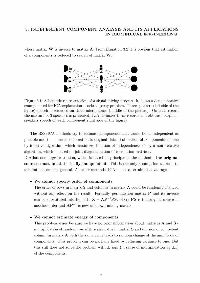
X = AS, (3.1)
where X is a mixture of source signals, A is the mixing matrix that characterizes environ-
ment, through which source signals pass, and S are the source signals. X and S get the size
n x m, where n is number of sources and m is length of record in samples. Mixture matrix
A is then of size n x n (in general A does not need to be square, but many algorithms
assume this ”property”). Figure 3.1 shows schematic representation of the mixing process.
Components can be obtained using the following expression:
S = A−1X = WX, (3.2)
5
3. INDEPENDENT COMPONENT ANALYSIS AND ITS APPLICATIONSIN BIOMEDICAL ENGINEERING
where matrix W is inverse to matrix A. From Equation 3.2 it is obvious that estimation
of a components is reduced to search of matrix W.
Figure 3.1: Schematic representation of a signal mixing process. It shows a demonstrativeexample used for ICA explanation - cocktail party problem. Three speakers (left side of thefigure) speech is recorded on three microphones (middle of the picture). On each recordthe mixture of 3 speeches is presented. ICA de-mixes these records and obtains ”original”speakers speech on each component(right side of the figure)
The BSS/ICA methods try to estimate components that would be as independent as
possible and their linear combination is original data. Estimation of components is done
by iterative algorithm, which maximizes function of independence, or by a non-iterative
algorithm, which is based on joint diagonalization of correlation matrices.
ICA has one large restriction, which is based on principle of the method - the original
sources must be statistically independent. This is the only assumption we need to
take into account in general. As other methods, ICA has also certain disadvantages:
• We cannot specify order of components
The order of rows in matrix S and columns in matrix A could be randomly changed
without any effect on the result. Formally permutation matrix P and its inverse
can be substituted into Eq. 3.1: X = AP−1PS, where PS is the original source in
another order and AP−1 is new unknown mixing matrix.
• We cannot estimate energy of components
This problem arises because we have no prior information about matrices A and S -
multiplication of random row with scalar value in matrix S and division of competent
column in matrix A with the same value leads to random change of the amplitude of
components. This problem can be partially fixed by reducing variance to one. But
this still does not solve the problem with ± sign (in sense of multiplication by ±1)
of the components.
6

In many ICA algorithms it is assumed that the data is centred and whitened.
Without loss of generality data could be centred:
x = y − Ey, (3.3)
where y is original data, E is expectation of data and x is centered data.
Whitening can be expressed by:
z = Vx = VAs, (3.4)
where z is whitened data, V is whitening matrix, x are mixed signals, A is mixing matrix
and s are desired source signals. Whitening operation removes correlation between signals
(note that correlation does not mean signals are independent)[2]. So correlation matrix of
pre-whitened data z is: EzzT = I.
3.2 Algorithms
This section provides the quick overview of fundamental ICA algorithms. These are as
follows:
• Fast Independent Component Analysis (FastICA)
• Fourth-Order Blind Identification (FOBI)
• Joint Approximate Diagonalization of Eigen matrices (JADE)
• Algorithm for Multiple Unknown Source Extraction based on EVD (AMUSE)
• Second-Order Blind Identification (SOBI)
Last two are not in proper sense ICA algorithms because they use only second order statis-
tics for source estimation. They estimate sources without using independence and they
solve BSS problem. These algorithms are widely used and are fundamental for other ICA
algorithms, therefore we mention them here.
7

3. INDEPENDENT COMPONENT ANALYSIS AND ITS APPLICATIONSIN BIOMEDICAL ENGINEERING
3.2.1 FastICA
FastICA algorithm was introduced by A. Hyvarinen [2] and it has a couple of modifications
based on target function. The introduced separation algorithm is based on maximizing non-
Gaussianity function.
This algorithm is based on the idea of using central limit theorem as a measure of
independence. Central limit theorem states: The distribution of a sum of independent
random variables tends toward a Gaussian distribution [10]. We can say that sum of
two independent random variables has a distribution that is closer to Gaussian than the
original two random variables. So maximizing non-Gaussianity leads to obtaining more
independent signals.
Non-Gaussianity can be measured in many ways. One way is to use kurtosis, which is
basically normalized version of fourth-order moment [8]:
kurt(y) = Ey4 − 3(Ey2)2, (3.5)
where y is a random signal. In our case y is normalized to unit variance so Equation 3.5
simplifies to kurt(y) = Ey4 − 3.
Basic algorithm needs gradient of kurtosis:
∂|kurt(wTz)|∂w
=
sign(kurt(wTz))
(E(wTz)4
∂w− 3(E(wTz)2)2
∂w
)=
sign(kurt(wTz))(4Ez(wTz)3 − 12E||wT ||2EwT
)=
4sign(kurt(wTz))[E z(wTz)3 − 3w||w||2].
(3.6)
Since w is calculated on unit sphere it needs to be normalized in every step. Last term
in previous equation changes only norm of w and thus it could be omitted. Using (3.6) an
algorithm was obtained:
1. w(t) = w(t− 1) + αsign(kurt(w(t− 1)Tz))E z(w(t− 1)Tz)3
2. w(t) = w(t)||w(t)||
The algorithm stops when the criterion on kurtosis reaches the desired value. In some
cases algorithm cannot reach specified desired value and in order to avoid infinite loops the
maximal loop count value is specified. This another stopping criterion enables algorithm
8
to work properly in difficult cases of similar signal distributions.
This basic algorithm is converted to fast fixed point algorithm by equating the gradient
of kurtosis with w. This means we obtain:
w = α[E z(wTz)3 − 3w||w||2], (3.7)
this equation suggests the following algorithm:
1. w(t) = α[E z(w(t− 1)Tz)3 − 3w(t− 1)]
2. w(t) = w(t)||w(t)||
3.2.2 FOBI
FOBI was proposed by Cardoso [11]. Consider quadratically weighted covariance matrix:
Ω = EzzT ||z||2, (3.8)
where z is pre-whitened data (see Eq. 3.4). Assuming data pre-whitened by ICA model it
follows:
Ω = EVAssT (VA)T ||VAs||2 = WTEssT ||s||2W, (3.9)
where VA is orthogonal and W = (VA)T . Using independence and unit variance (it does
not affect independence property of signals) of si matrix Ω stands for:
Ω = Wdiag(Es2i ||s||2)W =
Wdiag(Es2in∑j=1
s2j)W =
Wdiag(Es2i (s2i +n∑
j=1,j 6=i
s2j))W =
Wdiag(Es4i +n∑
j=1,j 6=i
Es2i Es2j)W =
Wdiag(Es4i + n− 1)W
(3.10)
Last equation shows that matrix W can be obtained by eigenvalue decomposition of
matrix Ω, which is decomposed to diagonal matrix consisting of the fourth order cumulants
of si and to orthogonal matrix W. This algorithm is the most efficient algorithm for ICA
9
3. INDEPENDENT COMPONENT ANALYSIS AND ITS APPLICATIONSIN BIOMEDICAL ENGINEERING
computation. FOBI has restriction, under which it works, namely all ICs must have
different kurtosis.
3.2.3 JADE
JADE [3] is an extension of FOBI. For whitened data, we can write fourth-order-cross-
cumulant tensor [8] as:
F (M) = E(zTMzzzT ) − 2M− tr(M)I, (3.11)
where M is eigenmatrix of cumulant, z is whitened data, I is unit matrix and tr(M) is
trace of matrix defined as:
tr(M) =n∑i
mii. (3.12)
Using (3.11) whitened correlation matrices can be defined alternatively as:
Ω = F (I) = E||z||2zzT − (n+ 2)I. (3.13)
Thus we can take a matrix M and replace matrix I in FOBI algorithm. This matrix would
have a linear combinations of cumulants of independent components as its eigenvalues. Now
we take more than one matrix, jointly diagonalize them and find the best result.
3.2.4 AMUSE
AMUSE [12] is an algorithm based on prior knowledge of data structure - data are signals
so they are time dependent. Let us denote lag-time covariance matrix as:
Cxτ = Ex(t)xT (t− τ), (3.14)
where x is vector of signals samples in time t and τ is lag-time.
An issue is that whitening data does not make data independent. The key for solv-
ing this problem is time-lagged covariance matrix, it can be used instead of high-order
statistics. Using this time-lagged covariance matrix gives us certain extra information to
estimate model, under certain conditions (samples of signals must be taken at the same
times and delayed correlations between different output signals vanish), and no high-order
10

information is needed. AMUSE uses the simplest case with only one time lag τ . Mostly
τ equals 1.
Consider whitened data z, then we can write for separating matrix W:
Wz(t) = s(t),
Wz(t− τ) = s(t− τ),(3.15)
where s are samples of signals in given time. Next consider lagged covariance matrix:
Czτ =
1
2
[Czτ + (Cz
τ )T]
(3.16)
Substituting z from (3.15) we get:
Czτ =
1
2WT
[Es(t)sT (t− τ)+ Es(t− τ)sT (t)
]W = WTCs
τW (3.17)
Due to the independence of signals si(t) the covariance matrix Csτ is diagonal, name it D
and rewrite previous equation:
Czτ = WTDW (3.18)
We can see that mixing matrix W is a part of eigenvalue decomposition of Czτ . From the
previous the following algorithm could be constructed:
1. Whiten data x to z
2. Compute eigenvalue decomposition of Czτ .
3. Rows of matrix W are given by eigenvectors.
3.2.5 SOBI
SOBI [1] is an extension of AMUSE algorithm. It uses more than one time-lag τ . The
diagonalization of all corresponding lagged covariance matrices is needed. Because the
covariance matrices got different eigenvalues, the formulation of functions, which express
the degree of diagonalization of matrices, is needed.
Simple function for measuring this degree of diagonalization of matrix M is:
off(M) =∑i 6=j
mij, (3.19)
11
3. INDEPENDENT COMPONENT ANALYSIS AND ITS APPLICATIONSIN BIOMEDICAL ENGINEERING
which is the sum of off-diagonal elements. Minimization of sum of off-diagonal elements in
several matrices is desired. Denoting S a set of chosen time lags τ criteria for minimization
stands for:
J (W) =∑τ∈S
off(WCxτW
T )2, (3.20)
where J (W) is objective function, W is separating matrix, Cxτ is time-lagged covariance
matrix of data x. Minimization under this constraint gives the estimation method and it
is performed by gradient descend method or simultaneous estimation of eigenvalue decom-
position for several matrices.
3.3 Example
As an example serves us mixture of ECG signal and uniformly distributed random noise
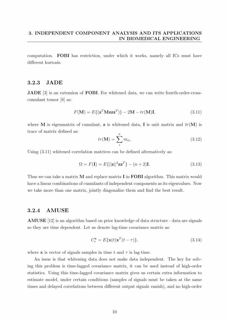
(Figure 3.2). Both signals are 10 seconds long. Signals have zero mean.
Figure 3.2: Example signals - upper is ECG signal, lower is uniformly distributed randomnoise.
12

Next both signals were mixed with randomly generated mixing matrix of size 2x2:
A =
(0.416494 0.374777
0.652350 0.885340
)(3.21)
The mixing result is shown on (Figure 3.3). It is obvious that noise totally corrupted
useful signal.
Figure 3.3: Mixture of signals from Figure 3.2. Noise corrupted signal completely. Bothsignals look similar, but they are different. If we look at the y-scale, we can observe thedifference in amplitudes.
Figure 3.4 shows resulting signals computed using estimated de-mixing matrix (result
of FastICA algorithm):
W =
(−0.184404 −0.002134
0.135876 0.002434
)(3.22)
The ECG signal is separated from noise. Results in Figure 3.4 show two disadvantages of
ICA. First - scale of resulting signal is the not same as of original data. Second - signal
13
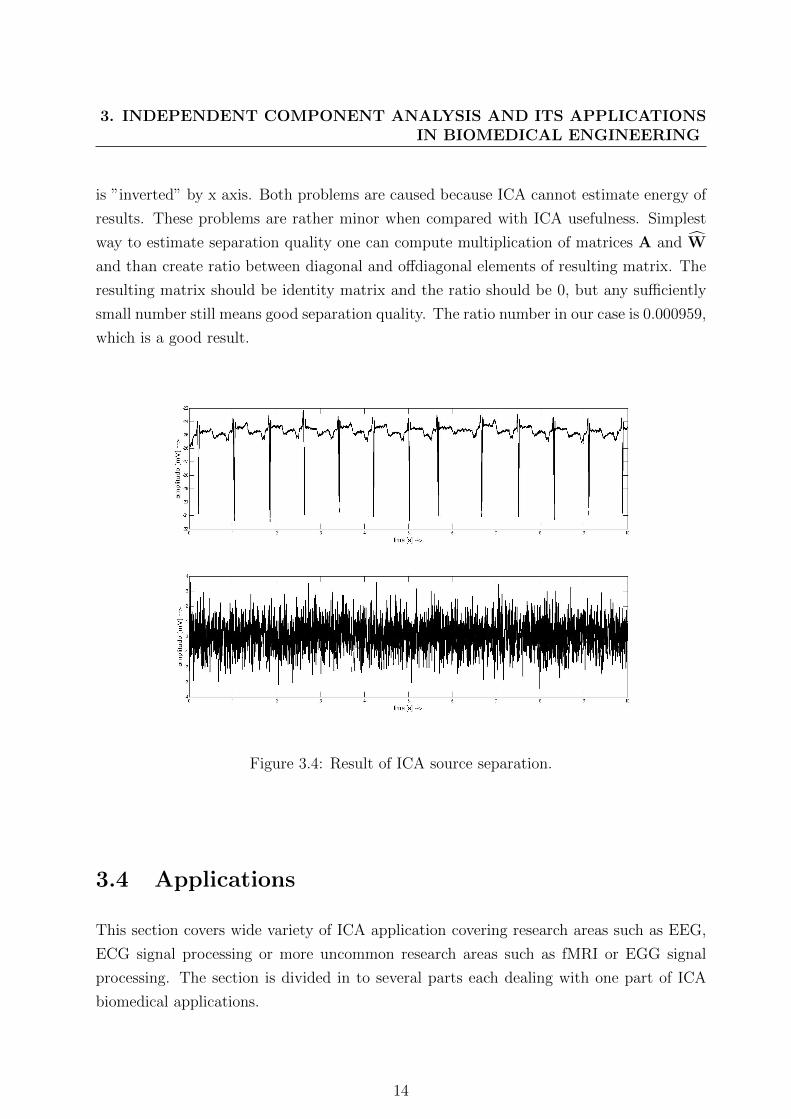
3. INDEPENDENT COMPONENT ANALYSIS AND ITS APPLICATIONSIN BIOMEDICAL ENGINEERING
is ”inverted” by x axis. Both problems are caused because ICA cannot estimate energy of
results. These problems are rather minor when compared with ICA usefulness. Simplest
way to estimate separation quality one can compute multiplication of matrices A and W
and than create ratio between diagonal and offdiagonal elements of resulting matrix. The
resulting matrix should be identity matrix and the ratio should be 0, but any sufficiently
small number still means good separation quality. The ratio number in our case is 0.000959,
which is a good result.
Figure 3.4: Result of ICA source separation.
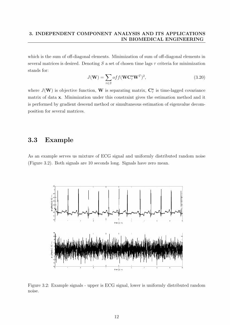
3.4 Applications
This section covers wide variety of ICA application covering research areas such as EEG,
ECG signal processing or more uncommon research areas such as fMRI or EGG signal
processing. The section is divided in to several parts each dealing with one part of ICA
biomedical applications.
14

3.4.1 ECG applications
At present only few ECG problems have been addressed. First problem, on which ICA
was applied, was artefact and noise removal. Pioneer work of Wisbec et al.[13] in
1998 deployed Fast ICA algorithm for breath artefacts removal. It presents preliminary
results. The ECG with artefacts were measured by a non-standard electrode system and
the method was tested on 10 records. The results are interesting - breathing artefact has
sub-Gaussian distribution and it was separated in one component.
In the same year Barros et al.[14] presented their work, where ICA was implemented
using neural networks. ICA gradient based algorithm was adapted for neural network with
self-adaptive step size calculation. Performance of the algorithm was measured on artificial
data created using MIT-BIH noise stress database, which contains 3 types of noise. The
data have been preprocessed by high pass filter. Resulting algorithm provides faster con-
vergence than standard ICA algorithm because of neural network deployment. Researchers
employed a measure for separation quality based on knowledge of mixing and demixing
matrix.
After these two works other researchers provided their solutions of noise removal prob-
lem based on ICA [5, 15, 16, 17, 18, 19, 20, 21, 22, 23, 24, 25, 26]. From them we selected
as the most interesting the following ones:
• He et al. [5] (2006) proposed an automatic method based on JADE algorithm. ECG
records used in this study were measured from three electrodes. The noise removal
technique for selection of noisy components is based on thresholding of kurtosis and
variance of components. The presented algorithm deals only with low amplitude
noises.
• Chawla et al. [15] (2008) deployed JADE algorithm on three channel ECG. No
comparable results were reported and the method is vaguely described, so the re-
producibility of research is limited. This work employed kurtosis and variance for
detection of noisy component in the same way as He et al.[5].
• Milanesi et al. [17] (2008) deployed FastICA and its modification for motion artefact
removal from holter recordings. They studied ICA for convolutive mixtures and con-
strained ICA. The study proposes two measures of noise elimination - error estimate
and correlation coefficients. It also employed statistical analysis of results obtained
on data from 9 patients, which are over 5 minutes long.
15

3. INDEPENDENT COMPONENT ANALYSIS AND ITS APPLICATIONSIN BIOMEDICAL ENGINEERING
• Chawla [20] (2011) presents and summarizes his latest work. This article contains
approach for noise removal presented by Milanesi et al. [17] and combines it with
author’s own PCA-ICA method. His technique is tested on CSE database [27].
• Acharyya et al. [22] (2010) deployed FastICA algorithm on MIT-BIH 3 channel ECG
database in order to remove artefacts from electrocardiogram. They developed an
algorithm for detection of component containing ECG based on Pearson correlation
coefficient. This approach does not deal with signal reconstruction and noise reduc-
tion. The ECG morphology changes were not discussed.
• DiPietroPaolo et al. [23] (2006) used TDSEP [28] algorithm in magnetocardiography
(MCG) analysis in order to reduce artefacts accompanying the MCG measurement.
For detection of artifact-containing components three rules have been used. They are
based on kurtosis, Pearson’s correlation coefficient and power spectra computation.
The authors used data from rest and exercise MCG.
• Oster et al. [25] (2009) applied JADE algorithm for detection of ECG in recordings
done during magnetic resonance imaging (MRI). The data are strongly corrupted by
MRI artefacts and the JADE algorithm in combination with wavelet transform is
able to extract ECG from the measured signals.
Further ICA application area in ECG processing is extraction of fetal ECG (fECG) from
records obtained by electrodes placed on mother body. Lathauwer et al. [29] presented
his pioneer work in 1994. Here blind separation of fECG was based on 4th order cumu-
lants. Researchers mathematically formulated problem of fECG estimation and presented
an example of extracted signals. In 2000 Lathauwer et al. [30] continued their work, ex-
tended database of records and discussed applicability of ICA on twin fECG. Cardoso [31]
worked on this problem in 1998 and showed usability of ICA in this problem. Zaroso et al.
[32] (2001) presented their method based on Givens rotations and compared their method
with method based on Adaptive Noise Canceller filter. For comparison they projected
extracted sources from the component domain back to the signal domain showing contri-
bution of different electrodes to fECG. In 2006 Sameni et al. [33] proposed their method
based on JADE algorithm for fECG extraction. The work tries to interpret independent
components and compares them with vector cardiogram. The researchers reported good
separation quality. Another application of JADE algorithm for fECG arose in 2009 when
Lee et al. [34] proposed their method for fetal magnetocardiogram (fMCG) extraction.
16
The automatic method selects components containing fMCG based on their kurtosis. In
2011 Camargo-Olivares et al. [35] presented their method based on multidimensional ICA
(MICA) approach. In order to get better fECG separation results, they estimated mater-
nal ECG and used it as another input to ICA method. In MICA algorithm different ICA
algorithms were used (JADE, FastICA, πCA), but the results were considered as similar.
Another application is extraction of atrial activity for atrial flutter analysis.
In 2000 Rieta et al. [36] applied ICA for QRST cancellation problem. They used syn-
thetic and real data for evaluation of algorithm effectiveness. Data were preprocessed by
notch and bandpass filter in order to remove noises before extracting atrial activity. Re-
searchers compared their method with other two standard methods. Research uncovered
that ICA based separation is better. In following years they extended their work and in
2003 presented FastICA application on extraction problem [37]. Finally in 2004 a summary
paper [38] was released comparing different algorithms (FastICA, JADE, AMUSE). This
paper also explains why ICA can be applied to this problem and introduced ordering of
separated components based on kurtosis. Presented analysis was done on 7 recordings of
different patients. Castells et al. [39] (2005) continued work of Rieta and they introduced
two stage separation algorithm based on FastICA and SOBI. Their paper also introduces
measures for estimation of separation quality - Root-Mean-Square error, correlation coeffi-
cients and degree of spectral content around main peak. Another work came from Zaroso
et al. [40] (2008). Paper proposed RobustICA method in framework defined by Castells
et al. [39]. Chang et al. (2010) extended the methodology proposed by Rieta and Castells
[36, 37, 38, 39]. They used JADE and SOBI algorithm in order to extract sources con-
taining atrial fibrillation and then used these sources for classification of atrial fibrillation.
The method increased specificity of atrial fibrillation classification. In 2011 Donoso et al.
[41] proposed method for atrial fibrilation extraction based on FastICA algorithm. They
reported preliminary results on data from 4 subjects. In the same year (2011) Taralunga
et al. [42, 43] applied JADE algorithm in combination with Event Synchronous Canceller
(ESC) on data from St. Peterburg DB [7]. They proved that ESC enhanced the JADE
algorithm ability for extraction of atrial activity.
Application of ICA in ECG signal classification is another biomedical research area
with increasing number of research papers. The first papers proposed by Yu et al. [44, 45]
in 2007 and 2008 presented an application of FastICA for beat classification combining
independent components and RR interval as a feature vector for different classification
systems. Yu et al. [46] proposed beat classification based on selection of independent com-
17
3. INDEPENDENT COMPONENT ANALYSIS AND ITS APPLICATIONSIN BIOMEDICAL ENGINEERING
ponents obtained by FastICA or JADE algorithm. The classification itself is done by SVM
[47] classifier on MIT/BIH Arrhythmia Database [48]. In 2011 Wu et al. [49] presented the
SVM based classification of ECG using features extracted by FastICA algorithm. Finally
in 2012 Huang et al. [50] proposed method for beat classification using ECG extracted
features combined with features obtained from independent components computed by Fas-
tICA algorithm.
Newly emerging application of ICA is ECG beat detection. The very first research has
been done by Wiklund et al. [51] in 2007. The researchers present beat detection method
for smart clothing application. The first step of method is preprocessing of data done by
FastICA algorithm. Next work proposed Chawla et al. [52] in 2008, who presented PCA-
ICA R-peak detection algorithm using JADE for denoising and PCA for estimation of data
segment. The paper discusses the method only. He continues his work and presented new
results in 2011 in [6].
Previous papers represent main-stream research in ECG applications of ICA, but there
are several other papers presenting other applications:
• Vetter et al. [53] (2000) presented application for measuring cardiac output based on
ICA applied on RR and QT intervals.
• Zhu et al. [54] (2008) presented a method for separation of interesting waveforms
into different components using 98 channel ECG data obtained from 6 subjects. The
method reported several components containing waves of interest. Content of other
components was not discussed.
• Owis et al. [55] (2002) applied convolutive ICA for classification of arrhythmias.
Independent components serve as input for k-NN, Bayes and minimum distance
classifiers. Data from MIT-BIH Arrhythmia database were cropped into 3 second
segments.
• Granegger et al. [56, 57] (2009) used JADE algorithm in application with data col-
lected on ICU patients during cardio pulmonary resuscitation (CPR). The work aims
at CPR artefact removal in order to enhance work of automatic external defibrillator.
The authors used algorithm based on kurtosis calculation.
• Ostertag et al. [58] (2011) proposed method for reconstructing ECG precordial leads
using FastICA algorithm. They developed a patient specific transformation, which
provides good results.
18

• Monasterio et al. [59] compare several BSS techniques and other separation tech-
niques for multilead T-wave alternans detection. They conclude that BSS algorithms
are not well suited for this type of task.
3.4.2 EEG applications
Electroencephalographic and magnetoencephalographic (EEG/MEG) signal processing us-
ing ICA was its first application in biomedical field. Many researchers use the same prin-
ciples in their work and thus we will review only selected papers representing the funda-
mentals of EEG ICA applications.
The first application of ICA was event related potential (ERP) detection and
analysis. The first works were published by Makeig et al. [60, 61] in 1996 and 1997. Re-
searchers used InfoMax algorithm [62] for separation of EEG activities (alpha, theta) for
detection of ERP. Experiments were performed on 10 recordings containing 30 minutes of
EEG. Makeig’s work was extended by Jung et al. [63, 64] (2000, 2001). Database used for
ERP analysis contains 50 patient recordings and researchers used ERP image technique for
evaluation of separated components. Vigario et al. [65] (2000) applied FastICA algorithm
to EEG data for ERP and ocular artefacts detection and removal. This paper summed up
their research and did not contain results. In 2003 Richards [66] used extended Infomax
algorithm [62] for source localization from 128 lead EEG measurements. The algorithm
was tested on 5 simulated datasets and the author concludes that ICA is better for source
localization task than PCA. Two years later Debener et al. [67] (2005) used RUNICA al-
gorithm within the EEGLab [68] framework in order to detect auditory ERP. The patients
were split into two groups differing with strength and frequency of tones, which were played
to them. The algorithm clustered components obtained from RUNICA into two groups
corresponding to patients groups. In 2008 Debener et al. [69] used the Infomax algorithm
for source localization in patient with cochlear implant during the auditory ERP trial.
They showed that cochlear implant patient used the same sources for resolving auditory
events as ”normal” patients. Liu et al. [70] in 2011 presented comparison of several ICA
algorithm for ERP detection in presence of noise. They conclude that SOBI algorithm
outperforms all other ICA algorithms in solving ERP task. Finally Chen et al. [71] (2012)
used Infomax for ERP extraction. The identification of independent components contain-
ing ERP is based on standard deviation computation.
Artefact removal in EEG by ICA was first reported by Vigario [72] in 1997. Fas-
tICA algorithm was applied on simulated and real children data preprocessed by bandpass
19

3. INDEPENDENT COMPONENT ANALYSIS AND ITS APPLICATIONSIN BIOMEDICAL ENGINEERING
filter. Research reported separation of ocular artefacts and K-complexes in different com-
ponents. Following first paper in this area several others appeared [73, 74, 75, 76, 77, 78,
79, 80, 81, 82, 83, 84, 85, 86, 87, 88, 89, 90]. We selected the most interesting ones:
• Tang et al. [73, 74] (2002) applied SOBI algorithm on 122 channel MEG data in
source localization and artefact removal issues. The data from 4 patients were first
preprocessed and then components were obtained. The papers explores possibilities
of SOBI usage in MEG data processing.
• Ossadtchi et al. [75] (2002) used Infomax algorithm for preprocessing during epileptic
spikes localization task. Components with artefact activity were discarded for next
steps of the algorithm. The algorithm was tested on 4 patients and provides efficient
way to deal with epileptic spikes detection.
• Poree et al. [76] (2006) used FastICA for hypnogram estimation from EEG, EOG
and EMG data obtained from 14 patients. Researchers reported very good results in
their study.
• Hu et al. [77] (2007) deployed FastICA algorithm for identification and removal of
scalp reference signal in intracranial recordings of three patients. The method for
selection of relevant components is fully automatic.
• Joyce et al. [78] (2004) proposed automatic method for EOG artefacts reduction in
EEG data. They employed SOBI algorithm for independent components estimation.
• McMenamin et al. [79] (2011) validated approach of ICA applying for EMG artefact
removal from EEG data. Infomax algorithm was used for estimation of independent
components. Researchers concluded that ICA can deal with strong artefacts, but it
could not be used as only one noise removal technique.
• Le Van et al. [82] (2006) deployed FastICA in combination with Bayess classifier for
detection of epilepsy seizures. Researchers used wide variety of features computed on
independent components in order to identify correct epileptic seizure components.
• Cao et al. [83] (2003) developed a new algorithm for high-level additive noise ex-
traction from EEG signals. Researchers used variation of EASI algorithm [91], which
works very efficiently with sub- and super-Gaussian distributions.
20

• Milanesi et al. [84] (2008) developed a modification of FastICA algorithm for dealing
with convolutive mixtures. The key idea is that convolution changes into linear
mixing in frequency domain and ICA could estimate sources as usually. The FastICA
needs to be adapted for dealing with complex values. The paper show interesting
result obtained by the application of algorithm to 9 EEG recordings.
• Korhonen et al. [86] (2011) used FastICA modification for large muscle artefacts
removal from transcranial magnetic stimulation (TMS). The TMS artefacts have
been completely removed by the applied algorithm.
• Ma et al. [87] (2011) used SOBI and Infomax algorithm for detection of EOG artefact
using comparison of components with artefact pattern. The detection of components
with EOG is based on Euclidian distance and it is patient and threshold dependent.
• Cong et al. [90] (2010) employed FastICA modification for de-noising of EEG. The
key idea of modification lies in re-computation of de-mixing matrix after each learning
step according to filtering of components. The final de-mixing matrix is then de-
mixing and denoising matrix. Proposed method was tested on 102 patients.
Preceding two research areas represent a main stream research in EEG ICA applications.
Following papers represent the other published works dealing with EEG using ICA in
other than previous applications:
• Zhukov et al. [92] (2000) proposed a method for multiple source localization based on
ICA. They used simulated 32 channels data for validation of the approach. Results
proved that ICA is able to extract extra information from the data.
• Tang et al. [93] (2005) applied SOBI algorithm on 128 channel EEG in order to test
its abilities on high-density EEG. The paper concludes that SOBI algorithm could be
used in several applications such as noise reduction, neuronal sources extraction, SNR
improvement in somatosenzory evoked potentials or for source activity localization.
• Cichocki et al. [94] (2005) used AMUSE algorithm for detection of early stages of
Alzheimer’s disease.
• Swan et al. [95] (2011) used Infomax algorithm for blink artefact removal during
Deep Brain Stimulation of the subthalamic nucleus within the Parkinson’s disease
patients.
21
3. INDEPENDENT COMPONENT ANALYSIS AND ITS APPLICATIONSIN BIOMEDICAL ENGINEERING
• De Lucia et al. [96] (2008) deployed FastICA algorithm for epileptic spikes detection.
The researchers used independent component domain features for classification task
using Bayess classifier.
• Selvam et al. [97] (2011) used Wavelet-ICA algorithm (Wavelet transform + SOBI
algorithm) for detection of brain tumors from 19 lead EEG. Again researchers used
features computed on independent components for classification using multilayer feed
forward neural network.
3.4.3 Other biomedical applications
This part deals with minor biomedical research areas, in which has been BSS/ICA methods
used.
3.4.3.1 EMG applications
Electromyography (EMG) is another research area, where ICA has been successfully de-
ployed. There are several papers describing research efforts in processing EMG using ICA:
• Costa Jr. et al. [98] (2010) employed FastICA algorithm in order to remove ECG
artefact from EMG recording of lumbar muscles. He reported two approaches - one
failed in separation of ECG and EMG and second based on using time-delayed signals
was successful in separating activities.
• Mak et al. [99] (2010) used also FastICA algorithm for removal of ECG from trunk
muscle surface EMG. Researchers tested their approach on simulated data and re-
ported good separation results.
• Ahsan et al. [100] (2010) reviewed the applications of ICA in EMG signal processing
in context of improvement of quality of live for elderly people.
• Ren et al. [101] (2010) deployed ICA for extraction of motor units activities within
the EMG recording.
• Farina et al. [102, 103] (2004,2008) applied SOBI algorithm for separation of two
muscle activities during the force-varying task.
• Subasi et al. [104] (2010) used FastICA algorithm for dimension reduction during
the detection of muscle fatigue task. Artificial neural network was deployed for
classification task and ICA served as preprocessing step.
22

• Willigenburg et al. [105] (2012) deployed FastICA for ECG removal from EMG
recordings. Researchers compared ICA technique to high-pass filtering and adaptive
filtering and concluded that ICA performs better in several cases. The performance
of ICA algorithm strongly depended on type of data.
3.4.3.2 fMRI and other medical image processing applications
Another application fields of ICA are functional Magnetic Resonance Imaging (fMRI) and
Magnetic Resonance Spectroscopy (MRS). In last decade there were several papers pub-
lished in this field:
• McKeown et al. [106, 107] (1998) first used ICA in fMRI application. Researchers
used Infomax and JADE algorithms for detection of region of interest in fMRI images.
ICA increased accuracy of separation and localization of sources.
• Calhoun et al. [108] (2003) published review paper comparing performances of JADE,
FastICA and Infomax algorithms. Researchers used their own data for the evalua-
tion of the ICA algorithms. The comparison criteria was based on Kullback-Leibler
divergence [109].
• Pulkkinen et al. [110] (2005) published a work about application of FastICA algo-
rithm on MRS data. FastICA was used for tumour detection.
• Debener et al. [111] (2006) written an overview of methods for analysis of simul-
taneously recorded EEG and fMRI. Researchers mentioned ICA as one method for
separation of non-brain signals from EEG during the trial.
• Wang et al. [112] (2012) presented Fast-FENICA algorithm for detection of func-
tional networks in fMRI data. ICA increased the detection rate.
• Rodriguez et al. [113] (2012) used Infomax algorithm for denoising fMRI data. The
approach was tested on 16 patient and the noise reduction was increased.
• Zhang et al. [114] (2012) employed Infomax and ICASSC (modification of FastICA)
algorithms in fMRI stop signal task. BSS/ICA increased ability to localize cognitive
centres within the brain in all 59 patients.
• Lei et al. [115] (2011) used FastICA algorithm for blink removal in EEG and region
detection in fMRI during the analysis of EEG-fMRI analysis of subcortical regions
of brain.
23

3. INDEPENDENT COMPONENT ANALYSIS AND ITS APPLICATIONSIN BIOMEDICAL ENGINEERING
• Kim et al. [116] (2011) employed FastICA for detection of regions during fMRI
semantic decision task. Research reported increased number of regions detected.
• Moeller et al. [117] compared ICA with General Linear Model analysis in application
within EEG-fMRI recordings. Both methods have similar results.
3.4.3.3 Abdominal phonograms application
In year 2009 Jimenez-Gonzalez et al. [118] initialized research of abdominal foetal phono-
grams using Single-channel Independent Component Analysis (SCICA) which combines
ICA algorithm TDSEP with back projection of components in order to obtain informa-
tion from a single channel recording. Research was extended and the deeper analysis of
resulting components [119, 120].
3.4.3.4 EGG applications
Another minor application field of ICA is EGG signal processing. There have been several
papers published in this research area:
• Wang et al. [121] (1997) applied neural network implementation of FastICA for
detection of EGG activity within the recording contaminated by respiratory, motion
and ECG artefacts.
• Peng et al. [122] (2007) compared FastICA with ICA with reference algorithm for
extraction of slow gastric wave. Both methods increased the probability of correct
extraction of gastric wave.
• Mika et al. [123] used FastICA algorithm for extraction of normogastric rhythm from
the EGG measurement.
3.4.3.5 Measuring HR and temperature from video
Finally ICA is used for analysis of video sequences in order to estimate heart rate (HR)
by Poh et al. [124] in 2010. ICA was able to extract enough information from RGB video
images of face to estimate the HR correctly. Another paper from this interesting research
area comes from Tsouri et al. [125] (2012). ICA was able to extract pulse rate from video
using constrained ICA.
24

Chapter 4
Electrocardiography and signal
processing
Although the main focus of the thesis is on ICA application to ECG signals, we also need
to describe briefly the heart and its function. This chapter is written for this purpose
- it summarizes basic facts about heart anatomy, its function and the basics of ECG
measurement and showing the most common lead system used in electrocardiography. All
figures used in this section are from [126], which provides every picture freely available for
any use.
4.1 Anatomy and function of human heart
The heart (Fig. 4.1) is an organ, which pumps oxygenated blood throughout the body to
important organs and deoxygenated blood to lungs. It can be understood as two separate
pumps - one pump (left) pumps the blood to peripheral organs, and second pump (right)
pumps the blood to lungs.
Left and right sides of the heart consist of two chambers - an atrium and a ventricle.
For controlling of the blood flow there exist four valves - tricuspid, pulmonary, mitral and
aortic. The mitral valve separates left atrium and ventricle and the tricuspid valve sep-
arates right atrium and ventricle. Pulmonary valve control the blood flow from heart to
lungs and the aortic valve directs blood to the body circulation system.
Walls of the heart are formed by cardiac muscle (myocardium). This muscle is responsi-
ble for the mechanical work done by the heart (= pumping the blood). For controlling the
pumping process specialized muscle cells that conduct electrical impulses evolved. These
25

4. ELECTROCARDIOGRAPHY AND SIGNAL PROCESSING
impulses are called action potential and they are responsible for forming the ECG waveform
on the body surface.
Figure 4.1: Basic heart anatomy schema - there are four chambers, two on the left (rightheart) side responsible for pumping the blood to lungs and two on the right (left heart)responsible for pumping the blood to body. Picture used with permission from [126].
In order to distribute oxygen to whole body human heart never stops. It works in
periodic cycles. A cycle works as follows: Deoxygenated blood flows through supererior
vena cava to the right atrium. When the atrium is contracted, blood is pumped to the
right ventricle. From the right ventricle the blood flows through pulmonary artery to
the lungs. Lungs remove carbon dioxide from blood cells and replace it with oxygen.
Oxygenated blood returns to the left atrium and after another contraction it is pumped
to the left ventricle. Finally the blood is forced out of the heart through aorta to the
systemic circulation. The contraction period is called systole, during which the heart fills
with blood. The relaxation period is called diastole. From electrical point of view the cycle
has two stages - depolarization (activation) and repolarization (recovery).
26
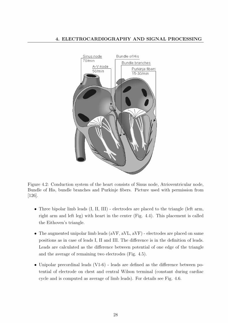
4.2 The conduction system of the heart
To maintain the cardiac cycle the heart developed a special cell system for generating
electrical impulses and by these impulses mechanical contraction of the heart muscle is
ensured. This system is called conduction system (Fig. 4.2). It conveys impulses rapidly
through the heart. Normal rhythmical impulse, which is responsible for contractions, is
generated in the sinoatrial (SA) node. Then, propagates to the right and left atrium and
to the atrioventricular node (AV). The impulse is delayed in the AV node in order to allow
proper contraction of the atria. Thus all blood volume in the atria is forced out to the
ventricles before its contraction. Atrium and ventricles are electrically connected by bundle
of His. From here, the impulse is conducted to the right and left ventricle. The pathway
to the ventricles is divided to the left bundle branch and right bundle branch. Further, the
bundles ramify into the Purkinje fibers that diverge to the inner sides of the ventricular
walls.
The primary pacemaker of the heart is the sinoatrial node. However, other specialized
cells in the heart (AV node, etc.) can also generate impulses but with lower frequency.
If the connection from the atria to the atrioventricular node is broken, the AV node is
considered as the main pacemaker. If the conduction system fails at the bundle of His, the
ventricles will beat at the rate determined by their own region. All cardiac cell types have
also different waveform of their action potentials (Fig. 4.3).
4.3 Generation and recording of ECG
Human body is a good electrical conductor, hence electrical activity of the heart can be
measured using surface electrodes. Electrodes record the projection of summary resul-
tant vector, which describes the main direction of electrical impulses in the heart. The
projection is named electrocardiogram. Different placement of electrodes provides spatio-
temporal variations of the cardiac electrical field. The difference between a pair of elec-
trodes is referred to as a lead. A large amount of possible lead systems has been invented;
depending on a diagnostic purpose, a lead system is chosen and electrodes placed on accu-
rate position. The most commonly used system is standard 12-lead ECG system defined
by Einthoven [127]:
27
4. ELECTROCARDIOGRAPHY AND SIGNAL PROCESSING
Figure 4.2: Conduction system of the heart consists of Sinus node, Atrioventricular node,Bundle of His, bundle branches and Purkinje fibers. Picture used with permission from[126].
• Three bipolar limb leads (I, II, III) - electrodes are placed to the triangle (left arm,
right arm and left leg) with heart in the center (Fig. 4.4). This placement is called
the Eithoven’s triangle.
• The augmented unipolar limb leads (aVF, aVL, aVF) - electrodes are placed on same
positions as in case of leads I, II and III. The difference is in the definition of leads.
Leads are calculated as the difference between potential of one edge of the triangle
and the average of remaining two electrodes (Fig. 4.5).
• Unipolar precordinal leads (V1-6) - leads are defined as the difference between po-
tential of electrode on chest and central Wilson terminal (constant during cardiac
cycle and is computed as average of limb leads). For details see Fig. 4.6.
28

Figure 4.3: Schematic representation of ECG waveform generation by summing of differentaction potentials. Picture used with permission from [126].
Figure 4.4: Schematic representation of Einthoven triangle electrode placement. Pictureused with permission from [126].
29

4. ELECTROCARDIOGRAPHY AND SIGNAL PROCESSING
Figure 4.5: Schematic representation of augmented limb leads calculation. Picture usedwith permission from [126].
Figure 4.6: Precordial leads electrodes positions. Picture used with permission from [126].
30

4.3.1 ECG wave form description
As we mentioned earlier ECG wave is formed as a projection of summarized potential
vector of the heart. ECG wave has several peaks and ”formations”, which is useful for its
diagnosis (Fig. 4.7). These are:
• P wave - Represents the wave of depolarization that spreads from the SA node
throughout the atria, and is usually 0.08 to 0.1 seconds (80-100 ms) length.
• PR interval - Reflects the time the electrical impulse takes to travel from the sinus
node through the AV node and entering the ventricles. Usually 120 to 200 ms long.
• PR segment - Corresponds to the time between the end of atrial depolarization to
the onset of ventricular depolarization. Last about 100 ms.
• QRS complex - Represents ventricular depolarization. The duration of the QRS
complex is normally 0.06 to 0.1 seconds.
• Q wave - Represents the normal left-to-right depolarisation of the interventricular
septum.
• R wave - Represents early depolarization of the ventricles.
• S wave - Represents late depolarization of the ventricles.
• S-T segment - Following the QRS is the time at which the entire ventricle is de-
polarized and roughly corresponds to the plateau phase of the ventricular action
potential.
• Q-T interval - Represents the time for both ventricular depolarization and repolariza-
tion to occur, and therefore roughly estimates the duration of an average ventricular
action potential. This interval can range from 0.2 to 0.4 seconds depending upon
heart rate.
• T wave - Represents ventricular repolarization and is longer in duration than depo-
larization.
31
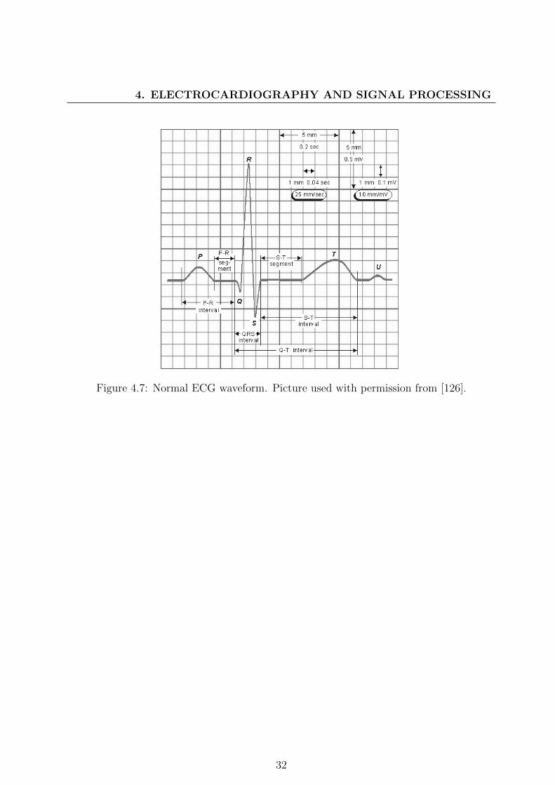
4. ELECTROCARDIOGRAPHY AND SIGNAL PROCESSING
Figure 4.7: Normal ECG waveform. Picture used with permission from [126].
32

Chapter 5
Independent Component Analysis for
ECG de-noising: proposed method
5.1 Introduction
During recording of ECG the recorded signal is disturbed with interferences coming from
both technical and biological sources, thus the very first step of any ECG analysis is noise
reduction. Any noise reduction technique must be very accurate in order to preserve as
much information in data as possible. At present many ECG applications are connected
with holter measurement and telemedicine applications. In these cases various types of
uncommon noise disturb the useful signal. Many techniques are used for noise reduction in
ECG - current most used techniques are traditional filtering (basic digital filters, adaptive
filters) and wavelet filtering. As we presented in Chapter 3 there are still many problems in
current algorithms, which result in imperfect noise reduction or distortion of useful signal.
These facts led us to the idea of a new algorithm proposal satisfying our requirements on
de-noising.
We developed an algorithm for solving the problem of electrode cable movement (ECM)
artefact, which appears during ECG measuring with a holter device. This artefact mimics
normal ECG activity in frequency domain (Fig. 5.1) thus standard filtering cannot be
used because it also removes ECG. Our method deploys the JADE algorithm, which is a
well-known BSS algorithm based on joint diagonalization of cumulant matrices. The JADE
is able to separate ECM artefact from ECG activity in component domain. Combination
of JADE and basic classifier for noise component detection provides us with framework
capable to deal with ECM artefact.
33

5. INDEPENDENT COMPONENT ANALYSIS FOR ECG DE-NOISING:PROPOSED METHOD
The work described here is based on several other authors publications [128, 129] and
it is available at http://bio.felk.cvut.cz/~kuziljak/.
Figure 5.1: Power spectra of noise signal and ECG data. Both spectra are overlapping andnoise is mimicking normal ECG signal.
5.2 Proposed Algorithm
As we said before the main concept of our algorithm lies in performing JADE separation
algorithm on ECG signal containing noise. The resulting components can be divided into
two groups - components containing mostly noise and components containing mostly ECG
activity. Because of well-known permutation indeterminacy of ICA the order of components
is random and thus one needs to develop a detection algorithm for identification of noise
components. The algorithm work-flow is shown in Algorithm 1. Next we will describe each
important step of the algorithm.
Algorithm 1 De-noising algorithm
Input: ECG signal1: Pre-process (subtract mean of signals)2: Estimate components using JADE3: Compute features4: Detect noisy components using CART5: Set noisy components to zero6: Transform components back to signal domain7: Post-process
Output: Filtered ECG signal
34

5.2.1 Preprocessing and component estimation
Very first step of the algorithm contains mean subtraction. Next independent components
are estimated using JADE algorithm. The algorithm is based on joint diagonalization of
fourth order cross-cumulant tensor and is explained in the section 3.2.3.
5.2.2 Feature computation
In order to detect noise containing components the algorithm computes set of features for
each component. These features are chosen based on prior knowledge of data. The selected
feature set contains these features:
• Mean of component samples – mean is defined as:
x =1
N
N−1∑i=0
xi, (5.1)
where xi is the i-th sample selected from component x with length N .
• Variance of component samples – unbiased estimate of variance is defined as:
var(x) =1
N − 1
N−1∑i=0
(xi − x)2. (5.2)
• Kurtosis of component samples – kurtosis is normalized version of fourth-order mo-
ment:
kurt(x) = Ex4 − 3(Ex2)2, (5.3)
where E is the expectation of component x. Kurtosis has been chosen because it
was shown that ECG has super-Gaussian distribution [5, 6, 4].
• Correlation coefficient of component with 50 Hz sinus wave and p-value of hypothesis
of no correlation. Pearson correlation coefficient is defined as [130]:
ρx,y = corr(x, y) =cov(x, y)
σxσy=E(x− x)(y − y)
σxσy, (5.4)
where cov(x, y) is covariance of signal x and y and σx,σy are their standard deviations
defined as:
σx =√var(x). (5.5)
35

5. INDEPENDENT COMPONENT ANALYSIS FOR ECG DE-NOISING:PROPOSED METHOD
P-value for hypothesis of no correlation between signals is estimated using t value
[130]:
t =r
sr, (5.6)
where r is correlation coefficient and sr is standard error of correlation coefficient:
sr =√
1−r2n−2 .
• Standard deviation of peak-to-peak distances and number of detected peaks – this
feature is used, because ECG has periodical rhythm with dominant R peaks, thus
we can asume that ECG component standard deviation is smaller than standard
deviation of noisy component deviation, which has no dominant peaks by its nature.
Peaks were detected using Pan-Tomkins detection algorithm (see section 7.3.1.2).
For the same reason the number of peaks detected in the recording normalized by
the length of recording were used.
5.2.3 Noise component detection using CART algorithm
Features discussed in previous section are then classified using a classification tree, which
has been learned with CART algorithm using Gini’s impurity index [131] using annotated
components data obtained by application of JADE to recordings from MIT/BIH Arrhyth-
mia Database. The algorithm constructs a binary tree by splitting data into left and right
node using maximalization of decrease of impurity defined as:
id(t) = i(t)− pLi(tL)− pRi(tR), (5.7)
where i(t) =∑J
j=1 p(j|t)(1 − p(j|t)) = 1 −∑J
j=1 p2(j|t) with j = 1, 2, ..., J and J is the
number of items in the training set, pL(pR) is the proportions of data in left (right) node.
Gini’s impurity index is the measure how often a randomly chosen element from the set
would be incorrectly labelled if it were randomly labelled with one label in the set according
to the distribution of the labels in the set. It reaches zero when all cases fall into one
category.
After constructing the CART tree we deployed 10-fold cross-validation in order to estimate
best level for tree pruning. The error from cross-validation is shown on Fig. 5.2 with
selected number of nodes. Pruning is performed as removal of n lover levels of binary
tree according to selected level from cross-validation. Resulting tree is shown on Fig.5.3.
36

We can see, that only two features were selected for classification of components, namely
kurtosis and standard deviation of RR intervals.
Figure 5.2: Result of 10-fold cross-validation used for estimation of tree pruning level. Thebest choice is selected by error and number of terminal nodes.
Figure 5.3: Resulting binary classification tree for noise component detection. (0 – ECGcomponent, 1 – Noise component)
5.2.4 Removing noisy components and backward transform
Components identified as noise containing are removed by setting their values to zero.
This leads to removal of these components from all ECG signals in signal domain after
backward transform. Zeroing can be performed also as zeroing corresponding column in
37

5. INDEPENDENT COMPONENT ANALYSIS FOR ECG DE-NOISING:PROPOSED METHOD
mixing matrix. This leads to the same result. The backward transform is performed
by multiplying components with the mixing matrix, which is computed as inverse to the
de-mixing matrix.
5.2.5 Postprocessing
After removal of components containing noise and transforming them back to the signal
domain the high frequency noise sometimes appears. Therefore the final step contains the
low pass filter with first zero at 117 Hz, delay 5 samples and gain 0.93. Filter frequency
response is shown on Fig. 5.4.
Figure 5.4: Frequency response of postprocessing low pass filter
38

5.3 Algorithm summary
The algorithm works with signal in predetermined way. First the mean value is subtracted
from the data, then independent components using JADE algorithm are estimated. After
estimation of components the algorithm points out those containing noise corrupting the
ECG using decision tree and finally the components with noise are set to zero. After zero-
ing data are projected back into signal domain and smoothing filter is applied.
Our method is efficient to deal with electrode cable movement artefact, for which it has
been developed, but its performance is good in presence of other noises. Its main disad-
vantage is wrong estimation of independent components, which could lead to reduction of
useful signal. This problem could be solved by applying different BSS/ICA algorithm.
Experiments with developed algorithm are described in Chapter 7 and the results are
presented in Chapter 8.
39

5. INDEPENDENT COMPONENT ANALYSIS FOR ECG DE-NOISING:PROPOSED METHOD
40

Chapter 6
Independent Component Analysis for
ECG beat detection enhancing:
proposed method
6.1 Introduction
Beat detection is the most fundamental task in electrocardiography, the positions of QRS
or ventricular beats serve as a basis for further analysis of electrocardiogram (ECG). It is
for example the most crucial task in heart rate variability (HRV) analysis, which analyses
RR interval changes during long periods of time in order to detect heart disorders. The ne-
cessity of correct detection arises with each application of QRS detection algorithms. Many
researchers proposed large variety of methods for solving this task, eg.[132, 133, 134, 135, 6].
The most commonly used in the real world applications are Pan-Tompkins QRS detection
algorithm [134] and its modification introduced by Hamilton and Tompkins [136] or Chris-
tov’s beat detection algorithm [135].
Pan-Tompkins and its modification Hamilton-Tompkins algorithms are based on patient-
specific detection threshold. The algorithms work on single lead ECG, which is modified
by set of preprocessing digital filters in order to enhance positions of QRS complexes.
More recently, Christov released his beat detection algorithm, which is based on com-
bined adaptive threshold and transformation of multidimensional ECG signal in to single
complex lead data. The complex lead transformation is more robust in presence of noise
corrupting useful ECG and the algorithm is more stable.
In many applications strong artefacts from biological or technical sources could appear
41
6. INDEPENDENT COMPONENT ANALYSIS FOR ECG BEATDETECTION ENHANCING: PROPOSED METHOD
and cause distortion of ECG signals. Many algorithms for beat detection perform much
worse in that case. Christov’s detection algorithm deals with noise that accompanies the
ECG by transforming the ECG signal to signal called complex lead, which enhances posi-
tion of QRS complex and reduces effect of common artefacts presented in ECG. But this
algorithm has its limits, too. We try to improve its performance by transforming ECG
signal to components using Independent Component Analysis (ICA).
In our study we used JADE [3], which is from the family of joint diagonalization based
ICA algorithms. It works with fourth order cross-cummulant tensor in order to obtain best
solution for BSS problem.
The offline algorithm successfully combines ICA method JADE with Christov’s beat
detection algorithm creating a signal called complex component, on which beats were de-
tected using combined adaptive threshold. The transformation to complex component is
similar to transformation to the complex lead, but it introduces one more step - inde-
pendent components estimation and selection of ECG activity. Thus the algorithm works
properly in presence of strong artefacts and noises.
This algorithm has been published in [137] and all source codes are available at http:
//bio.felk.cvut.cz/~kuziljak/.
6.2 Christov’s beat detection algorithm
The Christov’s beat detection algorithm has been developed in 2004 for purpose of beat
detection in multidimensional ECG. It combines information from every available ECG
signal (originally from orthogonal lead system X-Y-Z) in recording creating so-called com-
plex lead, which is used for detection of beat positions. This algorithm contains several
processing steps as it is shown on work-flow chart in Fig. 6.1. In the next subsection we
will describe each step in detail and at the end we will summarize the algorithm and its
efficiency.
42

Figure 6.1: Christov’s beat detection algorithm work-flow
6.2.1 Signal preprocessing
At the very first beginning ECG signals are processed by set of moving average (MA) filters
in order to remove power line interference and electromyographic noise:
• Power line interference moving average filter has first zero on frequency of power grid
(for EU is 50 Hz).
• Electromyographic moving average filter average over 28 ms interval of ECG signal,
which means that the first zero of the filter is around 35 Hz.
43

6. INDEPENDENT COMPONENT ANALYSIS FOR ECG BEATDETECTION ENHANCING: PROPOSED METHOD
We implemented both filters using recursive moving average defined by equation:
y[n] =x[n]
N− x[n−N ]
N+ y[n− 1], (6.1)
where y is the output signal, x is the input signal, n is the signal sample index and N is
the filter order. The main advantage of recursive filter is its smaller impulse response and
faster computation. On the other hand it could be unstable because it is an IIR filter. The
order of filter N can be computed using: N = fsfn
, where fs is sampling frequency and fn
is frequency of noise. The frequency response of filter is shown on Fig. 6.2 for fs=500 Hz
and fn=50 Hz.
Figure 6.2: Frequency response of recursive MA filter with first zero at 50 Hz for fs=500Hz.
44

6.2.2 Complex lead transform and post-processing filtration
The Christov’s beat detection algorithm is based on complex lead transform, which is
defined as follows:
X[n] =1
L
L∑j=1
|xj(n+ 1)− xj(n− 1)|, (6.2)
where X[n] is nth sample of signal calculated by complex lead transform, L is number of
measured ECG leads, xj(n) is nth sample from jth lead. The transform can be seen as
average difference calculated from all leads. Example of calculated complex lead is shown
in Fig. 6.3.
After complex lead transform the signal is filtered with MA filter with first zero around
25 Hz in order to suppress noise created by differentiation procedure in complex lead
transform. We again used the recursive MA implementation defined by Eq. 6.1.
Figure 6.3: Complex Lead (down) estimated from ECG (top)
45

6. INDEPENDENT COMPONENT ANALYSIS FOR ECG BEATDETECTION ENHANCING: PROPOSED METHOD
6.2.3 Combined adaptive threshold for beat detection
After the calculation of complex lead Christov employed combined adaptive threshold
MFR (see Fig. 6.4 for example) to find peaks, which correspond to beat positions. The
combined adaptive threshold is calculated as sum of three thresholds:
• Threshold M (adaptive steep-slope threshold) - reflects the amplitude of currently
detected beats
• Threshold F (adaptive integrating threshold) - reflects the presence of high frequency
noise in data and increases the combined threshold in that case
• Threshold R (adaptive beat expectation threshold) - intended to deal with heartbeats
of normal amplitude followed by beats with very small amplitude
Figure 6.4: Complex Lead with combined adaptive threshold MFR
6.2.4 Algorithm summary
The algorithm can work in two modes, namely real-time and pseudo-real-time, which
performs back search in case of no detection for long time. The algorithm is then pushed
back to last detected beat and starts search with lowered combined adaptive threshold.
Christov’s algorithm is robust against the presence of common noises (base line wander,
grid noise, EMG, etc.), because of MA filters used in preprocessing step of the algorithm.
On the other hand its performance in presence of non-standard noises is not sufficient.
The algorithm time performance is strongly affected by back search procedure and our
implementation in MATLAB needs around 6.4 seconds for 30 minutes 2-lead ECG to
process it.
46
6.3 Proposed algorithm
The main idea, which lies behind our proposed algorithm is the question - can we enhance
ECG activity before ECG is transformed into complex lead? The algorithm, which en-
hances the activity, should have extracted ECG from unknown mixture of ECG and other
signals (power line interference, breathing, etc.) and passed this extracted sources into the
complex lead transform.
The problem formulation contains the answer: use method for solving BSS such as
ICA to enhance ECG activity. The algorithm is then easily derived from preceeding one –
instead of using the ECG use selected components obtained by JADE algorithm and these
components transform into complex component (similar to complex lead). JADE preserves
overall spatial distribution of ECG activity and separates it from other activities. The al-
gorithm work flowchart is shown in Algorithm 2. At first the algorithm looks the same as
original Christov’s algorithm, however our change is hidden in the method for estimation
of complex lead - it is replaced with method for estimation of complex component.
Algorithm 2 QRS detection algorithm
Input: ECG signal1: Estimate complex component (see Alg. 3)2: Initialize thresholds (M, F, R) and their buffers3: for all samples of complex component do4: Update weights (M, F, R)5: if current sample > (M + F + R) (beat detected) then6: Update thresholds buffers and thresholds7: Store detected beat position8: else if beat is not detected for longer than twice average RR interval then9: Start detection again from last detected beat with lowered threshold10: end if11: end forOutput: Beat positions
6.3.1 Complex component transform
Complex component transform is our modification of complex lead transform described in
Christov’s original paper. The preprocessing using recursive MA filters remains the same.
The change lies in adding one more step between preprocessing and complex lead transform
– the component estimation step.
47

6. INDEPENDENT COMPONENT ANALYSIS FOR ECG BEATDETECTION ENHANCING: PROPOSED METHOD
As we mentioned above the algorithm used for component estimation is JADE. This
algorithm is fully described in Section 3.2.3. Here we will use only basic notation for ICA
formulation:
S = WX, (6.3)
where S are estimated components, W is demixing matrix and X are originally measured
ECG signals containing ECG and noise. After the components estimation we need to iden-
tify components containing ECG and noise. Because JADE is data driven transformation,
we never know, in which order and with which scale we obtain the estimated components.
In order to make the algorithm fully automatic, one needs to create a robust decision sys-
tem for detection of ECG activity.
Our decision algorithm is based on the observation that ECG has super-Gaussian dis-
tribution [5, 6, 4], thus the values of its kurtosis are larger than kurtosis values of other
presented signals (e. g., EMG is nearly Gaussian with kurtosis close to zero). Kurtosis is
defined as:
kurt(s) = Es4 − 3(Es2)2, (6.4)
where E is the expectation and s is one of estimated components. Now we need to
identify ECG components in S. Because JADE is data driven algorithm, we need to
normalize kurtosis in order to find general solution for component identification:
kurti =kurti∑N−1j=0 kurtj
, (6.5)
where kurti is kurtosis of one component and N is the number of components.
Using combination with sorting this normalization enables us to find components, whose
sum of kurtosis is larger than 75% of sum of all kurtosis values. All components found by
previous algorithm are identified as ECG activity and used for computation of complex
lead using transform defined by Eq. 6.2. After computation the recursive MA filter is
applied similarly to application in original algorithm and the resulting signal is returned
to main procedure. Algorithm for computing complex component transform is depicted in
Algorithm 3.
6.3.2 Algorithm summary
The algorithm works in pseudo-realtime mode, which contains search-back procedure for
identification of low amplitude beats.
48

Algorithm 3 Algorithm for complex component estimation
Input: ECG signal1: Pre-process input signal (Remove mean; Filter power line interference and muscle
artefacts)2: Estimate components using JADE algorithm3: Calculate kurtosis of components4: Normalize values of kurtosis with maximal kurtosis calculated5: Sort descending normalized values of kurtosis and permute components accordingly to
order of their normalized kurtosis6: Select first n components whose sum ≥ threshold7: Calculate complex component from n selected components8: Post-process complex component (Filter noise magnified by complex lead transforma-
tion)Output: Complex component
Our method adds one more step between filter preprocessing of original signals and
complex lead transform to original Christov’s algorithm. This step estimates the ECG
activity using JADE algorithm, which makes (see in next Chapters) the algorithm much
more robust in presence of strong noises and artefacts. Our implementation with JADE
algorithm is slower than original Christov’s algorithm. This is understandable and the
algorithm needs 7.6 seconds to complete the detection on 30 minutes 2-lead ECG.
49

6. INDEPENDENT COMPONENT ANALYSIS FOR ECG BEATDETECTION ENHANCING: PROPOSED METHOD
50

Chapter 7
Evaluation of proposed algorithms
This chapter summarizes the experiments with our proposed algorithms. First we will
speak about used data and procedure for creation of noise in them. Next the evaluation
criteria will be mentioned and the state-of-the-art algorithms will be discussed.
7.1 Data
7.1.1 Databases
In the evaluation experiments we used data freely available on MIT medical data storage
Physionet [7]:
• MIT-BIH Arrhytmia database [48] - contains 48 half-hour two channel ambulatory
ECG recordings digitized at 360 samples per second with 11-bit resolution over 10
mV range annotated by two or more cardiologist. All recordings contain modified
limb lead II. Second lead is usually modified lead V1, occasionally V2 or V5 and in
one instance V4. Four recordings contain paced beats.
• Normal Sinus Rhythm database [7] - contains 18 long-term two channel ECG record-
ings of subjects with no significant arrhythmias digitized at 128 samples per seconds
per channel with 12-bit resolution over 10 mV range.
• European ST-T database [138] - contains 90 long-term two channel ambulatory ECG
recordings of subjects with myocardial ischemia diagnosis or suspicion. The record-
ings were digitized at 250 samples per second with 12-bit resolution over 20 mV
range.
51

7. EVALUATION OF PROPOSED ALGORITHMS
• Long Term ST database [139] - contains 86 lengthy ECG recordings of 80 subjects
with significant ST changes. Individual recording contains two or three ECG leads.
Each signal was digitized at 250 samples per second with 12-bit resolution over ± 10
mV range.
• QT database [140] - contains 105 fifteen minutes two channel ECG recordings without
significant baseline wander or artefacts. Recordings were digitized at 250 samples per
second.
• MIT Long Term database [7] - contains 7 long term two or three channel ECG
recordings digitized at 128 samples per second.
• MIT-BIH ST Change database [141] - contains 28 ECG two channel recordings of
different length recorded during exercise and exhibiting ST depression. Signals were
digitized at 360 samples per second.
7.1.2 Simulated noise
According to [132] one can expect various kinds of noise presented in ECG. The most
common ones are:
• Power line interference - consists of 50 Hz (60 Hz in U.S.) pickup and its harmonics,
typical amplitude is up to 50 percent of peak-to-peak ECG amplitude
• Motion artifacts - transient baseline changes caused by changes in electrode-skin
impedance, the cause of impedance change is the slow motion of electrode on the
body surface, typical amplitude is up to 500 percent of peak-to-peak ECG amplitude
• Muscle contraction - generates artefactual millivolt-level potentials, its standard de-
viation is around 10 percent of peak-to-peak ECG amplitude
• Baseline drift and ECG amplitude modulation with respiration - is represented as
slow sinusoidal component at respiration frequency, its amplitude variation is 15
percent of peak-to-peak ECG amplitude and typical frequencies at 0.15 Hz to 3 Hz
In addition to these four noises we observed noise typically generated by electrode
cable movement during holter recording of ECG. This noise has large amplitudes up to
200 percent of peak-to-peak ECG amplitudes and typical power spectra with peaks at 1.5,
3.16, 6.3 and 8 Hz.
52
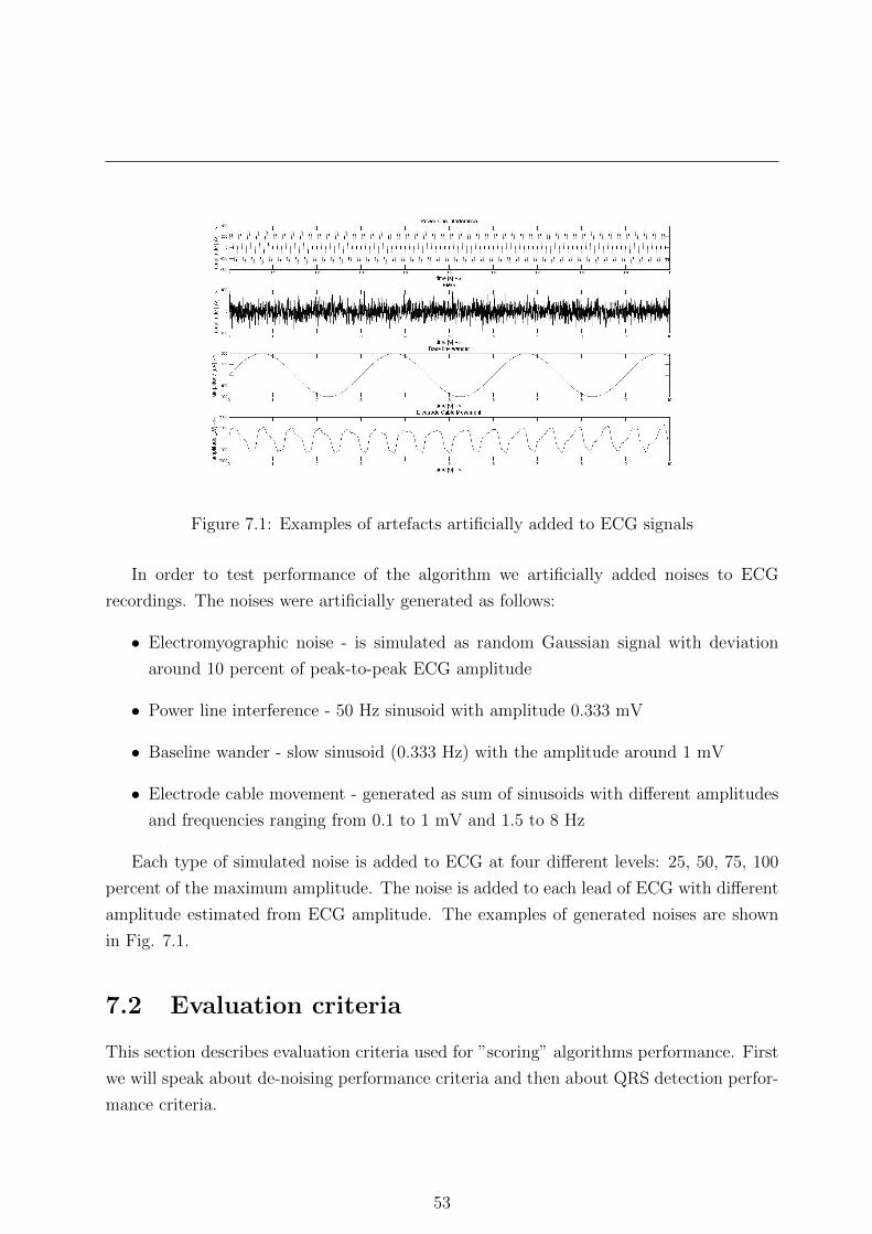
Figure 7.1: Examples of artefacts artificially added to ECG signals
In order to test performance of the algorithm we artificially added noises to ECG
recordings. The noises were artificially generated as follows:
• Electromyographic noise - is simulated as random Gaussian signal with deviation
around 10 percent of peak-to-peak ECG amplitude
• Power line interference - 50 Hz sinusoid with amplitude 0.333 mV
• Baseline wander - slow sinusoid (0.333 Hz) with the amplitude around 1 mV
• Electrode cable movement - generated as sum of sinusoids with different amplitudes
and frequencies ranging from 0.1 to 1 mV and 1.5 to 8 Hz
Each type of simulated noise is added to ECG at four different levels: 25, 50, 75, 100
percent of the maximum amplitude. The noise is added to each lead of ECG with different
amplitude estimated from ECG amplitude. The examples of generated noises are shown
in Fig. 7.1.
7.2 Evaluation criteria
This section describes evaluation criteria used for ”scoring” algorithms performance. First
we will speak about de-noising performance criteria and then about QRS detection perfor-
mance criteria.
53

7. EVALUATION OF PROPOSED ALGORITHMS
7.2.1 De-noising algorithm
For evaluation of de-noising algorithm we used Root-Mean-Square Error (RMSE), which
is good statistical index for case, when the original clear signal is known. RMSE is defined
as:
RMSE =
√√√√ 1
N
N−1∑i=0
(xi − yi)2, (7.1)
where xi is the i-th sample of original signal, yi is the i-th sample of filtered signal and
N is the number of samples in both signals. The values of RMSE close to 0 means that
filtering technique reduces noise efficiently with minimal changes in useful signal.
7.2.2 QRS detection algorithm
For evaluation of QRS detection algorithm we used standard statistical indices [142] Sen-
sitivity (Se), Positive predictivity (P+) and F-measure (F ), which are derived from three
parameters:
• Correctly detected beats (True positives=TP )
• Falsely detected beats (False positives=FP )
• Undetected beats (False negatives=FN)
Sensitivity is the parameter describing how many beats are correctly detected. Thus its
value is calculated using this equation:
Se =TP
TP + FN. (7.2)
Positive predictivity characterizes the algorithm in sense of the false detection of beats. Its
value is estimated as follows:
P+ =TP
TP + FP. (7.3)
The F-measure statistics is defined as harmonic mean of Sensitivity and Positive predic-
tivity:
F = 2Se ∗ P+
Se+ P+. (7.4)
All statistical indices range from 0 % (worst) to 100 % (best). The values over 95 % are
considered as good results.
54

7.3 Evaluation methods
This section provides detailed review of referential methods used for comparison with our
algorithms. Then we will describe the testing methodology.
7.3.1 State-of-the-art methods
Here we describe the state-of-the-art methods used for comparison with our methods. First
we will describe de-noising algorithms and then beat detection algorithms.
7.3.1.1 De-noising algorithms
Standard filtering algorithm is based on set of filters designed for suppression of each
common type of noise.
First deployed filter is single-frequency adaptive noise canceller (ANC) [143]. This type
of adaptive filter is based on LMS algorithm. The structure of filter is depicted on Fig.
7.2. The ANC filter has two inputs: measured distorted signal x and noise model signal u′.
Signal u′ is phase-shifted by π2
and both original u′ and shifted signal u′′ are multiplied by
weights computed by LMS algorithm. Signals are then summed up into signal u and added
to measured signal x with switched sign. Resulting error signal e is the desired filtered
signal and also serves as input to update LMS rule for weights w1 and w2. The update
equation has form:
w[n+ 1] = w[n] + µe[n]
[u′[n]
u′′[n]
], (7.5)
where w =
[w1
w2
]is weight vector, e is error signal, µ is convergence constant and u′, u′′
are input noise model signals.
Second filter in the set is notch filter for EMG artefact suppression. Notch filter is
band-stop IIR filter with transfer function [144]:
H(z) =1
2
(1 + a2)− 2a1z−1 + (1 + a2)z
−2
1− a1z−1 + a2z−2, (7.6)
where:
a1 =2 cos( 2πfn
fs)
1+tan( 2πBW2fs
),
a2 =1−tan( 2πBW
2fs)
1+tan( 2πBW2fs
),
(7.7)
55

7. EVALUATION OF PROPOSED ALGORITHMS
Figure 7.2: Single-frequency adaptive noise canceller
Figure 7.3: Notch filter frequency response for fn=33 Hz, BW=0.8 and fs=500 Hz
where fn is noise frequency, BW is bandwidth and fs is sampling frequency. Frequency
response of notch filter used in set is shown on Fig. 7.3.
56

Last filter deployed in the set is a median filter for suppression of baseline wander. The
algorithm of applying of median filter follows:
• Decimate ECG signal with decimation factor 20.
• Filter resulting signal with median filter of length 10.
• Interpolate filtered signal into original length with low-pass interpolation. Interpo-
lated signal represents base line wander.
• Subtract interpolated signal from original ECG.
Median filter removes any remaining parts of ECG remaining in data and the signal could
be considered as base line wander. Median filter is defined by equation:
y[n] = medianx[n+ i], i = 0, ..., N − 1, (7.8)
where y represents filtered signal, x represents original signal and N is length of filter.
Wavelet decomposition based algorithm is described in [145]. The wavelet decom-
position is widely used algorithm for denoising of various signals, thus we decided to use
it as one of the comparison methods. The algorithm contains three steps:
• Wavelet decomposition step – choose wavelet and perform wavelet decomposition of
the signal at level N
• Threshold detail coefficients using hard thresholding method
• Wavelet reconstruction step – perform reconstruction based on original approxima-
tion coefficients and modified detail coefficients
Wavelet transform is defined as [146]:
Wf(u, s) =
∫ ∞−∞
f(t)1√sψ∗(
t− us
)dt, (7.9)
where u is translation constant, s is scale of transform, f(t) is the original signal and ψ is
a wavelet function.
Inverse wavelet transform is then defined:
f(t) =
∫ ∞−∞
Wf(u, s)1√sψ(t− us
)dt, (7.10)
57

7. EVALUATION OF PROPOSED ALGORITHMS
The signal thresholding algorithm was developed by Donoho [147]. We used hard-
thresholding of detail wavelet coefficients. Hard thresholding is defined as follows:
f(n) =
X, for |X| > λ,
0, for |X| ≤ λ,(7.11)
where X is the detail coefficient and λ is defined threshold.
In our study we used Daubechies wavelet DB6, which is depicted on Fig. 7.4.
Figure 7.4: Daubechies wavelet DB6
7.3.1.2 QRS detection algorithms
Christov’s beat detection algorithm was described in Chapter 6. It is a basis of our
algorithm for beat detection and it was used as one state-of-the-art method for evaluation.
Pan-Tompkins beat detection algorithm was published in 1985 [134]. It is one of
the most commonly used algorithms. The algorithm works on one dimensional signal
(preferred is lead II). It works as shown in Fig. 7.5 – first the signal is transformed by set
of filters in order to enhance beat position and second the adaptive threshold is deployed
for detection of correct positions.
58
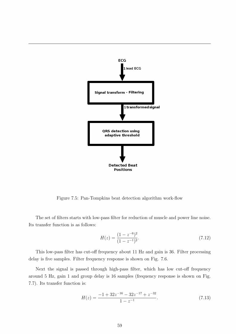
Figure 7.5: Pan-Tompkins beat detection algorithm work-flow
The set of filters starts with low-pass filter for reduction of muscle and power line noise.
Its transfer function is as follows:
H(z) =(1− z−6)2
(1− z−1)2. (7.12)
This low-pass filter has cut-off frequency about 11 Hz and gain is 36. Filter processing
delay is five samples. Filter frequency response is shown on Fig. 7.6.
Next the signal is passed through high-pass filter, which has low cut-off frequency
around 5 Hz, gain 1 and group delay is 16 samples (frequency response is shown on Fig.
7.7). Its transfer function is:
H(z) =−1 + 32z−16 − 32z−17 + z−32
1− z−1. (7.13)
59

7. EVALUATION OF PROPOSED ALGORITHMS
Figure 7.6: Low-pass filter frequency response for fs=200 Hz
Figure 7.7: High-pass filter frequency response for fs=200 Hz
60

Figure 7.8: Derivative filter frequency response for fs=200 Hz
After band-pass filtering (implemented by low-pass and high-pass filter) the signal 5-
point derivative is taken using derivative filter with transfer function:
H(z) =1
8(2 + z−1 − z−3 − 2z−4). (7.14)
The derivative approximates ideal derivative between dc and 30 Hz and has group delay
2 samples. Filter frequency response is shown on Fig. 7.8.
The output of derivative filter is squared and finally it is filtered by MA filter of length
30 samples. Its frequency response is shown on Fig. 7.9.
After averaging resulting signal (for example see Fig. 7.10) is passed to second step of
algorithm, which detects beat position using adaptive thresholds. Thresholds are computed
from two parameters:
• SPKI - running estimate of signal (ECG) peak,
• NPKI - running estimate of the noise peak.
61
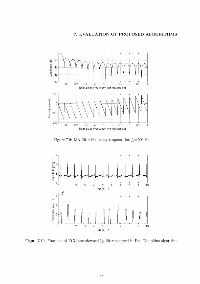
7. EVALUATION OF PROPOSED ALGORITHMS
Figure 7.9: MA filter frequency response for fs=200 Hz
Figure 7.10: Example of ECG transformed by filter set used in Pan-Tompkins algorithm
62

These two parameters are recomputed every time the peak in ECG is found (PEAKI). The
parameters are recomputed as follows:
SPKI = 0.125PEAKI + 0.875SPKI, if PEAKI is ECG
NPKI = 0.125PEAKI + 0.875NPKI, if PEAKI is noise.(7.15)
Every time the SPKI and NPKI parameters are recomputed adaptive thresholds are
recomputed:
TH1 = NPKI + 0.25(SPKI −NPKI),
TH2 = 0.5TH1,(7.16)
where TH1 is basic detection threshold and TH2 is threshold used during search-back
procedure, when no ECG beat is detected for long duration of time. The time duration
required for starting search-back procedure is determined as 1.66∗average of last eight RR
intervals.
7.3.2 Evaluation algorithm
The evaluation procedure was developed in order to test ECG processing algorithms on
standard ECG databases (mentioned above) contaminated with different types of noise
with different strength. Our methodology provides general framework for this testing. The
evaluation algorithm work flow is shown in Algorithm 4. Basically the method has three
main parts:
• Noisy recording creation part - in which the noise is created and added to ECG
records according to procedure described in Section 7.1.2.
• ECG processing algorithms part - in this part all tested algorithms ”do their work”
and the results are stored.
• Evaluation criteria computation part - here the evaluation criteria for tested algo-
rithms are computed according to Section 7.2.
63

7. EVALUATION OF PROPOSED ALGORITHMS
Algorithm 4 Algorithm for complex component estimation
Input: ECG signal database1: Noisy recording creation and adding to ECG signals2: ECG processing using evaluated algorithms3: Evaluation of results
Output: Results according to selected criteria
64

Chapter 8
Results of proposed algorithms
This Chapter summarizes results of our algorithms compared to state-of-the-art methods.
First we will present the de-noising algorithm and its results and then the beat detection
algorithm. Results are demonstrated using graphs showing the important information at
a glance.
8.1 Denoising algorithm
This section describes results obtained by our de-noising algorithm and the referential
methods. First part of the section describes results using graphical description and all
available ECG databases. Second part provides testing conclusions.
8.1.1 Results
Figures 8.1, 8.2, 8.3, 8.4, 8.5, 8.6, 8.7 and 8.8 show summarized results of our algorithm,
basic filtering algorithm and Wavelet de-noising algorithm. Each figure shows the evolution
of RMSE according to the percentage of noise amplitude added to signals (Section 7.1.2).
For more detailed results see Appendix A. This section is divided into subsections according
to databases used for algorithm testing. The last subsection shows performance of tested
algorithms on all available data.
65

8. RESULTS OF PROPOSED ALGORITHMS
8.1.1.1 Results on MIT-BIH Arrhythmia Database
Figure 8.1 shows summarized results on MIT/BIH Arrhythmia Database. First observa-
tion shows us that our de-noising algorithm is similar or better in presence of all noises
than other methods - basic filtering has problems in presence of strong EMG artefact and
electrode cable movement and the Wavelet based de-noising algorithm has difficulties with
base line wander and the electrode cable movement.
The main purpose, for which the algorithm was designed, was removal of electrode
(a) Power line interference (b) Base line wander
(c) EMG (d) Electrode cable movement
Figure 8.1: Summarized values of RMS for the developed algorithm and different types ofartefacts on MIT/BIH Arrhythmia Database. We can observe that our algorithm performscorrectly in presence of all noises. The filtering method has problem with strong EMGartefact and Wavelet method with Base line wander artefact.
cable movement artefact and we can observe that our algorithm is the only one, which was
able to reduce noise from ECG, thus we can say our algorithm is more stable in presence
of uncommon noise than the other tested algorithms but it works properly in presence of
any type of tested noise.
In presence of standard noises state-of-the-art algorithms work properly - filters remove
66
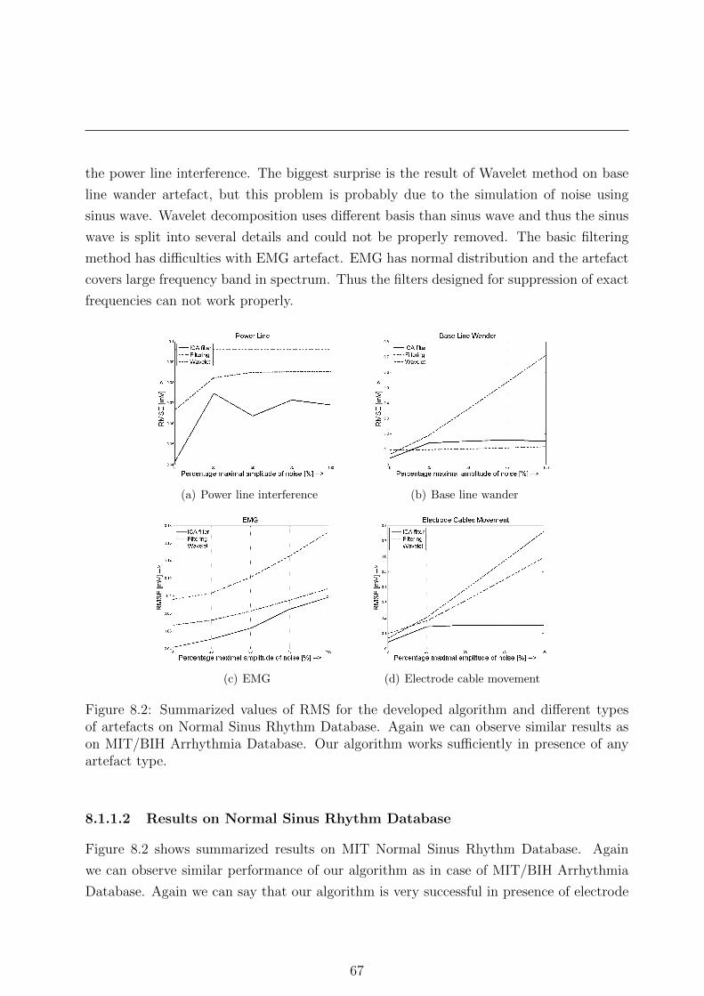
the power line interference. The biggest surprise is the result of Wavelet method on base
line wander artefact, but this problem is probably due to the simulation of noise using
sinus wave. Wavelet decomposition uses different basis than sinus wave and thus the sinus
wave is split into several details and could not be properly removed. The basic filtering
method has difficulties with EMG artefact. EMG has normal distribution and the artefact
covers large frequency band in spectrum. Thus the filters designed for suppression of exact
frequencies can not work properly.
(a) Power line interference (b) Base line wander
(c) EMG (d) Electrode cable movement
Figure 8.2: Summarized values of RMS for the developed algorithm and different typesof artefacts on Normal Sinus Rhythm Database. Again we can observe similar results ason MIT/BIH Arrhythmia Database. Our algorithm works sufficiently in presence of anyartefact type.
8.1.1.2 Results on Normal Sinus Rhythm Database
Figure 8.2 shows summarized results on MIT Normal Sinus Rhythm Database. Again
we can observe similar performance of our algorithm as in case of MIT/BIH Arrhythmia
Database. Again we can say that our algorithm is very successful in presence of electrode
67

8. RESULTS OF PROPOSED ALGORITHMS
cable movement artefact and it works similar or better in presence of common noises. One
exception is base line wander artefact, where our algorithm has slightly worse performance
than standard filtering method. Wavelet method has difficulties with base line wander and
electrode cable movement. On the other hand basic filtering shows that it has problems
with EMG and power line artefacts.
(a) Power line interference (b) Base line wander
(c) EMG (d) Electrode cable movement
Figure 8.3: Summarized values of RMS for the developed algorithm and different typesof artefacts on European ST-T database. We can observe results similar to previousdatabases. Our algorithm works properly in presence of all noises types. Wavelet methodagain fails in case of base line wander and electrode cable movement and standard filteringfails in case of EMG.
8.1.1.3 Results on European ST-T database
Figure 8.3 shows the results on European ST-T database. As in previous case we observe
similar results - our algorithm works similar or better than tested referential methods. In
case of power line interference our algorithm performance is slightly worse than performance
of Wavelet method and in presence of base line wander than performance of basic filtering.
68
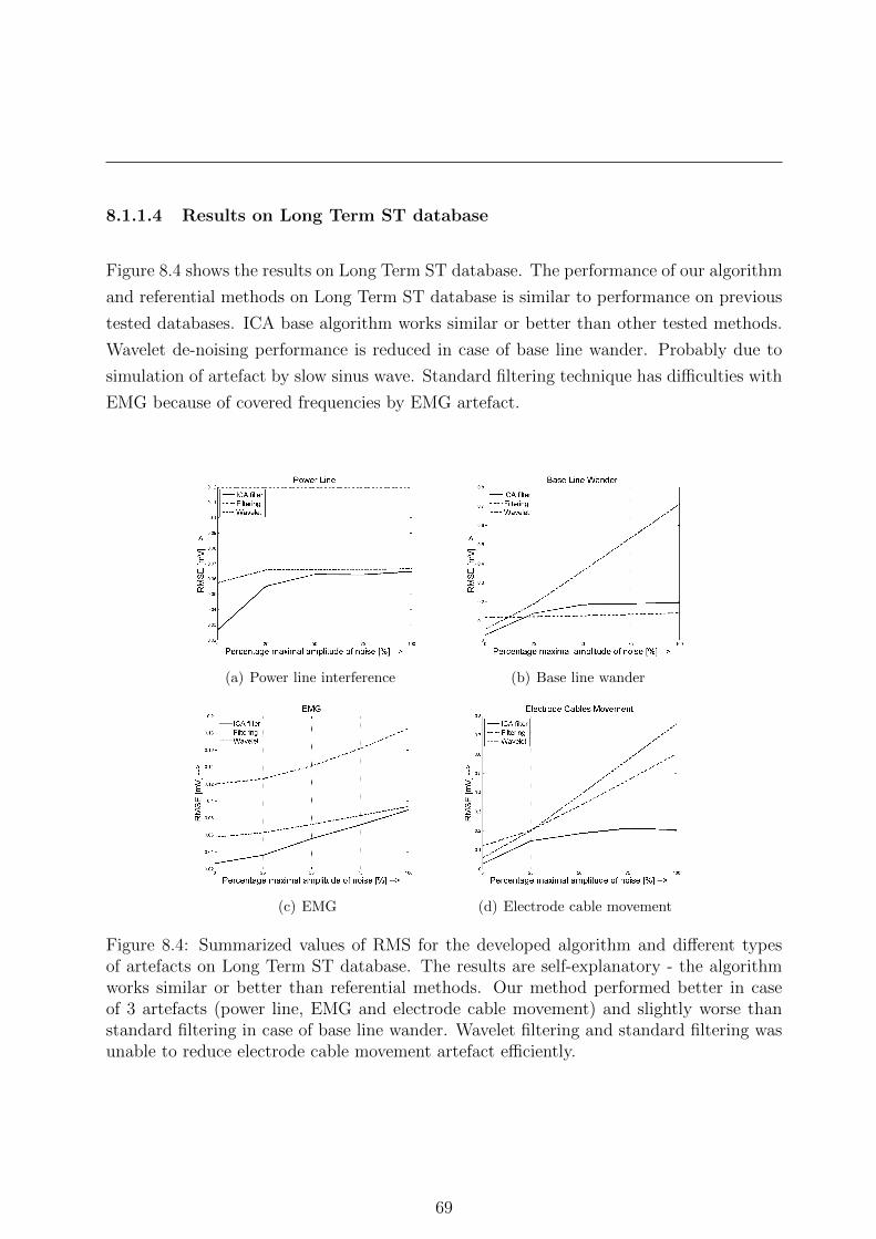
8.1.1.4 Results on Long Term ST database
Figure 8.4 shows the results on Long Term ST database. The performance of our algorithm
and referential methods on Long Term ST database is similar to performance on previous
tested databases. ICA base algorithm works similar or better than other tested methods.
Wavelet de-noising performance is reduced in case of base line wander. Probably due to
simulation of artefact by slow sinus wave. Standard filtering technique has difficulties with
EMG because of covered frequencies by EMG artefact.
(a) Power line interference (b) Base line wander
(c) EMG (d) Electrode cable movement
Figure 8.4: Summarized values of RMS for the developed algorithm and different typesof artefacts on Long Term ST database. The results are self-explanatory - the algorithmworks similar or better than referential methods. Our method performed better in caseof 3 artefacts (power line, EMG and electrode cable movement) and slightly worse thanstandard filtering in case of base line wander. Wavelet filtering and standard filtering wasunable to reduce electrode cable movement artefact efficiently.
69

8. RESULTS OF PROPOSED ALGORITHMS
8.1.1.5 Results on QT database
Figure 8.5 shows the results on QT database. Our method performance is slightly worse
than standard filtering method in presence of power line artefact and base line wander,
but its performance is still comparable with it. We can again observe that our algorithm
excelled in reduction of electrode cable movement artefact.
(a) Power line interference (b) Base line wander
(c) EMG (d) Electrode cable movement
Figure 8.5: Summarized values of RMS for the developed algorithm and different types ofartefacts on QT database. In presence of all four tested noises our algorithm performanceis similar or better than performance of referential methods.
70

8.1.1.6 Results on MIT Long Term database
Figure 8.6 shows results on MIT Long Term database. Again the results of our algorithm
are similar to previous results. Our algorithm is capable to deal with all tested noises.
In presence of EMG and base line wander our method performance is slightly worse than
better of referential methods.
(a) Power line interference (b) Base line wander
(c) EMG (d) Electrode cable movement
Figure 8.6: Summarized values of RMS for the developed algorithm and different typesof artefacts on MIT Long Term database. The results are similar to results on previouslymentioned databases.
8.1.1.7 Results on MIT-BIH ST Change database
Figure 8.7 shows results on MIT-BIH ST Change database. Results on MIT-BIH ST
Change database are similar to previous results. All algorithms work similarly to the
case of MIT Long Term database. Our method, which was designed for electrode cable
movement artefact, is capable to deal with all tested types of noise presented in signal.
Wavelet de-noising performs slightly worse than filtering in case of power line interference.
71

8. RESULTS OF PROPOSED ALGORITHMS
(a) Power line interference (b) Base line wander
(c) EMG (d) Electrode cable movement
Figure 8.7: Summarized values of RMS for the developed algorithm and different types ofartefacts on MIT-BIH ST Change database. The results are similar to previous results andwe can observe that our algorithm performed similar or better than referential methods.
8.1.1.8 Summary results on all databases
Summary results obtained across all databases show (Fig. 8.8) that our algorithm, which
was designed for purpose of removal of electrode cable movement artefact, is suitable for
removal of all types of noises. We can observe that our algorithm performs accurately in
general. The method has one big advantage - in case of no artefact/noise presented our
method distorts the resulting the ECG signal of at least. This is very desirable property
in case of any medical signal.
72

(a) Power line interference (b) Base line wander
(c) EMG (d) Electrode cable movement
Figure 8.8: Summarized values of RMS for the developed algorithm and different types ofartefacts on all databases. We can observe that our algorithm performs well on all typesof artefact. The wavelet de-noising performance is reduced on data containing base linewander and electrode cable movement artefact. Basic filtering performs worse on EMGand electrode cable movement artefact containing data.
Wavelet de-noising works well in case of power line interference and EMG noise, but
fails in case of base line wander and electrode cable movement noise. This is probably due
to the simulation of base line wander noise using sinus wave. Wavelet decomposition uses
different basis than sinus wave and thus the sinus wave is split into several details and
could not be properly removed.
Basic filtering procedure performs very accurately on data containing power line inter-
ference and base line wander. The problem arises with EMG and electrode cable artefact.
EMG artefact was simulated as random vector with normal distribution and it influences
all frequency bands in spectrum. Thus the filters designed for suppression of predefined
frequencies can not filter whole EMG activity. The same problem is with electrode cable
movement artefact, which covers different frequencies than filtered frequencies and artefact
was not reduced.
73

8. RESULTS OF PROPOSED ALGORITHMS
8.1.2 Conclusions
We have developed an ICA based de-noising ECG algorithm for dealing with uncommon
noises presented in data during holter and telemedicine applications. This algorithm is
able to deal with very strong noises and preserves as much information as possible. Our
method connects the well-known JADE algorithm with decision tree in order to identify
noise and reduce it.
Method behaviour was tested on standard databases available freely on-line and it
was compared to common filtering and Wavelet threshold de-noising techniques. Tests
were performed using simulated noises (3 common and 1 uncommon). The uncommon
noise tests algorithm ability to deal with unpredictable noise events common for nowadays
holter and telemedicine applications.
Our method performs much more stable than other methods – it outperforms both
state-of-the-art methods in unpredictable noise events and it works similarly to both meth-
ods with ECG data contaminated with base line wander or EMG.
Time requirements of our algorithm is increased due to computational complexity of
JADE algorithm and Pan-Tompkins beat detection algorithm (used in feature extraction
step), but changing the BSS or QRS complex detection algorithm will lead to better time
performance. Memory requirements are also slightly increased but the advantage of our
method outperforms its flaws, especially with nowadays hardware development.
8.2 QRS detection algorithm
This section describes results of our QRS detection method based on Christov’s beat detec-
tion algorithm and referential methods. Results are presented using graphs of F-measure,
which is harmonic mean of Sensitivity and Specificity (Section 7.2.2). Last part of the
section provides testing conclusions.
8.2.1 Results
Figures 8.9, 8.10, 8.11, 8.12, 8.13, 8.14 and 8.16 show summarized results of our algorithm,
Tompkins algorithm and Christov’s algorithm. Each figure shows the evolution of F-
measure according to the percentage of noise amplitude added to signals. For more details
see Appendix B.
74
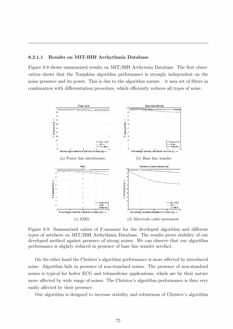
8.2.1.1 Results on MIT-BIH Arrhythmia Database
Figure 8.9 shows summarized results on MIT/BIH Arrhytmia Database. The first obser-
vation shows that the Tompkins algorithm performance is strongly independent on the
noise presence and its power. This is due to the algorithm nature – it uses set of filters in
combination with differentiation procedure, which efficiently reduces all types of noise.
(a) Power line interference (b) Base line wander
(c) EMG (d) Electrode cable movement
Figure 8.9: Summarized values of F-measure for the developed algorithm and differenttypes of artefacts on MIT/BIH Arrhythmia Database. The results prove stability of ourdeveloped method against presence of strong noises. We can observe that our algorithmperformance is slightly reduced in presence of base line wander artefact.
On the other hand the Christov’s algorithm performance is more affected by introduced
noise. Algorithm fails in presence of non-standard noises. The presence of non-standard
noises is typical for holter ECG and telemedicine applications, which are by their nature
more affected by wide range of noises. The Christov’s algorithm performance is then very
easily affected by their presence.
Our algorithm is designed to increase stability and robustness of Christov’s algorithm
75

8. RESULTS OF PROPOSED ALGORITHMS
in presence of unpredictable events (artefacts). This goal is achieved.
Our algorithm is very successful in presence of noise, which mimics the QRS complex
activity in frequency domain (electrode cable movement artefacts). Its stability is caused
by extraction of ECG activity during computation of complex component signal, which is
the basis for QRS complex detection.
8.2.1.2 Results on Normal Sinus Rhythm Database
Figure 8.10 shows summarized results on MIT Normal Sinus Rhythm Database. We can
observe, again, that Tompkins algorithm performance is stable in presence of any noise
presented in ECG. The performance of Christov’s algorithm is similar to our method with
exception of electrode cable movement artefact. In such a case the Tompkins algorithm
(a) Power line interference (b) Base line wander
(c) EMG (d) Electrode cable movement
Figure 8.10: Summarized values of F-measure for the developed algorithm and differenttypes of artefacts on Normal Sinus Rhythm Database. Our algorithm works much morestable in presence of any type of noise. We can also observe that the separation of Baseline wander artefact was correct – the results are better than on MIT/BIH ArrhythmiaDatabase.
76
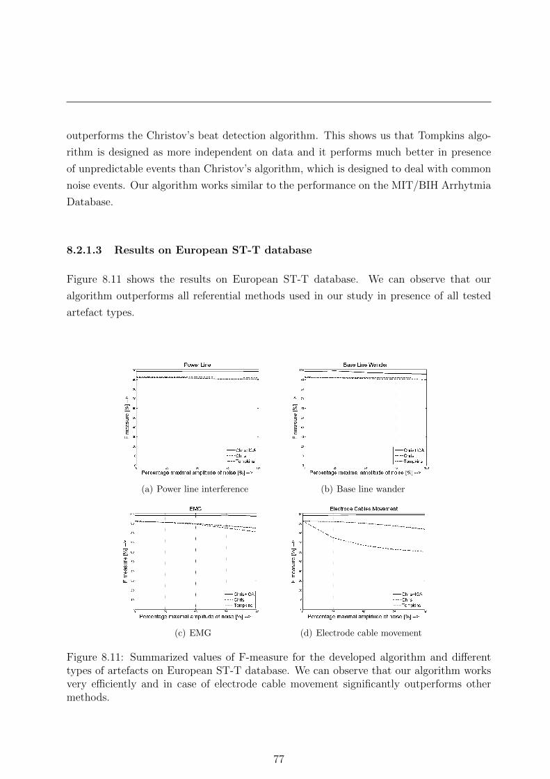
outperforms the Christov’s beat detection algorithm. This shows us that Tompkins algo-
rithm is designed as more independent on data and it performs much better in presence
of unpredictable events than Christov’s algorithm, which is designed to deal with common
noise events. Our algorithm works similar to the performance on the MIT/BIH Arrhytmia
Database.
8.2.1.3 Results on European ST-T database
Figure 8.11 shows the results on European ST-T database. We can observe that our
algorithm outperforms all referential methods used in our study in presence of all tested
artefact types.
(a) Power line interference (b) Base line wander
(c) EMG (d) Electrode cable movement
Figure 8.11: Summarized values of F-measure for the developed algorithm and differenttypes of artefacts on European ST-T database. We can observe that our algorithm worksvery efficiently and in case of electrode cable movement significantly outperforms othermethods.
77
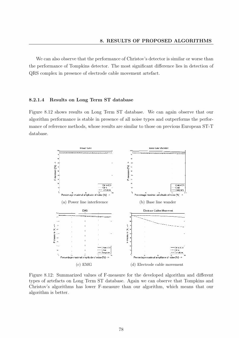
8. RESULTS OF PROPOSED ALGORITHMS
We can also observe that the performance of Christov’s detector is similar or worse than
the performance of Tompkins detector. The most significant difference lies in detection of
QRS complex in presence of electrode cable movement artefact.
8.2.1.4 Results on Long Term ST database
Figure 8.12 shows results on Long Term ST database. We can again observe that our
algorithm performance is stable in presence of all noise types and outperforms the perfor-
mance of reference methods, whose results are similar to those on previous European ST-T
database.
(a) Power line interference (b) Base line wander
(c) EMG (d) Electrode cable movement
Figure 8.12: Summarized values of F-measure for the developed algorithm and differenttypes of artefacts on Long Term ST database. Again we can observe that Tompkins andChristov’s algorithms has lower F-measure than our algorithm, which means that ouralgorithm is better.
78
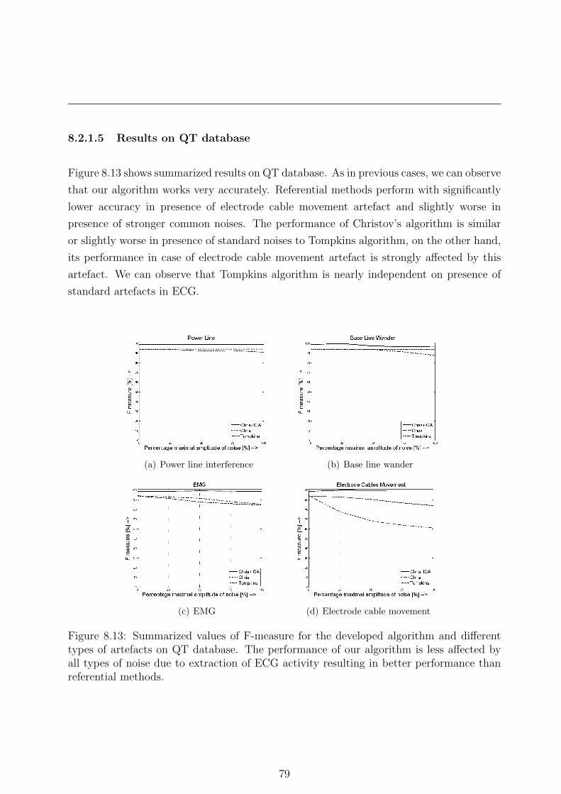
8.2.1.5 Results on QT database
Figure 8.13 shows summarized results on QT database. As in previous cases, we can observe
that our algorithm works very accurately. Referential methods perform with significantly
lower accuracy in presence of electrode cable movement artefact and slightly worse in
presence of stronger common noises. The performance of Christov’s algorithm is similar
or slightly worse in presence of standard noises to Tompkins algorithm, on the other hand,
its performance in case of electrode cable movement artefact is strongly affected by this
artefact. We can observe that Tompkins algorithm is nearly independent on presence of
standard artefacts in ECG.
(a) Power line interference (b) Base line wander
(c) EMG (d) Electrode cable movement
Figure 8.13: Summarized values of F-measure for the developed algorithm and differenttypes of artefacts on QT database. The performance of our algorithm is less affected byall types of noise due to extraction of ECG activity resulting in better performance thanreferential methods.
79
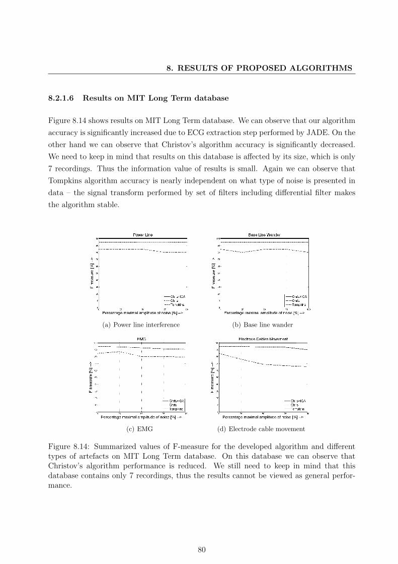
8. RESULTS OF PROPOSED ALGORITHMS
8.2.1.6 Results on MIT Long Term database
Figure 8.14 shows results on MIT Long Term database. We can observe that our algorithm
accuracy is significantly increased due to ECG extraction step performed by JADE. On the
other hand we can observe that Christov’s algorithm accuracy is significantly decreased.
We need to keep in mind that results on this database is affected by its size, which is only
7 recordings. Thus the information value of results is small. Again we can observe that
Tompkins algorithm accuracy is nearly independent on what type of noise is presented in
data – the signal transform performed by set of filters including differential filter makes
the algorithm stable.
(a) Power line interference (b) Base line wander
(c) EMG (d) Electrode cable movement
Figure 8.14: Summarized values of F-measure for the developed algorithm and differenttypes of artefacts on MIT Long Term database. On this database we can observe thatChristov’s algorithm performance is reduced. We still need to keep in mind that thisdatabase contains only 7 recordings, thus the results cannot be viewed as general perfor-mance.
80

8.2.1.7 Results on MIT-BIH ST Change database
Figure 8.16 shows results on MIT-BIH ST Change database. We can observe, again, that
our method is able to detect QRS complexes much more accurately than the other tested
methods and thus outperforms them in sense of F-measure characteristics.
(a) Power line interference (b) Base line wander
(c) EMG (d) Electrode cable movement
Figure 8.15: Summarized values of F-measure for the developed algorithm and differenttypes of artefacts on MIT-BIH ST Change database. Again our algorithm outperformsothers on data contaminated with different types of noise.
8.2.1.8 Summary results on all databases
Figure 8.16 shows results across all databases. We can observe that performances of all
algorithms are stable across all databases. Our algorithm is significantly less affected by
electrode cable movement artefact than the others. The performance of both referential
algorithms works similar in general on large scale data. One difference between them is
in presence of electrode cable movement artefact, which affects significantly harder the
performance of Christov’s algorithm.
81
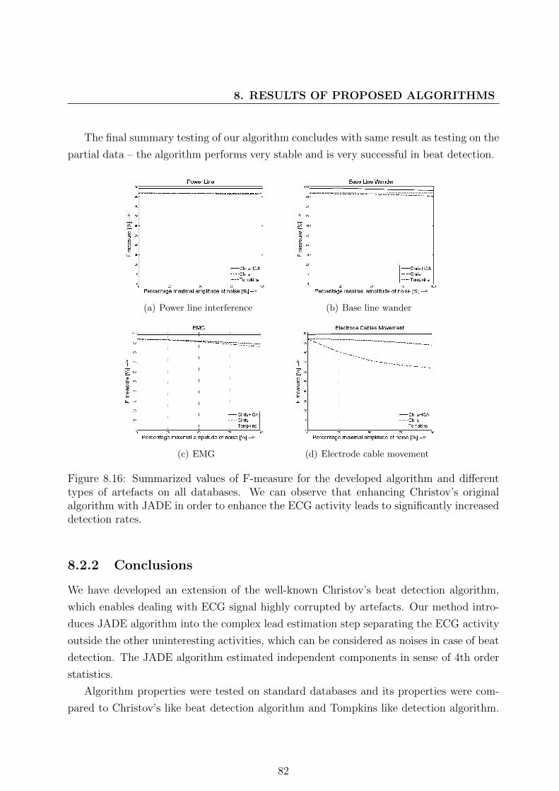
8. RESULTS OF PROPOSED ALGORITHMS
The final summary testing of our algorithm concludes with same result as testing on the
partial data – the algorithm performs very stable and is very successful in beat detection.
(a) Power line interference (b) Base line wander
(c) EMG (d) Electrode cable movement
Figure 8.16: Summarized values of F-measure for the developed algorithm and differenttypes of artefacts on all databases. We can observe that enhancing Christov’s originalalgorithm with JADE in order to enhance the ECG activity leads to significantly increaseddetection rates.
8.2.2 Conclusions
We have developed an extension of the well-known Christov’s beat detection algorithm,
which enables dealing with ECG signal highly corrupted by artefacts. Our method intro-
duces JADE algorithm into the complex lead estimation step separating the ECG activity
outside the other uninteresting activities, which can be considered as noises in case of beat
detection. The JADE algorithm estimated independent components in sense of 4th order
statistics.
Algorithm properties were tested on standard databases and its properties were com-
pared to Christov’s like beat detection algorithm and Tompkins like detection algorithm.
82

Tests were performed using simulated noises - power line interference, base line wander,
EMG and electrode cable movement. First three noises are common types presented in
recorded data and the last one is uncommon one. We decided to add this, because we
are dealing with holter ECG recordings and this data contains many unpredictable noise
events, which are not usual at all.
Our enhanced version of Christov’s beat detection algorithm performs much more sta-
ble in presence of noise and it outperforms Christov’s and Tompkins like algorithms, which
are designed for the typical cases of resting ECGs.
Time requirements of our algorithm is increased only by computational complexity of
JADE algorithm, which is one of the fastest BSS algorithms (it runs in milliseconds on 30
minutes 2 lead ECG sampled with sampling frequency 500 Hz, computational complex-
ity could be found in [4, Ch. 18]). So the benefit of ECG activity estimation is much
greater than loss of computational time. The amount of space required for data storing is
also slightly increased - JADE algorithm stores several matrices with cumulants and the
amount of data stored is dependent on number of ECG leads.
83

8. RESULTS OF PROPOSED ALGORITHMS
84

Chapter 9
Conclusions
We have developed new algorithms for de-noising and beat detection of ECG signals. We
also created extensive state-of-the-art of ICA applications in biomedical research area. This
chapter summarizes our results.
Create state-of-the-art of ICA applications in Biomedical Engi-
neering research area with main focus on application in ECG sig-
nal processing.
We provided a review of most frequent ICA algorithms and their applications to biomedical
signal processing. In detail it is described in Chapter 3.
Independent Component Analysis is a general tool, which could uncover hidden pro-
cesses within the biomedical signal. The last decade in research proves that ICA has been
successfully used in many research areas ranging from traditional areas such as EEG or
ECG research to more unconventional such as fMRI or EGG signals analysis.
The power of ICA lies in its basic assumptions - thus it can found processes that can-
not be found by any other technique. However results of separation by ICA should be
investigated carefully. ICA is a data driven technique and results could seem great but the
opposite might be true. The knowledge about data is necessary for differentiation of good
and bad separation result. For example we need to be able to differentiate between noise
and ECG activity within the estimated component and we need also identify problematic
separation, which could have great impact on results.
Many algorithms for solving ICA/BSS task emerged, but only few are commonly used
in biomedical research. The best method for biomedical area cannot be selected and one
85

9. CONCLUSIONS
should test several algorithms when searching for the best suited one for the researched
application. Each algorithm has different assumptions, which need to be satisfied. Loosing
assumptions from mind could lead to wrong conclusions.
We went through a large amount of papers covering most of the biomedical research
using ICA. Several questions arose during this work. First and most crucial question is
the reproducibility of experiments – many papers provide relatively clear algorithm expla-
nation, but the algorithm testing is performed on unpublished data, which were collected
by the authors of those papers. So the main problem is that nobody could say whether
the implementation of proposed method works correctly because it cannot be tested on
the same dataset. With this problem several others come – there are not many standard
databases freely available online. Of course there are well-known exceptions such as Phy-
sionet.org databases. The most reasonable solution is to make data freely available or to
test algorithms on both data – own and standard data.
Another problem that many papers have is lack of time and memory requirements of
the developed method (algorithm). Also there are missing crucial information such as
computer and software specifications, on which the experiments were performed.
According to our opinion, the best solution for solving all these problems is to make
implementations of the algorithm freely available to download. This will solve many ques-
tions and difficulties, which nowadays researchers deal with. For ICA algorithms there
exists an online database of ICA algorithms implementations - ICALAB [148], which could
serve as an example to others.
The idea we posted here is not new. We can mention a very interesting paper written
by Patrick Vandewalle [149], which deals with similar questions in the area of signal pro-
cessing.
Based on our extensive analysis of current research efforts in biomedical ICA applica-
tions we decided to develop two algorithms. One for de-noising of ECG signals obtained
during measuring of holter ECG and second for beat detection in cases of noisy ECG
recordings.
86
Propose and develop the algorithm for denoising of ECG signals
using ICA.
We created an algorithm for artefact reduction from ECG. Our algorithm is capable to
deal with strong noise in ECG data and enables QRS detection and visual examination
(for details see Chapter 5). The algorithm deals with uncommon noises presented in ECG
data during holter and telemedicine applications. Our method connects well-known JADE
algorithm with decision tree in order to identify noise and reduce it.
Method behaviour was tested on standard databases and it was compared to common
filtering and Wavelet threshold de-noising techniques. Tests were performed using simu-
lated noises (3 common and 1 uncommon).
Our method performed similar or better than both referential methods in presence of
all common types of artefacts. Our method was the only one method that successfully
reduced the electrode cable movement artefact. In that case both referential methods per-
formance was decreased with stronger noise presented.
JADE based filtering has one advantage against both referential methods – our method
least distorts the resulting signal in case of no noise presented in data. This ability is
very valuable in medical applications. Time requirements of our algorithm is increased
due computational complexity of JADE algorithm and Pan-Tompkins beat detection algo-
rithm (used in feature extraction step), but changing the BSS or QRS complex detection
algorithm will lead to better time performance. Memory requirements are also slightly in-
creased but the advantage of our method outperforms its flaws, especially with nowadays
hardware development.
Propose and develop the algorithm for beat detection using ICA.
We have developed an extension of the well-known Christov’s beat detection algorithm,
which enables dealing with ECG signal highly corrupted by artefacts (for details see Chap-
ter 6). Our method introduces JADE algorithm into the complex lead estimation step
separating the ECG activity outside the other uninteresting activities, which can be con-
sidered as noises in case of beat detection.
Algorithm properties were tested on standard databases and its properties were com-
pared to Christov’s like beat detection algorithm and Tompkins like detection algorithm.
Tests were performed using simulated noises - power line interference, base line wander,
87

9. CONCLUSIONS
EMG and electrode cable movement. First three noises are common types presented in
recorded data and the last one is uncommon one. We decided to add this, because we
are dealing with holter ECG recordings and this data contains many unpredictable noise
events, which are not usual at all.
Our enhanced version of Christov’s beat detection algorithm performs much more stable
in presence of noise and it outperformed Christov’s and Tompkins like algorithms, which
are designed for the typical cases of resting ECGs.
The computational complexity of our algorithm is increased only by computational
complexity of JADE algorithm, which is one of the fastest BSS algorithms. So the benefit
of ECG activity estimation is much greater than loss of computational time. The amount
of space required for data storing is also slightly increased - JADE algorithm stores several
matrices with cumulants and the amount of data stored is dependent on number of ECG
leads.
Develop a testing framework for evaluation of proposed algo-
rithms.
We developed the standardized procedure, which enables us to test performance of our
algorithms against the state-of-the-art methods (see Chapter 7). This procedure is versatile
and modular, thus we used it in testing of both methods. The information was easily
obtained and analysed. Our framework enables us effective tuning of the methods.
Final remarks and future work
All goals we formulated were successfully fulfilled. We proposed two algorithms – one for
de-noising of ECG recording containing noise and second the algorithm for beat detection
with noisy ECG recordings. Both algorithms were tested using our testing framework
against the state-of-the-art methods and results are good. Both algorithms are capable
to deal with common types of noises and also with uncommon ones. This makes them
very useful in applications within telemedicine issues and holter recordings, where the
environment is rapidly changing and the distortions corrupting ECG signals could be very
different for those normally present in ECG within laboratory or ambulatory measurement
of ECG.
88

Our future work will aim at extensive testing of our developed algorithms. We will
also test different ICA algorithms within our developed methods in order to test their
dependency on performance of used BSS algorithm. We also plan to test new decision
algorithms for recognition of noisy components in order to enhance algorithms performance.
89

9. CONCLUSIONS
90

Appendix A: Detailed results of
de-noising algorithms
Tables show detailed results visualised in graphs plotted
Tables are organized as follows: each type of noise corresponds to one row of the table,
each column of the table corresponds to one type of algorithm and noise level (for details
see. 7.1.2) used during testing. On top of every four columns, which corresponds to one
tested algorithm, one can find performance of corresponding algorithm on clear signal.
We can observe the same results as in above mentioned graphs. Our method is capable
to deal with strong noises and also reduce the electrode cable movement artefact efficiently.
We can also observe that our method is stable in presence of any type of noise.
ECG signals values is in units of mV , thus the values of RMS around 40 µV should be
considered as very good result. They mean that the noise reduction in ECG signal is good
enough to preserve ECG signal diagnostics. We can observe that our method provides
slightly worse results, but these results are stable in presence of any noise of any strength.
This is very desired feature of any ECG signal processing algorithm.
RMS [µV]
ICA filter Basic Filtering Wavelet denoising
Clear signal 34.40 123.97 56.98
Noise typeNoise level [%] Noise level [%] Noise level [%]
25 50 75 100 25 50 75 100 25 50 75 100
Power Line Interference 65.58 68.83 67.19 66.64 123.97 123.97 123.97 123.97 66.28 67.20 67.41 67.60
EMG 47.43 63.27 75.96 91.31 131.46 148.33 170.28 195.32 62.79 72.40 82.69 92.86
Base Line Wander 148.07 191.22 198.85 196.15 126.54 132.14 138.87 146.03 189.08 360.17 534.82 710.49
Electrode Cable Movement 155.52 195.36 206.56 210.91 203.88 330.21 465.11 603.35 201.24 385.12 571.72 758.67
Table 1: Detailed summary results on all databases
91
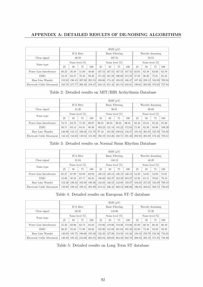
APPENDIX A: DETAILED RESULTS OF DE-NOISING ALGORITHMS
RMS [µV]
ICA filter Basic Filtering Wavelet denoising
Clear signal 40.59 167.55 53.73
Noise typeNoise level [%] Noise level [%] Noise level [%]
25 50 75 100 25 50 75 100 25 50 75 100
Power Line Interference 68.22 56.38 51.69 49.60 167.55 167.55 167.55 167.55 62.65 63.48 63.60 63.79
EMG 53.19 63.47 78.24 92.36 171.32 181.59 196.68 215.25 57.82 66.20 75.81 85.45
Base Line Wander 152.82 196.45 207.80 205.55 169.66 174.42 180.23 186.47 187.30 359.12 534.09 709.94
Electrode Cable Movement 163.78 217.77 208.39 218.47 234.13 351.42 481.53 616.81 199.61 383.98 570.82 757.84
Table 2: Detailed results on MIT/BIH Arrhythmia Database
RMS [µV]
ICA filter Basic Filtering Wavelet denoising
Clear signal 41.36 96.01 66.60
Noise typeNoise level [%] Noise level [%] Noise level [%]
25 50 75 100 25 50 75 100 25 50 75 100
Power Line Interference 74.71 63.72 71.50 69.07 96.01 96.01 96.01 96.01 82.34 85.01 85.22 85.36
EMG 50.71 63.13 84.81 98.38 103.23 121.18 145.22 172.82 72.40 82.56 94.98 108.29
Base Line Wander 140.86 152.15 160.26 151.70 97.18 101.92 108.64 116.27 191.04 361.05 535.39 710.92
Electrode Cable Movement 142.42 152.63 149.84 151.88 180.19 312.26 450.71 591.46 202.92 385.88 572.46 759.51
Table 3: Detailed results on Normal Sinus Rhythm Database
RMS [µV]
ICA filter Basic Filtering Wavelet denoising
Clear signal 31.84 140.13 46.29
Noise typeNoise level [%] Noise level [%] Noise level [%]
25 50 75 100 25 50 75 100 25 50 75 100
Power Line Interference 65.37 67.99 63.82 62.02 140.13 140.13 140.13 140.13 54.28 54.65 54.82 55.01
EMG 45.00 58.16 67.17 80.35 146.98 162.37 182.29 204.97 52.38 61.51 70.62 79.16
Base Line Wander 152.36 196.43 192.80 189.36 142.03 146.52 152.08 158.07 184.63 357.65 533.09 709.19
Electrode Cable Movement 159.95 189.42 198.31 201.99 213.44 336.42 469.54 606.66 196.94 382.61 569.74 756.83
Table 4: Detailed results on European ST-T database
RMS [µV]
ICA filter Basic Filtering Wavelet denoising
Clear signal 26.56 119.86 57.39
Noise typeNoise level [%] Noise level [%] Noise level [%]
25 50 75 100 25 50 75 100 25 50 75 100
Power Line Interference 55.22 62.96 62.74 64.83 119.86 119.86 119.86 119.86 65.89 66.18 66.32 66.49
EMG 36.35 55.64 71.98 89.62 125.96 141.08 161.64 185.50 62.80 72.38 83.03 93.50
Base Line Wander 140.05 185.75 190.66 195.30 122.48 127.98 134.58 141.62 188.42 359.79 534.56 710.31
Electrode Cable Movement 146.93 185.35 213.00 204.55 202.84 329.69 464.58 602.78 200.83 385.18 571.85 758.80
Table 5: Detailed results on Long Term ST database
92

RMS [µV]
ICA filter Basic Filtering Wavelet denoising
Clear signal 38.37 83.78 52.14
Noise typeNoise level [%] Noise level [%] Noise level [%]
25 50 75 100 25 50 75 100 25 50 75 100
Power Line Interference 67.43 76.04 73.28 72.16 83.78 83.78 83.78 83.78 61.76 62.79 63.04 63.27
EMG 49.33 67.74 76.76 92.43 95.64 118.87 146.75 177.20 58.98 68.93 78.85 88.71
Base Line Wander 141.17 167.44 181.08 167.93 87.49 95.15 103.95 113.01 188.23 359.76 534.53 710.26
Electrode Cable Movement 148.07 175.63 182.35 189.02 176.22 311.40 451.07 592.24 200.13 384.14 570.82 757.92
Table 6: Detailed results on QT database
RMS [µV]
ICA filter Basic Filtering Wavelet denoising
Clear signal 29.78 176.82 75.57
Noise typeNoise level [%] Noise level [%] Noise level [%]
25 50 75 100 25 50 75 100 25 50 75 100
Power Line Interference 57.75 76.90 80.57 85.11 176.82 176.82 176.82 176.82 88.28 93.34 94.64 94.81
EMG 98.60 108.73 126.04 144.77 184.99 205.51 233.45 266.14 83.80 97.91 112.94 128.60
Base Line Wander 164.79 288.66 292.64 272.36 179.68 186.17 193.99 202.16 193.61 362.39 536.28 711.59
Electrode Cable Movement 173.65 299.37 297.49 272.79 244.06 360.36 489.42 624.22 205.77 387.67 573.90 761.21
Table 7: Detailed results on MIT Long Term database
RMS [µV]
ICA filter Basic Filtering Wavelet denoising
Clear signal 38.41 123.38 99.37
Noise typeNoise level [%] Noise level [%] Noise level [%]
25 50 75 100 25 50 75 100 25 50 75 100
Power Line Interference 81.89 90.05 93.15 92.71 123.38 123.38 123.38 123.38 111.29 113.62 113.93 114.09
EMG 56.34 76.69 91.08 108.26 131.51 151.04 177.01 206.67 105.28 116.60 130.00 143.65
Base Line Wander 165.03 241.71 269.39 284.80 126.02 131.09 137.19 143.95 208.61 371.37 542.54 716.36
Electrode Cable Movement 171.68 252.84 282.56 319.58 203.72 329.45 464.57 602.99 220.20 396.70 580.90 766.86
Table 8: Detailed results on MIT-BIH ST Change database
93

APPENDIX B: DETAILED RESULTS OF DE-NOISING ALGORITHMS
94

Appendix B: Detailed results of beat
detection algorithms
Tables show detailed results visualised in graphs plotted on Fig.
Tables are organized as follows: each type of noise corresponds to one row of the table,
each column of the table corresponds to one type of algorithm and noise level (for details
see. 7.1.2) used during testing. On top of every four columns, which corresponds to one
tested algorithm, one can find performance of corresponding algorithm on clear signal.
Again we can observe the same results as in above mentioned graphs. Our method
proves to be more robust to noises than other beat detection methods with one exception
- the base line wander artefact, when the estimation of components converge to solution
with divided sinus wave into several components with kurtosis higher than standard ECG
activity.
We can observe that our method has same performance as original Christov’s algorithm,
but its strength lies in suppression of irregular types of noises, for which the original
Christov’s algorithm was not designed.
F-measure [%]
Christov’s algorithm + ICA Christov’s algorithm Hammilton’s algorithm
Clear signal 98.81 93.88 93.99
Noise typeNoise level [%] Noise level [%] Noise level [%]
25 50 75 100 25 50 75 100 25 50 75 100
Power Line Interference 98.81 98.80 98.80 98.81 93.89 93.83 93.82 93.76 93.90 93.44 92.98 92.57
EMG 98.85 98.78 98.79 98.39 93.12 91.37 88.69 86.13 93.29 92.10 90.60 89.01
Base Line Wander 99.11 98.09 97.17 96.69 93.05 92.58 91.82 90.81 93.98 93.98 93.98 94.00
Electrode Cable Movement 99.08 99.13 99.19 99.20 80.71 71.87 67.14 64.21 93.77 92.48 90.57 87.91
Table 9: Detailed summarized results on all databases
95

APPENDIX B: DETAILED RESULTS OF DE-NOISING ALGORITHMS
F-measure [%]
Christov’s algorithm + ICA Christov’s algorithm Hammilton’s algorithm
Clear signal 99.07 94.02 96.61
Noise typeNoise level [%] Noise level [%] Noise level [%]
25 50 75 100 25 50 75 100 25 50 75 100
Power Line Interference 99.07 99.07 99.07 99.07 94.03 93.82 93.81 93.80 96.52 96.43 96.04 95.86
EMG 99.04 99.02 98.70 98.52 93.15 92.52 88.92 85.85 96.37 95.41 94.45 92.86
Base Line Wander 98.30 96.49 96.18 96.04 93.93 93.33 90.67 89.65 96.61 96.61 96.59 96.59
Electrode Cable Movement 98.99 99.09 99.24 99.22 79.81 69.12 64.71 62.47 95.89 95.23 93.05 90.63
Table 10: Detailed results on MIT/BIH Arrhythmia Database
F-measure [%]
Christov’s algorithm + ICA Christov’s algorithm Hammilton’s algorithm
Clear signal 99.17 99.24 95.17
Noise typeNoise level [%] Noise level [%] Noise level [%]
25 50 75 100 25 50 75 100 25 50 75 100
Power Line Interference 99.17 99.17 99.17 99.17 99.24 99.24 99.24 99.24 95.00 94.73 94.22 93.87
EMG 99.17 99.11 98.98 98.93 98.46 99.02 97.86 96.02 94.90 94.36 93.68 92.98
Base Line Wander 99.22 99.22 99.11 99.00 99.15 98.34 97.85 97.40 95.18 95.16 95.16 95.15
Electrode Cable Movement 99.23 99.22 99.24 99.24 91.96 87.00 79.19 72.98 94.87 94.94 94.98 94.31
Table 11: Detailed results on Normal Sinus Rhythm Database
F-measure [%]
Christov’s algorithm + ICA Christov’s algorithm Hammilton’s algorithm
Clear signal 98.55 92.76 92.50
Noise typeNoise level [%] Noise level [%] Noise level [%]
25 50 75 100 25 50 75 100 25 50 75 100
Power Line Interference 98.53 98.52 98.52 98.52 92.65 92.62 92.54 92.53 92.33 91.66 90.67 90.52
EMG 98.65 98.56 98.95 98.04 91.36 89.52 85.46 81.46 91.48 90.11 87.77 85.51
Base Line Wander 99.28 97.67 96.52 96.08 92.04 91.28 90.66 90.29 92.50 92.50 92.53 92.55
Electrode Cable Movement 98.93 99.10 99.20 99.14 75.12 67.18 62.94 60.76 92.27 90.50 87.75 84.25
Table 12: Detailed results on European ST-T database
F-measure [%]
Christov’s algorithm + ICA Christov’s algorithm Hammilton’s algorithm
Clear signal 98.72 94.46 93.93
Noise typeNoise level [%] Noise level [%] Noise level [%]
25 50 75 100 25 50 75 100 25 50 75 100
Power Line Interference 98.71 98.71 98.71 98.71 94.62 94.54 95.14 94.99 93.88 93.61 93.39 92.84
EMG 98.69 98.52 98.34 98.21 94.09 91.92 90.32 89.02 93.61 92.89 91.83 90.58
Base Line Wander 98.83 98.51 97.19 96.35 93.35 92.62 92.41 91.53 93.93 93.93 93.93 93.94
Electrode Cable Movement 98.87 98.87 98.88 98.99 84.60 74.57 69.33 65.73 94.00 92.92 91.66 89.37
Table 13: Detailed results on Long Term ST database
96

F-measure [%]
Christov’s algorithm + ICA Christov’s algorithm Hammilton’s algorithm
Clear signal 98.75 94.23 93.84
Noise typeNoise level [%] Noise level [%] Noise level [%]
25 50 75 100 25 50 75 100 25 50 75 100
Power Line Interference 98.76 98.76 98.76 98.76 94.22 94.15 94.14 94.13 93.87 92.45 92.68 90.94
EMG 98.89 99.26 99.05 98.61 92.85 91.64 88.19 85.63 91.45 87.77 86.06 84.63
Base Line Wander 99.94 97.33 96.77 96.44 94.16 93.66 91.27 87.71 93.82 93.70 93.70 93.77
Electrode Cable Movement 99.94 99.77 99.87 99.86 77.75 68.32 63.98 61.08 92.97 90.49 86.85 84.12
Table 14: Detailed results on QT database
F-measure [%]
Christov’s algorithm + ICA Christov’s algorithm Hammilton’s algorithm
Clear signal 99.94 84.85 94.93
Noise typeNoise level [%] Noise level [%] Noise level [%]
25 50 75 100 25 50 75 100 25 50 75 100
Power Line Interference 99.94 99.94 99.94 99.94 84.85 84.86 80.28 80.29 94.98 94.77 94.78 95.12
EMG 99.92 99.80 99.94 99.94 87.39 80.29 79.95 79.85 94.33 92.98 91.47 90.79
Base Line Wander 99.96 99.95 99.96 99.96 80.25 84.83 84.80 80.20 94.95 94.94 94.94 94.96
Electrode Cable Movement 99.96 99.94 99.87 99.63 76.34 69.03 67.08 65.74 94.75 94.22 93.88 90.14
Table 15: Detailed results on MIT Long Term database
F-measure [%]
Christov’s algorithm + ICA Christov’s algorithm Hammilton’s algorithm
Clear signal 99.57 95.82 96.87
Noise typeNoise level [%] Noise level [%] Noise level [%]
25 50 75 100 25 50 75 100 25 50 75 100
Power Line Interference 99.57 99.57 99.57 99.57 95.83 95.87 95.86 95.83 96.90 96.89 96.84 96.74
EMG 99.57 99.57 99.58 99.59 95.81 95.58 95.15 93.83 96.65 96.18 95.44 94.56
Base Line Wander 99.60 99.62 99.61 99.58 95.80 95.87 95.85 95.72 96.88 96.89 96.89 96.89
Electrode Cable Movement 99.61 99.63 99.62 99.61 91.49 85.06 80.77 77.88 96.64 95.83 95.28 94.15
Table 16: Detailed results on MIT-BIH ST Change database
97

REFERENCES
98
References
[1] A. Belouchrani, K. Abed-Meraim, J. F. Cardoso, and E. Moulines, “A blind source
separation technique using second-order statistics,” Signal Processing, IEEE Trans-
actions on, vol. 45, no. 2, pp. 434–444, 1997. 1, 11
[2] A. Hyvarinen and E. Oja, “Independent component analysis: algorithms and appli-
cations,” Neural Netw., vol. 13, pp. 411–430, May 2000. 1, 5, 7, 8
[3] J. F. Cardoso and A. Souloumiac, “Blind beamforming for non-gaussian signals,”
Radar and Signal Processing, IEE Proceedings F, vol. 140, no. 6, pp. 362–370, 1993.
1, 10, 42
[4] P. Comon and C. Jutten, Handbook of Blind Source Separation. Elsevier, 2010. 1,
35, 48, 83
[5] T. He, G. Clifford, and L. Tarassenko, “Application of independent component anal-
ysis in removing artefacts from the electrocardiogram,” Neural Computing & Appli-
cations, vol. 15, no. 2, pp. 105–116, 2006. 1, 15, 35, 48
[6] M. P. S. Chawla, “Pca and ica processing methods for removal of artifacts and noise
in electrocardiograms: A survey and comparison,” Applied Soft Computing Journal,
vol. 11, no. 2, pp. 2216–2226, 2011. 1, 18, 35, 41, 48
[7] A. L. Goldberger, L. A. N. Amaral, L. Glass, J. M. Hausdorff, P. C. Ivanov, R. G.
Mark, J. E. Mietus, G. B. Moody, C. K. Peng, and H. E. Stanley, “PhysioBank,
PhysioToolkit, and PhysioNet : Components of a New Research Resource for Com-
plex Physiologic Signals,” Circulation, vol. 101, pp. e215–220, June 2000. 4, 17, 51,
52
99

REFERENCES
[8] A. Hyvarinen, J. Karhunen, and E. Oja, Independent Component Analysis. Wiley,
2001. 5, 8, 10
[9] P. Comon, “Independent component analysis, a new concept?,” Signal Process.,
vol. 36, pp. 287–314, April 1994. 5
[10] R. E. Walpole, R. H. Myers, S. L. Myers, and K. E. Ye, Probability and Statistics for
Engineers and Scientists. Prentice Hall, 2011. 8
[11] J. F. Cardoso, “Source separation using higher order moments,” in Acoustics,
Speech, and Signal Processing, 1989. ICASSP-89., 1989 International Conference
on, pp. 2109–2112 vol.4, 1989. 9
[12] L. Molgedey and H. G. Schuster, “Separation of a mixture of independent signals
using time delayed correlations,” Physical Review Letters, vol. 72, pp. 3634–3637,
1994. 10
[13] J. O. Wisbeck, A. K. Barros, A. K. B. Yy, and R. G. Ojeda, “Application of ica in
the separation of breathing artifacts in ecg signals,” 1998. 15
[14] A. K. Barros, A. Mansour, and N. Ohnishi, “Removing artifacts from electrocardio-
graphic signals using independent components analysis,” Neurocomputing, vol. 22,
pp. 173–186, 11/20 1998. 15
[15] M. P. S. Chawla, H. K. Verma, and V. Kumar, “Artifacts and noise removal in
electrocardiograms using independent component analysis,” International journal of
cardiology, vol. 129, pp. 278–281, 9/26 2008. 15
[16] H. Xing and J. Hou, “A noise elimination method for ecg signals,” in Bioinformatics
and Biomedical Engineering , 2009. ICBBE 2009. 3rd International Conference on,
pp. 1–3, 2009. 15
[17] M. Milanesi, N. Martini, N. Vanello, V. Positano, M. F. Santarelli, and L. Landini,
“Independent component analysis applied to the removal of motion artifacts from
electrocardiographic signals,” Medical & biological engineering & computing, vol. 46,
pp. 251–261, MAR 2008. 15, 16
[18] G. Agrawal, M. Singh, V. R. Singh, and H. R. Singh, “Reduction of artifacts in
12-channel ecg signals using fastica algorithm,” JOURNAL OF SCIENTIFIC & IN-
DUSTRIAL RESEARCH, vol. 67, pp. 43–48, JAN 2008. 15
100

[19] F. Castells, A. Cebrian, and J. Millet, “The role of independent component analysis
in the signal processing of ecg recordings,” BIOMEDIZINISCHE TECHNIK, vol. 52,
no. 1, pp. 18–24, 2007. 15
[20] M. P. S. Chawla, “Pca and ica processing methods for removal of artifacts and noise
in electrocardiograms: A survey and comparison,” APPLIED SOFT COMPUTING,
vol. 11, pp. 2216–2226, MAR 2011. 15, 16
[21] M. Milanesi, N. Martini, N. Vanello, V. Positano, M. F. Santarelli, R. Paradiso, D. D.
Rossi, and L. Landini 2006. 15
[22] A. Acharyya, K. Maharatna, B. M. Al-Hashimi, and S. Mondal, “Robust Channel
Identification Scheme: Solving Permutation Indeterminacy of ICA for Artifacts Re-
moval from ECG,” in 2010 ANNUAL INTERNATIONAL CONFERENCE OF THE
IEEE ENGINEERING IN MEDICINE AND BIOLOGY SOCIETY (EMBC), IEEE
Engineering in Medicine and Biology Society Conference Proceedings, pp. 1142–1145,
IEEE Engn Med & Biol Soc (EMBS), 2010. 32nd Annual International Conference of
the IEEE Engineering-in-Medicine-and-Biology-Society (EMBC 10), Buenos Aires,
ARGENTINA, AUG 30-SEP 04, 2010. 15, 16
[23] D. DiPietroPaolo, H.-P. Mueller, G. Nolte, and S. N. Erne, “Noise reduction in
magnetocardiography by singular value decomposition and independent component
analysis,” MEDICAL & BIOLOGICAL ENGINEERING & COMPUTING, vol. 44,
pp. 489–499, JUN 2006. 15, 16
[24] Y. Tu, X. Fu, D. Li, C. Huang, Y. Tang, S. Ye, and H. Chen, “A Novel Method for
Automatic Identification of Motion Artifact Beats in ECG Recordings,” ANNALS
OF BIOMEDICAL ENGINEERING, vol. 40, pp. 1917–1928, SEP 2012. 15
[25] J. Oster, O. Pietquin, R. Abaecherli, M. Kraemer, and J. Felblinger, “Independent
component analysis-based artefact reduction: application to the electrocardiogram
for improved magnetic resonance imaging triggering,” PHYSIOLOGICAL MEA-
SUREMENT, vol. 30, pp. 1381–1397, DEC 2009. 15, 16
[26] I. Romero, “PCA and ICA applied to Noise Reduction in Multi-lead ECG,” in
2011 COMPUTING IN CARDIOLOGY, pp. 613–616, European Soc Cardiol; Zhe-
jiang Univ; EMB; IEEE; Drager; Mortara; Philips; Univ Rochester, Telemetr &
Holter ECG Warehouse; Physiol Measurement; Mindray; GE Healthcare; Zoll; Edan;
101
REFERENCES
GSMA, 2011. Conference on Computing in Cardiology, Hangzhou, PEOPLES R
CHINA, SEP 18-21, 2011. 15
[27] J. L. Willems, C. Abreu-Lima, P. Arnaud, J. H. van Bemmel, C. Brohet, R. Degani,
B. Denis, J. Gehring, I. Graham, G. van Herpen, H. Machado, P. W. Macfarlane,
J. Michaelis, S. D. Moulopoulos, P. Rubel, and C. Zywietz, “The diagnostic per-
formance of computer programs for the interpretation of electrocardiograms,” New
England Journal of Medicine, vol. 325, no. 25, pp. 1767–1773, 1991. 16
[28] A. Ziehe and K.-R. Muller, “Tdsep - an efficient algorithm for blind separation using
time structure,” 1998. 16
[29] L. D. Lathauwer, B. D. Moor, J. Vandewalle, G. Spain, L. D. Lathauwer, D. Callaerts,
and B. D. Moor, “Fetal electrocardiogram extraction by source subspace separation,”
in In Proc. HOS’95, pp. 134–138, 1994. 16
[30] L. D. Lathauwer, B. D. Moor, and J. Vandewalle, “Fetal electrocardiogram extraction
by blind source subspace separation,” IEEE Trans.Biomed.Eng, vol. 47, pp. 567–572,
2000. 16
[31] J. F. Cardoso, “Fetal electrocardiogram extraction by source subspace separation,” in
IEEE International Conference on Acoustics Speech and Signal Processing, pp. 1941–
1944, 1998. 16
[32] V. Zarzoso and A. K. Nandi, “Noninvasive fetal electrocardiogram extraction: blind
separation versus adaptive noise cancellation,” Biomedical Engineering, IEEE Trans-
actions on, vol. 48, no. 1, pp. 12–18, 2001. 16
[33] R. Sameni, C. Jutten, and M. B. Shamsollahi, “What ICA provides for ECG process-
ing: Application to noninvasive fetal ECG extraction,” in 2006 IEEE International
Symposium on Signal Processing and Information Technology, Vols 1 and 2, pp. 656–
661, IEEE Comp Soc; IEEE Signal Proc Soc, 2006. 6th IEEE International Sympo-
sium on Signal Processing and Information Technology, Vancouver, CANADA, AUG
28-30, 2006. 16
[34] Y. Lee and S. Jiang, “Fetal Signal Reconstruction Based on Independent Components
Analysis,” in 2009 3RD INTERNATIONAL CONFERENCE ON BIOINFORMAT-
ICS AND BIOMEDICAL ENGINEERING, VOLS 1-11, pp. 932–934, IEEE Engi-
neering Med & Biol Soc; Gordon Life Sci Inst; Fudan Univ; Beijing Univ Posts &
102
Telecommunicat; Beijing Inst Technol; Wuhan Univ; Journal Biomed Sci & Engn,
2009. 3rd International Conference on Bioinformatics and Biomedical Engineering,
Beijing, PEOPLES R CHINA, JUN 11-16, 2009. 16
[35] J. L. Camargo-Olivares, R. Martın-Clemente, S. Hornillo-Mellado, M. M. Elena, and
I. Roman, “The maternal abdominal ecg as input to mica in the fetal ecg extraction
problem,” IEEE Signal Processing Letters, vol. 18, no. 3, pp. 161–164, 2011. 17
[36] J. J. Rieta, V. Zarzoso, J. Millet-Roig, R. Garcia-Civera, and R. Ruiz-Granell, “Atrial
activity extraction based on blind source separation as an alternative to qrst cancel-
lation for atrial fibrillation analysis,” in Computers in Cardiology 2000, pp. 69–72,
2000. 17
[37] J. J. Rieta, F. Castells, C. Sanchez, and J. Igual, “Ica applied to atrial fibrillation
analysis,” in ICA 03, (Nara, Japan), pp. 59–64, apr 2003. 17
[38] J. J. Rieta, F. Castells, C. Sanchez, V. Zarzoso, and J. Millet, “Atrial activity ex-
traction for atrial fibrillation analysis using blind source separation,” Biomedical
Engineering, IEEE Transactions on, vol. 51, no. 7, pp. 1176–1186, 2004. 17
[39] F. Castells, J. J. Rieta, J. Millet, and V. Zarzoso, “Spatiotemporal blind source sepa-
ration approach to atrial activity estimation in atrial tachyarrhythmias,” Biomedical
Engineering, IEEE Transactions on, vol. 52, no. 2, pp. 258–267, 2005. 17
[40] V. Zarzoso and P. Comon, “Robust independent component analysis for blind source
separation and extraction with application in electrocardiography,” in Engineering
in Medicine and Biology Society, 2008. EMBS 2008. 30th Annual International Con-
ference of the IEEE, pp. 3344 –3347, aug. 2008. 17
[41] F. Donoso, E. Lecannelier, E. Pino, and A. Rojas, Reliable atrial activity extraction
from ECG atrial fibrillation signals, vol. 7042 LNCS of Lecture Notes in Computer
Science (including subseries Lecture Notes in Artificial Intelligence and Lecture Notes
in Bioinformatics). 2011. 17
[42] D. Taralunga, M. Ungureanu, R. Strungaru, and W. Wolf, “Performance comparison
of four ica algorithms applied for fecg extraction from transabdominal recordings,”
in ISSCS 2011 - International Symposium on Signals, Circuits and Systems, Pro-
ceedings, pp. 499–502, 2011. 17
103
REFERENCES
[43] D. D. Taralunga, M. Ungureanu, W. Wolf, and R. Strungaru, “Atrial Fibrillation:
Evaluation of the ESC/ICA method on simulated ECG signals,” in 2011 E-HEALTH
AND BIOENGINEERING CONFERENCE (EHB), Romania Sect; Romania Sect
EMB Chapter; IEEE, 2011. 3rd International Conference on E-Health and Bioengi-
neering (EHB), Univ Med & Pharm, Iasi, ROMANIA, NOV 24-26, 2011. 17
[44] S.-N. Yu and K.-T. Chou, “Integration of independent component analysis and neural
networks for ecg beat classification,” EXPERT SYSTEMS WITH APPLICATIONS,
vol. 34, pp. 2841–2846, MAY 4 2008. 17
[45] S.-N. Yu and K.-T. Chou, “A switchable scheme for ecg beat classification based on
independent component analysis,” EXPERT SYSTEMS WITH APPLICATIONS,
vol. 33, pp. 824–829, NOV 2007. 17
[46] S.-N. Yu and K.-T. Chou, “Selection of significant independent components for
ECG beat classification,” EXPERT SYSTEMS WITH APPLICATIONS, vol. 36,
pp. 2088–2096, MAR 2009. 17
[47] R. O. Duda, D. G. Stork, and P. E. Hart, Pattern classification and scene analysis.
Wiley, 2 ed., 2000. 18
[48] G. Moody and R. Mark, “The impact of the mit-bih arrhythmia database,” Engi-
neering in Medicine and Biology Magazine, IEEE, vol. 20, pp. 45 –50, may-june 2001.
18, 51
[49] Y. Wu and L. Zhang, ECG classification using ICA features and support vector
machines, vol. 7062 LNCS of Lecture Notes in Computer Science (including subseries
Lecture Notes in Artificial Intelligence and Lecture Notes in Bioinformatics). 2011.
18
[50] K. Huang, L. Zhang, and Y. Wu, “Ecg classification based on non-cardiology feature,”
in Advances in Neural Networks – ISNN 2012 (J. Wang, G. Yen, and M. Polycarpou,
eds.), vol. 7368 of Lecture Notes in Computer Science, pp. 179–186, Springer Berlin
/ Heidelberg, 2012. 18
[51] U. Wiklund, M. Karlsson, N. Ostlund, L. Berglin, K. Lindecrantz, S. Karlsson, and
L. Sandsjo, “Adaptive spatio-temporal filtering of disturbed ECGs: a multi-channel
approach to heartbeat detection in smart clothing,” MEDICAL & BIOLOGICAL
ENGINEERING & COMPUTING, vol. 45, pp. 515–523, JUN 2007. 18
104

[52] M. P. S. Chawla, H. K. Verma, and V. Kumar, “A new statistical pca–ica algorithm
for location of r-peaks in ecg,” International journal of cardiology, vol. 129, pp. 146–
148, 9/16 2008. 18
[53] R. Vetter, N. Virag, J. M. Vesin, P. Celka, and U. Scherrer, “Observer of autonomic
cardiac outflow based on blind source separation of ecg parameters,” Biomedical
Engineering, IEEE Transactions on, vol. 47, no. 5, pp. 578–582, 2000. 18
[54] Y. Zhu, A. Shayan, W. Zhang, T. L. Chen, T.-P. Jung, J.-R. Duann, S. Makeig, and
C.-K. Cheng, “Analyzing high-density ecg signals using ica,” Biomedical Engineering,
IEEE Transactions on, vol. 55, no. 11, pp. 2528–2537, 2008. 18
[55] M. Owis, A. Youssef, and Y. Kadah, “Characterisation of electrocardiogram signals
based on blind source separation,” Medical and Biological Engineering and Comput-
ing, vol. 40, no. 5, pp. 557–564, 2002. 10.1007/BF02345455. 18
[56] M. Granegger, T. Werther, and H. Gilly, “Use of independent component analysis
for reducing CPR artefacts in human emergency ECGs,” RESUSCITATION, vol. 82,
pp. 79–84, JAN 2011. 18
[57] M. Granegger, T. Werther, M. Roehrich, and H. Gilly, “CPR Artifact Reduction in
the Human ECG Using Independent Component Analysis,” in WORLD CONGRESS
ON MEDICAL PHYSICS AND BIOMEDICAL ENGINEERING, VOL 25, PT 4:
IMAGE PROCESSING, BIOSIGNAL PROCESSING, MODELLING AND SIMU-
LATION, BIOMECHANICS (Dossel, O and Schlegel, WC, ed.), vol. 25 of IFMBE
Proceedings, pp. 980–983, IUPESM; IOMP, 2010. World Congress on Medical Physics
and Biomedical Engineering, Munich, GERMANY, SEP 07-12, 2009. 18
[58] M. H. Ostertag and G. R. Tsouri, “Reconstructing ECG Precordial Leads from a
Reduced Lead Set using Independent Component Analysis,” in 2011 ANNUAL IN-
TERNATIONAL CONFERENCE OF THE IEEE ENGINEERING IN MEDICINE
AND BIOLOGY SOCIETY (EMBC), pp. 4414–4417, IEEE; Engn Med & Biol Soc
(EMBS), 2011. 33rd Annual International Conference of the IEEE Engineering-in-
Medicine-and-Biology-Society (EMBS), Boston, MA, AUG 30-SEP 03, 2011. 18
[59] V. Monasterio, G. D. Clifford, and J. Pablo Martinez, “Comparison of Source Sepa-
ration Techniques for Multilead T-Wave Alternans Detection in the ECG,” in 2010
ANNUAL INTERNATIONAL CONFERENCE OF THE IEEE ENGINEERING IN
105
REFERENCES
MEDICINE AND BIOLOGY SOCIETY (EMBC), IEEE Engineering in Medicine
and Biology Society Conference Proceedings, pp. 5367–5370, IEEE Engn Med & Biol
Soc (EMBS), 2010. 32nd Annual International Conference of the IEEE Engineering-
in-Medicine-and-Biology-Society (EMBC 10), Buenos Aires, ARGENTINA, AUG
30-SEP 04, 2010. 19
[60] S. Makeig, A. J. Bell, T. ping Jung, and T. J. Sejnowski, “Independent compo-
nent analysis of electroencephalographic data,” in Advances in Neural Information
Processing Systems, pp. 145–151, MIT Press, 1996. 19
[61] S. Makeig, T.-P. Jung, A. J. Bell, D. Ghahremani, and T. J. Sejnowski, “Blind
separation of auditory event-related brain responses into independent components,”
Proceedings of the National Academy of Sciences of the United States of America,
vol. 94, pp. 10979–10984, 09/30 1997. 19
[62] A. J. Bell and T. J. Sejnowski, “An information-maximization approach to blind
separation and blind deconvolution.,” Neural computation, vol. 7, no. 6, pp. 1129–
1159, 1995. 19
[63] T.-P. Jung, S. Makeig, M. Westerfield, J. Townsend, E. Courchesne, and T. J. Se-
jnowski, “Removal of eye activity artifacts from visual event-related potentials in
normal and clinical subjects,” Clinical Neurophysiology, vol. 111, no. 10, pp. 1745 –
1758, 2000. 19
[64] T.-P. Jung, S. Makeig, M. Westerfield, J. Townsend, E. Courchesne, and T. J. Se-
jnowski, “Analysis and visualization of single-trial event-related potentials,” Human
brain mapping, vol. 14, pp. 166–185, 2001. 19
[65] R. Vigario, J. Sarela, V. Jousmiki, M. Hamalainen, and E. Oja, “Independent com-
ponent approach to the analysis of eeg and meg recordings,” Biomedical Engineering,
IEEE Transactions on, vol. 47, no. 5, pp. 589–593, 2000. 19
[66] J. Richards, “Recovering dipole sources from scalp-recorded event-related-potentials
using component analysis: principal component analysis and independent component
analysis,” INTERNATIONAL JOURNAL OF PSYCHOPHYSIOLOGY, vol. 54,
pp. 201–220, NOV 2004. 19
[67] S. Debener, S. Makeig, A. Delorme, and A. K. Engel, “What is novel in the novelty
oddball paradigm? functional significance of the novelty p3 event-related potential
106

as revealed by independent component analysis,” Cognitive Brain Research, vol. 22,
pp. 309–321, 3 2005. 19
[68] A. Delorme and S. Makeig, “Eeglab: An open source toolbox for analysis of single-
trial eeg dynamics including independent component analysis,” Journal of neuro-
science methods, vol. 134, no. 1, pp. 9–21, 2004. 19
[69] S. Debener, J. Hine, S. Bleeck, and J. Eyles, “Source localization of auditory evoked
potentials after cochlear implantation,” PSYCHOPHYSIOLOGY, vol. 45, pp. 20–24,
JAN 2008. 19
[70] H. Liu, C. Q. Chang, K. D. K. Luk, and Y. Hu, “Comparison of Blind Source
Separation Methods in Fast Somatosensory-Evoked Potential Detection,” JOURNAL
OF CLINICAL NEUROPHYSIOLOGY, vol. 28, pp. 170–177, APR 2011. 19
[71] H. Chen, B. Li, and Z. Chen, “Automatic extracting event-related potentials within
several trials using Infomax ICA algorithm,” JOURNAL OF SCIENTIFIC & IN-
DUSTRIAL RESEARCH, vol. 71, pp. 468–473, JUL 2012. 19
[72] R. N. Vigario, “Extraction of ocular artefacts from eeg using independent component
analysis,” Electroencephalography and clinical neurophysiology, vol. 103, pp. 395–404,
9 1997. 19
[73] A. C. Tang and B. A. Pearlmutter, Independent components of magnetoencephalog-
raphy: localization, pp. 129–162. Cambridge, MA, USA: MIT Press, 2003. 20
[74] A. C. Tang, B. A. Pearlmutter, N. A. Malaszenko, and D. B. Phung, “Independent
components of magnetoencephalography: Single-trial response onset times,” Neu-
roImage, vol. 17, pp. 1773–1789, 12 2002. 20
[75] A. Ossadtchi, R. M. Leahy, J. C. Mosher, N. Lopez, and W. Sutherling, “Auto-
mated interictal spike detection and source localization in meg using ica and spatial-
temporal clustering,” in Biomedical Imaging, 2002. Proceedings. 2002 IEEE Inter-
national Symposium on, pp. 785–788, 2002. 20
[76] F. Poree, A. Kachenoura, H. Gauvrit, C. Morvan, G. Carrault, and L. Senhadji,
“Blind source separation for ambulatory sleep recording,” Information Technology
in Biomedicine, IEEE Transactions on, vol. 10, no. 2, pp. 293–301, 2006. 20
107

REFERENCES
[77] S. Hu, M. Stead, and G. A. Worrell, “Automatic identification and removal of scalp
reference signal for intracranial eegs based on independent component analysis,”
Biomedical Engineering, IEEE Transactions on, vol. 54, no. 9, pp. 1560–1572, 2007.
20
[78] C. A. Joyce, I. F. Gorodnitsky, and M. Kutas, “Automatic removal of eye movement
and blink artifacts from eeg data using blind component separation,” Psychophysi-
ology, vol. 41, pp. 313–325, 03 2004. ID: citeulike:1189182. 20
[79] B. W. McMenamin, A. J. Shackman, L. L. Greischar, and R. J. Davidson, “Elec-
tromyogenic artifacts and electroencephalographic inferences revisited,” NeuroImage,
vol. 54, no. 1, pp. 4–9, 2011. 20
[80] C. James and O. Gibson, “Temporally constrained ica: an application to artifact
rejection in electromagnetic brain signal analysis,” Biomedical Engineering, IEEE
Transactions on, vol. 50, pp. 1108 –1116, sept. 2003. 20
[81] G. Barbati, C. Porcaro, F. Zappasodi, P. Rossini, and F. Tecchio, “Optimization of
an independent component analysis approach for artifact identification and removal
in magnetoencephalographic signals,” CLINICAL NEUROPHYSIOLOGY, vol. 115,
pp. 1220–1232, MAY 2004. 20
[82] P. LeVan, E. Urrestarazu, and J. Gotman, “A system for automatic artifact removal
in ictal scalp EEG based on independent component analysis and Bayesian clas-
sification,” CLINICAL NEUROPHYSIOLOGY, vol. 117, pp. 912–927, APR 2006.
20
[83] J. Cao, N. Murata, S. Amari, A. Cichocki, and T. Takeda, “A robust approach
to independent component analysis of signals with high-level noise measurements,”
IEEE TRANSACTIONS ON NEURAL NETWORKS, vol. 14, pp. 631–645, MAY
2003. 20
[84] M. Milanesi, N. Martini, N. Vanello, V. Positano, M. F. Santarelli, and L. Lan-
dini, “Independent component analysis applied to the removal of motion artifacts
from electrocardiographic signals,” MEDICAL & BIOLOGICAL ENGINEERING
& COMPUTING, vol. 46, pp. 251–261, MAR 2008. 20, 21
108

[85] D. Mantini, R. Franciotti, G. L. Romani, and V. Pizzella, “Improving MEG source
localizations: An automated method for complete artifact removal based on inde-
pendent component analysis,” NEUROIMAGE, vol. 40, pp. 160–173, MAR 1 2008.
20
[86] R. J. Korhonen, J. C. Hernandez-Pavon, J. Metsomaa, H. Maki, R. J. Ilmoniemi, and
J. Sarvas, “Removal of large muscle artifacts from transcranial magnetic stimulation-
evoked EEG by independent component analysis,” MEDICAL & BIOLOGICAL EN-
GINEERING & COMPUTING, vol. 49, pp. 397–407, APR 2011. 20, 21
[87] J. Ma, S. Bayram, P. Tao, and V. Svetnik, “High-throughput ocular artifact re-
duction in multichannel electroencephalography (EEG) using component subspace
projection,” JOURNAL OF NEUROSCIENCE METHODS, vol. 196, pp. 131–140,
MAR 15 2011. 20, 21
[88] Y. Lu, P. Cao, J. Sun, J. Wang, L. Li, Q. Ren, Y. Chen, and X. Chai, “Using inde-
pendent component analysis to remove artifacts in visual cortex responses elicited by
electrical stimulation of the optic nerve,” JOURNAL OF NEURAL ENGINEERING,
vol. 9, APR 2012. 20
[89] L. Parra, C. Spence, A. Gerson, and P. Sajda, “Recipes for the linear analysis of
EEG,” NEUROIMAGE, vol. 28, pp. 326–341, NOV 1 2005. 20
[90] F. Cong, I. Kalyakin, T. Huttunen-Scott, H. Li, H. Lyytinen, and T. Ris-
taniemi, “SINGLE-TRIAL BASED INDEPENDENT COMPONENT ANALYSIS
ON MISMATCH NEGATIVITY IN CHILDREN,” INTERNATIONAL JOURNAL
OF NEURAL SYSTEMS, vol. 20, pp. 279–292, AUG 2010. 20, 21
[91] J. . Cardoso and B. H. Laheld, “Equivariant adaptive source separation,” IEEE
Transactions on Signal Processing, vol. 44, no. 12, pp. 3017–3030, 1996. 20
[92] L. Zhukov, D. Weinstein, and C. Johnson, “Independent component analysis for
EEG source localization - An algorithm that reduces the complexity of localizing
multiple neural sources,” IEEE ENGINEERING IN MEDICINE AND BIOLOGY
MAGAZINE, vol. 19, pp. 87–96, MAY-JUN 2000. 21
[93] A. Tang, M. Sutherland, and C. McKinney, “Validation of SOBI components from
high-density EEG,” NEUROIMAGE, vol. 25, pp. 539–553, APR 1 2005. 21
109
REFERENCES
[94] A. Cichocki, S. Shishkin, T. Musha, Z. Leonowicz, T. Asada, and T. Kurachi, “EEG
filtering based on blind source separation (BSS) for early detection of Alzheimer’s
disease,” CLINICAL NEUROPHYSIOLOGY, vol. 116, pp. 729–737, MAR 2005. 21
[95] N. Swann, H. Poizner, M. Houser, S. Gould, I. Greenhouse, W. Cai, J. Strunk,
J. George, and A. R. Aron, “Deep Brain Stimulation of the Subthalamic Nucleus
Alters the Cortical Profile of Response Inhibition in the Beta Frequency Band:
A Scalp EEG Study in Parkinson’s Disease,” JOURNAL OF NEUROSCIENCE,
vol. 31, pp. 5721–5729, APR 13 2011. 21
[96] M. De Lucia, J. Fritschy, P. Dayan, and D. S. Holder, “A novel method for automated
classification of epileptiform activity in the human electroencephalogram-based on
independent component analysis,” MEDICAL & BIOLOGICAL ENGINEERING &
COMPUTING, vol. 46, pp. 263–272, MAR 2008. 22
[97] V. S. Selvam and S. Shenbagadevi, “Brain Tumor Detection using Scalp EEG with
Modified Wavelet-ICA and Multi Layer Feed Forward Neural Network,” in 2011
ANNUAL INTERNATIONAL CONFERENCE OF THE IEEE ENGINEERING IN
MEDICINE AND BIOLOGY SOCIETY (EMBC), pp. 6104–6109, 2011. 33rd Annual
International Conference of the IEEE Engineering-in-Medicine-and-Biology-Society
(EMBS), Boston, MA, AUG 30-SEP 03, 2011. 22
[98] J. Costa Junior, D. Ferreira, J. Nadal, and A. Miranda de Sa´ and, “Reducing elec-
trocardiographic artifacts from electromyogram signals with independent component
analysis,” in Engineering in Medicine and Biology Society (EMBC), 2010 Annual
International Conference of the IEEE, pp. 4598 –4601, 31 2010-sept. 4 2010. 22
[99] J. N. F. Mak, Y. Hu, and K. D. K. Luk, “An automated ecg-artifact removal method
for trunk muscle surface emg recordings,” Medical Engineering and Physics, vol. 32,
no. 8, pp. 840–848, 2010. 22
[100] M. R. Ahsan, M. I. Ibrahimy, and O. O. Khalifa, “Advances in electromyogram signal
classification to improve the quality of life for the disabled and aged people,” Journal
of Computer Science, vol. 6, no. 7, pp. 706–715, 2010. 22
[101] X. Ren, S. Mo, H. Hua, and L. Deng, “Resolving superimposed action potentials for
emg signal decomposition,” in Bioinformatics and Biomedical Engineering (iCBBE),
2010 4th International Conference on, pp. 1 –4, june 2010. 22
110

[102] D. Farina, C. Fevotte, C. Doncarli, and R. Merletti, “Blind separation of linear in-
stantaneous mixtures of nonstationary surface myoelectric signals,” Biomedical En-
gineering, IEEE Transactions on, vol. 51, no. 9, pp. 1555–1567, 2004. 22
[103] D. Farina, M.-F. Lucas, and C. Doncarli, “Optimized wavelets for blind separation of
nonstationary surface myoelectric signals,” IEEE TRANSACTIONS ON BIOMED-
ICAL ENGINEERING, vol. 55, pp. 78–86, JAN 2008. 22
[104] A. Subasi and M. Kiymik, “Muscle fatigue detection in emg using time–frequency
methods, ica and neural networks,” Journal of Medical Systems, vol. 34, pp. 777–785,
2010. 22
[105] N. W. Willigenburg, A. Daffertshofer, I. Kingma, and J. H. van Dieen, “Removing
ECG contamination from EMG recordings: A comparison of ICA-based and other
filtering procedures,” JOURNAL OF ELECTROMYOGRAPHY AND KINESIOL-
OGY, vol. 22, pp. 485–493, JUN 2012. 23
[106] M. J. McKeown, S. Makeig, G. G. Brown, T.-P. Jung, S. S. Kindermann, R. S. Kin-
dermann, A. J. Bell, and T. J. Sejnowski, “Analysis of fmri data by blind separation
into independent spatial components,” Human brain mapping, vol. 6, pp. 160–188,
1998. 23
[107] M. J. McKeown and T. J. Sejnowski, “Independent component analysis of fMRI
data: examining the assumptions,” Human brain mapping, vol. 6, pp. 368–372, 1998.
ID: MCK-98a. 23
[108] V. D. Calhoun, T. Adali, L. K. Hansen, J. Larsen, and J. J. Pekar, “Ica of functional
mri data: An overview,” in Proceedings of the International Workshop on Indepen-
dent Component Analysis and Blind Signal Separation, pp. 281–288, 2003. 23
[109] S. Kullback and R. A. Leibler The Annals of Mathematical Statistics. 23
[110] J. Pulkkinen, A. M. Hakkinen, N. Lundbom, A. Paetau, R. A. Kauppinen, and
Y. Hiltunen, “Independent component analysis to proton spectroscopic imaging data
of human brain tumours,” European Journal of Radiology, vol. 56, pp. 160–164, 11
2005. 23
111
REFERENCES
[111] S. Debener, M. Ullsperger, M. Siegel, and A. K. Engel, “Single-trial EEG-fMRI
reveals the dynamics of cognitive function,” TRENDS IN COGNITIVE SCIENCES,
vol. 10, pp. 558–563, DEC 2006. 23
[112] N. Wang, W. Zeng, and L. Chen, “A Fast-FENICA method on resting state fMRI
data,” JOURNAL OF NEUROSCIENCE METHODS, vol. 209, pp. 1–12, JUL 30
2012. 23
[113] P. A. Rodriguez, V. D. Calhoun, and T. Adali, “De-noising, phase ambiguity cor-
rection and visualization techniques for complex-valued ICA of group fMRI data,”
PATTERN RECOGNITION, vol. 45, pp. 2050–2063, JUN 2012. 23
[114] S. Zhang and C.-s. R. Li, “Functional networks for cognitive control in a stop sig-
nal task: Independent component analysis,” HUMAN BRAIN MAPPING, vol. 33,
pp. 89–104, JAN 2012. 23
[115] X. Lei, P. Xu, C. Luo, J. Zhao, D. Zhou, and D. Yao, “fMRI Functional Networks for
EEG Source Imaging,” HUMAN BRAIN MAPPING, vol. 32, pp. 1141–1160, JUL
2011. 23
[116] K. K. Kim, P. Karunanayaka, M. D. Privitera, S. K. Holland, and J. P. Szaflarski,
“Semantic: association investigated with functional MRI and independent compo-
nent analysis,” EPILEPSY & BEHAVIOR, vol. 20, pp. 613–622, APR 2011. 24
[117] F. Moeller, P. Levan, and J. Gotman, “Independent Component Analysis (ICA) of
Generalized Spike Wave Discharges in fMRI: Comparison with General Linear Model-
Based EEG-fMRI,” HUMAN BRAIN MAPPING, vol. 32, pp. 209–217, FEB 2011.
24
[118] A. Jimenez-Gonzalez and C. J. James, “Time-structure based reconstruction of phys-
iological independent sources extracted from noisy abdominal phonograms,” Biomed-
ical Engineering, IEEE Transactions on, vol. 57, no. 9, pp. 2322–2330, 2010. 24
[119] A. Jimenez-Gonzalez and C. J. James, “Extracting sources from noisy abdominal
phonograms: a single-channel blind source separation method,” Medical & biological
engineering & computing, vol. 47, pp. 655–664, JUN 2009. 24
[120] A. Jimenez-Gonzalez and C. J. James, “On the interpretation of the independent
components underlying the abdominal phonogram: a study of their physiological
112

relevance,” PHYSIOLOGICAL MEASUREMENT, vol. 33, pp. 297–314, FEB 2012.
24
[121] Z. Wang, Z. He, and J. Chen, “Blind egg separation using ica neural networks,” in
Engineering in Medicine and Biology Society, 1997. Proceedings of the 19th Annual
International Conference of the IEEE, vol. 3, pp. 1351 –1354 vol.3, oct-2 nov 1997.
24
[122] C. Peng, X. Qian, and D. Ye, “Electrogastrogram extraction using independent
component analysis with references,” Neural Computing and Applications, vol. 16,
pp. 581–587, 2007. 24
[123] B. Mika, E. Tkacz, P. Kostka, and Z. Budzianowski, “An improvement of normo-
gastric rhythm extraction from electrogastrographic (egg) signal using independent
component analysis,” in Engineering in Medicine and Biology Society, 2009. EMBC
2009. Annual International Conference of the IEEE, pp. 356 –359, sept. 2009. 24
[124] M. Z. Poh, D. J. McDuff, and R. W. Picard, “Non-contact, automated cardiac pulse
measurements using video imaging and blind source separation,” Optics Express,
vol. 18, no. 10, pp. 10762–10774, 2010. 24
[125] G. R. Tsouri, S. Kyal, S. Dianat, and L. K. Mestha, “Constrained independent
component analysis approach to nonobtrusive pulse rate measurements,” JOURNAL
OF BIOMEDICAL OPTICS, vol. 17, JUL 2012. 24
[126] J. Malmivuo and R. Plonsey, Bioelectromagnetism : Principles and Applications of
Bioelectric and Biomagnetic Fields. Oxford University Press, USA, 1 ed., July 1995.
25, 26, 28, 29, 30, 32
[127] W. Einthoven, “Weiteres uber das Elektrokardiogramm,” Pflugers Archiv-european
Journal of Physiology, vol. 122, pp. 517–584, 1908. 27
[128] J. Kuzilek, L. Lhotska, and M. Hanuliak, “An automatic method for holter ecg
denoising using ica,” in Proceedings of the 4th International Symposium on Applied
Sciences in Biomedical and Communication Technologies, ISABEL ’11, (New York,
NY, USA), pp. 3:1–3:5, ACM, 2011. 34
[129] J. Kuzilek, L. Lhotska, and M. Hanuliak, “Processing holter ecg signal corrupted
with noise: Using ica for qrs complex detection,” in Applied Sciences in Biomedical
113
REFERENCES
and Communication Technologies (ISABEL), 2010 3rd International Symposium on,
pp. 1–4, Nov. 34
[130] J. Zar, Biostatistical analysis. Prentice Hall, 1984. 35, 36
[131] L. Breiman, Classification and regression trees. The Wadsworth and Brooks-Cole
statistics-probability series, Chapman & Hall, 1984. 36
[132] G. Friesen, T. Jannett, M. Jadallah, S. Yates, S. Quint, and H. Nagle, “A comparison
of the noise sensitivity of nine qrs detection algorithms,” Biomedical Engineering,
IEEE Transactions on, vol. 37, pp. 85 –98, jan. 1990. 41, 52
[133] S. Pal and M. Mitra, “Empirical mode decomposition based ecg enhancement and
qrs detection,” Computers in biology and medicine, vol. 42, no. 1, pp. 83–92, 2012.
41
[134] J. Pan and W. J. Tompkins, “A real-time qrs detection algorithm,” Biomedical En-
gineering, IEEE Transactions on, vol. BME-32, no. 3, pp. 230–236, 1985. 41, 58
[135] I. Christov, “Real time electrocardiogram qrs detection using combined adaptive
threshold,” BioMedical Engineering OnLine, vol. 3, no. 1, p. 28, 2004. M3:
10.1186/1475-925X-3-28. 41
[136] P. S. Hamilton and W. J. Tompkins, “Quantitative investigation of qrs detection
rules using the mit/bih arrhythmia database.,” IEEE Transactions on Biomedical
Engineering, vol. BME-33, no. 12, pp. 1157–1165, 1986. 41
[137] J. Kuzilek and L. Lhotska, “Electrocardiogram beat detection enhancement using
independent component analysis,” Medical Engineering and Physics, 2012. Article
in Press. 42
[138] A. Taddei, G. Distante, M. Emdin, P. Pisani, G. B. Moody, C. Zeelenberg, and
C. Marchesi, “The european st-t database: standard for evaluating systems for the
analysis of st-t changes in ambulatory electrocardiography,” European heart journal,
vol. 13, no. 9, pp. 1164–1172, 1992. 51
[139] F. Jager, A. Taddei, G. B. Moody, M. Emdin, G. Antolic, R. Dorn, A. Smrdel,
C. Marchesi, and R. G. Mark, “Long-term st database: A reference for the devel-
opment and evaluation of automated ischaemia detectors and for the study of the
114
dynamics of myocardial ischaemia,” Medical and Biological Engineering and Com-
puting, vol. 41, no. 2, pp. 172–182, 2003. 52
[140] P. Laguna, R. G. Mark, A. Goldberg, and G. B. Moody, “Database for evaluation
of algorithms for measurement of qt and other waveform intervals in the ecg,” in
Computers in Cardiology, pp. 673–676, 1997. 52
[141] P. Albrecht, ST Segment Characterization for Long Term Automated ECG Analysis.
Massachusetts Institute of Technology, Department of Electrical Engineering and
Computer Science, 1983. 52
[142] G. Clifford, F. Azuaje, and P. McSharry, Advanced methods and tools for ECG data
analysis. Artech House engineering in medicine & biology series, Artech House, 2006.
54
[143] B. Widrow, J. Glover Jr., and J. McCool, “Adaptive noise cancelling: principles and
applications,” Proceedings of the IEEE, vol. 63, no. 12, pp. 1692–1716, 1975. 55
[144] K. Hirano, S. Nishimura, and S. K. Mitra, “Design of digital notch filters.,” IEEE
Transactions on Communications, vol. COM-22, no. 7, pp. 964–970, 1974. 55
[145] P. Patil and M. Chavan, “A wavelet based method for denoising of biomedical sig-
nal,” in Pattern Recognition, Informatics and Medical Engineering (PRIME), 2012
International Conference on, pp. 278–283, March. 57
[146] S. Mallat, A Wavelt Tour of Signal Processing. Wavelet Analysis & Its Applications,
Acad. Press, 1999. 57
[147] D. L. Donoho, “De-noising by soft-thresholding,” IEEE Transactions on Information
Theory, vol. 41, no. 3, pp. 613–627, 1995. 58
[148] A. Cichocki and S.-i. Amari, Adaptive Blind Signal and Image Processing: Learning
Algorithms and Applications. New York, NY, USA: John Wiley & Sons, Inc., 2002.
86
[149] P. Vandewalle, J. Kovacevic, and M. Vetterli, “Reproducible research in signal pro-
cessing,” Signal Processing Magazine, IEEE, vol. 26, pp. 37 –47, may 2009. 86
115
Related Documents


![ECG Signal processing (2) ECE, UA. ECG signal processing - Case [1] Diagnosis of Cardiovascular Abnormalities From Compressed ECG: A Data Mining-Based.](https://static.cupdf.com/doc/110x72/56649dbf5503460f94ab3016/ecg-signal-processing-2-ece-ua-ecg-signal-processing-case-1-diagnosis.jpg)