Incremental value of adenosine stress cardiac magnetic resonance in coronary artery disease detection ☆ Eulália Pereira a, ⁎ ,1 , Nuno Bettencourt a,b,c,1 , Nuno Ferreira a , Andreas Schuster b,d , Amedeo Chiribiri b , João Primo a , Madalena Teixeira a , Lino Simões a , Adelino Leite-Moreira c , José Silva-Cardoso c , Vasco Gama a , Eike Nagel b a Cardiology Department, Centro Hospitalar de Gaia/Espinho, Portugal b Kings College London, United Kingdom c Cardiovascular R&D Unit, Faculty of Medicine, University of Porto, Portugal d Department of Cardiology and Pneumology, Heart Research Centre, Georg-August-University, Göttingen, Germany abstract article info Article history: Received 12 December 2012 Received in revised form 31 May 2013 Accepted 13 July 2013 Available online 12 August 2013 Keywords: Adenosine stress cardiac magnetic resonance Exercise treadmill test Coronary artery disease Invasive fractional flow reserve X-ray coronary angiography Pretest probability of coronary artery disease Introduction: Cardiac magnetic resonance myocardial perfusion imaging (CMR-MPI) is considered a state- of-the-art non-invasive modality for ischemia detection but its additive value in a multiple-test strategy in patients with suspected coronary artery disease (CAD) is not fully validated. We aimed to evaluate CMR-MPI integration with exercise treadmill test (ETT) for the diagnostic workup of patients with suspected CAD, having invasive fractional flow reserve (FFR) as reference standard. Methods: In this prospective single-center study, patients with suspected CAD underwent sequential ETT, CMR-MPI and X-ray invasive coronary angiography (XA). Significant CAD was defined by the presence of stenosis N 40% with FFR ≤ 0.8 in vessels N 2 mm or ≥ 90% stenosis/occlusion. Results: 80 symptomatic patients (68% male, 61 ± 8 years) were enrolled. Compared to ETT, CMR-MPI showed similar sensitivity (81%) and higher specificity (93 vs. 58%, p b 0.001) for CAD detection (prevalence = 46%) translating into better diagnostic performance (AUC 0.87 vs. 0.70; p = 0.002). CMR-MPI improved accuracy in- dependently of ETT in all patients with high pre-test probability and in intermediate-probability patients but those with a clearly positive-ETT (symptoms + ST-shift), in whom ETT correctly identified CAD. In the low- probability group CMR-MPI was useful as a gatekeeper for XA after a positive-ETT. The best integrating protocol achieved a global accuracy of 89% (AUC 0.88) and was clearly superior to an approach based solely in ETT (AUC 0.70, p b 0.001), yet similar to isolated CMR-MPI (AUC 0.87, p = ns). Conclusions: CMR-MPI has high sensitivity and specificity for CAD detection and may be combined with ETT in a diagnostic workflow aiming to increase accuracy and reduce the number of unnecessary catheterizations. © 2013 Elsevier Ireland Ltd. All rights reserved. 1. Introduction Coronary artery disease (CAD) remains one of the leading causes of morbidity and mortality [1]. Clinical suspicion of CAD is a common cause to refer patients to cardiology and medical history and physical examinations alone may not be sufficient to detect or exclude CAD with a high certainty. Subsidiary tests are often needed to confirm the diagnosis of CAD and assess its functional consequences and the most appropriate treatment option. Choosing the best diagnostic test should be driven by patient's pretest probability of CAD (based on age, gender and typicality of symptoms) and the accuracy, risks, limitations and costs of available techniques [3]. Patients deemed to have a high likelihood of CAD can be directly referred to X-ray invasive coronary angiography (XA) if symptomatic despite maximal medical therapy, but otherwise benefit from non-invasive risk stratification mainly for prognostic information. For patients with low likelihood of CAD, the most appropriate non- invasive diagnostic strategy remains uncertain. The intermediate pretest population is the group most commonly encountered in the outpatient clinical setting and most likely to benefit from non-invasive diagnostic and prognostic testing [3]. A multiple-test strategy has several advantages over direct referral to catheterization, namely: lower exposure to ionizing radiation; identification of patients who will actually benefit from revascularization, avoiding decision based International Journal of Cardiology 168 (2013) 4160–4167 ☆ Each of all the authors takes responsibility for all aspects of the reliability and freedom from bias of the data presented and their discussed interpretation. ⁎ Corresponding author at: Cardiology Department, Centro Hospitalar de Vila Nova de Gaia/Espinho, EPE, Rua Conceição Fernandes, 4434-502 Vila Nova de Gaia, Portugal. Tel.: +351 934318023. E-mail address: [email protected] (E. Pereira). 1 Both authors contributed equally to this work. 0167-5273/$ – see front matter © 2013 Elsevier Ireland Ltd. All rights reserved. http://dx.doi.org/10.1016/j.ijcard.2013.07.114 Contents lists available at ScienceDirect International Journal of Cardiology journal homepage: www.elsevier.com/locate/ijcard

Welcome message from author
This document is posted to help you gain knowledge. Please leave a comment to let me know what you think about it! Share it to your friends and learn new things together.
Transcript
International Journal of Cardiology 168 (2013) 4160–4167
Contents lists available at ScienceDirect
International Journal of Cardiology
j ourna l homepage: www.e lsev ie r .com/ locate / i j ca rd
Incremental value of adenosine stress cardiac magnetic resonance incoronary artery disease detection☆
Eulália Pereira a,⁎,1, Nuno Bettencourt a,b,c,1, Nuno Ferreira a, Andreas Schuster b,d, Amedeo Chiribiri b,João Primo a, Madalena Teixeira a, Lino Simões a, Adelino Leite-Moreira c, José Silva-Cardoso c,Vasco Gama a, Eike Nagel b
a Cardiology Department, Centro Hospitalar de Gaia/Espinho, Portugalb Kings College London, United Kingdomc Cardiovascular R&D Unit, Faculty of Medicine, University of Porto, Portugald Department of Cardiology and Pneumology, Heart Research Centre, Georg-August-University, Göttingen, Germany
☆ Each of all the authors takes responsibility for all aspecfrom bias of the data presented and their discussed interp⁎ Corresponding author at: Cardiology Department, Cen
Gaia/Espinho, EPE, Rua Conceição Fernandes, 4434-502Tel.: +351 934318023.
E-mail address: [email protected] (E. Per1 Both authors contributed equally to this work.
0167-5273/$ – see front matter © 2013 Elsevier Ireland Lhttp://dx.doi.org/10.1016/j.ijcard.2013.07.114
a b s t r a c t
a r t i c l e i n f oArticle history:
Received 12 December 2012Received in revised form 31 May 2013Accepted 13 July 2013Available online 12 August 2013Keywords:Adenosine stress cardiac magnetic resonanceExercise treadmill testCoronary artery diseaseInvasive fractional flow reserveX-ray coronary angiographyPretest probability of coronary artery disease
Introduction: Cardiac magnetic resonance myocardial perfusion imaging (CMR-MPI) is considered a state-of-the-art non-invasive modality for ischemia detection but its additive value in a multiple-test strategyin patients with suspected coronary artery disease (CAD) is not fully validated. We aimed to evaluateCMR-MPI integration with exercise treadmill test (ETT) for the diagnostic workup of patients withsuspected CAD, having invasive fractional flow reserve (FFR) as reference standard.Methods: In this prospective single-center study, patients with suspected CAD underwent sequential ETT,CMR-MPI and X-ray invasive coronary angiography (XA). Significant CAD was defined by the presence ofstenosis N40% with FFR ≤0.8 in vessels N2 mm or ≥90% stenosis/occlusion.Results: 80 symptomatic patients (68%male, 61 ± 8 years) were enrolled. Compared to ETT, CMR-MPI showedsimilar sensitivity (81%) and higher specificity (93 vs. 58%, p b 0.001) for CAD detection (prevalence = 46%)translating into better diagnostic performance (AUC 0.87 vs. 0.70; p = 0.002). CMR-MPI improved accuracy in-dependently of ETT in all patients with high pre-test probability and in intermediate-probability patients but
those with a clearly positive-ETT (symptoms + ST-shift), in whom ETT correctly identified CAD. In the low-probability group CMR-MPI was useful as a gatekeeper for XA after a positive-ETT. The best integrating protocolachieved a global accuracy of 89% (AUC 0.88) and was clearly superior to an approach based solely in ETT (AUC0.70, p b 0.001), yet similar to isolated CMR-MPI (AUC 0.87, p = ns).Conclusions: CMR-MPI has high sensitivity and specificity for CAD detection and may be combined with ETT in adiagnostic workflow aiming to increase accuracy and reduce the number of unnecessary catheterizations.© 2013 Elsevier Ireland Ltd. All rights reserved.
1. Introduction
Coronary artery disease (CAD) remains one of the leading causes ofmorbidity and mortality [1]. Clinical suspicion of CAD is a commoncause to refer patients to cardiology and medical history and physicalexaminations alone may not be sufficient to detect or exclude CADwith a high certainty. Subsidiary tests are often needed to confirm the
ts of the reliability and freedomretation.tro Hospitalar de Vila Nova deVila Nova de Gaia, Portugal.
eira).
td. All rights reserved.
diagnosis of CAD and assess its functional consequences and the mostappropriate treatment option.
Choosing the best diagnostic test should be driven by patient'spretest probability of CAD (based on age, gender and typicality ofsymptoms) and the accuracy, risks, limitations and costs of availabletechniques [3]. Patients deemed to have a high likelihood of CAD canbe directly referred to X-ray invasive coronary angiography (XA) ifsymptomatic despite maximal medical therapy, but otherwise benefitfrom non-invasive risk stratification mainly for prognostic information.For patients with low likelihood of CAD, the most appropriate non-invasive diagnostic strategy remains uncertain. The intermediate pretestpopulation is the group most commonly encountered in the outpatientclinical setting and most likely to benefit from non-invasive diagnosticand prognostic testing [3]. A multiple-test strategy has severaladvantages over direct referral to catheterization, namely: lowerexposure to ionizing radiation; identification of patients who willactually benefit from revascularization, avoiding decision based
4161E. Pereira et al. / International Journal of Cardiology 168 (2013) 4160–4167
solely on visual assessment of CAD [4–7]; reduce the need for unneces-sary invasive investigation and, ultimately, costs. Because of high avail-ability and low costs, an exercise treadmill testing (ETT) is the mostcommonly used test to confirm the cardiac nature of the symptomsand to provide objective evidence of inducible ischemia. However,its accuracy is limited, especially in women [8]. Cardiac magnetic reso-nance (CMR) provides high spatial and temporal resolution images ofmyocardial perfusion [9], myocardial function, and scar having provedutility in the guidance of patients with CAD [10,11]. Adenosinestress-rest CMR myocardial perfusion imaging (CMR-MPI) has beenfavorably compared with other established techniques [12–15]and proved to have an excellent accuracy for the detection of CADwhen compared to the functional invasive gold-standard, fractionalflow reserve (FFR) [16,17]. However, CMR availability is still limitedand associated costs are relatively elevated. There are numerous re-ports and meta-analyses of the performance of ETT for the diagnosisof coronary disease [18–22]. Using exercise ST-depression N0.1 mVor 1 mm to define a positive test, the reported sensitivity and speci-ficity for the detection of significant coronary disease range between23–100% (mean 68%) and 17–100% (mean 77%), respectively [3].In contemporary meta-analyses, the reported sensitivity and speci-ficity of vasodilator stress-induced CMR range between 89–91% and76–81%, on a patient-based analysis [14,23,24].
The main purpose of this study was to compare the diagnosticaccuracy of ETT and CMR-MPI for the detection of significant CAD,using invasive FFR as the reference standard, in a group of symptom-atic patients referred to our cardiology outpatient clinic. Secondly,we aimed to identify the best protocol for the diagnosis of CADhaving ETT and CMR-MPI as available non-invasive tests.
2. Methods
2.1. Study design
During a 22-month period (February 2010 to November 2011) we prospective-ly screened 176 consecutive patients referred by general physicians to our hospitaloutpatient cardiology clinic due to clinical suspicion of CAD. Inclusion criteria of thestudy were: age N40 years, symptoms compatible with CAD and at least one ofcardiovascular risk factor. Exclusion criteria were: previous myocardial infarction,previous percutaneous coronary intervention or coronary artery bypass grafting,unstable CAD, valvular heart disease, pregnancy, renal insufficiency (creatinineclearance ≤60 mL/min) and standard contraindications to CMR, contrast media,adenosine, and gadolinium. A total of 121 patients were eligible for enrollment.The pre-test probability for obstructive CAD was estimated using the Diamondand Forrester classification [2], regarding b15% as low, 15–85% as intermediate,and N85% as high probability. All patients were referred for a sequential protocolof 3 exams including an ETT, CMR-MPI and XA, within a period of no more than4 weeks. Before each of these procedures, patients were instructed to refrainfrom smoking, coffee, tea, aminophylline, beta-blockers, calcium-channel antago-nists and nitrates for 24 h. Written informed consent was obtained from all partic-ipants and the study protocol was approved by the ethical committee of ourinstitution.
2.2. ETT protocol
Treadmill exercise tolerance testingwasperformedusing the standardBruce protocol.Candidates for ETTwere capable of performing at leastmoderate physical functioning andhad no disabling comorbidity (including frailty, marked obesity, chronic obstructivepulmonary disease, or orthopedic limitations). The following data was recorded: restingheart rate (HR) and blood pressure, peak exercise stress heart rate, exercise duration,achievement of 85% of age predicted HR and total workload (Metabolic Equivalents,METS), reason for test termination, reproduction of clinical symptoms, degree of STsegment shift, presence of arrhythmia and heart rate recovery (HRR) at 1 min. The ETTwas dichotomously classified in two categories: 1. Suggestive of CAD: reproduction ofclinical symptoms during effort and/or ST-segment elevation or horizontal or down-sloping ST-segment depression [≥1 mm (0.1 mV) for ≥60–80 ms after the end of theQRS complex], and2.Not suggestive of CAD: inconclusive or absence of symptoms/abnormalST response. In addition each of these two categories was subdivided into two sub-categories, resulting in four potential outcomes: clearly positive ETT (symptoms plus STshift), borderline positive ETT (isolated symptoms or ST shift), inconclusive ETT (85% ofmaximumHR or 6METS not achieved in the absence of symptoms or ST shift) and negativeETT (no symptoms and no ST shift).
2.3. Stress perfusion CMR protocol
CMR-MPI was performed on a 1.5 T scanner (Symphony, Siemens, Erlangen, Germany)with a 6-channel anterior chest coil and spinal coils within the gantry table. Maximalhyperemia was achieved with intravenous adenosine (140 μg/kg/min) infusion for5 min. After scout images, 3 short-axis slices (basal, mid-ventricular and apical)were planned. Within the last 2 min of infusion, a bolus of Gadobutrol (dose0.07 mmol/kg) (Gadovist, Bayer HealthCare Pharmaceuticals, Berlin, Germany),was injected at a rate of 4 mL/s with a power injector (MedRad Europe, Maastricht,Netherlands). The 3 short-axis slices were imaged during the first pass of the bolusof gadolinium using a gradient echo pulse sequence with a single saturation pre-pulse perR–R interval shared over the three slices. Typical sequence parameters were: echo time,1.18 ms; repetition time, 192 ms; inversion time, 110 ms; flip angle, 12°; slice thickness,10 mm; field of view, 290–460 mm; matrix, 192 × 128 mm; in-plane spatial resolution,1.5–2.4 mm2; bandwidth, 789 Hz per pixel. Patients were asked to hold their breath onfull expiration for the duration of the first pass of the gadolinium bolus. The duration ofthe scan varied according to the patient's heart rate andwas continued for 50 cardiac cycles.During adenosine infusion, patient symptoms, heart rate, blood pressure, and electrocardio-gram were monitored. Adenosine infusion was discontinued immediately after the stressCMR perfusion acquisition. After a pause of ≥2 min, long- and short-axis cine imageswere obtained using a steady-state free precession sequence with retrospective gatingduring an end-expiratory breath hold. Short-axis coverage of the entire cardiac volumewas performed to allow volumetric and functional analyses. Ten minutes after the firstgadolinium injection, perfusion imaging was repeated at rest, using the same parametersused for the stress acquisition. Additional gadolinium (0.06 mmol/kg) was then adminis-tered in order to achieve a total of 0.2 mmol/kg for late gadolinium-enhancement imaging.Afterwards, a phase-sensitive inversion-recovery sequence was performed ≥10 min afterthe last administration of contrast. The exact same sequence parameters were used for allpatient scans during the course of the study. In addition, the field of view remained constantfor all perfusion scans. Detailed methodology of CMR-MPI performance and interpretationwas previously described elsewhere [17].
Studies were analyzed on per-patient level using the standard American College ofCardiology/American Heart Association 17-segment model [25]. Two experienced (levelIII SCMR) independent readers (A.C. and A.S., 7 and 5 years of training, respectively),who were blinded to all patient clinical information and to previous test results analyzedall CMR images. In cases of disagreement between observers, a third blinded level III read-er (N.B., 5 years of training) adjudicated. CMR-MPI intra-observer and inter-observeragreements were tested. The Cohen's kappa statistic was used to assess agreement.
2.4. X-ray coronary angiography and FFR assessment
All X-ray angiograms were performed according to standard techniques. The inter-ventional cardiologists involved in this procedure were unaware of the CMR-MPI andETT results and were asked to analyze all the coronary segments and to characterize allvisually perceived stenosis, assigning them to one of two groups according tostenosis severity (≤40% and N40%). When stenosis N40% was visually perceived,FFR was assessed using a pressure wire (Pressure Wire Certus, St Jude Medical, StPaul, MN, USA) under steady-state hyperemia – obtained with adenosine infusion(140 μg/kg/min) over 3–6 min – and recorded on RadiAnalyzer (St Jude Medical, St Paul,MN, USA). Arteries were recorded as having significant flow-limiting disease if they had ste-nosis ≥90% (≥50% in left main stem), or had an FFR measurement ≤0.80. This significantCADwasdefined as the reference standard againstwhichETT andCMR-MPIwere compared.
3. Statistical methods
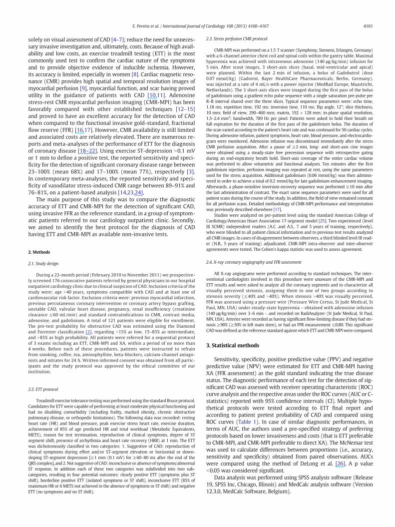
Sensitivity, specificity, positive predictive value (PPV) and negativepredictive value (NPV) were estimated for ETT and CMR-MPI havingXA (FFR assessment) as the gold standard indicating the true diseasestatus. The diagnostic performance of each test for the detection of sig-nificant CAD was assessed with receiver operating characteristic (ROC)curve analysis and the respective areas under the ROC curves (AUC or C-statistics) reported with 95% confidence intervals (CI). Multiple hypo-thetical protocols were tested according to ETT final report andaccording to patient pretest probability of CAD and compared usingROC curves (Table 1). In case of similar diagnostic performances, interms of AUC, the authors used a pre-specified strategy of preferringprotocols based on lower invasiveness and costs (that is ETT preferableto CMR-MPI, and CMR-MPI preferable to direct XA). The McNemar testwas used to calculate differences between proportions (i.e., accuracy,sensitivity and specificity) obtained from paired observations. AUCswere compared using the method of DeLong et al. [26]. A p valueb0.05 was considered significant.
Data analysis was performed using SPSS analysis software (Release19, SPSS Inc, Chicago, Illinois) and MedCalc analysis software (Version12.3.0, MedCalc Software, Belgium).
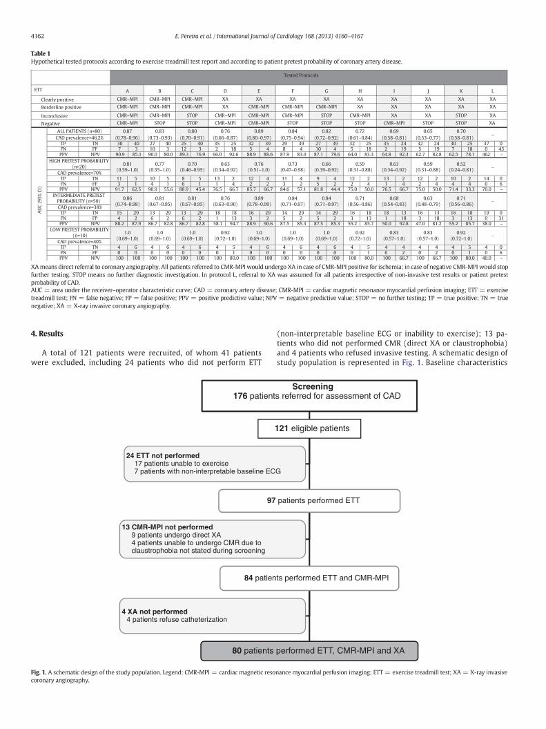
Table 1Hypothetical tested protocols according to exercise treadmill test report and according to patient pretest probability of coronary artery disease.
Tested Protocols
ETT A B C D E G H I J K L
Clearly positive CMR–MPI CMR–MPI CMR–MPI XA XA XA XA XA XA XA XA
Borderline positive CMR–MPI CMR–MPI CMR–MPI XA CMR–MPI CMR–MPI XA XA XA XA XA
Inconclusive CMR–MPI CMR–MPI STOP CMR–MPI CMR–MPI STOP CMR–MPI XA XA STOP XA
Negative CMR–MPI STOP STOP CMR–MPI CMR–MPI STOP STOP CMR–MPI STOP STOP XA
AU
C (
95
% C
I)
ALL PATIENTS (n=80) 0.87
(0.78–0.96)
0.83
(0.73–0.93)
0.80
(0.70–0.91)
0.76
(0.66–0.87)
0.89
(0.80–0.97)
0.82
(0.72–0.92)
0.72
(0.61–0.84)
0.69
(0.58–0.81)
0.65
(0.53–0.77)
0.70
(0.58–0.81)–
CAD prevalence=46.2%TP TN 30 40 27 40 25 40 35 25 32 39 29 39 27 39 32 25 35 24 32 24 30 25 37 0FN FP 7 3 10 3 12 3 2 18 5 4 8 4 10 4 5 18 2 19 5 19 7 18 0 43PPV NPV 90.9 85.1 90.0 80.0 89.3 76.9 66.0 92.6 88.9 88.6 87.9 83.0 87.1 79.6 64.0 83.3 64.8 92.3 62.7 82.8 62.5 78.1 46.2 –
HIGH PRETEST PROBABILITY (n=20)
0.81(0.59–1.0)
0.77(0.55–1.0)
0.70(0.46–0.95)
0.63(0.34–0.92)
0.76(0.51–1.0)
0.66(0.39–0.92)
0.59(0.31–0.88)
0.63(0.34–0.92)
0.59(0.31–0.88)
0.52(0.24–0.81)
–
CAD prevalence=70%TP TN 11 5 10 5 8 5 13 2 12 4 11 4 9 4 12 2 13 2 12 2 10 2 14 0FN FP 3 1 4 1 6 1 1 4 2 2 3 2 5 2 2 4 1 4 2 4 4 4 0 6PPV NPV 91.7 62.5 90.9 55.6 88.9 45.4 76.5 66.7 85.7 66.7 84.6 57.1 81.8 44.4 75.0 50.0 76.5 66.7 75.0 50.0 71.4 33.3 70.0 –
INTERMEDIATE PRETEST PROBABILITY (n=50)
0.86
(0.74–0.98)
0.81
(0.67–0.95)
0.81
(0.67–0.95)
0.76
(0.63–0.90)
0.89
(0.78–0.99)
0.84
(0.71–0.97)
0.71
(0.56–0.86)
0.68
(0.54–0.83)
0.63
(0.48–0.79)
0.71
(0.56–0.86)–
CAD prevalence=38%TP TN 15 29 13 29 13 29 18 18 16 29 14 29 14 29 16 18 18 13 16 13 16 18 19 0FN FP 4 2 6 2 6 2 1 13 3 2 5 2 5 2 3 13 1 18 3 18 3 13 0 31PPV NPV 88.2 87.9 86.7 82.8 86.7 82.8 58.1 94.7 88.9 90.6 87.5 85.3 87.5 85.3 55.2 85.7 50.0 92.8 47.0 81.2 55.2 85.7 38.0 –
LOW PRETEST PROBABILITY
(n=10)1.0
(0.69–1.0)
1.0
(0.69–1.0)
1.0
(0.69–1.0)
0.92
(0.72–1.0)
1.0
(0.69–1.0)
F
XA
CMR–MPI
CMR–MPI
STOP
0.84
(0.75–0.94)
0.73(0.47–0.98)
0.84
(0.71–0.97)
1.0
(0.69–1.0)
1.0
(0.69–1.0)
0.92
(0.72–1.0)
0.83
(0.57–1.0)
0.83
(0.57–1.0)
0.92
(0.72–1.0)–
CAD prevalence=40%TP TN 4 6 4 6 4 6 4 5 4 6 4 6 4 6 4 5 4 4 4 4 4 5 4 0FN FP 0 0 0 0 0 0 0 1 0 0 0 0 0 0 0 1 0 2 0 2 0 1 0 6PPV NPV 100 100 100 100 100 100 100 80.0 100 100 100 100 100 100 100 80.0 100 66.7 100 66.7 100 80.0 40.0 –
XAmeans direct referral to coronary angiography. All patients referred to CMR-MPI would undergo XA in case of CMR-MPI positive for ischemia; in case of negative CMR-MPI would stopfurther testing. STOP means no further diagnostic investigation. In protocol L, referral to XA was assumed for all patients irrespective of non-invasive test results or patient pretestprobability of CAD.AUC = area under the receiver–operator characteristic curve; CAD = coronary artery disease; CMR-MPI = cardiac magnetic resonance myocardial perfusion imaging; ETT = exercisetreadmill test; FN = false negative; FP = false positive; PPV = positive predictive value; NPV = negative predictive value; STOP = no further testing; TP = true positive; TN = truenegative; XA = X-ray invasive coronary angiography.
4162 E. Pereira et al. / International Journal of Cardiology 168 (2013) 4160–4167
4. Results
A total of 121 patients were recruited, of whom 41 patientswere excluded, including 24 patients who did not perform ETT
176 patient
97
84 patien
80 patients
4 XA not performed4 patients refuse catheterization
13 CMR-MPI not performed9 patients undergo direct XA4 patients unable to undergo CMR due toclaustrophobia not stated during screening
24 ETT not performed17 patients unable to exercise7 patients with non-interpretable baseline EC
Fig. 1. A schematic design of the study population. Legend: CMR-MPI = cardiac magnetic resocoronary angiography.
(non-interpretable baseline ECG or inability to exercise); 13 pa-tients who did not performed CMR (direct XA or claustrophobia)and 4 patients who refused invasive testing. A schematic design ofstudy population is represented in Fig. 1. Baseline characteristics
Screenings referred for assessment of CAD
121 eligible patients
patients performed ETT
ts performed ETT and CMR-MPI
performed ETT, CMR-MPI and XA
G
nance myocardial perfusion imaging; ETT = exercise treadmill test; XA = X-ray invasive
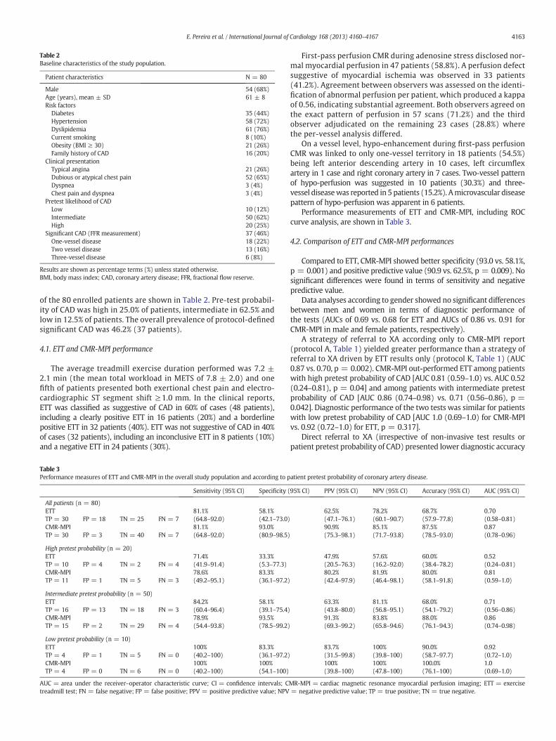
Table 2Baseline characteristics of the study population.
Patient characteristics N = 80
Male 54 (68%)Age (years), mean ± SD 61 ± 8Risk factors
Diabetes 35 (44%)Hypertension 58 (72%)Dyslipidemia 61 (76%)Current smoking 8 (10%)Obesity (BMI ≥ 30) 21 (26%)Family history of CAD 16 (20%)
Clinical presentationTypical angina 21 (26%)Dubious or atypical chest pain 52 (65%)Dyspnea 3 (4%)Chest pain and dyspnea 3 (4%)
Pretest likelihood of CADLow 10 (12%)Intermediate 50 (62%)High 20 (25%)
Significant CAD (FFR measurement) 37 (46%)One-vessel disease 18 (22%)Two vessel disease 13 (16%)Three-vessel disease 6 (8%)
Results are shown as percentage terms (%) unless stated otherwise.BMI, body mass index; CAD, coronary artery disease; FFR, fractional flow reserve.
4163E. Pereira et al. / International Journal of Cardiology 168 (2013) 4160–4167
of the 80 enrolled patients are shown in Table 2. Pre-test probabil-ity of CAD was high in 25.0% of patients, intermediate in 62.5% andlow in 12.5% of patients. The overall prevalence of protocol-definedsignificant CAD was 46.2% (37 patients).
4.1. ETT and CMR-MPI performance
The average treadmill exercise duration performed was 7.2 ±2.1 min (the mean total workload in METS of 7.8 ± 2.0) and onefifth of patients presented both exertional chest pain and electro-cardiographic ST segment shift ≥1.0 mm. In the clinical reports,ETT was classified as suggestive of CAD in 60% of cases (48 patients),including a clearly positive ETT in 16 patients (20%) and a borderlinepositive ETT in 32 patients (40%). ETT was not suggestive of CAD in 40%of cases (32 patients), including an inconclusive ETT in 8 patients (10%)and a negative ETT in 24 patients (30%).
Table 3Performance measures of ETT and CMR-MPI in the overall study population and according to p
Sensitivity (95% CI) Specificity (
All patients (n = 80)ETT 81.1% 58.1%TP = 30 FP = 18 TN = 25 FN = 7 (64.8–92.0) (42.1–73.0)CMR-MPI 81.1% 93.0%TP = 30 FP = 3 TN = 40 FN = 7 (64.8–92.0) (80.9–98.5)
High pretest probability (n = 20)ETT 71.4% 33.3%TP = 10 FP = 4 TN = 2 FN = 4 (41.9–91.4) (5.3–77.3)CMR-MPI 78.6% 83.3%TP = 11 FP = 1 TN = 5 FN = 3 (49.2–95.1) (36.1–97.2)
Intermediate pretest probability (n = 50)ETT 84.2% 58.1%TP = 16 FP = 13 TN = 18 FN = 3 (60.4–96.4) (39.1–75.4)CMR-MPI 78.9% 93.5%TP = 15 FP = 2 TN = 29 FN = 4 (54.4–93.8) (78.5–99.2)
Low pretest probability (n = 10)ETT 100% 83.3%TP = 4 FP = 1 TN = 5 FN = 0 (40.2–100) (36.1–97.2)CMR-MPI 100% 100%TP = 4 FP = 0 TN = 6 FN = 0 (40.2–100) (54.1–100)
AUC = area under the receiver–operator characteristic curve; CI = confidence intervals; Ctreadmill test; FN = false negative; FP = false positive; PPV = positive predictive value; NPV
First-pass perfusion CMR during adenosine stress disclosed nor-mal myocardial perfusion in 47 patients (58.8%). A perfusion defectsuggestive of myocardial ischemia was observed in 33 patients(41.2%). Agreement between observers was assessed on the identi-fication of abnormal perfusion per patient, which produced a kappaof 0.56, indicating substantial agreement. Both observers agreed onthe exact pattern of perfusion in 57 scans (71.2%) and the thirdobserver adjudicated on the remaining 23 cases (28.8%) wherethe per-vessel analysis differed.
On a vessel level, hypo-enhancement during first-pass perfusionCMR was linked to only one-vessel territory in 18 patients (54.5%)being left anterior descending artery in 10 cases, left circumflexartery in 1 case and right coronary artery in 7 cases. Two-vessel patternof hypo-perfusion was suggested in 10 patients (30.3%) and three-vessel diseasewas reported in 5 patients (15.2%). Amicrovascular diseasepattern of hypo-perfusion was apparent in 6 patients.
Performance measurements of ETT and CMR-MPI, including ROCcurve analysis, are shown in Table 3.
4.2. Comparison of ETT and CMR-MPI performances
Compared to ETT, CMR-MPI showed better specificity (93.0 vs. 58.1%,p = 0.001) and positive predictive value (90.9 vs. 62.5%, p = 0.009). Nosignificant differences were found in terms of sensitivity and negativepredictive value.
Data analyses according to gender showed no significant differencesbetween men and women in terms of diagnostic performance ofthe tests (AUCs of 0.69 vs. 0.68 for ETT and AUCs of 0.86 vs. 0.91 forCMR-MPI in male and female patients, respectively).
A strategy of referral to XA according only to CMR-MPI report(protocol A, Table 1) yielded greater performance than a strategy ofreferral to XA driven by ETT results only (protocol K, Table 1) (AUC0.87 vs. 0.70, p = 0.002). CMR-MPI out-performed ETT among patientswith high pretest probability of CAD [AUC 0.81 (0.59–1.0) vs. AUC 0.52(0.24–0.81), p = 0.04] and among patients with intermediate pretestprobability of CAD [AUC 0.86 (0.74–0.98) vs. 0.71 (0.56–0.86), p =0.042]. Diagnostic performance of the two tests was similar for patientswith low pretest probability of CAD [AUC 1.0 (0.69–1.0) for CMR-MPIvs. 0.92 (0.72–1.0) for ETT, p = 0.317].
Direct referral to XA (irrespective of non-invasive test results orpatient pretest probability of CAD) presented lower diagnostic accuracy
atient pretest probability of coronary artery disease.
95% CI) PPV (95% CI) NPV (95% CI) Accuracy (95% CI) AUC (95% CI)
62.5% 78.2% 68.7% 0.70(47.1–76.1) (60.1–90.7) (57.9–77.8) (0.58–0.81)90.9% 85.1% 87.5% 0.87(75.3–98.1) (71.7–93.8) (78.5–93.0) (0.78–0.96)
47.9% 57.6% 60.0% 0.52(20.5–76.3) (16.2–92.0) (38.4–78.2) (0.24–0.81)80.2% 81.9% 80.0% 0.81(42.4–97.9) (46.4–98.1) (58.1–91.8) (0.59–1.0)
63.3% 81.1% 68.0% 0.71(43.8–80.0) (56.8–95.1) (54.1–79.2) (0.56–0.86)91.3% 83.8% 88.0% 0.86(69.3–99.2) (65.8–94.6) (76.1–94.3) (0.74–0.98)
83.7% 100% 90.0% 0.92(31.5–99.8) (39.8–100) (58.7–97.7) (0.72–1.0)100% 100% 100.0% 1.0(39.8–100) (47.8–100) (76.1–100) (0.69–1.0)
MR-MPI = cardiac magnetic resonance myocardial perfusion imaging; ETT = exercise= negative predictive value; TP = true positive; TN = true negative.
4164 E. Pereira et al. / International Journal of Cardiology 168 (2013) 4160–4167
for significant CAD (protocol L — Table 1, accuracy of 46.2%) thanselecting patients for invasive stratification according to ETT report(protocol K, 68.7%, p = 0.003) or according to CMR-MPI findings(protocol A, 87.5%, p b 0.001).
4.3. Integrated protocols including pretest probability of CAD, ETT andCMR-MPI
4.3.1. CMR-MPI performance according to ETT result (Table 1)In the global population, performing CMR-MPI prior to XA in
patients with a clearly positive ETT (protocol A, AUC 0.87) did notimprove diagnostic performance for CAD detection over direct XAreferral (protocol E, AUC 0.89), p = ns. Of the 16 patients with aclearly positive ETT (symptoms plus ST shift), 13 patients (81.2%)presented significant CAD on XA. In this group, CMR-MPI excludedischemia in only 1 of the 3 patients without CAD. Referring toCMR-MPI prior to XA in patients with borderline positive ETT(symptoms or ST shift) improved diagnostic performance (protocols Evs. D, p = 0.004). In patients with an inconclusive ETT, the addition ofCMR-MPI also improved diagnostic performance as compared todirect XA (protocols D vs. I, p = 0.009). Interestingly, in patients witha negative ETT, CMR-MPI performance showed some advantage overno further testing (protocols E vs. F, p = 0.075).
Accordingly, the best protocol integrating ETT and CMR-MPI for CADdetection, irrespective of patient pretest probability of CAD, wasprotocol E (AUC of 0.89; 95% CI 0.80–0.97), in which CMR-MPI wasperformed in all patients but those with a clearly positive ETT, towhom direct referral to XA was assumed. Of the 64 patients with aborderline positive, inconclusive or negative ETT, 40 patients (62.5%)presented no significant functional CAD on XA and 24 patients (37.5%)had significant disease — diagnostic accuracy of 65.6% (TP = 17,FP = 15, TN = 25, FN = 7). In the same group, diagnostic accuracyof CMR-MPI reached 90.6%, reducing the false positive and negativecases to 5 patients and 1 patient, respectively. This means that addition-al testing with stress imaging should be offered after a borderlinepositive, inconclusive or negative ETT. In Fig. 2, we present four repre-sentative cases showing true positive results of both tests (case A) andthe additive value of CMR-MPI over ETT (cases B–D).
4.3.2. CMR-MPI performance according to pre-test probability of CAD(Table 1)
Hypothetical protocols based on patient pretest probability of CADwere tested and the results are shown in Table 1.
For patients with high pretest probability of CAD, a strategy ofdirect referral to XA was less accurate than selecting patients forinvasive stratification according to CMR-MPI report (protocol Laccuracy 70.0% vs. protocol A 80.0%, p = 0.73). Protocol K (AUC0.52) based solely on ETT performance showed worst performancethan protocol A (AUC 0.81), based on CMR-MPI only, p = 0.04.CMR-MPI was particularly useful to redefine the need for invasive in-vestigation among patients with ETT suggestive of CAD (protocol A,AUC 0.81 vs. protocol D, AUC 0.63, p = ns), reducing the rate offalse positives. In this group, ETT is useless to select patients eitherto CMR-MPI or to XA. Protocol A was the best performing protocol(AUC 0.81, highest PPV) for patients with high pretest probabilityof CAD, in which CMR-MPI was performed prior to XA in all patients,irrespective of the ETT report.
For patients with intermediate pretest probability of CAD, a strat-egy of referral to XA based on only CMR-MPI result (protocol A accu-racy 88.0%) was significantly more accurate than a strategy based ononly ETT report (protocol K, 68.0%, p = 0.03) and a strategy of directreferral to XA (protocol L 38.0%, b0.001). In this group, CMR-MPIprior to XA was particularly useful to redefine the need for invasiveinvestigation in patients with borderline positive ETT (protocols Evs. D, p = 0.028), inconclusive ETT (protocols D vs. I, p = 0.016)and negative ETT (protocols I vs. L, p b 0.001). For patients with a
clinically and electrically positive ETT, CMR-MPI didn't improvethe diagnostic performance of ETT (protocols E vs. A, p = ns). Thebest performing protocol for patients with intermediate likelihoodof CAD was protocol E (AUC 0.89), in which CMR-MPI was assumedfor all patients prior to XA, except for those with a positive ETT whowould undergo direct XA.
For patients with low probability of CAD and a negative ETT,assuming no further investigation was as accurate as performingCMR-MPI (protocol J vs. protocol I, AUC 0.83 for both, p = ns). Alsofor patients with an inconclusive ETT, no further testing strategy(protocol K, AUC 0.92) showed similar performance than referringto CMR-MPI (protocol H, AUC 0.92), p = ns, and greater performancethan sending to direct XA (protocol J, AUC 0.83), p = ns. In thisgroup, no false negative results were reported for any protocol. Pa-tients with a borderline positive ETT would benefit from CMR-MPIprior to XA (protocol G, AUC 1.0) instead of direct referral to XA (pro-tocol K, AUC 0.92), p = ns, excluding the false positive result report-ed. For patients with positive ETT, no false positive results werereported when direct referral to XA was assumed (protocols E vs.protocol A, AUC 1.0 for both, p = ns). However, only one patient ofthis group presented a clearly positive ETT and assuming direct re-ferral to XA based on only one case report wouldn't be wise. Accord-ingly, protocol C was chosen as the best performing protocol (AUC1.0) for patients with low pretest probability of CAD.
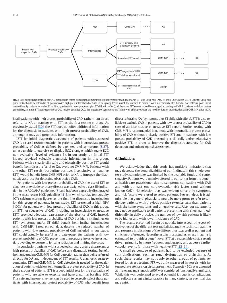
Based on the above results, in our population, the best performingalgorithm for the management of patients with suspected CAD,according to patient pretest probability of CAD, is presented inFig. 3. This protocol achieved a global accuracy of 89% (AUC = 0.88,95% CI 0.80–0.97) and was clearly superior to an approach based solelyin ETT (protocol K, AUC 0.70, p b 0.001), yet similar to isolated CMR-MPI (protocol A, AUC 0.87, p = ns). Interestingly, its diagnosticperformance did not significantly differ from an approach of performingETT to all patients, referring to XA the clearly positives and submittingto CMR-MPI all the others (protocol E, AUC = 0.89), p = ns. Thealgorithm based on pretest probability, however, implies lower costssince some patients only perform CMR-MPI and no ETT and a few othersmay be exempted from further testing after a negative ETT.
5. Discussion
In our population with a high prevalence of CAD (46.2%), ETTpresented a higher level of sensitivity (81.1%) and a lower level ofspecificity (58.1%) than previously reported [18]. Sensitivity andspecificity are inversely related, affected by the population tested(namely pretest probability and CAD prevalence [22]) and deter-mined by the methodology used, including the choice of a cut pointor discriminant value [8]. Our interpretation of the ETT includedsymptomatic response, exercise capacity, hemodynamic response,and ECG response as recommend in guidelines [8]. We followed themost commonly used definition for a positive ETT that is ≥1 mm ofhorizontal or downsloping ST-segment depression or elevation forN60 to 80 ms after the end of the QRS complex, especially whenthe ECG changes are accompanied by chest pain suggestive of angina[27]. If the discriminant value used was set high (ST depression≥2 mm) to ensure that nearly all normal subjects would have a nor-mal test, giving the test a high specificity (95.3%), then a substantialnumber of those with the disease would appear to be normal, reduc-ing the test sensitivity (48.6%). The low level of specificity of ETT inour population may also be explained by the inclusion of borderlinepositive reports (symptoms or ST shift during effort) in the group of ETTsuggestive of CAD, raising the rate of false positives from 3 to 18 cases(accordingly reducing specificity from 93.0 to 58.1%). However, if onlyclearly positive ETT reports were to be considered as positive tests,the sensitivity of ETT would diminish to 35.1%. The diagnostic perfor-mance of ETT in our study was also driven by the high prevalence ofCAD in our population, increasing the chance of establishing a diagnosis.
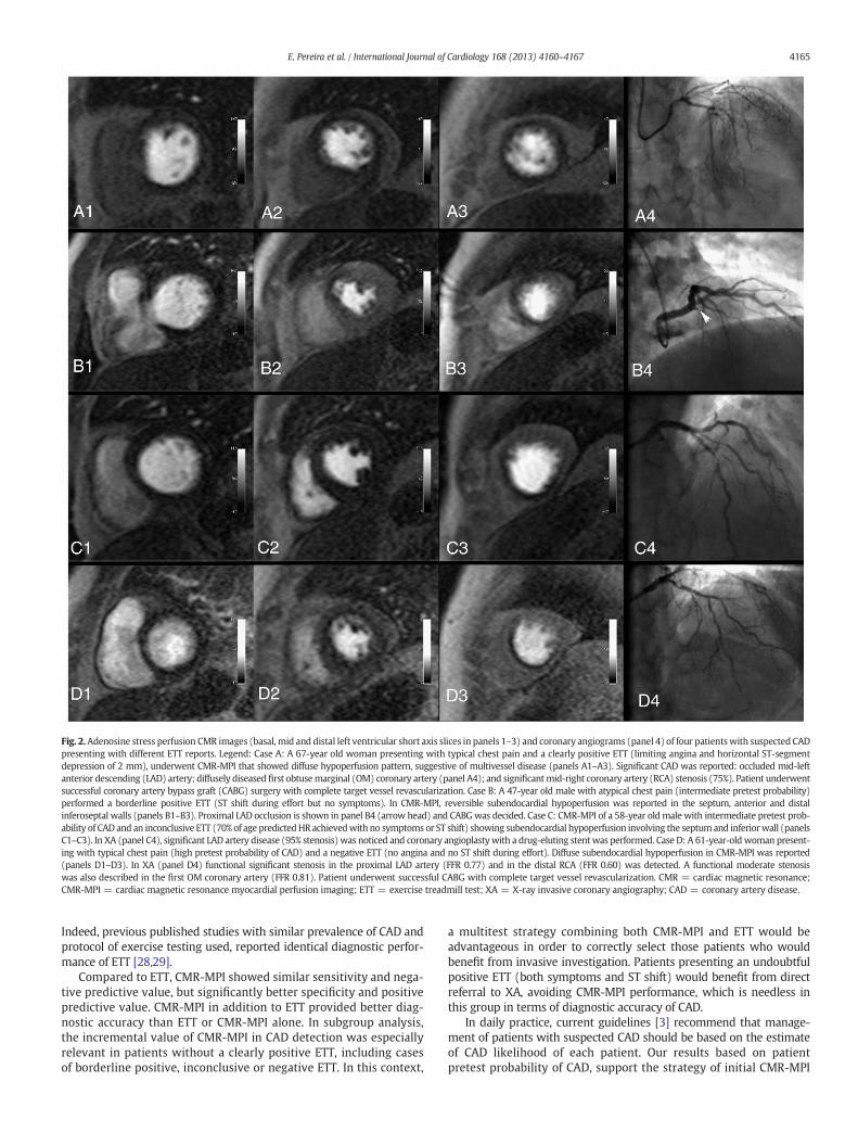
Fig. 2.Adenosine stress perfusion CMR images (basal, mid and distal left ventricular short axis slices in panels 1–3) and coronary angiograms (panel 4) of four patientswith suspected CADpresenting with different ETT reports. Legend: Case A: A 67-year old woman presenting with typical chest pain and a clearly positive ETT (limiting angina and horizontal ST-segmentdepression of 2 mm), underwent CMR-MPI that showed diffuse hypoperfusion pattern, suggestive of multivessel disease (panels A1–A3). Significant CAD was reported: occluded mid-leftanterior descending (LAD) artery; diffusely diseased first obtusemarginal (OM) coronary artery (panel A4); and significantmid-right coronary artery (RCA) stenosis (75%). Patient underwentsuccessful coronary artery bypass graft (CABG) surgery with complete target vessel revascularization. Case B: A 47-year old male with atypical chest pain (intermediate pretest probability)performed a borderline positive ETT (ST shift during effort but no symptoms). In CMR-MPI, reversible subendocardial hypoperfusion was reported in the septum, anterior and distalinferoseptal walls (panels B1–B3). Proximal LAD occlusion is shown in panel B4 (arrow head) and CABGwas decided. Case C: CMR-MPI of a 58-year oldmale with intermediate pretest prob-ability of CAD and an inconclusive ETT (70% of age predictedHR achievedwith no symptoms or ST shift) showing subendocardial hypoperfusion involving the septumand inferiorwall (panelsC1–C3). In XA (panel C4), significant LAD artery disease (95% stenosis) was noticed and coronary angioplastywith a drug-eluting stent was performed. Case D: A 61-year-oldwoman present-ing with typical chest pain (high pretest probability of CAD) and a negative ETT (no angina and no ST shift during effort). Diffuse subendocardial hypoperfusion in CMR-MPI was reported(panels D1–D3). In XA (panel D4) functional significant stenosis in the proximal LAD artery (FFR 0.77) and in the distal RCA (FFR 0.60) was detected. A functional moderate stenosiswas also described in the first OM coronary artery (FFR 0.81). Patient underwent successful CABG with complete target vessel revascularization. CMR = cardiac magnetic resonance;CMR-MPI = cardiac magnetic resonance myocardial perfusion imaging; ETT = exercise treadmill test; XA = X-ray invasive coronary angiography; CAD = coronary artery disease.
4165E. Pereira et al. / International Journal of Cardiology 168 (2013) 4160–4167
Indeed, previous published studies with similar prevalence of CAD andprotocol of exercise testing used, reported identical diagnostic perfor-mance of ETT [28,29].
Compared to ETT, CMR-MPI showed similar sensitivity and nega-tive predictive value, but significantly better specificity and positivepredictive value. CMR-MPI in addition to ETT provided better diag-nostic accuracy than ETT or CMR-MPI alone. In subgroup analysis,the incremental value of CMR-MPI in CAD detection was especiallyrelevant in patients without a clearly positive ETT, including casesof borderline positive, inconclusive or negative ETT. In this context,
a multitest strategy combining both CMR-MPI and ETT would beadvantageous in order to correctly select those patients who wouldbenefit from invasive investigation. Patients presenting an undoubtfulpositive ETT (both symptoms and ST shift) would benefit from directreferral to XA, avoiding CMR-MPI performance, which is needless inthis group in terms of diagnostic accuracy of CAD.
In daily practice, current guidelines [3] recommend that manage-ment of patients with suspected CAD should be based on the estimateof CAD likelihood of each patient. Our results based on patientpretest probability of CAD, support the strategy of initial CMR-MPI
Patient withsuspected CAD
Pretest probability ofCAD
High CMR-MPI XA
Intermediate
ETT clinically andelectrically positive
XA
Any other ETT result CMR-MPI XA
Low
ETT sugestive of CAD
(ST shift or symptoms)CMR-MPI XA
ETT not suggestive ofCAD
STOP
Fig. 3. Best performing protocol for CAD diagnosis in tested population combining patient pretest probability of CAD, ETT and CMR-MPI (AUC = 0.88, 95% CI 0.80–0.97). Legend: CMR-MPIprior to XA should be offered in all patients with high pretest likelihood of CAD; in this group ETT is a usefulness exam. In patients with intermediate likelihood of CAD, ETT is a good initialtest to identify patients who should be directly referred to XA (symptoms plus ST shift with effort); all the other ETT results should bemanaged according to CMR. In patients with low pretestprobability, an initial ETT not suggestive of CAD reliably excludes CAD; the presence of symptoms or ST shift with effort precludes the need for further investigationwith CMR-MPI prior to XA.
4166 E. Pereira et al. / International Journal of Cardiology 168 (2013) 4160–4167
in all patients with high pretest probability of CAD, rather than directreferral to XA or starting with ETT, as the first testing strategy. Aspreviously stated [30], the ETT does not offer additional informationfor the diagnosis in patients with high pretest probability of CAD,although it may add prognostic information.
ETT for initial diagnostic assessment of patients with suspectedCAD is a class I recommendation in patients with intermediate pretestprobability of CAD as defined by age, sex, and symptoms [8,27],unless unable to exercise or display ECG changes which make ECGnon-evaluable (level of evidence B). In our study, an initial ETTindeed provided valuable diagnostic information in this group.Patients with a clearly clinically and electrically positive ETT wouldbenefit from direct referral to XA, avoiding CMR-MPI. Patients withany other ETT result (borderline positive, inconclusive or negativeETT) would benefit from CMR-MPI prior to XA to improve the diag-nostic accuracy for detecting obstructive CAD.
For patients with low pretest probability of CAD, the use of ETT todiagnose or exclude coronary disease was assigned to a class IIb indica-tion in the ACC/AHA guidelines [8] and has been expressly discouragedin the most recent NICE guidelines [31], in which cardiac tomography(CT) calcium scoring figures as the first-line diagnostic investigationfor this group of patients. In our study, ETT presented a high NPV(100%) for patients with low pretest probability of CAD. In this group,an ETT not suggestive of CAD (including an inconclusive or negativeETT) provided adequate reassurance of the absence of CAD. Instead,patients with low pretest probability of CAD but high risk findings onETT (symptoms and/or ST shift) benefit from further investigationwith CMR-MPI. Based on our data, despite the reduced number ofpatients with low pretest probability of CAD included in our study,ETT could actually be useful as a gatekeeper for patients with lowpretest probability of CAD, preventing unnecessary invasive investiga-tion, avoiding exposure to ionizing radiation and limiting the costs.
In conclusion, patients with suspected coronary artery disease and ahigh pretest probability of CAD before any diagnostic testing, benefitfromundergoing CMR-MPI for CAD detection rather than being referreddirectly for XA and independent of ETT results. A diagnostic strategycombining ETT and CMR-MPI for CAD detection is needed for the evalu-ation of patients with low to intermediate pretest probability of CAD. Inthese groups of patients, ETT is a good initial test for the evaluation ofpatients who are able to exercise and have a normal baseline ECG.This safe and inexpensive test can be used to correctly select those pa-tients with intermediate pretest probability of CAD who benefit from
direct referral to XA (symptoms plus ST shift with effort). ETT is also re-liable to exclude CAD in patients with low pretest probability of CAD incase of an inconclusive or negative ETT report. Further testing withCMR-MPI is recommended in patients with intermediate pretest proba-bility of CAD without a clearly positive ETT and in patients with lowpretest probability of CAD presenting a clinically and/or electricallypositive ETT, in order to improve the diagnostic accuracy for CADdetection and enhancing risk assessment.
6. Limitations
We acknowledge that this study has multiple limitations thatmay decrease the generalizability of our findings. In this single-cen-ter study, sample size was limited by the available funds and centercapacity. Patients weremainly referred to our center from the gener-al health care practitioner due to symptoms compatible with CADand with at least one cardiovascular risk factor (and withoutknown CAD). No selection bias was evident since only symptomsand risk factors were used to select patients. Nevertheless, it is ad-missible that general physicians would be more prone to refer to car-diology patients with previous positive exercise tests than patientswith the same symptoms and a negative test. Also, our statementsmay not be applicable to all patients presenting with chest pain. Ad-ditionally, in daily practice, the number of low risk patients is likelyto be higher and with lower incidence of CAD.
The results presented herein do not take into account the cost ef-fectiveness of the different test modalities and the technical, trainingand resource implications of the different tests, as well as patient andclinician preferences. Nevertheless, in most studies, stress imaging isestimated to provide a benefit over ETT at a reasonable cost, a resultdriven primarily by more frequent angiography and adverse cardio-vascular events for those with negative ETT [32–34].
A small percentage of patients had to be excluded because ofcontraindications, such as renal dysfunction or arrhythmia. Assuch, these results may not apply to other groups of patients re-ferred for stress testing. FFR was only measured in vessels with in-termediate stenosis on visual assessment. Stenosis b50% was assumedas irrelevant and stenosis≥90%was considered functionally significant.While this was performed to avoid potential iatrogenic complications,and reflects current clinical practice in many centers, an eventual biasmay exist.
4167E. Pereira et al. / International Journal of Cardiology 168 (2013) 4160–4167
7. Conclusions
CMR-MPI has similar sensitivity and higher specificity for CADdetection compared with ETT. CMR-MPI can be used in a diagnosticworkflow aiming to increase accuracy and reduce the number ofunnecessary catheterizations, particularly in patients with interme-diate to high pretest probability of CAD. ETT performed well in thelow pretest probability patients and seems to be still a valuableoption to exclude CAD in this group of patients.
References
[1] Rosamond W, Flegal K, Furie K, et al. Heart disease and stroke statistics—2008update: a report from the American Heart Association Statistics Committee andStroke Statistics Subcommittee. Circulation 2008;117:e25–146.
[2] Diamond GA, Forrester JS. Analysis of probability as an aid in the clinical diagnosis ofcoronary-artery disease. N Engl J Med 1979;300:1350–8.
[3] Fox K, Garcia MA, Ardissino D, et al. Guidelines on the management of stable anginapectoris: executive summary: The Task Force on the Management of Stable AnginaPectoris of the European Society of Cardiology. Eur Heart J 2006;27:1341–81.
[4] BordenWB, Redberg RF, Mushlin AI, et al. Patterns and intensity of medical therapy inpatients undergoing percutaneous coronary intervention. JAMA 2011;305:1882–9.
[5] Shaw LJ, Weintraub WS, Maron DJ, et al. Baseline stress myocardial perfusion imag-ing results and outcomes in patients with stable ischemic heart disease randomizedto optimal medical therapy with or without percutaneous coronary intervention.Am Heart J 2012;164:243–50.
[6] Tonino PA, De Bruyne B, Pijls NH, et al. Fractional flow reserve versus angiographyfor guiding percutaneous coronary intervention. N Engl J Med 2009;360:213–24.
[7] De Bruyne B, Pijls NH, Kalesan B, et al. Fractional flow reserve-guided PCI versusmedical therapy in stable coronary disease. N Engl J Med 2012;367:991–1001.
[8] Gibbons RJ, Balady GJ, Bricker JT, et al. ACC/AHA 2002 guideline update for exercisetesting: summary article. A report of the American College of Cardiology/AmericanHeart Association Task Force on Practice Guidelines (Committee to Update the1997 Exercise Testing Guidelines). J Am Coll Cardiol 2002;40:1531–40.
[9] Nagel E, Klein C, Paetsch I, et al. Magnetic resonance perfusionmeasurements for thenoninvasive detection of coronary artery disease. Circulation 2003;108:432–7.
[10] Morton G, Schuster A, Perera D, et al. Cardiac magnetic resonance imaging to guidecomplex revascularization in stable coronary artery disease. Eur Heart J 2010;31:2209–15.
[11] Hussain ST, Paul M, Plein S, et al. Design and rationale of the MR-INFORM study:stress perfusion cardiovascular magnetic resonance imaging to guide the manage-ment of patients with stable coronary artery disease. J Cardiovasc Magn Reson2012;14:65.
[12] Greenwood JP, Maredia N, Younger JF, et al. Cardiovascular magnetic resonance andsingle-photon emission computed tomography for diagnosis of coronary heart dis-ease (CE-MARC): a prospective trial. Lancet 2012;379:453–60.
[13] Schwitter J,Wacker CM,WilkeN, et al.MR-IMPACT II:Magnetic Resonance Imaging forMyocardial Perfusion Assessment in Coronary artery disease Trial: perfusion-cardiacmagnetic resonance vs. single-photon emission computed tomography for the detec-tion of coronary artery disease: a comparative multicentre, multivendor trial. EurHeart J 2013;34:775–81.
[14] Nandalur KR, Dwamena BA, Choudhri AF, et al. Diagnostic performance of stresscardiac magnetic resonance imaging in the detection of coronary artery disease: ameta-analysis. J Am Coll Cardiol 2007;50:1343–53.
[15] Morton G, Chiribiri A, IshidaM, et al. Quantification of absolutemyocardial perfusionin patients with coronary artery disease: comparison between cardiovascular
magnetic resonance and positron emission tomography. J Am Coll Cardiol2012;60:1546–55.
[16] Watkins S, McGeoch R, Lyne J, et al. Validation of magnetic resonance myocardialperfusion imagingwith fractional flow reserve for the detection of significant coronaryheart disease. Circulation 2009;120:2207–13.
[17] Bettencourt N, Chiribiri A, Schuster A, et al. Cardiac magnetic resonance myocardialperfusion imaging for detection of functionally significant obstructive coronaryartery disease: a prospective study. Int J Cardiol 2012. http://dx.doi.org/10.1016/j.ijcard.2012.09.231.
[18] Gianrossi R, Detrano R, Mulvihill D, et al. Exercise-induced ST depression in thediagnosis of coronary artery disease. A meta-analysis. Circulation 1989;80:87–98.
[19] Kwok Y, Kim C, Grady D, et al. Meta-analysis of exercise testing to detect coronaryartery disease in women. Am J Cardiol 1999;83:660–6.
[20] Gibson RS. The diagnostic and prognostic value of exercise electrocardiography inasymptomatic subjects and stable symptomatic patients. Curr Opin Cardiol 1991;6:536–46.
[21] Ashley EA, Myers J, Froelicher V. Exercise testing in clinical medicine. Lancet2000;356:1592–7.
[22] Banerjee A, Newman DR, Van den Bruel A, et al. Diagnostic accuracy of exercisestress testing for coronary artery disease: a systematic review and meta-analysisof prospective studies. Int J Clin Pract 2012;66:477–92.
[23] Jaarsma C, Leiner T, Bekkers SC, et al. Diagnostic performance of noninvasive myo-cardial perfusion imaging using single-photon emission computed tomography, car-diac magnetic resonance, and positron emission tomography imaging for thedetection of obstructive coronary artery disease: a meta-analysis. J Am Coll Cardiol2012;59:1719–28.
[24] Hamon M, Fau G, Nee G, et al. Meta-analysis of the diagnostic performance of stressperfusion cardiovascular magnetic resonance for detection of coronary artery dis-ease. J Cardiovasc Magn Reson 2010;12:29.
[25] Cerqueira MD, Weissman NJ, Dilsizian V, et al. Standardized myocardial segmenta-tion and nomenclature for tomographic imaging of the heart: a statement forhealthcare professionals from the Cardiac Imaging Committee of the Council on Clin-ical Cardiology of the American Heart Association. Circulation 2002;105:539–42.
[26] DeLong ER, DeLong DM, Clarke-Pearson DL. Comparing the areas under two ormore correlated receiver operating characteristic curves: a nonparametric ap-proach. Biometrics 1988;44:837–45.
[27] Members C, Gibbons RJ, Chatterjee K, et al. ACC/AHA/ACP–ASIM guidelines forthe management of patients with chronic stable angina: executive summaryand recommendations. Circulation 1999;99:2829–48.
[28] Hung J, Chaitman BR, Lam J, et al. Noninvasive diagnostic test choices for theevaluation of coronary artery disease in women: a multivariate comparison ofcardiac fluoroscopy, exercise electrocardiography and exercise thallium myocardialperfusion scintigraphy. J Am Coll Cardiol 1984;4:8–16.
[29] Rollan MJ, San Roman JA, Vilacosta I, et al. Dobutamine stress echocardiography inthe diagnosis of coronary artery disease in women with chest pain: comparisonwith different noninvasive tests. Clin Cardiol 2002;25:559–64.
[30] Hachamovitch R, Hayes SW, Friedman JD, et al. Stress myocardial perfusionsingle-photon emission computed tomography is clinically effective and cost effec-tive in risk stratification of patients with a high likelihood of coronary artery disease(CAD) but no known CAD. J Am Coll Cardiol 2004;43:200–8.
[31] Skinner JS, Smeeth L, Kendall JM, et al. NICE guidance. Chest pain of recent onset:assessment and diagnosis of recent onset chest pain or discomfort of suspectedcardiac origin. Heart 2010;96:974–8.
[32] Garber AM, Solomon NA. Cost-effectiveness of alternative test strategies for thediagnosis of coronary artery disease. Ann Intern Med 1999;130:719–28.
[33] Lorenzoni R, Cortigiani L, Magnani M, et al. Cost-effectiveness analysis of noninvasivestrategies to evaluate patients with chest pain. J Am Soc Echocardiogr 2003;16:1287–91.
[34] Kuntz KM, Fleischmann KE, Hunink MG, et al. Cost-effectiveness of diagnostic strat-egies for patients with chest pain. Ann Intern Med 1999;130:709–18.
Related Documents