Cabico, John Friedman and George A. Diamond Rory Hachamovitch, Daniel S. Berman, Leslee J. Shaw, Hosen Kiat, Ishac Cohen, J. Arthur Cardiac Death and Myocardial Infarction Tomography for the Prediction of Cardiac Death: Differential Stratification for Risk of Incremental Prognostic Value of Myocardial Perfusion Single Photon Emission Computed Print ISSN: 0009-7322. Online ISSN: 1524-4539 Copyright © 1998 American Heart Association, Inc. All rights reserved. is published by the American Heart Association, 7272 Greenville Avenue, Dallas, TX 75231 Circulation doi: 10.1161/01.CIR.97.6.535 1998;97:535-543 Circulation. http://circ.ahajournals.org/content/97/6/535 World Wide Web at: The online version of this article, along with updated information and services, is located on the http://circ.ahajournals.org//subscriptions/ is online at: Circulation Information about subscribing to Subscriptions: http://www.lww.com/reprints Information about reprints can be found online at: Reprints: document. Permissions and Rights Question and Answer this process is available in the click Request Permissions in the middle column of the Web page under Services. Further information about Office. Once the online version of the published article for which permission is being requested is located, can be obtained via RightsLink, a service of the Copyright Clearance Center, not the Editorial Circulation in Requests for permissions to reproduce figures, tables, or portions of articles originally published Permissions: by guest on November 1, 2013 http://circ.ahajournals.org/ Downloaded from by guest on November 1, 2013 http://circ.ahajournals.org/ Downloaded from by guest on November 1, 2013 http://circ.ahajournals.org/ Downloaded from by guest on November 1, 2013 http://circ.ahajournals.org/ Downloaded from by guest on November 1, 2013 http://circ.ahajournals.org/ Downloaded from by guest on November 1, 2013 http://circ.ahajournals.org/ Downloaded from by guest on November 1, 2013 http://circ.ahajournals.org/ Downloaded from by guest on November 1, 2013 http://circ.ahajournals.org/ Downloaded from by guest on November 1, 2013 http://circ.ahajournals.org/ Downloaded from by guest on November 1, 2013 http://circ.ahajournals.org/ Downloaded from

Welcome message from author
This document is posted to help you gain knowledge. Please leave a comment to let me know what you think about it! Share it to your friends and learn new things together.
Transcript
Cabico, John Friedman and George A. DiamondRory Hachamovitch, Daniel S. Berman, Leslee J. Shaw, Hosen Kiat, Ishac Cohen, J. Arthur
Cardiac Death and Myocardial InfarctionTomography for the Prediction of Cardiac Death: Differential Stratification for Risk of
Incremental Prognostic Value of Myocardial Perfusion Single Photon Emission Computed
Print ISSN: 0009-7322. Online ISSN: 1524-4539 Copyright © 1998 American Heart Association, Inc. All rights reserved.
is published by the American Heart Association, 7272 Greenville Avenue, Dallas, TX 75231Circulation doi: 10.1161/01.CIR.97.6.535
1998;97:535-543Circulation.
http://circ.ahajournals.org/content/97/6/535World Wide Web at:
The online version of this article, along with updated information and services, is located on the
http://circ.ahajournals.org//subscriptions/
is online at: Circulation Information about subscribing to Subscriptions:
http://www.lww.com/reprints Information about reprints can be found online at: Reprints:
document. Permissions and Rights Question and Answer this process is available in the
click Request Permissions in the middle column of the Web page under Services. Further information aboutOffice. Once the online version of the published article for which permission is being requested is located,
can be obtained via RightsLink, a service of the Copyright Clearance Center, not the EditorialCirculationin Requests for permissions to reproduce figures, tables, or portions of articles originally publishedPermissions:
by guest on November 1, 2013http://circ.ahajournals.org/Downloaded from by guest on November 1, 2013http://circ.ahajournals.org/Downloaded from by guest on November 1, 2013http://circ.ahajournals.org/Downloaded from by guest on November 1, 2013http://circ.ahajournals.org/Downloaded from by guest on November 1, 2013http://circ.ahajournals.org/Downloaded from by guest on November 1, 2013http://circ.ahajournals.org/Downloaded from by guest on November 1, 2013http://circ.ahajournals.org/Downloaded from by guest on November 1, 2013http://circ.ahajournals.org/Downloaded from by guest on November 1, 2013http://circ.ahajournals.org/Downloaded from by guest on November 1, 2013http://circ.ahajournals.org/Downloaded from
Incremental Prognostic Value of Myocardial PerfusionSingle Photon Emission Computed Tomography for the
Prediction of Cardiac DeathDifferential Stratification for Risk of Cardiac Death and
Myocardial Infarction
Rory Hachamovitch, MD; Daniel S. Berman, MD; Leslee J. Shaw, PhD; Hosen Kiat, MD;Ishac Cohen, PhD; J. Arthur Cabico, BS, MCSE; John Friedman, MD; George A. Diamond, MD
Background—The incremental prognostic value of stress single photon emission computed tomography (SPECT) for theprediction of cardiac death as an individual end point and the implications for risk stratification are undefined.
Methods and Results—We identified 5183 consecutive patients who underwent stress/rest SPECT and were followed up for theoccurrence of cardiac death or myocardial infarction. Over a mean follow up of 6426226 days, 119 cardiac deaths and 158myocardial infarctions occurred (3.0% cardiac death rate, 2.3% myocardial infarction rate). Patients with normal scans were atlow risk (#0.5%/y), and rates of both outcomes increased significantly with worsening scan abnormalities. Patients whounderwent exercise stress and had mildly abnormal scans had low rates of cardiac death but higher rates of myocardial infarction(0.7%/y versus 2.6%/y; P,.05). After adjustment for prescan information, scan results provided incremental prognostic valuetoward the prediction of cardiac death. The identification of patients at intermediate risk of nonfatal myocardial infarction andlow risk for cardiac death by SPECT may result in significant cost savings when applied to a clinical testing strategy.
Conclusions—Myocardial perfusion SPECT yields incremental prognostic information toward the identification of cardiacdeath. Patients with mildly abnormal scans after exercise stress are at low risk for cardiac death but intermediate risk fornonfatal myocardial infarction and thus may benefit from a noninvasive strategy and may not require invasive management.(Circulation. 1998;97:535-543.)
Key Words: prognosis n tomography n perfusion
The application of prognostic testing is based on the premisethat patients identified as being at high risk for adverse
outcomes can be intervened on and the natural history of theirdisease altered so that their subsequent risk is reduced. Ideally,the prognostic utility of a test is defined with respect to aparticular outcome whose occurrence can be prevented by aspecific treatment, rather than being defined with respect to acombination of end points that are treated dissimilarly. Todate, studies evaluating the prognostic value of myocardialperfusion single photon emission computed tomography(SPECT) have used the combination of myocardial infarctionand cardiac death (“hard events”) or hard events and laterevascularization as study end points because of the relativelylow frequency of serious adverse outcomes and relatively smallstudy cohorts.1 Because the treatment of patients for preven-tion of each of these outcomes may differ, the application ofprognostic nuclear testing to patient management is potentiallydifficult.
Prospective, randomized clinical trials have shown severaltreatment modalities to reduce cardiac mortality in selectedpatient subsets.2–4 Recent trials of medical therapy have dem-onstrated reductions in both fatal and nonfatal myocardialinfarction rates and cardiac death.5–8 Hence, patients at risk fornonfatal myocardial infarction but not cardiac death maybenefit from aggressive medical management and not requirerevascularization. If nuclear testing could identify these pa-tients, significant cost savings could be realized by the potentialreduction in referral to catheterization and revascularization.
To this end, the goals of the present study were threefold:(1) to define the incremental prognostic value of myocardialperfusion SPECT for the prediction of future cardiac death; (2)to define the ability of nuclear testing to risk stratify patientsinto three groups (low risk for both myocardial infarction andcardiac death, intermediate to high risk for myocardial infarc-tion but low risk for cardiac death, and at intermediate to highrisk for both outcomes); and (3) to determine the impact on
Received June 10, 1997 revision received September 29, 1997; accepted October 9, 1997.From the Departments of Imaging (Division of Nuclear Medicine) and Medicine (Division of Cardiology), Cedars-Sinai Medical Center, and the
Department of Medicine, UCLA School of Medicine, Los Angeles, Calif, and the Division of Cardiology, Department of Medicine, Duke UniversityMedical Center, Durham, NC. Dr Hachamovitch is currently at the Division of Cardiology, New York (NY) Hospital-Cornell Medical Center. Dr Shawis currently at the Department of Medicine (Division of Cardiology), Emory University, Atlanta, Ga.
This work was presented in part at the 45th Annual Scientific Sessions of the American College of Cardiology, Orlando, Fla, March 24–27, 1996.Correspondence to Daniel S. Berman, MD, Cedars-Sinai Medical Center, Room A042, 8700 Beverly Blvd, Los Angeles, CA 90048.© 1998 American Heart Association, Inc.
535
the cost of testing if patients at low risk for cardiac death butintermediate risk for nonfatal myocardial infarction weretreated medically and not referred to catheterization as initialtherapy.
MethodsStudy PopulationWe prospectively identified 5807 consecutive patients who under-went dual isotope SPECT with either exercise or pharmacologicalstress between January 1, 1991, and December 30, 1993, at Cedars-Sinai Medical Center, excluding patients with significant valvularheart disease or nonischemic cardiomyopathies. Of this initial popu-lation, 269 patients were lost to follow-up, and 4 had missing data.The remaining 5534 patients with successful follow-up (95%) wereincluded in this study. The 351 patients revascularized in the first 60days after nuclear testing were excluded from the prognostic portionof the analyses.9,10 Thus, the prognostic data presented here are basedon a subset of 5183 patients.
Rest Thallium ImagingAll patients underwent stress dual isotope myocardial perfusionSPECT as previously described.11 Initially, thallium-201 (2.5 to 3.5mCi) was injected intravenously at rest, with dose variation based onpatient weight. Rest thallium imaging was initiated 10 minutes afterinjection of the isotope.
Exercise Myocardial Perfusion ProtocolPatients performed a symptom-limited treadmill exercise test usingstandard protocols with a 12-lead ECG recording each minute ofexercise and continuous monitoring of leads aVF, V1, and V5. Atnear-maximal exercise, a 20- to 30-mCi dose of technetium-99msestamibi was injected (actual patient dose varied with patient weight),and exercise continued for 1 minute after injection. SPECT wasbegun 30 minutes after isotope injection. Whenever possible,b-blockers and calcium channel antagonists were discontinued 48hours before testing, and nitrate compounds were discontinued at least6 hours before testing.
Adenosine Myocardial Perfusion ProtocolPatients were instructed not to consume caffeine-containing products for24 hours before the test. After rest thallium SPECT, pharmacologicalstress was performed by use of adenosine infusion (140 mg z kg21 z min21
for 6 minutes). Sestamibi was injected at the end of the third minute ofinfusion, and SPECT was initiated '60 minutes after the end of theadenosine infusion.12
During both types of stress, blood pressure was measured andrecorded at rest, at the end of each stress stage, and at peak stress.Maximal degree of ST-segment change at 80 milliseconds after the Jpoint of the ECG was measured and assessed as horizontal, upsloping,or downsloping.
SPECT Acquisition ProtocolSPECT studies were performed as previously described with a circular180° acquisition for 64 projections at 20 seconds per projection.11
During imaging, two energy windows were used for thallium-201,and a 15% window centered on the 140 keV peak was used fortechnetium-99m sestamibi.
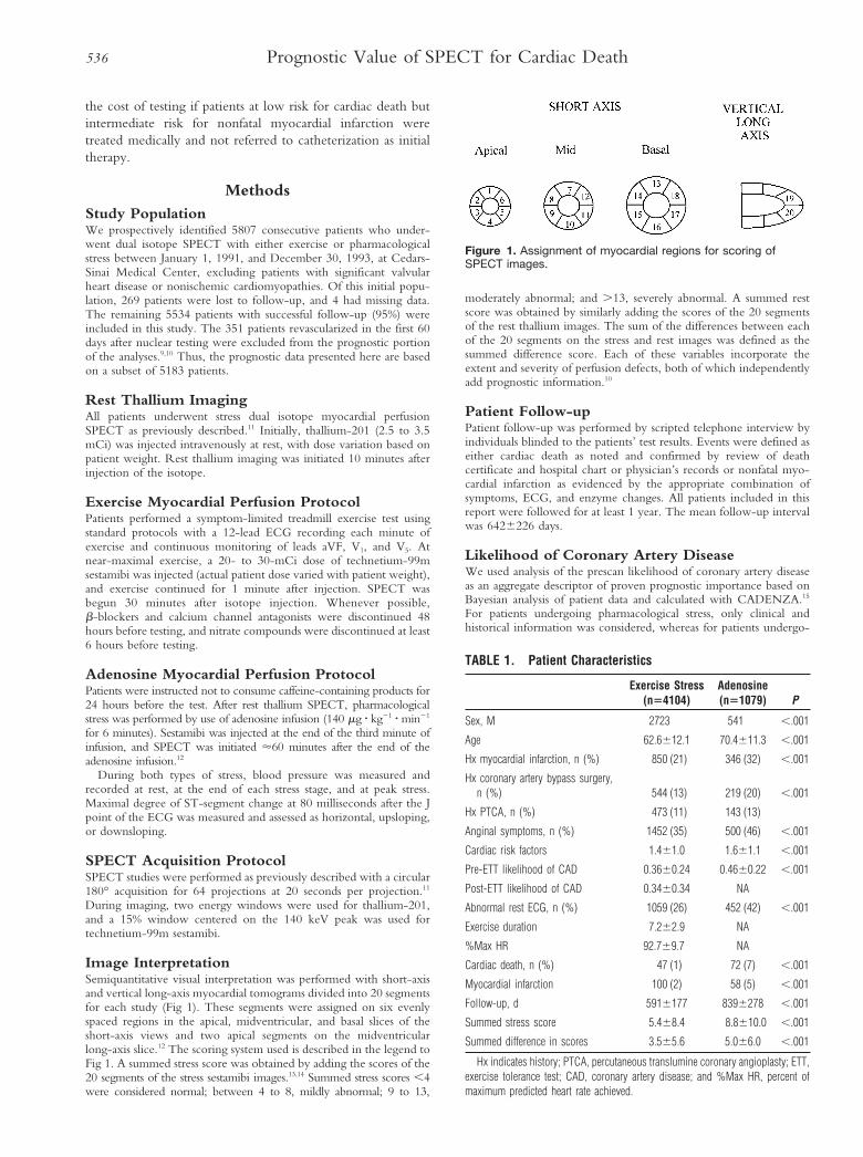
Image InterpretationSemiquantitative visual interpretation was performed with short-axisand vertical long-axis myocardial tomograms divided into 20 segmentsfor each study (Fig 1). These segments were assigned on six evenlyspaced regions in the apical, midventricular, and basal slices of theshort-axis views and two apical segments on the midventricularlong-axis slice.12 The scoring system used is described in the legend toFig 1. A summed stress score was obtained by adding the scores of the20 segments of the stress sestamibi images.13,14 Summed stress scores ,4were considered normal; between 4 to 8, mildly abnormal; 9 to 13,
moderately abnormal; and .13, severely abnormal. A summed restscore was obtained by similarly adding the scores of the 20 segmentsof the rest thallium images. The sum of the differences between eachof the 20 segments on the stress and rest images was defined as thesummed difference score. Each of these variables incorporate theextent and severity of perfusion defects, both of which independentlyadd prognostic information.10
Patient Follow-upPatient follow-up was performed by scripted telephone interview byindividuals blinded to the patients’ test results. Events were defined aseither cardiac death as noted and confirmed by review of deathcertificate and hospital chart or physician’s records or nonfatal myo-cardial infarction as evidenced by the appropriate combination ofsymptoms, ECG, and enzyme changes. All patients included in thisreport were followed for at least 1 year. The mean follow-up intervalwas 6426226 days.
Likelihood of Coronary Artery DiseaseWe used analysis of the prescan likelihood of coronary artery diseaseas an aggregate descriptor of proven prognostic importance based onBayesian analysis of patient data and calculated with CADENZA.15
For patients undergoing pharmacological stress, only clinical andhistorical information was considered, whereas for patients undergo-
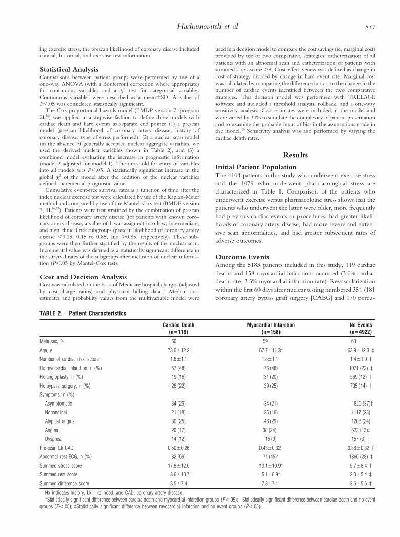
TABLE 1. Patient Characteristics
Exercise Stress(n54104)
Adenosine(n51079) P
Sex, M 2723 541 ,.001
Age 62.6612.1 70.4611.3 ,.001
Hx myocardial infarction, n (%) 850 (21) 346 (32) ,.001
Hx coronary artery bypass surgery,n (%) 544 (13) 219 (20) ,.001
Hx PTCA, n (%) 473 (11) 143 (13)
Anginal symptoms, n (%) 1452 (35) 500 (46) ,.001
Cardiac risk factors 1.461.0 1.661.1 ,.001
Pre-ETT likelihood of CAD 0.3660.24 0.4660.22 ,.001
Post-ETT likelihood of CAD 0.3460.34 NA
Abnormal rest ECG, n (%) 1059 (26) 452 (42) ,.001
Exercise duration 7.262.9 NA
%Max HR 92.769.7 NA
Cardiac death, n (%) 47 (1) 72 (7) ,.001
Myocardial infarction 100 (2) 58 (5) ,.001
Follow-up, d 5916177 8396278 ,.001
Summed stress score 5.468.4 8.8610.0 ,.001
Summed difference in scores 3.565.6 5.066.0 ,.001
Hx indicates history; PTCA, percutaneous translumine coronary angioplasty; ETT,exercise tolerance test; CAD, coronary artery disease; and %Max HR, percent ofmaximum predicted heart rate achieved.
Figure 1. Assignment of myocardial regions for scoring ofSPECT images.
536 Prognostic Value of SPECT for Cardiac Death
ing exercise stress, the prescan likelihood of coronary disease includedclinical, historical, and exercise test information.
Statistical AnalysisComparisons between patient groups were performed by use of aone-way ANOVA (with a Bonferroni correction where appropriate)for continuous variables and a x2 test for categorical variables.Continuous variables were described as a mean6SD. A value ofP,.05 was considered statistically significant.
The Cox proportional hazards model (BMDP version 7, program2L16) was applied in a stepwise fashion to define three models withcardiac death and hard events as separate end points: (1) a prescanmodel (prescan likelihood of coronary artery disease, history ofcoronary disease, type of stress performed), (2) a nuclear scan model(in the absence of generally accepted nuclear aggregate variables, weused the derived nuclear variables shown in Table 2), and (3) acombined model evaluating the increase in prognostic information(model 2 adjusted for model 1). The threshold for entry of variablesinto all models was P,.05. A statistically significant increase in theglobal x2 of the model after the addition of the nuclear variablesdefined incremental prognostic value.
Cumulative event-free survival rates as a function of time after theindex nuclear exercise test were calculated by use of the Kaplan-Meiermethod and compared by use of the Mantel-Cox test (BMDP version7, 1L16,17). Patients were first stratified by the combination of prescanlikelihood of coronary artery disease (for patients with known coro-nary artery disease, a value of 1 was assigned) into low, intermediate,and high clinical risk subgroups (prescan likelihood of coronary arterydisease ,0.15, 0.15 to 0.85, and .0.85, respectively). These sub-groups were then further stratified by the results of the nuclear scan.Incremental value was defined as a statistically significant difference inthe survival rates of the subgroups after inclusion of nuclear informa-tion (P,.05 by Mantel-Cox test).
Cost and Decision AnalysisCost was calculated on the basis of Medicare hospital charges (adjustedby cost-charge ratios) and physician billing data.18 Median costestimates and probability values from the multivariable model were
used in a decision model to compare the cost savings (ie, marginal cost)provided by use of two comparative strategies: catheterization of allpatients with an abnormal scan and catheterization of patients withsummed stress score .8. Cost-effectiveness was defined as change incost of strategy divided by change in hard event rate. Marginal costwas calculated by comparing the difference in cost to the change in thenumber of cardiac events identified between the two comparativestrategies. This decision model was performed with TREEAGEsoftware and included a threshold analysis, rollback, and a one-waysensitivity analysis. Cost estimates were included in the model andwere varied by 30% to simulate the complexity of patient presentationand to examine the probable input of bias in the assumptions made inthe model.19 Sensitivity analysis was also performed by varying thecardiac death rates.
Results
Initial Patient PopulationThe 4104 patients in this study who underwent exercise stressand the 1079 who underwent pharmacological stress arecharacterized in Table 1. Comparison of the patients whounderwent exercise versus pharmacologic stress shows that thepatients who underwent the latter were older, more frequentlyhad previous cardiac events or procedures, had greater likeli-hoods of coronary artery disease, had more severe and exten-sive scan abnormalities, and had greater subsequent rates ofadverse outcomes.
Outcome EventsAmong the 5183 patients included in this study, 119 cardiacdeaths and 158 myocardial infarctions occurred (3.0% cardiacdeath rate, 2.3% myocardial infarction rate). Revascularizationwithin the first 60 days after nuclear testing numbered 351 (181coronary artery bypass graft surgery [CABG] and 170 percu-
TABLE 2. Patient Characteristics
Cardiac Death(n5119)
Myocardial Infarction(n5158)
No Events(n54922)
Male sex, % 60 59 63
Age, y 73.6612.2 67.7611.3* 63.9612.3†‡
Number of cardiac risk factors 1.661.1 1.861.1 1.461.0†‡
Hx myocardial infarction, n (%) 57 (48) 76 (48) 1071 (22)†‡
Hx angioplasty, n (%) 19 (16) 31 (20) 569 (12)†‡
Hx bypass surgery, n (%) 26 (22) 39 (25) 705 (14)†‡
Symptoms, n (%)
Asymptomatic 34 (29) 34 (21) 1820 (37)‡
Nonanginal 21 (18) 25 (16) 1117 (23)
Atypical angina 30 (25) 46 (29) 1203 (24)
Angina 20 (17) 38 (24) 623 (13)‡
Dyspnea 14 (12) 15 (9) 157 (3)†‡
Pre-scan Lk CAD 0.5060.26 0.4360.32 0.3660.32†‡
Abnormal rest ECG, n (%) 82 (69) 71 (45)* 1366 (28)†‡
Summed stress score 17.6612.0 13.1610.9* 5.768.4†‡
Summed rest score 8.6610.7 5.168.9* 2.065.4†‡
Summed difference score 8.567.4 7.867.1 3.665.6†‡
Hx indicates history; Lk, likelihood; and CAD, coronary artery disease.*Statistically significant difference between cardiac death and myocardial infarction groups (P,.05); †Statistically significant difference between cardiac death and no event
groups (P,.05); ‡Statistically significant difference between myocardial infarction and no event groups (P,.05).
Hachamovitch et al 537
taneous transluminal coronary angioplasty [PTCA]; 6.7% earlyrevascularization rate).
Univariable AnalysisDescriptive patient characteristics and exercise and nuclearvariables in patients with or without events on follow-up arepresented in Table 2. Comparison of patients with cardiacdeath or myocardial infarction on follow-up with those with-out shows that the former were older, had more cardiac riskfactors, more frequently had an abnormal rest ECG, had agreater prescan likelihood of coronary disease, had morefrequent histories of previous myocardial infarction or revas-cularization, and more frequently experienced dyspnea as theirpresenting symptom (Table 2). In addition, patients who hadmyocardial infarction on follow-up more frequently had an-gina and less frequently were asymptomatic at the time oftesting relative to patients with no event on follow-up. Ofnote, 59% of patients who experienced cardiac death and 46%of patients who experienced myocardial infarction did notexperience anginal symptoms at the time of their nuclear test.Compared with patients who had myocardial infarction onfollow-up, those who had cardiac death were older and morefrequently had an abnormal rest ECG.
With respect to nuclear variables (Table 2), patients withevents on follow-up had more extensive and severe scanabnormalities with respect to both fixed and reversible defects.Compared with patients who had myocardial infarction onfollow-up, those who had cardiac death had more severe andextensive stress perfusion scan abnormalities caused by morenonreversible defects but similar amounts of ischemia.
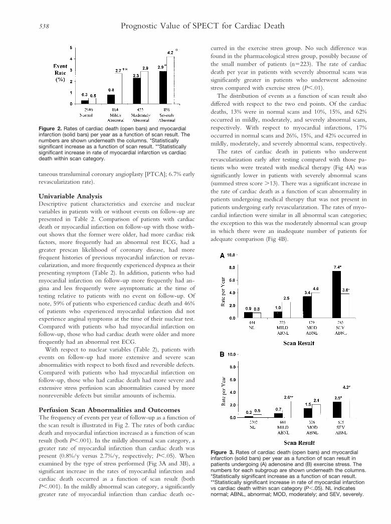
Perfusion Scan Abnormalities and OutcomesThe frequency of events per year of follow-up as a function ofthe scan result is illustrated in Fig 2. The rates of both cardiacdeath and myocardial infarction increased as a function of scanresult (both P,.001). In the mildly abnormal scan category, agreater rate of myocardial infarction than cardiac death waspresent (0.8%/y versus 2.7%/y, respectively; P,.05). Whenexamined by the type of stress performed (Fig 3A and 3B), asignificant increase in the rates of myocardial infarction andcardiac death occurred as a function of scan result (bothP,.001). In the mildly abnormal scan category, a significantlygreater rate of myocardial infarction than cardiac death oc-
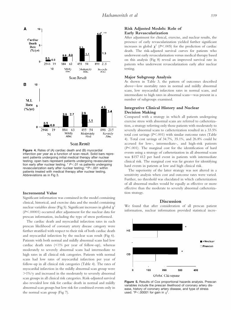
curred in the exercise stress group. No such difference wasfound in the pharmacological stress group, possibly because ofthe small number of patients (n5223). The rate of cardiacdeath per year in patients with severely abnormal scans wassignificantly greater in patients who underwent adenosinestress compared with exercise stress (P,.01).
The distribution of events as a function of scan result alsodiffered with respect to the two end points. Of the cardiacdeaths, 13% were in normal scans and 10%, 15%, and 62%occurred in mildly, moderately, and severely abnormal scans,respectively. With respect to myocardial infarctions, 17%occurred in normal scans and 26%, 15%, and 42% occurred inmildly, moderately, and severely abnormal scans, respectively.
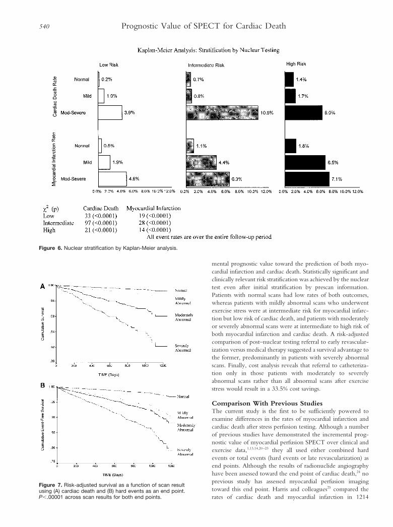
The rates of cardiac death in patients who underwentrevascularization early after testing compared with those pa-tients who were treated with medical therapy (Fig 4A) wassignificantly lower in patients with severely abnormal scans(summed stress score .13). There was a significant increase inthe rate of cardiac death as a function of scan abnormality inpatients undergoing medical therapy that was not present inpatients undergoing early revascularization. The rates of myo-cardial infarction were similar in all abnormal scan categories;the exception to this was the moderately abnormal scan groupin which there were an inadequate number of patients foradequate comparison (Fig 4B).
Figure 2. Rates of cardiac death (open bars) and myocardialinfarction (solid bars) per year as a function of scan result. Thenumbers are shown underneath the columns. *Statisticallysignificant increase as a function of scan result. **Statisticallysignificant increase in rate of myocardial infarction vs cardiacdeath within scan category.
Figure 3. Rates of cardiac death (open bars) and myocardialinfarction (solid bars) per year as a function of scan result inpatients undergoing (A) adenosine and (B) exercise stress. Thenumbers for each subgroup are shown underneath the columns.*Statistically significant increase as a function of scan result.**Statistically significant increase in rate of myocardial infarctionvs cardiac death within scan category (P,.05). NL indicatesnormal; ABNL, abnormal; MOD, moderately; and SEV, severely.
538 Prognostic Value of SPECT for Cardiac Death
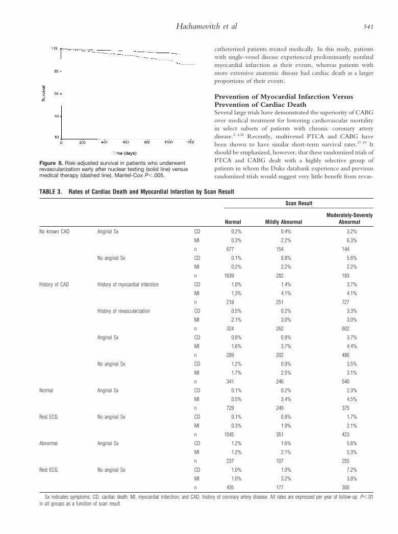
Incremental ValueSignificant information was contained in the model containingclinical, historical, and exercise data and the model containingnuclear variables alone (Fig 5). Significant increases in global x2
(P,.00001) occurred after adjustment for the nuclear data forprescan information, including the type of stress performed.
The cardiac death and myocardial infarction rates in eachprescan likelihood of coronary artery disease category werefurther stratified with respect to their risk of both cardiac deathand myocardial infarction by the nuclear scan result (Fig 6).Patients with both normal and mildly abnormal scans had lowcardiac death rates (,1% per year of follow-up), whereasmoderately to severely abnormal scans had intermediate tohigh rates in all clinical risk categories. Patients with normalscans had low rates of myocardial infarction per year offollow-up in all clinical risk categories (Table 4). The rates ofmyocardial infarction in the mildly abnormal scan group were.1%/y and increased in the moderately to severely abnormalscan groups in all clinical risk categories. Risk-adjusted survivalalso revealed low risk for cardiac death in normal and mildlyabnormal scan groups but low risk for combined events only inthe normal scan group (Fig 7).
Risk Adjusted Models: Role ofEarly RevascularizationAfter adjustment for clinical, exercise, and nuclear results, thepresence of early revascularization yielded further significantincreases in global x2 (P,.005) for the prediction of cardiacdeath. The risk-adjusted survival curves for patients whounderwent early revascularization versus medical therapy basedon this analysis (Fig 8) reveal an improved survival rate inpatients who underwent revascularization early after nucleartesting.
Major Subgroup AnalysisAs shown in Table 3, the pattern of outcomes describedabove—low mortality rates in normal and mildly abnormalscans, low myocardial infarction rates in normal scans, andintermediate to high rates in abnormal scans—was present in anumber of subgroups examined.
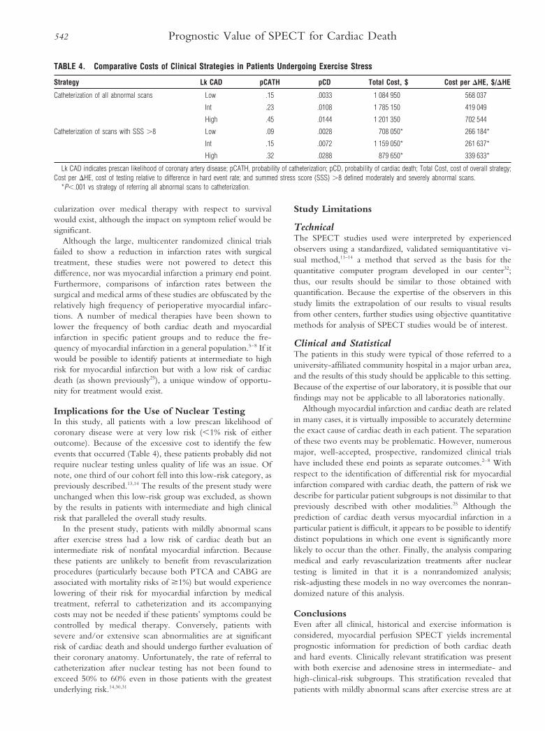
Integrative Clinical History and NuclearDecision MakingCompared with a strategy in which all patients undergoingexercise stress with abnormal scans are referred to catheteriza-tion, a strategy referring only those patients with moderately toseverely abnormal scans to catheterization resulted in a 33.5%total cost savings (P,.001) with similar outcome rates (Table4). Total cost savings of 34.7%, 35.1%, and 26.8% could beaccrued for low-, intermediate-, and high-risk patients(P,.001). The marginal cost for the identification of hardevents using a strategy of catheterization in all abnormal scanswas $157 412 per hard event in patients with intermediateclinical risk. The marginal cost was far greater for identifyinghard events in patients at low and high clinical risk.
The superiority of the latter strategy was not altered in asensitivity analysis when cost and outcome rates were varied.Further, no threshold was elucidated in which catheterizationof all abnormal studies would be equally as effective or moreeffective than the moderate to severely abnormal catheteriza-tion strategy.
DiscussionWe found that after consideration of all prescan patientinformation, nuclear information provided statistical incre-
Figure 4. Rates of (A) cardiac death and (B) myocardialinfarction per year as a function of scan result. Solid bars repre-sent patients undergoing initial medical therapy after nucleartesting; open bars represent patients undergoing revasculariza-tion early after nuclear testing. * P,.01 vs patients undergoingrevascularization early after nuclear testing; **P,.001 withinpatients treated with medical therapy after nuclear testing.Abbreviations as in Fig 3.
Figure 5. Results of Cox proportional hazards analysis. Prescanvariables include the prescan likelihood of coronary artery dis-ease, history of coronary artery disease, and type of stressused. *P,.00001 for gain in x2.
Hachamovitch et al 539
mental prognostic value toward the prediction of both myo-cardial infarction and cardiac death. Statistically significant andclinically relevant risk stratification was achieved by the nucleartest even after initial stratification by prescan information.Patients with normal scans had low rates of both outcomes,whereas patients with mildly abnormal scans who underwentexercise stress were at intermediate risk for myocardial infarc-tion but low risk of cardiac death, and patients with moderatelyor severely abnormal scans were at intermediate to high risk ofboth myocardial infarction and cardiac death. A risk-adjustedcomparison of post–nuclear testing referral to early revascular-ization versus medical therapy suggested a survival advantage tothe former, predominantly in patients with severely abnormalscans. Finally, cost analysis reveals that referral to catheteriza-tion only in those patients with moderately to severelyabnormal scans rather than all abnormal scans after exercisestress would result in a 33.5% cost savings.
Comparison With Previous StudiesThe current study is the first to be sufficiently powered toexamine differences in the rates of myocardial infarction andcardiac death after stress perfusion testing. Although a numberof previous studies have demonstrated the incremental prog-nostic value of myocardial perfusion SPECT over clinical andexercise data,1,13,14,20–23 they all used either combined hardevents or total events (hard events or late revascularization) asend points. Although the results of radionuclide angiographyhave been assessed toward the end point of cardiac death,24 noprevious study has assessed myocardial perfusion imagingtoward this end point. Harris and colleagues25 compared therates of cardiac death and myocardial infarction in 1214
Figure 6. Nuclear stratification by Kaplan-Meier analysis.
Figure 7. Risk-adjusted survival as a function of scan resultusing (A) cardiac death and (B) hard events as an end point.P,.00001 across scan results for both end points.
540 Prognostic Value of SPECT for Cardiac Death
catheterized patients treated medically. In this study, patientswith single-vessel disease experienced predominantly nonfatalmyocardial infarction as their events, whereas patients withmore extensive anatomic disease had cardiac death as a largerproportions of their events.
Prevention of Myocardial Infarction VersusPrevention of Cardiac DeathSeveral large trials have demonstrated the superiority of CABGover medical treatment for lowering cardiovascular mortalityin select subsets of patients with chronic coronary arterydisease.2–4,26 Recently, multivessel PTCA and CABG havebeen shown to have similar short-term survival rates.27–29 Itshould be emphasized, however, that these randomized trials ofPTCA and CABG dealt with a highly selective group ofpatients in whom the Duke databank experience and previousrandomized trials would suggest very little benefit from revas-
TABLE 3. Rates of Cardiac Death and Myocardial Infarction by Scan Result
Scan Result
Normal Mildly AbnormalModerately-Severely
Abnormal
No known CAD Anginal Sx CD 0.2% 0.4% 3.2%
MI 0.3% 2.2% 6.3%
n 677 154 144
No anginal Sx CD 0.1% 0.8% 5.6%
MI 0.2% 2.2% 2.2%
n 1639 282 183
History of CAD History of myocardial infarction CD 1.0% 1.4% 3.7%
MI 1.3% 4.1% 4.1%
n 218 251 727
History of revascularization CD 0.5% 0.2% 3.3%
MI 2.1% 3.0% 3.0%
n 324 262 602
Anginal Sx CD 0.8% 0.8% 3.7%
MI 1.6% 3.7% 4.4%
n 289 202 486
No anginal Sx CD 1.2% 0.9% 3.5%
MI 1.7% 2.5% 3.1%
n 341 246 540
Normal Anginal Sx CD 0.1% 0.2% 2.3%
MI 0.5% 3.4% 4.5%
n 729 249 375
Rest ECG No anginal Sx CD 0.1% 0.8% 1.7%
MI 0.3% 1.9% 2.1%
n 1545 351 423
Abnormal Anginal Sx CD 1.2% 1.6% 5.6%
MI 1.2% 2.1% 5.3%
n 237 107 255
Rest ECG No anginal Sx CD 1.0% 1.0% 7.2%
MI 1.0% 3.2% 3.8%
n 435 177 300
Sx indicates symptoms; CD, cardiac death; MI, myocardial infarction; and CAD, history of coronary artery disease. All rates are expressed per year of follow-up. P,.01in all groups as a function of scan result.
Figure 8. Risk-adjusted survival in patients who underwentrevascularization early after nuclear testing (solid line) versusmedical therapy (dashed line). Mantel-Cox P,.005.
Hachamovitch et al 541
cularization over medical therapy with respect to survivalwould exist, although the impact on symptom relief would besignificant.
Although the large, multicenter randomized clinical trialsfailed to show a reduction in infarction rates with surgicaltreatment, these studies were not powered to detect thisdifference, nor was myocardial infarction a primary end point.Furthermore, comparisons of infarction rates between thesurgical and medical arms of these studies are obfuscated by therelatively high frequency of perioperative myocardial infarc-tions. A number of medical therapies have been shown tolower the frequency of both cardiac death and myocardialinfarction in specific patient groups and to reduce the fre-quency of myocardial infarction in a general population.5–8 If itwould be possible to identify patients at intermediate to highrisk for myocardial infarction but with a low risk of cardiacdeath (as shown previously25), a unique window of opportu-nity for treatment would exist.
Implications for the Use of Nuclear TestingIn this study, all patients with a low prescan likelihood ofcoronary disease were at very low risk (,1% risk of eitheroutcome). Because of the excessive cost to identify the fewevents that occurred (Table 4), these patients probably did notrequire nuclear testing unless quality of life was an issue. Ofnote, one third of our cohort fell into this low-risk category, aspreviously described.13,14 The results of the present study wereunchanged when this low-risk group was excluded, as shownby the results in patients with intermediate and high clinicalrisk that paralleled the overall study results.
In the present study, patients with mildly abnormal scansafter exercise stress had a low risk of cardiac death but anintermediate risk of nonfatal myocardial infarction. Becausethese patients are unlikely to benefit from revascularizationprocedures (particularly because both PTCA and CABG areassociated with mortality risks of $1%) but would experiencelowering of their risk for myocardial infarction by medicaltreatment, referral to catheterization and its accompanyingcosts may not be needed if these patients’ symptoms could becontrolled by medical therapy. Conversely, patients withsevere and/or extensive scan abnormalities are at significantrisk of cardiac death and should undergo further evaluation oftheir coronary anatomy. Unfortunately, the rate of referral tocatheterization after nuclear testing has not been found toexceed 50% to 60% even in those patients with the greatestunderlying risk.14,30,31
Study Limitations
TechnicalThe SPECT studies used were interpreted by experiencedobservers using a standardized, validated semiquantitative vi-sual method,11–14 a method that served as the basis for thequantitative computer program developed in our center32;thus, our results should be similar to those obtained withquantification. Because the expertise of the observers in thisstudy limits the extrapolation of our results to visual resultsfrom other centers, further studies using objective quantitativemethods for analysis of SPECT studies would be of interest.
Clinical and StatisticalThe patients in this study were typical of those referred to auniversity-affiliated community hospital in a major urban area,and the results of this study should be applicable to this setting.Because of the expertise of our laboratory, it is possible that ourfindings may not be applicable to all laboratories nationally.
Although myocardial infarction and cardiac death are relatedin many cases, it is virtually impossible to accurately determinethe exact cause of cardiac death in each patient. The separationof these two events may be problematic. However, numerousmajor, well-accepted, prospective, randomized clinical trialshave included these end points as separate outcomes.2–8 Withrespect to the identification of differential risk for myocardialinfarction compared with cardiac death, the pattern of risk wedescribe for particular patient subgroups is not dissimilar to thatpreviously described with other modalities.25 Although theprediction of cardiac death versus myocardial infarction in aparticular patient is difficult, it appears to be possible to identifydistinct populations in which one event is significantly morelikely to occur than the other. Finally, the analysis comparingmedical and early revascularization treatments after nucleartesting is limited in that it is a nonrandomized analysis;risk-adjusting these models in no way overcomes the nonran-domized nature of this analysis.
ConclusionsEven after all clinical, historical and exercise information isconsidered, myocardial perfusion SPECT yields incrementalprognostic information for prediction of both cardiac deathand hard events. Clinically relevant stratification was presentwith both exercise and adenosine stress in intermediate- andhigh-clinical-risk subgroups. This stratification revealed thatpatients with mildly abnormal scans after exercise stress are at
TABLE 4. Comparative Costs of Clinical Strategies in Patients Undergoing Exercise Stress
Strategy Lk CAD pCATH pCD Total Cost, $ Cost per DHE, $/DHE
Catheterization of all abnormal scans Low .15 .0033 1 084 950 568 037
Int .23 .0108 1 785 150 419 049
High .45 .0144 1 201 350 702 544
Catheterization of scans with SSS .8 Low .09 .0028 708 050* 266 184*
Int .15 .0072 1 159 050* 261 637*
High .32 .0288 879 650* 339 633*
Lk CAD indicates prescan likelihood of coronary artery disease; pCATH, probability of catheterization; pCD, probability of cardiac death; Total Cost, cost of overall strategy;Cost per DHE, cost of testing relative to difference in hard event rate; and summed stress score (SSS) .8 defined moderately and severely abnormal scans.
*P,.001 vs strategy of referring all abnormal scans to catheterization.
542 Prognostic Value of SPECT for Cardiac Death

low risk for cardiac death but intermediate risk for nonfatalmyocardial infarction and thus may benefit from medicalmanagement and may not require invasive intervention.
AcknowledgmentThis work was supported in part by a grant from Dupont-Pharma.
References1. Brown KA. Prognostic value of thallium-201 myocardial perfusion
imaging: a diagnostic tool comes of age. Circulation. 1991;83:363–381.2. CASS Principle Investigators and Their Associates. Myocardial infarction
and mortality in the Coronary Artery Surgery Study randomized trial.N Engl J Med. 1984;310:750–758.
3. Alderman EL, Bourassa MG, Cohen LS, Davis KB, Kaiser GG. Killip T,Mock MB, Pettinger M, Robertson TL, for the CASS Investigators.Ten-year follow-up of survival and myocardial infarction in the ran-domized Coronary Artery Surgery Study. Circulation. 1990;82:1629–1646.
4. Detre KM, Takaro P, Hultgren H, Peduzzi P, for the Study Participants.Long term mortality and morbidity results of the Veterans Administrationrandomized trial of coronary artery bypass surgery. Circulation. 1985;72(suppl V)V-84-V-89.
5. Manson JE, Stampfer MJ, Colditz GA, Willet WC, Rosner B, Speizer FE,Hennekens CH. A prospective trial of aspirin use and primary preventionof cardiovascular disease in women. JAMA. 1991;265:521–527.
6. The Steering Committee of the Physicians’ Health Study Research Group.Preliminary report: findings from the aspirin component of the ongoingphysicians’ health study. N Engl J Med. 1988;318:262–264.
7. Scandinavian Simvastatin Survival Study Group. Randomized trial of cho-lesterol lowering in 4444 patients with coronary heart disease: the Scan-dinavian Simvastatin Survival Study (4S). Lancet. 1994;344:1383–1389.
8. Shepard J, Cobbie SM, Ford I, Ford I, Isles CG, Lorimer AR, MacfarlanePW, McKillop JH, Packard CJ, for the West of Scotland Coronary Pre-vention Study Group. Prevention of coronary heart disease with pravastatinin men with hypercholesterolemia. N Engl J Med. 1995;333:1301–1307.
9. Staniloff HM, Forrester JS, Berman DS, Swan HJC. Prediction of death,myocardial infarction, and worsening chest pain using thallium scintigraphyand exercise electrocardiography. J Nucl Med. 1986;27:1842–1848.
10. Ladenheim ML, Pollock BH, Rozanski A, Berman DS, Staniloff HM,Forrester JS, Diamond GA. Extent and severity of myocardial hypoper-fusion as predictors of prognosis in patients with suspected coronary arterydisease. J Am Coll Cardiol. 1986;7:464–471.
11. Berman DS, Kiat H, Friedman JD, Wang FP, Van Train K, Matzer L,Maddahi J, Germano G. Separate acquisition rest thallium-201/stress tech-netium 99m sestamibi dual-isotope myocardial perfusion single-photonemission computed tomography: a clinical validation study. J Am CollCardiol. 1993;22:1455–1464.
12. Berman DS, Kiat H, Van Train K, Germano G, Maddahi J, Friedman JD.Myocardial perfusion imaging with technetium-99m-sestamibi: com-parative analysis of available imaging protocols. J Nucl Med. 1994;35:681–688.
13. Berman DS, Hachamovitch R, Kiat H, Cohen I, Cabico A, Wang FP,Friedman JD, Diamond GA. Incremental prognostic value and cost impli-cations of normal exercise Tc-99m sestamibi myocardial perfusion SPECT.J Am Coll Cardiol. 1995;26:639–647.
14. Hachamovitch R, Berman DS, Kiat H, Cohen I, Cabico JA, Friedman J,Diamond GA. Exercise myocardial perfusion SPECT in patients withoutknown coronary artery disease: incremental prognostic value and impact onsubsequent patient management. Circulation. 1996;93:905–914.
15. Diamond GA, Staniloff HM, Forrester JS, Pollock BH, Swan HJC.Computer assisted diagnosis in the noninvasive evaluation of patients withsuspected coronary artery disease. J Am Coll Cardiol. 1983;1:444–455.
16. Dixon WJ, ed. BMDP Statistical Software Manual. Berkeley, Calif: Uni-versity of California Press; 1992.
17. Kaplan EL, Meier P. Nonparametric estimation from incomplete obser-vations. J Am Stat Assoc. 1958;53:457–481.
18. Mark DB, Hlatky MA, Califf RM, Naylor C, Lee K, Armstrong P, BarbashG, White H, Simoon M, Nelson C, Topol E. Cost effectiveness ofthrombolytic therapy with tissue plasminogen activator as compared withstreptokinase for acute myocardial infarction. N Engl J Med. 1995;332:1418–1424.
19. Weinstein MC, Fineberg HV. Clinical Decision Analysis. Philadelphia, Pa:WB Saunders Co; 1980:12–36.
20. Ladenheim M, Kotler T, Pollock B, Berman DS, Diamond G. Incrementalprognostic power of clinical history, exercise electrocardiography andmyocardial perfusion scintigraphy in suspected coronary artery disease.Am J Cardiol. 1987;59:270–277.
21. Pollock SG, Abbott RD, Boucher CA, Beller GA, Kaul S. Independentand incremental prognostic value of tests performed in hierarchical order toevaluate patients with suspected coronary artery disease. Circulation. 1992;85:237–248.
22. Iskandrian AS, Chae SC, Heo J, Stanberry CD, Wasserleben V, Cave V.Independent and incremental prognostic value of exercise single-photonemission computed tomographic thallium imaging in coronary arterydisease. J Am Coll Cardiol. 1993;22:665–670.
23. Hachamovitch R, Berman DS, Kiat H, Merz CNB, Cohen I, FriedmanJD, Germano G, van Train K, Diamond GA. Comparison of incrementalprognostic value, risk stratification and cost effectiveness of rest/exerciseTl-201/Tc-99m sestamibi SPECT in women and men. J Am Coll Cardiol.1996;28:34–44.
24. Lee KL, Pryor DB, Pieper KS, Harrell FE, Jr, Califf RM, Mark DB, HlatkyMA, Coleman RE, Cobb FR, Jones RH. The prognostic value of radio-nuclide angiography in medically-treated patients with coronary arterydisease. Circulation. 1990;82:1705–1717.
25. Harris PJ, Lee KL, Harrell, Jr, FE, Behar VS, Rosati RA. Outcome inmedically treated coronary artery disease. Circulation. 1980;62:718–726.
26. Yusuf S, Zucker D, Peduzzi P, Takaro T, Kennedy JW, Davis K, Killip T,Passamani E, Norris C, Mathur V, Varnausakas E, Chaimers TC. Effect ofcoronary artery bypass surgery on survival: overview of 10-year results fromrandomized trials by the Coronary Artery Bypass Surgery Trialists Collab-oration. Lancet. 1994;344:563–570.
27. King SB, Lembo NJ, Weintraub WS, Kosinski AS, Barnhart HX, KutnerMH, Alazrki NP, Guyton RA, Zhao XQ. A randomized trial comparingcoronary angioplasty with coronary bypass surgery. N Engl J Med. 1994;331:1044–1050.
28. RITA trial participants. Coronary angioplasty versus coronary artery bypasssurgery: the Randomized Intervention Treatment of Angina (RITA) trial.Lancet. 1993;341:573–580.
29. Pocock SJ, Henderson RA, Rickards AF, Rickards AF, Hampton JR, KingSB II, Hamm CW, Puel J, Hueb W, Goy JJ, Rodriguez A. Meta-analysisof randomized trials comparing coronary angioplasty with bypass surgery.Lancet. 1995;346:1184–1189.
30. Bateman TM, O’Keefe JH, Dong VM, Barnhart C, Ligon RW. Coronaryangiographic rates after stress myocardial perfusion single-photon emissioncomputed tomographic scintigraphy. J Nucl Cardiol. 1995;2:217–223.
31. Hachamovitch R, Berman DS, Kiat H, Merz CNB, Cohen I, FriedmanJD, Germano G, Van Train K, Diamond GA. Sex-related differences inclinical management after exercise nuclear testing. J Am Coll Cardiol.1995;26:1457–1464.
32. Garcia EV, Cooke D, Van Train KF, Folks R, Peifer J, DePuey G,Maddahi J, Alazraki N, Galt J, Ezquerra N, Ziffer J, Areeda J, Berman DS.Technical aspects of myocardial SPECT imaging with technetium-99msestamibi. Am J Cardiol. 1990;66:23E–31E.
Hachamovitch et al 543
Related Documents


![Cerebral Perfusion and Cerebral Autoregulation after Cardiac ...downloads.hindawi.com/journals/bmri/2018/4143636.pdftic hypothermia a er cardiac arrest []. Previously, Yenari et al.](https://static.cupdf.com/doc/110x72/60541d03139eb04f8664781d/cerebral-perfusion-and-cerebral-autoregulation-after-cardiac-tic-hypothermia.jpg)


![Alignment of Cardiac Structures in H215O Perfusion PET … · Alignment of Cardiac Structures in [15O]H 2O Perfusion ... because the tracer is freely dif- ... 5.2.2 Alignment of Cardiac](https://static.cupdf.com/doc/110x72/5afc599a7f8b9a944d8c05e0/alignment-of-cardiac-structures-in-h215o-perfusion-pet-of-cardiac-structures.jpg)


